Embed Size (px)
Citation preview

Journal of Neurosclence Methods, 24 (1988) 259-269 259 Elsevier
NSM 00830
Intraventricular microdialysis: a new method for determining monoamine metabolite concentrations in the cerebrospinal fluid of
freely moving rats
Jill B Becker, Frank Adams and Terry E Robinson
The Untverslt~, of Michigan, Department of Psycholog), Neurosctence Laborator~ Buddmg, Ann Arbor MI (U S 4 )
(Received 30 September 1987) (Revised 6 January 1988)
(Accepted 8 February 1988)
Key words Cerebrospinal fluid, Doparmne metabohte, Microdialysls, Monoamlne metabohte. Haloperi- dol, Probenecid, Adrenal medulla graft
A new method Is described to estimate the cerebrospmal fluid (CSF) concentrations of m o n o a m m e metabolltes (dlhydroxyphenvl~ aceuc acid (DOPAC) homovamlhc acid (HVA) and 5-hydroxymdoleacetlc aod (5-HIAA)) in the lateral ventricle of freely moving rats by use of in xavo rmcrodlalysxs Both the basehne concentrations of these metabohtes and the rate of dopamme (DA) turnover (estimated by the accumulation of total DA metabohtes after 200 m g / k g probenecid) were within the range reported when other methods were used to sample CSF A series of prellmanary studies were conducted to demonstrate that thas method can be used to repeatedly sample CSF, and to show that the method is sensitive to local changes in doparmnergac activity induced by lesions, drugs or grafts (1) Unilateral 6-hydroxydopanune (6-OHDA) lesions of the substantla mgra produced a sigmflcant decrease in the CSF concentrations of DOPAC and HVA lpsllateral to the lesion, relative to the contralateral side or to concentrations m animals without lesions (2) When left and nght lateral ventricles were sampled simultaneously in ammals with a unilateral 6 -OHDA lesion halopendol induced an increase in DOPAC and HVA concentrations in CSF on both sides of the brain Interestingly, the halopendol-mduced increase in CSF concentrations of DA metabohies was greater adjacent to the intact s tr latum of rats with umlateral 6 -OHDA lesions than in ammals with no lesion (3) Finally, in ammals with adrenal medulla tissue grafted into the lateral ventricle there was an increase In the CSF concentration of DOPAC compared to pregraft values or to those of ammals with control grafts
Introduction
The concentrations of ddaydroxyphenylacetlc acid (DOPAC), homovandhc acid (HVA) and 5- hydroxylndoleacetlc acid (5-HIAA) in cerebrospi- nal fluid (CSF) are frequently used as indirect indices of brain monoamme actlvaty (Nielsen and Moore. 1982, Nielsen et al, 1983, Hutson et al, 1984a, b, Mlgnot and Laude, 1985) However, our knowledge of how monoamme metabohtes an the
Correspondence J B Becker, The Umversi ty of Mlctugan, De- par tment of Psychology Neurosclence Laboratory Building, 1103 E Huron St , Ann Arbor, MI, 48104-1687, U S A
CSF relate to brain monoamlne activity is very incomplete, in part because the methods available for sampling CSF are either technically cumber- some, or have other dlsadantages For example, push-pull perfuslon has been used to repeatedly sample the lateral ventricles (Nielsen and Moore, 1982), but (1) extended push-pull experiments are techmcally difficult because of problems in maintalmng an exact balance between the inflow and outflow lines for long periods of ume, and the consequences of even brief blockage can be disas- trous, and (2) it is difficult to accurately estimate CSF concentrations of metabohtes with thas method because the CSF obtained from the "pull" cannula is greatly diluted by the effluent from the
0165-0270/88/$03 50 © 1988 Elsevier Scmnce Pubhshers B V (Biomedical Dlwslon)

260
'~push" cannula A second method revolves withdrawing CSF using a siphoning system (Sarna et al , 1983, Kornhuber et al . 1986, De La Riga and Yeo, 1985, Mignot and Laude, 1985), but frequent withdrawal of CSF without replacing fluid may be problematic because of resultant changes in mtracerebral pressure Finall2¢, the re- peated samphng of mulUple sites simultaneously is difficult w~th previous methods
In tins paper a rmcrodialysls technique is de- scribed for the repeated sampling of monoamlne metabohtes in the CSF of freely moving ammals, over extended penods of time (see Ungerstedt, 1984 for a review of mlcrodmlysls) This method has a number of advantages over previous meth- ods (1) With mlcrodlalysis no fluids are intro- duced or withdrawn from the ventricle, so changes m mtracerebral pressure are rmmrmzed (2) More than one s~te within the ventncular system of an animal can be sampled at the same time w~thout increasing the risk or discomfort to the ammal (3) The dmlysis probes can be inexpensively con- structed from readily avadable matermls, and are reusable (4) Sterlhty can be maintained because bacteria do not cross the dialysis membrane (5) Many samples can be collected m succession (6) Finally, by calculating recovery xn vitro prior to an experiment, it is possible to control for varla- bahty among probes and to obtain a relatively accurate estimate of the absolute concentrations of monoamane metabohtes tn CSF
Methods
SubJects Adult female Long-Evans rats (Charles Raver
Breeders) were housed individually with food and water freely available The hghts in the ammal colony were maintained on a 14 10 (hght dark) cycle, hghts on at 08 00 h All experiments were conducted between 12.00 and 18 00 h
Surgery For the experiments with freely moving ammals
guide cannulae constructed from 18 gauge than wall stainless steel tubing were implanted stereo- taxacally above the lateral ventricle, 1 m m ventral
from skull surface Some ammals also received d unilateral 6 -OHDA lesion ot the subbtantla nigra at this time, as described previously (Robinson et a l , 1982) For the acute experiments ammals re- ceived 6 -OHDA lesions but grade cannulae were not implanted All ammals were allowed a mini- mum of 4 weeks for recovery prior to mtraventnc- ular mlcrodlalysls
Construction of the mlcrodtalysts probe The rmcrodlalysis probe consisted of a reusable
assembly with a single-use rmcrodialysis mem- brane that was attached to the assembly pnor to each dialysis Stainless steel tubing (33 gauge 30 cm, 20 gauge 8 mm and 25 gauge tinn wall 17 mm) was cut to the lengths indicated, degreased and acid etched to roughen the outer surfaces A 75 cm piece of 0 67 m m inside diameter (i d ) rmcrollne tubing was washed in methanol and the surface gloss removed by roughening about 2 cm of one end with an emery board
The mlcrodlalysls assembly is illustrated schematically m Fig 1 and was constructed as follows A 26 gauge needle was inserted into the lumen of the rmcrohne tubing (Fig 1L) and then angled to pierce through the wall about 1 cm from the roughened end The 33 gauge tubing (Fig 1M) was threaded through the needle and the needle was withdrawn leaving the 33 gauge tubing m place The 25 gauge tubing (Fig 1H) was inserted so that exactly 10 m m of 25 gauge tubing pro- truded from the end of the mlcrohne tubing The 33 gauge tubing extended through the 25 gauge tubing and protruded exactly 35 mm Tins arrangement was then secured using 2-Ton epoxy (Fig 1G) When the epoxy had dried the 20 gauge tubing (Fig 1I) was placed over the 25 gauge tubing and epoxaed flush with the mlcrohne tub- ing Tins is the basic umt of the dialysis assembly The epoxy was allowed to cure completely before proceeding To protect the 33 gauge tubing, 0 25 mm i d nucroline tubing (Fig 1F) was threaded over the stainless steel tubing The entire assembly end of the dialysis probe was then coated with epoxy as indicated in Fig 1 by the dotted hne After the epoxy had cured a 5 mm piece of silast~c tubing (0625 m m i d ) was apphed over the epoxled end of the rmcrohne tubing (not shown)

G-
/ \
I
I I I I I I
/ / I I I
.._J
K Fig 1 Schematic diagram of the grade cannula/stylet and nucrodlalysis probe for intraventncular mlcrodlalysls (not drawn to scale) The 18 gauge (than wall) grade cannula (D) is closed with a stylet constructed from 20 gauge tubing (E) that is beveled and sealed w~th epoxy The styler xs fixed m poslt~on by 18 gauge tubing (B) crimped over the end and tygon tubing (C) to seal the connection between guide and styler The top opemng of the styler ~s sealed w~th epoxy (A) The dlalys~s probe consists of a reuseable assembly and a replaceable dialysis membrane (N) (Construction and components de- scribed m detail m the text ) Fluid enters wa rmcrohne tubing
(L) and exits via 33 gauge tubing (F)
A length of hollow fiber dmlysls tubing (poly- sulfone, 650 t~m o d , 500 I~m 1 d , MW cut off 3000) approximately 12 mm long was held under a dissecting scope and the closed tips of a pmr of No 5 Dumont forceps were inserted m one end of the fiber about 2 mm The membrane was stretched shghtly by allowing the forceps to open Then, the dialysis fiber was shpped over the 25 gauge tubing and tacked xn place w~th 5-Mm epoxy (Fig 1J) One hour later a razor blade was used to cut the dialysis membrane to a length of 4 mm (from the end of the 25 g stainless steel cannula), leaving the 33 g tubing recessed from the end of the dialysis tubing 5-Mln epoxy was mixed thoroughly and
261
allowed to cure for 3 5 mm The end surface of the dialysis tubing was coated with epoxy and strings of epoxy were drawn over the septum unul the end of the tube was closed off (Fig 1K) After this had cured 2-Ton epoxy was layered over the 5-Min epoxy on the tip of the probe, to make the epoxy seal water resistant Probes were tested the next day by gently pushing filtered water (0 2 /~m filters) through the mlcrohne tubing If llqmd emerged from the 33 gauge stainless steel tubing, the probe was viable Probes were stored m 0 47c hypochlonte m the refrigerator until needed Prior to use, the probes were thoroughly flushed w~th filtered water in order remove the glycerin that permeates the dialysis fiber
Intraventrtcular rntcrodtalvs~s An arhficlal CSF was prepared fresh daily in
boiled HPLC grade water (2% sodmm bi- carbonate, 2 mM phosphoric acid w~th 120 0 mM NaC1, 1 25 mM CaC12, 4 8 mM KC1, and 1 2 mM MgC12, pH 7 3) and was pumped through the dialysis probe at 1 0 I~l/mm using a synnge pump (CMA/100, Carnegie Medlcln-Bloanalytlcal Sys- tems) In order to esumate the CSF concentra- tions of monoarmne metabohtes, the recovery of known concentrations of these compounds was determined m wtro for each probe immediately prior to use (Ungerstedt, 1984) After it had been deterrmned that recovery was satisfactory for a given probe (15-25%) they were inserted into the lateral ventricle
Chronl~ dialysis procedure Animals were hghtly anesthetized with ether and the styler was removed from the guide cannula After the guide cannula was cleared by the insertion of a dummy cannula, the dialysis probe was inserted until the grade cannula contacted the rmcrohne tubing The probe was secured in place by the sflastic tubing, which fit snugly over the guide cannula To pre- vent the dialysis probe from being subjected to the torque created when the animal moved, the rat was fitted w~th a harness that was attached to a llqmd commutator by a flexible hollow stainless steel tether (through which the dIalysxs probe inlet line was fed) Dlalysate was collected into 200 ttl tubes mounted on the cable just above the har- ness Samples were collected over 15 mm in tubes

262
4
~" 3 0
t r I - z 2 I.iJ 0 z 0
1 i, (/) (3
A, ~" 8
i,,i.
_z
z 4 0
0. 2 0
I I I I I I 0
45 60 75 90 105 120 135 45
TIME (mm)
B I I
I I I I I I
60 75 90 105 120 135
TIME (mm)
Fig 2 Basal concentraUons (/~M) of monoanune metabohtes m CSF (corrected for recovery) deternuned from m wvo nucrodlalysa~ Samples were collected over 15 rmn intervals for 90 nun wxth a flow rate of 1 / d / n u n A average basal concentrations ol DOPAC (sohd squares), 5-HIAA (dmmonds) and HVA (open squares) in CSF (n = 12) B concentrations of DOPAC m CSF for individual
ammals contnbutmg to the means depicted m A (above)
contaJmng 15 #1 of a stablhzang solution (0 05 M HCIO 4 w~th 2 mM EDTA, 0 1 mM metablsulfite and 40 pg//~l of dflaydroxybenzylanune (DHBA) as an internal standard) ConcentraUons of mono- armne metabohtes m the dlalysate were measured by HPLC-EC (Becker et al , 1984) All values for CSF concentrations of monoarmne metabohtes were corrected for probe recovery as deterrmned m wtro At the end of the dialysis procedure, the probe was removed and the guide cannula was resealed w~th the stylet At the conclusion of the experiment, stnatal ussue was assayed for DA content by HPLC-EC (Robinson et a l , 1982), all lesions were > 95% DA depletlon
Acute dlalysts procedure Ammals were anesthetized with ethyl urethane (2000 mg/kg, i p) and dmlysls probes were inserted into the lateral ventricle using standard stereotaxac procedure The probes were cemented in place with dental acryhc After the dental acryhc had hardened, animals were removed from the stereotaxac apparatus and body temperature was ma_mtaaned at 37 °C with a Deltaphase Isothermal heating pad Prehmmary experiments indicated that mjury-xnduced release had subsided m anesthetized ammals by this ume Samples were collected (as described above) as soon as ammals were removed from the stereo- taxac instrument and body temperature was stable Animals did not recover from anesthesm following
the acute procedure, stnatal t~ssue was assayed for DA content as described above
Materials All stainless steel tubing was obtained from
Small Parts (Mlarm, FL) Macrohne tubing was obtained from Cole-Palmer Instrument (Ctucago, IL) SdasUc tubing was obtamed from Dow Corn- mg (M}dland, MI) Dialysis tubing (HIP3-20) was obtained from Armcon (Danvers, MA) Epoxy used was made by Devcon (Danvers, MA) All chemicals used were HPLC grade or reagent grade Standards for monoarmnes and metabohtes were obtained from Sigma (St Lores, MO)
Stattst~cs Data were analyzed by analysis of variance and
subsequent patrwlse comparisons using Stat View 512 + on a Macintosh Plus
Results
Experiment 1 Basal efflux of monoamme metabo- htes mto CSF
After waltmg 1 h for Injury-reduced release to subside samples were collected at 15 mm intervals As illustrated m Fig 2A, the average concentra-
O9 100 ¢/)
i-.-
n-. 5 o
0
DOPAC HVA REPEATED INTRAVENTRICULAR DIALYSIS
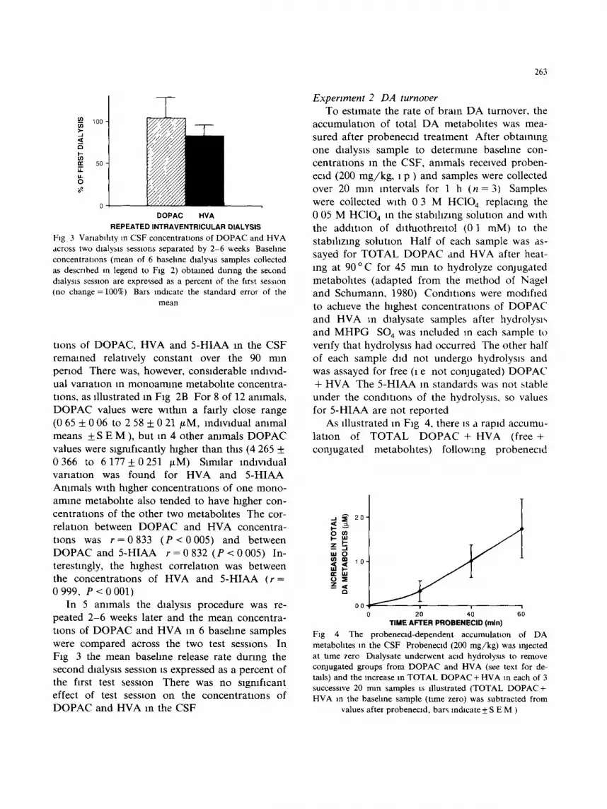
Fig 3 Varmblhty in CSF concentrations of DOPAC and HVA across two dialysis sessions separated by 2-6 weeks Baseline concentrations (mean of 6 baseline dmlysls samples collected as descnbed in legend to Fig 2) obtmned dunng the second dialysts session are expressed as a percent of the first session (no change =100%) Bars indicate the standard error of the
mean
tlons of DOPAC, HVA and 5-HIAA in the CSF remained relatively constant over the 90 nun period There was, however, considerable individ- ual variation m monoarmne metabohte concentra- tmns, as illustrated m Fig 2B For 8 of 12 ammals, DOPAC values were within a fairly close range (0 65 + 0 06 to 2 58 + 0 21 t~M, individual ammal means + S E M ), but m 4 other ammals DOPAC values were significantly tugher than thas (4 265 + 0 366 to 6 177 + 0 251 /~M) Smular lndlwdual variation was found for HVA and 5-HIAA Ammals wtth tugher concentratmns of one mono- amine metabohte also tended to have higher con- centratlons of the other two metabohtes The cor- relation between DOPAC and HVA concentra- tions was r = 0 8 3 3 ( P < 0 0 0 5 ) and between DOPAC and 5-HIAA r = 0 8 3 2 ( P < 0 0 0 5 ) In- terestingly, the highest correlatmn was between the concentratmns of HVA and 5-HIAA (r = 0999, P < 0 0 0 1 )
In 5 animals the dmlysls procedure was re- peated 2-6 weeks later and the mean concentra- tions of DOPAC and HVA in 6 basehne samples were compared across the two test sesstons In Fig 3 the mean baseline release rate dunng the second dmlysxs session is expressed as a percent of the first test session There was no slgmflcant effect of test session on the concentratxons of DOPAC and HVA in the CSF
263
Expenrnent 2 DA turnover To estimate the rate of brain DA turnover, the
accumulauon of total DA metabolltes was mea- sured after probenecld treatment After obtaining one dialysis sample to determine basehne con- centratlons in the CSF, animals received proben- ecld (200 mg/kg , 1 p ) and samples were collected over 20 nun intervals for 1 h ( n = 3) Samples were collected with 0 3 M HC104 replacing the 0 05 M HC104 in the stabtllzang solution and w~th the addition of dlthlothreltol (0 1 raM) to the stablhzlng solution Half of each sample was as- sayed for TOTAL DOPAC and HVA after heat- ing at 9 0 ° C for 45 mln to hydrolyze conjugated metabolltes (adapted from the method of Nagel and Schumann, 1980) Conditions were modified to achieve the tughest concentrations of DOPAC and HVA xn dlalysate samples after hydrolysis and M H P G SO 4 was included m each sample to verify that hydrolysis had occurred The other half of each sample did not undergo hydrolysis and was assayed for free (1 e not conjugated) DOPAC + HVA The 5-HIAA m standards was not stable under the conditions of the hydrolysis, so values for 5-HIAA are not reported
As illustrated in F~g 4, there ~s a rapid accumu- latton of T O T A L DOPAC + HVA (free + conjugated metabolites) following probenecld
i ~ 20"
, - 0
o ,5
O0 ! ! i
20 40 60 TIME AFTER PROBENECID (rain)
Fig 4 The probenecld-dependent accumulation of DA metabohtes in the CSF Probenecld (200 mg/kg) was injected at time zero Daalysate underwent acid hydrolysis to remove conjugated groups from DOPAC and HVA (see text for de- tails) and the increase m TOTAL DOPAC + HVA in each of 3 successive 20 man samples is illustrated (TOTAL DOPAC+ HVA in the basehne sample (Ume zero) was subtracted from
values after probenecld, bars Indicate ± S E M )

2 6 4
b lockade of ac l&c m e t a b o h t e efflux There was also a s lgmflcant e levat ion m the C S F concent ra - t ions of free D O P A C and H V A after p robenec ld admmls t ra t aon ( P = 0 0 0 6 ) In a d d m o n , the amoun t of T O T A L D O P A C or H V A was greater than values ob t a ined for the non -hydro lyzed sam- ples ind ica t ing tha t con juga ted D O P A C and H V A
(fJ
2
"1" 1
::L
A.
4 "
i i
O 3"
O 2 - O,.
O Q
:::L
B.
i1
0 2
z
-7 , / , 1
:3_
0
F~g 5 The effect of a unilateral 6-OHDA leston of the substantm mgra on CSF concentralaons of the monoarmne metabohtes HVA (A), DOPAC (B) and 5-HIAA (C) Each bar represents the mean+S E M from 12 ammals Samples were obtmned from the lateral ventricle of ammals with no lesion (hatched bars) and from the lateral ventricle lpsflateral to a umlateral 6-OHDA lesion (darker hatched bars) * Differs
from ammals with no lesion (P < 0 001)
are also col lected m d la lysa te from the lateral ventr icle F o r example , the concen t ra t ion ol free D O P A C was 1 50 + 0 42 # M wl'ule the concent ra - t ion of T O T A L D O P A C was 2 52 _+ 0 83 /~M ( P < 0 005)
Experiment 3 The mfluence of 6-OHDA lesions on CSF concentrations of monoamme metabohte~
Baseline concen t ra t ions of m o n o a m m e metabo- htes m the la te ra l ventricle, lpsflateral to a 6- O H D A lesion (n = 12), were s lgmficant ly lower than an an imals wi thout lesaons (n = 12, F ig 5) D O P A C concent ra taons m the la teral ventr icle ad jacen t to a D A - d e p l e t e d s t r l a tum were 79% lower and the H V A concen t ra t ions 78% lower than an cont ro l ammal s The basal concentra taons of 5 - H I A A were no t sagmficantly affected by a unda te ra l 6 - O H D A lesion (F ig 5C)
Expertrnent 4 Halopertdol-mduced increase m CSF concentrattons of DA metabohte~
Chromc dtalysts procedure Afte r a 45 nun sta- b lhza t lon per iod , C S F c onc e n t r a uons of D O P A C and H V A were measu red bxlaterally in freely mov- ing an imals Basehne concen t ra t ions (mean of two 20 nun samples) of D O P A C and H V A were slg- mf i an t ly lower on the D A - d e p l e t e d side than con- t ra la tera l to the lesion (all an imals had > 95% D A deple t ion) The percen t decrease ( les ioned side c o m p a r e d to the non- lesaoned sade) m D O P A C was 58 35___802% ( P = 0 0 0 8 4 ) and for HVA, 54 81 + 12 39% ( P < 0 001) In response to halo- p e n d o l (2 m g / k g ) the C S F concent raUons of D O P A C and H V A increased at the same ra te on bo th the les ion and in tac t sides as ind ica t ed by a s lgmficant effect of bo th side and t ime with no in te rac t ion (effect of sade F 15 = 16 01, P < 0 001, tame F15 = 3 02, P = 0 013, sxde × tame interac-
t ion F 5132=0581 , P = 0 7 1 , F ig 6) Acute chalysls procedure In ure thane-anes the-
ttzed ammal s wath uni la te ra l 6 - O H D A lesions ( > 95% D A deple t ion) the base l ine concen t ra t ions of D O P A C + H V A m the C S F were 8 2 2 + 3 9 % lower on the les ioned side than on the in tac t side There was no t a statxstlcally sagmficant a symmet ry in base l ine (or s tamulated) concen t r anons of D O P A C and H V A m the sham opera ted cont ro ls (mean di f ferent m basa l concen t ra t ions be tween

265
-t-
=L
12
10
O 8
o ¢~ 6
=E :=L 4 -
2 -
i i i I i i 0
B H1 H2 H3 H4 H5
TIME (30 rmn intervals)
Be
B H3 H4 H5
TIME (30 mln intervals)
Fig 6 Halopendol-mduced increase in CSF concentraUons of DA metabohtes CSF concentrations of DA metabolues were measured bilaterally m freely moving rats with a umlateral 6-OHDA lesion of the substantm mgra dunng basehne (B, mean of two 20
mm samples) and following haloperldol (2 m g / k g , H1-H5 samples were collected over 30 nun) Probe adjacent to intact s tnatum
closed symbols Probe adjacent to DA depleted strlatum open symbols A HVA concentration (~M) m CSF (mean + S b M ,
n = 12) B DOPAC concentraUons m CSF
the two sides 21 1 + 20 7%) and therefore the val- ues were averaged for compar i son with 6 - O H D A lesion group
As would be p red ic t ed f rom the results in freely moving ammals , D O P A C and H V A concent ra - t ions in C S F increased b i la te ra l ly fol lowing halo- per ido l t r ea tment (0 5 m g / k g ) in ammal s wi th
6 - O H D A lesions ( F 110 = 29 43, P < 0 005, F ig 7) However , there was also a s ignif icant side x t ime in terac t ion ( F 1 ~0 = 18 86, P --- 0 0015), indi- ca t ing that the ra te of the h a l o p e n d o l - l n d u c e d increase m D O P A C + H V A was greater on the no lesion side than on the lesion side (F ig 7)
Interes t ingly, m the ammals wi th 6 - O H D A le- sions, the response of the in tac t side to h a l o p e n - dol was also di f ferent f rom that of the non- le-
s ioned cont ro l ammal s ( F 116 = 5 66, P < 0 03, ma in effect, F 116 = 3 886, P = 0 066, in te rac t ion) H a l o p e n d o l e levated D O P A C + H V A concent ra - t ions on the in tac t side of an imals w~th 6 - O H D A lesions to a greater extent than m cont ro l an imals wi th no lesion ( P < 0 03, F ig 7)
Experiment 5 Basehne concentrations of mono- amine metabohtes tn the CSF of ammals before and after adrenal medulla or control grafts
In 18 freely moving rats wi th un i la te ra l 6- O H D A lesions of the subs tan t l a mgra base l ine measures of D A me taboh te s In the C S F were ob ta ined as descr ibed above Fo l lowing the d la ly-
sis p rocedure , a m m a l s received grafts of adrena l chromaff In cells or ad rena l cor tex tissue A dre na l me du l l a t issue f rom 2 donor rats of the same s t ra in (n = 7) or an equal vo lume of ad rena l cor tex t issue (n = 8) was inser ted mto the la teral ven tnc le via a chron ica l ly i m p l a n t e d cannu la a imed at the
15
=t
-e 10 +
O et O
5 =E 2.
o LESION INTACT CONTROL
6-OHDA LESION ANIMALS Fig 7 Effect of halopendol on the mean CSF concentraUon
(ktM) of D O P A C + H V A in urethane anesthetized ammals with a unilateral 6-OHDA lesion of the substanUa mgra (n = 6)
and in control ammals (n = 6) In ammals with 6-OHDA
lesions values are given for both the side ipsllateral to the lesion (Lesion) and contralateral to the lesion (Intact) Values
from the two hemispheres were pooled for the no lesion
controls (control ammals) BASAL EFFLUX, hatched bars, PEAK EFFLUX POST-HALDOL darker hatched bars * Sig- nificantly less than the concentrauons of D O P A C + HVA m
CSF from the lateral ventricle adjacent to the intact s~de • * The halopendol-mduced increase in DOPAC + HVA was
slgmficantly greater than control values on the intact side (P < 0 05)
266
z o_ I,- ,r.r I-- Z Ill O Z O O
01 [ ] DOPAC
/ • HVA
05
00 PRE-GRAFT CORTEX MEDULLA
2 MONTHS POST-GRAFT
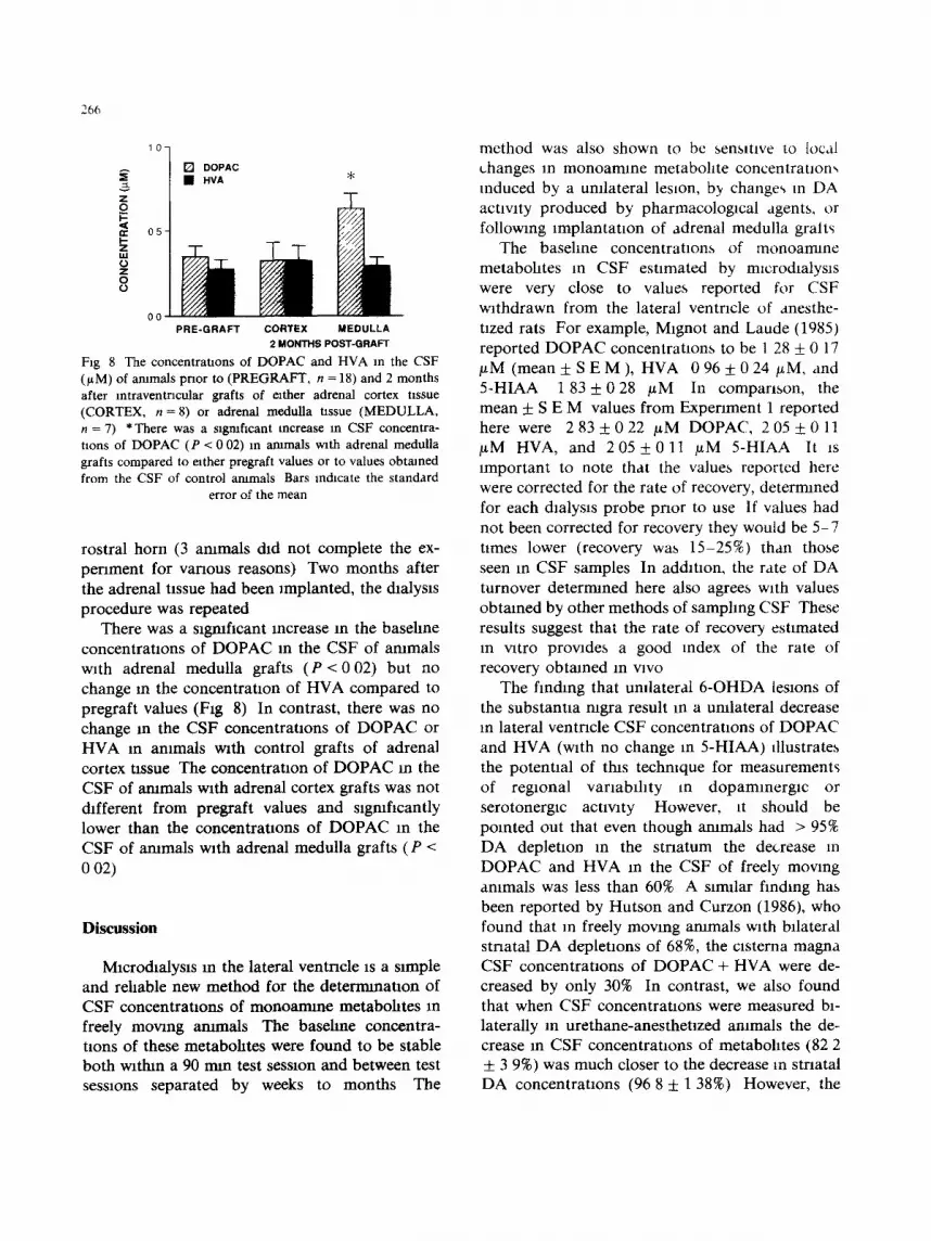
Fig 8 The concentrattons of DOPAC and HVA in the CSF (/~M) of ammals prior to (PREGKAFT, n = 18) and 2 months after lntraventrlcular grafts of either adrenal cortex nssue (CORTEX, n = 8) or adrenal medulla ttssue (MEDULLA, n = 7) * There was a slgmficant increase m CSF concentra- tions of DOPAC (P < 0 02) in ammals with adrenal medulla grafts compared to either pregraft values or to values obtained from the CSF of control ammals Bars mdtcate the standard
error of the mean
rostral horn (3 ammals &d not complete the ex- periment for various reasons) Two months after the adrenal nssue had been implanted, the dlalys~s procedure was repeated
There was a significant increase in the basehne concentrations of DOPAC m the CSF of ammals with adrenal medulla grafts (P < 002) but no change m the concentration of HVA compared to pregraft values (Ftg 8) In contrast, there was no change m the CSF concentrations of DOPAC or HVA m a m m a l s with control grafts of adrenal cortex tissue The concentration of DOPAC m the CSF of arumals with adrenal cortex grafts was not different from pregraft values and slgmficantly lower than the concentrations of DOPAC in the CSF of ammals with adrenal medulla grafts (P < 0 02)
Discussion
Mlcrodtalysls m the lateral ventncle is a s~mple and rehable new method for the determination of CSF concentrations of monoarmne metabohtes m freely moving ammals The baseline concentra- nons of these metabohtes were found to be stable both wattun a 90 mm test session and between test sessions separated by weeks to months The
method was also shown to be sensitive to local changes tn monoamme metabolite concentrattons reduced by a unilateral les~on, b2¢ changes m DA activity produced by pharmacological agents, or following implantation of adrenal medulla gralts
The baseline concentrations of monoarmne metabohtes in CSF estimated by mtcrodlalys~s were very close to values reported for CSF withdrawn from the lateral ventricle of anesthe- tized rats For example, Mlgnot and Laude (1985) reported DOPAC concentrations to be 1 28 _+ 0 17 / ~ M ( m e a n + S E M ) , HVA 0 9 6 ± 0 2 4 / ~ M , and 5-HIAA 183 ± 028 /~M In companson, the mean ± S E M values from Expenment 1 reported here were 2 8 3 ± 0 2 2 /~M DOPAC, 2 0 5 ± 0 1 1 /~M HVA, and 2 0 5 ± 0 1 1 /~M 5-HIAA It is important to note that the values reported here were corrected for the rate of recovery, deternuned for each &alysls probe prior to use If values had not been corrected for recovery they would be 5-7 times lower (recovery was 15-25%) than those seen in CSF samples In ad&tlon, the rate of DA turnover determined here also agrees with values obtained by other methods of samphng CSF These results suggest that the rate of recovery estimated in vitro provides a good index of the rate of recovery obtained in vwo
The finding that umlateral 6-OHDA lesions of the substantia nlgra result m a umlateral decrease in lateral ventricle CSF concentrations of DOPAC and HVA (with no change in 5-HIAA) illustrates the potential of this technique for measurements of regional varxablhty m dopamlnergic or serotonerglc activity However, ~t should be pointed out that even though animals had > 95% DA depletion m the stnatum the decrease in DOPAC and HVA in the CSF of freely moving animals was less than 60% A s~mllar finding has been reported by Hutson and Curzon (1986), who found that in freely mowng ammals w~th bilateral strlatal DA depletions of 68%, the Clsterna magna CSF concentrations of DOPAC + HVA were de- creased by only 30% In contrast, we also found that when CSF concentranons were measured bi- laterally m urethane-anesthetlzed ammats the de- crease in CSF concentrations of metabolates (82 2 + 3 9%) was much closer to the decrease in stnatal DA concentrations (96 8 ± 1 38%) However, the

strlatal DA depletion was still s~gnlficantly greater (P < 0 01) than the depletion in the CSF con- centrations of DA metabolites
There are a number of factors that could account for the difference in the effect of 6-OHDA lesions on striatal DA concentrations vs CSF con- centratlons of DA metabohtes The relatively l'ngh concentrations of metabohtes in CSF could reflect the contribution of extrastraatal DA neurons to metabohtes In the CSF, as suggested by Hutson and Curzon (1986) This Idea is not supported, however, by prehrmnary results from this lab In a subset of the animals that contributed to this report, we measured the concentration of DA in the nucleus accumbens plus olfactory tubercle (ACC + TUB) in addluon to strlatum There was not a significant correlation between the percent difference in the concentrauons of DA metabo- htes in the CSF ( ( in t ac t - leslon)/intact × 100%) and the percent difference m stnatal (r = 0 411) or ACC + TUB (r = 0 445) DA concentrations For example, In one animal with a 99 8% DA deple- tion in strlatum and a 93 9% depletion in ACC + TUB, the CSF concentration of DOPAC + HVA on the lesioned side was only decreased by 35 2% compared to the intact side In contrast, the animal with the greatest decrease in CSF concentrations of DOPAC + HVA (81 5%) had a 96 2% DA de- pletion in striatum (the smallest lesion in this group of animals) and only a 42 4% depletion in ACC + TUB Therefore. CSF concentrations of DA metabohtes in the lateral ventricle do not s~mply reflect the tissue concentrations of D A m adjacent brain regions, but further investigation is required to determine their orlgm
Another possible explanation for the relatively high concentrations of dopamme metabohtes in CSF ~psllateral to a 6-OHDA lesion is that there is increased activity in the rema~mng DA neurons on the lesioned side This idea is supported by the results of a related mlcrodlalysls study Animals with unilateral 6-OHDA lesions have relatively normal concentrations of DA in stnatal extracell- ular fluid on the lesioned side, even when that side is depleted up to 99% (Robinson and Whlshaw, 1988) The normahzatlon of extracellular DA in strIatum following recovery from a lesion is prob- ably due to a number of compensatory responses,
267
including an increase in DA synthesis and release from the remaining terminals, and the loss of DA reuptake sites (Hefti et al , 1985, Robinson and Whlshaw, 1988, Zlgmond et al, 1986) It is possi- ble, therefore, that the concentrations of DA metabohtes in the CSF reflect in part this com- pensatory increase m the activity of the remaining DA neurons in the strlatum
In urethane-anesthetlzed animals the haloperl- dol induced increase in DA metabohtes was potentiated in the ventricle contralateral to a 6- O H D A lesion, compared to the response m sham-operated control animals This suggests that a unilateral 6-OHDA lesion alters dopamlnerglc activity in the striatum both on the side con- trilateral to the lesion as well as the lesioned side (Fig 7) The idea that there is increased dopanunerglc activity in the striatum contralateral to a 6-OHDA lesion is generally consistent with the concept of reciprocal regulation of the two nlgrostriatal DA systems and the observation that a nlgral lesion results in increased DA release from the contralateral stnatum (Nieoullon et al, 1977, Robinson and Whlshaw, 1988, Zetterstrom et al, 1986) This interpretation is also consistent with reports that unilateral damage to the nxgrostrlatal DA system results In a persistent decrease in the spontaneous activity of strlatal cells on the side contralateral to the lesion (Garcia-Rlll et al, 1980, Hull et al, 1974), and with behavioral evidence for a small down-regu- lation of DA receptors on that side (Costall et al, 1983)
Finally, in animals with adrenal medulla grafts. there was an increase in the basehne concentra- tions of DOPAC compared to either pre-graft or control graft concentrations of DOPAC whereas there was no change in the baseline concentration of HVA These findings illustrate that wtth this method, concentrations of monoarmne metabo- htes in the CSF are stable over time following control manipulations In ad&tIon selecuve changes in CSF concentrations of these metabo- lltes can be detected following Implantation of a catecholamlne secreting tissue Interestingly, m animals with adrenal medulla grafts, DA was not detected in the CSF (noreplnephrlne and epi- nephrine were also not detectable) These results
268
wtll be reported m greater detail elsewhere (Becker and Freed, 1988)
In conclusion, the use of mlcrodmlys~s to mea- sure CSF concentraUons of monoarmne metabo- htes m freely moving rats provides a valuable new technique for explonng the relations between brain actw~ty and the neurochermcal constituents of the CSF This method allows the CSF to be continu- ously sampled without the introduction or withdrawal of fluids, and tt can be used to easily sample muluple regaonal sites simultaneously
Acknowledgements
We would hke to thank CJ Moore, K M Bentgen and J -H Cha for their technical assis- tance This research was supported by a grant from the NIH to J B B (NS22157) The develop- ment of the dmlysls system was supported m part by grants from the H H Rackham Graduate School and the Scottish Rate to TER We thank Merrell-Dow Pharmaceuticals for the gift of the deslrmpramme Dr Becker is supported by Re- search Career Development Award NS01056 Dr Robinson is supported by Research Career Devel- opment Award NS00844
References
Becker, J B and Freed, W J (1988) Neurochemacal correlates of behaworal changes following mtraventncular adrenal medulla grafts mtravenmcular dialysis In freely moving rats, Prog Brain Res, m press
Becket, J B, Casta~aeda, E, Robinson, T E and Beer, M E (1984) A simple in vatro techmque to measure the release of endogenous dopamme and dthydroxyphenylacetac acid from stnatal tissue using bagh performance hquld chromatogra- phy wlth electrochemical detection, J Neuroso Methods. 11 19-28
Costall, B, Kelly. M E and Naylor, R J (1983) Does con- tralateral circling revolve action of drugs on hyposensmve stnatal dopamane receptors in the hemisphere contralateral to denervauon 9, Neuropharmacology, 22 295-302
De La Rava, C F and Yeo, J A G (1985) Repeated determina- tion of cerebrospmal fltud amine metabohtes by automated direct sampling from an Implanted cannuta m freely mov- ing rats, J Neurosc Methods, 14 233-240
Garcta-Rall, E, Hull, C D, Cherubim, E, Levme, M S and Buchwald, N A (1980) The spontaneous hnng patterns of
forebram neurons, V Time course of change~ m caudatt umt actwlty following dopamme-depletmg lesions Brain Res 190 415-424
Heftl F Enz A and Melamed, E (1985) Partial lesions m the mgrostnatal pathway m the rat Acceleratxon of transmitter synthesis and release of surviving doparmnergi~ neurones by drugs Neuropharmacology 24 19-23
Hull, C D Levme. M S Buchwald, N A, Heller A and Brow- rag, R A (1974) The spontaneous fmng pattern of fore- brain neurons I The effects of dopamme and non-dopa- mane depleting lesions on caudate umt firing patterns Braan Res, 73 241-262
Hutson, P H and Curzon, G (1986) Dopanune metabohtes m rat clsternal cerebrospmal fluid major contribution from extrastnatal dopamme neurone~, J Neurochem 46 186-190
Hutson. P H, Sarna, G S, Kantamanenl, B D. and ( urzon G (1984a) Concurrent deterrmnatlon of brain dopamme and 5-hydroxytryptarmne turnovers m individual freely moving rats using repeated samphng of cerebrospmal fired J Neu- rochem, 43 151-159
Hutson, P H , Sarna, G S and Curzon. G (1984b) Determina- tion of daily variations of brain 5-hydroxytryptarmne and dopamlne turnovers and of the clearance of their ao&c metabohtes in conscious rats by repeated ~amphng of cerebrospinal fired, J Neurochem, 43 291-293
Kornhuber, M E, Kornhuber, J and Ctmmak, U 0986) A method for repeated CSF samphng m the freely moving rat J Neuroso Methods, 17 63-68
Mlgnot E and Laude, D (1985) Study of dopamme turno,,er by momtormg the decline of dopamlne metabohtes in rat CSF after alpha-methyl-para-tyrosme, J Neurochem 45 1527-1533
Nagel M and Schumann, H -J (1980) A senslUve method for detern'unauon of conjugated cetacholarmnes m blood plasma, J Chn Chem Chn Blochem 18 431-432
Nielsen J A and Moore, K E (1982) Effects of chloral hy- drate, gamma-butyrolactone, and eqmthesm on the efflux of dopannne and 5-hydroxytryptamme metabohtes into cerebroventncular perfusates of rats J Neurochem, 39 235-238
N~elsen, J A and Moore, K E (1983) 6-Hydroxydopannne and 5,7-dihydroxytrvptarmne metabohtes in cerebroventncular perfusates of rats, Pharmacol Blochem Behav 19 905-907
Nielsen, J A, Chapln, D S and Moore, K E (1983) Differen- tial effects of d-amphetarmne, fl-phenylethylarmne, cocaine and methylphemdate on the rate of doparmne synthesis m ternunals of mgrostnatal and mesohmblc neurons and on the efflux of dopamme metabohtes into cerebrovenmcular perfusates of rats, Life Scl. 33 1899-1907
Nieoullon. A, Cheramy. A and Glowmsto, J (1977) Interde- pendence of the mgrostnatal dopammerglc systems on the two sides of the brain m the cat, Science. 198 416-418
Robmson, T E and Whlshaw, I Q (1988) Normalization of extracellular dopamme m strlatum following recovery from a partial umlateral 6-OHDA lesmn of the substantm mgra

a microdlalys~s study m freely moving rats Brain Res In Press
Robinson T E , Becker J B and Presty, S K (1982) Long-term facdltanon of amphetalmne-lnduced rotational behavior and stnatal dopalmne release produced by a single ex- posure to amphetamine sex differences, Brain Res 253 231-241
Sarna G S, Hutson, P H Tncklebank, M D and Curzon G (1983) Determinat ion of brain 5 -hydroxyt ryp tamme turnover m freely mowng rats using repeated samphng of cerebrospmal fired J Neurochem 40 383-388
Ungerstedt, U (1984) Measurement of neurotransmatter re- lease by lntracramal dialysis In C A Marsden (Ed), Mea-
269
surement or Neurotransrmtter Release In Vivo, Wile,/ N e t York pp 81-105
Zetterstrom T , Herrera-Marsctutz M and Ungerstedt, U (1986) Simultaneous measurement of doparmne release and rotational behavior m 6-hvdroxydopamme denervated rats using mtracerebral dialysxs Brain Res 376 1 7
Zlgrnond, M J Stachowlak, M K , Berger, T W and Stncker E M (1986) Neurochenucal events underlying continued function despite injury to monoamlnerglc systems In G M Glhd, A G o n o and G W Kreutzberg (Eds) , Processes of Recovery from Neural Trauma, Exp Brain Res , Suppl 13 119-128


