Embed Size (px)
Citation preview
ARTICLE IN PRESS
http://www.elsevier.com/locate/biophyschem
DTD 5
Biophysical Chemistry
Interaction of piroxicam with micelles: Effect of hydrophobic
chain length on structural switchover
Hirak Chakraborty, Munna Sarkar*
Chemical Sciences Division, Saha Institute of Nuclear Physics, 1/AF, Bidhannagar, Calcutta-700 064, India
Received 22 March 2005; received in revised form 28 April 2005; accepted 28 April 2005
Abstract
Molecules, whose pKa values can be easily fine-tuned by their microenvironment, are expected to be profoundly affected by the
heterogeneous environments of membranes. Membrane parameters can have a strong effect in choosing a particular structural form of a
molecule for incorporation/interaction. A case study has been presented for piroxicam, a non-steroidal anti-inflammatory drug of oxicam
group, whose targets are cyclooxygenases, which are membrane active proteins. The structural dynamism of piroxicam is reflected in the ease
with which it can switchover or convert from one prototropic form to the other guided by its environment. In this work we have studied the
effect of varying hydrophobic chain length and surface charges in fine-tuning the interaction of piroxicam with micelles. Interaction of
piroxicam with three types of micelles with identical negatively charged head groups and varying tail lengths viz., sodium dodecyl sulfate
(S12S), sodium decyl sulfate (S10S) and sodium octyl sulfate (S8S) shows that there is a shift in the apparent pKa in the direction that favors
the switchover or conversion from the anionic form to the global neutral form. The binding constants of piroxicam with three micelles show a
linear dependence on chain length. Interaction was also studied with micelles having oppositely charged head groups and different chain
lengths viz., dodecyl N,N,N-trimethyl ammonium bromide (DTAB) and cetyl N,N,N-trimethyl ammonium bromide (CTAB). For micelles
having identical chain lengths but oppositely charged head groups viz., S12S and DTAB, pKa shifts in two opposite directions compared to
that in the absence of any surfactant. This is expected when electrostatic force is the only driving force. This case study demonstrates the
effect of hydrophobic chain length and surface charges in fine-tuning the equilibrium between different structural forms of piroxicam. Our
results also imply that for structurally dynamic drugs like piroxicam the nature of the biomembranes, characterized by different membrane
parameters, should play a crucial role in choosing a particular structural form of the drug that will be finally presented to their targets.
D 2005 Elsevier B.V. All rights reserved.
Keywords: Piroxicam; Structural dynamism; Membrane parameter; Micellar chain length; Change in pKa; Optical spectroscopy
1. Introduction
Interaction of molecules, whose pKa values can be easily
altered, is fine-tuned by the parameters of their micro-
environment [1–5]. Modulation of pKa values of a
chromophore by sol–gel entrapment has been already
demonstrated. The change in the microenvironment
achieved in the entrapped sol–gel matrix has been
implicated to be the reason behind the modulation of pKa
[2]. Piroxicam [4-hydroxy-2-methyl-N-(pyridin-2-yl)-2H-
1,2-benzothiazine-3-carboxamide-1,1-dioxide], a drug
0301-4622/$ - see front matter D 2005 Elsevier B.V. All rights reserved.
doi:10.1016/j.bpc.2005.04.016
* Corresponding author. Fax: +91 33 23374637.
E-mail address: [email protected] (M. Sarkar).
belonging to the Non-Steroidal Anti-Inflammatory Drugs
(NSAIDs) is a structurally dynamic molecule whose pKa
can be easily altered. Piroxicam is capable of existing in
several prototropic forms like the neutral, zwitterionic and
anionic forms in solution. The structural dynamism of
piroxicam is reflected in the ease with which it can
switchover or convert from one prototropic form to the
other [6–11]. The neutral and zwitterionic forms are
spectroscopically indistinguishable and are together termed
as Fglobal neutral_. The anionic form has spectral properties
that are quite distinct from that of the global neutral form
and hence spectroscopic techniques can be used to detect
them in solution. Target proteins of piroxicam viz., the
cyclooxygenases are membrane active proteins [12]. Before
117 (2005) 71 – 77
BIOCHE-04615; No of Pages 7

ARTICLE IN PRESSH. Chakraborty, M. Sarkar / Biophysical Chemistry 117 (2005) 71–7772
approaching the target proteins piroxicam needs to interact
with the membranes. It is therefore interesting to see how
different membrane parameters like surface charges, hydro-
phobic effect, length of the fatty acid chain, etc. would
affect the structures of piroxicam. Micelles with their
hydrophilic surface and hydrophobic interior serve as
simple membrane mimetic system that allows a controlled
study of the effect of different membrane parameters on the
structural dynamism of ligand molecules. In our earlier
studies [7] with oxicam NSAIDs we have shown how
surface charge of the micelles guides which prototropic
forms of the drugs would be incorporated in the micelles. In
this type of incorporation not only a particular prototropic
form of piroxicam will be selectively incorporated, but also
a switchover of other prototropic forms of piroxicam occur
to the one selected for incorporation. The equilibrium of this
switchover has been found to be extremely sensitive to
surface charge such that, even small modification of the
surface charge can fine-tune this equilibrium [8]. The
switchover between different prototropic forms could be
explained in terms of shift in apparent pKa values for
differently charged micelles. What could not be explained
was why for two oppositely charged micelles the shift in
pKa values occurred in the same direction compared to that
in absence of micelles [7]. This also indicated that factors
other than surface charges, like hydrophobic effect/varying
chain length might fine-tune the switchover equilibrium.
In this work, we have tried to parse the effect of varying
hydrophobic chain length from that of electrostatic effect of
micellar surface charges on the switchover equilibrium.
Three types of micelles with identical negatively charged
NCH3
SO O
O N
H
O
N
H
NCH3
SO O
O
H
N
O
H
N
_
+
Open conformer of
piroxicam
Zwitterionic form ofpiroxicam
Fig. 1. Structure of different proto
head groups and varying tail length viz., sodium dodecyl
sulfate (S12S), sodium decyl sulfate (S10S) and sodium
octyl sulfate (S8S), as well as two types of micelles with
identical positively charged head group but different chain
length viz., cetyl N,N,N-trimethyl ammonium bromide
(CTAB) and dodecyl N,N,N-trimethyl ammonium bromide
(DTAB) were chosen for this study. Binding of piroxicam
with sodium alkyl series (SNS) was studied using UV–VIS
absorption and fluorescence spectroscopy. Our study clearly
demonstrates the influence of chain length of micelles (that
modulate the hydrophobic interior) on the structural switch-
over of piroxicam leaving apart the effect of surface charge.
2. Materials and methods
Piroxicam was purchased from Sigma Chemicals (USA)
and was used without further purification. Dodecyl N,N,N-
trimethyl ammonium bromide (DTAB), cetyl N,N,N-
trimethyl ammonium bromide (CTAB) and sodium dodecyl
sulfate (S12S) were purchased from Merck, Germany.
Sodium octyl sulfate (S8S) and sodium decyl sulfate
(S10S) were purchased from Fluka. Water was glass
distilled thrice before use. Stock solutions of piroxicam of
concentration 0.5 mM were prepared in ethanol (Merck,
Germany) and exact concentration was adjusted by triple
distilled water. Each aqueous solution contains a maximum
of 6% (v/v) of ethanol. We have changed the pH of the
working solutions by adding dil. HCl and/or dil. NaOH to
them. The volume of acid (HCl) added to the working
solutions is exactly equal to the volume of acid that is
NCH3
SO O
O
N
O
H
N
H....
NCH3
SO O
O
N
O
H
N
_
Closed conformer of
piroxicam
Anionic form of piroxicam
tropic forms of piroxicam.

ARTICLE IN PRESS
250 300 350 400 450
0.1
0.2
0.3
0.4
0.5
0.6
0.7
0.8
Increasing conc. of S12S
O.D
Wavelength (nm)
250 300 350 400 4500.0
0.1
0.2
0.3
0.4
0.5
0.6
0.7
0.8
Increasing conc.of S10S
O.D
(a)
(b)
H. Chakraborty, M. Sarkar / Biophysical Chemistry 117 (2005) 71–77 73
needed to acidify a volume of water equal to the working
solution to attain that particular pH. Similar procedure was
carried out to make the solutions alkaline by adding NaOH.
It should be mentioned that pH of the solution was also
checked in the presence of highest concentration of
micelles. However, the changes observed were well within
the experimental error limit. Solution at pH 5.5 indicates
that no acid or alkali was added to the aqueous solutions.
Samples were checked for photochemical changes during
spectral scan time and no change was found.
We kept the surfactant concentration well above the
CMC value during the determination of pKa to ensure the
presence of adequate number of micelles in the solution, so
as to keep micellar effect uniform at every pH. The
concentration of piroxicam was kept constant at 30 AMfor all samples.
Absorption spectra were recorded with Thermo Spec-
tronic spectrophotometer model UNICAM UV500. Baseline
correction was done with water before recording each set of
data. Fluorescence measurements were performed using
Hitachi spectrofluorimeter model 4010. All emission spectra
were corrected for instrument response at each wavelength.
A 2�10-mm path length quartz cell was used for all
fluorescence measurements to avoid any blue edge dis-
tortion of the spectrum due to inner filter effect [13]. All
measurements were done with freshly prepared samples and
at a constant temperature of 298 K.
Wavelength (nm)
250 300 350 400 4500.0
0.1
0.2
0.3
0.4
0.5
0.6
0.7
0.8
Increasing conc.of S8S
O.D
Wavelength (nm)
(c)
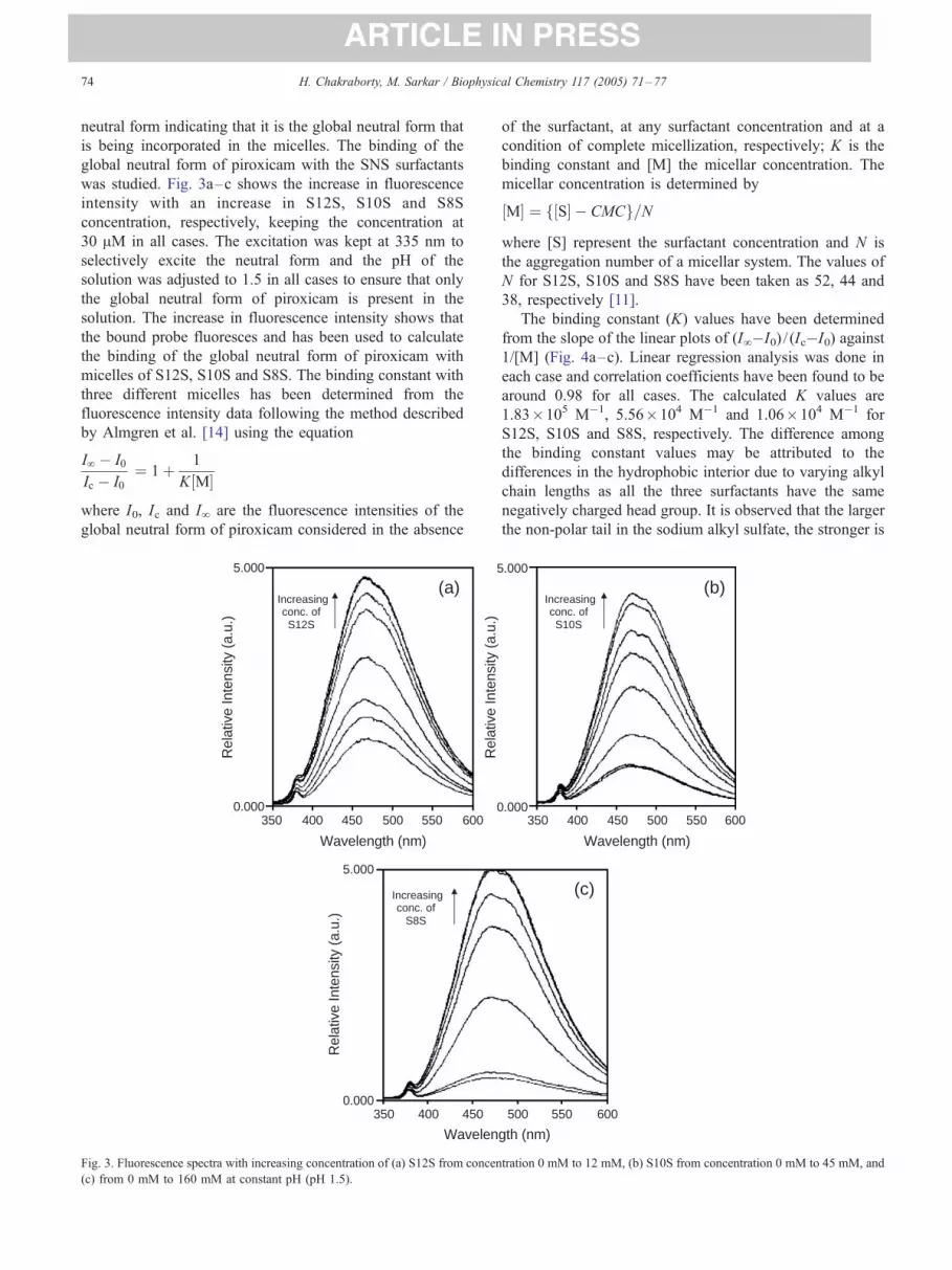
Fig. 2. Absorption spectra of piroxicam with increasing concentration of (a)
S12S from concentration 0 mM to 12 mM at pH 2.42, (b) S10S from
concentration 0 mM to 45 mM at pH 2.05, and (c) S8S from concentration
0 mM to 160 mM at pH 1.75.
3. Results and discussion
Piroxicam can exist in different structural forms (Fig. 1).
The neutral and the zwitterionic forms are spectroscopically
indistinguishable and henceforth will be together termed as
global neutral form. This molecule is structurally dynamic
and can easily switchover or convert from global neutral to
anionic form and vice versa, depending on its immediate
environment [7,8,11].
The equilibrium between the global neutral and the
anionic form of piroxicam is affected by the environment in
solution and is represented by
N–A
where FN_ represents the global neutral form and FA_represents the anionic form of piroxicam.
Fig. 2 shows the absorption spectrum of piroxicam with
increase in the concentration of S12S (Fig. 2a), S10S (Fig. 2b)
and S8S (Fig. 2c) at pH 2.42, 2.05 and 1.75, respectively.
As will be shown later the apparent pKa values in the
presence of S8S, S10S and S12S are different. For this
reason, the pH of (a)–(c) were adjusted at different values
so as to be able to demonstrate the inter conversion between
only two structural forms. It should be mentioned that this
data has not been used for binding calculation. From Fig. 2
it is evident that increase in concentration of an SNS
surfactant (N =number of –CH2 group in the hydrophobic
tail) leads to an increase in the peak of the neutral form (335
nm) with the concomitant decrease in the peak of the
anionic form (363 nm). A clear isosbestic point around 352
nm is seen in all the three figures (Fig. 2a–c). The presence
of this isosbestic point clearly points out that there exists an
equilibrium between only two species, viz., global neutral
and the anion. Increase in SNS concentration leads to a
decrease in anionic form with a concomitant increase of the
ARTICLE IN PRESSH. Chakraborty, M. Sarkar / Biophysical Chemistry 117 (2005) 71–7774
neutral form indicating that it is the global neutral form that
is being incorporated in the micelles. The binding of the
global neutral form of piroxicam with the SNS surfactants
was studied. Fig. 3a–c shows the increase in fluorescence
intensity with an increase in S12S, S10S and S8S
concentration, respectively, keeping the concentration at
30 AM in all cases. The excitation was kept at 335 nm to
selectively excite the neutral form and the pH of the
solution was adjusted to 1.5 in all cases to ensure that only
the global neutral form of piroxicam is present in the
solution. The increase in fluorescence intensity shows that
the bound probe fluoresces and has been used to calculate
the binding of the global neutral form of piroxicam with
micelles of S12S, S10S and S8S. The binding constant with
three different micelles has been determined from the
fluorescence intensity data following the method described
by Almgren et al. [14] using the equation
IV � I0
Ic � I0¼ 1þ 1
K M½ �
where I0, Ic and IV are the fluorescence intensities of the
global neutral form of piroxicam considered in the absence
(a) Increasingconc. of S12S
Increasingconc. of
S8S
5.000
0.000350 400 450 500 550 600
350 400 450
5.000
0.000
5
0
Wavelength (nm)
Wavelen
Rel
ativ
e In
tens
ity (
a.u.
)
Rel
ativ
e In
tens
ity (
a.u.
)
Rel
ativ
e In
tens
ity (
a.u.
)
Fig. 3. Fluorescence spectra with increasing concentration of (a) S12S from concen
(c) from 0 mM to 160 mM at constant pH (pH 1.5).
of the surfactant, at any surfactant concentration and at a
condition of complete micellization, respectively; K is the
binding constant and [M] the micellar concentration. The
micellar concentration is determined by
M½ � ¼ S½ � � CMCf g=N
where [S] represent the surfactant concentration and N is
the aggregation number of a micellar system. The values of
N for S12S, S10S and S8S have been taken as 52, 44 and
38, respectively [11].
The binding constant (K) values have been determined
from the slope of the linear plots of (IV�I0) / (Ic�I0) against
1/[M] (Fig. 4a–c). Linear regression analysis was done in
each case and correlation coefficients have been found to be
around 0.98 for all cases. The calculated K values are
1.83�105 M�1, 5.56�104 M�1 and 1.06�104 M�1 for
S12S, S10S and S8S, respectively. The difference among
the binding constant values may be attributed to the
differences in the hydrophobic interior due to varying alkyl
chain lengths as all the three surfactants have the same
negatively charged head group. It is observed that the larger
the non-polar tail in the sodium alkyl sulfate, the stronger is
(b)
(c)
Increasingconc. of S10S
350 400 450 500 550 600
500 550 600
.000
.000
Wavelength (nm)
gth (nm)
tration 0 mM to 12 mM, (b) S10S from concentration 0 mM to 45 mM, and

ARTICLE IN PRESS
2.0x10⁴ 4.0x10⁴ 6.0x10⁴ 8.0x10⁴ 1.0x1050.7
0.8
0.9
1.0
1.1
1.2
1.3
1.4
1.5
0.7
0.8
0.9
1.0
1.1
1.2
1.3
1.4
1.5
r=0.98
(I∞-I
0)/(
I c-I
0)(I
∞-I
0)/(
I c-I
0)(I
∞-I
0)/(
I c-I
0)
[M]-1 (lit.mol-1)
[M]-1 (lit.mol-1)
[M]-1 (lit.mol-1)
0.00 7.50x10³ 1.50x10⁴ 2.25x10⁴ 3.00x10⁴
1.6
0.7
0.8
0.9
1.0
1.1
1.2
1.3
1.4
1.5
1.6
r=0.98
0 1x10³ 2x10³ 3x10³ 4x10³ 5x10³ 6x10³ 7x10³
r=0.97
(a)
(b)
(c)
Fig. 4. Plot of (IV�I0) / (Ic�I0) vs. [M]�1 (where [M] represents the
micellar concentration) for (a) S12S, (b) S10S, and (c) S8S, at pH 1.5 where
r is the correlation coefficient.
8 9 10 11 12
4.0
4.2
4.4
4.6
4.8
5.0
5.2
5.4r=0.99
logK
Chain length
Fig. 5. Profile of log K vs. chain length for the SNS series, where r is the
correlation coefficient.
H. Chakraborty, M. Sarkar / Biophysical Chemistry 117 (2005) 71–77 75
the binding. The log K vs. carbon number (n) for the sodium
alkyl sulfates is shown in Fig. 5. The correlation is fairly
linear and fits the equation
log K ¼ 1:58þ 0:309n
This shows that there exists a direct correlation between
complexation and that of the micellar core. The value of the
intercept (1.58) refers to the log K at zero carbon number of
the hydrophobic tail of the surfactant, i.e., when the
interaction with only the head group is considered.
Fig. 2a–c also indicates that the anionic form of
piroxicam (A) is being converted to the neutral form with
an increase in concentration of SNS surfactant. The
switchover or the conversion of one prototropic form to
the other occurs depending on the nature of the micelles. In
our earlier studies we have shown that the surface charge of
the micelles modulates the switchover between FN_ and FA_[7]. Doping the uniformly charged micelles even with a
small amount of oppositely charged head groups was
capable of fine-tuning the N and A equilibrium [8]. The
underlying cause behind the switchover between two
prototropic forms of piroxicam was found to be the change
in the apparent pKa value in the presence of the differently
charged micelles [7]. However it could not be explained
why both CTAB and S12S micelles having oppositely
charged head groups show the change in the apparent pKa
value in the same direction as compared to that in the
absence of any surfactant. If electrostatic effect was the only
modulating factor of the N and A equilibrium then it is
expected that the shift in apparent pKa values in the
presence of S12S and CTAB should be in two opposite
directions compared to that in the absence of any surfactant.
The shift in the apparent pKa values in the same direction
for S12S and CTAB micelles indicated that factors other
than surface charges might also affect the N and A
equilibrium. It should be mentioned that S12S has 12-alkyl
carbon, whereas CTAB has 16-alkyl carbon. We have
measured the apparent pKa in the presence of S12S, S10S
and S8S, where the head group is the same with a negative
surface charge and the chain lengths are 12, 10 and 8 CH2
groups, respectively. The same experiment has been done in
the case of CTAB and DTAB micelles also, where the
surface charge is positive and the –CH2 chain length is 16

ARTICLE IN PRESS
8 9 10 11 122.4
2.6
2.8
3.0
3.2
3.4
3.6 r=0.99
pKa
Chain length
Fig. 7. Plot of pKa of piroxicam vs. chain length for the SNS series, where r
is the correlation coefficient.
H. Chakraborty, M. Sarkar / Biophysical Chemistry 117 (2005) 71–7776
and 12, respectively. The apparent pKa in the absence and in
the presence of different surfactants was determined by
measuring the ratio of concentration of the anion to the
neutral forms at different pH form with the absorbance
values at 363 and 335 nm, respectively. The extinction
coefficients used for the anion and the global neutral forms
are 2.43�104 and 3.52�104 M�1.cm�1, respectively. The
point of inflection is considered as the apparent pKa of
piroxicam in the absence and in the presence of different
surfactants (Fig. 6). It should be noted that we have kept the
micellar concentration well above the CMC values [15,16]
in the full range of the pH variation studies. The concen-
trations of S12S, S10S, S8S, CTAB and DTAB were kept
constant at 12 mM, 50 mM, 150 mM, 1.5 mM and 20 mM,
respectively. The apparent pKa value in the absence of any
surfactant is 2.57. For the SNS series, the surface charges
are the same, i.e., negative, the apparent pKa values vary
linearly with increasing chain length (Fig. 7). The values are
2.73, 3.04 and 3.42 for S8S, S10S and S12S, respectively.
For this reason the pH of (a)–(c) were adjusted so as to be
able to see the complete conversion of one prototropic form
to the other. Fig. 7 shows that the increase in the hydro-
phobic chain length would favor the incorporation of neutral
form, i.e., the N and A equilibrium will shift to the left. The
longer the chain length higher is the hydrophobicity of the
micelles. The hydrophobic environment of the micelles
preferentially stabilizes the global neutral form and hence
micelles with higher chain length support the formation of
global neutral form leading to an increase in pKa value. It
will be interesting to compare S12S with DTAB where the
surface charges are opposite but the alkyl chain lengths are
identical. In this case it is expected that the electrostatic
effect would modulate the equilibrium, as the chain lengths
are the same. As expected, the shift in the apparent pKa
values is in two opposite directions compared to that in the
absence of any surfactant, 3.42 for S12S and 1.99 for
1 3 4 5
0.8
1.2
1.6
2.0
2.4
WTSURF S8S S10S S12S DTAB CTAB
[Ani
on]/[
Neu
tral
]
pH2 6
Fig. 6. Trace of [anion]/[neutral] of piroxicam without micelles (+), in the
presence of CTAB (g), in the presence of DTAB (‚), in the presence of
S8S (O), in the presence of S10S (?), in the presence of S12S (4).
DTAB. However, when the chain length is increased as in
the case of CTAB, the apparent pKa is shifted in the same
direction as the SNS series, i.e., to a higher value (2.67) than
in the absence of any surfactant (2.57).
The above result clearly parses the effect of electro-
statics from that of hydrophobic chain length. It also
indicates how critical the interplay of these two factors is,
in modulating the FN_ to FA_ equilibrium. One can
therefore say that even small changes in membrane
parameters like hydrophobic effect and electrostatics are
important to determine which form of a structurally
dynamic molecule will be incorporated in it. Thus the
diverse nature of biomembrane in vivo, characterized by
their different membrane parameters, should be the
decisive factor in choosing which structural form of the
drug will be finally presented to its target cyclooxyge-
nases. This also points to the intriguing possibility that the
presentation of different structural forms to the cyclo-
oxygenases target might affect their functions. However
future studies on this aspect can only answer the above
question.
4. Concluding remarks
Our study shows that for a structurally dynamic molecule
like piroxicam, the modulation of hydrophobic core of
micelles due to variation in alkyl chain length is an
important parameter. It not only fine-tunes the switchover
equilibrium between global neutral and anionic forms, but
also guides the binding of the neutral form that is favored
for incorporation in this case.
Acknowledgement
The authors are extremely thankful to Prof. Yuksel
Bayrak of Trakya University, Turkey for his kind gift of
DTAB. We would also like to thank Mr. Arabinda Mallick

ARTICLE IN PRESSH. Chakraborty, M. Sarkar / Biophysical Chemistry 117 (2005) 71–77 77
of Jadavpur University, Ms. Rona Banerjee and Ms. Sujata
Roy of our Laboratory for their kind cooperation.
References
[1] A.S. Klymchenko, Y. Mely, A.P. Demchenko, G. Duportail, Simulta-
neous probing of hydration and polarity of lipid bilayers with 3-
hydroxyflavone fluorescent dyes, Biochim. Biophys. Acta 1665
(2004) 6–19.
[2] C. Rottaman, D. Avnir, Getting a library of activities from a single
compound: tunability and very large shifts in acidity constants
induced by sol–gel entrapped micelles, J. Am. Chem. Soc. 123
(2001) 5730–5734.
[3] M.K. Nayak, S.K. Dogra, Inter- and intramolecular hydrogen bond in
methyl 2-hydroxy-9H-1-carbazole carboxylate: effect of solvents and
acid concentration, J. Photochem. Photobiol., A Chem. 161 (2004)
169–183.
[4] S. Moses, J. Guharay, P.K. Sengupta, An assessment of the usefulness
of 5-hydroxytryptophan as an optical probe, Spectrochim. Acta, Part
A: Mol. Biomol. Spectrosc. 55 (1999) 1127–1132.
[5] M.M. Balamurali, S.K. Dogra, Excited state intramolecular proton
transfer in 2-(2¶-amino-3-pyridyl)-benzimidazole: effect of solvents,
Chem. Phys. 305 (2004) 95–103.
[6] J. Bordner, P.D. Hammen, E.B. Whipple, Deuterium isotope effects on
carbon 13NMR shifts and the tautomeric equilibrium in N-substituted
pyridyl derivatives of piroxicam, J. Am. Chem. Soc. 111 (1989)
6572–6578.
[7] H. Chakraborty, R. Banerjee, M. Sarkar, Incorporation of NSAIDs in
micelles: implication of structural switchover in drug–membrane
interaction, Biophys. Chem. 104 (2003) 315–325.
[8] H. Chakraborty, M. Sarkar, Optical spectroscopic and Tem studies of
catanionic micelles of CTAB/SDS and their interaction with a NSAID,
Langmuir 20 (2004) 3551–3558.
[9] J.M. Geckle, D.M. Rescek, E.B. Whipple, Zwitterionic piroxicam in
polar solution, Magn. Reson. Chem. 27 (1989) 150–154.
[10] E. Redenti, M. Zanol, P. Ventura, G. Fronza, A. Comotti, P. Taddei, A.
Bertoluzza, Raman and solid state 13C-NMR investigation of the
structure of the 1:1 amorphous piroxicam:beta-cyclodextrin inclusion
compound, Biospectroscopy 5 (1999) 243–251.
[11] R. Banerjee, M. Sarkar, Spectroscopic studies of microenvironment
dictated structural forms of piroxicam and meloxicam, J. Lumin. 99
(2002) 255–263.
[12] C.J. Hawkey, COX-2 inhibitors, Lancet 353 (1999) 307–314.
[13] J.R. Lackowicz, Principles of Fluorescence Spectroscopy, Kulwer
Academic/Plenum Publishers, New York, 1999.
[14] M. Almgren, F. Grieser, J.K. Thomas, Dynamic and static aspects of
solubilization of neutral arenes in ionic micellar solutions, J. Am.
Chem. Soc. 101 (1979) 279–291.
[15] R. Ranganathan, L. Tran, B.L. Bales, Surfactant- and salt-induced
growth of normal sodium alkyl sulfate micelles well above their
critical micelle concentrations, J. Phys. Chem., B 104 (2000)
2260–2264.
[16] H. Cang, D.D. Brace, M.D. Fayer, Dynamic partitioning of an
aromatic probe between the headgroup and core regions of cationic
micelles, J. Phys. Chem., B 105 (2001) 10007–10015.


