Embed Size (px)
Citation preview

_______________________ Received January 2009; in final form March 2009.
ROMANIAN J. BIOPHYS., Vol. 19, No. 2, P. 105–116, BUCHAREST, 2009
INTERACTION BETWEEN CEFTAZIDIME AND BACTERIAL PORIN OmpF ANALYZED BY FLUORESCENCE
MIHAELA BACALUM*, HELGE WEINGART**, M. RADU*
*Department of Life and Environmental Physics, “Horia Hulubei” National Institute for Physics and Nuclear Engineering, PO Box MG6, Măgurele, 077125, Romania, [email protected]
**School of Engineering and Science, Jacobs University Bremen, Bremen, 28759, Germany
Abstract. Porins found in the outer membrane of Gram negative bacteria are considered to be one of the pathways used by antibiotics to cross the membrane. However, in time, bacteria have developed different mechanisms to fight the antibiotics preventing them to reach the target. Studying the interaction between outer membrane porin F (OmpF) and antibiotics will help understand better these mechanisms. The insertion of the OmpF (extracted from Escherichia coli) into homogeneous and heterogeneous liposomes membranes was studied by fluorescence resonance energy transfer (FRET) technique. The quenching of OmpF tryptophane residues fluorescence by Ceftazidime (a β-Lactam family antibiotic) allowed to describe the interaction between the antibiotic and the porin.
Key words: antibiotics, FRET, liposomes, OmpF, protein insertion.
INTRODUCTION
Because of its composition, the outer membrane of Gram negative bacteria forms an efficient barrier which protects the organism against toxic substances present in the external medium (detergents, antibiotics) [15]. Depending on their structures, antibiotics may reach their target passing through the lipid barrier of the membrane (for hydrophobic antibiotics) or diffusing through proteins channels (for hydrophilic antibiotics) [2]. Porins (proteins channels) are permeable to small hydrophilic solutes (< 600 Da) allowing nutrients to enter eliminating the wastes [16], but also being one of the ways used by antibiotics to reach their target [18]. Because of the outer membrane features and the mechanisms developed to fight the antibiotics, in time, Gram negative bacteria are able to become resistant to antibiotics. Bacteria may develop resistance to one or more classes of antimicrobial agents through different mechanisms: altering the target site by mutation or
Mihaela Bacalum, Helge Weingart, M. Radu 2 106
enzymatic modification, modifying the structure of their outer membrane and reducing the permeability or uptake, synthesizing efflux pumps that extrude the antibiotics from the cell and also acquiring new genetic material from other resistant organisms [11, 19]. For these reasons it is important to better understand the mechanisms of porins – antibiotics interactions.
The goal of this study was to investigate the interactions between antibiotics and porins reconstituted in liposomes membranes.
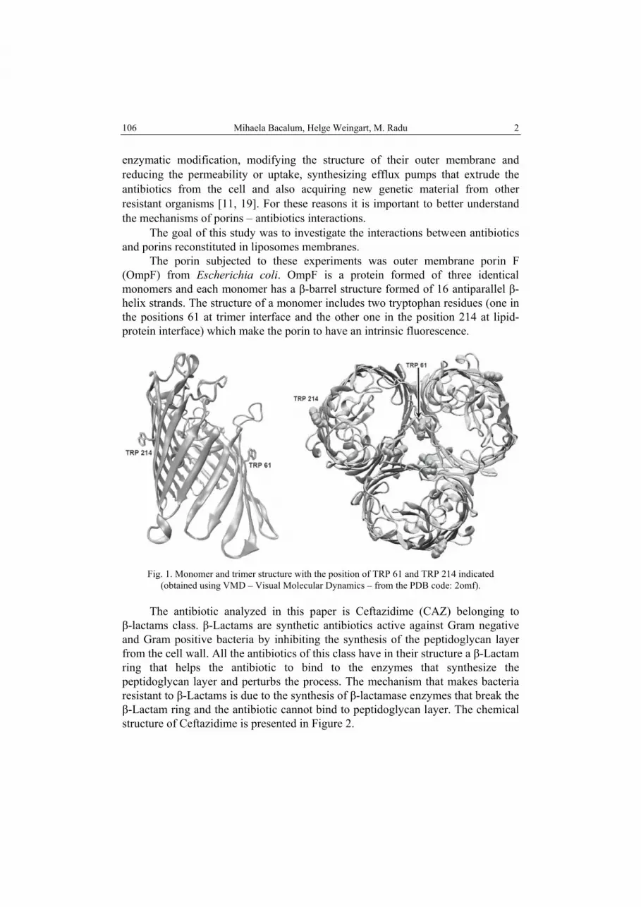
The porin subjected to these experiments was outer membrane porin F (OmpF) from Escherichia coli. OmpF is a protein formed of three identical monomers and each monomer has a β-barrel structure formed of 16 antiparallel β-helix strands. The structure of a monomer includes two tryptophan residues (one in the positions 61 at trimer interface and the other one in the position 214 at lipid-protein interface) which make the porin to have an intrinsic fluorescence.
Fig. 1. Monomer and trimer structure with the position of TRP 61 and TRP 214 indicated
(obtained using VMD – Visual Molecular Dynamics – from the PDB code: 2omf).
The antibiotic analyzed in this paper is Ceftazidime (CAZ) belonging to β-lactams class. β-Lactams are synthetic antibiotics active against Gram negative and Gram positive bacteria by inhibiting the synthesis of the peptidoglycan layer from the cell wall. All the antibiotics of this class have in their structure a β-Lactam ring that helps the antibiotic to bind to the enzymes that synthesize the peptidoglycan layer and perturbs the process. The mechanism that makes bacteria resistant to β-Lactams is due to the synthesis of β-lactamase enzymes that break the β-Lactam ring and the antibiotic cannot bind to peptidoglycan layer. The chemical structure of Ceftazidime is presented in Figure 2.

3 Interaction between Ceftazidime and OmpF
107
Fig. 2. Chemical structure of Ceftazidime.
In this study the fluorescence techniques were used to prove both the OmpF insertion in bilayers (by fluorescence resonance energy transfer) and the interaction between OmpF and antibiotics (by fluorescence quenching). FRET (fluorescence resonant energy transfer) is one of the biophysical methods used to characterize the interactions between molecules located at small distances ~ 2 – 8 nm [10]. By quenching it is possible to reveal if two molecules are interacting watching the decrease of fluorescence intensity of the sample [10].
The liposomes used in these experiments are small unilamellar vesicles (SUV) and were prepared from a mixture of lipids (DPhPC-cholesterol-sphingomyelin) for FRET and quenching experiments and from diphytanoyl-phosphatidylcholine (DPhPC) only for a part of quenching experiments.
In a previous study we used a FRET procedure to prove the insertion of proteins in the lipid bilayer of liposomes made of DPhPC [1]. The procedure was initially verified revealing the insertion of a hydrophobic peptide as Gramicidin A in liposomes bilayer (Gramicidin A easily inserts in lipid bilayers) and afterwards used to prove the OmpF insertion of the same type of liposomes [1]. Now, the intention is to see how OmpF inserts in the bilayer of liposomes made from a mixture of lipids, and whether the lipid composition of the bilayer may affect the insertion. DPhPC-cholesterol-sphingomyelin mixture was used because the interactions between these lipids may lead to formation of microdomains in the lipid bilayer similar with the ones from the cell membrane (lipid rafts) and may make the insertion more difficult [4].
After the insertion was proved, the interaction between the porin inserted in liposomes and Ceftazidime was studied.

Mihaela Bacalum, Helge Weingart, M. Radu 4 108
MATERIALS AND METHODS
The liposomes used in our study, SUV (small unilamelar vesicles), were prepared from DPhPC, Sphingomyelin (SM) and Cholesterol (Chl) form Avanti Polar (USA). N,N,N –Trimethyl – 4 – (6 – phenyl – 1,3,5 – hexatrien – 1 – yl)phenylammonium p – toluenesulfonate (TMA-DPH) and Ceftazidime were provided by Sigma Chemicals (Germany) and the OmpF was purified by the group of Prof. M. Winterhalter, Jacobs University Bremen, Germany, following the protocol described in [5]. The OmpF was in a stock solution of 1 mg/ml in PBS with 1% detergent (Octyl-POE). All solutions were prepared in 10 mM PBS solution, pH 7.4.
The liposomes were prepared after the following protocol [14]: the lipids (DPhPC, Chl and SM) were dissolved in chloroform at a given concentration as stock solutions and preserved at –20ºC. Appropriate volumes from these solutions were mixed (the molar ratio of lipids was 1:1:1) with a mixture of methanol-chloroform (1/1, v/v). The lipid film formed at the bottom of a tube after removing the solvent by drying under nitrogen flow was hydrated with 3 ml PBS. After hydration, the solution was vigorously vortexed to obtain a suspension of multilamellar vesicles. This suspension was sonicated in an ultrasonic bath (MRC D80H Ultrasonic cleaner 80 W) until clarity (~30 min) resulting in a suspension of SUV which was used in the experiments after a proper dilution to a final lipid concentration of ~78 µM.
The preparation of liposomes containing the porin was done as follows: after the lipidic film was dried, a small volume of porin stock solution (a few microliters) was added together with an appropriate volume of PBS assuring both the desired molar ratio OmpF / lipids (~1/3000) and the minimal detergent (Octyl-POE) concentration necessary to solubilise the protein (0.3% [17]). After the hydration of the lipidic film, the suspension was sonicated at clarity and an adequate volume of PBS was added to reach the desired lipid concentration (~78 µM). In this way the detergent is finally diluted by a factor ~1000 and in such conditions its influence can be neglected [17].
Fluorescence measurements were performed on a Fluoromax 3 (Horiba Jobin-Yvon) spectrofluorometer equipped with a termostated cell holder. All the spectra were recorded at 37 ºC. The recordings were made in the range of 290 to 500 nm for emission with the excitation set at 283 nm and the slit width of excitation and emission of 3 mm. The spectra recorded were corrected for the spectral sensitivity of the emission channel of the fluorometer.
Before studying the interaction between the antibiotic and porin molecules the insertion of the porins into the lipid bilayer of liposomes was proved using FRET technique. In this type of experiment the donors were the tryptophan residues from the porin and the acceptor was TMA-DPH, a fluorescent molecule that has a hydrophobic chain (DPH) and a hydrophilic part that allows this

5 Interaction between Ceftazidime and OmpF
109
molecule to be anchored at the surface of membrane. FRET protocol was first verified in previous experiments following the insertion of Gramicidin A, a hydrophobic peptide that has four tryptophan residues used as donor (results presented in [1]). In order to reveal FRET, four types of spectra were recorded: the spectrum of liposomes, the spectrum of liposomes doped with OmpF, the spectrum of liposomes doped with TMA-DPH and the spectrum of liposomes doped with porin and TMA-DPH. The first two spectra were subtracted to obtain the emission spectrum of the donor and the third and fourth spectra were subtracted to obtain the emission spectra for the probes that contain also the donor and the acceptor. These spectra were used to compute FRET efficiency (E):
E = 1 – Fn/F0 (1)
where: Fn is the maximal intensity of the donor emission in the presence of the acceptor (n counting the acceptor concentration value) and F0 the intensity of the donor emission in the absence of the acceptor.
In the second part of the study the interaction between the OmpF inserted in liposomes bilayers and ceftazidime was analyzed. After adding the drug in the cuvette with the liposome suspension, the sample was let for 10 minutes to reach an equilibrium state. Four types of spectra were recorded: the spectrum of liposomes, the spectrum of liposomes doped with OmpF, the spectrum of liposomes with antibiotics and the spectrum of liposomes doped with protein and antibiotics. The spectra were corrected for the inner filter effect [10], and then subtracted to obtain only the emission of porin alone and the emission of porin in the presence of antibiotic. The maximal values of OmpF emission intensity were fitted with Stern-Volmer equation to derive the binding constant of the antibiotic to the porin.
The experiments were repeated in triplicate and the average values and standard deviation were computed for the FRET efficiency or binding constant.
RESULTS
First, the results concerning the insertion of OmpF in the lipid membranes of liposomes made from a mixture of lipids are presented. The absorption and emission spectra of Tryptophan, OmpF and TMA-DPH and the overlapping of OmpF emission spectra and TMA-DPH absorption spectra proves that the main condition for energy transfer to occur was fulfilled for Trp-TMA-DPH pair [1].
In Figure 3 are presented the spectra of liposomes that have a different concentration of TMA-DPH (increasing concentrations from 0.083 to 0.42 µM) and in which OmpF was inserted (maintained at a constant concentration 18 nM); the spectra are corrected as described in Material and Methods section. Energy transfer was proved by the decrease of tryptophan emission (the peak seen at approximately 320 nm) simultaneously with the increase of TMA-DPH emission (420–500 nm) for increasing values of acceptor concentration.

Mihaela Bacalum, Helge Weingart, M. Radu 6 110
Fig. 3. FRET spectra for various acceptor concentrations recorded on liposomes
made from a mixture of lipids and doped with OmpF and TMA-DPH.
Fig. 4. FRET efficiency against acceptor concentration for liposomes doped
with OmpF and TMA-DPH.

7 Interaction between Ceftazidime and OmpF
111
FRET efficiency calculated using the maximum values of OmpF fluorescence emission in the absence and presence of TMA-DPH is represented in Figure 4. The value of FRET efficiency increases with the increase of TMA-DPH concentration as expected. Because FRET takes place at distance <5 nm for this pair of molecules [10] and it is known that TMA-DPH is able to insert itself in liposomes bilayer the results prove that OmpF was also inserted in the bilayer.
The quenching of OmpF fluorescence by ceftazidime was analyzed to evaluate the binding constant of antibiotic. Ceftazidime concentration ranges between 6.1–36.6 µM for a constant OmpF concentration (27 nM). Spectra were recorded at 37 ºC, in the range of 300–350 nm for emission, excitation being set at 283 nm [10]. In order to see whether the lipid composition of bilayer has an influence on the CAZ-OmpF interaction, the experiments were realized on two types of liposomes: simple liposomes made of DPhPC and liposomes made of a mixture (DPhPC, SM and Chl).
In Figure 5 the spectra of OmpF in the presence of ceftazidime are presented in the case of OmpF inserted in simple liposomes. The addition of the antibiotic induced a gradual decrease of the OmpF emission intensity. The experiment was repeated for the liposomes made from a mixture of lipids and the results are presented in Figure 6. Similar behaviour of OmpF emission was revealed as in the case of liposomes with a homogeneous lipidic composition.
The fluorescence quenching induced by Ceftazidime may be explained by an interaction between the tryptophan residues from OmpF and CAZ. Consequently, the tryptophan may be seen as a component of the binding site for Ceftazidime.
DISCUSSION
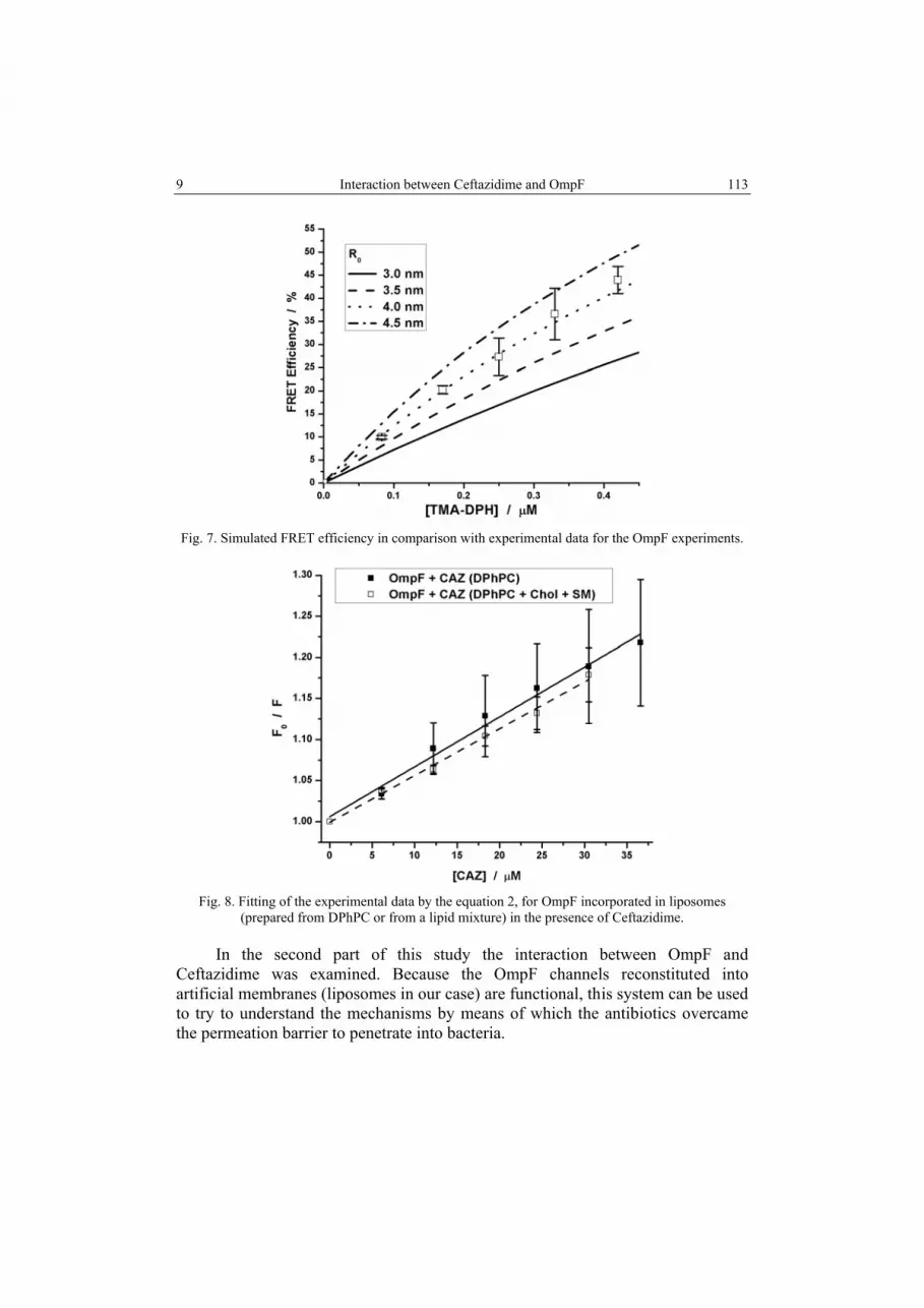
Comparing the results obtained in the first experiments with the results reported in [1] we can conclude that OmpF inserts in lipid bilayers with heterogeneous composition in a similar manner as in homogeneous lipid membranes. In order to evaluate the smallest distance between OmpF and TMA-DPH we used a theoretical model for computing FRET efficiency for a uniform planar distribution of donors and acceptors [20]. The smallest distance between the donor and acceptor in such a case is reasonably approximated by the Forster radius (R0). In Figure 7 the data obtained from the computing model are compared with the experimental data resulting in a value for R0 of ~4 nm. In the case of OmpF insertion in liposomes with uniform lipid composition the R0 value was ~3.5 nm [1] which is very close to the R0 value reported in the present experiment. Only a small increase in R0 value occurred when heterogenic liposomes were used, fact explained by a little bit larger extent of FRET in this case.

Mihaela Bacalum, Helge Weingart, M. Radu 8 112
Fig. 5. Spectra of OmpF and different concentrations of Ceftazidime
(liposomes prepared from DPhPC).
Fig. 6. Spectra of OmpF and different concentrations of Ceftazidime
(liposomes prepared from a mixture of DPhPC, ChL and SM).
9 Interaction between Ceftazidime and OmpF
113
Fig. 7. Simulated FRET efficiency in comparison with experimental data for the OmpF experiments.
Fig. 8. Fitting of the experimental data by the equation 2, for OmpF incorporated in liposomes
(prepared from DPhPC or from a lipid mixture) in the presence of Ceftazidime.
In the second part of this study the interaction between OmpF and Ceftazidime was examined. Because the OmpF channels reconstituted into artificial membranes (liposomes in our case) are functional, this system can be used to try to understand the mechanisms by means of which the antibiotics overcame the permeation barrier to penetrate into bacteria.

Mihaela Bacalum, Helge Weingart, M. Radu 10 114
Table 1
The binding constant obtained for Ceftazidime and OmpF inserted into liposomes
Type of lipids KD (1/µM) SD (1/µM) DPhPC 0.00607 0.000315 DPhPC, Chl and SM (1:1:1) 0.0057 0.000218
Earlier research established that the transport pathway of β-lactams through bacterial outer membrane is represented by the porins. The E. coli mutants deficient in OmpF and OmpC are more resistant to β-Lactams [7], particularly to Ceftazidime [8]. On the other hand, in experiments done on a suspension of OmpF trimers in solution, presence of spermidine and streptomycin induced a quenching of tryptophan fluorescence in a concentration dependent manner [9]. The authors suggest the binding of the cations to the pore, a binding constant of 18.1 µM being calculated for streptomycin. This hypothesis was tested analyzing the OmpF tryptophan fluorescence quenching in the presence of aminoglycosides [6]. They interpreted their results as rather a consequence of an aggregation of porins induced by the aminoglycosides than a quenching produced by the direct interaction of aminoglycosides with the protein molecules. More recently, a study concerning the quinolones interaction with OmpF in solution suggests again the presence of a porin – antibiotics interaction producing a non-fluorescent protein-drug complex resulting in the quenching of OmpF tryptophane [13].
In this paper we tried to come closer to a more realistic model incorporating first the OmpF molecules in the lipid bilayer of liposomes and afterwards analyzing the OmpF interaction with β-Lactams, particularly with Ceftazidime. For each type of liposomes used in these experiments the results were plotted and presented in Figure 8. The linearity of the fluorescence quenching ratio dependence against the CAZ concentration (Figure 8) suggests that the quenching of the OmpF fluorescence is produced during the passage of CAZ molecules through the protein pore by a temporary forming of a complex. Fitting the maximum emission intensity by a standard model for the fluorescence quenching (the Stern-Volmer equation) the binding constant was calculated:
0D1 [ ]F K Q
F= + (2)
where F0 and F are the maximum fluorescence intensity of the tryptophan in the absence and presence of Ceftazidime, respectively and [Q] the concentration of antibiotic.
The binding constant was calculated in both cases and the results are presented in Table 1. As the insertion experiments already revealed, the lipid composition did not significantly influence the OmpF functionality. The binding constant has similar values for both investigated cases (Table 1) proving that the interaction CAZ-OmpF is produced in the same way.

11 Interaction between Ceftazidime and OmpF
115
Theoretical studies reported in literature about β-lactam antibiotics also support the hypothesis of transient binding of the antibiotics molecules passing through the OmpF channel. Using molecular modeling the translocation of β-Lactam antibiotics, particularly of ampicilin, through OmpF was studied and depending on their chemical properties the antibiotics exhibit different mechanisms of penetration through the porin [12].
CONCLUSIONS
In this study we have shown that OmpF, regardless of the liposomes lipid composition in which it was inserted, has a similar behaviour in the presence of Ceftazidime, a β-Lactam antibiotic. The fluorescence quenching results sustain the supposition according to which the passage of antibiotic molecule through the pore involves the formation of a temporary complex between OmpF and Ceftazidime.
Acknowledgements. The authors thank Mr. Tivadar Mach for helpful discussions. This work
was financed by the Romanian Ministry of Education and Research by means of the research grant no. 168/2006 (CEEX VIASAN).
R E F E R E N C E S
1. BACALUM, M., M. RADU, Insertion of proteins in the lipid bilayer of liposomes revealed by FRET, Rom. J. Biophys., 2007, 17, 129–138.
2. CECCARELLI, M., C. DANELON, A. LAIO, M. PARRINELLO, Microscopic mechanism of antibiotics translocation through a porin, Biophys. J., 2004, 87, 58–64.
3. DANELON, C., E.M. NESTOROVICH, M. WINTERHALTER, M. CECCARELLI, S.M. BEZRUKOV, Interaction of zwitterionic penicillins with the OmpF channel facilitates their translocation, Biophys. J. , 2006, 90, 1617–1627.
4. DIETRICH, C., L.A. BAGATOLLI, Z.N. VOLOVYK, N.L. THOMPSON, M. LEVI, K. JACOBSON, E. GRATTON, Lipid rafts reconstituted in model membranes, Biophys. J., 2001, 80, 1417–1428.
5. GARAVITO, R.M., J.P. ROSENBUSCH, Isolation and crystallization of bacterial porin, Methods Enzymol., 1986, 125, 309–328.
6. HANCOCK, R.E., S.W. FARMER, Z. LI, K. POOLE, Interaction of aminoglycosides with the outer membranes and purified lipopolysaccharide and OmpF porin of Escherichia coli, Antimicrobial Agents and Chemotherapy, 1991, 35, 1309–1314.
7. HARDER, K.J., H. NIKAIDO, M. MATSUHASHI, Mutants of Escherichia coli that are resistant to certain beta-lactam compounds lack the OmpF porin, Antimicrobial Agents and Chemotherapy, 1981, 20, 549–552.
8. HASHIZUME, T., M. SANADA, S. NAKAGAWA, N. TANAKA, Comparison of transport pathways of catechol-substituted cephalosporins, bo-1236 and bo-1341, through the outer membrane of Escherichia coli, J. Antibiot., 1990, 43, 1617–1620
9. KOBAYASHI, Y., T. NAKAE, The mechanism of OmpF-porin pores of Escherichia coli, Eur. J. Biochem., 1985, 151, 231–236.
10. LAKOWICZ, J.R., Principles of Fluorescence Spectroscopy, Springer, New York, 2006.

Mihaela Bacalum, Helge Weingart, M. Radu 12 116
11. LAMBERT, P.A., Mechanisms of antibiotic resistance in Pseudomonas aeruginosa, Journal of the Royal Society of Medicine Supplement, 2002, 95, 22–26.
12. NESTOROVICH, E.M., C. DANELON, M. WINTERHALTER, S.M. BEZRUKOV, Designed to penetrate: Time-resolved interaction of single antibiotic molecules with bacterial pores, PNAS, 2002, 99, 9789–9794.
13. NEVES, P., E. BERKANE, P. GAMEIRO, M. WINTERHALTER, de Castro, B., Interaction between quinolones antibiotics and bacterial outer membrane porin OmpF, Biophys. Chem., 2005, 113, 123–128.
14. NEW, R.R.C., Liposomes: A Practical Approach, Oxford University Press, Oxford, 1990. 15. NIKAIDO, H., Outer membrane barrier as a mechanism of antimicrobial resistance,
Antimicrobial Agents and Chemotherapy, 1989, 33, 1831–1836. 16. NIKAIDO, H., M. VAARA, Molecular basis of bacterial outer membrane permeability,
Microbiological Reviews, 1985, 49, 1–32. 17. O’KEEFFE, A.H., J.M. EAST, A.G. LEE, Selectivity in lipid binding to the bacterial outer
membrane protein OmpF, Biophys. J., 2000, 79, 2066–2074. 18. PAGES, J.M., C.E. JAMES, M. WINTERHALTER, The porin and the permeating antibiotic: a
selective diffusion barrier in Gram-negative bacteria, Nature Reviews Microbiology, 2008, 6, 893–903.
19. TENOVER, F. C., Mechanisms of antimicrobial resistance in bacteria, The American Journal of Medicine, 2006, 119, S3–S10.
20. WOBLER, P.K., B.S., HUDSON, An analytic solution to the Forster energy transfer problem in two dimension, Biophys. J., 1979, 28, 197–2 10.


