Embed Size (px)
Citation preview
Insights on P-Glycoprotein’s Efflux Mechanism Obtained byMolecular Dynamics SimulationsRicardo J. Ferreira, Maria-Jose U. Ferreira, and Daniel J. V. A. dos Santos*
Research Institute for Medicine and Pharmaceutical Sciences (iMed.UL), Faculty of Pharmacy, University of Lisbon,Av. Prof. Gama Pinto, 1649-003 Lisbon, Portugal
*S Supporting Information
ABSTRACT: P-Glycoprotein (P-gp) is often involved in multidrug resistance (MDR) to the pharmacological action of a widenumber of anticancer agents. In this article, a series of molecular dynamics simulations of murine’s P-gp were developed,elucidating the importance of the lipid membrane and linker sequence in the protein structure stability. The behavior of severalmolecules inside the drug-binding pocket revealed a striking difference between substrates or modulators, and motion patternswere identified that could be correlated with conformational alterations due to substrate binding, corresponding to the initial stepin the efflux mechanism. Only one “entrance gate” to the drug-binding pocket was found and, in the presence of a substrate, leadsto changes in the motion patterns of the transporter into an efflux-like movement.
P-Glycoprotein (P-gp) is the most representative member ofthe ABC transporter proteins superfamily, often implicated
in the multidrug-resistance phenomenon (MDR).1,2 Thistransporter expels a wide-range of substrates out of the cell(mainly hydrophobic) through an ATP-dependent mecha-nism.2,3 It has a molecular weight of ∼170 kDa organized intwo functional units with pseudo-2-fold molecular symmetry,each one comprising six transmembranar α-helices domains(TMD) and one cytoplasmic nucleotide-binding domain(NBD), linked by a small polypeptidic sequence.4
There is still great controversy about the efflux mechanism ofthis type of transporter. The first studies claimed that theextrusion of the molecules can occur directly from the cyto-plasmic compartment5 or that the protein could act accordingto a flippase model.6 More recently, the hydrophobic vacuum-cleaner model7 became the most accepted theory, supported byseveral studies that correlate the diffusion of the transportedmolecules within the inner leaflet of the cellular membrane withinteractions at the drug-binding site (DBS).3,8
The large majority of the most recent studies accept analternate cycle for the ATP hydrolysis,9,10 emphasizing the factthat only transported substrates at the DBS, in conjunction withATP binding, can induce conformational changes in the proteinstructure leading to the extrusion of the molecule.11 As a directconsequence, the energy driven from ATP hydrolysis must beused to reset the transporter into its original conformation, thusallowing new catalytic cycles.10 However, the communicationroutes between DBS-NBD and the extent of the conforma-tional changes leading to the efflux mechanism are still mainlyunknown. In addition, the lipidic composition12−14 and globalpassive permeation rate of molecules through the plasmaticmembrane15,16 also seem to be as important as the transporteritself, working together as a single entity rather than twodistinct structures.17,18
The crystallization of membrane proteins is proved to bedifficult, greatly diminishing the number of available structuresin the Protein Data Bank (PDB). Nevertheless, these proteins
are of great importance due to their physiological functions asneurotransmitter receptors, ion channels, or, in the case of P-gp,ATP-driven substrate transporters. In particular, the crystallo-graphic structure of such transporters can be used as aframework to allow a better comprehension of their mechanismof action. For instance, the crystallographic informationobtained from BtuCD, a vitamin B12 importer, suggested theexistence of communication between drug-binding andnucleotide-binding sites as a way to induce the catalytic cycle,possibly triggered by long-range conformational changesdeveloped by the binding and/or hydrolysis of ATP.19
Recently, a murine P-glycoprotein crystallographic structurewas made available in the PDB20,21 (ID: 3G5U) with a 3.8 Åresolution.22 This information made possible the identificationof one large internal chamber of approximately 6000 Å3thedrug-binding pocket (DBP)delimited by the TMD butopened to the intracellular compartment and the inner leafletof the membrane through two “gates” formed by the pairsTMD 4/6 and 10/12.22 Inside the binding pocket, 73 exposedresidues were identified, of which 15 are polar and only twocharged or possibly charged (His60 and Glu871). Three drug-binding sites were proposed based on the chemical character-istics of the amino acids present in each one and on dockedstructures with the inhibitors QZ59-RRR and QZ59-SSS.Therefore, this study refers to the possible simultaneousbinding of two compounds inside the DBP.22 Several studiesalso propose that the internal cavity may be filled with watermolecules and that 70% of the area attributed to the DBS is dueto the transmembranar domains 3, 5, 6, 8, 11, and 12, alsoimplying TMD 6 and 12 in the translocation process.23,24
In the meantime, the lack of a crystallographic structure ledto the development of several homology models for ABCtransporters mainly derived from bacterial templates suchas Sav1866,25,26 MalK,27 or MsbA.28 Although later proved
Received: February 1, 2012Published: May 9, 2012
Article
pubs.acs.org/JCTC
© 2012 American Chemical Society 1853 dx.doi.org/10.1021/ct300083m | J. Chem. Theory Comput. 2012, 8, 1853−1864
inaccurate as prediction models29,30 largely due to errorsintroduced during the homology process, it was possible toobtain relevant information related with the ABC transportersprimary function. These models, obtained from apo structures(open,27,28 semiopen,27 or closed28 conformational states) orin the presence of nucleotides (AMP-PNP,28,31 ADP,32 andADP-Vi28), were employed in later studies that suggested therigid-body rotation of the NBD as the major driving force forthe induction of TMD reorganizations and NBD dimerization.The TMD reorganization is indeed supported by studies whichclaim that the rotation of the helices into an asymmetric con-formational state can be the major event that alters the affinityof the DBS,10,33,34 leading to the ejection of the molecule fromthe internal cavity to the outside of the cell. More recentcomputational studies have also characterized the magnitude ofsuch conformational alterations in stages after and before ATPhydrolysis,35 in NBD dimerization,36 and following the removalof ATP/Mg2+,37 confirming the pivotal role of TMD6 in theefflux mechanism.37 These observations are of great value forthe analysis of the P-gp translocation mechanism, due to thesimilarity between the structures.The murine crystallographic structure has also been recently
used as a framework for an increasing number of dockingstudies.38,39 Such studies are important because they canprovide useful information about the intrinsic molecularproperties that discriminate molecules like verapamil40 ortariquidar,41 known P-gp inhibitors, from substrates likevinblastine40 or colchicine.1 However, the presence of watermolecules inside the DBS can be a major drawback in this typeof study, usually made in the absence of water. In addition, thepublished crystallographic structure was obtained in theabsence of a lipid bilayer membrane and is incomplete, lackingthe information correspondent to the structure of a linkerregion between the Ala627 and Ala683, located about half-wayinto the P-gp sequence. This region and its flexibility wereproved to be important for the regulation of the substrates'specificity toward human P-gp, also interfering with theconformational change that accompanies the hydrolysis of theATP molecule.42,43
Despite all evidence, a clear demonstration of the transporternormal modes of motion is still missing, along with theclarification of the NBD dimerization and helix rotation as theprincipal motion pattern associated with the efflux mechanism.In this particular case, computational investigations can bevaluable tools for the identification of such patterns. Computa-tional models have been used for the simulation of biologicalsystems in order to further understand the data obtainedthrough previous studies, thus enlightening the biologicalprocess mechanism of action, substrate binding or conforma-tional alterations. More specifically, the application of themolecular dynamics (MD) technique to P-gp models can unveilsome answers regarding the structure’s stability, drug-bindinglocations, translocation processes, and interactions with thesurrounding lipid bilayer. From such techniques, normal-modeanalysis and functional mode analysis of the protein movementsare important tools, allowing the comparison between simula-tions of the apo and holo structures, with one or severalmolecules inside the DBP. All of the above methods could leadto the identification of the movements intimately related withthe translocation process, aiming for a better understanding ofthe first steps of the efflux mechanism.
In this paper, we describe a series of P-gp computationalMD simulations based on the murine crystallographic data. Theobjectives are as follows:
i. to refine and validate a P-gp structure, suitable forutilization in later studies; the missing linker sequenceand lipid bilayer membrane will be assessed, in order toevaluate the importance of such structures in thestabilization of the protein
ii. to evaluate the behavior of several molecules (substratesor inhibitors), inside the DBP, by analyzing the type andnumber of contacts established and major residuesinvolved in the ligand−protein interactions
iii. to assess modifications in the normal motion patternsdisplayed by the transporter, in order to identifyconformational changes that can be associated with theefflux mechanism, when perturbed by a molecule suitableto be transported
■ MATERIAL AND METHODSInitial Structures and Software. The murine P-glyco-
protein structure (ID: 3G5U) was obtained from the ProteinData Bank (www.pdb.org)20 and parametrized according to theGROMOS9644,45 force field with the 53a646 parameter set.Two lipid bilayers (dimiristoyl-phosphatidylcholineDMPCand 1-palmitoyl-2-oleoylphosphatidylcholinePOPC) and re-spective parametrization were obtained from previous simulationsby Poger et al.47,48 This choice of lipid parametrization took intoaccount the ability to correctly reproduce properties of lipidbilayers,49 namely, the area and volume per lipid ratios47,50,51 andfluid-phase47,48,52,53 or solvation47,48,53 properties.Protein manipulation, protonation, and linker modeling
were made in MOE 2009.10.54 The GROMACS simulationpackages, 4.5.x,55−57 were used for the MD simulations andlater bilayer constructions. The Inf lateGRO58 script was initiallyemployed in bilayer builds and later substituted by theg_membed59 module available in the GROMACS package.The GridMAT-MD60 script was used to calculate areas per lipid(AL) of the protein-bilayer systems. Functional mode analysis(FMA) was done as described by Hub and de Groot,61
covariance analysis62 with auxiliary programs available inGROMACS, and principal component analysis (PCA) per-formed using the ProDy63,64 software. Substrates and inhibitorsparametrization were prepared online using the PRODRG65 orthe Automated Topology Builder and Repository66 servers,and Mulliken partial charges were assigned through ab initiocalculations at the Hartree−Fock level of theory using the6-31G(d) basis set in the Gaussian 0367 program. VMD68 andMOE were used for molecular inspection and visualization.
Systems Construction. Initial Evaluation. The crystallo-graphic P-gp structure was solvated (41 664 water molecules)and the system’s total charge neutralized with the addition of 25chlorine atoms (SOL-I). After energy minimization, a 1 ns NVTequilibration run was performed (spatially restraining theprotein’s heavy atoms) followed by a 10 ns NPT unrestrictedrun. Another system was built comprising the crystallographicP-gp inserted into a 417 lipid DMPC membrane (DMPC-I),water soaked (57 628 molecules) and neutralized (with anadequate number of counterions). The position of themembrane relative to P-gp was defined using informationobtained from the OPM database.69 The system was energyminimized and equilibrated with two sequential NVT (1 ns) andNPT (10 ns) runs, spatially restraining the protein’s heavy atoms.
Journal of Chemical Theory and Computation Article
dx.doi.org/10.1021/ct300083m | J. Chem. Theory Comput. 2012, 8, 1853−18641854
Finally, an unrestricted NPT run was performed, with a durationof 30 ns.Linker. The folding of the missing linker was assessed
through secondary structure prediction online servers such asNetSurfP,70 APSSP,71 PROF,72 and CFSSP.73 The segment wasthen built with the Protein Builder module of MOE, followedby protonation and energy minimization. The distance betweenthe amine and carboxyl groups of terminal residues was kept tothe crystallographic length of the linker (4.75 Å). The resultingstructure was then solvated (with the correct number ofcounterions added) and further equilibrated in GROMACSthrough a series of eight MD simulations (NVT), each one at500 ps duration and with different initial parameters (temper-ature, thermostat, and/or interaction cutoff radius) in order toobtain different starting points for the runs. An NPT run foreach one of the eight linker structures followed for 4 ns usingthe same simulation parameters. Using these run data, allobtained structures were then evaluated regarding the system’sfinal energies, a Ramachandran74 plot for structural stabilityand visual evaluation. The more suitable conformation was thenadded to the whole protein with the protonation status of theentire protein evaluated once more through MOE’s Proto-nate3D module.Construction of the Membrane Systems. The correct
relative position of the lipid bilayer was defined according tothe OPM database69 information. After membrane insertion,the system was solvated and neutralized with an adequatenumber of chloride ions. A 20 ns equilibration run followed byrestraining all heavy atoms of the P-gp crystallographic structurebut three residues directly connected to each side of the linker,in order to allow a better adjustment between this segment andthe entire structure. The resulting P-gp structure inserted into aPOPC bilayer was the starting point for the structure refinementstep.Refinement Protocol. The whole P-gp structure was inserted
into a previously equilibrated POPC membrane, water soaked(55 909 molecules), and neutralized with 26 chlorine ions,originating a system with 204 227 atoms. The system wasenergy minimized and a 100 ps NVT equilibration run followed,above the phospholipids gel-fluid transition phase,75 at 303 K.Using the final structure of this run, two systems were obtainedby applying two equilibration protocols. In the first (POPC-I),the initial 3 ns were made with heavy-atom spatial restraints,thus allowing the correct adjustment between the proteinstructure and the surrounding lipids. A 100 ns production runfollowed. On the second (POPC-II), the spatial restriction ofthe protein’s heavy atoms was progressively removed, througha series of three successive NPT simulations of 500 ps each.The first one restrained heavy atoms from the mainchain(allowing the equilibration of the residue side chains to the lipidenvironment). The second restrained the backbone, and finally,the third restrained the α-carbons. The resulting system wasused in a 100 ns production run.Insertion of Ligands. For the systems with ligands inside
the DBP, six molecules (inhibitorsverapamil,40 tariquidar,41
latilagascene E,76 QZ59-SSS22and substratescolchicine,1
vinblastine;1,40 chemical structures available in Table S1 ofSupporting Information) were positioned inside the internalcavity described by Aller et al. in three different positions, thusobtaining 18 new systems. After minimization, the systemswere allowed to equilibrate for 1 ns, keeping the protein heavyatoms' positions restrained, after which three 20 ns productionruns were performed (no restrictions applied). The two runs
containing the largest molecules (vinblastine and tariquidar) inthe top position of the DBP were later extended to 40 ns.Protein−ligand contacts were evaluated at every 250 ps tocalculate the type, nature, and maximum number of establishedinteractions using the LIGPLOT77 software, in conjunctionwith HBPLUS.78
An additional system containing a vinblastine molecule at theentrance gate was also built. The molecule was inserted using adocking protocol available in MOE by performing 10 runs ofsimulated annealing, each comprising six cycles and defining aniteration limit of 4000 at an initial temperature of 1000 K, afterwhich the best ranked pose was chosen. The resulting systemwas treated under the conditions above-described for theligands inside the DBP.
Simulation Parameters. In all simulations, periodicboundary conditions (PBC) were applied. Simple energyminimizations were performed using the steepest descentmethod. All NVT runs used a Velocity-rescale (V-rescale)thermostat.79 NPT runs used the Nose−Hoover80,81 thermostatand the Parrinello−Rahman82,83 barostat for temperature (303 K)and pressure coupling (1 bar), respectively. In the presence ofmembranes, the pressure equilibration was achieved through asemi-isotropic pressure coupling. The system compressibility wasdefined as 4.5 × 10⁻⁵ bar−1, and the initial box was defined withdimensions xyz of 12.75 × 12.75 × 16.55 nm. The Particle MeshEwald (PME) method with cubic interpolation,84,85 with anidentical cutoff radius for electrostatic and van der Waalsinteractions (12 Å), was employed. The SETTLE86 (for watermolecules) or LINCS87 algorithms were used to constrain allbond lengths.
■ RESULTSP-gp Structure Refinement. As an initial evaluation of the
crystallographic structure stability, a 10 ns simulation without alipid bilayer was performed (SOL-I), revealing increased TMDdisorganization and NBD separation leading to an irreversibledistortion of the structure. This clearly shows the importanceof the linker sequence and of the lipid bilayer. Therefore, theprotein was inserted into membranes comprising differentlipids (DMPC or POPC) in order to minimize the principalstructural abnormalities observed.The insertion of the protein, without the linker sequence, in
each membrane type revealed a striking difference. It is knownthat the optimum insertion angle for the P-gp in a lipidmembrane has a value of approximately 4°,69 but in the DMPCmembrane simulation (DMPC-I) this value rose to approx-imately 18° after 10 ns, while in a POPC membrane theinsertion angle did not suffer significant alterations from theinitial insertion angle. This difference between both systemscan be explained by the difference between the thickness of thehydrophobic core of the simulated POPC (3.27 ± 0.03 nm)and DMPC membranes (2.85 ± 0.06 nm) against the 3.18 ±0.12 nm69 estimated for the P-gp (hydrophobic mismatch of3.3 Å for DMPC). Therefore, in a system containing a DMPCbilayer, a tilting is expected to occur to maximize the contactarea between hydrophobic regions of the P-gp and the lipids.87
Additionally, eukaryotic membranes are richer in polyunsatu-rated lipids than saturated ones, a fact by which the POPC lipidis more adequate for the simulation of such transporters, inagreement with a previous study on a similar framework.88
In order to assess the importance of the linker in the stabilityof the whole structure, the corresponding secondary structurewas built on the basis of secondary structure prediction
Journal of Chemical Theory and Computation Article
dx.doi.org/10.1021/ct300083m | J. Chem. Theory Comput. 2012, 8, 1853−18641855
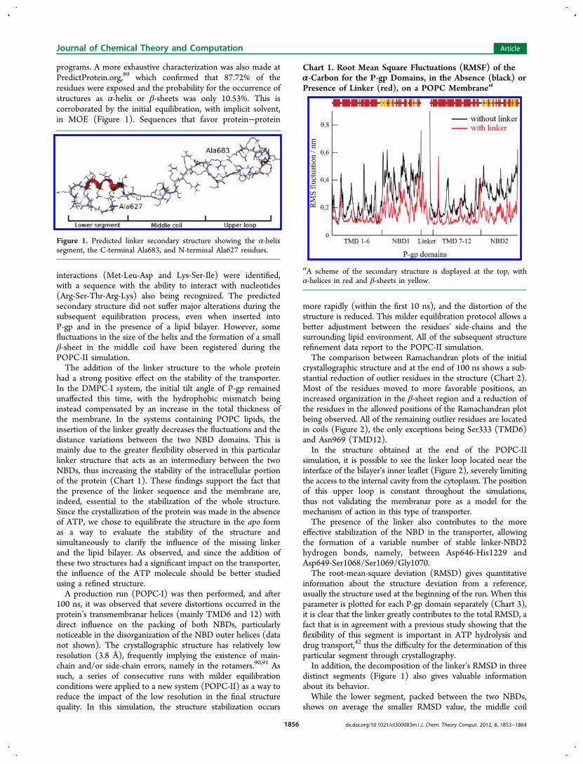
programs. A more exhaustive characterization was also made atPredictProtein.org,89 which confirmed that 87.72% of theresidues were exposed and the probability for the occurrence ofstructures as α-helix or β-sheets was only 10.53%. This iscorroborated by the initial equilibration, with implicit solvent,in MOE (Figure 1). Sequences that favor protein−protein
interactions (Met-Leu-Asp and Lys-Ser-Ile) were identified,with a sequence with the ability to interact with nucleotides(Arg-Ser-Thr-Arg-Lys) also being recognized. The predictedsecondary structure did not suffer major alterations during thesubsequent equilibration process, even when inserted intoP-gp and in the presence of a lipid bilayer. However, somefluctuations in the size of the helix and the formation of a smallβ-sheet in the middle coil have been registered during thePOPC-II simulation.The addition of the linker structure to the whole protein
had a strong positive effect on the stability of the transporter.In the DMPC-I system, the initial tilt angle of P-gp remainedunaffected this time, with the hydrophobic mismatch beinginstead compensated by an increase in the total thickness ofthe membrane. In the systems containing POPC lipids, theinsertion of the linker greatly decreases the fluctuations and thedistance variations between the two NBD domains. This ismainly due to the greater flexibility observed in this particularlinker structure that acts as an intermediary between the twoNBDs, thus increasing the stability of the intracellular portionof the protein (Chart 1). These findings support the fact thatthe presence of the linker sequence and the membrane are,indeed, essential to the stabilization of the whole structure.Since the crystallization of the protein was made in the absenceof ATP, we chose to equilibrate the structure in the apo formas a way to evaluate the stability of the structure andsimultaneously to clarify the influence of the missing linkerand the lipid bilayer. As observed, and since the addition ofthese two structures had a significant impact on the transporter,the influence of the ATP molecule should be better studiedusing a refined structure.A production run (POPC-I) was then performed, and after
100 ns, it was observed that severe distortions occurred in theprotein’s transmembranar helices (mainly TMD6 and 12) withdirect influence on the packing of both NBDs, particularlynoticeable in the disorganization of the NBD outer helices (datanot shown). The crystallographic structure has relatively lowresolution (3.8 Å), frequently implying the existence of main-chain and/or side-chain errors, namely in the rotamers.90,91 Assuch, a series of consecutive runs with milder equilibrationconditions were applied to a new system (POPC-II) as a way toreduce the impact of the low resolution in the final structurequality. In this simulation, the structure stabilization occurs
more rapidly (within the first 10 ns), and the distortion of thestructure is reduced. This milder equilibration protocol allows abetter adjustment between the residues' side-chains and thesurrounding lipid environment. All of the subsequent structurerefinement data report to the POPC-II simulation.The comparison between Ramachandran plots of the initial
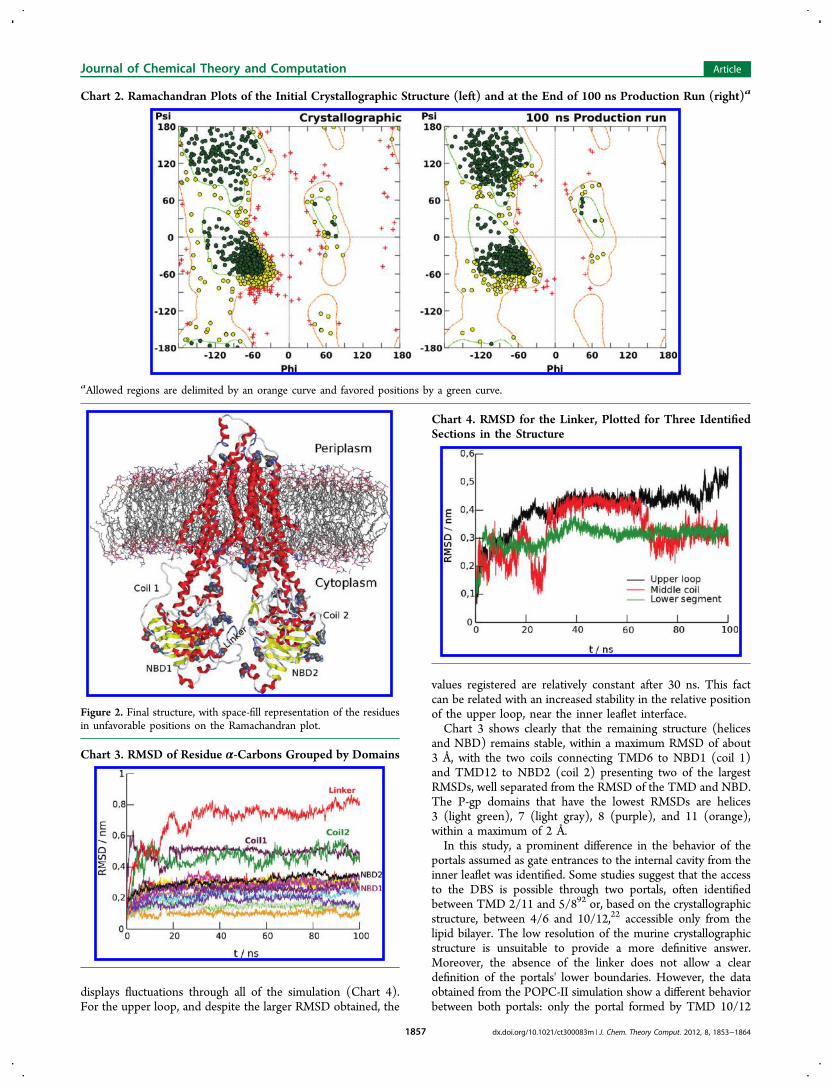
crystallographic structure and at the end of 100 ns shows a sub-stantial reduction of outlier residues in the structure (Chart 2).Most of the residues moved to more favorable positions, anincreased organization in the β-sheet region and a reduction ofthe residues in the allowed positions of the Ramachandran plotbeing observed. All of the remaining outlier residues are locatedin coils (Figure 2), the only exceptions being Ser333 (TMD6)and Asn969 (TMD12).In the structure obtained at the end of the POPC-II
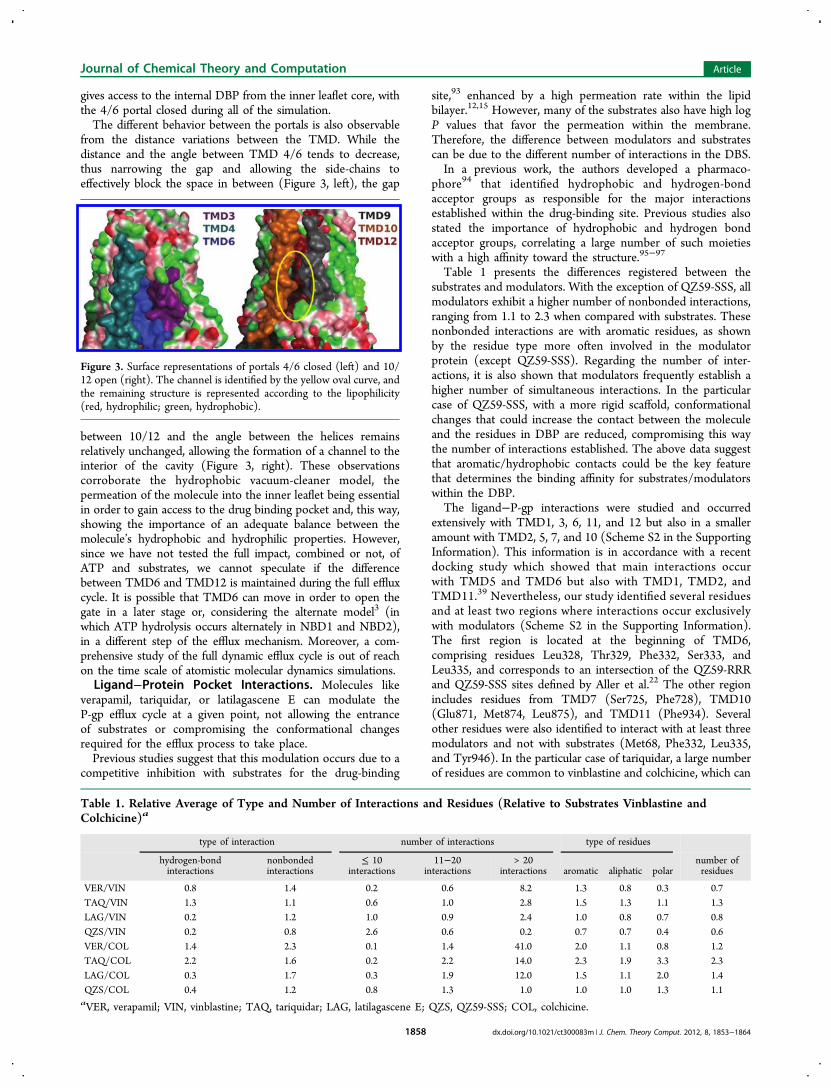
simulation, it is possible to see the linker loop located near theinterface of the bilayer’s inner leaflet (Figure 2), severely limitingthe access to the internal cavity from the cytoplasm. The positionof this upper loop is constant throughout the simulations,thus not validating the membranar pore as a model for themechanism of action in this type of transporter.The presence of the linker also contributes to the more
effective stabilization of the NBD in the transporter, allowingthe formation of a variable number of stable linker-NBD2hydrogen bonds, namely, between Asp646-His1229 andAsp649-Ser1068/Ser1069/Gly1070.The root-mean-square deviation (RMSD) gives quantitative
information about the structure deviation from a reference,usually the structure used at the beginning of the run. When thisparameter is plotted for each P-gp domain separately (Chart 3),it is clear that the linker greatly contributes to the total RMSD, afact that is in agreement with a previous study showing that theflexibility of this segment is important in ATP hydrolysis anddrug transport,42 thus the difficulty for the determination of thisparticular segment through crystallography.In addition, the decomposition of the linker’s RMSD in three
distinct segments (Figure 1) also gives valuable informationabout its behavior.While the lower segment, packed between the two NBDs,
shows on average the smaller RMSD value, the middle coil
Figure 1. Predicted linker secondary structure showing the α-helixsegment, the C-terminal Ala683, and N-terminal Ala627 residues.
Chart 1. Root Mean Square Fluctuations (RMSF) of theα-Carbon for the P-gp Domains, in the Absence (black) orPresence of Linker (red), on a POPC Membranea
aA scheme of the secondary structure is displayed at the top, withα-helices in red and β-sheets in yellow.
Journal of Chemical Theory and Computation Article
dx.doi.org/10.1021/ct300083m | J. Chem. Theory Comput. 2012, 8, 1853−18641856
displays fluctuations through all of the simulation (Chart 4).For the upper loop, and despite the larger RMSD obtained, the
values registered are relatively constant after 30 ns. This factcan be related with an increased stability in the relative positionof the upper loop, near the inner leaflet interface.Chart 3 shows clearly that the remaining structure (helices
and NBD) remains stable, within a maximum RMSD of about3 Å, with the two coils connecting TMD6 to NBD1 (coil 1)and TMD12 to NBD2 (coil 2) presenting two of the largestRMSDs, well separated from the RMSD of the TMD and NBD.The P-gp domains that have the lowest RMSDs are helices3 (light green), 7 (light gray), 8 (purple), and 11 (orange),within a maximum of 2 Å.In this study, a prominent difference in the behavior of the
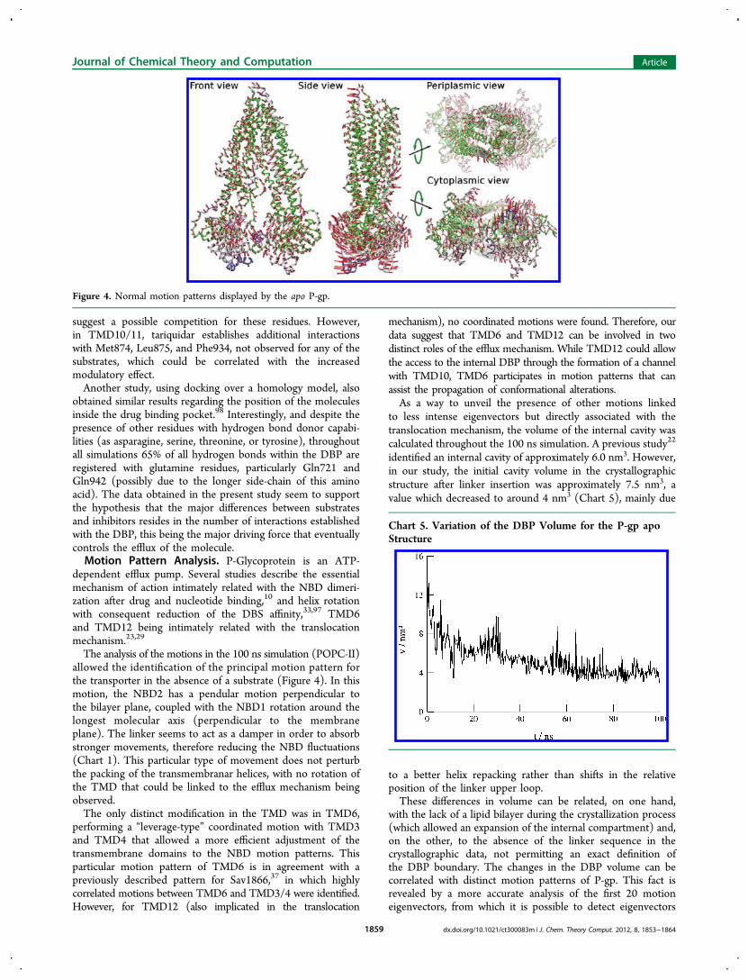
portals assumed as gate entrances to the internal cavity from theinner leaflet was identified. Some studies suggest that the accessto the DBS is possible through two portals, often identifiedbetween TMD 2/11 and 5/892 or, based on the crystallographicstructure, between 4/6 and 10/12,22 accessible only from thelipid bilayer. The low resolution of the murine crystallographicstructure is unsuitable to provide a more definitive answer.Moreover, the absence of the linker does not allow a cleardefinition of the portals' lower boundaries. However, the dataobtained from the POPC-II simulation show a different behaviorbetween both portals: only the portal formed by TMD 10/12
Chart 2. Ramachandran Plots of the Initial Crystallographic Structure (left) and at the End of 100 ns Production Run (right)a
aAllowed regions are delimited by an orange curve and favored positions by a green curve.
Figure 2. Final structure, with space-fill representation of the residuesin unfavorable positions on the Ramachandran plot.
Chart 3. RMSD of Residue α-Carbons Grouped by Domains
Chart 4. RMSD for the Linker, Plotted for Three IdentifiedSections in the Structure
Journal of Chemical Theory and Computation Article
dx.doi.org/10.1021/ct300083m | J. Chem. Theory Comput. 2012, 8, 1853−18641857
gives access to the internal DBP from the inner leaflet core, withthe 4/6 portal closed during all of the simulation.The different behavior between the portals is also observable
from the distance variations between the TMD. While thedistance and the angle between TMD 4/6 tends to decrease,thus narrowing the gap and allowing the side-chains toeffectively block the space in between (Figure 3, left), the gap
between 10/12 and the angle between the helices remainsrelatively unchanged, allowing the formation of a channel to theinterior of the cavity (Figure 3, right). These observationscorroborate the hydrophobic vacuum-cleaner model, thepermeation of the molecule into the inner leaflet being essentialin order to gain access to the drug binding pocket and, this way,showing the importance of an adequate balance between themolecule’s hydrophobic and hydrophilic properties. However,since we have not tested the full impact, combined or not, ofATP and substrates, we cannot speculate if the differencebetween TMD6 and TMD12 is maintained during the full effluxcycle. It is possible that TMD6 can move in order to open thegate in a later stage or, considering the alternate model3 (inwhich ATP hydrolysis occurs alternately in NBD1 and NBD2),in a different step of the efflux mechanism. Moreover, a com-prehensive study of the full dynamic efflux cycle is out of reachon the time scale of atomistic molecular dynamics simulations.Ligand−Protein Pocket Interactions. Molecules like
verapamil, tariquidar, or latilagascene E can modulate theP-gp efflux cycle at a given point, not allowing the entranceof substrates or compromising the conformational changesrequired for the efflux process to take place.Previous studies suggest that this modulation occurs due to a
competitive inhibition with substrates for the drug-binding
site,93 enhanced by a high permeation rate within the lipidbilayer.12,15 However, many of the substrates also have high logP values that favor the permeation within the membrane.Therefore, the difference between modulators and substratescan be due to the different number of interactions in the DBS.In a previous work, the authors developed a pharmaco-
phore94 that identified hydrophobic and hydrogen-bondacceptor groups as responsible for the major interactionsestablished within the drug-binding site. Previous studies alsostated the importance of hydrophobic and hydrogen bondacceptor groups, correlating a large number of such moietieswith a high affinity toward the structure.95−97
Table 1 presents the differences registered between thesubstrates and modulators. With the exception of QZ59-SSS, allmodulators exhibit a higher number of nonbonded interactions,ranging from 1.1 to 2.3 when compared with substrates. Thesenonbonded interactions are with aromatic residues, as shownby the residue type more often involved in the modulatorprotein (except QZ59-SSS). Regarding the number of inter-actions, it is also shown that modulators frequently establish ahigher number of simultaneous interactions. In the particularcase of QZ59-SSS, with a more rigid scaffold, conformationalchanges that could increase the contact between the moleculeand the residues in DBP are reduced, compromising this waythe number of interactions established. The above data suggestthat aromatic/hydrophobic contacts could be the key featurethat determines the binding affinity for substrates/modulatorswithin the DBP.The ligand−P-gp interactions were studied and occurred
extensively with TMD1, 3, 6, 11, and 12 but also in a smalleramount with TMD2, 5, 7, and 10 (Scheme S2 in the SupportingInformation). This information is in accordance with a recentdocking study which showed that main interactions occurwith TMD5 and TMD6 but also with TMD1, TMD2, andTMD11.39 Nevertheless, our study identified several residuesand at least two regions where interactions occur exclusivelywith modulators (Scheme S2 in the Supporting Information).The first region is located at the beginning of TMD6,comprising residues Leu328, Thr329, Phe332, Ser333, andLeu335, and corresponds to an intersection of the QZ59-RRRand QZ59-SSS sites defined by Aller et al.22 The other regionincludes residues from TMD7 (Ser725, Phe728), TMD10(Glu871, Met874, Leu875), and TMD11 (Phe934). Severalother residues were also identified to interact with at least threemodulators and not with substrates (Met68, Phe332, Leu335,and Tyr946). In the particular case of tariquidar, a large numberof residues are common to vinblastine and colchicine, which can
Figure 3. Surface representations of portals 4/6 closed (left) and 10/12 open (right). The channel is identified by the yellow oval curve, andthe remaining structure is represented according to the lipophilicity(red, hydrophilic; green, hydrophobic).
Table 1. Relative Average of Type and Number of Interactions and Residues (Relative to Substrates Vinblastine andColchicine)a
type of interaction number of interactions type of residues
hydrogen-bondinteractions
nonbondedinteractions
≤ 10interactions
11−20interactions
> 20interactions aromatic aliphatic polar
number ofresidues
VER/VIN 0.8 1.4 0.2 0.6 8.2 1.3 0.8 0.3 0.7TAQ/VIN 1.3 1.1 0.6 1.0 2.8 1.5 1.3 1.1 1.3LAG/VIN 0.2 1.2 1.0 0.9 2.4 1.0 0.8 0.7 0.8QZS/VIN 0.2 0.8 2.6 0.6 0.2 0.7 0.7 0.4 0.6VER/COL 1.4 2.3 0.1 1.4 41.0 2.0 1.1 0.8 1.2TAQ/COL 2.2 1.6 0.2 2.2 14.0 2.3 1.9 3.3 2.3LAG/COL 0.3 1.7 0.3 1.9 12.0 1.5 1.1 2.0 1.4QZS/COL 0.4 1.2 0.8 1.3 1.0 1.0 1.0 1.3 1.1aVER, verapamil; VIN, vinblastine; TAQ, tariquidar; LAG, latilagascene E; QZS, QZ59-SSS; COL, colchicine.
Journal of Chemical Theory and Computation Article
dx.doi.org/10.1021/ct300083m | J. Chem. Theory Comput. 2012, 8, 1853−18641858
suggest a possible competition for these residues. However,in TMD10/11, tariquidar establishes additional interactionswith Met874, Leu875, and Phe934, not observed for any of thesubstrates, which could be correlated with the increasedmodulatory effect.Another study, using docking over a homology model, also
obtained similar results regarding the position of the moleculesinside the drug binding pocket.98 Interestingly, and despite thepresence of other residues with hydrogen bond donor capabi-lities (as asparagine, serine, threonine, or tyrosine), throughoutall simulations 65% of all hydrogen bonds within the DBP areregistered with glutamine residues, particularly Gln721 andGln942 (possibly due to the longer side-chain of this aminoacid). The data obtained in the present study seem to supportthe hypothesis that the major differences between substratesand inhibitors resides in the number of interactions establishedwith the DBP, this being the major driving force that eventuallycontrols the efflux of the molecule.Motion Pattern Analysis. P-Glycoprotein is an ATP-
dependent efflux pump. Several studies describe the essentialmechanism of action intimately related with the NBD dimeri-zation after drug and nucleotide binding,10 and helix rotationwith consequent reduction of the DBS affinity,33,97 TMD6and TMD12 being intimately related with the translocationmechanism.23,29
The analysis of the motions in the 100 ns simulation (POPC-II)allowed the identification of the principal motion pattern forthe transporter in the absence of a substrate (Figure 4). In thismotion, the NBD2 has a pendular motion perpendicular tothe bilayer plane, coupled with the NBD1 rotation around thelongest molecular axis (perpendicular to the membraneplane). The linker seems to act as a damper in order to absorbstronger movements, therefore reducing the NBD fluctuations(Chart 1). This particular type of movement does not perturbthe packing of the transmembranar helices, with no rotation ofthe TMD that could be linked to the efflux mechanism beingobserved.The only distinct modification in the TMD was in TMD6,
performing a “leverage-type” coordinated motion with TMD3and TMD4 that allowed a more efficient adjustment of thetransmembrane domains to the NBD motion patterns. Thisparticular motion pattern of TMD6 is in agreement with apreviously described pattern for Sav1866,37 in which highlycorrelated motions between TMD6 and TMD3/4 were identified.However, for TMD12 (also implicated in the translocation
mechanism), no coordinated motions were found. Therefore, ourdata suggest that TMD6 and TMD12 can be involved in twodistinct roles of the efflux mechanism. While TMD12 could allowthe access to the internal DBP through the formation of a channelwith TMD10, TMD6 participates in motion patterns that canassist the propagation of conformational alterations.As a way to unveil the presence of other motions linked
to less intense eigenvectors but directly associated with thetranslocation mechanism, the volume of the internal cavity wascalculated throughout the 100 ns simulation. A previous study22
identified an internal cavity of approximately 6.0 nm3. However,in our study, the initial cavity volume in the crystallographicstructure after linker insertion was approximately 7.5 nm3, avalue which decreased to around 4 nm3 (Chart 5), mainly due
to a better helix repacking rather than shifts in the relativeposition of the linker upper loop.These differences in volume can be related, on one hand,
with the lack of a lipid bilayer during the crystallization process(which allowed an expansion of the internal compartment) and,on the other, to the absence of the linker sequence in thecrystallographic data, not permitting an exact definition ofthe DBP boundary. The changes in the DBP volume can becorrelated with distinct motion patterns of P-gp. This fact isrevealed by a more accurate analysis of the first 20 motioneigenvectors, from which it is possible to detect eigenvectors
Figure 4. Normal motion patterns displayed by the apo P-gp.
Chart 5. Variation of the DBP Volume for the P-gp apoStructure
Journal of Chemical Theory and Computation Article
dx.doi.org/10.1021/ct300083m | J. Chem. Theory Comput. 2012, 8, 1853−18641859
3, 4, 10, and 15 as contributing to the motions associated withvariations in the internal cavity volume.Functional mode analysis (FMA) reveals that eigenvectors 3
and 4 are simple variations of the main oscillatory motioncharacterized by vectors 1 to 5. However, vectors 10 and 15 canbe associated with a change in the distance between the NBDsand with helix rotation motions around the main P-gp molecularaxis, leading to a contraction/expansion of the internal cavity,but to a very small degree. However, these vectors show thatwith the decrease of the distance between NBD1 and NBD2,the transmembranar domains tend to adjust through a clockwiserotation about the long molecular axis, leading to an expansionof the internal cavity.To our knowledge, and corroborating previous studies in
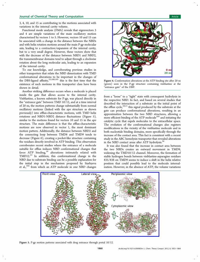
other transporters that relate the NBD dimerization with TMDconformational alterations to be important in the changes ofthe DBS-ligand affinity,33,100,101 this is the first time that theexistence of such motions in this transporter class have beenshown in detail.Another striking difference occurs when a molecule is placed
inside the gate that allows access to the internal cavity.Vinblastine, a known substrate for P-gp, was placed directly inthe “entrance gate” between TMD 10/12, and at a time intervalof 20 ns, the motion patterns change substantially from normaloscillatory motions (linked with the apo structure as shownpreviously) into efflux-characteristic motions, with TMD helixrotations and NBD1-NBD2 distance fluctuations (Figure 5),similar to the motions found for vectors 10 and 15 in the apostructure. The main difference is that the efflux-characteristicmotions are now observed in vector 1, the most dominantmotion pattern. Additionally, the distance between NBD2 andthe connecting loop between TMD8 and TMD9 tends todecrease (Figure 6), creating a pocket-like structure containingthe residues directly involved in ATP binding. This observationcorroborates recent studies where the entrance of a moleculesuitable for efflux induces NBD conformational changes thatfavor ATP binding,10 alterations intimately related withTMD12.23 In addition, this conformational change in theNBD due to substrate binding can be a possible explanation forthe initial step in the mechanism proposed by Siarhyevaet al.,102 from which an ATP molecule in one NBD changes
from a “loose” to a “tight” state with consequent hydrolysis inthe respective NBD. In fact, and based on several studies thatdescribed the interaction of a substrate as the initial point ofthe efflux cycle,10,11 this signal produced by the substrate at thegate can produce conformational alterations, resulting in anapproximation between the two NBD structures, allowing amore efficient binding of the ATP molecule102 and initiating thecatalytic cycle that expels molecules to the extracellular space.The evolution of the conformational changes also registersmodifications in the vicinity of the vinblastine molecule and inboth nucleotide binding domains, more specifically through theincrease of the contact area. This fact is consistent with a recentstudy in the ABC hemolysin transporter that revealed alterationsin the NBD contact areas after ATP hydrolysis.36
It was also found that the increase in contact area betweenthe two NBDs creates an outward movement in TMD9,widening the TMD10/12 channel. Moreover, the formation ofstable hydrogen bonds between vinblastine-asparagine residues835/838 on TMD9 seems to induce a shift in the helix relativeposition that could possibly lead to the molecule internal-ization. However, in the absence of ATP, the volume variations
Figure 5. P-gp motion patterns associated with drug entrance through portal 10/12.
Figure 6. Conformation alterations at the ATP binding site after 20 ns(green) seen in the P-gp structure containing vinblastine at the“entrance gate” of the DBP.
Journal of Chemical Theory and Computation Article
dx.doi.org/10.1021/ct300083m | J. Chem. Theory Comput. 2012, 8, 1853−18641860
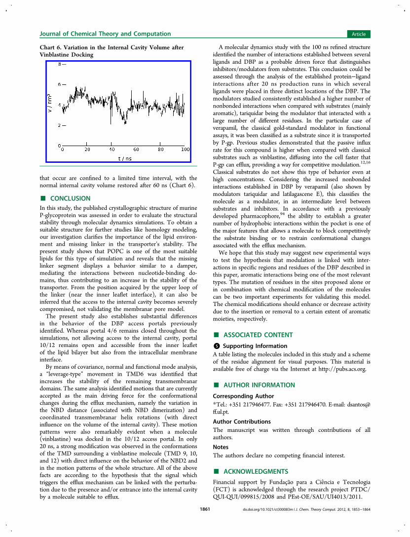
that occur are confined to a limited time interval, with thenormal internal cavity volume restored after 60 ns (Chart 6).
■ CONCLUSIONIn this study, the published crystallographic structure of murineP-glycoprotein was assessed in order to evaluate the structuralstability through molecular dynamics simulations. To obtain asuitable structure for further studies like homology modeling,our investigation clarifies the importance of the lipid environ-ment and missing linker in the transporter’s stability. Thepresent study shows that POPC is one of the most suitablelipids for this type of simulation and reveals that the missinglinker segment displays a behavior similar to a damper,mediating the interactions between nucleotide-binding do-mains, thus contributing to an increase in the stability of thetransporter. From the position acquired by the upper loop ofthe linker (near the inner leaflet interface), it can also beinferred that the access to the internal cavity becomes severelycompromised, not validating the membranar pore model.The present study also establishes substantial differences
in the behavior of the DBP access portals previouslyidentified. Whereas portal 4/6 remains closed throughout thesimulations, not allowing access to the internal cavity, portal10/12 remains open and accessible from the inner leafletof the lipid bilayer but also from the intracellular membraneinterface.By means of covariance, normal and functional mode analysis,
a “leverage-type” movement in TMD6 was identified thatincreases the stability of the remaining transmembranardomains. The same analysis identified motions that are currentlyaccepted as the main driving force for the conformationalchanges during the efflux mechanism, namely the variation inthe NBD distance (associated with NBD dimerization) andcoordinated transmembranar helix rotations (with directinfluence on the volume of the internal cavity). These motionpatterns were also remarkably evident when a molecule(vinblastine) was docked in the 10/12 access portal. In only20 ns, a strong modification was observed in the conformationsof the TMD surrounding a vinblastine molecule (TMD 9, 10,and 12) with direct influence on the behavior of the NBD2 andin the motion patterns of the whole structure. All of the abovefacts are according to the hypothesis that the signal whichtriggers the efflux mechanism can be linked with the perturba-tion due to the presence and/or entrance into the internal cavityby a molecule suitable to efflux.
A molecular dynamics study with the 100 ns refined structureidentified the number of interactions established between severalligands and DBP as a probable driven force that distinguishesinhibitors/modulators from substrates. This conclusion could beassessed through the analysis of the established protein−ligandinteractions after 20 ns production runs in which severalligands were placed in three distinct locations of the DBP. Themodulators studied consistently established a higher number ofnonbonded interactions when compared with substrates (mainlyaromatic), tariquidar being the modulator that interacted with alarge number of different residues. In the particular case ofverapamil, the classical gold-standard modulator in functionalassays, it was been classified as a substrate since it is transportedby P-gp. Previous studies demonstrated that the passive influxrate for this compound is higher when compared with classicalsubstrates such as vinblastine, diffusing into the cell faster thatP-gp can efflux, providing a way for competitive modulation.12,16
Classical substrates do not show this type of behavior even athigh concentrations. Considering the increased nonbondedinteractions established in DBP by verapamil (also shown bymodulators tariquidar and latilagascene E), this classifies themolecule as a modulator, in an intermediate level betweensubstrates and inhibitors. In accordance with a previouslydeveloped pharmacophore,94 the ability to establish a greaternumber of hydrophobic interactions within the pocket is one ofthe major features that allows a molecule to block competitivelythe substrate binding or to restrain conformational changesassociated with the efflux mechanism.We hope that this study may suggest new experimental ways
to test the hypothesis that modulation is linked with inter-actions in specific regions and residues of the DBP described inthis paper, aromatic interactions being one of the most relevanttypes. The mutation of residues in the sites proposed alone orin combination with chemical modification of the moleculescan be two important experiments for validating this model.The chemical modifications should enhance or decrease activitydue to the insertion or removal to a certain extent of aromaticmoieties, respectively.
■ ASSOCIATED CONTENT
*S Supporting InformationA table listing the molecules included in this study and a schemeof the residue alignment for visual purposes. This material isavailable free of charge via the Internet at http://pubs.acs.org.
■ AUTHOR INFORMATION
Corresponding Author*Tel.: +351 217946477. Fax: +351 217946470. E-mail: [email protected].
Author ContributionsThe manuscript was written through contributions of allauthors.
NotesThe authors declare no competing financial interest.
■ ACKNOWLEDGMENTS
Financial support by Fundacao para a Ciencia e Tecnologia(FCT) is acknowledged through the research project PTDC/QUI-QUI/099815/2008 and PEst-OE/SAU/UI4013/2011.
Chart 6. Variation in the Internal Cavity Volume afterVinblastine Docking
Journal of Chemical Theory and Computation Article
dx.doi.org/10.1021/ct300083m | J. Chem. Theory Comput. 2012, 8, 1853−18641861
■ ABBREVIATIONS
P-gp, P-glycoprotein; MDR, multidrug resistance; TMD,transmembranar domains; NBD, nucleotide-binding domains;ATP, adenosine triphosphate; DBS, drug-binding site; DBP,drug-binding pocket; DMPC, dimiristoylphosphatilylcholine;POPC, 1-palmitoyl-2-oleoyl-phospha-tidylcholine; MD, molec-ular dynamics; PBC, periodic boundary conditions; RMSD,root-mean-square deviation; RMSF, root-mean-square fluctua-tion
■ REFERENCES(1) Juliano, R. L.; Ling, V. A surface glycoprotein modulating drugpermeability in Chinese hamster ovary cell mutants. Biochim. Biophys.Acta 1976, 455, 152.(2) Sharom, F. J. The P-glycoprotein Efflux Pump: How Does itTransport Drugs? J. Membr. Biol. 1997, 160, 161−175.(3) Senior, A. E.; Al-Shawi, M. K.; Urbatsch, I. L. The catalytic cycleof P-glycoprotein. FEBS Lett. 1995, 377, 285−289.(4) Borges-Walmsley, M. I.; McKeegan, K. S.; Walmsley, A. R.Structure and function of efflux pumps that confer resistance to drugs.Biochem. J. 2003, 376, 313−338.(5) Altenberg, G. A.; Vanoye, C. G.; Horton, J. K.; Reuss, L.; Julie, K.Unidirectional fluxes of rhodamine 123 in multidrug-resistant cells:evidence against direct drug extrusion from the plasma membrane.Proc. Natl. Acad. Sci. U. S. A. 1994, 91, 4654−4657.(6) Higgins, C. F.; Gottesman, M. M. Is the multidrug transporter aflippase? Trends Biochem. Sci. 1992, 17, 18−21.(7) Raviv, Y.; Pollard, H. B.; Bruggemann, E. P.; Pastan, I.;Gottesman, M. M. Photosensitized labeling of a functional multidrugtransporter in living drug-resistant tumor cells. J. Biol. Chem. 1990,265, 3975−3980.(8) Romsicki, Y.; Sharom, F. J. The membrane lipid environmentmodulates drug interactions with the P-glycoprotein multidrugtransporter. Biochemistry 1999, 38, 6887−6896.(9) Jones, P. M.; George, A. M. Opening of the ADP-bound activesite in the ABC transporter ATPase dimer: Evidence for a constantcontact, alternating sites model for the catalytic cycle. Proteins 2009,75, 387−396.(10) Callaghan, R.; Ford, R. C.; Kerr, I. D. The translocationmechanism of P-glycoprotein. FEBS Lett. 2006, 580, 1056−1063.(11) Martin, C.; Berridge, G.; Mistry, P.; Higgins, C.; Charlton, P.;Callaghan, R. Drug Binding Sites on P-Glycoprotein Are Altered byATP Binding Prior to Nucleotide Hydrolysis. Biochemistry 2000, 39,11901−11906.(12) Eytan, G. D.; Regev, R.; Oren, G.; Assaraf, Y. G. The role ofpassive transbilayer drug movement in multidrug resistance and itsmodulation. J. Biol. Chem. 1995, 271, 12897−12902.(13) Urbatsch, I. L.; Senior, A. E. Effects of lipids on ATPase activityof purified Chinese hamster P-glycoprotein. Arch. Biochem. Biophys.1995, 316, 135−140.(14) Orlowski, S.; Martin, S.; Escargueil, A. P-glycoprotein and ’lipidrafts’: some ambiguous mutual relationships (floating on them,building them or meeting them by chance?). Cell. Mol. Life Sci.2006, 63, 1038−1059.(15) Eytan, G.; Kuchel, P. Mechanism of Action of P-Glycoprotein inRelation to Passive Membrane Permeation. Int. Rev. Cytol. 1999, 190,175−250.(16) Regev, R.; Katzir, H.; Yeheskely-Hayon, D.; Eytan, G. D.Modulation of P-glycoprotein-mediated multidrug resistance byacceleration of passive drug permeation across the plasma membrane.FEBS Lett. 2007, 274, 6204−6214.(17) Lee, A. G. Lipid−protein interactions in biological membranes:a structural perspective. Biochim. Biophys. Acta 2003, 1612, 1−40.(18) Klappe, K.; Hummel, I.; Hoekstra, D.; Kok, J. W. Lipiddependence of ABC transporter localization and function. Chem. Phys.Lipids. 2009, 161, 57−64.
(19) Locher, K. P.; Lee, A. T.; Rees, D. C. The E. coli BtuCDstructure: A framework for ABC transporter architecture andmechanism. Science 2002, 296, 1091−1098.(20) Berman, H. M.; Westbrook, J.; Feng, Z.; Gilliland, G.; Bhat,T. N.; Weissig, H.; Shindyalov, I. N.; Bourne, P. E. The Protein DataBank. Nucleic Acids Res. 2000, 28, 235−242.(21) Berman, H. M.; Battistuz, T.; Bhat, T. N.; Bluhm, W. F.;Bourne, P. E.; Burkhardt, K.; Feng, Z.; Gilliland, G. L.; Iype, L.; Jain,S.; Fagan, P.; Marvin, J.; Padilla, D.; Ravichandran, V.; Schneider, B.;Thanki, N.; Weissig, H.; Westbrook, J. D.; Zardecki, C. The ProteinData Bank. Acta Crystallogr., Sect. D: Biol. Crystallogr. 2002, 58, 899−907.(22) Aller, S. G.; Yu, J.; Ward, A.; Weng, Y.; Chittaboina, S.; Zhuo,R.; Harrell, P. M.; Trinh, Y. T.; Zhang, Q.; Urbatsch, I. L.; Chang, G.Structure of P-glycoprotein reveals a molecular basis for poly-specificdrug binding. Science 2009, 323, 1718−1722.(23) Crowley, E.; O’Mara, M. L.; Reynolds, C.; Tieleman, D. P.;Storm, J.; Kerr, I. D.; Callaghan, R. Transmembrane helix 12modulates progression of the ATP catalytic cycle in ABCB1.Biochemistry 2009, 48, 6249−6258.(24) Rothnie, A.; Storm, J.; Campbell, J.; Linton, K. J.; Kerr, I. D.;Callaghan, R. The coupling mechanism of P-glycoprotein involvesresidue L339 in the sixth membrane spanning segment. J. Biol. Chem.2004, 279, 34913−34990.(25) Ecker, G. F.; Stockner, T.; Chiba, P. Computational models forprediction of interactions with ABC-transporters. Drug DiscoveryToday 2008, 13, 311−317.(26) Dawson, R. J.; Locher, K. P. Structure of a bacterial multidrugABC transporter. Nature 2006, 443, 180−185.(27) O’Mara, M. L.; Tieleman, D. P. P-glycoprotein models of theapo and ATP-bound states based on homology with Sav1866 andMalK. FEBS Lett. 2007, 581, 4217−4222.(28) Ward, A.; Reyes, C. L.; Yu, J.; Roth, C. B.; Chang, G. Flexibilityin the ABC transporter MsbA: Alternating access with a twist. Proc.Natl. Acad. Sci. U. S. A. 2007, 104, 19005−19010.(29) Crowley, E.; Callaghan, R. Multidrug efflux pumps: drug binding− gates or cavity? FEBS J. 2010, 277, 530−539.(30) Chang, G. Retraction of “Structure of MsbA from Vibriocholera: A Multidrug Resistance ABC Transporter Homolog in aClosed Conformation” [J. Mol. Biol. (2003) 330 419−430]. J. Mol.Biol. 2007, 369, 596.(31) Dawson, R. J.; Locher, K. P. Structure of the multidrug ABCtransporter Sav1866 from Staphylococcus aureus in complex withAMP-PNP. FEBS Lett. 2007, 581, 935−938.(32) Dong, J.; Yang, G.; McHaourab, H. S. Structural basis of energytransduction in the transport cycle of MsbA. Science 2005, 308, 1023−1028.(33) Rosenberg, M. F.; Velarde, G.; Ford, R. C.; Martin, C.; Berridge,G.; Kerr, I. D.; Callaghan, R.; Schmidlin, A.; Wooding, C.; Linton,K. J.; Higgins, C. F. Repacking of the transmembrane domains ofP-glycoprotein during the transport ATPase cycle. EMBO J. 2001, 20,5615−5625.(34) Rosenberg, M. F.; Callaghan, R.; Modok, S.; Higgins, C. F.;Ford, R. C. Three-dimensional structure of P-glycoprotein: thetransmembrane regions adopt an asymmetric configuration in thenucleotide-bound state. J. Biol. Chem. 2005, 280, 2857−2862.(35) Oliveira, A. S.; Baptista, A. M.; Soares, C. M. Conformationalchanges induced by ATP-hydrolysis in an ABC transporter: amolecular dynamics study of the Sav1866 exporter. Proteins 2011,79, 1977−1990.(36) Damas, J. M.; Oliveira, A. S.; Baptista, A. M.; Soares, C. M.Structural consequences of ATP hydrolysis on the ABC transporterNBD dimer: Molecular dynamics studies of HlyB. Protein Sci. 2011,20, 1220−1230.(37) Becker, J.-P.; Depret, G.; Van Bambeke, F.; Tulkens, P. M.;Prevost, M.; Bambeke, F. V. Molecular models of human P-glycoprotein in two different catalytic states. BMC Struct. Biol. 2009,9, 1−18.
Journal of Chemical Theory and Computation Article
dx.doi.org/10.1021/ct300083m | J. Chem. Theory Comput. 2012, 8, 1853−18641862
(38) Dolghih, E.; Bryant, C.; Renslo, A. R.; Jacobson, M. P.Predicting binding to P-Glycoprotein by Flexible Receptor Docking.PLoS Comput. Biol. 2011, 7, e1002083 DOI: 10.1371/journal.-pcbi.1002083.(39) Jabeen, I.; Wetwitayaklung, P.; Klepsch, F.; Parveen, Z.; Chiba,P.; Ecker, G. F. Probing the stereoselectivity of P-glycoprotein-synthesis, biological activity and ligand docking studies of a set ofenantiopure benzopyrano[3,4-b][1,4]oxazines. Chem. Commun.(Camb.) 2011, 47, 2586−2588.(40) Tsuruo, T.; Lida, H.; Tsukagoshi, S.; Sakurai, Y. Overcoming ofVincristine Resistance in P388 Leukemia in Vivo and in Vitro throughEnhanced Cytotoxicity of Vincristine and Vinblastine by Verapamil.Cancer Res. 1981, 41, 1967−1972.(41) Mistry, P.; Stewart, A. J.; Dangerfield, W.; Okiji, S.; Liddle, C.;Bootle, D.; Plumb, J. a.; Templeton, D.; Charlton, P. In vitro and invivo reversal of P-glycoprotein-mediated multidrug resistance by anovel potent modulator, XR9576. Cancer Res. 2001, 61, 749−758.(42) Hrycyna, C. A.; Airan, L. E.; Germann, U. A.; Ambudkar, S. V.;Pastan, I.; Gottesman, M. M. Structural flexibility of the linker regionof human P-glycoprotein permits ATP hydrolysis and drug transport.Biochemistry 1998, 37, 13660−13673.(43) Sato, T.; Kodan, A.; Kimura, Y.; Ueda, K.; Nakatsu, T.; Kato, H.Functional role of the linker region in purified human P-glycoprotein.FEBS J. 2009, 276, 3504−3516.(44) Scott, W. R. P.; Hunenberger, P. H.; Tironi, I. G.; Mark, A. E.;Billeter, S. R.; Fennen, J.; Torda, A. E.; Huber, T.; Kruger, P.; vanGunsteren, W. F. The GROMOS Biomolecular Simulation ProgramPackage. J. Phys. Chem. A 1999, 103, 3596−3607.(45) Bonvin, A. M.; Mark, A. E.; van Gunsteren, W. F. TheGROMOS96 benchmarks for molecular simulation. Comput. Phys.Commun. 2000, 128, 550−557.(46) Oostenbrink, C.; Villa, A.; Mark, A. E.; van Gunsteren, W. F. Abiomolecular force field based on the free enthalpy of hydration andsolvation: the GROMOS force-field parameter sets 53A5 and 53A6.J. Comput. Chem. 2004, 25, 1656−1676.(47) Poger, D.; Mark, A. E. On the Validation of MolecularDynamics Simulations of Saturated and cis-MonounsaturatedPhosphatidylcholine Lipid Bilayers: A Comparison with Experiment.J. Chem. Theory Comput. 2010, 6, 325−336.(48) Poger, D.; Van Gunsteren, W. F.; Mark, A. E. A new force fieldfor simulating phosphatidylcholine bilayers. J. Comput. Chem. 2010, 31,1117−1125.(49) Berger, O.; Edholm, O.; Jahnig, F. Molecular DynamicsSimulations of a Fluid Bilayer of Dipalmitoylphosphatidylcholine atFull Hydration, Constant Pressure, and Constant Temperature.Biophys. J. 1997, 72, 2002−2013.(50) Chiu, S.-W.; Clark, M. A.; Balaji, V.; Subramaniam, S.; Scott, H. L.;Jakobsson, E. Incorporation of Surface Tension into Molecular DynamicsSimulation of Interface: A Fluid Phase Lipid Bilayer Membrane. Biophys.J. 1995, 69, 1230−1245.(51) Kukol, A. Lipid models for United-atom Molecular DynamicsSimulations of Proteins. J. Chem. Theory Comput. 2009, 5, 615−626.(52) Chandrasekhar, I.; Kastenholz, M.; Lins, R. D.; Oostenbrink, C.;Schuler, L. D.; Tieleman, D. P.; van Gunsteren, W. F. A consistentpotential energy parameter set for lipids: dipalmitoylphospha-tidylcho-line as a benchmark of the GROMOS96 45A3 force field. Eur. Biophys.J. 2003, 32, 67−77.(53) Chandrasekhar, I.; Oostenbrink, C.; van Gunsteren, W. F.Simulating the physiological phase of hydrated DPPC bilayers: TheEster Moiety. Soft Mater. 2004, 2, 27−45.(54) Molecular Operating Environment (MOE), 2009.10; ChemicalComputing Group Inc.: Montreal, QC, Canada, 2010.(55) Berendsen, H. GROMACS: A message-passing parallelmolecular dynamics implementation. Comput. Phys. Commun. 1995,91, 43−56.(56) Lindahl, E.; Hess, B.; van der Spoel, D. GROMACS 3.0: apackage for molecular simulation and trajectory analysis. J. Mol. Model.2001, 7, 306−317.
(57) van der Spoel, D.; Lindahl, E.; Hess, B.; Groenhof, G.; Mark,A. E.; Berendsen, H. J. GROMACS: fast, flexible, and free. J. Comput.Chem. 2005, 26, 1701−1718.(58) Kandt, C.; Ash, W. L.; Tieleman, D. P. Setting up and runningmolecular dynamics simulations of membrane proteins. Methods 2007,41, 475−488.(59) Wolf, M. G.; Hoefling, M.; Aponte-Santamaría, C.; Grubmuller,H.; Groenhof, G. g_membed: Efficient insertion of a membraneprotein into an equilibrated lipid bilayer with minimal perturbation.J. Comput. Chem. 2010, 31, 2169−2174.(60) Allen, W. J.; Lemkul, J. A.; Bevan, D. R. GridMAT-MD: a grid-based membrane analysis tool for use with molecular dynamics.J. Comput. Chem. 2009, 30, 1952−1958.(61) Hub, J. S.; de Groot, B. L. Detection of functional modes inprotein dynamics. PLoS Comput. Biol. 2009, 5, e1000480DOI: 10.1371/journal.pcbi.1000480.(62) Amadei, A.; Linssen, A. B.; Berendsen, H. J. Essential dynamicsof proteins. Proteins 1993, 17, 412−425.(63) Bakan, A.; Meireles, L. M.; Bahar, I. ProDy: protein dynamicsinferred from theory and experiments. Bioinformatics 2011, 27, 1575−1577.(64) Bahar, I.; Lezon, T. R.; Bakan, A.; Shrivastava, I. H. Normalmode analysis of biomolecular structures: functional mechanisms ofmembrane proteins. Chem. Rev. 2010, 110, 1463−1497.(65) Schuttelkopf, A. W.; van Aalten, D. M. PRODRG: a tool forhigh-throughput crystallography of protein-ligand complexes. ActaCrystallogr., Sect. D: Biol. Crystallogr. 2004, 60, 1355−1363.(66) Malde, A. K.; Zuo, L.; Breeze, M.; Stroet, M.; Poger, D.; Nair, P. C.;Oostenbrink, C.; Mark, A. E. An Automated force field Topology Builder(ATB) and repository: version 1.0. J. Chem. Theory Comput. 2011, 7,4026−4037.(67) Frisch, M. J.; Trucks, G. W.; Schlegel, H. B.; Scuseria, G. E.;Robb, M. A.; Cheeseman, J. R.; Montgomery, J. A., Jr.; Vreven, T.;Kudin, K. N.; Burant, J. C.; Millam, J. M.; Iyengar, S. S.; Tomasi, J.;Barone, V.; Mennucci, B.; Cossi, M.; Scalmani, G.; Rega, N.;Petersson, G. A.; Nakatsuji, H.; Hada, M.; Ehara, M.; Toyota, K.;Fukuda, R.; Hasegawa, J.; Ishida, M.; Nakajima, T.; Honda, Y.; Kitao,O.; Nakai, H.; Klene, M.; Li, X.; Knox, J. E.; Hratchian, H. P.; Cross,J. B.; Bakken, V.; Adamo, C.; Jaramillo, J.; Gomperts, R.; Stratmann,R. E.; Yazyev, O.; Austin, A. J.; Cammi, R.; Pomelli, C.; Ochterski,J. W.; Ayala, P. Y.; Morokuma, K.; Voth, G. A.; Salvador, P.;Dannenberg, J. J.; Zakrzewski, V. G.; Dapprich, S.; Daniels, A. D.;Strain, M. C.; Farkas, O.; Malick, D. K.; Rabuck, A. D.; Raghavachari,K.; Foresman, J. B.; Ortiz, J. V.; Cui, Q.; Baboul, A. G.; Clifford, S.;Cioslowski, J.; Stefanov, B. B.; Liu, G.; Liashenko, A.; Piskorz, P.;Komaromi, I.; Martin, R. L.; Fox, D. J.; Keith, T.; Al-Laham, M. A.;Peng, C. Y.; Nanayakkara, A.; Challacombe, M.; Gill, P. M. W.;Johnson, B.; Chen, W.; Wong, M. W.; Gonzalez, C.; Pople, J. A.Gaussian 03, revision D.01; Gaussian, Inc.: Pittsburg, PA, 2004.(68) Humphrey, W.; Dalke, A.; Schulten, K. VMD - Visual MolecularDynamics. J. Mol. Graphics 1996, 14, 33−38.(69) Lomize, M. A.; Lomize, A. L.; Pogozheva, I. D.; Mosberg, H. I.OPM: orientations of proteins in membranes database. Bioinformatics2006, 22, 623−625.(70) Petersen, B.; Petersen, T. N.; Andersen, P.; Nielsen, M.;Lundegaard, C. A generic method for assignment of reliability scoresapplied to solvent accessibility predictions. BMC Struct. Biol. 2009,9, 51.(71) Raghava, G. P. S. APSSP2: A combination method for proteinsecondary structure prediction based on neural network and examplebased learning. CASP5 2002, A-132.(72) Ouali, M.; King, R. D. Cascaded multiple classifiers forsecondary structure prediction. Protein Sci. 2000, 9, 1162−1176.(73) Chou, P. Y.; Fasman, G. D. Prediction of protein conformation.Biochemistry 1974, 13, 222−245.(74) Lovell, S. C.; Davis, I. W.; Arendall, W. B., III; de Bakker, P. I.W.; Word, J. M.; Prisant, M. G.; Richardson, J. S.; Richardson, D. C.Structure validation by Cα geometry: φ,ψ and Cβ deviation. Proteins2003, 50, 437−450.
Journal of Chemical Theory and Computation Article
dx.doi.org/10.1021/ct300083m | J. Chem. Theory Comput. 2012, 8, 1853−18641863
(75) Leekumjorn, S.; Sum, A. K. Molecular Characterization of Geland Liquid-Crystalline Structures of Fully Hydrated POPC and POPEBilayers. J. Phys. Chem. B 2007, 111, 6026−6033.(76) Duarte, N.; Gyemant, N.; Abreu, P. M.; Molnar, J.; Ferreira,M.-J. U. New macrocyclic lathyrane diterpenes, from Euphorbia lagascae,as inhibitors of multidrug resistance of tumour cells. Planta Med. 2006,72, 162−168.(77) Wallace, A. C.; Laskowski, R. A.; Thornton, J. M. LIGPLOT: aprogram to generate schematic diagrams of protein-ligand interactions.Protein Eng. 1995, 8, 127−134.(78) McDonald, I. K.; Thornton, J. M. Satisfying hydrogen bondingpotential in proteins. J. Mol. Biol. 1994, 238, 777−793.(79) Bussi, G.; Donadio, D.; Parrinello, M. Canonical samplingthrough velocity rescaling. J. Chem. Phys. 2007, 126, 14101−14107.(80) Hoover, W. Canonical dynamics: Equilibrium phase-spacedistributions. Phys. Rev. A 1985, 31, 1695−1697.(81) Nose, S.; Klein, M. L. Constant pressure molecular dynamics formolecular systems. Mol. Phys. 1983, 50, 1055−1076.(82) Parrinello, M.; Rahman, A. Polymorphic transitions in singlecrystals: A new molecular dynamics method. J. Appl. Phys. 1981, 52,7182−7190.(83) Darden, T.; York, D.; Pedersen, L. Particle mesh Ewald: AnN·log(N) method for Ewald sums in large systems. J. Chem. Phys.1993, 98, 10089.(84) Essmann, U.; Perera, L.; Berkowitz, M. L.; Darden, T.; Lee, H.;Pedersen, L. G. A smooth particle mesh Ewald method. J. Chem. Phys.1995, 103, 8577−8593.(85) Miyamoto, S.; Kollman, P. A. Settle: An analytical version of theSHAKE and RATTLE algorithm for rigid water models. J. Comput.Chem. 1992, 13, 952−962.(86) Hess, B.; Bekker, H.; Berendsen, H. J. C.; Fraaije, J. G.; LINCS,A Linear Constraint Solver for Molecular Simulations. J. Comput.Chem. 1997, 18, 1463−1472.(87) Duque, D.; Li, X.-jun; Katsov, K.; Schick, M. Molecular theoryof hydrophobic mismatch between lipids and peptides. J. Chem. Phys.2002, 116, 10478−10481.(88) Sun, T.; Liu, M.; Chen, W.; Wang, C. Molecular dynamicssimulation of the transmembrane subunit of BtuCD in the lipid bilayer.Sci. China Life Sci. 2010, 53, 620−630.(89) Rost, B.; Yachdav, G.; Liu, J. The PredictProtein server. NucleicAcids Res. 2004, 32, W321 DOI: 10.1093/nar/gkh377.(90) Kleywegt, G. J. Validation of protein crystal structures. ActaCrystallogr. 2000, D56, 249−265.(91) Law, R. J.; Capener, C.; Baaden, M.; Bond, P. J.; Campbell, J.;Patargias, G.; Arinaminpathy, Y.; Sansom, M. S. P. Membrane proteinstructure quality in molecular dynamics simulation. J. Mol. GraphicsModel. 2005, 24, 157−165.(92) Loo, T. W.; Clarke, D. M. Recent progress in understanding themechanism of P-glycoprotein-mediated drug efflux. J. Membr. Biol.2005, 206, 173−185.(93) Akiyama, S.; Cornwell, M. M.; Kuwano, M.; Pastan, I.;Gottesman, M. M. Most drugs that reverse multidrug resistance alsoinhibit photoaffinity labeling of P-glycoprotein by a vinblastine analog.Mol. Pharmacol. 1988, 33, 144−147.(94) Ferreira, R. J.; dos Santos, D. J.; Ferreira, M.-J.; Guedes, R. C.Toward a better pharmacophore description of P-glycoproteinmodulators, based on macrocyclic diterpenes from Euphorbia species.J. Chem. Inf. Model 2011, 51, 1315−1324.(95) Wiese, M.; Pajeva, I. K. Structure-activity relationships ofmultidrug resistance reversers. Curr. Med. Chem. 2001, 8, 685−713.(96) Pajeva, I. K.; Wiese, M. Structure-activity relationships oftariquidar analogs as multidrug resistance modulators. AAPS J. 2009,11, 435−444.(97) Ekins, S.; Kim, R. B.; Leake, B. F.; Dantzig, A. H.; Schuetz, E. G.;Lan, L.-B.; Yasuda, K.; Shepard, R. L.; Winter, M. A.; Schuetz, J. D.;Wikel, J. H.; Wrighton, S. A. Three-Dimensional Quantitative Structure-Activity Relationships of Inhibitors of P-Glycoprotein. Mol. Pharmacol.2002, 61, 964−973.
(98) Becker, J. P.; van Bambeke, F.; Tulkens, P. M.; Prevost, M.Dynamics and structural changes induced by ATP binding inSAV1866, a bacterial ABC exporter. J. Phys. Chem. B 2010, 114,15948−15957.(99) Ramachandra, M.; Ambudkar, S. V.; Chen, D.; Hrycyna, C. A.;Dey, S.; Gottesman, M. M.; Pastan, I. Human P-Glycoprotein ExhibitsReduced Affinity for Substrates during a Catalytic Transition State.Biochemistry 1998, 37, 4693−5038.(100) Gutmann, D. A. P.; Ward, A.; Urbatsch, I. L.; Chang, G.; vanVeen, H. W. Understanding polyspecificity of multidrug ABCtransporters: closing in on the gaps in ABCB1. Trends Biochem. Sci.2010, 35, 36−42.(101) Gyimesi, G.; Ramachandran, S.; Kota, P.; Dokholyan, N. V.;Sarkadi, B.; Hegedus, T. ATP hydrolysis at one of the two sites in ABCtransporters initiates transport related conformational transitions.Biochim. Biophys. Acta 2011, 1808, 2954−2964.(102) Siarheyva, A.; Liu, R.; Sharom, F. Characterization of anasymmetric occluded state of P-glycoprotein with two boundnucleotides: implications for catalysis. J. Biol. Chem. 2010, 285,7575−7586.
Journal of Chemical Theory and Computation Article
dx.doi.org/10.1021/ct300083m | J. Chem. Theory Comput. 2012, 8, 1853−18641864


