Embed Size (px)
Citation preview
Journal of Biotechnology 125 (2006) 11–21
Influence of DNA condensation state on transfection efficiency inDNA/polymer complexes: An AFM and DLS comparative study
C. Volcke a,∗,1, S. Pirotton b,1, Ch. Grandfils d,∗∗,1, C. Humbert a,1,P.A. Thiry a, I. Ydens c, P. Dubois c, M. Raes b,∗ ∗ ∗
a Laboratoire de Spectroscopie Moleculaire de Surface (LASMOS), University of Namur, 5000 Namur, Belgiumb Research Unit of Cell Biology (URBC), University of Namur, 5000 Namur, Belgium
c Laboratory of Polymeric and Composite Materials (LPCM), University of Mons-Hainaut, Mons, Belgiumd Interfaculty Center of Biomaterials, University of Liege, Liege, Belgium
Received 17 August 2005; received in revised form 30 January 2006; accepted 17 February 2006
Abstract
Atomic force microscopy (AFM) is used to describe the formation process of polymer/DNA complexes. Two main objectivesof this research are presented. The first one is to apply AFM as an effective tool to analyse DNA molecules and differentpolycation/DNA complexes in order to evaluate their degree of condensation (size and shape). The other one is to search fora relationship between the condensation state of DNA and its transfection efficiency. In this study, linear methacrylate based
polymers and globular SuperFect polymers are used in order to induce DNA condensation. Ternary complexes, composed ofmethacrylate based polymers and polyethylene glycol (PEG)-based copolymers, are also investigated. AFM allows us to confirmgood condensation conditions and relate them (or not) to transfection efficiencies. These AFM results (obtained after dryingin air) are compared with measurements deduced from Dynamic Light Scattering (DLS) experiments performed in water. Thiscomparison allowed us to identify the structural modifications resulting from deposition on the mica surface.© 2006 Elsevier B.V. All rights reserved.Keywords: Atomic force microscopy; DNA; Gene therapy vehicle; Dynamic Light Scattering
∗ Corresponding author. Tel.: +32 81 72 4712; fax: +32 81 72 4718.∗∗ Corresponding author. Tel.: +32 4 36 63 506;
fax: +32 4 36 63 623.∗∗∗ Corresponding author. Tel.: +32 81 72 4124; fax: +32 81 72 4135.
E-mail addresses: [email protected] (C. Volcke),[email protected] (Ch. Grandfils),[email protected] (M. Raes).
1 These authors contributed equally.
1. Introduction
The development of an efficient gene delivery vehi-cle is a major challenge for gene therapy. The DNAcondensation process has thus drawn a large inter-est in biology the last decade. Two types of vectorsof nucleic acids exist: viral agents (like recombinantretroviruses and adenoviruses (Ledley, 1996; Wilson,1995)) and synthetic vectors (as cationic lipids and
0168-1656/$ – see front matter © 2006 Elsevier B.V. All rights reserved.doi:10.1016/j.jbiotec.2006.02.010
12 C. Volcke et al. / Journal of Biotechnology 125 (2006) 11–21
polymers). However, viral agents yet suffer from alimited success in delivering genes, partially due tothe immune and toxic response they induce, but alsobecause of biosafety problems (Miller, 2003; Maguire-Zeiss and Federoff, 2004; Feldman, 2003). To avoidthese problems, non-viral carriers are presently inves-tigated in many laboratories. For example, DNA con-densation is achieved with proteins like protamine(Allen et al., 1997) or liposomes and lipid inter-mediates (Gao and Huang, 1996). Promising resultsare also obtained with polycationic polymers (van deWetering et al., 1997, 1998), such as poly-l-lysine(Choi et al., 1999; Toncheva et al., 1998) or protonatedamino-functionalised polymethacrylate (Pirotton et al.,2004). Such polymer/DNA complexes are formedthrough electrostatic interactions between the nega-tively charged phosphate groups of DNA and proto-nated amino groups of the polymer.
Although polycationic polymers have some advan-tages over viral vectors, their efficiency as a transfect-ing agent is still limited (Itaka et al., 2003). Indeed, sev-eral biophysical requirements on such polyelectrolyteparticles are encountered. Apart from the polymer char-acteristics (charge density, molecular weight), whichhave been previously discussed (Pirotton et al., 2004),we wondered if the physico-chemical properties of theformed polymer/DNA complexes influence the trans-fection efficiency process. Indeed, a physical view ofthe condensates (including their dimensions) relatedto a biological view (including transfection efficiency)cnftt1mimYeafi
scH
ticles from the environment. The interactions betweenthe serum proteins and the complexes often impair thetransfection in vitro. In vivo, these interactions lead tothe rapid elimination of the complexes by phagocytos-ing macrophages (Howard et al., 2000). These prob-lems can be avoided by the introduction of hydrophilicsequences of poly(ethylene glycol) (PEG) at the surfaceof the complexes. Previous results indeed highlightedthat cell transfection with binary complexes is feasibleexclusively in the absence of serum and that, on thecontrary, the PEGylated complexes allowed transfec-tion even in the presence of serum. The presence ofthe hydrophilic sequences of PEG at the complex sur-face also improved the hemocompatibility propertiesof the complexes (Pirotton et al., 2004). These kinds ofcomplexes are also analysed by AFM.
Finally, globular dendrimeric polymers are also usedto condense DNA. Dendrimers represent a class ofpolymers that exhibit a molecular architecture char-acterized by regular dendritic branching with a radialsymmetry (Frechet, 1994). Their structural differencecompared to linear polycationic polymers induces dif-ferent condensed structures, which are also observedand discussed in detail.
More precisely, in this research, linear methacrylatebased polycationic polymers (PDMAEMA), PEGy-lated linear methacrylate based polycationic polymersand globular SuperFect dendrimers are used in orderto induce DNA condensation. AFM data obtainedon PDMAEMA/DNA binary complexes are exposedawtbtDdfiod
2
2
g
ould facilitate improvement in this gene delivery tech-ology (Dunlap et al., 1997). In this context, atomicorce microscopy (AFM) has proven to be an excellentool having the ability to image soft biological struc-ures like cells, bacteria or proteins (Radmacher et al.,992; Razatos et al., 1998; Reich et al., 2001). DNAolecules, such as plasmids in particular, have been
ntensively studied and detailed structures of individualolecules have yet been revealed (Hansma et al., 1992;ang et al., 1992; Lyubchenko et al., 1993; Argamant al., 1997). Moreover, AFM measurements have thedvantage on DLS experiments that they can be per-ormed after deposition on any surface and have anntrinsic better resolution.
Polycationic polymethacrylate derived chains areufficient to condense DNA in order to produce binaryomplexes capable of transfecting cells in culture.owever, these chains alone do not protect the par-
nd discussed as a function of the polymer/DNAeight ratio and transfection efficiency. Characteris-
ics of ternary complexes are then compared to theinary complex properties. Measurements obtained onhese two kinds of complexes are thus compared toynamic Light Scattering data, obtained in wet con-itions. The globular SuperFect/DNA structures arenally exposed. In order to appreciate the architecturef polymer/DNA complexes, an image of uncondensedsDNA has been provided for comparison.
. Materials and methods
.1. Complex formation
The pCMV� plasmid, with the �-galactosidaseene of Escherichia coli under control of the CMV pro-
C. Volcke et al. / Journal of Biotechnology 125 (2006) 11–21 13
moter (Clontech, USA), was amplified and purified atlarge scale by Plasmid Factory (Germany). It is a plas-mid of 7.2 kb (corresponding to a Mw = 4.77 × 10⁶ Da)with a high supercoiled content and low bacterialendotoxins (<0.1 E.U./�g DNA). It has been purchasedin water (1 mg/ml). The poly(2-dimethylaminoethylmethacrylate) (PDMAEMA) homopolymers andP(DMAEMA-b-MAPEG) palm-tree like copolymers(where MAPEG stands for methacrylate end-functionalised polyethylene glycol macromonomer)are synthesized by atom transfer radical polymerisa-tion (ATRP) as described elsewhere (Pantoustier etal., 2003; Pirotton et al., 2004). The average molarmass (Mn) and the polydispersity index (Mw/Mn) ofthe homopolymer are 49,100 and 1.59, respectively,as determined by size exclusion chromatography withreference to poly(methylmethacrylate) standards.For the palm-tree like copolymer, Mn is 24,100and the polydispersity index is 1.45. The averagemole number of tertiary amino groups per gram ofpolymer (N) is determined by titration: it is 6.32 and4.22 mmol/g for the homopolymer and the copolymer,respectively.
Ternary complexes are prepared as follows: a smallvolume of concentrated PDMAEMA is mixed with theDNA and left to condense for 30 min at room tem-perature. Afterwards, the copolymer is added for asecond incubation during 30 min. When PDMAEMAis tested alone, the solvent is added after the first 30 minincubation instead of the PEG-based copolymer. Thefifsfc1Q
2
fiH(DCpd
well plates (2 × 10⁴ cells/well) in complete culturemedium. The cells are then incubated with the com-plexes, prepared as described above, in DMEM + 5%FBS under a humidified 5% CO2 atmosphere, at 37 ◦C.After 3 h, the excess of complexes is removed, the cellsare washed with DMEM and further cultured for 24 hin culture medium with 5% FBS. In order to evalu-ate the �-galactosidase expression, cells are washedtwice with PBS and lysed with the mammalian pro-tein extraction reagent (M-PER), obtained from Pierce(USA). Hundred microliter of �-galactosidase sub-strate (o-nitrophenyl �-d-galactopyranoside or ONPG,4.4 mM) are mixed with 100 �l of cell lysate and incu-bated at 37 ◦C. Half an hour later, A405 (absorbanceat 405 nm wavelength) is measured. For each sam-ple, the protein concentration is determined accord-ing to the BIO-RAD Protein Assay protocol withbovine serum albumin (BSA) as standard. Resultsare expressed in arbitrary units (a.u.) per 100 �g ofproteins, as the mean standard deviation of triplicatedeterminations.
For transfection experiments, DNA needs to be con-densed by polycations. Complexes formed with twotypes of polycationic polymers, differing by their struc-ture, have been analysed: on the one hand, SuperFect(SF) is a polyamidoamine dendrimer of the sixth gen-eration and has a globular structure. On the other hand,PDMAEMA is a polymer with a linear structure. Bothtypes of condensing agents have a similar molar mass(
2
pofawiAs(arbop
nal concentration of DNA is 20 �g/ml. Complexes areormed in water for the AFM and in a HEPES-bufferedolution (HBS: 20 mM HEPES, 155 mM NaCl, pH 7.4)or the transfection experiments. The SuperFect/DNAomplexes are prepared by immersion in water during5 min at a 6/1 (w/w) ratio. SuperFect is provided byiagen, Germany.
.2. Transfection
Cos-7 cells are cultured in Dulbecco’s Modi-ed Eagle’s Medium (DMEM) supplemented withEPES (20 mM), penicillin (100 U/ml), streptomycin
100 �g/ml) and Fœtal Bovine Serum (FBS) (10%).MEM and FBS were obtained from Gibco Invitrogenorporation (UK). Penicillin and streptomycin wereurchased from Biowhittaker (Verviers, Belgium). Theay before transfection, the cells are seeded in 12-
45,000–49,000).
.3. AFM imaging
Prior to AFM measurements, the complex sus-ension is diluted 10-times with water and 2 �lf the resulting solution is then deposited onto areshly cleaved mica substrate. Samples are imagedfter water evaporation. Experiments are performedith a commercial STM/AFM (Nanoscope IIIa, Dig-
tal Instruments) operating in tapping mode (TM)FM, using standard silicon cantilevers with ∼42 N/m
pring constant and 369.22 kHz working frequencytrue resonance frequency = 369.32 kHz, A0 = 23.3 nmnd Asp = 5.5 nm). All images are recorded in air atoom temperature, at a scan speed of 2.5 Hz. Theackground slope is resolved using first or secondrders polynomial functions. No further filtering iserformed.
14 C. Volcke et al. / Journal of Biotechnology 125 (2006) 11–21
2.4. Dynamic Light Scattering
Particle size measurements are performed byDynamic Light Scattering (DLS) analysis adoptinga Brookhaven Instrument (BI 2030 AT linear digitalcorrelator; 128 channels). The time-variations of thescattered light intensity (emitted by particles in solu-tion) are analysed at 90◦. Samples are incubated at25 ◦C in water. The incident Ar laser wavelength isfixed at 488 nm (∼10 mW). The quality of the instru-ment is checked with a Beckman Coulter certified latexstandard (PCS control L300). A minimum of three mea-surements is performed on each sample. The autocor-relation curves are deconvoluted with either Cumulant,Exponential Sampling or Contin methods.
3. Results and discussion
Results exposed in this paper highlight the for-mation of complexes from dsDNA in the presenceof polymers. Previous studies using the AFM tech-nique demonstrated the condensation of closed andlinear dsDNA in the presence of different condensingagents (Dunlap et al., 1997; Rackstraw et al., 2001;Vijayanathan et al., 2004; Lin et al., 1998; Martin etal., 2000; Reschel et al., 2002; Hansma et al., 1998).Such agents produce different sizes of condensatesand require different incubation times and concentra-tions. Concerning the polymers used in our experiment,oba
3
(PcTdtsdisD
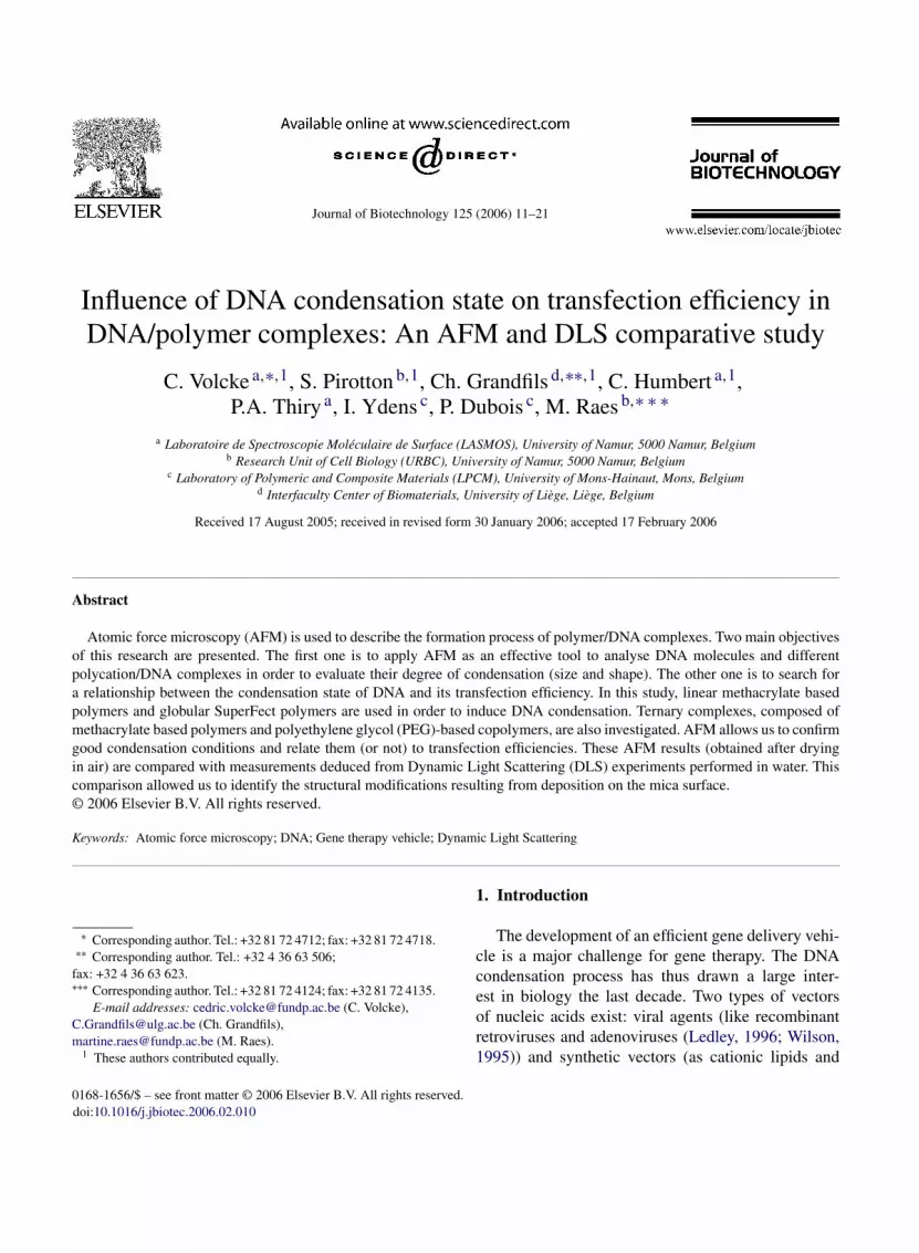
Fig. 1. High-resolution TM-AFM image (1 �m × 1 �m) of plasmidDNA observed on mica.
everywhere in the image, the average apparent heightand width of DNA molecules correspond to 0.6 and20.0 nm, respectively. The DNA strands are piled up atthe upper left part of Fig. 1 (indicated by an arrow). Atthis point, the DNA strands have a height of 1.0 nm,about twice a helix apparent height.
3.2. Methacrylate based complexes
3.2.1. PDMAEMA/DNA binary complexesThe DNA used to prepare the complexes is the
pCMV� plasmid, encoding the �-galactosidase ofEscherichia coli. The transfection is defined as theintroduction of exogenous genetic material, its subse-quent internalization into a host cell (the transfectedcell) and the subsequent expression of the encodedprotein. In our case, the transfection efficiency is deter-mined by measuring the �-galactosidase activity (�-gal) in transfected cells. Fig. 2 exhibits results oftransfection experiments (representing the �-gal activ-ity as a function of the polymer/DNA weight ratio)carried out at a fixed plasmid concentration and a vary-ing PDMAEMA concentration. Cationic polymers thuselectrostatically bind to plasmid molecules (presentinga net negative charge), leading to polymer–DNA com-plexes. It seems evident that PDMAEMA promotes thecellular uptake and its subsequent expression. More-over, the transfection efficiency is dramatically affectedby the weight ratio and presents a maximum. The max-i
ptimal conditions (charge, polymer/DNA ratios, incu-ation times. . .) have been determined previously andpplied in this paper (Pirotton et al., 2004).
.1. Plasmid DNA
We performed AFM imaging in the tapping modeTM-AFM). The results are stable and reproducible.lasmid DNA molecules deposited onto freshlyleaved mica substrate are imaged after drying in air.he substrate appears to be homogeneously flat withouble-stranded DNA (dsDNA) molecules on it. Aypical TM-AFM high-resolution image obtained bycanning over a small area is presented in Fig. 1. ThesDNA molecules display a closed geometry, look-ng like relaxed circles, with little twisting of thetrands. This structure is characteristic of uncondensedNA morphology (Rackstraw et al., 2001). Almost
mal �-gal activity is observed when complexes are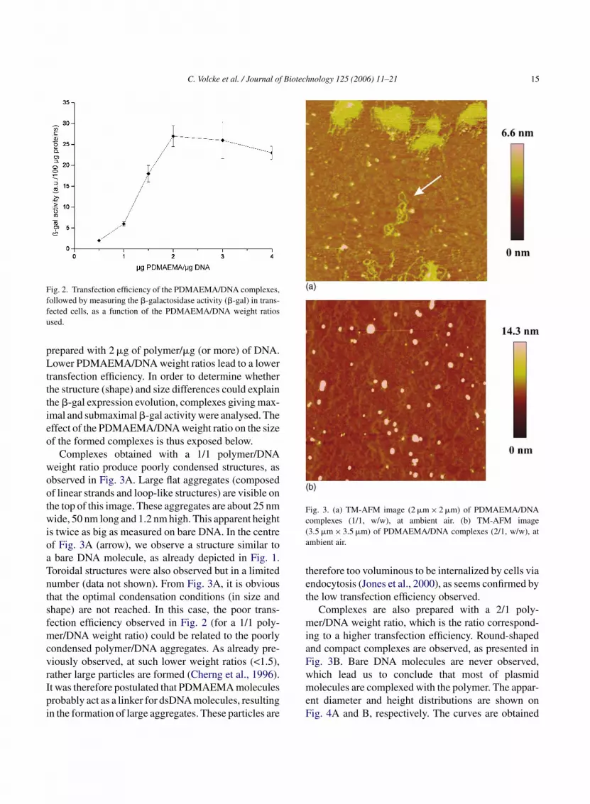
C. Volcke et al. / Journal of Biotechnology 125 (2006) 11–21 15
Fig. 2. Transfection efficiency of the PDMAEMA/DNA complexes,followed by measuring the �-galactosidase activity (�-gal) in trans-fected cells, as a function of the PDMAEMA/DNA weight ratiosused.
prepared with 2 �g of polymer/�g (or more) of DNA.Lower PDMAEMA/DNA weight ratios lead to a lowertransfection efficiency. In order to determine whetherthe structure (shape) and size differences could explainthe �-gal expression evolution, complexes giving max-imal and submaximal �-gal activity were analysed. Theeffect of the PDMAEMA/DNA weight ratio on the sizeof the formed complexes is thus exposed below.
Complexes obtained with a 1/1 polymer/DNAweight ratio produce poorly condensed structures, asobserved in Fig. 3A. Large flat aggregates (composedof linear strands and loop-like structures) are visible onthe top of this image. These aggregates are about 25 nmwide, 50 nm long and 1.2 nm high. This apparent heightis twice as big as measured on bare DNA. In the centreof Fig. 3A (arrow), we observe a structure similar toa bare DNA molecule, as already depicted in Fig. 1.Toroidal structures were also observed but in a limitednumber (data not shown). From Fig. 3A, it is obviousthat the optimal condensation conditions (in size andshape) are not reached. In this case, the poor trans-fection efficiency observed in Fig. 2 (for a 1/1 poly-mer/DNA weight ratio) could be related to the poorlycondensed polymer/DNA aggregates. As already pre-viously observed, at such lower weight ratios (<1.5),rather large particles are formed (Cherng et al., 1996).It was therefore postulated that PDMAEMA moleculesprobably act as a linker for dsDNA molecules, resultingin the formation of large aggregates. These particles are
Fig. 3. (a) TM-AFM image (2 �m × 2 �m) of PDMAEMA/DNAcomplexes (1/1, w/w), at ambient air. (b) TM-AFM image(3.5 �m × 3.5 �m) of PDMAEMA/DNA complexes (2/1, w/w), atambient air.
therefore too voluminous to be internalized by cells viaendocytosis (Jones et al., 2000), as seems confirmed bythe low transfection efficiency observed.
Complexes are also prepared with a 2/1 poly-mer/DNA weight ratio, which is the ratio correspond-ing to a higher transfection efficiency. Round-shapedand compact complexes are observed, as presented inFig. 3B. Bare DNA molecules are never observed,which lead us to conclude that most of plasmidmolecules are complexed with the polymer. The appar-ent diameter and height distributions are shown onFig. 4A and B, respectively. The curves are obtained
16 C. Volcke et al. / Journal of Biotechnology 125 (2006) 11–21
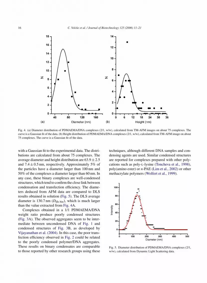
Fig. 4. (a) Diameter distribution of PDMAEMA/DNA complexes (2/1, w/w), calculated from TM-AFM images on about 75 complexes. Thecurve is a Gaussian fit of the data. (b) Height distribution of PDMAEMA/DNA complexes (2/1, w/w), calculated from TM-AFM image on about75 complexes. The curve is a Gaussian fit of the data.
with a Gaussian fit to the experimental data. The distri-butions are calculated from about 75 complexes. Theaverage diameter and height distribution are 63.9 ± 2.5and 7.4 ± 0.5 nm, respectively. Approximately 5% ofthe particles have a diameter larger than 100 nm and50% of the complexes a diameter larger than 60 nm. Inany case, these binary complexes are well-condensedstructures, which tend to confirm the close link betweencondensation and transfection efficiency. The diame-ters deduced from AFM data are compared to DLSresults obtained in solution (Fig. 5). The DLS averagediameter is 130.7 nm (Ddls bin), which is much largerthan the value extracted from Fig. 4A.
Complexes obtained in a 1/1 PDMAEMA/DNAweight ratio produce poorly condensed structures(Fig. 3A). The observed aggregates seem to be inter-mediate between uncondensed DNA of Fig. 1 andcondensed structures of Fig. 3B, as developed byVijayanathan et al. (2004). In this case, the poor trans-fection efficiency observed in Fig. 2 could be relatedto the poorly condensed polymer/DNA aggregates.These results on binary condensates are comparableto those reported by other research groups using these
techniques, although different DNA samples and con-densing agents are used. Similar condensed structuresare reported for complexes prepared with other poly-cations such as poly-l-lysine (Toncheva et al., 1998),poly(amino ester) or n-PAE (Lim et al., 2002) or othermethacrylate polymers (Wolfert et al., 1999).
Fig. 5. Diameter distribution of PDMAEMA/DNA complexes (2/1,w/w), calculated from Dynamic Light Scattering data.

C. Volcke et al. / Journal of Biotechnology 125 (2006) 11–21 17
Fig. 6. TM-AFM image (4 �m × 4 �m) of copolymer/polymer/DNA ternary complexes (5/2/1, w/w/w), at ambient air.
3.2.2. Ternary complexesA typical image of ternary complexes is presented
in Fig. 6. The complexes are prepared with 2 �g ofPDMAEMA and 5 �g of the copolymer. This is a poly-mer/DNA ratio giving maximal transfection activity(data not shown). The ternary complexes form verycompact structures, similar to the PDMAEMA/DNA
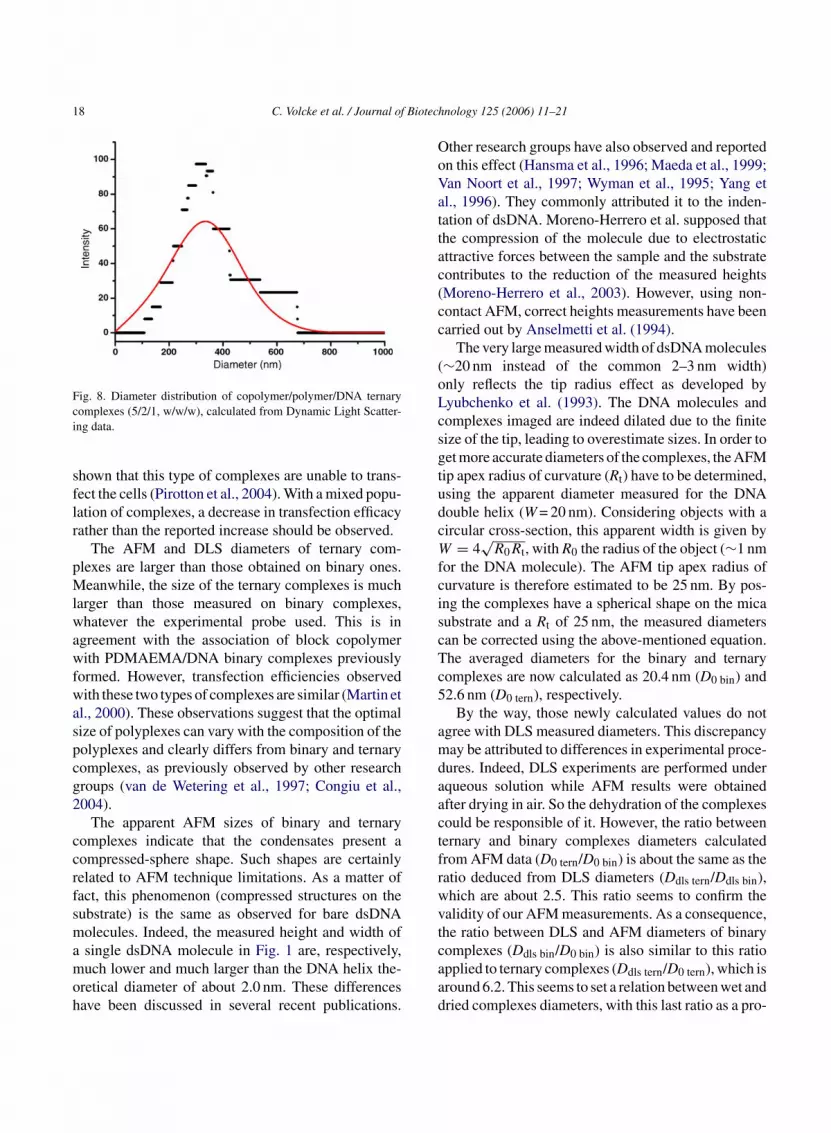
complexes. No tubular structure in or around the con-densates is visible. Apparent diameter and height dis-tributions are shown on Fig. 7A and B, respectively.These distributions are calculated from about 100 com-plexes. The apparent diameter and height mean valuesare 102.6 ± 2.0 and 9.7 ± 0.1 nm, respectively. Thesedimensions are about twice larger than those measuredfor the binary complexes. DLS experiments revealedthe diameter distribution of wet complexes, as shownin Fig. 8. The average diameter is 329.6 nm (Ddls tern),which is about three times larger than the one calculatedfrom Fig. 7A (obtained after drying in air).
The AFM image of Fig. 6 highlights the pres-ence of dense structures, indicating the formation ofternary complexes. Therefore, the two-steps prepara-tion method of those complexes prevents the interferingof the PEG during the condensation of the DNA withthe PDMAEMA, as previously observed (Rackstraw etal., 2001). Nevertheless, it was not demonstrated thatPEG copolymer does not substitute the PDMAEMA.However, it is unlikely because of the transfection effi-cacy of the ternary complexes. Indeed, if substitutionoccurred, there would be copolymer/DNA complexesin the population and previous studies have clearly
Fig. 7. (a) Diameter distribution of copolymer/polymer/DNA ternary comp1 istributic ve is a G
00 complexes. The curve is a Gaussian fit of the data. (b) Height dalculated from TM-AFM images on about 100 complexes. The cur
lexes, (5/2/1, w/w/w), calculated from TM-AFM images on abouton of copolymer/polymer/DNA ternary complexes (5/2/1, w/w/w),aussian fit of the data.
18 C. Volcke et al. / Journal of Biotechnology 125 (2006) 11–21
Fig. 8. Diameter distribution of copolymer/polymer/DNA ternarycomplexes (5/2/1, w/w/w), calculated from Dynamic Light Scatter-ing data.
shown that this type of complexes are unable to trans-fect the cells (Pirotton et al., 2004). With a mixed popu-lation of complexes, a decrease in transfection efficacyrather than the reported increase should be observed.
The AFM and DLS diameters of ternary com-plexes are larger than those obtained on binary ones.Meanwhile, the size of the ternary complexes is muchlarger than those measured on binary complexes,whatever the experimental probe used. This is inagreement with the association of block copolymerwith PDMAEMA/DNA binary complexes previouslyformed. However, transfection efficiencies observedwith these two types of complexes are similar (Martin etal., 2000). These observations suggest that the optimalsize of polyplexes can vary with the composition of thepolyplexes and clearly differs from binary and ternarycomplexes, as previously observed by other researchgroups (van de Wetering et al., 1997; Congiu et al.,2004).
The apparent AFM sizes of binary and ternarycomplexes indicate that the condensates present acompressed-sphere shape. Such shapes are certainlyrelated to AFM technique limitations. As a matter offact, this phenomenon (compressed structures on thesubstrate) is the same as observed for bare dsDNAmolecules. Indeed, the measured height and width ofa single dsDNA molecule in Fig. 1 are, respectively,much lower and much larger than the DNA helix the-oretical diameter of about 2.0 nm. These differenceshave been discussed in several recent publications.
Other research groups have also observed and reportedon this effect (Hansma et al., 1996; Maeda et al., 1999;Van Noort et al., 1997; Wyman et al., 1995; Yang etal., 1996). They commonly attributed it to the inden-tation of dsDNA. Moreno-Herrero et al. supposed thatthe compression of the molecule due to electrostaticattractive forces between the sample and the substratecontributes to the reduction of the measured heights(Moreno-Herrero et al., 2003). However, using non-contact AFM, correct heights measurements have beencarried out by Anselmetti et al. (1994).
The very large measured width of dsDNA molecules(∼20 nm instead of the common 2–3 nm width)only reflects the tip radius effect as developed byLyubchenko et al. (1993). The DNA molecules andcomplexes imaged are indeed dilated due to the finitesize of the tip, leading to overestimate sizes. In order toget more accurate diameters of the complexes, the AFMtip apex radius of curvature (Rt) have to be determined,using the apparent diameter measured for the DNAdouble helix (W = 20 nm). Considering objects with acircular cross-section, this apparent width is given byW = 4
√R0Rt, with R0 the radius of the object (∼1 nm
for the DNA molecule). The AFM tip apex radius ofcurvature is therefore estimated to be 25 nm. By pos-ing the complexes have a spherical shape on the micasubstrate and a Rt of 25 nm, the measured diameterscan be corrected using the above-mentioned equation.The averaged diameters for the binary and ternarycomplexes are now calculated as 20.4 nm (D0 bin) and5
amdaactfrwvtcaad
2.6 nm (D0 tern), respectively.By the way, those newly calculated values do not
gree with DLS measured diameters. This discrepancyay be attributed to differences in experimental proce-
ures. Indeed, DLS experiments are performed underqueous solution while AFM results were obtainedfter drying in air. So the dehydration of the complexesould be responsible of it. However, the ratio betweenernary and binary complexes diameters calculatedrom AFM data (D0 tern/D0 bin) is about the same as theatio deduced from DLS diameters (Ddls tern/Ddls bin),hich are about 2.5. This ratio seems to confirm thealidity of our AFM measurements. As a consequence,he ratio between DLS and AFM diameters of binaryomplexes (Ddls bin/D0 bin) is also similar to this ratiopplied to ternary complexes (Ddls tern/D0 tern), which isround 6.2. This seems to set a relation between wet andried complexes diameters, with this last ratio as a pro-
C. Volcke et al. / Journal of Biotechnology 125 (2006) 11–21 19
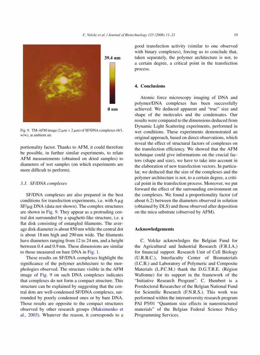
Fig. 9. TM-AFM image (2 �m × 2 �m) of SF/DNA complexes (6/1,w/w), at ambient air.
portionality factor. Thanks to AFM, it could thereforebe possible, in further similar experiments, to relateAFM measurements (obtained on dried samples) todiameters of wet samples (on which experiments aremore difficult to perform).
3.3. SF/DNA complexes
SF/DNA complexes are also prepared in the bestconditions for transfection experiments, i.e. with 6 �gSF/�g DNA (data not shown). The complex structuresare shown in Fig. 9. They appear as a protruding cen-tral dot surrounded by a spaghetti-like structure, i.e. aflat disk consisting of entangled filaments. The aver-age disk diameter is about 850 nm while the central dotis about 18 nm high and 290 nm wide. The filamentshave diameters ranging from 12 to 24 nm, and a heightbetween 0.4 and 0.9 nm. These dimensions are similarto those measured on bare DNA in Fig. 1.
These results on SF/DNA complexes highlight thesignificance of the polymer architecture to the mor-phologies observed. The structure visible in the AFMimage of Fig. 9 on such DNA complexes indicatesthat complexes do not form a compact structure. Thisstructure can be explained by suggesting that the cen-tral dots are well-condensed SF/DNA complexes, sur-rounded by poorly condensed ones or by bare DNA.Those results are opposite to the compact structuresobserved by other research groups (Maksimenko eta
good transfection activity (similar to one observedwith binary complexes), forcing us to conclude that,taken separately, the polymer architecture is not, toa certain degree, a critical point in the transfectionprocess.
4. Conclusions
Atomic force microscopy imaging of DNA andpolymer/DNA complexes has been successfullyachieved. We deduced apparent and “true” size andshape of the molecules and the condensates. Ourresults were compared to the dimensions deduced fromDynamic Light Scattering experiments, performed inwet conditions. These experiments demonstrated anoriginal approach, based on direct observations, whichreveal the effect of structural factors of complexes onthe transfection efficiency. We showed that the AFMtechnique could give informations on the crucial fac-tors (shape and size), we have to take into account inthe elaboration of new transfection vectors. In particu-lar, we deduced that the size of the complexes and thepolymer architecture is not, to a certain degree, a criti-cal point in the transfection process. Moreover, we putforward the effect of the surrounding environment onthe complexes. We found a proportionality factor (ofabout 6.2) between the diameters observed in solution(obtained by DLS) and those observed after depositionon the mica substrate (observed by AFM).
A
tf((MW“PfpPmP
l., 2003). Whatever the reason, it corresponds to acknowledgements
C. Volcke acknowledges the Belgian Fund forhe Agricultural and Industrial Research (F.R.I.A.)or financial support. Research Unit of Cell BiologyU.R.B.C.), Interfaculty Center of BiomaterialsI.C.B.) and Laboratory of Polymeric and Composite
aterials (L.P.C.M.) thank the D.G.T.R.E. (Regionallonne) for its support in the framework of the
Initiative Research Program”. C. Humbert is aostdoctoral Researcher of the Belgian National Fundor Scientific Research (F.N.R.S.). This work waserformed within the interuniversity research programAI P5/01 “Quantum size effects in nanostructuredaterials” of the Belgian Federal Science Policyrogramming Services.
20 C. Volcke et al. / Journal of Biotechnology 125 (2006) 11–21
References
Allen, M.J., Bradbury, E.M., Balhorn, R., 1997. AFM analysis ofDNA–protamine complexes bound to mica. Nucleic Acids Res.25, 2221–2226.
Anselmetti, D., Dreier, M., Luthi, R., Richmond, T., Meyer, E.,Frommer, J., Gunterodt, H.-J., 1994. Biological materials stud-ied with dynamic force microscopy. J. Vac. Sci. Technol. B 12,1500–1503.
Argaman, M., Golan, R., Thomson, N.H., Hansma, H.G., 1997. Phaseimaging of moving DNA molecules and DNA molecules repli-cated in the atomic force microscope. Nucleic Acids Res. 25,4379–4384.
Cherng, J.-Y., van de Wetering, P., Talsma, H., Crommelin,D.A., Hennink, W.E., 1996. Effect of size and serum proteinson transfection efficiency of poly((2-dimethylamino)ethylmethacrylate)-plasmid nanoparticles. Pharm. Res. 13,1038–1042.
Choi, J.S., Lee, E.J., Choi, Y.H., Jeong, Y.J., Park, J.S., 1999.Poly(ethylene glycol)-block-poly(l-lysine) dendrimer: novel lin-ear polymer/dendrimer block copolymer forming a sphericalwater-soluble polyionic complex with DNA. Bioconjug. Chem.10, 62–65.
Congiu, A., Pozzi, D., Esposito, C., Castellano, C., Mossa, G., 2004.Correlation between structure and transfection efficiency: a studyof DC–Chol–DOPE/DNA complexes. Colloids Surf. B Biointer-faces 36, 43–48.
Dunlap, D.D., Maggi, A., Soria, M.R., Monaco, L., 1997.Nanoscopic structure of DNA condensed for gene delivery.Nucleic Acids Res. 25, 3095–3101.
Feldman, S.H., 2003. Components of gene therapy experimentationthat contribute to relative risk. Comp. Med. 53, 147–158.
Frechet, J.M., 1994. Functional polymers and dendrimers: reactiv-ity, molecular architecture and interfacial energy. Science 263,1710–1715.
G
H
H
H
H
I
DNA and poly(ethylene glycol)-poly(l-lysine) block copolymeras serum-tolerable polyplex system: physicochemical propertiesof micelles relevant to gene transfection efficiency. Biomaterials24, 4495–4506.
Jones, N.A., Hill, I.R.C., Stolnik, S., Bignotti, F., Davis, S.S., Garnett,M.C., 2000. Polymer chemical structure is a key determinantof physicochemical and colloidal properties of polymer–DNAcomplexes for gene delivery Biochim. Biophys. Acta 1517, 1–18.
Ledley, F.D., 1996. Pharmaceutical approach to somatic gene ther-apy. Pharm. Res. 13, 1595–1614.
Lyubchenko, Y., Shlyakhtenko, L., Harrington, R., Oden, P., Lindsay,S., 1993. Atomic force microscopy of long DNA: imaging in airand under water. Proc. Natl. Acad. Sci. U.S.A. 90, 2137–2140.
Lim, Y., Kim, S., Suh, H., Park, J., 2002. Biodegradable, endosomedisruptive, and cationic network-type polymer as a highly effi-cient and nontoxic gene delivery carrier. Bioconjug. Chem. 13,952–957.
Lin, Z., Wang, C., Feng, X., Liu, M., Li, J., Bai, C., 1998. The observa-tion of the local ordering characteristics of spermidine-condensedDNA: atomic force microscopy and polarizing microscopy stud-ies. Nucleic Acids Res. 26, 3228–3234.
Maeda, Y., Matsumoto, T., Kawai, T., 1999. Observation of single-and double-stranded DNA using non-contact atomic forcemicroscopy. Appl. Surf. Sci. 140, 400–405.
Maguire-Zeiss, K.A., Federoff, H.J., 2004. Safety of viral vectors forneurological gene therapies. Curr. Opin. Mol. Ther. 6, 473–481.
Maksimenko, A.V., Mandrouguine, V., Gottikh, M.B., Bertrand, J.-R., Majoral, J.-P., Malvy, C., 2003. Optimisation of dendrimer-mediated gene transfer by anionic oligomers. J. Gene Med. 5,61–71.
Martin, A.L., Davies, M.C., Rackstraw, B.J., Roberts, C.J., Stolnik,S., Tendler, S.J.B., Williams, P.M., 2000. Observation of DNA-polymer condensate formation in real time at a molecular level.FEBS Lett. 480, 106–112.
Miller, A.D., 2003. The problem with cationic liposome/micelle-
M
V
P
P
R
ao, X., Huang, L., 1996. Potentiation of cationic liposome-mediated gene delivery by polycations. Biochemistry 35,1027–1036.
ansma, H.G., Vesenka, J., Siegerist, C., Kelderman, G., Morrett, H.,Sinsheimer, R.L., Elings, V., Bustmante, C., Hansma, P.K., 1992.Reproducible imaging and dissection of plasmid DNA under liq-uid with the atomic force microscope. Science 256, 1180–1184.
ansma, H.G., Revenko, I., Kim, K., Laney, D.E., 1996. Atomicforce microscopy of long and short double-stranded, single-stranded and triple-stranded nucleic acids. Nucleic Acids Res.24, 713–720.
ansma, H.G., Golan, R., Hsieh, W., Lollo, C.P., Mullen-Ley, P.,Kwoh, D., 1998. DNA condensation for gene therapy as mon-itored by atomic force microscopy. Nucleic Acids Res. 26,2481–2487.
oward, K.A., Dash, P.R., Read, M.L., Ward, K., Tomkins, L.M.,Nazarova, O., Ulbrich, K., Seymour, L.W., 2000. Influence ofhydrophilicity of cationic polymers on the biophysical proper-ties of polyelectrolyte complexes formed by self-assembly withDNA. Biochim. Biophys. Acta 1475, 245–255.
taka, K., Yamauchi, K., Harada, A., Nakamura, K., Kawaguchi,H., Kataoka, K., 2003. Polyion complex micelles from plasmid
based non-viral vector systems for gene therapy. Curr. Med.Chem. 10, 1195–1211.
oreno-Herrero, F., Colchero, J., Baro, A.M., 2003. DNA height inscanning force microscopy. Ultramicroscopy 96, 167–174.
an Noort, S.J.T., Van der Werf, K.O., De Grooth, B.G., Van Hulst,N.F., Greve, J., 1997. Height anomalies in tapping mode atomicforce microscopy in air caused by adhesion. Ultramicroscopy 69,117–127.
antoustier, N., Moins, S., Wautier, M., Degee, P., Dubois,P., 2003. Solvent-free synthesis and purification of poly[2-(dimethylamino)ethyl methacrylate] by atom transfer radicalpolymerisation. Chem. Commun. 3, 340–342.
irotton, S., Muller, C., Pantoustier, N., Botteman, F., Collinet,S., Grandfils, C., Dandrifosse, G., Degee, P., Dubois, P., Raes,M., 2004. Enhancement of transfection efficiency throughrapid and noncovalent post-PEGylation of Poly(dimethylamino-ethylmethacrylate)/DNA complexes. Pharm. Res. 21, 1471–1479.
ackstraw, B.J., Martin, A.L., Stolnik, S., Roberts, C.J., Garnett,M.C., Davies, M.C., Tendler, S.J.B., 2001. Microscopic investi-gations into PEG-cationic polymer-induced DNA condensation.Langmuir 17, 3185–3193.
C. Volcke et al. / Journal of Biotechnology 125 (2006) 11–21 21
Radmacher, M., Tilmann, R.W., Fritz, M., Gaub, H.E., 1992. Frommolecules to cells: imaging soft samples with the atomic forcemicroscope. Science 257, 1900–1905.
Razatos, A., Ong, Y.-L., Sharma, M.M., Georgiou, G., 1998. Molec-ular determinants of bacterial adhesion monitored by atomicforce microscopy. Proc. Natl. Acad. Sci. U.S.A. 95, 11059–11064.
Reich, Z., Kapon, R., Pilpel, Y., Zmora, S., Scolnik, Y., 2001.Scanning force microscopy in the applied biological sciences.Biotechnol. Adv. 19, 451–485.
Reschel, T., Konak, C., Oupicky, D., Seymour, L.W., Ulbrich, K.,2002. Physical properties and in vitro transfection efficiency ofgene delivery vectors based on complexes of DNA with syntheticpolycations. J. Control. Release 81, 201–217.
Toncheva, V., Wolfert, M.A., Dash, P.R., Oupicky, D., Ulrich,K., Seymour, L., Schacht, E.H., 1998. Novel vectors for genedelivery formed by self-assembly of DNA with poly(l-lysine)grafted with hydrophilic polymers. Biochim. Biophys. Acta1380, 354–368.
Vijayanathan, V., Thomas, T., Antony, T., Shirahata, A., Thomas,T.J., 2004. Formation of DNA nanoparticles in the presenceof novel polyamine analogues: a laser light scattering andatomic force microscopic study. Nucleic Acids Res. 32, 127–134.
van de Wetering, P., Cherng, J.-Y., Talsma, H., Hennink, W.E.,1997. Relation between transfection efficiency and cytotoxic-ity of poly(2-(dimethylamino)ethyl methacrylate)/plasmid com-plexes. J. Control. Release 49, 59–69.
van de Wetering, P., Cherng, J.-Y., Talsma, H., Crommelin, D.J.A.,Hennink, W.E., 1998. 2-(dimethylamino)ethyl methacrylatebased (co)polymers as gene transfer agents. J. Control. Release53, 145–153.
Wilson, J.M., 1995. Adenovirus-mediated gene transfer to liver. Adv.Drug Deliv. Rev. 17, 303–307.
Wolfert, M.A., Dash, P.R., Nazarova, O., Oupicky, D., Seymour,L.W., Smart, S., Strohalm, J., Ulrich, K., 1999. Polyelectrolytevectors for gene delivery: influence of cationic polymer on bio-physical properties of complexes formed with DNA. Bioconjug.Chem. 10, 993–1004.
Wyman, C., Grotkopp, E., Bustamante, C., Nelson, H.C.M., 1995.Determination of heat-shock transcription factor 2 stoichiome-try at looped DNA complexes using scanning force microscopy.EMBO J. 14, 117–123.
Yang, J., Takeyasu, K., Shao, Z., 1992. Atomic force microscopy ofDNA molecules. FEBS 301, 173–176.
Yang, G., Vesenka, J.P., Bustamante, C.J., 1996. Effects of tip-sampleforces and humidity on the imaging of DNA with a scanning forcemicroscope. Scanning 18, 344–350.


