Embed Size (px)
Citation preview
McCarthy et al. Skeletal Muscle 2012, 2:8
Skeletal Musclehttp://www.skeletalmusclejournal.com/content/2/1/8
METHODOLOGY Open Access
Inducible Cre transgenic mouse strain for skeletalmuscle-specific gene targetingJohn J McCarthy1,2, Ratchakrit Srikuea2,5, Tyler J Kirby3, Charlotte A Peterson1,4 and Karyn A Esser1,2,6*
Abstract
Background: The use of the Cre/loxP system for gene targeting has been proven to be a powerful tool forunderstanding gene function. The purpose of this study was to create and characterize an inducible, skeletalmuscle-specific Cre transgenic mouse strain.
Methods: To achieve skeletal muscle-specific expression, the human α-skeletal actin promoter was used to driveexpression of a chimeric Cre recombinase containing two mutated estrogen receptor ligand-binding domains.
Results: Western blot analysis, PCR and β-galactosidase staining confirmed that Cre-mediated recombination wasrestricted to limb and craniofacial skeletal muscles only after tamoxifen administration.
Conclusions: A transgenic mouse was created that allows inducible, gene targeting of floxed genes in adultskeletal muscle of different developmental origins. This new mouse will be of great utility to the skeletal musclecommunity.
Keywords: Skeletal muscle-specific, Cre recombinase, Inducible
BackgroundThe ability to manipulate the murine genome hasproven to be instrumental in the understanding of genefunction in vivo. In particular, the use of the Cre/loxPsystem has allowed investigators to circumvent the lim-itations of conventional gene targeting strategy by pro-viding temporal and tissue-specific control over geneexpression [1]. A number of different skeletal muscle-specific Cre mice have been used to alter gene expres-sion during embryonic development and in the adult. Inthe most popular strains, the muscle creatine kinase(MCK), human α-skeletal actin (HSA), myogenic factor5 (Myf5), myosin light chain 1/3 fast (MLC1/3f ), myo-genic differentiation 1 (Myod1), myogenin (Myog) orpaired box gene 7 (Pax7) promoters have been used todrive expression [2-18]. A search of the CREATE con-sortium database (http://www.creline.org/) revealed that,of the dozen or so skeletal muscle fiber-specific Cremice available, only two were inducible. One strain useda mutated estrogen receptor ligand-binding domain
* Correspondence: [email protected] for Muscle Biology, University of Kentucky, Lexington, KY 40536, USA2Department of Physiology, College of Medicine, University of Kentucky,Lexington, KY 40536, USAFull list of author information is available at the end of the article
© 2012 McCarthy et al.; licensee BioMed CentCommons Attribution License (http://creativecoreproduction in any medium, provided the orig
(CreMer) to control the timing of Cre-mediated recom-bination via tamoxifen activation [3]. The second indu-cible mouse strain employed a muscle-specific Tet-Onsystem to control Cre expression following administra-tion of the tetracycline analogue doxycycline [18].To provide a readily available mouse strain to the skel-
etal muscle community, we generated our own skeletalmuscle-specific, inducible Cre strain and characterizedthe effectiveness of recombination in both limb and cra-niofacial muscles. The design of our transgene was basedon a previous Cre transgenic strain that achieved a highdegree of skeletal muscle specificity using the HSA pro-moter [2]. We modified the previously described HSA-Cre transgene by substituting Cre with a MerCreMer(MCM) cDNA, thus making the system inducible by re-quiring tamoxifen binding to induce Cre-mediated re-combination [2]. Characterization of the HSA-MCMmouse demonstrated skeletal muscle-specific expressionof the MCM protein and that recombination only oc-curred following tamoxifen administration. Moreover, inaddition to limb muscle, we observed recombination incraniofacial muscle, thus expanding the utility of thismouse strain for the study of gene function in skeletalmuscles of different developmental origins.
ral Ltd. This is an Open Access article distributed under the terms of the Creativemmons.org/licenses/by/2.0), which permits unrestricted use, distribution, andinal work is properly cited.

Cre MerHSA promoter BGI Mer AMerCre
Figure 1 A schematic of the HSA-MCM transgene. The promoterand first exon (−2,000 to +239 relative to the transcription start site)of the human α-skeletal actin (HSA) gene drives expression of theMerCreMer (MCM) gene which harbors a mutated estrogen receptor(Mer) ligand-binding domain on each terminus of the Crerecombinase gene. The β-globin intron ΙΙ (BGI) and poly(A) tail wereincorporated into the transgene to ensure proper splicing andtranscript stability, respectively. The positions of the PCR primersused for genotyping are indicated by half-arrows.
McCarthy et al. Skeletal Muscle 2012, 2:8 Page 2 of 7http://www.skeletalmusclejournal.com/content/2/1/8
MethodsCloning of the HSA-MerCreMer transgeneThe design of the transgene is based on a previousmuscle-specific Cre transgene reported by Miniou andcolleagues (1999); however, we replaced the Cre cDNAwith an inducible form of Cre described by Verrou et al.(1999) [2,19]. The inducible Cre contained a mutated es-trogen receptor (Mer) ligand-binding domain at boththe N- and C-termini and was designated as MerCreMer(MCM) to be consistent with the cardiac-specific MCMstrain [20]. To generate the transgene, the promoter andfirst exon (−2,000 to +239 relative to the transcriptionstart site) of the HSA gene was amplified from humangenomic DNA (Promega, Madison, WI, USA) andcloned into ClaI site of the SG5 expression vector (Agi-lent Technologies, Santa Clara, CA, USA) upstream ofthe β-globin intron II. The MCM cDNA was then ampli-fied from the pANMerCreMer expression vector (a kindgift from Dr Reth) and cloned into the EcoRI site of thepSG5-HSA plasmid to generate the pSG5-HSA-MCM.The mutation (G525R) introduced into the estrogen re-ceptor ligand-binding domain has been shown to abolishestradiol binding while retaining the ability to bind 4-hydroxytamoxifen [21]. The plasmid was then sequencedfor verification. The HSA-MCM transgene was releasedfrom the plasmid by HindIII/NsiI enzyme digestion, gel-purified using the QIAquick Gel Extraction Kit accord-ing to the manufacturer’s directions (Qiagen, Valencia,CA, USA) and then provided to the University ofKentucky Transgenic Mouse facility for microinjection.
Generation and screening of HSA-MCM transgenic linesAll animal procedures were conducted in accordancewith institutional guidelines for the care and use of la-boratory animals as approved by the Institutional Ani-mal Care and Use Committee of the University ofKentucky. The HSA-MCM transgene was introducedinto F2 embryos derived from the mating of C57BL/6 XC3H (B6C3F1) parents. Production of mice was per-formed by the staff of the University of Kentucky Trans-genic Mouse facility. Genomic DNA was isolated fromtail biopsies of eight offspring using the DNeasy Blood &Tissue Kit (Qiagen) and screened for the presence of theHSA-MCM transgene by PCR using the following pri-mers: forward, 5′-GCATGGTGGAGATCTTTGA-3′; re-verse, 5′-GCTTCTGTCCG TTTGCCGGTCG-3′. Theprimers spanned the C-terminus MerCre junction (seeFigure 1) and produced a 717-bp product. Five of theoffspring were positive for the presence of the HSA-MCM transgene. To determine which of the HSA-MCMfounder lines showed germline transmission and werecapable of inducible, muscle-specific Cre-mediated re-combination, each line was bred to a lacZ reportermouse line (B6;129 S4-Gt(ROSA)26Sortm1Sor/J, stock
number 003474) purchased from The Jackson Labora-tory (Bar Harbor, ME, USA) and originally described bySoriano [22]. A second reporter mouse strain, containinga lacZ with a nuclear localization signal, was also usedto assess the ability of the HSA-MCM strain to drive in-ducible recombination and label adult skeletal musclenuclei. This second reporter mouse was described byYamamoto and colleagues and purchased from The Jack-son Laboratory (FVB.Cg-Gt(ROSA)26Sortm1(CAG-lacZ,-
EGFP)Glh/J, stock number 012429) [23].
Western blot analysisTo expand the potential utility of the HSA-MCM strain,we determined the expression of the MCM protein inskeletal muscles of different developmental origins. Togenerate total protein lysates for Western blot analysis,tissue samples were collected from limb muscles (Gstn,gastrocnemius; Pln, plantaris; Sol, soleus; EDL, extensordigitorum longus; Quad, quadriceps; TA, tibialis anter-ior), craniofacial muscles (EOM, extraocular muscle;Mastr, masseter; Tong, tongue), the heart (Hrt), samplescontaining smooth muscle (Stom, stomach; S. Int, smallintestine) and nonmuscle tissue (Lung; Panc, pancreas;Liver; Brain; Fat; Spln, spleen; Kdny, kidney). Tissuesamples (about 20 mg) were homogenized by using thePolytron PowerGen 125 (Fischer Scientific, Suwanee,GA, USA) in homogenization buffer (1% Nonidet P-40,0.5% sodium deoxycholate, 0.1% SDS, 50 mM NaCl,400 mM KCl, 25 mM β-glycerophosphate, 50 mM NaF,5 mM benzamidine, 20 mM Tris�HCl (pH 7.6), 1 mMethylenediaminetetraacetic acid, 1 mM sodium orthova-nadate, 5 mMN-ethylmaleimide, 1 mM phenylmethyl-sulfonyl fluoride) supplemented with protease inhibitorcocktail (catalog no. P8340; Sigma-Aldrich, St Louis,MO, USA). The muscle homogenates were then centri-fuged for 10 min at 10,000 g at 4°C, and the protein con-centration of the supernatant was determined using theBradford protein assay (Bio-Rad Laboratories, Hercules,CA, USA). Ten micrograms per sample were separatedby SDS-PAGE (8% gel) and then transferred to
McCarthy et al. Skeletal Muscle 2012, 2:8 Page 3 of 7http://www.skeletalmusclejournal.com/content/2/1/8
nitrocellulose membrane (0.2 μm) (Bio-Rad Laborator-ies). The membrane was incubated in blocking buffer(5% nonfat dry milk in Tris-buffered saline (TBS) plus0.1% Tween-20 (TBS-T)) for 1 hour at roomtemperature and then incubated in blocking buffer over-night at 4°C with a 1:3,000 dilution of the primary anti-body. An antibody against the estrogen receptor-α (ERα)(MC-20; Santa Cruz Biotechnology, Santa Cruz, CA,USA) was used to detect the MCM protein and anti-bodies against γ-tubulin and glucose-6-phosphate de-hydrogenase (T359 and A-9527, respectively; Sigma-Aldrich) were used to evaluate loading between samples.The ERα antibody was able to distinguish between theendogenous ERα (66 kDa) and MCM (112 kDa) proteinsbased on their significant difference in molecular weightas previously shown [19]. After the overnight incubation,the membrane was washed for 5 minutes four times inTBS-T and then incubated with a horseradish peroxid-ase-conjugated secondary antibody (2 ng/ml) for 45minutes at room temperature in blocking buffer. Follow-ing this incubation, the membrane was washed again inTBS-T as described above, incubated for 5 minutes inchemiluminescence substrate (ECL Primer WesternBlotting Detection Reagent; GE Healthcare, Piscataway,NJ, USA) and then visualized by exposure to X-ray film.
β-galactosidase assayTissue was excised and mounted on an aluminum-cov-ered cork block, covered in O.C.T. compound, frozen inliquid nitrogen-cooled isopentane and then stored at−80°C until sectioning. Tissue sections (10 μm) were air-dried for 30 minutes, rehydrated in PBS for 10 minutes,fixed in 0.2% glutaraldehyde for 7 minutes at roomtemperature and then washed briefly three times in PBS.Fixed sections were then incubated overnight in 5-bromo-4-chloro-3-indolyl-β-D-galactopyranoside (X-gal)working solution at 37°C in a humidified chamber. TheX-gal working solution contained 5 mM potassium hex-acyanoferrate(III), 5 mM potassium hexacyanoferrate(II)trihydrate, 2 mM MgCl2 and 1 mg/ml of X-gal. Follow-ing the overnight incubation, sections were washed threetimes for 5 minutes per wash in PBS, dehydrated in 95%ethanol for 1 minute twice, 100% ethanol for 1 minutetwice, cleared for 1 minute in xylene and then mountedon a coverslip using Permount mounting media. For nu-clear localized β-galactosidase detection, transcardialperfusion was performed using ice-cold PBS containing10 U of heparin followed by freshly prepared, ice-cold4% paraformaldehyde. The Gstn muscle was dissectedout and fixed for an additional 60 minutes in 4% paraf-ormaldehyde, which was followed by a series of rinses inPBS. Tissue was cryoprotected by then being placed in a15% (wt/vol) sucrose solution until equilibration, fol-lowed by immersion in a 30% sucrose solution until
equilibration, each performed at 4°C. Tissue was thentransferred to a 1:1 (vol/vol) mixture of 30% sucrose andO.C.T. compound (Tissue-Tek; Sakura Finetek USA,Inc, Torrance, CA,) for 30 minutes and then embeddedin O.C.T. compound and frozen in an ethanol, dry-icesolution. Tissue sections were viewed using a NikonE600 microscope (Nikon Inc, Melville, NY, USA), andimages were captured with a SPOT RT digital camera(Diagnostic Instruments, Inc, Sterling Heights, MI, USA)and a PowerMac G4 computer (Apple Computer Inc,Cupertino, CA, USA) equipped with SPOT RT softwareversion 4.0 (Diagnostic Instruments, Inc).
PCR analysis of cre-mediated recombinationPCR was performed to assess recombination followingtamoxifen administration. The PCR conditions and pri-mer sequences used were as described by Takehashiet al. [24].
Results and discussionA schematic of the HSA-MCM transgene is presented inFigure 1. We used the HSA promoter (−2,000 to +239)to drive skeletal muscle-specific expression of the CrecDNA as others have done [2]. Additionally, the β-globinintron II was incorporated into the transgene to ensureproper splicing [2,25]. To make the system inducible, wereplaced the Cre cDNA with a modified Cre that con-tained a Mer ligand-binding domain at both N- and C-termini, thereby creating the MCM chimeric protein[19,21]. We decided to use the MCM protein based onthe finding that it has been shown to be more tightlyregulated (that is, less recombinant in the absence oftamoxifen) than the single CreMer fusion protein, withno loss in recombination efficiency [19] .To confirm the ability of the HSA promoter to drive
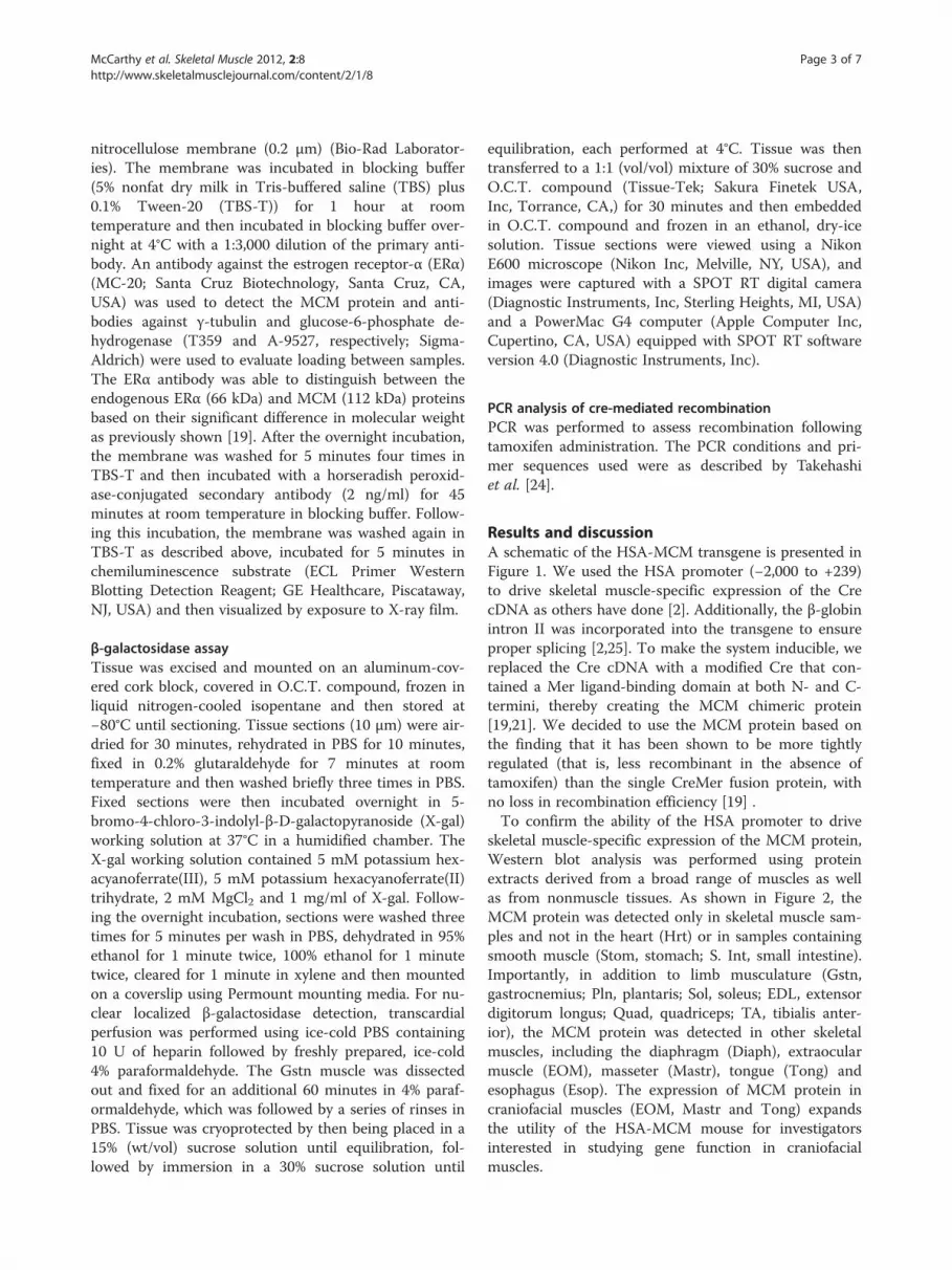
skeletal muscle-specific expression of the MCM protein,Western blot analysis was performed using proteinextracts derived from a broad range of muscles as wellas from nonmuscle tissues. As shown in Figure 2, theMCM protein was detected only in skeletal muscle sam-ples and not in the heart (Hrt) or in samples containingsmooth muscle (Stom, stomach; S. Int, small intestine).Importantly, in addition to limb musculature (Gstn,gastrocnemius; Pln, plantaris; Sol, soleus; EDL, extensordigitorum longus; Quad, quadriceps; TA, tibialis anter-ior), the MCM protein was detected in other skeletalmuscles, including the diaphragm (Diaph), extraocularmuscle (EOM), masseter (Mastr), tongue (Tong) andesophagus (Esop). The expression of MCM protein incraniofacial muscles (EOM, Mastr and Tong) expandsthe utility of the HSA-MCM mouse for investigatorsinterested in studying gene function in craniofacialmuscles.
MCM
GAPDH
TUBG1
Hrt EsopLung
StomPan
cLive
rS.In
tGst
nPln Sol
EDLGst
nBra
inFat Spln
KdnyEOM
Mastr
Tong
QuadTA Diap
h
150
100
37
50
Figure 2 Skeletal muscle-specific expression of MerCreMer (MCM) protein. Western blot analysis of different muscle and nonmuscle tissuesfrom the human α-skeletal actin (HSA)-MCM transgenic strain detected the MCM protein (112 kDa) only in skeletal muscle (Esop, esophagus; Gstn,gastrocnemius; Pln, plantaris; Sol, soleus; EDL, extensor digitorum longus; EOM, extraocular muscle; Mastr, masseter; Tong, tongue; Quad,quadriceps; TA, tibialis anterior; Diaph, diaphragm) and not in the heart (Hrt), samples containing smooth muscle (Stom, stomach; S. Int, smallintestine) or nonmuscle tissue (Lung; Panc, pancreas; Liver; Brain; Fat; Spln, spleen; Kdny, kidney). Both glyceraldehyde 3-phosphatedehydrogenase (GAPDH) and tubulin, γ1 (TUBG1) were used as loading controls.
McCarthy et al. Skeletal Muscle 2012, 2:8 Page 4 of 7http://www.skeletalmusclejournal.com/content/2/1/8
Having established the skeletal muscle-specific expres-sion of the MCM protein, we next wanted to determinethe effectiveness of the system to mediate recombinationin response to tamoxifen administration. To provide a
kb
32
1
0.4
v t v t v t v
Gstn Pln Sol
A
B
STOP
3.2 kb
LacZ
0.4 kb
Crereco
floxed allele
deleted allele
primers
primers
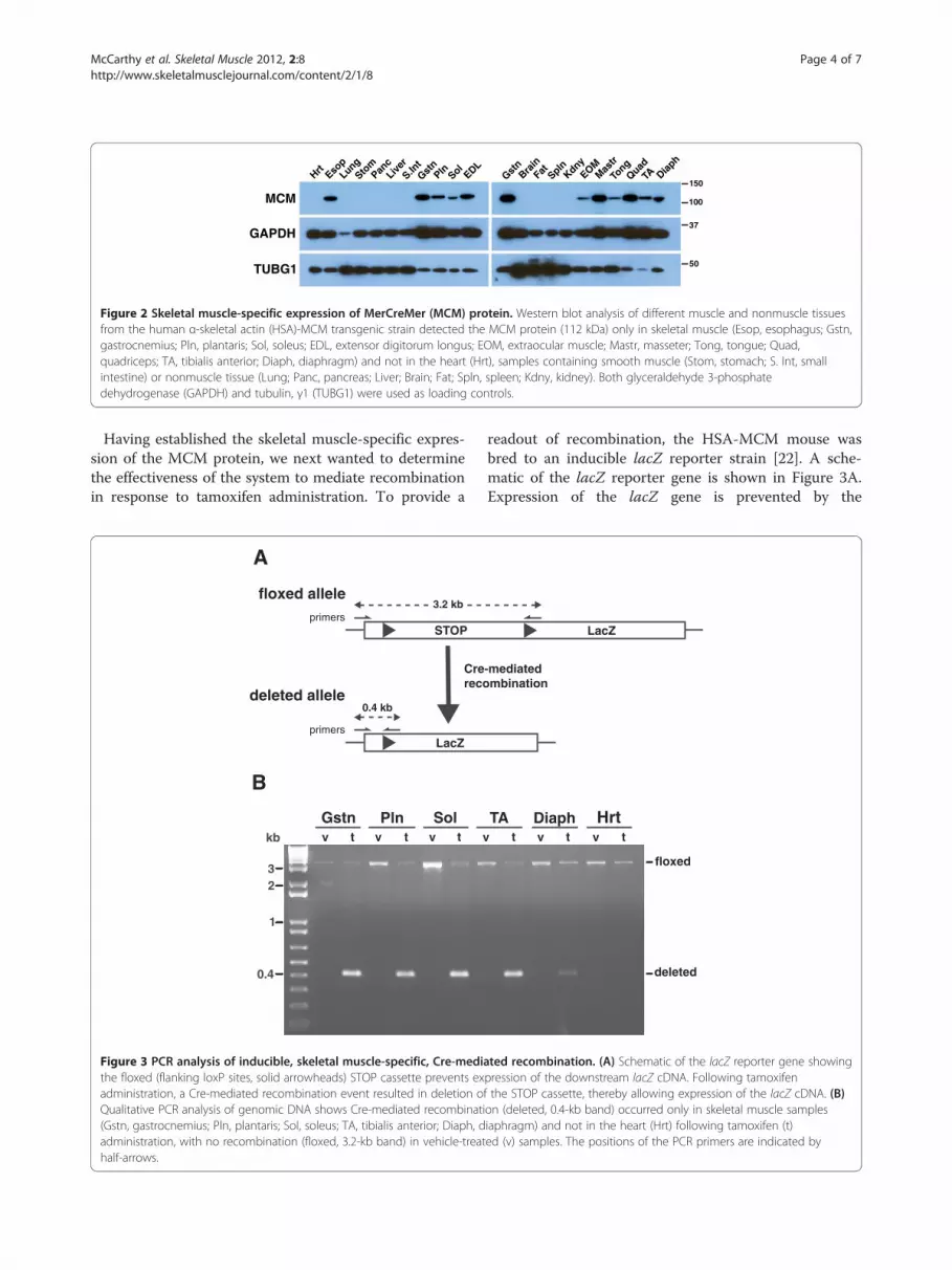
Figure 3 PCR analysis of inducible, skeletal muscle-specific, Cre-medithe floxed (flanking loxP sites, solid arrowheads) STOP cassette prevents exadministration, a Cre-mediated recombination event resulted in deletion oQualitative PCR analysis of genomic DNA shows Cre-mediated recombinati(Gstn, gastrocnemius; Pln, plantaris; Sol, soleus; TA, tibialis anterior; Diaph, dadministration, with no recombination (floxed, 3.2-kb band) in vehicle-treathalf-arrows.
readout of recombination, the HSA-MCM mouse wasbred to an inducible lacZ reporter strain [22]. A sche-matic of the lacZ reporter gene is shown in Figure 3A.Expression of the lacZ gene is prevented by the
t v t v t
TA Diaph Hrt
floxed
deleted
LacZ
-mediatedmbination
ated recombination. (A) Schematic of the lacZ reporter gene showingpression of the downstream lacZ cDNA. Following tamoxifenf the STOP cassette, thereby allowing expression of the lacZ cDNA. (B)on (deleted, 0.4-kb band) occurred only in skeletal muscle samplesiaphragm) and not in the heart (Hrt) following tamoxifen (t)ed (v) samples. The positions of the PCR primers are indicated by

Tamoxifen Vehicle
Gstn
Pln
Sol
EDL
Diaph
EOM
Hrt
Liver
TA
VehicleTamoxifen
BA
Figure 4 Skeletal muscle-specific recombination as assessed by β-galactosidase activity. The HSA-MCM strain was bred to a lacZ reportermouse to visually assess tamoxifen-induced recombination. (A) consistent with the Western blot analysis and PCR results, strong β-galactosidaseexpression (blue precipitate) was observed only in skeletal muscle (Gstn, gastrocnemius; Pln, plantaris; Sol, soleus; TA, tibialis anterior; EDL,extensor digitorum longus; Diaph, diaphragm; EOM, extraocular muscle) and not in the heart (Hrt) or liver following tamoxifen administration. (B)When we used a second lacZ reporter mouse (that contained a nuclear localization signal), β-galactosidase-positive, “blue” myonuclei wereobserved in Gstn skeletal muscle after tamoxifen treatment, but not in vehicle-treated Gstn. Inset shows enlarged image of labeled nuclei thatreside within the muscle fiber.
McCarthy et al. Skeletal Muscle 2012, 2:8 Page 5 of 7http://www.skeletalmusclejournal.com/content/2/1/8

McCarthy et al. Skeletal Muscle 2012, 2:8 Page 6 of 7http://www.skeletalmusclejournal.com/content/2/1/8
upstream presence of a STOP cassette that is flanked byloxP (floxed) sites. Upon exposure to tamoxifen, Crepromotes a recombination event that leads to excision ofthe STOP cassette and expression of lacZ cDNA. PCRanalysis was employed to detect this recombinationevent using genomic DNA isolated from different skel-etal muscles as a template. As shown in Figure 3B, re-combination was detected only in skeletal musclesamples following tamoxifen administration, consistentwith the expression of the MCM protein. These resultsdemonstrate the MCM protein is capable of effective re-combination that is tightly regulated, as no recombin-ation event was detected in vehicle-treated samples.To localize Cre-mediated recombination at the cellular
level, we performed histology to detect LacZ expressionby assaying β-galactosidase activity following addition ofthe X-gal substrate. Consistent with the Western blot andPCR analyses, we observed X-gal staining in all skeletalmuscle fibers of tamoxifen-treated mice (see Figure 4A).Importantly, no X-gal staining was observed in vehicle-treated samples, confirming that the MCM-mediated re-combination requires tamoxifen treatment. We did, how-ever, detect low-level X-gal staining in the heart, and,though this finding was somewhat surprising because ofthe lack of detection of the MCM protein in the heart, itis consistent with the findings of Collins et al. [26]. Usinga quantitative Northern blot method, Collins and collea-gues found a low level of α-skeletal actin mRNA in theadult mouse heart [26]. Thus, the minor amount of X-galstaining in the heart likely reflects the function of theHSA promoter rather than a transgenic artifact or leaki-ness of the MCM system.Although all skeletal muscles were positive for X-gal
staining, variability in the intensity of staining amongdifferent skeletal muscle samples was apparent. It is im-portant to note that there does not appear to be a linearrelationship between X-gal staining intensity and thelevel of recombination, as our PCR results show similarlevels of recombination in the soleus and gastrocnemiusmuscles; yet the staining intensities between these twomuscles are different. Moreover, there does not appearto be any relationship between X-gal staining intensityand either type I or type II fiber composition, consistentwith what has been reported for other HSA-Cre strains[2,3]. For example, the Gstn and Pln muscles are com-posed of> 90% type II fibers, but X-gal staining intensityis much stronger in Gstn than Pln muscle fibers (seeFigure 4) [27]. The variability in the intensity of X-galstaining among the different muscle samples likelyreflects differences in the activity of the Rosa26 pro-moter and/or stability of the E. coli β-galactosidase pro-tein across the different muscles.The HSA-MCM mouse is also useful for driving re-
porter genes with different subcellular localization in
muscle fibers, as demonstrated in Figure 4B. The HSA-MCM mouse was bred to a lacZ reporter mouse thatcontained a nuclear localization signal. X-gal staining ongastrocnemius (Gstn) muscle sections showed “blue”myonuclei were detectable following tamoxifen treat-ment (Figure 4B). Furthermore, there are no labeledmyonuclei in the muscle from vehicle-treated mice, indi-cating that the system is tightly regulated at this age (12to 14 weeks of age). To provide an additional measure ofrecombination efficiency, we counted the number of X-gal-positive nuclei associated with approximately 900fibers from the Gstn muscle. On the basis of the resultsof this analysis, we determined that there are approxi-mately 0.91 myonuclei per fiber, a value comparable tothe 0.88 myonuclei per fiber we recently reported [28].Although these results demonstrate that the HSA-MCMstrain can achieve a high level of recombination effi-ciency, they should be interpreted with caution as thereis evidence suggesting that the β-galactosidase proteincan translocate to “unlabeled” nuclei [29].
ConclusionsCollectively, the results of our study provide convincingevidence that the HSA-MCM strain allows robust,tightly controlled, Cre-mediated recombination specific-ally within adult skeletal muscle in an inducible manner.Furthermore, the ability of the HSA promoter to driveMCM expression in craniofacial muscles, in addition tolimb musculature, greatly expands the usefulness of theHSA-MCM mouse strain to the skeletal muscle commu-nity. The HSA-MCM mouse will be freely available uponrequest.
Abbreviationsbp: Base pair; HSA: Human skeletal muscle actin; kb: Kilobase; kDa: Kilodalton;Mer: Mutated estrogen receptor; MCM: MerCreMer; PBS: Phosphate-bufferedsaline; PCR: Polymerase chain reaction; SDS: Sodium dodecyl sulfate.
Competing interestsThe authors have no financial or nonfinancial competing interests to declare.
AcknowledgementsThe authors thank Jyothi Mula for her technical expertise.
GrantsThis work was supported by funds from the Center for Muscle Biology at theUniversity of Kentucky and grants AR45617 and AR055246 (National Instituteof Arthritis and Musculoskeletal and Skin Disorders (NIAMS), NationalInstitutes of Health (NIH)) (to KAE) and grants AR060701 (NIAMS) andAG034453 (National Institute on Aging, NIH) (to CAP and JJM).
Author details1Center for Muscle Biology, University of Kentucky, Lexington, KY 40536, USA.2Department of Physiology, College of Medicine, University of Kentucky,Lexington, KY 40536, USA. 3Integrated Biomedical Sciences Program, Collegeof Medicine, University of Kentucky, Lexington, KY 40536, USA. 4Departmentof Rehabilitation Sciences, College of Health Sciences, University of Kentucky,Lexington, KY 40536, USA. 5Department of Physiology, Faculty of Science,Mahidol University, Bangkok 10400, Thailand. 6Department of Physiology,College of Medicine, University of Kentucky, 800 Rose Street, Lexington, KY40536, USA.

McCarthy et al. Skeletal Muscle 2012, 2:8 Page 7 of 7http://www.skeletalmusclejournal.com/content/2/1/8
Authors’ contributionsJJM cloned the transgene, characterized the HSA-MCM mouse and wrotethe manuscript. RS and TK assisted in the characterization of the mouse. KAEand CAP developed the experimental design and edited the manuscript. Allauthors read and approved the final manuscript.
Received: 13 January 2012 Accepted: 7 May 2012Published: 7 May 2012
References1. Kos CH: Cre/loxP system for generating tissue-specific knockout mouse
models. Nutr Rev 2004, 62:243–246.2. Miniou P, Tiziano D, Frugier T, Roblot N, Le Meur M, Melki J: Gene targeting
restricted to mouse striated muscle lineage. Nucleic Acids Res 1999, 27:e27.
3. Schuler M, Ali F, Metzger E, Chambon P, Metzger D: Temporally controlledtargeted somatic mutagenesis in skeletal muscles of the mouse. Genesis2005, 41:165–170.
4. Leu M, Bellmunt E, Schwander M, Farinas I, Brenner HR, Muller U: Erbb2regulates neuromuscular synapse formation and is essential for musclespindle development. Development 2003, 130:2291–2301.
5. Bruning JC, Michael MD, Winnay JN, Hayashi T, Horsch D, Accili D, GoodyearLJ, Kahn CR: A muscle-specific insulin receptor knockout exhibits featuresof the metabolic syndrome of NIDDM without altering glucosetolerance. Mol Cell 1998, 2:559–569.
6. Tallquist MD, Weismann KE, Hellstrom M, Soriano P: Early myotomespecification regulates PDGFA expression and axial skeletondevelopment. Development 2000, 127:5059–5070.
7. Haldar M, Hancock JD, Coffin CM, Lessnick SL, Capecchi MR: A conditionalmouse model of synovial sarcoma: insights into a myogenic origin.Cancer Cell 2007, 11:375–388.
8. Bothe GW, Haspel JA, Smith CL, Wiener HH, Burden SJ: Selective expressionof Cre recombinase in skeletal muscle fibers. Genesis 2000, 26:165–166.
9. Chen JC, Mortimer J, Marley J, Goldhamer DJ: MyoD-cre transgenic mice: amodel for conditional mutagenesis and lineage tracing of skeletalmuscle. Genesis 2005, 41:116–121.
10. Mourkioti F, Slonimsky E, Huth M, Berno V, Rosenthal N: Analysis of CRE-mediated recombination driven by myosin light chain 1/3 regulatoryelements in embryonic and adult skeletal muscle: a tool to study fiberspecification. Genesis 2008, 46:424–430.
11. Kanisicak O, Mendez JJ, Yamamoto S, Yamamoto M, Goldhamer DJ:Progenitors of skeletal muscle satellite cells express the muscledetermination gene, MyoD. Dev Biol 2009, 332:131–141.
12. Li S, Czubryt MP, McAnally J, Bassel-Duby R, Richardson JA, Wiebel FF,Nordheim A, Olson EN: Requirement for serum response factor forskeletal muscle growth and maturation revealed by tissue-specific genedeletion in mice. Proc Natl Acad Sci USA 2005, 102:1082–1087.
13. Keller C, Hansen MS, Coffin CM, Capecchi MR: Pax3:Fkhr interferes withembryonic Pax3 and Pax7 function: implications for alveolarrhabdomyosarcoma cell of origin. Genes Dev 2004, 18:2608–2613.
14. Mathew SJ, Hansen JM, Merrell AJ, Murphy MM, Lawson JA, Hutcheson DA,Hansen MS, Angus-Hill M, Kardon G: Connective tissue fibroblasts andTcf4 regulate myogenesis. Development 2011, 138:371–384.
15. Nishijo K, Hosoyama T, Bjornson CR, Schaffer BS, Prajapati SI, Bahadur AN,Hansen MS, Blandford MC, McCleish AT, Rubin BP, Epstein JA, Rando TA,Capecchi MR, Keller C: Biomarker system for studying muscle, stem cells,and cancer in vivo. FASEB J 2009, 23:2681–2690.
16. Lepper C, Conway SJ, Fan CM: Adult satellite cells and embryonic muscleprogenitors have distinct genetic requirements. Nature 2009,460:627–631.
17. Hori K, Cholewa-Waclaw J, Nakada Y, Glasgow SM, Masui T, Henke RM,Wildner H, Martarelli B, Beres TM, Epstein JA, Magnuson MA, Macdonald RJ,Birchmeier C, Johnson JE: A nonclassical bHLH-Rbpj transcription factorcomplex is required for specification of GABAergic neurons independentof Notch signaling. Genes Dev 2008, 22:166–178.
18. Rao P, Monks DA: A tetracycline-inducible and skeletal muscle-specificCre recombinase transgenic mouse. Dev Neurobiol 2009, 69:401–406.
19. Verrou C, Zhang Y, Zurn C, Schamel WW, Reth M: Comparison of thetamoxifen regulated chimeric Cre recombinases MerCreMer and CreMer.Biol Chem 1999, 380:1435–1438.
20. Sohal DS, Nghiem M, Crackower MA, Witt SA, Kimball TR, Tymitz KM,Penninger JM, Molkentin JD: Temporally regulated and tissue-specificgene manipulations in the adult and embryonic heart using atamoxifen-inducible Cre protein. Circ Res 2001, 89:20–25.
21. Danielian PS, White R, Hoare SA, Fawell SE, Parker MG: Identification ofresidues in the estrogen receptor that confer differential sensitivity toestrogen and hydroxytamoxifen. Mol Endocrinol 1993, 7:232–240.
22. Soriano P: Generalized lacZ expression with the ROSA26 Cre reporterstrain. Nat Genet 1999, 21:70–71.
23. Yamamoto M, Shook NA, Kanisicak O, Yamamoto S, Wosczyna MN, CampJR, Goldhamer DJ: A multifunctional reporter mouse line for Cre- andFLP-dependent lineage analysis. Genesis 2009, 47:107–114.
24. Takehashi M, Kanatsu-Shinohara M, Inoue K, Ogonuki N, Miki H, Toyokuni S,Ogura A, Shinohara T: Adenovirus-mediated gene delivery into mousespermatogonial stem cells. Proc Natl Acad Sci U S A 2007, 104:2596–2601.
25. Breathnach R, Harris BA: Plasmids for the cloning and expression of full-length double-stranded cDNAs under control of the SV40 early or lategene promoter. Nucleic Acids Res 1983, 11:7119–7136.
26. Collins T, Joya JE, Arkell RM, Ferguson V, Hardeman EC: Reappearance ofthe minor alpha-sarcomeric actins in postnatal muscle. Am J Physiol 1997,273:C1801–C1810.
27. Agbulut O, Noirez P, Beaumont F, Butler-Browne G: Myosin heavy chainisoforms in postnatal muscle development of mice. Biol Cell 2003,95:399–406.
28. McCarthy JJ, Mula J, Miyazaki M, Erfani R, Garrison K, Farooqui AB, Srikuea R,Lawson BA, Grimes B, Keller C, Van Zant G, Campbell KS, Esser KA, Dupont-Versteegden EE, Peterson CA: Effective fiber hypertrophy in satellitecell-depleted skeletal muscle. Development 2011, 138:3657–3666.
29. Yang J, Ontell MP, Kelly R, Watkins SC, Ontell M: Limitations of nlsβ-galactosidase as a marker for studying myogenic lineage or the efficacyof myoblast transfer. Anat Rec 1997, 248:40–50.
doi:10.1186/2044-5040-2-8Cite this article as: McCarthy et al.: Inducible Cre transgenic mousestrain for skeletal muscle-specific gene targeting. Skeletal Muscle 20122:8.
Submit your next manuscript to BioMed Centraland take full advantage of:
• Convenient online submission
• Thorough peer review
• No space constraints or color figure charges
• Immediate publication on acceptance
• Inclusion in PubMed, CAS, Scopus and Google Scholar
• Research which is freely available for redistribution
Submit your manuscript at www.biomedcentral.com/submit


![9RcdZ^cRe bfZed `gVc WRc^ 3Z]]d - Daily Pioneer](https://img.dokumen.tips/doc/110x75/6328c27ccedd78c2b50e363b/9rcdzcre-bfzed-gvc-wrc-3zd-daily-pioneer.jpg)


