Embed Size (px)
Citation preview
ARTICLES
Inactivation of the p53 pathwayin retinoblastomaNikia A. Laurie1*, Stacy L. Donovan1*, Chie-Schin Shih1, Jiakun Zhang1, Nicholas Mills2,6, Christine Fuller3,Amina Teunisse7, Suzanne Lam7, Yolande Ramos7, Adithi Mohan1, Dianna Johnson8, Matthew Wilson3,4,8,Carlos Rodriguez-Galindo5, Micaela Quarto9, Sarah Francoz10, Susan M. Mendrysa11, R. Kiplin Guy2,Jean-Christophe Marine10, Aart G. Jochemsen7 & Michael A. Dyer1,8
Most human tumours have genetic mutations in their Rb and p53 pathways, but retinoblastoma is thought to be anexception. Studies suggest that retinoblastomas, which initiate with mutations in the gene retinoblastoma 1 (RB1), bypassthe p53 pathway because they arise from intrinsically death-resistant cells during retinal development. In contrast to thisprevailing theory, here we show that the tumour surveillance pathway mediated by Arf, MDM2, MDMX and p53 is activatedafter loss of RB1 during retinogenesis. RB1-deficient retinoblasts undergo p53-mediated apoptosis and exit the cell cycle.Subsequently, amplification of the MDMX gene and increased expression of MDMX protein are strongly selected for duringtumour progression as a mechanism to suppress the p53 response in RB1-deficient retinal cells. Our data provide evidencethat the p53 pathway is inactivated in retinoblastoma and that this cancer does not originate from intrinsicallydeath-resistant cells as previously thought. In addition, they support the idea that MDMX is a specific chemotherapeutictarget for treating retinoblastoma.
Tumorigenesis involves sequential genetic lesions in pathways thatregulate biological processes such as cell proliferation and cell sur-vival1,2. It has been proposed that both the p16Ink4a–CycD/Cdk4–pRband Arf–MDM2/MDMX–p53 pathways must be inactivated duringtumorigenesis2. The primary role of the Rb pathway is to regulatecell proliferation3,4, and that of the p53 pathway is to regulate res-ponses to cellular insults (such as DNA damage or oncogenicstress)5–7. These pathways may be inactivated by mutations in theirrespective tumour suppressor genes, RB1 and p53 (also known asTP53) or in genes encoding modulators and/or effectors in thesepathways. For example, some cancers have MDM2 gene amplifica-tions that suppress the p53 pathway by reducing steady-stateamounts of the p53 protein8–10. When MDM2-mediated destabiliza-tion of p53 is blocked by the inhibitor nutlin-3 in tumours withMDM2 gene amplifications, the p53 pathway is restored and tumourcells undergo p53-mediated cell-cycle arrest, cell death, or both11,12.Therefore, identifying genetic perturbations in the Rb and p53 path-ways can provide chemotherapeutic targets.
Retinoblastomas that arise from cells that have lost RB1 have notbeen found to contain subsequent genetic lesions in the p53 gene13 orp53 pathway14. Studies on Rb;p107-deficient mouse retinae have ledto the proposal that retinoblastoma is a unique tumour that bypassesthe p53 pathway because its cell of origin is intrinsically resistant todeath15,16. This theory has important implications for cancer geneticsand treatment. It suggests that, depending on the cell of origin, cancercan proceed down a ‘fast track’ of tumorigenesis, because the cells areprogrammed to bypass some tumour suppressor pathways16. If this istrue, then therapeutic targets may differ, depending on the initiating
genetic lesion and the pathways bypassed. Specifically, if the retino-blastoma cell of origin bypasses the p53 pathway, then chemotherapytargeting that pathway is inappropriate. Alternatively, if p53 anddownstream targets are intact and functional, then therapy thatinduces a p53 response may be effective.
RB1 loss induces p14ARF in human retinae
A key component of the p53 tumour surveillance pathway is p14ARF
(ref. 3). When Rb activity is lost, the transcription factor E2F activatestranscription of p14ARF (ref. 17); p14ARF then inactivates MDM2 (ref.18), leading to p53-mediated apoptosis and exit from the cell cycle. Ifretinoblastomas arise from intrinsically death-resistant cells, thentumour cells with genetic perturbations that inactivate the p53 path-way will have no growth advantage. To determine whether the Arf–MDM2/MDMX–p53 oncogenic stress response pathway is intact inretinoblastoma, we isolated RNA and genomic DNA from humanretinoblastomas. Expression of p14ARF was increased 71- to 500-foldin the tumour samples as compared with normal human fetal retinae(Fig. 1a). Similar analyses of mouse retinoblastomas19 showed a 74-to 430-fold induction of p19Arf expression (data not shown).
We acutely knocked down the expression of RB1 in fetal week 14(FW14) primary human retinae using an RB1 siRNA20 and foundthat p14ARF was induced (Supplementary Fig. 1a–g). Similar resultswere obtained using a Cre-expressing plasmid in RbLox/Lox;p1072/2
mouse retinae (data not shown). These data suggest that loss of RB1in the developing human retina or loss of Rb and p107 in mouseretinae causes derepression of Arf and activation of the tumour sur-veillance mechanism.
*These authors contributed equally to this work.
1Department of Developmental Neurobiology, 2Department of Chemical Biology and Therapeutics, 3Department of Pathology, 4Department of Surgery, Division of Ophthalmology,and 5Department of Hematology–-Oncology, St Jude Children’s Research Hospital, Memphis, Tennessee 38105, USA. 6Department of Cellular and Molecular Pharmacology, UCSF,San Francisco, California 94143, USA. 7Department of Molecular and Cell Biology, Leiden University Medical Center, 2300 RC Leiden, The Netherlands. 8Department ofOphthalmology, University of Tennessee Health Science Center, Memphis, Tennessee 38163, USA. 9FIRC Institute of Molecular Oncology, 20139 Milan, Italy. 10Laboratory forMolecular Cancer Biology, Flanders Interuniversity Institute for Biotechnology, B-9052 Ghent, Belgium. 11Basic Medical Sciences, Purdue University, West Lafayette, Indiana 47907,USA.
Vol 444 | 2 November 2006 | doi:10.1038/nature05194
61Nature Publishing Group ©2006

MDMX is amplified in retinoblastoma
Analysis by bacterial artificial chromosome comparative genomehybridization (BAC-CGH) showed that MDMX21, a gene related toMDM2, was amplified in three of seven fresh retinoblastoma samples;this amplification correlated with an increase in MDMX messengerRNA (Fig. 1b) and protein (Fig. 1c). We extended these data toinclude 49 paraffin-embedded retinoblastoma samples and carriedout fluorescent in situ hybridization (FISH) analysis for MDMX andMDM2 and immunohistochemistry for p53, p21, MDM2 andMDMX (Fig. 1d–k and Supplementary Fig. 1h).
The MDMX gene copy number and the proportion of p53(t 5 20.3321; P 5 0.0096) and p21 (t 5 20.2565; P 5 0.0447)immunopositive cells were inversely correlated, similar to previousstudies in human breast tumours22. Of note, 32 of 49 (65%) humanretinoblastomas had extra copies of MDMX, and 5 of 49 (10%) hadextra copies of MDM2 (Supplementary Table 1).
Functional p53 pathway downstream of MDMX
Perturbations of one gene in the p53 pathway relieves the selectivepressure to inactivate other genes in the same pathway2. For example,a cell line (Rh18) with an MDM2 gene amplification has wild-typep53 and shows a robust p53 response to 5 Gy of ionizing radiation23
(Supplementary Fig. 2). To test whether the p53 pathway is func-tional downstream of MDMX in retinoblastoma cells, we exposedWeri1 and Y79 human retinoblastoma cell lines (Supplementary Fig.3) to 5 Gy of ionizing radiation24,25. We used ML-1 leukaemia cellswith wild-type p53 as a positive control26, and the p53-deficient mouseretinoblastoma cell line SJMRBL-8 as a negative control (N.L. andM.A.D., unpublished data). After irradiation, the Y79, Weri1 andML-1 cells showed an increase in p53 protein, phosphorylation of
MDMX
Retinoblastoma
Tonsil
Retinoblastoma
Tonsil
MDMX
g f
e d
MDMX MDMX
k j
i h
p2 1 + –
04-1
209
05-2
91
04-2
24
04-0
688
05-0
090
05-1
96
04-0
894
β-actin
MDMX
c
0 50
100 150 200 250 300
MDMX
Hu
FW10
Hu
FW12
Hu
FW16
Hu
FW20
04-1
209
05-2
9104
-224
04-0
688
05-0
090
05-1
9604
-089
4
0 50
100 150 200 250
1,000
Hu
FW10
Hu
FW12
Hu
FW16
Hu
FW20
04-1
209
05-2
9104
-224
04-0
688
05-0
090
05-1
9604
-089
4
a
b
Nor
mal
ized
re
lativ
e fo
ld
Nor
mal
ized
re
lativ
e fo
ld
p14ARF
p53
Figure 1 | MDMX amplification correlates with decreased activity of thep53 pathway. a, b, Real-time RT–PCR analysis of p14ARF and MDMXexpression was done on normal human fetal retinae at four stages ofdevelopment and on seven retinoblastomas. Data from duplicate sampleswere normalized to GAPDH expression and are plotted as fold changesrelative to that of FW10 human retinae (1.0-fold). Error bars represent thes.d. of two experiments. c, Samples analysed in a and b were immunoblottedfor MDMX and b-actin. d–g, FISH analysis for MDMX was done on 49primary untreated retinoblastomas; tonsil was used as a normal diploidcontrol. Blue fluorescence is the nuclear counterstain; green fluorescence isthe internal chromosomal control; red fluorescence is MDMX. h–k, Thesame 49 retinoblastomas were immunostained for MDMX, MDM2, p53 andp21. A retinoblastoma sample lacking MDMX was used as a negative control(i). Scale bars, 10 mm.
0 3 6 9
12 15 18 21 Y79 5 Gy 24 h
4N 2N 7 30 63
DIC DIC
p53 p53-Ser15
DAPI DAPI
0
20
40
60
80
100
ML-1 Y79 Weri1 ML-1 Y79 Weri1 ML-1 Y79 Weri1
ML-1 Y79 Weri1 ML-1 Y79 Weri1 ML-1 Y79 Weri1
p5 3 Untreated p53 (pSer15) p2 1
0
10
20
30
40
50 BrdU
0 2 4 6 8
10 12 14
Caspase TUNE L
0.003 0.0002 0.0003 0.003
0.0002
0.00006
0.006 0.02
0.005
0.02 0.002
0.018 0.005
Imm
unop
ositi
ve (%
)
c
g f
d
h
e
a b
i
l
j k
m n o
p53
p21
Actin
Y79 Wer i ML1 RBL8 5 Gy – + – + – + – +
p53
DIC
DAPI
p53
DIC
DAPI
p21
DIC
DAPI
p21
DIC
DAPI
Untreated Untreated 5 Gy 4.5h
5 Gy 4.5 hUntreated 5 Gy 4.5 h
Untreated 5 Gy 4.5 h
Untreated 5 Gy 24 hUntreated
5 Gy 24 h
Untreated 5 Gy 24 h
5 Gy 4.5h
0 3 6 9
12 15 18 21
4N 2N
Y79 untreated
52 42 6
Tubulin
p2 1
p5 3
MDMX
Wer
i1 c
ontr
ol
Wer
i1 u
ntre
ated
MC
F7
MC
F7 5
C3
0.04 0.004
0.003
2468
101214
2
4
6
8
10
125 Gy IRUntreated
Caspase
5 Gy IRUntreated
BrdU
0.003
0.00040.0006
0 0 010203040506070
5 Gy IRUntreated
p53
5 Gy IRUntreated
p53 pSer15
0.006
GFP0
10203040506070
0 2 4 6 8
10 12 14
0
10
20
30
40
50
Imm
unop
ositi
ve (%
)
0
20
40
60
80
100
pSer15
Num
ber
of c
ells
(×10
2 )N
umb
er o
f cel
ls (×
102 )
Wer
i1 M
DM
X s
iRN
A
GFP GFP
MDMX siRNA
p53 siRNA
Wer
i1 im
mun
opos
itive
(%)
p53
siRNA
MDM
X
siRNA GFP p53
siRNA
MDM
X
siRNA GFP p53
siRNA
MDM
X
siRNA GFP p53
siRNA
MDM
X
siRNA
Figure 2 | Retinoblastoma cells show a p53 response to DNA damage.a–h, Weri1, Y79, ML-1 and SJMRBL-8 cells were exposed to 5 Gy of ionizingradiation. Immunofluorescence (a), immunoblotting (b) and dissociatedcell scoring (c–e) confirmed that Weri1, Y79 and ML-1 cells induce the p53pathway 4.5 h after ionizing radiation (IR). p53-deficient SJMRBL-8 cells didnot activate the p53 pathway (b). f–i, ML-1, Y79 and Weri1 cells exited thecell cycle and/or initiated apoptosis 24 h after exposure to ionizing radiation.j, Weri1 cells transfected with the MDMX siRNA showed a reduction inMDMX protein and induction of the p53 target p21. k–o, Weri1 cellstransfected with the p53 siRNA showed loss of p53 protein expression andvery few p531 cells in response to ionizing radiation. Cells transfected withthe MDMX siRNA showed a more robust p53 response to ionizing radiation.In c–h and l–o, the proportion of immunopositive cells were scored induplicate (250 cells each) from independent experiments. Error barsrepresent the s.d. of three (c–h) or two (l–o) experiments. P values are shownabove the relevant bars. DIC, differential interference contrast microscopy.Scale bars, 10 mm.
ARTICLES NATURE | Vol 444 | 2 November 2006
62Nature Publishing Group ©2006
p53 on Ser 15, accumulation of p53 targets p21 and MDM2 (Fig. 2a–eand Supplementary Fig. 4a–e), cell-cycle exit and apoptosis (Fig. 2f–iand Supplementary Fig. 4f, g). Fresh retinoblastoma tumour cellsfrom untreated enucleated eyes showed a similar p53 response toirradiation (Supplementary Fig. 5).
We next tested whether the response to ionizing radiation was p53dependent and whether MDMX modulates the p53 pathway inretinoblastoma. Y79 and Weri1 cells were transfected with a vectorencoding short interfering RNAs (siRNAs) targeted to p53 orMDMX22,27 (Fig. 2j) and 48 h later were exposed to 5 Gy of ionizingradiation. Cells transfected with the p53 siRNA contained fewer acti-vated caspase-3-positive cells, fewer TdT-mediated dUTP nick endlabelling (TUNEL)-positive cells, and fewer fragmented nuclei char-acteristic of late-stage apoptosis 24 h after exposure to 5 Gy of ion-izing radiation (Fig. 2k–n and Supplementary Fig. 4h, i). Cellstransfected with the p53 siRNA also contained more 5-bromodeox-yuridine (BrdU)-positive cells (Fig. 2o). Conversely, cells expressingthe MDMX siRNA had a similar or more robust response to 5 Gy ofionizing radiation than did the controls (Fig. 2k–o and Supplemen-tary Fig. 4h, i). Similar results were obtained with lentiviral vectorsexpressing MDMX and p53 siRNAs (Supplementary Fig. 6).
To confirm that the p53 pathway is intact downstream of MDMXin retinoblastoma cells, we ectopically expressed p53, which has beenshown to elicit a robust p53 response in p53-null cells but not in wild-type cells23,28. Neither cell proliferation nor viability was altered afterectopic p53 expression in Weri1 or Y79 cells (Supplementary Fig. 4j–l).
MDMX promotes retinoblastoma in mice
Inactivation of Rb and p107 can lead to retinoblastoma in chimaericmice29, and p107-deficient mice with Rb deletion targeted to the
developing retina are susceptible to retinoblastoma15,19,30; however,the penetrance is low, tumour progression is slow, and the tumoursare not as aggressive or invasive as human retinoblastomas31,32. Bycontrast, mice lacking p107, Rb and p53 develop 100% penetrantbilateral retinoblastoma that is aggressive and invasive19,31,32.Although these data indicate that p53 has a role in retinoblastoma,they do not recapitulate the precise genetic changes that occur inhuman retinoblastomas, which express wild-type p53.
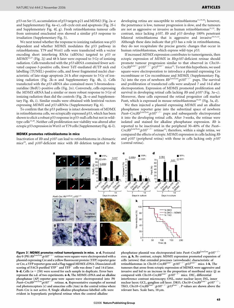
If increased MDMX expression contributes to tumorigenesis, thenectopic expression of MDMX in Rb;p107-deficient retinae shouldpromote tumour progression similar to that observed in Chx10–Cre;RbLox/2;p1072/2;p53Lox/2 mice32. To test this hypothesis, we usedsquare-wave electroporation to introduce a plasmid expressing Crerecombinase or Cre recombinase and MDMX (Supplementary Fig.7a) into the eyes of newborn RbLox/Lox;p1072/2 pups. The survivaland proliferation of transfected cells were analysed 7 and 14 d afterelectroporation. Expression of MDMX promoted proliferation andsurvival in developing retinal cells lacking Rb and p107 (Fig. 3a–c).Moreover, these cells expressed the retinal progenitor cell markerPax6, which is expressed in mouse retinoblastomas19,20 (Fig. 3a, d).
We then injected a plasmid expressing MDMX and an alkalinephosphatase reporter gene into the subretinal space of newbornPax6–Cre;RbLox/Lox;p1072/2 pups and subsequently electroporatedit into the developing retinal cells. After 3 weeks, the retinae wereisolated and stained for alkaline phosphatase expression. Rb isreported to be inactivated in the peripheral 30–40% of the Pax6–Cre;RbLox/Lox;p1072/2 retinae33; therefore, within a single retina, wecompared the effects of ectopic MDMX expression in cells lacking Rband p107 (peripheral retina) with those in cells lacking only p107(central retina).
Figure 3 | MDMX promotes retinal tumorigenesis in mice. a–d, Postnatalday 0 (P0) RbLox/Lox;p1072/2 retinae were square-wave electroporated with aplasmid expressing Cre and a yellow fluorescent protein (YFP) reporter geneor Cre, a YFP reporter gene and a Flag-tagged MDMX cDNA. Dissociated cellscoring of FACS-purified YFP1 and YFP2 cells was done 7 and 14 d later.b–d, Cells (n 5 250) were scored for each sample in duplicate. Error barsrepresent the s.d. of two experiments. e–h, The MDMX cDNA and an alkalinephosphatase (AP) reporter gene were square-wave electroporated into P0Pax6–Cre;RbLox/Lox;p1072/2 retinae. e, Representative examples of normalrod photoreceptors (r) and amacrine cells (Am) in the central retina wherePax6–Cre is not active. f, Single alkaline-phosphatase-labelled cells wereevident in hyperplastic peripheral retinae when the control alkaline
phosphatase plasmid was electroporated into Pax6–Cre;RbLox/Lox;p1072/2
eyes. g, h, By contrast, ectopic MDMX expression promoted expansion ofcells (arrows) that extended processes (arrowheads) characteristic ofretinoblastomas in Chx10–Cre;RbLox/2;p1072/2;p53Lox/2 mice. i–k, Thetumours that arose from ectopic expression of MDMX were aggressive andinvasive and led to an increase in the proportion of moribund mice (j) ascompared with Chx10–Cre;RbLox/2;p1072/2 mice. DIC, differentialinterference contrast microscopy; ONL, outer nuclear layer; INL, innernuclear layer; GCL, ganglion cell layer. DKO, Chx10–Cre;RbLox/2;p1072/2;TKO, Chx10–Cre;RbLox/2;p1072/2;p53Lox/2. P values are shown above therelevant bars. Scale bars, 10 mm.
NATURE | Vol 444 | 2 November 2006 ARTICLES
63Nature Publishing Group ©2006
Ectopic expression of MDMX in the central retina had little effecton proliferation or differentiation (Fig. 3e). Some hyperplasia formedin the periphery of Pax6–Cre;RbLox/Lox;p1072/2 retinae, but weobserved minimal clonal expansion of individual cells in that region
(Fig. 3f). When MDMX was expressed in cells lacking Rb and p107 inthe peripheral retina that lacked hyperplasia, clonal expansionoccurred (Fig. 3g, h). Moreover, those cells showed morphologicalfeatures (Supplementary Fig. 7b–j and Supplementary Tables 2–4)and aggressive invasive retinoblastoma similar to those of Chx10–Cre;RbLox/2;p53Lox/2;p1072/2 mice (Fig. 3i–k).
MDMX promotes human retinoblastoma
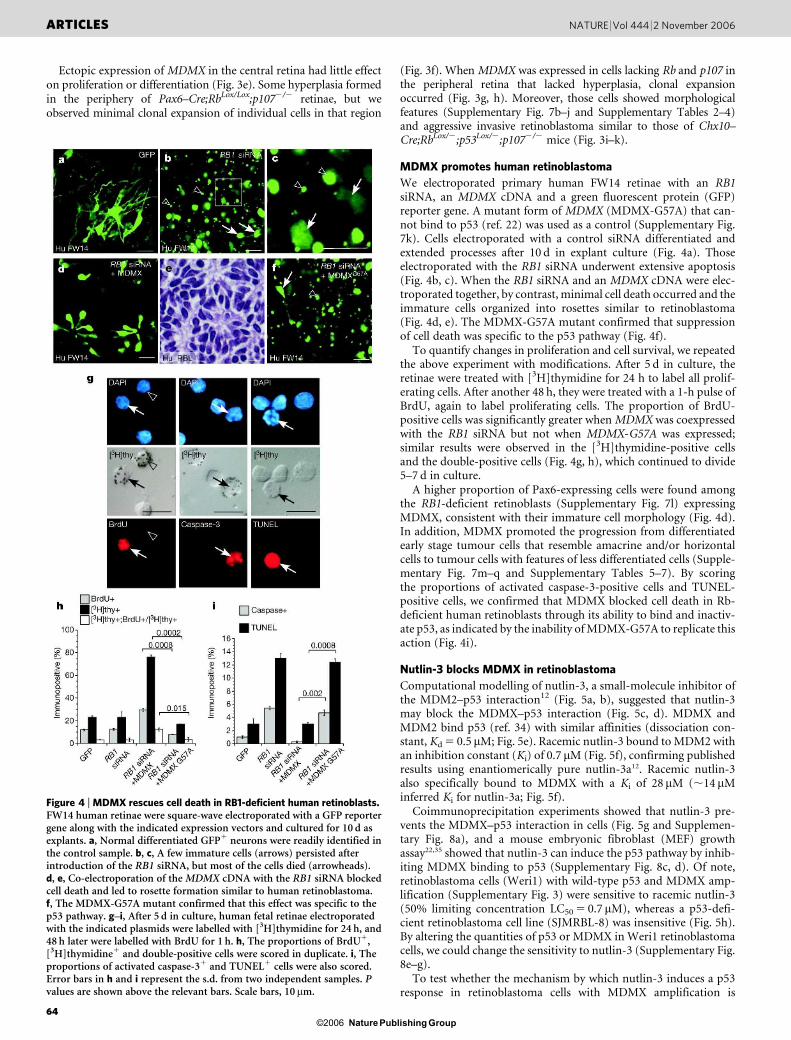
We electroporated primary human FW14 retinae with an RB1siRNA, an MDMX cDNA and a green fluorescent protein (GFP)reporter gene. A mutant form of MDMX (MDMX-G57A) that can-not bind to p53 (ref. 22) was used as a control (Supplementary Fig.7k). Cells electroporated with a control siRNA differentiated andextended processes after 10 d in explant culture (Fig. 4a). Thoseelectroporated with the RB1 siRNA underwent extensive apoptosis(Fig. 4b, c). When the RB1 siRNA and an MDMX cDNA were elec-troporated together, by contrast, minimal cell death occurred and theimmature cells organized into rosettes similar to retinoblastoma(Fig. 4d, e). The MDMX-G57A mutant confirmed that suppressionof cell death was specific to the p53 pathway (Fig. 4f).
To quantify changes in proliferation and cell survival, we repeatedthe above experiment with modifications. After 5 d in culture, theretinae were treated with [3H]thymidine for 24 h to label all prolif-erating cells. After another 48 h, they were treated with a 1-h pulse ofBrdU, again to label proliferating cells. The proportion of BrdU-positive cells was significantly greater when MDMX was coexpressedwith the RB1 siRNA but not when MDMX-G57A was expressed;similar results were observed in the [3H]thymidine-positive cellsand the double-positive cells (Fig. 4g, h), which continued to divide5–7 d in culture.
A higher proportion of Pax6-expressing cells were found amongthe RB1-deficient retinoblasts (Supplementary Fig. 7l) expressingMDMX, consistent with their immature cell morphology (Fig. 4d).In addition, MDMX promoted the progression from differentiatedearly stage tumour cells that resemble amacrine and/or horizontalcells to tumour cells with features of less differentiated cells (Supple-mentary Fig. 7m–q and Supplementary Tables 5–7). By scoringthe proportions of activated caspase-3-positive cells and TUNEL-positive cells, we confirmed that MDMX blocked cell death in Rb-deficient human retinoblasts through its ability to bind and inactiv-ate p53, as indicated by the inability of MDMX-G57A to replicate thisaction (Fig. 4i).
Nutlin-3 blocks MDMX in retinoblastoma
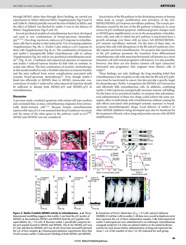
Computational modelling of nutlin-3, a small-molecule inhibitor ofthe MDM2–p53 interaction12 (Fig. 5a, b), suggested that nutlin-3may block the MDMX–p53 interaction (Fig. 5c, d). MDMX andMDM2 bind p53 (ref. 34) with similar affinities (dissociation con-stant, Kd 5 0.5 mM; Fig. 5e). Racemic nutlin-3 bound to MDM2 withan inhibition constant (Ki) of 0.7 mM (Fig. 5f), confirming publishedresults using enantiomerically pure nutlin-3a12. Racemic nutlin-3also specifically bound to MDMX with a Ki of 28mM (,14 mMinferred Ki for nutlin-3a; Fig. 5f).
Coimmunoprecipitation experiments showed that nutlin-3 pre-vents the MDMX–p53 interaction in cells (Fig. 5g and Supplemen-tary Fig. 8a), and a mouse embryonic fibroblast (MEF) growthassay22,35 showed that nutlin-3 can induce the p53 pathway by inhib-iting MDMX binding to p53 (Supplementary Fig. 8c, d). Of note,retinoblastoma cells (Weri1) with wild-type p53 and MDMX amp-lification (Supplementary Fig. 3) were sensitive to racemic nutlin-3(50% limiting concentration LC50 5 0.7 mM), whereas a p53-defi-cient retinoblastoma cell line (SJMRBL-8) was insensitive (Fig. 5h).By altering the quantities of p53 or MDMX in Weri1 retinoblastomacells, we could change the sensitivity to nutlin-3 (Supplementary Fig.8e–g).
To test whether the mechanism by which nutlin-3 induces a p53response in retinoblastoma cells with MDMX amplification is
Figure 4 | MDMX rescues cell death in RB1-deficient human retinoblasts.FW14 human retinae were square-wave electroporated with a GFP reportergene along with the indicated expression vectors and cultured for 10 d asexplants. a, Normal differentiated GFP1 neurons were readily identified inthe control sample. b, c, A few immature cells (arrows) persisted afterintroduction of the RB1 siRNA, but most of the cells died (arrowheads).d, e, Co-electroporation of the MDMX cDNA with the RB1 siRNA blockedcell death and led to rosette formation similar to human retinoblastoma.f, The MDMX-G57A mutant confirmed that this effect was specific to thep53 pathway. g–i, After 5 d in culture, human fetal retinae electroporatedwith the indicated plasmids were labelled with [3H]thymidine for 24 h, and48 h later were labelled with BrdU for 1 h. h, The proportions of BrdU1,[3H]thymidine1 and double-positive cells were scored in duplicate. i, Theproportions of activated caspase-31 and TUNEL1 cells were also scored.Error bars in h and i represent the s.d. from two independent samples. Pvalues are shown above the relevant bars. Scale bars, 10mm.
ARTICLES NATURE | Vol 444 | 2 November 2006
64Nature Publishing Group ©2006
through MDM2 rather than through direct binding, we carried outexperiments in Mdm2-deficient MEFs (Supplementary Figs 9 and 10and Table 8). MdmX partially rescued the loss of Mdm2 in MEFs, andthe effect of MdmX was blocked by nutlin-3 even in the absence ofMdm2 (Supplementary Figs 9 and 10).
Several preclinical models of retinoblastoma have been developedand used to test combinations of broad-spectrum chemother-apy19,32,36. One drug, topotecan, induces a p53 response in retinoblas-toma cells that is similar to that induced by 5 Gy of ionizing radiation(Supplementary Fig. 8h, i). Nutlin-3 also induces a p53 response inthese cells (Supplementary Fig. 8j–n). The combination of topotecanand nutlin-3 synergistically killed retinoblastoma cells in culture(Supplementary Fig. 8o) and in our preclinical retinoblastoma mod-els36 (Fig. 5i–m). Combined subconjunctival injection of topotecanand nutlin-3 reduced tumour burden 82-fold with no systemic orocular side-effects. The best combination of systemic chemotherapyin this model resulted in only a fivefold reduction in tumour burden,and the mice suffered from severe complications associated withsystemic broad-spectrum chemotherapy36. Even though nutlin-3binds less efficiently to MDMX than to MDM2, intraocular con-centrations of nutlin-3 achieved by subconjunctival injection shouldbe sufficient to disrupt both MDM2–p53 and MDMX–p53 inretinoblastomas.
Discussion
A previous study correlated apoptosis with retinal-cell-type markersand concluded that, in mice, retinoblastoma originates from intrins-ically death-resistant cells15,16. Because human retinoblastomasexpress wild-type p53, it was assumed that the p53 pathway was intactand the status of the other genes in the pathway (such as p14ARF,MDM2 and MDMX) was not considered.
Inactivation of the Rb pathway in the developing mouse or humanretina leads to ectopic proliferation and activation of the Arf–MDM2/MDMX–p53 tumour surveillance pathway. The ectopic pro-liferation caused by the loss of the Rb pathway is balanced, to someextent, by p53-mediated apoptosis. Additional genetic changes (suchas MDMX gene amplification) occur in the preneoplastic retinoblas-toma cells, and cells in which the p53 pathway is inactivated have agrowth advantage over those with an intact Arf–MDM2/MDMX–p53 tumour surveillance network. On the basis of these data, wepropose that cells with disruptions in the Rb and p53 pathways clon-ally expand and form retinoblastoma. We propose that inactivationof the p53 pathway promotes the transition from differentiatedretinoblastoma cells with amacrine/horizontal cell features to a moreimmature cell with retinal progenitor cell features. It is also possible,however, that there are two distinct tumour cell types (amacrine/horizontal and progenitor) that originate from distinct cells oforigin16.
These findings not only challenge the long-standing belief thatretinoblastoma is the exception to the rule that the Rb and p53 path-ways must be inactivated in cancer, but also provide a specific targetfor chemotherapy. Nutlin-3 antagonizes the MDMX–p53 interactionand efficiently kills retinoblastoma cells. In addition, combiningnutlin-3 with topotecan synergistically increases tumour cell killing.On the basis of our preclinical studies, we propose that subconjunc-tival administration of these two drugs could achieve the same syn-ergistic effect in individuals with retinoblastoma without causing theside-effects associated with prolonged systemic exposure to broad-spectrum chemotherapeutic drugs. Local delivery of nutlin-3 orother MDMX inhibitors being developed may also be beneficial forthe treatment of breast, colon, lung and prostate cancers with MDMXamplifications.
Untreated Nutlin-3 Topotecan Nutlin-3+ topotecan
1
2
3
4
5 Photons s
–1 cm–2 (×
106)
0
10
20
30
40
50
60
70
80
4 5 60 1 2 3 4 5 60 1 2 3 4 5 60 1 2 3 4 5 60 1 2 3
Untreated(n=20)
Rel
ativ
e p
hoto
ns s
–1 (f
old
)
Time (d)
0
5
10
15
20
Nutlin-3(n=16)
Time (d)
0
5
10
15
20
Topotecan(n=14)
Time (d)
0
5
10
15
20
Nutlin-3+ topotecan(n=16)
Time (d)
MDMX MDM2
1000.1 1 10 1,000
0.7 µM
28 µM
MDMX Nutlin-3a
MDM2 Nutlin-3a
SJmRBL-8
0
20
40
60
80
100
120
Via
ble
cel
ls (%
)
Weri1
0.0010.01 0.11 10
0.7 µM
% P
rote
in b
ound
% P
rote
in b
ound
MDMXMDM2
1000.01 0.1 1 10
Nutlin-3:
IP:
Anti-p
53
Anti-H
A
Anti-p
53
+– – + –
Totallysate
Tubulin
c
a b
dh
g
f
e
020406080
100
0
20
40
60
80
100
mlkji
Protein concentration (µM)
0.5 µM
MDM2(N20)
MDM2(N20)
MDM2(N20)
MDMX(BL1258)
MDMX(BL1258)p53(FL393)
p53(FL393)
Nutlin-3 concentration (µM) Nutlin-3concentration (µM)
Figure 5 | Nutlin-3 inhibits MDMX activity in retinoblastoma. a–d, Three-dimensional modelling suggests that nutlin-3 can bind the p53 pocket ofMDMX. e, Recombinant MDM2 and MDMX bind the p53 peptide withequal affinity (Kd 5 0.5 mM). f, Racemic nutlin-3 specifically competes forthe p53 peptide bound to MDM2 and MDMX. The Ki for MDM2–p53 was0.7 mM, and that for MDMX–p53 was 28 mM. Error bars in e and f representthe s.d. of four samples. g, Coimmunoprecipitation experiments show that10 mM racemic nutlin-3 reduced p53 binding of both MDM2 and MDMX.
h, Sensitivity of Weri1 (black line; LC50 5 0.7 mM) and p53-deficientSJMRBL-8 (red line) cells to nutlin-3. All data were scored in duplicate; errorbars represent the s.d. of three independent samples. i–m, Subconjunctivalnutlin-3 and topotecan were administered to an orthotopic retinoblastomaxenograft model for 5 d. Data are plotted relative to the initial luciferaseactivity for each mouse before administration of drug and represent themean 6 s.d. of the number of mice (14–20) indicated for each group.
NATURE | Vol 444 | 2 November 2006 ARTICLES
65Nature Publishing Group ©2006
METHODSA detailed description of materials and methods is given in Supplementary
Information. Individual protocols are provided on the authors’ website
(http://www.stjude.org/dyer).
Mouse and rat strains. Rb1/2 mice were obtained from The Jackson Laboratory;
p53Lox/Lox and RbLox/Lox mice from the National Cancer Institute; p107 knockout
mice from T. Jacks; Chx10–Cre mice from C. Cepko; and Pax6–Cre mice from R.
Ashery-Padan. All mice were crossed to C57Bl/6 mice purchased from Charles
River Laboratories. Timed-pregnant Sprague Dawley rats were obtained from
Charles River Laboratories. The xenograft model of retinoblastoma has been
described36.
Human retinae. Human retinae were obtained from Advanced Bioscience
Resources. They were maintained in culture by using protocols that we pre-
viously developed for mouse retinal cultures.
Received 30 June; accepted 24 August 2006.
1. Hahn, W. C. & Weinberg, R. A. Modelling the molecular circuitry of cancer. NatureRev. Cancer 2, 331–-341 (2002).
2. Vogelstein, B. & Kinzler, K. W. Cancer genes and the pathways they control.Nature Med. 10, 789–-799 (2004).
3. Sherr, C. J. & McCormick, F. The RB and p53 pathways in cancer. Cancer Cell 2,103–-112 (2002).
4. Chau, B. N. & Wang, J. Y. Coordinated regulation of life and death by RB. NatureRev. Cancer 3, 130–-138 (2003).
5. Vogelstein, B., Lane, D. & Levine, A. J. Surfing the p53 network. Nature 408,307–-310 (2000).
6. Oren, M. Decision making by p53: life, death and cancer. Cell Death Differ. 10,431–-442 (2003).
7. Prives, C. & Hall, P. A. The p53 pathway. J. Pathol. 187, 112–-126 (1999).8. Honda, R., Tanaka, H. & Yasuda, H. Oncoprotein MDM2 is a ubiquitin ligase E3 for
tumor suppressor p53. FEBS Lett. 420, 25–-27 (1997).9. Kubbutat, M. H., Jones, S. N. & Vousden, K. H. Regulation of p53 stability by
Mdm2. Nature 387, 299–-303 (1997).10. Momand, J., Jung, D., Wilczynski, S. & Niland, J. The MDM2 gene amplification
database. Nucleic Acids Res. 26, 3453–-3459 (1998).11. Yang, Y. et al. Small molecule inhibitors of HDM2 ubiquitin ligase activity stabilize
and activate p53 in cells. Cancer Cell 7, 547–-559 (2005).12. Vassilev, L. T. et al. In vivo activation of the p53 pathway by small-molecule
antagonists of MDM2. Science 303, 844–-848 (2004).13. Kato, M. V. et al. Loss of heterozygosity on chromosome 17 and mutation of the
p53 gene in retinoblastoma. Cancer Lett. 106, 75–-82 (1996).14. Nork, T. M., Poulsen, G. L., Millecchia, L. L., Jantz, R. G. & Nickells, R. W. p53
regulates apoptosis in human retinoblastoma. Arch. Ophthalmol. 115, 213–-219(1997).
15. Chen, D. et al. Cell-specific effects of RB or RB/p107 loss on retinal developmentimplicate an intrinsically death-resistant cell-of-origin in retinoblastoma. CancerCell 5, 539–-551 (2004).
16. Dyer, M. A. & Bremner, R. The search for the retinoblastoma cell of origin. NatureRev. Cancer 5, 91–-101 (2005).
17. Aslanian, A., Iaquinta, P. J., Verona, R. & Lees, J. A. Repression of the Arf tumorsuppressor by E2F3 is required for normal cell cycle kinetics. Genes Dev. 18,1413–-1422 (2004).
18. Lowe, S. W. & Sherr, C. J. Tumor suppression by Ink4a-Arf: progress and puzzles.Curr. Opin. Genet. Dev. 13, 77–-83 (2003).
19. Zhang, J., Schweers, B. & Dyer, M. A. The first knockout mouse model ofretinoblastoma. Cell Cycle 3, 952–-959 (2004).
20. Donovan, S., Schweers, B., Martins, R., Johnson, D. & Dyer, M. A. Compensationby tumor suppressor genes during retinal development in mice and humans. BMCBiol. 4, 14 (2006).
21. Shvarts, A. et al. MDMX: a novel p53-binding protein with some functionalproperties of MDM2. EMBO J. 15, 5349–-5357 (1996).
22. Danovi, D. et al. Amplification of Mdmx (or Mdm4) directly contributes to tumorformation by inhibiting p53 tumor suppressor activity. Mol. Cell. Biol. 24,5835–-5843 (2004).
23. McKenzie, P. P., McPake, C. R., Ashford, A. A., Vanin, E. F. & Harris, L. C. MDM2does not influence p53-mediated sensitivity to DNA-damaging drugs. Mol. CancerTher. 1, 1097–-1104 (2002).
24. Canman, C. E. et al. Activation of the ATM kinase by ionizing radiation andphosphorylation of p53. Science 281, 1677–-1679 (1998).
25. Kastan, M. B., Lim, D. S., Kim, S. T. & Yang, D. ATM—a key determinant of multiplecellular responses to irradiation. Acta Oncol. 40, 686–-688 (2001).
26. Kastan, M. B., Onyekwere, O., Sidransky, D., Vogelstein, B. & Craig, R. W.Participation of p53 protein in the cellular response to DNA damage. Cancer Res.51, 6304–-6311 (1991).
27. Brummelkamp, T. R., Bernards, R. & Agami, R. A system for stableexpression of short interfering RNAs in mammalian cells. Science 296, 550–-553(2002).
28. Baker, S. J., Markowitz, S., Fearon, E. R., Willson, J. K. & Vogelstein, B. Suppressionof human colorectal carcinoma cell growth by wild-type p53. Science 249,912–-915 (1990).
29. Robanus-Maandag, E. et al. p107 is a suppressor of retinoblastoma developmentin pRb-deficient mice. Genes Dev. 12, 1599–-1609 (1998).
30. MacPherson, D. et al. Cell type-specific effects of Rb deletion in the murine retina.Genes Dev. 18, 1681–-1694 (2004).
31. Dyer, M. A. & Harbour, J. W. in Clinical Ocular Oncology Ch. 66 (eds Singh, A. D.,Damato, B., Murphree, A. L. & Perry, J. D.) (Elsevier, London, in the press).
32. Dyer, M. A., Rodriguez-Galindo, C. & Wilson, M. W. Use of preclinical models toimprove treatment of retinoblastoma. PLoS Med. 2, e332 (2005).
33. Marquardt, T. et al. Pax6 is required for the multipotent state of retinal progenitorcells. Cell 105, 43–-55 (2001).
34. Lu, F. et al. Proteomimetic libraries: design, synthesis, and evaluation ofp53–-MDM2 interaction inhibitors. J. Comb. Chem. 8, 315–-325 (2006).
35. Dang, J. et al. The RING domain of Mdm2 can inhibit cell proliferation. Cancer Res.62, 1222–-1230 (2002).
36. Laurie, N. A. et al. Topotecan combination chemotherapy in two new rodentmodels of retinoblastoma. Clin. Cancer Res. 11, 7569–-7578 (2005).
Supplementary Information is linked to the online version of the paper atwww.nature.com/nature.
Acknowledgements We thank L. Harris, G. Zambetti and M. Baron for discussions;S. Pounds for statistical analysis; M. Roussel for MDMX and MDM2 retroviruses;B. Schulman and D. Bashford for assistance with MDMX–-nutlin-3 modelling;A. McArthur for editorial assistance; J. Gray for assistance with real-time RT–-PCRand genomic DNA preparations; and F. Carlotti and M. Rabeling for advice onlentiviral experiments and production of lentivirus stocks. This work was supportedby grants (to M.A.D.) from the National Eye Institute, Cancer Center Support fromthe National Cancer Institute, the American Cancer Society, Research to PreventBlindness, the Pearle Vision Foundation, the International Retinal ResearchFoundation and the American Lebanese Syrian Associated Charities (ALSAC).M.A.D. is a Pew Scholar. This work was also supported by funding from theAssociation for International Cancer Research (A.G.J.) and EC FP6 (A.G.J. andJ.-C.M.), the Dutch Cancer Society (Y.R.), the Belgian Foundation against Cancer(J.-C.M.) and Televie (S.F.). This publication was supported in part by a grant fromthe National Cancer Institute.
Author Information Reprints and permissions information is available atwww.nature.com/reprints. The authors declare no competing financial interests.Correspondence and requests for materials should be addressed to M.A.D.([email protected]).
ARTICLES NATURE | Vol 444 | 2 November 2006
66Nature Publishing Group ©2006


