Embed Size (px)
Citation preview

c
ed to occur
rurse ofityFP/P35delfordecreasingproduced,fects, withtible to bothsis
Research in Microbiology 156 (2005) 1014–1025
www.elsevier.com/locate/resmi
In vivo apoptosis induction and reduction of infectivity byanAutographa californicamultiple nucleopolyhedrovirusp35−
recombinant in hemocytes from the velvet bean caterpillarAnticarsia gemmatalis(Hübner) (Lepidoptera: Noctuidae)
Eni Braga da Silveiraa, Bruno Arrivabene Cordeirob, Bergmann Morais Ribeirob,Sônia Nair Báob,∗
a Departamento de Biologia Celular, Instituto de Ciências Biológicas, Universidade Estadual de Campinas, C.P. 6109,Campinas, SP 13.083-863, Brazil
b Departamento de Biologia Celular, Instituto de Ciências Biológicas, Universidade de Brasília, Brasília, DF 70.919-970, Brazil
Received 12 January 2005; accepted 2 June 2005
Available online 18 July 2005
Abstract
Baculoviruses have long been shown to regulate apoptosis in cultured insect cells. Recently, this phenomenon was also reportin vivo, reinforcing the importance of apoptosis in insect immunity against viruses. The vP35del virus, anAutographa californicamultiplenucleopolyhedrovirus (AcMNPV) recombinant, was previously shown to induce apoptosis inAnticarsia gemmataliscultured cells. In ordeto verify the AcMNPV interaction with hemocytes, apoptosis induction in vivo and its effects upon infectivity, we studied the cointrahemocoelic infection of recombinant viruses (vHSGFP and vHSGFP/P35del) inA. gemmatalislarvae. Insect development and mortalwere monitored and infection progress was followed by light, fluorescence and electron microscopy. For all doses tested, vHSGcaused lower mortality than vHSGFP. Mortality of 95% occurred with a dose of 4× 10⁶ PFUs of vHSGFP, which was reduced to 60%vHSGFP/P35del. GFP expression was first observed at 3 h p.i. for the two viruses, increasing for vHSGFP (40% at 120 h p.i.) andfor vHSGFP/P35del (0% at 120 h p.i.). The virus vHSGFP/P35del induced apoptosis in hemocytes, with some budded virus beingand fragmented cells were observed between 24 and 72 h p.i. The recombinant vHSGFP induced typical wild-type cytopathic eflow production of occluded viruses until 120 h p.i. Plasmatocytes and granular hemocytes type 1 were the hemocytes most suscepviruses. For these experimental conditions, we concluded thatA. gemmatalisis a semi-permissive host to AcMNPV; moreover, apoptoreduces AcMNPV infectivity and thep35gene is essential for blocking apoptosis in this system. 2005 Elsevier SAS. All rights reserved.
Keywords:Apoptosis; Baculoviridae; GFP; Hemocytes; Hemolymph; Lepidoptera; Ultrastructure
e-ro-are
ofies
ionheseage-ouslarge
1. Introduction
The familyBaculoviridaeconsists of enveloped, doublstranded DNA viruses which are pathogenic toward arthpods, especially lepidoptera larvae. Baculovirus speciesdistributed between two genera,Granulovirus(GV) andNu-
* Corresponding author.E-mail address:[email protected] (S.N. Báo).
0923-2508/$ – see front matter 2005 Elsevier SAS. All rights reserved.doi:10.1016/j.resmic.2005.06.001
cleopolyhedrovirus(NPV). Autographa californicamulti-ple nucleopolyhedrovirus (AcMNPV) is the type-speciestheNucleopolyhedrovirusgenus and the best-known specamong baculoviruses [3].
Infection specificity and consequent safety in relatto non-target organisms are characteristics that make tviruses agents of great potential for integrated pest manment [2,24]. Baculoviruses are also useful as heterologgene expression vectors, since their genome supports

E.B. da Silveira et al. / Research in Microbiology 156 (2005) 1014–1025 1015
cane in-
ionns
, theto
ion,ion.qui-
pes
bac-y ofod,tion,to-s),ss tos atract
il35
vi-es,in-
opa-
re-ro,d-re-
nity,e o
iga-s-
unele o
an-
usinde-
enym,48 hb-
in-
vo
on-llyereedted
pa
ed,ere
erein-s,
inedelne
inop-yy
andu-o-ere
wasent
sid-ion.red.ol-
hebyvi-
DNA inserts at non-essential gene loci. Foreign proteinsbe successfully expressed upon infection of a permissivsect cell line [18,26].
Apoptosis is a phenomenon of cellular self-destructthat can be triggered by diverse stimuli, and viral infectioare among them. By activating an apoptotic responsehost cell can abort viral infection, avoiding its spreadthe whole organism. For survival and progeny productviruses had to circumvent this response during evolutOne strategy for achieving such circumvention is the acsition of antiapoptotic genes [21,35].
Baculoviruses are known to possess at least two tyof antiapoptotic genes:p35and iap. The genep35encodesa broad-spectrum caspase inhibitor, described in fiveulovirus species up to date [10,13,14,19,28]. Another waapoptosis inhibition by P35, still not completely understois related to reactive oxygen species (ROS) neutralizaand prevention of cytochrome C liberation by the michondria [29]. The IAPs (inhibitors of apoptosis proteinwhich were first described inCydia pomonella granuloviru(CpGV), are also found in organisms ranging from yeasthumans. IAPs are metalloproteins that inhibit apoptosiupstream pathways before caspase activation or by inteing directly with these enzymes [9].
AcMNPV possessesp35 and iap genes; however, untnow, antiapoptotic function was observed only for the Pprotein. Apoptosis induction by p35 mutant (p35−) AcM-NPV viruses is best characterized forSpodoptera frugiperdaand its derived cell line, SF-21, with a reduction inral infectivity and progeny production [7,9]. In other casdespite presenting intact antiapoptotic genes, AcMNPVduces apoptosis and displays reduced infectivity and prgation in some insect cell lines [5,27,41] and inSpodopteralitura larvae [40].
Despite the existence of a great deal of informationlated to mutant baculovirus induction of apoptosis in vitonly recently it was reported in vivo [6,40]. These stuies have shown that apoptosis is an important antiviralsponse in insects which appear to lack acquired immuand that the strategies to counteract cell death are onthe determinants of baculovirus host-range [7]. Investtions in this field, especially those focusing on in vivo asays, are important for shedding new light on insect immstrategies against pathogens and for emphasizing the roapoptosis during the evolution of immunity in these orgisms.
In a previous study [33], it was reported that the virvP35del, derived from AcMNPV, which has a deletionthe p35 gene, induced massive apoptosis in a cell linerived from A. gemmatalis(UFL-AG-286) [30]. Apoptosisoccurred between 9 and 16 h p.i., with a total lack of progproduction [33]. In contrast, a mutant (vApAg) derived froA. gemmatalismultiple nucleopolyhedrovirus (AgMNPV)induced apoptosis in a delayed manner, between 24 andwith some progeny production [4,33]. Recently, it was oserved that vApAg induces apoptosis inA. gemmatalislarval
-
f
f
,
hemocytes in a manner very similar to that of apoptosisduction in UFL-AG-286 cells (Silveira et al., submitted).
In this work, we investigated apoptosis induction in viby an AcMNPVp35− virus in A. gemmatalislarval hemo-cytes and its effects on infectivity. Recombinant viruses ctaining theegfp gene were inoculated intrahemocoelicainto 4th instar larvae. Insect development and mortality wmonitored, infection progress in hemocytes was followby light and transmission electron microscopy, and infeccells were counted under fluorescence microscopy.
2. Materials and methods
2.1. Insects and viruses
A. gemmatalis1st instar larvae, obtained from Embra(Brazil), were reared on an artificial diet [17] at 24–27◦C,with a 12:12 h dark/light regime. The molts were monitorand 4th instar larvae (between 12 and 24 h after molt) wused for all experiments.
The recombinants vHSGFP and vHSGFP/P35del wpropagated in BTI-Tn-5B1-4 (Tn-5B) cells [15] and matained in TC-100 medium (GIBCO-BRL Life TechnologieGrand Island, NY) supplemented with 10% fetal bovserum at 27◦C. The viruses vHSGFP and vHSGFP/P35are derived from the AcMNPV L1 strain and have the geegfpunder control of theDrosophila melanogasterconsti-tutive promoterhsp 70at a site adjacent to the polyhedrgene [6]. Additionally, vHSGFP/P35del has the antiaptotic genep35 deleted [8]. The inocula were titered bthe TCID50 method following the protocol described bO’Reilly et al. [26].
2.2. Bioassays
To observe the effects of viruses on insect mortalitydevelopment, larvae were injected with 20 µl of viral inoclum directly into the hemocoel by using an insulin micrsyringe. Three different concentrations of inoculum wused (2×10⁶, 2×10⁷, 2×10⁸ PFU/ml) for each virus, andtwenty larvae were inoculated for each one. Each larvareared separately in a plastic cup. Mortality and developmwere reported every day for 25 days. Larvae were conered dead if they did not react to mechanical stimulatDeaths attributed to injection trauma were not consideControls were obtained by the inoculation of an equal vume of TC-100 medium and by no inoculation.
2.3. Infected cell countings
To verify the percentage of infected cells during ttime of infection and the structural alterations promotedviruses in hemocytes, insects were injected with 20 µl ofral inoculum at the highest concentration (2×10⁸ PFU/ml),

1016 E.B. da Silveira et al. / Research in Microbiology 156 (2005) 1014–1025
nti-Mlyfil-
anioncells
of
inereu-0de,ffer
iumer),d insecand.
yrevi-
rvaldult
a
hs
in-erensid-GFP
edFord
d as
asncy, theNoe
ofed
C).eadnd(notated
piteatheized
None
eris-o-
e 1oe),
l atptly1 areeadme-this
3 hells
).S-p.i.
wasf flu-after
useally
as described. Hemolymph samples were collected in acoagulant buffer (98 mM NaOH, 186 mM NaCl, 1.7 mEDTA, 41 mM citric acid, pH 4.5) [23], observed directin a fluorescence microscopy under the blue excitationter, and by DIC (differential interferential contrast) inAxiophot Zeiss microscope. For each time after infect(3, 24, 48, 72, 96 and 120 h), at least seven hundredwere counted in two subsamples obtained from a mixhemolymph collected from ten insects.
2.4. Transmission electron microscopy
To observe the ultrastructural alterations inducedhemocytes by viral infection, hemolymph samples wcollected from insects injected with 20 µl of viral inoclum (2 × 10⁸ PFU/ml) after 12, 24, 48, 72, 96 and 12h p.i. Samples were fixed for 30 min (2% glutaraldehy2% paraformaldehyde in 0.1 M sodium cacodylate bu(pH 7.4) with 5% sucrose), centrifuged at 750g for 5 min,the pellet washed in the same buffer, post-fixed (1% osmtetroxide, 0.8% potassium ferricyanide in the same buffcontrasted in block with 0.5% uranyl acetate, dehydrateacetone, and embedded in Spurr’s resin. The ultrathintions were contrasted with uranyl acetate/lead citrateobserved in a TEM JEOL 100C and JEOL 1011 at 80 kV
2.5. Hemocyte identification
A. gemmatalislarval hemocyte-types were identified bstructural and ultrastructural characteristics described pously [32,34].
3. Results
3.1. Bioassays
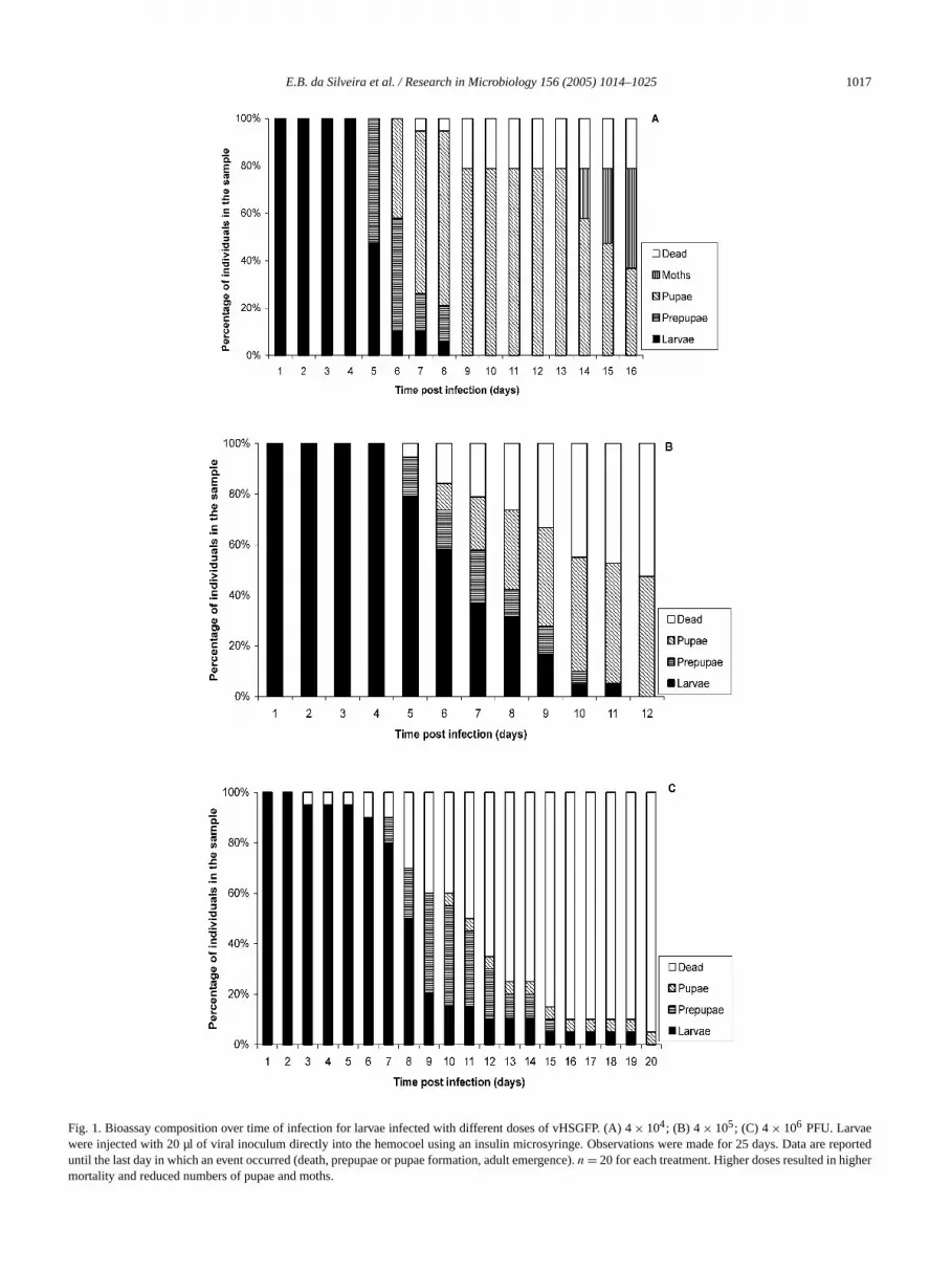
Increasing doses of vHSGFP resulted in increasing lamortality, decreasing larval pupation, and decreasing aemergence (Table 1, Fig. 1). The dose of 4× 10⁴ PFU/larva
Table 1Percentage of deadA. gemmatalislarvae, non-viable pupae and moths inbioassay of different doses of vHSGFP and vHSGFP/P35del
Dose Virus Dead larvae Non-viable pupae Mot(PFU) (%)a (%) (%)
vHSGFP 21 37 424× 104 vHSGFP/P35del 0 30 70
vHSGFP 53 47 04× 105 vHSGFP/P35del 22 73 5
vHSGFP 95 5 04× 106 vHSGFP/P35del 60 40 0
a n = 20 for each treatment. Larvae were injected with 20 µl of viraloculum directly into the hemocoel using an insulin microsyringe and wobserved for 25 days. Deaths attributed to injection trauma were not coered. Deleterious effects were milder for vHSGFP/P35del than for vHSfor all doses tested.
-
resulted in 21% death, while 79% of individuals formpupae and 53% of these emerged as moths (Fig. 1A).4 × 105 PFU/larva, larval mortality was around 53%, an47% of pupae were formed, but none of them emergeadults (Fig. 1B). For 4× 10⁶ PFU/larva, 95% of individualsdied. One pupa was formed (5%), but it did not emergean adult (Fig. 1C). For vHSGFP/P35del, the same tendewas observed, but in comparison to the previous systemviral effects were milder for each dose (Table 1, Fig. 2).larvae injected with 4× 10⁴ PFU died. All of them becampupa, and 70% emerged as adults (Fig. 2A). For 4× 10⁵
PFU/larva, larval mortality was around 22% and 78%individuals formed pupa; however only one (5%) reachadulthood (Fig. 2B). For 4×10⁶ PFU/larva, 60% of individ-uals died, 40% became pupa, but none emerged (Fig. 2
For the vHSGFP highest dose, some liquefaction of dlarvae was observed, but for the majority of them afor lower doses, there was melanization of cadaversshown). Some pupae were abnormal for larvae inoculwith either vHSGFP (4× 10⁴ and 4× 10⁵ PFU/larva) orvHSGFP/P35del (all doses), especially for the latter. Desmolting inhibition, the larva body hypertrophied in suchway that the normal proportions between the body andhead were lost. Later, the body decreased in size, melanand hardened. Prolegs and head persisted (not shown).of the abnormal pupae emerged.
3.2. Hemocyte identification
Six hemocyte types were recognized inA. gemmatalislarvae based on structural and ultrastructural characttics previously described [32,34]. They included prohemcytes (pr), plasmatocytes (pl), granular hemocytes typ(gh1), granular hemocytes type 2 (gh2), oenocytoids (and spherulocytes (sph).
3.3. DIC, fluorescence microscopy and cell counting
Fluorescence occurred preferentially for gh1 and pall times post-infection studied. These cells were promrecognized because of their characteristic shapes. Ghround, rich in thin surface projections (filopodia) and sprsymmetrically, maintaining a circular shape. Pl are sowhat elongated or fusiform, and spread maintainingasymmetrical shape.
For both systems, GFP expression was observed fromp.i., (around 3%) (Fig. 3). The number of fluorescent cremained similar for both viruses until 48 h p.i. (8–12%After 48 h p.i., the increasing tendency continued for vHGFP, which reached 40% of fluorescencent cells at 120 hFor vHSGFP/P35del, a plateau (9% of fluorescent cells)established between 24 and 72 h p.i. Then, the number oorescent cells decreased, reaching values around 0.01%120 h p.i. (Fig. 3).
For vHSGFP/P35del, apoptotic cells, recognized becaof their fragmentation into apoptotic bodies, were eventu
E.B. da Silveira et al. / Research in Microbiology 156 (2005) 1014–1025 1017
e reporher
Fig. 1. Bioassay composition over time of infection for larvae infected with different doses of vHSGFP. (A) 4× 10⁴; (B) 4 × 10⁵; (C) 4× 10⁶ PFU. Larvaewere injected with 20 µl of viral inoculum directly into the hemocoel using an insulin microsyringe. Observations were made for 25 days. Data arteduntil the last day in which an event occurred (death, prepupae or pupae formation, adult emergence).n = 20 for each treatment. Higher doses resulted in higmortality and reduced numbers of pupae and moths.

1018 E.B. da Silveira et al. / Research in Microbiology 156 (2005) 1014–1025
. Data arted
Fig. 2. Bioassay composition over time of infection for larvae infected with different doses of vHSGFP/P35del. (A) 4× 10⁴;. (B) 4× 10⁵; (C) 4× 10⁶ PFU.Larvae were injected with 20 µl of viral inoculum directly into the hemocoel using an insulin microsyringe. Observations were made for 25 daysereported until the last day in which an event occurred (death, prepupae or pupae formation, adult emergence).n = 20 for each treatment. Higher doses resulin higher mortality and reduced numbers of pupae and moths.

E.B. da Silveira et al. / Research in Microbiology 156 (2005) 1014–1025 1019
time. Va
Fig. 3. Percentage ofegfp-expressing cells over time of infection with vHSGFP and vHSGFP/P35del. Larvae were injected with 4× 10⁶ PFU of virus, cohortsof ten insects were sacrificed for hemolymph collection and at least seven-hundred cells were counted in a DIC/fluorescence microscope at eachluesare plotted as means± SD.ndp.i.as-
forr ofe in-4G,n).
in-P,or-
d asdedfib-of24 hm-
onwasgerimessm
n to
per-, inbleop-uenre-totic
en-vedntstedem-athfter
gh1,le towas
plGh1di-esto
C).an
e
rent
p.i.,sidsop-eny
thesus-
found at 12 h p.i., more frequently at 24 (Figs. 4A–4D) a48 h p.i. (not shown), and then became rare after 96 h(not shown). After 48 h p.i., there was some polyhedrasembly for vHSGFP (Figs. 4E, 4F), which did not occurvHSGFP/P35del (not shown). After 96 h p.i., the numbegh2 slightly increased for both systems. These cells wertact and had an extremely low rate of fluorescence (Figs.4H). Infected sph were also occasionally found (not show
3.4. Transmission electron microscopy
At 12 h p.i., the major part of the hemocytes weretact for both systems. At that time of infection, for vHSGFrare cells, especially pl and gh1, presented sign of viral mphogenesis such as nuclear hypertrophy, nucleocapsisembly, envelopment, nuclear envelope folding with budvirus protrusion through the cytoplasm, and, eventually,rillar aggregate accumulation (Fig. 5A). Higher numbershemocytes displayed the events described above afterp.i. (Figs. 5B–5D). After 48 h p.i., some polyhedra assebly was visualized (Figs. 5E–5F), but until late in infecti(72–120 h p.i.) the number of cells presenting polyhedravery low. At 72 and 120 h p.i., fibrillar aggregates were larand nuclear envelope folds were more extensive, sometforming organized arrays of tubes throughout the cytopla(Fig. 5C). Pr, pl, gh1, oe and eventually sph were showbe susceptible to vHSGFP.
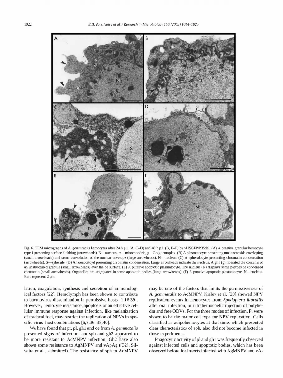
For vHSGFP/P35del, after 12 h p.i., some nuclear hytrophy and nuclear envelope folding also occurred, butsome cases, these events were associated with surfacebing, chromatin condensation and fragmentation into aptotic bodies. These apoptotic events became more freqat 24 and 48 h p.i. (Figs. 6A–6F). Many apoptotic cells psented segregation of organelles in some of the apop
-
b-
t
bodies formed (Figs. 6E, 6F). Despite chromatin condsation, apoptotic body formation for sph was not obser(Fig. 6C). Exocytosis of gh1 unstructured granule contewas observed in association with filopodia emission directo an apoptotic oe (Fig. 6D). Some nucleocapsid assbly and envelopment were also found, despite cell de(Fig. 6B). Apoptosis and replication events decreased a96 h p.i., and there was no polyhedra assembling. Pl,pr, oe and, eventually, sph were shown to be susceptibvHSGFP/P35del, especially the first two cell types, asthe case for vHSGFP.
Necrosis of infected cells and phagocytic activity ofand gh1 were observed for both systems (Figs. 7A–7F).were shown to emit thin long projections which could berected to small cell fragments, forming multiple phagosomsimultaneously (Figs. 7A–7B), or which could be directedentire cells or cell fragments of large dimensions (Fig. 7Pl were shown to engulf entire cells more frequently thgh1 (Fig. 7E).
4. Discussion
In this work, we have shown that AcMNPV lacking thp35 gene causes apoptosis inA. gemmatalishemocytes.However, the intensity and speed of death were diffefrom that induced in a cell line derived fromA. gemmatalis.In hemocytes, apoptosis occurred between 24 and 72 hat low frequency, with the occurrence of some nucleocapassembling and enveloping. For UFL-AG-286 cells, aptosis was massive and rapid (until 16 h), without progproduction [33].
One possible reason for this discrepancy could bedifference between the two cellular models as concerns

1020 E.B. da Silveira et al. / Research in Microbiology 156 (2005) 1014–1025
hvation andra
Fig. 4. DIC and fluorescence (blue excitation filter) microscopy micrographs of hemocytes fromA. gemmatalislarvae infected intrahemocoelically wit4× 106 PFU of vHSGFP/P35del (A–D) and vHSGFP (E–H). Hemolymph was collected in anticoagulant buffer and a sample was taken for obserphotography. (A–D) 24 h p.i. Arrows indicate apoptotic bodies, arrowheads indicate gh2. (E–F) 120 h p.i. Arrows indicate cells presenting polyhed. (G–H)120 h p.i. Arrow indicate gh1, small arrowheads indicate gh2. Bars represent 20 µm.
e-
86ve
ofdel,ionesere-
sisir-torsro.en
ilar
re-ctionility
i-byas
ss
c-ditha
ys-ore
ceptibility to AcMNPV, despite UFL-AG-286 cells being drived from the same organism, suggesting thatA. gemmatalishas tissue-specific susceptibility to AcMNPV. UFL-AG-2cells are permissive to AcMNPV [31]. However, we hademonstrated that between 24 and 48 h p.i., only 8%hemocytes were infected by vHSGFP and vHSGFP/P35and after 120 h p.i. vHSGFP reached only 40% of infectin hemocytes, which indicates a low rate of infection in thcells. A similar rate of infection was found for the samecombinant inS. frugiperdahemocytes [6].
Another explanation for the low occurrence of apoptocould be the turnover of hemocytes by cell division in cculation and in hemopoetic organs, added to putative facthat can neutralize virions, which does not occur in vitHowever, vApAg, which is derived from AgMNPV, has beshown to induce more intense apoptosis inA. gemmatalishemocytes between 24 and 72 h p.i., under very sim
experimental conditions (Silveira et al., submitted). Thisinforces the hypothesis that less intense apoptosis induby vHSGFP/P35del is related to the reduced susceptibof A. gemmatalishemocytes to AcMNPV.
A. gemmatalislarvae have been shown to be sempermissive or permissive to high doses of AcMNPVintrahemocoelic infection, since 95% of larval mortality wobtained with the injection of 4× 10⁶ PFUs. InTrichoplu-sia ni, this virus induces 95% death with an injection of lethan 1 PFU [6], and AgMNPV causes 50% death inA. gem-mataliswith 0.1 PFU (Silveira et al., submitted).
The absence of thep35 gene further reduced the infetivity level, with maximum larval mortality of 60% obtainewith a 10 times higher dose than that used for vHSGFP wsimilar mortality (53%). This probably can be related todrop in BV production and a consequent reduction in stemic virus spread. For other systems, apoptosis can m

E.B. da Silveira et al. / Research in Microbiology 156 (2005) 1014–1025 1021
ling andsma(arroar eipress (large
Fig. 5. TEM micrographs ofA. gemmatalishemocytes infected by vHSGFP. (A) 12 h p.i. A putative plasmatocyte, presenting nucleocapsids assembinitial enveloping (large arrowhead). Note some groups of long immature nucleocapsids (small arrowhead). N—nucleus. (B) 24 h p.i. A putative platocytepresenting nucleocapsids assembling and enveloping (small arrowhead), a nuclear fibrillar aggregate (*) associated with a membranous profilew) andnuclear envelope folding (large arrowheads). N—nucleus. (C) 72 h p.i. Putative granular hemocyte type 1 presenting a peculiar profile of the nuclenvelopethat protrudes as organized tubes throughout the cytoplasm (large arrowheads). N—nucleus; (*) fibrillar aggregate; small arrowheads–nucleocapsds. (D) 72 hp.i. Formation of transport vesicles containing nucleocapsids (arrowheads). N—nucleus; C—cytoplasm. (E) 120 h p.i. A putative plasmatocyte,entingpolyhedra assembly (*). N—nucleus; C—cytoplasm. (F) 120 h p.i. A necrotic cell with the nucleus filled with polyhedra (*), fibrillar aggregatearrowheads), and membranous profiles (small arrowheads). Bars represent 1.5 µm.
anes
ib-el,ivedto
de-dis-
byous
eas-hislar-op-
rcu-sespsu-
drastically reduce infectivity in vivo. InS. frugiperdalarvae,1000-fold more BVs of AcMNPVp35− and 25-fold morePIBs (polyhedral inclusion bodies) are required to reachLC50 in comparison to the wild-type or revertant virus[11,12].
In addition to mortality, another deleterious effect attruted to viral infection, mainly caused by vHSGFP/P35dwas the production of abnormal pupae. These were derfrom larvae that had survived infection and attemptedform pupae despite incomplete molting. This probablyrives from successful expression of the enzyme EGT (ec
teroyd UDP-glucosyltransferase), which inhibits moltinginactivating the ecdysone hormone [25]. This anomalphenotype had been previously reported inS. frugiperdalar-vae infected by vHSGFP/P35del, which presented decring tissue fluorescence during the time of infection [8]. Teffect suggests a milder infection that did not result inval death, but that was sufficient to impair normal develment.
Insect hemocytes are a complex of cell types that cilate in hemolymph. They are involved in defense responsuch as wound repair, phagocytosis, nodulation, enca
1022 E.B. da Silveira et al. / Research in Microbiology 156 (2005) 1014–1025
ocyteenvelondensat
conof condN—nu
Fig. 6. TEM micrographs ofA. gemmatalishemocytes after 24 h p.i. (A, C–D) and 48 h p.i. (B, E–F) by vHSGFP/P35del. (A) A putative granular hemtype 1 presenting surface blebbing (arrowheads). N—nucleus, m—mitochondria, g—Golgi complex. (B) A plasmatocyte presenting nucleocapsidsoping(small arrowheads) and some convolution of the nuclear envelope (large arrowheads). N—nucleus. (C) A spherulocyte presenting chromatin cion(arrowheads). S—spherule. (D) An oenocitoyd presenting chromatin condensation. Large arrowheads indicate the nucleus. A gh1 (g) liberated thetents ofan unstructured granule (small arrowheads) over the oe surface. (E) A putative apoptotic plasmatocyte. The nucleus (N) displays some patchesensedchromatin (small arrowheads). Organelles are segregated in some apoptotic bodies (large arrowheads). (F) A putative apoptotic plasmatocyte.cleus.Bars represent 2 µm.
log-ute39].cel
tione-
d tolsoil-PV
s of
e-erellsntedd in
edeenA-
lation, coagulation, synthesis and secretion of immunoical factors [22]. Hemolymph has been shown to contribto baculovirus dissemination in permissive hosts [1,16,However, hemocyte resistance, apoptosis or an effectivelular immune response against infection, like melanizaof tracheal foci, may restrict the replication of NPVs in spcific virus–host combinations [6,8,36–38,40].
We have found that pr, pl, gh1 and oe fromA. gemmatalispresented signs of infection, but sph and gh2 appearebe more resistant to AcMNPV infection. Gh2 have ashown some resistance to AgMNPV and vApAg ([32], Sveira et al., submitted). The resistance of sph to AcMN
-
may be one of the factors that limits the permissivenesA. gemmatalisto AcMNPV. Kislev et al. [20] showed NPVreplication events in hemocytes fromSpodoptera litorallisafter oral infection, or intrahemocoelic injection of polyhdra and free ODVs. For the three modes of infection, Pl wshown to be the major cell type for NPV replication. Ceclassified as adipohemocytes at that time, which preseclear characteristics of sph, also did not become infectethose experiments.
Phagocytic activity of pl and gh1 was frequently observagainst infected cells and apoptotic bodies, which has bobserved before for insects infected with AgMNPV and v

E.B. da Silveira et al. / Research in Microbiology 156 (2005) 1014–1025 1023
vHS-nucleus;1 nucleus;(p). Arr
n entiree virogen
Fig. 7. Phagocytic activity and infected cell necrosis ofA. gemmatalishemocytes infected by vHSGFP and vHSGFP/P35del. TEM micrographs. (A, B)GFP. A granular hemocyte type 1 (g) emitted long thin projections to engulf remnants of an infected cell. Small arrowheads indicate BVs. N—m—mitochondria; p—phagosomes. (C) vHSGFP/P35del. An apoptotic body (a) was surrounded by gh1 (g) phylopodia (small arrowheads). N—ghnu—nuclear remnants in the apoptotic body; s—structured granule. (D) vHSGFP/P35del. A gh1 presenting phagosomes containing cell remnantsow-heads indicate a phagosome presenting a relatively intact apoptotic body. N—nucleus. (E) vHSGFP/P35del. A plasmatocyte that had engulfed acell.Arrowheads indicate phagosome limits. EN—engulfed cell nucleus; PN—plasmatocyte nucleus. (F) vHSGFP. A necrotic cell presenting a maturicstroma (vs). Arrowheads indicate nucleocapsids. Bars represent 1.5 µm.
tedad-cy-
lls,To-s ars asinst
onsein
-on-osisn of
PVtes
pAg ([32], Silveira et al., submitted). Pl and gh1 are reporto be the only Lepidoptera larval hemocytes capable ofhering to foreign surfaces, which is important for phagotosis, nodulation and encapsulation [22].
A recurrent event found forA. gemmatalishemocytesinfected by baculoviruses is the necrosis of infected cewhich generally presented mature virogenic stroma.gether, phagocytosis and necrosis of infected hemocyteclues to a putative recognition of these virus-infected cellaltered self, and the triggering of cytotoxic responses agathem, in addition to apoptosis [32].
e
To date, apoptosis is the best-described antiviral respin insects [7]. Apoptosis reduces baculovirus replicationvitro, and AcMNPV mutants lacking thep35gene have reduced infectivity inS. frugiperdalarvae when compared tthe wild-type virus [9]. More recently, apoptosis in vivo iduced by baculovirus infection was demonstrated. Apoptwas shown to be correlated with reduced viral propagatioAcMNPV in a non-susceptible host,S. litura [40] and withreduced infectivity of vHSGFP/P35del inS. frugiperda[8].
In the present work, we have shown that an AcMNp35− virus induces apoptosis in vivo in larval hemocy

1024 E.B. da Silveira et al. / Research in Microbiology 156 (2005) 1014–1025
ion,ur-ect
onsf inet-
l asto
sosapa.forDr.
ti-Pq,
an,lls
ons,
G.n-tes,le,na-ego,
ct
es-
5–
The
ith3)
d the
y a91)
ion0–
oning
ed
1–
an
ew
er-
08.lar:69
he
tiont
on
d toebr.
-es
u-
in-ems,
con-
y989)
-
dd,ksol-
n,
01)
.E.ti-es.
ous
8.linear
i-
ean),
from a semi-permissive host upon intrahemocoelic injectwith some reduction in infectivity. This corroborates the crent belief that apoptosis is an important element in insimmunity against viral infections. Other defense weapdemonstrated here were phagocytosis and necrosis ofected cells, which requires rigorous investigation for a bter understanding of antiviral immunity in insects, as weldifferential susceptibility of the different hemocyte typesbaculovirus infection.
Acknowledgements
We are grateful to Dr. M.E.B. Castro (Embrapa RecurGenéticos e Biotecnologia) and Dr. F. Moscardi (EmbrSoja) for supplyingAnticarsia gemmataliseggs and larvaeWe also thank Dr. R.J. Clem (Kansas State University)supplying vHSGFP and vHSGFP/P35del viruses, andJ. Maruniak (University of Florida) for English grammacal revision. This work was supported by CAPES, CNPRONEX/FAP/DF, FINEP, and FINATEC.
References
[1] J.W. Barrett, A.J. Brownwright, M.J. Primavera, A. RetnakarS.R. Palli, Concomitant primary infection of the midgut epithelial ceand the hemocytes ofTrichoplusia niby Autographa californicanu-cleopolyhedrovirus, Tissue Cell 30 (1998) 602–616.
[2] B.C. Black, L.A. Brennan, P.M. Dierks, I.E. Gard, Commercializatiof baculoviral insecticides, in: L.K. Miller (Ed.), The BaculovirusePlenum Press, New York, 1997, pp. 341–387.
[3] G.W. Blissard, B. Black, N. Crook, B.A. Keddie, R. Possee,Rohrmann, D. Theilmann, L. Volkman, in: M.H.V. Van Regemortel, C.M. Fauquet, D.H.L. Bishop, E.B. Carstens, M.K. EsS.M. Lemon, J. Maniloff, M.A. Mayo, D.J. McGeoch, C.R. PringR.B. Wickner (Eds.), Virus Taxonomy: Seventh Report of the Intertional Comittee on Taxonomy of Viruses, Academic Press, San Di2000, pp. 195–202.
[4] M.E.B. Castro, B.M. Ribeiro, Production of viral progeny in insecells undergoing apoptosis induced by a mutantAnticarsia gemmatalisnucleopolyhedrovirus, Microbiol. Res. 156 (2001) 369–376.
[5] N. Chejanovsky, E. Gershburg, The wild-typeAutographa californicanuclear polyhedrosis virus induces apoptosis ofSpodoptera littoraliscells, Virology 209 (1995) 519–525.
[6] T.E. Clarke, R.J. Clem, Lack of involvement of haemocytes in thetablishment and spread of infection inSpodoptera frugiperdalarvaeinfected with the baculovirusAutographa californicaM nucleopolyhe-drovirus by intrahaemocolic injection, J. Gen. Virol. 83 (2002) 1561572.
[7] T.E. Clarke, R.J. Clem, Insect defenses against virus infection:role of apoptosis, Int. Rev. Immunol. 22 (2003) 401–424.
[8] T.E. Clarke, R.J. Clem, In vivo induction of apoptosis correlating wreduced infectivity during baculovirus infection, J. Virol. 77 (2002227–2232.
[9] R.J. Clem, Baculoviruses and apoptosis: The good, the bad anugly, Cell Death Differ. 8 (2001) 137–143.
[10] R.J. Clem, M. Fechheimer, L.K. Miller, Prevention of apoptosis bbaculovirus gene during infection of insect cells, Science 254 (191388–1390.
[11] R.J. Clem, L.K. Miller, Apoptosis reduces both the in vitro replicatand the in vivo infectivity of a baculovirus, J. Virol. 67 (1993) 3733738.
-
[12] R.J. Clem, M. Robson, L.K. Miller, Influence of infection routethe infectivity of baculovirus mutants lacking the apoptosis-inhibitgenep35and the adjacent genep94, J. Virol. 68 (1994) 6759–6762.
[13] X. Dai, X. Shi, Y. Pang, D. Su, Prevention of baculovirus-inducapoptosis of BTI-Tn-5B1-4 (Hi5) cells by thep35gene ofTrichoplusiani multicapsid nucleopolyhedrovirus, J. Gen. Virol. 80 (1999) 1841845.
[14] Q. Du, D. Lehavi, O. Faktor, Y. Qi, N. Chejanovsky, Isolation ofapoptosis suppressor gene of theSpodoptera littoralisnucleopolyhe-drovirus, J. Virol. 73 (1999) 1278–1285.
[15] R.R. Granados, L. Guoxun, A.C.G. Derksen, K.A. McKenna, A ninsect cell line fromTrichoplusia ni (BTI-Tn-5B1-4) susceptible toTrichoplusia nisingle enveloped nuclear polyhedrosis virus, J. Invtebr. Pathol. 64 (1994) 260–266.
[16] R.R. Granados, K.A. Lawler, In vivo pathway ofAutographa califor-nicabaculovirus invasion and infection, Virology 108 (1981) 297–3
[17] G.L. Greene, N.C. Leppla, W.A. Dickerson, Velvetbean caterpilA rearing procedure and artificial medium, J. Econ. Entomol.(1976) 487–488.
[18] D.L. Jarvis, Baculovirus expression vectors, in: L.K. Miller (Ed.), TBaculoviruses, Plenum Press, New York, 1997, pp. 237–266.
[19] S.G. Kamita, K. Majima, S. Maeda, Identification and characterizaof the p35 gene of theBombyx morinuclear polyhedrosis virus thaprevents virus-induced apoptosis, J. Virol. 67 (1993) 455–463.
[20] N. Kislev, I. Harpaz, A. Zelcer, Electron-microscopic studieshaemocytes of the Egyptian Cottonworm,Spodoptera litorallis(Bois-duval) infected with a nuclear-polyhedrosis virus, as comparenoninfected hemocytes. II. Virus-infected hemocytes, J. InvertPathol. 14 (1969) 245–257.
[21] A.H. Koyama, T. Fukumori, M. Fujita, H. Irie, A. Adachi, Physiological significance of apoptosis in animal virus infection, MicrobInfect. 2 (2000) 1111–1117.
[22] M.D. Lavine, M.R. Strand, Insect hemocytes and their role in immnity, Insect Biochem. Mol. Biol. 32 (2002) 1295–1309.
[23] G.P. Mead, N.A. Ratcliffe, L.R. Renwrantz, The separation ofsect hemocyte types on Percoll gradients: Methodology and problJ. Insect Physiol. 32 (1986) 167–177.
[24] F. Moscardi, Assessment of the application of baculoviruses fortrol of lepidoptera, Annu. Rev. Entomol. 44 (1999) 257–289.
[25] D.R. O’Reilly, L.K. Miller, A baculovirus blocks insect molting bproducing ecdysteroid UDP-glucosyl transferase, Science 245 (11110–1112.
[26] D.R. O’Reilly, L.K. Miller, V.A. Luckow (Eds.), Baculovirus Expression Vectors: A Laboratory Manual, Freeman, New York, 1992.
[27] S.R. Palli, G.F. Caputo, S.S. Sohi, A.J. Brownright, T.R. LaB.J. Cook, M. Primavera, B.M. Arif, A. Retnakaran, CfMNPV blocAcMNPV-induced apoptosis in a continuous midgut cell line, Virogy 222 (1996) 201–213.
[28] Y. Pang, J.X. Yu, L.H. Wang, X.H. Hu, W.D. Bao, G. Li, C. CheH. Han, S.N. Hu, H.M. Yang, Sequence analysis of theSpopdopteralitura multicapsid nucleopolyhedrovirus genome, Virology 287 (20391–404.
[29] S. Sahdev, T.K. Taneja, M. Mohan, N.K. Sah, A.K. Khar, SHasnain, M. Athar, Baculoviral p35 inhibits oxidant-induced acvation of mitochondrial apoptotic pathway, Biochem. Biophys. RComm. 307 (2003) 483–490.
[30] P.J. Sieburth, J.E. Maruniak, Growth characteristics of a continuline from the velvetbean caterpillar,Anticarsia gemmatalisHübner(Lepidoptera: Noctuidae), In Vitro Cell Dev. Biol. 24 (1988) 195–19
[31] P.J. Sieburth, J.E. Maruniak, Susceptibility of an established cellof Anticarsia gemmatalis(Lepidoptera: Noctuidae) to three nuclepolyhedrosis viruses, J. Invertebr. Pathol. 52 (1988) 453–458.
[32] E.B. Silveira, B.A. Cordeiro, B.M. Ribeiro, S.N. Báo, Morphologcal characterization ofAnticarsia gemmatalisM nucleopolyhedrovirusinfection in haemocytes from its natural larval host, the velvet bcaterpillarAnticarsia gemmatalis(Hübner) (Lepidoptera: NoctuidaeTissue Cell 36 (2004) 171–180.

E.B. da Silveira et al. / Research in Microbiology 156 (2005) 1014–1025 1025
p-
999)
al
. 35
ene
mo-is
age,
l
on
o-in
pein)
to-
[33] E.B. Silveira, B.M. Ribeiro, S.N. Báo, Morphological studies of apotosis in insect cells infected with vApAg, anAnticarsia gemmatalisnucleopolyhedrovirus mutant, J. Submicrosc. Cytol. Pathol. 31 (1543–554.
[34] E.B. Silveira, B.M. Ribeiro, S.N. Báo, Characterization of larvhaemocytes from the velvetbean caterpillarAnticarsia gemmatalis(Hübner) (Lepidoptera: Noctuidae), J. Submicrosc. Cytol. Pathol(2003) 129–139.
[35] J.G. Teodoro, P.E. Branton, Regulation of apoptosis by viral gproducts, J. Virol. 71 (1997) 1739–1746.
[36] D. Trudeau, J.O. Washburn, L.E. Volkman, Central role of haecytes inAutographa californicaM nucleopolyhedrosis pathogenesin Heliothis virescensandHelicoverpa zea, J. Virol. 75 (2001) 996–1003.
[37] J.O. Washburn, E.J. Haas-Stapleton, F.F. Tan, N.E. BeckL.E. Volkman, Co-infection ofManduca sextalarvae with polyd-
navirus from Cotesia congregataincreases susceptibility to fatainfection by Autographa californica M nucleopolyhedrovirus,J. Insect Physiol. 46 (2000) 179–190.
[38] J.O. Washburn, B.A. Kirkpatrick, L.E. Volkman, Insect protectiagainst viruses, Nature 383 (1996) 767.
[39] J.O. Washburn, B.A. Kirkpatrick, L.E. Volkman, Comparative pathgenesis of Autographa californica M nuclear polyhedrosis viruslarvae ofTrichoplusia niandHeliothis virescens, Virology 209 (1995)561–568.
[40] P. Zhang, K. Yang, X. Daí, Y. Pang, D. Su, Infection of wild-tyAutographa californicamulticapsid nucleopolyhedrovirus inducesvivo apoptosis ofSpodoptera lituralarvae, J. Gen. Virol. 83 (20023003–3011.
[41] P. Zhang , B. Yang, X.J. Dai, Y. Pang, J. Zhong, D.M. Su, Apopsis of Spodoptera lituracells induced by AcMNPVie-1 gene, ActaBiochim. Biophys. Sin. 34 (2002) 707–711.


