Embed Size (px)
Citation preview
RESEARCH ARTICLE
In vitro screening of 65 mycotoxins for
insecticidal potential
Mieczysława Irena BoguśID1,2*, Anna Katarzyna Wrońska1, Agata Kaczmarek1,
Martyna Boguś-Sobocińska2
1 Witold Stefański Institute of Parasitology, Polish Academy of Sciences, Warszawa, Poland, 2 Biomibo ul,
Warszawa, Poland
Abstract
The economic losses and threats to human and animal health caused by insects and the
pathogens transmitted by them require effective and environmentally-friendly methods of
controlling them. One such group of natural biocontrol agents which may be used as biopes-
ticides is that of the entomopathogenic fungi and their toxic secondary metabolites (myco-
toxins). The present in vitro work examined the insecticidal potential of 65 commercially-
available mycotoxins against the insect Sf-9 cell line. Mammalian Caco-2 and THP-1 cell
lines served as reference controls to select insecticidal mycotoxins harmless to mammalian
cells. All tested mycotoxins significantly reduced the in vitro proliferation of the Sf-9 cells and
evoked morphological changes. Ten of the mycotoxins found to strongly inhibit Sf-9 prolifer-
ation also had moderate or no effect on Caco-2 cells. The THP-1 cells were highly resistant
to the tested mycotoxins: doses 103 times higher were needed to affect viability and mor-
phology (1 μg/ml for THP-1 versus 1 ng/ml for Sf-9 and Caco-2). Nine mycotoxins signifi-
cantly decreased Sf-9 cell proliferation with minor effects on mammalian cells: cyclosporins
B and D, cytochalasin E, gliotoxin, HC toxin, paxilline, penitrem A, stachybotrylactam and
verruculogen. These may be good candidates for future biopesticide formulations.
Introduction
Mycotoxins constitute a group of varied compounds produced naturally by fungi as secondary
metabolites which pose a risk to human and animal health and can cause a variety of ill effects
from allergic responses to immunosuppression and cancer. They are typically not essential to
the growth and reproduction of the producing organism [1]. Many mycotoxins suppress the
immune functions of mammals by decreasing the proliferation of activated lymphocytes,
impairing the phagocytic function of macrophages, modulating apoptosis, and suppressing
cytokine production. Their impairment of immune-related organs alters the susceptibility of
the host to the pathogens [1, 2]. In turn, many secondary metabolites produced by fungi play a
role as pathogenicity or virulence factors in plants [3]. Fortunately, of over the 300 mycotoxins
which have been identified, only a few regularly contaminate food and animal feed, and pose
any serious risk to human and animal health. Hence, a number of studies are available on the
PLOS ONE
PLOS ONE | https://doi.org/10.1371/journal.pone.0248772 March 18, 2021 1 / 18
a1111111111
a1111111111
a1111111111
a1111111111
a1111111111
OPEN ACCESS
Citation: Boguś MI, Wrońska AK, Kaczmarek A,
Boguś-Sobocińska M (2021) In vitro screening of
65 mycotoxins for insecticidal potential. PLoS ONE
16(3): e0248772. https://doi.org/10.1371/journal.
pone.0248772
Editor: Kris Audenaert, Ghent University, BELGIUM
Received: October 5, 2020
Accepted: March 5, 2021
Published: March 18, 2021
Copyright: © 2021 Boguś et al. This is an open
access article distributed under the terms of the
Creative Commons Attribution License, which
permits unrestricted use, distribution, and
reproduction in any medium, provided the original
author and source are credited.
Data Availability Statement: All relevant data are
within the manuscript and its Supporting
Information files.
Funding: This work was supported by the
Marshal’s Office of the Mazowieckie Voivodeship
grant RPMA.01.02.00-14-5626/16 awarded to
commercial company BIOMIBO. The funder
provided support in the form of salaries for authors
MB-S and MIB but did not have any additional role
in the study design, data collection and analysis,
decision to publish, or preparation of the
manuscript. The specific roles of these authors are
articulated in the author contributions section.
occurrence and detection of the toxic effects of aflatoxins, ochratoxins, fumonisins, patulin
and zearalenone, as well as trichothecenes such as deoxynivalenol and T2 toxin, and their
mode of action against humans and animals [4–6]. Data on other identified mycotoxins appear
sporadically.
Entomopathogenic fungi are natural enemies of insects, and their role in the regulation of
insect populations is relatively well described [7]. In response to the need to reduce the amount
of chemical insecticides, interest has been growing in the use of entomopathogenic fungi as
bio-insecticides [8]. This interest stems from the fact that these organisms are naturally present
in the environment, typically have a narrow host range, and as the mycotoxins produced in
insect hosts have limited ways to enter the environment, there is little chance that they may
contaminate foodstuffs [7–9]. Few mycotoxins produced by entomopathogenic fungi are cur-
rently commercially available: beauvericin produced by Beauveria bassiana, cytochalasin C
produced byMetarhizium anisopliae, and cyclosporin H produced by Tolypocladium inflatum.
A single mold species may produce many different mycotoxins, and several species may pro-
duce the same mycotoxin [3–5]; in addition, there are reports on the insecticidal properties of
several mycotoxins [10–12] for which there is currently no information about their relation-
ship with entomopathogenic fungi. Therefore, the present study examines whether commer-
cially-available mycotoxins have the potential to be used as insecticides to control populations
of harmful insects. It should be noted that several mycotoxins have already found applications
in clinical medicine as antibiotics, growth promoters, and various drugs [13]. Most mycotox-
ins are very stable [14, 15]; however, while this may be an advantage when used in field condi-
tions to combat insect pests, this may be associated with a risk of excessive accumulation in the
environment. Fortunately, significant progress have been made in identifying microorganisms
and microbial enzymes that can effectively degrade mycotoxins in an environmentally-friendly
manner [16, 17], which offers hope for solving the problem of such accumulation. Although invivo studies using insects provide useful information on toxicity toward the target organism,
they are time consuming and demand the use of high levels of tested compounds. In contrast,
in vitro cytotoxicity tests are less expensive, more reproducible, and much faster. The aim of
this study was to identify commercially-available mycotoxins that could act as promising can-
didates for further studies on potential insecticides. A testable hypothesis was to check whether
the commercially-available mycotoxins could affect the morphology and in vitro proliferation
of insect cells. The study evaluates the sensitivity of the Sf-9 cell line from fall armyworm, Spo-doptera frugiperda, an important crop pest, toward 65 mycotoxins and compares the findings
with those obtained from two human cell lines, Caco-2 and THP-1, which are commonly used
as in vitromammalian models.
Materials and methods
Mycotoxins
The following mycotoxins on were administered to the Sf-9, Caco-2, and THP-1 cells cultures:
3-acetyldeoxynivalenol, aflatoxicol, aflatoxin B1, aflatoxin B2, aflatoxin G1, aflatoxin G2, afla-
toxin M1, aflatoxin M2, alternariol, alternariol-9-methyl ether, α-amanitin, β-amanitin, γ-
amanitin, antibiotic PF 1052, apicidin, beauvericin, brefeldin A, chaetocin, citreoviridin, citri-
nin, cyclopiazonic acid, cyclosporin A, cyclosporin B, cyclosporin C, cyclosporin D, cyclo-
sporin H, cytochalasin A, cytochalasin B, cytochalasin C, cytochalasin D, cytochalasin E,
deoxynivalenol, diacetoxyscirpenol, fumagillin, fumigaclavine A, fumonisin B1, fumonisin B2,
fusarenon X, gliotoxin, HC toxin, HT-2-toxin, moniliformin, moniliformin sodium salt,
mycophenolic acid, neosolaniol, ochratoxin A, ochratoxin B, patulin, paxilline, penitrem A,
phomopsin A, roquefortine C, skyrin, stachybotrylactam, sterigmatocystin, strobilurin B, T2
PLOS ONE Insecticidal potential of mycotoxins
PLOS ONE | https://doi.org/10.1371/journal.pone.0248772 March 18, 2021 2 / 18
Competing interests: This work was supported by
the Marshal’s Office of the Mazowieckie
Voivodeship grant RPMA.01.02.00-14-5626/16
awarded to commercial company BIOMIBO. The
funder provided support in the form of salaries for
authors MB-S and MIB. Commercial affiliation of
MB-S and MIB does not alter our adherence to all
PLOS ONE policies on sharing data and materials.
tetraol, T2 toxin, T2 triol, tenuazonic acid, territrem B, verruculogen, wortmannin, zearale-
none, and α-zearalanol. Mycotoxins were purchased via Axxora platform (http://www.axxora.
com). All mycotoxins were dissolved in 99.8% ethanol (POCH) for use in in vitro tests.
Culture of Sf-9 cells
The Sf-9 cell line from Spodoptera frugiperda pupal ovarian tissue (Thermo Fisher Scientific)
was cultured in Gibco Grace’s Insect Medium (Thermo Fisher Scientific) supplemented with
10% Fetal Bovine Serum (FBS; Thermo Fisher Scientific), 10 mg/ml gentamycin (Sigma
Aldrich) and 250 μg/ml amphotericin B (Sigma Aldrich). To examine the effect of the myco-
toxins, the cells were transferred to 24-well plates (Nest). The cell density was 1 x 10⁶ cells/well
in 400 μl. After 24-hour incubation at 27˚C, the mycotoxins dissolved in ethanol were added
to the cells. The final concentration of toxin in the well was 1 ng/ml. The dose was selected on
the basis of pilot studies in which various concentrations (0.1–50 ng/ml) were tested on SF-9
cells (not shown). The cells were incubated for 24 hours in optimal conditions. After that time,
the effect of the toxins on the cells was examined. Two negative controls were used: C1-
untreated cells and C2—cells that received only ethanol (1% per well). Each control was per-
formed in 20 independent replications. The effect of each mycotoxin on Sf-9 cell line was
tested in 3 independent replications performed separately for proliferation assays and for
microscopy.
Culture of Caco-2 cells
A Caco-2 continuous line of heterogeneous human epithelial colorectal adenocarcinoma cells
were purchased in Sigma Aldrich. The cells were cultured in Dulbecco modified Eagle medium
(DMEM; Merck) supplemented with 10% Fetal Bovine Serum (FBS; Merck), 10 mg/ml genta-
mycin (Sigma Aldrich) and 250 μg/ml amphotericin B (Sigma Aldrich). The cells were grown
in 25 cm2 culture flasks (Nest) and incubated at 37˚C in a humidified atmosphere with 5%
CO2. The cell line was subcultured at 70–80% confluence. Briefly, the cells were detached with
0.25% trypsin / 0.02% sodium EDTA solution (Merck) and split 1: 3–1: 6 (i.e., seeded at a den-
sity of 2–4 × 10⁵ cells/cm2).
To examine the effects of the mycotoxins, the cells were seeded in 24-well plates (Nest) at a
density of 1.2 × 10⁵ cells/well and allowed to grow in 400 μl medium until confluence. Myco-
toxins dissolved in ethanol were added to the cells at a final concentration of 1 ng/ml. The
dose was selected on the basis of pilot studies in which various concentrations (0.1–50 ng/ml)
were tested on Caco-2 cells (not shown). The cells were incubated for 24 hours under optimal
conditions (37˚C in a humidified atmosphere with 5% CO2). After that time, the effect of tox-
ins on cells was examined. Two negative controls were used: C1- untreated cells, and C2—cells
that received only ethanol (1% per well). Each control was performed in 20 independent repli-
cations. The effect of each mycotoxin on Caco-2 cell line was tested in 3 independent replica-
tions performed separately for proliferation assays and for microscopy.
Culture of THP-1 cells
THP-1 cells (Sigma Aldrich), a pro-monocytic cell line, were cultured in RPMI 1640 (Merck)
supplemented with 10% Fetal Bovine Serum (FBS; Merck), 10 mg/ml gentamycin (Sigma
Aldrich) and 250 μg/ml amphotericin B (Sigma Aldrich). The cells were seeded at a density of
7 x 10⁵ per well in a 24-well plate (Nest), to a total of 400 μl medium and incubated at 37˚C in
a humidified atmosphere of 5% CO2 for 24 hours. After this time, the test mycotoxins diluted
in ethanol were added to the cells.
PLOS ONE Insecticidal potential of mycotoxins
PLOS ONE | https://doi.org/10.1371/journal.pone.0248772 March 18, 2021 3 / 18
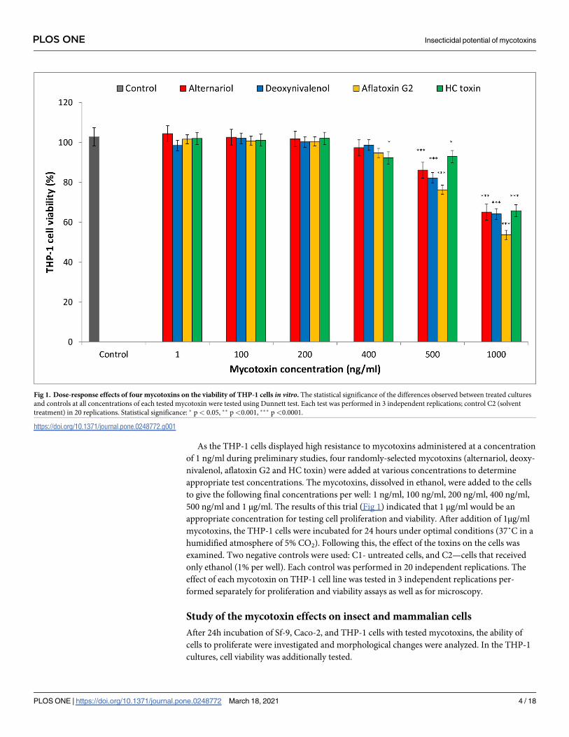
As the THP-1 cells displayed high resistance to mycotoxins administered at a concentration
of 1 ng/ml during preliminary studies, four randomly-selected mycotoxins (alternariol, deoxy-
nivalenol, aflatoxin G2 and HC toxin) were added at various concentrations to determine
appropriate test concentrations. The mycotoxins, dissolved in ethanol, were added to the cells
to give the following final concentrations per well: 1 ng/ml, 100 ng/ml, 200 ng/ml, 400 ng/ml,
500 ng/ml and 1 μg/ml. The results of this trial (Fig 1) indicated that 1 μg/ml would be an
appropriate concentration for testing cell proliferation and viability. After addition of 1μg/ml
mycotoxins, the THP-1 cells were incubated for 24 hours under optimal conditions (37˚C in a
humidified atmosphere of 5% CO2). Following this, the effect of the toxins on the cells was
examined. Two negative controls were used: C1- untreated cells, and C2—cells that received
only ethanol (1% per well). Each control was performed in 20 independent replications. The
effect of each mycotoxin on THP-1 cell line was tested in 3 independent replications per-
formed separately for proliferation and viability assays as well as for microscopy.
Study of the mycotoxin effects on insect and mammalian cells
After 24h incubation of Sf-9, Caco-2, and THP-1 cells with tested mycotoxins, the ability of
cells to proliferate were investigated and morphological changes were analyzed. In the THP-1
cultures, cell viability was additionally tested.
Fig 1. Dose-response effects of four mycotoxins on the viability of THP-1 cells in vitro. The statistical significance of the differences observed between treated cultures
and controls at all concentrations of each tested mycotoxin were tested using Dunnett test. Each test was performed in 3 independent replications; control C2 (solvent
treatment) in 20 replications. Statistical significance: � p< 0.05, �� p<0.001, ��� p<0.0001.
https://doi.org/10.1371/journal.pone.0248772.g001
PLOS ONE Insecticidal potential of mycotoxins
PLOS ONE | https://doi.org/10.1371/journal.pone.0248772 March 18, 2021 4 / 18
Cell Counting Kit-8 (CCK-8, Abbkine) was used to measure cell proliferation. This reagent
based on WST-8 (water-soluble tetrazolium salt) is reduced by the dehydrogenases in cells to
give an orange colored product (formazan). The amount of the formazan in cells was directly
proportional to the number of living cells. Briefly, 40 μl of CCK-8 reagent was added to each
well containing cell cultures (test or control), the plates were then incubated for four hours
under the described above optimal conditions for each cell type. Absorbance readings were
taken at 450 nm using a Synergy HT Multi-Mode Microplate Reader (BioTek). The results are
presented as a percentage of control (C2) findings, assumed to be 100%.
Of the tested cells, only the THP-1 line is non-adherent. Therefore, it was possible to check
the viability of these cells with propidium iodide (PI; Invitrogen). The final concentration of
the reagent in the wells of culture plates was 10 μg/ml. After a 10-minute incubation period at
room temperature, an Arthur cell counter (NanoEnTek) was used to calculate the number of
cells. The results are presented as a percentage of dead cells. Similar attempts to detach adher-
ent Sf-9 and Caco-2 cells, both mechanically and enzymatically, resulted in very high mortality,
which prevented the measurement of cell viability in these cultures.
Phase contrast microscope images of control and treated Sf-9, Caco-2, and THP-1 cells
were taken at various time points (from five minutes up to 24 hours after treatment) using a
Axio Vert.A1 fluorescence inverted microscope (Zeiss) equipped with Axio Cam ICc 5 camera
(Zeiss) and Zen lite 2012 software (Zeiss).
Statistical analysis
Statistical analysis was performed using STATISTICA 6.1 software (StatSoft Polska). Any sta-
tistical relationships were evaluated using the one-way ANOVA, t test and Dunnett test for
post hoc analysis. The Kolmogorov-Smirnov (K-S) test, skewness and kurtosis were used to
check normality of controls. All data are shown as means ± SD, and values of p� 0.05 were
considered as significant.
Results
Effects of 65 tested mycotoxins on the proliferation and viability of insect
and mammalian cells
From the data presented in Table 1, it appears that all tested mycotoxins significantly reduced
in vitro proliferation of Sf-9 cells as compared with control cells (C2) which received an appro-
priate amount of solvent (ethanol). Regarding the Sf-9 cells the strongest effect (a fall in cell
proliferation to 13–30% of that observed in control C2) was observed in the case of 24-hour
incubation with 1 ng/ml of 19 mycotoxins: 3-acetyldeoxynivalenol, aflatoxins B1 and B2, alter-
nariol, alternariol-9-methyl ether, beauvericin, brefeldin A, cyclosporin D, cytochalasin E,
fumonisin B2, gliotoxin, ochratoxin A, patulin, paxilline, penitrem A, stachybotrylactam, ster-
igmatocystin, T2 tetraol, and verruculogen. In contrast, the weakest effect (cell proliferation
drop to 70–91% of that observed in control C2) was detected in the case of 13 mycotoxins: afla-
toxicol, aflatoxin M1, γ-amanitin, antibiotic PF 1052, citrinin, cytochalasin D, fumagillin, HT-
2-toxin, roquefortine C, T2 triol, wortmannin, zearalenone, and α-zearalanol.
In the case of Caco-2 cells, no effect on cell proliferation was found after application of afla-
toxin M2, alternariol (which exerted potent effect on Sf-9 cells), γ-ammanitin, cyclosporine B,
cytochalasin A or diacetoxyscripenol. It is noteworthy that verruculogen, which inhibited the
proliferation of Sf-9 cells to 16 ± 3%, had the opposite effect on Caco-2 (140 ± 12% of cell pro-
liferation). The lowest values of Caco-2 cell proliferation were measured after the application
of T2 tetraol (35 ± 3%), stachybotrylactam (37 ± 6%), cytochalasin B (39 ± 4%) or wortmannin
PLOS ONE Insecticidal potential of mycotoxins
PLOS ONE | https://doi.org/10.1371/journal.pone.0248772 March 18, 2021 5 / 18
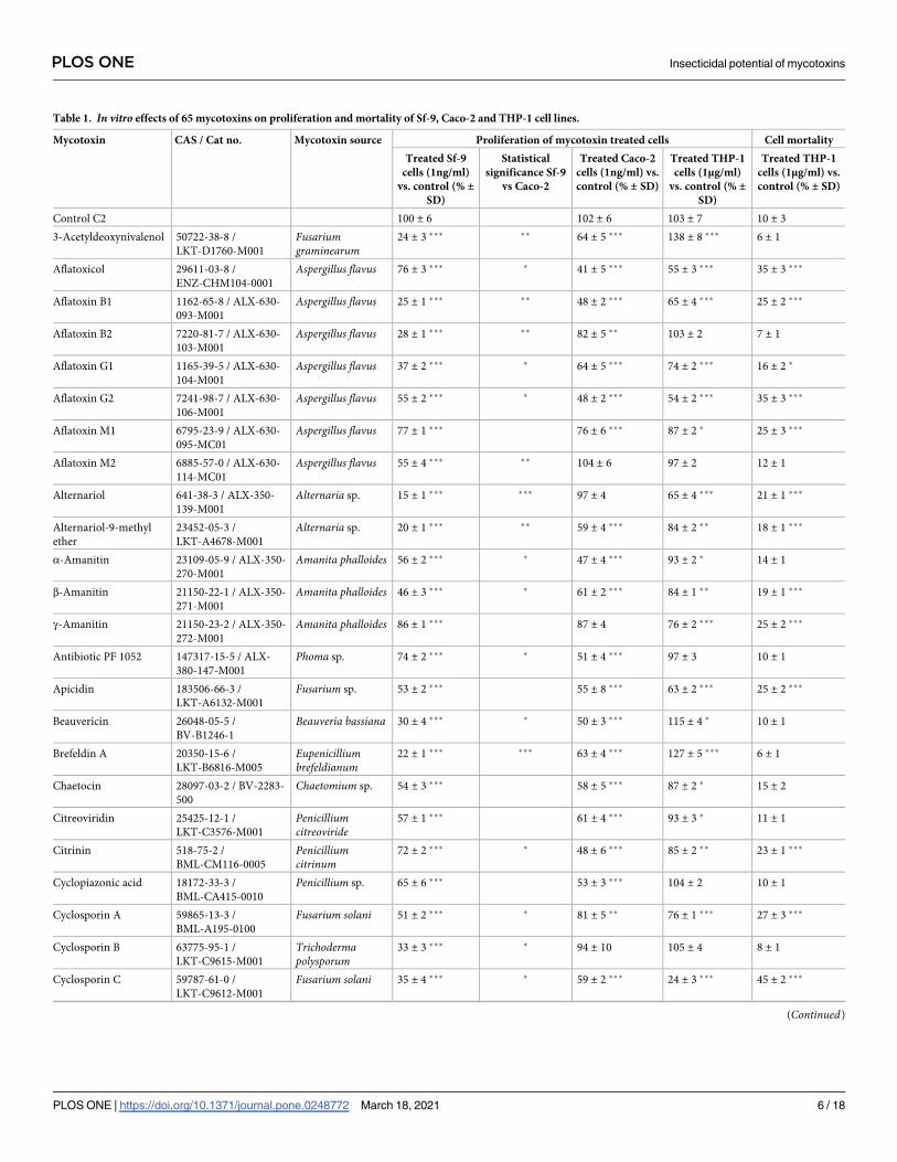
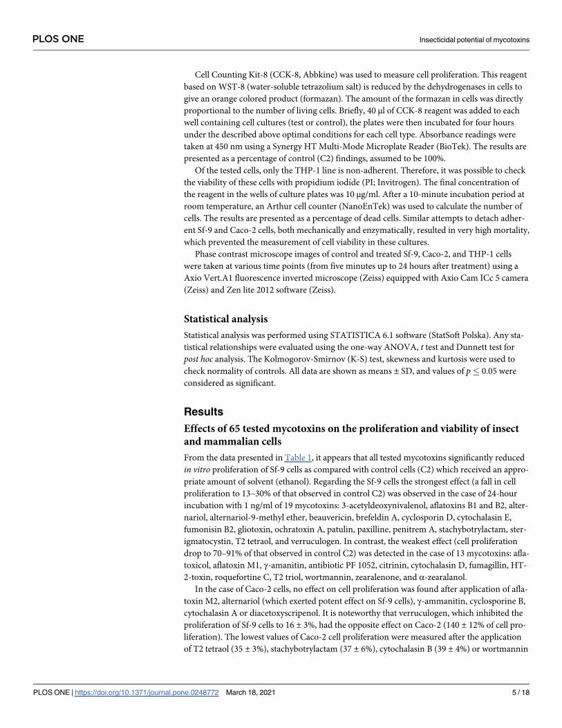
Table 1. In vitro effects of 65 mycotoxins on proliferation and mortality of Sf-9, Caco-2 and THP-1 cell lines.
Mycotoxin CAS / Cat no. Mycotoxin source Proliferation of mycotoxin treated cells Cell mortality
Treated Sf-9
cells (1ng/ml)
vs. control (% ±SD)
Statistical
significance Sf-9
vs Caco-2
Treated Caco-2
cells (1ng/ml) vs.
control (% ± SD)
Treated THP-1
cells (1μg/ml)
vs. control (% ±SD)
Treated THP-1
cells (1μg/ml) vs.
control (% ± SD)
Control C2 100 ± 6 102 ± 6 103 ± 7 10 ± 3
3-Acetyldeoxynivalenol 50722-38-8 /
LKT-D1760-M001
Fusariumgraminearum
24 ± 3 ��� �� 64 ± 5 ��� 138 ± 8 ��� 6 ± 1
Aflatoxicol 29611-03-8 /
ENZ-CHM104-0001
Aspergillus flavus 76 ± 3 ��� � 41 ± 5 ��� 55 ± 3 ��� 35 ± 3 ���
Aflatoxin B1 1162-65-8 / ALX-630-
093-M001
Aspergillus flavus 25 ± 1 ��� �� 48 ± 2 ��� 65 ± 4 ��� 25 ± 2 ���
Aflatoxin B2 7220-81-7 / ALX-630-
103-M001
Aspergillus flavus 28 ± 1 ��� �� 82 ± 5 �� 103 ± 2 7 ± 1
Aflatoxin G1 1165-39-5 / ALX-630-
104-M001
Aspergillus flavus 37 ± 2 ��� � 64 ± 5 ��� 74 ± 2 ��� 16 ± 2 �
Aflatoxin G2 7241-98-7 / ALX-630-
106-M001
Aspergillus flavus 55 ± 2 ��� � 48 ± 2 ��� 54 ± 2 ��� 35 ± 3 ���
Aflatoxin M1 6795-23-9 / ALX-630-
095-MC01
Aspergillus flavus 77 ± 1 ��� 76 ± 6 ��� 87 ± 2 � 25 ± 3 ���
Aflatoxin M2 6885-57-0 / ALX-630-
114-MC01
Aspergillus flavus 55 ± 4 ��� �� 104 ± 6 97 ± 2 12 ± 1
Alternariol 641-38-3 / ALX-350-
139-M001
Alternaria sp. 15 ± 1 ��� ��� 97 ± 4 65 ± 4 ��� 21 ± 1 ���
Alternariol-9-methyl
ether
23452-05-3 /
LKT-A4678-M001
Alternaria sp. 20 ± 1 ��� �� 59 ± 4 ��� 84 ± 2 �� 18 ± 1 ���
α-Amanitin 23109-05-9 / ALX-350-
270-M001
Amanita phalloides 56 ± 2 ��� � 47 ± 4 ��� 93 ± 2 � 14 ± 1
β-Amanitin 21150-22-1 / ALX-350-
271-M001
Amanita phalloides 46 ± 3 ��� � 61 ± 2 ��� 84 ± 1 �� 19 ± 1 ���
γ-Amanitin 21150-23-2 / ALX-350-
272-M001
Amanita phalloides 86 ± 1 ��� 87 ± 4 76 ± 2 ��� 25 ± 2 ���
Antibiotic PF 1052 147317-15-5 / ALX-
380-147-M001
Phoma sp. 74 ± 2 ��� � 51 ± 4 ��� 97 ± 3 10 ± 1
Apicidin 183506-66-3 /
LKT-A6132-M001
Fusarium sp. 53 ± 2 ��� 55 ± 8 ��� 63 ± 2 ��� 25 ± 2 ���
Beauvericin 26048-05-5 /
BV-B1246-1
Beauveria bassiana 30 ± 4 ��� � 50 ± 3 ��� 115 ± 4 � 10 ± 1
Brefeldin A 20350-15-6 /
LKT-B6816-M005
Eupenicilliumbrefeldianum
22 ± 1 ��� ��� 63 ± 4 ��� 127 ± 5 ��� 6 ± 1
Chaetocin 28097-03-2 / BV-2283-
500
Chaetomium sp. 54 ± 3 ��� 58 ± 5 ��� 87 ± 2 � 15 ± 2
Citreoviridin 25425-12-1 /
LKT-C3576-M001
Penicilliumcitreoviride
57 ± 1 ��� 61 ± 4 ��� 93 ± 3 � 11 ± 1
Citrinin 518-75-2 /
BML-CM116-0005
Penicilliumcitrinum
72 ± 2 ��� � 48 ± 6 ��� 85 ± 2 �� 23 ± 1 ���
Cyclopiazonic acid 18172-33-3 /
BML-CA415-0010
Penicillium sp. 65 ± 6 ��� 53 ± 3 ��� 104 ± 2 10 ± 1
Cyclosporin A 59865-13-3 /
BML-A195-0100
Fusarium solani 51 ± 2 ��� � 81 ± 5 �� 76 ± 1 ��� 27 ± 3 ���
Cyclosporin B 63775-95-1 /
LKT-C9615-M001
Trichodermapolysporum
33 ± 3 ��� � 94 ± 10 105 ± 4 8 ± 1
Cyclosporin C 59787-61-0 /
LKT-C9612-M001
Fusarium solani 35 ± 4 ��� � 59 ± 2 ��� 24 ± 3 ��� 45 ± 2 ���
(Continued)
PLOS ONE Insecticidal potential of mycotoxins
PLOS ONE | https://doi.org/10.1371/journal.pone.0248772 March 18, 2021 6 / 18
Table 1. (Continued)
Mycotoxin CAS / Cat no. Mycotoxin source Proliferation of mycotoxin treated cells Cell mortality
Treated Sf-9
cells (1ng/ml)
vs. control (% ±SD)
Statistical
significance Sf-9
vs Caco-2
Treated Caco-2
cells (1ng/ml) vs.
control (% ± SD)
Treated THP-1
cells (1μg/ml)
vs. control (% ±SD)
Treated THP-1
cells (1μg/ml) vs.
control (% ± SD)
Cyclosporin D 63775-96-2 /
BML-T109-0001
Fusarium solani 24 ± 1 ��� � 73 ± 11 ��� 76 ± 2 ��� 17 ± 2 ��
Cyclosporin H 83602-39-5 /
LKT-C9614-M001
Tolypocladiuminflatum
35 ± 2 ��� � 48 ± 6 ��� 104 ± 3 8 ± 1
Cytochalasin A 14110-64-6 /
LKT-C9878-M001
Drechsleradematoidea
37 ± 2 ��� ��� 89 ± 4 75 ± 3 ��� 25 ± 3 ���
Cytochalasin B 14930-96-2 /
BML-T108-0005
Drechsleradematoidea
55 ± 3 ��� � 39 ± 4 ��� 106 ± 2 9 ± 1
Cytochalasin C 22144-76-9 /
LKT-C9880-M001
Metarhiziumanisopliae
64 ± 1 ��� 69 ± 5 ��� 45 ± 3 ��� 39 ± 1 ���
Cytochalasin D 22144-77-0 /
LKT-C9881-M001
Zygosporiummansonii
75 ± 4 ��� 75 ± 4 ��� 93 ± 3 � 13 ± 1
Cytochalasin E 36011-19-5 /
LKT-C9882-M001
Aspergillus clavatus 24 ± 3 ��� � 42 ± 3 ��� 93 ± 3 � 19 ± 1 ���
Deoxynivalenol 51481-10-8 / ALX-630-
115-M001
Fusarium sp. 34 ± 2 ��� � 67 ± 6 ��� 64 ± 3 ��� 24 ± 2 ���
Diacetoxyscripenol 2270-40-8 /
LKT-D3200-M001
Fusarium sp. 46 ± 2 ��� � 100 ± 11 18 ± 1 ��� 69 ± 2 ���
Fumagillin 23110-15-8 /
LKT-C9882-M001
Aspergillusfumigatus
85 ± 2 ��� � 55 ± 6 ��� 75 ± 3 ��� 23 ± 4 ���
Fumigaclavine A 6879-59-0 / ALX-630-
110-M001
Aspergillus sp. 65 ± 4 ��� 75 ± 4 ��� 49 ± 8 ��� 34 ± 2 ���
Fumonisin B1 116355-83-0 /
BML-SL220-0001
Fusariummoniliforme
40 ± 2 ��� 49 ± 8 ��� 93 ± 2 12 ± 2
Fumonisin B2 116355-84-1 /
BML-SL219-0001
Aspergillus niger 13 ± 2 ��� �� 59 ± 4 ��� 106 ± 3 7 ± 1
Fusarenon X 23255-69-8 /
LKT-F8272-M001
Fusarium sp. 64 ± 3 ��� 67 ± 5 ��� 94 ± 2 12 ± 2
Gliotoxin 67-99-2 / BML-PI129-
0002
Gladiocladiumfimbriatum
24 ± 2 ��� �� 41 ± 2 ��� 78 ± 4 ��� 31 ± 1 ���
HC toxin 83209-65-8 /
BML-GR320-0001
Cochlioboluscarbonum
31 ± 2 ��� � 47 ±5 ��� 66 ± 3 ��� 23 ± 2 ���
HT-2 toxin 26934-87-2 / ALX-630-
113-M001
Fusariumtricinctum (�)
86 ± 2 �� �� 55 ± 4 ��� 107 ± 4 8 ± 1
Moniliformin 31876-38-7 /
LKT-M5853-M001
Fusarium sp. 60 ± 5 ��� � 86 ± 5 � 96 ± 3 11 ± 1
Moniliformin sodium
salt
71376-34-6 / ALX-630-
111-M001
Fusariummoniliforme
54 ± 2 ��� � 86 ± 6 � 54 ± 2 ��� 35 ± 3 ���
Mycophenolic acid 24280-93-1 /
BML-A249-0100
Penicillium brevi-compactum
39 ± 3 ��� � 62 ± 7 ��� 99 ±2 18 ± 1 ���
Neosolaniol 36519-25-2 /
LKT-N1858-M001
Fusarium sp. 34 ± 3 ��� � 60 ± 4 ��� 56 ± 4 ��� 35 ± 2 ���
Ochratoxin A 303-47-9 / ALX-630-
089-M001
Aspergillusochraceus
22 ± 1 ��� �� 63 ± 4 ��� 99 ± 3 13 ± 2
Ochratoxin B 4825-86-9 Aspergillus sp. 34 ± 2 ��� � 75 ± 8 ��� 93 ± 3 � 16 ± 2 �
Patulin 149-29-1 / ALX-270-
111-M001
Penicilliumexpansum
17 ± 3 ��� � 45 ± 7 ��� 106 ±4 13 ± 2
Paxilline 57186-25-1 /
BML-KC155-0005
Penicillium paxilli 24 ± 2 ��� �� 73 ± 4 ��� 55 ± 3 ��� 32 ± 2 ���
(Continued)
PLOS ONE Insecticidal potential of mycotoxins
PLOS ONE | https://doi.org/10.1371/journal.pone.0248772 March 18, 2021 7 / 18
(39 ± 2%). Application of these four mycotoxins to Sf-9 cells exerted either a similar effect, i.e.
strong inhibition of cell proliferation (T2 tetraol, stachybotrylactam), or slightly decreased pro-
liferation (cytochalasin B, wortmannin). The following mycotoxins strongly decreased the pro-
liferation of Sf-9 cells (drop to 13–30%) but displayed a moderate or no effect on the
proliferation of Caco-2 cells: 3-acetyldeoxynivalenol, alternariol, alternariol-9-methyl ether,
brefeldin A, cyclosporines B and C, ochratoxin A, paxilline, sterigmatocystin, and verruculo-
gen (which stimulated proliferation of Caco-2 cells; Table 1).
Table 1. (Continued)
Mycotoxin CAS / Cat no. Mycotoxin source Proliferation of mycotoxin treated cells Cell mortality
Treated Sf-9
cells (1ng/ml)
vs. control (% ±SD)
Statistical
significance Sf-9
vs Caco-2
Treated Caco-2
cells (1ng/ml) vs.
control (% ± SD)
Treated THP-1
cells (1μg/ml)
vs. control (% ±SD)
Treated THP-1
cells (1μg/ml) vs.
control (% ± SD)
Penitrem A 12627-35-9 /
BML-KC157-0001
Penicilliumpalitans
15 ± 1 ��� �� 43 ± 2 ��� 66 ± 2 ��� 24 ± 2 ���
Phomopsin A 12627-35-9 / ALX-350-
417-M001
Phomopsisleptostromiformis
35 ± 2 ��� �� 68 ± 4 ��� 110 ± 6 9 ± 2
Roquefortine C 58735-64-1 / ALX-350-
342-MC05
Penicilliumroqueforti
79 ± 2 ��� �� 56 ± 3 ��� 76 ± 3 ��� 26 ± 1 ���
Skyrin 602-06-2 / BV-2043-1 Talaromyces sp. 34 ± 2 ��� � 59 ± 4 ��� 113 ± 4 � 6 ± 1
Stachybotrylactam 163391-76-2 / ALX-
630-112-M005
Stachybotrys sp. 17 ± 1 ��� � 37 ± 6 ��� 85 ± 2 �� 18 ± 2 ���
Sterigmatocystin 10048-13-2 / ALX-630-
116-M001
Aspergillusversicolor
24 ± 2 ��� ��� 63 ± 3 ��� 54 ± 2 ��� 32 ± 2 ���
Strobilurin B 65105-52-4 / ALX-380-
144-M001
Strobilurus sp. 59 ± 2 ��� 58 ± 13 ��� 106 ± 4 8 ± 2
T2 tetraol 34114-99-3 /
LKT-T0003-M001
Fusarium sp. 17 ± 2 ��� � 35 ± 3 ��� 122 ± 3 �� 7 ± 2
T2 toxin 21259-20-1 / ALX-630-
101-M001
Fusariumtricinctum
57 ± 4 ��� � 84 ± 6 � 87 ± 3 � 24 ± 3 ���
T2 triol 34114-98-2 /
LKT-T0004-M001
Fusarium sp. 75 ± 2 ��� 84 ± 15 � 96 ± 2 11 ± 1
Tenuazonic acid 610-88-8 / ALX-350-
317-MC05
Alternaria sp. 53 ± 2 ��� �� 71 ± 2 ��� 65 ± 4 ��� 35 ± 3 ���
Territrem B 70407-20-4 / ALX-630-
117-MC05
Aspergillus terreus 40 ± 2 ��� �� 76 ± 4 ��� 103 ± 2 8 ± 2
Verruculogen 12771-72-1 /
LKT-V1870-M001
Penicilliumverruculosum
16 ± 3 ��� �� 140 ± 12 ��� 94 ± 3 13 ± 1
Wortmannin 19545-26-7 /
LKT-W5769-M001
Penicilliumwortmannii
85 ± 3 ��� ��� 39 ± 2 ��� 79 ± 2 ��� 21 ± 2 ���
Zearalenone 17924-92-4 / ALX-630-
105-M010
Giberella zeae 91 ± 3 � �� 40 ± 4 ��� 94 ± 3 14 ± 2
α-Zearalanol 26538-44-3 /
LKT-Z161022-M001
Fusarium sp. 87 ± 2 � � 49 ± 7 ��� 110 ± 4 9 ± 1
Tested mycotoxins were added to the cell cultures at final concentrations of 1 ng/ml (Sf-9 and Caco-2) and 1 μg/ml (THP-1). Proliferation tests were performed using
Cell Counting Kit-8 (Abbkine) as described in Materials and methods. Cell mortality tests were performed on non-adherent THP-1 cells using propidium iodide and
Arthur cell counter (NanoEnTek). Control C2—ethanol treated cells. Each control was performed in 20 independent replications. The effect of each mycotoxin on each
cell line was tested in 3 independent replications. The results are presented as a percentage of control C2 findings, assumed to be 100%. All mycotoxins were purchased
via the Axxora platform (http://www.axxora.com). (�) Semisynthetic, derived from T2 toxin from Fusarium tricinctum. Statistical significance:
� p < 0.05
�� p < 0.001
��� p < 0.0001. Dunnett test was applied for mycotoxins-to-control comparisons while t test was used to compare the reactivity of Sf-9 cells with Caco-2 cells.
https://doi.org/10.1371/journal.pone.0248772.t001
PLOS ONE Insecticidal potential of mycotoxins
PLOS ONE | https://doi.org/10.1371/journal.pone.0248772 March 18, 2021 8 / 18
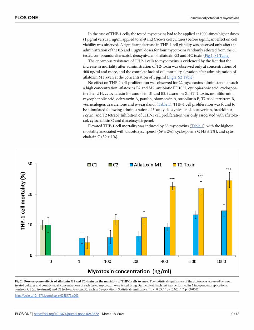
In the case of THP-1 cells, the tested mycotoxins had to be applied at 1000-times higher doses
(1 μg/ml versus 1 ng/ml applied to Sf-9 and Caco-2 cell cultures) before significant effect on cell
viability was observed. A significant decrease in THP-1 cell viability was observed only after the
administration of the 0.5 and 1 μg/ml doses for four mycotoxins randomly selected from the 65
tested compounds: alternariol, deoxynivalenol, aflatoxin G2 and HC toxin (Fig 1, S1 Table).
The enormous resistance of THP-1 cells to mycotoxins is evidenced by the fact that the
increase in mortality after administration of T2-toxin was observed only at concentrations of
400 ng/ml and more, and the complete lack of cell mortality elevation after administration of
aflatoxin M1, even at the concentration of 1 μg/ml (Fig 2, S2 Table).
No effect on THP-1 cell proliferation was observed for 22 mycotoxins administered at such
a high concentration: aflatoxins B2 and M2, antibiotic PF 1052, cyclopiazonic acid, cyclospor-
ine B and H, cytochalasin B, fumonisin B1 and B2, fusarenon X, HT-2 toxin, moniliformin,
mycophenolic acid, ochratoxin A, patulin, phomopsin A, strobilurin B, T2 triol, territrem B,
verruculogen, zearalenone and α-zearalanol (Table 1). THP-1 cell proliferation was found to
be stimulated following administration of 3-acetyldeoxynivalenol, beauvericin, brefeldin A,
skyrin, and T2 tetraol. Inhibition of THP-1 cell proliferation was only associated with aflatoxi-
col, cytochalasin C and diacetoxyscirpenol.
Elevated THP-1 cell mortality was induced by 33 mycotoxins (Table 1), with the highest
mortality associated with diacetoxyscirpenol (69 ± 2%), cyclosporine C (45 ± 2%), and cyto-
chalasin C (39 ± 1%).
Fig 2. Dose-response effects of aflatoxin M1 and T2-toxin on the mortality of THP-1 cells in vitro. The statistical significance of the differences observed between
treated cultures and controls at all concentrations of each tested mycotoxin were tested using Dunnett test. Each test was performed in 3 independent replications;
controls: C1 (no treatment) and C2 (solvent treatment), each in 3 replications. Statistical significance: � p< 0.05, �� p<0.001, ��� p<0.0001.
https://doi.org/10.1371/journal.pone.0248772.g002
PLOS ONE Insecticidal potential of mycotoxins
PLOS ONE | https://doi.org/10.1371/journal.pone.0248772 March 18, 2021 9 / 18
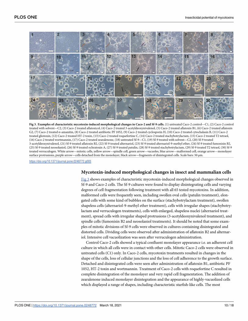
Mycotoxin-induced morphological changes in insect and mammalian cells
Fig 3 shows examples of characteristic mycotoxin-induced morphological changes observed in
Sf-9 and Caco-2 cells. The Sf-9 cultures were found to display disintegrating cells and varying
degrees of cell fragmentation following treatment with all 65 tested mycotoxins. In addition,
malformed cells were frequently seen, including swollen oval cells (patulin treatment), elon-
gated cells with some kind of bubbles on the surface (stachybotrylactam treatment), swollen
shapeless cells (alternariol-9-methyl ether treatment), cells with irregular shapes (stachybotry-
lactam and verruculogen treatments), cells with enlarged, shapeless nuclei (alternariol treat-
ment), spread cells with irregular shaped protrusions (3-acetyldeoxynivalenol treatment), and
spindle cells (fumonisin B2 and neosolaniol treatments). It should be noted that some exam-
ples of mitotic divisions of Sf-9 cells were observed in cultures containing disintegrated and
distorted cells. Dividing cells were observed after administration of aflatoxin B2 and alternar-
iol. Intensive cell vacuolization was seen after verruculogen administration.
Control Caco-2 cells showed a typical confluent monolayer appearance i.e. an adherent cell
culture in which all cells were in contact with other cells. Mitotic Caco-2 cells were observed in
untreated cells (C1) only. In Caco-2 cells, mycotoxin treatments resulted in changes in the
shape of the cells, loss of cellular junctions and the loss of cell adherence to the growth surface.
Detached and disintegrated cells were seen after administration of aflatoxin B1, antibiotic PF
1052, HT-2 toxin and wortmannin. Treatment of Caco-2 cells with roquefortine C resulted in
complete disintegration of the monolayer and very rapid cell fragmentation. The addition of
zearalenone induced monolayer disintegration and the appearance of highly-vacuolized cells
which displayed a range of shapes, including characteristic starfish-like cells. The most
Fig 3. Examples of characteristic mycotoxin-induced morphological changes in Caco-2 and Sf-9 cells. (1) untreated Caco-2 control—C1, (2) Caco-2 control
treated with solvent—C2, (3) Caco-2 treated aflatoxicol, (4) Caco-2 treated 3-acetyldeoxynivalenol, (5) Caco-2 treated aflatoxin B1, (6) Caco-2 treated aflatoxin
G2, (7) Caco-2 treated α-amanitin, (8) Caco-2 treated antibiotic PF 1052, (9) Caco-2 treated cyclosporin H, (10) Caco-2 treated cytochalasin B, (11) Caco-2
treated gliotoxin, (12) Caco-2 treated HT-2 toxin, (13) Caco-2 treated roquefortine C, (14) Caco-2 treated stachybotrylactam, (15) Caco-2 treated T2 tetraol,
(16) Caco-2 treated wortmannin, (17) Caco-2 treated zearalenone, (18) untreated Sf-9—C1, (19) Sf-9 treated with solvent—C2, (20) Sf-9 treated
3-acetyldeoxynivalenol, (21) Sf-9 treated aflatoxin B2, (22) Sf-9 treated alternariol, (23) Sf-9 treated alternariol-9-methyl ether, (24) Sf-9 treated fumonisin B2,
(25) Sf-9 treated neosolaniol, (26) Sf-9 treated ochratoxin A, (27) Sf-9 treated patulin, (28) Sf-9 treated stachybotrylactam, (29) Sf-9 treated T2 tetraol, (30) Sf-9
treated verruculogen. White arrow—mitotic cells, yellow arrow—spindle cell, green arrow—vacuoles, blue arrow—malformed cell, orange arrow—monolayer
surface protrusions, purple arrow—cells detached from the monolayer, black arrow—fragments of disintegrated cells. Scale bars: 50 μm.
https://doi.org/10.1371/journal.pone.0248772.g003
PLOS ONE Insecticidal potential of mycotoxins
PLOS ONE | https://doi.org/10.1371/journal.pone.0248772 March 18, 2021 10 / 18
frequently-observed change was the formation of monolayer surface protrusions: convex
structures similar to blisters. Such structures, of various shapes and sizes, were observed after
the administration of aflatoxicol, 3- acetyldeoxynivalenol, aflatoxin G2, α-amanitin, antibiotic
PF 1052, cyclosporin H, cytochalasin B, stachybotrylactam, T2 tetraol, and wortmannin.
THP-1 cells were found to be much more resistant to mycotoxins than Sf-9 and Caco-2
cells, as a 1000-fold greater dose was typically required to elicit a response. Typical mycotoxin-
induced changes in THP-1 cell morphology are presented in Fig 4. Such swollen and deformed
cells were seen after treatment with aflatoxicol, aflatoxin B2, cyclosporin C, cyclosporin H,
cytochalasin C, diacetoxyscripenol, patulin and paxilline. Disintegrated and distorted cells
were observed in cultures treated with aflatoxin G2, cytochalasin C, diacetoxyscripenol, fumi-
gaclavine A, moniliformin, patulin, paxilline and sterigmatocystin. Vacuolized THP-1 cells
appeared after the application of aflatoxicol, fumigaclavine A, moniliformin, patulin, paxilline
and sterigmatocystin. Treatment with aflatoxin G2 resulted in the mass disintegration of cells,
while the administration of ochratoxin A caused the cells to clump together. Moniliformin
treatment induced vacuolization and cell disintegration, but cell divisions appeared to
continue.
Discussion
The damage caused to crops and stored food by harmful insects, as well as their transmission
of pathogens causing serious diseases for humans, animals and plants require more effective
control of pest populations. The insecticides currently used for this purpose are typically syn-
thetic ones based on non-selective chemical compounds which accumulate in the environment
and pose a serious threat to bio-diversity; growing pressure to minimize these deleterious
effects has led to increased interest in the potential to harness the natural enemies of insects.
Among various potential biocontrol agents, entomopathogenic fungi seem to be the most
promising [18, 19].
The fungal pathogenesis of insects typically follows a series of events comprising conidial
adhesion to the insect host cuticle, followed by penetration of the cuticle via fungal enzymes,
the impairment of host immune responses, dissemination within the host and transmission
from the host [19, 20]. Toxic metabolites released by the fungus into the hemocoel of the insect
play an important role in this multi-step process; although the exact function of these fungal
toxins is not always clear, it has been proposed that mycotoxins could be released directly to
poison and kill the infected host or to suppress its immune system, thus facilitating the devel-
opment of the mycosis [21]. Mycotoxins can also enter the interior of the insect body through
the digestive tract, as fungi are desirable sources of food rich in proteins and sterols [22].
Numerous mycotoxins, with various chemical structures, show strong insecticidal activity
against insect pests. Four decades ago, the insecticidal activities of aflatoxin B1, rubratoxin B,
patulin and diacetoxyscirpenol have been examined in contact tests against Drosophila mela-nogaster [23], as has beauvericin against Calliphora erythrocephala, Aedes aegypti, Lygus spp.,
Spodoptera frugiperda and Schizaphis graminum [24]. Fungal ribotoxins, i.e. extracellular
highly specific ribonucleases, were found to efficiently kill Galleria mellonella larvae and to dis-
play toxicity against two different insect cell lines: Sf-9 from Spodoptera frugiperda and Tni
High Five from Trichoplusia ni [25]. In addition, tolypin, produced by two Tolypocladium spe-
cies, was toxic to insects upon injection [26], leucinostins and efrapeptins, linear peptide toxins
showing ATPase inhibitory activity, were active in foliar spray assays against Leptinotarsadecemlineata [27], and destruxins were found to display a strong lethal action against insects
acting through a range of harmful processes on the cellular level [19]. Another study found a
range of 22 mycotoxins to have a strong influence on the growth and mortality ofHelicoverpa
PLOS ONE Insecticidal potential of mycotoxins
PLOS ONE | https://doi.org/10.1371/journal.pone.0248772 March 18, 2021 11 / 18
zea and Spodoptera frugiperda: aflatoxin B1, ochratoxin A, sterigmatocistin, citrinin, cyclopia-
zonic acid, penicillic acid, deoxynivalenol, diacetoxyscripenol, dihydrodeoxynivalenol, dihy-
droxycalonectrin, hydroxycalonectrin, moniliformion, sambucinol, T2 toxin, zearalenone,
chaetoglobosin C, cytochalasin H, paspaline, paxilline, penitrem A, roseotoxin B, and
Fig 4. Examples of characteristic mycotoxin-induced morphological changes in THP-1 cells. (1) untreated THP-1
control—C1, (2) THP-1 control treated with solvent—C2, (3) aflatoxicol, (4) aflatoxin B2, (5) aflatoxin G2, (6)
cyclosporin C, (7) cyclosporin H, (8) cytochalasin C, (9) diacetoxyscripenol, (10) fumigaclavine A, (11) moniliformin,
(12) ochratoxin A, (13) patulin, (14) paxilline, (15) sterigmatocystin. White arrow—mitotic cells, red arrow—swelled
and deformed cell, green arrow—vacuoles, black arrow—fragments of disintegrated cells. Scale bars: 50 μm.
https://doi.org/10.1371/journal.pone.0248772.g004
PLOS ONE Insecticidal potential of mycotoxins
PLOS ONE | https://doi.org/10.1371/journal.pone.0248772 March 18, 2021 12 / 18
verruculogen [22]. The action of aflatoxin B1 and T2 toxin against insects has been particularly
widely studied; it has been found that while cockroaches are relatively resistant to aflatoxin B1,
stored-product insects are relatively resistant to T2 toxin, and while Drosophila melanogaster is
susceptible to aflatoxin B1, the larvae of Bombyx mori,Heliotis zea and Spodoptera frugiperdaare sensitive to both aflatoxin B1 and T2 toxin [22]. Interestingly, many mycophagous species
of Drosophila can tolerate the fungal poison α-amanitin in wild mushrooms and in artificial
diet [28]. The activities of other lesser known insecticidal mycotoxins have been reviewed in
other papers [29, 30]. Due to the tremendous variety of secondary metabolites produced by
fungi, intensive studies concerning the insecticidal potential of mycotoxins are needed. As it is
costly and time-consuming to use insects to test the insecticidal potential of mycotoxins,
screening studies can be conducted more efficiently in vitro using insect cell lines [31].
Our data clearly show that all 65 tested mycotoxins significantly reduced the proliferation
of insect Sf-9 cells in vitro. Sixteen of the mycotoxins used in the present study against the Sf-9
cells, including beauvericin, deoxynivalenol, diacetoxyscripenol, fusarenon X, fumonisin B1,
gliotoxin, moniliformin, ochratoxin A and zearalenone (Table 1), have previously been tested
by Fornelli and co-workers [31]. After converting our doses (1 ng/ml) to the micromolar con-
centrations used in the previous study, it was found that similar results were yielded only for
beauvericin and gliotoxin; our Sf-9 cultures seem to be less sensitive to diacetoxyscripenol and
fusarenon X than observed by Farnelli et al [31]. However, our cells were much more sensitive
to deoxynivalenol, fumonisin B1, moniliformin, ochratoxin A and zearalenone. These discrep-
ancies might result from the use of different tests to measure cell proliferation: although both
tests are based on the activity of the mitochondrial succinate–tetrazolium reductase system,
the two studies used different substrates (MTT versus WST-8).
The insect cell line Sf-9 is widely used to express recombinant proteins as the cultures are
inexpensive and easy to scale up. However, care should be taken while growing Sf-9 cells, as
they are extremely sensitive to fluctuations in temperature, cell density and agitation [32]. The
application of fumonisin B2 to the Sf-9 cell cultures in the present study inhibited cell prolifera-
tion and resulted in the appearance of spindle cells (Table 1, Fig 2). Similar effects were reported
by Zhang and co-workers, who report Sf-9 cell growth to be arrested at the G2/M phase, loss of
adhesion, swelling and vacuole formation, depolarization of the cell membrane potential and
hyperpolarization of the mitochondrial membrane potential following fumonisin B1 treatment
[33]. The difference in susceptibility of the insect Sf-9 cells against the tested mycotoxins com-
pared with that of the mammalian Caco-2 and THP-1 cells can be attributed to differences in
cell lipid composition. Compared to mammalian cells, Sf-9 cells have reduced levels of sphingo-
myelin and elevated levels of phosphatidylethanolamine, and even when grown in mammalian
serum, the plasma membranes of Sf-9 cells display very low cholesterol content and correspond-
ingly low cholesterol to phospholipid ratio [34]. In vitro cultured insect cells have also demon-
strated higher sensitivity to cholera toxin than tested mammalian cells [35].
Our research has shown that the THP-1 cells are extremely resistant to most tested mycoto-
toxins. Furthermore, some mycotoxins, including 3-acetyldeoxynivalenol, beauvericin, brefel-
din A, skyrin and T2 tetraol, stimulate the proliferation of THP-1 cells. The THP-1 cell line
has been reported to be resistant to several drugs, and monocytic THP-1 human cell lines dis-
play different susceptibility to engineered nanoparticles compared to intestinal epithelial
Caco-2 [36, 37]. Caco-2 has also been found to be more sensitive to cyclopiazonic acids than
THP-1 cells [38], which is consistent with our observations (Table 1). However, more research
is required to fully identify the reasons for the considerable difference observed between insect
cells and the two types of mammalian cells to the same mycotoxins; despite progress in this
area, the mechanisms underlying the disruptive influence of mycotoxins on the cellular
machinery remain unraveled [39, 40].
PLOS ONE Insecticidal potential of mycotoxins
PLOS ONE | https://doi.org/10.1371/journal.pone.0248772 March 18, 2021 13 / 18
Most studies focus on the major Fusariummycotoxins which have been shown to cause a
broad variety of toxic effects in animals. Trichothecenes, small amphipathic molecules that
passively move across cell membranes, display multiple inhibitory effects on the primary
metabolism of eukaryotic cells, including the inhibition of protein, DNA and RNA synthesis;
in tissues with high cell turnover rates, such inhibition results in the alteration in cell prolifera-
tion [40, 41]. At the cellular level deoxynivalenol activates mitogen-activated protein kinase
(MAPK) by a mechanism called the ribotoxic stress response, which drives both cytokine gene
expression and apoptosis in macrophages [42]. Nivalenol and deoxynivalenol, which regularly
co-occur in nature, share highly similar chemical structures and many toxicological properties,
such as the inhibition of cell proliferation, induction of interleukin-8 secretion, and the
involvement of MAPKs and nuclear factor κΒ in the signal transduction pathways associated
with toxicities [40]. Zearalenone has been shown to be immunotoxic, hepatotoxic and nephro-
toxic and an enhancer of lipid peroxidation; however, because of its structural similarity with
the estrogen hormones, the major target of zearalenone is the mammalian reproductive system
[40, 43]. Fumonisins, neurodegenerative mycotoxins, interfere with the biosynthesis of sphin-
golipids by inhibiting ceramide synthase, which impairs the metabolism of arachidonic acid
and leads to degeneration of the sphingolipid-rich tissues [40]. Beauvericin, a cyclic hexadepsi-
peptide showing antimicrobial, antiviral, anti-tumor, cytotoxic, ionophoric, apoptotic and
immunosuppressive activities, also demonstrates strong insecticidal activity against a broad
spectrum of insects. Beauvericin increases the permeability of biological membranes by form-
ing a complex with essential cations (Ca2+, Na+, K+) and/or cation-selective channels in lipid
membranes, which affects cell homeostasis and the uncoupling of oxidative phosphorylation
[40, 44]. The molecular mechanism of action of moniliformin is still obscure; however, its
structural similarity to pyruvate suggests that moniliformin affects energy metabolism via the
inhibition of mitochondrial pyruvate and α-ketoglutarate oxidation during the Krebs cycle
[40]. The major effect of T2 toxin is inhibition of protein synthesis leading to disruption of
DNA and RNA synthesis. The mechanisms of action of the T2 toxin and its metabolites against
animals and humans are still not clearly understood [40].
Our comparison of the impact of individual mycotoxins on the three examined cell lines
has allowed the selection of several promising candidates for further evaluation as potential
insecticides. The mycotoxins cyclosporin B and D, cytochalasin E, gliotoxin, HC toxin, paxil-
line, penitrem A, stachybotrylactam and verruculogen significantly decreased Sf-9 cell prolifer-
ation while displaying rather mild effects on mammalian cells. The best candidate for
mycotoxin-based insecticide appears to be cyclosporin B, which is known to be toxic to mos-
quito larvae [45]. In contrast, nothing is known of the insecticidal activity of cyclosporin D: a
weak immunosuppressor, as well as a potent inhibitor of tumor-promoting phorbol ester invivo and Ca2+/calmodulin dependent elongation factor 2 phosphorylation in vitro [46]. A simi-
lar lack of insecticidal data exists regarding cytochalasin E, an actin microfilament depolymer-
izing agent which inhibits angiogenesis and tumor growth [47], as well as for gliotoxin, which
has a range of cytotoxic activities: blockage of membrane thiol groups, inhibition of the chy-
motrypsin-like activity of the 20S proteasome, induction of apoptosis in macrophages and thy-
mocytes, and the activation of transcription factor NF-κB in response to a variety of stimuli in
lymphocytes, and displays anti-inflammatory and anti-tumor activity in vivo [48], HC toxin, a
potent, cell-permeable histone deacetylase inhibitor which displays antiprotozoal and antineo-
plastic activity and induces cell cycle arrest and apoptosis in tumor cells [49], and paxilline,
penitrem, stachybotrylactam and verruculogen which all act as selective K+ channel blockers
[50–53].
Alternariol, ochratoxin A, patulin, and sterigmatocystin cannot be considered as future
myco-insecticides, as despite being mycotoxins displaying high cytotoxicity against Sf-9 cells
PLOS ONE Insecticidal potential of mycotoxins
PLOS ONE | https://doi.org/10.1371/journal.pone.0248772 March 18, 2021 14 / 18
with minor impact on Caco-2 and THP-1 cells, they are nevertheless known to display high
toxicity and carcinogenicity in animals and humans [54–57]; the same applies to fumonisin,
proposed by Zand and co-workers as bio-insecticide [33]. In addition, brefeldin and T2 tetraol
show little potential for the control of insect pests as they have been found to stimulate the pro-
liferation of human monocytic leukemia THP-1 cells (Table 1). The next step, therefore, in the
identification of possible insecticidal candidates would be to perform a series of in vivo tests
investigating the action of cyclosporins B and D, cytochalasin E, gliotoxin, HC toxin, paxilline,
penitrem A, stachybotrylactam, and verruculogen against a range of insect pests.
It is worth mentioning the limitations of research carried out on cell lines. The observed dif-
ferences between the insect cell line and mammalian cell lines cannot be interpreted as true
differences between insects and mammals and the characteristics of the cell lines might be not
representative of those of the organism they originate from. Furthermore, while the genomes
of wild insect populations are typically highly heterogeneous, the Sf-9 cell line derived fromovarian tissues of the fall army worm might be genetically homogeneus as it is in the case of
Hi5 cells originating from Trichoplusia ni ovaries [58]. A comparative genomic analysis of T.
ni with Bombyx mori revealed high levels of genome synteny; however, genome synteny analy-
sis of T. ni and Hi5 (a T. ni-derived cell line) indicated the presence of extensive genome rear-
rangements in the cell line and provided the first genomic evidence of a high degree of
instability in chromosomes in lepidopteran cell lines [59]. For this reason, studies carried out
only on cell lines may not reflect the true insecticidal activity of the tested mycotoxins, as the
findings, or lack of such, observed on cell lines might not reflect effects observed on whole
organisms. This difference could be due to a number of possible reasons such as a lack of
mycotoxin key receptors expressed on the surface of the cultured cells, which are present in
differentiated cells, or that mycotoxins which can easily penetrate the cell membrane in vitroare not able to bypass the cuticle/peritrophic membrane, or that mycotoxins may not be pro-
cessed by host enzymes in vitro or in vivo. Deleterious side effects, such as carcinogenic effects
may also interfere with the response of the whole organism. Further experiments are necessary
to analyze all these parameters. Using of another insect cell line and hemocytes (insect immu-
nocompetent cells) will explain if the insecticidal effect is insect cell line and/or cell type
specific.
Supporting information
S1 Table. Proliferation of THP-1 cells after treatment with various concentrations of four
randomly-selected mycotoxins—raw data.
(XLSX)
S2 Table. Mortality of THP-1 cells after treatment with various concentrations of two ran-
domly-selected mycotoxins.
(XLSX)
S3 Table. Proliferation of Caco-2 and Sf-9 cells treated with 65 mycotoxins—raw data.
(XLSX)
Author Contributions
Conceptualization: Mieczysława Irena Boguś.
Data curation: Anna Katarzyna Wrońska, Agata Kaczmarek.
Formal analysis: Anna Katarzyna Wrońska, Agata Kaczmarek, Martyna Boguś-Sobocińska.
PLOS ONE Insecticidal potential of mycotoxins
PLOS ONE | https://doi.org/10.1371/journal.pone.0248772 March 18, 2021 15 / 18
Funding acquisition: Mieczysława Irena Boguś.
Investigation: Anna Katarzyna Wrońska, Agata Kaczmarek, Martyna Boguś-Sobocińska.
Methodology: Anna Katarzyna Wrońska.
Project administration: Mieczysława Irena Boguś.
Supervision: Mieczysława Irena Boguś.
Validation: Anna Katarzyna Wrońska, Agata Kaczmarek.
Visualization: Mieczysława Irena Boguś.
Writing – original draft: Mieczysława Irena Boguś.
References
1. Bennett JW, Bentley R. What’s in a name? Microbial secondary metabolism. Adv. Appl. Microbiol.
1989; 34, 1–28.
2. Park S-H, Kim D, Kim J, Moon Y. Effects of Mycotoxins on Mucosal Microbial Infection and Related
Pathogenesis. Toxins 2015; 7, 4484–4502. https://doi.org/10.3390/toxins7114484 PMID: 26529017
3. Bennet JW, Klich M. Mycotoxins. Clin. Microbiol. Rev. 2003; 16, 497–516. https://doi.org/10.1128/cmr.
16.3.497-516.2003 PMID: 12857779
4. Boevre M, Mavungu JD, Landshchoot S, Audenaert K, Eeckhout M, Maene P. Natural occurrence of
mycotoxins and their masked forms in food and feed products. World Mycotoxin J. 2012; 5, 207–219.
5. Pitt JI. Toxigenic fungi: Which are important? Med. Mycol. 2000; 38,17–22. PMID: 11204142
6. Alshannaq A, Yu JH. Occurrence, toxicity, and analysis of major mycotoxins in food. Int. J. Environ.
Res. Public Health. 2017; 14. pii: E632. https://doi.org/10.3390/ijerph14060632 PMID: 28608841
7. Goettel MS, Eilenberg J, Glare T. Entomopathogenic fungi and their role in regulation of insect popula-
tions. In: Gilbert LI, Gill SS. editors. Insect Control: Biological and Synthetic Agents. Academic Press;
2010. pp. 387–437.
8. Charnley AK, Collins SA. Entomopathogenic Fungi and Their Role in Pest Control. In: Kubicek C, Druz-
hinina I. editors. Environmental and Microbial Relationships. The Mycota IV ; Springer, Berlin, Heidel-
berg; 2007. pp. 159–187.
9. Qiongbo H, Li F, Zhang Y. Risks of Mycotoxins from Mycoinsecticides to Humans. BioMed Res. Inter.
2016; http://dx.doi.org/10.1155/2016/3194321.
10. Guodong N. Toxicity of mycotoxins to insects and underlying molecular and biochemical mechanisms.
Ph.D. Dissertation University of Illinois at Urbana-Champaign. 2010; http://hdl.handle.net/2142/17002
11. Tanada Y, Kaya HK. Insect Pathology. Academic Press, San Diego, London; 1993.
12. Reiss J. Insecticidal and larvicidal activities of the mycotoxins aflatoxin B1, rubratoxin B, patulin and dia-
cetoxyscirpenol towards Drosophila melanogaster. Chem.-Biol. Interac. 1975; 10, 339–342. https://doi.
org/10.1016/0009-2797(75)90055-1 PMID: 806368
13. Stein RA, Bulboacă AE. Mycotoxins. In: Dodd CER, Aldsworth T, Stein RA, Cliver DO, Riemann HP.
editors. Foodborne Diseases (Third edition). Academic Press; 2017; pp. 407–446.
14. Bullerman LB, Bianchini A. Stability of mycotoxins during food processing. Int. J. Food Microbiol. 2007;
119, 140–146. https://doi.org/10.1016/j.ijfoodmicro.2007.07.035 PMID: 17804104
15. Pineda-Valdes G, Bullerman LB. Thermal stability of moniliformin at varying temperature, pH, and time
in an aqueous environment. J. Food Protect. 2000; 63, 1598–1601.
16. Ji C, Fan Y, Zhao L. Review on biological degradation of mycotoxins. Animal Nutrition, 2016; 2, 127–
133. https://doi.org/10.1016/j.aninu.2016.07.003 PMID: 29767078
17. Loi M, Fanelli F, Liuzzi VC, Logrieco AF, MulèG. Mycotoxin biotransformation by native and commercial
enzymes: present and future perspectives. Toxins, 2017; 9, 111. https://doi.org/10.3390/
toxins9040111 PMID: 28338601
18. Shah PA, Pell JK. Entomopathogenic fungi as biological control agents. Appl. Microbiol. Biotech. 2003;
61, 413–423. https://doi.org/10.1007/s00253-003-1240-8 PMID: 12764556
19. Khachatorians GG. Biochemistry and molecular biology of entomopathogenic fungi. In: Howard DH,
Miller JD. editors. The Mycota VI, Springer, Berlin, Heidelberg; 1996. pp. 331–363.
PLOS ONE Insecticidal potential of mycotoxins
PLOS ONE | https://doi.org/10.1371/journal.pone.0248772 March 18, 2021 16 / 18
20. St Leger RJ, Bidochka MJ. Insect-fungal interactions. In: Soderhall K, Iwanaga KS, Vasta GR. Editors.
New Directions in Invertebrate Immunology. SOS Publications, Fairhaven, New York; 1996. pp. 443–
479.
21. Vilcinskas A, Gotz P. Parasitic fungi and their interactions with the insect immune system. Adv. Parasi-
tol. 1999; 43, 267–313.
22. Dowd PF. Insect interactions with mycotoxin-producing fungi and their hosts. In: Bhatnagar D, Lillehoj
EB, Arora DK. editors. Handbook of Applied Mycology. Mycotoxins in Ecological Systems V; 1992. pp.
137–155.
23. Reiss J. Insecticidal and larvicidal activities of the mycotoxins aflatoxin B1, rubratoxin B, patulin and dia-
cetoxyscirpenol towards Drosophila melanogaster. Chem.-Biol. Interac. 1975; 10, 339–342. https://doi.
org/10.1016/0009-2797(75)90055-1 PMID: 806368
24. Wang Q, Xu L. Beauvericin, a bioactive compound produced by fungi: a short review. Molecules. 2012;
17, 2367–2377. https://doi.org/10.3390/molecules17032367 PMID: 22367030
25. Olombrada M, Herrero-Galan E, Tello D, Oñaderra M, Gavilanes JG, Martınez-del-Pozo A, et al. Fungal
extracellular ribotoxins as insecticidal agents. Insect Biochem. Mol. Biol. 2013; 43, 39–46. https://doi.
org/10.1016/j.ibmb.2012.10.008 PMID: 23153726
26. Weiser J, Matha V. Tolypin, a new insecticidal metabolite of fungi of the genus Tolypocladium. J. Invert.
Pathol. 1988; 51, 94–96. https://doi.org/10.1016/0022-2011(88)90093-6 PMID: 3351326
27. Krasnoff SB, Gupta S, St Leger RJ, Renwick JAA, Roberts DW. Antifungal and insecticidal properties of
efrapeptins: metabolites of the fungus Tolypocladium niveum. J. Invert. Pathol. 1991; 58, 180–188
28. Stump AD, Jablonski SE, Bouton L, Wilder JA. Distribution and mechanism of α-amanitin tolerance in
mycophagous Drosophila (Diptera: Drosophilidae). Environ. Entomol. 2011; 40, 1604–1612. https://
doi.org/10.1603/EN11136 PMID: 22217779
29. Anke H, Sterner O. Insecticidal and nematicidal metabolites from fungi. In: Osiewacz HD. editor. The
Mycota X. Springer, Berlin, Heidelberg;, 2002. pp. 109–127.
30. Panaccione DG, Annis SL. Significance of fungal peptide secondary metabolites in the agri-food indus-
try. In: Khachatorians GG, Arora DK. editors. Applied Mycology and Biotechnology. Volume 1. Agricul-
ture and Food Production; 2001. pp. 115–143.
31. Fornelli F, Minervini F, Logrieco A. Cytotoxicity of fungal metabolites to lepidopteran (Spodoptera frugi-
perda) cell line (SF9). J. Invert. Pathol. 2004; 85, 74–79.
32. Koth CMM, Payandeh, J. Strategies for the cloning and expression of membrane proteins. Advan. Prot.
Chem. Struct. Biol. 2009; 76, 43–86.
33. Zhang H, Zhang L, Diao X, Li N, Liu C. Toxicity of the mycotoxin fumonisin B1 on the insect Sf9 cell line.
Toxicon. 2017; 129,20–27. https://doi.org/10.1016/j.toxicon.2017.01.018 PMID: 28153490
34. Parathath S, Connelly MA, Rieger RA, Klein SM, Abumrad NA, De La Llera-Moya M, et al. Changes in
plasma membrane properties and phosphatidylcholine subspecies of insect Sf9 cells due to expression
of scavenger receptor class B, type I, and CD36. J. Biol. Chem. 2004; 279, 41310–41318. https://doi.
org/10.1074/jbc.M404952200 PMID: 15280390
35. Baines D. New approaches to insect tissue culture. In: Vlak JM, de Gooijer CD, Tramper J, Miltenbur-
ger HG. editors. Insect Cell Cultures: Fundamental and Applied Aspects. Kluver Academic Publisher
New York, Boston, Dordrecht, London, Moscow; 2002. pp. 13–22.
36. Fadeev RS, Solovieva ME, Slyadovskiy DA, Zakharov SG, Fadeeva IS, Senotov AS, et al. Cell aggre-
gation increases drug resistance of acute myeloid leukemia cells. Biochemistry (Moscow) Supplement
Series A: Membrane and Cell Biology. 2015; 9, 135–143.
37. Tilton SC, Karin NJ, Tolic A, Xie Y, Lai X, Hamilton RF Jr, et al. Three human cell types respond to
multi-walled carbon nanotubes and titanium dioxide nanobelts with cell-specific transcriptomic and
proteomic expression patterns. Nanotoxicology. 2014; 8, 533–548. https://doi.org/10.3109/17435390.
2013.803624 PMID: 23659652
38. Hymery N, Masson F, Barbier G, Coton E. Cytotoxicity and immunotoxicity of cyclopiazonic acid on
human cells. Toxicol. In Vitro. 2014; 28, 940–947. https://doi.org/10.1016/j.tiv.2014.04.003 PMID:
24747294
39. Riley RT, Norred WP. Mechanisms of mycotoxicity. In: Howard DH, Miller JD. editors. The Mycota VI.
Springer, Berlin, Heidelberg; 1996. pp.193–211.
40. Escriva L, Font G, Manyes L. In vivo toxicity studies of fusarium mycotoxins in the last decade: A review.
Food Chem. Toxicol. 2015; 78,185–206 https://doi.org/10.1016/j.fct.2015.02.005 PMID: 25680507
41. Alassane-Kpembi I, Kolf-Clauw M, Gauthier T, Abrami R, Abiola FA, Oswald IP, et al. New insights into
mycotoxin mixtures: the toxicity of low doses of type B trichothecenes on intestinal epithelial cells is syn-
ergistic. Toxicol. Appl. Pharmacol. 2013; 272, 191–198. https://doi.org/10.1016/j.taap.2013.05.023
PMID: 23735874
PLOS ONE Insecticidal potential of mycotoxins
PLOS ONE | https://doi.org/10.1371/journal.pone.0248772 March 18, 2021 17 / 18
42. Zhou HR, Jia Q, Pestka JJ. Ribotoxic stress response to the trichothecene deoxynivalenol in the macro-
phage involves the SRC family kinase Hck. Toxicol Sci. 2005; 85,916–926. https://doi.org/10.1093/
toxsci/kfi146 PMID: 15772366
43. Pistol GC, Gras MA, Marin DE, Israel-Roming F, Stancu M, Taranu I. Natural feed contaminant zearale-
none decreases the expressions of important pro- and anti-inflammatory mediators and mitogen-acti-
vated protein kinase/NF-kB signalling molecules in pigs. Br. J. Nutr. 2014; 111, 452–464. https://doi.
org/10.1017/S0007114513002675 PMID: 23962703
44. Ruiz MJ, Franzova P, Juan-Garcia A, Font G. Toxicological interactions between the mycotoxins beau-
vericin, deoxynivalenol and T-2 toxin in CHO-K1 cells in vitro. Toxicon 2011; 58, 315–326. https://doi.
org/10.1016/j.toxicon.2011.07.015 PMID: 21821061
45. Weiser J, Matha V. The insecticidal activity of cyclosporines on mosquito larvae J. Invertebr. Pathol.
1988; 51, 92–93.
46. Gschwendt M, Kittstein W, Marks F. The weak immunosuppressant cyclosporine D as well as the
immunologically inactive cyclosporine H are potent inhibitors in vivo of phorbol ester TPA-induced bio-
logical effects in mouse skin and of Ca2+/calmodulin dependent EF-2 phosphorylation in vitro. Bio-
chem. Biophys. Res. Commun. 1988; 150, 545–551. https://doi.org/10.1016/0006-291x(88)90428-7
PMID: 3342035
47. Ikewaki N, Yamada A, Inoko H. Depolymerization of actin filament by cytochalasin E induces interleu-
kin-8 production and up-regulates CD54 in the HeLa epithelial cell line. Microbiol. Immunol. 2003; 47,
775–783. https://doi.org/10.1111/j.1348-0421.2003.tb03435.x PMID: 14605444
48. Vigushin DM, Mirsaidi N, Brooke G, Sun C, Pace P, Inman L, et al. Gliotoxin is a dual inhibitor of farne-
syltransferase and geranylgeranyltransferase I with antitumor activity against breast cancer in vivo.
Med. Oncol. 2004; 21, 21–30. https://doi.org/10.1385/MO:21:1:21 PMID: 15034210
49. Joung KE, Kim DK, Sheen YY. Antiproliferative effect of trichostatin A and HC-toxin in T47D human
breast cancer cells. Arch. Pharm. Res. 2004; 27, 640–645. https://doi.org/10.1007/BF02980164 PMID:
15283467
50. Gribkoff VK, Lum-Ragan JT, Boissard CG, Post-Munson DJ, Meanwell NA, Starrett JE, et al. Effects of
channel modulators on cloned large-conductance calcium-activated potassium channels. Mol. Pharma-
col. 1996; 50, 206–217. PMID: 8700114
51. Asano S, Bratz IN, Berwick ZC, Fancher IS, Tune JD, Dick GM. Penitrem A as a tool for understanding
the role of large conductance Ca2+/voltage-sensitive K+ channels in vascular function. J. Pharmacol.
Exp. Therap. 2012; 342, 453–460.
52. Feng JM, Li M, Zhao JL, Jia XN, Liu JM, Zhang M, et al. Three new phenylspirodrimane derivatives with
inhibitory effect towards potassium channel Kv1.3 from the fungus Stachybotrys chartarum. J. Asian
Nat. Prod. Res. 2019; 5, 1–8. https://doi.org/10.1080/10286020.2018.1551372 PMID: 30614271
53. Knaus H-G, McManus OB, Lee SH, Schmalhofer WA, Garcia-Calvo M, Helms LMH, et al. Tremorgenic
indole alkaloids potently inhibit smooth muscle high-conductance calcium-activated potassium chan-
nels. Biochemistry. 1994; 33, 5819–5828. https://doi.org/10.1021/bi00185a021 PMID: 7514038
54. Solhaug A, Eriksen GS, Holme JA. Mechanisms of action and toxicity of the mycotoxin alternariol: a
review. Basic Clin. Pharmacol. Toxicol. 2016; 119, 533–539. https://doi.org/10.1111/bcpt.12635 PMID:
27341187
55. Pfohl-Leszkowicz A, Manderville RA. Ochratoxin A: an overview on toxicity and carcinogenicity in ani-
mals and humans. Mol. Nutr. Food Res. 2007; 51, 61–99. https://doi.org/10.1002/mnfr.200600137
PMID: 17195275
56. Pfeiffer E, Gross K, Metzler M. Aneuploidogenic and clastogenic potential of the mycotoxins citrinin and
patulin. Carcinogenesis. 1998; 19, 1313–1318. https://doi.org/10.1093/carcin/19.7.1313 PMID:
9683194
57. Dıaz Nieto CH, Granero AM, Zon MA, Fernandez H. Sterigmatocystin: a mycotoxin to be seriously con-
sidered. Food. Chem. Toxicol. 2018; 118, 460–470. https://doi.org/10.1016/j.fct.2018.05.057 PMID:
29842907
58. Fu Y, Yang Y, Zhang H, Gwen Farley G, Wang J, Quarles KA, et al. The genome of the Hi5 germ cell
line from Trichoplusia ni, an agricultural pest and novel model for small RNA biology. eLife. 2018; 7:
e31628. https://doi.org/10.7554/eLife.31628 PMID: 29376823
59. Chen W, Yang X, Tetreau G, Song X, Coutu C, Hegedus D, et al. A high-quality chromosome-level
genome assembly of a generalist herbivore, Trichoplusia ni. Mol. Ecol. Resour. 2019; 19, 485–496.
https://doi.org/10.1111/1755-0998.12966 PMID: 30449074
PLOS ONE Insecticidal potential of mycotoxins
PLOS ONE | https://doi.org/10.1371/journal.pone.0248772 March 18, 2021 18 / 18


