Embed Size (px)
Citation preview
1
Title: In Vitro Generation of Human NK cells Expressing Chimeric Antigen Receptor through
Differentiation of Gene-Modified Hematopoietic Stem Cells
Authors: Emily Lowe, Ph.D. 1, Laurel C. Truscott, M.D. 2,3 and Satiro N. De Oliveira, M.D. 2,3
1 Department of Microbiology, Immunology & Molecular Genetics, David Geffen School of
Medicine at UCLA, Los Angeles, CA
2 Department of Pediatrics, David Geffen School of Medicine at UCLA, Los Angeles, CA
3 Division of Hematology/Oncology, Mattel Children's Hospital-UCLA Medical Center, Los
Angeles, CA
Corresponding Author:
Satiro De Oliveira, M.D.
10833 Le Conte Avenue,
A2-410 MDCC,
MC 175217,
Los Angeles, CA 90095-1752
P 310-825-6708
F 310-825-3706
Running Head: CAR-NK cells from HSC
2
Summary
NK cells represent a very promising source for adoptive cellular approaches for cancer
immunotherapy, and extensive research has been conducted, including clinical trials. Gene
modification of NK cells can direct their specificity and enhance their function, but the efficiency
of gene transfer techniques is very limited. Here we describe two protocols designed to generate
mature human NK cells from gene-modified hematopoietic stem cells. These protocols use
chimeric antigen receptor as the transgene, but could potentially be modified for the expression
any particular transgene in human NK cells.
Key words: hematopoietic stem cells, gene transfer, lentiviral vector, OP9 cell line, NK cells
3
1. Introduction
Natural Killer (NK) cells are innate immune cells that mediate spontaneous cytotoxicity against
tumor and virus-infected cells, and many clinical trials have attempted to harness their properties
for cellular therapies (1–6). Gene transfer technology can enhance the efficacy of such efforts by
improving NK cell survival or function, or engineering antigen specificity (7–9).
Chimeric antigen receptors (CAR) are engineered fusion proteins that combine the antigen
specificity of antigen-binding moieties of monoclonal antibodies and intracellular activation
motifs capable to activate immune cells. Preliminary evidence suggests that NK cells with
specificity directed by chimeric antigen receptors may have enhanced cytotoxicity (10–12).
Generation of mature NK cells from hematopoietic stem cells provides the opportunity of
generation of younger NK cells and expansion of specific gene-modified clones starting from a
smaller number of previously isolated and cryopreserved initial cells, with the added advantage of
generation of multiple batches from the same donor (13–16). In this chapter we describe a protocol
for NK cell differentiation from human hematopoietic stem cells (HSC) modified to express
chimeric antigen receptors using co-culture with a feeder stroma of murine OP9-DL1 cells in
presence of human recombinant cytokines (15). Alternatively, we describe a feeder-free protocol
for generation of gene-modified NK cells from human hematopoietic stem cells using insulin-like
growth factor 1 (IGF-1) (17). The transgene used in this protocol is a CD19-specific CAR, but the
HSC could potentially receive any type of persistent gene modification for expression in
differentiated NK cells.
4
2. Materials
2.1. Isolation and cryopreservation of umbilical cord blood HSC
1. Human primary cells from umbilical cord blood collected within 48 hours from the CD34
isolation procedure (see Note 1).
2. Ficoll-Paque PLUS (GE Healthcare Life Sciences).
3. CD34 MicroBead Kit UltraPure (Miltenyi Biotec).
4. MidiMACS Starting Kit with LS columns (Miltenyi Biotec).
5. MACS BSA Stock Solution (Miltenyi Biotec).
6. autoMACS Rinsing Solution (Miltenyi Biotec).
7. Dulbecco’s PBS (DPBS).
8. Freezing medium: 10% DMSO in heat-inactivated fetal bovine serum (Omega).
2.2. Lentiviral transduction of HSC
1. Cryopreserved primary human CD34-positive hematopoietic stem cells (HSC) isolated
from umbilical cord blood (see Note 1).
2. Transduction medium: Freshly prepared X-Vivo15 medium (Lonza), 50 ng/mL of
recombinant human Stem Cell Factor (SCF) (R&D Systems), 50 ng/mL of recombinant
human Flt-3 ligand (R&D Systems) and 50 ng/mL of recombinant human thrombopoietin
(R&D Systems).
3. Fibronectin fragment CH-296 RetroNectin (Takara Shuzo Co.).
4. 48 well non-tissue culture-treated plate, sterile.
5. Third-generation lentiviral vector carrying second-generation chimeric antigen receptor
construct (Fig. 1a) (see Note 2).
5
2.3. Differentiation of HSC to NK cell lineage
2.3.1. Co-culture with OP9-DL1 stromal cells
1. OP9 stromal cell line (ATCC) gene-modified to express Delta-like 1 (OP-DL1) (see Note
3).
2. Alpha-20 medium: alpha-Minimum Essential Medium (alpha-MEM) enriched with 20%
of heat-inactivated fetal bovine serum (Omega), 2 mM L-glutamine, 50 U/mL penicillin
and 50 µg/mL streptomycin.
3. NK differentiation medium: alpha-20 medium, enriched with 5ng/mL of recombinant
human SCF (R&D Systems), 5ng/mL of recombinant human Flt-3 ligand (R&D Systems),
5ng/mL of IL-7 (R&D Systems) and 10ng/mL of IL-15 (R&D Systems).
4. 12 well tissue culture-treated plates, sterile.
5. T-75 and T-150 culture flasks.
6. 0.05% trypsin EDTA 1x.
7. Dulbecco’s PBS (DPBS).
8. 70 μm cell strainer
2.3.2. Feeder-free differentiation protocol
1. AIM V CC medium: AIM V Complete medium (Life Technologies), 5% human AB serum
(Corning) and 2 mM GlutaMAX (Life Technologies), freshly supplemented with 30 ng/mL
of recombinant human cytokines SCF (R&D Systems), 50 ng/mL of recombinant human
Flt-3 ligand (R&D Systems), 50 ng/mL of recombinant human IL-15 (R&D Systems) and
100 ng/mL of recombinant human IGF-1 (R&D Systems).
6
2. Falcon 48, 12 and 6 well tissue culture-treated microplates, sterile.
3. Methods
3.1. Isolation and cryopreservation of umbilical cord blood HSC (see Note 1)
Isolation of CD34-positive cells from umbilical cord blood collected within 48 hours will increase
cell recovery. The cord blood unit can be kept at room temperature until isolation procedure. The
isolation procedure should be conducted in a biosafety cabinet to ensure sterility and personal
protection.
1. Dilute umbilical cord blood with equal volume of PBS.
2. Pipette 15 mL of Ficoll into a 50 mL conical tube.
3. Layer 35 mL of cord blood onto Ficoll.
4. Centrifuge at 400 g for 30 minutes at 20oC without brake.
5. Prepare isolation buffer by diluting MACS BSA Stock Solution into autoMACS Rinsing
Solution according to the manufacturer’s instructions, and keep it ice-cold.
6. Collect the buffy coat of all post-centrifugation Ficoll tubes with a sterile Pasteur pipette
or a serological pipette into a new 50 mL conical tube.
7. Dilute such collected buffy coat with equal volume of PBS.
8. Centrifuge at 300 g for 15 minutes at 4oC, with brake on.
9. Remove supernatant.
10. Collect all cell pellets into one 50 mL conical tube, and complete to 50 mL with ice-cold
isolation buffer.
11. Count cells.
12. Centrifuge at 250 g for 5 minutes at 4oC, with brake on.
7
13. Remove supernatant and resuspend up to 1x10⁸ cells in 300 µL of ice-cold isolation
buffer.
14. Follow the manufacturer’s instructions to use the reagents of the Miltenyi Biotec CD34
MicroBead Kit UltraPure and the MidiMACS Kit to perform immunomagnetic positive
selection of CD34 HSC using the LS columns.
15. After completion of the isolation protocol, count the CD34-positive HSC and use
immediately for transduction, or cryopreserve for posterior use. HSC cells kept in culture
with human cytokines will undergo mostly onto myeloid lineage differentiation and lose
expression of CD34.
3.2. Lentiviral transduction of HSC
3.2.1. High-titer lentivirus for modification of HSC
Our protocol for packaging and titer analysis of high-titer lentiviral vectors for gene modification
of primary human cells is very extensive and has been published elsewhere (15, 18). In brief, such
vectors should have titer of at least 5x10⁷–1x10⁸ TU/mL to ensure very efficient transduction, what
requires post-packaging procedures to obtain 100-1000-fold concentration. The vector used in this
protocol is a third-generation replication-incompetent HIV-based lentivirus carrying a CD19-
specific CAR and enhanced green fluorescent protein (EGFP) (Fig. 1) (see Note 2).
3.2.2. Lentiviral transduction of HSC
Before a NK differentiation protocol is initiated, the investigators should evaluate in preliminary
experiments the optimal transduction conditions using the available preparations of lentiviral
vectors in human HSC (18) (see Note 4). Gene-modified HSC should be placed in NK
8
differentiation culture conditions immediately after lentiviral transduction, and full expression of
integrated vector copies will take place around ten days after transduction. We routinely use
overnight cytokine pre-stimulation of HSC in plates coated with fibronectin fragment CH-296
RetroNectin before treatment with the lentiviral vector (15, 18).
1. RetroNectin coating of transduction plate: fill the wells of a non-tissue culture-treated 48
well plate with 500 µL/well of RetroNectin 20 µg/mL in PBS and incubate for 2h in room
temperature; after aspirating the RetroNectin solution, fill the wells with 1 mL/well of
blocking solution with 2% fetal bovine serum (FBS) in PBS and incubate for 30 minutes
in room temperature; remove the blocking solution and wash twice with 1mL/well of wash
buffer with 0.025M HEPES in PBS (see Note 5).
2. Resuspend human CD34-positive cells at 1x10⁶ cells/mL in transduction medium.
3. Plate 400 μL/well in the RetroNectin coated 48 well transduction plate (from step 1).
4. Incubate the plate for 14h in 5% CO2 at 37oC for pre-stimulation; the cells will attach to
the bottom coated with RetroNectin.
5. After pre-stimulation, add the necessary volume of lentiviral vector to ensure transduction
efficiency at a viral vector copy number of 1-3 copies/cell (15, 18) as determined in
preliminary experiments using available vector preparations (see Note 4).
6. Incubate transduction plate with added lentiviral vectors for 24h in 5% CO2 at 37oC before
transferring HSC to differentiation cultures.
3.3. Culture and passaging of OP9-DL1 stromal cells:
It is fundamental to keep healthy, low-passage, stromal cells for co-culture with the primary human
cells (see Notes 3 and 6).
9
1. Start OP9-DL1 stromal cell culture with 2x10⁶-5x10⁶ cells in a T-75 flask using alpha-20
culture medium without any cytokines. (see Note 7)
2. To passage OP9-DL1: first carefully aspirate and remove the culture medium; rinse with
20 mL of PBS at room temperature, and completely remove PBS.
3. Add 12 mL of trypsin for a T-75 flask (or 30 mL for a T-150 flask) and incubate at 37oC
for 5 to 15 minutes until cell layer is dispersed.
4. Add at least the same volume of alpha-20 medium to resuspend cells in order to inactivate
the trypsin.
5. Count the cells and add 2x10⁶-5x10⁶ cells to a T-75 flask, or 5x10⁶-10x106 cells to a T-150
flask, for continued culture (see Note 8).
6. For the co-culture with human cells, plate 2x10⁵-5x10⁵ per well in a tissue culture-treated
12 well plate, in alpha-20 medium (see Note 9).
3.4. Differentiation of HSC to NK cell lineage
3.4.1. Differentiation by co-culture with OP9-DL1 stromal cells:
All co-cultures should be performed using freshly prepared NK differentiation medium from the
first day.
1. Vigorously pipette the HSC in the transduction plate after adding 0.5-1mL of NK
differentiation medium per well, in order to detach all cells adherent to the bottom.
2. Count cells and adjust concentration to 5x10⁵ cells/mL in NK differentiation medium.
3. Carefully remove the alpha-20 medium from the confluent OP-DL1 stroma cultured in the
12 well plates (see Note 9).
10
4. Seed the transduced HSC cells suspended in the NK differentiation medium by slow and
gentle pipetting against the wall, at 3x10⁵-5x10⁵ cells per well.
5. Gently add freshly prepared NK differentiation medium by slow pipetting against the wall
for a total volume of 2 mL per well.
6. Split HSC every 3-4 days; vigorously pipette using P-1000 to resuspend cells and detach
stroma; remove stromal cells using a 70µm cell strainer unto a 50mL conical tube, then
repeat steps 2-5 in this section (see Note 10).
7. Maintain co-culture of transduced HSC with stromal cells for 35-40 days to obtain
functional NK cells (see Notes 11 and 12) (Fig. 2a). CD56+ NK cells can be sorted via
fluorescence activated cell sorting (FACS) or immunomagnetic bead selection for
continued culture or functional assays such as cytotoxicity assessment.
3.4.2. Feeder-free differentiation culture (alternate protocol):
All media changes and splits should be performed using AIM V CC from the first day (see
Note 13).
1. Vigorously pipette the HSC in the transduction plate after adding 0.5-1mL of AIM V CC
per well, in order to detach all cells adherent to the bottom.
2. Transfer cells to an appropriately sized conical tube. Centrifuge at 300g for 10 minutes.
Remove supernatant and resuspend cell pellet by flicking the tube (see Note 14). Add 1
mL (or appropriate volume) of AIM V CC.
3. Count cells using a viability dye (e.g., trypan blue) and adjust viable cell concentration to
0.5x10⁶ cells/mL in AIM V CC.
11
4. Plate cells into appropriately sized wells of tissue-culture treated microplates. For example,
plate as many as 0.5x10⁶ cells in 1 mL of AIM V CC into one well of a 24 well-plate, or
up to 3x10⁶ cells in 6 mL of AIM V CC into the well of a 6 well-plate.
5. Cells are incubated at 37°C in a humidified atmosphere with 5% CO2.
6. Differentiating cells will need to be split up to twice a week. Pipet to detach gently adherent
cells and transfer half the media and cells to a neighboring well that has not been used or
to a new equally sized plate. Replenish the media to the original volume in both wells with
fresh AIM V CC (see Note 15).
7. In order to remove cellular debris, every 10-12 days the cells should be washed and
completely replenished with fresh media and cytokines. Pipet to detach the slightly
adherent cells, ignoring the strongly adherent cell populations. Transfer the non-adherent
cells to conical tubes and centrifuge at 300g for 10 minutes. Remove supernatant and
resuspend cell pellet in an appropriate volume of AIM V CC for counting cells. Count cells
using a viability dye and adjust viable cell concentration to 1x10⁶ cells/mL in AIM V CC.
Plate cells into appropriately sized wells of Falcon tissue culture treated microplates. For
example, plate as many as 5x10⁶ cells in 5 mL AIM V CC into one well of a 6 well-plate.
(see Note 15).
8. CD56+ NK cells will begin to appear around Day 14, peak around Day 28 and begin to
decline thereafter (Fig. 2b).
9. Between days 14 and 28, CD56+ NK cells can be sorted via fluorescence activated cell
sorting (FACS) or immunomagnetic bead selection for continued culture or functional
assays such as cytotoxicity assessment. Sorted NK cells can be cultured at 1x10⁶ cells/mL,
but will no longer appreciably expand, in AIM V medium (Life Technologies)
12
supplemented with 5% human AB serum (Corning) and 2 mM GlutaMAX (Life
Technologies) and 20 ng/mL of recombinant human IL-15 (R&D Systems). In our hands,
sorted NK cells in this media remain cytotoxic for approximately two weeks longer.
4. Notes
1. The presented protocols can be applied to any source of human CD34-positive cells
(14). However, the final yield of differentiated NK cells is higher using fetal liver
or umbilical cord blood, as opposed to bone marrow or mobilized peripheral stem
cells (19).
2. The presence of a co-delivered marker, such as enhanced green fluorescent protein
(EGFP) or a selection component, will allow enrichment for gene-modified NK cell
precursors at any step of the differentiation protocol.
3. It is not clear if DL1 expression is necessary for successful generation of mature
and functional NK cells. Preliminary evidence suggests that OP9-DL1 co-culture
promotes faster differentiation and higher cell proliferation (13, 15, 20).
4. In clinically-relevant experiments, the viral vector copy number per cell in HSC
should be kept within 1-3 copies/cell, in order to avoid genotoxicity.
5. A plate coated with RetroNectin can be preserved at 4oC for up to 7 days with every
well filled with 1mL of wash buffer.
6. In the co-culture protocol have used non-irradiated OP9-DL1 stromal cells.
7. Start OP9-DL1 stromal cell culture 1-2 weeks before HSC transduction, in order to
have at least a full T-75 flask with confluent stroma before starting the protocol.
13
8. Passage OP9-DL1 cells cultured in flasks every 3-5 days and when no more
confluent than 90% split cells at 1:5-1:10, i.e., using one-fifth to one-tenth of the
volume of cell suspension from one flask to re-seed a new flask. Fully confluent
stroma decays in about 10-14 days. Timing of the passage of OP9-DL1 cells with
the seeding of plates for the co-culture with HSC will optimize the protocol and
avoid waste.
9. OP9-DL1 cells should be at 90% confluence before seeding the human cells, what
usually takes 2-4 days after seeding on a tissue culture-treated 12 well plate.
10. Seed the HSC over fresh confluent OP9-DL1 stromal cells each time HSC are
passaged. During the first two weeks the human cells will proliferate faster and may
require passaging more often.
11. In the OP9-DL1 co-culture protocol, differentiated NK cells will develop
cytotoxicity against the stromal cells around day 35 of culture, as a surrogate of
successful differentiation.
12. The use of a NK cell expansion protocol will increase the final yield of the cell
generation when used in sequence to the protocols described in this chapter. Cells
should be transferred to the expansion protocol at day 30 or later, when cells have
higher expression of CD16 and other mature NK cell-specific markers.
13. In preparation of AIM V CC, human AB serum must be heat inactivated at 56°C
for 30 min and sterile filtered to remove excess lipids. Alternatively, the AIM V
Complete medium can be sterile filtered prior to the addition of cytokines.
14
14. In order to resuspend the cell pellet, it is important to flick the tube to disperse the
cells rather than to directly pipet the pellet, which can cause shearing and decrease
viability of cells at this point.
15. As HSC differentiate, adherent cells such as endothelial cells and macrophages will
become observable. NK and NK progenitors, however, are not strongly adherent;
therefore, during splits, pipet to detach cells that are easily detachable and do not
attempt to remove strongly adherent cells. If the well becomes overgrown with
adherent cells, do not continue to use it for NK differentiation and expansion.
5. Acknowledgement
Support was provided by the UCLA Department of Pediatrics (K-12 UCLA Child Health Research
Center Development Award (CHRCDA), Gwynne Hazen Cherry Memorial Fund, Pediatric
Cancer Research Foundation, UCLA Children’s Discovery and Innovation Institute, UCLA
Jonsson Comprehensive Cancer Center, UCLA/CFAR Virology Core Lab, UCLA Clinical and
Translational Science Institute Grant UL1TR000124, Lights Camera Cure and St. Baldrick’s
Foundation.
6. References
1. K.S. Campbell and J. Hasegawa (2013) Natural killer cell biology: an update and future directions., The Journal of allergy and clinical immunology. 132, 536–44.
2. R.W. Childs and M. Berg (2013) Bringing natural killer cells to the clinic: ex vivo manipulation, ASH Education Program Book. 2013, 234–246.
3. J.O.J. Davies, K. Stringaris, J. a. Barrett, et al. (2014) Opportunities and limitations of natural killer cells as adoptive therapy for malignant disease, Cytotherapy. 16, 1453–1466.
15
4. W. Leung (2014) Infusions of allogeneic natural killer cells as cancer therapy, Clinical Cancer Research. 20, 3390–3400.
5. L. Moretta, E. Montaldo, P. Vacca, et al. (2014) Human Natural Killer Cells : Origin , Receptors , Function , and Clinical Applications, Int Arch Allergy Immunol. 164, 253–264.
6. D. a. Knorr, V. Bachanova, M.R. Verneris, et al. (2014) Clinical utility of natural killer cells in cancer therapy and transplantation, Seminars in Immunology. 26, 161–172.
7. F. Micucci, A. Zingoni, M. Piccoli, et al. (2006) High-efficient lentiviral vector-mediated gene transfer into primary human NK cells, Experimental Hematology. 34, 1344–1352.
8. D.R. Shook and D. Campana (2011) Natural killer cell engineering for cellular therapy of cancer., Tissue antigens. 78, 409–15.
9. J. Chu, Y. Deng, D.M. Benson, et al. (2014) CS1-specific chimeric antigen receptor (CAR)-engineered natural killer cells enhance in vitro and in vivo antitumor activity against human multiple myeloma., Leukemia. 28, 917–27.
10. L. Boissel, M. Betancur, W.S. Wels, et al. (2009) Transfection with mRNA for CD19 specific chimeric antigen receptor restores NK cell mediated killing of CLL cells., Leukemia research. 33, 1255–9.
11. L. Boissel, M. Betancur-Boissel, W. Lu, et al. (2013) Retargeting NK-92 cells by means of CD19- and CD20-specific chimeric antigen receptors compares favorably with antibody-dependent cellular cytotoxicity., Oncoimmunology. 2, e26527.
12. W. Glienke, R. Esser, C. Priesner, et al. (2015) Advantages and applications of CAR-expressing natural killer cells, Frontiers in Pharmacology. 6, 1–7.
13. M. De Smedt, T. Taghon, I. Van de Walle, et al. (2007) Notch signaling induces cytoplasmic CD3 epsilon expression in human differentiating NK cells., Blood. 110, 2696–2703.
14. M. Luevano, A. Madrigal, and A. Saudemont (2012) Generation of natural killer cells from hematopoietic stem cells in vitro for immunotherapy, Cellular and Molecular Immunology. 9, 310–320.
15. S.N. De Oliveira, C. Ryan, F. Giannoni, et al. (2013) Modification of hematopoietic stem/progenitor cells with CD19-specific chimeric antigen receptors as a novel approach for cancer immunotherapy., Human gene therapy. 24, 824–39.
16. J. Cany, A.B. van der Waart, M. Tordoir, et al. (2013) Natural Killer Cells Generated from Cord Blood Hematopoietic Progenitor Cells Efficiently Target Bone Marrow-Residing Human Leukemia Cells in NOD/SCID/IL2Rgnull Mice, PLoS ONE. 8, 1–11.
16
17. F. Ni, R. Sun, B. Fu, et al. (2013) IGF-1 promotes the development and cytotoxic activity of human NK cells., Nature communications. 4, 1479.
18. A.R. Cooper, S. Patel, S. Senadheera, et al. (2011) Highly efficient large-scale lentiviral vector concentration by tandem tangential flow filtration, Journal of Virological Methods. 177, 1–9.
19. M. Luevano, A. Domogala, M. Blundell, et al. (2014) Frozen cord blood hematopoietic stem cells differentiate into higher numbers of functional natural killer cells in vitro than mobilized hematopoietic stem cells or freshly isolated cord blood hematopoietic stem cells, PLoS ONE. 9,.
20. R.N. La Motte-mohs, E. Herer, and J.C. Zu (2005) Induction of T-cell development from human cord blood hematopoietic stem cells by Delta-like 1 in vitro, In Vitro. 105, 1431–1439.
17
Figure Legends
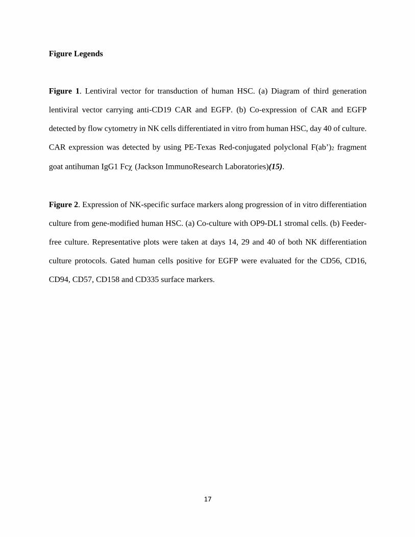
Figure 1. Lentiviral vector for transduction of human HSC. (a) Diagram of third generation
lentiviral vector carrying anti-CD19 CAR and EGFP. (b) Co-expression of CAR and EGFP
detected by flow cytometry in NK cells differentiated in vitro from human HSC, day 40 of culture.
CAR expression was detected by using PE-Texas Red-conjugated polyclonal F(ab’)2 fragment
goat antihuman IgG1 Fcχ (Jackson ImmunoResearch Laboratories)(15).
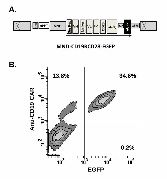
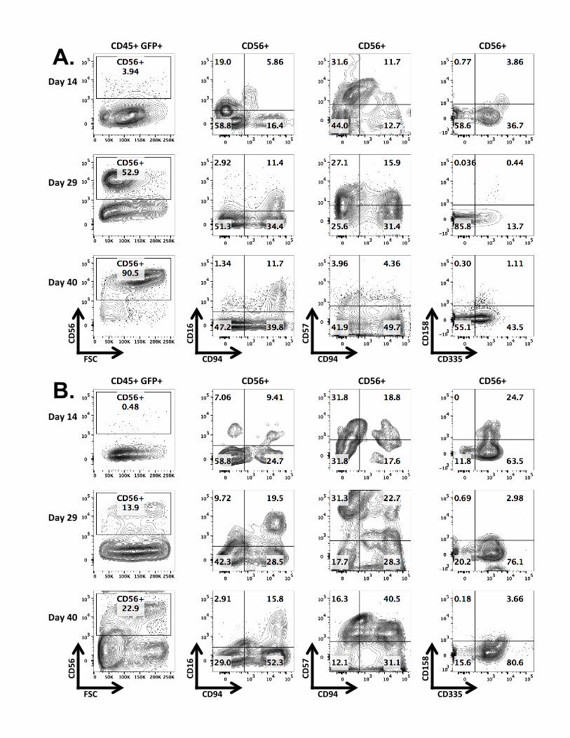
Figure 2. Expression of NK-specific surface markers along progression of in vitro differentiation
culture from gene-modified human HSC. (a) Co-culture with OP9-DL1 stromal cells. (b) Feeder-
free culture. Representative plots were taken at days 14, 29 and 40 of both NK differentiation
culture protocols. Gated human cells positive for EGFP were evaluated for the CD56, CD16,
CD94, CD57, CD158 and CD335 surface markers.
Sig
Pep
MND cPPT VH VL
Link
er
CD3ζ Fc
CD
28
RR
E
eGFP
WPRE T2A
MND-CD19RCD28-EGFP
A.
B.
EGFP
Anti-
CD
19 C
AR
0.2%
34.6%13.8%
CD45+ GFP+ CD56+
Day 14
Day 29
Day 40
CD56+ CD56+
CD335
CD158
CD94
CD57
CD94
CD16
FSC
CD56
CD45+ GFP+ CD56+
Day 14
Day 29
Day 40
CD56+ CD56+
CD335
CD158
CD94
CD57
CD94
CD16
FSC
CD56
A.
B.


