Embed Size (px)
Citation preview
IN VITRO CELLULAR TROPISM OF HUMANB-LYMPHOTROPIC VIRUS (HUMAN HERPESVIRUS-6)
BY PAOLO LUSSO,* PHILLIP D. MARKHAM,I ERWIN TSCHACHLER,*FULVIA DI MARZO VERONESE,I S. ZAKI SALAHUDDIN,*
DHARAM V. ABLASHI,* SAVITA PAHWA,° KAI KROHN,* ANDROBERT C. GALLO
From the *Laboratory of Tumor Cell Biology, National Cancer Institute, National Institutes ofHealth, Bethesda, Maryland 20892; IBionetics Research, Inc., Rockville, Maryland 20850; and
the 4Department ofPediatric Immunology, North Shore University Hospital, Manhasset,New York 11030
The human B-lymphotropic virus (HBLV),' recently isolated from the periph-eral blood of six patients affected by various hematological disorders (1), wasshown by detailed molecular and morphological characterizations to be a uniquemember of the Herpesviridae family (2, 3) . HBLV is an enveloped virus, 200 nmin diameter, with an electron-dense icosahedral core composed of 162 capso-mers (1, 3) containing a large double-stranded DNA genome (> 110.000 bp) . Asa result of these observations, the taxonomic designation human herpesvirus-6(HHV-6) was proposed (4, 5) . Despite extensive seroepidemiological studies andattempts to obtain HBLV isolates from diversified patients, no definite etiolog-ical association with human disease has been established to date .HBLV can infect fresh normal mononuclear cells in vitro, yielding character-
istic morphologically altered lymphocytes, capable of surviving in culture for15-20 d. Based primarily on the study of freshly isolated cells from infectedpatients, a B lymphocyte tropism was initially suggested for HBLV (1). However,subsequent in vitro studies demonstrated that HBLV could also infect cell linesof various origins, including B and T lymphocytes, megakaryocytes, and glialcells (4) . In addition, two recent letters describing the isolation from Africanpatients of human herpesviruses genetically indistinguishable from HBLV alsosuggested the infectability of other cell types (6, 7) . Although in vitro studiesusing established cell lines are not necessarily predictive of the in vivo cellulartropism of a viral agent, the information so far available indicates that HBLVmay have a wide range of infectable cells .
In the present report we investigated further the in vitro cellular tropism ofHBLV using fresh normal mononuclear cells from various tissue sources.Infected cells were subjected to extensive immunological and molecularcharacterizations .
P. Lusso is a Fellow of Fogarty International Foundation, E. Tschachler is a Fellow of Max KadeFoundation, and K. Krohn is the recipient of a visiting scientist award from Fogarty InternationalFoundation .
Abbreviations used in this paper. HBLV, human B-lymphotropic virus ; MN, mononuclear cells ;PE, phycoerythryn ; TCIDS� , 50% tissue culture infectious dose ; TdT, terminal deoxyribo-nucleotydiltransferase .J . Exp. MED. C The Rockefeller University Press - 0022-1007/88/05/1659/12 $2.00
1659Volume 167 May 1988 1659-1670
on Decem
ber 1, 2015jem
.rupress.orgD
ownloaded from
Published May 1, 1988
1660
IN VITRO CELLULAR TROPISM OF HUMAN HERPESVIRUS-6
Materials and MethodsSource of Mononuclear Cells.
Heparinized samples of adult peripheral blood, bonemarrow, and umbilical cord blood from healthy donors were collected under sterile con-ditions . Tonsils and thymuses, obtained from children undergoing cardiac surgery inter-ventions, were extensively minced and the cells in suspension were washed out with ster-ile culture medium. Mononuclear cells (MN) from all sources were isolated bycentrifugation on Ficoll-Hypaque gradients (Pharmacia Fine Chemicals, Piscataway, NJ) .
Source of Virus.
HBLV was propagated in fresh, activated umbilical cord blood leu-kocytes as previously described (1) . For the in vitro infection of mononuclear cells, eitherconditioned medium from infected cord blood cultures or partially purified HBLV,obtained by density banding on sucrose gradients, was used . The titer or 50% tissue cul-ture infectious dose (TCIDbo) was determined by infecting triplicate cultures of activatedcord blood cells with serial 10-fold dilutions of the virus stock .
Infection of Mononuclear Cells In Vitro .
Mononuclear cells, including the selectedCD2- or CD3- subpopulations (see below), were initially activated by cultivation for 24h at 37°C in humidified 5% C02 atmosphere in culture medium (RPMI 1640) supple-mented with 15% FCS and 1 gg/ml PHA-P . Cells were then washed, pelleted, resus-pended in 2 ml stock HBLV (titer ^-109 TCIDao) and incubated for 2 h at 37°C . Thecells were subsequently washed and resuspended in culture medium supplemented with10% FCS . 6-8 d after infection, dead cells were removed by Ficoll-Hypaque gradientcentrifugation and infected cells were cultured for additional 2-4 d, before immunolog-ical or molecular characterizations . At that time, >90% of the cells were HBLV infected,as detected by IFA (see below) or by in situ hybridization with the specific probepZVH14, as described (2) .
Immunofluorescence Analysis. A wide panel of mouse mAbs and polyclonal antiserawere used to characterize HBLV-infected cells by either direct or indirect IFA, as pre-viously described (1, 8) . Purified unlabeled, FITC or phycoerythryn (PE)-conjugatedmAbs Leu-1, Leu-3a, Leu-4, Leu-7, Leu-9, Leu-11, Leu-M1, Leu-M2, Leu-M3, HPCA-1, anti-TCR-a/,Q heterodimer (WT31), anti-IL-2-R, anti-leukocyte antigen (HLel) andanti-CR2 were purchased from Becton Dickinson & Co . (Mountain View, CA) ; OKT3,OKT4, OKT4a, OKT6, OKT8, OKT9, OKT11, OKDR, and OKBcALL from Ortho Phar-maceutical (Raritan, NJ) ; MY7, MY9, B1, and B4 from Coulter Immunology (Hialeah,FL) ; DRC and Kil from Dako Corp. (Santa Barbara, CA) . Polyclonal rabbit anti-terminaldeoxynucleotydiltransferase (TdT) was obtained from Bethesda Research Laboratories(Gaithersburg, MD); polyclonal goat anti-human IgG or IgM (y or J chain specific,respectively) from Kirkegaard & Perry Laboratories, Inc . (Gaithersburg, MD) . Humansera containing specific antibodies towards HBLV were also used at 1 :40 dilution forindirect IFA, after extensive absorption with HBLV- umbilical cord blood cells. Polyclo-nal FITC-conjugated goat anti-mouse IgG or IgM, goat anti-rabbit IgG and goat anti-human IgG antisera (Kirkegaard & Perry Laboratories, Inc .,) were used as second anti-bodies . Cytoplasmic staining was carried out on cytocentrifuge preparations fixed for 10min in cold acetone . Nuclear staining for TdT was performed on cytocentrifuge prepa-rations fixed for 30 min in cold methanol . All samples were scored under a fluorescencemicroscope (Leitz), counting >200 cells per test . Flow cytofluorometric analysis was per-formed using a Becton Dickinson & Co . FACS (FRCS 440 ; Spectra Physics, MountainView, CA) equipped with an Argon laser . Live infected cells were treated for 1 h with0 .5% paraformaldehyde before FACS analysis . Debris and dead cells were excluded fromthe analysis by conventional scatter setting system . Fluorescein (green) and PE (red) emis-sion lights were collected with band pass filters 530/30 and 575/26, respectively, andlogarithmically amplified . Isotype-matched mAbs of irrelevant specificity were used onthe same cells as negative controls .
Radio Immune Precipitation Assay (RIPA). HBLV-infected cord blood cells and thecontrol cell line ET62 were radioactively labeled by incubation for 18 h in medium con-taining 50 gCi/ml ["S]cysteine . Labeled cells were washed in PBS and disrupted with 10nM sodium phosphate (pH 7.2) containing 0.5% NaCI, 1% Triton X-100, 0 .5% sodiumdeoxycholate and 0.1 % SDS (PBS-TDS) . The lysate was adsorbed overnight at room tem-
on Decem
ber 1, 2015jem
.rupress.orgD
ownloaded from
Published May 1, 1988
LUSSO ET AL .
1661
perature with protein A-Sepharose . Immunoprecipitation analysis was performed byaddition to labeled and clarified extract (1 ml) of 5 Ag purified OKT3 mAb and 0.2 mlof a 10% suspension of protein A-Sepharose bound to rabbit anti-mouse K light chain .The samples were incubated overnight at 4°C . Immunoprecipitates were collected by cen-trifugation, washed repeatedly in PBS-TDS, resuspended in Laemmli sample buffer,heated for 2 min at 90°C, and analyzed by SDS-PAGE .
Northern Blot Analysis.
Cellular RNA was extracted from HBLV-infected cord bloodMN cells and control cell lines by the guanidine isothiocyanate procedure, as previouslydescribed (9) . Total cellular RNA (20 Ag/lane) was electrophoresed through 1 .2% for-maldehyde-agarose gels and transferred to nylon membrane filters (Zeta Probe ; Bio-RadLaboratories, Cambridge, MA) . After baking for 2 h at 80°C, filters were hybridized at37'C for 12 h to nick-translated probes in 4X SSC, 4X Denhardt's solution, 0.1% SDS,50% formamide, 10% dextran sulfate, 50 mM sodium phosphate pH 6.5, 250 Ag/mlyeast RNA. The filters were then washed two times in 2 X SSC/0.196 SDS at room tem-perature and two times in 0.2X SSC/0.496 SDS at 65°C . The probes used for hybridiza-tion were labeled by nick translation to a specificity of -lOs cpm/,ag and included TCR8(Bgl II cDNA fragment derived from JUR-,B2 clone, coding for most of the C,#2 constantdomain, a kind gift of Dr. T. Mak, Ontario Cancer Institute, Toronto Canada), TCR-a(cDNA clone pY14, a kind gift of Dr . T. Mak), TCR-y (derived from clone pTy-1, bysubcloning into Eco RI sites of pGEM4, a kind gift of Dr. D . Dialynas, Dept . of Bio-chemistry and Molecular Biology, Harvard University, Cambridge, MA), and IgM heavychain (a genomic subclone of 1 .3 kb from the Eco RI site of CA 1 to the Eco RI site ofCA. 3 into the Eco RI site of pbr322, a kind gift of Dr . P . Leder, Dept . of Genetics,Harvard Medical School, Boston, MA) .
Immunoselection by Magnetic Microspheres. Monosized magnetic microspheres coatedwith polyclonal goat anti-mouse IgG (DYNAL, Fort Lee, NJ) were used to isolate theCD2- or CD3- cell subsets, as described (10) . Briefly, freshly isolated umbilical cordblood mononuclear cells (2 X 10⁷) were incubated for 30 min at 4°C in PBS supple-mented with 1% FCS with 2 Ag/ml of appropriate purified mAb. The cells were thenwashed three times with cold PBS and incubated at 4°C under continuous rotation inthe presence of goat anti-mouse IgG-covered magnetic microspheres (40 :1, beads/puta-tive positive cells ratio) . After 40 min the rosetting and nonrosetting cell populationswere selected by placing the culture tube in contact with a cobalt-samarium magnet for1 min and pipetting off the unbound cells . The proportion of residual CD2 + or CD3+cells after this procedure was <I%, as detected by indirect immunofluorescence .
Complement-mediated Cellular Cytotoxicity . After treatment with magnetic beads, thedepletion of CD2+ or CD3 + mononuclear cells was completed by complement-mediatedcell lysis . CD2- or CD3-depleted subsets were incubated for 30 min at 4°C with theappropriate mAb (OKTI l, OKT3, respectively, IgG2a subclass), washed three times, andresuspended in rabbit complement (PelFreeze Biologicals, Rogers, AR) for 1 h undercontinuous rotation . Cells were then washed three times in cold PBS and their viabilitywas tested by Trypan blue dye exclusion .
ResultsIn Vitro Infection of Fresh Mononuclear Cells with HBLV.
Approximately 72 hafter infection of PHA-treated cells, characteristic HBLV-infected cells began toappear within the cultures from all the tissue sources examined (Fig . 1) . Littleor no infection was observed when MN cells were exposed to the virus withoutprevious in vitro activation by PHA. The morphologically altered infected cellssubsequently became the predominant population, since the majority of thesmall, uninfected cells died within the first week in culture . The cell viability wasrestored to >90% after removal of dead cells at day 6-8 by Ficoll-Hypaquegradient centrifugation . After two more days in culture, >90% of the viablecells were HBL', as evaluated by indirect IFA or by in situ hybridization . All
on Decem
ber 1, 2015jem
.rupress.orgD
ownloaded from
Published May 1, 1988
1662 IN VITRO CELLULAR TROPISM OF HUMAN HERPESVIRUS-6
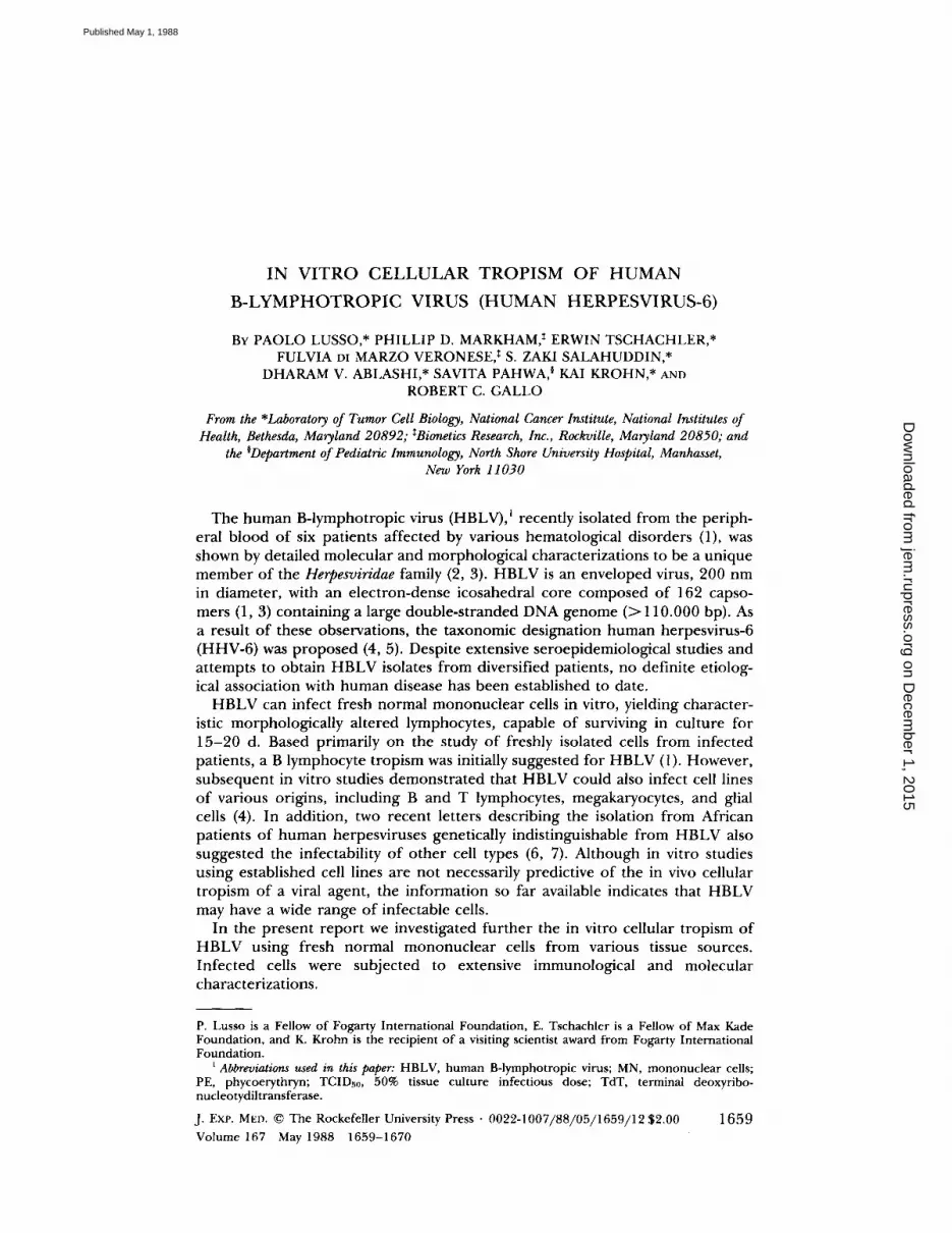
FIGURE: 1 .
Characteristic electron microscopy appearance of an HBLV-infected T lympho-cyte 8 d after infection . Mononuclear cells from normal human umbilical cord blood wereinfected with HBLV 24 h after activation with PHA. Intranuclear and intracytoplasmic imma-ture particles are recognizable, as well as mature virions outside the cell,
the immunological and molecular tests were carried out on these homogeneousHBLV+ cell populations . In the early phases of infection (day 5-8), the viableHBLV-infected cells demonstrated a consistent, albeit limited, spontaneous pro-liferative activity (compared with unstimulated MN cells from the same donors,as detected by [3H]thymidine incorporation, data not shown), but were unreac-tive to PHA and/or IL-2 (either crude or recombinant preparations) added tothe cultures (data not shown) .
Immunological Analysis of HBLV-infected Cells. As illustrated in Table I,HBLV-infected cells from normal adult peripheral blood, umbilical cord blood,or thymic tissues displayed phenotypic characteristics of immature T lympho-cytes, by either direct or indirect IFA. Analogous results were obtained afterinfection of mononuclear cells from tonsil and bone marrow samples . Theresults depicted in the table refer to day 8-10 homogeneous HBLV-infected cellpopulations, but the same phenotypic pattern was also observed on single mor-phologically altered lymphocytes during the early phases of culture infection.Virtually all HBLV+ cells expressed the CD7 antigen, the earliest known T-lym-phocytic marker along the intrathymic T cell differentiation pathway, the CD5antigen, a pan-T marker, and the CD2 antigen, the receptor for sheep erythro-
on Decem
ber 1, 2015jem
.rupress.orgD
ownloaded from
Published May 1, 1988
LUSSO ET AL .
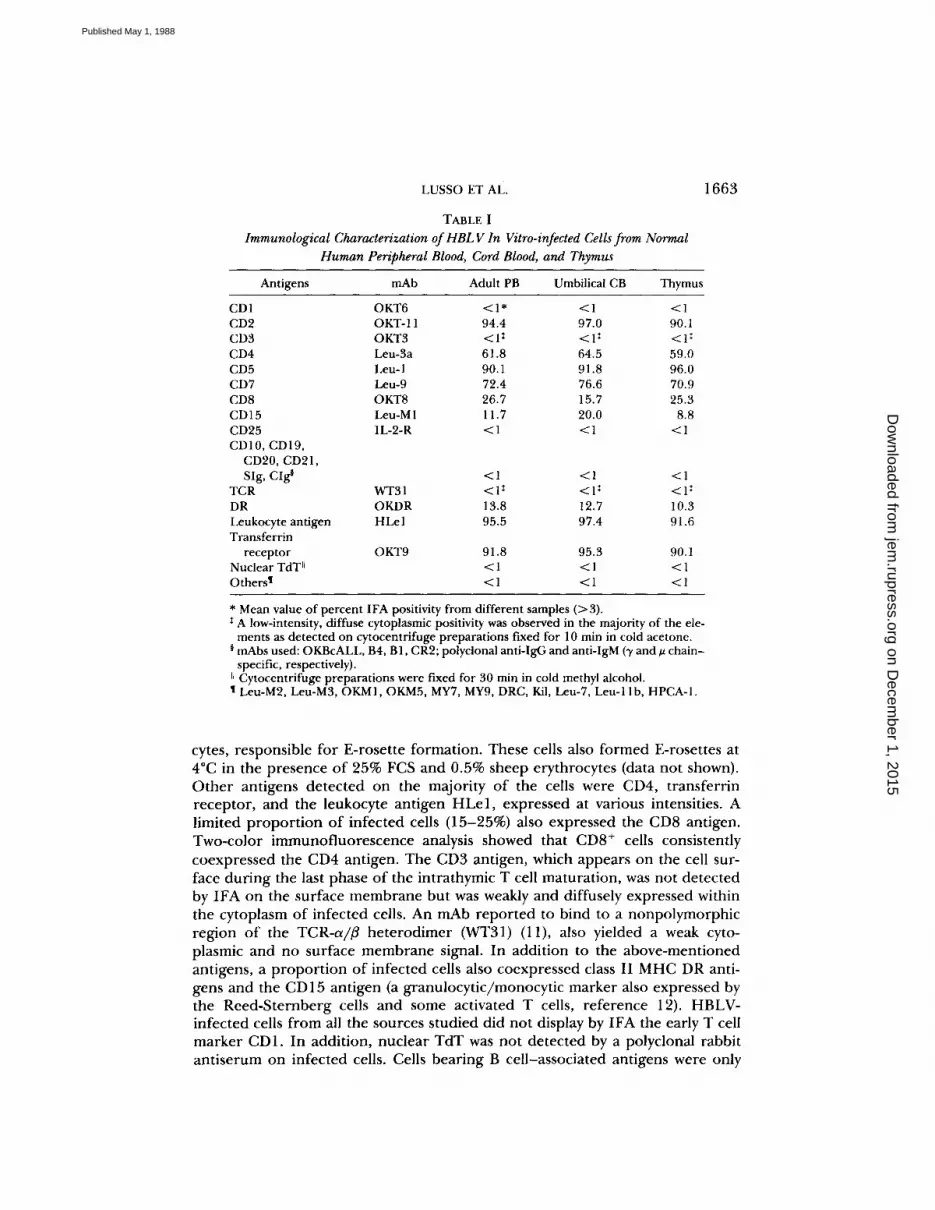
TABLE IImmunological Characterization of HBLV In Vitro-infected Cellsfrom Normal
Human Peripheral Blood, Cord Blood, and Thymus
* Mean value of percent IFA positivity from different samples (>3) .I A low-intensity, diffuse cytoplasmic positivity was observed in the majority of the ele-ments as detected on cytocentrifuge preparations fixed for 10 min in cold acetone.
¢ mAbs used : OKBcALL, B4, 131, CR2; polyclonal anti-IgG and anti-IgM (y and A chain-specific, respectively) .
~~ Cytocentrifuge preparations were fixed for 30 min in cold methyl alcohol.t Leu-M2, Leu-M3, OKM1, OKM5, MY7, MY9, DRC, Kil, Leu-7, Leu-11b, HPCA-1 .
1663
cytes, responsible for E-rosette formation. These cells also formed E-rosettes at4°C in the presence of 25% FCS and 0.5% sheep erythrocytes (data not shown).Other antigens detected on the majority of the cells were CD4, transferrinreceptor, and the leukocyte antigen HLe1, expressed at various intensities . Alimited proportion of infected cells (15-25%) also expressed the CD8 antigen.Two-color immunofluorescence analysis showed that CD8+ cells consistentlycoexpressed the CD4 antigen. The CD3 antigen, which appears on the cell sur-face during the last phase of the intrathymic T cell maturation, was not detectedby IFA on the surface membrane but was weakly and diffusely expressed withinthe cytoplasm of infected cells. An mAb reported to bind to a nonpolymorphicregion of the TCR-a/# heterodimer (WT31) (11), also yielded a weak cyto-plasmic and no surface membrane signal . In addition to the above-mentionedantigens, a proportion of infected cells also coexpressed class II MHC DR anti-gens and the CD15 antigen (a granulocytic/monocytic marker also expressed bythe Reed-Sternberg cells and some activated T cells, reference 12). HBLV-infected cells from all the sources studied did not display by IFA the early T cellmarker CD1. In addition, nuclear TdT was not detected by a polyclonal rabbitantiserum on infected cells. Cells bearing B cell-associated antigens were only
Antigens mAb Adult PB Umbilical CB Thymus
CD1 OKT6 <1* <1 <1CD2 OKT-11 94.4 97.0 90 .1CD3 OKT3 <lt <1 : <1 "CD4 Leu-3a 61 .8 64 .5 59 .0CD5 Leu-1 90.1 91 .8 96 .0CD7 Leu-9 72 .4 76 .6 70 .9CD8 OKT8 26.7 15 .7 25 .3CD15 Leu-M1 11 .7 20 .0 8.8CD25 IL-2-R <1 <1 <1CD10, CD19,CD20, CD21,SIg, CIg§ <1 <1 <1
TCR WT31 <lI <lt <I'DR OKDR 13.8 12 .7 10 .3Leukocyte antigen HLel 95 .5 97.4 91 .6Transferrin
receptor OKT9 91 .8 95.3 90.1Nuclear TdT11 <1 <1 <1Othersl <1 <1 <1
on Decem
ber 1, 2015jem
.rupress.orgD
ownloaded from
Published May 1, 1988
1664
IN VITRO CELLULAR TROPISM OF HUMAN HERPESVIRUS-6
sm
100
100
Leu 3a
OK T3
100
100
100
FLUORESCENCE INTENSITYILOGio UNITS)
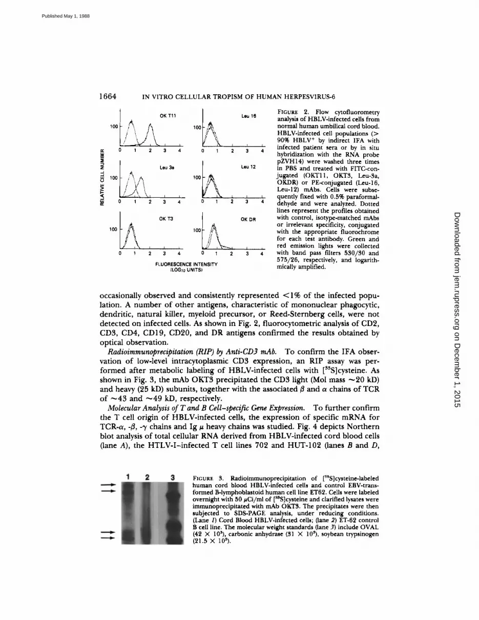
FIGURE 2. Flow cytofluorometryanalysis of HBLV-infected cells fromnormal human umbilical cord blood.HBLV-infected cell populations (>9096 HBLV' by indirect IFA withinfected patient sera or by in situhybridization with the RNA probepZVH14) were washed three timesin PBS and treated with FITC-con-jugated (OKT11, OKT3, Leu-3a,OKDR) or PE-conjugated (Leu-16,Leu-12) mAbs. Cells were subse-quently fixed with 0.596 paraformal-dehyde and were analyzed . Dottedlines represent the profiles obtainedwith control, isotype-matched mAbsor irrelevant specificity, conjugatedwith the appropriate fluorochromefor each test antibody . Green andred emission lights were collectedwith band pass filters 530/30 and575/26, respectively, and logarith-mically amplified .
occasionally observed and consistently represented <1% of the infected popu-lation . A number of other antigens, characteristic of mononuclear phagocytic,dendritic, natural killer, myeloid precursor, or Reed-Sternberg cells, were notdetected on infected cells . As shown in Fig. 2, fluorocytometric analysis of CD2,CD3, CD4, CD19, CD20, and DR antigens confirmed the results obtained byoptical observation.
Radioimmunoprecipitation (RIP) by Anti-CD3 mAb.
To confirm the IFA obser-vation of low-level intracytoplasmic CD3 expression, an RIP assay was per-formed after metabolic labeling of HBLV-infected cells with [s5S]cysteine . Asshown in Fig. 3, the mAb OKT3 precipitated the CD3 light (Mol mass ^-20 kD)and heavy (25 kD) subunits, together with the associated ,B and a chains of TCRof "̂43 and ,̂ 49 kD, respectively .
Molecular Analysis of T and B Cell-specific Gene Expression.
To further confirmthe T cell origin of HBLV-infected cells, the expression of specific mRNA forTCR-a, -ß, -y chains and Ig 1 heavy chains was studied. Fig. 4 depicts Northernblot analysis of total cellular RNA derived from HBLV-infected cord blood cells(lane A), the HTLV-I-infected T cell lines 702 and HUT-102 (lanes B and D,
FIGURE 3. Radioimmunoprecipitation of ["°S]cysteine-labeledhuman cord blood HBLV-infected cells and control EBV-trans-formed B-lymphoblastoid human cell line ET62 . Cells were labeledovernight with 50 ,uCi/ml of ["sS]cysteine and clarified lysates wereimmunoprecipitated with mAb OKT3 . The precipitates were thensubjected to SDS-PAGE analysis, under reducing conditions .(Lane 1) Cord Blood HBLV-infected cells ; (lane 2) ET-62 controlB cell line . The molecular weight standards (lane 3) include OVAL(42 X 10"), carbonic anhydrase (31 X 10"), soybean trypsinogen(21 .5 X 10") .
on Decem
ber 1, 2015jem
.rupress.orgD
ownloaded from
Published May 1, 1988

LUSSO ET AL .
1665
FIGURE 4. Northern blotanalysis of TCR and IgMRNA expression in HBLV-infected cord blood cells. 20kg of total cellular RNAderived from HBLV-infectedcord blood cells (a and b, laneA), HTLV-I-infected T celllines 702 (lane B) and HUT-102 (lane D), and EBV-infected B-lymphoblastoidline RE (lane G) were electro-phoresed under denaturingconditions and transferredonto nylon membrane filters .The filters were probed withcDNA fragment specific forTCR-ß (a, t% panel), TCR-a(a, bottom panel), TCR-y (b, bot-tom panel), or a CIL-specificgenomic clone (b, top panel) .18S and 28S ribosomal RNAsare indicated as size markers.
respectively), and the EBV-infected B cell line RE (lane C) . Full-size TCR-a and-0 messages were identified in HBLV-infected cord blood cells (Fig . 4 a, top andbottom). TCR-^y transcripts were not detectable in these cells (Fig 4 b, bottom), butwere present in the control HTLV-I-infected T cell line 702 . Similarly, mRNAfor CA, was not identified in HBLV-infected cord blood cells, but was present inthe control B cell line RE (Fig . 4 b, top) . All lanes contained identical amountsof total RNA, as verified by ethidium bromide staining of the RNA gel .
Infection of IL-2-dependent Cultured TLymphocytes.
In addition to fresh normalmononuclear cells, normal T lymphocytes expanded in vitro in the presence ofIL-2 for > 1 mo were successfully infected with HBLV (Fig . 5) . The yield ofinfected cells from IL-2-dependent cultures was definitely lower than usingfresh mononuclear cells and progressively declined in long-term IL-2-stimulatedcultures . Very limited infection was indeed observed exposing the HBLV IL-2-dependent T lymphocytes cultured in vitro for >60 d .
Infection of Selected Mononuclear Cell Subsets.
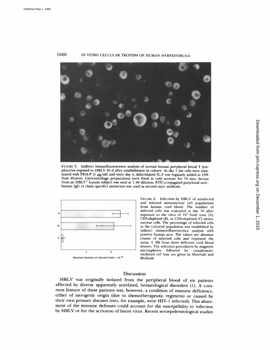
To further address the questionof the origin and maturation level of HBLV target cells, CD2 - or CD3- MNcell populations were obtained from human cord blood by means of immuno-selection with mAbs and goat anti-mouse IgG-coated magentic microspheres,followed by complement-mediated cell lysis . Control unfractionated MN cellswere exposed to rabbit complement alone, without previous mAb treatment .Equal numbers of selected and unselected cells were subsequently activated withPHA, infected with HBLV, and placed into culture, as described above . As illus-trated in Fig. 6, the absolute number of HBLV-infected cells recovered fromthe CD3- MN cell populations of three different donors 10 d after infectionwas comparable to that from total MN cells (mean ± SD from 107 initial cells :2.3 ± 0.3 X 10⁶ from total MN cells vs . 2 .1 ± 0.7 X 10⁶ from CD3-depletedfractions) . By contrast, virtually no infected cells were detectable after 10 d inCD2-depleted cell cultures (mean 0.1 ± 0.1 X 10⁶ from 107 initial cells) .
on Decem
ber 1, 2015jem
.rupress.orgD
ownloaded from
Published May 1, 1988
1666
IN VITRO CELLULAR TROPISM OF HUMAN HERPESVIRUS-6
FIGURE 5.
Indirect immunfluorescence analysis of normal human peripheral blood T lym-phocytes exposed to HBLV 40 d after establishment in culture. At day l the cells were stim-ulated with PHA-P (1 jug/ml) and since day 4, delectinated IL-2 was regularly added at 10%final dilution . Cytocentrifuge preparations were fixed in cold acetone for 10 min. Serumfrom an HBLV+ human subject was used at 1 :40 dilution. FITC-conjugated polyclonal anti-human IgG (f chain specific) antiserum was used as second layer antibody.
FIGURE 6.
Infection by HBLV of unselectedand selected mononuclear cell populationsfrom human cord blood. The number ofinfected cells was evaluated at day 10 afterexposure to the virus of 10 7 fresh total (A),CD3-depleted (B), or CD2-depleted ((,') mono-nuclear cells . The percentage of infected cellsin the cultured population was established byindirect immunofluorescence analysis withpositive human sera. The values are absolutecounts of infected cells and represent themean ± SD from three different cord blooddonors . The selection procedures by magneticmicrospheres followed by complement-mediated cell lysis are given in Materials andMethods.
DiscussionHBLV was originally isolated from the peripheral blood of six patients
affected by diverse apparently unrelated, hematological disorders (1) . A com-mon feature of these patients was, however, a condition of immune deficiency,either of iatrogenic origin (due to chemotherapeutic regimens) or caused bytheir own primary diseases (two, for example, were HIV-1 infected). This abate-ment of the immune defenses could account for the susceptibility to infectionby HBLV or for the activaton of latent virus. Recent seroepidemiological studies
on Decem
ber 1, 2015jem
.rupress.orgD
ownloaded from
Published May 1, 1988
LUSSO ET AL .
1667
by means of immunofluorescence techniques on fixed HBLV-infected cellsshowed a widespread high prevalence of HBLV-specific antibodies in severalhematological disorders, of both neoplastic and non-neoplastic origin (Ablashi,D. V., et al ., manuscript in preparation; Streicher, H. Z ., et al ., manuscript inpreparation) . In particular, significant seropositivity was observed in variouslymphoproliferative disorders, advanced acquired immune deficiency syndrome(AIDS), and the so-called chronic mononucleosis syndrome or chronic fatiguesyndrome (13) . Inaddition, HBLV-specificDNAwas recently identifiedbySouthernblot analysis within the neoplastic tissues from three patients affected by non-Hodgkin's lymphoma of B cell origin (14) . Despite these observations, nodefinitive evidence to date has linked HBLV to any specific clinical entity inhumans .The present report demonstrates that HBLV is electively infectious for fresh
T lymphocytes in vitro and exerts on them a strong cytopathic effect . Despiteconsiderable efforts to elucidate the maturation stage of the T cells susceptibleto HBLV infection, it remains questionable whether they belong to a discrete,quasimature differentiation phase or, conversely, are fully mature lymphocytesundergoing important phenotypic and functional alterations after the interac-tion with the virus. In this study, HBLV-infected T cells displayed some imma-ture phenotypic characteristics (15) . For example, they lack the surface mem-brane TCR/CD3 complex and coexpress, at least partially, the CD4 and CD8antigens. The simultaneous expression of these latter markers is characteristicof the cortical thymic population, which is largely CD 1 + (16) . The lack of CD Ideterminants on HBLV-infected cells (included those derived from thymic tis-sues) is not indicative per se, since it has recently been reported (17) that CD1expression is not an indispensable intermediate step along the T cell differentia-tion pathway outside the thymic environment.
In contrast with the immature phenotypic features, however, the analysis atthe RNA level demonstrated a mature pattern of TCR gene expression byHBLV-infected cells . Indeed, HBLV-infected cells displayed full-size TCR-a and-ß chain, but not -f chain mRNA, which is expressed at early stages of T cellmaturation (18) . In addition, the nuclear TdT, another early T cell marker, wasnegative on HBLV-infected cells. We attempted to address the question of thematuration level of the HBLV target cells by infecting immunoselected mono-nuclear cell populations. The results of these experiments indicate that at leasta consistent proportion of the susceptible cells resides within the CD3-, CD2+immature T cell compartment. Another indication of immaturity is provided bypreliminary data from our laboratory, suggesting that HBLV-infected T cells areunable to secrete the lymphokine IL-2 after stimulation in vitro with PHA andphorbol esters .
As further evidence of the T cell tropism of HBLV, we also showed that nor-mal T lymphocytes maintained in culture in the presence of IL-2 for > 1 mowere easily infected . Furthermore, it was previously reported that several estab-lished cell lines of various lineage origin, including T cells at early stages of dif-ferentiation, could be infected by HBLV (19) . In contrast, it is possibly relevantthat the T cell lines H9 and PEER, which are apparently refractory to HBLVinfection (Lusso, P., preliminary data), display a phenotype corresponding to anadvanced T cell maturation stage (surface CD3+).
on Decem
ber 1, 2015jem
.rupress.orgD
ownloaded from
Published May 1, 1988
1668
IN VITRO CELLULAR TROPISM OF HUMAN HERPESVIRUS-6
While it is clear from this study that T cells represent a major target forHBLV infection, a linkage with B cell-related lymphoproliferative disorders issuggested by the direct detection of HBLV in tumor tissues from non-Hodgkinlymphomas of B cell type (14) . This may suggest an indirect role for B cell tumo-rigenesis as was hypothesized for HIV-1 (20, 21) or HTLV-I (22), presumablyacting through chronic antigenic stimulation and suppression of the T cell sur-veillance on immune-activated or EBV-transformed B lymphocytic clones . Inlight of the cellular tropism manifested by HBLV in vitro, we are currentlyinvestigating the possible involvement of this virus in the pathogenesis of variousT cell-related hematological disorders, as well as of neoplasms of B lymphocyticorigin . In addition, due to the dramatic cytopathic effect exerted in vitro on Tlymphocytes, we are considering a possible role of HBLV in immunodeficiencyconditions .
SummaryWe investigated the cellular tropism of human B-lymphotropic virus (HBLV)
(also designated Human Herpesvirus-6) in vitro by infecting fresh MN cells fromnormal human adult peripheral blood, umbilical cord blood, bone marrow, ton-sil, and thymus . Cultures from all the sources examined contained infectablecells, as shown by the appearance of characteristic enlarged, round-shaped,short-lived cells expressing HBLV-specific markers. Detailed immunologicalanalysis demonstrated that the vast majority of these cells expressed T cell-asso-ciated antigens (i .e ., CD7, CD5, CD2, CD4, and to a lesser extent, CD8). TheCD3 antigen and the TCR-a/0 heterodimer were not detectable on the surfacemembrane, but were identified within the cytoplasm of HBLV-infected cells, byboth immunofluorescence and radioimmunoprecipitation assay. A proportion ofthe HBLV-infected cell population also expressed the CD15 and class II MHCDR antigens . By means of immunoselection procedures it was possible to showthat a consistent proportion of HBLV-infectable cells were contained within theCD3-depleted immature T cell population, while the depletion of CD2 + cellscompletely abrogated the infectability of the cultures . Northern blot analysisconfirmed the T cell origin of HBLV-infected cells, demonstrating the ex-pression of full size TCR-a and -,B chain mRNA. In addition to fresh T cells,HBLV was able to infect normal T lymphocytes expanded in vitro with IL-2 for>30 d.These results indicate that HBLV is selectively T cell tropic in the course of
the in vitro infection of normal mononuclear cells and may therefore be directlyinvolved in the pathogenesis of T cell related hematological disorders . In par-ticular, in light of the cytopathic effect exerted in vitro on CD4+ T lympho-cytes, a possible role of HBLV in immune deficiency conditions should be con-sidered.
The authors are grateful to Susan A. Barbieri and Jake Fullen for excellent technicalassistance .
Receivedfor publication 31 December 1987 .
on Decem
ber 1, 2015jem
.rupress.orgD
ownloaded from
Published May 1, 1988
LUSSO ET AL .
1669
References
1 . Salahuddin, S . Z ., D . V . Ablashi, P. D . Markham, S . F . Josephs, S . Sturzenegger, M .Kaplan, G . Halligan, P . Biberfeld, F . Wong-Staal, B . Kramarsky, and R . C. Gallo .1986 . Isolation of a new virus, HBLV, in patients with lymphoproliferative disor-ders . Science (Wash. DC). 234:596 .
2 . Josephs, S . F ., S . Z . Salahuddin, D . V. Ablashi, F . Schachter, F . Wong-Staal, and R.C . Gallo . 1986 . Genomic analysis of the human B-lymphotropic virus (HBLV) . Sci-ence (Wash. DC). 234:601 .
3 . Biberfeld, P ., B . Kramarsky, S . Z . Salahuddin, R . C . Gallo . 1987 . Ultrastructuralcharacterization of a new human B lymphotropic DNA virus (HBLV) isolated frompatients with lymphoproliferative disease . J.. Natl. Cancer Inst . 79:933 .
4 . Ablashi, D . V ., S . Z . Salahuddin, S . F . Josephs, F . Imam, P . Lusso, and R . C . Gallo .1987 . HBLV (or HHV-6) in human cell lines . Nature (Lord.) . 329:207 .
5 . Lusso P., S . Z . Salahuddin, D. V . Ablashi, R . C . Gallo, F . di Marzo Veronese, andP. D . Markham. 1987 . Diverse tropism for HBLV (human herpes virus 6) . Lancet .ii :743 .
6 . Downing, R . G ., N . Sewankambo, D . Serwadda, D . Serwadda, R . Honess, D . Craw-ford, R. Jarrett, and B. E. Griffin. 1987 . Isolation of human lymphotropic herpesviruses from Uganda . Lancet . ii :390 .
7 . Tedder, R. S ., M . Briggs, C . H . Cameron, R . Honess, D . Robertson, and H. Whittle .1987 . A novel lymphotropic herpesvirus . Lancet. ii :390 .
8 . Gathings, W., A . Lawton, and M. D . Cooper . 1977 . Immunofluorescen t studies ofthe development and pre-B cells, B lymphocytes and immunoglobulin isotype diver-sity in humans . Eur. J. Immunol . 7:804 .
9 . Chirgwin, J . M., A . E . Przybyla, R. J . McDonald, and W. J . Rutter . 1979 . Isolatio nof biologically active ribonucleic acid from sources enriched in ribonuclease . Bio-chemistry . 18:5294 .
10 . Lea, T., F . Vartdal, C . Davies, and J . Ugeistac . 1985 . Magnetic monosized polymerparticle for fast and specific fractionation of human mononuclear cells . Scand . J.Immunol . 22:207 .
11 . Spits, H ., J . Borst, W. Tax, P . Capel, C . Terhorst, and J . de Vries. 1985 . Character-istics of a monoclonal antibody (WT31) that recognizes a common epitope on theHuman T cell receptor for antigen . J. Immunol . 135:1922 .
12 . Wieczorek, R., J . S . Burke, and D . M. Knowles . 1985 . Leu M1 antigen expressionin T-cell neoplasia . Am. J. Pathol . 121 :374 .
13 . Komaroff, A . 1987 . The chronic mononucleosis syndromes . Hosp. Pract . 22:71 .14 . Josephs, S . F ., A . Buchbinder, H . Z . Streicher, D . V . Ablashi, S . Z . Salahuddin, H.-
G . Guo, F . Wong-Staal, J . Cossman, M. Raffeld, J . Sundeen, P. Levine, R . Biggar,G . R . F . Krueger, R. I . Fox, and R. C . Gallo . 1988 . Detection of human B-lymphotropic virus (human herpesvirus 6) sequences in B-cell lymphoma tissues of threepatients . Leukemia . In press .
15 . Acuto, O., R . E . Hussey, K . A. Fitzgerald, J . P . Protentis, S . C . Meuer, S . F . Schloss-man, and E . L . Reinherz . 1983 . The human T cell receptor : appearance in ontogenyand biochemical relationship of and subunits on IL-2 dependent clones and T celltumors . Cell . 34:717 .
16 . Reinherz, E. L., P . C . Kung, G. Goldstein, R . Levey, and S . F . Schlossman . 1980 .Discrete stages of human intrathymic differentiation : analysis of normal thymocytesand leukemic lymphoblasts of T-cell lineage . Proc. Natl. Acad . Sci . USA . 77:1588 .
17 . Larocca, L. M., M . Piantelli, N . Maggiano, and P . Musiani . 1987 . T cell surfacemarkers expression by immature human thymocytes in in vitro culture : role of la+accessory cells . J. Immunol. 138:2410 .
on Decem
ber 1, 2015jem
.rupress.orgD
ownloaded from
Published May 1, 1988

1670
IN VITRO CELLULAR TROPISM OF HUMAN HERPESVIRUS-6
18 . Raulet, D . H., R . D . Garmon, H. Saito, and S . Tonegawa . 1985 . Developmental reg-ulation of T-cell receptor gene expression . Nature (Lond.). 314:103 .
19 . Ablashi, D . V ., P . Lusso, C. Hung, S . Z . Salahuddin, S . F . Josephs, T . Llana, B .Kramarsky, P . Biberfeld, P . D . Markham, and R . C . Gallo . 1988 . Utilizatio n ofhuman hematopoietic cell lines for the propagation, detection, and characterizationof HBLV (human herpesvirus 6) . Int. J. Cancer. In press .
20 . Groopman, J . E ., J . L. Sullivan, C . Mulder, D . Ginsburg, S . H . Orkin, C . J . O'Hara,K. Falchuk, F . Wong-Staal, and R . C . Gallo . 1986 . Pathogenesi s of B cell lymphomain a patient with AIDS . Blood. 67:612 .
21 . Pellicci, P . G ., D . M. Knowles, Z . A. Arlin, R . Wieczorek, P . Luciw, D . Dina, C .Basilico, and R. Dalla-Favera . 1986 . Multiple monoclonal B cell expansions and c-myc oncogene rearrangements in acquired immune deficiency syndrome-related lym-phoproliferative disorders . J. Exp. Med. 164:2049 .
22 . Mann, D ., P . DeSantis, G . Mark, A . Pfeifer, M . Newman, N . Gibbs, M . Popovic, M .G . Sarngadharan, R . C . Gallo, J . Clark, and W. Blattner . 1987 . HTLV-I-associatedB-cell CLL: indirect role for retrovirus in leukemogenesis . Science (Wash . DC) .236:1103 .
on Decem
ber 1, 2015jem
.rupress.orgD
ownloaded from
Published May 1, 1988


