Embed Size (px)
Citation preview
HAL Id: hal-02507521https://hal.archives-ouvertes.fr/hal-02507521
Submitted on 6 Apr 2020
HAL is a multi-disciplinary open accessarchive for the deposit and dissemination of sci-entific research documents, whether they are pub-lished or not. The documents may come fromteaching and research institutions in France orabroad, or from public or private research centers.
L’archive ouverte pluridisciplinaire HAL, estdestinée au dépôt et à la diffusion de documentsscientifiques de niveau recherche, publiés ou non,émanant des établissements d’enseignement et derecherche français ou étrangers, des laboratoirespublics ou privés.
In Search of the Ancestral Organization and PhylotypicStage of PoriferaAlexander Ereskovsky
To cite this version:Alexander Ereskovsky. In Search of the Ancestral Organization and Phylotypic Stage of Porifera.Ontogenez / Russian Journal of Developmental Biology, MAIK Nauka/Interperiodica, 2019, 50 (6),pp.317-324. �10.1134/S1062360419060031�. �hal-02507521�
ISSN 1062-3604, Russian Journal of Developmental Biology, 2019, Vol. 50, No. 6, pp. 317–324. © Pleiades Publishing, Inc., 2019.Published in Russian in Ontogenez, 2019, Vol. 50, No. 6, pp. 398–406.
REVIEWS
In Search of the Ancestral Organization and Phylotypic Stage of Porifera
A. V. Ereskovskya, b, c, *aSt. Petersburg State University, St. Petersburg, 199034 Russia
bInstitut Méditerranéen de Biodiversité et d’Ecologie marine et continentale (IMBE),Aix-Marseille Université, CNRS, IRD, Marseille, France
cKoltzov Institute of Developmental Biology of Russian Academy of Sciences, Moscow, 119334 Russia*e-mail: [email protected]
Received May 6, 2019; revised June 28, 2019; accepted July 6, 2019
Abstract—Each animal phylum has its own bauplan. The phylotypic stage is the ontogenetic stage duringwhich the phylum level characteristics appear. This stage refers to different stages of development in differentanimals. Sponges are one of the simplest, and probably the oldest multicellular lineage of extant animals. Onthe basis of the analysis of sponge development during (i) sexual and asexual reproduction, (ii) regenerationfrom small body fragments, and (iii) cell reaggregation, we suggest a hypothetical variant of their phylotypicstage (spongotype): the mono-oscular juvenile—the rhagon. The major feature, which permits to consider therhagon as the phylotypic stage of the Porifera is the final, definitive position of all the cellular and anatomicalelements of the future adult sponge. It seems that at the rhagon stage the pattern of the axial complex of anla-gen is already formed, and only growth processes occur at the later stages.
Keywords: Porifera, phylotypic stage, Bauplan, rhagon, metamorphosis, regeneration, budding, gemmulehatching, evolutionDOI: 10.1134/S1062360419060031
INTRODUCTIONBauplan (construction plan) is a key notion of
developmental biology and evolutionary morphology,applied during the establishment of new taxonomicphyla and the construction of high-level classification.It is the bauplan, or morphological type, that wasassumed by Cuvier (1817) to be the basis for the divi-sion of animals into four large groups (vertebrates,mollusks, articulates and radiates). Bauplan can beunderstood as the type of construction of a givenorganism, as formed within a certain group and char-acterized by original architectonics.
There are two main concepts involved in bauplan.The first stems from Owen’s ideas concerning thearchetype (Owen, 1848), and is based on the compar-ison of adult animal structures, with no considerationof the preceding stages of development. It is well-known, however, that similar developmental typesmay result in very different adult animals, while differ-ent developmental types may produce similar organ-isms (see for references: Ivanova-Kazas, 1995; Gilbertand Raunio, 1997). The second concept is based onthe comparison of the structure of the larvae, whichare rather conservative in their evolution (see: Raff,1996), and also fails to take into account embryonicdevelopment.
Contemporary to Cuvier, the notion of the “devel-opmental plan” was introduced by von Baer (1828),for whom each body plan was seen as being created bya particular kind of developmental organization—theType. Therefore, the developmental plan sensu vonBaer is the bauplan in the period of its ontogenetic for-mation, and von Baer’s observations were to providefoundational evidence supporting Darwin’s theory ofcommon descent (Darwin, 1859).
Each animal phylum thus has its own bauplan andconsequently, must have its own developmental plan.There are, however, groups of phyla or classes, which,while differing in development, do possess a commonstage. This is the case of the coelomic Spiralia, com-prising the phyla Annelida, Mollusca, and Sipuncul-ida. The adult representatives of these phyla are essen-tially different, as are their morphogeneses, but mostof them have a common stage—the trochophore. Atthis stage, the bauplan of this animals group as a wholereveals itself. Seidel (1960) was the first to focus on thisstage, naming it the Korpergrundgestalt, while Sander(1983) later termed it the phylotypic stage. Both theseauthors considered such a stage to be decisive in thedevelopment of an animal group. They attached nophylogenetic significance to the intermediate pro-
317
318 ERESKOVSKY
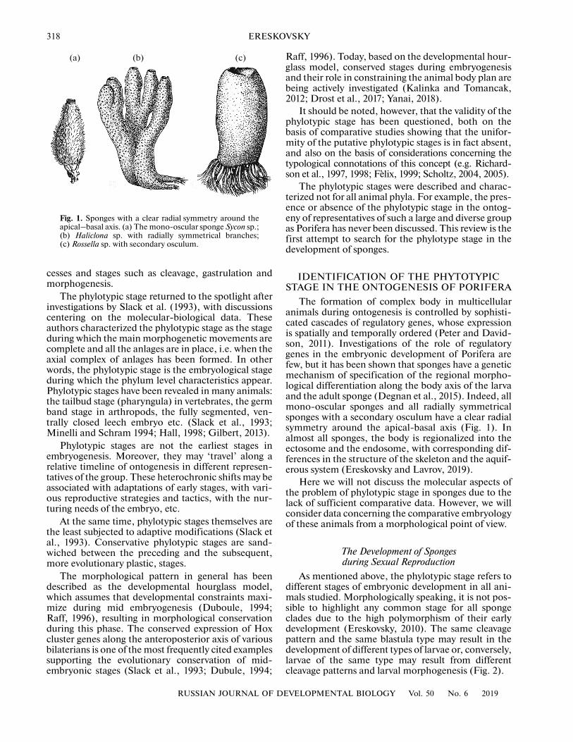
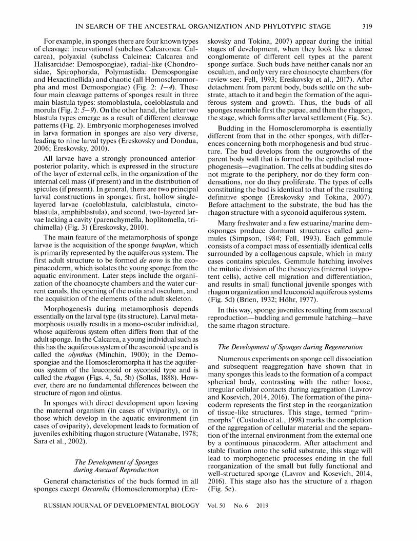
Fig. 1. Sponges with a clear radial symmetry around theapical–basal axis. (a) The mono-oscular sponge Sycon sp.;(b) Haliclona sp. with radially symmetrical branches;(c) Rossella sp. with secondary osculum.
(a) (b) (c)
cesses and stages such as cleavage, gastrulation andmorphogenesis.
The phylotypic stage returned to the spotlight afterinvestigations by Slack et al. (1993), with discussionscentering on the molecular-biological data. Theseauthors characterized the phylotypic stage as the stageduring which the main morphogenetic movements arecomplete and all the anlages are in place, i.e. when theaxial complex of anlages has been formed. In otherwords, the phylotypic stage is the embryological stageduring which the phylum level characteristics appear.Phylotypic stages have been revealed in many animals:the tailbud stage (pharyngula) in vertebrates, the germband stage in arthropods, the fully segmented, ven-trally closed leech embryo etc. (Slack et al., 1993;Minelli and Schram 1994; Hall, 1998; Gilbert, 2013).
Phylotypic stages are not the earliest stages inembryogenesis. Moreover, they may ‘travel’ along arelative timeline of ontogenesis in different represen-tatives of the group. These heterochronic shifts may beassociated with adaptations of early stages, with vari-ous reproductive strategies and tactics, with the nur-turing needs of the embryo, etc.
At the same time, phylotypic stages themselves arethe least subjected to adaptive modifications (Slack etal., 1993). Conservative phylotypic stages are sand-wiched between the preceding and the subsequent,more evolutionary plastic, stages.
The morphological pattern in general has beendescribed as the developmental hourglass model,which assumes that developmental constraints maxi-mize during mid embryogenesis (Duboule, 1994;Raff, 1996), resulting in morphological conservationduring this phase. The conserved expression of Hoxcluster genes along the anteroposterior axis of variousbilaterians is one of the most frequently cited examplessupporting the evolutionary conservation of mid-embryonic stages (Slack et al., 1993; Dubule, 1994;
RUSSIAN JOURNAL OF D
Raff, 1996). Today, based on the developmental hour-glass model, conserved stages during embryogenesisand their role in constraining the animal body plan arebeing actively investigated (Kalinka and Tomancak,2012; Drost et al., 2017; Yanai, 2018).
It should be noted, however, that the validity of thephylotypic stage has been questioned, both on thebasis of comparative studies showing that the unifor-mity of the putative phylotypic stages is in fact absent,and also on the basis of considerations concerning thetypological connotations of this concept (e.g. Richard-son et al., 1997, 1998; Fèlix, 1999; Scholtz, 2004, 2005).
The phylotypic stages were described and charac-terized not for all animal phyla. For example, the pres-ence or absence of the phylotypic stage in the ontog-eny of representatives of such a large and diverse groupas Porifera has never been discussed. This review is thefirst attempt to search for the phylotype stage in thedevelopment of sponges.
IDENTIFICATION OF THE PHYTOTYPIC STAGE IN THE ONTOGENESIS OF PORIFERA
The formation of complex body in multicellularanimals during ontogenesis is controlled by sophisti-cated cascades of regulatory genes, whose expressionis spatially and temporally ordered (Peter and David-son, 2011). Investigations of the role of regulatorygenes in the embryonic development of Porifera arefew, but it has been shown that sponges have a geneticmechanism of specification of the regional morpho-logical differentiation along the body axis of the larvaand the adult sponge (Degnan et al., 2015). Indeed, allmono-oscular sponges and all radially symmetricalsponges with a secondary osculum have a clear radialsymmetry around the apical-basal axis (Fig. 1). Inalmost all sponges, the body is regionalized into theectosome and the endosome, with corresponding dif-ferences in the structure of the skeleton and the aquif-erous system (Ereskovsky and Lavrov, 2019).
Here we will not discuss the molecular aspects ofthe problem of phylotypic stage in sponges due to thelack of sufficient comparative data. However, we willconsider data concerning the comparative embryologyof these animals from a morphological point of view.
The Development of Spongesduring Sexual Reproduction
As mentioned above, the phylotypic stage refers todifferent stages of embryonic development in all ani-mals studied. Morphologically speaking, it is not pos-sible to highlight any common stage for all spongeclades due to the high polymorphism of their earlydevelopment (Ereskovsky, 2010). The same cleavagepattern and the same blastula type may result in thedevelopment of different types of larvae or, conversely,larvae of the same type may result from differentcleavage patterns and larval morphogenesis (Fig. 2).
EVELOPMENTAL BIOLOGY Vol. 50 No. 6 2019
IN SEARCH OF THE ANCESTRAL ORGANIZATION AND PHYLOTYPIC STAGE 319
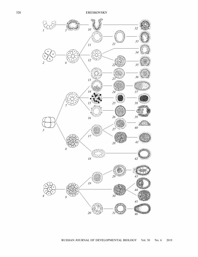
For example, in sponges there are four known typesof cleavage: incurvational (subclass Calcaronea: Cal-carea), polyaxial (subclass Calcinea: Calcarea andHalisarcidae: Demospongiae), radial-like (Chondro-sidae, Spirophorida, Polymastiida: Demospongiaeand Hexactinellida) and chaotic (all Homoscleromor-pha and most Demospongiae) (Fig. 2: 1–4). Thesefour main cleavage patterns of sponges result in threemain blastula types: stomoblastula, coeloblastula andmorula (Fig. 2: 5–9). On the other hand, the latter twoblastula types emerge as a result of different cleavagepatterns (Fig. 2). Embryonic morphogeneses involvedin larva formation in sponges are also very diverse,leading to nine larval types (Ereskovsky and Dondua,2006; Ereskovsky, 2010).
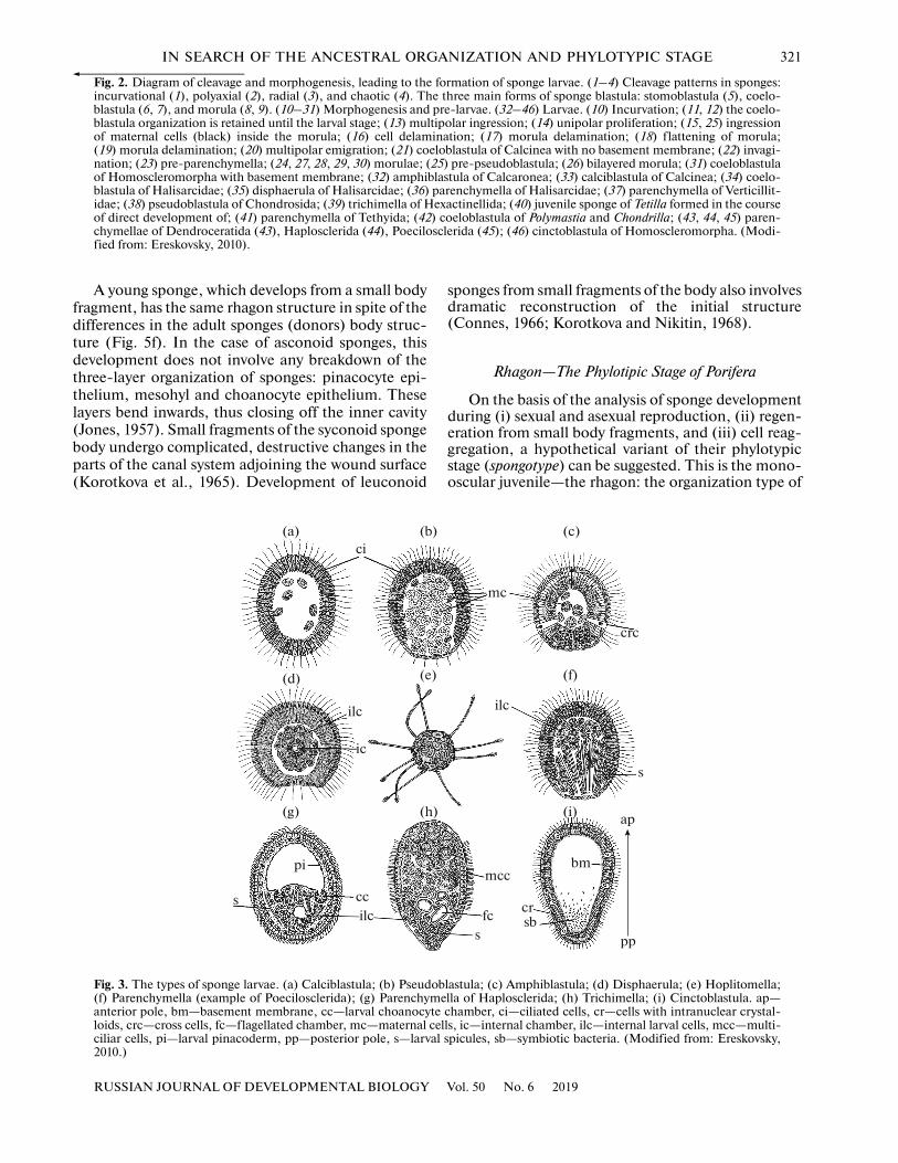
All larvae have a strongly pronounced anterior-posterior polarity, which is expressed in the structureof the layer of external cells, in the organization of theinternal cell mass (if present) and in the distribution ofspicules (if present). In general, there are two principallarval constructions in sponges: first, hollow single-layered larvae (coeloblastula, calciblastula, cincto-blastula, amphiblastula), and second, two-layered lar-vae lacking a cavity (parenchymella, hoplitomella, tri-chimella) (Fig. 3) (Ereskovsky, 2010).
The main feature of the metamorphosis of spongelarvae is the acquisition of the sponge bauplan, whichis primarily represented by the aquiferous system. Thefirst adult structure to be formed de novo is the exo-pinacoderm, which isolates the young sponge from theaquatic environment. Later steps include the organi-zation of the choanocyte chambers and the water cur-rent canals, the opening of the ostia and osculum, andthe acquisition of the elements of the adult skeleton.
Morphogenesis during metamorphosis dependsessentially on the larval type (its structure). Larval meta-morphosis usually results in a mono-oscular individual,whose aquiferous system often differs from that of theadult sponge. In the Calcarea, a young individual such asthis has the aquiferous system of the asconoid type and iscalled the olynthus (Minchin, 1900); in the Demo-spongiae and the Homoscleromorpha it has the aquifer-ous system of the leuconoid or syconoid type and iscalled the rhagon (Figs. 4, 5a, 5b) (Sollas, 1888). How-ever, there are no fundamental differences between thestructure of ragon and olintus.
In sponges with direct development upon leavingthe maternal organism (in cases of viviparity), or inthose which develop in the aquatic environment (incases of oviparity), development leads to formation ofjuveniles exhibiting rhagon structure (Watanabe, 1978;Sara et al., 2002).
The Development of Sponges during Asexual Reproduction
General characteristics of the buds formed in allsponges except Oscarella (Homoscleromorpha) (Ere-
RUSSIAN JOURNAL OF DEVELOPMENTAL BIOLOGY
skovsky and Tokina, 2007) appear during the initialstages of development, when they look like a denseconglomerate of different cell types at the parentsponge surface. Such buds have neither canals nor anosculum, and only very rare choanocyte chambers (forreview see: Fell, 1993; Ereskovsky et al., 2017). Afterdetachment from parent body, buds settle on the sub-strate, attach to it and begin the formation of the aqui-ferous system and growth. Thus, the buds of allsponges resemble first the pupae, and then the rhagon,the stage, which forms after larval settlement (Fig. 5c).
Budding in the Homoscleromorpha is essentiallydifferent from that in the other sponges, with differ-ences concerning both morphogenesis and bud struc-ture. The bud develops from the outgrowths of theparent body wall that is formed by the epithelial mor-phogenesis—evagination. The cells at budding sites donot migrate to the periphery, nor do they form con-densations, nor do they proliferate. The types of cellsconstituting the bud is identical to that of the resultingdefinitive sponge (Ereskovsky and Tokina, 2007).Before attachment to the substrate, the bud has therhagon structure with a syconoid aquiferous system.
Many freshwater and a few estuarine/marine dem-osponges produce dormant structures called gem-mules (Simpson, 1984; Fell, 1993). Each gemmuleconsists of a compact mass of essentially identical cellssurrounded by a collagenous capsule, which in manycases contains spicules. Gemmule hatching involvesthe mitotic division of the thesocytes (internal totypo-tent cells), active cell migration and differentiation,and results in small functional juvenile sponges withrhagon organization and leuconoid aquiferous systems(Fig. 5d) (Brien, 1932; Höhr, 1977).
In this way, sponge juveniles resulting from asexualreproduction—budding and gemmule hatching—havethe same rhagon structure.
The Development of Sponges during Regeneration
Numerous experiments on sponge cell dissociationand subsequent reaggregation have shown that inmany sponges this leads to the formation of a compactspherical body, contrasting with the rather loose,irregular cellular contacts during aggregation (Lavrovand Kosevich, 2014, 2016). The formation of the pina-coderm represents the first step in the reorganizationof tissue-like structures. This stage, termed “prim-morphs” (Custodio et al., 1998) marks the completionof the aggregation of cellular material and the separa-tion of the internal environment from the external oneby a continuous pinacoderm. After attachment andstable fixation onto the solid substrate, this stage willlead to morphogenetic processes ending in the fullreorganization of the small but fully functional andwell-structured sponge (Lavrov and Kosevich, 2014,2016). This stage also has the structure of a rhagon(Fig. 5e).
Vol. 50 No. 6 2019
320 ERESKOVSKY
1
2
3
4
5
6
7
8
9
10
11
12
13
14
15
16
17
18
19
20
21
22
23
24
25
26
27
28
29
30
31
32
33
34
35
36
37
38
39
40
41
42
43
44
45
46
RUSSIAN JOURNAL OF DEVELOPMENTAL BIOLOGY Vol. 50 No. 6 2019
IN SEARCH OF THE ANCESTRAL ORGANIZATION AND PHYLOTYPIC STAGE 321
A young sponge, which develops from a small bodyfragment, has the same rhagon structure in spite of thedifferences in the adult sponges (donors) body struc-ture (Fig. 5f). In the case of asconoid sponges, thisdevelopment does not involve any breakdown of thethree-layer organization of sponges: pinacocyte epi-thelium, mesohyl and choanocyte epithelium. Theselayers bend inwards, thus closing off the inner cavity(Jones, 1957). Small fragments of the syconoid spongebody undergo complicated, destructive changes in theparts of the canal system adjoining the wound surface(Korotkova et al., 1965). Development of leuconoid
RUSSIAN JOURNAL OF DEVELOPMENTAL BIOLOGY
Fig. 3. The types of sponge larvae. (a) Calciblastula; (b) Pseudo(f) Parenchymella (example of Poecilosclerida); (g) Parenchymanterior pole, bm—basement membrane, cc—larval choanocytloids, crc—cross cells, fc—flagellated chamber, mc—maternal cciliar cells, pi—larval pinacoderm, pp—posterior pole, s—larva2010.)
ci
pi
ilc
ilc
ic
s cc
(a) (b)
(d) (e)
(g) (h)
sponges from small fragments of the body also involvesdramatic reconstruction of the initial structure(Connes, 1966; Korotkova and Nikitin, 1968).
Rhagon—The Phylotipic Stage of Porifera
On the basis of the analysis of sponge developmentduring (i) sexual and asexual reproduction, (ii) regen-eration from small body fragments, and (iii) cell reag-gregation, a hypothetical variant of their phylotypicstage (spongotype) can be suggested. This is the mono-oscular juvenile—the rhagon: the organization type of
Fig. 2. Diagram of cleavage and morphogenesis, leading to the formation of sponge larvae. (1–4) Cleavage patterns in sponges:incurvational (1), polyaxial (2), radial (3), and chaotic (4). The three main forms of sponge blastula: stomoblastula (5), coelo-blastula (6, 7), and morula (8, 9). (10–31) Morphogenesis and pre-larvae. (32–46) Larvae. (10) Incurvation; (11, 12) the coelo-blastula organization is retained until the larval stage; (13) multipolar ingression; (14) unipolar proliferation; (15, 25) ingressionof maternal cells (black) inside the morula; (16) cell delamination; (17) morula delamination; (18) f lattening of morula;(19) morula delamination; (20) multipolar emigration; (21) coeloblastula of Calcinea with no basement membrane; (22) invagi-nation; (23) pre-parenchymella; (24, 27, 28, 29, 30) morulae; (25) pre-pseudoblastula; (26) bilayered morula; (31) coeloblastulaof Homoscleromorpha with basement membrane; (32) amphiblastula of Calcaronea; (33) calciblastula of Calcinea; (34) coelo-blastula of Halisarcidae; (35) disphaerula of Halisarcidae; (36) parenchymella of Halisarcidae; (37) parenchymella of Verticillit-idae; (38) pseudoblastula of Chondrosida; (39) trichimella of Hexactinellida; (40) juvenile sponge of Tetilla formed in the courseof direct development of; (41) parenchymella of Tethyida; (42) coeloblastula of Polymastia and Chondrilla; (43, 44, 45) paren-chymellae of Dendroceratida (43), Haplosclerida (44), Poecilosclerida (45); (46) cinctoblastula of Homoscleromorpha. (Modi-fied from: Ereskovsky, 2010).
Vol. 50 No. 6 2019
blastula; (c) Amphiblastula; (d) Disphaerula; (e) Hoplitomella;ella of Haplosclerida; (h) Trichimella; (i) Cinctoblastula. ap—
e chamber, ci—ciliated cells, cr—cells with intranuclear crystal-ells, ic—internal chamber, ilc—internal larval cells, mcc—multi-l spicules, sb—symbiotic bacteria. (Modified from: Ereskovsky,
mc
crc
ilc
ap
s
s
mcc
fc crsb
pp
bm
(c)
(f)
(i)
322
RUSSIAN JOURNAL OF D
ERESKOVSKY
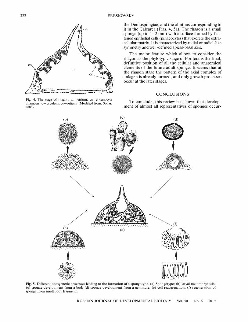
Fig. 5. Different ontogenetic processes leading to the formatio(c) sponge development from a bud; (d) sponge developmensponge from small body fragment.
(
(b) (
(e)
Fig. 4. The stage of rhagon. at—Atrium; cc—choanocytechambers; o—osculum; os—ostium. (Modified from: Sollas,1888).
os
o
atcc
the Demospongiae, and the olinthus corresponding toit in the Calcarea (Figs. 4, 5a). The rhagon is a smallsponge (up to 1–2 mm) with a surface formed by flat-tened epithelial cells (pinacocytes) that excrete the extra-cellular matrix. It is characterized by radial or radial-likesymmetry and well-defined apical-basal axis.
The major feature which allows to consider therhagon as the phylotypic stage of Porifera is the final,definitive position of all the cellular and anatomicalelements of the future adult sponge. It seems that atthe rhagon stage the pattern of the axial complex ofanlagen is already formed, and only growth processesoccur at the later stages.
CONCLUSIONS
To conclude, this review has shown that develop-ment of almost all representatives of sponges occur-
EVELOPMENTAL BIOLOGY Vol. 50 No. 6 2019
n of a spongotype. (a) Spongotype; (b) larval metamorphosis;t from a gemmule; (e) cell reaggregation; (f) regeneration of
a)
c) (d)
(f)
IN SEARCH OF THE ANCESTRAL ORGANIZATION AND PHYLOTYPIC STAGE 323
ring in different situations (sexual/asexual reproduc-tion, regeneration) and by different set of morphogen-esis lead to the stage common for all Porifera: that ofthe rhagon, which can be characterized by its struc-tural similarity across this phylum. We propose thatthis stage can be considered not only as a phylotipicstage, but also as a model of putative ancestralsponge—a spongotype. The stage of rhagon is typicalfor Demospongiae, and it corresponds to olintus,characteristic of Calcarea (Figs. 4, 5a). In order toconfirm or disprove these conclusions, it is necessaryto conduct a detailed study of the molecular mecha-nisms that regulate the formation of rhagon in repre-sentatives of different phylogenetic groups of sponges.Such a study will improve our understanding of themechanisms involved in the evolution of the bodyplans of sponges and other multicellular animals.
FUNDING
This work was supported by grant no. 17-14-01089 of theRussian Science Foundation.
COMPLIANCE WITH ETHICAL STANDARDS
The authors declare that they have no conflict of interest.This article does not contain any studies involving animalsor human participants performed by any of the authors.
REFERENCESBrien, P., Contribution à l'étude de la régénération naturelle
chez les Spongillidae Spongilla lacustris (L.); Ephydatiafluviatilis (L.), Archives de Zoologie expérimentale etgénérale, 1932, vol. 74, pp. 461–506.
Connes, R., Contribution à l'étude histologique des pre-miers stades d’embryogenèse somatique chez Tethyalyncurium Lamarck, Bull. Soc. Zool. France, 1966,vol. 91, pp. 639–645.
Custodio, M.R., Prokic, I., Steffen, R., Koziol, C., Boroje-vic, R., Brummer, F., Nickel, M., and Müller, W.E.G.,Primmorphs generated from dissociated cells of thesponge Suberites domuncula: a model system for studiesof cell proliferation and cell death, Mech Ageing Dev.,1998, vol. 105, pp. 45–59.
Cuvier, G., Le Règne Animal Distribute d’après Son Organi-zation, Paris, 1817, vol. 1.
Darwin, C., On the Origin of Species, Murray, 1859.Degnan, B.M., Adamska, M., Richards, G.R., Larroux, C.,
Leininger, S., Bergum, B., Calcino, A., Maritz, K.,Nakanishi, N., and Degnan, S.M., Porifera, in Evolu-tionary Developmental Biology of Invertebrates, Wan-ninger, A., Ed., Wein: Springer, 2015, vol. 1, pp. 65–106.
Drost, H.-G., Janitza, P., Grosse, I., and Quint, M., Cross-kingdom comparison of the developmental hourglass,Curr. Opin. Gen. Dev., 2017, vol. 45, pp. 69–75.
Duboule, D., Temporal colinearity and the phylotypic pro-gression: a basis for the stability of a vertebrate Bauplanand the evolution of morphologies through heterochro-ny, Dev. Suppl., 1994, pp. 135–142.
RUSSIAN JOURNAL OF DEVELOPMENTAL BIOLOGY
Embryology. Constructing the Organism, Gilbert, S.F. andRaunio, A.M., Eds., Sunderland: Sinauer Associates,1997.
Ereskovsky, A.V., The Comparative Embryology of Sponges,Dordrecht: Springer-Verlag, 2010.
Ereskovsky, A.V., and Dondua, A.K., The problem of germlayers in sponges (Porifera) and some issues concerningearly metazoan evolution, Zool. Anz., 2006, vol. 245,pp. 65–76.
Ereskovsky, A. and Lavrov, A., Porifera, in InvertebrateHistology, Elise, E.B. and La Douceur, E.E.B., Eds.,Wiley, 2019 (in press).
Ereskovsky, A.V., and Tokina, D.B., Asexual reproductionin homoscleromorph sponges (Porifera; Homosclero-morpha), Mar. Biol., 2007, vol. 151, pp. 425–434.
Ereskovsky, A.V., Geronimo, A., and Pérez, T., Asexualand puzzling sexual reproduction of the Mediterraneansponge Haliclona fulva (Demospongiae): life cycle andcytological structures, Invert. Biol., 2017, vol, 136,pp. 403–421.
Fell, P.E., Porifera, in Reproductive Biology of Invertebrates,vol. 6: Asexual Propagation and Reproductive Strategies,Adiyodi, K.G. and Adiyodi, R.G., Eds., Chichester:Wiley, 1993, pp. 1–44.
Gilbert S.F., Developmental Biology, 10th ed., Sunderland:Sinauer Associates, 2013.
Hall, B.K., Evolutionary Developmental Biology, 2nd ed.,Amsterdam: Kluwer, 1998.
Höhr, D., Differenzierungsvorgänge in der keimendenGemmula von Ephydatia fluviatilis, Wilhelm Roux’sArch., 1977, vol. 182, pp. 329–346.
Ivanova-Kazas, O.M., Evolutionary Embryology of Animals,St. Petersburg: Nauka, 1995.
Jones, W.C., The contractility and healing behaviour ofpieces of Leucosotenia complicate, Quart. J. Microsc.Sci., 1957, vol. 98, pp. 203–217.
Kalinka, A.T., and Tomancak, P., The evolution of earlyanimal embryos: conservation or divergence?, TrendsEcol. Evol., 2012, vol. 27, pp. 385–393.
Korotkova, G.P. and Nikitin, N.S., The peculiarities of themorphogenesis during the development of cornacuspongeHalichondria panicea from the small part of the body. Re-constructional processes and immunological reactions, inMorphological Investigations of Different Stages of Develop-ment of the Marine Organisms, Tokin, B.P., Ed., Lenin-grad: Nauka, 1969, pp. 17–26.
Korotkova, G. P., Efremova, S. M., and Kadantseva, A. G.,The peculiarities of morphogenesis of the developmentof Sycon lingua from the small part of the body, Vestn.Leningr. Univ., 1965, vol. 4, no. 21, pp. 14–30.
Lavrov, A.I., and Kosevich, I.A., Sponge cell reaggregation:mechanisms and dynamics of the process, Russ. J. Dev.Biol., 2014, vol. 45, pp. 205–223.
Lavrov, A.I., and Kosevich I.A., Sponge cell reaggregation:cellular structure and morphogenetic potencies of multi-cellular aggregates, J. Exp. Zool. A Ecol. Genet. Physiol.,2016, vol. 325, pp. 158–177.
Minchin, E.A., Sponges—phylum Porifera, in Treatise onZoology, vol. 2: The Porifera and Coelenterata, RayLankaster, E., Ed., London: Adam and Charles Black,1900.
Vol. 50 No. 6 2019
324 ERESKOVSKY
Minelli, A., and Schram, F.R., Owen revisited: a reapprais-al of morphology in evolutionary biology, Bijdr Dier-kunde, 1994, vol. 64, pp. 65–74.
Owen, R., On the Archetype and Homologies of the VertebrateSkeleton, John van Voorst, Paternoster Row, 1848.
Peter, I.S., and Davidson, E.H., Evolution of gene regula-tory networks controlling body plan development, Cell,2011, vol. 144, pp. 970–985.
Raff, R.A., The Shape of Life: Genes, Development and theEvolution of Animal Form, Univ. of Chicago Press, 1996.
Richardson, M.K. Hanken, J., Gooneratne, M.L., Pieau, C.,Raynaud, A., Selwood, L., and Wright, G.M., There isno highly conserved embryonic stage in the vertebrates:implications for current theories of evolution and devel-opment, Anat. Embryol., 1997, vol. 196, pp. 91–106.
Richardson, M.K., Allen, S.P., Wright, G.M., Raynaud, A.,and Hanken, J., Somite number and vertebrate evolu-tion, Development, 1998, vol. 125, pp. 151–160.
Sander, K., Specification of the basic body plan in insectembryogenesis, Adv. Insect Physiol., 1976, vol. 12,pp. 125–238.
Sarà, A., Cerrano, C., and Sarà, M., Viviparous develop-ment in the Antarctic sponge Stylocordyla borealisLoven, 1868, Polar Biol., 2002, vol. 25, pp. 425–431.
Scholtz, G., Baupläne versus ground patterns, phyla versusmonophyla: aspects of patterns and processes in evolu-tionary developmental biology, in Evolutionary Devel-
opmental Biology of Crustacea, Balkema Scholtz, G.,Ed., 2004, pp. 3–16.
Scholtz, G., Homology and ontogeny: pattern and processin comparative developmental biology, Theory Biosci.,2005, vol. 124, pp. 121–143.
Seidel, F., Körpergrundgestalt und Keimstruktur: eineErörterung über die Grundlagen der vergleichendenund experimentellen Embryologie und deren Gültigkeitbei phylogenetischen Überlegungen, Zool. Anz., 1960,vol. 164, pp. 245–305.
Simpson, T.L., The Cell Biology of Sponges, New York:Springer-Verlag, 1984.
Slack, J.M.W., Holland, P.M.H., and Graham, C.F., Thezootype and the zootypic stage, Nature, 1993, vol. 361,pp. 490–492.
Sollas, W.J., Report on the Tetractinellida collected byH.S.M. Challendger during the years 1873–1876, Rep.Sci. Res. Voyage Challenger Zool., 1888, vol. 25, pp. 1–458.
von Baer, K.E., Űber Entwickelungsgeschichte der Thiere:Beobachtung und Reflektion, Köenigsberg, 1828.
Watanabe, Y., The development of two species of Tetilla(Demosponge), Nat. Sci. Rep. Ochanomizu Univ., 1978,vol. 29, pp. 71–106.
Yanai, I., Development and evolution through the lens ofglobal gene regulation, Trends Genet., 2018, vol. 34,pp. 11–20.
RUSSIAN JOURNAL OF DEVELOPMENTAL BIOLOGY Vol. 50 No. 6 2019