Embed Size (px)
Citation preview
Proc. Natl. Acad. Sci. USAVol. 93, pp. 9276-9281, August 1996Pharmacology
Involvement of Asn-293 in stereospecific agonist recognition andin activation of the .82-adrenergic receptor
(stereospecificity/intrinsic activity/isoproterenol/propranolol/ligand binding)
KERSTIN WIELAND*, HELENE M. ZUURMONDt, CORNELIUS KRASEL*, AD P. IJZERMANt, AND MARTIN J. LOHSE*t
*Department of Pharmacology, University of Wurzburg, Versbacher Strasse 9, 97078 Wurzburg, Germany; and tLeiden/Amsterdam Center for Drug Research,Division of Medicinal Chemistry, P.O. Box 9502, 2300 RA Leiden, The Netherlands
Communicated by James Black, King's College of Medicine and Dentistry of King's College London, London, United Kingdom, May 21, 1996(received for review February 1, 1996)
ABSTRACT To investigate the molecular mechanism forstereospecific binding of agonists to g32-adrenergic receptorswe used receptor models to identify potential binding sites forthe (3-OH-group of the ligand, which defines the chiral center.Ser-165, located in transmembrane helix IV, and Asn-293,situated in the upper half of transmembrane helix VI, wereidentified as potential binding sites. Mutation of Ser-165 toAla did not change the binding of either isoproterenol isomeras revealed after transient expression in human embryonickidney (HEK)-293 cells. In contrast, a receptor mutant inwhich Asn-293 was replaced by Leu showed substantial loss ofstereospecific isoproterenol binding. Adenylyl cyclase stimu-lation by this mutant after stable expression in CHO cellsconfirmed the substantial loss of stereospecificity for isopro-terenol. In a series of agonists the loss of affinity in theLeu-293 mutant receptor was strongly correlated with theintrinsic activity of the compounds. Full agonists showed a10-30-fold affinity loss, whereas partial agonists had almostthe same affinity for both receptors. Stereospecific recogni-tion of antagonists was unaltered in the Leu-293 mutantreceptor. These data indicate a relationship between ste-reospecificity and intrinsic activity of agonists and suggestthat Asn-293 is important for both properties of the agonist-receptor interaction.
(32-Adrenergic receptors (,82-AR) are often studied as a modelsystem for the large superfamily of G-protein-coupled recep-tors. These receptors most likely contain seven transmembranea-helices, and their topography has been verified using bio-chemical and immunological techniques to identify extra- andintracellular domains (1, 2). The binding of agonists to thesereceptors has been studied in much detail to understand themolecular mechanisms of ligand docking and receptor activa-tion (3). A series of mutagenesis experiments plus the analysisof large numbers of ligands has allowed the identification ofseveral of the amino acids involved in agonist binding to ,82-AR(reviewed in refs. 4 and 5). The current concept of agonistbinding proposes that the positively charged nitrogen interactswith Asp-113 in transmembrane domain III (6, 7), and that thetwo catechol-OH-groups form hydrogen bonds with Ser-204and Ser-207 in transmembrane domain V (8).
Despite these advances, one of the key properties of agonistsat these receptors has not been clarified: their stereospecificbinding. Stereospecificity of 3-adrenergic agonists is definedby their (3-OH-group, which is located at the chiral center.(32-AR bind their agonists such as isoproterenol in a stereospe-cific manner, with the (-)isomer being about 40-times morepotent than the (+)isomer, both in the high-affinity state(equivalent to the receptor/G-protein complex) and in thelow-affinity state (9). In addition to its potential role in
stereospecificity, the ,B-OH-group might also be involved in theagonistic properties of these compounds (3). The amino acidsthat interact with this (3-OH-group and may therefore beresponsible for stereospecificity have not been identified.Apart from relatively low-resolution electron diffraction
results obtained with rhodopsin (10), there are no biophysicaldata on G-protein-coupled receptors. Several authors havetherefore developed computer models of G-protein-coupledreceptors to predict their structure and modes of interactionwith their ligands. The first group of such models uses theknown structure of bacteriorhodopsin, which also containsseven transmembrane a-helices (11), to develop a backbone ofthe transmembrane helices of G-protein-coupled receptors(12, 13). The second group of models avoids the use ofbacteriorhodopsin as a template because it is a developmen-tally probably unrelated archaebacterial proton pump and nota receptor (14). The latter models are based essentially onsequence alignments of many G-protein-coupled receptorsand their hydrophobicity profiles or their polarity conservedpositions (15, 16). Finally, both sets of information have oftenbeen used in attempts to obtain models of G-protein-coupledreceptors (reviewed in refs. 17 and 18).
Obviously, all of these models contain a great deal ofspeculation. However, assuming that the helices are arrangedin a circular and sequential (anti-clockwise when viewed fromthe top) manner [which is the prevailing hypothesis eventhough a different order has also been proposed by Pardo et al.(19)], then anchoring agonists at helices III and V, as describedabove, leaves essentially helices IV and VI as potential an-choring points for the (3-OH-group.Trumpp-Kallmeyer et al. (13) have proposed that Ser-165 in
helix IV binds the (3-OH-group, but this hypothesis has not yetbeen investigated experimentally. In the present study, we haveused computer modelling of the (32-AR to identify Asn-293 asanother potential interaction site for this (3-OH-group, andhave investigated the roles of these amino acids by site-directedmutagenesis.
MATERIALS AND METHODSMaterials. Oligonucleotides were synthesized on an Applied
Biosystems DNA synthesizer. CHO-10001 cells were kindlyprovided by M. Gottesman (National Institutes of Health).125Iodo-cyanopindolol (1251-CYP) and [a-32P]ATP were ob-tained from New England Nuclear, and the latter was purifiedas described by Walseth and Johnson (20). Stereoisomers ofisoproterenol (>99% purity) and dobutamine were purchasedfrom Research Biochemicals (Natick, MA), and stereoisomersof propranolol (>98.5% purity) were from Sigma. The fol-lowing compounds were kindly provided by: (-)- and (+)epi-
Abbreviations: (32-AR, (human) 132-adrenergic receptor; 125I-CYP,125Iodo-cyanopindolol.ITo whom reprint requests should be addressed.
9276
The publication costs of this article were defrayed in part by page chargepayment. This article must therefore be hereby marked "advertisement" inaccordance with 18 U.S.C. §1734 solely to indicate this fact.
Dow
nloa
ded
by g
uest
on
Janu
ary
24, 2
022
Proc. Natl. Acad. Sci. USA 93 (1996) 9277
nephrine (O. E. Brodde, Halle, Germany) and (-)- and(+)norepinephrine (A. Kaumann, Cambridge, U.K.)Modelling of the Human p2-AR. Since no structural infor-
mation on G-protein-coupled receptors was available at theoutset of these studies, a putative model of the 132-AR wasgenerated according to the method of Lewell (21). It was basedon the atomic coordinates of bacteriorhodopsin as obtainedfrom the Brookhaven Protein Data Bank (reference code1BRD). Because there is no significant sequence homologybetween bacteriorhodopsin and the 832-AR, the sequencealignment proposed by Lewell with rhodopsin as an interme-diate was used, and the side chains of bacteriorhodopsin weresubstituted by those of the human f32-AR. Unfavorable stericinteractions were removed and subsequently energy minimi-zations were performed both in the presence and in theabsence of the reference ligand (-)isoproterenol according tothe procedures described for the modelling of the A1 adenosinereceptor (12). No rotations or translational motions of thehelical backbones were allowed.To obtain docking of (-)isoproterenol into the }32-AR
cavity (see Fig. 1), three distance constraints were applied inline with mutagenesis data (7, 8). Thus, the distance betweenthe positively charged nitrogen atom of (-)isoproterenol andthe negatively charged Asp-113 (both terminal oxygen atoms)was 3.5 A (ionic interaction), and the distance between Ser-204(oxygen atom) and the meta-OH (hydrogen atom) of thecatechol group of (-)isoproterenol was 2.1 A (hydrogenbond), as was the distance between the catechol para-OH-group and Ser-207. Further energy minimization after removalof the distance constraints preserved these interactions.
All manipulations were carried out using the softwarepackage QUANTA/CHARMM, version 3.3.1 (Molecular Simula-tions, Waltham, MA) on a Silicon Graphics XZ4000 worksta-tion.
Mutagenesis of p2-AR cDNA. The cDNA for the human12-AR (22) was cloned into the expression vector pBC-CMV-SK (23) to generate the vector pBC-CMV-P32AR. Mu-tation of the codon for amino acid 165 was carried outaccording to Kunkel (24). Site-directed mutagenesis of thecodon for amino acid 293 took advantage of the fact that aunique HpaI restriction site is located directly adjacent to thiscodon: The vector was linearized at this site with HpaI, and thegap was bridged with a 38-mer mutant oligonucleotide con-taining in its center the codon CTC (Leu) for amino acid 293.A piece of mutant receptor cDNA corresponding to the entirecoding region was then generated by polymerase chain reac-tion (PCR) under standard conditions (30 cycles) using theoligonucleotide-annealed linear vector (100 ng) as a templateand primers corresponding to nucleotides 1-18 (forward) and1242-1225 (reverse) of the receptor cDNA. A 318-bp BglII-EcoRV fragment containing the mutated region was excisedfrom the PCR products and inserted into the correspondingsites of pBC-CMV-f32AR. The sequences of all resultantcDNAs were verified by automated sequencing.
Transfection of Human Embryonic Kidney (HEK)-293Cells and Generation of Stable CHO Cell Lines. For initialstudies, wild-type and mutant f32-AR were transiently ex-pressed in HEK-293 cells. Transfection of these cells by thecalcium phosphate/N,N-bis(2-hydroxyethyl)-2-aminoethane-sulfonic acid method (25) resulted 2 days later in the expres-sion of receptors at a density of several pmol/mg membraneprotein. More detailed studies were done with CHO cell linesstably expressing these receptors. These cell lines were ob-tained by transfecting CHO-10001 cells with the respectiveexpression vectors plus pSV2neo using DOTAP (BoehringerMannheim) as transfection reagent and G418 (GIBCO) toselect positive clones as described earlier (23, 26). Clones withcomparable densities (about 0.2 pmol/mg membrane protein)were selected for the experiments. Additional clones were alsotested to check for potential artifacts in individual clones.
Radioligand Binding Studies. Ligand binding to 132-AR wasanalyzed using 125I-CYP and crude cell membranes preparedas described earlier (27) using an incubation time of 1 hr at30°C. Saturation studies were done with radioligand concen-trations from 2 pM to 200 pM, using 1 ,uM (-)propranolol todefine nonspecific binding. Competition studies were donewith a radioligand concentration of 30 pM. Unless statedotherwise, all radioligand binding assays contained 100 ,uMGTP to uncouple 132-AR from Gs and thereby generatemonophasic competition curves for agonists, as well as antag-onists.
Adenylyl Cyclase Assays. The function of f32-AR was as-sessed by determining their capacity to stimulate the adenylylcyclase activity in membranes prepared from the CHO celllines stably expressing the receptor variants. Membranes wereprepared as above, and adenylyl cyclase activity was deter-mined by measuring the generation of [32P]cAMP from[a-32P]ATP as described (28). The incubation was done for 30min at 30°C.Data Analysis. Radioligand binding data were analyzed by
nonlinear curve fitting using the program SCTFIT, which allowsanalysis for models containing multiple-binding sites (29, 30).Concentration-response curves for adenylyl cyclase stimula-tion were analyzed by nonlinear curve fitting to the Hillequation as described (31).
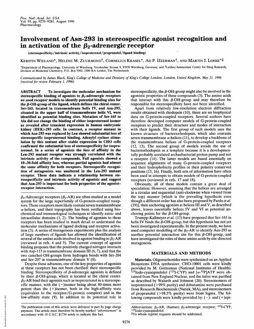
RESULTSTo identify amino acids in the human X32-AR that might bindthe 3-OH-group of 13-adrenergic receptor agonists, comput-erized visualization of the receptors and their ligand bindingpocket was performed as described in Materials and Methods.At the outset of the present study, no structural information onany G-protein-coupled receptor was available. Therefore, thecoordinates of bacteriorhodopsin were used to generate thebackbone for the 132-AR. Fig. 1 shows the helical backbonewith the ligand (-)isoproterenol fitted to its putative bindingsite formed by Asp-113, Ser-204, and Ser-207. Anchoring theligand to the respective side chains still leaves rotationalfreedom of the ligand due to the flexibility of the ethanolamineside chain. This allowed visualization of amino acids that couldinteract with the 13-OH-group. Ser-165 represents one possibleinteracting amino acid as suggested by Trumpp-Kallmeyer etal. (13). However, by changing the torsion angles in the ligand,the 13-OH-group could be brought into even closer vicinity toAsn-293, situated in transmembrane helix VI. Energy mini-mization yielded a putative hydrogen bond between the hy-drogen atom of the 13-OH-group and the oxygen atom of thecarboxamide function in Asn-293, suggesting that this residuemight be important for stereospecificity in agonist binding. Noother amino acids were identified in this model that could forma bond with the 1-OH-group.To test the role of these amino acids, mutants of the human
{32-AR were generated in which Ser-165 was replaced by Ala,or Asn-293 was replaced by Leu. The mutations resulted in sidechains that were similar in size to those in the wild-typereceptor, but incapable of forming a hydrogen bond. Transientexpression of these receptors in HEK-293 cells revealed thatthe mutations did not significantly affect the affinity for125I-CYP (Table 1). Competition for 125I-CYP binding by thestereoisomers of isoproterenol was used to investigate theability of the receptor variants to bind agonists in a stereospe-cific manner (Table 1). (-)Isoproterenol was almost 40-foldmore potent than (+)isoproterenol at wild-type receptors, andvery similar data were obtained for the 165-Ala mutant. Incontrast, in the 293-Leu mutant the stereospecificity of iso-proterenol was reduced to about 6-fold. This reduction wasessentially due to a loss in affinity of the (-)stereoisomer,whereas the affinity of the (+)stereoisomer was only slightlyaltered. These data are compatible with a stereospecific in-
Pharmacology: Wieland et al.
Dow
nloa
ded
by g
uest
on
Janu
ary
24, 2
022
9278 Pharmacology: Wieland et al.
FIG. 1. Model visualizing the binding of (-)isoproterenol to the human f32-AR. The receptor a-helical backbone (helices III-VI) is viewedfrom the extracellular side, tilted by =20°. (-)Isoproterenol and the amino acids thought to be involved in ligand binding are indicated: Asp-113(helix III) and Ser-204 and Ser-207 (helix V). Also indicated are Ser-165 (helix IV) and Asn-293 (helix VI), the residues that might be in close vicinityto the ,-OH-group of (- )isoproterenol. The atoms are represented as follows: white, hydrogen atoms connected to either oxygen or nitrogen (mosthydrogens are not shown to improve clarity); black, carbon atoms; blue, nitrogen atoms; red, oxygen atoms. The figure was generated using theprograms MOLSCRIPT and RASTER3D.
teraction of the side chain of Asn-293, but not of Ser-165, withthe (3-OH-group in isoproterenol.To test whether a contribution of Ser-165 to agonist binding
might be visible in 293-Leu mutant receptors, we also gener-ated a double mutant (165-Ala, 293-Leu). However, thisdouble mutant was not much different from the 293-Leumutant. The affinities of both isomers of isoproterenol were1.7-fold lower than in the 293-Leu mutant, and the stereospec-ificity was the same, indicating that in the 293-Leu receptormutant Ser-165 also does not participate in ligand binding.
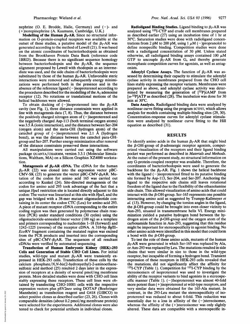
Wild-type and 293-Leu mutant receptors were then stablyexpressed in CHO cells and clones with comparable receptordensities (182 ± 12 and 170 ± 20 fmol/mg membrane protein)were used for functional analysis of the two receptor variants.The maximal stimulation of adenylyl cyclase activity by (-)iso-proterenol and by forskolin was similar in CHO cell mem-branes containing either wild-type or Leu-293 mutant recep-
tors, indicating that the mutant receptor was fully capable ofactivating Gs (Fig. 2, legend). Concentration-response curvesof the isomers of isoproterenol revealed a very marked ste-reospecificity of the wild-type receptor (Fig. 2). This appearsto be due to the fact that (+)isoproterenol is a partial agonistin these assays and to a significant receptor reserve in thesecells. Using wild-type receptors we found a 134-fold higherpotency of (-)isoproterenol as compared with (+)isoproter-enol, again mainly due to a loss in the potency of (-)isopro-
terenol. This stereoselectivity was reduced to 13-fold for the293-Leu mutant. As in the binding experiments, this reductionwas mainly due to a loss in the potency of (-)isoproterenol,whereas the potency of (+)isoproterenol was only marginallyreduced.The differential effects of the 293-Leu mutations on the
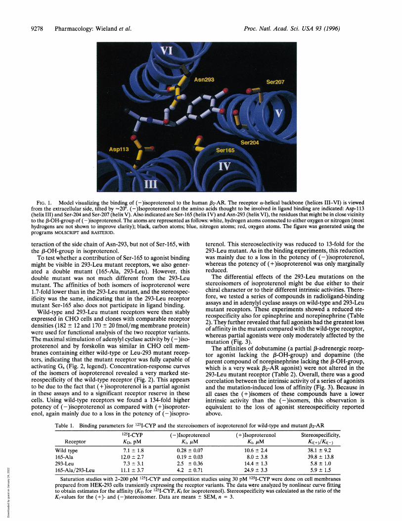
stereoisomers of isoproterenol might be due either to theirchiral character or to their different intrinsic activities. There-fore, we tested a series of compounds in radioligand-bindingassays and in adenylyl cyclase assays on wild-type and 293-Leumutant receptors. These experiments showed a reduced ste-reospecificity also for epinephrine and norepinephrine (Table2). They further revealed that full agonists had the greatest lossof affinity in the mutant compared with the wild-type receptor,whereas partial agonists were only moderately affected by themutation (Fig. 3).The affinities of dobutamine (a partial ,B-adrenergic recep-
tor agonist lacking the 3-OH-group) and dopamine (theparent compound of norepinephrine lacking the 83-OH-group,which is a very weak X32-AR agonist) were not altered in the293-Leu mutant receptor (Table 2). Overall, there was a goodcorrelation between the intrinsic activity of a series of agonistsand the mutation-induced loss of affinity (Fig. 3). Because inall cases the (+)isomers of these compounds have a lowerintrinsic activity than the (-)isomers, this observation isequivalent to the loss of agonist stereospecificity reportedabove.
Table 1. Binding parameters for 1251-CYP and the stereoisomers of isoproterenol for wild-type and mutant {32-AR
1251-Cyp (- )Isoproterenol (+ )Isoproterenol Stereospecificity,Receptor KD, pM Ki, ,uM K1, ,uM Ki(+)Ki(-)
Wild type 7.1 ± 1.8 0.28 ± 0.07 10.6 ± 2.4 38.1 ± 9.2165-Ala 12.0 ± 2.7 0.19 ± 0.03 8.0 ± 3.8 39.8 ± 13.8293-Leu 7.3 ± 3.1 2.5 ± 0.36 14.4 ± 1.3 5.8 ± 1.0165-Ala/293-Leu 11.1 ± 3.7 4.2 ± 0.71 24.9 ± 3.3 5.9 ± 1.5
Saturation studies with 2-200 pM 1251-CYP and competition studies using 30 pM 1251-CYP were done on cell membranesprepared from HEK-293 cells transiently expressing the receptor variants. The data were analyzed by nonlinear curve fittingto obtain estimates for the affinity (KD for 1251-CYP, Ki for isoproterenol). Stereospecificity was calculated as the ratio of theKi-values for the (+)- and (-)stereoisomer. Data are means + SEM, n = 3.
Proc. Natl. Acad. Sci. USA 93 (1996)
Dow
nloa
ded
by g
uest
on
Janu
ary
24, 2
022
Proc. Natl. Acad. Sci. USA 93 (1996) 9279
10-9 lo-7 io-5 io-3lQnPMrnTrnMPKl IAMX
LL
wLEO
0-
Zro
W UJ
WO°H-i-0z LO)L0
ccn~0CC)
160
120 F
80H
40-
WT 293-LiorFrIo I CrMIvLt\IIJ EC50(+)ISO 3400±940 4200±860 nM
EC50(-)ISO 25±3.4 330±28 nM
FIG. 2. Stimulation of adenylyl cyclase activity by isoproterenol via wild-type and 293-Leu mutant (32-AR. (A) Stimulation of adenylyl cyclaseactivity in membranes prepared from CHO cells expressing wild-type (circles) or 293-Leu mutant (squares) (32-AR by (-)isoproterenol (solidsymbols) or (+)isoproterenol (open symbols). Maximal activation by (-)isoproterenol (set to 100%) was 114 ± 5 and 127 ± 9 pmol cAMP/mgof protein/min in the wild-type and mutant receptors, respectively. Stimulation by 10 ,uM forskolin in these membranes was 202 + 7 and 229 ±15 pmol cAMP/mg of protein/min. Data are means ± SEM of four independent experiments with duplicate samples. (B) Stereospecificity of theadenylyl cyclase stimulation by isoproterenol via wild-type or 293-Leu mutant 132-AR. The data shown inA were analyzed by nonlinear curve fittingand the stereospecificity for isoproterenol was calculated as the ratio of the EC50 values for the (+) and the (-)stereoisomer. These EC50 valuesare indicated below the panel. Data are means + SEM, n = 3.
No such relationship was found for a group of antagonists.We tested the stereoisomers of two neutral antagonists, al-prenolol and metoprolol, and of propranolol that reducedbasal adenylyl cyclase activity in membranes prepared fromcells expressing wild-type or 293-Leu mutant receptors (datanot shown). The 293-Leu mutation caused modest alterationsin the affinities of these compounds, but their stereospecificrecognition was not altered when compared with wild-typereceptors (Table 3). This indicates that the changes in ste-reospecificity induced by this mutation were restricted toagonists.
DISCUSSIONBinding of agonists, as well as antagonists, to ,82-AR receptorsis generally thought to occur to the transmembrane a-helices.This was initially concluded from studies showing that mutantreceptors obtained by deletion of extracellular segments were
Table 2. Affinities of agonists for wild-type and 293-Leumutant ,32-AR
Affinity Ki, ,iM
Agonists Wild type 293-LeuWith a (3-OH-group
(-)Epinephrine 0.17 + 0.01 6.2 + 1.1(+)Epinephrine 2.1 ± 0.24 31 ± 5.0
stereospecificity 13 + 1.3 5.0 + 0.4(-)Norepinephrine 3.9 + 0.80 43 + 2.8(+)Norepinephrine 174 + 6.3 800 + 110
stereospecificity 49 + 9.3 18 + 1.3(±)Clenbuterol 0.024 + 0.008 0.025 ± 0.007(+)Clenbuterol 0.56 + 0.26 0.30 ± 0.12(+)Terbutaline 5.4 + 2.4 8.3 ± 2.6
Without a X3-OH-group(±)Dobutamine 66 + 21 63 ± 11dopamine 400 ± 150 550 ± 210
The affinities (and stereospecificities when two pure isomers wereavailable) were determined in competition experiments with '251-CYPand calculated as in Table 1.
still capable of binding ligands (32). Later studies involvingsite-directed mutagenesis identified specific amino acid resi-dues that appear to interact with specific determinants of,-adrenergic agonists. These include the anchoring points
100
1-1
CY)0)cmJ
C\l
U1)CO
r-
0
CVt)0)CMJ
10
1
0 20 40 60 80 100 120
INTRINSIC ACTIVITY [% of (-)ISOPROTERENOL]
FIG. 3. Comparison of the intrinsic activities of agonists and theloss in affinity induced by the 293-Leu mutation in the human 132-AR.Intrinsic activities were determined as the maximal stimulation ofadenylyl cyclase activity in membranes prepared from CHO cells stablyexpressing the respective receptors (see Fig. 2). They were normalizedto the maximal activity of (-)isoproterenol tested in the sameexperiments. The mutation-induced loss in affinity was determinedfrom inhibition of 1251-CYP binding to wild-type and 293-Leu mutantreceptors and is expressed as Ki(293-Leu mutant)/Ki(wild-type). Ab-breviated compounds are as follows: CLEN, clenbuterol; DOB, do-butamine; DOP, dopamine; EPI, epinephrine; ISO, isoproterenol;NEPI, norepinephrine; TERB, terbutaline. Data are means SEM ofthree to six experiments.
1' . '
-EPIr'I
+EPI
+NEPI
±DOB
Pharmacology: Wieland et al.
Dow
nloa
ded
by g
uest
on
Janu
ary
24, 2
022
9280 Pharmacology: Wieland et al.
Table 3. Stereoselective binding of antagonists to wild-type and293-Leu mutant }32-AR
Stereospecificity,
Compound Ki( +)isomer/Ki(-)isomerreceptor Wild type 293-Leu
Propranolol 104 ± 27 112 ± 41Alprenolol 22 ± 1 19 ± 1Metoprolol 21 ± 4 25 ± 3
Membranes were prepared from CHO cells stably expressing wild-type or 293-Leu mutant }32-AR (182 ± 12 and 170 ± 20 fmol/mgmembrane protein, respectively), and 125I-CYP binding was measuredin the presence of 10-12 to 10-4 M of the (-) and (+)isomers of theindicated antagonists. The data were analyzed by nonlinear curvefitting to obtain estimates for the affinities of the compounds (Ki).Stereospecificity was calculated as the ratio of the K; values for the (+)and the (-)stereoisomer. Data are means ± SEM, n = 3.
Asp-113 for the amine group of the ligands and Ser-204 andSer-207 for the catechol-OH-groups (7, 8). In contrast to thesewell-accepted interactions, there is no certainty regarding apotential interaction site for the (3-OH-group of agonists thatmight explain the well-known and pharmacologically veryrelevant stereospecificity of agonist binding.Computer modelling has led Trumpp-Kallmeyer et al. (13)
to suggest Ser-165 in transmembrane helix IV as a potentialanchoring point for this 3-OH-group. However, it has beendifficult to verify this hypothesis, because mutation of Ser-165to Ala resulted in a (32-AR mutant that failed to be expressedat significant levels (8). Green et al. (33) have investigated anaturally occurring Thr-164-Ile mutant human ,82-AR, whichshowed a 4-fold decreased affinity for isoproterenol, but onlymodest decreases for dobutamine and dopamine. The authorspostulate that an alteration at position 164 might affect bindingat Ser-165, and that their data indicate a role for Ser-165 instereospecific-agonist binding. However, the stereospecificityof isoproterenol was entirely maintained in the Ile-164 recep-tor variant, and, furthermore, the G-protein-coupling of thismutant was significantly impaired, indicating a more general-ized alteration of receptor structure. In contrast to thesehypotheses, we now find that the 165-Ala (2-AR was indistin-guishable from the wild-type receptor in its affinities for125I-CYP and the stereoisomers of isoproterenol, suggestingthat Ser-165 does not participate in ligand binding.A potential role of transmembrane helix VI in binding the
13-OH-group was suggested by the model shown in Fig. 1implying that Asn-293 might be capable of forming the pos-tulated hydrogen bond. It should be stressed that such receptormodels are at best crude approximations, which can be used asmeans of visualization of emerging experimental data, such asstructure-activity relationships of 13-adrenergic ligands anddata from mutagenesis studies. Interestingly, in the modeldeveloped by Baldwin (15) on the basis of sequence compar-isons of G-protein-coupled receptors plus emerging structuralinformation on rhodopsin, Asn-293 faces the central portion ofthe receptor lumen at the same horizontal level as the threeother anchoring points (i.e. Asp-113, Ser-204, and Ser-207). Itsposition is entirely compatible with an orientation of the sidechain toward the ligand as proposed here.Replacement ofAsn-293 with Leu resulted in a receptor that
displayed reduced stereospecificity for agonists. This effect isapparently accompanied by very few alterations in otherproperties of the mutant receptor. In particular, the 293-Leumutant receptor retained unaltered binding of agonists thatlack the (3-OH-group (dobutamine and dopamine) as well asof the antagonist radioligand 125I-CYP, showed the samestereoselectivity for several antagonists, and was fully func-tional in mediating activation of adenylyl cyclase. All of theseproperties are compatible with the view that Asn-293 plays arole in forming a hydrogen bond with the (3-OH-group of
agonists. Although our data suggest that Asn-293 is importantfor stereospecific agonist binding, replacement with Leu didnot completely abolish this stereoselectivity. This suggests thatother interaction sites contribute to such stereoselectivity.Such interactions could occur with the polypeptide backboneof transmembrane helix VI or with additional amino acid sidechains. However, the most likely side chain candidate, Ser-165,does not apparently contribute to such interactions.Among the adrenergic receptors, only the three f3-adrener-
gic receptor subtypes, but not the al- or a2-receptors, containan Asn residue in the corresponding position in transmem-brane helix VI. This Asn occurs in the upper part of this helix,in a region that is more divergent than the rest of this highlyconserved transmembrane helix. The a-adrenergic receptorsalso show stereospecific agonist binding, even though thedifferences in affinity for the stereoisomers of epinephrine, themost potent agonist, are in most subtypes only about 10-fold(34-36). These observations are compatible with the hypoth-esis that agonists bind differently to a- and ,3-adrenergicreceptors (37). A homologous Asn also occurs in some otherG-protein-coupled receptors. For example, it is present in allcloned adenosine receptors where it has been implicated inligand binding (12).
In all compounds examined, the (-)isomer of agonistsproved to have not only higher affinity but also higher intrinsicactivity than the (+)isomer. Thus, the loss of affinity in the293-Leu mutant receptors was always greater for agonists ofhigher intrinsic activity. Interestingly, partial agonists that lackthe 1-OH-group, such as dobutamine and dopamine, alsofitted well in the correlation given in Fig. 3. Therefore, themutant receptors reveal a relationship between the intrinsicactivity of agonists and the presence and correct orientation ofthe (3-OH-group, which is "active" in the (-)isomers of theagonists studied here. We speculate that it is the interactionbetween this (3-OH-group and Asn-293 that contributes to theformation of an active receptor. The end of the third cytosolicloop and the beginning of transmembrane helix VI have beenshown in many studies to be the most critical region forG-protein activation (38-40). Asn-293 is situated in the middleof helix VI, and it is easy to imagine that an interaction of thisamino acid with agonists might cause an altered conformationof this critical G-protein activator region.
In summary, our studies have identified Asn-293 of thehuman (32-AR as a major determinant for agonist stereospec-ificity as well as intrinsic activity. No stereospecificity deter-minants have been reported in other G-protein-coupled re-ceptors, and future studies will have to show whether there aregeneral principles that govern stereoselective ligand binding ofsuch receptors. The identification of an interaction point forthe (-OH-group does not only contribute to our understandingof ligand binding to the (32-AR but also reveals clues as to howthis binding might result in the formation of the active state ofthe receptor.
These studies were supported by grants from the Deutsche For-schungsgemeinschaft, the European Community, and the Fonds derChemischen Industrie. We thank Peter Sohlemann for discussions onthe mutant construction, Sebastian Freund and Sabine Andexinger forhelp with cell ttansfections and adenylyl cyclase assays, and TommasoCosta and Gebhard Schertler for discussion of the results.
1. Dohlman, H. G., Bouvier, M., Benovic, J. L., Caron, M. G. &Lefkowitz, R J. (1987) J. Biol. Chem. 262, 14282-14288.
2. Wang, H., Lipfert, L., Malbon, C. C. & Bahouth, S. (1989)J. Biol.Chem. 264, 14424-14431.
3. Gerskowitch, V. P., Girdlestone, D. & Jenkinson, D. H. (1994)Trends Pharmacol. Sci. 15, 355-361.
4. Strader, C. D., Sigal, I. S. & Dixon, R. A. F. (1989) FASEB J. 3,1825-1832.
5. Savarese, T. M. & Fraser, C. M. (1992) Biochem. J. 283, 1-19.
Proc. Natl. Acad. Sci. USA 93 (1996)
Dow
nloa
ded
by g
uest
on
Janu
ary
24, 2
022
Pharmacology: Wieland et al.
6. Strader, C. D., Sigal, I. S., Register, R., Candelore, M. R., Rands,E. & Dixon, R. A. F. (1987) Proc. Natl. Acad. Sci. USA 84,4384-4388.
7. Strader, C. D., Sigal, I. S., Candelore, M. R., Rands, E., Hill,W. S. & Dixon, R. A. F. (1988) J. Biol. Chem. 263, 10267-10271.
8. Strader, C. D., Candelore, M. R., Hill, W. S., Sigal, I. S. & Dixon,R. A. F. (1989) J. Bio. Chem. 264, 13572-13578.
9. IJzerman, A. P., Bultsma, T., Timmerman, H. & Zaagsma, J.(1984) Naunyn-Schmiedeberg's Arch. Pharmacol. 327, 293-298.
10. Schertler, G. F. X., Villa, C. & Henderson, R. (1993) Nature(London) 362, 770-772.
11. Henderson, R., Baldwin, J. M., Ceska, T. A., Zemlin, F., Beck-mann, E. & Downing, K. H. (1990) J. Mol. Bio. 213, 899-929.
12. IJzerman, A. P., van Galen, P. J. & Jacobson, K. A. (1992) DrugDes. Discovery 9, 49-56.
13. Trumpp-Kallmeyer, S., Hoflack, J., Bruinvels, A. & Hibert, M.(1992) J. Med. Chem. 35, 3448-3462.
14. Soppa, J. (1994) FEBS Lett. 342, 7-11.15. Baldwin, J. M. (1993) EMBO J. 12, 1693-1703.16. Zhang, D. & Weinstein, H. (1994) FEBS Lett. 337, 207-212.17. Findlay, J. & Eliopoulos, E. (1990) Trends Pharmacol. Sci. 11,
492-499.18. Hibert, M. F., Trumpp-Kallmeyer, S., Hoflack, J. & Bruinvels, A.
(1993) Trends Pharmacol Sci. 14, 7-12.19. Pardo, L., Ballesteros, J. A., Osman, R. & Weinstein, H. (1992)
Proc. Natl. Acad. Sci. USA 89, 4009-4012.20. Walseth, T. F. & Johnson, R. A. (1979) Biochim. Biophys. Acta
526, 11-31.21. Lewell, X. Q. (1992) Drug Des. Discovery 9, 29-48.22. Kobilka, B. K., Dixon, R. A. F., Frielle, T., Dohlman, H. G.,
Bolanowski, M. A., Sigal, I. S., Yang-Feng, T. L., Francke, U.,Caron, M. G. & Lefkowitz, R. J. (1987) Proc. Natl. Acad. Sci.USA 84, 46-50.
23. Lohse, M. J. (1992) Naunyn-Schmiedeberg's Arch. Pharmacol.345, 444-451.
24. Kunkel, T. A. (1985) Proc. Natl. Acad. Sci. USA 82, 488-492.
Proc. Natl. Acad. Sci. USA 93 (1996) 9281
25. Chen, C. & Okayama, H. (1987) Mol. Cell. Bio. 7, 2745-2752.26. Freund, S., Ungerer, M. & Lohse, M. J. (1994) Naunyn-
Schmiedeberg's Arch. Pharmacol. 350, 49-56.27. Lohse, M. J., Benovic, J. L., Caron, M. G. & Lefkowitz, R. J.
(1990) J. Biol. Chem. 265, 3202-3209.28. Pippig, S., Andexinger, S., Daniel, K., Puzicha, M., Caron, M. G.,
Lefkowitz, R. J. & Lohse, M. J. (1993) J. Biol. Chem. 268,3201-3208.
29. De Lean, A., Hancock, A. A. & Lefkowitz, R. J. (1982) Mol.Pharmacol. 21, 5-16.
30. Lohse, M. J., Lenschow, V. & Schwabe, U. (1984) Mol. Pharma-col. 26, 1-9.
31. Lohse, M. J., Klotz, K.-N. & Schwabe, U. (1986) Mo. Pharmacol.30, 403-409.
32. Dixon, R. A. F., Sigal, I. S., Rands, E., Register, R. B., Candelore,M. R., Blake, A. D. & Strader, C. D. (1987) Nature (London) 326,73-79.
33. Green, S. A., Cole, G., Jacinto, M., Innis, M. & Liggett, S. B.(1993) J. Bio. Chem. 268, 23116-23121.
34. Kobilka, B. K., Matsui, H., Kobilka, T. S., Yang-Feng, T. L.,Francke, U., Caron, M. G., Lefkowitz, R. J. & Regan, J. W.(1987) Science 238, 650-656.
35. Cotecchia, S., Schwinn, D. A., Randall, R. R., Lefkowitz, R. J.,Caron, M. G. & Kobilka, B. K. (1988) Proc. Natl. Acad. Sci. USA85, 7159-7163.
36. Regan, J. W., Kobilka, T. S., Yang-Feng, T. L., Caron, M. G.,Lefkowitz, R. J. & Kobilka, B. K. (1988) Proc. Natl. Acad. Sci.USA 85, 6301-6305.
37. Kobilka, B. K., Kobilka, T. S., Daniel, K., Regan, J. W., Caron,M. G. & Lefkowitz, R. J. (1988) Science 240, 1310-1316.
38. Munch, G., Dees, C., Hekman, M. & Palm, D. (1991) Eur.J. Biochem. 198, 357-364.
39. Okamoto, T., Murayama, Y., Hayashi, Y., Inagaki, M., Ogata, E.& Nishimoto, I. (1991) Cell 67, 723-730.
40. Kjelsberg, M. A., Cotecchia, S., Ostrowski, J., Caron, M. G. &Lefkowitz, R. J. (1992) J. Biol. Chem. 267, 1430-1433.
Dow
nloa
ded
by g
uest
on
Janu
ary
24, 2
022


