Embed Size (px)
Citation preview

IL-1 Up-Regulates Osteopontin Expression in
Experimental Crescentic Glomerulonephritis in the
Rat
Xue Q. Yu,† Jun-Ming Fan,*David J. Nikolic-Paterson,* Nianshen Yang,†
Wei Mu,* Raimund Pichler,‡ Richard J. Johnson,‡
Robert C. Atkins,* and Hui Y. Lan*From the Departments of Nephrology and Medicine,* Monash
Medical Centre, Monash University, Clayton, Australia; the
Department of Nephrology,† The First Hospital, Sun Yat-Sen
University of Medical Sciences, Guangzhou, China; and the
Division of Nephrology,‡ University of Washington Medical
School, Seattle, Washington
Osteopontin (OPN) is a macrophage chemotactic andadhesion molecule that acts to promote macrophageinfiltration in rat anti-glomerular basement mem-brane (GBM) glomerulonephritis. The present studyinvestigated the role of interleukin-1 (IL-1) in the up-regulation of renal OPN expression in this diseasemodel. Accelerated anti-GBM glomerulonephritis wasinduced in groups of six rats. Animals were treated bya constant infusion of the IL-1 receptor antagonist orsaline (control) over days 21 to 14 (induction phase)or days 7 to 21 (established disease). In normal ratkidney, OPN was expressed in a few tubules (<5%)and absent from glomeruli. During the developmentof rat anti-GBM disease (days 7 to 21), there wassubstantial up-regulation of OPN mRNA and proteinexpression in glomeruli (>5 cells per glomerularcross-section) and tubular epithelial cells (50–75%OPN-positive). Up-regulation of OPN expression wasassociated with macrophage accumulation within thekidney, severe proteinuria, loss of renal function,and severe histological damage including glomerularcrescentic formation and tubulointerstitial fibrosis. Incontrast, IL-1 receptor antagonist treatment of eitherthe induction phase of disease or established diseasesignificantly reduced OPN mRNA and protein expres-sion in glomeruli (275–85%, P < 0.001) and tubules(245–60%, P < 0.001). The reduction in OPN expres-sion was associated with significant inhibition ofmacrophage accumulation and progressive renal in-jury. In vitro , the addition of IL-1 to the normal rattubular epithelial cell line NRK52E up-regulated OPNmRNA and protein levels, an effect that was dose-dependent and inhibited by the addition of IL-1 recep-tor antagonist, thus demonstrating that IL-1 can actdirectly to up-regulate renal OPN expression. In con-
clusion, this study provides in vivo and in vitro evi-dence that IL-1 up-regulates OPN expression in exper-imental kidney disease and support for the argumentthat inhibition of OPN expression is one mechanismby which IL-1 receptor antagonist treatment sup-presses macrophage-mediated renal injury. (Am J
Pathol 1999, 154:833–841)
Osteopontin (OPN) is a highly acidic glycoprotein that
contains an adhesive arginine-glycine-aspartic acid se-
quence.1 OPN functions as a cell adhesion and migration
molecule which can bind to a number of ligands includ-
ing the avb3 integrin (vitronectin receptor), CD44, colla-
gen type I, and fibronectin.1–3 A wide range of cell types
including osteoclasts, some epithelia, macrophages, T
cells, smooth muscle cells, and some tumors has been
shown to express OPN in a constitutive or inducible fash-
ion.2–9 The adhesive functions of OPN are thought to be
involved in diverse biological activities such as bone
absorption, tumor metastasis, and inhibition of renal
stone formation.2,9–11 A functional role for OPN in mono-
cyte infiltration at sites of inflammation has recently been
established. OPN, which binds avidly to macrophages,
induces prominent monocyte infiltration when injected
subcutaneously in mice.12 In addition, macrophage ac-
cumulation induced by intradermal injection of the che-
moattractant N-formyl-met-leu-phe in rats is inhibited by
administration of a neutralizing anti-OPN antibody.13
Macrophage infiltration is thought to play an important
role in mediating renal injury in both immune and non-
immune forms of kidney disease.14 A clear association
between up-regulation of OPN expression and mac-
rophage infiltration has been described in a wide range
of experimental models of glomerular and interstitial
nephritis.15–22 A functional role for OPN in promoting
macrophage-mediated renal injury has recently been
demonstrated in rat crescentic anti-glomerular basement
Supported by grants from the National Health and Medical Research
Council of Australia (930825, 971295), the Australian Kidney Foundation
(G18/97, G8R/98), the Guangdong Science and Technology Council of
the Peoples Republic of China (95015), and the United States Public
Health Service (DK-47659, DK-43422, and EEC9529161).
Accepted for publication November 18, 1998.
Address reprint requests to Dr. Hui Y. Lan, Department of Nephrology,
Monash Medical Centre, 246 Clayton Road, Clayton, Victoria 3168, Aus-
tralia. E-mail: [email protected].
American Journal of Pathology, Vol. 154, No. 3, March 1999
Copyright © American Society for Investigative Pathology
833
membrane (GBM) glomerulonephritis.23 Administration of
a neutralizing anti-OPN antibody during the induction
phase of the disease (days 0 to 7) significantly inhibited
glomerular and interstitial macrophage and T cell infiltra-
tion and the associated glomerular injury (proteinuria),
loss of renal function, and histological damage including
glomerular crescent formation and tubulointerstitial le-
sions. Furthermore, delaying administration of the anti-
OPN antibody until disease was established caused a
partial reversal of renal injury and damage.23
Having established the functional importance of OPN
in promoting macrophage-mediated renal injury, the next
issue is to identify the factors that up-regulate OPN ex-
pression within the kidney. We postulate that the cytokine
interleukin-1 (IL-1) may be an important inducer of renal
OPN expression, based on two observations. First, IL-1
has been shown to up-regulate OPN gene expression in
chrondocytes and osteoblasts.24,25 Second, blocking
IL-1 activity inhibits macrophage infiltration and renal
injury in rat anti-GBM disease.26,27 Therefore, the current
study examined whether IL-1 is an important inducer of
OPN expression in experimental crescentic glomerulone-
phritis.
Materials and Methods
Experimental Glomerulonephritis
This study used archival material from two previous stud-
ies.26,27 Accelerated autologous anti-GBM glomerulone-
phritis was induced in inbred male Sprague-Dawley rats
(150–200 g) by subcutaneous immunization with 5 mg
normal rabbit IgG in Freund’s complete adjuvant, fol-
lowed 5 days later (termed day 0) by an intravenous
injection of 10 ml/kg rabbit anti-rat GBM serum (12.5 mg
IgG/ml). In the first experiment, disease was induced in
two groups of six rats which were treated from day 21
until being killed at day 14 (induction phase of disease)
by a constant infusion of either human recombinant IL-1
receptor antagonist (IL-1ra) (Amgen, Boulder, CO) or
saline by means of an Alzet 2002 miniosmotic pump
implanted under the skin of the back. In the second
experiment, disease was induced in three groups of six
rats each. One group was killed on day 7 with no treat-
ment. The other two groups were treated with IL-1ra or
saline starting on day 7 and maintained until the animals
were killed on day 21 (established disease). One group
of six normal rats was also studied.
Immunohistochemistry
Immunohistochemical staining for OPN protein and mac-
rophage accumulation was performed on formalin-fixed,
paraffin-embedded sections using a microwave antigen
retrieval method.22,23,28 Sections were dewaxed and
treated with 10 minutes of microwave oven heating in 400
ml of 0.01 mol/L sodium citrate, pH 6.0, at 2450 MHz and
800W. After preincubation with 10% fetal calf serum
(FCS) and 10% normal goat serum in PBS for 20 minutes,
sections were drained and then labeled with mouse
mAbs to rat OPN (MPIIIB10, obtained from the Develop-
mental Studies Hybridoma Bank, Iowa City, IA)29,30 or rat
macrophages (ED1)31,32 for 60 minutes, washed 3 times
in phosphate-buffered saline (PBS) and endogenous per-
oxidase inactivated by incubation in 0.3% H2O2 in meth-
anol. Sections then were washed in PBS, incubated with
peroxidase-conjugated goat anti-mouse IgG, washed in
PBS, incubated with mouse peroxidase antiperoxidase
complexes, and developed with 3,3-diaminobenzidine to
produce a brown color.
Double immunostaining was used to detect OPN and
macrophages within the same section. After staining with
the ED1 mAb was completed, as described above, sec-
tions were given a second round of microwave oven
heating to block antibody cross-reactivity and enhance
detection of OPN. Following precincubation as above,
sections were incubated with the MPIIIB10 mAb, then
incubated sequentially with alkaline phosphatase-conju-
gated goat anti-mouse IgG and mouse alkaline phospha-
tase anti-alkaline phosphatase complexes and devel-
oped with Fast Blue BB Salt (Ajax Chemicals, Melbourne,
Australia). No staining was seen in negative control sec-
tions using the 73.5 IgG1 (anti-human CD45R) irrelevant
isotype control mAb. All peroxidase- and alkaline phos-
phatase-conjugated antibodies and complexes were
purchased from Dakopatts (Glostrup, Denmark).
Probes
A 1-kb cRNA probe was generated from the rat smooth
muscle osteopontin cDNA clone 2B7.8 Sense and anti-
sense cRNA probes were labeled with either [35S] or
digoxigenin (DIG)-UTP using a RNA polymerase kit
(Boehringer Mannheim, Mannheim, Germany). A 358-bp
cRNA antisense riboprobe for rat glyceraldehyde-3-
phosphate dehydrogenase (GAPDH) was also DIG-la-
beled. Probe labeling was determined by liquid scintilla-
tion counting (35S-labeled probes) or by dot blotting
(DIG-labeled probes).
In Situ Hybridization
In situ hybridization was performed on 4-mm paraffin sec-
tions of formalin-fixed tissues using a radioactive method
as described previously.33 Tissue sections were hybrid-
ized with 300,000 cpm/section of sense or anti-sense
OPN cRNA probe at 55°C. After washing, the hybridized
probe was detected by emulsion photography. Only low
levels of background hybridization were seen using the
sense probe (0 to 2 grains/cell).
Quantitation of Immunohistochemistry and in
Situ Hybridization Staining
Positively stained cells were quantitated in tissue sec-
tions as previously described.22 Briefly, the number of
cells labeled with the antisense OPN cRNA probe (de-
fined as .5 grains per cell) or the different mAbs were
counted under high power in at least 50 glomerular
834 Yu et alAJP March 1999, Vol. 154, No. 3
cross-sections (gcs) per animal. The number of tubules
labeled with the antisense OPN cRNA probe or the dif-
ferent mAbs were scored under high power in at least
1000 cortical tubules. All scoring was performed on
coded slides. Data are expressed as the mean 6 SE for
groups of six animals.
Cell Culture
A normal rat kidney epithelial-derived cell line, NRK52E,
was obtained from the American Type Culture Collection
(Manassas, VA) and cultured in Dulbecco’s minimal es-
sential medium (DMEM) containing 2% fetal calf serum
(FCS). Cells were grown to confluence in 125-cm2 plastic
tissue culture flasks, the medium was changed to serum-
free, and the cells were then cultured with or without
recombinant mouse IL-1a (1, 10, or 20 ng/ml) for up to 5
days. Preincubation of cells with IL-1ra (20mg/ml) for 30
minutes was used specifically to block IL-1 stimulation of
cultured cells.
Northern Blot Analysis
Northern blotting was performed as previously de-
scribed.22,34 Briefly, total cellular RNA from cultured
NRK52E cells was extracted using the RNAzol reagent
(Gibco BRL, Gaithersburg, MD) and 20-mg samples de-
natured with glyoxal and dimethylsulfoxide, size-fraction-
ated on 1.2% agarose gels, and capillary-blotted onto
positively charged nylon membranes (Boehringer Mann-
heim). Membranes were hybridized overnight at 68°C
with DIG-labeled cRNA probes in DIG Easy Hyb solution
(Boehringer Mannheim). Following hybridization, mem-
branes were washed finally in 0.1 3 SSC/0.1% sodium
dodecyl sulfate (SDS) at 68°C. Bound probes were de-
tected using sheep anti-DIG antibody (Fab) conjugated
with alkaline phosphatase and development with CPD-
Star enhanced chemiluminescence (Boehringer Mann-
heim). Chemiluminescence emissions were captured on
Kodak XAR film and densitometry analysis performed
using the Gel-Pro Analyzer program (Media Cybernetics,
Silver Spring, MD).
Western Blot Analysis
NRK52E cells were grown in 125-cm2 flasks with or with-
out IL-1 and analyzed by Western blotting as previously
described.35 Cells were washed in PBS and then lysed in
1 ml of 1% Nonidet P-40, 25 mmol/L Tris-HCl, 150 mmol/L
NaCl, 10 mmol/L EDTA, pH 8.0, containing a 1:50 dilution
of a protease inhibitor cocktail (P2714, Sigma-Aldrich
Co., Castle Hill, Australia) for 30 minutes on ice. Samples
were centrifuged at 14,000 g for 5 minutes to pellet cell
debris. Samples (20 mg) were mixed with SDS polyacryl-
amide gel electrophoresis sample buffer, boiled for 5
minutes, electrophoresed on a 10% SDS polyacrylamide
gel, and electroblotted onto Hybond-ECL nitrocellulose
membrane (Amersham International, Buckinghamshire,
UK). The membrane was blocked in PBS containing 5%
skimmed milk powder, 1% FCS, and 0.02% Tween 20
and then incubated for 1 hour with 5mg/ml of MPIIIB10
mAb diluted in the above buffer. After washing, the mem-
brane was incubated with a 1:20,000 dilution of peroxi-
dase-conjugated goat anti-mouse IgG in PBS containing
1% normal goat serum and 1% FCS. The blot was then
developed using the ECL detection kit (Amersham) to
produce a chemiluminescence signal which was cap-
tured on X-ray film.
Results
OPN Expression in Normal Rat Kidney
In situ hybridization and immunostaining identified OPN
mRNA and protein expression by some glomerular pari-
etal epithelial cells in normal rat kidney (Figure 1A). In
addition, constitutive OPN expression was seen in the
thick ascending limbs of the loop of Henle, accounting for
less than 5% (3.3 6 1.8%) of cortical tubules (Figures 1A,
2A, and 2B).
IL-1ra Treatment Suppresses OPN Expression
in the Induction Phase of Rat Crescentic
Glomerulonephritis (Experiment 1)
There was marked up-regulation of OPN mRNA and pro-
tein expression on day 14 of rat anti-GBM glomerulone-
phritis in saline-treated rats. In glomeruli, both podocytes
and infiltrating macrophages (shown by double immuno-
staining) expressed OPN, whereas crescents showed
strong OPN mRNA and protein staining (Figures 1B, 2A,
2B, and 3A). There was also a striking increase in tubular
OPN expression (50.6 6 3.2% OPN-positive cortical tu-
bules), most particularly in proximal tubular epithelial
cells, which was associated with focal infiltration of ED1-
positive macrophages and tubular damage (Figures 1B,
2B, and 3A).
IL-1ra treatment significantly inhibited glomerular and
tubular up-regulation of OPN expression on day 14 of
anti-GBM disease (Figures 1C, 2A, 2B, and 3B). This was
associated with reduced macrophage accumulation within
the glomeruli (10 6 2.1 versus 4.8 6 1.9 cells/gcs in saline
and IL-1ra treated rats, respectively; P , 0.01) and in the
cortical tubulointerstitium (170 6 41 versus 29 6 2.5 cells/
mm2; P , 0.01). As previously described,26 IL-1ra treatment
suppressed proteinuria (332 6 80 versus 260 6 62 mg/
24hrs; P , 0.05), prevented a decline in creatinine clear-
ance (0.66 6 0.1 versus 0.96 6 0.25 ml/min; P , 0.01), and
reduced histological injury, including glomerular crescent
formation (13 6 11.2% versus 1.2 6 1.6%, P , 0.01).
IL-1ra Treatment Suppresses OPN Expression
in Established Rat Crescentic
Glomerulonephritis (Experiment 2)
The ability of IL-1 to up-regulate OPN expression during
the progressive phase of established renal injury was
examined in Experiment 2. Crescentic disease was es-
IL-1 Up-Regulates OPN in Renal Injury 835AJP March 1999, Vol. 154, No. 3
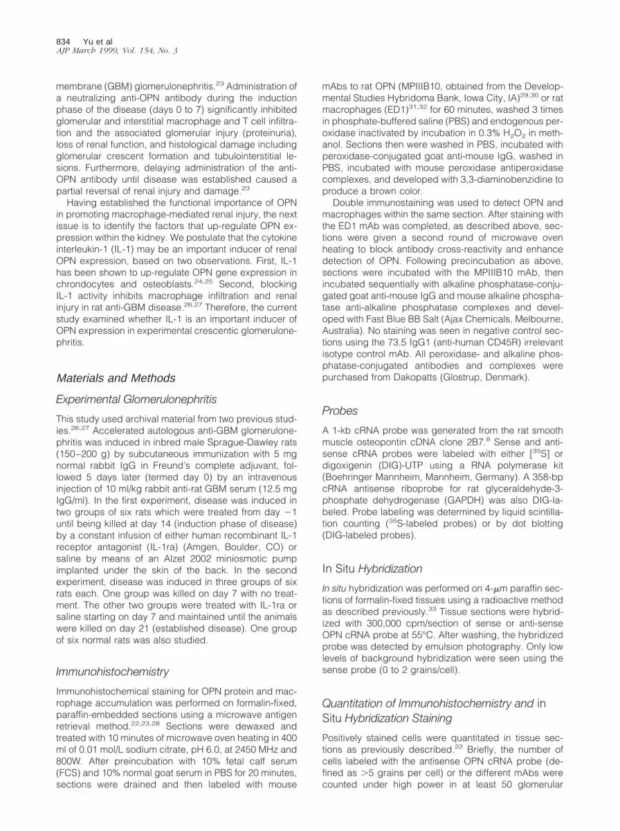
Figure 1. In situ hybridization showing OPN mRNA expression in rat anti-GBM disease. A: Normal rat kidney showing no OPN mRNA within the glomerular tuft,but OPN expression (black grains) is seen in some parietal epithelial cells and in the thick ascending limbs of Henle. B: Marked up-regulation of OPN mRNA withina glomerular crescent (*) and in many cortical tubules on day 14 of saline-treated anti-GBM disease (Experiment 1), which is substantially inhibited by IL-1ratreatment (C). D: Day 7 of untreated anti-GBM disease, clearly showing up-regulation of glomerular and tubular OPN mRNA. E: There is a further increase in renalOPN mRNA expression after saline treatment over days 7 to 21 (Experiment 2), with strong OPN mRNA expression seen within areas of severe tissue damagesuch as glomerular crescentic formation (*) and tubular atrophy and fibrosis. In contrast, IL-1ra treatment of established anti-GBM disease over days 7 to 21completely abrogated the increase in OPN expression within glomeruli and tubules (F). Original magnification, 3250.
836 Yu et alAJP March 1999, Vol. 154, No. 3
tablished by day 7 after anti-GBM serum administration
and was associated with significant up-regulation of glo-
merular and tubular OPN expression (Figures 1D, 2C,
and 2D). Saline treatment from days 7 to 21 saw a further
increase in glomerular and tubular OPN mRNA and pro-
tein expression (Figures 1E, 2C, 2D, and 3C) in associa-
tion with a further decline in creatinine clearance and a
progressive increase in proteinuria as previously de-
scribed.27 Double immunohistochemistry showed a tight
association between OPN expression and macrophage
accumulation in areas of severe tissue damage, such as
glomerular crescent formation and tubulointerstitial le-
sions (Figure 3C).
IL-1ra treatment of established anti-GBM disease from
days 7 to 21 caused a significant reduction in glomerular
and tubular OPN mRNA and protein expression (Figures
1F, 2C, 2D, and 3D). Indeed, the number of glomerular
OPN-positive cells was reduced to levels below that seen
on day 7 of disease, before the induction of IL-1ra treat-
ment (Figure 2C). This was associated with recovery of
normal renal function, a minor reduction in proteinuria,
and prevention of further histological damage.27
IL-1 Up-Regulates OPN Expression by Renal
Tubular Epithelial Cells in Vitro
To investigate whether IL-1 can act directly to up-regulate
OPN expression in the development of rat anti-GBM glo-
merulonephritis, we examined the effect of adding IL-1 to
the level of OPN mRNA expression in the normal rat
epithelial cell line NRK52E. This cell line was chosen
because tubular epithelial cells are the major site of OPN
production within the injured kidney. Northern blot anal-
ysis showed that the NRK52E cell line constitutively ex-
presses low levels of OPN mRNA (Figure 4). Within 6
hours of IL-1a stimulation, there was an increase in OPN
mRNA levels that peaked after 24 hours (threefold induc-
tion). The IL-1-induced up-regulation of OPN mRNA lev-
els was dose-dependent and was maintained for a 5-day
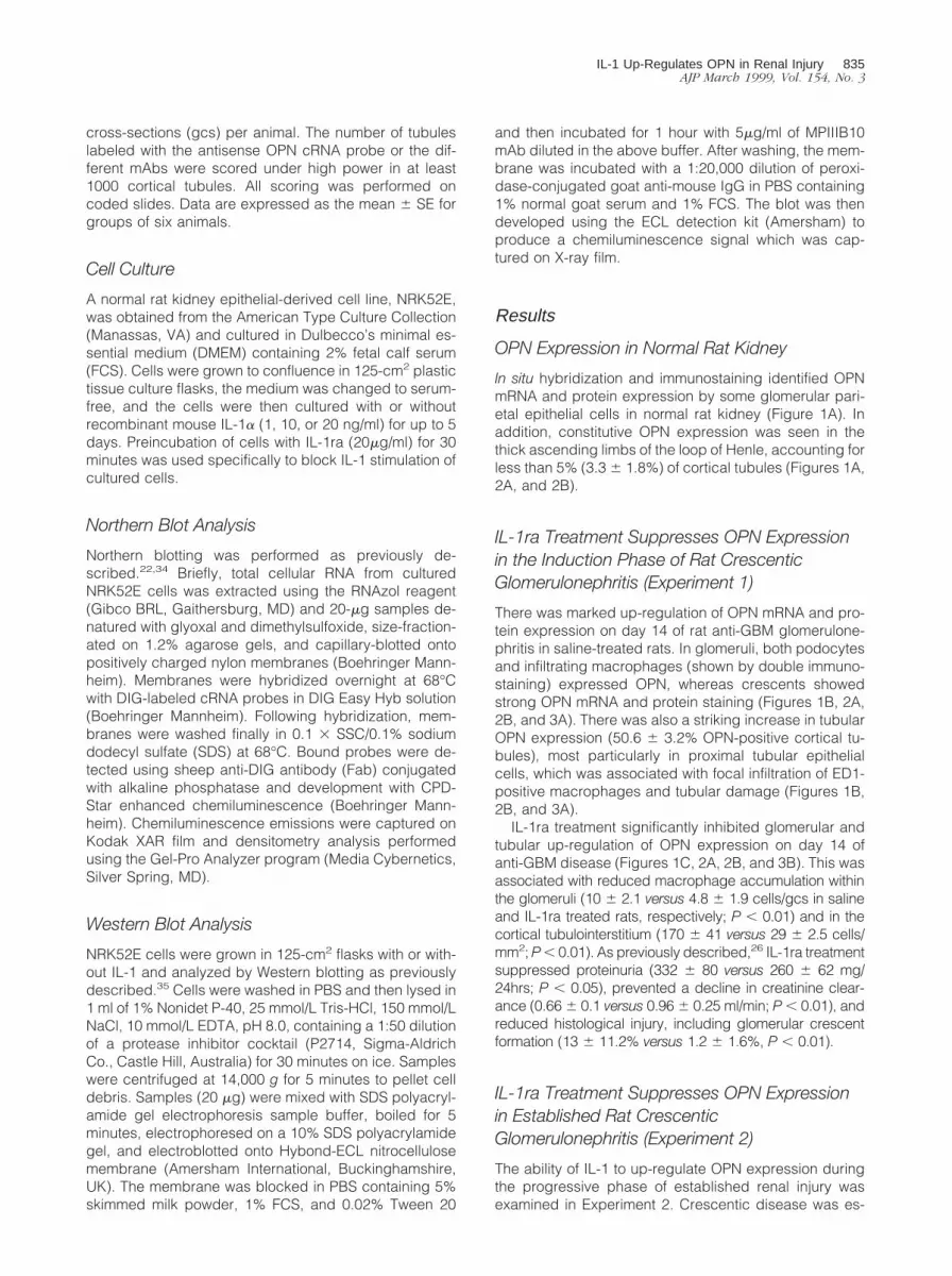
Figure 2. Quantitation of OPN mRNA and protein expression in rat anti-GBM disease. The number of glomerular cells and tubules expressing OPN mRNA orprotein was scored in stained tissue sections from saline-treated (closed bars) or IL-1ra-treated (hatched bars) anti-GBM disease or normal rats (open bars) . A-B:Experiment 1 with IL-1ra or saline treatment over days 0 to 14 of anti-GBM disease. C-D: Experiment 2 with IL-1ra or saline treatment over days 7 to 21 of anti-GBMdisease. One group of animals received no treatment before being killed on day 7 (dotted bars). The number of glomerular cells expressing OPN mRNA or protein(A and C) and the percent of cortical tubules expressing OPN mRNA or protein (B and D) were scored. Data are mean 6 SE for six animals. *, P , 0.05; **, P ,
0.01; ***, P , 0.001 by ANOVA.
IL-1 Up-Regulates OPN in Renal Injury 837AJP March 1999, Vol. 154, No. 3
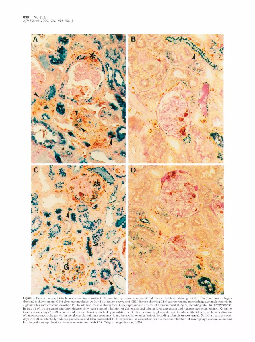
Figure 3. Double immunohistochemistry staining showing OPN protein expression in rat anti-GBM disease. Antibody staining of OPN (blue) and macrophages(brown) is shown in anti-GBM glomerulonephritis. A: Day 14 of saline treated anti-GBM disease showing OPN expression and macrophage accumulation withina glomerulus with crescent formation (*). In addition, there is strong focal OPN expression in an area of tubulointerstitial injury, including tubulitis (arrowheads).B: Day 14 of IL-1ra-treated anti-GBM disease showing a marked inhibition of glomerular and tubular OPN expression and macrophage accumulation. C: Salinetreatment over days 7 to 21 of anti-GBM disease showing marked up-regulation of OPN expression by glomerular and tubular epithelial cells, with colocalizationof numerous macrophages within the glomerular tuft, in a crescent (*), and in tubulointerstitial lesions, including tubulitis (arrowheads). D: IL-1ra treatment overdays 7 to 21 substantially reduces glomerular and tubulointerstitial OPN expression in association with a marked inhibition of macrophage accumulation andhistological damage. Sections were counterstained with PAS. Original magnification, 3250.
838 Yu et alAJP March 1999, Vol. 154, No. 3

culture period (Figures 4 and 5). Preincubation of cells
with an excess of the IL-1ra completely blocked IL-1
up-regulation of OPN mRNA expression, demonstrating
specificity of the effect. Western blotting showed that
NRK52E cells constitutively express OPN protein of ap-
proximately 68 kd, consistent with previous studies of
NRK52E cells and rat mesangial cells.36,37 IL-1 increased
OPN protein levels within NRK52E cells in a dose-depen-
dent fashion and this was completely inhibited by prein-
cubation of cells with the IL-1ra (Figure 6).
Discussion
This study has shown that IL-1 is an important factor in
up-regulating OPN gene expression in experimental
crescentic glomerulonephritis. The ability of IL-1ra treat-
ment to inhibit the induction of OPN expression and to
down-regulate OPN expression in established anti-GBM
disease clearly demonstrates a functional role for IL-1 in
the up-regulation of OPN in experimental crescentic glo-
merulonephritis. Indeed, the inhibition of renal macro-
phage infiltration seen with IL-1ra treatment is entirely
consistent with the ability of anti-OPN antibody treatment
to suppress macrophage infiltration and subsequent re-
nal injury in this model.23,26,27 Furthermore, the ability of
IL-1 to increase OPN mRNA levels rapidly in cultured
tubular epithelial cells demonstrates that IL-1 acts di-
rectly to up-regulate OPN expression in renal cells.
This is the first demonstration that IL-1 can up-regulate
OPN expression in kidney cells. The ability of IL-1 to
up-regulate OPN expression in renal epithelial cells is
consistent with previous studies reporting that IL-1 in-
creases OPN mRNA expression by cultured chrondo-
cytes and osteoblasts.24,25 However, it should be noted
that regulation of OPN gene expression varies greatly
between different cell types. For example, IL-1 failed to
increase OPN gene expression in cultured rat mesangial
Figure 4. Interleukin-1 up-regulates OPN mRNA expression by cultured ratkidney epithelial cells. A: NRK52E cells were stimulated with various con-centrations of murine IL-1a for 6 or 24 hours. Total cellular RNA wasanalyzed by Northern blotting using OPN and GAPDH probes. B: Thenormalized OPN to GAPDH mRNA ratio is shown.
Figure 5. Interleukin-1 causes sustained up-regulation of OPN mRNA bycultured rat kidney epithelial cells. A: NRK52E cells were stimulated withvarious concentrations of murine IL-1a for 3 or 5 days in the presence orabsence of 50mg/ml human IL-1ra. Total cellular RNA was analyzed byNorthern blotting using OPN and GAPDH probes. B: The normalized OPN toGAPDH mRNA ratio is shown. Data are mean 6 SE. *, P , 0.05; **, P , 0.01versus the unstimulated control by t-test with Welch’s correction.
Figure 6. IL-1 up-regulates OPN protein expression by cultured rat kidneyepithelial cells. NRK52E cells were cultured with various concentrations ofmurine IL-1a for 3 days in the presence or absence of 50mg/ml IL-1ra. Twentymicrograms of cell lysates were analyzed by Western blotting usingthe MPIIIB10 anti-OPN mAb. A predominant band of approximately 68 kdwas seen.
IL-1 Up-Regulates OPN in Renal Injury 839AJP March 1999, Vol. 154, No. 3

cells.37 In addition, vitamin D can induce or suppress
OPN mRNA levels in osteoblasts depending on their state
of differentiation.38 Furthermore, platelet-derived growth
factor induces OPN expression in vascular smooth mus-
cle cells and marrow stromal cells, but not in NRK52E
tubular epithelial cells.36,39,40
It was interesting that the increase in renal OPN mRNA
and protein production seen in rat crescentic glomerulo-
nephritis was very similar to the pattern of up-regulation
of renal IL-1b expression described previously in this
disease model,41 suggesting that up-regulation of OPN
expression within the injured kidney may be due to local
IL-1 production. In particular, strong IL-1b and OPN
mRNA and protein expression were seen in glomerular
and tubular epithelial cells, including glomerular cres-
cents and areas of tubulointerstitial injury. Whereas dou-
ble staining showed that some glomerular macrophages
expressed OPN, consistent with in vitro studies,6 podo-
cytes were the major cell type expressing OPN in the
glomerulus.22 Therefore, the suppression of glomerular
OPN expression by IL-1ra treatment is largely attributed
to inhibition of podocyte OPN production, with a lesser
effect due to the reduction in macrophage infiltration and
macrophage OPN expression.
It was intriguing that the rapid induction of OPN mRNA
expression seen following the addition of IL-1 to cultured
tubular epithelial cells was sustained for 5 days, in con-
trast with the 2 days required to detect TGF-b-induced
up-regulation of OPN mRNA in this cell line.36 Since IL-1
can induce TGF-b synthesis by tubular epithelial cells, it
may be the case that the sustained IL-1 up-regulation of
OPN expression by the cultured tubular epithelial cells
reflects a secondary effect of IL-1-induced production of
TGF-b. This possibility warrants further investigation.
IL-1 plays a crucial role in inducing renal macrophage
infiltration.42 Indeed, IL-1 induces a broad range of che-
mokines and leukocyte adhesion molecules such as in-
tercellular adhesion molecule-1 (ICAM-1) and monocyte
chemoattractant protein-1, which, acting in concert, fa-
cilitate the extravasation of blood leukocytes and their
subsequent focal accumulation within the kidney. Block-
ing the action of just one IL-1-inducible adhesion or che-
motactic molecule can inhibit leukocyte infiltration in ex-
perimental kidney disease,43,44 thereby demonstrating
the interdependence of these molecules. This is illus-
trated by the ability of anti-OPN antibody treatment to
suppress leukocyte infiltration and subsequent renal in-
jury in rat anti-GBM glomerulonephritis without affecting
up-regulation of ICAM-1 expression.23 This implies that
ICAM-1 is necessary for leukocyte-endothelial interac-
tions but that renal OPN expression is required for migra-
tion and localization of macrophages and T cells within
the kidney, resulting in local tissue damage.
In summary, this study provides in vivo and in vitro
evidence that IL-1 up-regulates OPN expression in ex-
perimental kidney disease and argues that inhibition of
OPN expression is one of the mechanisms by which
IL-1ra treatment suppresses macrophage-mediated re-
nal injury in experimental crescentic glomerulonephritis.
References
1. Butler WT: Structural and functional domains of osteopontin. Ann N Y
Acad Sci 1995, 760:6–11
2. Reinholt FP, Hultenby K, Oldberg A, Heinegard D: Osteopontin: a
possible anchor of osteoclasts to bone. Proc Natl Acad Sci USA 1990,
87:4473–4475
3. Weber GF, Ashkar S, Glimcher MJ, Cantor H: Receptor-ligand inter-
action between CD44 and osteopontin (Eta-1). Science 1996, 271:
509–512
4. Brown L F, Berse B: Van de Water L, Papadopoulos-Sergiou A,
Perruzzi CA, Manseau EJ, Dvorak HF, Senger DR: Expression and
distribution of osteopontin in human tissues: widespread association
with luminal epithelial surfaces. Mol Biol Cell 1992, 3:1169–1180
5. Lopez CA, Hoyer JR, Wilson PD, Waterhouse P, Denhardt DT: Heter-
ogeneity of osteopontin expression among nephrons in mouse kid-
neys and enhanced expression in sclerotic glomeruli. Lab Invest
1993, 69:355–363
6. Miyazaki Y, Setoguchi M, Yoshida S, Higuchi Y, Akizuki S, Yamamoto
S: The mouse osteopontin gene: expression in monocytic lineages
and complete nucleotide sequence. J Biol Chem 1990, 265:14432–
14438
7. Patarca R, Freeman GJ, Singh RP, Wei FY, Durfee T, Blattner F,
Regnier DC, Kozak CA, Mock BA, Morse III HC, Jerrells TR, Cantor H:
Structural and functional studies of the early T lymphocyte activation
1 (Eta-1) gene: definition of a novel T cell-dependent response asso-
ciated with genetic resistance to bacterial infection. J Exp Med 1989,
170:145–161
8. Giachelli C, Bae N, Lombardi D, Majesky M, Schwartz S: Molecular
cloning and characterization of 2B7, a rat mRNA which distinguishes
smooth muscle cell phenotypes in vitro and is identical to osteopontin
(secreted phosphoprotein I, 2aR). Biochem Biophys Res Commun
1991, 177:867–873
9. Senger DR, Perruzzi CA, Gracey CF, Papadopoulos A, Tenen DG:
Secreted phosphoproteins associated with neoplastic transformation:
close homology with plasma proteins cleaved during blood coagula-
tion. Cancer Res 1988, 48:5770–5774
10. Ross FP, Chappel J, Alvarez JI, Sander D, Butler WT, Farach-Carson
MC, Mintz KA, Robey PG, Teitelbaum SL, Cheresh DA: Interactions
between the bone matrix proteins osteopontin and bone sialoprotein
and the osteoclast integrin avb3 potentiate bone resorption. J Biol
Chem 1993, 268:9901–9907
11. Shiraga H, Min W, VanDusen WJ, Clayman MD, Miner D, Terrell CH,
Sherbotie JR, Foreman JW, Przysiecki C, Neilson EG, Hoyer JR:
Inhibition of calcium oxalate crystal growth in vitro by uropontin:
another member of the aspartic acid-rich protein superfamily. Proc
Natl Acad Sci USA 1992, 89:426–430
12. Singh RP, Patarca R, Schwartz J, Singh P, Cantor H: Definition of a
specific interaction between the early T lymphocyte activation 1
(Eta-1) protein and murine macrophages in vitro and its effect upon
macrophages in vivo. J Exp Med 1990, 171:1931–1942
13. Giachelli CM, Lombardi D, Johnson RJ, Murry CE, Almeida M: Evi-
dence for a role of osteopontin in macrophage infiltration in response
to pathological stimuli in vivo. Am J Pathol 1998, 152:353–358
14. Nikolic-Paterson DJ, Lan HY, Atkins RC: Macrophages in immune
renal injury. Immunologic Renal Diseases. Edited by EG Neilson, WG
Couser. Philadelphia, Lippincott-Raven, 1977, pp 575–592
15. Pichler R, Giachelli CM, Lombardi D, Pippin J, Gordon K, Alpers CE,
Schwartz SM, Johnson RJ: Tubulointerstitial disease in
glomerulonephritis: potential role of osteopontin (uropontin). Am J
Pathol 1994, 144:915–926
16. Giachelli CM, Pichler R, Lombardi D, Denhardt DT, Alpers CE,
Schwartz SM, Johnson RJ: Osteopontin expression in angiotensin
II-induced tubulointerstitial nephritis. Kidney Int 1994, 45:515–524
17. Pichler RH, Franceschini N, Young BA, Hugo C, Andoh T F, Burd-
mann EA, Shankland SJ, Alpers CE, Bennett WM, Couser WG, John-
son RJ: Pathogenesis of cyclosporine nephropathy: roles of angio-
tensin II and osteopontin. J Am Soc Nephrol 1995, 6:1186–1196
18. Diamond JR, Kees-Folts D, Ricardo SD, Pruznak A, Eufemio M: Early
and persistent up-regulated expression of renal cortical osteopontin
in experimental hydronephrosis. Am J Pathol 1995, 146:1455–1466
19. Eddy AA, Giachelli CM: Renal expression of genes that promote
interstitial inflammation and fibrosis in rats with protein-overload pro-
teinuria. Kidney Int 1995, 47:1546–1557
840 Yu et alAJP March 1999, Vol. 154, No. 3

20. Kleinman JG, Worcester EM, Beshensky AM, Sheridan AM, Bonven-
tre JV, Brown D: Upregulation of osteopontin expression by ischemia
in rat kidney. Ann N Y Acad Sci USA 1995, 760:321–323
21. Magil AB, Pichler R, Johnson RJ: Osteopontin in chronic puromycin
aminonucleoside nephrosis. J Am Soc Nephrol 1997, 8:1383–1390
22. Lan HY, Yu XQ, Yang N, Nikolic-Paterson DJ, Mu W, Pichler R,
Johnson RJ, Atkins RC: De novo glomerular osteopontin expression
in rat crescentic glomerulonephritis. Kidney Int 1998, 53:136–145
23. Yu XQ, Nikolic-Paterson DJ, Mu W, Giachelli CM, Atkins RC, Johnson
RJ, Lan HY: A functional role for osteopontin in experimental cres-
centic glomerulonephritis in the rat. Proc Assoc Am Physicians 1998,
110:50–64
24. Margerie D, Flechtenmacher J, Buttner FH, Karbowski A, Puhl W,
Schleyerbach R, Bartnik E: Complexity of IL-1 b induced gene ex-
pression pattern in human articular chondrocytes. Osteoarthritis Car-
tilage 1997, 5:129–138
25. Jin CH. Miyaura C, Ishimi Y, Hong MH, Sato T, Abe E, Suda T:
Interleukin 1 regulates the expression of osteopontin mRNA by os-
teoblasts. Mol Cell Endocrinol 1990, 74:221–228
26. Lan HY, Nikolic-Paterson DJ, Zarama M, Vannice JL, Atkins RC:
Suppression of experimental glomerulonephritis by the IL-1 receptor
antagonist. Kidney Int 1993, 43:479–485
27. Lan HY, Nikolic-Paterson DJ, Mu W, Vannice JL, Atkins RC: Interleu-
kin-1 receptor antagonist halts the progression of established rat
crescentic glomerulonephritis in the rat. Kidney Int 1995, 47:1303–
1309
28. Lan HY, Mu W, Nikolic-Paterson DJ, Atkins RC: A novel, simple,
reliable, and sensitive method of multiple immunoenzymic staining:
use of microwave oven heating to block antibody cross-reactivity and
retrieve antigens. J Histochem Cytochem 1995, 43:97–102
29. Liaw L, Almeida M, Downey W, Hart CE, Schwartz SM, Giachelli CM:
Osteopontin promotes vascular cell adhesion and spreading and is
chemotactic for smooth muscle cells in vitro. Circ Res 1994, 74:214–
224
30. Liaw L, Lombardi DM, Almeida MM, Schwartz SM, deBlois D, Gia-
chelli CM: Neutralizing antibodies directed against osteopontin inhibit
rat carotid neointimal thickening after endothelial denudation. Arte-
rioscler Thromb Vasc Biol 1997, 17:188–193
31. Dijkstra CD, Dopp EA, Joling P, Kraal G: The heterogeneity of mono-
nuclear phagocytes in lymphoid organs: distinct macrophage sub-
populations in the rat recognized by monoclonal antibodies ED1, ED2
and ED3. Immunology 1985, 54:589–599
32. Damoiseaux JG, Dopp EA, Calame W, Chao D, MacPherson GG,
Dijkstra CD: Rat macrophage lysosomal membrane antigen recog-
nized by monoclonal antibody ED1. Immunology 1994, 83:140–147
33. Wilcox JN, Smith KM, Schwartz SM, Gordon D: Localization of tissue
factor in the normal vessel wall and in the atherosclerotic plaque.
Proc Natl Acad Sci USA 1989, 86:2839–2843
34. Hattori, M, Nikolic-Paterson DJ, Lan HY, Kawaguchi H, Ito K, Atkins
RC: Up-regulation of ICAM-1 and VCAM-1 expression during macro-
phage recruitment in lipid induced glomerular injury in ExHC rats.
Nephrology 1995, 1:221–232
35. Nikolic-Paterson DJ, Jun Z, Tesch GH, Lan HY, Foti R, Atkins RC: De
novo CD44 expression by proliferating mesangial cells in anti-Thy-1
nephritis in the rat: J Am Soc Nephrol 1996, 7:1006–1014
36. Malyankar UM, Almeida M, Johnson RJ, Pichler RH, Giachelli CM:
Osteopontin regulation in cultured rat renal epithelial cells. Kidney Int
1997, 51:1766–1773
37. Nagasaki T, Ishimura E, Shioi A, Jono S, Inaba M, Nishizawa Y, Morii
H, Otani S: Osteopontin gene expression and protein synthesis in
cultured rat mesangial cells. Biochem Biophys Res Commun 1997,
233:81–85
38. Gerstenfeld LC, Zurakowski D, Schaffer JL, Nichols DP, Toma CD,
Broess M, Bruder SP, Caplan AI: Variable hormone responsiveness of
osteoblast populations isolated at different stages of embryogenesis
and its relationship to the osteogenic lineage. Endocrinology 1996,
137:3957–3968
39. Green RS, Lieb ME, Weintraub AS, Gacheru SN, Rosenfield CL, Shah
S, Kagan HM, Taubman MB: Identification of lysyl oxidase and other
platelet-derived growth factor-inducible genes in vascular smooth
muscle cells by differential screening. Lab Invest 1995 73:476–482
40. Tanaka H, Liang CT: Effect of platelet-derived growth factor on DNA
synthesis and gene expression in bone marrow stromal cells derived
from adult and old rats. J Cell Physiol 1995, 164:367–375
41. Tesch GH, Yang N, Yu H, Lan HY, Foti R, Chadban SJ, Atkins RC,
Nikolic-Paterson DJ: Intrinsic renal cells are the major source of
interleukin-1b synthesis in normal and diseased rat kidney. Nephrol
Dial Transplant 1997, 12:1109–1115
42. Nikolic-Paterson DJ, Lan HY, Atkins RC: IL-1 receptor antaogonism.
Semin Nephrol 1996, 16:583–590
43. Nishikawa, K, Guo YJ, Miyasaka M, Tamatani T, Collins AB, Sy MS,
McCluskey RT, Andres G: Antibodies to intercellular adhesion mole-
cule 1/lymphocyte function-associated antigen 1 prevent crescent
formation in rat autoimmune glomerulonephritis. J Exp Med 1993,
177:667–677
44. Lloyd CM, Minto AW, Dorf ME, Proudfoot A, Wells TN, Salant DJ,
Gutierrez-Ramos JC. RANTES, and monocyte chemoattractant pro-
tein-1 (MCP-1) play an important role in the inflammatory phase of
crescentic nephritis, but only MCP-1 is involved in crescent formation,
and interstitial fibrosis. J Exp Med 1997, 185:1371–1380
IL-1 Up-Regulates OPN in Renal Injury 841AJP March 1999, Vol. 154, No. 3


