Embed Size (px)
Citation preview

Identification of Cinnabarinic Acid as a NovelEndogenous Aryl Hydrocarbon Receptor Ligand ThatDrives IL-22 ProductionMargaret M. Lowe1., Jeff E. Mold1., Bittoo Kanwar1,2, Yong Huang3, Alexander Louie3,
Michael P. Pollastri4, Cuihua Wang4, Gautam Patel4, Diana G. Franks5, Jennifer Schlezinger6,
David H. Sherr6, Allen E. Silverstone7, Mark E. Hahn5", Joseph M. McCune1*"
1 Division of Experimental Medicine, Department of Medicine, University of California San Francisco, San Francisco, California, United States of America, 2 Division of
Gastroenterology, Department of Pediatrics, University of California San Francisco, San Francisco, California, United States of America, 3 Drug Studies Unit, Department of
Bioengineering and Therapeutic Sciences, University of California San Francisco, San Francisco, California, United States of America, 4 Department of Chemistry and
Chemical Biology, Northeastern University, Boston, Massachusetts, United States of America, 5 Biology Department, Woods Hole Oceanographic Institution, Woods Hole,
Massachusetts, United States of America, 6 Department of Environmental Health, School of Public Health, Boston University, Boston, Massachusetts, United States of
America, 7 Department of Microbiology and Immunology, State University of New York Upstate Medical University, Syracuse, New York, United States of America
Abstract
The aryl hydrocarbon receptor (AHR) binds to environmental toxicants including synthetic halogenated aromatichydrocarbons and is involved in a diverse array of biological processes. Recently, the AHR was shown to control hostimmunity by affecting the balance between inflammatory T cells that produce IL-17 (Th17) and IL-22 versus regulatory Tcells (Treg) involved in tolerance. While environmental AHR ligands can mediate this effect, endogenous ligands are likely tobe more relevant in host immune responses. We investigated downstream metabolites of tryptophan as potential AHRligands because (1) tryptophan metabolites have been implicated in regulating the balance between Th17 and Treg cellsand (2) many of the AHR ligands identified thus far are derivatives of tryptophan. We characterized the ability of tryptophanmetabolites to bind and activate the AHR and to increase IL-22 production in human T cells. We report that the tryptophanmetabolite, cinnabarinic acid (CA), is an AHR ligand that stimulates the differentiation of human and mouse T cellsproducing IL-22. We compare the IL-22-stimulating activity of CA to that of other tryptophan metabolites and definestimulation conditions that lead to CA production from immune cells. Our findings link tryptophan metabolism to AHRactivation and define a novel endogenous AHR agonist with potentially broad biological functions.
Citation: Lowe MM, Mold JE, Kanwar B, Huang Y, Louie A, et al. (2014) Identification of Cinnabarinic Acid as a Novel Endogenous Aryl Hydrocarbon ReceptorLigand That Drives IL-22 Production. PLoS ONE 9(2): e87877. doi:10.1371/journal.pone.0087877
Editor: Michael Platten, University Hospital of Heidelberg, Germany
Received November 22, 2013; Accepted December 30, 2013; Published February 3, 2014
Copyright: � 2014 Lowe et al. This is an open-access article distributed under the terms of the Creative Commons Attribution License, which permitsunrestricted use, distribution, and reproduction in any medium, provided the original author and source are credited.
Funding: This work was supported in part by National Institutes of Health (NIH) grants OD000329 and R01AI40312 (to JMM), R01ES006272 (to MEH),P42ES007381 (Superfund Research Program at Boston University to JS, DHS and MEH), R21CA134882 (to JS), NIH Training Grant T32 GM007175 (MML), and theHarvey V. Berneking Living Trust. BK is supported by Career Development Awards from the NIH/National Institute of Diabetes and Digestive and Kidney Diseases(DK083334) and the NASPGHAN Foundation. JEM is a recipient of the Human Frontiers Science Program Long-Term Fellowship (LT000231/2011-L). JMM is arecipient of the NIH Director’s Pioneer Award Program, part of the NIH Roadmap for Medical Research, through grant DPI OD00329. The funders had no role instudy design, data collection and analysis, decision to publish, or preparation of the manuscript.
Competing Interests: JEM has applied for a patent regarding Use of cinnabarinic acid as a modulator of immune responses in autoimmune disorders" (patentapplication number 13/579,893). This patent will not alter the authors’ ability to share data and materials as required by PLOS ONE policies.
* E-mail: [email protected]
. These authors contributed equally to this work.
" These authors also contributed equally to this work.
Introduction
The enzyme indole 2,3-dioxygenase (IDO) contributes to the
innate and adaptive immune response in settings such as
autoimmunity, microbial pathogenesis, and pregnancy [1–3].
IDO mediates the first, rate-limiting step in tryptophan metabo-
lism to kynurenine and is upregulated under certain inflammatory
conditions, most notably in response to interferons [4]. Its activity
may affect immunity through two non-exclusive mechanisms: (a)
creation of a local ‘‘amino acid starvation’’ response [4] and (b)
generation of downstream metabolites with specific immunomod-
ulatory or cytotoxic functions [5]. Tryptophan metabolites
generated by IDO can suppress T cell activation and modulate
T cell differentiation, although the mechanism of these effects
remains largely unknown [6,7]. Recent studies have shown that
tryptophan metabolites can alter the balance of Treg and Th17
cells, two related populations of CD4+ T cells with opposing
functions during immune responses [8].
Treg and Th17 cells share similar developmental pathways and
may arise from a common progenitor [9]. Differentiation into a
Treg or Th17 cell may be governed by the presence of
inflammatory cytokines [10], retinoic acid [11], and/or activation
of the aryl hydrocarbon receptor (AHR) [12,13]. The AHR is a
cytosolic transcription factor that is involved in many biological
processes, including development, cellular differentiation and
proliferation, xenobiotic metabolism, and the immune response
PLOS ONE | www.plosone.org 1 February 2014 | Volume 9 | Issue 2 | e87877

[12]. To date, the best-studied AHR ligands are halogenated and
polycyclic aromatic hydrocarbons such as 2,3,7,8-tetrachlorodi-
benzo-p-dioxin (TCDD) [14]. Only a few candidate endogenous
ligands have been identified, many of which are tryptophan
derivatives such as 2-(19H-indole-39-carbonyl)-thiazole-4-carbox-
ylic acid methyl ester (ITE), tryptamine, indirubin, 6-formylin-
dolo[3,2-b]carbazole (FICZ), and kynurenic acid [14,15]. More
recently it has also been reported that L-kynurenine, a proximal
downstream product of IDO metabolism, activates the AHR [16].
The highly conserved nature of the AHR signaling pathway has
prompted the search for additional natural ligands that can be
directly linked to physiological functions and established as true
endogenous ligands.
Although the AHR was initially proposed to affect Treg and
Th17 development, a Th17-associated cytokine, IL-22, is even
more specifically dependent upon AHR activation [13]. Ahr2/2
mice retain the ability to generate some Th17 cells but are
compromised in terms of IL-22 production [13,17]. Human T cell
differentiation also exhibits distinct requirements for the AHR:
activation of the AHR in stimulated human T cells was found to
inhibit Th17 differentiation and to promote the differentiation of
CD4+ T cells that produce IL-22 [18]. We therefore used
expansion of IL-22-producing cells as a screen for tryptophan
metabolites downstream of IDO that might act as AHR ligands.
We identified an endogenous tryptophan metabolite, cinnabarinic
acid (CA), as an AHR ligand that can increase IL-22 production in
both human and mouse CD4+ T cells.
Materials and Methods
Ethics StatementHeparinized blood from healthy adult volunteers was collected
under protocols approved for this study by the University of
California, San Francisco Committee on Human Research
(CHR), approval number 10-03852. Subjects gave written
informed consent in accordance with the Declaration of Helsinki.
Mouse experiments were performed in compliance with the
University of California, San Francisco Institutional Animal Care
and Use Committee guidelines with protocols approved for this
study (Protocol AN085678). Mice were housed under specific
pathogen free conditions at San Francisco General Hospital and
were fed standard chow.
Chemicals and miceCinnabarinic acid was synthesized utilizing a single-step
reaction sequence. A suspension of 1 g 2-amino-3-hydroxybenzoic
acid (1 g, 6.53 mmol) in 250 mL of methanol was stirred at 25uCfor 15 min. Diacetoxyiodobenzene (4.31 g, 13.39 mmol) was
added to the reaction mixture in portions. The color of the
reaction mixture gradually changed from pale yellow-pink to red.
Stirring was continued for 12 h at room temperature. The fine red
precipitate was collected by filtration and washed with methanol,
affording 0.58 g of cinnabarinic acid (29.6% yield). 1HNMR
(400 MHz, d6-DMSO) d 9.71 (s, 1H), 8.78 (s, 1H), 7.93 (d,
J = 8.0 Hz, 1H), 7.75 (d, J = 8.0 Hz, 1H), 7.59 (dd, overlapped,
1H), 6.59 (s, 1H). 13CNMR (100.4 MHz, d6-DMSO) d 178.1,
169.1, 166.3, 152.5, 150.5, 147.6, 142.4, 129.0, 128.8, 127.9,
126.2, 120.2, 104.9, 92.7. LCMS found 301.2, [M+H]+. The
purity of CA was 96.9% as measured by LC/MS with UV
detection; FICZ was not present as determined by single ion
monitoring of the MS spectrum. FICZ was synthesized as
described previously [19–21]. 4-fluoro-3-hydroxyanthranilic acid
(4-F-3-HAA) was synthesized by Drs. Bill Todd and Barry
Carpenter [22] and provided by Robert Schwarcz. 3-Hydroxyan-
thranilic acid (3-HAA) (.99.6% purity), quinolinic acid (QA),
picolinic acid (PA), 3-hydroxykynurenine (3-HKA), kynurenic acid
(KYA), L-kynurenine (L-KYN), laccase (from T. versicolor), and a-
naphthoflavone were purchased from Sigma. All chemicals were of
the highest purity commercially available—typically $98%. For
some experiments, laccase was heat killed by incubation at 95uCfor five minutes. The AHR antagonist, CH-223191 (2-methyl-2H-
pyrazole-3-carboxylic acid (2-methyl-4-o-tolylazo-phenyl)-amide)
[23], was purchased from CalBiochem. TCDD was purchased
from Ultra Scientific. 1-(1-propynyl)pyrene (1-PP) was the
generous gift of Cornelis Elferink (Univ. of Texas Medical Branch,
Galveston, TX). C57BL/6 mice were purchased from Jackson
Laboratory. Ahr2/2 mice on a B6 background were derived from
the line created by Schmidt et al [24].
In vitro human cell cultureFor assays involving total PBMCs, 36105 cells obtained by
ficoll-hypaque density gradient centrifugation were plated with
56104 irradiated allogeneic cells in a 96 well U-bottom plate in
200 mL RPMI with 10% FBS. Cells were stimulated with plate-
bound anti-CD3 (0.5 mg/mL in PBS, BD, SP34) and soluble anti-
CD28 (0.5 mg/mL, BD) in the presence of tryptophan metabolite
or DMSO control. The AHR antagonist CH-223191 (10 mM) was
used to block AHR in some experiments. Metabolites and
inhibitors were replenished on day 2 or 3.
For naıve CD4+ T cell sorting, human PBMCs from adult donors
or from de-identified cord blood (MD Anderson) were stained with
anti-CD3-Alexa700 (BD), anti-CD4-ECD (Invitrogen), anti-CD8-
PeCy5.5 (Invitrogen), anti-CD45RA-FITC (BD), anti-CD95-APC
(BD), anti-CD25-PE (BD), anti-CCR7-PeCy7 (BD), and anti-CD27-
APCCy7 (eBioscience). Naıve CD4+ T cells were sort-purified as
CD3+CD4+CD82 CD45RA+CCR7+CD952CD252CD27+. 16105
cells were plated with 5–106104 irradiated allogeneic stimulators in
200 mL XVIVO-20 serum free media (Lonza) in 96 well U-bottom
plates. Cells were incubated with CA or DMSO control under
polarizing conditions with plate-bound 0.5 mg/mL anti-CD3 (SP34)
and 0.5 mg/mL soluble anti-CD28, 10 ng/mL IL-21 (eBioscience),
IL-1b, IL-23, 10 mg/mL anti-IFNc, and 5 mg/mL anti-IL-4 and
anti-IL12 blocking antibodies (R&D). Cord blood samples were
instead stimulated with CD3/CD28 Dynabeads (Invitrogen) and
additionally incubated with TGF-b (R&D). Cytokines, blocking
antibodies, and metabolite were added again on days 2 and 4.
The optimal day for restimulation was determined to be day 5 or
6; cells were re-stimulated with phorbol 12-myristate 13-acetate
(PMA)/ionomycin in the presence of brefeldin A (BFA) and stained
for cytokine production Cells were stained extracellularly with anti-
CD4-ECD and anti-CD8-PeCy5.5, and Aqua viability dye
(Invitrogen), and fixed and permeabilized (BD Cytofix/Cytoperm).
Samples were stained intracellularly with anti-CD3-Alexa700 or
anti-CD3-APC-Cy7 (BD) to detect internalized CD3 on activated
cells, as well as anti-IL-17-AlexaFluor647 (eBioscience), anti-IL-22-
PE (R&D), and anti-IFNc-PB (eBioscience) antibodies. All events
were acquired on an LSRII (BD) and analyzed with FlowJo v7-9.3.2
(Treestar). CD4+ cells were gated as live, CD3+, CD4+CD82
lymphocytes. The descriptor ‘‘Total events collected’’ represents all
ungated events (including both live and dead events). At least 10,000
CD4+ events were analyzed per sample.
Human regulatory T cell differentiationNaıve CD4+ T cells were sorted as described in above. 200,000
naıve T cells were incubated in a 96 well U-bottom plate in
200 mL XVIVO-20 media. Cells were stimulated with CD3/
CD28 Dynabeads (Invitrogen) in the presence of CA or DMSO
vehicle control. Some cells were additionally treated with TGF-b
A Novel AHR Ligand Driving IL-22 Production
PLOS ONE | www.plosone.org 2 February 2014 | Volume 9 | Issue 2 | e87877

(RND). Cells were refed by removing 100 mL of media and adding
100 mL of 26 metabolite/TGF-b on days 2 and 4, and were
stained on day 6 for flow analysis. Cells were stained on days 6
with Aqua viability dye, anti-CD3-APCCy7, anti-CD4-ECD, anti-
CD8-PeCy5.5, and anti-CD25-PeCy7. Cells were fixed and
permeabilized with the FOXP3/Transcription Factor Staining
Buffer Set (eBioscience) and stained intranuclearly with anti-
FOXP3-APC or PE (eBioscience, clone PCH101).
Human Treg suppressor assayTregs were generated as described above, in the presence of CA
(10 mM) or DMSO control. On day 6 of culture, Dynabeads were
removed, viable cells were quantified following propidium iodide
staining on the Accuri C6 (BD). Autologous PBMCs were isolated
and labeled with 5(6)-carboxyfluorescein N-hydroxysuccinimidyl
ester (CFSE), and 100,000 PBMCs were plated in a 96-well U
bottom plate in XVIVO-20 with varying ratios of Tregs. Cells
were stimulated with plate-bound 0.5 mg/mL anti-CD3 (HIT3a)
and 0.5 mg/mL soluble anti-CD28 for four days, then stained with
anti-CD3, anti-CD4, and anti-CD8.
In vitro mouse cell cultureNaıve CD4+ T cells were sort-purified from mouse splenocytes
following depletion of non-CD4+ T cells with the MACS CD4+ T
cell Isolation Kit II (Miltenyi). Cells were stained with anti-CD3-
PB (BD), anti-CD4-PE (BD), anti-CD62L-PeCy7 (BD), anti-
CD25-APCCy7 (eBioscience), and anti-CD45Rb (BD), and sorted
as CD3+CD4+CD252CD62L+CD45Rbbright. Cells were stimulat-
ed with 4 mL anti-CD3/CD28 Dynalbeads/well (Invitrogen),
10 ng/mL IL-1b (Peprotech), 20 ng/mL IL-6 (Peprotech),
10 ng/mL TGF-b (R&D), and 10 mg/mL anti-IFNc and anti-
IL-12/23 (UCSF Hybridoma Core) in 200 mL XVIVO-20 media.
CA, KYA, L-KYN, FICZ, or laccase were added to individual
wells; an equivalent volume of vehicle (DMSO) was added to
control wells. 1-(1-propynyl)pyrene (1-PP) was used for CYP1A1
inhibition in some experiments. On day 2, cells were transferred to
a 48-well plate. Cytokines, blocking antibodies, and metabolites,
enzymes, or inhibitors were added at 26 concentration on days 2
and 4.
Cells were re-stimulated on day 5, which was determined to be
optimal, with PMA/ionomycin in the presence of BFA for 4–
6 hours and stained for cytokine production Cells were stained
extracellularly with anti-CD4-QDot 605 (Invitrogen) and anti-
CD8-PeCy5.5 or anti-CD8-PB (Caltag), Aqua viability dye, and
intracellularly with anti-CD3-PB or anti-CD3-PeCy5 (BD), anti-
IL-22-PE (eBioscience), anti-IL-17-APC (BD), and anti-IFNc-
APCCy7 (BD).
RNA was collected following restimulation with PMA and
ionomycin for 5 hours in RLT lysis buffer. RNA was purified with
Qiagen RNeasy columns. RNA input was standardized per
experiment by Nanodrop before cDNA transcription reactions
(Omniscript). Taqman primers for Il22 (Mm00444241_m1) and
Hprt1 (Mm00446968_m1) were used to quantify cDNA transcript
in reactions with Taqman Universal PCR master mix. Reactions
were run in a StepOnePlus analyzer.
Mouse regulatory T cell differentiationNaıve CD4+ T cells were sorted from mouse splenocytes as
described above. 200,000 cells were stimulated with 4 mL/well
CD3/CD28 Dynalbeads in 96 well U bottom plates in 200 mL
XVIVO-20 media in the presence of CA or DMSO vehicle
control. TGF-b was added to some wells. On days 2 and 4, 100 mL
of media were removed, and 100 mL of 26 cytokine/metabolite
were re-added. On day 5, Dynalbeads were removed magnetically
and cells were stained for flow analysis. Cells were stained with
anti-CD4-QDot 605, anti-CD8-PeCy5.5, Aqua viability dye, anti-
CD3-PB, and anti-CD25-APCCy7 (BD). Cells were fixed and
permeabilized with the FOXP3/Transcription Factor Staining
Buffer Set (eBioscience) and stained intranuclearly with anti-
FOXP3-PE (eBioscience).
AHR reporter assayMouse H1G1.1c3 cells (courtesy of Dr. M. Denison, UC Davis)
were prepared as described previously [25], except that 60,000
cells were added to each well of a 96-well, black-sided plate in
200 ml of selective medium and incubated at 37uC for 24 hours.
The medium was replaced with 100 ml of non-selective medium
prior to compound application. A TCDD standard curve plate
was prepared by applying vehicle (DMSO, 0.5%) or TCDD
(10214–1029 M), with each concentration applied to 8 wells. For
agonist experiments, vehicle or test compound were applied at a
single concentration in each column, excluding two untreated
columns. For antagonism experiments, compound application was
immediately followed by dosing with either vehicle or TCDD
(10210 M). The plates were incubated at 33uC for 24 hours and
EGFP fluorescence was read with a fluorometric plate reader
(Synergy2, Biotek Instruments). Excitation and emission wave-
lengths were 485 nm (20 nm bandwidth) and 530 nm (25 nm
bandwidth). Untreated well fluorescence was subtracted from
experimental well fluorescence. Data were averaged from eight
replicate wells. The gain was adjusted between experiments so that
wells exposed to 10210 M TCDD wells produced 15,000 RFUs
per well; subsequent plates in the experiment were analyzed with
that gain setting.
The specificity of the fluorescence measured in the H1G1 cells
treated with CA, HAA, and tryptamine was determined by
concurrently treating Hepa-1 cells (the parent line of the H1G1
cell line) with CA, HAA and tryptamine at the same concentra-
tions. Treatment, incubation, and analysis were carried out as
above. Fluorescence measured in Hepa-1 cells was subtracted
from the fluorescence measured in H1G1 cells treated with the
same concentration of CA, HAA or tryptamine to correct for
background fluorescence.
In vitro AHR competitive-binding assayHuman AHR protein was synthesized from an AHR expression
construct (pSporthAHR2, courtesy of Dr. C. Bradfield, U. of
Wisconsin, Madison, WI) [26] using a TnT-Quick Coupled
Reticulocyte Lysate System (Promega). Competition with 2,3,7,8-
tetrachloro[1,6-3H]dibenzo-p-dioxin ([3H]TCDD; 35 Ci/mmol;
Chemsyn Science Laboratories) for binding to human AHR was
measured by velocity sedimentation on sucrose gradients in a
vertical tube rotor [27]. Single TnT reactions (50 ml) were diluted
1:1 with MEEDMG buffer and incubated overnight at 4uC with
[3H]TCDD (2 nM) 6 competitors (dissolved in DMSO) before
application to sucrose gradients. Nonspecific binding was deter-
mined by using TnT reactions containing an empty vector.
CYP1A1 inductionZebrafish embryos [TL strain; 48 or 72 hours post fertilization
(hpf)] were exposed to CA (100 mM) or DMSO for 6 hours.
Following exposure, three replicate groups of 20 embryos from
each treatment group were frozen in liquid nitrogen. In one
experiment, 72-hpf embryos exposed for 6 hr to CA or DMSO
were subsequently held in clean water and sampled at 96 hpf.
Total RNA was isolated from frozen embryos using RNA STAT-
60 (Tel-Test B, Inc.). cDNA was synthesized from 2 mg of total
RNA using Omniscript reverse transcriptase (Qiagen). Real-time
A Novel AHR Ligand Driving IL-22 Production
PLOS ONE | www.plosone.org 3 February 2014 | Volume 9 | Issue 2 | e87877

RT-PCR for cyp1a and beta-actin was performed using the iQ
SYBR Green Supermix (Bio-Rad) in an iCycler iQ Real-Time
PCR Detection System (Bio-Rad) as described previously [28].
Fold induction of cyp1a by CA was calculated using the DDCT
method [29].
Human PBMCs were isolated by ficoll hypaque density gradient
centrifugation and plated in 48-well plates at a density between
0.5–26106 cells per well in 1 mL of RPMI. Cells were stimulated
with 1 mg/mL PHA in the presence of tryptophan metabolite or
DMSO for 8–20 hours and harvested in RLT lysis buffer. RNA
was purified with Qiagen RNeasy columns. RNA input was
standardized per experiment by Nanodrop before cDNA tran-
scription reactions (Omniscript). Taqman primers for CYP1A1
(Hs00153120_m1) and HPRT1 (Hs99999909_m1) were used to
quantify cDNA transcript in reactions with Taqman Universal
PCR master mix. Reactions were run in a StepOnePlus analyzer.
Mouse lymphocytes were isolated from brachial, axillary, and
inguinal lymph nodes, and were then plated in 96 well plates at a
density of 16106 cells/well in 200 mL of RPMI, cultured for
4 hours, and lysed in RLT lysis buffer. RNA isolation and RT-
PCR was handled as with human cells, except with Taqman
mouse Cyp1a1 (Mm00487217_m1) and Hprt1 (Mm00446968_m1)
primers.
CYP1A1 inhibition1.8 pmol of human CYP1A1 + P450 Reductase Supersomes
(BD) were used per reaction in the luminescent-based P450-Glo
CYP1A1 Assay (Promega). Briefly, CYP1A1 Supersomes, lucifer-
in-CEE substrate, and test compound were equilibrated in white
opaque 96-well plates (Pierce) for 10 minutes at 37uC per kit
protocol. NADPH Regenerating Solution (Promega) was added,
and reactions were terminated after 15 minutes at 37uC by
addition of luciferase detection reagent. Luminescence was read
with the SpectraMax M2 microplate reader using SoftMax Pro
software (Molecular Devices) and averaged over 6–8 reads per
well.
CA detectionPBMCs were plated at a concentration of 2.5–56106 cells/well
in 200 mL of RPMI. Cells were left unstimulated or incubated with
50 mg/mL LPS, 10 ng/mL PMA, 100 ng/mL IFNc, or 1–5 mg/
mL concanavalin A (conA) for 16 hours. Supernatants were frozen
for detection of CA by LC/MS/MS.
LC/MS/MSCA was measured by liquid chromatography-tandem mass
spectrometry (LC/MS/MS). Samples (20 ml) were mixed with
100 ml of internal standard, piroxicam (100 ng/ml) in acetonitrile,
vortexed for 1 min, and centrifuged at 3000 rpm for 10 min. The
supernatant was transferred to an autosampler vial and 8 ml was
injected to the LC/MS/MS system. The standard curve was
generated by serial diluting CA standard solution in water. The
mass detector was an API 5000 triple quadrapole (Applied
Biosystems), equipped with a Turbo Ion Spray source. The system
was set in positive ionization mode. The ion spray voltage was
5500 V and the source temperature was 600uC. The values for
CAD, CUR, GS1, and GS2 were 6, 15, 55, and 75 respectively.
The multiple reaction monitor was set at 301.1 – 265.0 m/z for
CA and 332.0 – 94.9 m/z for piroxicam. A Shimadzu system was
used for the HPLC, consisting of a pump, solvent degasser,
autosampler and column oven, which was set to 30uC. The mobile
phase, consisting of 40% acetonitrile, 0.1% trifluoroacetic acid
containing 5 mM ammonium acetate, was pumped through a
Synergi Polar RP (4.6675 mm, 4 mm particle size) column with a
flow rate of 1.0 ml/min. Data were acquired and processed by
Analyst 1.5.1 software.
The limit of quantification (LOQ) and limit of detection (LOD)
for CA were 7.81 ng/ml and 3 ng/ml, respectively. The
tryptophan, L-KYN, and 3-HAA levels were measured in API-
5000 with a similar method as reported before [30]. CA values
below LOQ were treated as K LOQ for statistical analysis [31].
Statistical analysisStatistical tests used to analyze data are denoted individually
within figure legends (GraphPad Prism v.4.0c). P values ,0.05
were considered statistically significant.
Results
3-Hydroxyanthranilic acid (3-HAA) increases thefrequency of IL-22-expressing CD4+ T cells in vitro
Human PBMCs were stimulated with antibodies against CD3
and CD28 in vitro in the presence of different tryptophan
metabolites, including 3-hydroxykynurenine (3-HKA), 3-hydro-
xyanthranilic acid (3-HAA), picolinic acid (PA), and quinolinic
acid (QA). 3-HKA and 3-HAA, but not the downstream
metabolites PA or QA, were able to promote IL-22 production
in stimulated CD4+ T cells (Fig. 1A). Though donors differed in
the frequency of IL-22+ cells detected following 3-HAA exposure,
3-HAA was able to promote a 2-fold or greater expansion of IL-
22-producing cells in each (Fig. 1B). Statistically significant
expansion of these cells was seen beginning at 25 mM 3-HAA
(Fig. 1B, aggregate donors). Additional analysis revealed that this
increase also occurred within total events collected and was
therefore not likely an artifact of preferential cell death (Fig. S1).
To determine whether the expansion of IL-22-producing cells
within this population was AHR-dependent, we stimulated human
PBMCs in the presence or absence of a potent AHR antagonist
(CH-223191) and observed that CH-223191 abolished the
increase in IL-22 production caused by 3-HAA (Fig. 1, C and
D). Within PBMC cultures stimulated in the presence of 3-HAA,
upregulation of IL-22 was only seen in CD4+ T cells (Fig. 2A) and
not in CD8+ T cells (Fig. 2B). These IL22+ CD4+ T cells
frequently co-expressed IFNc but not IL-17A; interestingly, IL-
22+IL172 CD4+ T cells identified in humans also frequently co-
express IFNc (Fig. 2A) [18]. 3-HAA suppressed IL-17 production
within CD4+ T cells, as has been previously reported [30].
3-HKA and 3-HAA are not AHR ligands but may beprecursors to an AHR ligand
The above studies identified 3-HAA and, to a lesser extent, 3-
HKA as potential ligands of the AHR. The ability of these
metabolites as well as that of PA to bind and activate the AHR was
assayed using two techniques. First, we determined whether each
compound could bind to AHR, using a well-established assay that
measures the ability of compounds to displace [3H]TCDD from
full-length human AHR protein expressed in vitro. Modest binding
of 3-HAA and PA was evident, but only at very high
concentrations (1000 mM, Fig. 3A). We also measured AHR
activation and antagonism within a reporter cell line expressing
AHR responsive elements upstream of a GFP reporter. While a
slight increase in AHR activity measured by fluorescence was
found using 3-HAA as an agonist (Fig. 3B), this failed to reach
significance, and none of the other metabolites downstream of 3-
HKA tested were found to have activity as either agonists or
antagonists (Fig. 3, C and D). These results suggested that 3-HAA
might be an upstream precursor of an endogenous AHR ligand.
A Novel AHR Ligand Driving IL-22 Production
PLOS ONE | www.plosone.org 4 February 2014 | Volume 9 | Issue 2 | e87877

Figure 1. 3-HKA and 3-HAA promote IL-22 expression in stimulated human CD4+ T cells. (A) Flow cytometric analysis of CD4+ T cellsfollowing stimulation of human PBMCs in the presence of 100 mM 3-HKA, 3-HAA, PA, or QA for six days. Data represent at least three independentexperiments. (B) Flow cytometric analysis of CD4+ T cells from individual and aggregate donors following stimulation of PBMCs in the presence ofincreasing concentrations of 3-HAA (mM) for six days. Individual donor data are pooled from at least three independent experiments. Error barsindicate SD. Fold change in IL-22 expression versus vehicle control is statistically different from 1 (Wilcoxon signed rank test; *, p = 0.0312; **,p = 0.0078; N = 8 donors). (C) Flow cytometric analysis of CD4+ T cells following stimulation of human PBMCs in the presence of 3-HAA +/2 the AHRantagonist, CH-223191. Data are representative of at least three independent experiments. (D) Comparison of IL-22 production in CD4+ T cellsfollowing stimulation of human PBMCs in the presence of DMSO, 3-HKA (50 mM), or 3-HAA (50 mM), with or without an AHR antagonist, N = 6. Pvalues were calculated by Mann-Whitney. Error bars indicate SD.doi:10.1371/journal.pone.0087877.g001
Figure 2. IL-22+ upregulation occurs in CD4+8- T cells that are IL-17 negative but express IFNc. (A) Cytokine production for liveCD3+CD4+CD82 T cells from human PBMC cultures that were stimulated with anti-CD3 and anti-CD28 antibodies and allogeneic APCs for six dayswith DMSO or 50 mM 3-HAA. Panel (right) depicts fold-changes in 9 donors relative to DMSO. *, p,0.05 indicates data analyzed by Wilcoxon signedrank test is statistically different than 1. (B) Cytokine production for live CD3+CD8+CD42 T cells in PBMC cultures, stimulated as above with DMSO or 3-HAA (50 mM). Data are representative of three experiments.doi:10.1371/journal.pone.0087877.g002
A Novel AHR Ligand Driving IL-22 Production
PLOS ONE | www.plosone.org 5 February 2014 | Volume 9 | Issue 2 | e87877

Inhibition of 3-HAA metabolism increases IL-22production
Because neither PA nor QA, the primary metabolites down-
stream of 3-HAA, was found to have any impact on IL-22
production, we thought it unlikely that either was an AHR ligand.
The enzyme upstream of PA and QA, 3-hydroxyanthranilate 3,4-
dioxygenase (HAAO), is also expressed by monocytes and
macrophages, the same cell populations that express IDO under
inflammatory conditions [32]. To determine whether downstream
intermediates of 3-HAA generated through HAAO were acting as
AHR ligands, the specific inhibitor, 4-F-3-HAA, was used to block
HAAO activity (Fig. 4, A and B). Contrary to this hypothesis,
HAAO inhibition within stimulated PBMC cultures did not block
the ability of 3-HAA to upregulate IL-22 production; at higher
concentrations of 3-HAA, HAAO inhibition increased IL-22
production (Fig. 4B). This suggested that an AHR ligand might
arise from an alternative pathway of 3-HAA metabolism, which
would be favored during HAAO inhibition and would result in
increased IL-22 production.
Identification of CA as an AHR ligand downstream of3-HAA
3-HAA is susceptible to oxidation, resulting in the formation of
CA through 3-HAA dimerization, both in solution [33] and in cell
culture (Fig. 4C) [19]. As a tricyclic aromatic compound, the
structural features of CA resemble those of some AHR ligands.
Additionally, CA has potent effects on thymocyte maturation [19],
similar to those observed in cultured thymocytes treated with the
AHR agonist, TCDD [34]. To test the hypothesis that it is an
AHR ligand, CA was synthesized and tested for its ability to bind
to and activate the AHR in vitro. CA induced GFP in the AHR
reporter cell line (Fig. 4D) and the response to CA was inhibited by
the AHR antagonist, CH-223191 (Fig. 4E). CA also displaced
[3H]TCDD from in vitro-expressed human AHR and was effective
Figure 3. 3-HAA fails to bind or activate the AHR. (A) Displacement of [3H]TCDD from the human AHR in the presence of 3-HAA or PA. Data for100 mM concentrations were from 2–4 experiments; data for 1000 mM concentrations were from one or two experiments. (B) Fluorescence of an AHR-responsive reporter construct measured after incubation of a stably transfected murine hepatoma cell line in the presence of increasingconcentrations of 3-HAA. TCDD (5*10211 M) is a positive control. Error bars are SD. N = 6. (C) Fluorescence of the AHR-reporter construct followingincubation of transfected cells with 3-HKA, PA, or QA. Tryptamine and FICZ are positive controls. (D) Inhibition of TCDD (10210-M)-induced activationof the AHR reporter construct. CH-223191 is a positive control. *, p,0.05 (ANOVA, Scheffe’s). Error bars are SD.doi:10.1371/journal.pone.0087877.g003
A Novel AHR Ligand Driving IL-22 Production
PLOS ONE | www.plosone.org 6 February 2014 | Volume 9 | Issue 2 | e87877

at much lower concentrations than those required for 3-HAA and
3-HKA (Fig. 4F).
CA upregulates IL-22 within CD4+ T cells in an AHR-dependent manner
Treatment of adult human PBMCs with CA resulted in
upregulation of IL-22, but not IL-17, within human CD4+ T cells
in a dose-dependent manner (Fig. 5A, S2). To determine whether
CA was acting on T cells directly, adult human and mouse naıve
CD4+ T cells were sort-purified and incubated with tryptophan
metabolites under polarizing conditions (see Methods for details).
In the presence of 25 mM CA, human naıve CD4+ T cells
upregulated the production of IL-22 and IFN-c to a greater extent
than those incubated with an equivalent concentration of 3-HAA
(Fig. 5B, S3). Given the role of the AHR in regulating Treg
differentiation [12], we additionally tested whether CA was able to
affect the differentiation of Tregs as measured by expression of
FOXP3 and functional activity. An expansion of FOXP3+ cells
was seen in human naıve CD4+ T cells stimulated in the presence
of CA (Fig. 5C, S5). However, the T cells exposed to CA were not
more suppressive than DMSO control cells, despite the greater
abundance of FOXP3+ cells (Fig. 5D).
CA treatment of mouse naıve CD4+ T cells also resulted in
significant expansion of IL-22+ cells and a trend towards increased
Figure 4. CA is an endogenous AHR agonist generated by oxidative dimerization of 3-HAA. (A) Metabolic pathways downstream of 3-HAA catalyzed by the enzyme, 3-hydroxyanthranilate 3,4-dioxygenase (HAAO), including intermediates upstream of PA and QA. (B) Flow cytometricanalysis of CD4+ T cells in human PBMCs stimulated in the presence of varying concentrations of 3-HAA (50 mM or 100 mM) with or without the HAAOinhibitor, 4-F-3-HAA (50 mM). Data are representative of three experiments. (C) An alternative pathway of 3-HAA metabolism can generate CA eitherby non-enzymatic (oxidation) or enzymatic (such as laccase or ceruloplasmin) processes. (D) Fluorescence [measured in relative fluorescence units(RFUs)] of the AHR-responsive reporter construct in cells incubated with varying concentrations of 3-HAA or CA. Tryptamine was a positive control. *,p,0.05 (ANOVA, Scheffe’s). Error bars represent SD. (E) Fluorescence of an AHR-responsive reporter construct measured following incubation ofH1G1.1c3 cells with DMSO control, CA (200 mM), 3-HAA (200 mM), or the positive control FICZ (100 nM), with or without the AHR antagonist CH-223191 (50 mM). *, p,0.01. **, p,0.001. ***, p,0.0001. (2-way ANOVA, Bonferroni’s post-test). Error bars are SD. (F) [3H]TCDD displacement from invitro translated human AHR protein by incubation with varying concentrations of CA, 3-HAA, or 3-HKA. Data are representative of four (CA and 3-HAA) or two (3-HKA) independent experiments.doi:10.1371/journal.pone.0087877.g004
A Novel AHR Ligand Driving IL-22 Production
PLOS ONE | www.plosone.org 7 February 2014 | Volume 9 | Issue 2 | e87877

IL-17+ cells (Fig. 6A, 6B, S5). However, when sort-purified naıve
CD4+ T cells isolated from Ahr2/2 mice were treated with CA
under polarizing conditions, no expansion of IL-22 producing cells
occurred (Fig. 6A), demonstrating that this effect was AHR-
dependent. In addition, an AHR antagonist inhibited CA-driven
IL-22 production in CD4+ T cells from C57BL/6 (Ahr+/+) mice
(Fig. 6C), further supporting the conclusion that CA acts via AHR
to increase production of IL-22. Unlike in human cells, incubation
of mouse naıve cells with CA suppressed generation of FOXP3+
cells (Fig. 6D, S6). Nevertheless, the lack of suppressive activity in
human cells treated with CA indicates that this phenotypic
difference between species may not result in a change in function.
In contrast, the expansion of IFN-c producing cells seen in human
cells was not observed within mouse naıve cell cultures (Fig. 6E).
Induction of CYP1A1 and IL-22 and comparison of CA toother tryptophan metabolites
CA was additionally tested for its ability to induce the well-
known AHR responsive gene CYP1A1 in vivo and in vitro. In
zebrafish embryos, a model vertebrate in vivo system, Cyp1a was
strongly induced after 6 hours of exposure to CA (Fig. 7A, upper
panel); this effect was lost 18 hours after washout of CA (Fig. 7A,
lower panel). The degree of induction (25- to 50- fold) was
comparable to that produced by FICZ under similar experimental
conditions (,30-fold) [35]. CA also caused statistically significant
induction of Cyp1a1 in mouse lymphocytes in vitro (Fig. 7B). No
Cyp1a1 induction was detected in lymphocytes derived from Ahr2/
2 mice (Fig. 7B), demonstrating that CA was acting through the
AHR. Human PBMCs also exhibited a statistically significant,
although modest, induction of CYP1A1 (Fig. 7C).
Figure 5. CA increases the differentiation of IL-22+ human CD4+ T cells in vitro. (A) Flow cytometric analysis of CD4+ T cells from humanPBMCs stimulated in the presence of DMSO or increasing doses of CA (left). Fold change in IL-22 production in CD4+ T cells from human PBMCs frommultiple donors stimulated in the presence of CA versus DMSO control (right panel). Data were analyzed by Wilcoxon signed rank test for significantdeviation from a theoretical median of 1.000. *p,0.05. (B) Flow cytometric analysis of sorted naıve human CD4+ T cells stimulated under polarizingconditions (with IL-21, IL-1b, IL-23, anti-IFNc, anti-IL-4 and anti-IL12) with DMSO, 3-HAA (25 mM), or CA (25 mM). Data on IL-22, IFNc, and IL-17production from three independent experiments are shown in the panel on the right. Error bars are SD. (C) Flow cytometric analysis of FOXP3staining following stimulation of sorted naıve human CD4+ T cells in the presence of DMSO or CA without addition of TGF-b. Quantification of%FOXP3+CD25+ T cells of CD4+ cells from six donors in seven independent experiments (right panel) with increasing concentrations of TGF-b. Datawere analyzed by two-way ANOVA and Holm-Sidak multiple comparisons test. *, p,0.05. **, p,0.01. Error bars are SD. (D) Flow cytometric analysis ofCFSE-labeled responder CD8+ T cells incubated with autologous Tregs generated in the presence of CA or DMSO control. Data are representative ofthree independent experiments.doi:10.1371/journal.pone.0087877.g005
A Novel AHR Ligand Driving IL-22 Production
PLOS ONE | www.plosone.org 8 February 2014 | Volume 9 | Issue 2 | e87877

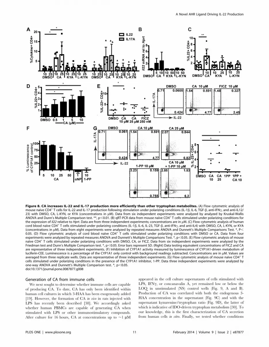
CA was then compared to other tryptophan metabolites,
kynurenic acid (KYA) and L-kynurenine (L-KYN), which have
recently been identified as AHR agonists [15,16]. In both mouse
(Fig. 7D) and human (Fig. 7E) lymphocytes, CA induced Cyp1a1
but was less effective than these other tryptophan metabolites.
Surprisingly, however, CA was much more effective at increasing
IL-22 production in these cells; neither KYA nor L-KYN was able
to drive IL-22 or IL-17 protein production or increase Il22 mRNA
within mouse naıve T cells at concentrations effective at inducing
Cyp1a1 (Fig. 8A and B). Likewise, KYA and L-KYN did not
increase IL-22 as effectively as CA in human naıve T cells (Fig. 8C,
S7). To test the minimal effective dose for IL-22 induction, we
differentiated human naıve T cells in the presence of decreasing
doses of CA. Concentrations as low as 1 mM were found to
significantly increase IL-22 production (Fig. 8D, S8).
Recently, it has been reported that weak AHR agonists may be
capable of inducing AHR-responsive genes indirectly by inhibiting
the CYP1A1-mediated metabolism of FICZ, a tryptophan
photoproduct capable of activating the AHR [36]. To test whether
CA was indirectly increasing IL-22 production by inhibiting FICZ
metabolism, CA was first directly compared to FICZ in mouse
naıve cell cultures (Fig. 8E). Although FICZ did induce some IL-
22 production, IL-22 production was not greater than that caused
by CA, even when FICZ was titrated to 10 mM (Fig. 8E, right
panel). Next, the ability of CA to inhibit CYP1A1 activity, which
would block FICZ metabolism, was tested with a luciferin-based
reporter assay. CA was incubated with CYP1A1-loaded micro-
Figure 6. CA increases the differentiation of IL-22+ mouse CD4+ T cells in vitro. (A) Flow cytometric analysis of IL-17 and IL-22 production insorted naıve mouse CD4+ T cells from C57BL/6 (Ahr+/+) or Ahr2/2 mice stimulated under polarizing conditions (with IL-1b, IL-6, TGF-b, anti-IFNc, andanti-IL12/23) in the presence of DMSO or CA (35 mM). (B) Flow cytometric data for IL-22 (top panel) and IL-17 (bottom panel) production from sixindependent experiments in C57BL/6 mice were analyzed by Kruskal-Wallis ANOVA and Dunn’s Multiple Comparison test. *, p,0.05. **, p,0.01.DMSO controls for the 25 and 35 mM CA experiments are shown separately. (C) Flow cytometric analysis of IL-17 and IL-22 production in C57BL/6mouse naıve CD4+ T cells stimulated under polarizing conditions in the presence of DMSO or CA, with or without AHR antagonist, CH-223191(10 mM). Data are representative of three independent experiments. (D) Flow cytometric analysis of FOXP3 expression in sorted naıve wild-typemouse CD4+ T cells stimulated with increasing concentrations of TGFb. Quantification of %FOXP3+CD25+ T cells of CD4+ cells from four independentexperiments is shown in the panel on the right. Error bars are SD. (E) Flow cytometric analysis of IFN-c production in sorted naıve wild-type mouseCD4+ T cells stimulated under polarizing conditions (as in panel A) in the presence of DMSO or CA. Data are from seven independent experiments(right) were analyzed by Friedman ANOVA; no statistically significant differences were found.doi:10.1371/journal.pone.0087877.g006
A Novel AHR Ligand Driving IL-22 Production
PLOS ONE | www.plosone.org 9 February 2014 | Volume 9 | Issue 2 | e87877

somes and a CYP1A1 substrate. Dose-dependent inhibition of
CYP1A1 activity was seen with CA; however, this inhibition was
much less than that caused by known CYP1A1 inhibitors a-
napthoflavone and 1-(1-propynyl)pyrene (1-PP) (Fig. 8F). 1-PP in
particular has been shown to activate the AHR indirectly through
inhibition of CYP1A1-dependent metabolism of an endogenous
AHR ligand [37]. Thus, 1-PP was tested in mouse naıve cell
cultures to determine whether CYP1A1 inhibition could induce
IL-22 in this system. Importantly, concentrations of 1-PP capable
of completely inhibiting CYP1A1 were unable to induce IL-22,
providing evidence that induction of IL-22 by CA is not the result
of CYP1A1 inhibition (Fig. 8G). CA in the presence of 1-PP
retained its ability to induce IL-22, showing that 1-PP does not
affect the ability of the cells to respond to CA. Therefore, it seems
unlikely that CA is exerting its effects through altering FICZ
metabolism.
Figure 7. CA induces expression of AHR-responsive gene Cyp1a1, but with reduced efficacy versus other tryptophan metabolites.(A) Induction of cyp1a in vivo as measured by qRT-PCR. Zebrafish embryos at 48 or 72 hours post fertilization (hpf) were exposed to CA (100 mM) for6 hours, and either sampled immediately (54 or 78 hpf; top panel) or placed in clean water and sampled at 96 hpf (24 hours after beginning ofexposure; bottom panel). Cyp1a mRNA was normalized to beta-actin and to the average DMSO value. (DMSO values for 78 and 96-hpf embryos weresimilar). Values represent fold-change in CA-treated versus DMSO control embryos; each panel represents an experiment sampling three replicategroups of twenty embryos per group. (B) Induction of Cyp1a1 in wild-type (Ahr+/+) or Ahr2/2 mouse total lymphocytes incubated with CA (50 mM) inRPMI for 4 hours. Cyp1a1 was measured by qRT-PCR and normalized to Hprt. Error bars represent SD. P values were calculated with the Mann-Whitney test from lymphocyte cultures from three individual mice. (C) Induction of CYP1A1 PHA-stimulated human total PBMCs after 12–20 hours ofincubation with CA (50 mM) in RPMI. CYP1A1 was measured by qRT-PCR and normalized to HPRT. Data shown are pooled experiments from sixindividual donors. Error bars are SD. P values were calculated with the Mann-Whitney test. (D, E) Induction of mouse Cyp1a1 (D) and human CYP1A1(E) measured by qRT-PCR relative to Hprt or HPRT. Mouse lymphocytes (D) and PHA-stimulated human PBMCs (E) were incubated with CA, KYA, L-KYN. Values are pooled from at least three independent experiments and represent fold-change versus averaged DMSO control. Metaboliteconcentrations are in mM. Error bars represent SD.doi:10.1371/journal.pone.0087877.g007
A Novel AHR Ligand Driving IL-22 Production
PLOS ONE | www.plosone.org 10 February 2014 | Volume 9 | Issue 2 | e87877
Generation of CA from immune cellsWe next sought to determine whether immune cells are capable
of producing CA. To date, CA has only been identified within
human cell cultures in which 3-HAA has been exogenously added
[19]. However, the formation of CA in vivo in rats injected with
LPS has recently been described [38]. We accordingly asked
whether human PBMCs are capable of generating CA when
stimulated with LPS or other immunostimulatory compounds.
After culture for 16 hours, CA at concentrations up to ,1 mM
appeared in the cell culture supernatants of cells stimulated with
LPS, IFNc, or concanavalin A, yet remained low or below the
LOQ in unstimulated (NS) control wells (Fig. 9, A and B).
Production of CA was correlated with both the endogenous 3-
HAA concentration in the supernatant (Fig. 9C) and with the
supernatant kynurenine/tryptophan ratio (Fig. 9D), the latter of
which is indicative of IDO-driven tryptophan metabolism [30]. To
our knowledge, this is the first characterization of CA secretion
from human cells in vitro. Finally, we tested whether conditions
Figure 8. CA increases IL-22 and IL-17 production more efficiently than other tryptophan metabolites. (A) Flow cytometric analysis ofmouse naıve CD4+ T cells for IL-22 and IL-17 production following stimulation under polarizing conditions (IL-1b, IL-6, TGF-b, anti-IFNc, and anti-IL12/23) with DMSO, CA, L-KYN, or KYA (concentrations in mM). Data from six independent experiments were analyzed by analyzed by Kruskal-WallisANOVA and Dunn’s Multiple Comparison test. **, p,0.01. (B) qRT-PCR data from mouse naıve CD4+ T cells stimulated under polarizing conditions forthe expression of Il22 relative to Hprt. Data are from three independent experiments; concentrations are in mM. (C) Flow cytometric analysis of humancord blood naıve CD4+ T cells stimulated under polarizing conditions (IL-1b, IL-6, IL-23, TGF-b, anti-IFNc, and anti-IL4) with DMSO, CA, L-KYN, or KYA(concentrations in mM). Data from eight experiments were analyzed by repeated measures ANOVA and Dunnett’s Multiple Comparisons Test. *, P,0.05. (D) Flow cytometric analysis of cord blood naıve CD4+ T cells stimulated under polarizing conditions with DMSO or CA. Data from fourexperiments were analyzed by repeated measures ANOVA and Dunnett’s Multiple Comparisons Test. *, p,0.05. (E) Flow cytometric analysis of mousenaıve CD4+ T cells stimulated under polarizing conditions with DMSO, CA, or FICZ. Data from six independent experiments were analyzed by theFriedman test and Dunn’s Multiple Comparison test. *, p,0.05. Error bars represent SD. (Right) Data testing equivalent concentrations of FICZ and CAare representative of three independent experiments. (F) Inhibition of CYP1A1 activity measured by luminescence of CYP1A1-driven metabolism ofluciferin-CEE. Luminescence is a percentage of the CYP1A1 only control with background readings subtracted. Concentrations are in mM. Values areaveraged from three replicate wells. Data are representative of three independent experiments. (G) Flow cytometric analysis of mouse naıve CD4+ Tcells stimulated under polarizing conditions in the presence of the CYP1A1 inhibitor, 1-PP. Data three independent experiments were analyzed byone-way ANOVA and Dunnett’s Multiple Comparison test. *, p,0.05.doi:10.1371/journal.pone.0087877.g008
A Novel AHR Ligand Driving IL-22 Production
PLOS ONE | www.plosone.org 11 February 2014 | Volume 9 | Issue 2 | e87877

likely to lead to CA generation could affect IL-22 production in
vitro. The fungal enzyme, laccase, has been described as capable of
catalyzing the formation of CA from 3-HAA [39]. When laccase
alone was introduced into mouse naıve CD4+ T cell cultures under
polarizing conditions, IL-22 production was doubled, possibly
from formation of CA or a related dimerization product from
tryptophan metabolites in the media (Fig. 9E). Laccase that had
been heat killed was unable to increase IL-22 production,
demonstrating the requirement for its enzymatic activity.
Discussion
We report here that CA is an endogenous tryptophan
metabolite that acts via the AHR to stimulate production of IL-
22 in human and mouse lymphocytes. As a downstream
metabolite of IDO capable of binding the AHR, CA provides
one of the first examples of a chemical mediator from an
evolutionarily conserved pathway capable of driving IL-22
production through AHR activation.
The conclusion that CA is acting via the AHR is supported by
several lines of evidence, including CA displacement of
[3H]TCDD from the human AHR, induction of Cyp1a in
zebrafish embryos in vivo and in human and mouse lymphocytes,
AHR-dependent reporter gene induction in H1G1 cells, loss of
effects in cells from Ahr2/2 mice, and the ability of an AHR
antagonist to block the stimulation of IL-22 production by the CA
precursor 3-HAA and CA itself in cells from wild-type mice. An
alternative interpretation of the results in cells from Ahr-null mice
is that the lack of AHR affects the differentiation of cells destined
to become IL-22-producing CD4+ cells and thus interferes with
their ability to respond to any inducers of IL-22, regardless of the
proximal mechanism involved. In that case, the results in cells
from Ahr-null mice would be non-informative about the proximal
mechanisms that are mediating the effects of CA in cells from wild-
type mice. However, the other evidence from AHR binding
studies, reporter gene assays, induction of CYP1A1, and inhibition
of CA-stimulated IL-22 production by an AHR antagonist all
point to a role for the AHR in mediating the effects of CA on IL-
22 production.
We have directly compared the ability of CA to induce IL-22 to
that of other reported tryptophan-derived AHR agonists (e.g.,
FICZ, L-KYN and KYA) [15,16,40]. Amongst the metabolites
downstream of IDO (L-KYN and KYA), the ability of CA to
increase IL-22 production from naıve T cells is comparable to that
observed with tryptophan photoproduct FICZ (Fig. 8E). Neither
Figure 9. CA is generated by human PBMCs stimulated in vitro. (A) Detection of CA by LC/MS/MS in supernatants of human PBMCs stimulatedwith LPS (50 mg/mL). The left chromatogram is representative of LPS-treated samples, and the right represents a CA standard peak. (B) Measurementof CA in supernatants of human PBMCs cultured with LPS (50 mg/mL), IFNc (100 ng/mL), or conA (1 mg/mL) for 16 hours versus non-stimulated (NS)controls. Data points represent individual treated wells from human donors. The dashed line represents the limit of quantification (LOQ = 7.81 ng/mL)of CA. Data points below LOQ were treated as K LOQ, while data points below the limit of detection (LOD = 3 ng/mL) were treated as zero. Datafrom different stimulation conditions were compared by one way ANOVA (Kruskal-Wallis, Dunn’s Multiple Comparison). *P,0.001, **P,0.01 versusnon-stimulated wells. (C) Correlation of the CA concentration in supernatants of human PBMCs with the concentration of 3-HAA (Spearman’s rankcorrelation). Each data point represents an individual sample treated +/2 LPS, LPS/PMA, IFNc, or conA. Samples with CA below LOQ were assigned avalue of K LOQ. (D) Correlation of CA secretion in supernatants of human PBMCs with the ratio of kynurenine/tryptophan in the supernatant(Spearman’s rank correlation). (E) Incubation of sorted naıve mouse CD4+ T cells with DMSO, the fungal enzyme laccase, or heat-killed laccase underpolarizing conditions (as in panels A, B). Data pooled from four independent experiments were analyzed by one way ANOVA (Kruskal-Wallis withDunn’s Multiple Comparison Test). * p,0.05. Error bars are SD.doi:10.1371/journal.pone.0087877.g009
A Novel AHR Ligand Driving IL-22 Production
PLOS ONE | www.plosone.org 12 February 2014 | Volume 9 | Issue 2 | e87877

L-kynurenine nor kynurenic acid increased IL-22 production as
effectively as CA in mouse or human T cells under the tested
concentrations and conditions, despite the ability of these two
compounds to more effectively induce CYP1A1, an AHR-
responsive gene, in human and mouse lymphocytes. CA may be
a selective AHR modulator (SAhRM) [14,41], more potently
inducing IL-22 than CYP1A1. Indeed, several AHR ligands that
bind the AHR and elicit AHR-dependent effects, but that are
weak inducers of CYP1A1, have been described previously [14,41-
44]. The actions of such SAhRMs can be cell- and species-specific
[14,45], and SAhRMs with selective immunomodulatory activity
(although not involving IL-22) have been reported previously [46].
The molecular mechanism by which AHR activation leads to
enhanced IL-22 expression is not yet well understood, but it
appears to involve interaction of AHR with RORct at the Il22
gene [47]. Whether this mechanism involves direct DNA binding
by the CA-activated AHR or a DNA-binding-independent
mechanism (such as tethering to DNA-bound RORct) [48]
remains to be investigated.
In addition to demonstrating that CA is an AHR agonist that
promotes IL-22 production, we show that CA can be produced by
stimulated human PBMCs in the absence of exogenous 3-HAA.
Potential enzymatic modulators that can regulate the generation of
CA from 3-HAA would predictably affect the resolution of
inflammation. Such enzymes include ceruloplasmin [39], super-
oxide dismutase [33], catalase [33], and the fungal virulence
factor, laccase [39]. It is interesting to note that ceruloplasmin
recently has been shown to be protective in mouse models of
inflammatory bowel disease, where IL-22 has also been shown to
be protective [49,50]. CA might also be generated through non-
enzymatic reactions favored under oxidizing conditions [51], such
as those found in the context of inflammatory responses. For
instance, neutrophils, which produce reactive oxygen species
(ROS) in an antimicrobial oxidative burst, also express high levels
of IDO in the setting of fungal infections [52]. In such cells, co-
expression of IDO and enzymes involved in generating ROS
might skew the tryptophan metabolic pathways towards the
generation of CA over PA or QA. Although CA is effective at
driving IL-22 production only at the upper limit of secreted
concentrations detected in our assays (,1 mM) (Figs. 8D, 9B), it is
important to note that the relationship between concentrations of
AHR ligands required for effects in vitro versus those required in
vivo remain unknown. Differential concentrations of serum
proteins, for example, within in vitro assays may reduce the
bioavailability and the apparent potency of AHR agonists [53]. In
addition, and as in the case for spatially-regulated secretion of
cytokines [54], local concentrations of ligands at points of cell-cell
contact within hematolymphoid organs may approximate or even
exceed those that can be achieved in vitro or be found in the
peripheral circulation in vivo.
Environmentally generated ligands for the AHR have been
recently shown to affect homeostasis between the immune system
and commensal microflora in the gut mucosa [55]. AHR
activation was found to be critical for maintenance of local
intraepithelial lymphocyte (IEL) subsets that in turn regulate the
homeostasis of and prevent bacterial dissemination across the
mucosal epithelium. In a separate report, innate lymphoid cells
producing IL-22 (ILC22) in the gut were also shown to be AHR-
dependent [56]. In some of these cases, AHR activation was
induced by exogenous ligands; for gastrointestinal immunity, the
presumptive AHR ligands were dietary, whereas tryptophan
photoproducts such as FICZ may be generated by UV exposure
of the skin. By contrast, removal of dietary AHR ligands had no
effect on the function of ILC22s [55]. Immune development
accordingly appears to be guided by both environmental and
endogenous AHR ligands. As in the case of CA, however, the
availability, effective concentrations, and tissue distribution of
environmentally derived ligands in vivo remains to be completely
described.
We have shown previously that tryptophan catabolism can
result in a loss of Th17 cells in the context of HIV disease through
generation of 3-HAA [30]. We hypothesize that this loss,
particularly within the gut mucosa, allows for ongoing inflamma-
tion due to continued microbial translocation. Conversion of 3-
HAA into CA could reverse the effects of 3-HAA within immune
cells, and thereby restore IL-22-producing cells in the context of
increased IDO activity. This would allow for the resolution of the
inflammatory signaling cascade by strengthening the mucosal
barrier, thus stopping a vicious cycle that might otherwise drive
disease progression [30]. Although IL-22 was initially linked to IL-
17 as a pro-inflammatory cytokine, recent evidence suggests that it
probably plays an independent immunoregulatory role in the
context of non-hematopoietic cells, maintaining epithelial cell
homeostasis in the mucosal tissues [50,57,58]. If so, the pathways
that lead to the generation of CA may operate in tandem with the
immunosuppressive mechanisms linked to tryptophan metabolism
to generate a population of IL-22 producing cells that plays a
specific role in tissue repair following inflammation [58]. These
findings prompt future investigation into the potential roles that
CA may play in numerous biological settings in which the AHR is
involved.
Supporting Information
Figure S1 Effects of 3-HAA on IL-22 production fromCD4+ T cells in total human PBMCs. Flow cytometric
analysis of the frequency of CD4+IL22+ T cells relative to total
events collected (left panel) and average fold change for individual
donors relative to DMSO control (right panel) following
stimulation of PBMCs in the presence of increasing concentrations
of 3-HAA (mM) for six days. Error bars indicate SD. Data were
analyzed by one-way ANOVA with Dunnett’s multiple compar-
isons test (left panel) and one sample t test comparing to a
theoretical mean of 1 (right panel). *, p,0.05; **, p,0.01.
(TIF)
Figure S2 Effects of CA on IL-22 production from CD4+
T cells in total human PBMCs. Fold change in frequency of
IL-22 CD4+ T cells relative to total events from human PBMCs
from multiple donors stimulated in the presence of CA versus
DMSO control. Data were analyzed by one sample t test for
significant deviation from a theoretical mean of 1.000. *p,0.05.
Error bars are SD.
(TIF)
Figure S3 Effects of CA on cytokine production fromhuman CD4++ T cells in total naıve cell cultures. Flow
cytometric analysis of the frequency of CD4+IL22+ T cells relative
to total events collected from sorted naıve human CD4+ T cells
stimulated under polarizing conditions (with IL-21, IL-1b, IL-23,
anti-IFNc, anti-IL-4, and anti-IL12) with DMSO, 3-HAA
(25 mM), or CA (25 mM). Data on IL-22, IFNc, and IL-17
production are from three independent experiments. Error bars
are SD.
(TIF)
Figure S4 Effects of CA on human Treg differentiationin total naıve cell cultures. Quantification of
%FOXP3+CD25+ T cells of total events from naıve CD4+ T cells
stimulated in the presence of CA (10 or 25 mM) or DMSO with
A Novel AHR Ligand Driving IL-22 Production
PLOS ONE | www.plosone.org 13 February 2014 | Volume 9 | Issue 2 | e87877

increasing concentrations of TGF-b. Data from six donors in
seven independent experiments were analyzed by two-way
ANOVA and Holm-Sidak’s multiple comparisons test. *, p,
0.05. Error bars are SD.
(TIF)
Figure S5 Effects of CA on cytokine production frommouse CD4+ T cells in total naıve cell cultures. Flow
cytometric analysis of the frequency of IL22+ (left) and IL17+
(right) CD4+ T cells relative to total events from sorted naıve
mouse CD4+ T cells from C57BL/6 mice stimulated under
polarizing conditions (with IL-1b, IL-6, TGF-b, anti-IFNc, and
anti-IL12/23) in the presence of CA (25 or 35 mM) or DMSO
(DMSO controls for the 25 and 35 mM CA experiments are shown
separately). Data from six independent experiments were analyzed
by one-way ANOVA and Bonferroni’s multiple comparisons test.
*, p,0.05. Error bars are SD.
(TIF)
Figure S6 Effects of CA on mouse Treg differentiation intotal naıve cell cultures. Flow cytometric analysis of frequency
of FOXP3+CD25+ CD4+ T cells relative to total events from
sorted naıve wild-type mouse CD4+ T cells stimulated with
increasing concentrations of TGFb in the presence of DMSO or
CA (10 or 25 mM). Quantification of %FOXP3+CD25+ T cells of
CD4+ cells from four independent experiments is shown. Error
bars are SD.
(TIF)
Figure S7 Effects of tryptophan metabolites on IL-22production from human CD4+ T cells in total naıve cellcultures. Flow cytometric analysis of human cord blood naıve
CD4+ T cells stimulated under polarizing conditions (IL-1b, IL-6,
IL-23, TGF-b, anti-IFNc, and anti-IL4) with DMSO, CA, L-
KYN, or KYA (concentrations in mM). Frequency of IL22+ CD4+
T cells relative to total events from eight experiments was analyzed
by one-way ANOVA and Dunnett’s Multiple Comparisons Test.
*, P,0.05.
(TIF)
Figure S8 Dose-response effect of CA on IL-22 produc-tion from human CD4+ T cells in total naıve cellcultures. Flow cytometric analysis of cord blood naıve CD4+ T
cells stimulated under polarizing conditions (as in Figure S7) with
DMSO or CA (0.5, 1, 5, or 10 mM). Frequency of IL22+ CD4+ T
cells relative to total events from four experiments was analyzed by
repeated measures one-way ANOVA and Dunnett’s Multiple
Comparisons Test. *, p,0.05.
(TIF)
Author Contributions
Conceived and designed the experiments: MML JEM BK YH MEH DGF
JS DS AES JMM. Performed the experiments: MML JEM AL CW GP
MEH DGF JS. Analyzed the data: MML JEM MEH JS. Contributed
reagents/materials/analysis tools: MPP CW GP. Wrote the paper: MML
JEM MEH JMM. Revising and editing manuscript: MML JEM BK AL
YH MPP CW DGF JS DS AES MEH JMM GP.
References
1. Hayashi T, Beck L, Rossetto C, Gong X, Takikawa O, et al (2004) Inhibition of
experimental asthma by indoleamine 2,3-dioxygenase. J Clin Invest 114: 270–
279.
2. Hayashi T, Rao SP, Takabayashi K, Van Uden JH, Kornbluth RS, et al (2001)
Enhancement of innate immunity against Mycobacterium avium infection by
immunostimulatory DNA is mediated by indoleamine 2,3-dioxygenase. Infect
Immun 69: 6156–6164.
3. Munn DH, Zhou M, Attwood JT, Bondarev I, Conway SJ, et al (1998)
Prevention of allogeneic fetal rejection by tryptophan catabolism. Science 281:
1191–1193.
4. Mellor AL, Munn DH (2004) IDO expression by dendritic cells: tolerance and
tryptophan catabolism. Nature Reviews Immunology 4: 762–774.
5. Terness P, Bauer TM, Rose L, Dufter C, Watzlik A, et al (2002) Inhibition of
allogeneic T cell proliferation by indoleamine 2,3-dioxygenase-expressing
dendritic cells: mediation of suppression by tryptophan metabolites. J Exp
Med 196: 447–457.
6. Hayashi T, Mo JH, Gong X, Rossetto C, Jang A, et al (2007) 3-
Hydroxyanthranilic acid inhibits PDK1 activation and suppresses experimental
asthma by inducing T cell apoptosis. Proc Natl Acad Sci U S A 104: 18619–
18624.
7. Romani L, Fallarino F, De Luca A, Montagnoli C, D’Angelo C, et al (2008)
Defective tryptophan catabolism underlies inflammation in mouse chronic
granulomatous disease. Nature 451: 211–215.
8. Romani L, Zelante T, De Luca A, Fallarino F, Puccetti P (2008) IL-17 and
therapeutic kynurenines in pathogenic inflammation to fungi. J Immunol 180:
5157–5162.
9. Bettelli E, Carrier Y, Gao W, Korn T, Strom TB, et al (2006) Reciprocal
developmental pathways for the generation of pathogenic effector TH17 and
regulatory T cells. Nature 441: 235–238.
10. Mangan PR, Harrington LE, O’Quinn DB, Helms WS, Bullard DC, et al (2006)
Transforming growth factor-b induces development of the TH17 lineage.
Nature 441: 231–234.
11. Mucida D, Park Y, Kim G, Turovskaya O, Scott I, et al (2007) Reciprocal TH17
and regulatory T cell differentiation mediated by retinoic acid. Science 317:
256–260.
12. Quintana FJ, Basso AS, Iglesias AH, Korn T, Farez MF, et al (2008) Control of
T(reg) and T(H)17 cell differentiation by the aryl hydrocarbon receptor. Nature
453: 65–71.
13. Veldhoen M, Hirota K, Westendorf AM, Buer J, Dumoutier L, et al (2008) The
aryl hydrocarbon receptor links TH17-cell-mediated autoimmunity to environ-
mental toxins. Nature 453: 106–109.
14. Denison MS, Soshilov AA, He G, DeGroot DE, Zhao B (2011) Exactly the same
but different: promiscuity and diversity in the molecular mechanisms of action of
the aryl hydrocarbon (dioxin) receptor. Toxicol Sci 124: 1–22.
15. DiNatale BC, Murray IA, Schroeder JC, Flaveny CA, Lahoti TS, et al (2010)
Kynurenic acid is a potent endogenous aryl hydrocarbon receptor ligand that
synergistically induces interleukin-6 in the presence of inflammatory signaling.Toxicol Sci 115: 89–97.
16. Opitz CA, Litzenburger UM, Sahm F, Ott M, Tritschler I, et al (2011) Anendogenous tumour-promoting ligand of the human aryl hydrocarbon receptor.
Nature 478: 197–203.
17. Veldhoen M, Hirota K, Christensen J, O’Garra A, Stockinger B (2009) Natural
agonists for aryl hydrocarbon receptor in culture medium are essential for
optimal differentiation of Th17 T cells. J Exp Med 206: 43–49.
18. Trifari S, Kaplan CD, Tran EH, Crellin NK, Spits H (2009) Identification of a
human helper T cell population that has abundant production of interleukin 22and is distinct from TH-17, TH1 and TH2 cells. Nat Immunol 10: 864–871.
19. Hiramatsu R, Hara T, Akimoto H, Takikawa O, Kawabe T, et al (2008)Cinnabarinic acid generated from 3-hydroxyanthranilic acid strongly induces
apoptosis in thymocytes through the generation of reactive oxygen species and
the induction of caspase. J Cell Biochem 103: 42–53.
20. Wahlstrom N, Romero I, Bergman J (2004) Synthesis of Metabolites of the Ah
Receptor Ligand 6-Formylindolo [3, 2-b] carbazole. European Journal ofOrganic Chemistry 2004: 2593–2602.
21. Wahlstroem N, Stensland B, Bergman J (2004) Synthesis of 2, 3 -Diindolylmethanes and Substituted Indolo [3, 2-b] carbazoles. Synthesis 2004:
1187–1194.
22. Todd WP, Carpenter BK, Schwarcz R (1989) Preparation of 4-halo-3-hydroxyanthranilates and demonstration of their inhibition of 3-hydroxyan-
thranilate oxygenase activity in rat and human brain tissue. Prep Biochem 19:155–165.
23. Kim SH, Henry EC, Kim DK, Kim YH, Shin KJ, et al (2006) Novel compound2-methyl-2H-pyrazole-3-carboxylic acid (2-methyl-4-o-tolylazo-phenyl)-amide
(CH-223191) prevents 2,3,7,8-TCDD-induced toxicity by antagonizing the aryl
hydrocarbon receptor. Mol Pharmacol 69: 1871–1878.
24. Schmidt JV, Su GH, Reddy JK, Simon MC, Bradfield CA (1996)
Characterization of a murine Ahr null allele: involvement of the Ah receptorin hepatic growth and development. Proceedings of the National Academy of
Sciences 93: 6731–6736.
25. Nagy SR, Sanborn JR, Hammock BD, Denison MS (2002) Development of a
green fluorescent protein-based cell bioassay for the rapid and inexpensive
detection and characterization of Ah receptor agonists. Toxicological Sciences65: 200–210.
26. Dolwick KM, Schmidt JV, Carver LA, Swanson HI, Bradfield CA (1993)Cloning and expression of a human Ah receptor cDNA. Mol Pharmacol 44:
911–917.
27. Karchner SI, Franks DG, Kennedy SW, Hahn ME (2006) The molecular basis
for differential dioxin sensitivity in birds: role of the aryl hydrocarbon receptor.
Proc Natl Acad Sci U S A 103: 6252–6257.
A Novel AHR Ligand Driving IL-22 Production
PLOS ONE | www.plosone.org 14 February 2014 | Volume 9 | Issue 2 | e87877

28. Evans BR, Karchner SI, Franks DG, Hahn ME (2005) Duplicate aryl
hydrocarbon receptor repressor genes (ahrr1 and ahrr2) in the zebrafish Danio
rerio: structure, function, evolution, and AHR-dependent regulation in vivo.
Arch Biochem Biophys 441: 151–167.
29. Livak KJ, Schmittgen TD (2001) Analysis of Relative Gene Expression Data
Using Real-Time Quantitative PCR and the 2(- DDCT) Method. Methods 25:
402–408.
30. Favre D, Mold J, Hunt PW, Kanwar B, Loke P, et al (2010) Tryptophan
catabolism by indoleamine 2,3-dioxygenase 1 alters the balance of TH17 to
regulatory T cells in HIV disease. Sci Transl Med 2: 32ra36.
31. Hing JP, Woolfrey SG, Greenslade D, Wright PM (2001) Analysis of
toxicokinetic data using NONMEM: impact of quantification limit and
replacement strategies for censored data. J Pharmacokinet Pharmacodyn 28:
465–479.
32. Saito K, Chen CY, Masana M, Crowley JS, Markey SP, et al (1993) 4-Chloro-3-
hydroxyanthranilate, 6-chlorotryptophan and norharmane attenuate quinolinic
acid formation by interferon-gamma-stimulated monocytes (THP-1 cells).
Biochem J 291 (Pt 1): 11–14.
33. Christen S, Southwell-Keely PT, Stocker R (1992) Oxidation of 3-hydroxyan-
thranilic acid to the phenoxazinone cinnabarinic acid by peroxyl radicals and by
compound I of peroxidases or catalase. Biochemistry 31: 8090–8097.
34. Besteman EG, Zimmerman KL, Holladay SD (2005) Tetrachlorodibenzo-p-
dioxin (TCDD) inhibits differentiation and increases apoptotic cell death of
precursor T-cells in the fetal mouse thymus. Journal of immunotoxicology 2:
107–114.
35. Jonsson ME, Franks DG, Woodin BR, Jenny MJ, Garrick RA, et al (2009) The
tryptophan photoproduct 6-formylindolo[3,2-b]carbazole (FICZ) binds multiple
AHRs and induces multiple CYP1 genes via AHR2 in zebrafish. Chem Biol
Interact 181: 447–454.
36. Wincent E, Bengtsson J, Mohammadi Bardbori A, Alsberg T, Luecke S, et al
(2012) Inhibition of cytochrome P4501-dependent clearance of the endogenous
agonist FICZ as a mechanism for activation of the aryl hydrocarbon receptor.
Proc Natl Acad Sci U S A 109: 4479–4484.
37. Levine-Fridman A, Chen L, Elferink CJ (2004) Cytochrome P4501A1 promotes
G1 phase cell cycle progression by controlling aryl hydrocarbon receptor
activity. Mol Pharmacol 65: 461–469.
38. Fazio F, Lionetto L, Molinaro G, Bertrand HO, Acher F, et al (2012)
Cinnabarinic acid, an endogenous metabolite of the kynurenine pathway,
activates type 4 metabotropic glutamate receptors. Mol Pharmacol 81: 643–656.
39. Eggert C, Temp U, Dean JF, Eriksson KE (1995) Laccase-mediated formation of
the phenoxazinone derivative, cinnabarinic acid. FEBS Lett 376: 202–206.
40. Mezrich JD, Fechner JH, Zhang X, Johnson BP, Burlingham WJ, et al (2010)
An interaction between kynurenine and the aryl hydrocarbon receptor can
generate regulatory T cells. J Immunol 185: 3190–3198.
41. Safe S, McDougal A (2002) Mechanism of action and development of selective
aryl hydrocarbon receptor modulators for treatment of hormone-dependent
cancers (Review). Int J Oncol 20: 1123–1128.
42. Hu W, Sorrentino C, Denison MS, Kolaja K, Fielden MR (2007) Induction of
cyp1a1 is a nonspecific biomarker of aryl hydrocarbon receptor activation:
results of large scale screening of pharmaceuticals and toxicants in vivo and in
vitro. Mol Pharmacol 71: 1475–1486.43. Chen I, McDougal A, Wang F, Safe S (1998) Aryl hydrocarbon receptor-
mediated antiestrogenic and antitumorigenic activity of diindolylmethane.
Carcinogenesis 19: 1631–1639.44. Zhang S, Kim K, Jin UH, Pfent C, Cao H, et al (2012) Aryl hydrocarbon
receptor agonists induce microRNA-335 expression and inhibit lung metastasisof estrogen receptor negative breast cancer cells. Mol Cancer Ther 11: 108–118.
45. Jin UH, Lee SO, Safe S (2012) Aryl hydrocarbon receptor (AHR)-active
pharmaceuticals are selective AHR modulators in MDA-MB-468 and BT474breast cancer cells. J Pharmacol Exp Ther 343: 333–341.
46. Murray IA, Morales JL, Flaveny CA, Dinatale BC, Chiaro C, et al (2010)Evidence for ligand-mediated selective modulation of aryl hydrocarbon receptor
activity. Mol Pharmacol 77: 247–254.47. Qiu J, Heller JJ, Guo X, Chen ZM, Fish K, et al (2012) The aryl hydrocarbon
receptor regulates gut immunity through modulation of innate lymphoid cells.
Immunity 36: 92–104.48. Patel RD, Murray IA, Flaveny CA, Kusnadi A, Perdew GH (2009) Ah receptor
represses acute-phase response gene expression without binding to its cognateresponse element. Lab Invest 89: 695–707.
49. Bakhautdin B, Febbraio M, Goksoy E, de la Motte CA, Gulen MF, et al (2013)
Protective role of macrophage-derived ceruloplasmin in inflammatory boweldisease. Gut 62: 209–219.
50. Sugimoto K, Ogawa A, Mizoguchi E, Shimomura Y, Andoh A, et al (2008) IL-22 ameliorates intestinal inflammation in a mouse model of ulcerative colitis.
J Clin Invest 118: 534–544.51. Manthey MK, Pyne SG, Truscott RJ (1990) Mechanism of reaction of 3-
hydroxyanthranilic acid with molecular oxygen. Biochim Biophys Acta 1034:
207–212.52. Bozza S, Fallarino F, Pitzurra L, Zelante T, Montagnoli C, et al (2005) A crucial
role for tryptophan catabolism at the host/Candida albicans interface.J Immunol 174: 2910–2918.
53. Hestermann EV, Stegeman JJ, Hahn ME (2000) Serum alters the uptake and
relative potencies of halogenated aromatic hydrocarbons in cell culturebioassays. Toxicol Sci 53: 316–325.
54. Pulecio J, Petrovic J, Prete F, Chiaruttini G, Lennon-Dumenil AM, et al (2010)Cdc42-mediated MTOC polarization in dendritic cells controls targeted delivery
of cytokines at the immune synapse. J Exp Med 207: 2719–2732.55. Li Y, Innocentin S, Withers DR, Roberts NA, Gallagher AR, et al (2011)
Exogenous stimuli maintain intraepithelial lymphocytes via aryl hydrocarbon
receptor activation. Cell 147: 629–640.56. Lee JS, Cella M, McDonald KG, Garlanda C, Kennedy GD, et al (2012) AHR
drives the development of gut ILC22 cells and postnatal lymphoid tissues viapathways dependent on and independent of Notch. Nat Immunol 13: 144–151.
57. Zenewicz LA, Yancopoulos GD, Valenzuela DM, Murphy AJ, Karow M, et al
(2007) Interleukin-22 but not interleukin-17 provides protection to hepatocytesduring acute liver inflammation. Immunity 27: 647–659.
58. Eyerich S, Eyerich K, Pennino D, Carbone T, Nasorri F, et al (2009) Th22 cellsrepresent a distinct human T cell subset involved in epidermal immunity and
remodeling. J Clin Invest 119: 3573–3585.
A Novel AHR Ligand Driving IL-22 Production
PLOS ONE | www.plosone.org 15 February 2014 | Volume 9 | Issue 2 | e87877


![Inhibition of 7,12-dimethylbenz[a]anthracene-induced rat mammary tumor growth by aryl hydrocarbon receptor agonists](https://img.dokumen.tips/doc/110x75/63528ee00f35c933db00b694/inhibition-of-712-dimethylbenzaanthracene-induced-rat-mammary-tumor-growth-by.jpg)


