Embed Size (px)
Citation preview
International Journal of Oral Biology, Vol. 36, No. 2, June 30 2011, p. 59~64Copyright ⓒ 2011, The Korean Academy of Oral Biology
59
InternationalJournal of
Oral Biology
Identification of Anthocyanin from The Extract of Soybean Seedcoat
Sun Mi Park1, Jina Kim
1, Tran Huu Dung
2, Le Thanh Do
1, Do Thi Anh Thu
1,
Mi Kyung Sung3, Jong Sang Kim
4, and Hoon Yoo
1*
1Department of Pharmacology and Dental Therapeutics, School of Dentistry, Chosun University, Gwangju 501-759, Korea2Department of Analysis, Faculty of Pharmacy, Hue Medicine and Pharmacy University, Hue city, Vietnam3Department of Food & Nutrition, College of Human Ecology, Sookmyung University, Seoul 140-742, Korea4Department of Animal Science and Biotechnology, College of Agriculture, Kyungpook National University, Daegu 702-701, Korea
(received March 9, 2011 ; revised June 1, 2011 ; accepted June 10, 2011)
Anthocyanins are naturally occuring phytochemicals and
the main components of the coloring of plants, flowers and
fruits. They are known to elicit antioxidative, anti-inflamma-
tory and cancer preventive activity. In this study, we investi-
gated anthocyanins in black / yellow soybean seedcoats using
different methods of detection - thin layer chromatography
(TLC), capillary zone electrophoresis (CZE) and HPLC an-
alysis. The anthocyanins in soybean seedcoats were extracted
by five independent methods of extraction and the aglycons
(anthocyanidins) of the corresponding anthocyanins were
prepared by acid mediated hydrolysis. The anthocyanin /
anthocyanidin in black soybean seedcoat showed charac-
teristic TLC mobility, CZE electrophoretic retention and
HPLC migration time while little of anthocyanins were
detected from yellow soybean seedcoat. The extracted
anthocyanins showed pH dependent retention time in CZE
and spectral change in UV-Vis spectrum. HPLC analysis of
the hydrolyzed extract of black soybean seedcoat identified
the presence of four anthocyanidins. The major anthocyanin
in black soybean seedcoat was cyanin (cyanidin-3-O-glucoside),
with the relative order of anthocyanidin in cyanidin >
delphinidin > petunidin > pelargonidin.
Key words: anthocyanin / anthocyanidin, soybean seedcoat,
TLC, HPLC, CZE
Introduction
Anthocyanins are naturally occuring phytochemicals and
the main components of the coloring of plants, flowers, fruits
and beans. They are widely consumed in our daily dine and
known to elicit antioxidative, anti-inflammatory activity and
cancer preventive activity (Hui et. al., 2010; Koide et. al.,
1997; Lin,Chou, 2009; Shin et. al., 2009; Slavin et. al., 2009).
Particularly, soybean has been one of the main nutritional rich
sources for the intake of anthocyanins in our daily diet.
Anthocyanidins, aglycons of anthocyanins with the sugar
moieties removed, also show anti-cancer effects against
multiple cancer cell types, inhibiting the growth of certain
cancer cells or the transformation of cells (Bin Hafeez et. al.,
2008; Cvorovic et. al., 2010; Hyun,Chung, 2004; Meiers et.
al., 2001). Although the exact mechanisms in cancer pre-
ventive activity are not fully understood, the suppression of
proliferation and angiogenesis and the induction of cancer
cell apoptosis have been reported in many articles (Fernandes
et. al., 2010; Matsunaga et. al., 2010; Stoner et. al., 2010; Yun
et. al., 2009).
Anthocyanins have a group of phenolic compounds (called
flavonoids) and their structures were characterized by the
basic flavylium cation and various substituents. A variety of
protonated, deprotonated, hydrated and isomeric forms exist
and the relative proportion of these molecules is strongly
dependent on pH environment. At low pH (< pH 2),
anthocyanin exists in solution as an orange to purple
flavilyum (red color species). As the pH is raised to 4.5,
hydration and proton-transfer reactions occur and colorless
carbinol pseudobase (or chalcone) becomes the main
chemical species. At pH 7 or higher condition, unstable blue
quinoidal forms become the main species in solution.
*Corresponding author: Hoon Yoo, Department of Pharmacologyand Dental Therapeutics, School of Dentistry, Chosun University,375 Seosuk-dong, Dong-gu, Gwangju 501-759, Korea.Tel & Fax.: +82-62-230-6894, E-mail: [email protected].
60 Sun Mi Park, Jina Kim, Tran Huu Dung, Le Thanh Do1, Do Thi Anh Thu, Mi Kyung Sung, Jong Sang Kim, and Hoon Yoo
Cancer in the oral cavity is an aggressive tumor with high
mortality (Lee et. al., 2010; Lee et. al., 2008). Despite of
many studies on the anti-tumor effects of anthocyanin /
anthocyanidins in various human cancer cell types, little is
known about the fate and the anti-tumor effect of anthocyanin
/ anthcyanidin in oral cavity (Thomasset et. al., 2009). Thus,
our interest in molecular pathway of tumor suppression in oral
cavity motivated us to initiate the analysis of anthocyanin /
anthocyanidin in soybean seedcoat. In this study, anthocyanin
rich extracts of black / yellow soybean seedcoat were pre-
pared by different methods of extraction and the extract was
exposed to aqueous acidic condition for the analysis of
aglycons of anthocyanins. Anthocyanins / anthocyanidins were
detected by TLC, CZE, HPLC analysis and UV-Vis spectrum.
Materials and Methods
Materials
Cyanidin, delphinidin, and other anthocyanidins were pur-
chased from Extrasynthese (Genay, France) and their purities
were greater than 99%. Stock solution of delphinidin was
prepared at concentration of 5 mg/ml in ethanol. The soy-
beans (Glycine max (L.) Merr.), which had been harvested in
2005, were provided by National Agricultural Cooperative
Federation (NACF, Yeocheon, Chonnam, Korea). Cellulose
TLC and silica gel TLC and other chemicals were purchased
from Sigma (St Louis, MO).
Preparation of anthocyanin-rich extract from soybean
seedcoat
Soybean seeds were washed with distilled water and heated
for 2 h at 100oC prior to taking off seedcoat. Extraction of
anthocyanin from soybean seedcoat was carried out by five
different methods: 1) 70% ethanol (80oC, 3 h), 2) Hot water
(100oC, 3 h), 3) 70% acetone (80
oC, 3 h), 4) 1% HCl in 20%
methanol in distilled water (4oC, 48 h), 5) 1% HCl, 40%
methanol in distilled water (4oC, 48 h). 100 mg of black /
yellow soybean seedcoat was placed in the media of 1 ml
extraction solution for 48 h. The extracted solution was
centrifuged for 3 min at 13,000 rpm and the supernatant was
transferred into a new tube prior to the sep-pak purification.
Finally the extract was vacuum-evaporated at 30oC.
Preparation of anthocyanidins
The aglycons of anthocyanins were prepared by acid
hydrolysis of anthocyanins isolated from seedcoats of black
(or yellow) soybean. The anthocyanin mixtures (200 µl) in
100 µl of 2 N HCl were hydrolyzed under an atmosphere of
nitrogen for 3 h at 98oC.
TLC analysis
Samples taken before / after hydrolysis were spotted on
TLC plate of 20 × 60 mm glass coated with cellulose or silica
gel with a layer thickness of 0.1 mm. TLC plates were
developed in mobile phase of hydrogen chloride : formic
acid : water (volume ratio, 7.1 : 51.4 : 41.4) for cellulose TLC
and ethylacetate: formic acid: water (volume ratio, 50 : 3 : 2)
for silica gel TLC, respectively.
CZE analysis of anthocyanin extracts
CZE was carried out with a constant voltage (25 kV) at 20oC
using a P/ACE MDQ Capillary Electrophoresis System
(Beckman Coulter, Brea, CA) equipped with a standard
cassette containing an uncoated fused-silica capillary (50 µm
I.D. and 375 µm O.D.; × 72.5 cm long with effective length of
60.0 cm) and a photodiode array detector. The capillary was
conditioned before injection by washing with 0.1 M sodium
hydroxide, and then with ultra pure water. The running buffer
used for analysis was (1) 30 mM borate, 100 mM SDS, pH
9.0; (2) 200 mM phosphate : acetonitrile, 2:1 volume ratio, pH
1.5; (3) 200 mM phosphate : acetonitrile, 2:1 volume ratio, pH
1.8. The sample solutions (1 mg/ml in running buffer) were
loaded onto the capillary with a pressure mode (0.5 psi for 5.0
sec) (Saenz-Lopez et. al., 2003).
Analytical HPLC separation
The analysis of anthocyanidins in soybean seedcoats was
carried out using a HPLC system (Waters 2487, USA)
equipped with a C18 reverse phase column (4.6 mm × 250
mm). The extract (200 µl) was mixed with 100 µl of 2 N HCl
in 40% methanol solution, and then incubated at 100oC.
Samples were taken at various time periods prior to injection.
HPLC was run by isocratic elution mode using 18% solvent
B (0.4% TFA in acetonitrile) in solvent A (0.4% TFA in
distilled water) at a flow rate of 1.0 ml/min. The elution
profile was monitored by UV-detection at 530 nm.
UV-Vis spectral study
UV-Vis spectra of anthocyanin extract from black soybean
seedcoat were obtained in buffer solutions with different pH
values (Choung et. al., 2001). Small aliquots of the extract
were diluted in either 200 mM phosphate : acetonitrile, 2:1
volume ratio, pH 1.5 or 30 mM borate, 100 mM SDS, pH 9.0.
The spectrum was recorded in the range of 300 nm to 700 nm
using a UV-Vis spectrophotometer (Shimadzu, Japan).
Results
Thin layer chromatography analysis
Extracts of black / yellow soybean seedcoats were prepared
as mentioned in Materials and methods. In TLC analysis, the
anthocyanin rich extract of black soybean seedcoat was
hydrolyzed to the anthocyanidins which appeared as a second
spot with different Rf values from the anthocyanin. The acid
hydrolysis to convert anthocyanin into anthocyanidin was
completed within 60 minutes with higher Rf value of
anthocyanidin in silicagel chromatography. On the contrary,
anthocyanidins in cellulose TLC were appeared with lower

Identification of Anthocyanin from The Extract of Soybean Seedcoat 61
value of Rf than anthocyanins (Fig. 1). Similar type of
migration patterns were observed from the extracts prepared
by other methods (data not shown).
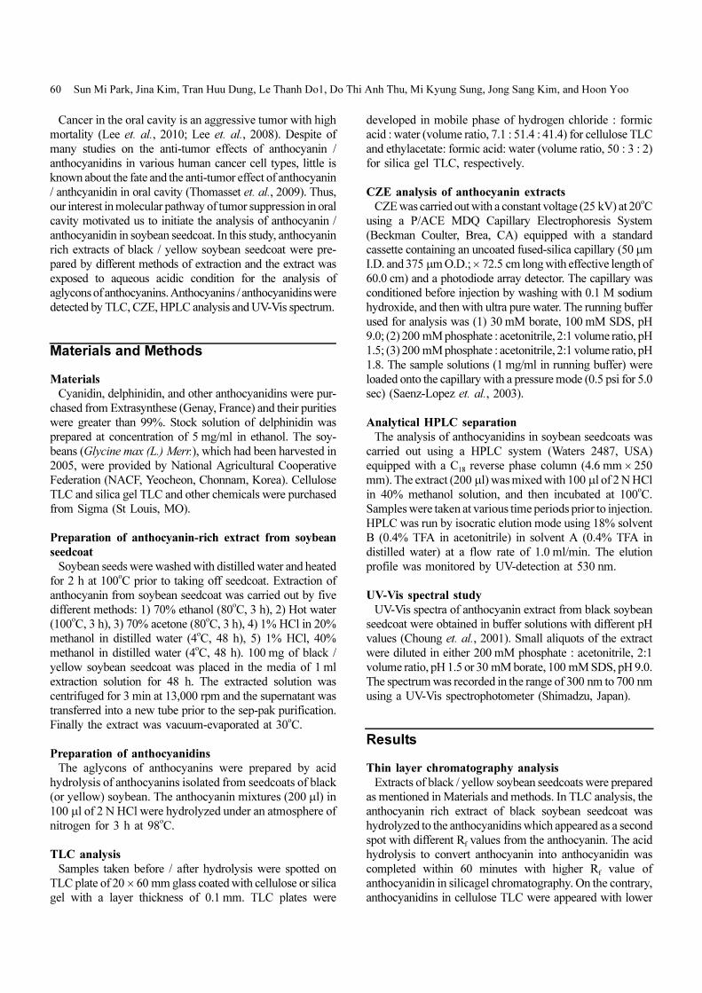
Capillary zone electrophoresis analysis of the extract
In order to study electrophoretic property of anthocyanin /
anthocyanidin in CZE, the stock solution of the extract was
applied to the P/ACE MDQ Capillary Electrophoresis
System, using running buffers of 30 mM borate, 100 mM
SDS, pH 9. The electropherogram of the extracts of black
soybean seedcoat, prepared by five different conditions,
showed similar peak retention time of anthocyanins while the
extracts from yellow soybean seedcoat did not show peaks
(Fig. 2). Under the acidic buffer solution (200 mM phosphate
: acetonitrile, 2:1 volume ratio with pH 1.5 or pH 1.8), electro-
pherograms of extracts of black soybean seedcoat showed a
broaden peak with almost identical retention time except the
extract prepared by the hot water. Again no detectable peak
was observed from the extracts of yellow soybean seedcoat.
Also the migration time of anthocyanin under the buffer
condition of pH 1.8 was longer than the one of pH 1.5
probably due to the equilibrium shift to the form of neutral
quinoidal base (Fig. 3).
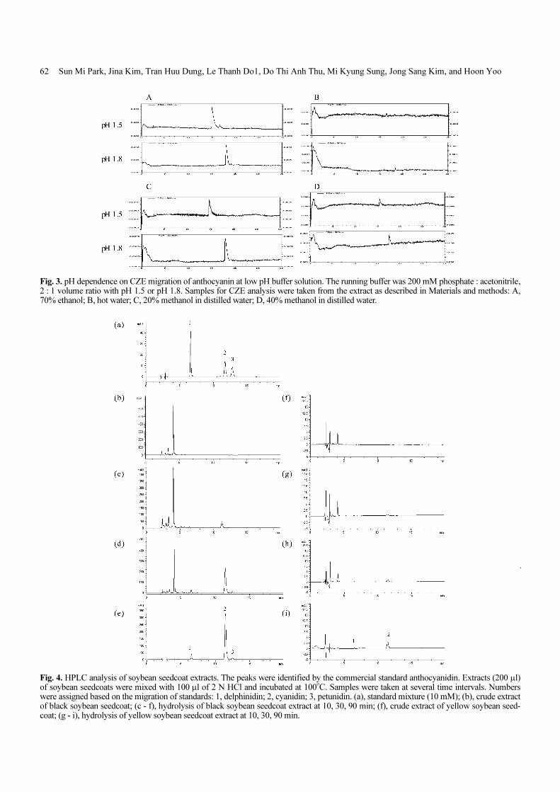
HPLC analysis of anthocyanidin in soybean seedcoat
extract
The anthocyanidins of soybean seedcoats were analyzed
by a reversed phase HPLC system monitored at 530 nm.
The extract of soybean seedcoat was acid hydrolyzed with 2
N HCl in 40% methanol. Samples were taken at various
time intervals and run by isocratic elution mode as described
in Materials and methods. HPLC analysis of hydrolyzed
extract of black soybean seedcoat (Fig. 4) showed a charac-
teristic peak of individual anthocyanidin which was identified
by validating with the retention time of the corresponding
standard. The initial peak of anthocyanin was disappeared
gradually as the hydrolysis is processed. The migration time
was fast in order of delphinidin > cyanidin > petunidin. Con-
trary to the black soybean, the anthocyanin extract hydrolyzed
from yellow soybean seedcoat showed barely detectable
peaks which were identified to be cyanidin and delphinidin.
In addition, further analysis with the HPLC chromatogram of
black soybean seedcoat allowed us to identify the presence of
another anthocyanidin, a pelargonidin, as shown in Fig. 5.
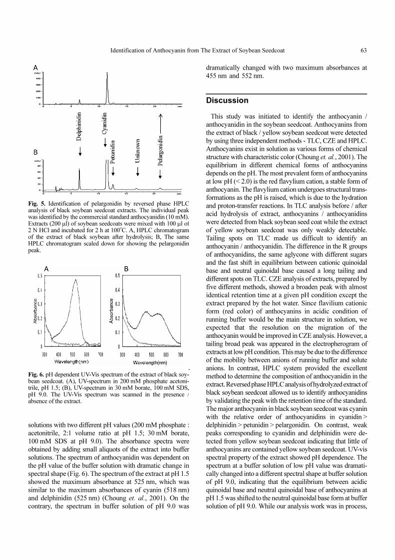
UV-Vis spectral property of anthocyanin rich extract
The UV-Vis spectrophotograms were obtained using buffer
Fig. 1. TLC analysis of extract. Acid hydrolysis of black (A) / yel-low (B) soybean seedcoat extracts in silica gel plate: 1, beforehydrolysis; 2, after hydrolysis. Mobile phase for silica gel TLC wasethylacetate : formic acid : water (volume ratio of 50 : 3 : 2). (C)Cellulose chromatography of acid hydrolysis of black soybean seedcoat extracts; 1, non-hydrolyzed black soybean seedcoat extraction;2 - 5, hydrolyzed from 10 to 40 min. Mobile phase was hydrogenchloride : formic acid : water (volume ratio of 7.1 : 51.4 : 41.4).
Fig. 2. CZE electropherogram of extracts of soybean seedcoats at 520 nm, using running buffers of 30 mM borate, 100 mM SDS, pH 9. A,extracts of black soybean seedcoats; B, extracts of yellow soybean seedcoats.
62 Sun Mi Park, Jina Kim, Tran Huu Dung, Le Thanh Do1, Do Thi Anh Thu, Mi Kyung Sung, Jong Sang Kim, and Hoon Yoo
Fig. 3. pH dependence on CZE migration of anthocyanin at low pH buffer solution. The running buffer was 200 mM phosphate : acetonitrile,2 : 1 volume ratio with pH 1.5 or pH 1.8. Samples for CZE analysis were taken from the extract as described in Materials and methods: A,70% ethanol; B, hot water; C, 20% methanol in distilled water; D, 40% methanol in distilled water.
Fig. 4. HPLC analysis of soybean seedcoat extracts. The peaks were identified by the commercial standard anthocyanidin. Extracts (200 µl)of soybean seedcoats were mixed with 100 µl of 2 N HCl and incubated at 100
o
C. Samples were taken at several time intervals. Numberswere assigned based on the migration of standards: 1, delphinidin; 2, cyanidin; 3, petunidin. (a), standard mixture (10 mM); (b), crude extractof black soybean seedcoat; (c - f), hydrolysis of black soybean seedcoat extract at 10, 30, 90 min; (f), crude extract of yellow soybean seed-coat; (g - i), hydrolysis of yellow soybean seedcoat extract at 10, 30, 90 min.
Identification of Anthocyanin from The Extract of Soybean Seedcoat 63
solutions with two different pH values (200 mM phosphate :
acetonitrile, 2:1 volume ratio at pH 1.5; 30 mM borate,
100 mM SDS at pH 9.0). The absorbance spectra were
obtained by adding small aliquots of the extract into buffer
solutions. The spectrum of anthocyanidin was dependent on
the pH value of the buffer solution with dramatic change in
spectral shape (Fig. 6). The spectrum of the extract at pH 1.5
showed the maximum absorbance at 525 nm, which was
similar to the maximum absorbances of cyanin (518 nm)
and delphinidin (525 nm) (Choung et. al., 2001). On the
contrary, the spectrum in buffer solution of pH 9.0 was
dramatically changed with two maximum absorbances at
455 nm and 552 nm.
Discussion
This study was initiated to identify the anthocyanin /
anthocyanidin in the soybean seedcoat. Anthocyanins from
the extract of black / yellow soybean seedcoat were detected
by using three independent methods - TLC, CZE and HPLC.
Anthocyanins exist in solution as various forms of chemical
structure with characteristic color (Choung et. al., 2001). The
equilibrium in different chemical forms of anthocyanins
depends on the pH. The most prevalent form of anthocyanins
at low pH (< 2.0) is the red flavylium cation, a stable form of
anthocyanin. The flavylium cation undergoes structural trans-
formations as the pH is raised, which is due to the hydration
and proton-transfer reactions. In TLC analysis before / after
acid hydrolysis of extract, anthocyanins / anthocyanidins
were detected from black soybean seed coat while the extract
of yellow soybean seedcoat was only weakly detectable.
Tailing spots on TLC made us difficult to identify an
anthocyanin / anthocyanidin. The difference in the R groups
of anthocyanidins, the same aglycone with different sugars
and the fast shift in equilibrium between cationic quinoidal
base and neutral quinoidal base caused a long tailing and
different spots on TLC. CZE analysis of extracts, prepared by
five different methods, showed a broaden peak with almost
identical retention time at a given pH condition except the
extract prepared by the hot water. Since flavilium cationic
form (red color) of anthocyanins in acidic condition of
running buffer would be the main structure in solution, we
expected that the resolution on the migration of the
anthocyanin would be improved in CZE analysis. However, a
tailing broad peak was appeared in the electropherogram of
extracts at low pH condition. This may be due to the difference
of the mobility between anions of running buffer and solute
anions. In contrast, HPLC system provided the excellent
method to determine the composition of anthocyanidin in the
extract. Reversed phase HPLC analysis of hydrolyzed extract of
black soybean seedcoat allowed us to identify anthocyanidins
by validating the peak with the retention time of the standard.
The major anthocyanin in black soybean seedcoat was cyanin
with the relative order of anthocyanidins in cyanidin >
delphinidin > petunidin > pelargonidin. On contrast, weak
peaks corresponding to cyanidin and delphinidin were de-
tected from yellow soybean seedcoat indicating that little of
anthocyanins are contained yellow soybean seedcoat. UV-vis
spectral property of the extract showed pH dependence. The
spectrum at a buffer solution of low pH value was dramati-
cally changed into a different spectral shape at buffer solution
of pH 9.0, indicating that the equilibrium between acidic
quinoidal base and neutral quinoidal base of anthocyanins at
pH 1.5 was shifted to the neutral quinoidal base form at buffer
solution of pH 9.0. While our analysis work was in process,
Fig. 5. Identification of pelargonidin by reversed phase HPLCanalysis of black soybean seedcoat extracts. The individual peakwas identified by the commercial standard anthocyanidin (10 mM).Extracts (200 µl) of soybean seedcoats were mixed with 100 µl of2 N HCl and incubated for 2 h at 100
o
C. A, HPLC chromatogramof the extract of black soybean after hydrolysis; B, The sameHPLC chromatogram scaled down for showing the pelargonidinpeak.
Fig. 6. pH dependent UV-Vis spectrum of the extract of black soy-bean seedcoat. (A), UV-spectrum in 200 mM phosphate acetoni-trile, pH 1.5; (B), UV-spectrum in 30 mM borate, 100 mM SDS,pH 9.0. The UV-Vis spectrum was scanned in the presence /absence of the extract.
64 Sun Mi Park, Jina Kim, Tran Huu Dung, Le Thanh Do1, Do Thi Anh Thu, Mi Kyung Sung, Jong Sang Kim, and Hoon Yoo
another study characterizing anthocyanins in the black soy-
bean was reported by Lee’s group, using HPLC with diode
array detection and electro spray ionization / mass spectrometry
(HPLC-DAD-ESI/MS) (Lee et. al., 2009). In their study, antho-
cyanins were extracted from the coat of black soybeans with
1% TFA in methanol and the anthocyanins in extracts were
characterized without hydrolysis. Interestingly, the identified
hydrolyzed anthocyanins (anthocyanidins) by our HPLC
analysis were consistent with their finding which was based
on the fragmentation patterns of HPLC-DAD-ESI/MS.
In conclusion, anthocyanins in soybean seedcoats, which
were investigated by different methods of extraction and
detection, revealed the presence of four anthocyanins in black
soybean seedcoats while yellow soybean seedcoat had little
of anthocyanins. The most major anthocyanin in black
soybean seedcoat was cyanin (cyanidin-3-O-glucoside) with
the relative order of anthocyanidin in cyanidin > delphinidin >
petunidin > pelargonidin.
Acknowledgement
This study was supported by Korea Science and Engin-
eering Foundation (KOSEF R01-2005-000-10602-0).
References
Bin Hafeez B, Asim M, Siddiqui IA, Adhami VM, Murtaza I,
Mukhtar H. Delphinidin, a dietary anthocyanidin in pig-
mented fruits and vegetables: a new weapon to blunt prostate
cancer growth. Cell Cycle. 2008;7:3320-6.
Choung MG, Baek IY, Kang ST, Han WY, Shin DC, Moon HP,
Kang KH. Isolation and determination of anthocyanins in
seed coats of black soybean (Glycine max (L.) Merr.). J
Agric Food Chem. 2001;49:5848-51.
Cvorovic J, Tramer F, Granzotto M, Candussio L, Decorti G,
Passamonti S. Oxidative stress-based cytotoxicity of delphinidin
and cyanidin in colon cancer cells. Arch Biochem Biophys.
2010;501:151-7.
Fernandes I, Faria A, Azevedo J, Soares S, Calhau C, De
Freitas V, Mateus N. Influence of anthocyanins, derivative
pigments and other catechol and pyrogallol-type phenolics
on breast cancer cell proliferation. J Agric Food Chem.
2010;58:3785-92.
Hui C, Bin Y, Xiaoping Y, Long Y, Chunye C, Mantian M,
Wenhua L. Anticancer activities of an anthocyanin-rich
extract from black rice against breast cancer cells in vitro
and in vivo. Nutr Cancer. 2010;62:1128-36.
Hyun JW, Chung HS. Cyanidin and Malvidin from Oryza
sativa cv. Heugjinjubyeo mediate cytotoxicity against human
monocytic leukemia cells by arrest of G(2)/M phase and
induction of apoptosis. J Agric Food Chem. 2004;52:2213-7.
Koide T, Hashimoto Y, Kamei H, Kojima T, Hasegawa M,
Terabe K. Antitumor effect of anthocyanin fractions extracted
from red soybeans and red beans in vitro and in vivo. Cancer
Biother Radiopharm. 1997;12:277-80.
Lee JH, Kang NS, Shin SO, Shin SH, Lim SG, Suh DY, Baek
IY, Park KY, Ha TJ. Characterisation of anthocyanins in the
black soybean (Glycine max L.) by HPLC-DAD-ESI/MS
analysis. Food Chemistry. 2009;112:226-231.
Lee MH, Kim MM, Kim JK, Kim DK, Kim HR, Kim HJ, Kim
CS. Ethanol Extracts of Angelica decursiva Induces Apoptosis
in Human Oral Cancer Cells. International Journal of Oral
Biology. 2010;35:215-20.
Lee YH, Jung JE, Lee JC, Moon HJ, Lee NH, Jhee EJ, Yi HK.
The enhancement of apoptosis by combined with proteasome
inhibitor and DNA synthetic inhibitor in oral cancer.
International Journal of Oral Biology. 2008;33:25-31.
Lin YC, Chou CC. Effect of heat treatment on total phenolic
and anthocyanin contents as well as antioxidant activity of
the extract from Aspergillus awamori-fermented black
soybeans, a healthy food ingredient. Int J Food Sci Nutr.
2009;60:627-36.
Matsunaga N, Tsuruma K, Shimazawa M, Yokota S, Hara H.
Inhibitory actions of bilberry anthocyanidins on angiogenesis.
Phytother Res. 2010;24 Suppl 1:S42-7.
Meiers S, Kemeny M, Weyand U, Gastpar R, von Angerer E,
Marko D. The anthocyanidins cyanidin and delphinidin are
potent inhibitors of the epidermal growth-factor receptor. J
Agric Food Chem. 2001;49:958-62.
Saenz-Lopez R, Fernandez-Zurbano P, Tena MT. Development
and validation of a capillary zone electrophoresis method for
the quantitative determination of anthocyanins in wine. J
Chromatogr A. 2003;990:247-58.
Shin DY, Lee WS, Lu JN, Kang MH, Ryu CH, Kim GY, Kang
HS, Shin SC, Choi YH. Induction of apoptosis in human
colon cancer HCT-116 cells by anthocyanins through sup-
pression of Akt and activation of p38-MAPK. Int J Oncol.
2009;35:1499-504.
Slavin M, Kenworthy W, Yu LL. Antioxidant properties,
phytochemical composition, and antiproliferative activity of
Maryland-grown soybeans with colored seed coats. J Agric
Food Chem. 2009;57:11174-85.
Stoner GD, Wang LS, Seguin C, Rocha C, Stoner K, Chiu S,
Kinghorn AD. Multiple berry types prevent N-nitrosomethyl-
benzylamine-induced esophageal cancer in rats. Pharm Res.
2010;27:1138-45.
Thomasset S, Teller N, Cai H, Marko D, Berry DP, Steward WP,
Gescher AJ. Do anthocyanins and anthocyanidins, cancer
chemopreventive pigments in the diet, merit development as
potential drugs? Cancer Chemother Pharmacol. 2009;64:201-
11.
Yun JM, Afaq F, Khan N, Mukhtar H. Delphinidin, an
anthocyanidin in pigmented fruits and vegetables,
induces apoptosis and cell cycle arrest in human colon
cancer HCT116 cells. Mol Carcinog. 2009;48:260-70.


