Embed Size (px)
Citation preview

Pergamon PII: SO161-5890(97)00042-d
Molecula,- Immunology, Vol. 34. No. 6, 441 -452, pp. 1997 (’ 1997 Elsevier Science Ltd. All rights reserved
Printed in Great Britain 0 16 I -5X90 ‘97 $17.00 + 0.00
HUMANIZED PORCINE VCAM-SPECIFIC MONOCLONAL ANTIBODIES WITH CHIMERIC IgG2/G4 CONSTANT REGIONS
BLOCK HUMAN LEUKOCYTE BINDING TO PORCINE ENDOTHELIAL CELLS
JOHN P. MUELLER,*MICHELLE A. GIANNONI, SANDRA L. HARTMAN, EILEEN A. ELLIOTT, STEPHEN P. SQUINTO, LOUIS A. MATIS and
MARK J. EVANS
Alexion Pharmaceuticals Inc., Departments of Immunobiology and Molecular Development. 25 Science Park Box 15. New Haven. CT 06511, U.S.A.
(First rewired 21 Jarmar?, 1997; accepted in reoisrd,fbrm I April 1997)
Abstract--Discordant xenografts surviving the initial hyperacute rejection phase may be subject to cellular rejection processes mediated by infiltrating leukocytes including T cells, NK cells and mono- cytes. The stable adhesion of these cell types to endothelial cells is due to the molecular interaction of the integrins VLA-4 and LFA-I with their ligands vascular cell adhesion molecule (VCAM) and ICAM- present on the endothelial cells. Human VLA-4 binds to porcine VCAM, and blocking mAbs specific for porcine VCAM have been developed. We have localized the epitope of the anti- porcine VCAM blocking mAbs 2A2 and 3F4 to domains I and 2. respectively. Humanized antibodies (IgG4 isotype) were constructed from these anti-porcine VCAM antibodies and demonstrated to inhibit adhesion of Ramos, Jurkat and YT cells, as well as purified resting and activated human T cells, to porcine aortic endothelial cells (PAEC). These cell types express both LFA-I as well as VLA- 4, suggesting blockade of human VLA-4 interaction with porcine VCAM may alone be sufficient to significantly impair adhesion of human leukocytes to porcine endothelial cells. The chimeric anti- porcine VCAM (pVCAM) HuG4 antibodies promoted increased adhesion of Fc receptor (FcR) positive cells such as U937 monocytic cells to PAEC. In contrast, chimeric anti-porcine VCAM antibodies created using the CHl and hinge region from human IgG2 and the CH2 and CH3 regions from human IgG4 (HuG2/G4 antibodies) inhibited binding of FcR positive cells to PAEC. These chimeric anti-pVCAM antibodies should allow delineation of the in riro role of VLA-4:‘VCAM interaction in porcine-to-primate xenotransplants. Further. the design of the HuG2:‘G4 antibodies should render them efficacious in multiple settings requiring elimination of FcR binding. ‘(’ 1997 Elsevier Science Ltd.
&,I, words: adhesion molecules, antibodies, Fc receptors. inflammation. xenotransplantation
INTRODUCTION
Although much progress has been made in elucidating the molecular mechanisms of hyperacute rejection of xeno- transplants (reviewed in Somerville and D’Apice, 1993), the mechanisms of cell mediated rejection of xenografts remain unclear. The cellular infiltrate observed 3days following transplantation of guinea pig hearts into comp- lement (C)-depleted rats is comprised predominantly of macrophages and natural killer cells, with low numbers of T lymphocytes present (Blakely et al., 1994). Similarly. the cellular infiltrate present 6 days after transplantation of porcine fetal islet clusters into rats is comprised pre- dominantly of macrophages with low numbers of T lymphocytes present primarily at the periphery of the
*Author to whom correspondence should be addressed. Tel: 203-776- 1790: Fax: 203-772-3655; E-mail: muellerj@ alxn. corn.
graft (Wallgren et al., 199.5). In ritro porcine aortic endo- thelial cells (PAEC) as well as antigen presenting peri- pheral blood cells can directly activate human CD4f and CD8 + T lymphocytes (Rollins et al., I994a; Murray et al., 1994; Yamada et al., 1995). Thus the initial phases of cellular rejection of xenotransplants are likely to involve binding and activation of multiple types of leuko- cytes including not only T lymphocytes but also natural killer cells and monocytes.
The stable adhesion of lymphocytes, natural killer cells, and monocytes to endothelial cells is mediated pre- dominantly by the interaction of the integrins LFA-1 (CDlla/CD18) and VLA-4 (CD49d/CD29) with their cognate ligands ICAM- and VCAM-I on endothelial cells (Allavena et al., 1991; Jonjic et al., 1992). For both monocytes (Luscinskas et al., 1994) and T lymphocytes (Alon et al., 1995; Luscinskas et al.. 1995) the binding of VLA-4 to VCAM is an essential component of the in citro adhesive interactions required for stable arrest under
441

442 J. P. MUELLER rt al.
conditions of flow. In vitro, the transmigration of NK cells and monocytes across human umbilical vein endo- thelial cell (HUVEC) monolayers also utilizes both LFA- 1 /lCAM-l- and VLA-4/VCAM-dependent pathways (Bianchi et al., 1993; Meershaert and Furie, 1994; Chu- luyan et al., 1995). In ciao, the interaction of LFA-1 with ICAM- may not be completely essential for extrava- sation of lymphocytes or monocytes since both of these cell types are present in inflammatory infiltrates of leuko- cyte adhesion deficiency patients who have a genetic deficiency in CD18 and thus lack integrins such as LFA- 1 (Hawkins et al., 1992). Finally, numerous studies have demonstrated the potential of blocking antibodies directed against LFA-1 (Issekutz, 1992; Scheynius et al., 1993), VLA-4 (Issekutz, 1991; Yednock et al., 1992; Chisholm rt al., 1993), ICAM- (Zeng et al., 1994), or VCAM (Orosz et al., 1993; Silber et al., 1995) to inhibit the accumulation of lymphocytes at inflammatory sites in animals.
For xenogeneic cellular interactions it is not yet fully defined which adhesion molecule interactions will func- tion across species barriers. It is likely that at least some xenogeneic receptor-ligand interactions will be non- functional. For example, mouse LFA-1 does not bind to human ICAM- (Johnston et al., 1990). To explore the molecular basis of adhesive interactions between distinct classes of adhesion molecules on human and porcine cells we have cloned porcine E-selectin and VCAM- 1 and have demonstrated that both are capable of mediating the adhesion of human leukocytes (Rollins et al., 1994a; Mueller et al., 1995). Further, we have developed murine monoclonal antibodies (mAbs) specific for porcine VCAM which block binding of human VLA-4 to porcine VCAM (Mueller et al.. 1995). The functional blocking activity of these antibodies suggests that they will be useful in suppressing the cellular response to a porcine xenograft. However, murine mAbs are not ideally suited for use in primates due to their short half-life and high immunogenicity (Khazaeli et al., 1994). Here we have utilized antibody engineering strategies to create human- ized anti-porcine VCAM antibodies that block adhesion of human natural killer cells, monocytes, and T lympho- cytes to activated PAEC. Furthermore, we show the choice of human isotype has critical implications for the potential therapeutic use of humanized antibodies in xenotransplantation.
MATERIALS AND METHODS
Cells
Ramos, Jurkat and U-937 cells were obtained from the American Type Culture Collection, YT cells were the gift of Dr J. M. Wang (National Cancer Institute, Frederick, MD, U.S.A.), porcine aortic endothelial cells (PAEC) were obtained from Cell Systems (Kirkland, WA, U.S.A.) and 293-EBNA cells were obtained from Invitrogen (San Diego, CA, U.S.A.). U-937 cells were maintained in RPM1 1640 supplemented with 15% heat-inactivated FBS and 2 mM L-glutamine. Ramos and Jurkat cells were
maintained in RPM1 1640 supplemented with 10% heat- inactivated FBS, 2 mM L-glutamine, 20mM HEPES, 50 PM fi-mercaptoethanol, 100 units/ml penicillin and lOOpg/ml streptomycin. YT were maintained in RPM1 1640 containing 10% heat-inactivated FBS, 2 mM L-glut- amine, 100 units/ml penicillin and 100 pug/ml strepto- mycin. PAEC were maintained in medium A consisting of M 199 supplemented with 20% heat-inactivated FBS. 0.05 mg/ml ECGF (Biomedical Technologies, Stoughton. MA, U.S.A.), 20mM HEPES and 0.1 mgjml heparin, and were used within three passages. 293-EBNA cells were grown in Dulbecco’s modified Eagle’s medium sup- plemented with 5% heat-inactivated FBS. 100 units/ml penicillin, 100 pg/ml streptomycin and 250 Lcgiml G418. Human T cells were purified as described previously (Mueller rt a/., 1995).
Production of’ VCA Ml[qJLsions
A plasmid containing the human IgGl Fc region was obtained from Dr M. J. Lenardo (NIH, Bethesda, MD, U.S.A.). DNA fragments containing various domains of human or porcine VCAM were obtained by PCR amplification of appropriate templates and joined in frame to the human IgGl Fc coding region using stan- dard molecular biology techniques. The cassettes enco- ding the fusion proteins were ligated into the expression plasmid pAPEX-3 (Evans et al., 1995a). VCAM/Ig fusion proteins were produced by transfection of 293-EBNA cells as described previously (Evans et al., 1995a). Three days following transfection the media were harvested. centrifuged to remove debris, and stored at 4 C. For ELISA, PolySorp 96-well microtiter plates (Nunc, Naper- ville, IL, U.S.A.) were coated overnight at 4 C with lOOngjwel1 goat anti-human IgG Fc (Sigma, St. Louis, MO, U.S.A.) in 0.1 M carbonate buffer, pH 9.6. The plates were then washed three times with PBS supplemented with 0.5% Tween 20 and incubated with blocking buffer (PBS supplemented with 0.05% Tween 20) at 37 C for 1 hr. After three further washes, various dilutions of con- ditioned media in blocking buffer were added and incu- bated at 37’ C for 1 hr. Following three further washes, 2A2 or 3F4 mAb in blocking buffer was added and incu- bated for 1 hr at 37 C. After additional washing, per- oxidase-conjugated goat anti-mouse IgG Fc (Sigma) diluted 1:2000 in blocking buffer was added and incu- bated at 37’ C for 1 hr. After three final washes the plate was developed for 15 min with 0.4mgjml o-phenylene- diamine dihydrochloride (Sigma). Reactions were stopped by addition of 1 M sulfuric acid. Quantitation was per- formed by measuring absorbance at 490 nm using a Bio- Rad model 3550 plate reader.
Production qf’chimeric~ antibodies
Variable regions were cloned from the 2A2 and 3F4 hybridomas by PCR amplification as described pre- viously (Evans et al., 1995a, 19956). These sequences have been submitted to the EMBL/GenBank/DDBL under accession numbers U78799-U78802. The variable regions were joined to genomic DNA cassettes containing
Anti-porcine VCAM antibodies block human leukocyte adhesion 443
either the intact human IgG4 constant regions or the first constant region and hinge linker region from human IgG2 followed by the second and third constant regions from human IgG4 using standard molecular biology techniques. Chimeric antibodies were produced in 293- EBNA cells and purified by chromatography on protein A Sepharose as described (Evans et al., 1995a). Each chimeric antibody preparation was greater than 95% pure as judged by Coomassie staining of SDS poly- acrylamide gels. F(ab’)2 was prepared by digestion of the antibodies with hcin (Pierce, Rockford, IL. U.S.A.) followed by protein A chromatography.
Cells used were obtained by trypsinization (PAEC and 293-EBNA) or centrifugation (Ramos, Jurkat, YT and U-937 cells). Harvested cells were washed once with 4 C PBS containing 1% heat-inactivated FBS (PBS/l) and aliquoted at 50000--100000 cells per well in 96-well V- bottom plates (Nunc). Cells were washed two additional times with PBS/ 1, and then incubated with PBS/ 1 con- taining IO/cg/ml primary Ab or PBS/l alone for 30min at 4 C. For some experiments the chimeric HuG4 or HuG2iG4 was centrifuged at 1 x 10-k for 1 hr prior to addition to the cells. The cells were then washed twice with PBS: I and resuspended in PBS/l containing the appropriate FITC-conjugated secondary antibodies (I :200 dilution) for 30 min at 4 ‘C. The cells were again washed twice with PBS/l and resuspended in PBS. Cell fluorescence was determined using a Becton Dickinson FACSort.
On day 0 PAEC were plated in medium A at a density of IO 000 cells per well in a 96-well flat bottom plate. On day 1 the PAEC were refed medium A supplemented with 1 pg/rnl LPS (Sigma. E. coli 011 l:B4). On day 2, human cells were resuspended in the appropriate growth medium supplemented with 1OpM calcein AM (Molecular Probes, Eugene, OR, U.S.A.) and incubated at 37’ C for 30min. The cells were subsequently washed three times with growth medium and resuspended at 6 x 10h cells/ml in RPM1 I640 supplemented with 1% heat-inactivated FBS (RPMI’I). The PAEC were washed twice with RPMIil and refed 50pl/well of RPMI/I containing the indicated concentrations of antibodies. After adding calc- ein-labeled cells at a density of 3 x IO5 cells/well (50~1), the plate was centrifuged at 2008 for 1 min and incubated at 37 C for 15 min. The nonadherent cells were removed by washing three to five times with RPMI/I. Adherent cells were lysed with HBSS containing 1% SDS and calcein fluorescence was measured with a Millipore Cyto- Fluor 2350 using an excitation wavelength of 485 nm and an emission wavelength of 530nm. Background flu- orescence was determined from wells containing PAEC that did not receive calcein-labeled cells. Samples were performed in duplicate in each experiment. Adherence was determined by comparing the residual fluorescence concentration in each well to the input fluorescence con-
centration. In each experiment the specific fluorescence signal obtained in the absence of competing antibody was defined as 100%.
For adhesion assays using purified soluble pVCAM (Mueller et a/., 1995) PolySorp 96-well microtiter plates (Nunc) were coated overnight at 4 C with 200ng/well purified soluble pVCAM in 0.1 M carbonate buffer, pH 9.6. After blocking the plate with RPM1 1640 sup- plemented with lOmg/ml BSA (RPMI/BSA), the plates were incubated with RPMI;BSA containing antibody for 1 hr at 37 C. Subsequently, 3 x IO’calcein-labeled U-937 cells in RPMI!BSA were added to each well. The plate was centrifuged upright for I min at 2OOg and incubated at 37 C for 15 min. The plate was then centrifuged inverted at 2008 for 3 min. Adherent cells were lysed by the addition of HBSS containing 1% SDS and calcein fluorescence was measured with a Millipore CytoFluor 2350 using an excitation wavelength of 485nm and an emission wavelength of 530 nm.
RESULTS
Ident{fication of’ porcinr SCAM tkmrtins reco,qni:ed hi, blocking n?Ah.s
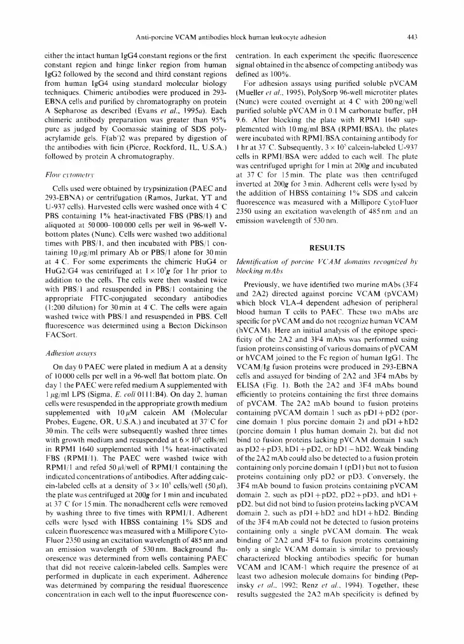
Previously. we have identified two murine mAbs (3F4 and 2A2) directed against porcine VCAM (pVCAM) which block VLA4 dependent adhesion of peripheral blood human T cells to PAEC. These two mAbs are specific for pVCAM and do not recognize human VCAM (hVCAM). Here an initial analysis of the epitope speci- ficity of the 2A2 and 3F4 mAbs was performed using fusion proteins consisting of various domains of pVCAM or hVCAM joined to the Fc region of human IgGl. The VCAMjIg fusion proteins were produced in 293-EBNA cells and assayed for binding of 2A2 and 3F4 mAbs by ELISA (Fig. I). Both the 2A2 and 3F4 mAbs bound efficiently to proteins containing the first three domains of pVCAM. The 2A2 mAb bound to fusion proteins containing pVCAM domain I such as pD1 +pD2 (por- cine domain I plus porcine domain 2) and pDI + hD2 (porcine domain 1 plus human domain 2), but did not bind to fusion proteins lacking pVCAM domain I such as pD2 + pD3, hD I + pD2, or hD I + hD2. Weak binding of the 2A2 mAb could also be detected to a fusion protein containing only porcine domain 1 (pD I ) but not to fusion proteins containing only pD2 or pD3. C’onversely, the 3F4 mAb bound to fusion proteins containing pVCAM domain 2. such as pD1 fpD2, pD2+pD3, and hDl+ pD2. but did not bind to fusion proteins lacking pVCAM domain 2. such as pD1 + hD2 and hD1 $- hD2. Binding of the 3F4 mAb could not be detected to fusion proteins containing only a single pVCAM domain. The weak binding of 2A2 and 3F4 to fusion proteins containing only a single VCAM domain is similar to previously characterized blocking antibodies specific for human VCAM and ICAM-I which require the presence of at least two adhesion molecule domains for binding (Pep- insky et rrl.. 1992; Renz ct al.. 1994). Together, these results suggested the 2A2 mAb specificity is defined by
444 J. P. MUELLER et al.
Leader VCAM HuGl I I Dl I 02 I D3
FXiY Dl I 02 I D3
I I Dl I D2 I
I D2 I D3
Exii Dl I hD2. 1
h\w hD1 I D2 I
!sN hD1 I h02’ 1
I I Dl 1
I D2 I
D3
zAz3YE4
100 100
119 104
92 67
0 96
74 0
0 100
0 0
5 0
0 1
1 0
ALum
100
105
95
106
122
100
102
96
100
115 Fig. I. Binding of 3F4 and 2A2 mAbs to VCAMjIg fusion proteins. Proteins consisting of domains 1 (Dl), 2 (D2), or 3 (D3) from porcine VCAM (open boxes) or human VCAM (gray boxes) joined to the Fc region of human IgGl (black boxes) were produced in 293-EBNA cells. In some constructs the pVCAM secretory leader sequence was replaced with the secretory leader sequence from human CD59 (striped boxes). The fusion proteins were captured by incubating diluted conditioned media in 96-well plates coated with goat anti-human IgG (Fc specific). After incubation with 2A2 or 3F4 mAb, bound murine mAb was detected using peroxidase-conjugated goat anti-mouse IgG (Fc specific). The total amount of each fusion protein captured per well was quantitated using peroxidase-conjugated goat anti-human IgG (Fc specific). The absorbance signal obtained with the fusion protein containing the intact pVCAM domains 1, 2 and 3 was defined as 100% for each antibody. Binding of the 2A2 and 3F4 mAbs to the remaining fusion proteins is reported as a percentage of this value. Results
shown are from a representative experiment of several performed.
pVCAM domain 1 whereas the 3F4 mAb specificity is defined by pVCAM domain 2.
Inhibition of human leukocyte binding to PAEC by chim- eric anti-porcine I/CAM mAbs
The use of murine antibodies in porcine-to-primate xenotransplantation models or ultimately in porcine-to- human xenotransplants poses several problems including a short half-life of the murine antibody plus development of an immune response directed against the murine anti- body. These problems have been significantly reduced by the use of chimeric antibodies consisting of the murine variable regions joined to human constant regions (Khazaeli et al., 1994). To create anti-pVCAM chimeric antibodies we cloned the variable regions from both 2A2 and 3F4. The murine variable heavy regions were joined to the human IgG4 constant region (HuG4) since this isotype does not activate complement (C) (Tao rt al., 1993) the predominant mediator of hyperacute rejection of xenografts. The murine variable light regions were joined to the human kappa constant region. Chimeric ch2A2 HuG4 and ch3F4 HuG4 antibodies were pro- duced by coexpression of the chimeric heavy and light chains in 293-EBNA cells and tested for binding to pVCAM by FACS analysis using 293 cells expressing pVCAM (Fig. 2(A)). Both chimeric mAbs produced identical staining profiles as the corresponding murine mAbs, while neither chimeric mAb bound to untrans-
fected 293 cells. Additionally, both the chimeric anti- bodies and murine mAbs produced identical FACS profiles using LPS-activated porcine aortic endothelial cells (Fig. 2(B)), confirming the chimeric antibodies retained the ability to bind to pVCAM expressed on PAEC.
The ability of the ch2A2 HuG4 and ch3F4 HuG4 anti- bodies to block the interaction between human VLA-4 and porcine VCAM was assessed by measuring inhibition of Ramos cell binding to activated PAEC (Fig. 3). The murine 3F4 mAb inhibited this binding by 90% when present at concentrations greater than 5 pg/ml. A control humanized antibody directed against human C5 (h5Gl. 1 HuG4) did not inhibit the binding of Ramos to activated PAEC. The ch2A2 HuG4 and ch3F4 HuG4 produced nearly identical inhibition curves as the murine 3F4 mAb, demonstrating the retention of binding avidity and cell adhesion blocking capabilities in these chimeric antibodies.
The ability of the ch2A2 HuG4 and ch3F4 HuG4 mAbs to inhibit binding of cells expressing both VLA-4 and LFA-1 was next examined using the Jurkat T cell line. Both the ch2A2 HuG4 and ch3F4 HuG4 antibodies inhibited the binding of Jurkat to activated PAEC by up to 90% while the control antibody h5Gl. 1 HuG4 did not inhibit binding at any concentration (Fig. 4(A)). Simi- larly, the ch2A2 HuG4 and ch3F4 HuG4 antibodies inhibited the binding of both resting (Fig. 4(B)) and acti- vated (Fig. 4(C)) T cells to activated PAEC, although the

Anti-porcine VCAM antibodies block human leukocyte adhesion 44s
A 210
I
mAb 2101 chAb
2”
0
; 2101 P I
2A2i oh2’;k 100 10' 102 103 104 100 10' 102 103 104
3F4
0 200
i mAb
Fluorescence intensity
200
1 chAb
0 100 10' 102 103 104
0 100 10' 102 103 104
PAEC
Fig. 2. Binding of 3F4 and 2A2 chimeric antibodies to pVCAM. (A) Chimeric mAbs containing the variable regions of either 2A2 or 3F4 joined to human IgG4 and kappa constant regions were produced in 293-EBNA cells and purified by protein G Sepharose chromatography. Untransfected 293-EBNA cells (293) or 293-EBNA cells expressing pVCAM (293/pVCAM) were incubated with IO pg/ml purified murine 2A2 mAb, murine 3F4 mAb. ch2A2 HuG4, or ch3F4 HuG4. Bound antibody was detected using either FITC-conjugated goat anti-mouse IgG or FITC-conjugated goat anti-human IgG followed by flow cytometry using a Becton Dickinson FACSort. Data are dis- played as histograms with fluorescence intensity plotted on the .u-axis and relative cell number plotted on the y-axis. Back- ground staining by the secondary antibodies is shown in the panels labeled 2”. (B) PAEC were activated by treatment with 1 /cg/rnl LPS for approximately 16 hr. Cells were then stained with either the murine 2A2 and 3F4 mAbs (left panel) or the ch2A2 HuG4 and ch3F4 HuG4 antibodies (right panel). Back- ground staining by the secondary antibodies (2“) is indicated.
maximal inhibition achieved was reduced to approxi- mately 60%. Addition of a blocking mAb directed against human VLA-4 also maximally inhibited adhesion by 60% (Fig. 4(C)), confirming the binding inhibition observed was due to blockade of the molecular inter- action between human VLA-4 and porcine VCAM.
The potential of the ch2A2 HuG4 and ch3F4 HuG4 antibodies lo inhibit binding of natural killer cells and monocytes to activated PAEC was examined using the
d 3F4 mAb -f- ch3F4 HuG4 - ch2A2 HuG4 - h5Gl .l HuG4
IO
Fig. 3. Binding of Ramos cells to PAEC in the presence of mAbs. On day 0, PAEC were plated at 10 000 cells per well in medium A in 96-well plates. On day 1 the PAEC were activated by adding fresh medium A supplemented with 1 pg/ml LPS. After 16 to 24 hr incubation, the PAEC were washed and refed RPM1 1640 containing 1% FCS plus the indicated con- centrations of the murine 3F4 mAb (3F4 mAb). the chimeric 2A2 and 3F4 mAbs (ch2A2 HuG4 and ch3F4 HuG4) or a humanized negative control antibody (h5G 1. I HuG4). Calcein- labeled Ramos cells were added and allowed to adhere to the PAEC under static conditions for IS min. After nonadherent cells were removed by washing, the remaining bound cells were lysed with SDS. Calcein fluorescence of the SDS supernatant was measured with a Millipore CytoFluor 3350. In each cxperi- ment the binding of Ramos cells to PAEC in the absence of antibody was defined as 100%. Results shown are the mean of two to four experiments. with each value determined in dupli-
cate in each experiment.
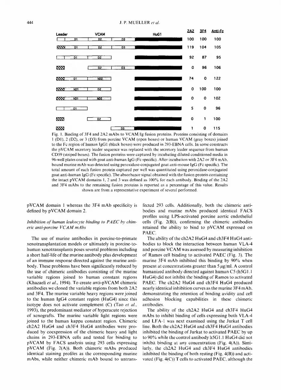
cell lines YT (Yodio et al., 1985) and U-937 (Sundstron and Nilsson, 1976), respectively. Binding of YT cells to activated PAEC was strongly inhibited by concentrations of lOpg/ml ch2A2 HuG4 and ch3F4 HuG4 antibodies (Fig. 5(A)). In contrast, the ch2A2 HuG4 and ch3F4 HuG4 antibodies did not inhibit binding of U-937 cells to activated PAEC (Fig. 5(B)). One possible explanation for this failure to inhibit U-937 cell binding could be the presence of elevated levels of VLA4 on U-937 cells. However, FACS analysis revealed the presence of com- parable levels of CD49d and activated CD29 on U-937 cells as on other cell lines such as Jurkat whose adhesion to PAEC was inhibited by the chimeric anti-pVCAM antibodies (Fig. 6). Interestingly, the adhesion of U-937 to PAEC was consistently augmented by approximately 50% in all experiments. This increase was specific for the anti-pVCAM antibodies as no increase in IJ-937 binding to PAEC was detected in the presence of the control h5Gl.l HuG4 antibody (Fig. 5(B)). This suggested that binding of U-937 to PAEC could be increased through FcR binding to the anti-pVCAM HuG4 antibodies on the PAEC surface. In support of this hypothesis, FACS analysis confirmed the presence of significant levels of FqRI (CD64), FcyRII (CD32) and Fc;~RIII (CDl6) on the surface of U-937 cells. In contrast, Jurkat and YT cells had no detectable Fey receptors while Ramos cells showed the presence of only low levels of FqRII (Fig.
6). Binding of the HuG4 chimeric antibodies to U-937
could be directly demonstrated by flow cytometry (Fig.

446 J. P. MUELLER et ~1.
140
120 Al
= 0 e loo-
is =E 80-
!ji 80- 52 ‘2 40- --o-- ch3F4 HuG4
2 0- + ch2A2 HuG4
- -t- h5Gl .l HuG4 0 I 111111 I I111111 II1In-l
1
0.1 AIntibody (p&Y]
100
40
20- B
- - ch3F4 HUG 2 0 - + ch2A2 HuG4
- + h5Gl.l HuG4 0 I I111111 I I I,,,, , I I11111
0.1 Alntibody (p&m!)
100
-o- Anti-hVLA-4 --e ch3F4 HuG4 + ch2A2 HuG4 ---t h5Gl .l HuG4
. I I IV I”, I I I 4 I ,I,, , I I 0.1
Akibocly (pg/ln9) lb0
Fig. 4. Binding of Jurkat and human T cells to PAEC in the presence of HuG4 antibodies. Binding experiments were per- formed as described in Fig. 3 using calcein-labeled Jurkat cells (A) or calcein-labeled purified human T cells (B and C). Human T cells were either assayed immediately after purification (B) or cultured in the presence of 1 pg/ml PHA for 5 days followed by 1 day without PHA stimulation (C) prior to performing the adhesion assay. Results shown are the mean of two to four experiments (A and B) or from an individual experiment (C). Anti-hVLA-4 denotes the blocking murine mAb HP2i 1 directed
against the 24 subunit of human VLA-4
7). Both the HuG4 antibodies as well as the anti-human C5 HuG4 antibody bound to U-937 cells. In contrast, 293/pVCAM cells bound only the anti-pVCAM anti- bodies but not the anti-human CS HuG4 antibody. No binding of the HuG4 chimeric antibodies could be detected with Ramos, Jurkat, or YT cells. These results suggest that the HuG4 chimeric antibodies bound to U- 937 cells through interaction of the HuG4 Fc region with the Fc receptors (FcRs) present on U-937 cells. Finally,
---c ch3F4 HuG4
+ ch2A2 HuG4
0.1 Akibody (&m?)
100
Fig. 5. Binding of YT and U-937 to PAEC in the presence of HuG4 mAbs. Adherence of calcein-labeled YT cells (A) or LJ- 937 cells (B) to PAEC was assayed as described in Fig. 3. Results
shown are the mean of two to four experiments.
F(ab’)2 fragments prepared from ch3F4 HuG4 strongly inhibited binding of U-937 cells to PAEC (Fig. 8) whereas F(ab’)2 fragments prepared from h5Gl. 1 HuG4 did not inhibit the binding. This result demonstrates that, if anti- body crosslinking is prevented, blockade of the inter- action between human VLA-4 and porcine VCAM can effectively inhibit the adhesion of human monocytes to PAEC.
Inhibition qf’FcR binding qf’humanized mAbs tvith crltered constunt regions
Although F(ab’)2 fragments proved capable of inhibit- ing adhesion of U-937 cells, antibody fragments are sub- optimal for chronic treatments, as they have much shor- ter in ciao half-lives than intact antibodies (Cove11 et ul., 1986). The use of intact antibodies with human constant regions as anti-rejection agents is complicated by the capacity of such antibodies to fix C and/or to bind FcRs. Human antibodies of the G4 isotype are devoid of C activity (Tao et al., 1993; Smith et al., 1995). Human antibodies of the G2 isotype do not activate C or bind FcRs (Canfield and Morrison, 1991; Burton and Woof, 1992). Rather than altering the specific residues known to mediate C fixation and FcR binding, we explored the possibility of creating an antibody with a composite human constant region. To create chimeric mAbs which would not activate C or bind to FcRs, we utilized the overall similarity in the human G2 and G4 primary
200 0 1 10”
so
0 I IO”
2 50 0 1 IO”
so
0 I Id’
Fig.
6.
E
xpre
ssio
n of
V
LA--
I. LF
A-I
an
d Fc
Rs
on
the
surf
ace
of
Ram
oh.
Jurk
at.
YT
and
U-9
37
cells
. C
ells
ue
re
incu
bate
d kv
ith I
O!c
g m
l of
the
fo
llo~v
ing
prim
ary
antib
odie
s:
mou
se
anti-
hu
man
C
D4Y
d.
rat
anti-
actiK
ited
hum
an
CD
2Y.
mow
e an
ti-hu
man
C
DI
Ix.
mou
se
anti-
hum
an
C‘D
64 (
FqR
I).
mou
se
anti-
hum
an
CD
32
(Fc;
,RII
) or
mou
se
anti-
hum
an
CD
16
(Fcy
RfI
I).
Afte
r li;
tshi
ng.
cells
\~e
rs i
ncub
ated
n
ith e
ither
FI
TC-c
onju
gate
d go
at
anti-
mou
se
IgG
or
FIT
C-c
onju
gate
d go
at
anti-
rat
IfG
as
app
ropr
iate
. Ea
ch
pane
l pr
esen
ts
the
resu
lts
obta
ined
in
the
pr
esen
ce
of p
rimar
y pl
us
seco
ndar
y an
tibod
y or
in
the
pres
ence
of
sec
onda
ry
antib
ody
alon
e.
VLA
-4
CD
29
2OC
!!!!
.--
(. IO
’ IO
’ IO
’ 10
’ 25
0
LFA
- I
CL)
113
CD
64
Fc r
ecep
tors
CD
32
CD
16
L (
IO’
102
IO)
IO.’
251
&
!(!’
’ (1
I u
- IO
7 IO
4 I
” IO
IO
2 IO
’ IO
J
b IO
IO
? 10
I(
+
$l!iL
__
i I!)’
IO
IO’
I OJ

448 J. P. MUELLER et al.
HuG4 HuG2/G4
250
$ i 25; I(.
250 ~ lI lo2 lo' lo425;i 1o2 IO3 1o4
IO0 10' 102 103 104 100 10' 102 103 104
525;k 25;k 100 IO' 102 IO3 IO4 100 10' 102 IO3 IO4
1250~ 250, i Ll
- 2” n - h5Gl.l - 2A2
“1- io” 10’ 102 103 104 ioo 10' 102 103 104
Fig. 7. Binding of chimeric antibodies to 293/pVCAM, Ramos, Jurkat, YT and U-937 cells. Cells were incubated with lOpg/ml h5Gl.l HuG4, ch2A2 HuG4, or ch3F4 HuG4 (left panels) or 10 pg/ml h5Gl. 1 HuG2/G4, ch2A2 HuG2/G4, or ch3F4 HuG2/G4 (right panels). After washing, cells were incubated with FITC-conjugated goat anti-human IgG except for Ramos cells which were incubated with FITC-conjugated goat anti- human IgG (y chain specific). Each panel presents the results obtained with the indicated cell line for h5Gl. 1 HuG4, ch2A2 HuG4 and ch3F4 HuG4 mAbs as well as the secondary anti-
body alone (2’) control.
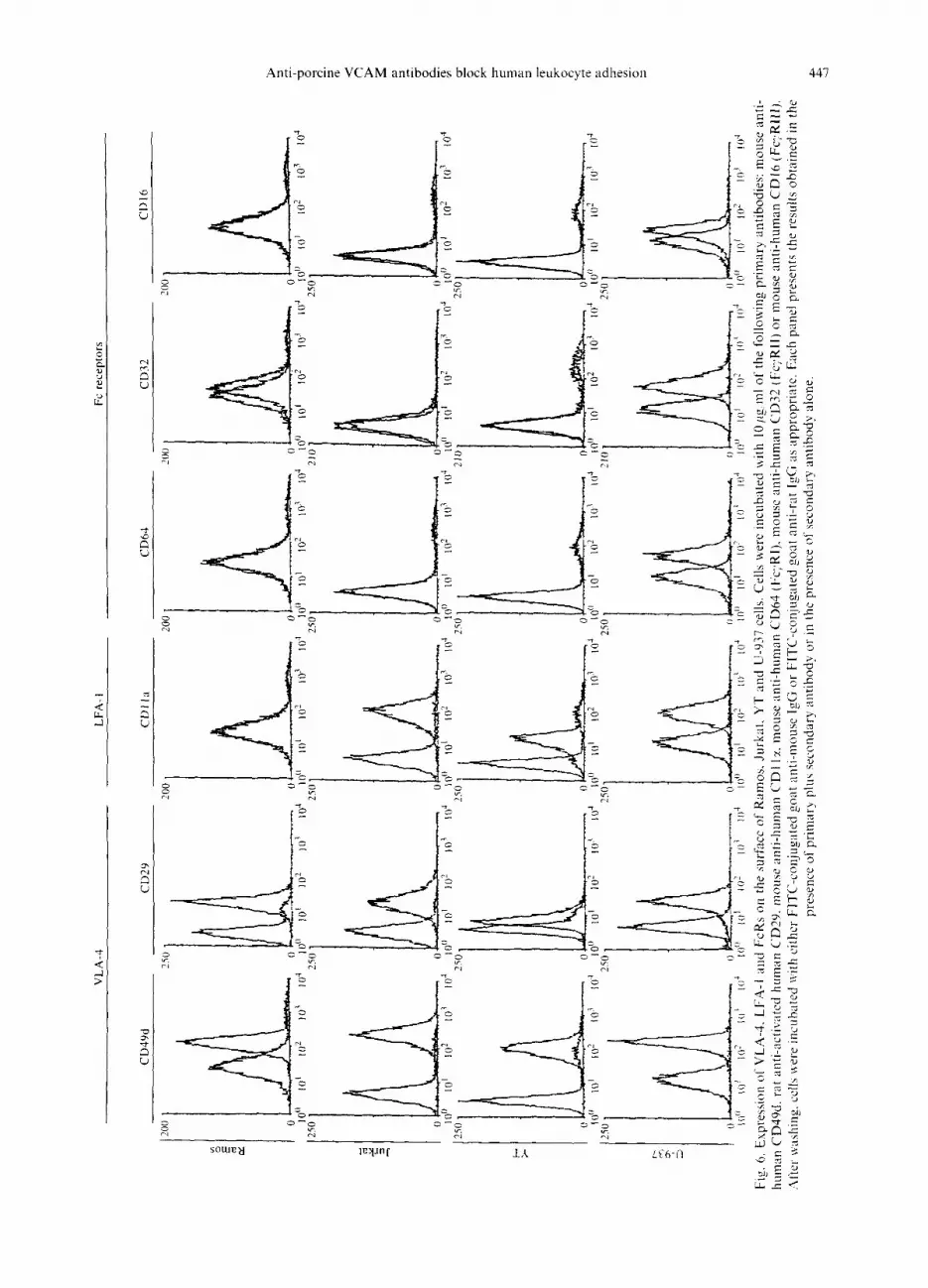
sequences to design a HuG2/G4 composite antibody con- sisting of the first constant region domain and the hinge region from the HuG2 isotype joined to the second and third constant region domains from the HuG4 isotype (Fig. 9). The resulting mAb did not contain the antibody sequences necessary for FcR binding, nor any new epi- topes that would likely be immunogenic.
Binding to FcR was examined by using the monocytic cell line U937. FACS analysis demonstrated that the
loo- =
~Eao- 2: m - 60- $E 52 JO-
- 20-
+ ch3F4 HuG4 - h5Gl .l HuG4
IO
Fig. 8. Binding of U-937 cells to PAEC in the presence of F(ab’)2 fragments. F(ab’)2 fragments were prepared from h5Gl.l HuG4 and ch3F4 HuG4 by digestion with ficin. Un- digested antibody and Fc fragments were removed by protein A chromatography. Adherence of calcein-labeled U-937 cells to PAEC in the presence of F(ab’)2 was assayed as described
in Fig. 3. Results shown are the mean of five experiments.
resulting ch3F4 HuG2/G4, ch2A2 HuG2/G4 and h5Gl. 1 HuG2/G4 antibodies did not bind to U-937 cells (Fig. 7). The ch3F4 HuG2/G4 and ch2A2 HuG2/G4 antibodies maintained the ability to bind pVCAM as demonstrated both by binding to 293/pVCAM cells (Fig. 7) and by inhibition of Jurkat and YT binding to activated PAEC (Fig. 10). For both Jurkat and YT cells inhibition of binding with the chimeric HuG2/G4 was equivalent to the HuG4 or original murine anti-pVCAM antibodies. The binding of U-937 cells to PAEC was now potently inhibited by the presence of either ch2A2 HuG2/G4 or ch3F4 HuG2/G4 antibodies. Finally, the chimeric anti- bodies were examined for the ability to block the adhesion of U-937 cells to purified soluble pVCAM (Fig. 11). Addition of the ch3F4 HuG4 antibody failed to inhibit binding of U-937 cells to pVCAM unless very high con- centrations of antibody were added. In contrast the ch3F4 HuG2/G4 antibody potently inhibited the binding of U-937 cells to pVCAM, while the negative control h5Gl.l HuG2/G4 antibody did not block binding. Together, these results demonstrate the anti-pVCAM HuG2/G4 antibodies specifically inhibited the adhesive interaction of human VLA-4 with porcine VCAM and that disruption of this interaction was sufficient to inhibit binding of numerous human cell types to porcine endo- thelial cells.
DISCUSSION
Current success in allotransplantation has resulted from the development of effective global immuno- suppressive therapies that prevent cellular graft rejection. Recent attempts to develop more specific immunosup- pressive strategies have focused on inhibiting interactions between integrins and costimulatory molecules on T cells with their ligands on graft endothelial cells and other antigen presenting cells (reviewed in Faustman, 1995; Guinan et al., 1994). The success of allotransplantation has been limited by the resulting shortage of human donor organs. Recent progress in overcoming the initial
Anti-porcine VCAM antibodies block human leukocyte adhesion
HuG2 HuGUG4 HuG4
HuG2 HuG2/G4 HuG4
HuG2 HuG2/G4 HuG4
HuG2 HuG2/G4 HuG4
HuG2 HuG2/G4 HuG4
STKGPSVFPLAPCSRSTSESTAALGCLVKDWEPVTVSWNSGALTSGV STKGPSWLAPCSRSTSESTAALGCLVKDWEPVTVSWNSGALTSGV STKGPSWLAPCSRSTSESTAALGCLVKDWEPVTVSWNSGALTSGV
HTFPAVLQSSGLYSLSSWTVPSSN-FGTQTYTCNVDHKPSNTKVDKTVE HTFPAVLQSSGLYSLSSVVTVPSSNFGTQTYTCNVDHKPSNTKVDKTVE HTFPAVLQSSGLYSLSSWTVPSS>@-YTCNVDHKPSNTKVD~=VE
235 RKCCVECPPCPAPPV- AGPSVFLF RKCCVECPPCPAPPV- AGPSVFLF &KmCPgCPAPEwPSVFLF
PPKPKDTLMISRTPEVTCWVDVSHEDPEVQ FNWYVDGVEVHNAKTKPR PPKPKDTL&iISRTPEVTCVWDVSQEDPEVQ FNWYVDGVEVHNAKTKPR PPKF’KD-ILMISRTPEVTCWVDVSqEDPEVQ FNWYVDGVEVHNAKTKPR
331 EEQFNSTFRWSVLTWHQDWLNGKEYKCKVSNKGLPAPIEKTISKTKG EEQFNSTYRWSVLTVLHQDWLNGKEYKCKVSNKGLPSSIEKTISKAKG EEQFNSwWSVLTVLHQDWLNGKEYKCKVSNKGLPEIEKTISKA=KG
449
Fig. 9. Sequence of the CHI, hinge and CH2 regions of human IgG2 (HuG2), human IgG4 (HuG4). and the chimeric HuG2/G4 construct. Positions where differences occur between the three molecules are double underlined. The amino acid sequence at position 235/236 determines ability to bind to Fcg receptors (Canfield and Morrison, 1991; Duncan et cd., 1988; Alegre et ul., 1992; Burton and Woof, 1992) while the presence of a serine at position 331 confers the inability to activate C (Tao er
a/., 1993).
hyperacute rejection of discordant xenografts suggests the potential for use of porcine xenografts as a clinical option to circumvent this chronic shortage. Although there are limited data regarding the fate of discordant xenografts which survive the initial hyperacute rejection phase, xenograft recipients will likely require continued cellular immunosuppression for the prevention of graft rejection. In this light, xenografts offer a unique oppor- tunity for the development of blocking reagents specific for the graft, thereby avoiding global immunosuppression of the recipient.
To investigate the interactions occurring between human leukocytes and porcine endothelial cells, we have cloned the porcine VCAM molecule and created mAbs specific for porcine VCAM that do not bind to human VCAM (Mueller et ul., 1995). Two of these antibodies (3F4 and 2A2) are capable of blocking the interaction between human VLA-4 and porcine VCAM. Here we demonstrate that the binding specificity for 2A2 is located within domain 1 of pVCAM while the binding specificity for 3F4 is located within domain 2. This is identical to the results found with human VCAM, where blocking antibodies have also been shown to be directed against either domains 1 or 2 (Osborn et al., 1992; Vonderheide and Springer, 1992).
The use of murine mAbs in humans and primates pre- sents numerous problems including a short antibody half- life and a high degree of immunogenicity (Khazaeli et al.,
1994). To optimize the use of the anti-porcine VCAM antibodies in porcine-to-primate xenograft experiments we developed chimeric antibodies using the variable regions cloned from the 2A2 and 3F4 antibodies joined to the constant regions of the human IgG4,K isotype. Given the extensive activation of C which occurs within xenografts, the human IgG4 isotype was chosen due to its inability to activate C. The resultant chimeric antibodies (ch2A2 HuG4 and ch3F4 HuG4 antibodies) retained
specificity for porcine VCAM and were as potent as the original murine mAbs at blocking adhesion of Ramos cells to activated PAEC by greater than 90% (Fig. 3). In contrast to Ramos, the binding of resting or activated human T cells to activated PAEC was inhibited only approximately 50% by either the chimeric antibodies or the original mAbs (Fig. 4). Activation of human T cells by PAEC can be blocked by antibodies directed against either CD2 or LFA-1 (Rollins et al., 19946), suggesting either of these molecules could mediate the remaining adhesion by interacting with porcine CD.58 or porcine ICAM- 1, respectively.
The adhesion of human lymphocytes, monocytes or NK cells to activated HUVEC is dependent on the inter- action of both LFA-1 with ICAM- and VLA-4 with VCAM (Allavena et al., 1991; Jonjic Pt al., 1992; Vonderheide and Springer, 1992). For each cell type, blocking the VLA-4/VCAM interaction by itself pro- duces only a small reduction in adhesion, while antibodies which disrupt the LFA-l/ICAM-1 interaction provide a greater, although still partial, inhibition of adhesion. Addition of antibodies blocking both VLA-4/VCAM and LFA-l/ICAM-1 is required to completely block adhesion of these cell types to HUVEC. Surprisingly, binding of LFA-1 +-VLA+4 Jurkat or YT cells to activated PAEC could be inhibited nearly completely by the ch3F4 HuG4 and ch2A2 HuG4 antibodies. One possible explanation for this could be the lack of interaction between human LFA-1 and porcine ICAM. The conservation of ICAM- 1 is relatively low between species, with human and mouse ICAM- being only 50% conserved at the amino acid level (reviewed in Carlos and Harlan, 1994), and in uitro
mouse LFA-1 does not bind to human ICAM-1. Alter- natively, human LFA-1 may interact with porcine ICAM-1, but with a reduced affinity. For example, human VLA-4 binds to human VCAM at two inde- pendent sites, one within domain 1 and one within
450 J. P. MUELLER ef ul.
0
1 \\ I
0.1 Alntitiody (p&n!)
100
40
20-
oo-
80-
80-
40-
20-
0 I I I,,,,, I I,,,,,, I I ,l,rn
0.1 &body (p&m?) 100
120 Cl -
loo- =
& 80- ag m _ 80- ig
q 40-
- 20-
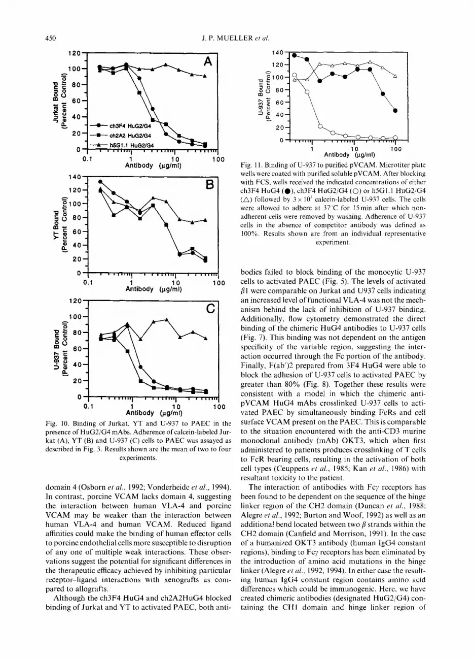
Fig. 10. Binding of Jurkat, YT and U-937 to PAEC in the presence of HuG2/G4 mAbs. Adherence of calcein-labeled Jur- kat (A), YT (B) and U-937 (C) cells to PAEC was assayed as described in Fig. 3. Results shown are the mean of two to four
experiments.
domain 4 (Osborn et al., 1992; Vonderheide et al., 1994). In contrast, porcine VCAM lacks domain 4, suggesting the interaction between human VLA-4 and porcine VCAM may be weaker than the interaction between human VLA-4 and human VCAM. Reduced ligand affinities could make the binding of human effector cells to porcine endothelial cells more susceptible to disruption of any one of multiple weak interactions. These obser- vations suggest the potential for significant differences in the therapeutic efficacy achieved by inhibiting particular receptor-ligand interactions with xenografts as com- pared to allografts.
Although the ch3F4 HuG4 and ch2A2HuG4 blocked binding of Jurkat and YT to activated PAEC, both anti-
h e100-
TE $8 80-
k;f 60- ?O D$ 40-
1 Antibodyl&ml)
100
Fig. 11. Binding of U-937 to purified pVCAM. Microtiter plate wells were coated with purified soluble pVCAM. After blocking with FCS, wells received the indicated concentrations of either ch3F4 HuG4 ( ??), ch3F4 HuG2/G4 (0) or h5Gl. I HuG2/G4 (A) followed by 3 x 10’ calcein-labeled U-937 cells. The cells were allowed to adhere at 37°C for 15 min after which non- adherent cells were removed by washing. Adherence of U-937 cells in the absence of competitor antibody was defined as 100%. Results shown are from an individual representative
experiment.
bodies failed to block binding of the monocytic U-937 cells to activated PAEC (Fig. 5). The levels of activated /?l were comparable on Jurkat and U937 cells indicating an increased level of functional VLA-4 was not the mech- anism behind the lack of inhibition of U-937 binding. Additionally, flow cytometry demonstrated the direct binding of the chimeric HuG4 antibodies to U-937 cells (Fig. 7). This binding was not dependent on the antigen specificity of the variable region, suggesting the inter- action occurred through the Fc portion of the antibody. Finally, F(ab’)2 prepared from 3F4 HuG4 were able to block the adhesion of U-937 cells to activated PAEC by greater than 80% (Fig. 8). Together these results were consistent with a model in which the chimeric anti- pVCAM HuG4 mAbs crosslinked U-937 ceils to acti- vated PAEC by simultaneously binding FcRs and cell surface VCAM present on the PAEC. This is comparable to the situation encountered with the anti-CD3 murine monoclonal antibody (mAb) 0KT3, which when first administered to patients produces crosslinking of T cells to FcR bearing cells, resulting in the activation of both cell types (Ceuppens et al., 1985; Kan et al., 1986) with resultant toxicity to the patient.
The interaction of antibodies with FQ receptors has been found to be dependent on the sequence of the hinge linker region of the CH2 domain (Duncan rt al., 1988; Alegre rr al., 1992; Burton and Woof, 1992) as well as an additional bend located between two ,!3 strands within the CH2 domain (Canfield and Morrison, 1991). In the case of a humanized 0KT3 antibody (human lgG4 constant regions), binding to Fey receptors has been eliminated by the introduction of amino acid mutations in the hinge linker (Alegre et al., 1992, 1994). In either case the result- ing human lgG4 constant region contains amino acid differences which could be immunogenic. Here, we have created chimeric antibodies (designated HuG2/G4) con- taining the CHl domain and hinge linker region of

Anti-porcine VCAM antibodies block human leukocyte adhesion 451
human IgG2 followed by the CH2 and CH3 domains of the human lgG4 constant region. Due to the similarities between these two isotypes at the amino acid level, the resultant antibody contained no novel peptides less than 30 amino acids in length likely to be presented by MHC class II molecules and invoke an immune response. The chimeric HuG2/G4 antibodies no longer bound to U- 937 cells. Moreover, the ch2A2 HuG2/G4 and ch3F4 HuG2/G4 antibodies blocked the binding of Jurkat and YT cells to activated PAEC as potently as the chimeric HuG4 antibodies, demonstrating the alteration of the Fc region did not reduce the antibody avidities. Moreover, the chimeric anti-pVCAM HuG2/G4 recombinant anti- bodies nearly completely blocked the binding of U-937 cells to activated PAEC and to purified pVCAM. Finally, as predicted from the low affinity of IgG2 and IgG4 for human Clq, our preliminary results indicate that the HuG4 and HuG2/G4 recombinant antibodies show little or no Clq binding (data not shown).
In summary, we have developed humanized mAbs specific for porcine VCAM which potently block the adhesion of multiple human inflammatory cell types to porcine endothelial cells. The lack of binding of these antibodies to human VCAM leaves intact the interaction of human leukocytes with human endothelial cells. The adhesion-blocking properties of these antibodies will allow delineation of the role of VLA-4/VCAM inter- actions in cellular mediated rejection of porcine-to-pri- mate xenografts. Additionally, the HuG2/G4 antibody design should prove useful in humanization of other anti- bodies intended for human use where elimination of FcR binding and C activation may be desirable.
REFERENCES
Alegre M.-L.. Collins A. M., Pulito V. L., Brosius R. A., Olson W. C., Zivin R. A., Knowles R., Thistlethwaite J. R., Jolliffe L. K. and Bluestone J. A. (1992) Effect of a single amino acid mutation on the activating and immunosuppressive proper- ties of a “humanized” OKT3 mAb. J. Immun. 148, 3461- 3468.
Alegre M.-L.. Peterson L. J., Xu D., Sattar H. A., Jeharajah D. R.. Kowalkowski K., Thistlethwaite J. R., Zivin R. A., Jolliffe L. and Bluestone J. A. (1994) A non-activating “humanized” anti-CD3 mAb retains immunosuppressive properties in rive. Trmqbz~ution 57, 1537-l 543.
Allavena P.. Paganin C., Martin-Padura I., Peri G., Baboli M., Dejana E., Marchisio P. C. and Montovani A. (1991) Molecules and structures involved in the adhesion of natural killer cells to vascular endothelium. J. E.up. MelI. 173, 439- 448.
Alon R., Karsner P. D.. Carr M. W., Finger E. B., Hemlet M. E. and Springer T. A. (1995) The integrin VLA-4 supports tethering and rolling in ilow on VCAM-I. J. Cell Biol. 128, 1243~-1253.
Bianchi G.. Sironi M., Ghibaudi E., Selvaggini C., Elites M., Allavena P. and Montovani A. (1993) Migration of natural killer cells across endothelial cell monolayers. J. Imtnun. 151, 5135.--5144.
Blakely M. L.. Werf W. J. van der, Berndt M. C., Dalmasso A. P.. Bach F. H. and Hancock W. W. (1994) Activation of
intragraft endothelial and mononuclear cells during dis- cordant xenograft rejection. Transplantation 58, 1059-1066.
Burton D. R. and Woof J. M. (1992) Human antibody effector function. Adv. Itnmun. 51, l-18.
Canfield S. M. and Morrison S. L. (1991) The binding affinity of human IgG for its high affinity FcR is determined by multiple amino acids in the CH2 domain and is modulated by the hinge region. J. Exp. Med. 173, 1483%1491.
Carlos T. M. and Harlan J. M. (1994) Leukocyte-endothelial adhesion molecules. Blood 84, 2068--Z IO I.
Ceuppens J. L., Bloemmen F. J. and Wauwe J. P. van (1985) T cell unresponsiveness to the mitogenic activity of OKT3 antibody results from a deficiency of monocyte Fey receptors for murine IgG2a and inability to cross-link the T3-Ti complex. J. fmmutz. 135, 3882-3886.
Chisholm P. L., Williams C. A. and Lobb R. R. (1993) Mono- clonal antibodies to the integrin r4 subunit inhibit the murine contact hypersensitivity response. Eur. J. Immun. 23, 62l- 628.
Chuluyan H. E., Osborn L.. Lobb R. and lssekutz A. C. (1995) Domains 1 and 4 of vascular cell adhesion molecule-l (CD106) both support very late activation antigen-4 (CD49d/CD29)-dependent monocyte transendothelial migra- tion. J. Immun. 155, 3 135-3 144
Cove11 D. G., Barbet J., Holton 0. D., Black C. D.. Parker P. J. and Weinstein J. N. (1986) Pharmacokinetics of monoclonal immunoglobulin G I, F(ab’)2, and Fab’ in mice. CU~YY Rec. 46, 3969-3978.
Duncan A. R.. Woof J. M., Partridge L. J.. Burton D. R. and Winter G. (1988) Localization of the binding site for the human high-affinity FcR on IgG. iYafure 332, 563. 564.
Evans M. J.. Hartman S. L., Wolff D. W.. Rollins S. A. and Squint0 S. P. (1995~) Rapid expression of an anti-human C5 chimeric Fab utilizing a vector that replicates in COS and 293 cells. J. Imtnun. Met/~. 184, 123- 138.
Evans M. J.. Rollins S. A., Wolff D. W.. Rother R. P.. Norin A. J.. Therrien D. M., Grijalva G. A., Mueller J. P., Nye S. H., Squint0 S. P. and Wilkins J. A. (199%) ItI ri/ro and in rile inhibition of C activity by a single-chain Fv fragment recognizing human C5. Mol. Immzrn. 32, 1183-I 195.
Faustman D. (1995) Strategies for circumventing transplant rejection: modification of cells, tissues and organs. Trcndv
Biorech. 13, 100~-105. Guinan E. C.. Gribben J. G., Boussiotis V. A.. Freeman G. J.
and Nadler L. M. (1994) Pivotal role of the B7:CD28 path- way in transplantation tolerance and tumor immunity. B/o& 84, 3261-3282.
Hawkins H. K.. Heffelfinger S. C. and Anderson D. C. (1992) Leukocyte adhesion deficiency: clinical and postmortem observations. Ped. Pathol. 12, 119-l 30.
Issekutz T. B. (1991) Inhibition of in rirw lymphocyte migration to inflammation and homing to lymphoid tissues by the TA- 2 mAb: a likely role for VLA-4 in riut. J. Immwt. 147, 417X- 4184.
Issekutz T. B. (1992) Inhibition of lymphocyte endothelial adhesion and itI ko lymphocyte migration to cutaneous inflammation by TA-3, a new mAb to rat LFA-I. J. Itnmun. 149,3394- 3402.
Johnston S. C., Dustin M. L.. Hibbs M. L. and Springer T. A. (1990) On the species specificity of the interaction of LFA-I with intercellular adhesion molecules. J. Itwmrn. 145, 1181 - 1187.
Jonjic N., Jilec P., Bernasconi S., Peri G., Martin-Padura I.. Cenzuales S.. Dejana E. and Mantovani A. (1992) Molecules involved in the adhesion and cytotoxicity of activated mon- ocytes on endothelial cells. J. Itnmutt. 148, 20X0- 2083.

J. P. MUELLER et al.
Kan E. A. R., Wright S. D., Welte K. and Wand C. Y. (1986) FcRs on monocytes cause OKT3-treated lymphocytes to internalize T3 and to secrete IL-2. Cell. Immun. 98, 181-185.
Khazaeli M. B., Conry R. M. and LoBuglio A. F. (1994) Human immune response to mAbs. J. Zmmunother. 15, 42- 52.
Luscinskas F. W., Kansas G. S., Ding H., Pizcueta P., Sch- leiffenbaum B. E., Tedder T. F. and Gimbrone M. A. (1994) Monocyte rolling, arrest and spreading on IL-l-activated vascular endothelium under flow is mediated via sequential action of L-selectin, /31-integrins, and /?2-integrins. J. Cell Biol. 125, 1417-1427.
Luscinskas F. W., Ding H. and Lichtman A. H. (1995) P- selectin and vascular cell adhesion molecule 1 mediate rolling and arrest, respectively, of CD4+ T lymphocytes on tumor necrosis factor a-activated vascular endothelium under flow. J. Exp. Med. 181, 1179-1186.
Meershaert J. and Furie M. B. (1994) Monocytes use either CD 11 /CD 18 or VLA4 to migrate across human endothelium in vitro. J. Immun. 152, 1915-1926.
Mueller J. P., Evans M. J., Cofiell R., Rother R. P., Matis L. A. and Elliott E. A. (1995) Porcine vascular cell adhesion molecule (VCAM) mediates endothelial cell adhesion to human T cells. Transplantation 60, 1299-1306.
Murray A. G., Khodadoust M. M., Pober J. S. and Bothwell A. L. (1994) Porcine aortic endothelial cells activate human T cells: direct presentation of MHC antigens and costimu- lation by ligands for human CD2 and CD28. Immunity 1, 57-63.
Orosz C. G., Ohye R. G., Pelletier R. P., van Buskirk A. M., Huang E., Morgan C., Kincade P. W. and Ferguson R. M. (1993) Treatment with anti-vascular cell adhesion molecule 1 mAb induces long-term murine cardiac allograft acceptance. Transplantation 56, 453-460.
Osborn L., Vassallo C. and Benjamin C. D. (1992) Activated endothelium binds lymphocytes through a novel binding site in the alternately spliced domain of vascular cell adhesion molecule-l. J. Exp. Med. 176, 99-107.
Pepinsky B., Hession C., Chen L.-L., Moy P., Burkly L., Jaku- bowski A., Chow E. P., Benjamin C., Chi-Rosso G., Luhow- skyj S. and Lobb R. (1992) Structure/function studies on vascular cell adhesion molecule- 1. J. Biol. Chem. 267, 17820- 17826.
Renz M. E., Chiu H. H., Jones S., Fox J., Kin K. J., Presta L. G. and Fong S. (1994) Structural requirements for adhesion of soluble recombinant murine vascular cell adhesion mol- ecule-l to ct4gl. J. CeNBiol. 125, 1395-1406.
Rollins S. A., Evans M. J., Johnson K. K., Elliott E. A., Squint0 S. P., Matis L. A. and Rother R. P. (1994a) Molecular and functional analysis of porcine E-selectin reveals a potential role in xenograft rejection. Biochem. BiophJs. Rrs. Commun. 204, 763-771.
Rollins S. A., Kennedy S. P., Chodera A. J., Elliott E. A., Zavoico G. B. and Matis L. A. (1994h) Evidence that acti- vation of human T cells by porcine endothelium involves direct recognition of porcine SLA and costimulation by por-
tine ligands for LFA-1 and CD2. Transplantation 57, 1709- 1716.
Scheynius A., Camp R. L. and Pur& E. (1993) Reduced contact sensitivity reactions in mice treated with mAbs to leukocyte function-associated molecule-l and intercellular adhesion molecule-l. J. Immun. 150, 655-663.
Silber A., Newman W., Sasseville V. G., Pauley D., Beall D., Walsh D. G. and Ringler D. J. (1995) Recruitment of lym- phocytes during cutaneous delayed hypersensitivity in non- human primates is dependent on E-selectin and VCAM-I J. Clin. Invest. 93, 1554-l 563.
Smith R. 1. F., Coloma M. J. and Morrison S. L. (199.5) Addition of a FL-tailpiece to IgG results in polymeric anti- bodies with enhanced effector functions including C- mediated cytolysis by IgG4. J. Immun. 154, 2226-2236.
Somerville C. A. and D’Apice A. J. F. (1993) Future directions in transplantation: xenotransplantation. Kidney Int. 42, SI 12.
Sundstron C. and Nilsson K. (1976) Establishment and charac- terization of a human histiocytic lymphoma cell line (U-937). ht. J. Cancer 17, 565-569.
Tao M.-H., Smith R. 1. F. and Morrison S. L. (1993) Structural features of human immunoglobulin G that determine isotype- specific difTerences in C activation. J. Exp. Med. 178, 661- 667.
Vonderheide R. H., Springer T. A. (1992) Lymphocyte adhesion through very late antigen 4: evidence for a novel binding site in the alternatively spliced domain of vascular cell adhesion molecule 1 and an additional a4 integrin counter- receptor on stimulated endothelium. J. E.Y~. Med. 175, 1422- 1433.
Vonderheide R. H., Tedder T. F., Springer T. A. and Staunton D. E. (1994) Residues within a conserved amino acid motif of domains 1 and 4 of VCAM-1 are required for binding to VLA-4. J. Cell Biol. 125, 215-222.
Wallgren A. C., Karlssonparra A. and Korsgren 0. (1995) The main infiltrating cell in xenograft rejection is a CD4( +) macrophage and not a T lymphocyte. Transplarztation 60, 594-60 1,
Yamada K., Sachs D. H. and DerSimonian H. (1995) Human anti-porcine xenogeneic T cell response. Evidence for allele specificity of mixed leukocyte reaction and for both direct and indirect pathways of recognition. J. Immun. 155, 5249- 5256.
Yednock T. A., Cannon C., Fritz L. C., Sanchex-Madrid F., Steinman L. and Karin N. (1992) Prevention of experimental autoimmune encephalomyelitis by antibodies against ct4gl integrin. Nature 356, 63-66.
Yodio J., Teshigawara K., Nikaido T., Fukui J. K., Noma T., Honjo T., Takigawa M., Minato M., Tsudo M., Uchiyama T. and Maeda M. (1985) TCGF (IL-2)-receptor inducing factor(s) I. Regulation of IL-2 receptor on a natural killer- like cell line (YT cells). J. Immun. 164, 1623-l 627.
Zeng Y., Gage A., Montag A., Rothlein R., Thistlethwaite J. R. and Bluestone J. A. (1994) Inhibition of transplant rejec- tion by pretreatment of xenogeneic pancreatic islet cells with anti-ICAM-I antibodies. Transplantution 58, 681-689.


