Embed Size (px)
Citation preview

Arch Virol (2004)DOI 10.1007/s00705-004-0325-8
Human papillomavirus type 16 E5 protein colocalizeswith the antiapoptotic Bcl-2 protein
E. Auvinen1, A. Alonso2, and P. Auvinen3
1Department of Virology, Helsinki University Central Hospital,University of Helsinki, Helsinki, Finland
2Applied Tumor Virology, German Cancer Research Center,Heidelberg, Germany
3Institute of Biotechnology, University of Helsinki, Helsinki, Finland
Received August 6, 2003; accepted February 19, 2004Published online May 10, 2004 c© Springer-Verlag 2004
Summary. Human papillomavirus type 16 E5 protein contributes to cellulartransformation by increasing the mitogenic stimulus from growth factor receptorsto the nucleus. In order to study the biological mechanisms of the E5 protein weperformed site-directed mutagenesis of the E5 gene. Wild-type as well as mutantE5 proteins were transiently expressed in human cervical epithelial cells, and cellmorphology, expression of proteins involved in cell adhesion, and localizationof the different proteins were studied. Little differences in cell morphology orexpression kinetics were observed between the different E5 proteins, except forrelocalization of a mutant E5 protein where a hydrophobic leucine membraneanchor was mutated to positively charged amino acids. This mutant E5 proteinlocalized to lamellipodia, which are motility-associated structures at the leadingedge of motile cells. In our experimental conditions, 100% of E5-expressingepithelial cells died by four days of expression, possibly due to toxicity or dis-turbance of the membrane compartment by the E5 protein. Most interestingly, aremarkable colocalization of the E5 protein with the Bcl-2 antiapoptotic proteinon intracellular membranes was established.
Introduction
The E5 open reading frame (ORF) of human papillomavirus type 16 encodes a83 amino acid long transmembrane protein [4, 22]. Expression of the E5 proteinleads to enhanced signaling from the epidermal growth factor (EGF) receptorin response to EGF ligand binding by direct activation of signal transductionpathways and by enhanced recycling of the receptor to the plasma membrane [10,11, 12]. Direct binding of the HPV 16 E5 protein to the EGF receptor (EGFR)
E. Auvinen et al.
has been suggested on the basis of overexpression experiments in COS cells[25]. Signaling is mediated through several pathways, e.g. the MAP kinase andthe protein kinase C pathways [11, 12, 21]. The E5 protein binds to the 16 kDasubunit of vacuolar H+-ATPase and inhibits the acidification of endosomes, whichis essential for the degradation of endocytosed receptor-ligand complexes [10, 43].Binding to 16 kDa subunit contributes to E5 function but was recently shown notto be sufficient for the disruption of 16 kDa proton pump function [1, 2]. Further,it has been shown that EGFR activation by E5 can be functionally dissociatedfrom 16 kDa binding [38].
HPV 16 E5 protein localizes in the Golgi apparatus, ER, nuclear membrane,and in cytoplasmic vesicles [10, 35]. Golgi localization of bovine papillomavirus(BPV) type 1 E5 protein is essential for platelet-derived growth factor receptor(PDGFR) – dependent transformation of cells [7, 41], indicating a close rela-tionship between localization and function. BPV E5 binds PDGFR through aglutamine residue, which is critical for the oncogenic function of BPV E5 but isnot conserved in the HPV 16 E5 protein [19, 20, 30, 31]. Recent findings suggestan alternative mechanism of BPV E5 transformation through activation of the PI-3kinase, which does not require Golgi localization of BPV E5 [44].
Inhibition of gap-junctional intercellular communication (GJIC) between ep-ithelial cells may contribute to cellular transformation and is known to occur atthe early stages of carcinogenesis [49]. Inhibition of GJIC has been reported in thecase of HPV 16 and BPV 4 E5 proteins [16, 34]. However, in a recent report GJICdownregulation and cellular transformation by BPV 4 E5 were reported to be twoindependent functions [3], showing the complexity of E5 protein functions.
In the present study we constructed a series of HPV 16 E5 mutants by site-directed mutagenesis in order to study the biological functions of the protein. Weshow here that the functions of the E5 protein are not mediated by morphologicalalterations in the actin cytoskeleton, microtubulus network, or adhesion to theextracellular matrix. In our experimental setting, expression of the E5 proteinfor 96 hours leads to cell death, but no involvement of apoptosis could be es-tablished. Disruption of a hydrophobic leucine anchor sequence within the N-terminus revealed an important domain anchoring the E5 protein to its intracellularlocalization. Most importantly, the E5 protein was shown to colocalize with theBcl-2 antiapoptotic protein in the intracellular membranes, particularly ER.
Materials and methods
Expression constructs
The HPV 16 E5 ORF was subcloned, as described in Oetke et al. [35], by PCR into the pEGFP-C1 vector (Clontech, Palo Alto, CA). The E5 ORF was also fused to the 10-amino-acid BPV1 E2 hinge epitope in the 3F12-pCG vector (Quattromed, Tartu, Estonia) [28]. Site-directedmutagenesis was performed by PCR using two complementary oligonucleotides (Table 1;modified from [24]). The GFP as well as dsRed-N1 and dsRed-C1 vectors (Clontech) wereused as control plasmids. The expression plasmids for wild-type, constitutively activated(Q61L), and dominant negative (T17N) forms of Rac1 provided with the myc-epitope were

E5-wt-mutant
Table 1. HPV 16 E5 mutants and their putative altered functions
Name Change Putative effect
M1 R58A abolish dimerizationC59A
M2 C14A abolish dimerizationC28AC30A
M3 C24A abolish dimerizationC26A
M5 Y39F remove reactive OH-group
M6 Y63F remove reactive OH-groupsY68F
M7 F57A neutralize phenylalanines to alaninesF60A
M9 S8A disrupt phosphorylationT9AT10A
M10 L22K change membrane anchor to positive chargeL23K
kindly provided by Dr.A. Hall. The Bcl-2 and Bcl-xL constructs were a gift from Dr. XiaodongLi.
Cells and transfections
HeLa human epithelial cells were grown on coverslips in modified Eagle’s medium sup-plemented with 10% fetal calf serum, L-glutamine, penicillin, and streptomycin. Transienttransfections were performed using FuGene (Roche Molecular Biochemicals, Mannheim,Germany), and the cells were fixed with 4% paraformaldehyde 22 h post transfection if nototherwise mentioned.
Immunofluorescent staining
Immunofluorescent staining was performed on fixed cells. Double antibody stainings weredone sequentially, and phalloidin staining, where indicated, was performed after antibodyincubations for 30′ at RT. The different E5 proteins as well as the GFP control were visualizedwith the help of the GFP epitope without an antibody staining, except for the E5 proteintagged with BPV E2 hinge. The antibodies were to LAMP-1, EEA-1, or Rab-7 (gifts),paxillin (Zymed, San Francisco, CA), vinculin (Serotec, Oxford, UK), FAK (TransductionLaboratories, Lexington, KY), Golgi 58 K protein (Sigma, Saint Louis, MO), β-COP (Sigma),GM130 Golgi protein (BD Pharmingen, San Diego, CA), Golgin-97 (Molecular Probes,Eugene, OR), β-tubulin (Sigma), myc-epitope (Upstate Biotechnology, Lake Placid, NY),BPV E2 hinge epitope (Quattromed), Bcl-2 (Santa Cruz Biotechnology, Santa Cruz, CA),Bcl-xL (Santa Cruz), cleaved caspase-3 (Cell Signaling Technology, Beverly, MA), andcytochrome c (Sigma). Filamentous actin was stained with rhodamin-labeled phalloidin
E. Auvinen et al.
(Molecular Probes). ER and mitochondria were stained in living cells using the ER-Trackerand MitoTracker fluorescent probes (Molecular probes). Nuclei were stained with Hoechst33342 (Molecular Probes).
Assays for apoptosis
To examine the possibility of apoptotic cell death, TMR red in situ cell death detection assaybased on the TUNEL reaction (Roche Molecular Biochemicals) was performed followedby Hoechst nuclear staining. For positive control, apoptosis was induced by staurosporinetreatment, and staining without enzyme was performed as a negative control for the TUNELassay. A caspase inhibitor Z-VAD-FMK (Sigma) was used to study the putative apoptoticpathways involved. Cells were treated with 40 µmol/l inhibitor for 30′ and TUNEL stainingwas performed after 24, 48, 72, and 96 h. The capability of either Bcl-2 or Bcl-xL antiapoptoticproteins to inhibit E5-induced cell death was studied in cotransfection experiments. Singletransfections, GFP control transfections, as well as nontransfected cells were included in eachexperiment. Also, E5-transfected cells were stained for cytochrome c and the cleaved form ofcaspase-3. PARP cleavage was studied in immunoblotting using an antibody directed to PARPcleavage site (Biosource, Keystone, CO). DNA fragmentation in E5-transfected cells wasstudied by ethidium bromide staining of agarose gels. Coimmunoprecipitation experimentsof E5 and Bcl-2 were performed with antibodies to Bcl-2 and GFP (Clontech).
Results
Expression of wild-type and mutant E5 proteinsin human epithelial cells
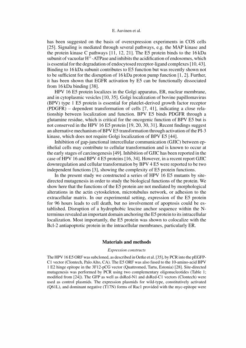
HPV 16 E5 coding sequence was expressed as a GFP-E5 fusion protein [35].There are no reliable antibodies available to the HPV 16 E5 protein and thus theGFP epitope was used to visualize the protein. Also, the E5 gene was clonedinto the 3F12-pCG vector in order to utilize the BPV E2 hinge epitope, which isdetectable with a monoclonal antibody [28]. This epitope was used to controlfor possible artefacts caused by the GFP epitope. The transfection efficiencyfor the GFP control plasmid was repeatedly shown to be higher than for theE5 expression plasmid. When transiently expressed in human epithelial HeLacells, GFP was present throughout the cytoplasm and also in the nucleus, whereasGFP-E5 was at the ER, in small vesicles, and in the Golgi (22 h; Fig. 1B, 2C).
Fig. 1. Cotransfection of HeLa cells with expression constructs for GFP-E5 (B) and hinge-E5(C) shows colocalization of both proteins. A, Hoechst nuclear stain

E5-wt-mutant
Cotransfection of GFP-E5 and hinge-E5 showed similar localization of the hinge-E5 protein and GFP-E5 confirming that the large GFP epitope did not affect E5localization (Fig. 1). A considerable decrease in the number of positive cells andin the level of E5-expression took place with time, and by the end of the 96 hfollow-up time no more E5-expressing cells could be found, suggesting lethaleffects of E5 expression. This applied to both GFP- and hinge-tagged E5 proteins,whereas the expression of the GFP control plasmid remained similar throughoutthe 96 h follow-up. We also cotransfected cells with E5 together with one of the redfluorescent protein plasmids dsRed-N1 or dsRed-C1 to confirm the death of E5-expressing cells. The number of cells coexpressing E5 and red fluorescent proteindecreased along with time, whereas the number of control cells coexpressing GFPand dsRed remained similar, confirming specific death of E5-expressing cells.
Point mutations within the E5 ORF were created by PCR. The spatial demandsof each individual amino acid as well as the predicted secondary structure of thewild type protein were retained (Table 1). In M1, M2, and M3, cysteine residuescapable of forming disulphur bridges were mutated to alanine. Dimerization hasbeen established for BPV 1 E5 [40] and suggested for HPV 16 E5 [14, 29]. In M5and M6 reactive hydroxyl groups of tyrosine residues putatively responsible forprotein–protein interactions within the membrane compartment were changed tophenylalanine, which is spatially similar to tyrosine. A phenylalanine residue wasmutated to neutral alanine in M7, and a putative serine-threonine–threonine caseinkinase II phosphorylation site at residues 8–10 was replaced with alanines in M9.In M10 a hydrophobic leucine membrane anchor sequence was mutated to pos-itively charged lysine residues (“positive-inside-rule”). Transfection efficiency,expression level, expression kinetics, and localization of wild-type and mutant E5proteins were similar, except for M10. Therefore most figures represent the resultswith wild-type E5, M9, and M10.
Subcellular localization of the E5 proteins
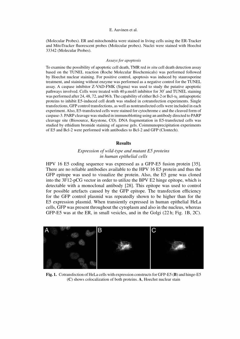
Based on earlier observations made by us and by others we studied the putativeGolgi localization of the different E5 proteins in HeLa cells. The E5 protein wasvisualized with the help of the GFP epitope without antibody staining. Partialcolocalization of the E5 fusion proteins with the intermediate-Golgi 58 K proteinwas observed (GFP control, wtE5, M9, and M10 are shown in Fig. 2), whereasno colocalization was observed with GM130, or golgin-97. There were littledifferences in the subcellular localization between the different E5 proteins asthe design of the mutations allowed us to expect (Table 1). In the case of M10,where a leucine membrane anchor was mutated to positively charged lysines, anadditional peripheral localization to membrane lamellipodia, actin-rich structuresat the leading edge of motile cells, was observed (Fig. 2G, arrows).
The partially vesicular fluorescence pattern of the E5 protein suggested theinvolvement of the endosomal machinery. However, staining of E5-expressingcells with antibodies to EEA-1, a marker for early endosomes [42], Rab-7 forlate endosomes [9], or LAMP-1 for lysosomes [8], respectively, did not show
E. Auvinen et al.
Fig
.2.
E5
prot
eins
show
part
ial
colo
caliz
atio
nw
ithth
ein
term
edia
teG
olgi
58K
prot
ein.
(A,C
,E,G
),G
FPflu
ores
cenc
esh
owin
gG
FPal
one
(vec
tor
cont
rol)
,wild
-typ
eE
5,M
9,an
dM
10,r
espe
ctiv
ely;
(B,D
,F,H
),st
aini
ngfo
rth
ein
term
edia
teG
olgi
58K
prot
ein.
Arr
ows
in(G
)po
intt
oM
10lo
caliz
edin
lam
ellip
odia
E5-wt-mutant
colocalization with E5 fluorescence, although these structures were present inE5-expressing as well as in control cells. Nor was any colocalization seen withβ-COP.
Effect of Rac1 GTPase on M10 localization
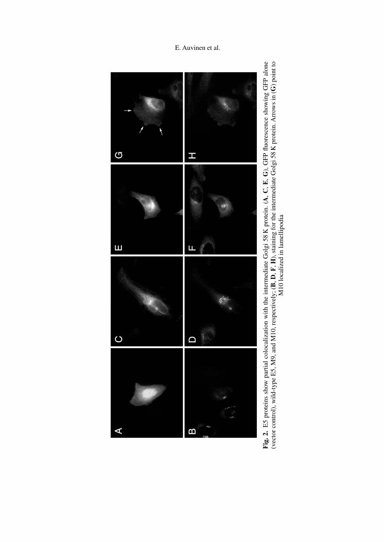
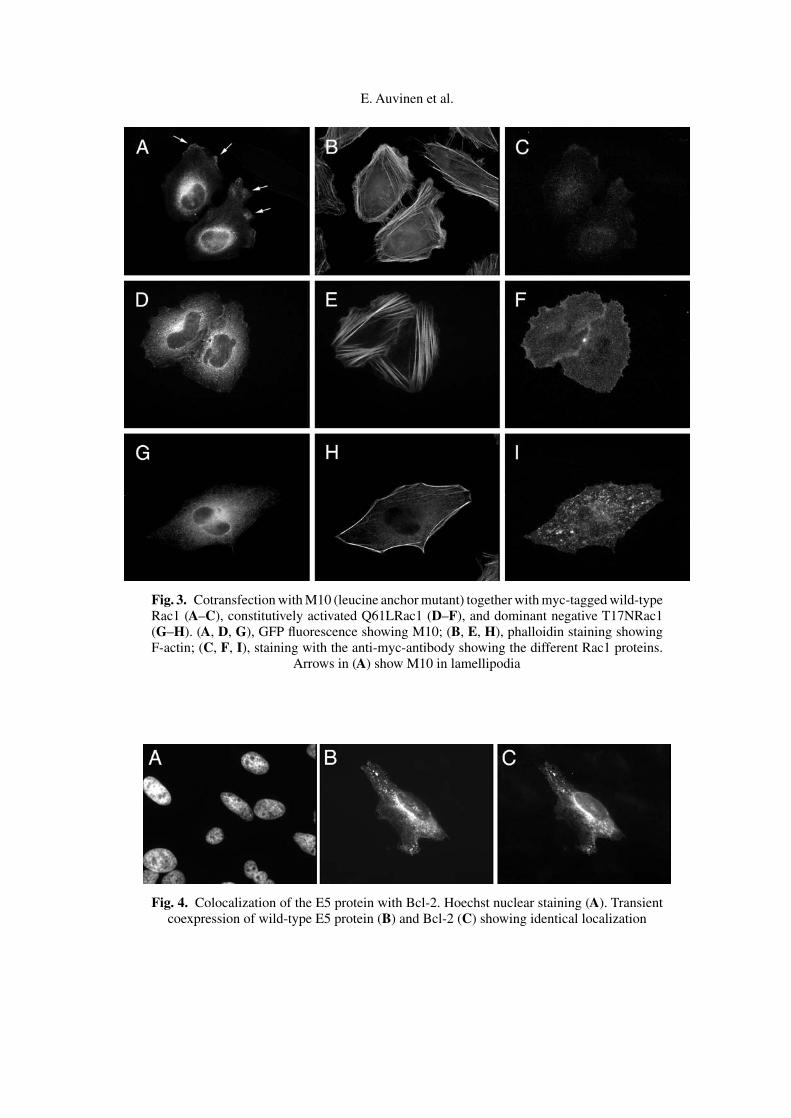
The small GTPase Rac1 plays a key role in the formation of lamellipodia [23].In a cotransfection experiment using wtE5, M9, or M10 either alone or togetherwith wild-type (wt), constitutively activated (Q61L), or dominant negative (T17N)Rac1, we aimed to study the mechanism responsible for the different intracellularlocalization of M10. When expressed alone, M10 was localized in lamellipodia,in the ER and in the Golgi (Fig. 2G). However, expression of M10 per se didnot induce or enhance the formation of lamellipodia. Staining for F-actin wassimilar to nontransfected cells. Transfection with wtRac1 increased the number oflamellipodia, where M10 accumulated together with cotransfected Rac1 ((Fig. 3A,arrows; 3C). The cells contained stress fibers (Fig. 3B) depicting Rac1 phenotype.In cells expressing high levels of Q61LRac1, the M10 E5 protein was at theperinucleus (Golgi) and in vesicles, and also at the cell periphery (3D), whileQ61LRac1 localized extensively to the plasma membrane (3F). Strong and denseF-actin stress fibers were observed (Fig. 3E). Peripheral localization of M10 inthe cells expressing high levels of T17NRac1 was abolished (3G), and T17NRac1showed mostly vesicular staining in the cytoplasm (3I). A reduction in actin stressfibers due to Rac1 inactivation was evident (Fig. 3H). Localization of wild-typeE5 or mutant M9 was not affected by any of the Rac1 constructs indicating thatlamellipodial localization and the effect of Rac1 activity on the localization of theprotein is specific for M10.
Assays for cell death and colocalization of E5 with Bcl-2
Because we observed decreased amounts of E5-expressing cells along with time,we decided to study the involvement of apoptosis at different time points. Cellswere TUNEL stained at 16, 18, 20, 22, 24, 48, 72, and 96 h post transfection.We observed less E5-expressing cells with time, and 96 h post transfection allE5-expressing cells had died. A chemical caspase inhibitor Z-VAD-FMK, orcoexpression of either Bcl-2 or Bcl-xL antiapoptotic proteins could not inhibitcell death. Nor did immunofluorescence for cytochrome c or cleaved caspase-3,or immunoblotting for PARP cleavage reveal apoptotic events. Staining with thefluorescent ER-Tracker, MitoTracker, or nuclear stains was similar to nontrans-fected cells. We thus suggest that cell death may take place due to unspecificeffects of E5 expression on cellular membrane compartments rather than due toapoptosis.
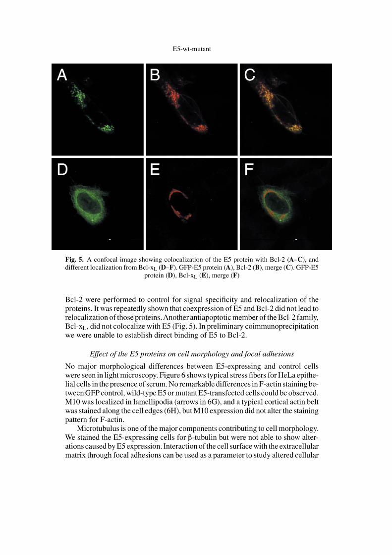
Most importantly, we found that the E5 protein colocalizes with Bcl-2 anti-apoptotic protein as shown by conventional (Fig. 4) as well as by confocal (Fig. 5)microscopy, although Bcl-2 did not inhibit E5-mediated cell death. All mutantE5 proteins colocalized with Bcl-2 as well. Single transfections with E5 and
E. Auvinen et al.
Fig. 3. Cotransfection with M10 (leucine anchor mutant) together with myc-tagged wild-typeRac1 (A–C), constitutively activated Q61LRac1 (D–F), and dominant negative T17NRac1(G–H). (A, D, G), GFP fluorescence showing M10; (B, E, H), phalloidin staining showingF-actin; (C, F, I), staining with the anti-myc-antibody showing the different Rac1 proteins.
Arrows in (A) show M10 in lamellipodia
Fig. 4. Colocalization of the E5 protein with Bcl-2. Hoechst nuclear staining (A). Transientcoexpression of wild-type E5 protein (B) and Bcl-2 (C) showing identical localization
E5-wt-mutant
Fig. 5. A confocal image showing colocalization of the E5 protein with Bcl-2 (A–C), anddifferent localization from Bcl-xL (D–F). GFP-E5 protein (A), Bcl-2 (B), merge (C). GFP-E5
protein (D), Bcl-xL (E), merge (F)
Bcl-2 were performed to control for signal specificity and relocalization of theproteins. It was repeatedly shown that coexpression of E5 and Bcl-2 did not lead torelocalization of those proteins.Another antiapoptotic member of the Bcl-2 family,Bcl-xL, did not colocalize with E5 (Fig. 5). In preliminary coimmunoprecipitationwe were unable to establish direct binding of E5 to Bcl-2.
Effect of the E5 proteins on cell morphology and focal adhesions
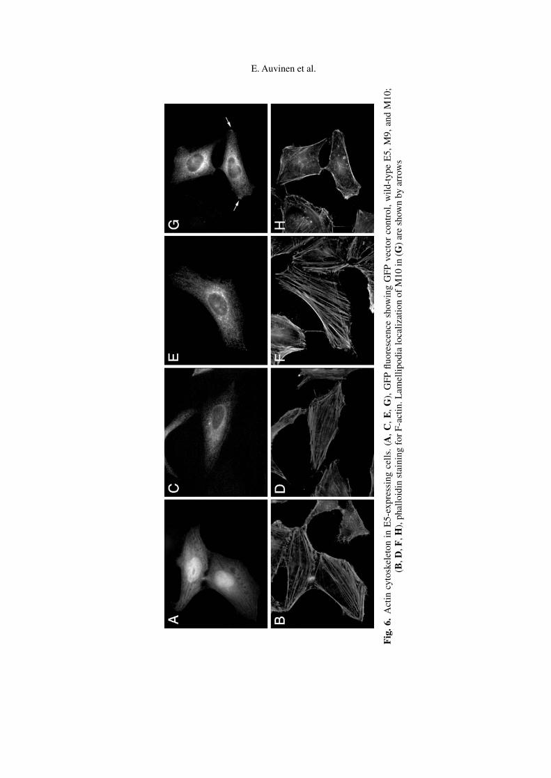
No major morphological differences between E5-expressing and control cellswere seen in light microscopy. Figure 6 shows typical stress fibers for HeLa epithe-lial cells in the presence of serum. No remarkable differences in F-actin staining be-tween GFP control, wild-type E5 or mutant E5-transfected cells could be observed.M10 was localized in lamellipodia (arrows in 6G), and a typical cortical actin beltwas stained along the cell edges (6H), but M10 expression did not alter the stainingpattern for F-actin.
Microtubulus is one of the major components contributing to cell morphology.We stained the E5-expressing cells for β-tubulin but were not able to show alter-ations caused by E5 expression. Interaction of the cell surface with the extracellularmatrix through focal adhesions can be used as a parameter to study altered cellular
E. Auvinen et al.
Fig
.6.
Act
incy
tosk
elet
onin
E5-
expr
essi
ngce
lls.
(A,
C,
E,
G),
GFP
fluor
esce
nce
show
ing
GFP
vect
orco
ntro
l,w
ild-t
ype
E5,
M9,
and
M10
;(B
,D,F
,H),
phal
loid
inst
aini
ngfo
rF-
actin
.Lam
ellip
odia
loca
lizat
ion
ofM
10in
(G)
are
show
nby
arro
ws
E5-wt-mutant
properties [39]. No differences in focal adhesions were seen as studied by stainingfor paxillin or other focal adhesion components (data not shown).
Discussion
In the present study site-directed mutants of the HPV 16 E5 gene were establishedin order to study the biological functions of the protein.As many cancers are causedby abnormalities in cell division, migration of cells, and cell adhesion – functionsthat depend on the cytoskeleton – we wanted to study the effects of E5 expressionon cell morphology. In the present study we used HeLa cells, which have beenconfirmed not to contain the E5 ORF of HPV [32] and can thus be applied to studythe morphological effects of the E5 protein. Most importantly, HeLa cells are anexcellent model to study cell morphology and changes therein, because they arelarge and flat, they naturally contain stress fibers, cortical actin, lamellipodia, andfilopodia, and these structures are readily formed in HeLa cells upon induction.Due to the lack of E5 antibodies the GFP epitope was used to detect the protein.Interestingly, the small BPV E2 hinge epitope [28] enabled the detection of theE5 protein and thus proved useful in tagging hydrophobic membrane proteins.
A total of eight mutants conserving the spatial demands and putative proteinconformation were constructed. All E5 proteins were shown to localize in the ERand Golgi, as has been previously shown by Conrad et al. and by us using mouseCOS cells or HaCaT human skin keratinocytes [10, 35]. Some colocalization wasfound with the 58 K protein, a resident of the Golgi intermediate compartment.Interestingly, Thomsen et al. have shown that the E5 protein changes the morphol-ogy of mouse fibroblasts and causes a block in endocytotic trafficking [46]. Toour knowledge colocalization of E5 with components of the endocytotic pathwayhas not been shown. In the present study, staining for antibodies to organelle-specific markers showed that E5-expressing cells contain early endosomes, lateendosomes, and lysosomes, respectively, but none of these markers colocalizedwith E5 fluorescence. Importantly, M10 with leucine-to-lysine mutations at aminoacids 22 and 23 in the first putative transmembrane domain revealed a sequencecontributing to the intracellular localization of the E5 protein. Disruption of amembrane anchor sequence caused partial relocalization of the protein to theplasma membrane-associated lamellipodia, which are formed in response to thesmall GTPase Rac1 activity [15, 48]. We showed that inactivation of endogenousRac1 function abolishes lamellipodia and, consequently, M10 localization. Themechanism for the lamellipodial localization still remains to be elucidated, al-though the possibility that M10 per se would induce lamellipodia formation wasexcluded.
Immortalized and transformed cells have altered morphology and motility[5, 33]. Earlier reports about E5 function suggest a possible involvement of theactin cytoskeleton. However, we did not observe major morphological effectsof the E5 proteins on the actin cytoskeleton despite of careful examination of alarge number of cells, although Thomsen et al. reported disruption of the F-actincytoskeleton in E5-expressing fibroblasts [46]. Microtubulus network and the

E. Auvinen et al.
components of focal adhesions, such as paxillin, were similar in E5-expressingand control cells. Paxillin binding and disruption of the actin cytoskeleton bythe E6 oncogene of bovine papillomavirus has been reported [47], and it wasrecently shown that paxillin binding is necessary but not sufficient for cellulartransformation by E6 [13]. HPV 16 E7 oncoprotein interacts with filamentousactin and represses transcription of fibronectin, an important molecule in celladhesion and migration [37]. The role of the E5 protein may be to increase thesusceptibility of the cells to E6 and E7 function.
We also showed a lethal, non-apoptotic effect of the E5 protein in humancervical epithelial cells when expressed at high levels for several days. We suggestthat the E5 protein may interfere with the membrane compartments of the cell andcause cell death by an unspecific mechanism. Transient and low-level expressionof the E5 protein seems to take place in natural infections, as suggested by Kellet al. [29], and this would allow cell survival. We cannot exlude the possibil-ity that cells expressing tiny amounts of the E5 protein would have survived.A considerable proportion of codons within the HPV 16 E5 ORF are ineffi-ciently translated in human cells. One could hypothesize that low and/or transientE5 expression would provide an evolutionary advantage, because it would leadto increased cell proliferation, whereas high or long-lasting expression wouldbe lethal. Bible et al. have shown that certain natural variants of the HPV16 E5 gene associate with neoplasia suggesting that natural mutations withinthe gene encoding the E5 protein may have an impact [6]. Interestingly,a role for E5 in later stages of viral replication has recently been reported[17, 18].
We find colocalization of the E5 protein with Bcl-2 highly intriguing. TheBcl-2 family consists of proapoptotic and antiapoptotic members, and their neteffect on cells seems to depend on a balance between these two opposite functions[36]. Bcl-2 is an antiapoptotic member of the Bcl-2 family, it resides in the ER,mitochondria, and nuclear membranes, and it has recently been shown to fulfillits antiapoptotic function from the ER as well as from mitochondria [45]. Inter-estingly, both pro- and anti-apoptotic effects of the E5 protein have been reported.Kabsch et al. have reported sensitization of human keratinocytes to osmotic stress-induced apoptosis by the E5 protein [26]. Recently it was published that theHPV 16 E5 protein would protect human primary keratinocytes from apoptosisinduced by UV B irradiation [50], or impair TRAIL- or FasL-mediated apoptosis[27]. Although a direct interaction of the E5 protein with the antiapoptotic Bcl-2protein could not be established in this study, E5 may interfere with Bcl-2 orother members of the family, thus modifying the cellular response to apoptoticsignals.
Acknowledgements
We thank A. Hall (University College London, UK) for the Rac1 constructs, H. Stenmark(Radium Hospital, Oslo, Norway), K. Karlsson (University of Umea, Sweden), and M. Zerial(EMBL) for the antibodies to EEA1, LAMP-1, and Rab 7, X. Li (University of Helsinki) forthe Bcl-2 and Bcl-xL constructs, Mrs. Rita Fingerroos and Mrs. Virpi Paivinen for excellent

E5-wt-mutant
technical assistance and T. Talpsepp for helpful discussions. Quattromed, Tartu, Estoniaprovided the 3F12-pCG expression vector and a BPV E2 hinge antibody. This study wassupported by the Finnish Cancer Institute, Maud Kuistila Memorial Foundation, as well asElla and Georg Ehrnrooth Foundation grants to E.A.
References1. Adam JL, Briggs MW, McCance DJ (2000) A mutagenic analysis of the E5 protein of
human papillomavirus type 16 reveals that E5 binding to the vacuolar H+-ATPase isnot sufficient for biological activity, using mammalian and yeast expression systems.Virology 272: 315–325
2. Ashby ADM, Meagher L, Campo MS, Finbow ME (2001) E5 transforming proteins ofpapillomaviruses do not disturb the activity of the vacuolar H+-ATPase. J Gen Virol 82:2353–2362
3. Ashrafi GH, Pitts JD, Faccini AM, McLean P, O’Brien V, Finbow ME, Campo MS (2000)Binding of bovine papillomavirus type 4 E8 to ductin (16 K proteolipid), down-regulationof gap junction intercellular communication and full cell transformation are independentevents. J Gen Virol 81: 689–694
4. Auvinen E, Crusius K, Steuer B, Alonso A (1997) Human papillomavirus type 16 E5protein. Int J Oncol 11: 1297–1304
5. Banyard J, Anand-Apte B, Symons M, Zetter BR (2000) Motility and invasion aredifferentially modulated by Rho family GTPases. Oncogene 19: 580–591
6. Bible JM, Mant C, Best JM, Kell B, Starkey WG, Raju KS, Seed P, Biswas C, Muir P,Banatvala JE, Cason J (2000) Cervical lesions are associated with human papillomavirustype 16 intratypic variants that have high transcriptional activity and increased usage ofcommon mammalian codons. J Gen Virol 81: 1517–1527
7. Burkhardt A, Willingham M, Gay C, Jeang KT, Schlegel R (1989) The oncoproteinof bovine papillomavirus is oriented asymmetrically in Golgi and plasma membranes.Virology 170: 334–339
8. Carlsson SR, Roth J, Piller F, Fukuda M (1988) Isolation and characterization ofhuman lysosomal membrane glycoproteins, h-lamp-1 and h-lamp-2. J Biol Chem 263:18911–18919
9. Chavrier P, Parton RG, Hauri HP, Simons K, Zerial M (1990) Localization of lowmolecular weight GTP binding proteins to exocytic and endocytic compartments. Cell62: 317–329
10. Conrad M, Bubb VJ, Schlegel R (1993) The human papillomavirus type 6 and 16 E5proteins are membrane-associated proteins which associate with the 16-kilodalton pore-forming protein. J Virol 67: 6170–6178
11. Crusius K, Auvinen E, Alonso A (1997) Enhancement of EGF- and PMA-mediatedMAP Kinase activation in cells expressing the human papillomavirus type 16 E5 protein.Oncogene 15: 1437–1444
12. Crusius K, Auvinen E, Steuer B, Gaissert H, Alonso A (1998) The human papillomavirustype 16 E5-protein modulates ligand-dependent activation to the EGF receptor family inthe human epithelial cell line HaCaT. Exp Cell Res 241: 76–83
13. Das K, Bohl J, Vande Pol SB (2000) Identification of a second transforming function inbovine papillomavirus type 1 E6 and the role of E6 interactions with paxillin, E6BP, andE6AP. J Virol 74: 812–816
14. Disbrow GL, Sunitha I, Baker CC, Hanover J, Schlegel R (2003). Codon optimization ofthe HPV-16 E5 gene enhances protein expression. Virology 311: 105–114

E. Auvinen et al.
15. Eaton S, Auvinen P, Luo L, JanYN, Simons K (1995) CDC42 and Rac1 control differentactin-dependent processes in the Drosophila wing disc epithelium. J Cell Biol 131:151–164
16. Faccini AM, Cairney M, Ashrafi GH, Finbow ME, Campo MS, Pitts JD (1996) Thebovine papillomavirus type 4 E8 protein binds to ductin and causes loss of gap junctionalintercellular communication in primary fibroblasts. J Virol 70: 9041–9045
17. Fehrmann F, Klumpp DJ, Laimins LA (2003) Human papillomavirus type 31 E5protein supports cell cycle progression and activates late viral functions upon epithelialdifferentiation. J Virol 77: 2819–2831
18. Genther SM, Sterling S, Duensing S, Munger K, Sattler C, Lambert P (2003) Quantitativerole or the human papillomavirus type 16 E5 gene during the productive stage of the virallife cycle. J Virol 77: 2832–2842
19. Goldstein DJ, Andresson T, Sparkowski JJ, Schlegel R (1992) The BPV-1 E5 protein, the16 kDa membrane pore-forming protein and the PDGF receptor exist in a complex thatis dependent on hydrophobic transmembrane interactions. EMBO J 11: 4851–4859
20. Goldstein DJ, Kulke R, DiMaio D, Schlegel R (1992) A glutamine residue in themembrane-associating domain of the bovine papillomavirus type 1 E5 oncoproteinmediates its binding to a transmembrane component of the vacuolar H+-ATPase. J Virol66: 405–413
21. Gu Z, Matlashewski G (1995) Effect of human papillomavirus type 16 oncogenes onMAP kinase activity. J Virol 69: 8051–8056
22. Halbert CL, Galloway DA (1988) Identification of the E5 open reading frame of humanpapillomavirus type 16. J Virol 62: 1071–1075
23. Hall A (1998) Rho GTPases and the actin cytoskeleton. Science 279: 509–51424. Ho SN, Hunt HD, Horton RM, Pullen JK, Pease LR (1989) Site-directed mutagenesis by
overlap extension using the polymerase chain reaction. Gene 77: 51–5925. Hwang ES, Nottoli T, DiMaio D (1995) The HPV16 E5 protein: expression, detection,
and stable complex formation with transmembrane proteins in COS cells. Virology 211:227–233
26. Kabsch K, Alonso A (2002) The human papillomavirus type 16 (HPV-16) E5 proteinsensitizes human keratinocytes to apoptosis induced by osmotic stress. Oncogene 21:947–953
27. Kabsch K, Alonso A (2002) The human papillomavirus type 16 E5 protein impairsTRAIL- and FasL-mediated apoptosis in HaCaT cells by different mechanisms. J Virol76: 12162–12172.
28. Kaldalu N, Lepik D, KristjuhanA, Ustav M (2000) Monitoring and purification of proteinsusing bovine papillomavirus E2 epitope tags. Biotechniques 28: 456–462
29. Kell B, Jewers RJ, Cason J, Pakarian F, Kaye JN, Best JM (1994) Detection of E5oncoprotein in human papillomavirus type 16-positive cervical scrapes using antibodiesraised to synthetic peptides. J Gen Virol 75: 2451–2456
30. Klein O, Polack GW, Surti T, Kegler-Ebo D, Smith SO, DiMaio D (1998) Role ofglutamine 17 of the bovine papillomavirus E5 protein in platelet-derived growth factorβ receptor activation and cell transformation. J Virol 72: 8921–8932
31. Kulke R, Horwitz BH, Zibello T, DiMaio D (1992) The central hydrophobic domain ofthe bovine papillomavirus E5 transforming protein can be functionally replaced by manyhydrophobic amino acid sequences containing a glutamine. J Virol 66: 505–511
32. Luft F, Klaes R, Nees M, Durst M, Heilmann V, Melsheimer P, von Knebel DoeberitzM (2001) Detection of integrated papillomavirus sequences by ligation-mediated PCR(DIPS-PCR) and molecular characterization in cervical cancer cells. Int J Cancer 92:9–17

E5-wt-mutant
33. Malliri A, Symons M, Hennigan RF, Hurlstone AF, Lamb RF, Wheeler T, Ozanne BW(1998) The transcription factor AP-1 is required for EGF-induced activation of rho-likeGTPases, cytoskeletal rearrangements, motility, and in vitro invasion of A431 cells. JCell Biol 143: 1087–1099
34. Oelze I, Kartenbeck J, Crusius K, Alonso A (1995) Human papillomavirus type 16 E5protein affects cell–cell communication in an epithelial cell line. J Virol 69: 4489–4494
35. Oetke C, Auvinen E, Pawlita M, Alonso A (2000) Human papillomavirus type 16 E5protein localizes to the Golgi apparatus but does not grossly affect cellular glycosylation.Arch Virol 145: 2183–2191
36. Reed JC (1997). Cytochrome c: can’t live with it – can’t live without it. Cell 91: 559–56237. Rey O, Lee S, Park NH (2000) Human papillomavirus type 16 E7 oncoprotein represses
transcription of human fibronectin. J Virol 74: 4912–491838. Rodrıguez MI, Finbow ME, Alonso A (2000) Binding of human papillomavirus 16 E5 to
the 16 kDa subunit c (proteolipid) of the vacuolar H+-ATPase can be dissociated from theE5-mediated epidermal growth factor receptor overactivation. Oncogene 19: 3727–3732
39. Sattler M, Pisick E, Morrison PT, Salgia R (2000) Role of the cytoskeletal protein paxillinin oncogenesis. Crit Rev Oncogen 11: 63–76
40. Schlegel R, Wade-Glass M (1987) E5 transforming polypeptide of bovine papillomavirus.Cancer Cells 5: 87–91
41. Sparkowski J, Anders J, Schlegel R (1995) E5 oncoprotein retained in the endoplasmicreticulum/cis Golgi still induces PDGF receptor autophosphorylation but does nottransform cells. EMBO J 14: 3055–3063
42. Stenmark H, Aasland R, Toh BH, D’Arrigo A (1996) Endosomal localization of theautoantigen EEA1 is medited by a zinc-binding FYVE finger. J Biol Chem 271: 24048–24054
43. Straight SW, Herman B, McCance DJ (1995) The E5 oncoprotein of humanpapillomavirus type 16 inhibits the acidification of endosomes in human keratinocytes.J Virol 69: 3185–3192
44. Suprynowicz FA, Sparkowski J, Baege A, Schlegel R (2000) E5 oncoprotein mutantsactivate phosphoinositide 3-kinase independently of platelet-derived growth factorreceptor activation. J Biol Chem 275: 5111–5119
45. Thomenius MJ, Wang NS, Reineks EZ, Wang Z, Distelhorst CW (2003) Bcl-2 on theendoplasmic reticulum regulates bax activity by binding to BH3-only proteins. J BiolChem 278: 6243–6250
46. Thomsen P, van Deurs B, Norrild B, Kayser L (2000) The HPV 16 E5 oncogene inhibitsendocytic trafficking. Oncogene 19: 6023–6032
47. Tong X, Salgia T, Li JL, Griffin JD, Howley PM (1997) The bovine papillomavirus E6oncoprotein interacts with paxillin and disrupts the actin cytoskeleton. Proc Natl AcadSci 94: 4412–4417
48. Van Aelst L, D’Souza-Schorey C (1997) Rho GTPases and signaling networks. GenesDevel 11: 2295–2322
49. Yamasaki H (1990) Gap junctional intercellular communication and carcinogenesis.Carcinogenesis 11: 1051–1058
50. Zhang B, Spandau DF, Roman A (2001) E5 protein of human papillomavirus type 16protects human foreskin keratinocytes from UV B-irradiation-induced apoptosis. J Virol76: 220–231
Author’s address: Eeva Auvinen, Haartman Institute, Department of Virology, P.O. Box21, University of Helsinki, FI-00014, Finland; e-mail: [email protected]


