Embed Size (px)
Citation preview

APPLIED AND ENVIRONMENTAL MICROBIOLOGY,0099-2240/01/$04.0010 DOI: 10.1128/AEM.67.1.179–184.2001
Jan. 2001, p. 179–184 Vol. 67, No. 1
Copyright © 2001, American Society for Microbiology. All Rights Reserved.
Human Adenoviruses and Coliphages in Urban Runoff-Impacted Coastal Waters of Southern California
SUNNY JIANG,1* RACHEL NOBLE,2 AND WEIPING CHU1
Department of Environmental Analysis and Design, University of California, Irvine, California 92697,1
and Southern California Coastal Water Research Project, Westminster, California, 92683-52182
Received 9 August 2000/Accepted 17 October 2000
A nested-PCR method was used to detect the occurrence of human adenovirus in coastal waters of SouthernCalifornia. Twenty- to forty-liter water samples were collected from 12 beach locations from Malibu to theborder of Mexico between February and March 1999. All sampling sites were located at mouths of major riversand creeks. Two ultrafiltration concentration methods, tangential flow filtration (TFF) and vortex flow filtra-tion (VFF), were compared using six environmental samples. Human adenoviruses were detected in 4 of the 12samples tested after nucleic acid extraction of VFF concentrates. The most probable number of adenoviralgenomes ranged from 880 to 7,500 per liter of water. Coliphages were detected at all sites, with the concen-tration varying from 5.3 to 3332 PFU/liter of water. F-specific coliphages were found at 5 of the 12 sites, withthe concentration ranging from 5.5 to 300 PFU/liter. The presence of human adenovirus was not significantlycorrelated with the concentration of coliphage (r 5 0.32) but was significantly correlated (r 5 0.99) withF-specific coliphage. The bacterial indicators (total coliforms, fecal coliforms, and enterococci) were found toexceed California recreational water quality daily limits at 5 of the 12 sites. However, this excess of bacterialindicators did not correlate with the presence of human adenoviruses in coastal waters. The results of thisstudy call for both a reevaluation of our current recreational water quality standards to reflect the viral qualityof recreational waters and monitoring of recreational waters for human viruses on a regular basis.
Southern California and its beaches are unique recreationaland economic resources due to the long coastline and year-round temperate climate. More than 100 million people world-wide visit Southern California beaches annually to surf, swim,sunbathe, snorkel, and scuba dive (State of California Eco-nomic Resources data, 1993). Therefore, the safety of beachwaters is of concern to both the state and county health de-partments and beachgoers. For example, the closure of Hun-tington Beach in Orange County, Calif., due to elevated levelsof bacterial indicators during the summer of 1999, caused atremendous economic loss to the county and state (www.ocsd.com/Environment/Monitoring The Environment) and raisedconcerns among local residents regarding the safety of coastalrecreational waters. Southern California is one of the mostdensely populated coastal regions in the country. Populationgrowth results in conversion of open land into nonpermeablesurfaces, which increases the rate of runoff and can impactwater quality through addition of sediment, toxic chemicals,microbial pathogens, and nutrients to the coastal ocean.
To govern the public health and safety of recreationalbeaches, the State of California has instated beach-monitor-ing programs in accordance with U.S. Environmental Protec-tion Agency (EPA) policy. Bacterial indicators, such as totalcoliforms, fecal coliforms, and enterococci, are routinely mon-itored by more than 20 agencies at 510 stations along theSouthern California coast (Southern California Coastal WaterResearch Project, 1998 [http://www.sccwrp.org/regional/98bight/micbio/]). Routine bacteriological monitoring has reduced the
incidence of waterborne diseases related to recreational watercontact, but water-related illnesses, particularly those causedby viral pathogens, are still of concern (3).
More than 100 different types of viruses are found in humanwaste and are potentially transmitted by water (3). These vi-ruses are more resistant to environmental conditions and sew-age treatment processes, including chlorination and UV radi-ation, than many of the sewage-associated bacteria (11). Inlaboratory studies, enteric viruses have been reported to sur-vive for 2 to 130 days in seawater. These survival periodssurpass those reported for coliform bacteria in similar environ-ments (23). Therefore, the use of bacterial indicators to predictthe virological quality of water is questionable.
The human enteric virus group, which includes Norwalkvirus, rotavirus, hepatitis A virus, adenovirus, and enterovirus,is one of the leading causes of human illness. Adenoviruses arethe only human enteric viruses that contain DNA rather thanRNA. Specifically, adenoviruses 40 and 41 have been recog-nized as important etiological agents of gastroenteritis in chil-dren (10, 32), second in importance only to rotaviruses. Re-spiratory adenoviruses are capable of causing large, persistentepidemics (22), and it is suspected that they may be moreimportant than enteric viruses as a contagion during recre-ational water activities because of their potential transmissionin aerosols. Studies conducted in Europe have suggested usingadenovirus as an index of human viral pollution since this viruswas most often detected in aquatic samples (27). Adenoviruseshave been consistently found in greater numbers than entero-viruses in raw sewage around the world (18, 20, 21) and aresubstantially more stable than either poliovirus or hepatitis Avirus in tap water and seawater (13). For example, adenovirussurvives three to five times longer in seawater than poliovirus,taking 99 days for a 2-log reduction of infectivity (13). The
* Corresponding author. Mailing address: Department of Environ-mental Analysis and Design, University of California, Irvine, CA92697. Phone: (949) 824-5527. Fax: (949) 824-2056. E-mail: [email protected].
179

presence of adenovirus in U.S. coastal water has not beeninvestigated. Here we report the detection of human adenovi-rus in Southern California coastal waters by nested PCR and apreliminary examination of their relationship to coliphage, F-specific coliphage, and bacterial indicators.
MATERIALS AND METHODS
Sampling sites. Water samples were taken from the wavewash (point zerolocation where the freshwater input meets the ocean) of 12 freshwater outletsfrom Malibu to the Mexican border between 8 February and 1 March 1999 (Fig.1). The locations sampled were Malibu Lagoon, Santa Monica Canyon stormdrain, Los Angeles River, San Gabriel River, Santa Ana River, Aliso Creek, SanJuan Creek, San Luis Rey River, Moonlight Creek, Los Penosquitos Lagoon, SanDiego River, and Tijuana River. Twenty- to forty-liter water samples werecollected with acid-washed and sample-rinsed carboys at each sampling site andtransported back to the laboratory for immediate processing. The sample salinitywas determined by refractometry.
Concentration of viruses from water samples. To compare the efficiency ofviral recovery, six of the water samples (from Malibu Lagoon, Santa MonicaCanyon storm drain, Los Angeles River, San Gabriel River, Santa Ana River,and Aliso Creek) were concentrated both by tangential flow filtration (TFF) witha 30-kDa spiral filter cartridge (29) and by vortex flow filtration (VFF) with a100-kDa filter cartridge (19). Samples from the other six locations were concen-trated using only VFF.
Using TFF, 20 liters of water was first sequentially filtered through a 142-mm-diameter glass fiber filter and a 0.22-mm-pore-size Durapore filter to removeparticulates. This filtration step is necessary to prevent clogging of the spiral filtercartridge (Amicon, Inc.). The filtrates were concentrated from 20 liters to ca. 150ml using the TFF system and then further concentrated using a Centriprep-30ultraconcentration unit to a final volume of about 1 ml (R. Noble and J. Fuhr-man, submitted for publication). Using VFF, 20-liter water samples were con-
centrated directly to 40 to 80 ml without removal of particulates from the sample(19, 24).
Detection of indicator bacteria and coliphages. Water samples were collectedat the same time from all sites for analysis of total coliforms, fecal coliforms, andenterococci by local agencies following the EPA standard protocols for bacterialindicator analysis (9). Coliphage densities were determined using either freshlyVFF-concentrated or unconcentrated water samples. One milliliter and 0.1 ml ofsample were mixed with 1 ml of exponential Escherichia coli host in 1% soft agar,then overlaid over bottom nutrient agar. Two E. coli hosts ATCC 15597 and HS(pFamp)R, were used. E. coli ATCC 15597 is a general host for both DNA andRNA coliphage; E. coli HS (pFamp)R contains a plasmid coding for bothampicillin and streptomycin resistance and is a specific host for F-specific co-liphage (25). When HS (pFamp)R was employed the bottom agar contained 15mg each of ampicillin and streptomycin per ml to prevent background growth ofindigenous bacteria. On each host, plaques were enumerated after 12 h ofincubation at 37°C.
Detection of human adenovirus by nested PCR. Concentrated water sampleswere either used directly for PCR or further purified to remove PCR inhibitorsusing the method originally developed by Boom et al. (4) with minor modifica-tions. In brief, 50 ml of viral concentrate was lysed by 900 ml of guanidiniumthiocyanate (GuSCN) lysis buffer at room temperature for 10 min. Then 40 ml ofsilica particles (Sigma Chemical Inc.) was added and nucleic acids were adsorbedat room temperature for 10 min with gentle shaking. Silica beads were pelleted,washed, and briefly dried. The nucleic acid was eluted from the beads using 50ml of Tris-EDTA buffer at a temperature of 56°C.
For samples showing negative results by both the above methods, 10 ml ofVFF concentrate was further concentrated by ultracentrifugation at 41,000 rpm(Beckman rotor SW41) for 2 h. The pellets were resuspended in 200 ml ofTris-EDTA buffer and nucleic acid purified by GuSCN extraction as describedabove.
Nested PCR for human adenovirus was performed following the protocol ofPina et al. (27) with minor modifications. Adenovirus-specific primers were59-GCCGCAGTGGTCTTACATGCACATC-39 and 59-CAGCACGCCGCGG
FIG. 1. Sampling locations for detection of human adenoviruses, coliphage, and fecal bacterial indicators in coastal waters of SouthernCalifornia. All sampling sites are located at the mouth of a river or creek: 1, Malibu Lagoon; 2, Santa Monica Canyon Creek; 3, Los Angeles River;4, San Gabriel River; 5, Santa Ana River; 6, Aliso Creek; 7, San Juan Creek; 8, San Luis Rey River; 9, Moonlight Creek; 10, Los PenosquitosLagoon; 11, Sand Diego River; 12, Tijuana River.
180 JIANG ET AL. APPL. ENVIRON. MICROBIOL.
ATGTCAAAGT-39, yielding an amplicon of 300 bp. The nested primers were59-GCCACCGAGACGTACTTCAGCCTG-39 and 59-TTGTACGAGTACGCGGTATCCTCGCGGTC-39, with the resulting amplicon being 143 bp. For eachPCR, 1 ml of sample was used in a 25-ml reaction mixture containing 10 mMTris-HCl (pH 8.3), 50 mM KCl, 13 TaqM (Eppendorf Inc.), 1.3 mM MgCl2, 200mM deoxynucleoside triphosphosphate and a 0.1 mM concentration of eachprimer. The PCR cycles were 94°C for 4 min, followed by 30 cycles of 92°C for1.5 min, 55°C for 1.5 min, and 72°C for 2 min and a final extension at 72°C for10 min. The second round of PCR was performed with the nested primers usingthe same conditions as described above. PCR products were resolved on 2%agarose. For PCR-positive samples, multiple analyses (5 to 12 replicates) wereperformed for each sample to estimate the most probable number using Tho-mas’s formula (9).
Recovery of VNA by GuSCN extraction. To determine the viral nucleic acid(VNA) recovery using the GuSCN-silica bead extraction method and the pres-ence of PCR inhibition, concentrated seawater samples from Malibu Lagoon,Moonlight Creek, and San Diego River that were negative for adenovirus bynested PCR were seeded with 105 viral genomes of sample per ml. The seededsample was extracted by the GuSCN-silica bead method, and serial dilutionswere made in deionized (DI) water to determine the recovery of viral genomeafter extraction. In parallel, the seeded sample was also diluted with the con-centrated seawater to determine the effect of PCR inhibition among concen-trated water samples. Seeding and extraction were performed twice using eachsample to determine the variability associated with these procedures.
Statistical analysis. Pearson linear correlation was used to analyze the rela-tionship between the abundance of human viruses and other parameters testedin this study. The correlation was considered significant at a 95% confidencelevel. The Microsoft Excel program (Microsoft Inc.) was used for the statisticalanalysis.
RESULTS
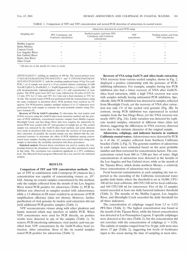
Comparison of TFF and VFF concentration methods. Theuse of TFF in combination with Centriprep-30 (Amicon Inc.)concentration was capable of concentrating viruses ca. 105-fold. Among six coastal samples concentrated by this method,only the sample collected from the mouth of the Los AngelesRiver tested PCR positive for adenovirus (Table 1). PCR in-hibition was observed in samples seeded with adenoviruses,while a 1:5 dilution in DI water resulted in an increase of PCRamplification efficiency (data not shown). However, furtherpurification of viral genome by nucleic acid extraction did notyield additional PCR-positive samples (Table 1).
VFF coconcentrates viruses along with other plankton andhumic material which may interfere or inhibit PCR. WhenVFF concentrates were used for PCR directly, no positiveresults were detected in any of the samples (Table 1). Toremove PCR-interfering substances, viral nucleic acid was pu-rified from VFF concentrates by the GuSCN-silica bead ex-traction. After extraction, three of the six coastal samplestested PCR positive for adenovirus (Table 1).
Recovery of VNA using GuSCN and silica beads extraction.VNA recovery from various seeded samples, shown in Fig. 2,displayed a positive relationship with the presence of PCR-inhibiting substances. For example, samples having less PCRinhibition also had a lower recovery of VNA after GuSCN-silica bead extraction, while a high VNA recovery was seenwith samples initially having substantial PCR inhibition. Spe-cifically, little PCR inhibition was detected in samples collectedfrom Moonlight Creek, yet the recovery of VNA after extrac-tion was only 1% of the seeded viral genome (Fig. 2B). Incontrast, strong PCR inhibition was experienced in seededsamples from the San Diego River, yet the VNA recovery wasnearly 100% (Fig. 2A). Little variation was detected for repli-cate seeded samples, extracted at different times (data notshown), suggesting the differences in VNA recovery observedwere due to the intrinsic character of the original sample.
Adenovirus, coliphage, and indicator bacteria in southernCalifornia coastal waters. Adenoviruses were detected by PCRin 4 of the 12 samples collected from Southern Californiabeaches (Table 2; Fig. 3). The genomic numbers of adenovirusin each sample were estimated based on the most probablenumber and then corrected for concentration factors. The con-centrations varied from 880 to 7,500 per liter of water. Highconcentrations of adenovirus were detected at the mouths ofthe Los Angeles and San Gabriel rivers, while at the mouth ofthe Tijuana River, which drains northern Mexico, a relativelylower concentration of adenovirus was detected.
Fecal bacterial contamination at each sampling site was re-ported as the exceeding of the California recreational waterquality daily limits, where the threshold is set at 10,000, CFU/100 ml for total coliforms, 400 CFU/100 ml for fecal coliforms,and 104 CFU/100 ml for enterococci. Five of the 12 samplestested exceeded at least one daily bacterial indicator threshold(Table 2). The mouths of the Malibu Lagoon, Los AngelesRiver, and Moonlight Creek exceeded the daily threshold forall three indicators.
The concentration of coliphage ranged from 5.3 to 3,332PFU/liter (Table 2). The highest concentration was found atthe mouth of the Tijuana River, while the lowest concentrationwas detected at Los Penosquitos Lagoon. F-specific coliphageswere detected at five sites (Table 2), but the concentration didnot correlate with the concentration of coliphage (r 5 0.20).The salinity of samples ranged from 9 to 34 ppt, with most sitesabove 27 ppt (Table 2), suggesting low levels of freshwaterinput to the ocean during the time of sampling at most sites.
TABLE 1. Comparison of TFF and VFF concentration and nested-PCR detection of adenovirus in coastal waters
Sampling sitea
Adenovirus detection by nested PCR using:
TFF, Centriprep, andCentricon concentrates
Purified nucleic acid from TFF,Centriprep, and Centricon
concentrates
VFFconcentrates
Purified nucleic acid fromVFF concentrates
Malibu Lagoon 2 2 2 2Santa Monica 2 2 2 2Canyon CreekLos Angeles River 1 1 2 1San Gabriel River 2 2 2 1Santa Ana River 2 2 2 1Aliso Creek 2 2 2 2
a All sites are at the mouth of a river or creek.
VOL. 67, 2001 HUMAN ADENOVIRUS AND COLIPHAGE IN COASTAL WATERS 181

DISCUSSION
This study demonstrates that ultrafiltration combined withnested PCR is an effective method of detecting human adeno-viruses from 20 liters of coastal water. Ultrafiltration concen-tration requires minimal manipulation of the water sample,avoiding the complicated procedures that cause viral inactiva-tion (i.e., pH adjustment) or PCR inhibition (i.e., beef extractelution and organic flocculation) used in the EPA standardfiltration-elution method. Compared with the EPA standardviral concentration method, in which volumes of at least 100liters of water are typically filtered, the ultrafiltration methodhas a higher efficiency of viral recovery (24).
In comparing the two ultrafiltration methods used in thisstudy, TFF concentration required preremoval of plankton andother suspended solids; therefore, the final viral concentratescontained fewer potential PCR inhibitors. With TFF, adeno-
virus was detected directly by PCR without the need for furtherpurification of viral nucleic acid. This method offers a distinctadvantage in that viral particles are intact prior to PCR. Pro-tected by the viral protein coat, VNA is not subjected to deg-radation due to nuclease activity. This is particularly importantfor detecting RNA viruses such as enterovirus, because RNA isknown to be rapidly degraded once exposed to the environ-ment. This method has been successfully used for PCR detec-tion of enterovirus in Santa Monica Bay in a previous study(Noble and Fuhrman, submitted for publication). In contrast,the VFF method is more time-efficient than TFF, by bypassingthe filtration of a large volume of water prior to viral concen-tration, but tends to concentrate more PCR inhibiting sub-stances with the viruses. VNA extraction and purification arenecessary to yield PCR-positive results. In the side-by-sidecomparison of the two methods using environmental water
FIG. 2. Efficiency of VNA recovery from environmental samples by the GuSCN-silica bead extraction method. Concentrated samples fromeither San Diego River (A) or Moonlight Creek (B) were seeded with adenoviruses to a final concentration of 105/ml. Lane 1, negative control;lanes 2 to 7, seeded samples were diluted from 100 to 1025 in seawater concentrates and detected by nested PCR; lanes 8 to 13, seededenvironmental samples were extracted by the GuSCN-silica bead method and diluted with sterilized DI water from 100 to 1025, respectively; laneM, 25-bp molecular weight ladder.
TABLE 2. Adenovirus, coliphage, and bacterial indicators in Southern California beach waters
Samplinglocationa
Date(day/mo/yr)
Salinity(‰)
Indicatorexceedanceb
Coliphage(PFU/liter)c
F-specific coliphage(PFU/liter)d
Adenovirus(genomes/liter)e
Malibu Lagoon 2/8/99 10 Tc, Fc, En 192 6 40.7 ,2Santa Monica 2/8/99 9 En 96 6 0.2 5.5Canyon CreekLos Angeles River 2/9/99 28 Tc, Fc, En 472.5 6 112 300 7.5 3 103
San Gabriel River 2/16/99 28 106.2 6 30.7 38 2.3 3 103
Santa Ana River 2/16/99 33 9.5 6 1.5 ,2 9.24 3 102
Aliso Creek 2/16/99 31 23 6 16 4.6
San Juan Creek 3/1/99 24 En 20.5 6 6 ,6.5San Luis Rey River 3/1/99 27 20 6 13.9 ,3Moonlight Creek 3/1/99 34 Tc, Fc, En 37.5 6 4.4 ,3Los Penosquitos Lagoon 2/22/99 32.5 5.3 6 2.5 ,3.53San Diego River 2/22/99 31 367.5 6 22.7 ,3
Tijuana River 2/22/99 29.5 3,332 6 80.9 22.6 6 7.8 8.8 3 102
a All sites are near mouths of rivers or creeks.b Bacterial indicators—total coliform (Tc), fecal coliform (Fc), and enterococci (En)—that exceed California recreational water daily limits.c Plaque assay using E. coli ATCC 15597 as a host.d Plaque assay using E. coli HS (pFamp)R as a host.e Determined by nested PCR.
182 JIANG ET AL. APPL. ENVIRON. MICROBIOL.

samples, VFF detected adenoviruses more often than TFF(Table 1). Based on a small number (six) of environmentalsamples collected during this study, it appears that the TFFmethod has a lower viral recovery than VFF. This may be dueto the loss of particle-associated viruses when samples werefiltered to remove other plankton and suspended solids priorto TFF concentration. Many viruses are known to attach toparticulate material. Previous studies have indicated that alarge portion of viruses can be lost during 0.2-mm-pore-sizefiltration (24).
The PCR detection of viruses in coastal waters offers advan-tages over current tissue culture methods. PCR is both lesslaborious and highly sensitive (e.g., see references 2, 7, 16). Inaddition, the method is capable of detecting viruses that areeither difficult to grow in tissue culture or replicate withoutproducing cytopathic effects in cells (6, 26). Adenovirus be-longs to this group of viruses and therefore is not easily de-tected by culture methods. Furthermore, the PCR method isalso highly specific and capable of differentiating differenttypes of viruses. The primers used in this study were previouslyshown to specifically amplify 47 serotypes of human adenovi-ruses (28). During our experiments, important control mea-sures were included to ensure the quality of our PCR results.Precautions were taken to prevent cross-contamination, i.e.,UV sterilization of PCR equipment and the working environ-ment, utilization of aerosol tips, etc. Positive and negativecontrols were included with each series of reactions.
The application of the nested-PCR protocol in our studyfurther increased the sensitivity and specificity of detection.The nested protocol for adenovirus detection was shown in aprevious study (1) to have a sensitivity of detecting a singlepurified viral particle. Therefore, nested PCR provided thesensitivity level necessary to detect a few viral particles in thesample concentrate. Nested PCR also increased the specificityof detection. Routinely, specific PCR products are often con-firmed by probing with an internal oligonucleotide probe (12,15, 31). In the nested PCR assay, two specific internal primersare used, which increases the specificity of reaction in much thesame way.
A major obstacle of PCR detection of viruses in aquaticenvironmental samples is the presence of organic material thatinhibits the PCR. As a result, many studies have focused ondeveloping an efficient method of VNA extraction from suchcomplex samples (e.g., 12, 14, 17, 31). Puig et al. (28) compareda simple GuSCN-silica bead method with several other meth-ods for VNA extraction and found this method to be the mostefficient at recovering viral RNA and/or DNA from complexsamples. In this study, we have shown that the efficiency ofextraction of this method was sample dependent (Fig. 2).Therefore, it was not possible to have a general correctionfactor to correct for losses during purification procedures.
Of the 12 samples tested in this study, 4 were positive forhuman adenovirus. The presence of these human viruses didnot correlate with the exceedance of daily limits of bacterialindicators in coastal waters. At the three sites where all threeindicator bacteria were above daily limits, the mouths ofMalibu Lagoon, Los Angeles River, and Moonlight Creek,human viruses were only detected at the Los Angeles Riversite. In contrast, while no bacterial indicator exceeded the dailythreshold at the mouths of the San Gabriel and Santa Anarivers, viral concentrations of 2,300 and 924/liter were detectedat each site, respectively. These results support previous find-ings by Griffin et al. (15) for the coastal marine environmentand suggest that the current coastal recreational water qualitystandards are inadequate to reflect the viral quality of thewater.
The presence of human viruses was also not correlated withthe concentration of coliphage. The Tijuana River had thehighest concentration of coliphage but a relatively low concen-tration of adenovirus. However, a correlation exists betweenthe abundance of human adenovirus and F-specific coliphage.The correlation coefficient for samples taken from the mouthsof the Los Angeles, San Gabriel, Santa Ana, and Tijuana riverswas 0.99. These results agree with the previous report of asignificant association of F-specific phage with human entericviruses in oysters and their harvest waters (8) yet differ fromthat reported for Florida coastal waters (15), where high fre-quencies of human viruses were detected (95%) by PCR butfew or no coliphage were found in their samples. Future stud-ies that include a broader geographical area of the marinecoast and a larger database are necessary to provide insightsinto the utility of F-specific coliphage as a human viral indica-tor.
Human adenoviruses were detected at the mouths of fourmajor urban rivers in Southern California, pointing to urbanrunoff as a source of coastal viral contamination. This is notsurprising, because a wide distribution of adenovirus has beenpreviously reported for urban river waters (30) and a riverestuary (5). It is noteworthy that the river flow rate at all foursites was relatively low, as indicated by the salinity, at the timeof sample collection. This was due to an unseasonally low levelof precipitation during the winter of 1999. A higher level ofcontamination may be expected during heavy rainfall.
Although it is alarming to detect the presence of humanviruses in one-third of the beach locations tested in SouthernCalifornia, little is known about the infectivity of these viruses.The PCR method detects both infectious and damaged viruses.Caution should be made at the interpretation of these results.If none or only a very small portion of viruses are infectious,
FIG. 3. Detection of adenoviral genomes from the VFF-concen-trated environmental samples by nested PCR. Lane N, negative con-trol; lane P, positive control; lane 1, mouth of Los Angeles River; lane2, mouth of San Gabriel River; lane M, 100-bp molecular weightladder.
VOL. 67, 2001 HUMAN ADENOVIRUS AND COLIPHAGE IN COASTAL WATERS 183

there is little impact or public health concern. The detection ofadenovirus should be viewed as an index for human fecalpollution and the presence of other human viruses. Therefore,this study calls for a reevaluation of our current water qualitystandards to reflect the viral quality of recreational waters andto possibly include monitoring of recreational waters for hu-man viruses in certain areas.
ACKNOWLEDGMENTS
Funding for this research was provided by a California Sea Grantawarded to S. Jiang (grant NA66RG0477).
Special thanks go to the Southern California Coastal ResearchProject and local agencies for close collaboration on the sample col-lection and determination of bacterial indicators in water samples. Wealso thank J. A. Fuhrman (University of Southern California) forproviding working space and equipment for R. T. Noble.
REFERENCES
1. Allard, A., B. Albinsson, and G. Wadell. 1992. Detection of adenoviruses instools from healthy persons and patients with diarrhea by two-step polymer-ase chain reaction. J. Med. Virol. 37:149–157.
2. Atmar, R. L., T. G. Metcalf, F. H. Neil, and M. K. Esteas. 1993. Detection ofenteric viruses in oysters by using the polymerase chain reaction. Appl.Environ. Microbiol. 59:631–635.
3. Berg, G. (ed.). 1983. Viral pollution of the environment. CRC Press, BocaRaton, Fla.
4. Boom, R., C. J. A. Sol, M. M. Salimans, C. L. Jansen, P. M. E. Wertheim-VanDillen, and J. Van Der Noordaa. 1990. Rapid and simple method for puri-fication of Nucleic Acids. J. Clin. Microbiol. 28:495–503.
5. Castingnolles, N., F. Petit, I. Mendel, L. Simon, L. Cattolico, and C. Buffet-Janvresse. 1998. Detection of adenovirus in the waters of the Seine riverestuary by nested-PCR. Mol. Cell. Probes 12:175–180.
6. Chapron, C. D., N. A. Ballester, J. H. Fontaine, C. N. Frades, and A. B.Margonlin. 2000. Detection of astroviruses, enteroviruses, and adenovirustypes 40 and 41 in surface waters collected and evaluated by the informationcollection rule and an integrated cell culture-nested PCR procedure. Appl.Environ. Microbiol. 66:2520–2525.
7. Chung, H., L. A. Jaykus, and M. D. Sobsey. 1996. Detection of humanenteric viruses in oysters by in vivo and in vitro amplification of nucleic acids.Appl. Environ. Microbiol. 62:3772–3778.
8. Chung, H., L.-A. Jaykus, G. Lovelace, and M. D. Sobsey. 1998. Bacterio-phages and bacteria as indicators of enteric viruses in oysters and theirharvest waters. Water Sci. Technol. 38:37–44.
9. Clesceri, L. S., A. E. Greenberg, and A. D. Eaton. 1998. Standard methodsfor the examination of water and wastewater, 20th ed. American publichealth association, Washington, D.C.
10. Cruz, J. R., P. Caceres, F. Cano, J. Flores, A. Bartlett, and B. Torun. 1990.Adenovirus types 40 and 41 and rotaviruses associated with diarrhea inchildren from Guatemala. J. Clin. Microbiol. 28:1780–1784.
11. De Leon, R., and L. Jaykus. 1997. Detection of the presence of bacteria andviruses in shellfish. In C. Hurst, G. R. Knudsen, M. J. McInerney, L. D.Stetzenbach, and M. V. Walter (ed.), Manual of environmental microbiol-ogy. American Society for Microbiology. Washington, D.C.
12. De Leon, R., Y. S. C. Shieh, R. S. Baric, and M. D. Sobey. 1990. Detectionof enteroviruses and hepatitis A virus in environmental samples by geneprobes and polymerase chain reaction, p. 833–853. In Proceedings of theWater Quality Conference, vol. 18. American Water Works Association,Denver, Colo.
13. Enriquez, C. E., C. J. Hurst, and C. P. Gerba. 1995. Survival of the entericadenoviruses 40 and 41 in tap, sea, and wastewater. Water Res. 29:2548–2553.
14. Gilgen, M., B. Wegmuller, P. Burkhalter, H. P. Buhler, U. Muller, J. Luthy,and U. Candrian. 1995. Reverse transcription PCR to detect enteroviruses insurface water. Appl. Environ. Microbiol. 61:1226–1231.
15. Griffin, D. W., C. J. Gibson, E. K. Lipp, K. Riley, J. H. Paul, and J. B. Rose.1999. Detection of viral pathogens by reverse transcriptase PCR and ofmicrobial indicators by standard methods in the canals of the Florida Keys.Appl. Environ. Microbiol. 65:4118–4125.
16. Hafliger, D., M. Gilgen, J. Luthy, and P. Hubner. 1997. Seminested RT-PCRsystems for small round structured viruses and enteric viruses detection inseafood. Int. J. Food Microbiol. 37:27–36.
17. Ijzerman, M. M., D. R. Dahling, and G. S. Fout. 1997. A method to removeenvironmental inhibitors prior to the detection of waterborne enteric virusesby reverse transcription-polymerase chain reaction. J. Virol. Methods 63:145–153.
18. Irving, L. G., and F. A. Smith. 1981. One-year survey of enteroviruses,adenoviruses, and reoviruses isolated from effluent at an activated-sludgepurification plant. Appl. Environ. Microbiol. 41:51–59.
19. Jiang, S. C., J. M. Thurmond, S. L. Pichard, and J. H. Paul. 1992. Concen-tration of microbial populations from aquatic environments by vortex flowfiltration. Mar. Ecol. Prog. Ser. 80:101–107.
20. Krikelis, V., N. Spyrou, P. Markoulatos, and C. Serie. 1985. Detection ofindigenous enteric viruses in raw sewage effluents of the city of Athens,Greece, during a two-year survey. Water Sci. Technol. 17:159–164.
21. Krikelis, V., N. Spyrou, P. Markoulatos, and C. Serie. 1985. Seasonal dis-tribution of enteroviruses in domestic sewage. Can. J. Microbiol. 31:24–25.
22. McNeil, K. M., R. M. Hendrix, J. L. Lindner, R. R. Benton, S. C. Monteith,M. A. Tuchscherer, G. C. Gray, and J. C. Gaydos. 1999. Large, persistentepedemic of adenovirus type 4-associated acute respiratory disease in U.S.army trainess. Emerging Infect. Dis. 5:798–801.
23. Melnick, J. L., and C. P. Gerba. 1989. The ecology of enteroviruses in naturalwaters. Crit. Rev. Environ. Control 10:65.
24. Paul, J., S. Jiang, and J. Rose. 1991. Concentration of viruses and dissolvedDNA from aquatic environments by vortex flow filtration. Appl. Environ.Microbiol. 67:2197–2204.
25. Paul, J. H., J. B. Rose, S. C. Jiang, P. London, X. Zhou, and C. Kellogg. 1997.Coliphage and indigenous phage in Mamala Bay, Oahu, Hawaii. Appl. En-viron. Microbiol. 63:133–138.
26. Payment, P., and M. Trudel. 1987. Detection and quantitation of humanenteric viruses in wastewaters: increased sensitivity using human immuneserum globulin-immunoperoxidase assay on MA-104 cells. Can. J. Microbiol.33:568–570.
27. Pina, S., M. Puig, F. Lucina, J. Jofre, and R. Girones. 1998. Viral pollutionin the environment and in shellfish: human adenovirus detection by PCR asan index of human viruses. Appl. Environ. Microbiol. 64:3376–3382.
28. Puig, M., J. Jofre, F. Lucena, A. Allard, G. Wadell, and R. Girones. 1994.Detection of adenovirus and enteroviruses in polluted waters by nested PCRamplification. Appl. Environ. Microbiol. 60:2963–2970.
29. Suttle, C. A., A. M. Chan, and M. T. Cottrell. 1991. Use of ultrafiltration toisolate viruses from seawater which are pathogens of marine phytoplankton.Appl. Environ. Microbiol. 57:721–726.
30. Tani, N., Y. Dohi, N. Kurumatani, and K. Yonemasu. 1995. Seasonal distri-bution of adenoviruses, enteroviruses and reoviruses in urban river water.Microbiol. Immunol. 39:577–580.
31. Tsai, Y. L., M. D. Sobsey, L. R. Sangermano, and C. J. Palmer. 1993. Simplemethod of concentrating enteroviruses and hepatitis A virus from sewageand ocean water for rapid detection by reverse transcriptase-polymerasechain reaction. Appl. Environ. Microbiol. 59:3488–3491.
32. Uhnoo, I., G. Wadell, L. Svensson, E. Olding-Stenkvist, and R. Mobby. 1986.Aetiology and epidemiology of acute gastroenteritis in Swedish children.J. Infect. 13:73–89.
184 JIANG ET AL. APPL. ENVIRON. MICROBIOL.


