Embed Size (px)
Citation preview
HIV/SIV Infection Primes Monocytes and Dendritic Cellsfor ApoptosisMireille Laforge1., Laure Campillo-Gimenez1., Valerie Monceaux2, Marie-Christine Cumont2, Bruno
Hurtrel2, Jacques Corbeil3, John Zaunders4, Carole Elbim1,5., Jerome Estaquier1,2,3,6.*
1 INSERM U955, Faculte Creteil Henri Mondor, Creteil, France, 2 Unite de Physiopathologie des Infections Lentivirales, Institut Pasteur, Paris, France, 3 Universite Laval,
Centre de Recherche en Infectiologie, Quebec, Canada, 4 St Vincent’s Centre for Applied Medical Research, St Vincent’s Hospital, Darlinghurst, Australia, 5 Universite Paris
Descartes, UMR S 872, Paris, France, 6 Assistance Publique-Hopitaux de Paris, Hopital Henri Mondor, Creteil, France
Abstract
Subversion or exacerbation of antigen-presenting cells (APC) death modulates host/pathogen equilibrium. Wedemonstrated during in vitro differentiation of monocyte-derived macrophages and monocyte-derived dendritic cells(DCs) that HIV sensitizes the cells to undergo apoptosis in response to TRAIL and FasL, respectively. In addition, we foundthat HIV-1 increased the levels of pro-apoptotic Bax and Bak molecules and decreased the levels of anti-apoptotic Mcl-1 andFLIP proteins. To assess the relevance of these observations in the context of an experimental model of HIV infection, weinvestigated the death of APC during pathogenic SIV-infection in rhesus macaques (RMs). We demonstrated increasedapoptosis, during the acute phase, of both peripheral blood DCs and monocytes (CD14+) from SIV+RMs, associated with adysregulation in the balance of pro- and anti-apoptotic molecules. Caspase-inhibitor and death receptors antagonistsprevented apoptosis of APCs from SIV+RMs. Furthermore, increased levels of FasL in the sera of pathogenic SIV+RMs weredetected, compared to non-pathogenic SIV infection of African green monkey. We suggest that inappropriate apoptosis ofantigen-presenting cells may contribute to dysregulation of cellular immunity early in the process of HIV/SIV infection.
Citation: Laforge M, Campillo-Gimenez L, Monceaux V, Cumont M-C, Hurtrel B, et al. (2011) HIV/SIV Infection Primes Monocytes and Dendritic Cells forApoptosis. PLoS Pathog 7(6): e1002087. doi:10.1371/journal.ppat.1002087
Editor: Jeffrey Lifson, SAIC-Frederick, United States of America
Received November 11, 2010; Accepted April 13, 2011; Published June 23, 2011
Copyright: � 2011 Estaquier et al. This is an open-access article distributed under the terms of the Creative Commons Attribution License, which permitsunrestricted use, distribution, and reproduction in any medium, provided the original author and source are credited.
Funding: ML was supported by grant from ANRS. LCG was supported by grant from MRET of PARIS XI University. Funding from the ANRS to JE supported thiswork. JC acknowledges the support of the Canada Research Chair program. JZ is partly supported by grants from the Australian NHMRC. The funders had no rolein study design, data collection and analysis, decision to publish, or preparation of the manuscript.
Competing Interests: The authors have declared that no competing interests exist.
* E-mail: [email protected]
. These authors contributed equally to this work.
Introduction
Monocytes originating from the bone marrow are released into
peripheral blood, where they circulate for several days before
entering tissues, and replenish tissue macrophage populations in
the steady state. Monocytes constitute a considerable systemic
reservoir of myeloid precursors. Monocytes exhibit developmental
plasticity, with the capability of differentiating into either
macrophages or dendritic cells (DCs) in vitro depending on the
cytokine milieu. They can enter in lymphoid tissues during
inflammation and give rise to macrophages and inflammatory
DCs [1,2,3]. Classical DCs represent a distinct lineage of myeloid
cells that are also present in the blood and can migrate into the
tissues [3]. Mononuclear phagocytes are critical for both innate
and adaptive immunity. Recruited to inflammatory sites, cDCs,
inflammatory DCs and macrophages play a critical role in the
protection against pathogens [3,4,5,6].
Mononuclear phagocytes and DCs which express CD4 receptor
and chemokine co-receptors represent important cellular targets
for human immunodeficiency virus type-1 (HIV-1). Circulating
monocytes can be latently infected and productive infection can be
initiated during differentiation into macrophages [7,8]. Mononu-
clear phagocytes are rendered defective specifically by the
envelope glycoprotein that impairs maturation and cytokine
secretion [9,10]. This contributes to the development of immune
deficiency observed during HIV infection [11,12,13,14].
The most striking feature of AIDS is the increased death and
progressive depletion of CD4+ T lymphocytes which leads to
immunodeficiency [15]. CD4+ T cells from HIV-infected
individuals and SIV-infected rhesus macaques are more sensitive
to undergo apoptosis due to the effects of death-receptors
[16,17,18,19,20,21,22,23,24,25]. Moreover, in the absence of
viral replication, HIV or SIV primes CD4+ T cells for apoptosis in
vitro [25,26,27]. In contrast, the impact of HIV on apoptosis of
monocytes and DCs has not been extensively studied.
Monocytes, but not macrophages, are prone to undergo
apoptosis after death-receptor ligation [16,28,29,30,31]. Death
receptors include Fas/CD95, TRAIL-Receptor, and TNF-Recep-
tor. The engagement of death-receptors by their counterparts,
FasL, TRAIL and TNF, either in soluble form or at the
membrane surface of the cells, induce death-signaling cascades.
The molecular ba.sis of resistance to death-receptors-mediated
apoptosis involves FLIP (cellular-FLICE-inhibitory protein ex-
pressed during differentiation of APCs [31,32,33]), an inhibitor of
the DISC (death-inducing signaling complex) [34]. Moreover,
apoptosis initiated by growth factor deprivation can be prevented
by a decoy-receptor that blocked Fas and FasL interaction
[31,35,36], and mice carrying functional mutations of Fas-FasL
PLoS Pathogens | www.plospathogens.org 1 June 2011 | Volume 7 | Issue 6 | e1002087
displayed elevated monocytic cell counts [37]. In addition, to the
extrinsic pathway that involves death-receptors and their coun-
terparts, apoptosis regulation in mononuclear phagocytes includes
also the intrinsic pathway. Thus, among the anti-apoptotic
members, Mcl-1 predominates in differentiated cells [38].
Mitochondrial outer membrane integrity is highly controlled,
primarily through interactions between pro- and anti-apoptotic of
the members of the Bcl-2 protein family. On activation, Bax and
Bak proteins undergo extensive conformational changes leading to
mitochondria permeabilization and cell death [39].
Subversion of monocyte apoptosis by intracellular bacteria or
parasites is used by pathogens to favor their own replication and
dissemination within the host when death is inhibited
[40,41,42,43,44,45,46]. In contrast, massive cell death of infected
macrophages induced by the Ebola virus contributes to patho-
genesis by abolishing innate immunity [47]. Several viral infections
are also associated with the death of DCs [45,48], although DCs,
unlike monocytes, are mostly resistant to FasL-induced cell death
[33,49,50,51,52].
Differentiated macrophages infected by HIV in vitro are more
resistant to TRAIL-mediated cell death triggered by the envelope
protein [53] whereas another report suggests that HIV-infected
macrophages are more prone to undergo apoptosis [54]. In the
peripheral blood of chronically HIV-infected individuals and SIV-
infected rhesus macaques (RMs), reduced numbers of DCs are
found [55,56,57,58,59,60,61] consistent with increased death of
those cells [62,63,64]. Furthermore, in chronically SIV-infected
RMs, massive turnover of peripheral monocytes undergoing
apoptosis have been reported [65]. In viremic HIV-infected
individuals it has been shown that both spontaneous and IFN-a-
induced monocyte cell death are elevated compared to controls
[66] although another report describes monocytes resistant to cell
death, associated with antiapoptotic gene profiles [67]. However,
little information exists on the precise molecular mechanisms
involved and only few studies have assessed these processes early
after infection.
Indeed, an increasing amount of evidence suggests that the
acute phase dictates the rate of progression towards AIDS.
Experimental infection of RMs of Chinese origin is an extremely
valuable model to investigate these early events [22,68,69,70,71].
The aims of the present study were to determine whether HIV/
SIV infection early after viral exposure sensitizes mononuclear
phagocytes for apoptosis and to elucidate the molecular mecha-
nisms behind the process. We assessed the relevance of apoptosis
inducing processes during the acute phase of pathogenic lentiviral
infection of RMs.
We demonstrated that in vitro and in vivo, monocytes and DCs
exposed to HIV/SIV are sensitized to death-receptors ligation-
mediated cell death. Among death-ligands, TRAIL and FasL were
the most potent at promoting apoptosis of monocytes and DCs,
respectively. Lower amounts of FLIP and Mcl-1 and an increase in the
levels of the active form of Bax and Bak proteins were found. A broad
caspase inhibitor prevented cell death and increased the number of
TNF-a productive mononuclear cells. Thus, the inappropriate death of
circulating mononuclear phagocytes during the acute phase could
favor the development of a state of immunodeficiency.
Results
HIV-1 infection impairs cytokine production andmaturation of MØ and DCs
Blood monocytes are non-cycling, non-proliferating cells
incapable of supporting viral replication. Indeed, establishment
of productive infection coincides with entry into G1/S phase of the
cell cycle [72], and GM-CSF is one of the main cytokines that
promotes and sustains productive infection [7,8,73,74,75]. We
infected monocyte-derived macrophages (MØ)- and monocyte-
derived DCs (immature DCs) during differentiation. One day after
the process of differentiation was initiated, with either GM-CSF
and IL-6 for MØ or GM-CSF and IL-4 for DCs, the R5 HIV-1
tropic strain, HIV-1BaL was added to simulate the presence of
HIV-1 during the maturation process. This contrasts with the
addition of virus at the end of the differentiation process utilized in
most, if not all, published studies [53,76,77]. After 5 days, we
assessed the percentage of infected MØ and DCs based on
intracellular p24 staining by flow cytometry. As expected, we
found that the percentage of MØ infected by the R5 tropic strain
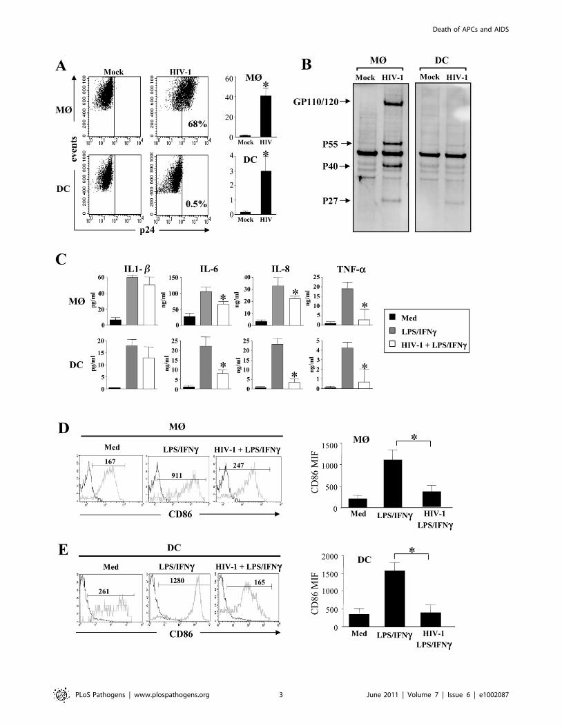
HIV-1BaL was higher than DCs (Figure 1A). The percentage of
HIV-infected MØ varied (40%67) among individual prepara-
tions, whereas DCs from the same individuals displayed less than
3%61 of infected cells, consistent with previous reports [13,78].
To confirm intracellular staining of p24 antigen, the cells were
lysed and western blots performed to detect the profile of viral
antigens, using sera from HIV-infected individuals. In MØ, we
observed a typical profile displaying both envelope glycoprotein
and gag protein, whereas none of these bands were clearly
observed in DCs (Figure 1B). We then assessed the capacity of
MØ and DCs to produce cytokines and express co-stimulatory
molecules in response to LPS and IFN-c. We found that activated-
MØ as well as activated-DCs, incubated in the presence of HIV-1,
secreted less pro-inflammatory cytokines such as IL-6, IL-8 and
TNF-a as compared to uninfected cells. No difference was
observed for IL-1b secretion (Figure 1C). Moreover, stimulation
with LPS and IFN-c induced lower expression of the co-
stimulatory molecule CD86 (Figure 1D, E) and the maturation
marker CD83 (data not shown), at the surface of HIV-infected
cells as compared to uninfected cells (CD86 mean expression,
MØ: 3506150 vs 10906230; DCs: 4006220 vs 15706230).
Thus, HIV infection during the process of APC differentiation
impacted cytokine secretion and cellular maturation.
Author Summary
Antigen-presenting cells (APCs) are critical for both innateand adaptive immunity. They have a profound impact onthe hosts’ ability to combat microbes. Dysfunction andpremature death by apoptosis of APCs may contribute toan abnormal immune response unable to clear pathogens.Circulating blood monocytes exhibit developmental plas-ticity, with the capability of differentiating into eithermacrophages or dendritic cells (DCs), and they representimportant cellular targets for HIV-1. We report that HIVinfection renders monocytes/macrophages and DCs invitro more prone to undergo apoptosis and this height-ened susceptibility is associated with changes in theexpression of anti- and pro-apoptotic molecules. Ourresults show that during the acute phase of SIV-infectionof rhesus macaques, monocytes and DCs are more proneto die by apoptosis. They express lower levels of Mcl-1 andFLIP proteins, two anti-apoptotic molecules, but higherexpression of the active form of Bax and Bak, thegatekeepers of the mitochondria, major sensor of theapoptotic machinery. Because the early events areimportant in the pathogenesis of this disease, early deathof APCs should play a major role leading to the defectiveimmune response. Strategies aimed at preventing death ofAPCs could be beneficial in helping the immune responseto fight HIV-1.
Death of APCs and AIDS
PLoS Pathogens | www.plospathogens.org 2 June 2011 | Volume 7 | Issue 6 | e1002087
Death of APCs and AIDS
PLoS Pathogens | www.plospathogens.org 3 June 2011 | Volume 7 | Issue 6 | e1002087
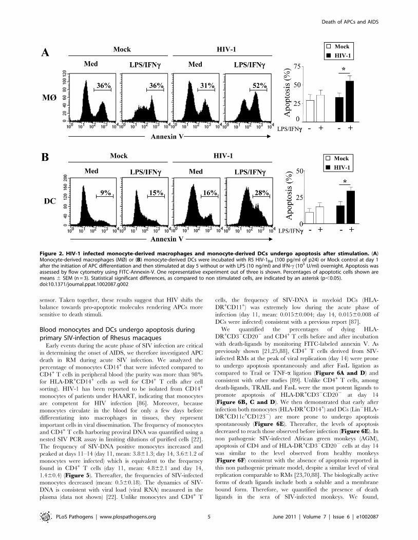
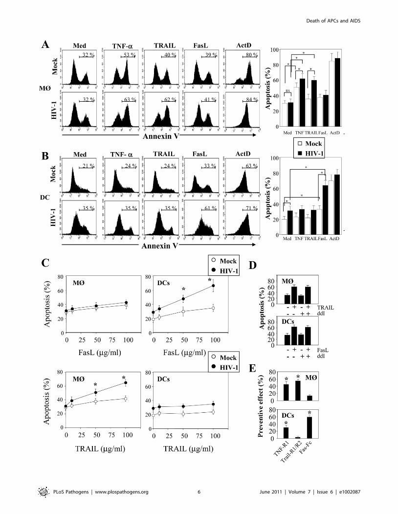
HIV-1 infection sensitizes MØ and DCs to deathreceptors-mediated apoptosis by downregulating theexpression of FLIP
We then examined whether MØ and DCs were more prone to
die at day 5 post-infection with HIV-1BaL. After stimulation with
LPS and IFNc, we observed a significant increase in the
percentage of apoptotic cells from HIV-infected culture as
compared to non-infected cells (MØ, 55%67 vs 31%63.8,
p,0.01; DCs, 36%67 vs 16%63.9, p,0.01). No major differ-
ence was observed after stimulation of uninfected cells (Figure 2A,B). The extrinsic apoptotic pathway involves members of the
death-receptor family including CD95 (Fas), TNF-R and DR4/
DR5 (Trail-R1/R2) [79]. Upon ligation of these death-receptors
by their ligands, the association of the adaptor molecule FADD
with the initiator caspases forms a death-inducing signaling
complex (DISC) leading to apoptosis [80]. We assessed whether
MØ and DCs in the presence of HIV-1BaL are sensitive to death-
receptor ligands including TNF-a, TRAIL and FasL. Actinomycin
D (Act D) was used as a positive control for cell death. First, both
uninfected and HIV-infected MØ (5166% and 6267%, respec-
tively) were more sensitive to undergo apoptosis in response to
TNF-a in comparison with the medium alone (3064% and
3165%, respectively) whereas no similar effect was observed on
DCs (Figure 3A, B). Second, MØ infected with HIV-1BaL were
more prone to die in response to TRAIL as compared to non-
infected cells (6065% versus 3666%) (Figure 3A) but no
difference was observed for FasL (3864% versus 4166%). Finally,
HIV-1 infection increased the sensitivity of DCs to die
spontaneously (2064% uninfected versus 3166% in infected
DCs) and after FasL-ligation (6467% versus 3166% in medium
alone) but not to the binding of TRAIL (3266%) (Figure 3B).
Apoptosis was dependent on the amount of death ligands
(Figure 3C).
In order to determine if viral replication was necessary for
sensitization to apoptosis, we treated the cells with ddI (5 mM, a
dose that blocks viral replication in MØ; ,5% of p24+). Our
studies showed that in the presence or absence of ddI, both MØ
and DCs remain sensitive to TRAIL and FasL, respectively
(Figure 3D). Furthermore, we assessed whether stimulation with
LPS/IFN-c-mediated apoptosis may be modulated by antagonists
to death ligands using decoy receptors. We demonstrated that
decoy receptors of TNF (TNF-R1), and TRAIL (TRAIL-R2/
TRAIL-R1), reduced monocyte cell death-mediated by LPS/IFN-
c stimulation, whereas decoys receptors of Fas (Fas-Fc) and TNF
(TNF-R1) reduced DCs cell death (Figure 3E). Thus, despite the
fact that soluble TNF-a has no effect on DCs, TNF-R1 partially
inhibits cell death. Altogether, these results indicated that HIV
induces APC apoptosis after death-receptors ligation.
Since cells were more sensitive to undergo apoptosis, we next
assessed whether this effect was related either to a modulation in
the expression of death-receptors or in the regulation of the
signaling pathway. Although, MØ and DCs exhibited a greater
sensitivity to die in the presence of death ligands, we did not
observe any modulation of death-receptor expression, including
TRAIL-R1 and –R2 and Fas/CD95 on cells infected with HIV-
1Ba-L compared to uninfected cells (data not shown). The
molecular basis of resistance to death-receptors-mediated apopto-
sis involves the expression of FLIP (cellular-FLICE-inhibitory
protein), which is an inhibitor of the DISC (death-inducing
signaling complex) [34], and is expressed during differentiation of
APCs [31,32,33]. Therefore, we analyzed the expression of FLIP
in HIV-infected MØ and DCs. We observed that FLIP expression
is detectable by western blot at day 5 in both uninfected MØ and
DCs but decreased in HIV-infected cells (Figure 4A). Thus, the
amount of FLIP decreased by 57%64 in MØ and 46%65 in DCs
following HIV infection (Figure 4B). Altogether, our data suggest
that HIV-1 infection increased the propensity of mononuclear
phagocytes to undergo apoptosis in response to death-ligands,
possibly due to a decrease in the amount of FLIP. This process
occurred independently of any modulation of death receptor
expression.
HIV modulates the balance between pro- and anti-apoptotic members of the Bcl-2 family in MØ and DCs
The molecular basis of macrophage resistance to apoptosis
includes the expression of the anti-apoptotic Bcl-2 family
members, among which Mcl-1 predominates in differentiated
cells [38]. In the absence of growth receptor engagement, Mcl-1 is
degraded by the ubiquitin-proteasome pathway [81,82,83] or
cleaved by proteases [84,85]. We found a 50% decrease in
expression of Mcl-1 protein in infected-MØ, which was not
observed in DCs (Figure 4A). In addition, SDS-PAGE analysis
revealed that Mcl-1 migrated as a doublet suggesting the presence
of phosphorylated Mcl-1, primed by GSK-3, on threonine 163.
This phosphorylated form undergoes accelerated degradation
[81,83]. In HIV-infected MØ, this change in MCL isoforms was
clearly observed compared to uninfected cells, whereas no
difference was observed for DCs (Figure 4A). Additional bands
of approximately 34 KDa on western blots probed with Mcl-1
antibody were also detected (Figure 4A). These product bands
correspond to different translational products (Mcl-1S/DTM versus
Mcl-1Exon-1). It is important to note that Mcl-1Exon-1 is pro-
apoptotic [38]. The amount of Mcl-1Exon-1 protein was clearly
enhanced in DCs cultured in the presence of HIV-1BaL (fold
increase 2.1) as well as in MØ (fold increase 1.7) (Figure 4B).
Members of the Bcl-2 protein family, in particular Bax and Bak
proteins play a critical role in controlling apoptosis [39]. To assess
the early commitment of Bax and Bak activation, we subfractio-
nated the cells to isolate a mitochondria-enriched fraction. At day
5 of culture, we observed higher amounts of Bax and Bak proteins
within the enriched mitochondrial fraction derived from HIV-
1BaL-infected MØ and DCs compared to uninfected cells
(Figure 4C and D). Membrane insertion of Bax and Bak
supported a dynamic model in which mitochondria is a central
Figure 1. Impact of HIV infection on cytokine secretion and maturation of monocyte-derived macrophages and monocyte-derivedDCs. (A and B) HIV-1 infection of monocyte-derived macrophages (MØ) or monocyte-derived DCs. Cells were infected at day 1 after the initiation ofAPC differentiation without (Mock) or with the R5 HIV-1Bal (100 pg/ml of p24). At day 5, (A) the percentage of p24+ cells was determined by flowcytometry. Values shown are means 6 SEM (n = 6). Significant differences are indicated by an asterisk (p,0.05). (B) HIV viral proteins were detectedby western blotting using HIV-1+ sera. (C) HIV-1 decreases pro-inflammatory cytokines production. Cells at day 5 were stimulated with LPS (10 ng/ml)and IFN-c (103 U/ml) overnight. Cells incubated in the absence of R5 HIV-1Bal and in the absence of stimulation represent the negative control (Med).Supernatants were collected and assessed for the presence of IL-1b, IL-6, IL-8, and TNF-a by flow cytometry using bead array. Values shown are means6 SEM (n = 3). Significant differences are indicated by an asterisk (p,0.05). (D and E) HIV-1 decreases CD86 expression at the surface of stimulated(D) MØ or (E) DCs. Cells were stained with specific CD86 mAbs, and cell surface density was assessed by flow cytometry. One representativeexperiment out of three is shown; the mean of fluorescence intensity is indicated. CD86 expression values shown are means 6 SEM (n = 3). Significantdifferences are indicated by an asterisk (p,0.05).doi:10.1371/journal.ppat.1002087.g001
Death of APCs and AIDS
PLoS Pathogens | www.plospathogens.org 4 June 2011 | Volume 7 | Issue 6 | e1002087
sensor. Taken together, these results suggest that HIV shifts the
balance towards pro-apoptotic molecules rendering APCs more
sensitive to death stimuli.
Blood monocytes and DCs undergo apoptosis duringprimary SIV-infection of Rhesus macaques
Early events during the acute phase of SIV infection are critical
in determining the onset of AIDS, we therefore investigated APC
death in RM during acute SIV infection. We analyzed the
percentage of monocytes CD14+ that were infected compared to
CD4+ T cells in peripheral blood (the purity was more than 98%
for HLA-DR+CD14+ cells as well for CD4+ T cells after cell
sorting). HIV-1 has been reported to be isolated from CD14+
monocytes of patients under HAART, indicating that monocytes
are competent for HIV infection [86]. Moreover, because
monocytes circulate in the blood for only a few days before
differentiating into macrophages in tissues, they represent
important cells in viral dissemination. The frequency of monocytes
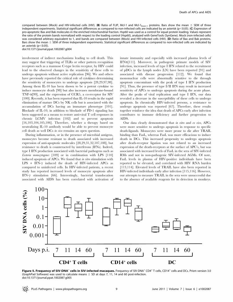
and CD4+ T cells harboring proviral DNA was quantified using a
nested SIV PCR assay in limiting dilutions of purified cells [22].
The frequency of SIV-DNA positive monocytes increased and
peaked at days 11–14 (day 11, mean: 3.861.3; day 14, 3.661.2 of
monocytes were infected) which is equivalent to the frequency
found in CD4+ T cells (day 11, mean: 4.862.1 and day 14,
1.460.4) (Figure 5). Thereafter, the frequencies of SIV-infected
monocytes decreased (mean: 0.560.18). The dynamics of SIV-
DNA is consistent with viral load (viral RNA) measured in the
plasma (data not shown) [22]. Unlike monocytes and CD4+ T
cells, the frequency of SIV-DNA in myeloid DCs (HLA-
DR+CD11+) was extremely low during the acute phase of
infection (day 11, mean: 0.01560.004; day 14, 0.01560.008 of
DCs were infected) consistent with a previous report [87].
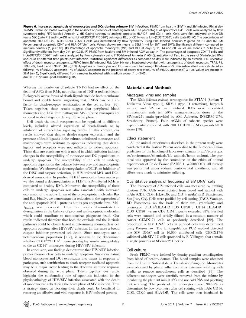
We quantified the percentages of dying HLA-
DR+CD32CD202 and CD4+ T cells before and after incubation
with death-ligands by monitoring FITC-labeled annexin V. As
previously shown [21,25,88], CD4+ T cells derived from SIV-
infected RMs at the peak of viral replication (day 14) were prone
to undergo apoptosis spontaneously and after FasL ligation as
compared to Trail or TNF-a ligation (Figure 6A and D) and
consistent with other studies [89]. Unlike CD4+ T cells, among
death-ligands, TRAIL and FasL were the most potent ligands to
promote apoptosis of HLA-DR+CD32CD202 at day 14
(Figure 6B, C and D). We then demonstrated that early after
infection both monocytes (HLA-DR+CD14+) and DCs (Lin2HLA-
DR+CD11c+CD1232) are more prone to undergo apoptosis
spontaneously (Figure 6E). Thereafter, the levels of apoptosis
decreased to reach those observed before infection (Figure 6E). In
non pathogenic SIV-infected African green monkeys (AGM),
apoptosis of CD4 and of HLA-DR+CD32CD202 cells at day 14
was similar to the level observed from healthy monkeys
(Figure 6F) consistent with the absence of apoptosis reported in
this non pathogenic primate model, despite a similar level of viral
replication comparable to RMs [23,70,88]. The biologically active
forms of death ligands include both a soluble and a membrane
bound form. Therefore, we quantified the presence of death
ligands in the sera of SIV-infected monkeys. We found,
Figure 2. HIV-1 infected monocyte-derived macrophages and monocyte-derived DCs undergo apoptosis after stimulation. (A)Monocyte-derived macrophages (MØ) or (B) monocyte-derived DCs were incubated with R5 HIV-1Bal (100 pg/ml of p24) or Mock control at day 1after the initiation of APC differentiation and then stimulated at day 5 without or with LPS (10 ng/ml) and IFN-c (103 U/ml) overnight. Apoptosis wasassessed by flow cytometry using FITC-Annexin-V. One representative experiment out of three is shown. Percentages of apoptotic cells shown aremeans 6 SEM (n = 3). Statistical significant differences, as compared to non stimulated cells, are indicated by an asterisk (p,0.05).doi:10.1371/journal.ppat.1002087.g002
Death of APCs and AIDS
PLoS Pathogens | www.plospathogens.org 5 June 2011 | Volume 7 | Issue 6 | e1002087
Death of APCs and AIDS
PLoS Pathogens | www.plospathogens.org 6 June 2011 | Volume 7 | Issue 6 | e1002087
concomitant with the increase of cell death in RMs, higher levels
of FasL two weeks post-infection (Figure 6G). In contrast, we did
not observe any increase in the levels of FasL in SIV-infected
AGM (Figure 6G). We have reported during the acute phase the
absence of TNF-a detection in the sera of both SIV-infected
species [90,91]. Although, we were unable to detect soluble
TRAIL in the sera of SIV-infected monkeys due to the
unavailability of appropriate reagents for its detection in non-
human primates (data not shown), it has been reported that there
is increased expression of Trail mRNA in SIV-infected RMs [92].
To assess the impact of soluble and membrane forms of death
ligands, we investigated whether apoptosis of monocytes and DCs
from SIV-infected RM may be modulated by antagonists to death
ligands using decoy receptors. We demonstrated that decoy
receptors of TNF (TNF-R1) and TRAIL (TRAIL-R2 but not
TRAIL-R1), and to a lesser extent decoy receptor of Fas (Fas-Fc),
reduced monocyte cell death, whereas decoys receptors of Fas and
TNF (TNF-R1) reduced DCs cell death (Figure 6H). Interest-
ingly, despite the fact that soluble TNF-a has no effect, antagonist
antibodies partially inhibited death suggesting that TNF-a at the
cell surface may participate in the death of APCs [93]. These
results suggest that apoptosis of mononuclear phagocytes involved
death-receptors and their counterparts.
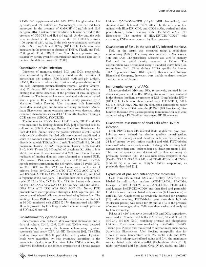
Apoptosis of blood mononuclear cells during primary SIVinfection involves a dysregulation in the balance of pro-and anti-apoptotic molecules
In order to analyze the apoptotic pathways in monocytes,
positive selection of CD14+ cells was performed from healthy and
SIV-infected RMs. Western blots probed with specific antibodies
to FLIP revealed that monocytes from SIV-infected RMs
displayed lower amounts of FLIP (Figure 7A), as compared to
healthy RMs. Thus, the absence of FLIP is consistent with the
increase sensitivity of these cells to undergo apoptosis after ligation
of death receptors. Moreover, we found that monocytes from SIV-
infected RMs had lower amounts of Mcl-1 (Figure 7A). In one
SIV-infected RM, we also detected an increased amount of the
proapoptotic form of Mcl-1Exon-1. To assess the expression of
active form of Bax and Bak proteins in APCs from healthy and
SIV+RMs, we used specific antibodies that detect conformational
changes as previously described [25]. In comparison to CD4+ T
cells, we found that 20%66 and 30%611 of monocytes from SIV-
infected RMs at day 14 express the active form of the pro-
apoptotic Bax and Bak molecules respectively as compared to
monocytes from non-infected RMs (less than 11%64). Similar
data were observed in DCs although to a lesser extent (Figure 7Band 7C). Thus, our results indicate that monocyte and DCs are
engaged in a process leading to mitochondria damage supporting
our observation that these cells are more prone to undergo
apoptosis during the acute phase. Furthermore, we used a broad
caspase inhibitor and demonstrated that by blocking caspase
activation, cell death of APCs was also prevented (Figure 7D).
We also demonstrated that the addition of caspase inhibitor led to
an increase in the number of cells expressing TNF-a after
stimulation with LPS + IFN-c stimulation (Figure 7E). Altogeth-
er, our data demonstrated a critical role of both the intrinsic and
extrinsic apoptotic pathways in controlling APC death during the
acute phase of SIV-infection.
Discussion
We demonstrate that monocytes and DCs are more prone to
undergo apoptosis in response to death-receptor ligation after in
vitro infection with HIV or ex vivo from SIV-infected RMs. In
addition, our data show that HIV/SIV infection is associated with
an increase in the active forms of the pro-apoptotic molecules Bax
and Bak and with a decrease in the anti-apoptotic Mcl-1 and FLIP
proteins in both cell types. Thus, these results suggest that both the
extrinsic and intrinsic pathways are involved in the death of APCs
during HIV/SIV infection. Broad inhibition of caspase activation
using a synthetic peptide prevented this death and increased the
number of TNF-a productive mononuclear cells.
Circulating monocytes are essential not only to replenish the
pool of tissue macrophage populations but also may differentiate
into inflammatory DCs in the tissues following microbial infection.
Because peripheral monocytes and DCs represent crucial
populations for the control of pathogens, this enhanced suscep-
tibility to die by apoptosis in the presence of death ligands could
have a major impact on the establishment of the adaptative
immune response early after infection. Interestingly, other
persistent viral infections such as lymphocytic choriomeningitis
virus (LCMV) and measles virus (MV), which are associated with a
generalized immune suppression in their natural hosts, also induce
death of accessory cells early after infection [94,95]. Our results
also demonstrated in vitro that incubation of monocyte-derived
MØ and DCs with HIV during differentiation not only increased
the susceptibility of these cells to undergo apoptosis but also
impaired their maturation and their capacity to produce
inflammatory cytokines after stimulation. Their down modulation
could have an impact on the hosts’ ability to mount an effective
SIV-specific immune response.
Our results showed abnormal early death of APCs was
associated with AIDS. The low level of infection of DCs suggests
that apoptosis is not necessarily associated with productive
infection. Moreover, during the acute phase, the percentage of
monocytes prone to undergo apoptosis (and expressing active form
of Bax and Bak) was higher than the frequency of SIV DNA+ cells.
Our data revealed that also in vitro HIV primes both monocytes
and DCs to undergo apoptosis in response to death ligands despite
the presence of an inhibitor of viral replication, ddI. In a similar
manner, the non pathogenic-primate model suggests that despite
intense viral replication during the acute phase [70], APCs are not
prone to undergo apoptosis. Altogether, these results point to the
Figure 3. HIV-1 sensitizes monocyte-derived macrophages and monocyte-derived DCs for Death receptor ligands. (A) Monocyte-derived macrophages (MØ) or (B) monocyte-derived DCs were incubated with R5 HIV-1Bal (100 pg/ml of p24) or Mock at day 1 after the initiation ofAPC differentiation. At day 5, the cells were then cultured overnight in the absence or presence of recombinant TNF-a, TRAIL and FasL (100 ng/ml). Apositive control of cell death was performed in the presence of Actinomycin D (10 mg/ml). Apoptosis was determined by flow cytometry using FITC-labeled Annexin V. Percentages of apoptotic cells shown are means 6 SEM (n = 3). Statistical significant differences are indicated by an asterisk(p,0.05). (C) Dose response of FasL and Trail. Percentages of apoptotic cells shown are means 6 SEM (n = 3). Statistical significant differences ascompared to untreated cells are indicated by an asterisk (p,0.05). (D) Cells, before infection, were incubated in the absence or presence of ddI(5 mM). At day 5, the cells were then cultured overnight in the absence or presence of either TRAIL or FasL (100 ng/ml). (E) Cells incubated with R5HIV-1Bal were stimulated at day 5 with LPS (10 ng/ml) and IFN-c (103 U/ml) overnight in the absence or presence of death receptor antagonists: TNF-R1, TRAIL-R1/TRAIL-R2, Fas-Fc (10 mg/ml). Preventive effect was calculated as follows: ((% of MØ/DC apoptosis - % of MØ/DC apoptosis in thepresence of decoy receptors)/(% of MØ/DC apoptosis)) X 100. Values are means 6 SEM (n = 3). Statistical significant differences as compared tountreated cells are indicated by an asterisk (p,0.05).doi:10.1371/journal.ppat.1002087.g003
Death of APCs and AIDS
PLoS Pathogens | www.plospathogens.org 7 June 2011 | Volume 7 | Issue 6 | e1002087
Figure 4. Expression of pro- and anti-apoptotic molecules in HIV-1 infected monocyte-derived macrophages and monocyte-derived DCs. (A) Monocytes-derived MØ or monocytes-derived DCs were incubated with R5 HIV-1Bal (100 pg/ml of p24) or Mock at day 1 after theinitiation of APC differentiation. At day 5, the cells were lysed and then the proteins were detected by immunoblotting with specific antibodiesagainst FLIP and Mcl-1. Actin was used as a control for equal protein loading. Values represent the ratio of the protein bands normalized with respectto the loading control, analyzed with GeneTools (SynGene). Mock (non-infected cells) was considered arbitrary equivalent to 1, and bands are
Death of APCs and AIDS
PLoS Pathogens | www.plospathogens.org 8 June 2011 | Volume 7 | Issue 6 | e1002087
involvement of indirect mechanisms leading to cell death. This
may suggest that triggering of TLRs or other pattern recognition
receptors such as a mannose C-type lectin receptor, by HIV could
lead to the observed changes in the sensitivity of these cells to
undergo apoptosis without active replication [96]. We and others
have previously reported the critical role of cytokines determining
the sensitivity of monocytes to undergo apoptosis [28,29,97,98].
Among them IL-10 has been shown to be a potent cytokine to
induce monocyte death [98] but also increases membrane-bound
TNF-a[99], and the expression of CCR5, a co-receptor for SIV
[100]. Recently, it has been reported that IL-10 results in the rapid
elimination of mature DCs by NK cells but is associated with the
accumulation of DCs having an immature phenotype [101].
Blockade of IL-10, in addition to blockade of PD-1 signaling has
been suggested as a means to restore anti-viral T cell responses in
chronic LCMV infection [102] and to prevent apoptosis
[16,103,104,105,106]. Therefore, whether a therapy based on
neutralizing IL-10 antibody would be able to prevent monocyte
cell death as well DCs in vivo remains an open question.
During inflammation, or in the presence of microbial antigens,
monocytes become resistant to death associated with increased
expression of anti-apoptotic molecules [28,29,31,32,107,108], but
resistance to death is counteracted by interferons (IFNs). Indeed,
type I IFN production associated with bacterial pathogens such as
Listeria monocytogenes [109] or in combination with LPS [110]
induced apoptosis of APCs. We found that in vitro stimulation with
LPS + IFN-c induced the death of HIV-infected APCs as
compared to uninfected cells. In HIV-infected patients, a recent
study has reported increased levels of monocyte apoptosis after
IFN-c stimulation [66]. Interestingly, bacterial translocation
associated with AIDS has been correlated with activation of
innate immunity and especially with increased plasma levels of
IFNa[111]. Moreover, in pathogenic primate models of SIV
infection, increased levels of type I IFN related to the recruitment
of pDCs in the lymph nodes (LN) have been reported [91] and
associated with disease progression [112]. We found that
mononuclear cells were abnormally sensitive to die through
apoptosis concomitant with the peak of type I IFN production
[91]. Thus, the presence of type I/II IFN may result in increased
sensitivity of APCs to undergo apoptosis during the acute phase.
After the peaks of viral replication and type I IFN, our data
revealed a decrease in the susceptibility of these cells to undergo
apoptosis. In chronically HIV-infected persons, a resistance to
undergo apoptosis was reported [67]. Therefore, these results
together reinforce the idea that death of APCs early after infection
contributes to immune deficiency and further progression to
AIDS.
Our data clearly demonstrated that in vitro and ex vivo, APCs
were more sensitive to undergo apoptosis in response to specific
death-ligands. Monocytes were more prone to die after TRAIL
binding than FasL, whereas FasL was more efficacious to induce
death in DCs. This increased propensity to undergo apoptosis
after death-receptor ligation was not related to an increased
expression of the death-receptors at the surface of APC’s, but was
associated with increased levels of FasL in the sera of SIV-infected
RMs and not in non-pathogenic SIV-infected AGMs. Of note,
FasL levels in plasma of HIV-positive individuals have been
reported to be elevated, and correlated with HIV RNA burden
[113,114]. Elevated levels of TRAIL have also been reported in
HIV-infected individuals early after infection [115,116]. However,
our attempts to measure TRAIL in the sera were unsuccessful due
to the absence of available reagents for its detection in monkeys.
compared between (Mock) and HIV-infected cells (HIV). (B) Ratio of FLIP, Mcl-1 and Mcl-1Exon-1 proteins. Bars show the mean 6 SEM of threeindependent experiments. Statistical significant differences as compared to non-infected cells are indicated by an asterisk (p,0.05). (C) Expression ofpro-apoptotic Bax and Bak molecules in the enriched mitochondrial fraction. Hsp60 was used as a control for equal protein loading. Values representthe ratio of the protein bands normalized with respect to the loading control (Hsp60), analyzed with GeneTools (SynGene). Mock (non-infected cells)was considered arbitrary equivalent to 1, and bands are compared between (Mock) and HIV-infected cells (HIV). (D) Ratio of Bax and Bak proteins.Bars show the mean 6 SEM of three independent experiments. Statistical significant differences as compared to non-infected cells are indicated byan asterisk (p,0.05).doi:10.1371/journal.ppat.1002087.g004
Figure 5. Frequency of SIV-DNA+ cells in SIV-infected macaques. Frequency of SIV-DNA+ CD4+ T cells, CD14+ cells and DCs. Prism version 3.0(GraphPad Software) was used to calculate means 6 SD at days 7, 11, 14 and 60 post-infection.doi:10.1371/journal.ppat.1002087.g005
Death of APCs and AIDS
PLoS Pathogens | www.plospathogens.org 9 June 2011 | Volume 7 | Issue 6 | e1002087

Death of APCs and AIDS
PLoS Pathogens | www.plospathogens.org 10 June 2011 | Volume 7 | Issue 6 | e1002087
Whereas the incubation of soluble TNF-a had no effect on the
death of APCs from RMs, neutralization of TNF-a reduced death.
Biologically active forms of death-ligands include both membrane
bound and soluble forms, suggesting that TNF-a can be a co-
factor for death-receptor sensitization at the cell surface [93].
Taken together, these results suggest that peripheral blood
monocytes and DCs from pathogenic SIV-infected macaques are
exposed to death-ligands during the acute phase.
Cell death via death receptors can be regulated at different
levels, including altered expression of death-ligands or by
inhibition of intracellular signaling events. In this context, our
results showed that despite death-receptor expression and the
presence of death-ligands in the culture, uninfected monocytes and
macrophages were resistant to apoptosis indicating that death-
ligands and receptors were not sufficient to induce apoptosis.
These data are consistent with a model in which infection induced
changes in the susceptibility of monocyte and DC populations to
undergo apoptosis. The susceptibility of the cells to undergo
apoptosis depends on the balance between pro- and anti-apoptotic
molecules. We observed a downregulation of FLIP, an inhibitor of
the DISC and caspase activation, in HIV-infected MØ- and DCs-
derived monocytes. In purified CD14+ monocytes from monkeys,
we also found a downregulation of FLIP in SIV-infected RMs as
compared to healthy RMs. Moreover, the susceptibility of these
cells to undergo apoptosis was also associated with increased
expression of the active forms of the pro-apoptotic molecules Bax
and Bak. Finally, we demonstrated a reduction in the expression of
the anti-apoptotic Mcl-1 proteins but its pro-apoptotic form, Mcl-
1Exon-1, was increased. Thus, our results demonstrated a
dysregulation in the balance of pro- and anti-apoptotic molecules,
which could contribute to mononuclear phagocyte death. Our
results indicated therefore that both the extrinsic and the intrinsic
pathways could be closely linked in determining mononuclear cell
apoptosis outcome after HIV/SIV infection. In this sense a broad
caspase inhibitor prevented cell death. Since monocytes are a
heterogeneous population [117], it remains to be determined
whether CD14dimCD16+ monocytes display similar susceptibility
to die as CD14+ monocytes during HIV/SIV infection.
In conclusion, our findings demonstrate that HIV/SIV infection
primes mononuclear cells to undergo apoptosis. Since circulating
blood monocytes and DCs extravasate into tissues in response to
pathogens, such sensitization to death-receptor mediated apoptosis
may be a major factor leading to the defective immune response
observed during the acute phase. Taken together, our results
highlight the confounding role of apoptosis induction in the
physiopathology of HIV/SIV infection associated with the death
of mononuclear cells during the acute phase of SIV infection. Thus
a strategy aimed at blocking their death could be beneficial in
restoring an effective anti-viral response in HIV-infected persons.
Materials and Methods
Macaques, virus and samplesTen RMs (Macaca mulatta) seronegative for STLV-1 (Simian T
Leukemia Virus type-1), SRV-1 (type D retrovirus), herpes-B
viruses, and SIVmac were utilized. RMs were inoculated
intravenously with ten 50% animal-infectious doses of the
SIVmac251 strain (provided by AM. Aubertin, INSERM U74,
Strasbourg, France). Four AGMs of sabaeus species were
experimentally infected with 300 TCID50 of SIVagm.sab92018
strain [70].
Ethics statementAll the animal experiments described in the present study were
conducted at the Institut Pasteur according to the European Union
guidelines for the handling of laboratory animals (http://ec.europa.
eu/environment/chemicals/lab_animals/home_en.htm). The pro-
tocol was approved by the committee on the ethics of animal
experiments of Ile de France (PARIS 1, #20080007). All surgery
was performed under sodium pentobarbital anesthesia, and all
efforts were made to minimize suffering.
Quantitative analysis of frequency of SIV DNA+ cellsThe frequency of SIV-infected cells was measured by limiting
dilution PCR. Cells were isolated from blood and stained with
mAbs (CD3, CD4, HLA-DR and CD14 mAbs) (BD Biosciences,
San Jose, CA). Cells were purified by cell sorting (FACS Vantage,
BD Bioscences) on the basis of their size, granularity and
phenotype (CD14+HLA-DR+CD32CD202; CD142HLA-DR+
CD32CD202 versus CD3+CD4+); purity exceeded 98%. Purified
cells were counted and serially diluted in a constant number of
carrier CEMX174 cells as previously described [22]. The
proportion of SIV DNA+ cells in purified cells was determined
using Poisson law. The limiting-dilution PCR method detected
one SIV DNA+ cell in 10,000 uninfected cells (CEMX174)
validated with SIV-1C cells (provided by F. Villinger), that contain
a single provirus of SIVmac251 per cell.
Cell cultureFresh PBMC were isolated by density gradient centrifugation
from blood of healthy donors. The blood samples were obtained
from the Institut National de la Transfusion Sanguine. Monocytes
were obtained by plastic adherence after extensive washing with
media to remove non-adherent cells as described [98]. The
adherent monocytes were carefully removed from the culture by
incubating the plate 30 min at 4uC and use cold PBS and pipetting
(not scraping). The purity of the monocytes exceed 90–95% as
determined by flow cytometry after cell staining with mAbs CD14,
CD3, CD20 and HLA-DR. The cells were then incubated in
Figure 6. Increased apoptosis of monocytes and DCs during primary SIV infection. PBMC from healthy (SIV2) and SIV-infected RM at day14 (SIV+) were incubated overnight in the absence or presence of death ligands. (A) The percentages of apoptotic CD4+ T cells were analyzed by flowcytometry using FITC-labeled Annexin V. (B) Gating strategy to analyze apoptotic HLA-DR+ and CD14+ cells. Cells were first analyzed on HLA-DRversus SSC (gate R1) and HLA-DR versus Lin+(CD3+CD14+CD20+) cells (gate R2), or CD14 versus Lin+(CD3+CD20+) cells (gate R3) (C) The percentages ofapoptotic HLA-DR+Lin2 (CD32CD142CD202) cells was determined by flow cytometry using FITC-labeled Annexin V gated on R1 and R2; (D)Percentage of apoptotic CD4+ and HLA-DR+Lin2 cells. Values shown are means 6 SEM (n = 4 for SIV2 and SIV+); Significantly different compared tomedium controls (*, p,0.05). (E) Percentage of apoptotic monocytes (MØ) and DCs at days 0, 11, 14 and 60; values are means 6 SEM (n = 6);Significantly different from day 0 (*, p,0.05). (F) PBMC from healthy and SIV-infected AGM at day 14. The percentages of apoptotic CD4+ T cells andHLA-DR+CD32CD202 cells were analyzed by flow cytometry using FITC-labeled Annexin V. (G) Quantitation of FasL in the sera of SIV-infected RMsand AGM at different time points post-infection. Statistical significant differences as compared to day 0 are indicated by an asterisk. (H) Preventiveeffect of death receptor antagonists. PBMC from SIV-infected RMs (day 14) were incubated overnight with antagonists of death receptors: TRAIL-R1,TRAIL-R2, Fas-Fc and TNF-R1 (10 mg/ml). Apoptosis of monocytes and DCs was quantified using FITC-Annexin-V. Preventive effect was calculated asfollows: ((% of MØ/DC apoptosis - % of MØ/DC apoptosis in the presence of decoy receptors)/(% of MØ/DC apoptosis)) X 100. Values are means 6SEM (n = 5); Significantly different from samples incubated with medium alone (*, p,0.05).doi:10.1371/journal.ppat.1002087.g006
Death of APCs and AIDS
PLoS Pathogens | www.plospathogens.org 11 June 2011 | Volume 7 | Issue 6 | e1002087

Figure 7. Expression of pro- and anti-apoptotic molecules in monocytes and mDCs during primary SIV infection. (A) Expression ofFLIP and Mcl-1 in purified CD14+ (MØ) from healthy RM (SIV2) and SIV-infected RMs (SIV+). After isolation, the cells were lysed and the proteins wereimmunoblotted with specific antibodies against the anti-apoptotic molecules FLIP and Mcl-1. Actin was used as a control for equal protein loading.Values represent the ratio of the FLIP and Mcl-1 bands and normalized with respect to the loading control. (B) Flow cytometric analysis of the activeform of the pro-apoptotic molecules Bax and Bak in CD4+ T cells, and monocytes (MØ) at days 0 and 14. (C) Percentage of active form of Bax and Bakamong monocyte and DC populations at days 0 and 14. Values are means 6 sem (n = 6); Significantly different from day 0 (*, p,0.05). (D and E)PBMC from SIV-infected RMs were incubated without or with Q-VD-OPH (10 mM) and then stimulated with LPS (10 ng/ml) overnight. (D) Foldincrease in surviving cells incubated with Q-VD-OPH is shown. (E) Numbers of HLA-DR+CD32CD202 expressing TNF-a in the absence or presence ofQ-VD-OPH after stimulation is shown. Bars show the mean 6 SEM of three independent experiments. Statistical significant differences as comparedto untreated cells are indicated by an asterisk (p,0.05).doi:10.1371/journal.ppat.1002087.g007
Death of APCs and AIDS
PLoS Pathogens | www.plospathogens.org 12 June 2011 | Volume 7 | Issue 6 | e1002087
RPMI-1640 supplemented with 10% FCS, 1% glutamine, 1%
pyruvate, and 1% antibiotics. Macrophages were derived from
monocytes in the presence of GM-CSF (10 ng/ml) and IL-6
(5 ng/ml) (R&D system) while dendritic cells were derived in the
presence of GM-CSF and IL-4 (10 ng/ml). At day one, the cells
were incubated in the presence of the R5 HIV-1BaL strain
(100 pg/ml of p24). At day 5, the cells were stimulated overnight
with LPS (10 ng/ml) and IFN-c (103 U/ml). Cells were also
incubated in the presence or absence of TNF-a, TRAIL and FasL
(200 ng/ml). Fresh PBMC from Non Human primates were
isolated by density gradient centrifugation from blood and use to
perform the different assays [21,25,88].
Quantitation of viral infectionInfections of monocyte-derived MØ and DCs, respectively,
were measured by flow cytometry based on the detection of
intracellular p24 antigen (RD1-labeled mAb anti-p24 antigen,
KC-57, Beckman coulter) after fixation and permeabilization of
the cells (Intraprep permeabilization reagent, Coulter Coultro-
nics). Productive HIV infection was also visualized by western
blotting that allows detection of the presence of viral antigens in
cell extracts. The immunoblots were incubated with sera obtained
from a pool of HIV+ infected patients (kindly provided by F
Mamano, Institut Pasteur). After treatment with horseradish
peroxidase-linked goat anti-human secondary antibodies (Amer-
sham Biosciences), immunoreactive proteins were detected using
enhanced chemiluminescence (ECL+ from GE Healthcare) using a
CCD camera (GBOX, SYNGENE).
The frequencies of SIV-infected CD4+ T cells, CD14+ and DCs
were measured by limiting-dilution PCR [22] of purified cells by
cell sorting (FACS Vantage; Becton Dickinson Biosciences, Le
Pont de Claix, France) using the positive selection of cells stained
with specific antibodies. Purified cells were counted and diluted in
series in a constant number of carrier CEM X 174 cells. Cells were
directly lysed with TPK buffer (10 mM Tris-HCl pH 8.3, 50 mM
potassium chloride, 2.5 mM magnesium chloride, 0.5% Nonidet
P-40, 0.5% Tween 20, 100 mg/ml of proteinase K). After 1 h at
56uC, proteinase K was inactivated at 95uC for 10 min. Twenty
replicates of limiting dilutions were submitted to a nested PCR.
SIV proviral DNA was amplified by nested PCR with SIV251-
specific primers surrounding the nef region. After 35 cycles (95uCfor 30 s, 60uC for 30 s, 72uC for 1 min.) with the first set of
primers, Preco (59-CAG AGG CTC TCT GCG ACC CTA C)
and K3 (59-GAC TGA ATA CAG AGC GAA ATG C), amplified
a fragment of 961 base pairs, 10 ml of product was re-amplified (30
cycles 95uC for 30 s, 55uC for 30 s, 72uC for 1 min) with primers
K1 (59-TGG AAG ATG GAT CCT CGC AAT CC) and A2 (59-
GGA CTA ATT TCC ATA GCC AGC CA). Nested PCR
products were electrophoresed through a 1.8% agarose gel. The
proportion of infected cells was determined using Poisson law. The
limiting-dilution PCR method was able to detect one infected cell
in 10 000 uninfected cells (CEM X 174) demonstrated with SIV-
1C cells (provided by F. Villinger), which contain a single provirus
of SIVmac251 per cell.
Pro-inflammatory cytokine assaysSupernatants were collected after overnight stimulation and 6
days of culture. IL-1ß, IL-6, IL-8, and TNF-a were detected
simultaneously by using the human inflammatory cytokine
cytometric bead array (CBA) kit (BD Bioscience) [90]. The CBA
working range was 20–5000 pg/ml for each cytokine. Cytokine
levels were quantified by flow cytometry according to the
manufacturer’s directions. For intracellular TNF-a staining, the
cells were incubated in the absence or presence of a broad caspase
inhibitor Q-VD(OMe)-OPH (10 mM, MBL biomedical), and
stimulated with LPS and IFN-c. After 8 h, the cells were first
stained with HLA-DR, CD3, and CD20 mAbs, washed and then
permeabilized, before staining with PE-TNF-a mAbs (BD
Biosciences). The number of HLA-DR+CD32CD202 cells
expressing TNF-a was measured by flow cytometry.
Quantitation of FasL in the sera of SIV-infected monkeysFasL in the serum was measured using a solid-phase
immunoassay (MBL). The assay uses anti-FasL mAbs (clones,
4H9 and 4A5). The peroxidase substrate was used to quantify
FasL and the optical density measured at 450 nm. The
concentration was determined using a standard curve based on
recombinant FasL. Three distinct ELISA specific for human
TRAIL purchased from R&D system, Diaclone and Kamiya
Biomedical Company, however, were unable to detect monkey
Trail in the sera/plasma.
Immunophenotyping of APCsMonocyte-derived MØ and DCs, respectively, cultured in the
absence or presence of the R5 HIV-1Bal strain, were then incubated
in the absence (medium) or presence of LPS (10 ng/ml) plus IFN-c(103 U/ml). Cells were then stained with FITC-CD14, APC-
CD11c, PerCP-HLA-DR, and PE-conjugated antibodies to either
CD83 (HB15e) or CD86 molecules (FUN-1) (BD Biosciences). Five
hundred thousand events corresponding to mononuclear cells were
acquired using a FACScalibur instrument (BD Biosciences).
Quantitative assessment of dead cells after HIV/SIVinfection
Fresh PBMC from SIV-infected RMs at different days post-
infection were isolated by density gradient centrifugation;
apoptosis of monocytes and dendritic cells was determined at
24 h of culture by cell surface staining and with FITC-labeled
annexin-V which is an early marker of dying cells detecting both
caspase-dependent and -independent cell death programs [118].
The level of apoptosis was determined by flow cytometry as
previously described [98]. We also used decoy receptors of Fas
(Fas-Fc), TRAIL (TRAIL-R1-Fc and TRAIL-R2-Fc) and TNF-a(TNF-R1-Fc) at a dose of 10 mg/ml (Alexis corporation) as
previously described [21].
Expression of pro- and anti-apoptotic moleculesCells from SIV-infected RMs and healthy RMs were first
labeled for cell surface markers (APC-HLA-DR, PE-CD14,
Lineage PerCP-CD3/CD20 versus APC-CD11c, PE-HLA-DR
and Lineage PerCP-CD3/CD20) and then fixed and permeabi-
lized. Cells were then incubated with anti-Bax (BD Biosciences) or
anti-Bak Abs (Calbiochem) as previously described for primates
[25]. After washing, FITC-labeled goat anti-rabbit IgG Ab
(Molecular probes) was added for 30 min at 4uC in the presence
of mouse immunoglobulins. Cells were then washed and analyzed
by flow cytometry.
Pellets of 36106 monocyte-derived MØ and DCs, respectively,
were lysed in Nonidet P-40 buffer (1% NP-40, 50 mM Tris-HCl
(pH 7.4), 150 mM NaCl) containing protease and phosphatase
inhibitors. Total lysates were resolved by SDS-PAGE (10–20%
Tricine gels, Novex) and transferred to nitrocellulose membranes
(Amersham Biosciences). After blocking nonspecific sites for
1 hour at room temperature with 5% nonfat milk and 0.2%
Tween 20 in phosphate-buffered saline (pH 7.4), the membrane
was incubated with rabbit anti-Bak (Calbiochem, clone 2–14),
rabbit polyclonal anti-Bax (Santa-Cruz, N-20), rabbit anti-Mcl-1
Death of APCs and AIDS
PLoS Pathogens | www.plospathogens.org 13 June 2011 | Volume 7 | Issue 6 | e1002087
(S19, Santa-Cruz), or rat anti-FLIP (DAVE-2, Alexis Corpora-
tion). To confirm equal protein loading and transfer, membranes
were reprobed with anti-actin monoclonal antibodies (Sigma).
After treatment with horseradish peroxidase-linked goat anti-
mouse or anti-rabbit secondary antibodies (Amersham Bioscienc-
es), immunoreactive proteins were detected using enhanced
chemiluminescence (ECL+ from GE Healthcare) using a CCD
camera (GBOX, SYNGENE).
Statistical analysesData are reported as means 6 SEM, and groups were
compared using Mann-Whitney test (Prism software, GraphPad,
San Diego CA). A p value ,0.05 was considered significant.
Acknowledgments
This paper is dedicated to the memory of Bruno Hurtrel. We acknowledge
Celine Gommet (Institut Pasteur) for her expertise in the follow-up of our
primate cohort.
Author Contributions
Conceived and designed the experiments: ML CE JE. Performed the
experiments: ML LCG VM MCC BH JE. Analyzed the data: ML LCG JC
JZ CE JE. Contributed reagents/materials/analysis tools: ML LCG VM
MCC BH. Wrote the paper: JC JZ CE JE.
References
1. Gordon S, Taylor PR (2005) Monocyte and macrophage heterogeneity. Nat
Rev Immunol 5: 953–964.
2. Luster AD, Alon R, von Andrian UH (2005) Immune cell migration in
inflammation: present and future therapeutic targets. Nat Immunol 6:
1182–1190.
3. Geissmann F, Manz MG, Jung S, Sieweke MH, Merad M, et al. (2010)
Development of monocytes, macrophages, and dendritic cells. Science 327:
656–661.
4. Palframan RT, Jung S, Cheng G, Weninger W, Luo Y, et al. (2001)
Inflammatory chemokine transport and presentation in HEV: a remote control
mechanism for monocyte recruitment to lymph nodes in inflamed tissues. J Exp
Med 194: 1361–1373.
5. Leon B, Lopez-Bravo M, Ardavin C (2007) Monocyte-derived dendritic cells
formed at the infection site control the induction of protective T helper 1
responses against Leishmania. Immunity 26: 519–531.
6. Leon B, Ardavin C (2008) Monocyte migration to inflamed skin and lymph
nodes is differentially controlled by L-selectin and PSGL-1. Blood 111:
3126–3130.
7. Ho DD, Rota TR, Hirsch MS (1986) Infection of monocyte/macrophages by
human T lymphotropic virus type III. J Clin Invest 77: 1712–1715.
8. McElrath MJ, Steinman RM, Cohn ZA (1991) Latent HIV-1 infection in
enriched populations of blood monocytes and T cells from seropositive patients.
J Clin Invest 87: 27–30.
9. Meyaard L, Schuitemaker H, Miedema F (1993) T-cell dysfunction in HIV
infection: anergy due to defective antigen-presenting cell function? Immunol
Today 14: 161–164.
10. Meltzer MS, Skillman DR, Hoover DL, Hanson BD, Turpin JA, et al. (1990)
Macrophages and the human immunodeficiency virus. Immunol Today 11:
217–223.
11. Ennen J, Seipp I, Norley SG, Kurth R (1990) Decreased accessory cell function
of macrophages after infection with human immunodeficiency virus type 1 in
vitro. Eur J Immunol 20: 2451–2456.
12. Granelli-Piperno A, Golebiowska A, Trumpfheller C, Siegal FP, Steinman RM
(2004) HIV-1-infected monocyte-derived dendritic cells do not undergo
maturation but can elicit IL-10 production and T cell regulation. Proc Natl
Acad Sci U S A 101: 7669–7674.
13. Smed-Sorensen A, Lore K, Walther-Jallow L, Andersson J, Spetz AL (2004)
HIV-1-infected dendritic cells up-regulate cell surface markers but fail to
produce IL-12 p70 in response to CD40 ligand stimulation. Blood 104:
2810–2817.
14. Taoufik Y, Lantz O, Wallon C, Charles A, Dussaix E, et al. (1997) Human
immunodeficiency virus gp120 inhibits interleukin-12 secretion by human
monocytes: an indirect interleukin-10-mediated effect. Blood 89:
2842–2848.
15. Hurtrel B, Petit F, Arnoult D, Muller-Trutwin M, Silvestri G, et al. (2005)
Apoptosis in SIV infection. Cell Death Differ 12 Suppl 1: 979–990.
16. Estaquier J, Idziorek T, Zou W, Emilie D, Farber CM, et al. (1995) T helper
type 1/T helper type 2 cytokines and T cell death: preventive effect of
interleukin 12 on activation-induced and CD95 (FAS/APO-1)-mediated
apoptosis of CD4+ T cells from human immunodeficiency virus-infected
persons. J Exp Med 182: 1759–1767.
17. Katsikis PD, Wunderlich ES, Smith CA, Herzenberg LA (1995) Fas antigen
stimulation induces marked apoptosis of T lymphocytes in human immuno-
deficiency virus-infected individuals. J Exp Med 181: 2029–2036.
18. Finkel TH, Tudor-Williams G, Banda NK, Cotton MF, Curiel T, et al. (1995)
Apoptosis occurs predominantly in bystander cells and not in productively
infected cells of HIV- and SIV-infected lymph nodes. Nat Med 1: 129–
134.
19. Estaquier J, Tanaka M, Suda T, Nagata S, Golstein P, et al. (1996) Fas-
mediated apoptosis of CD4+ and CD8+ T cells from human immunodeficiency
virus-infected persons: differential in vitro preventive effect of cytokines and
protease antagonists. Blood 87: 4959–4966.
20. Mueller YM, De Rosa SC, Hutton JA, Witek J, Roederer M, et al. (2001)
Increased CD95/Fas-induced apoptosis of HIV-specific CD8(+) T cells.
Immunity 15: 871–882.
21. Arnoult D, Petit F, Lelievre JD, Lecossier D, Hance A, et al. (2003) Caspase-
dependent and -independent T-cell death pathways in pathogenic simian
immunodeficiency virus infection: relationship to disease progression. Cell
Death Differ 10: 1240–1252.
22. Monceaux V, Estaquier J, Fevrier M, Cumont MC, Riviere Y, et al. (2003)
Extensive apoptosis in lymphoid organs during primary SIV infection predicts
rapid progression towards AIDS. AIDS 17: 1585–1596.
23. Silvestri G, Sodora DL, Koup RA, Paiardini M, O’Neil SP, et al. (2003)
Nonpathogenic SIV infection of sooty mangabeys is characterized by limited
bystander immunopathology despite chronic high-level viremia. Immunity 18:
441–452.
24. Li Q, Duan L, Estes JD, Ma ZM, Rourke T, et al. (2005) Peak SIV replication
in resting memory CD4+ T cells depletes gut lamina propria CD4+ T cells.
Nature 434: 1148–1152.
25. Viollet L, Monceaux V, Petit F, Ho Tsong Fang R, Cumont MC, et al. (2006)
Death of CD4+ T cells from lymph nodes during primary SIVmac251 infection
predicts the rate of AIDS progression. J Immunol 177: 6685–6694.
26. Estaquier J, Lelievre JD, Petit F, Brunner T, Moutouh-De Parseval L, et al.
(2002) Effects of antiretroviral drugs on human immunodeficiency virus type 1-
induced CD4(+) T-cell death. J Virol 76: 5966–5973.
27. Lelievre JD, Petit F, Arnoult D, Ameisen JC, Estaquier J (2005) Interleukin 7
increases human immunodeficiency virus type 1 LAI-mediated Fas-induced T-
cell death. J Virol 79: 3195–3199.
28. Mangan DF, Wahl SM (1991) Differential regulation of human monocyte
programmed cell death (apoptosis) by chemotactic factors and pro-inflamma-
tory cytokines. J Immunol 147: 3408–3412.
29. Mangan DF, Welch GR, Wahl SM (1991) Lipopolysaccharide, tumor necrosis
factor-alpha, and IL-1 beta prevent programmed cell death (apoptosis) in
human peripheral blood monocytes. J Immunol 146: 1541–1546.
30. Kitajima T, Caceres-Dittmar G, Tapia FJ, Jester J, Bergstresser PR, et al.
(1996) T cell-mediated terminal maturation of dendritic cells: loss of adhesive
and phagocytotic capacities. J Immunol 157: 2340–2347.
31. Perlman H, Pagliari LJ, Georganas C, Mano T, Walsh K, et al. (1999) FLICE-
inhibitory protein expression during macrophage differentiation confers
resistance to fas-mediated apoptosis. J Exp Med 190: 1679–1688.
32. Rescigno M, Piguet V, Valzasina B, Lens S, Zubler R, et al. (2000) Fas
engagement induces the maturation of dendritic cells (DCs), the release of
interleukin (IL)-1beta, and the production of interferon gamma in the absence
of IL-12 during DC-T cell cognate interaction: a new role for Fas ligand in
inflammatory responses. J Exp Med 192: 1661–1668.
33. Leverkus M, Walczak H, McLellan A, Fries HW, Terbeck G, et al. (2000)
Maturation of dendritic cells leads to up-regulation of cellular FLICE-
inhibitory protein and concomitant down-regulation of death ligand-mediated
apoptosis. Blood 96: 2628–2631.
34. Irmler M, Thome M, Hahne M, Schneider P, Hofmann K, et al. (1997)
Inhibition of death receptor signals by cellular FLIP. Nature 388: 190–195.
35. Kiener PA, Davis PM, Starling GC, Mehlin C, Klebanoff SJ, et al. (1997)
Differential induction of apoptosis by Fas-Fas ligand interactions in human
monocytes and macrophages. J Exp Med 185: 1511–1516.
36. Kikuchi H, Iizuka R, Sugiyama S, Gon G, Mori H, et al. (1996) Monocytic
differentiation modulates apoptotic response to cytotoxic anti-Fas antibody and
tumor necrosis factor alpha in human monoblast U937 cells. J Leukoc Biol 60:
778–783.
37. Dang-Vu AP, Pisetsky DS, Weinberg JB (1987) Functional alterations of
macrophages in autoimmune MRL-lpr/lpr mice. J Immunol 138: 1757–1761.
38. Marriott HM, Bingle CD, Read RC, Braley KE, Kroemer G, et al. (2005)
Dynamic changes in Mcl-1 expression regulate macrophage viability or
commitment to apoptosis during bacterial clearance. J Clin Invest 115:
359–368.
Death of APCs and AIDS
PLoS Pathogens | www.plospathogens.org 14 June 2011 | Volume 7 | Issue 6 | e1002087

39. Desagher S, Martinou JC (2000) Mitochondria as the central control point of
apoptosis. Trends Cell Biol 10: 369–377.
40. Keane J, Balcewicz-Sablinska MK, Remold HG, Chupp GL, Meek BB, et al.
(1997) Infection by Mycobacterium tuberculosis promotes human alveolar
macrophage apoptosis. Infect Immun 65: 298–304.
41. Kremer L, Estaquier J, Brandt E, Ameisen JC, Locht C (1997) Mycobacterium
bovis Bacillus Calmette Guerin infection prevents apoptosis of resting human
monocytes. Eur J Immunol 27: 2450–2456.
42. Oddo M, Renno T, Attinger A, Bakker T, MacDonald HR, et al. (1998) Fas
ligand-induced apoptosis of infected human macrophages reduces the viability
of intracellular Mycobacterium tuberculosis. J Immunol 160: 5448–5454.
43. Orlofsky A, Somogyi RD, Weiss LM, Prystowsky MB (1999) The murine
antiapoptotic protein A1 is induced in inflammatory macrophages and
constitutively expressed in neutrophils. J Immunol 163: 412–419.
44. Yrlid U, Wick MJ (2000) Salmonella-induced apoptosis of infected macro-
phages results in presentation of a bacteria-encoded antigen after uptake by
bystander dendritic cells. J Exp Med 191: 613–624.
45. Servet-Delprat C, Vidalain PO, Azocar O, Le Deist F, Fischer A, et al. (2000)
Consequences of Fas-mediated human dendritic cell apoptosis induced by
measles virus. J Virol 74: 4387–4393.
46. Akarid K, Arnoult D, Micic-Polianski J, Sif J, Estaquier J, et al. (2004)
Leishmania major-mediated prevention of programmed cell death induction in
infected macrophages is associated with the repression of mitochondrial release
of cytochrome c. J Leukoc Biol 76: 95–103.
47. Gupta M, Spiropoulou C, Rollin PE (2007) Ebola virus infection of human
PBMCs causes massive death of macrophages, CD4 and CD8 T cell sub-
populations in vitro. Virology 364: 45–54.
48. Hou W, So EY, Kim BS (2007) Role of dendritic cells in differential
susceptibility to viral demyelinating disease. PLoS Pathog 3: e124.
49. Rescigno M, Martino M, Sutherland CL, Gold MR, Ricciardi-Castagnoli P
(1998) Dendritic cell survival and maturation are regulated by different
signaling pathways. J Exp Med 188: 2175–2180.
50. Ashany D, Savir A, Bhardwaj N, Elkon KB (1999) Dendritic cells are resistant
to apoptosis through the Fas (CD95/APO-1) pathway. J Immunol 163:
5303–5311.
51. Koppi TA, Tough-Bement T, Lewinsohn DM, Lynch DH, Alderson MR
(1997) CD40 ligand inhibits Fas/CD95-mediated apoptosis of human blood-
derived dendritic cells. Eur J Immunol 27: 3161–3165.
52. Ouaaz F, Arron J, Zheng Y, Choi Y, Beg AA (2002) Dendritic cell development
and survival require distinct NF-kappaB subunits. Immunity 16: 257–270.
53. Swingler S, Mann AM, Zhou J, Swingler C, Stevenson M (2007) Apoptotic
killing of HIV-1-infected macrophages is subverted by the viral envelope
glycoprotein. PLoS Pathog 3: 1281–1290.
54. Cui M, Huang Y, Zhao Y, Zheng J (2008) Transcription factor FOXO3a
mediates apoptosis in HIV-1-infected macrophages. J Immunol 180: 898–906.
55. Pacanowski J, Kahi S, Baillet M, Lebon P, Deveau C, et al. (2001) Reduced
blood CD123+ (lymphoid) and CD11c+ (myeloid) dendritic cell numbers in
primary HIV-1 infection. Blood 98: 3016–3021.
56. Kamga I, Kahi S, Develioglu L, Lichtner M, Maranon C, et al. (2005) Type I
interferon production is profoundly and transiently impaired in primary HIV-1
infection. J Infect Dis 192: 303–310.
57. Donaghy H, Pozniak A, Gazzard B, Qazi N, Gilmour J, et al. (2001) Loss of
blood CD11c(+) myeloid and CD11c(–) plasmacytoid dendritic cells in patients
with HIV-1 infection correlates with HIV-1 RNA virus load. Blood 98:
2574–2576.
58. Feldman S, Stein D, Amrute S, Denny T, Garcia Z, et al. (2001) Decreased
interferon-alpha production in HIV-infected patients correlates with numerical
and functional deficiencies in circulating type 2 dendritic cell precursors. Clin
Immunol 101: 201–210.
59. Grassi F, Hosmalin A, McIlroy D, Calvez V, Debre P, et al. (1999) Depletion in
blood CD11c-positive dendritic cells from HIV-infected patients. AIDS 13:
759–766.
60. Barron MA, Blyveis N, Palmer BE, MaWhinney S, Wilson CC (2003) Influence
of plasma viremia on defects in number and immunophenotype of blood
dendritic cell subsets in human immunodeficiency virus 1-infected individuals.
J Infect Dis 187: 26–37.
61. Chehimi J, Campbell DE, Azzoni L, Bacheller D, Papasavvas E, et al. (2002)
Persistent decreases in blood plasmacytoid dendritic cell number and function
despite effective highly active antiretroviral therapy and increased blood
myeloid dendritic cells in HIV-infected individuals. J Immunol 168:
4796–4801.
62. Brown KN, Trichel A, Barratt-Boyes SM (2007) Parallel loss of myeloid and
plasmacytoid dendritic cells from blood and lymphoid tissue in simian AIDS.
J Immunol 178: 6958–6967.
63. Brown KN, Wijewardana V, Liu X, Barratt-Boyes SM (2009) Rapid influx and
death of plasmacytoid dendritic cells in lymph nodes mediate depletion in acute
simian immunodeficiency virus infection. PLoS Pathog 5: e1000413.
64. Meyers JH, Justement JS, Hallahan CW, Blair ET, Sun YA, et al. (2007)
Impact of HIV on cell survival and antiviral activity of plasmacytoid dendritic
cells. PLoS ONE 2: e458.
65. Hasegawa A, Liu H, Ling B, Borda JT, Alvarez X, et al. (2009) The level of
monocyte turnover predicts disease progression in the macaque model of
AIDS. Blood 114: 2917–2925.
66. Alhetheel A, Yakubtsov Y, Abdkader K, Sant N, Diaz-Mitoma F, et al. (2008)
Amplification of the signal transducer and activator of transcription I signaling
pathway and its association with apoptosis in monocytes from HIV-infected
patients. AIDS 22: 1137–1144.
67. Giri MS, Nebozyhn M, Raymond A, Gekonge B, Hancock A, et al. (2009)
Circulating monocytes in HIV-1-infected viremic subjects exhibit an
antiapoptosis gene signature and virus- and host-mediated apoptosis resistance.
J Immunol 182: 4459–4470.
68. Deeks SG, Kitchen CM, Liu L, Guo H, Gascon R, et al. (2004) Immune
activation set point during early HIV infection predicts subsequent CD4+ T-
cell changes independent of viral load. Blood 104: 942–947.
69. Monceaux V, Viollet L, Petit F, Ho Tsong Fang R, Cumont MC, et al. (2005)
CD8+ T cell dynamics during primary simian immunodeficiency virus
infection in macaques: relationship of effector cell differentiation with the
extent of viral replication. J Immunol 174: 6898–6908.
70. Cumont MC, Diop O, Vaslin B, Elbim C, Viollet L, et al. (2008) Early
divergence in lymphoid tissue apoptosis between pathogenic and nonpatho-
genic simian immunodeficiency virus infections of nonhuman primates. J Virol
82: 1175–1184.
71. Elbim C, Monceaux V, Francois S, Hurtrel B, Gougerot-Pocidalo MA, et al.
(2009) Increased neutrophil apoptosis in chronically SIV-infected macaques.
Retrovirology 6: 29.
72. Schuitemaker H, Kootstra NA, Fouchier RA, Hooibrink B, Miedema F (1994)
Productive HIV-1 infection of macrophages restricted to the cell fraction with
proliferative capacity. EMBO J 13: 5929–5936.
73. Gartner S, Markovits P, Markovitz DM, Kaplan MH, Gallo RC, et al. (1986)
The role of mononuclear phagocytes in HTLV-III/LAV infection. Science
233: 215–219.
74. Gendelman HE, Orenstein JM, Martin MA, Ferrua C, Mitra R, et al. (1988)
Efficient isolation and propagation of human immunodeficiency virus on
recombinant colony-stimulating factor 1-treated monocytes. J Exp Med 167:
1428–1441.
75. Perno CF, Yarchoan R, Cooney DA, Hartman NR, Webb DS, et al. (1989)
Replication of human immunodeficiency virus in monocytes. Granulocyte/
macrophage colony-stimulating factor (GM-CSF) potentiates viral production
yet enhances the antiviral effect mediated by 39-azido-2939-dideoxythymidine
(AZT) and other dideoxynucleoside congeners of thymidine. J Exp Med 169:
933–951.
76. Tomkowicz B, Lee C, Ravyn V, Cheung R, Ptasznik A, et al. (2006) The Src
kinase Lyn is required for CCR5 signaling in response to MIP-1beta and R5
HIV-1 gp120 in human macrophages. Blood 108: 1145–1150.
77. Cheung R, Ravyn V, Wang L, Ptasznik A, Collman RG (2008) Signaling
mechanism of HIV-1 gp120 and virion-induced IL-1beta release in primary
human macrophages. J Immunol 180: 6675–6684.
78. Sapp M, Engelmayer J, Larsson M, Granelli-Piperno A, Steinman R, et al.
(1999) Dendritic cells generated from blood monocytes of HIV-1 patients are
not infected and act as competent antigen presenting cells eliciting potent T-cell
responses. Immunol Lett 66: 121–128.
79. Arnoult D, Petit F, Lelievre JD, Estaquier J (2003) Mitochondria in HIV-1-
induced apoptosis. Biochem Biophys Res Commun 304: 561–574.
80. Peter ME, Budd RC, Desbarats J, Hedrick SM, Hueber AO, et al. (2007) The
CD95 receptor: apoptosis revisited. Cell 129: 447–450.
81. Maurer U, Charvet C, Wagman AS, Dejardin E, Green DR (2006) Glycogen
synthase kinase-3 regulates mitochondrial outer membrane permeabilization
and apoptosis by destabilization of MCL-1. Mol Cell 21: 749–760.
82. Opferman JT, Iwasaki H, Ong CC, Suh H, Mizuno S, et al. (2005) Obligate
role of anti-apoptotic MCL-1 in the survival of hematopoietic stem cells.
Science 307: 1101–1104.
83. Domina AM, Vrana JA, Gregory MA, Hann SR, Craig RW (2004) MCL1 is
phosphorylated in the PEST region and stabilized upon ERK activation in
viable cells, and at additional sites with cytotoxic okadaic acid or taxol.
Oncogene 23: 5301–5315.
84. Herrant M, Jacquel A, Marchetti S, Belhacene N, Colosetti P, et al. (2004)
Cleavage of Mcl-1 by caspases impaired its ability to counteract Bim-induced
apoptosis. Oncogene 23: 7863–7873.
85. Han J, Goldstein LA, Gastman BR, Froelich CJ, Yin XM, et al. (2004)
Degradation of Mcl-1 by granzyme B: implications for Bim-mediated
mitochondrial apoptotic events. J Biol Chem 279: 22020–22029.
86. Zhu T, Muthui D, Holte S, Nickle D, Feng F, et al. (2002) Evidence for human
immunodeficiency virus type 1 replication in vivo in CD14(+) monocytes and
its potential role as a source of virus in patients on highly active antiretroviral
therapy. J Virol 76: 707–716.
87. Choi YK, Whelton KM, Mlechick B, Murphey-Corb MA, Reinhart TA (2003)
Productive infection of dendritic cells by simian immunodeficiency virus in
macaque intestinal tissues. J Pathol 201: 616–628.
88. Estaquier J, Idziorek T, de Bels F, Barre-Sinoussi F, Hurtrel B, et al. (1994)
Programmed cell death and AIDS: significance of T-cell apoptosis in
pathogenic and nonpathogenic primate lentiviral infections. Proc Natl Acad
Sci U S A 91: 9431–9435.
89. Chehimi J, Papasavvas E, Tomescu C, Gekonge B, Abdulhaqq S, et al. (2010)
Inability of plasmacytoid dendritic cells to directly lyse HIV-infected autologous
CD4+ T cells despite induction of tumor necrosis factor-related apoptosis-
inducing ligand. J Virol 84: 2762–2773.
Death of APCs and AIDS
PLoS Pathogens | www.plospathogens.org 15 June 2011 | Volume 7 | Issue 6 | e1002087

90. Campillo-Gimenez L, Cumont MC, Fay M, Kared H, Monceaux V, et al.
(2010) AIDS progression is associated with the emergence of IL-17-producing
cells early after simian immunodeficiency virus infection. J Immunol 184:
984–992.
91. Campillo-Gimenez L, Laforge M, Fay M, Brussel A, Cumont MC, et al. (2010)
Nonpathogenesis of simian immunodeficiency virus infection is associated with
reduced inflammation and recruitment of plasmacytoid dendritic cells to lymph
nodes, not to lack of an interferon type I response, during the acute phase.
J Virol 84: 1838–1846.
92. Herbeuval JP, Nilsson J, Boasso A, Hardy AW, Vaccari M, et al. (2009)
HAART reduces death ligand but not death receptors in lymphoid tissue of
HIV-infected patients and simian immunodeficiency virus-infected macaques.
AIDS 23: 35–40.
93. Mueller C, Corazza N, Trachsel-Loseth S, Eugster HP, Buhler-Jungo M, et al.
(1999) Noncleavable transmembrane mouse tumor necrosis factor-alpha
(TNFalpha) mediates effects distinct from those of wild-type TNFalpha in
vitro and in vivo. J Biol Chem 274: 38112–38118.
94. Borrow P, Evans CF, Oldstone MB (1995) Virus-induced immunosuppression:
immune system-mediated destruction of virus-infected dendritic cells results in
generalized immune suppression. J Virol 69: 1059–1070.
95. Fugier-Vivier I, Servet-Delprat C, Rivailler P, Rissoan MC, Liu YJ, et al.
(1997) Measles virus suppresses cell-mediated immunity by interfering with the
survival and functions of dendritic and T cells. J Exp Med 186: 813–823.
96. Shan M, Klasse PJ, Banerjee K, Dey AK, Iyer SP, et al. (2007) HIV-1 gp120
mannoses induce immunosuppressive responses from dendritic cells. PLoS
Pathog 3: e169.
97. Ameisen JC, Estaquier J, Idziorek T, De Bels F (1995) The relevance of
apoptosis to AIDS pathogenesis. Trends Cell Biol 5: 27–32.
98. Estaquier J, Ameisen JC (1997) A role for T-helper type-1 and type-2 cytokines
in the regulation of human monocyte apoptosis. Blood 90: 1618–1625.
99. Barcellini W, Rizzardi GP, Marriott JB, Fain C, Shattock RJ, et al. (1996)
Interleukin-10-induced HIV-1 expression is mediated by induction of both
membrane-bound tumour necrosis factor (TNF)-alpha and TNF receptor type
1 in a promonocytic cell line. Aids 10: 835–842.
100. Sozzani S, Ghezzi S, Iannolo G, Luini W, Borsatti A, et al. (1998) Interleukin
10 increases CCR5 expression and HIV infection in human monocytes. J Exp
Med 187: 439–444.
101. Alter G, Kavanagh D, Rihn S, Luteijn R, Brooks D, et al. (2010) IL-10 induces
aberrant deletion of dendritic cells by natural killer cells in the context of HIV
infection. J Clin Invest 120: 1905–1913.
102. Brooks DG, Walsh KB, Elsaesser H, Oldstone MB (2010) IL-10 directly
suppresses CD4 but not CD8 T cell effector and memory responses following
acute viral infection. Proc Natl Acad Sci U S A 107: 3018–3023.
103. Ameisen JC, Estaquier J, Idziorek T (1994) From AIDS to parasite infection:
pathogen-mediated subversion of programmed cell death as a mechanism for
immune dysregulation. Immunol Rev 142: 9–51.
104. Clerici M, Sarin A, Coffman RL, Wynn TA, Blatt SP, et al. (1994) Type 1/type
2 cytokine modulation of T-cell programmed cell death as a model for humanimmunodeficiency virus pathogenesis. Proc Natl Acad Sci U S A 91:
11811–11815.
105. Estaquier J, Zaunders JJ (2010) Cytokines and the pathogenesis of HIVinfection. Eur Cytokine Netw 21: 195–196.
106. Kwon DS, Kaufmann DE (2010) Protective and detrimental roles of IL-10 inHIV pathogenesis. Eur Cytokine Netw 21: 208–214.
107. Estaquier J, Bloy C, Corallo F, Peron C, Ameisen JC (1998) The
immunomodulating glycoprotein extract from Klebsiella pneumoniae RU41740 exerts a suppressive effect on human monocyte death by apoptosis.
Immunopharmacology 39: 157–164.108. Fahy RJ, Doseff AI, Wewers MD (1999) Spontaneous human monocyte
apoptosis utilizes a caspase-3-dependent pathway that is blocked by endotoxinand is independent of caspase-1. J Immunol 163: 1755–1762.
109. Stockinger S, Materna T, Stoiber D, Bayr L, Steinborn R, et al. (2002)
Production of type I IFN sensitizes macrophages to cell death induced byListeria monocytogenes. J Immunol 169: 6522–6529.
110. Lehner M, Felzmann T, Clodi K, Holter W (2001) Type I interferons incombination with bacterial stimuli induce apoptosis of monocyte-derived
dendritic cells. Blood 98: 736–742.
111. Brenchley JM, Price DA, Schacker TW, Asher TE, Silvestri G, et al. (2006)Microbial translocation is a cause of systemic immune activation in chronic
HIV infection. Nat Med 12: 1365–1371.112. Durudas A, Milush JM, Chen HL, Engram JC, Silvestri G, et al. (2009)
Elevated levels of innate immune modulators in lymph nodes and blood areassociated with more-rapid disease progression in simian immunodeficiency
virus-infected monkeys. J Virol 83: 12229–12240.
113. Hosaka N, Oyaizu N, Kaplan MH, Yagita H, Pahwa S (1998) Membrane andsoluble forms of Fas (CD95) and Fas ligand in peripheral blood mononuclear
cells and in plasma from human immunodeficiency virus-infected persons.J Infect Dis 178: 1030–1039.
114. Silvestris F, Camarda G, Cafforio P, Dammacco F (1998) Upregulation of Fas
ligand secretion in non-lymphopenic stages of HIV-1 infection. AIDS 12:1103–1104.
115. Herbeuval JP, Hardy AW, Boasso A, Anderson SA, Dolan MJ, et al. (2005)Regulation of TNF-related apoptosis-inducing ligand on primary CD4+ T cells
by HIV-1: role of type I IFN-producing plasmacytoid dendritic cells. Proc NatlAcad Sci U S A 102: 13974–13979.
116. Gasper-Smith N, Crossman DM, Whitesides JF, Mensali N, Ottinger JS, et al.
(2008) Induction of plasma (TRAIL), TNFR-2, Fas ligand, and plasmamicroparticles after human immunodeficiency virus type 1 (HIV-1) transmis-
sion: implications for HIV-1 vaccine design. J Virol 82: 7700–7710.117. Ziegler-Heitbrock L, Ancuta P, Crowe S, Dalod M, Grau V, et al. (2010)
Nomenclature of monocytes and dendritic cells in blood. Blood 116: 74–80.
118. Petit F, Arnoult D, Viollet L, Estaquier J (2003) Intrinsic and extrinsic pathwayssignaling during HIV-1 mediated cell death. Biochimie 85: 795–811.
Death of APCs and AIDS
PLoS Pathogens | www.plospathogens.org 16 June 2011 | Volume 7 | Issue 6 | e1002087


