Embed Size (px)
Citation preview
RESEARCH ARTICLE
High Rates of Asymptomatic, Sub-microscopic Plasmodium vivax Infection andDisappearing Plasmodium falciparumMalaria in an Area of Low Transmission inSolomon IslandsAndreeaWaltmann1,2, AndrewW. Darcy3, Ivor Harris4, Cristian Koepfli1,2, John Lodo5,Ventis Vahi3, David Piziki3, G. Dennis Shanks4, Alyssa E. Barry1,2, MaxineWhittaker6,JamesW. Kazura7, Ivo Mueller1,2,8*
1 TheWalter & Eliza Hall Institute, Melbourne, Australia, 2 University of Melbourne, Melbourne, Australia,3 National Health Training and Research Institute, Ministry of Health, Honiara, Solomon Islands,4 Australian Army Malaria Institute, Brisbane, Australia, 5 National Vector Borne Disease Control Program,Ministry of Health, Honiara, Solomon Islands, 6 School of Population Health, University of Queensland,Brisbane, Australia, 7 CaseWestern Reserve University, Cleveland, Ohio, United States of America,8 Barcelona Centre for International Health Research (CRESIB), Barcelona, Spain
Abstract
Introduction
Solomon Islands is intensifying national efforts to achieve malaria elimination. A long history
of indoor spraying with residual insecticides, combined recently with distribution of long last-
ing insecticidal nets and artemether-lumefantrine therapy, has been implemented in Solo-
mon Islands. The impact of these interventions on local endemicity of Plasmodium spp.
is unknown.
Methods
In 2012, a cross-sectional survey of 3501 residents of all ages was conducted in Ngella,
Central Islands Province, Solomon Islands. Prevalence of Plasmodium falciparum, P. vivax,P. ovale and P.malariae was assessed by quantitative PCR (qPCR) and light microscopy
(LM). Presence of gametocytes was determined by reverse transcription quantitative PCR
(RT-qPCR).
Results
By qPCR, 468 Plasmodium spp. infections were detected (prevalence = 13.4%; 463 P.vivax, five mixed P. falciparum/P. vivax, no P. ovale or P.malariae) versus 130 by LM (preva-
lence = 3.7%; 126 P. vivax, three P. falciparum and one P. falciparum/P. vivax). The preva-
lence of P. vivax infection varied significantly among villages (range 3.0–38.5%, p<0.001)and across age groups (5.3–25.9%, p<0.001). Of 468 P. vivax infections, 72.9% were sub-
PLOS Neglected Tropical Diseases | DOI:10.1371/journal.pntd.0003758 May 21, 2015 1 / 18
OPEN ACCESS
Citation:Waltmann A, Darcy AW, Harris I, Koepfli C,Lodo J, Vahi V, et al. (2015) High Rates ofAsymptomatic, Sub-microscopic Plasmodium vivaxInfection and Disappearing Plasmodium falciparumMalaria in an Area of Low Transmission in SolomonIslands. PLoS Negl Trop Dis 9(5): e0003758.doi:10.1371/journal.pntd.0003758
Editor: Marcelo U. Ferreira, University of Sao Paulo,BRAZIL
Received: November 20, 2014
Accepted: April 13, 2015
Published: May 21, 2015
Copyright: © 2015 Waltmann et al. This is an openaccess article distributed under the terms of theCreative Commons Attribution License, which permitsunrestricted use, distribution, and reproduction in anymedium, provided the original author and source arecredited.
Data Availability Statement: The data are availableon the DRYAD digital repository. doi:10.5061/dryad.545tv.
Funding: This work was supported by the TransEPIconsortium funded by the Bill & Melinda Gates, theNational health and Medical Research Council ofAustralia (NHMRC, #1021544) and the SouthwestPacific International Centre of Excellence in MalariaResearch (NIH grant U19AI089686 “Research tocontrol and eliminate malaria in the SouthwestPacific”). This work was made possible through
microscopic, 84.5% afebrile and 60.0% were both sub-microscopic and afebrile. Local resi-
dency, low education level of the household head and living in a household with at least one
other P. vivax infected individual increased the risk of P. vivax infection. Overall, 23.5% of P.vivax infections had concurrent gametocytaemia. Of all P. vivax positive samples, 29.2%
were polyclonal by MS16 andmsp1F3 genotyping. All five P. falciparum infections were de-
tected in residents of the same village, carried the samemsp2 allele and four were positive
for P. falciparum gametocytes.
Conclusion
P. vivax infection remains endemic in Ngella, with the majority of cases afebrile and below
the detection limit of LM. P. falciparum has nearly disappeared, but the risk of re-introduc-
tions and outbreaks due to travel to nearby islands with higher malaria endemicity remains.
Author Summary
Solomon Islands, an island nation in the Southwest Pacific that has seen dramatic reduc-tions in malaria transmission over the past 20 years, is aiming for malaria elimination.There is an increasing recognition that a substantial reservoir of asymptomatic and oftensub-microscopic Plasmodium spp. infections exists even in low transmission settings.However, the potential role for these infections in sustaining transmission and the differ-ence in response of the two most common malaria parasites, P. vivax and P. falciparum, tointensified control remains unclear. In May-June 2012, we therefore performed a cross-sectional survey of 3501 residents of all ages of Ngella, a low transmission area in CentralIslands Province, to assess the prevalence of P. vivax and P. falciparum infection, deter-mine the proportion of sub-microscopic and afebrile infections and evaluate whethergametocytaemic, and thus potentially infectious, individuals are present. Our surveyshowed a marked epidemiological contrast between P. vivax and P. falciparum. Althoughprevalence varied significantly among different regions of Ngella, P. vivax remains firmlyendemic, with high rates of sub-microscopic, afebrile and genetically diverse infections.The presence of gametocytes among both sub-microscopic and microscopy positive,asymptomatic infections indicates that these infections contribute significantly to sustain-ing P. vivax transmission. P. falciparum, on the other hand, appears to be more amenableto control interventions. Only five P. falciparum infected individuals were detected, and allwere residents of the same village. These infections carried the samemsp2 clone. This dif-ference highlights the larger challenge of eliminating P. vivax compared to P. falciparumin areas where they are co-endemic. In particular, the challenge posed by the presence of alarge reservoir of silent P. vivax infections will need to be addressed if control of this para-site is to be accelerated and elimination achieved.
IntroductionNations in the Southwest Pacific have endured considerable malaria transmission, with thehighest Plasmodium falciparum burden outside the African continent and possibly the highestPlasmodium vivax transmission in the world [1]. Historically, transmission has ranged fromhyperendemic areas in West Papua (Indonesia) and Papua New Guinea [2] to high and
Persistent P. vivax Transmission in Solomon Islands
PLOS Neglected Tropical Diseases | DOI:10.1371/journal.pntd.0003758 May 21, 2015 2 / 18
Victorian State Government OperationalInfrastructure Support and Australian GovernmentNHMRC IRIISS. IM is supported by an NHMRCSenior Research Fellowship (#1043345) and AW issupported by an NHMRC Postgraduate Scholarship(#APP1056511). The funders had no role in studydesign, data collection and analysis, decision topublish, or preparation of the manuscript.
Competing Interests: The authors have declaredthat no competing interests exist.

moderate transmission in Solomon Islands and Vanuatu [3], which are the southwesternboundary of global malaria transmission. Intensified control over the last 20 years has resultedin remarkable declines in malaria transmission in this region [3,4], reviving the agenda of elim-ination. However, it is in these countries where outstanding progress towards elimination hasbeen made, that more knowledge is needed if the vision of malaria elimination is to be realized,such as reliable prevalence estimates, role of low-density, asymptomatic carriers and determi-nants of transmission maintenance.
In Solomon Islands, the incidence of clinical malaria cases diagnosed by light microscopy(LM) dropped by 90% from 442/1000 population in 1992 [5] to 44/1000 population in 2012[6]. These drops in incidence are similar to those achieved by the Malaria Eradication Programin Solomon Islands (1970–1975) [7]. National statistics based on passive surveillance indicatethat 65% of clinical malaria cases in 2012 were attributable to P. falciparum, 33% to P. vivaxand 2% to mixed P. falciparum/P. vivax. Conversely, active case detection surveys indicate thatP. vivax is the predominant species in the general population [6]. Current malaria transmissionappears to be focal, ranging from moderate to high levels in Honiara City (96/1000) and Gua-dalcanal (64/1000) to very low in Temotu (10.8/1000) and Isabel provinces (1.2/1000).
Temotu and Isabel are the only two provinces in which pilot elimination agenda has beenproposed to be actively pursued, having resulted in more intensive control activities and inter-ventions including stratification, active case detection, and the earlier roll out of control activi-ties (e.g. rapid diagnostic tests, RDTs and indoor residual spraying) than the rest of the country[3]. These provinces are also the only areas of Solomon Islands with recent surveys in whichboth LM and PCR-based diagnoses of Plasmodium spp. infections were performed [8,9]. In2008, a parasite prevalence of 2.7% by LM was found in Temotu, with P. vivax accounting for82.5% of infections. Only 5.5% of these infections were associated with febrile illness. Among asubset of 1,748 samples, which included LM positive, febrile and 10% of LM negative partici-pants, an additional 63 P. falciparum, 23 P. vivax and 10 mixed P. falciparum/P. vivax infec-tions were detected by PCR, indicating a 6.5% prevalence of sub-microscopic infections. Evenlower levels of infection were reported in Isabel in 2009: 1 of 8,554 participants had a LM-de-tectable P. falciparum infection (0.01%). In a random subset of 2001 participants, PCR identi-fied an additional 13 (0.55%) P. vivax infections.
PCR consistently detects at least twice as many infections as LM [10]. Numerous studieshave confirmed that sub-microscopic infections are a common feature of malaria endemicareas, spanning all age groups and involving both P. falciparum and P. vivax [11–13]. Althoughthese sub-microscopic infections are rarely associated with febrile illness, they have beenshown to be efficient gametocyte producers [14–19] and thus constitute a source of ongoingtransmission [10].
Given the lack of data from other areas of Solomon Islands, it is currently unknown whetherthe pattern of asymptomatic, low-density infection carriage identified in Temotu and Isabel[8,9] is unique to these elimination provinces. In addition, whereas these earlier surveys de-tected a large burden of sub-microscopic infections, they did not determine if these infectionswere also gametocytaemic and therefore did not assess their potential contribution to transmis-sion. Therefore, we conducted in May-June 2012 a household-based, cross-sectional survey inNgella, Central Islands Province to determine how common low-density, asymptomatic infec-tions are in communities where transmission is mesoendemic and whether these infections aregametocyte producers and hence, potential contributors to local transmission. This survey isthe first epidemiological description of malaria in Ngella since the 1970–1975 Malaria Eradica-tion Program [7] and the only one in Solomon Islands to employ highly sensitive molecular di-agnosis for the detection of both blood-stage parasites and gametocytes.
Persistent P. vivax Transmission in Solomon Islands
PLOS Neglected Tropical Diseases | DOI:10.1371/journal.pntd.0003758 May 21, 2015 3 / 18

Methods
Ethics statementThis study was approved by The Walter and Eliza Hall Institute Human Research Ethics Com-mittee (HREC number 12/01) and the Solomon Islands National Health Research Ethics Com-mittee (HRC12/022). The informed consent process recognized the community and culturalvalues of Solomon Islands. Following consultation with and approval by community leaders,community meetings were held to explain the aims, risks and potential benefits of the study.Individual informed consent was obtained from all participants or the parent or legal guardianof children<18 years of age. At the point of collection, all samples were de-identified.
Study siteNgella, previously known as the Florida Islands, consists of 3 islands, Anchor, Big Ngella andSmall Ngella, located approximately 27 miles north of Guadalcanal and 50 miles southwest ofMalaita (Fig 1). Along with Tulaghi, Savo, Russel and Buenavista Islands it forms part of theCentral Islands Province (Fig 1). Despite their proximity, the three islands of Ngella have di-verse geographical characteristics: Anchor Island is characterized by less dense rainforest andsandier soil. Big Ngella is heavily forested, although commercial deforestation is common, andsmaller villages are encountered in the Bay area around Tulagi, the provincial capital. Themore remote northern villages of Big and Small Ngella and those on the southern coast arelarger. The communities of the Utuha Channel lay in an extensive mangrove system and aresmaller in size. There is minimal seasonal variation in temperature and despite a northwesterlymonsoon from November-April, the distinction between wet and dry season is not pro-nounced. The most recent census estimates 26,051 inhabitants (approximately 60% of these re-side in Ngella), 49% females and a median age of 19.9 years [20]. There is significant migrationbetween Ngella and other malaria endemic areas, in particular Honiara (Guadalcanal) and Ma-laita provinces. These provinces are well connected to Ngella by a popular ferry service and nu-merous private, unscheduled motorized boat trips. The Ngella population is serviced by ahospital in Tulagi, six rural health sub-centres and ten nurse aid posts. National malaria statis-tics describe Ngella as mesoendemic, with a reported Annual Parasite Index [21] of 46.1/1000in 2012, P. falciparum being the main cause of malaria cases [6].
Overall API for Solomon Islands indicates that there were two transmission peaks in 2012for the months of February and October. As elsewhere in the country, long lasting insecticidalnets and indoor residual spraying are the mainstay of malaria control in Ngella. Cases are diag-nosed by LM or RDT and treatment with artemether-lumefantrine has been introduced na-tionally in 2008.
The last malaria epidemiological report of Ngella [7] described it as ‘the most malariousgroup in all Solomon Islands” and the “most difficult from which to clear malaria”. Malario-metric surveys preceding the Malaria Eradication Program (March 1965—January 1970, un-published World Health Organisation Field Reports (reviewed in [8]) identified a combinedparasite rate of 69.6% and a spleen rate of 69.3% in the 2–9 years age group. In the same sur-veys, villages on the North coast had spleen rates in the 80% range and qualified for the hyper-endemic classification [7], whereas the villages in the Bay area and South coast were noted tohave had spleen rates in the 30–50% range [7].
Study population and blood sample collectionA representative population sample was obtained with a household-based sampling strategy ofvillages in 5 distinct geographical regions (Fig 1B, Anchor Island, North Coast, Bay, South
Persistent P. vivax Transmission in Solomon Islands
PLOS Neglected Tropical Diseases | DOI:10.1371/journal.pntd.0003758 May 21, 2015 4 / 18
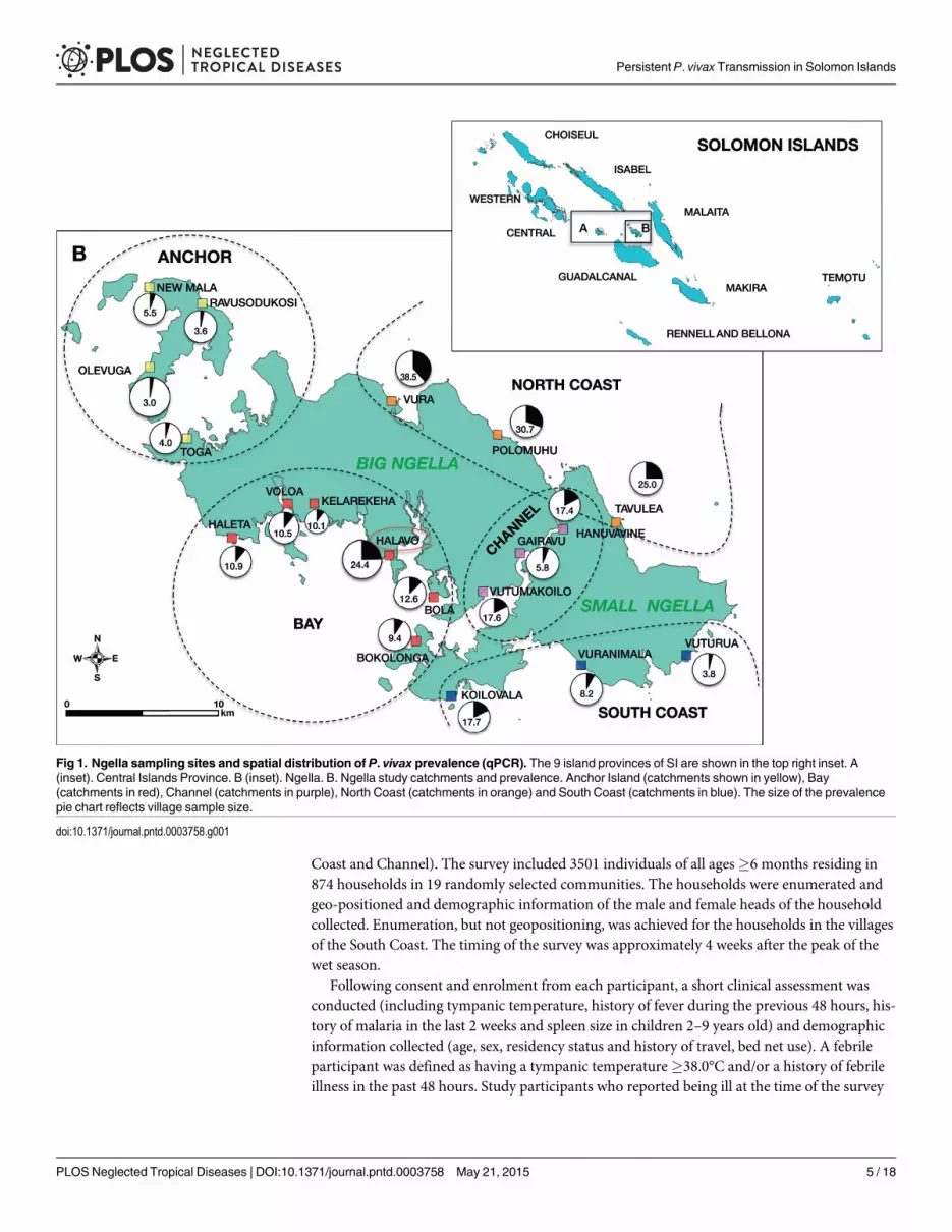
Coast and Channel). The survey included 3501 individuals of all ages�6 months residing in874 households in 19 randomly selected communities. The households were enumerated andgeo-positioned and demographic information of the male and female heads of the householdcollected. Enumeration, but not geopositioning, was achieved for the households in the villagesof the South Coast. The timing of the survey was approximately 4 weeks after the peak of thewet season.
Following consent and enrolment from each participant, a short clinical assessment wasconducted (including tympanic temperature, history of fever during the previous 48 hours, his-tory of malaria in the last 2 weeks and spleen size in children 2–9 years old) and demographicinformation collected (age, sex, residency status and history of travel, bed net use). A febrileparticipant was defined as having a tympanic temperature�38.0°C and/or a history of febrileillness in the past 48 hours. Study participants who reported being ill at the time of the survey
Fig 1. Ngella sampling sites and spatial distribution of P. vivax prevalence (qPCR). The 9 island provinces of SI are shown in the top right inset. A(inset). Central Islands Province. B (inset). Ngella. B. Ngella study catchments and prevalence. Anchor Island (catchments shown in yellow), Bay(catchments in red), Channel (catchments in purple), North Coast (catchments in orange) and South Coast (catchments in blue). The size of the prevalencepie chart reflects village sample size.
doi:10.1371/journal.pntd.0003758.g001
Persistent P. vivax Transmission in Solomon Islands
PLOS Neglected Tropical Diseases | DOI:10.1371/journal.pntd.0003758 May 21, 2015 5 / 18

were diagnosed by RDT (Access Bio, CareStart, USA) and treated if positive with artemether/lumefantrine, as per the national treatment guidelines. Where available the participant’s healthrecords were checked for recent anti-malarial treatment and applicable information recorded.
A 250 μL finger prick blood sample was collected into EDTA-Microtainer tubes (BectonDickinson, NJ, USA). 50 μL were immediately stabilized in 250 μL RNAProtect (Qiagen, Ger-many) for RNA studies and stored at ice pack cooling conditions until their transport to a cen-tralized field laboratory. Thick and thin films were prepared for determination of microscopicmalaria infection. Haemoglobin measurement was performed with Hemocue HB 301 analyzer.A measurement below 11g/dL was classified as anaemia. Upon return to the centralized labora-tory, the RNAProtect fractions were frozen immediately. The remaining 200 μL of whole bloodwas separated into red blood cells pellets and plasma and promptly frozen.
LM detection of Plasmodium spp. parasitesGiemsa stained blood films were examined under x1000 power. One hundred fields of viewwere examined before calling a sample “no parasites seen”. When a parasite was observed,counts of both white cells and parasites were commenced, and continued until 300 white cellshad been counted. The parasite count was then calculated, based on an assumed white cellcount of 8,000 white cells/ μL. However if no further parasites were observed, the process ofscanning to a total of 100 fields of view was completed. When only 1 parasite had been ob-served in 100 fields of view, an assumed count at the notional lower limit of detection of 10 par-asites/μL was applied, based on a further assumption of an average of 8 white cells per field ofview. All slides were stained within 24 hours at the regional malaria laboratory and read by ex-perienced microscopists, all of whom had completed WHO quality assurance courses. All LMpositive slides as well as the slides from all PCR positive / LM negative plus 10% of LM & PCRnegative slides were re-read by an Australian Level 1 expert microscopist that was blinded tothe PCR results. None of the 10% LM negative slides were found to be positive by the expertmicroscopist. In case of discrepancies between the two microscopy reads, the read of the expertmicroscopist was considered final.
DNA and RNA extractionGenomic DNA (gDNA) was isolated from red blood cell pellets (100 μL, corresponding to200 μL whole blood) using FavorPrep 96-well Genomic DNA kit (Favorgen, Taiwan). DNAwas eluted in 200 μL elution buffer and stored at -20°C. The RNA isolation procedure fromwhole blood in RNAProtect cell reagent has been described elsewhere [22], the only exceptionbeing an increased elution volume of 60μL of RNase-free water. Due to problems with storageof RNAProtect samples in the field, the quality of the RNA was tested using an RT-qPCR forthe human beta globin transcript [23]. This revealed a 10x lower total human RNA concentra-tion than in samples from a comparable study in Papua New Guinea [24]. RNA samples weretherefore concentrated 10-fold using a CentriVap Concentrator (Labconco, United States) be-fore testing for the presence of gametocytes.
Molecular detection of Plasmodium spp. parasitesAll 3501 DNA samples were first screened using a genus-specific qPCR targeting a conservedregion of the 18S rRNA gene [22]. Singleplex species-specific P. falciparum and P. vivax Taq-man qPCRs and a duplex P.malariae/P. ovale qPCR, targeting species-specific regions of 18SrRNA gene, were used to identify species as described previously [22,25]. Prevalence values re-ported in this study include only those infections confirmed by the species-specific qPCR Taq-man assays. Each detection experiment carried a dilution series of plasmids containing the
Persistent P. vivax Transmission in Solomon Islands
PLOS Neglected Tropical Diseases | DOI:10.1371/journal.pntd.0003758 May 21, 2015 6 / 18

target sequence of each PCR (104, 103, 102, 101, 5, 100 copies/μL), in duplicate, and were usedto determine standard curves and therefore estimate parasite densities (reported as 18S rRNAgene copy numbers/μL). All assays were run in 384-well plate format on the Roche LightCy-cler480 platform. Those infections detected by qPCR, but not by LM, were defined as sub-mi-croscopic infections. P. falciparum and P. vivax samples that were positive by species-specificTaqman qPCR were examined for presence of gametocytes using RT-qPCRs targeting thepfs25 and pvs25 orthologues, which are expressed only in mature gametocytes, as describedpreviously [22]. All gametocyte assays were also run in 384-well plate format on the RocheLightCycler480 platform.
Plasmodium spp. genetic diversityAll samples that were P. falciparum or P. vivax positive were genotyped to determine the multi-plicity of infection (MOI) using highly diverse size-polymorphic molecular markersmsp2 forP. falciparum andmsp1F3 and MS16 for P. vivax, respectively. PCR and capillary electrophore-sis were performed with slight modifications to the published protocols [26,27]. Genotypingdata was analyzed as described previously [26,27].
Statistical analysisStudy data were collected and managed using REDCap electronic data capture tools hosted atthe Walter and Eliza Hall Institute [28]. Analyses were done using the STATA12 statisticalsoftware package (College Station, TX). Differences in participant characteristics at enrolmentand prevalence differences among geographical areas and groups of individuals were assessedusing Chi-square (χ2) or Fisher’s exact tests. Differences in median ages and median householdsize were explored with quantile regression. Univariable and multivariable logistic regressionwere used for associations of P. vivax infection and exposure variables. Associations with P.vivax parasite density were investigated in simple and multivariable linear regression modelson only those subjects who tested positive to qPCR diagnosis. Poisson regression analyses wereutilized to explore associations between multiplicity of infection and exposure variables.
Results
Study populationA total of 3501 Ngella residents across 874 households were surveyed. The gender and age pro-files of the participants were representative of the Central Islands Province population, with52.5% females and a predominance of younger individuals (median age 18 years). The age dis-tribution was as follows:<2 years, 4.7%; 2–4 years, 10.6%; 5–9 years, 14.8%; 10–14 years,14.3%; 15–19 years, 7.5%; 20–39 years, 27.0%;>40 years, 21.3%. The majority of participants(95.2%) resided in the village for� 2 months. Of 447 participants who spent at least one nightoutside their village of residence in the last month, 69.1% travelled within Central Islands Prov-ince. 73.3% of participants reported having slept under a long lasting insecticidal net the nightbefore and 56.4% owned a bednet for longer than 24 months. Of all households, 84.5% ofhouseholds reported to have been sprayed with insecticide, and 70.4% of household headsspoke English. Of all participants, 687 (19.4%) had a history of fever in the previous two days,685 (19.7%) reported feeling unwell/sick at the time of survey and 23.3% had a haemoglobinmeasurement<11g/dL. No participant aged 2–9 years of age was found to have an enlargedspleen. A detailed description of demographic and clinical characteristics by geographical re-gion is given in S1 Table.
Persistent P. vivax Transmission in Solomon Islands
PLOS Neglected Tropical Diseases | DOI:10.1371/journal.pntd.0003758 May 21, 2015 7 / 18
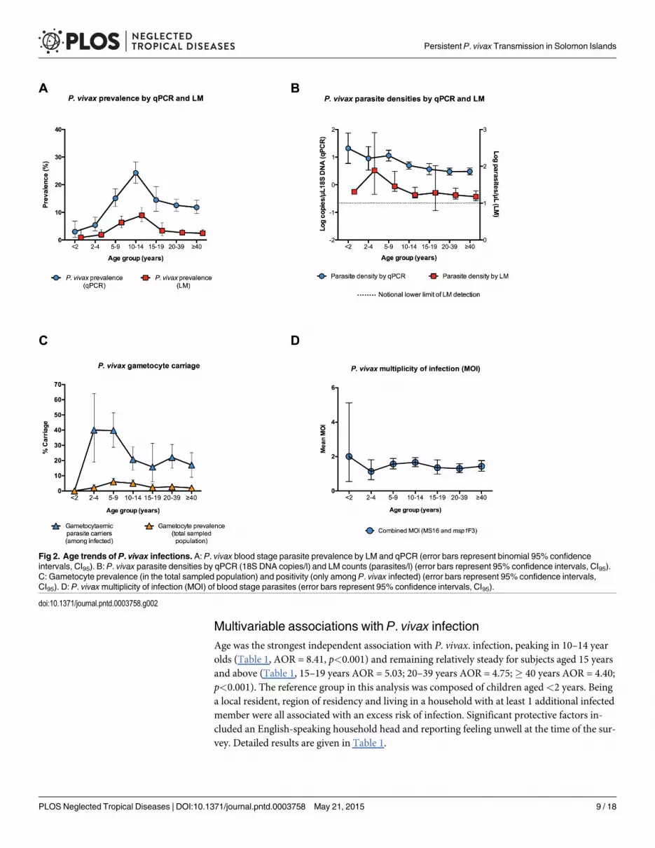
Prevalence of Plasmodium spp. infection by LMOverall, 130 individuals (3.7%) had Plasmodium spp. parasites detectable by LM: 126 P. vivax,three P. falciparummono-infections and one P. vivax/P. falciparummixed infection. No infec-tions with P.malariae or P. ovale were observed. The prevalence of P. vivax infection variedsignificantly by geographical region (p<0.001) (Fig 1B) and was lowest in the South Coast andAnchor regions (0.8%), followed by Channel (3.0%) and Bay (3.5%) and North Coast (11.7%).P. vivax prevalence showed strong age trends and peaked in adolescents 10–15 years of age(8.6%, p<0.001) (Fig 2A).
Prevalence of P. vivax by qPCROverall, 468 participants (13.4%) had qPCR-detectable infections: 463 were P. vivaxmono-in-fections and five were mixed P. falciparum/P. vivax infections (0.14%). The 126 P. vivax infec-tions and the one mixed infection by LM were confirmed by qPCR. Overall, 72.9% of P. vivaxinfections were sub-microscopic. In two of the catchments, Kelarekeha and Vuturua (Fig 1B),only sub-microscopic infections were observed among 59 and 156 individuals surveyed, re-spectively. P. vivax qPCR prevalence displayed spatial heterogeneity among the five geographi-cal areas and the 19 catchments, varying from 3.0–38.5%. Prevalence by qPCR was highest invillages on the North Coast (25.0–38.5%) and lowest on Anchor Island (3.0–5.5%) (Fig 1B).
Of 874 households sampled across Ngella, 559 had no infected members, 210 had only oneinfected member and 105 had two or more infected members. There was no association be-tween household size and probability of being infected (p = 0.550). Not taking into account anyother variables, there was an increased risk of being infected if at least one other member of thehousehold was infected (OR = 2.59, p<0.001, CI95[2.13, 3.16]).
P. vivax prevalence was age-dependent (p<0.001), lowest in<2 years (3.0%, n = 166) andpeaking in the 10–14 year old age group (24.3%, n = 499) (Fig 2A). Prevalence of infection didnot differ significantly between male and female participants (p>0.650). Participants who wereresidents (lived in the village� 2 months) were more frequently infected with P. vivax thannon-residents (infected residents: 13.7% vs. infected non-residents 8.3%, p = 0.045). Once resi-dency status was taken into account, recent travel (defined as spending at least 1 night awayfrom the village of residence in the last month) was not associated with a difference in infectionrisk (p = 0.300). Those living in a household where the household head speaks English, a proxyfor education level, were infected less frequently (12.0%) than those living in a householdwhere the head does not speak English (18.5%, p<0.001). There was a moderate increase inrisk of P. vivax infection in those who reported not having slept under a net the night beforecompared to net users (users: 12.6% vs. non-users: 15.6%, p = 0.022).
The majority of P. vivax-infected individuals (84.6%) neither reported febrile symptoms(defined as history of fever or measured fever at survey) nor feeling ill (85.4%). Six of the 26participants that had a measured fever at the time of the survey (tympanic temperature�38°C,18.8%) were infected with P. vivax. Compared to uninfected participants, those with a P. vivaxinfection were less likely to report having had febrile symptoms in the previous two days (unin-fected 20.0% vs. infected 15.2%, p = 0.014) or report feeling unwell at the time of survey (unin-fected 20.5% vs. infected 14.6%, p = 0.003). A total of 280 P. vivax infections (60.0%) were bothasymptomatic and sub-microscopic. There were no significant differences in the proportion ofasymptomatic P. vivax infections between different age groups (p> 0.200) and regions (p>0.240). Of 468 P. vivax-infected individuals, 19.6% had a haemoglobin<11 g/dL compared to23.9% of uninfected individuals (p = 0.045).
Persistent P. vivax Transmission in Solomon Islands
PLOS Neglected Tropical Diseases | DOI:10.1371/journal.pntd.0003758 May 21, 2015 8 / 18
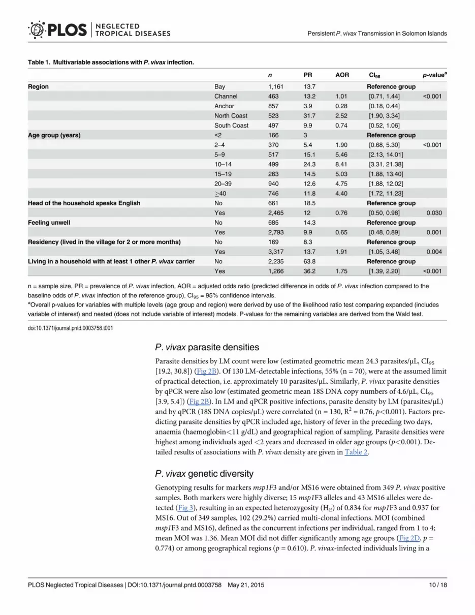
Multivariable associations with P. vivax infectionAge was the strongest independent association with P. vivax. infection, peaking in 10–14 yearolds (Table 1, AOR = 8.41, p<0.001) and remaining relatively steady for subjects aged 15 yearsand above (Table 1, 15–19 years AOR = 5.03; 20–39 years AOR = 4.75;� 40 years AOR = 4.40;p<0.001). The reference group in this analysis was composed of children aged<2 years. Beinga local resident, region of residency and living in a household with at least 1 additional infectedmember were all associated with an excess risk of infection. Significant protective factors in-cluded an English-speaking household head and reporting feeling unwell at the time of the sur-vey. Detailed results are given in Table 1.
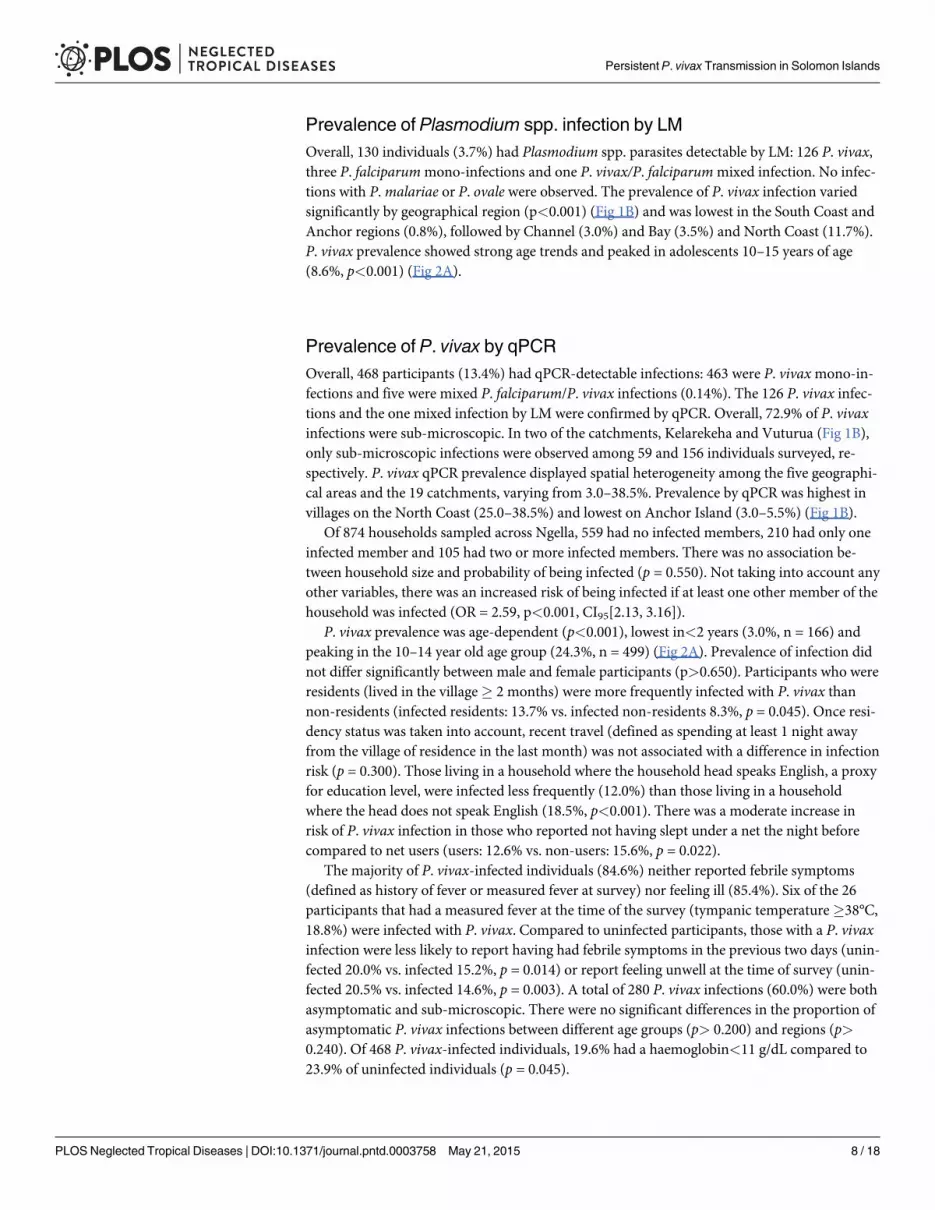
Fig 2. Age trends of P. vivax infections. A: P. vivax blood stage parasite prevalence by LM and qPCR (error bars represent binomial 95% confidenceintervals, CI95). B: P. vivax parasite densities by qPCR (18S DNA copies/l) and LM counts (parasites/l) (error bars represent 95% confidence intervals, CI95).C: Gametocyte prevalence (in the total sampled population) and positivity (only among P. vivax infected) (error bars represent 95% confidence intervals,CI95). D: P. vivaxmultiplicity of infection (MOI) of blood stage parasites (error bars represent 95% confidence intervals, CI95).
doi:10.1371/journal.pntd.0003758.g002
Persistent P. vivax Transmission in Solomon Islands
PLOS Neglected Tropical Diseases | DOI:10.1371/journal.pntd.0003758 May 21, 2015 9 / 18
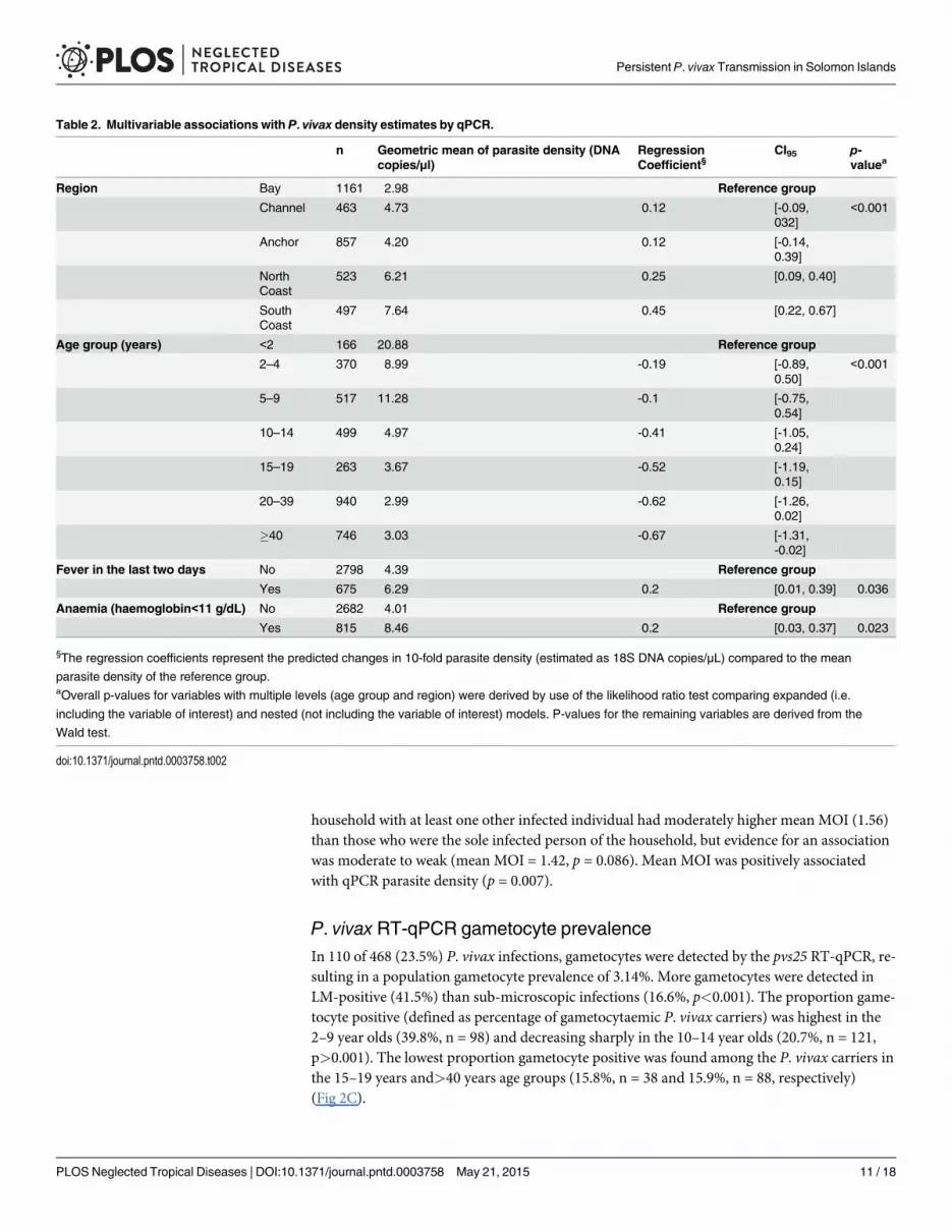
P. vivax parasite densitiesParasite densities by LM count were low (estimated geometric mean 24.3 parasites/μL, CI95[19.2, 30.8]) (Fig 2B). Of 130 LM-detectable infections, 55% (n = 70), were at the assumed limitof practical detection, i.e. approximately 10 parasites/μL. Similarly, P. vivax parasite densitiesby qPCR were also low (estimated geometric mean 18S DNA copy numbers of 4.6/μL, CI95[3.9, 5.4]) (Fig 2B). In LM and qPCR positive infections, parasite density by LM (parasites/μL)and by qPCR (18S DNA copies/μL) were correlated (n = 130, R2 = 0.76, p<0.001). Factors pre-dicting parasite densities by qPCR included age, history of fever in the preceding two days,anaemia (haemoglobin<11 g/dL) and geographical region of sampling. Parasite densities werehighest among individuals aged<2 years and decreased in older age groups (p<0.001). De-tailed results of associations with P. vivax density are given in Table 2.
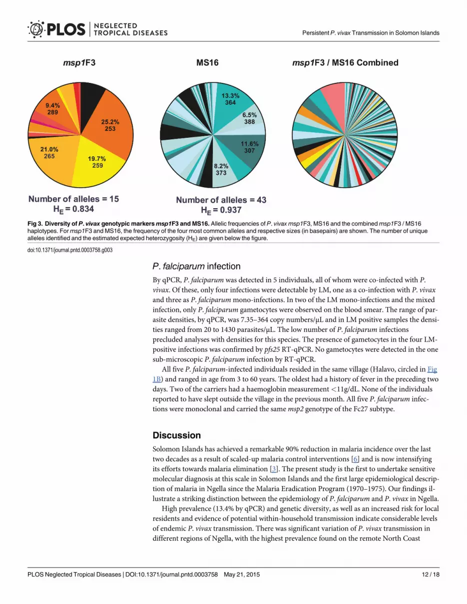
P. vivax genetic diversityGenotyping results for markersmsp1F3 and/or MS16 were obtained from 349 P. vivax positivesamples. Both markers were highly diverse; 15msp1F3 alleles and 43 MS16 alleles were de-tected (Fig 3), resulting in an expected heterozygosity (HE) of 0.834 formsp1F3 and 0.937 forMS16. Out of 349 samples, 102 (29.2%) carried multi-clonal infections. MOI (combinedmsp1F3 and MS16), defined as the concurrent infections per individual, ranged from 1 to 4;mean MOI was 1.36. Mean MOI did not differ significantly among age groups (Fig 2D, p =0.774) or among geographical regions (p = 0.610). P. vivax-infected individuals living in a
Table 1. Multivariable associations with P. vivax infection.
n PR AOR CI95 p-valuea
Region Bay 1,161 13.7 Reference group
Channel 463 13.2 1.01 [0.71, 1.44] <0.001
Anchor 857 3.9 0.28 [0.18, 0.44]
North Coast 523 31.7 2.52 [1.90, 3.34]
South Coast 497 9.9 0.74 [0.52, 1.06]
Age group (years) <2 166 3 Reference group
2–4 370 5.4 1.90 [0.68, 5.30] <0.001
5–9 517 15.1 5.46 [2.13, 14.01]
10–14 499 24.3 8.41 [3.31, 21.38]
15–19 263 14.5 5.03 [1.88, 13.40]
20–39 940 12.6 4.75 [1.88, 12.02]
�40 746 11.8 4.40 [1.72, 11.23]
Head of the household speaks English No 661 18.5 Reference group
Yes 2,465 12 0.76 [0.50, 0.98] 0.030
Feeling unwell No 685 14.3 Reference group
Yes 2,793 9.9 0.65 [0.48, 0.89] 0.001
Residency (lived in the village for 2 or more months) No 169 8.3 Reference group
Yes 3,317 13.7 1.91 [1.05, 3.48] 0.004
Living in a household with at least 1 other P. vivax carrier No 2,235 63.8 Reference group
Yes 1,266 36.2 1.75 [1.39, 2.20] <0.001
n = sample size, PR = prevalence of P. vivax infection, AOR = adjusted odds ratio (predicted difference in odds of P. vivax infection compared to the
baseline odds of P. vivax infection of the reference group), CI95 = 95% confidence intervals.aOverall p-values for variables with multiple levels (age group and region) were derived by use of the likelihood ratio test comparing expanded (includes
variable of interest) and nested (does not include variable of interest) models. P-values for the remaining variables are derived from the Wald test.
doi:10.1371/journal.pntd.0003758.t001
Persistent P. vivax Transmission in Solomon Islands
PLOS Neglected Tropical Diseases | DOI:10.1371/journal.pntd.0003758 May 21, 2015 10 / 18
household with at least one other infected individual had moderately higher mean MOI (1.56)than those who were the sole infected person of the household, but evidence for an associationwas moderate to weak (mean MOI = 1.42, p = 0.086). Mean MOI was positively associatedwith qPCR parasite density (p = 0.007).
P. vivax RT-qPCR gametocyte prevalenceIn 110 of 468 (23.5%) P. vivax infections, gametocytes were detected by the pvs25 RT-qPCR, re-sulting in a population gametocyte prevalence of 3.14%. More gametocytes were detected inLM-positive (41.5%) than sub-microscopic infections (16.6%, p<0.001). The proportion game-tocyte positive (defined as percentage of gametocytaemic P. vivax carriers) was highest in the2–9 year olds (39.8%, n = 98) and decreasing sharply in the 10–14 year olds (20.7%, n = 121,p>0.001). The lowest proportion gametocyte positive was found among the P. vivax carriers inthe 15–19 years and>40 years age groups (15.8%, n = 38 and 15.9%, n = 88, respectively)(Fig 2C).
Table 2. Multivariable associations with P. vivax density estimates by qPCR.
n Geometric mean of parasite density (DNAcopies/μl)
RegressionCoefficient§
CI95 p-valuea
Region Bay 1161 2.98 Reference group
Channel 463 4.73 0.12 [-0.09,032]
<0.001
Anchor 857 4.20 0.12 [-0.14,0.39]
NorthCoast
523 6.21 0.25 [0.09, 0.40]
SouthCoast
497 7.64 0.45 [0.22, 0.67]
Age group (years) <2 166 20.88 Reference group
2–4 370 8.99 -0.19 [-0.89,0.50]
<0.001
5–9 517 11.28 -0.1 [-0.75,0.54]
10–14 499 4.97 -0.41 [-1.05,0.24]
15–19 263 3.67 -0.52 [-1.19,0.15]
20–39 940 2.99 -0.62 [-1.26,0.02]
�40 746 3.03 -0.67 [-1.31,-0.02]
Fever in the last two days No 2798 4.39 Reference group
Yes 675 6.29 0.2 [0.01, 0.39] 0.036
Anaemia (haemoglobin<11 g/dL) No 2682 4.01 Reference group
Yes 815 8.46 0.2 [0.03, 0.37] 0.023
§The regression coefficients represent the predicted changes in 10-fold parasite density (estimated as 18S DNA copies/μL) compared to the mean
parasite density of the reference group.aOverall p-values for variables with multiple levels (age group and region) were derived by use of the likelihood ratio test comparing expanded (i.e.
including the variable of interest) and nested (not including the variable of interest) models. P-values for the remaining variables are derived from the
Wald test.
doi:10.1371/journal.pntd.0003758.t002
Persistent P. vivax Transmission in Solomon Islands
PLOS Neglected Tropical Diseases | DOI:10.1371/journal.pntd.0003758 May 21, 2015 11 / 18
P. falciparum infectionBy qPCR, P. falciparum was detected in 5 individuals, all of whom were co-infected with P.vivax. Of these, only four infections were detectable by LM, one as a co-infection with P. vivaxand three as P. falciparummono-infections. In two of the LMmono-infections and the mixedinfection, only P. falciparum gametocytes were observed on the blood smear. The range of par-asite densities, by qPCR, was 7.35–364 copy numbers/μL and in LM positive samples the densi-ties ranged from 20 to 1430 parasites/μL. The low number of P. falciparum infectionsprecluded analyses with densities for this species. The presence of gametocytes in the four LM-positive infections was confirmed by pfs25 RT-qPCR. No gametocytes were detected in the onesub-microscopic P. falciparum infection by RT-qPCR.
All five P. falciparum-infected individuals resided in the same village (Halavo, circled in Fig1B) and ranged in age from 3 to 60 years. The oldest had a history of fever in the preceding twodays. Two of the carriers had a haemoglobin measurement<11g/dL. None of the individualsreported to have slept outside the village in the previous month. All five P. falciparum infec-tions were monoclonal and carried the samemsp2 genotype of the Fc27 subtype.
DiscussionSolomon Islands has achieved a remarkable 90% reduction in malaria incidence over the lasttwo decades as a result of scaled-up malaria control interventions [6] and is now intensifyingits efforts towards malaria elimination [3]. The present study is the first to undertake sensitivemolecular diagnosis at this scale in Solomon Islands and the first large epidemiological descrip-tion of malaria in Ngella since the Malaria Eradication Program (1970–1975). Our findings il-lustrate a striking distinction between the epidemiology of P. falciparum and P. vivax in Ngella.
High prevalence (13.4% by qPCR) and genetic diversity, as well as an increased risk for localresidents and evidence of potential within-household transmission indicate considerable levelsof endemic P. vivax transmission. There was significant variation of P. vivax transmission indifferent regions of Ngella, with the highest prevalence found on the remote North Coast
Fig 3. Diversity of P. vivax genotypic markersmsp1F3 and MS16. Allelic frequencies of P. vivax msp1F3, MS16 and the combinedmsp1F3 / MS16haplotypes. Formsp1F3 and MS16, the frequency of the four most common alleles and respective sizes (in basepairs) are shown. The number of uniquealleles identified and the estimated expected heterozygosity (HE) are given below the figure.
doi:10.1371/journal.pntd.0003758.g003
Persistent P. vivax Transmission in Solomon Islands
PLOS Neglected Tropical Diseases | DOI:10.1371/journal.pntd.0003758 May 21, 2015 12 / 18
(25.0–38.5%), which prior to the Malaria Eradication spraying operations was described asholoendemic and having an environment highly favourable to the mosquito [7]. The lowestrates of P. vivax infection were observed on Anchor (3.9% by qPCR), where 15 years ago acommunity-based initiative eliminated a substantial number of breeding sites through environ-mental management [Lodo, personal communication]. It is therefore likely that the presence ofsuitable larval habitats and vector abundance may be key factors influencing P. vivax transmis-sion on Ngella.
It remains unclear whether autochthonous P. falciparum transmission remains in Ngella orparasites are being re-introduced by incoming travelers or returning residents from areas withhigher P. falciparum burden, such Guadalcanal or Malaita provinces. In this survey, only fiveP. falciparum cases were identified in the village of Halavo (Fig 1B). As all five infections car-ried the samemsp2 allele and four were gametocytaemic, a small local outbreak following re-cent re-introduction seems more likely. This is reminiscent of the situation in epidemic-proneareas of the Papua New Guinea highlands, where a clonal P. falciparum epidemic on a back-ground of endemic, low level P. vivax transmission has been reported [29]. P. falciparum popu-lations in neighbouring Guadalcanal province were in fact found to be of low genetic diversity[30,31]. Based on case statistics at the local health facilities, 30% of malaria cases detected inCentral Islands Province are caused by P. falciparum [6] indicating that either importation ofP. falciparum parasites is common or that low levels of endemic P. falciparum transmissionmay remain in some parts of Ngella. Further studies are therefore required to ascertain the ab-sence of endemic P. falciparum transmission in this area of Solomon Islands and whether thecases found are the result of inter-island travel.
The current situation of malaria in Ngella (i.e. 3.7% prevalence by LM, clear P. vivax domi-nance and absence of enlarged spleens in children 2–9yrs of age) is a consequence of the dra-matic reduction in malaria transmission achieved throughout Solomon Islands in the last 20years [6]. This change is similar to that encountered at the end of 1974, after approximately 5years of twice-yearly Malaria Eradication Program spraying. Then, prevalence in 2–9 year oldshad dropped from pre-spraying rates of 60% to 1.4% and P. vivax became predominant [7].Similar shifts in malaria epidemiology were also observed in the elimination provinces ofTemotu [9] and Isabel [8]. In Temotu, P. falciparum accounted for 17.5% of infections in popu-lation survey conducted in 2008 [9], but by 2012, the national program’s surveillance systemreported only P. vivax cases from both Temotu and Isabel [6]. This shift in the relative impor-tance of P. falciparum and P. vivax are not unique to Solomon Islands and have been reportedafter periods of sustained malaria control from other settings where P. falciparum and P. vivaxoccur sympatrically, such as the Amazon [21,32], Central America [4] and Thailand [33].
As in other endemic settings [12,13,34], P. vivax infections were of low density and PCRfound three times more infections than LM. The majority of infections were not accompaniedby febrile symptoms or anaemia. On the contrary, participants who reported feeling unwell orfebrile were less likely to be infected with P. vivax. While this significantly lower level of febrilesymptoms in P. vivax carriers is likely to be an artifact of the large samples size it does indicatethat P. vivax is not a common cause of fever in Ngella. Whereas asymptomatic P. vivax infec-tions have been commonly found in areas of high transmission [12,35,36], the advent of molec-ular diagnosis has revealed that even at low transmission the majority of infections in cross-sectional surveys are symptomless [11,37,38], including in the previous surveys in Temotu [9]and Isabel [8] where 97.1% and 92.9% of P. vivax infected individuals infections were asymp-tomatic, respectively.
Both the presence of P. vivax infections and their level of parasitaemia were found to bestrongly age-dependent, albeit in different ways: while P. vivax parasite densities decreasedwith age, prevalence of P. vivax infections rose throughout childhood and only started
Persistent P. vivax Transmission in Solomon Islands
PLOS Neglected Tropical Diseases | DOI:10.1371/journal.pntd.0003758 May 21, 2015 13 / 18
dropping in adolescents and adults. These contrasting patterns are most likely due to localmosquito biting behavior and acquisition of immunity. Anopheles farauti, the only coastal ma-laria vector in Solomon Islands, is biting predominantly in the early evening (i.e. before 10pm)and outdoors [39], when small children tend to be indoors but older ones still active. The in-crease in prevalence during childhood is thus likely to represent an increase in exposure to in-fective bites. At all levels of transmission, immunity to P. vivax tends to be more rapidlyacquired than that to P. falciparum [40]. Thus, the strong reduction in prevalence and parasitedensities with increasing age in Ngella indicate that P. vivax transmission there remains suffi-ciently high for relatively rapid acquisition of clinical and anti-parasite immunity.
Despite very low overall parasite densities, gametocytes were detected in almost a quarter ofall P. vivax infections (in 41.5% of LM-positive infections and 16.6% of sub-microscopic infec-tions). Given issues with RNA quality, it is likely that the gametocytaemic reservoir in Ngellawas underestimated in our survey and the true prevalence of gametocytes is higher, especiallyin the sub-microscopic group. Given the rapid and ongoing production of P. vivax gameto-cytes, most if not all, blood stage infections could harbor concurrent gametocytes [41]. Whilstsub-patent P. falciparum infections have been shown to infect up to 43.5% of mosquitoes[17,19], the role of sub-microscopic P. vivax gametocyte carriage in sustaining transmission ispoorly understood. The capacity of sub-microscopic P. vivax infections to infect mosquitoeshas been established in studies from Thailand [18,42,43], Sri Lanka [44], Peru [45] and malariatherapy settings [14,15], but at varying proportions and with weak associations of gametocytedensity. Although sub-patent infections may infect fewer mosquitoes, their higher prevalencein endemic settings may mean that the net transmission potential of low-density infections ishigher. In Ngella, asymptomatic, sub-microscopic infections of adolescents and adults maythus be an important source of local transmission.
These considerations may constitute a significant challenge to the success of the SolomonIslands malaria control program. The national malaria surveillance system, based on passivecase detection and irregular mass blood surveys, only employs traditional microscopy diagno-sis. This diagnostic test may not only underestimate the true burden of malaria in the SolomonIslands but also lack the means to detect and attack a substantial part of the P. vivax transmis-sion reservoir. Despite outstanding gains in the last two decades, traditional tools of the Solo-mon Islands malaria control program may therefore have reached their effectiveness in theface of a large and silent reservoir of P. vivax infection.
Our observation that people living in a household with another P. vivax infected individualis a noteworthy finding. Not only does it indicate likely within-household transmission, butalso highlights that reactive case detection strategies [46–48] and focal mass drug administra-tion [34] might be appropriately applied in Solomon Islands. In the Southwest Pacific, MDAcampaigns that included primaquine to target the undetectable liver stage parasites have previ-ously been successful in interrupting P. vivax transmission on Aneytium Island in Vanuatu[49] and Nissan Island in Papua New Guinea [50]. Combining automated registration of ob-served cases and rapid identification of transmission foci (e.g. in a spatial decision support sys-tem) [51] with reactive mass-screen and treat (MSAT) or with focal, household-based massdrug administration [52,53] should therefore be evaluated as possible additional malaria elimi-nation tools in Solomon Islands and neighbouring Vanuatu. All interventions will be mostefficacious if they include routine administration of primaquine to all P. vivax infected individ-uals. This will however require addressing the challenges posed by the potential primaquinetoxicity in G6PD deficient individuals.
Persistent P. vivax Transmission in Solomon Islands
PLOS Neglected Tropical Diseases | DOI:10.1371/journal.pntd.0003758 May 21, 2015 14 / 18
Supporting InformationS1 Checklist. STROBE checklist.(DOC)
S1 Table. Demographic and clinical characteristics of the study population, by geographi-cal area.(PDF)
AcknowledgmentsWe would like to extend our sincere gratitude to the communities and community leaders fortheir support and participation in this study. We acknowledge the indispensable and preciouswork carried out by the field teams (Freda Pitakaka, Everlyn Darcy, Luito Faarodo, DavidRamosala, Leila Tokasi, Anthony Harurihu, Robert Oreitaha, Rebecca Mae, Ethel Lodo, RobertKali), laboratory officer Rhomson Nuake, vector borne disease control program staff in Honi-ara (Albino Bobogare, Dr. Lyndes Wini, Erick Hale) and Tulagi (Benjamin Zimbo and FannyKeru) and Tulagi hospital microscopists (Jentar Saitoha, Stephen Lae, Matthew Mava andGeorge Linton). We also acknowledge Luke Marston for assisting with all aspects of field oper-ations. The support and guidance of Dr. Tanya Russell, Prof. Tom Burkot and Lt. Col. RobertCooper in Tulagi and the field was indispensable and highly appreciated. We thank StellaKyvetos and Joan Curtis for ensuring prompt and safe transport of consumables to SolomonIslands and field samples from Solomon Islands to Australia and Dr. Céline Barnadas and Dr.G.L.Abby Harrison for technical assistance in the laboratory. We also thank the manuscript re-viewers for their careful assessment and meaningful feedback during the peer review process.
DisclaimerThe opinions expressed are those of the authors and do not necessarily reflect those of the
Australian Defence Force.
Author ContributionsConceived and designed the experiments: AWD GDSMW JWK IM. Performed the experi-ments: AW AWD CK IH JL VV DP. Analyzed the data: AW CK AEB IM. Contributed re-agents/materials/analysis tools: AEB. Wrote the paper: AW CK GDS AEBMW JWK IM.
References1. Muller I, Bockarie M, Alpers M, Smith T. The epidemiology of malaria in Papua New Guinea. Trends
Parasitol. 2003; 19(6): 253–9. PMID: 12798082
2. Battle KE, Gething PW, Elyazar IR, Moyes CL, Sinka ME, Howes RE, et al. The global public health sig-nificance of Plasmodium vivax. Adv Parasitol. 2012; 80: 1–111. English. doi: 10.1016/B978-0-12-397900-1.00001-3 PMID: 23199486
3. Pacific Malaria Initiative Survey Group (PacMISC) on behalf of the Ministries of Health of Vanuatu, Sol-omon Islands. Malaria on isolated Melanesian islands prior to the initiation of malaria elimination activi-ties. Malar J. 2010; 9: 218. doi: 10.1186/1475-2875-9-218 PMID: 20659316
4. World Health Organisation (WHO). World Malaria Report 2013. Geneva, Switzerland: World HealthOrganisation, 2013.
5. Solomon Islands Vector Borne Disease Control Program (SI NVBDCP). Historical Annual Malaria Rec-ords. Honiara, Solomon Islands: Ministry of Health.
6. Solomon Islands Vector Borne Disease Control Program (SI NVBDCP). Annual Malaria Report 2012.Honiara, Solomon Islands: Ministry of Health, 2013.
7. Avery J. The Epidemiology of Disappearing Malaria in the Solomon Islands. Sheffield, United Kingdom:University of Sheffield; 1977. http://ethos.bl.uk/OrderDetails.do?uin = uk.bl.ethos.448417.
Persistent P. vivax Transmission in Solomon Islands
PLOS Neglected Tropical Diseases | DOI:10.1371/journal.pntd.0003758 May 21, 2015 15 / 18
8. Atkinson JA, Johnson ML, Wijesinghe R, Bobogare A, Losi L, O'Sullivan M, et al. Operational researchto inform a sub-national surveillance intervention for malaria elimination in Solomon Islands. Malar J.2012; 1: 101.
9. Harris I, Sharrock WW, Bain LM, Gray KA, Bobogare A, Boaz L, et al. A large proportion of asymptom-atic Plasmodium infections with low and sub-microscopic parasite densities in the low transmission set-ting of Temotu Province, Solomon Islands: challenges for malaria diagnostics in an elimination setting.Malar J. 2010; 9:254. doi: 10.1186/1475-2875-9-254 PMID: 20822506
10. Okell LC, Bousema T, Griffin JT, Ouedraogo AL, Ghani AC, Drakeley CJ. Factors determining the oc-currence of submicroscopic malaria infections and their relevance for control. Nat Commun. 2012; 12(3):1237.
11. Alves FP, Durlacher RR, Menezes MJ, Krieger H, Silva LH, Camargo EP. High prevalence of asymp-tomatic Plasmodium vivax and Plasmodium falciparum infections in native Amazonian populations. AmJ Trop Med Hyg. 2002 Jun; 66(6):641–8. PMID: 12224567
12. Mueller I, Widmer S, Michel D, Maraga S, McNamara DT, Kiniboro B, et al. High sensitivity detection ofPlasmodium species reveals positive correlations between infections of different species, shifts in agedistribution and reduced local variation in Papua New Guinea. Malar J. 2009; 8: 41. doi: 10.1186/1475-2875-8-41 PMID: 19284594
13. Steenkeste N, Rogers WO, Okell L, Jeanne I, Incardona S, Duval L, et al. Sub-microscopic malariacases and mixed malaria infection in a remote area of high malaria endemicity in Rattanakiri province,Cambodia: implication for malaria elimination. Malar J. 2010 Apr 22; 9:108. doi: 10.1186/1475-2875-9-108 PMID: 20409349
14. Boyd MF, Kitchen SF. On the infectiousness of patients infected with Plasmodium vivax and Plasmodi-um falciparum. Am J Trop Med Hyg. 1937 (17: ):253–62.
15. Jeffery GM, Eyles DE. Infectivity to Mosquitoes of Plasmodium falciparum as Related to GametocyteDensity and Duration of Infection. Am J Trop Med Hyg. 1955; 4(5):781–9. PMID: 13259002
16. Nwakanma D, Kheir A, Sowa M, Dunyo S, Jawara M, Pinder M, et al. High gametocyte complexity andmosquito infectivity of Plasmodium falciparum in the Gambia. Int J Parasitol. 2008 Feb; 38(2):219–27.PMID: 17709108
17. Ouedraogo AL, Bousema T, Schneider P, de Vlas SJ, Ilboudo-Sanogo E, Cuzin-Ouattara N, et al. Sub-stantial contribution of submicroscopical Plasmodium falciparum gametocyte carriage to the infectiousreservoir in an area of seasonal transmission. PLoS One. 2009; 4(12):e8410. doi: 10.1371/journal.pone.0008410 PMID: 20027314
18. Sattabongkot J, Maneechai N, Rosenberg R. Plasmodium vivax: gametocyte infectivity of naturally in-fected Thai adults. Parasitology. 1991 Feb; 102 Pt 1:27–31.
19. Schneider P, Bousema JT, Gouagna LC, Otieno S, van de Vegte-Bolmer M, Omar SA, et al. Submicro-scopic Plasmodium falciparum gametocyte densities frequently result in mosquito infection. Am J TropMed Hyg. 2007; 76(3): 470–4. PMID: 17360869
20. Solomon Islands Government (SIG). 2009. Volume I Report on 2009 Population And HousingCensus:Basic Tables and Census Description. Honiara, Solomon Islands. http://www.mof.gov.sb/Libraries/Statistics/2011_06__Report_on_2009_Population_Housing_Census.sflb.ashx.
21. Branch O, Casapia WM, Gamboa DV, Hernandez JN, Alava FF, Roncal N, et al. Clustered local trans-mission and asymptomatic Plasmodium falciparum and Plasmodium vivaxmalaria infections in a re-cently emerged, hypoendemic Peruvian Amazon community. Malar J. 2005 Dec; 4(6):27.
22. Wampfler R, Mwingira F, Javati S, Robinson L, Betuela I, Siba P, et al. Strategies for detection of Plas-modium species gametocytes. PLoS One. 2013 27; 8(9):e76316. doi: 10.1371/journal.pone.0076316PMID: 24312682
23. Irenge LM, Robert A, Gala JL. Quantitative assessment of human beta-globin gene expression in vitroby TaqMan real-time reverse transcription-PCR: comparison with competitive reverse transcription-PCR and application to mutations or deletions in noncoding regions. Clin Chem. 2005; 51(12): 2395–6.PMID: 16306108
24. Koepfli C, Robinson LJ, Rarau P, Salib M, Sambale N, Wampfler R, et al. Blood-stage parasitaemiaand age determine Plasmodium falciparum and P. vivax gametocytaemia in Papua New Guinea. PLoSOne. 2015: in press.
25. Rosanas-Urgell A, Mueller D, Betuela I, Barnadas C, Iga J, Zimmerman PA, et al. Comparison of diag-nostic methods for the detection and quantification of the four sympatric Plasmodium species in fieldsamples from Papua New Guinea. Malar J. 201014; 9: 361. English. doi: 10.1186/1475-2875-9-361PMID: 21156052
26. Falk N, Maire N, SamaW, Owusu-Agyei S, Smith T, Beck HP, et al. Comparison of PCR-RFLP andGenescan-based genotyping for analyzing infection dynamics of Plasmodium falciparum. Am J TropMed Hyg. 2006; 74(6): 944–50. PMID: 16760501
Persistent P. vivax Transmission in Solomon Islands
PLOS Neglected Tropical Diseases | DOI:10.1371/journal.pntd.0003758 May 21, 2015 16 / 18
27. Koepfli C, Ross A, Kiniboro B, Smith TA, Zimmerman PA, Siba P, et al. Multiplicity and diversity of Plas-modium vivax infections in a highly endemic region in Papua New Guinea. PLoS Negl Trop Dis. 2011; 5(12):e1424. doi: 10.1371/journal.pntd.0001424 PMID: 22206027
28. Harris PA, Taylor R, Thielke R, Payne J, Gonzalez N, Conde JG. Research electronic data capture(REDCap)-a metadata-driven methodology and workflow process for providing translational researchinformatics support. J Biomed Inform. 2009; 42(2): 377–81. doi: 10.1016/j.jbi.2008.08.010 PMID:18929686
29. Mueller I, Kaiok J, Reeder JC, Cortes A. The population structure of Plasmodium falciparum and Plas-modium vivax during an epidemic of malaria in the Eastern Highlands of Papua New Guinea. Am JTrop Med Hyg. 2002 Nov; 67(5): 459–64. PMID: 12479544
30. Ballif M, Hii J, Marfurt J, Crameri A, Fafale A, Felger I, et al. Monitoring of malaria parasite resistance tochloroquine and sulphadoxine-pyrimethamine in the Solomon Islands by DNAmicroarray technology.Malar J. 2010; 9:270. doi: 10.1186/1475-2875-9-270 PMID: 20925934
31. Sakihama N, Ohmae H, Bakote'e B, Kawabata M, Hirayama K, Tanabe K. Limited allelic diversity ofPlasmodium falciparummerozoite surface protein 1 gene from populations in the Solomon Islands. AmJ Trop Med Hyg. 2006 Jan; 74(1):31–40. PMID: 16407343
32. Oliveira-Ferreira J, Lacerda MV, Brasil P, Ladislau JL, Tauil PL, Daniel-Ribeiro CT. Malaria in Brazil: anoverview. Malar J. 2010 Apr 30; 9: 115. doi: 10.1186/1475-2875-9-115 PMID: 20433744
33. Putaporntip C, Hongsrimuang T, Seethamchai S, Kobasa T, Limkittikul K, Cui L, et al. Differential preva-lence of Plasmodium infections and cryptic Plasmodium knowlesimalaria in humans in Thailand. J In-fect Dis. 2009 Apr 15; 199(8):1143–50. doi: 10.1086/597414 PMID: 19284284
34. Katsuragawa TH, Gil LH, Tada MS, de Almeida e Silva A, Costa JD, Araujo Mda S, et al. The dynamicsof transmission and spatial distribution of malaria in riverside areas of Porto Velho, Rondonia, in theAmazon region of Brazil. Plos One. 2010 Feb 16; 5(2):e9245. doi: 10.1371/journal.pone.0009245PMID: 20169070
35. Pinto J, Sousa CA, Gil V, Ferreira C, Goncalves L, Lopes D, et al. Malaria in Sao Tome and Principe:parasite prevalences and vector densities. Acta Trop. 2000 Sep 18; 76(2):185–93. PMID: 10936578
36. Suarez-Mutis MC, Coura JR. [Changes in the epidemiological pattern of malaria in a rural area of themiddle Rio Negro, Brazilian Amazon: a retrospective analysis]. Cadernos de saude publica. 2007 Apr;23(4):795–804. Mudancas no padrao epidemiologico da malaria em area rural do medio Rio Negro,Amazonia brasileira: analise retrospectiva. Portuguese. PMID: 17435877
37. de AWGWM, Abeyasinghe RR, Premawansa S, Fernando SD. Usefulness of polymerase chain reac-tion to supplement field microscopy in a pre-selected population with a high probability of malaria infec-tions. Am J Trop Med Hyg. 2011 Jul; 85(1):6–11. doi: 10.4269/ajtmh.2011.10-0337 PMID: 21734117
38. Rodulfo H, de Donato M, Quijada I, Pena A. High prevalence of malaria infection in Amazonas State,Venezuela. Rev Inst Med Trop Sao Paulo. 2007 Mar-Apr; 49(2):79–85. PMID: 17505663
39. Bugoro H, Hii JL, Butafa C, Iro'ofa C, Apairamo A, Cooper RD, et al. The bionomics of the malaria vec-tor Anopheles farauti in Northern Guadalcanal, Solomon Islands: issues for successful vector control.Malar J. 2014; 13:56. doi: 10.1186/1475-2875-13-56 PMID: 24528850
40. Mueller I, Galinski MR, Tsuboi T, Arevalo-Herrera M, Collins WE, King CL. Natural acquisition of immu-nity to Plasmodium vivax: epidemiological observations and potential targets. Adv Parasitol. 2013;81:77–131. doi: 10.1016/B978-0-12-407826-0.00003-5 PMID: 23384622
41. Bousema T, Drakeley C. Epidemiology and infectivity of Plasmodium falciparum and Plasmodium vivaxgametocytes in relation to malaria control and elimination. Clin Microbiol Rev. 2011 Apr; 24(2):377–410. doi: 10.1128/CMR.00051-10 PMID: 21482730
42. Coleman RE, Kumpitak C, Ponlawat A, Maneechai N, Phunkitchar V, Rachapaew N, et al. Infectivity ofasymptomatic Plasmodium-infected human populations to Anopheles dirusmosquitoes in westernThailand. J Med Entomol. 2004 Mar; 41(2):201–8. PMID: 15061279
43. Sattabongkot J, Maneechai N, Phunkitchar V, Eikarat N, Khuntirat B, Sirichaisinthop J, et al. Compari-son of artificial membrane feeding with direct skin feeding to estimate the infectiousness of Plasmodiumvivax gametocyte carriers to mosquitoes. Am J Trop Med Hyg. 2003 Nov; 69(5):529–35. PMID:14695091
44. Gamagemendis AC, Rajakaruna J, Carter R, Mendis KN. Infectious Reservoir of Plasmodium vivaxand Plasmodium falciparumMalaria in an Endemic Region of Sri-Lanka. Am J Trop Med Hyg. 1991Oct; 45(4):479–87. PMID: 1951856
45. Bharti AR, Chuquiyauri R, Brouwer KC, Stancil J, Lin J, Llanos-Cuentas A, et al. Experimental infectionof the neotropical malaria vector Anopheles darlingi by human patient-derived Plasmodium vivax in thePeruvian Amazon. Am J Trop Med Hyg. 2006 Oct; 75(4):610–6. PMID: 17038681
Persistent P. vivax Transmission in Solomon Islands
PLOS Neglected Tropical Diseases | DOI:10.1371/journal.pntd.0003758 May 21, 2015 17 / 18
46. Rogawski ET, Congpuong K, Sudathip P, Satimai W, Sug-aram R, Aruncharus S, et al. Active case de-tection with pooled real-time PCR to eliminate malaria in Trat province, Thailand. Am J Trop Med Hyg.2012 May; 86(5):789–91. doi: 10.4269/ajtmh.2012.11-0617 PMID: 22556075
47. Littrell M, Sow GD, Ngom A, Ba M, Mboup BM, Dieye Y, et al. Case investigation and reactive case de-tection for malaria elimination in northern Senegal. Malar J. 2013; 12:331. doi: 10.1186/1475-2875-12-331 PMID: 24044506
48. Sturrock HJ, Novotny JM, Kunene S, Dlamini S, Zulu Z, Cohen JM, et al. Reactive case detection formalaria elimination: real-life experience from an ongoing program in Swaziland. PLoS One. 2013; 8(5):e63830. doi: 10.1371/journal.pone.0063830 PMID: 23700437
49. Kaneko A. A community-directed strategy for sustainable malaria elimination on islands: short-termMDA integrated with ITNs and robust surveillance. Acta Trop. 2010 Jun; 114(3):177–83. doi: 10.1016/j.actatropica.2010.01.012 PMID: 20132788
50. UCSF GHG. Review of Mass Drug Administration and Primaquine Use, prepared for the Bill andMelinda Gates Foundation. 2014. CA, U.S.A: University of California San Francisco. http://globalhealthsciences.ucsf.edu/sites/default/files/content/ghg/mei-review-of-mda-and-primaquine.pdf.
51. Kelly GC, Hale E, Donald W, Batarii W, Bugoro H, Nausien J, et al. A high-resolution geospatial surveil-lance-response system for malaria elimination in Solomon Islands and Vanuatu. Malar J. 2013; 12:108.doi: 10.1186/1475-2875-12-108 PMID: 23514410
52. Shanks GD. Control and elimination of Plasmodium vivax. Adv Parasitol. 2012; 80:301–41. doi: 10.1016/B978-0-12-397900-1.00006-2 PMID: 23199491
53. Hsiang MS, Hwang J, Tao AR, Liu Y, Bennett A, Shanks GD, et al. Mass drug administration for thecontrol and elimination of Plasmodium vivaxmalaria: an ecological study from Jiangsu province, China.Malar J. 2013; 12:383. doi: 10.1186/1475-2875-12-383 PMID: 24175930
Persistent P. vivax Transmission in Solomon Islands
PLOS Neglected Tropical Diseases | DOI:10.1371/journal.pntd.0003758 May 21, 2015 18 / 18


