Embed Size (px)
Citation preview
Title
Hemophagocytic syndrome associated with rheumatoid
arthritis: A case report and review of the
literature
Author(s)Katoh, N; Gono, T; Mitsuhashi, S; Fukushima, K;
Takei, Y; Matsuda, M; upkUupkh; Ikeda, S; gmnCHekh
Citation
URL http://hdl.handle.net/10091/1187
Right

RA with hemophagocytic syndrome Katoh N, et al
Case Report
Hemophagocytic syndrome associated with rheumatoid arthritis:
A case report and review of the literature
Nagaaki Katoh, Takahisa Gono, Shigeaki Mitsuhashi, Kazuhiro Fukushima,
Yo-ichi Takei, Masayuki Matsuda, Shu-ichi Ikeda
Department of Medicine (Neurology and Rheumatology), Shinshu University School of
Medicine, Matsumoto, Japan
Running title: RA with hemophagocytic syndrome
Correspondence and reprint requests to: Dr. Masayuki Matsuda, Third Department of
Medicine, Shinshu University School of Medicine, 3-1-1 Asahi, Matsumoto 390-8621, Japan
Tel: +81-263-37-2673
Fax: +81-263-37-3427
e-mail: [email protected]
1
RA with hemophagocytic syndrome Katoh N, et al
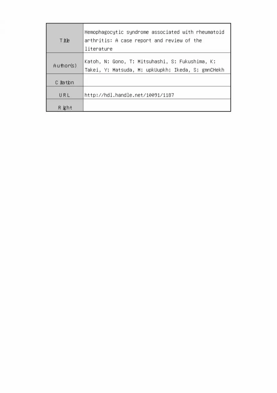
Abstract
We report a patient with rheumatoid arthritis (RA) who showed bicytopenia with
hyperferritinemia and hepatic dysfunction ascribable to hemophagocytic syndrome (HPS) 2
weeks after commencement of bucillamine. Pathology of the bone marrow showing
infiltration of macrophages confirmed the diagnosis of HPS. On the basis of renal dysfunction
with an increase in fibrin degradation products, disseminated intravascular coagulation was
considered to be concurrent with HPS. Oral prednisolone and cyclosporine A were started
right after cessation of bucillamine, and yielded complete normalization of hepatic and renal
function and hematology. As there was neither disease activity of RA nor associated infection
throughout the clinical course, bucillamine was suspected of being the cause of HPS in our
patient. HPS is a very rare complication in RA, but should be actively considered when
abnormalities in laboratory data, especially pancytopenia and hepatic dysfunction, quickly
worsen.
Key words: rheumatoid arthritis, bucillamine, corticosteroid, cyclosporine A, disseminated
intravascular coagulation, hemophagocytic syndrome
2

RA with hemophagocytic syndrome Katoh N, et al
Introduction
Hemophagocytic syndrome (HPS) is a potentially life-threatening disorder characterized
clinically by pancytopenia and liver dysfunction ascribable to profound activation of
macrophages mainly in the bone marrow. It is well known that infection by microorganisms,
such as the Epstein-Barr virus (EBV), malignancies, and acquired or congenital
immunodeficiencies sometimes induce HPS. Autoimmune disorders, particularly systemic
lupus erythematosus (SLE) and adult-onset Still’s disease, can also occasionally cause HPS
(1). Here, we report a patient with HPS complicating rheumatoid arthritis (RA). Bicytopenia
with hyperferritinemia and liver dysfunction typical of HPS developed 2 weeks after
commencement of bucillamine, and after cessation of this drug the administration of
corticosteroid and cyclosporine A cured the patient. RA is an autoimmune disease rarely
underlying HPS (2-4). In this report we review the literature and focus upon the pathogenesis
of HPS associated with RA.
Case report
A 52-year-old woman suddenly developed polyarthralgia resistant to non-steroidal
anti-inflammatory drugs (NSAIDs) with no precipitating cause or significant family history.
Three months later she was diagnosed as having RA on the basis of symmetrical swelling
with mild tenderness in multiple joints, including bilateral knees and hands, and a positive
rheumatoid factor (RF) (169 IU/ml, normal<10 IU/ml) in serum (5). In the classification of
severity and the global functional status in RA, she was considered to belong to stage 1 and
class 1, respectively (6, 7). She showed no signs or symptoms suggestive of other associated
3
RA with hemophagocytic syndrome Katoh N, et al
collagen diseases, but the anti-nuclear antibody with a speckled pattern was positive (×1280,
normal <×40). Matrix metalloproteinase-3 (80 ng/ml, normal 17.3-59.7 ng/ml) and IgG
(2034 mg/dl, normal 870-1700 mg/dl) in serum were increased slightly, and IgA (344 mg/dl,
normal 110-410 mg/dl) and IgM (122 mg/dl, normal 35-220 mg/dl) were within normal limits.
Although KL-6 in serum was slightly high (683 U/ml, 105-401 U/ml), no abnormal findings
were detectable in the chest roentgenogram. Salazosulfapyridine was unusable because of
adverse effects such as systemic rash with severe itching, and she was given bucillamine at a
dose of 200 mg/day in addition to NSAIDs. There were no abnormal findings in the routine
laboratory data right before commencement of bucillamine suggestive of renal or hepatic
dysfunction.
Polyarthralgia gradually improved, but she suddenly manifested vomiting and diarrhea
with general malaise and intermittent low-grade fever approximately 2 weeks after
commencement of bucillamine. On admission to our hospital, physical examination showed
no hepatosplenomegaly or lymphadenopathy. Laboratory data demonstrated decreases in
white blood cells (2,050/μl, normal 3,040-8,720/μl) and platelets (5.6×10⁴/μl, normal
13.7-37.8×10⁴/μl) and increases in total bilirubin (2.33 mg/dl, normal 0.3-1.2 mg/dl),
aspartate aminotransferase (390 U/l, normal 12-37 U/l), alanine aminotransferase (203 U/l,
normal 7-45 U/l), alkaline phosphatase (1715 U/l, normal 124-367 U/l), γ-glutamyl
transpeptidase (287 U/l, normal 6-30 U/l), blood urea nitrogen (35 mg/dl, normal 9-22 mg/dl),
creatinine (1.8 mg/dl, normal 0.4-0.8 mg/dl), triglyceride (290 mg/dl, normal 30-150 mg/dl),
lactate dehydrogenase (LDH, 2,681 U/l, normal 114-220 U/l) and ferritin (19,668 ng/ml,
normal 10-120 ng/ml). Differentiation of WBC was normal. C-reactive protein (CRP, 15.57
4

RA with hemophagocytic syndrome Katoh N, et al
mg/dl, normal <0.10 mg/dl) was markedly elevated along with an increase in D-dimer of
fibrin degradation products (FDP) (139.5 μg/ml, normal <1.0 μg/ml). Soluble interleukin
(IL)-2 receptor (5040 U/ml, 135-483 U/ml) and IL-6 (96.3 pg/ml, normal <2.41 pg/ml) in
serum were increased markedly. The anti-nuclear antibody (×1280) and RF (135 IU/ml) were
positive as in the previous examination, but other specific autoantibodies, including
anti-double-stranded DNA, anti-SS-A, and anti-SS-B antibodies, were undetectable. Both the
direct Coombs’ test and anti-platelet antibody were negative. The chest roentgenogram and
electrocardiogram were normal. Bone marrow aspirates demonstrated many hemophagocytic
macrophages (Fig.1). Intensive survey for infection showed no significant increases in serum
antibodies against any agents, including EBV, cytomegalovirus, and herpes simplex virus, or
elevation of β-D-glucan. There were no abnormal findings in either computed tomography of
the chest and abdomen or upper gastrointestinal endoscopy suggestive of malignancies.
Bucillamine was discontinued soon after admission, and prednisolone and cyclosporine
A were started at a dose of 60 mg/day and 150 mg/day, respectively, in addition to antibiotics,
intravenous immunoglobulin, anti-thrombin III and gabexate mesilate. Blood culture for
bacteria was negative throughout the clinical course. Her clinical symptoms quickly
disappeared in conjunction with an increase in platelets and decreases in CRP and LDH (Fig.
2). Hematology and indices of the liver and kidneys were completely normalized within 3
weeks after admission. She was discharged from our hospital 1 month after admission, and
prednisolone and cyclosporine A have been tapered in the outpatient clinic. She has remained
in good general condition with no arthralgia ascribable to RA at 10 mg/day of prednisolone
alone for 3 years to date.
5
RA with hemophagocytic syndrome Katoh N, et al
Discussion
The present patient showed bicytopenia in hematology, hepatic dysfunction, increased
inflammatory reactions, hypertriglyceridemia and hyperferritinemia, leading to a clinical
diagnosis of HPS (8, 9). Bone marrow aspirates showing many hemophagocytic macrophages
confirmed this diagnosis (8, 9). HPS is classified into primary and secondary forms, and the
lack of a significant family history indicating immunodeficiency suggests that the HPS in our
patient belonged to the latter (10). HPS is characterized clinically by high fever (8, 9), but its
secondary form often does not show this symptom, as in our patient (11). Considering that
renal dysfunction and a remarkable increase in FDP D-dimer were also present, disseminated
intravascular coagulation (DIC) was probably associated with HPS. As the routine laboratory
data showed no abnormal findings 2 weeks before admission, HPS and DIC seems to have
developed and quickly worsened during this short period. Both disorders may have increased
the endogenous production of corticosteroid, resulting in remission of polyarthritis due to RA
on admission to our hospital. Intensive treatment, including oral prednisolone and
cyclosporine A, showed a good therapeutic effect on both HPS and DIC in our patient, and
clinical symptoms quickly disappeared in conjunction with normalization of laboratory data.
The pathogenesis of HPS in the present patient remains unclear, but there are 4 possible
etiologies. The first is malignancy. Malignant lymphoma and visceral organ carcinoma
sometimes cause HPS (4), but in our patient a systemic survey, including CT and endoscopy,
demonstrated no abnormal findings suggestive of associated malignancies. The second is an
infection. It has been widely recognized that infectious agents, particularly viruses, can cause
6

RA with hemophagocytic syndrome Katoh N, et al
HPS (4, 12). Our patient, however, showed negative results in blood culture for bacteria and
no significant increases in the antibody titer against viruses, including EBV. There was no
evidence suggesting that infection may have played a role in the pathogenesis, although
antibiotics and intravenous immunoglobulin were given for a short period at the onset of
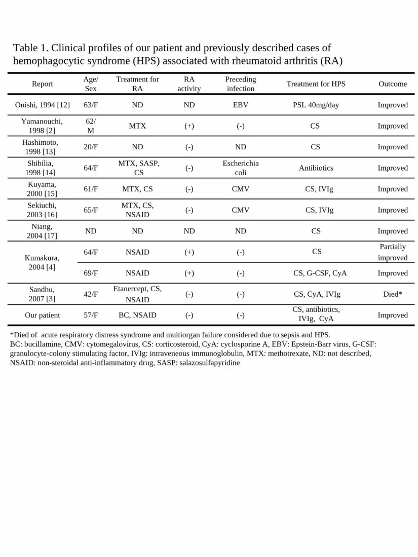
disease. The third is RA itself. HPS is sometimes associated with autoimmune disorders,
particularly SLE and adult-onset Still’s disease (1, 4, 13), but rarely with RA. The clinical
profiles of our patient and 10 previously described cases of HPS associated with RA are
shown in Table 1 (2-4, 14-19). Preceding infection by microorganisms, such as EBV and
cytomegalovirus, was confirmed in 4 patients, and only 3 developed HPS in conjunction with
a high disease activity of RA. The development of HPS in our patient was considered to be
unrelated to RA itself because the activity of this disease was depressed on admission to our
hospital.
The fourth possible etiology is a drug. Several recent reports have demonstrated that
anti-epileptic agents (20, 21), antibiotics (22, 23) and anti-rheumatic drugs (3, 24, 25) can act
as a trigger for HPS. Anti-rheumatic drugs inducing HPS comprise methotrexate and
etanercept. The clinical profiles of our patient and 8 previously described cases of
drug-induced HPS are shown in Table 2 (3, 20-25). Seven of the 8 previously described cases
showed development of HPS in 1 to 2 weeks after commencement of the drugs. HPS in our
patient also occurred 2 weeks after commencement of bucillamine, and this drug was
considered to play a central role in the pathogenesis. The appearance of systemic rash with
itching after administration of salazosulfapyridine may also support this hypothesis with
regard to drug intolerance. Hypersensitivity to drugs may cause a systemic inflammatory
7
RA with hemophagocytic syndrome Katoh N, et al
response with activation of macrophages and excessive production of cytokines, and lead to
the development of HPS as in the present patient (22), although the precise mechanisms
remain unclear.
In conclusion, HPS complicates RA with various etiologies, including infections,
malignancies, RA itself and drugs. Drug-induced HPS should be actively considered as a
possible complication when pancytopenia and liver dysfunction quickly worsen irrespective
of the disease activity of RA during treatment with anti-rheumatic drugs, such as bucillamine,
methotrexate and etanercept.
Acknowledgement
This work was supported by a grant from Neuroimmunological Disease Division, the
Ministry of Public Health, Labor and Welfare, Japan.
8
RA with hemophagocytic syndrome Katoh N, et al
References
1) Kumakura S, Ishikura H, Endo J, Kobayashi S. Autoimmune-associated hemophagocytosis.
Am J Hematol 50: 148-149, 1995.
2) Yamanouchi J, Yamauchi Y, Yokota E, Matsumoto I. Hemophagocytic syndrome in a
patient with rheumatoid arthritis. Ryumachi 38: 731-734, 1998 (in Japanese with English
abstract).
3) Sandhu C, Chesney A, Piliotis E, Buckstein R, Koren S. Macrophage activation syndrome
after etanercept treatment. J Rheumatol 34: 241-242, 2007.
4) Kumakura S, Ishikura H, Kondo M, Murakawa Y, Masuda J, Kobayashi S.
Autoimmune-associated hemophagocytic syndrome. Mod Rheumatol 14: 205-215, 2004.
5) Arnett FC, Edworthy SM, Bloch DA, et al. The American Rheumatism Association 1987
revised criteria for the classification of rheumatoid arthritis. Arthritis Rheum 31: 315-324,
1988.
6) Steinbrocker O, Traeger CH, Batterman RC. Therapeutic criteria in rheumatoid arthritis.
JAMA 140: 659-662, 1949.
7) Hochberg MC, Chang RW, Dwosh I, Lindsey S, Pincus T, Wolfe F. The American College
of Rheumatology 1991 revised criteria for the classification of global functional status in
rheumatoid arthritis. Arthritis Rheum 35: 498-502, 1992.
8) Imashuku S. Differential diagnosis of hemophagocytic syndrome: underlying disorders and
selection of the most effective treatment. Int J Hematol 66: 135-151, 1997.
9

RA with hemophagocytic syndrome Katoh N, et al
9) Tsuda H. Hemophagocytic syndrome (HPS) in children and adults. Int J Hematol 65:
215-226, 1997.
10) Farquhar JW, Claireaux AF. Familial hemophagocytic reticulosis. Arch Dis Child 27:
519-525, 1952.
11) Kumakura S, Kondo M, Tsumura H, Murakawa Y, Ishikura H, Kobayashi S.
Hemophagocytic syndrome. Nihon Rinsho Men’eki Gakkai Kaishi 23: 670-673, 2000 (in
Japanese).
12) Risdall RJ, McKenna RW, Nesbit ME, et al. Virus-associated hemophagocytic syndrome:
a benign histocytic proliferation distinct from malignant histiocytosis. Cancer 44:
993-1002, 1979.
13) Wong KF, Hui PK, Chan JK, Chan YW, Ha SY. The acute lupus hemophagocytic
syndrome. Ann Intern Med 114: 387-390, 1991.
14) Onishi R, Namiuchi S. Hemophagocytic syndrome in a patient with rheumatoid arthritis.
Intern Med 33: 607-611, 1994.
15) Hashimoto N, Nishii N, Hashimoto M, et al. Two cases of hemophaogocytic syndrome.
Onomichishibyoishi 13: 123-126, 1998 (in Japanese)
16) Shibilia J, Javier RM, Albert A, Cazenave JP, Kuntz JL. Pancytopenia secondary to
hemophagocytic syndrome in rheumatoid arthritis treated with methotrexate and
sulfasalazine. J Rheumatol 25: 1218-1220, 1998.
17) Kuyama J, Nakao H, Take H, et al. Cytomegalovirus-associated hemophagocytic
syndrome and multiple intestinal ulcers with perforation in a patient with rheumatoid
arthritis. Shiritsutoyonakabyouin Igakuzasshi 1: 53-58, 2000 (in Japanese).
10

RA with hemophagocytic syndrome Katoh N, et al
18) Sekiuchi M, Nakabayashi K, Marumo T, Arimura Y, Yamada A. Hemophagocytic
syndrome associated with hypercytokinemia in a patient with rheumatoid arthritis.
Ryumachi 43: 696-702, 2003 (in Japanese with English abstract).
19) Niang A, Diallo S, Ka MM, et al. Hemophagocytic syndrome complicating adult’s
seropositive rheumatoid arthritis. Rev Med Interne 25: 826-828, 2004 [abstract].
20) Gutierrez-Rave Pecero VM, Luque Marquez R, Ayerza Lerchundi MA, Fernandez Jurado
A. Phenytoin-induced hemocytophagic histiocytosis indistinguishable from malignant
histiocytosis. South Med J 84: 649-650, 1991.
21) Yang YC, Jou ST, Chang YH, Liang JS, Lee WT. Hemophagocytic syndrome associated
with antiepileptic drug. Pediatr Neurol 30: 358-360, 2004.
22) Lambotte O, Costedoat-Chalumeau N, Amoura Z, Piette JC, Cacoub P. Drug-induced
hemophagocytosis. Am J Med 112: 592-593, 2002.
23) Jain D, Dash S. Pancytopenia due to extensive hemophagocytosis following
anti-tubercular treatment. Am J Hematol 75: 118-119, 2004.
24) Ravelli A, Caria MC, Buratti S, Malattia C, Temporini F, Martini A. Methotrexate as a
possible trigger of macrophage activation syndrome in systemic juvenile idiopathic
arthritis. J Rheumatol 28: 865-867, 2001.
25) Ramanan AV, Schneider R. Macrophage activation syndrome following initiation of
Etanercept in a child with systemic onset juvenile rheumatoid arthritis. J Rheumatol 30:
401-403, 2003.
11
RA with hemophagocytic syndrome Katoh N, et al
Figures legends
Figure 1: Bone marrow aspirates showing hemophagocytic macrophage.
Figure 2: Clinical course of the patient. CRP: C-reactive protein, LDH: lactate dehydrogenase,
PLT: platelet
12

Figure 1
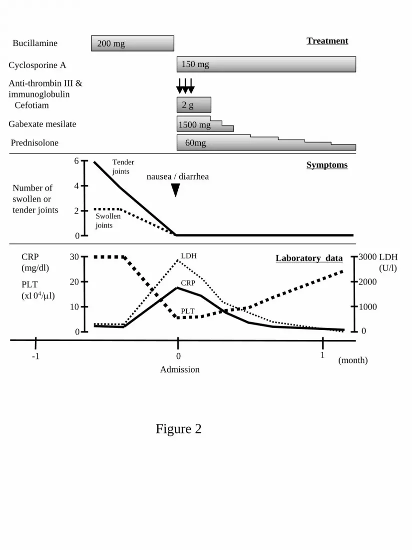
Figure 2
nausea / diarrhea
-1 0
PLT (x104/μl)
Cyclosporine A
1
10
0
20
30
Prednisolone
Gabexate mesilate
Cefotiam
Anti-thrombin III & immunoglobulin
60mg
PLT 1000
0
2000
3000CRP (mg/dl)
CRP
LDH LDH (U/l)
Bucillamine
Laboratory data
Symptoms
Treatment
(month)
Number of swollen or tender joints
200 mg
150 mg
2 g
1500 mg
2
0
4
6
Swollen joints
Tender joints
Admission
Report Age/ Sex
Treatment for RA
RA activity
Preceding infection Treatment for HPS Outcome
Onishi, 1994 [12] 63/F ND ND EBV PSL 40mg/day Improved
Yamanouchi, 1998 [2]
62/ M MTX (+) (-) CS Improved
Hashimoto, 1998 [13] 20/F ND (-) ND CS Improved
Shibilia, 1998 [14] 64/F MTX, SASP,
CS (-) Escherichia coli Antibiotics Improved
Kuyama, 2000 [15] 61/F MTX, CS (-) CMV CS, IVIg Improved
Sekiuchi, 2003 [16] 65/F MTX, CS,
NSAID (-) CMV CS, IVIg Improved
Niang, 2004 [17] ND ND ND ND CS Improved
Kumakura, 2004 [4]
64/F NSAID (+) (-) CS Partiallyimproved
69/F NSAID (+) (-) CS, G-CSF, CyA Improved
Sandhu, 2007 [3] 42/F
Etanercept, CS,NSAID
(-) (-) CS, CyA, IVIg Died*
Our patient 57/F BC, NSAID (-) (-)CS, antibiotics,
IVIg, CyA Improved
*Died of acute respiratory distress syndrome and multiorgan failure considered due to sepsis and HPS.BC: bucillamine, CMV: cytomegalovirus, CS: corticosteroid, CyA: cyclosporine A, EBV: Epstein-Barr virus, G-CSF: granulocyte-colony stimulating factor, IVIg: intraveneous immunoglobulin, MTX: methotrexate, ND: not described, NSAID: non-steroidal anti-inflammatory drug, SASP: salazosulfapyridine
Table 1. Clinical profiles of our patient and previously described cases of hemophagocytic syndrome (HPS) associated with rheumatoid arthritis (RA)

CS: corticosteroid, CyA: cyclosporine A, INH: isoniazid, IVIg: intraveneous immunoglobulin, ND: not described, PZA: pyrazinamide, REP: rifampicin
Report Age/ Sex Primary disease
Drug(s) suspected as a cause of HPS
Interval between commencement of drug(s)
and onset of HPS
Treatment for HPS Outcome
Gutierrez- Rave Pecero,
1991 [18]9/M ND (Epilepsy?) Phenytoin 2 weeks CS Improved
Ravelli, 2001 [22] 6/F Juvenile idiopathic
arthritis Methotrexate 1 day CyA Improved
Lambotte, 2002 [20]
45/F Superficial abscessVancomycin,Teicoplanin
10 days CS Improved
53/F Pneumocystis carinii pneumonia
Trimethoprim/ Sulfamethoxazole 9 days IVIg Improved
Ramanan, 2003 [23] 8/F Juvenile rheumatoid
arthritis Etanercept 2 weeks CS Improved
Jain, 2004 [21] 22/M Tuberculous lymphadenitis REP, INH, PZA 2 weeks (-) Improved
Yang, 2004 [19] 8/M Epilepsy Lamotrigine 2 weeks CS, IVIg Improved
Sandhu, 2007 [3] 42/F Rheumatoid arthritis Etanercept 2 months CS, CyA,
IVIg, Died
Our patient 57/F Rheumatoid arthritis Bucillamine 2 weeks ImprovedCS, antibiotics, IVIg, CyA
Table 2. Clinical profiles of our patient and previously described cases of drug-induced hemophagocytic syndrome (HPS)