Embed Size (px)
Citation preview
Research ArticleHemin Attenuates Cisplatin-Induced Acute Renal Injury inMale Rats
Mohamed A. Al-Kahtani,1 Ashraf M. Abdel-Moneim,1,2
Omar M. Elmenshawy,1,3 and Mohamed A. El-Kersh4,5
1 Department of Biological Sciences, Faculty of Science, King Faisal University, Al-Hassa 31982, Saudi Arabia2Department of Zoology, Faculty of Science, Alexandria University, Alexandria 21511, Egypt3 Department of Zoology, Faculty of Science, Al-Azhar University, Nasr City 11884, Egypt4Department of Chemistry, Faculty of Science, King Faisal University, Al-Hassa 31982, Saudi Arabia5 Department of Biochemistry, Faculty of Science, Alexandria University, Alexandria 21511, Egypt
Correspondence should be addressed to Mohamed A. Al-Kahtani; [email protected] Ashraf M. Abdel-Moneim; [email protected]
Received 11 May 2014; Revised 30 July 2014; Accepted 6 September 2014; Published 22 September 2014
Academic Editor: Madia Trujillo
Copyright © 2014 Mohamed A. Al-Kahtani et al.This is an open access article distributed under theCreativeCommonsAttributionLicense, which permits unrestricted use, distribution, and reproduction in anymedium, provided the originalwork is properly cited.
Background. The aim of this study is to investigate the protective effects of hemin (the heme oxygenase-1 [OH-1] inducer)against nephrotoxic effects induced by cisplatin [cis-diamminedichloroplatinum II (CP)] in male rats. Methods. The evaluationwas performed through monitoring renal redox parameters: lipid peroxidation (LPO), glutathione peroxidase (GPx), superoxidedismutase (SOD), glutathione reductase (GR), and reduced glutathione (GSH). The work also examined renal function tests (ureaand creatinine), tissue proinflammatorymediator like nitric oxide (NO), and kidney cytopathology.Results.A single intraperitonealdose of CP (10mg/kg b.w.) caused significant elevation of blood urea, serum creatinine, and renal LPO and NO, along withsignificant decline of the activities of GPx and GR, but renal SOD activity and GSH level were statistically insignificant as comparedto control group. Subcutaneous injection of hemin (40 𝜇mol/kg b.w.) partially ameliorated CP-induced renal damage, based onsuppression of blood urea, serum creatinine, the renal MDA and NO levels, and increased antioxidant capacity in CP-treated rats.The results of histopathological and ultrastructural investigations supported the renoprotective effect of hemin against CP-inducedacute toxicity.Conclusion.The induction of HO-1 by hemin is a promising approach in the treatment of CP-induced nephrotoxicity.However, further preclinical studies are warranted to test effectiveness of CP/hemin on the outcome of tumor chemotherapy.
1. Introduction
Cisplatin (cis-diamminedichloroplatinum(II), CP) is a highlyeffective chemotherapeutic agent against a large spectrum oftumor types [1–3]. However, the long-term clinical use of CPis limited by its serious side effects, mainly nephrotoxicity[4]. CP causes impairment of kidney function and acuterenal failure via multiple mechanisms including generationof oxygen/nitrogen species, DNA damage, tubulointerstitialinflammation, and apoptotic cell death [5–12]. A numberof studies have evaluated compounds as potential nephro-protectors against CP; these included natural antioxidants,modulators of nitric oxide synthesis, osmotic diuretics, andcytoprotective and antiapoptotic agents [13]. However, most
of them were not found suitable/safe for clinical practice.In this context, heme oxygenase-1 (HO-1), the rate-limitingenzyme in heme catabolism, might offer a promising alterna-tive. HO-1 (also known as heat shock protein 32) is inducedby free radical-initiated reactions, and its induction is con-sidered to be an adaptive response against oxidative tissuedamage [14–20]. In addition, HO-1 has been recognized toexhibit powerful anti-inflammatory and immunomodulatoryeffects [21]. Previous studies have shown that HO-1-inducingagents, as hemin, can mitigate nephrotoxic effects causedby a wide array of stressors, including mercury [15] andacetaminophen [20]. Based on the previous information, thepresent study aimed to examine whether the activation ofHO-1 (by hemin) would have protective effects against CP
Hindawi Publishing CorporationOxidative Medicine and Cellular LongevityVolume 2014, Article ID 476430, 9 pageshttp://dx.doi.org/10.1155/2014/476430
2 Oxidative Medicine and Cellular Longevity
induced nephrotoxicity in rats. For this purpose, we haveevaluated the status of renal lipid peroxidative assay and anti-oxidant defenses. In addition, detailed glomerular and tubu-lar pathologies were assessed.
2. Materials and Methods
2.1. Drugs and Chemicals. CP and hemin (powder) were pur-chased from Sigma Chemical Company, USA. Other chemi-cal reagents were of high-quality analytical grade. Hemin wasfirst dissolved in 0.1M NaOH, titrated to pH 7.4 with 0.1MHCl, and then diluted with normal saline (1 : 10 v/v), while CPwas prepared in normal saline.
2.2. Animals and Treatments. Healthy adult male rats (110–140 g) were obtained from animal house facility at King SaudUniversity, Saudi Arabia. Rats were housed in polyethylenecages under controlled laboratory conditions and providedwith standard rat chow and water ad libitum. They wereallowed 1 week of acclimatization before the initiation of theexperiment. Experimental protocol of this study complieswith theNIH ethical guidelines for themanipulation and careof laboratory animals. Rats were randomly assigned into 4groups (𝑛 = 6):
(i) saline group (control): rats received 3mL/kg 0.09%NaCl, intraperitoneally (i.p.);
(ii) CP group: rats received a single dose of CP, i.p.(10mg/kg);
(iii) CP+hemin group: rats received 40𝜇mol/kg hemin,subcutaneously (s.c.), 1 h following CP;
(iv) hemin group: rats received 40 𝜇mol/kg hemin, s.c.
Doses, duration, and routes of exposure were chosenaccording to previously published reports [20, 22]. All ratswere sacrificed under light ether anesthesia after 24 h ofthe last dose of specific treatment, and samples of trunkblood and kidneys were collected. Blood was centrifuged at5000 rpm for 10min and the separated sera were used formeasurement of renal function tests. Kidneys were decap-sulated and washed in cold isotonic saline. The cortex wascarefully separated from medulla as described earlier byBanday et al. [23].The kidney cortexwas homogenized (GlassCol homogenizer) and a 20%w/v homogenate was preparedin ice cold 50mM, pH 7.4 phosphate buffer saline. Thehomogenate was centrifuged at 5000 rpm for 20min and thesupernatant was then saved in aliquots to avoid sample thaw-ing and freezing and stored at −80∘C till used for assayingperoxidative damage and antioxidant status. Samples of theintact kidney tissues were used for light and electron micro-scopic studies.
2.3. Markers of Renal Toxicity. Serum levels of urea and crea-tinineweremeasured spectrophotometrically using commer-cial diagnostic kits (Human Gesellschaft fur Biochemica undDiagnostica mbH, Germany), according to the methodsdescribed by Tabacco et al. [24] and Bartels and Bohmer [25],respectively.
2.4. Oxidative Stress-Related Indices. Malondialdehyde(MDA), an index of fatty acid oxidation, was estimated inquantifiable amounts using Thiobarbituric Acid ReactiveSubstances (TBARS) assay kit (BioAssay Systems, CA, USA)according to the method of Ohkawa et al. [26]. In thisprocedure, MDA reacts with thiobarbituric acid (TBA) toform a pink-colored complex that has maximum absorbanceat 532 nm. MDA value was calculated in terms of nmol/g wettissue. Nitric oxide (NO) level was determined in kidneyhomogenates using Nitrate/Nitrite Colorimetric Assay Kit(BioAssay Systems, CA, USA) according to the manufac-turer’s instructions. NO production was measured followingreduction of nitrate to nitrite using improved Griess method[27]. Total NO synthetase (NOS) activity was detected byNOS assay kit (BioAssay Systems, CA, USA) [28]. Activitiesof glutathione peroxidase (GPx, EC 1.11.1.9) and superoxidedismutase (SOD, EC 1.15.1.1) as well as the level of reducedglutathione (GSH) in renal cortex were determined spec-trophotometrically, according to the standard detectionprotocol of analysis kits (BioAssay Systems, CA, USA)[29–31]. Glutathione reductase (GR, EC 1.6.4.2) activity wasassayed using a commercial kit from Cayman ChemicalCompany, USA [32]. Protein content was estimated by themethod of Lowry et al. [33].
2.5. Light Microscopy. Kidney taken from each animal wasfixed in 10% formalin solution, dehydrated in ascending seriesof ethanol, and embedded in paraffin. Sections (4 𝜇m-thick)were cut, stained with haematoxylin and eosin solutions, andexamined under light microscope (Nikon 80i, Japan).
2.6. Electron Microscopy. Small slices of kidney cortex (𝑛 =3 per group) were fixed in 3% glutaraldehyde in sodiumphosphate buffer (200mM, pH 7.2) for 3 h at 4∘C. Postfixationwas in cold 1% osmium tetroxide (Agar Sci. Ltd., England) for1 h. After flushing in phosphate buffer, the tissue samples weredehydrated in graded ethanol solutions and embedded inAraldite (Agar Sci. Ltd., England). Ultrathin sectioning (80–100 nm) was carried out using Leica EM UC6 (Leica Co.,Austria) ultramicrotome. Sections were mounted on grids,double stained with 2% uranyl acetate and lead citrate, andviewed under Jeol JEM 1011 transmission electronmicroscope(Jeol Ltd., Japan) at 80 kV.
2.7. Statistics. All variables were compared using one-wayanalysis of variance (ANOVA) followed by LSD multiplerange test. Differences at 𝑃 < 0.05 were considered signif-icant. Statistical tests were performed using SAS statisticalsoftware (SAS v. 9.2, SAS Institute, Inc., Cary, NC). Data werepresented as mean ± standard error (SE).
3. Results
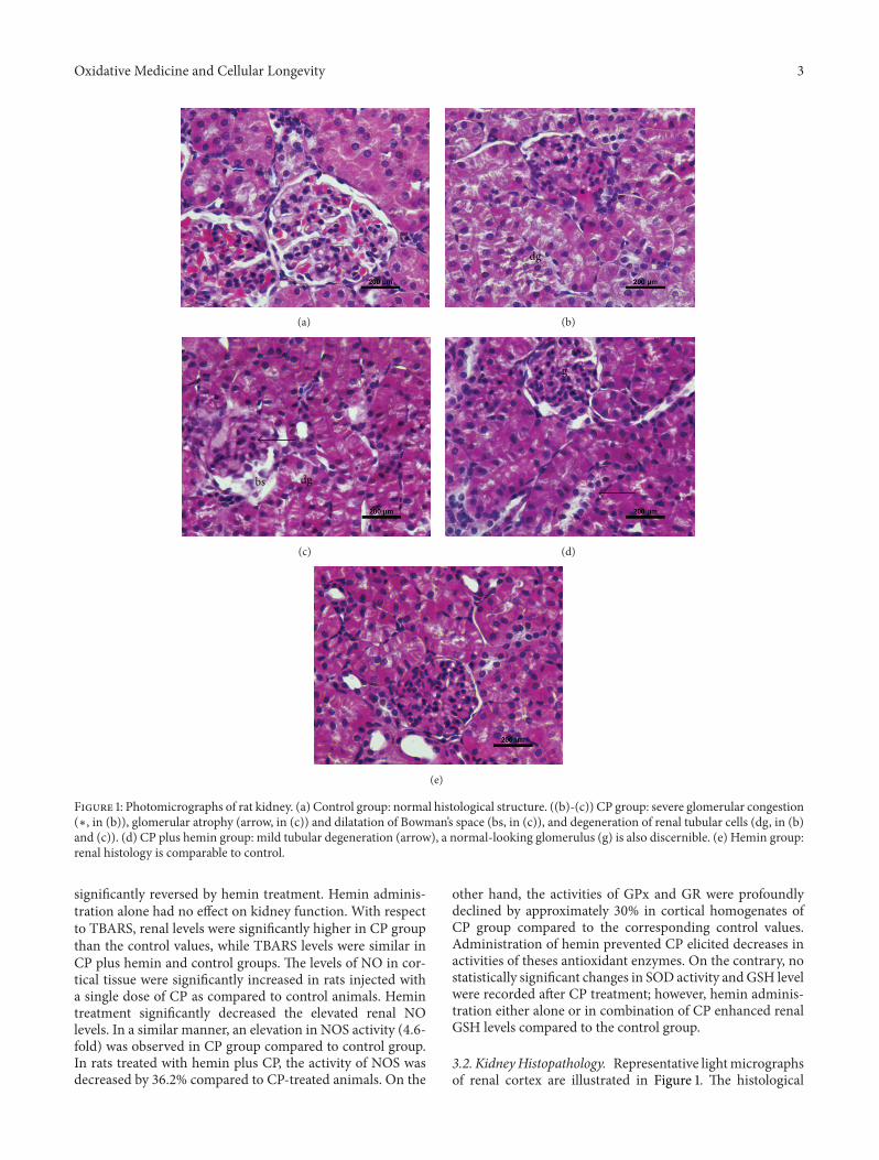
3.1. Biochemical Findings. The results of biochemical analysisin all studied groups are shown in Table 1. Treatment of malerats with CP resulted in significant increases in levels ofblood urea (3.1-fold) and creatinine (6.3-fold) compared tocontrol animals, indicating renal damage.These changeswere
Oxidative Medicine and Cellular Longevity 3
(a)
∗
dg
(b)
dgbs
(c)
g
(d)
(e)
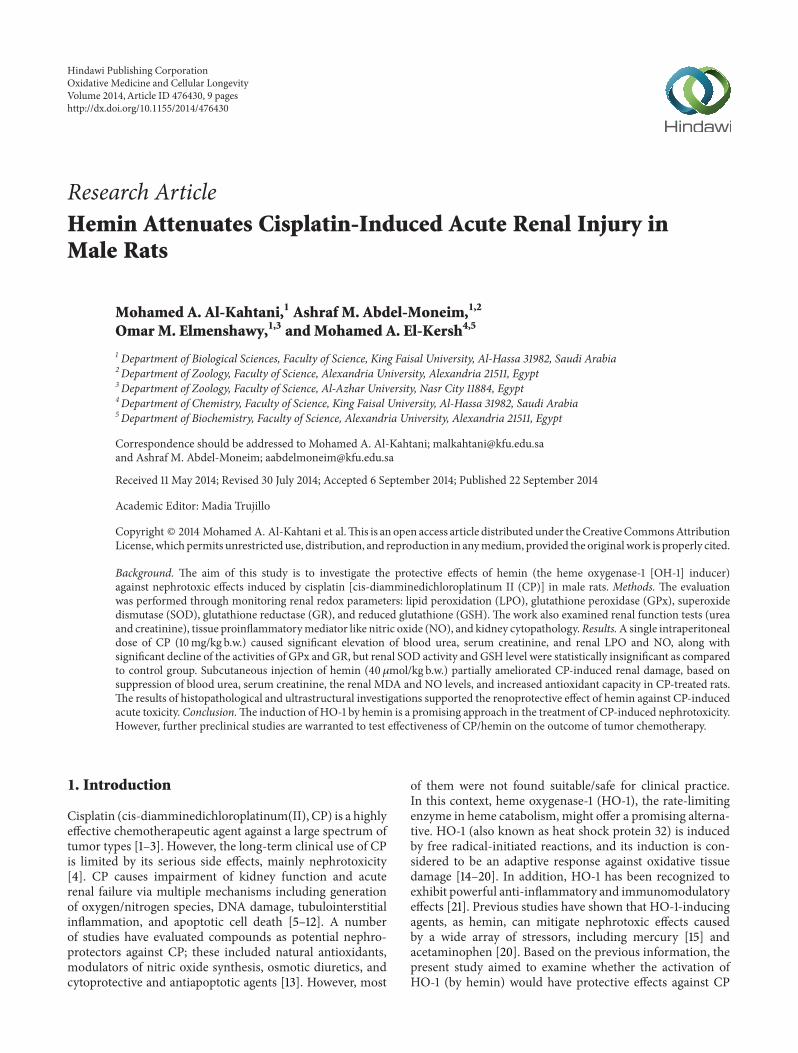
Figure 1: Photomicrographs of rat kidney. (a) Control group: normal histological structure. ((b)-(c)) CP group: severe glomerular congestion(∗, in (b)), glomerular atrophy (arrow, in (c)) and dilatation of Bowman’s space (bs, in (c)), and degeneration of renal tubular cells (dg, in (b)and (c)). (d) CP plus hemin group: mild tubular degeneration (arrow), a normal-looking glomerulus (g) is also discernible. (e) Hemin group:renal histology is comparable to control.
significantly reversed by hemin treatment. Hemin adminis-tration alone had no effect on kidney function. With respectto TBARS, renal levels were significantly higher in CP groupthan the control values, while TBARS levels were similar inCP plus hemin and control groups. The levels of NO in cor-tical tissue were significantly increased in rats injected witha single dose of CP as compared to control animals. Hemintreatment significantly decreased the elevated renal NOlevels. In a similar manner, an elevation in NOS activity (4.6-fold) was observed in CP group compared to control group.In rats treated with hemin plus CP, the activity of NOS wasdecreased by 36.2% compared to CP-treated animals. On the
other hand, the activities of GPx and GR were profoundlydeclined by approximately 30% in cortical homogenates ofCP group compared to the corresponding control values.Administration of hemin prevented CP elicited decreases inactivities of theses antioxidant enzymes. On the contrary, nostatistically significant changes in SOD activity andGSH levelwere recorded after CP treatment; however, hemin adminis-tration either alone or in combination of CP enhanced renalGSH levels compared to the control group.
3.2. KidneyHistopathology. Representative lightmicrographsof renal cortex are illustrated in Figure 1. The histological
4 Oxidative Medicine and Cellular Longevity
Table 1: The results of biochemical analysis in control and experimental groupsa.
Parameters Unit Control CP CP + hemin HeminUrea mg/dL 48.60 ± 1.50 151.61 ± 10.02b 86.61 ± 3.02b,c 53.60 ± 3.20c
Creatinine mg/dL 0.81 ± 0.11 5.14 ± 0.76b 1.45 ± 0.69c 0.78 ± 0.08c
TBARS nmole MDA/g tissue 6.44 ± 1.67 11.00 ± 1.05b 7.26 ± 2.74 4.06 ± 1.53c
NO 𝜇mole/g tissue 16.42 ± 0.98 19.58 ± 1.50b 13.16 ± 0.71b,c 11.86 ± 0.84b,c
NOS nmole/g tissue 5.56 ± 0.84 25.61 ± 7.50b 16.34 ± 2.19 6.07 ± 0.89c
GPx mU/mg protein 353.51 ± 44.43 238.49 ± 40.83b 311.15 ± 25.76 376.81 ± 30.18c
SOD U/mg protein 18.14 ± 2.76 14.01 ± 1.85 17.59 ± 1.83 18.08 ± 1.15GR mU/mg protein 98.79 ± 8.40 67.63 ± 6.03b 86.26 ± 9.13 105.75 ± 10.73c
GSH 𝜇mole/mg protein 6.68 ± 1.10 6.93 ± 0.59 9.15 ± 1.01b 10.05 ± 0.37b,c
TBARS: thiobarbituric acid reactive substances, MDA: malondialdehyde, NO: nitric oxide, NOS: nitric oxide synthetase, GPx: glutathione peroxidase, SOD:superoxide dismutase, GR: Glutathione reductase, and GSH: reduced glutathione.aResults are expressed as mean ± SE for six replicates.bSignificantly different from control at 𝑃 < 0.05 by one-way ANOVA.cSignificantly different from CP at 𝑃 < 0.05 by one-way ANOVA.
P
P
P
EE
E
Cap
Cap
MCs
MCs
10𝜇m
(a)
F
BBM
N
M
M
Ly
Ly
5𝜇m
(b)
L
MN
N
10𝜇m
(c)
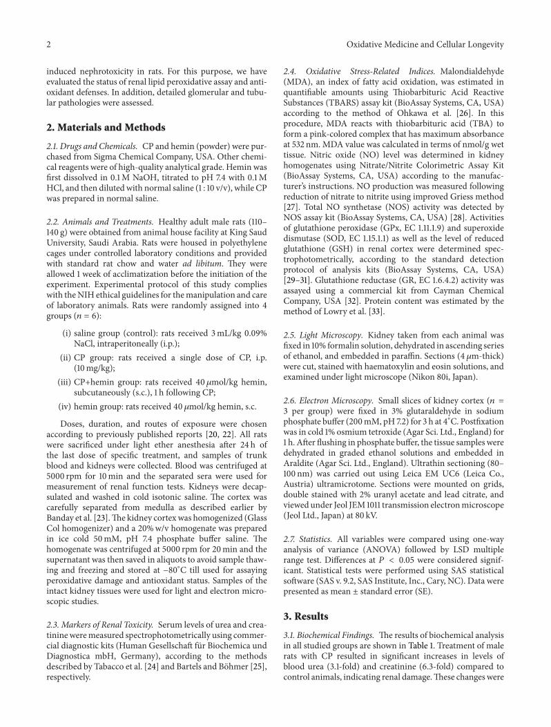
Figure 2: Electron micrographs of kidney of control rats. (a) Note the glomerulus has podocytes (P) which remain in contact with GBMby foot processes (arrows). The glomerular capillaries (Cap) are lined by endothelial cells (E) which are richly fenestrated and supported bymesangial cells (MCs) with their surrounding extracellular matrix. (b) Proximal tubules have well-developed brush border microvilli (BBM),and their lining epithelia contain large number of mitochondria (with normal feature) (M), few lysosomes (Ly), and many basal infoldings(F), N: nucleus. (c) Epithelial lining of distal tubule with apical nuclei (N), numerousmitochondria (M), short cisternae of rough endoplasmicreticulum, and few microvilli toward the lumen (L).
picture of kidney was normal in both control (Figure 1(a))and hemin-treated (Figure 1(e)) groups. In CP-treated rats(Figures 1(b)-1(c)), the renal corpuscles displayed extensivecongestion filling up the glomerular capillary loops. Someglomeruli were atrophied or lost with concurrent dilatation ofBowman’s space. The morphological deterioration was also
characterized by a widespread tubular cell swelling, necrosis,and degeneration, occurring primarily in proximal con-voluted tubules (PCTs) epithelia. In addition, peritubularinflammatory cell infiltrations and hemorrhagic foci wereclearly apparent after CP treatment. In contrast, these histo-logical abnormalities were found to be reduced in CP plus
Oxidative Medicine and Cellular Longevity 5
Table 2: Semiquantitative scoring of glomerular and tubulointerstitial lesions in control and experimental rats.
Group Control CP CP + hemin HeminGlomerular congestion — ++ + —Glomerular atrophy — ++ + —Peritubular inflammatory cell infiltration — +++ + —Tubular damage — +++ + —Scoring scale: none (—), mild (+), moderate (++), and severe (+++).
++
5𝜇m
FP
(a)
VV
N
Mb Ly
5𝜇m
(b)
5𝜇m
(c)
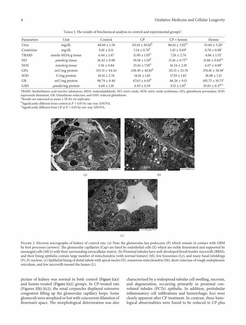
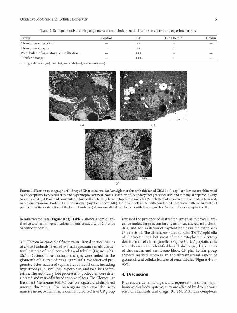
Figure 3: Electronmicrographs of kidney of CP-treated rats. (a) Renal glomerulus with thickenedGBM (++), capillary lumens are obliteratedby endocapillary hypercellularity and hypertrophy (arrows). Note also fusion of secondary foot processes (FP) andmesangial hypercellularity(arrowheads). (b) Proximal convoluted tubule cell containing large cytoplasmic vacuoles (V), clusters of deformed mitochondria (arrows),numerous lysosomal bodies (Ly), and lamellar (myeloid) body (Mb). Observe nucleus (N) with condensed chromatin pattern. Arrowheadpoints to partial destruction of the brush border. (c) Abnormal distal tubular cells with few organelles. Arrow indicates apoptotic cell.
hemin-treated rats (Figure 1(d)). Table 2 shows a semiquan-titative analysis of renal lesions in rats treated with CP withor without hemin.
3.3. Electron Microscopic Observations. Renal cortical tissuesof control animals revealed normal appearance of ultrastruc-tural patterns of renal corpuscles and tubules (Figures 2(a)–2(c)). Obvious ultrastructural changes were noted in theglomeruli of CP-treated rats (Figure 3(a)). We observed pro-gressive deformation of capillary endothelial cells, includinghypertrophy (i.e., swelling), hyperplasia, and focal loss of fen-estrae. The secondary foot processes of podocytes were dete-riorated and markedly fused in some places. The GlomerularBasement Membrane (GBM) was corrugated and displayeduneven thickening. The mesangium was expanded withmassive increase inmatrix. Examination of PCTs of CP group
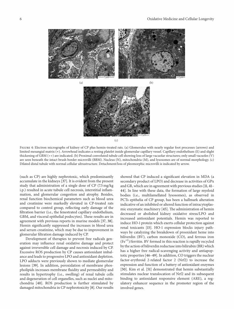
revealed the presence of destructed/irregular microvilli, api-cal vacuoles, large secondary lysosomes, altered mitochon-dria, and accumulation of myeloid bodies in the cytoplasm(Figure 3(b)). The distal convoluted tubules (DCTs) epitheliaof CP-treated rats lost most of their cytoplasmic electrondensity and cellular organelles (Figure 3(c)). Apoptotic cellswere also seen and identified by cell shrinkage, degradationof chromatin, and membrane blebs. CP plus hemin groupshowed marked recovery in the ultrastructural aspect ofglomeruli and cellular features of renal tubules (Figures 4(a)–4(c)).
4. Discussion
Kidneys are dynamic organs and represent one of the majorhomeostasis body systems; they are affected by diverse vari-eties of chemicals and drugs [34–36]. Platinum complexes
6 Oxidative Medicine and Cellular Longevity
∗
E
++
5𝜇m
(a)
M
M
N
BBM
V
Ly
2𝜇m
(b)
5𝜇m
(c)
Figure 4: Electron micrographs of kidney of CP plus hemin-treated rats. (a) Glomerulus with nearly regular foot processes (arrows) andlimited mesangial matrix (∗). Arrowhead indicates a resting platelet inside glomerular capillary vessel. Capillary endothelium (E) and slightthickening of GBM (++) are indicated. (b) Proximal convoluted tubule cell showing loss of large vacuolar structures; only small vacuoles (V)are seen beneath the intact brush border microvilli (BBM). Nucleus (N), mitochondria (M), and lysosomes are of normal morphology. (c)Dilated distal tubule with normal cellular ultrastructure. Detachment/loss of pleomorphic microvilli is indicated by arrow.
(such as CP) are highly nephrotoxic, which predominantlyaccumulate in the kidneys [37]. It is evident from the presentstudy that administration of a single dose of CP (7.5mg/kgi.p.) resulted in acute tubule cell necrosis, interstitial inflam-mation, and glomerular congestion and atrophy. Besides,renal function biochemical parameters such as blood ureaand creatinine were markedly elevated in CP-treated ratscompared to control group, reflecting early damage of thefiltration barrier (i.e., the fenestrated capillary endothelium,GBM, and visceral epithelial podocytes). These results are inagreement with previous reports in murine models [37, 38].Hemin significantly suppressed the increases in blood ureaand serum creatinine, which may be due to improvement inglomerular filtration damage induced by CP.
Development of therapies to prevent free radicals gen-eration may influence renal oxidative damage and protectagainst irreversible cell damage and necrosis induced by CP.Excessive ROS production by CP causes antioxidant imbal-ance and leads to progressive LPO and antioxidant depletion.LPO adducts were previously shown to mediate glomerularlesions [39]. In addition, peroxidation of membrane phos-pholipids increases membrane fluidity and permeability andresults in hypertrophy (i.e., swelling) of renal tubule cellsand degeneration of cell organelles, such as nuclei and mito-chondria [40]. ROS production is further stimulated bydamaged mitochondria in CP nephrotoxicity [8]. Our results
showed that CP induced a significant elevation in MDA (asecondary product of LPO) and decrease in activities of GPxandGR, which are in agreement with previous studies [11, 41–44]. In line with these data, the formation of large myeloidbodies (i.e., multilamellated lysosomes), as observed inPCTs epithelia of CP group, has been a hallmark alterationindicative of an inhibited or altered function of intracytoplas-mic enzymatic machinery [45]. The administration of hemindecreased or abolished kidney oxidative stress/LPO andincreased antioxidant potentials. Hemin was reported toinduce HO-1 protein which exerts cellular protection againstrenal toxicants [15]. HO-1 expression blocks injury path-ways by catalyzing the breakdown of prooxidant heme intobiliverdin (BV), carbon monoxide (CO), and ferrous iron(Fe+2)/ferritin. BV formed in this reaction is rapidly recycledby the action of biliverdin reductase into bilirubin (BR)whichhas a higher free radical-scavenging activity and antiapop-totic properties [46–49]. In addition, CO triggers the nuclearfactor-erythroid 2-related factor 2 (Nrf2) to increase theexpression and function of a battery of antioxidant enzymes[50]. Kim et al. [51] demonstrated that hemin substantiallystimulates nuclear translocation of Nrf2 and its subsequentbinding to antioxidant responsive element (ARE), a reg-ulatory enhancer sequence in the promoter region of theinvolved genes.
Oxidative Medicine and Cellular Longevity 7
Treatment with CP overdose induces nitrosative stress byNO and other nitrosylating agents and this was correlatedwith the expression of the inducible nitric oxide synthase(iNOS) protein [52]. In the current study, NO production inthe CP-treated group was significantly higher than that inthe control group; our results are consistent with the resultsobtained by Kart et al. [53], who have shown that there isstrong immunoreactivity against iNOS in the liver tissue ofthe CP-treated group. Recent work of Chirino et al. [54]reported that the downregulation of iNOS expressionreduced CP-induced renal damage and nitrosative stress.High NO levels exert toxicological effects by reacting withsuperoxide anion to generate short-lived but hyperactiveperoxynitrite radical with subsequent nitration of proteintyrosine residues [55, 56]. Also, NO output depletes intra-cellular GSH, which increases susceptibility to oxidativestress and aggravates renal tissue damage [57, 58], especiallyfor glomerular diseases (e.g., lupus nephritis) [59]. Hemin-mediated augmentation of HO-1 activity was proved to beefficient enough to reduce the NO-dependent pathologicaland inflammatory conditions [17, 60, 61]. Of note, HO-1 andits reaction product CO also suppress the expression of iNOSprotein by preventing the activation of nuclear factor-kappaB (NF-𝜅B) which upregulates the transcription of the iNOSgene [17, 62, 63].
In conclusion, this study is the first to demonstrate theprotective role of hemin (an HO-1 activator) against acutenephrotoxicity induced by CP. Treatment of hemin amelio-rated renal ultrastructural changes and dysfunction, and thiswas associated with reduction of LPO, overstimulation ofantioxidant capacity, and suppression of NO biosynthesis,the proinflammatory mediator. However, further preclinicalstudies are needed to verify whether coadministration of CPand hemin could affect the outcome of tumor chemotherapy.
Conflict of Interests
The authors declare that there is no conflict of interestsregarding the publication of this paper.
Acknowledgment
The authors thank the Deanship of Scientific Research, KingFaisal University, for financial andmoral support of this work(Project no. 130142).
References
[1] S. Atasayar, H. Gurer-Orhan,H.Orhan, B. Gurel, G. Girgin, andH. Ozgunes, “Preventive effect of aminoguanidine compared tovitamin E and C on cisplatin-induced nephrotoxicity in rats,”Experimental and Toxicologic Pathology, vol. 61, no. 1, pp. 23–32, 2009.
[2] I. Hassan, S. Chibber, and I. Naseem, “Ameliorative effect ofriboflavin on the cisplatin induced nephrotoxicity and hepato-toxicity under photoillumination,” Food and Chemical Toxicol-ogy, vol. 48, no. 8-9, pp. 2052–2058, 2010.
[3] A. Naqshbandi, M. W. Khan, S. Rizwan, S. U. Rehman, and F.Khan, “Studies on the protective effect of dietary fish oil on
cisplatin induced nephrotoxicity in rats,” Food and ChemicalToxicology, vol. 50, no. 2, pp. 265–273, 2012.
[4] M. A. C. Rodrigues, J. L. Rodrigues, N. M. Martins et al., “Car-vedilol protects against cisplatin-induced oxidative stress, redoxstate unbalance and apoptosis in rat kidney mitochondria,”Chemico-Biological Interactions, vol. 189, no. 1-2, pp. 45–51, 2011.
[5] L. M. G. Antunes, J. D. C. Darin, and M. de Lourdes P. Bianchi,“Protective effects of vitamin C against cisplatin-inducednephrotoxicity and lipid peroxidation in adult rats: a dose-dependent study,” Pharmacological Research, vol. 41, no. 4, pp.405–411, 2000.
[6] O. A. Badary, S. Abdel-Maksoud, W. A. Ahmed, and G. H.Owieda, “Naringenin attenuates cisplatin nephrotoxicity inrats,” Life Sciences, vol. 76, no. 18, pp. 2125–2135, 2005.
[7] H. D. C. Francescato, R. S. Costa, C. Scavone, and T. M. Coim-bra, “Parthenolide reduces cisplatin-induced renal damage,”Toxicology, vol. 230, no. 1, pp. 64–75, 2007.
[8] X. Yao, K. Panichpisal, N. Kurtzman, and K. Nugent, “Cisplatinnephrotoxicity: a review,” The American Journal of the MedicalSciences, vol. 334, no. 2, pp. 115–124, 2007.
[9] Y. I. Chirino, D. J. Sanchez-Gonzalez, C.M.Martınez-Martınez,C. Cruz, and J. Pedraza-Chaverri, “Protective effects of apoc-ynin against cisplatin-induced oxidative stress and nephrotoxi-city,” Toxicology, vol. 245, no. 1-2, pp. 18–23, 2008.
[10] J. M. Perez-Rojas, C. E. Guerrero-Beltran, C. Cruz, D. J.Sanchez-Gonzalez, C. M. Martınez-Martınez, and J. Pedraza-Chaverri, “Preventive effect of tert-butylhydroquinone oncisplatin-induced nephrotoxicity in rats,” Food and ChemicalToxicology, vol. 49, no. 10, pp. 2631–2637, 2011.
[11] R. Domitrovic, O. Cvijanovic, E. Pernjak-Pugel, M. Skoda, L.Mikelic, and Z. Crncevic-Orlic, “Berberine exerts nephro-protective effect against cisplatin-induced kidney damagethrough inhibition of oxidative/nitrosative stress, inflamma-tion, autophagy and apoptosis,” Food and Chemical Toxicology,vol. 62, pp. 397–406, 2013.
[12] H. Pan, K. Shen, X. Wang, H. Meng, C. Wang, and B. Jin,“Protective effect of metalloporphyrins against Cisplatin-induced kidney injury in mice,” PLoS ONE, vol. 9, no. 1, ArticleID e86057, 2014.
[13] B. H. Ali and M. S. Al Moundhri, “Agents ameliorating oraugmenting the nephrotoxicity of cisplatin and other platinumcompounds: a review of some recent research,” Food andChemical Toxicology, vol. 44, no. 8, pp. 1173–1183, 2006.
[14] H. P. Kim, H.-O. Pae, S. H. Back et al., “Heme oxygenase-1comes back to endoplasmic reticulum,” Biochemical and Bio-physical Research Communications, vol. 404, no. 1, pp. 1–5, 2011.
[15] R. Yoneya, H. Ozasa, Y. Nagashima et al., “Hemin pretreatmentameliorates aspects of the nephropathy induced by mercuricchloride in the rat,” Toxicology Letters, vol. 116, no. 3, pp. 223–229, 2000.
[16] H.Chiu, J. A. Brittingham, andD. L. Laskin, “Differential induc-tion of heme oxygenase-1 in macrophages and hepatocytesduring acetaminophen-induced hepatotoxicity in the rat: effectsof hemin and biliverdin,” Toxicology and Applied Pharmacology,vol. 181, no. 2, pp. 106–115, 2002.
[17] T. Wen, Z.-M. Wu, Y. Liu, Y.-F. Tan, F. Ren, and H. Wu,“Upregulation of heme oxygenase-1 with hemin prevents d-galactosamine and lipopolysaccharide-induced acute hepaticinjury in rats,” Toxicology, vol. 237, no. 1–3, pp. 184–193, 2007.
8 Oxidative Medicine and Cellular Longevity
[18] Y.-S. Chen, X.-X. Zhu, X.-Y. Zhao, H.-Y. Xing, and Y.-G. Li,“Hemin, a heme oxygenase-1 inducer, improves aortic endothe-lial dysfunction in insulin resistant rats,” Chinese MedicalJournal, vol. 121, no. 3, pp. 241–247, 2008.
[19] A. A. Fouad, H. A. Qureshi, A. I. Al-Sultan, M. T. Yacoubi, andA. A. Ali, “Protective effect of hemin against cadmium-inducedtesticular damage in rats,”Toxicology, vol. 257, no. 3, pp. 153–160,2009.
[20] A. A. Fouad, M. T. Yacoubi, andM. H. El-Bidawy, “Therapeuticpotential of hemin in acetaminophen nephrotoxicity in rats,”Environmental Toxicology and Pharmacology, vol. 27, no. 2, pp.277–282, 2009.
[21] C. S. T. Origassa and N. O. S. Camara, “Cytoprotective role ofheme oxygenase-1 and heme degradation derived end productsin liver injury,” World Journal of Hepatology, vol. 5, no. 10, pp.541–549, 2013.
[22] A. Hussein, A. A. E. Ahmed, S. A. Shouman, and S. Sharawy,“Ameliorating effect of DL-𝛼-lipoic acid against cisplatin-induced nephrotoxicity and cardiotoxicity in experimentalanimals,”Drug Discoveries &Therapeutics, vol. 6, no. 3, pp. 147–156, 2012.
[23] A. A. Banday, N. Farooq, S. Priyamvada, A. N. K. Yusufi, andF. Khan, “Time dependent effects of gentamicin on the enzymesof carbohydrate metabolism, brush border membrane andoxidative stress in rat kidney tissues,” Life Sciences, vol. 82, no.9-10, pp. 450–459, 2008.
[24] A. Tabacco, F. Meiattini, E. Moda, and P. Tarli, “Simplifiedenzymic/colorimetric serum urea nitrogen determination,”Clinical Chemistry, vol. 25, no. 2, pp. 336–337, 1979.
[25] H. Bartels and M. Bohmer, “Micro-determination of Creati-nine,” Clinica Chimica Acta, vol. 32, no. 1, pp. 81–85, 1971.
[26] H. Ohkawa, N. Ohishi, and K. Yagi, “Assay for lipid peroxidesin animal tissues by thiobarbituric acid reaction,” AnalyticalBiochemistry, vol. 95, no. 2, pp. 351–358, 1979.
[27] P. Bulau, D. Zakrzewicz, K. Kitowska et al., “Analysis of methy-larginine metabolism in the cardiovascular system identifiesthe lung as a major source of ADMA,” American Journal ofPhysiology—Lung Cellular and Molecular Physiology, vol. 292,no. 1, pp. L18–L24, 2007.
[28] D. Ghigo, C. Riganti, E. Gazzano, C. Costamagna, and A. Bosia,“Cycling of NADPH by glucose 6-phosphate dehydrogenaseoptimizes the spectrophotometric assay of nitric oxide synthaseactivity in cell lysates,”NitricOxide—Biology andChemistry, vol.15, no. 2, pp. 148–153, 2006.
[29] B. Jacobson, G. Quigley, and G. Lockitch, “Adaptation of glu-tathione peroxidase assay to the Technicon RA-1000,” ClinicalChemistry, vol. 34, no. 10, pp. 2164–2165, 1988.
[30] H. Ukeda, S. Maeda, T. Ishii, and M. Sawamura, “Spectropho-tometric assay for superoxide dismutase based on tetrazoliumsalt 3’-1-[(phenylamino)-carbonyl]-3,4-tetrazolium-bis(4-met-hoxy-6- nitro)benzenesulfonic acid hydrate reduction byxanthine-xanthine oxidase,” Analytical Biochemistry, vol. 251,no. 2, pp. 206–209, 1997.
[31] I. Carlberg and B.Mannervik, “Glutathione reductase,”Methodsin Enzymology, vol. 113, pp. 484–490, 1985.
[32] H. Lindenmaier, M. Becker, W. E. Haefeli, and J. Weiss, “Inter-action of progestins with the human multidrug resistance-associated protein 2 (MRP2),”DrugMetabolism andDisposition,vol. 33, no. 11, pp. 1576–1579, 2005.
[33] O. H. Lowry, N. J. Rosebrough, A. L. Farr, and R. J. Randall,“Protein measurement with the Folin phenol reagent,” TheJournal of biological chemistry, vol. 193, no. 1, pp. 265–275, 1951.
[34] A. M. Abdel-Moneim and K. M. Said, “Acute effect of cadmiumtreatment on the kidney of rats: biochemical and ultrastructuralstudies,” Pakistan Journal of Biological Sciences, vol. 10, no. 20,pp. 3497–3506, 2007.
[35] M. A. Al Kahtani, A. M. Abdel-Moneim, and W. M. El-Sayed,“The influence of taurine pretreatment on aluminum chlorideinduced nephrotoxicity in Swiss albino mice,” Histology andHistopathology, vol. 29, no. 1, pp. 45–55, 2014.
[36] N. A. Salem and E. A. Salem, “Renoprotective effect of grapeseed extract against oxidative stress induced by gentamicin andhypercholesterolemia in rats,” Renal Failure, vol. 33, no. 8, pp.824–832, 2011.
[37] N. E. Abdelmegmd,H.N. Chmaisseand, andN. S. AbouZeinab,“Silymarin ameliorates Cisplatin-induced hepatotoxicity in rats:histopathological and ultrastructural studies,” Pakistan Journalof Biological Sciences, vol. 13, no. 10, pp. 463–479, 2010.
[38] M. Morigi, B. Imberti, C. Zoja et al., “Mesenchymal stemcells are renotropic, helping to repair the kidney and improvefunction in acute renal failure,” Journal of the American Societyof Nephrology, vol. 15, no. 7, pp. 1794–1804, 2004.
[39] C. J. Binder, H. Weiher, M. Exner, and D. Kerjaschki, “Glo-merular overproduction of oxygen radicals in Mpv17 gene-inactivated mice causes podocyte foot process flattening andproteinuria. A model of steroid-resistant nephrosis sensitive toradical scavenger therapy,” The American Journal of Pathology,vol. 154, no. 4, pp. 1067–1075, 1999.
[40] E. C. Foulkes, Biological Membranes in Toxicology, Taylor andFrancis, Philadelphia, Pa, USA, 1988.
[41] K. Sueishi, K.Mishima,K.Makino et al., “Protection by a radicalscavenger edaravone against cisplatin-induced nephrotoxicityin rats,” European Journal of Pharmacology, vol. 451, no. 2, pp.203–208, 2002.
[42] S. Iseri, F. Ercan, N. Gedik, M. Yuksel, and I. Alican, “Simvas-tatin attenuates cisplatin-induced kidney and liver damage inrats,” Toxicology, vol. 230, no. 2-3, pp. 256–264, 2007.
[43] M. Ekor, G. O. Emerole, and E. O. Farombi, “Phenolic extractof soybean (Glycine max) attenuates cisplatin-induced nephro-toxicity in rats,” Food and Chemical Toxicology, vol. 48, no. 4, pp.1005–1012, 2010.
[44] Y.-N. Li, Y. Guo,M.-M. Xi et al., “Saponins fromAralia taibaien-sis attenuate D-galactose-induced aging in rats by activatingFOXO3a and Nrf2 pathways,” Oxidative Medicine and CellularLongevity, vol. 2014, Article ID 320513, 13 pages, 2014.
[45] A. Hermenean, A. Ardelean, M. Stan et al., “Protective effects ofnaringenin on carbon tetrachloride-induced acute nephrotoxi-city in mouse kidney,” Chemico-Biological Interactions, vol. 205,no. 2, pp. 138–147, 2013.
[46] D. E. Baranano, M. Rao, C. D. Ferris, and S. H. Snyder, “Bili-verdin reductase: a major physiologic cytoprotectant,” Proceed-ings of the National Academy of Sciences of the United States ofAmerica, vol. 99, no. 25, pp. 16093–16098, 2002.
[47] N. G. Abraham, J. Cao, D. Sacerdoti, X. Li, and G. Drummond,“Heme oxygenase: the key to renal function regulation,” Amer-ican Journal of Physiology: Renal Physiology, vol. 297, no. 5, pp.F1137–F1152, 2009.
[48] S.W. Ryter andA.M.Choi, “Heme oxygenase-1/carbonmonox-ide: novel therapeutic strategies in critical care medicine,”Current Drug Targets, vol. 11, no. 12, pp. 1485–1494, 2010.
[49] Y. Naito, T. Takagi, K. Uchiyama, and T. Yoshikawa, “Hemeoxygenase-1: a novel therapeutic target for gastrointestinaldiseases,” Journal of Clinical Biochemistry and Nutrition, vol. 48,no. 2, pp. 126–133, 2011.
Oxidative Medicine and Cellular Longevity 9
[50] K. Chan, X.-D. Han, and Y. W. Kan, “An important functionof Nrf2 in combating oxidative stress: detoxification of aceta-minophen,” Proceedings of the National Academy of Sciences ofthe United States of America, vol. 98, no. 8, pp. 4611–4616, 2001.
[51] Y.-C. Kim, H. Masutani, Y. Yamaguchi, K. Itoh, M. Yamamoto,and J. Yodoi, “Hemin-induced activation of the thioredoxingene byNrf2: a differential regulation of the antioxidant respon-sive element by a switch of its binding factors,” The Journal ofBiological Chemistry, vol. 276, no. 21, pp. 18399–18406, 2001.
[52] R. D. Curran, F. K. Ferrari, P. H. Kispert et al., “Nitric oxide andnitric oxide-generating compounds inhibit hepatocyte proteinsynthesis,”The FASEB Journal, vol. 5, no. 7, pp. 2085–2092, 1991.
[53] A. Kart, Y. Cigremis, M. Karaman, and H. Ozen, “Caffeic acidphenethyl ester (CAPE) ameliorates cisplatin-induced hepato-toxicity in rabbit,” Experimental and Toxicologic Pathology, vol.62, no. 1, pp. 45–52, 2010.
[54] Y. I. Chirino, J. Trujillo, D. J. Sanchez-Gonzalez et al., “SelectiveiNOS inhibition reduces renal damage induced by cisplatin,”Toxicology Letters, vol. 176, no. 1, pp. 48–57, 2008.
[55] R. Radi, G. Peluffo, M. N. Alvarez, M. Naviliat, and A. Cayota,“Unraveling peroxynitrite formation in biological systems,” FreeRadical Biology and Medicine, vol. 30, no. 5, pp. 463–488, 2001.
[56] P. Pacher, J. S. Beckman, and L. Liaudet, “Nitric oxide andperoxynitrite in health and disease,” Physiological Reviews, vol.87, no. 1, pp. 315–424, 2007.
[57] R. M. Clancy and S. B. Abramson, “Nitric oxide: a novel media-tor of inflammation,” Proceedings of the Society for ExperimentalBiology and Medicine, vol. 210, no. 2, pp. 93–101, 1995.
[58] P. Liu, C. E. Hock, R. Nagele, and P. Y. Wong, “Formation ofnitric oxide, superoxide, and peroxynitrite in myocardialischemia-reperfusion injury in rats,” The American Journal ofPhysiology, vol. 272, no. 5, part 2, pp. H2327–H2336, 1997.
[59] Y. Takeda, M. Takeno, M. Iwasaki et al., “Chemical inductionof HO-1 suppresses lupus nephritis by reducing local iNOSexpression and synthesis of anti-dsDNA antibody,” Clinical andExperimental Immunology, vol. 138, no. 2, pp. 237–244, 2004.
[60] B.-M. Choi, H.-O. Pae, Y.-M. Kim, and H.-T. Chung, “Nitricoxide-mediated cytoprotection of hepatocytes from glucosedeprivation-induced cytotoxicity: involvement of hemeoxygenase-1,” Hepatology, vol. 37, no. 4, pp. 810–823, 2003.
[61] S. Yang, H.-J. Shih, Y.-C. Chow et al., “The protective role ofheme oxygenase-1 induction on testicular tissues after testiculartorsion and detorsion,”The Journal of Urology, vol. 177, no. 5, pp.1928–1933, 2007.
[62] T. Ashino, R. Yamanaka, M. Yamamoto et al., “Negative feed-back regulation of lipopolysaccharide-induced inducible nitricoxide synthase gene expression by heme oxygenase-1 inductionin macrophages,” Molecular Immunology, vol. 45, no. 7, pp.2106–2115, 2008.
[63] A. Taye and B. M. Ibrahim, “Activation of renal haemeoxygenase-1 alleviates gentamicin-induced acute nephrotoxic-ity in rats,” Journal of Pharmacy and Pharmacology, vol. 65, no.7, pp. 995–1004, 2013.

Submit your manuscripts athttp://www.hindawi.com
Stem CellsInternational
Hindawi Publishing Corporationhttp://www.hindawi.com Volume 2014
Hindawi Publishing Corporationhttp://www.hindawi.com Volume 2014
MEDIATORSINFLAMMATION
of
Hindawi Publishing Corporationhttp://www.hindawi.com Volume 2014
Behavioural Neurology
EndocrinologyInternational Journal of
Hindawi Publishing Corporationhttp://www.hindawi.com Volume 2014
Hindawi Publishing Corporationhttp://www.hindawi.com Volume 2014
Disease Markers
Hindawi Publishing Corporationhttp://www.hindawi.com Volume 2014
BioMed Research International
OncologyJournal of
Hindawi Publishing Corporationhttp://www.hindawi.com Volume 2014
Hindawi Publishing Corporationhttp://www.hindawi.com Volume 2014
Oxidative Medicine and Cellular Longevity
Hindawi Publishing Corporationhttp://www.hindawi.com Volume 2014
PPAR Research
The Scientific World JournalHindawi Publishing Corporation http://www.hindawi.com Volume 2014
Immunology ResearchHindawi Publishing Corporationhttp://www.hindawi.com Volume 2014
Journal of
ObesityJournal of
Hindawi Publishing Corporationhttp://www.hindawi.com Volume 2014
Hindawi Publishing Corporationhttp://www.hindawi.com Volume 2014
Computational and Mathematical Methods in Medicine
OphthalmologyJournal of
Hindawi Publishing Corporationhttp://www.hindawi.com Volume 2014
Diabetes ResearchJournal of
Hindawi Publishing Corporationhttp://www.hindawi.com Volume 2014
Hindawi Publishing Corporationhttp://www.hindawi.com Volume 2014
Research and TreatmentAIDS
Hindawi Publishing Corporationhttp://www.hindawi.com Volume 2014
Gastroenterology Research and Practice
Hindawi Publishing Corporationhttp://www.hindawi.com Volume 2014
Parkinson’s Disease
Evidence-Based Complementary and Alternative Medicine
Volume 2014Hindawi Publishing Corporationhttp://www.hindawi.com


