Embed Size (px)
Citation preview
QATAR UNIVERSITY
COLLEGE OF ARTS AND SCIENCES
ROLE OF PROTEIN ARGININE METHYLTRANSFERASE 5 (PRMT5) IN WNT/β-
CATENIN PROLIFERATIVE SIGNALING IN BREAST CANCER
BY
HARSHITA SHAILESH
A Thesis Submitted to
the College of Arts and Sciences
in Partial Fulfillment of the Requirements for the Degree of
Doctorate of Philosophy in PhD in Biological and Environmental Sciences
June 2020
© 2020. HARSHITA SHAILESH. All Rights Reserved.
ii
COMMITTEE PAGE
The members of the Committee approve the Thesis of Harshita Shailesh
defended on 05/05/2020.
Prof. Saïd Sif Thesis/Dissertation Supervisor
Prof. Allal Ouhtit Committee Member
Prof. Samir Jaoua Committee Member
Prof. Serhiy Souchelnytskyi Committee Member
Approved:
Ibrahim Al-Kaabi, Dean, College of College of Arts and Sciences
iii
ABSTRACT
SHAILESH, HARSHITA, Doctorate : June : [2020],
Biological and Environmental Science
Title: Role of Protein Arginine Methyltransferase 5 (PRMT5) in WNT/β-CATENIN
proliferative signaling in breast cancer
Supervisor of Thesis: Prof. Saïd Sif.
Protein arginine methyltransferase 5 (PRMT5) activity is dysregulated in many aggressive
cancers and its enhanced levels are associated with increased tumor growth and survival.
In this study, we show that PRMT5 is overexpressed in breast cancer cell lines, and that it
promotes WNT/β-CATENIN proliferative signaling through epigenetic silencing of
pathway antagonists, DKK1 and DKK3, by binding to their promoter and inducing
symmetric dimethylation of H3R8 and H4R3, leading to enhanced expression of c-MYC,
CYCLIN D1 and SURVIVIN. Our findings also show that PRMT5 inhibition using
compound 5 (CMP5), reduces PRMT5 recruitment and PRMT5-induced epigenetic marks
in the promoter regions of DKK1 and DKK3, which consequently results in reduced
expression of CYCLIN D1 and SURVIVIN. Furthermore, CMP5 treatment either alone or
in combination with 5-Azacytidine and Trichostatin A restored expression of DKK1 and
DKK3 in TNBCs. In addition, PRMT5 inhibition in TNBCs inhibited AKT/mTOR
signaling by reducing phosphorylation of AKT and mTOR at Ser473 and Ser2448,
respectively. These molecular changes were associated with reduced proliferation,
migration and invasion of breast cancer cells, and induced their death.

iv
DEDICATION
Dedicated to my family
v
ACKNOWLEDGMENTS
I would like to thank my thesis advisor, Prof. Said Sif for providing me with the
opportunity to perform this study under his supervision. It was a great honor to work under
his supervision, which paved the way for me to grow as a young enthusiastic researcher. I
sincerely thank him for his constant support, encouragement, and timely help, without
which it would have been impossible to complete my study. He always supported me to
develop my rational thinking to answer scientific questions and gave me enough freedom
to improve my technical skills. His holistic approaches to solve research questions and
thought-provoking scientific conversations always inspired me.
In addition, I would like to thank my thesis committee members, Prof. Allal Ouhtit, Prof.
Samir Joua and Prof. Serhiy Souchelnytskyi for their help and advice. I would like to thank
Ms. Swapna Thomas and technical staff in the Department of Biological and
Environmental Sciences, Qatar University for their help and support.
Special thanks to my family members and friends for their encouragement during this
journey. I deeply appreciate the constant emotional support given by my parents, which
gave me a true strength to travel through all the difficulties and hardships. The constant
encouragement and help of my husband, Shailesh Kumar and daughter, Aradhya Shailesh
to achieve this success have made my journey tolerable and enjoyable. It gives me great
pleasure to appreciate my close friends Kavita Nair, Imane Saleh and Fatima Al-Hajiri for
their great moral support, help and cheer throughout my graduate study.
vi
TABLE OF CONTENTS
DEDICATION...................................................................................................................................iv
ACKNOWLEDGMENTS....................................................................................................................v
TABLEOFCONTENTS.....................................................................................................................vi
ListofTables.................................................................................................................................viii
ListofFigures.................................................................................................................................ix
CHAPTER1:INTRODUCTION...........................................................................................................1
CHAPTER2:LITERATUREREVIEW...................................................................................................9
2.1Introduction..........................................................................................................................9
2.2.Proteinargininemethyltransferase(PRMT5)...................................................................11
2.2.1Structureandenzymology..........................................................................................11
2.2.2.PRMT5targetproteins...............................................................................................14
2.2.3RoleofPRMT5duringembryoniclife.........................................................................18
2.2.4RoleofPRMT5duringneurogenesis...........................................................................19
2.2.5RoleofPRMT5duringgermcelldevelopment,hematopoiesisandimmuneresponse...............................................................................................................................................21
2.2.6PRMT5andcancer.......................................................................................................23
2.3WNT/β-CATENINsignaling.................................................................................................29
2.3.1 Discovery of WNT protein.........................................................................................29
2.3.2CanonicalWNT/β-CATENINsignaling.........................................................................29
2.4NaturalantagonistsoftheWNT/β-CATENINpathway......................................................33
2.4.1Axisinhibitory(AXIN)proteins....................................................................................34
2.4.2Dickkopf(DKK)familyproteins...................................................................................38
2.4.3SecretedFrizzled-relatedproteins(SFRPs).................................................................46
2.4.4.WNTinhibitoryfactor(WIF)1....................................................................................52
2.5WNT/β-CATENINtargetgenes...........................................................................................54
2.5.1.CYCLIND1...................................................................................................................55
2.5.2c-MYC...........................................................................................................................57
2.5.3SURVIVIN.....................................................................................................................59
2.6Breastcancer......................................................................................................................60
2.7WNT/β-CATENINsignalingactivationinbreastcancer:PossibleroleofPRMT5.............61
CHAPTER3.MATERIALSANDMETHODS.......................................................................................65
3.1Cellculture..........................................................................................................................65
vii
3.2RealTimePCR.....................................................................................................................65
3.3Westernblotanalysis.........................................................................................................69
3.4Chromatinimmunoprecipitation(ChIP)assay...................................................................69
3.5Transwellmigrationandinvasionassays...........................................................................71
3.6Proliferationassay..............................................................................................................72
3.7FlowCytometry..................................................................................................................72
3.8ELISAAssay.........................................................................................................................72
3.9Statisticalanalysis...............................................................................................................73
CHAPTER4:RESULTS.....................................................................................................................74
4.1PRMT5levelsareupregulatedinbreastcancercelllines.................................................74
4.2WNT/β-CATENINsignalingiselevatedinbreastcancer....................................................75
4.3PRMT5promotesWNT/β-CATENINactivationthroughrepressionofDKK1andDKK3...78
4.4PRMT5epigeneticallysilencesDKK1andDKK3bybindingtotheirpromoterandinducingH3R8andH4R3symmetricdimethylation................................................................82
4.5PRMT5inhibitionreducesviabilityofbreastcancercells.................................................85
4.6InhibitionofPRMT5derepressesDKK1andDKK3inBT549cells.....................................87
4.7InhibitionofPRMT5incombinationwithHDACsandDNMT3AderepressesDKK1andDKK3inHCC1937cells..............................................................................................................90
4.8InhibitionofPRMT5downregulatesWNT/β-CATENINtargetgenesbreastcancercells.95
4.9PRMT5inhibitionaltersitsrecruitmentandH3R8andH4R3symmetricmethylationinthepromoterregionofDKK1andDKK3..................................................................................98
4.10PRMT5modulatesAKT/mTORsignalinginbreastcancerinbreastcancer..................101
4.11PRMT5isrequiredformigrationandinvasionofTNBCcells........................................102
4.12PRMT5inhibitioninducesdeathofTNBCcells..............................................................104
CHAPTER5:DISCUSSION.............................................................................................................107
CHAPTER6:CONCLUSIONANDFUTUREWORK..........................................................................116
CONCLUSION...........................................................................................................................116
PROSPECTIVE..........................................................................................................................117
REFERENCES................................................................................................................................120
viii
LIST OF TABLES
Table 1: PRMT5 methylated proteins .............................................................................15
Table 2: List of real time RT-PCR primers and probe sets .............................................67
Table 3: List of real time ChIP primers and probe sets .................................................71
ix
LIST OF FIGURES
Figure 1: Arginine methylation of proteins carried out by PRMTs ..................................9
Figure 2: Catalytic domain of PRMT5 is highly conserved from yeast to human .........13
Figure 3: PRMT5 regulated processes ............................................................................24
Figure 4: WNT/ β-CATENIN Signaling overview .........................................................31
Figure 5: Antagonists of WNT pathway .........................................................................33
Figure 6: Working hypothesis .........................................................................................63
Figure 7: Expression of PRMT5 is elevated in breast cancer cells .................................75
Figure 8: Expression of WNT/β-CATENIN target genes is elevated in breast cancer cells
.........................................................................................................................................76
Figure 9: Expression of WNT/β-CATENIN antagonists, DKK1 and DKK3, is
downregulated in breast cancer cells ..............................................................................79
Figure 10: PRMT5 epigenetically suppresses expression of WNT/β-CATENIN
antagonists, DKK1 and DKK3 .......................................................................................83
Figure 11: PRMT5 inhibition reduces viability of breast cancer cells ...........................86
Figure 12: PRMT5 inhibition induces transcriptional derepression of WNT/β-CATENIN
antagonists, DKK1 and DKK3, in BT549 cells ...............................................................89
Figure 13: PRMT5 inhibitor in combination with HDCs and DNMT3A induces
transcriptional derepression of WNT/β-CATENIN antagonists, DKK1 and DKK3, in
HCC1937 cells ...............................................................................................................91
x
Figure 14: PRMT5 inhibition downregulates expression of CYCLIN D1 and SURVIVIN
in TNBC cells .................................................................................................................96
Figure 15: PRMT5 inhibition alters its recruitment and symmetric dimethylation of
histones, H3R8 and H4R3 in the promoter region of DKK1 and DKK3 ........................99
Figure 16: PRMT5 inhibition downregulates AKT/mTOR signaling in breast cancer.101
Figure 17: PRMT5 inhibition reduces migration and invasion of breast cancer cells ..103
Figure 18: PRMT5 inhibition induces apoptosis of breast cancer cells ........................105
Figure 19: Model for PRMT5-mediated upregulation of WNT/β-CATENIN signaling in
breast cancer ..................................................................................................................108
1
CHAPTER 1: INTRODUCTION
Post-translational modifications including methylation, phosphorylation, acetylation,
ubiquitination and sumoylation play an important role in regulating the biological and
cellular functions of various cytosolic and nuclear proteins (Karve & Cheema, 2011).
Arginine methylation is one such post-translational modification carried out by a group of
enzymes belonging to the protein arginine methyltransferase (PRMT) family. PRMTs are
broadly categorized into three main classes. Type I PRMTs catalyze monomethylation and
asymmetric dimethylation, while Type II PRMTs catalyze monomethylation and
symmetric dimethylation of specific arginine residues. Type III PRMTs catalyze only
monomethylation of arginine residues. Protein arginine methyltransferase 5 (PRMT5) is a
type II PRMT enzyme that catalyzes symmetric dimethylation of specific arginine residues
of target proteins (Branscombe et al., 2001). In vitro studies have shown that the PRMT5
enzymatic activity requires formation of a hetero-octameric complex with its co-factor,
methylosome protein 50 (MEP-50), which governs substrate specificity and enzyme-
substrate interaction with PRMT5 target proteins (Antonysamy et al., 2012; Burgos et al.,
2015). In addition to MEP-50, PRMT5 is also known to form a complex with other proteins
such as pICln and RIO kinase 1 (RioK1), which interact with PRMT5 and further regulates
its substrate specificity (Guderian et al., 2011).
PRMT5 regulates transcription of target genes by directly binding to their promoter regions
and inducing arginine methylation of promoter histones, which triggers chromatin
remodeling. PRMT5-induced symmetrical dimethylation of H3R8 and H4R3
2
predominantly result in transcriptional repression of target genes, except in few cases
where PRMT5-mediated histone methylation has been associated with transcriptional
activation (LeBlanc et al., 2012; Pal et al., 2004; Zhang et al., 2015). Apart from histones,
PRMT5 also methylates arginine residues of various non-histone proteins such as p53,
E2F1, ribosomal protein S10 (RPS10), rapidly accelerated fibrosarcoma (RAF) protein
kinase, and golgin (Shailesh et al., 2018).
Enhanced PRMT5 activity serves as one of the major drivers of cellular transformation and
tumor development in various cancers. Elevated levels of PRMT5 have been detected in
different types of cancers including mantle cell lymphoma (Pal et al., 2007), germ cell
tumors (Eckert et al., 2008), epithelial ovarian cancer (Bao et al., 2013), metastatic
melanoma (Nicholas et al., 2013), glioma (Han et al., 2014), colorectal cancers (Zhang et
al., 2015) and breast cancer (Wu et al., 2017). Increased expression of PRMT5 accelerates
G1 phase progression by elevating positive cell cycle regulators such as CDK4, CDK6,
CYCLIN D1, CYCLIN D2 and CYCLIN E1, and inactivating retinoblastoma (RB) protein
simultaneously (Wei et al., 2012). In addition, elevated levels of PRMT5 reduce the
expression of several tumor suppressor genes such as Suppressor of Tumorigenicity 7 (ST7)
and Protein Tyrosine Phosphatase Receptor-type O (PTPROt), by binding to their
promoter regions and inducing H3R8 and H4R3 symmetric methylation marks (Alinari et
al., 2015; Pal et al., 2007).
The WNT/β-CATENIN pathway is a well-regulated signaling module that controls various
biological processes during embryogenesis and adult homeostasis (Barker, 2008). Binding
3
of WNT ligand to its transmembrane receptor, Frizzled receptor, and co-receptor, low-
density lipoprotein receptor-related protein 5/6 (LRP5/6), facilitates attachment of the
dishevelled (DSH/DVL) protein to Frizzled receptor. Next, Frizzled-bound DSH/DVL
recruits glycogen synthase kinase 3beta (GSK-3β) to the cell membrane, which
phosphorylates LRP6 and promotes recruitment of Axis inhibitory protein 1 (AXIN1) to
the cell membrane. The net outcome of these events is inactivation of cytosolic destruction
complex composed of AXIN1, AXIN2, Protein-phosphatase-2A (PP2A), GSK-3β, Casein
kinase1 (CK1) and Adenomatous Polyposis Coli (APC). In the absence of WNT ligand,
the cytosolic destruction complex facilitates binding and phosphorylation of β-CATENIN,
followed by ubiquitination and proteasomal degradation of phospho β-CATENIN (Clevers
& Nusse, 2012; Zeng et al., 2005). However, binding of Wnt ligand to its receptor blocks
formation of the cytosolic destruction complex and inhibits β-CATENIN degradation.
Consequently, β-CATENIN levels rise in the cytosol, which then translocates to the
nucleus, where it associates with transcription factors such as T-Cell Factor and Lymphoid
Enhancer Factor (TCF/LEF), and promotes transcription of downstream target genes such
as c-MYC, CYCLIN D1 and SURVIVIN. (He et al., 1998; Tetsu & McCormick, 1999; Zhang
et al., 2001)
Aberrant activation of the WNT/β-CATENIN pathway is frequently observed in cancer
(Kahn, 2014), and is reported to be due to epigenetic silencing of pathway antagonists such
as WIF1, DKK3 and SFRPs, in various cancers including glioblastoma (Lambiv et al.,
2011), cervical cancer (Lee et al., 2008) and colorectal cancer (Suzuki et al., 2004).
However, the role of PRMT5 in regulating WNT/β-CATENIN signaling was not studied
4
until recently, when our group showed that PRMT5 promotes WNT/β-CATENIN signaling
in three different types of lymphoma cells by inhibiting expression of negative regulators
of the pathway such as AXIN2 and WIF1 (Chung et al., 2019).
Breast cancer is the most commonly diagnosed cancer, and the primary cause of cancer-
related deaths among women worldwide (Donepudi et al., 2014). Extensive studies in the
last two decades have revealed that different types of genetic and epigenetic alterations are
involved in breast cancer development; however, the role of PRMT5 in breast
carcinogenesis remains underexplored. An early study by Scoumanne et al. (2009) showed
that PRMT5 knockdown induces G1 cell cycle arrest in MCF7 cells, resulting in reduced
cell proliferation. Another study showed that breast cancer patients with elevated levels of
PRMT5 and Programmed Cell Death 4 (PDCD4) protein have poor survival rate when
compared to patients with high PDCD4 levels and reduced PRMT5 levels (Powers et al.,
2011). In vitro studies from the same group further confirmed that PRMT5 reduces the
tumor suppressor activity of PDCD4 by directly binding and methylating it. A later study
by Yang et al. (2015) revealed that PRMT5 levels are elevated in various breast cancer
cells including MCF7, MCF-10A, MDA-MB-231, and clinical samples of ductal
carcinoma, and that elevated PRMT5 levels positively correlate with increased mortality.
More recently, Chiang et al. (2017) showed that PRMT5 levels are elevated in breast cancer
stem cells (BCSCs), and that its knockdown reduces proliferation as well as self-renewal
of BCSCs in vitro and in vivo. Although these findings have shown that PRMT5 plays an
important role in breast carcinogenesis, the underlying mechanism through which PRMT5
promotes breast carcinogenesis is not studied extensively.
5
Based on these studies and our recent findings in lymphoma cells, we planned to investigate
the role of PRMT5 in regulating WNT/β-CATENIN proliferative signaling in breast
cancer. Our study objectives were
1) To determine whether PRMT5 activity is upregulated in various breast cancer cells with
different aggressiveness
2) To determine if expression of WNT/β-CATENIN inhibitors and downstream target
genes is altered
3) To determine whether PRMT5 can bind to the promoter region of WNT/β-CATENIN
antagonists and repress their transcription
4) To assess whether PRMT5 inhibition reduces expression of WNT/β-CATENIN
downstream target gene expression
5) To evaluate whether PRMT5 inhibition induces re-expression of the identified WNT/β-
CATENIN antagonist genes
6) To determine whether PRMT5 inhibition reduces the growth characteristics of breast
cancer cells including proliferation, migration and invasion, and, if its inhibition can also
induce breast cancer cell death
6
ABBREVIATIONS ADMA
AdoMet
APC
AXIN
CASP10
CDH1
C/EBP
CDK
CK1
CTD1
DIX
DKK
DNMT3A
eIF4E
FZD
GSK-3β
H2A
H3
H3R8
H4
Asymmetric dimethylarginine
S-adenosylmethionine
Adenomatous Polyposis Coli
Axis Inhibitory protein
Caspase-10
Cadherin 1
CCAAT Enhancer Binding Protein
Cyclin dependent kinase
Casein kinase 1
Chromatin licensing and DNA replication factor
1
Dishevelled and Axin
Dickkopf-related protein
DNA methyltransferase 3A
Eukaryotic elongation factor- 4E
Frizzled receptor
Glycogen synthase kinase 3beta
Histone 2A
Histone 3
Histone 3 Arginine 8
7
H4R3
HOXA9
HDAC
hSWI/SNF
LEF
LRP
MEP-50
miR
MMA
MMP
OCT4
NF-κB p65
NM23
PDCD4
PI3K
PRMT
PRMT5
PTPROt
RB
RPS10
SDMA
SFRP
Histone 4
Histone 4 arginine 3
Homeobox A9
Histone deacetylase
Human SWItch/Sucrose Non-Fermentable
Lymphoid enhancer factor
Low-density lipoprotein receptor-related protein
Methylosome protein 50
microRNA
Monomethylarginine
Matrix metalloproteinase
Octamer-binding transcription factor 4
Nuclear Factor-κB p65
Non-metastatic 23
Programmed cell death 4
Phosphoinositide 3-kinase
Protein arginine methyltransferase
Protein arginine methyltransferase 5
Protein tyrosine phosphatase receptor-type O
Retinoblastoma
Ribosomal protein S10
Symmetric dimethylarginine
8
SOX2
ST7
TCF
WIF1
Secreted frizzled-related protein
SRY (Sex Determining Region Y)-Box 2
Suppressor of tumorigenicity 7
T-cell factor
Wnt inhibitory factor 1
9
CHAPTER 2: LITERATURE REVIEW
2.1 Introduction
In eukaryotes, post-translational modification of specific amino acid residues of proteins
plays an important role in the regulation of various cellular processes such as DNA
replication, transcription and translation (Karve & Cheema, 2011). Post-translation
modifications of histone proteins include lysine acetylation, arginine and lysine
methylation, serine and threonine phosphorylation, and sumoylation as well as
ubiquitination of lysine residues. These modifications of histones are known to regulate
gene expression by inducing chromatin remodeling that alters accessibility of DNA to
various transcriptional activators and repressors (Bowman & Poirier, 2015).
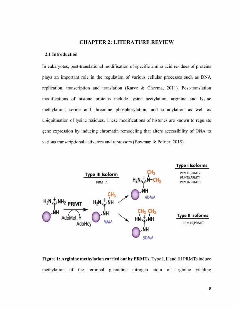
Figure 1: Arginine methylation carried out by PRMTs. Type I, II and III PRMTs induce
methylation of the terminal guanidine nitrogen atom of arginine yielding
10
monomethylarginine (MMA). Type I PRMTs including PRMT1, PRMT2, PRMT3,
PRMT4, PRMT6 and PRMT8 induce asymmetric dimethylation of arginine (ADMA).
Type II PRMT enzymes (PRMT5 and PRMT9) catalyze symmetric dimethylation of
arginine (SDMA), whereas Type III enzymes (PRMT7) catalyze formation of
monomethylarginine (MMA) (Caceres et al., 2018).
Histone arginine methylation is an important post-translational modification that is carried
out by enzymes belonging to the protein arginine methyltransferase (PRMT) family
(Guccione & Richard, 2019). PRMTs catalyze transfer of a methyl group from S-
adenosylmethionine (AdoMet) to the guanidino nitrogen of arginine, resulting in formation
of methylarginine. Three types of methylarginine are found in mammals (Fig. 1).
Monomethylarginine (MMA) is formed when a single methyl group is attached to one of
the terminal nitrogen atoms of guanidine group, whereas asymmetric dimethylarginine (ω-
NG, ω NG-dimethylarginine) is produced due to the addition of a second methyl group to
the same terminal guanidino nitrogen. In contrast, attachment of the second methyl group
to the other terminal nitrogen atom results in the formation of symmetric dimethylarginine
(ω- NG, ω-N’G-dimethylarginine). Based on the type of methylarginine they produce,
PRMTs are broadly classified into three groups: Type I PRMTs include PRMT 1, 2, 3, 4,
6 and 8, and catalyze monomethylation and asymmetric dimethylation of arginine residues;
Type II PRMTs include PRMT5 and PRMT9 that catalyze formation of monomethyl and
symmetric dimethylarginine; on the other hand, Type III PRMTs include PRMT7, which
promotes only monomethylation (Shailesh et al., 2018).
11
2.2. Protein arginine methyltransferase (PRMT5)
PRMT5 is a Type II PRMT that was first identified as Jak binding protein 1(JBP1) with a
molecular weight of 72.4 kDa in a study by Pollack et al. (1999), which focused on
identifying novel Jak2 interacting proteins in human cells. Using an in vitro methylation
assay, the authors of the same study demonstrated that JBP1 possesses methyltransferase
activity and can methylate histones, H2A and H4, and myelin basic protein. Sequence
homology showed that JBP1 is the human homolog of the Shk1 kinase binding protein1
(Skb1) gene of Schizosaccaromyces pombe and HSL7 (histone synthetic lethal 7) of
Saccharomyces cerevisiae. A later study that analyzed the amino acid sequence of JBP1
showed that JBP1 has three conserved domains at its C-terminus, which are similar to those
found in S-adenosyl-L-methionine-dependent protein arginine methyltransferases
(PRMTs). This finding led the authors to propose that JBP1 is a PRMT family member,
and therefore, named it as PRMT5. What further confirmed this notion is that endogenous
JBP1/PRMT5 runs as a 72 kDa protein that can methylate arginine residues of myelin basic
protein, histones, and the amino terminus of fibrillarin that is fused to glutathione S-
transferase (GST) (Rho et al., 2001). At the same time, Branscombe et al. (2001)
demonstrated that PRMT5 is a type II methyltransferase that can induce monomethylation
and symmetric dimethylation of several methyl acceptor proteins such as myelin basic
protein, bovine histone H2A, and a GST-fibrillarin (glutathione S-transferase-GAR).
2.2.1 Structure and enzymology
An early study that investigated the structure of PRMT5 by sedimentation analysis showed
that PRMT5 exists as a homo-oligomer complex including dimer and tetramer in vivo. The
12
same study also demonstrated that several covalent interactions such as disulphide bond
and non-covalent associations between C-terminal and N-terminal regions of different
PRMT5 monomers induce homo-oligomerization of PRMT5 (Rho et al., 2001). The C-
terminal domain of PRMT5 harboring methyltransferase activity is found to be
evolutionarily conserved from yeast to human (Fig. 2) (Rho et al., 2001; Shailesh et al.,
2018). The X-ray structure of human PRMT5 shows that it binds to MEP50 (Wdr77/
androgen receptor coactivator p44) to form a hetero-octomeric complex having a tetramer
of PRMT5 and tetramer of MEP-50. A detailed structural analysis of the PRMT5/MEP50
complex revealed that PRMT5 has a triosephosphate isomerase (TIM) barrel domain in the
N-terminal region, which interacts with the C-terminal catalytic domain of an adjacent
PRMT5 monomer in the octomeric complex. The crystal structure also showed that the
TIM barrel domain interacts with MEP50, and that the C-terminal catalytic domain
contains a Rossmann fold region, which serves as the AdoMet binding site, and a β-
sandwich domain in between C-and N-terminal domains that accomodates substrate
binding (Antonysamy et al., 2012). Binding of MEP50 potentiates the histone
methyltransferase activity of PRMT5 by enhancing its interaction with substrate
(Antonysamy et al., 2012; Burgos et al., 2015). In addition, CYCLIN D1/CDK4-mediated
phosphorylation of threonine 5 in MEP50 has been shown to further enhance PRMT5
enzymatic activity (Aggarwal et al., 2010). In contrast, phosphorylation of PRMT5 at
tyrosines 297, 304 and 306 by the oncogenic Jak2 mutant protein (V617F) disrupts its
association with MEP50 and reduces its histone methyltransferase activity (Liu et al.,
2011).
13
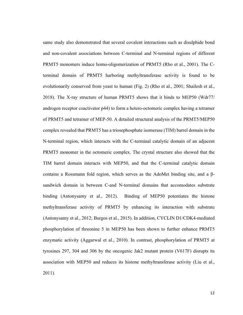
Figure 2: Catalytic domain of PRMT5 is highly conserved from yeast to human.
Alignment of amino acid sequences from H. sapiens (296–493), D. rerio (292–489), C.
elegans (348–554), D. melanogaster (272–466), A. thaliana (302–496), and S. cerevisiae
(317–518) was done using the Clustal Omega software. The conserved sequences are
highlighted using different colors and symbols (Blue star represents conserved identical
residues, red colon represents conserved highly similar residues; and green bullet
represents moderately similar residues) (Shailesh et al., 2018).
Apart from MEP-50, PRMT5 is also known to interact with various complexes such as the
human SWI/SNF complex, and proteins like pICln, and RioK1, which increase its
functional versatility. For example, in association with BRG1- and hBRM-based SWI/SNF

14
chromatin remodeling complexes, PRMT5 induces symmetric dimethylation of H3R8 and
H4R3 in the promoter region of tumor suppressor genes including ST7 and NM23, and
represses their transcription (Pal et al., 2004). On the other hand, PRMT5 interacts with the
Sm binding protein, PICln, and symmetrically methylates arginine residues of Sm family
proteins, SMD1 and SMD3. Methylation of SMD proteins promotes their association with
the SMN complex, which is a prerequisite step for formation of small nuclear
ribonucleoprotein core (SnRP) during mRNA splicing (Brahms, et al., 2001; Friesen et al.,
2001). In addition, PRMT5 can also bind to the cytosolic protein, RioK1 and promote
recruitment and methylation of the RNA binding protein, nucleolin, which in turn favors
its interaction with RNA during ribosome biogenesis (Guderian et al., 2011).
2.2.2. PRMT5 target proteins
PRMT5 is known to mediate methylation of histone proteins including histones, H3 and
H4 in the promoter region of target genes, and regulate their transcription. For example,
PRMT5-mediated methylation of H3R8 and H4R3 in the promoter region of tumor
suppressor genes like ST7 and PTPROt suppresses their transcription (Alinari et al., 2015;
Pal et al., 2007). Apart from histone proteins, PRMT5 also methylates several non-histone
proteins, which include transcriptional factors that regulate major cellular functions (Table
1). For example, during DNA damage PRMT5 associates with Strap (stress-responsive
activator of p300) to methylate p53 and enhances its binding efficiency to the promoter
region of its target gene, Cyclin-Dependent Kinase Inhibitor 1A (CDKN1A), thereby
leading to elevated expression of p21. The net outcome of these events is cell-cycle arrest.
However, depletion of PRMT5 reduces occupancy of p53 on the p21 promoter, which
15
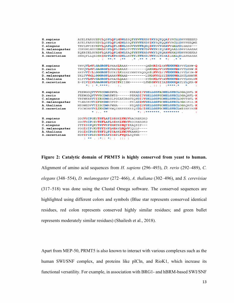
Table 1: Proteins that are methylated by PRMT5
PRMT5 substrates Biological role Citations
Histones (H3R2, H3R8,
H4R3), SPT5, FCP1,
MBD2, KAP1, N-MYC
PAX3, RNA polymerase II
Regulation of transcription
(Amente et al., 2005; di
Caprio et al., 2015; Kwak
et al., 2003; Pal et al.,
2004; Park et al., 2015;
Pesiridis et al., 2009; Tan
& Nakielny, 2006; Tsai et
al., 2013; Wu et al., 2015;
Zhao et al., 2016)
p53, ASK1, PDCD4,
CRN5
Regulation of apoptosis
(Chen et al., 2016; Fay et
al., 2014; Jansson et al.,
2008; Rastetter et al., 2015)
NF-κB/p65, HOXA9
Immune response
regulation
(Bandyopadhyay et al.,
2012; Harris et al., 2016)
cRAF, EGFR, RAF, E2F-
1, FEN1,
Androgen receptor,
srGAP2, PDGFRα
Regulation of cell
proliferation, migration,
differentiation, and
survival
(Andreu-Perez et al., 2011;
Calabretta et al., 2018; Guo
& Bao, 2010; Guo et al.,
2010; Mounir et al., 2016;
Zheng et al., 2013)
16
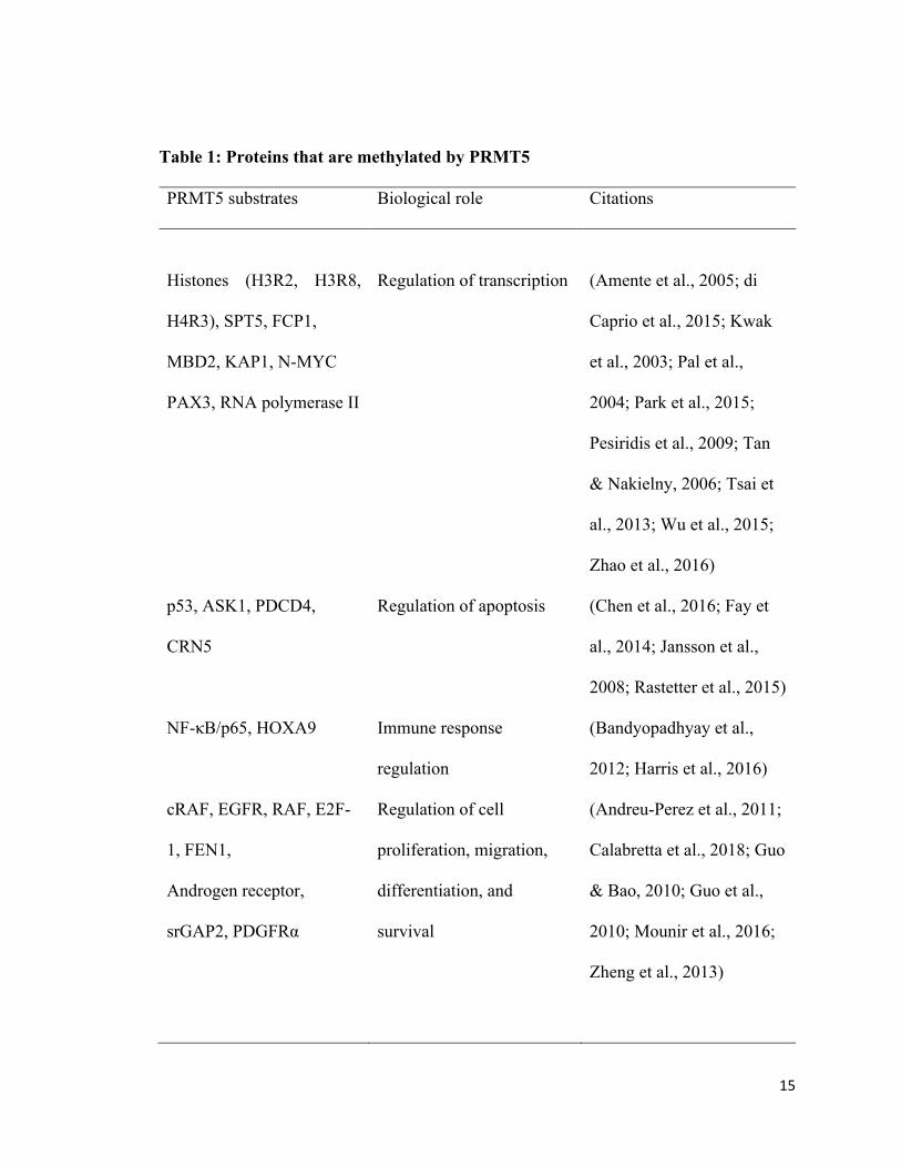
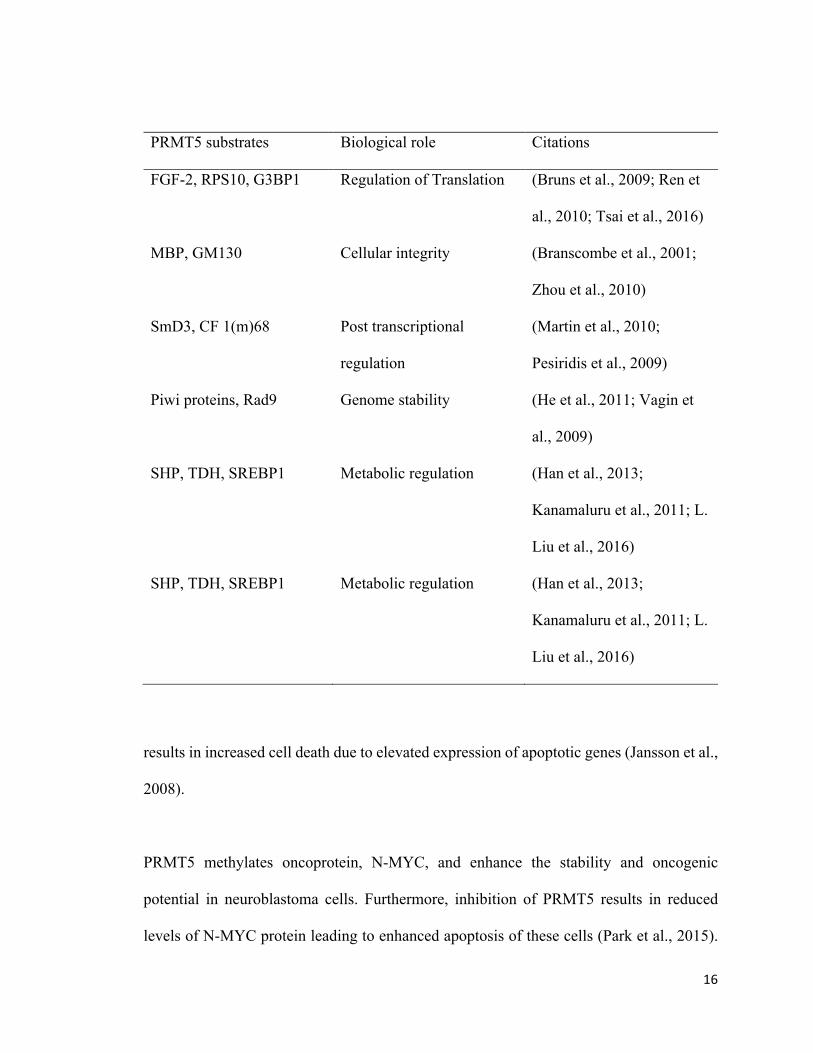
PRMT5 substrates Biological role Citations
FGF-2, RPS10, G3BP1 Regulation of Translation
(Bruns et al., 2009; Ren et
al., 2010; Tsai et al., 2016)
MBP, GM130 Cellular integrity
(Branscombe et al., 2001;
Zhou et al., 2010)
SmD3, CF 1(m)68 Post transcriptional
regulation
(Martin et al., 2010;
Pesiridis et al., 2009)
Piwi proteins, Rad9 Genome stability
(He et al., 2011; Vagin et
al., 2009)
SHP, TDH, SREBP1 Metabolic regulation (Han et al., 2013;
Kanamaluru et al., 2011; L.
Liu et al., 2016)
SHP, TDH, SREBP1 Metabolic regulation (Han et al., 2013;
Kanamaluru et al., 2011; L.
Liu et al., 2016)
results in increased cell death due to elevated expression of apoptotic genes (Jansson et al.,
2008).
PRMT5 methylates oncoprotein, N-MYC, and enhance the stability and oncogenic
potential in neuroblastoma cells. Furthermore, inhibition of PRMT5 results in reduced
levels of N-MYC protein leading to enhanced apoptosis of these cells (Park et al., 2015).
17
PRMT5-induced methylation of the NF-κB p65 subunit enhances its DNA binding activity
and target gene expression (Wei et al., 2013). In contrast, PRMT5-dependent methylation
of transcription factor E2F-1 targets it to ubiquitination, and hence reduces its ability to
induce apoptosis of tumor cells during DNA damage (Cho et al., 2012).
Apart from transcription, PRMT5 also controls other cellular processes by methylating a
variety of proteins such as ribosomal protein S10, golgin GM130, and rapidly accelerated
fibrosarcoma (RAF) protein kinase. For example, PRMT5-dependent methylation of the
N-terminal arginine residues of Golgi-associated protein, golgin GM130 is critical for the
formation and adequate linking of Golgi ribbons in the Golgi apparatus (GA). However,
PRMT5 knockdown results in fragmentation and aberrant tethering of GA ribbons,
indicating that PRMT5 is involved in the maintenance of GA architecture (Zhou et al.,
2010). PRMT5 interacts with and methylates the C-terminal arginine residues, Arg158 and
Arg160 of RPS10 protein, an integral component of the 40S ribosomal subunit complex.
Methylation of RPS10 protein is required for its localization into the granular component
(GC) region of nucleolus, and effective interaction with another ribosome assembly
protein, nucleophosmin/B23, thus favors an adequate assembly of 40S and 60S ribosome
subunits, during ribosome biogenesis, resulting in efficient translation of mRNAs.
However, studies using methylation mutant form of RSP10 showed the methylation
deficient RSP10 is unable to localize to GC region and interact with B23, and fails to
assemble into ribosomes. In addition, lack of methylation reduced the stability of the
mutant protein, subjecting it for proteasomal degradation, highlighting the fact that
PRMT5-mediated methylation is an essential event during ribosome biogenesis (Ren et al.,
18
2010). PRMT5 modulates RAS-ERK1/2 pathway activation by reducing the kinase activity
of RAF family protein, CRAF that phosphorylates ERK1/2 proteins. Here, PRMT5-
mediated methylation of R563 of CRAF protein reduces its stability, and hence, decreases
its catalytic activity, resulting in diminished ERK1/2 signal amplitude (Andreu-Perez et
al., 2011).
2.2.3 Role of PRMT5 during embryonic life
PRMT5 plays an important role during embryonic development. In mice, maternally
inherited cytosolic PRMT5 in the oocyte translocates into the nucleus to initiate cellular
differentiation events, and its cellular localization fluctuates dynamically during different
stages of embryonic development (Tee et al., 2010). In embryonic stem (ES) cells,
cytoplasmic PRMT5 associates with STAT3 and suppresses transcription of ES cell
differentiation genes, FGF5, GATA6, LHX1, FOX2, HOXA3, HOXA7 and HOXD9 to
maintain embryonic pluripotency. Moreover, PRMT5 knockdown in ES cells leads to
down-regulation of key pluripotency genes including OCT4, NANOG and REX1, whereas
complete loss of PRMT5 results in aberrant growth and subsequent lethality in mice (Tee
et al., 2010). Collectively, these results demonstrate that PRMT5 has an important role in
maintaining pluripotency during embryonic life of mice.
The role played by PRMT5 during human embryonic development is not studied
extensively; however, a study by Gkountela et al. (2014) demonstrated that depletion of
PRMT5 in human ES cells does not alter the transcription of key pluripotent genes
including OCT4, SOX2, and NANOG, indicating that PRMT5 does not play an important
19
role in regulating stem cell pluripotency in humans. Nevertheless, PRMT5 is important
during embryonic life in humans as it promotes proliferation of ES cells during their self-
renewal by inducing transcriptional repression of cyclin-dependent kinase inhibitor,
p57KIP2, which in turn promotes G1 to S progression.
2.2.4 Role of PRMT5 during neurogenesis
Various studies have shown that PRMT5 plays an essential role during neurogenesis.
PRMT5 helps to maintain stemness of neural stem cells in the cerebral cortex of the mouse
brain by associating with Schwann Cell Factor 1 (SC1)/PRDM4, a Positive Regulatory
Domain (PRDM) family transcription factor that modulates cell cycle progression. Binding
of PRMT5 with SC1 induces down-regulation of pro-mitotic genes including CYCLIN B1
and BUB1b, which suppresses differentiation of neuronal stem cells. However, during
neuronal stem cell differentiation, expression of SC1 and PRMT5 decreases with a
concomitant increase in CYCLIN B1 expression (Chittka et al., 2012). In addition, PRMT5
controls survival of neuronal stem cells and protects them from apoptosis. Depletion of
PRMT5 in the central nervous system (CNS) of Nestin-Cre transgenic mice leads to
abnormal CNS development ensued by postnatal lethality due to increased apoptosis of
neural stem and progenitor cells (NPCs). In this scenario, PRMT5 depletion leads to
reduced methylation of Sm proteins, resulting in abnormal splicing of mRNAs, especially
for proteins involved in cell cycle progression of NPCs. MDM4 mRNA is one such mRNA,
which undergoes alternative splicing due to aberrant spliceosomal processing. The
resulting MDM4S mRNA is short-lived and consequently contributes to p53 activation and
increased cell death (Bezzi et al., 2013).
20
Furthermore, a work by Huang et al. (2011) showed that PRMT5 levels gradually increase
during postnatal brain development, especially during active myelination of neurons. In
their systematic approach, the authors showed that PRMT5 gets localized in the nuclear
compartment of myelinating oligodendrocytes to promote their differentiation. In this
situation, PRMT5 suppresses expression of transcription regulators that inhibit
oligodendrocyte differentiation including inhibitors of differentiation/DNA binding (Id),
Id2 and Id4 by conserving the CpG methylation status at their promoters. Furthermore,
PRMT5 knockdown reduces expression of differentiation inducers such as SOX10 and
NKX2.2, indicating that PRMT5 positively controls their expression in oligodendrocyte
progenitor cells to induce cell differentiation (Huang et al., 2011).
In agreement with these results, Scaglione et al. (2018) further confirmed that PRMT5 has
a major role during differentiation and developmental myelination of oligodendrocytes.
PRMT5 levels are upregulated and mostly localized to the cytosolic compartment in
proliferating oligodendrocyte progenitor cells (OPCs) as compared to differentiating
OPCs, where its levels are reduced and mostly restricted to the nuclear compartment. In
mice, conditional ablation of PRMT5 in OPCs leads to reduction in number of
oligodendrocytes and results in severe hypomyelination of the brain on post-natal day 14,
followed by early mortality. Mechanistically, PRMT5 ablation leads to aberrant
differentiation of OPCs, followed by an activation of p53-dependent apoptosis, which
eliminates OPCs in the brain (Scaglione et al., 2018).
The mechanism by which PRMT5 regulates oligodendrocyte myelination and maturation
21
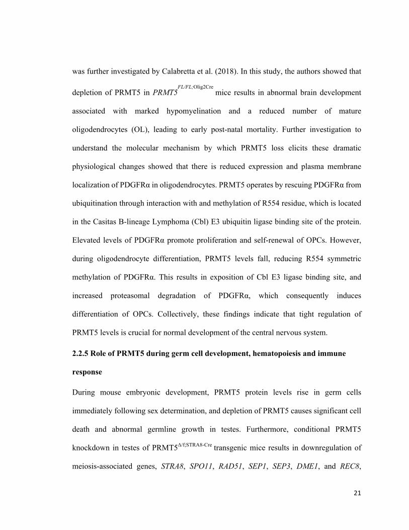
was further investigated by Calabretta et al. (2018). In this study, the authors showed that
depletion of PRMT5 in PRMT5FL/FL;Olig2Cre
mice results in abnormal brain development
associated with marked hypomyelination and a reduced number of mature
oligodendrocytes (OL), leading to early post-natal mortality. Further investigation to
understand the molecular mechanism by which PRMT5 loss elicits these dramatic
physiological changes showed that there is reduced expression and plasma membrane
localization of PDGFRα in oligodendrocytes. PRMT5 operates by rescuing PDGFRα from
ubiquitination through interaction with and methylation of R554 residue, which is located
in the Casitas B-lineage Lymphoma (Cbl) E3 ubiquitin ligase binding site of the protein.
Elevated levels of PDGFRα promote proliferation and self-renewal of OPCs. However,
during oligodendrocyte differentiation, PRMT5 levels fall, reducing R554 symmetric
methylation of PDGFRα. This results in exposition of Cbl E3 ligase binding site, and
increased proteasomal degradation of PDGFRα, which consequently induces
differentiation of OPCs. Collectively, these findings indicate that tight regulation of
PRMT5 levels is crucial for normal development of the central nervous system.
2.2.5 Role of PRMT5 during germ cell development, hematopoiesis and immune
response
During mouse embryonic development, PRMT5 protein levels rise in germ cells
immediately following sex determination, and depletion of PRMT5 causes significant cell
death and abnormal germline growth in testes. Furthermore, conditional PRMT5
knockdown in testes of PRMT5Δ/f;STRA8-Cre transgenic mice results in downregulation of
meiosis-associated genes, STRA8, SPO11, RAD51, SEP1, SEP3, DME1, and REC8,
22
leading to meiotic arrest and loss of germ cells during spermatogenesis. These findings
show that PRMT5 plays a key role during spermatogenesis by regulating expression of
meiosis-associated genes (Wang et al., 2015).
PRMT5 has shown to play a key role during adult-hematopoiesis. PRMT5 depletion in
hematopoietic stem and progenitor cells (HSPCs) of 2-month-old Mx1Cre+ Prmt5fl/fl mice
leads to bone marrow aplasia, pancytopenia and reduced thymus mass within 15 days of
the Cre induction. FACS analysis showed that PRMT5 depletion results in significant loss
of hematopoietic progenitor cells, reduction in erythroid differentiation in bone marrow,
and aberrant accumulation of thymocytes in thymus. Although PRMT5 loss induced initial
transient expansion of hematopoietic stem cells, functional analysis indicated that these
cells were functionally defective. Further analysis to understand the mechanism through
which PRMT5 maintains HSPCs population showed that it promotes G1/S transition in
these cells. PRMT5 also promotes cytokine-activated JAK/STAT and AKT/PI3K signaling
in HSPCs by inducing the expression of cell surface cytokine receptors (Liu et al., 2015).
Work by Tsutsui et al. (2013) informed that PRMT5 is involved in regulating the immune
system via modulation of expression of key genes of the immune response program. Under
normal conditions, PRMT5 represses the expression of C/EBPβ target genes including
TNF-α and IL-2, which are involved in acute phase immune response. In this instance,
PRMT5 associates with its binding partner MEP-50 and cyclin dependent kinases, CDK8
23
and CDK9, and is recruited to the promoter of C/EBPβ target genes, where it suppresses
their expression by promoting H4R3 symmetric dimethylation and DNMT3A recruitment.
These studies indicate that PRMT5 regulates development of germ cells, population of
hematopoietic stem and progenitor cell, and elicitation of immune response during adult
life by regulating the expression of distinct sets of gene in each context.
2.2.6 PRMT5 and cancer
Several studies have shown that elevated PRMT5 activity promotes cellular transformation
and enhanced tumor growth. PRMT5 expression is increased in a wide variety of cancer
cells including glioblastoma, melanoma, non-small cell lung carcinoma, lymphoma and
leukemia cells (Banasavadi-Siddegowda et al., 2017; Nicholas et al., 2013; Pal et al., 2007;
Shilo et al., 2013). An early study by Pal et al. (2004) showed that PRMT5 overexpression
induces transformation of NIH3T3 cells by directly suppressing the expression of tumor
suppressor genes including ST7 and NM23. A similar finding was reported by Wei et al.
2012, which showed that ectopic expression of PRMT5 induces enhanced growth of
NIH3T3 cells by elevating the levels of positive cell cycle regulators including CYCLIN
D1, CYCLIN D2, CYCLIN E1, CDK4 and CDK6, reducing expression of negative cell
cycle regulator such as retinoblastoma (RB) proteins, and enhancing PI3K/AKT signaling,
indicating that PRMT5 induces tumorigenicity by augmenting proliferative signals.
Another study by Alinari et al. (2015) further supported the fact that PRMT5 induces
cellular transformation by showing a rapid increase in PRMT5 protein level in 4 to 8 days
after EBV infection of normal B lymphocytes, which coincides with cellular
immortalization. Furthermore, our recent study showed that PRMT5 promotes growth and
24
proliferation of three different types of non-Hodgkin’s lymphoma cells by activating
WNT/β-CATENIN and AKT/GSK-3β proliferative signaling (Chung et al., 2019).
Collectively, these studies imply that elevated expression of PRMT5 promotes
transformation and growth of cancer cells by augmenting different proliferative signaling
pathways.
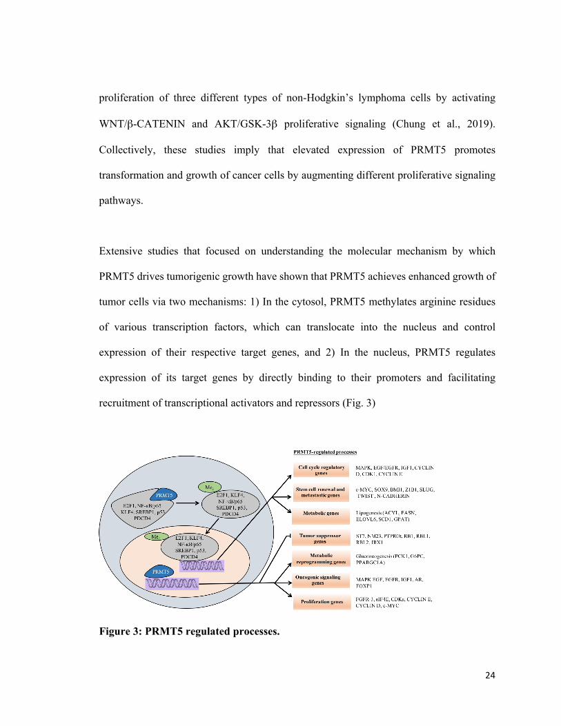
Extensive studies that focused on understanding the molecular mechanism by which
PRMT5 drives tumorigenic growth have shown that PRMT5 achieves enhanced growth of
tumor cells via two mechanisms: 1) In the cytosol, PRMT5 methylates arginine residues
of various transcription factors, which can translocate into the nucleus and control
expression of their respective target genes, and 2) In the nucleus, PRMT5 regulates
expression of its target genes by directly binding to their promoters and facilitating
recruitment of transcriptional activators and repressors (Fig. 3)
Figure 3: PRMT5 regulated processes.
25
Cytosolic PRMT5 induces arginine methylation of various transcription factors, which
translocate into the nucleus and regulate expression of their respective target genes.
Nuclear PRMT5 is also directly recruited to the promoter regions of specific target genes
to enhance cellular proliferation and tumorigenesis (Shailesh et al., 2018).
PRMT5 suppresses expression of tumor suppressor genes including Suppressor of
Tumorigenicity 7 (ST7) and Protein Tyrosine Phosphatase Receptor-type O (PTPRO) by
binding to their promoter and inducing H3R8 and H4R3 dimethylation in lymphoma and
leukemia (Alinari et al., 2015; Wang et al., 2008). PRMT5 also induces transcriptional
downregulation of RB family tumor suppressor genes including RB1, RBL1 and RBL2
through binding to their promoter and inducing symmetric methylation of H3R8 and H4R3
in chronic lymphocytic leukemia cells (Wang et al., 2008). However, only RBL2 protein
levels are reduced in these cells compared to normal B cells. Further analysis showed that
reduced expression of microRNAs that target 3ʹ-UTR of RB1 and RBL1 mRNAs, resulted
in enhanced translation and elevated protein expression (Wang et al., 2008). In a follow-
up study, Chung and co-workers (2013) showed that PRMT5 is able to inactivate RB1
protein by inducing phosphorylation of the protein at multiple sites including Ser-780, Ser-
795, Ser-807/Ser-811 through activation of the CYCLIN D1-CDK4/6 complex. The same
study also showed that PRMT5-mediated inactivation of RB1 and RBL2 leads to induced
expression of polycomb repressor complex proteins (PRC2), which eventually induce
transcriptional repression of pro-apototic genes including CASP10, DAP1, HOXA5 and
HRK in three different types of non-Hodgkin lymphoma cells. However, shRNA-mediated
26
downregulation of PRMT5 induces apoptosis of these cells by inducing transcriptional
derepression of RBL2 and dephosphorylation of RB1, which trigger reduced PRC2
expression and transcriptional derepression of pro-apototic genes (Chung et al., 2013).
In gastric cancer cells, PRMT5 promotes cell proliferation by suppressing the expression
of a different tumor suppressor gene, Iroquois Homeobox 1 (IRX1), by binding to its
promoter region and recruiting DNMT3A, which consequently leads to promoter
hypermethylation. PRMT5 knock-down induces expression of IRX1 and leads to reduced
growth and metastasis of gastric cancer cells in vitro (Liu et al., 2018). A very recent work
from our group has demonstrated that PRMT5 suppresses expression of tumor suppressors,
AXIN2 and WIF1, which serve as inhibitors of WNT/β-CATENIN proliferative signaling
in three different types of non-Hodgkin’s lymphoma cells (Chung et al., 2019). PRMT5
binds to the promoter region of AXIN2 and WIF1, and hypermethylates promoter H3R8
and H4R3 histones, leading to transcriptional repression. As a consequence, WNT/β-
CATENIN proliferative signaling becomes aberrantly activated, leading to elevated
expression of WNT/β-CATENIN downstream targets, CYCLIN D1, c-MYC and
SURVIVIN (Chung et al., 2019).
In contrast to inducing transcriptional repression of tumor suppressor genes, PRMT5 can
also promote transcriptional activation of oncogenes in several cancer cell types. PRMT5
increases transcription of proto-oncogenes including eukaryotic elongation Initiation
Factor-4E (eIF4E) and Fibroblast- derived Growth Factor Receptor-3 (FGFR3), by
27
directly binding and inducing H3R8 and H4R3 symmetric dimethylation marks in their
promoter region in colorectal cancer cells. However, knockdown of PRMT5 in these cells
resulted in reduced methylation of H3R8 and H4R3, and lowered expression eIF4E and
FGFR3, which was associated with reduced cell growth (Zhang et al., 2015). Another
study by Deng et al. (2017) showed that PRMT5 expression is elevated in prostate cancer
and positively correlated with expression of androgen receptor (AR). Using
immunoprecipitation assays, the authors showed that PRMT5 associates with SP1, and the
BRG1-based hSWI/SNF remodeler, and is recruited to the promoter region of AR gene,
where it induces H4R3 symmetric dimethylation and activates AR expression in LnCaP
prostate cancer cells. In addition, inducible PRMT5 knockdown leads to reduced
expression of AR and decreased cell growth in AR-positive cells in vitro and in mice
xenograft tumors. Similar to the knockdown experiment, pharmacological inhibition of
PRMT5 halted growth of AR-positive cells in an AR-dependent manner in vitro.
The role of PRMT5 in breast cancer stem cell (BCSCs) function was also investigated and
results showed that elevated levels of PRMT5 upregulate expression of transcription factor,
forkhead box protein 1 (FOXP1), which is involved in normal and stem cell function. Here,
PRMT5 binds to the promoter region of FOXP1 and methylates H3R2, which serves as a
recruitment epigenetic mark for the WDR5 subunit of SET1/MLL methyltransferase.
Consequently, H3K4 becomes methylated and results in FOXP1 transcriptional activation.
However, deactivation of PRMT5 enzymatic activity using GSK591 inhibitor or
interrupting WDR5 and SET/MLL1 interaction leads to a substantial reduction in
28
methylated H3K4 level as well as FOXP1 expression. The study also showed that PRMT5
knock-down diminishes the proliferative and self-renewing ability of breast cancer cells in
vitro and in vivo. The study also indicated that tumors excised from NSG (NOD/Scid/IL-
2Rγnull) mice injected with a PRMT5-specific shRNA had a less aggressive and more
differentiated phenotype as compared to their control littermates. Similar to the results of
PRMT5 knockdown, FOXP1 knockdown also resulted in reduced growth of breast cancer
cells in vitro as well as in xenograft tumors, indicating that PRMT5 promotes
tumorigenicity of breast cancer by regulating expression of FOXP1 (Chiang et al., 2017).
These studies indicate that elevated levels of PRMT5 promotes cancer growth by altering
expression of a variety of genes in a context dependent manner.
PRMT5-mediated methylation of several non-histone proteins has been shown to play an
important role in carcinogenesis. A systematic study by Wan et al. (2015) showed that
PRMT5 regulates genome stability and survival of cancer cells by methylating KLF4, a
major transcription factor that controls the expression of genes involved in cell-cycle
progression, genome-stability, cell adhesion, metabolism, apoptosis and stem cell renewal.
The mechanism used by PRMT5 here is that it associates with KLF4 protein and
methylates R374, R376 and R377 residues, which inhibits VHL/VBC E3 mediated
ubiquitination of KLF4 leading to enhanced stability of the protein. When PRMT5 is
depleted or KLF4 is mutated, its ubiquitination increases leading to a significant drop in its
protein level. The same study also demonstrated that overexpression of KLF4 in breast
cancer cells including MCF7 and MCF10A promotes expression of CYCLIN D2, CYCLIN
29
E1, CDK1, MAPK, IGF1 and EGF/EGFR as well as genes involved in stem cell renewal
and metastasis, which include c-MYC, SOX9, Z1B1, BM1, SLUG, TWIST and E-
CADHERIN. A further investigation by the same group showed that PRMT5 and KLF4
protein expression is substantially elevated in clinical samples of aggressive breast tumor
tissues compared to the adjacent normal tissues (Hu et al., 2015). These findings suggest
that PRMT5 controls cell survival, metastasis and stem-cell renewal of cancer cells by
modulating stability of KLF4 (Hu et al., 2015).
2.3 WNT/β-CATENIN signaling
2.3.1 Discovery of WNT protein
WNT1 was discovered by Nusse and Varmus in 1982 who named it INT-1, because this
locus was the preferred site for insertion of DNA from Murine Mammary tumor virus in
mouse bearing viral-induced mammary tumors (Nusse & Varmus, 1982). Later studies in
fruit flies showed that wingless (WG), a gene that regulates polarity of embryo segments
during larval growth is a Drosophila homolog of Int-1 (Rijsewijk et al., 1987). More studies
confirmed that INT-1 is a member of the evolutionarily conserved gene family that is now
referred to as WNT (wingless-type mouse mammary tumor virus integration site) gene
family. 19 WNT genes have been identified in mammals including humans till date
(Clevers & Nusse, 2012).
2.3.2 Canonical WNT/β-CATENIN signaling
WNT ligands belong to a large family of secretory proteins of 40 kDa in size that contain
conserved cysteine-rich domains (Tanaka et al., 2002). Binding of WNT proteins to their
cell membrane anchored receptors activates a group of downstream signaling modules
30
including the canonical WNT/β-CATENIN, noncanonical planar cell polarity
(WNT/PCP), and WNT/Ca 2+pathways.
WNT/β-CATENIN signaling is an evolutionarily conserved pathway that plays a prime
role during embryonic and adult life (Logan & Nusse, 2004). WNT/β-CATENIN regulates
various processes during embryonic development such as differentiation of embryonic
stem cells, formation embryonic body axis, and morphogenesis of various tissues that
originate from different germ layers (Davidson et al., 2012; Lan et al., 2006; Petersen &
Reddien, 2009). In adult life, WNT/β-CATENIN signaling is involved in maintenance of
the stem cell niche and promotes their differentiation during tissue and organ regeneration
(Chen et al., 2007; Fevr et al., 2007). Germline mutations of genes, which encode
components of WNT/β-CATENIN signaling lead to abnormal embryonic development and
several hereditary diseases, whereas somatic mutations and epigenetic alteration of these
genes during adult life lead to the onset of cancer (Clevers, 2006).
Canonical WNT/β-CATENIN signaling is initiated by binding of WNT ligand to the
membrane-bound Frizzled receptor (FZD), in cooperation with co-receptor, LDL receptor-
related protein family receptors (LRP) called LRP5/6 (Bhanot et al., 1996; Tamai et al.,
2000) (Fig. 4). Next, the C-terminal domain of the ligand bound Frizzled receptor directly
interacts with cytoplasmic Dishevelled protein (DSH/DVL) and promotes recruitment of
cytosolic glycogen synthase kinase 3beta (GSK-3β) to the plasma membrane (Wong et al.,
2003).
31
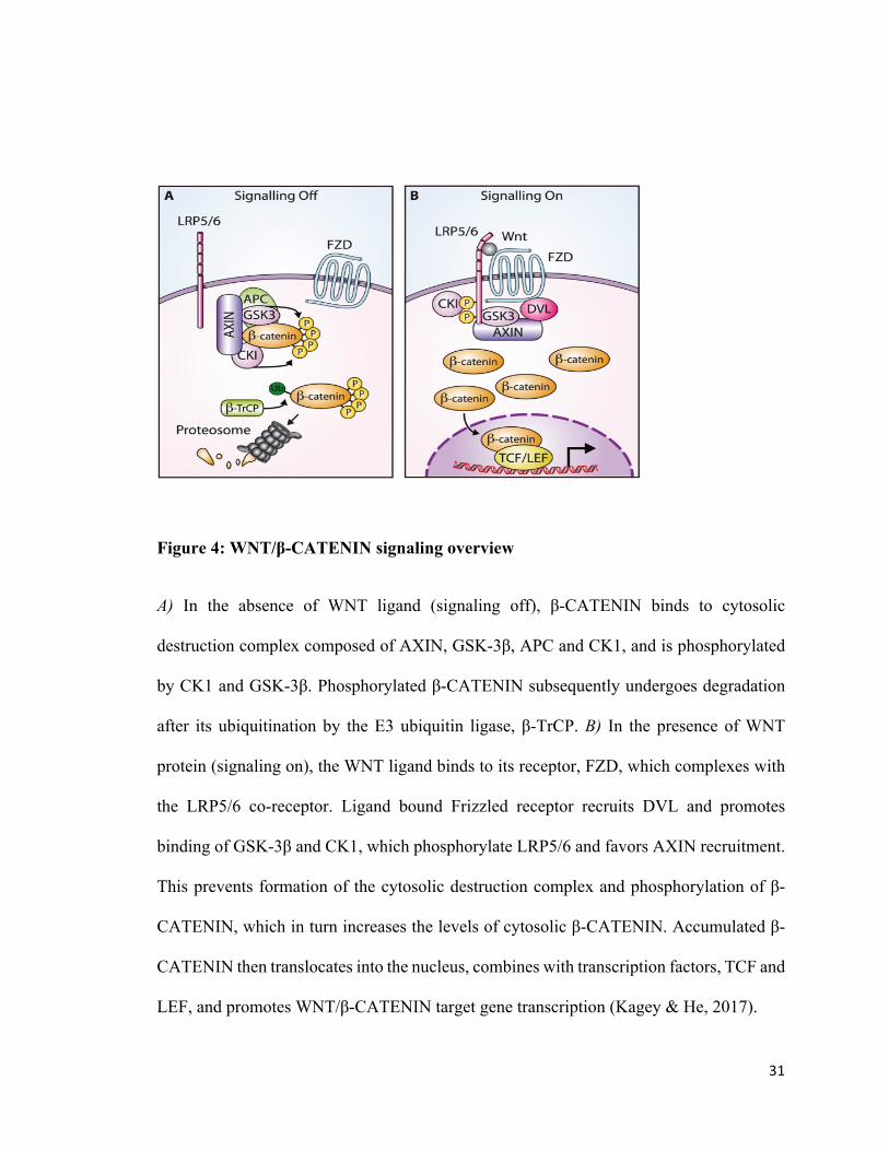
Figure 4: WNT/β-CATENIN signaling overview
A) In the absence of WNT ligand (signaling off), β-CATENIN binds to cytosolic
destruction complex composed of AXIN, GSK-3β, APC and CK1, and is phosphorylated
by CK1 and GSK-3β. Phosphorylated β-CATENIN subsequently undergoes degradation
after its ubiquitination by the E3 ubiquitin ligase, β-TrCP. B) In the presence of WNT
protein (signaling on), the WNT ligand binds to its receptor, FZD, which complexes with
the LRP5/6 co-receptor. Ligand bound Frizzled receptor recruits DVL and promotes
binding of GSK-3β and CK1, which phosphorylate LRP5/6 and favors AXIN recruitment.
This prevents formation of the cytosolic destruction complex and phosphorylation of β-
CATENIN, which in turn increases the levels of cytosolic β-CATENIN. Accumulated β-
CATENIN then translocates into the nucleus, combines with transcription factors, TCF and
LEF, and promotes WNT/β-CATENIN target gene transcription (Kagey & He, 2017).
32
The membrane-associated GSK-3β phosphorylates LRP5/6 and triggers casein kinase 1
(CK1) to induce further phosphorylation on LRP5/6. Frizzled associated DVL binds to
Axis inhibitory proteins (AXIN1 and AXIN2) through the DIX domain and favors binding
of AXINs to the cytosolic tail of phosphorylated LRP5/6 (Cliffe et al., 2003; Zeng et al.,
2005). Consequently, levels of the cytosolic destruction complex, consisting of AXINs
(AXIN1 and AXIN2), CK1, APC, and GSK-3β, decrease, which leads to reduced
phosphorylation of β-CATENIN and prevents its degradation. Subsequently, cytosolic β-
CATENIN levels rise and trigger its translocation into nucleus where it complexes with
TCF (T-cell factor) and LEF (lymphoid enhancer factor) and binds to the promoter region
of target genes, c-MYC, CYCLIN D1, and SURVIVIN. Upon binding, several chromatin
remodelers such as BRG1 and CBP/p300 are recruited to induce transcription (Barker et
al., 2001; Brunner et al., 1997; Kramps et al., 2002; Ma et al., 2005). In the absence of β-
CATENIN, TCF and LEF form complex with transducing-like enhancer protein
(TLE/Groucho) and recruit histone deacetylase enzymes to repress expression of target
genes (Cavallo et al., 1998; Chen, et al., 1999; Kagey & He, 2017).
During WNT off state, the cytosolic destruction complex keeps the level of β-CATENIN
low by binding and phosphorylating it (Kimelman & Xu, 2006). AXIN1 serves as a core
scaffolding protein in the destruction complex that binds to other components through
different domains. GSK-3β binds to the central domain of AXIN1 and phosphorylates
AXIN1 at Thr609 and Ser614 residues, which in turn promotes attachment of β-CATENIN
to AXIN1 (Jho et al.,1999). APC enhances AXIN1 multimerization by associating with it
33
through AXIN1:APC interaction domain (Pronobis et al., 2015). The second kinase, CK1
exhibits multiple interactions with various sites in the central domain of AXIN1 and
catalyzes phosphorylation of destruction complex bound β-CATENIN at Ser45, that favors
further phosphorylation of Ser33, Ser37 and Thr41 sites at N-terminal motif of β-
CATENIN by GSK-3β (Amit et al., 2002; Hagen et al., 2002). Phosphorylation of β-
CATENIN at Ser33 and Ser37 promotes binding of E3 ubiquitin ligase, β-TrCP, resulting
in proteasomal degradation of β-CATENIN (Latres et al., 1999; Spiegelman et al., 2000).
2.4 Natural antagonists of the WNT/β-CATENIN pathway
WNT/β-CATENIN signaling is tightly regulated by several endogenous inhibitors that
abrogate signal transduction through various mechanisms (Fig. 5). Secretory inhibitors like
WNT inhibitory factor 1 (WIF1) and secreted frizzled-related proteins (SFRPs) can
associate with WNT ligand directly and interrupt its binding to membrane receptors (Zhan
et al., 2017). Dickkopf (DKK) family proteins are another type of secretory antagonists
that bind to WNT co-receptor LRP5/6 to inhibit the association of ligand bound Frizzled
receptor to LRP5/6 (Kawano & Kypta, 2003). Elevated levels of destruction complex
components such as AXINs inactivate WNT/β-CATENIN signaling by inducing β-
CATENIN destruction (Nakamura et al., 1998). Reduced levels of WNT/β-CATENIN
antagonists have been reported in a variety of cancers including gastrointestinal cancer,
leukemia, melanoma and breast cancer, and contribute to enhanced tumor growth by
enhancing WNT/β-CATENIN activation (Zhan et al., 2017).
34
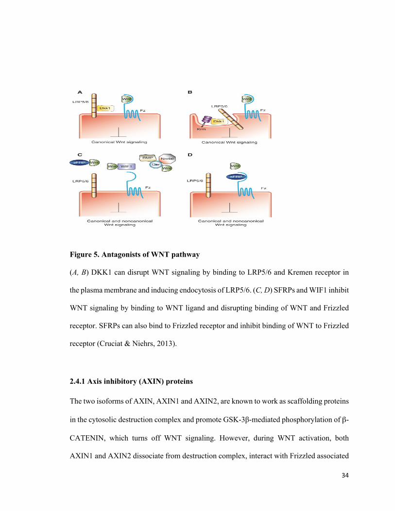
Figure 5. Antagonists of WNT pathway
(A, B) DKK1 can disrupt WNT signaling by binding to LRP5/6 and Kremen receptor in
the plasma membrane and inducing endocytosis of LRP5/6. (C, D) SFRPs and WIF1 inhibit
WNT signaling by binding to WNT ligand and disrupting binding of WNT and Frizzled
receptor. SFRPs can also bind to Frizzled receptor and inhibit binding of WNT to Frizzled
receptor (Cruciat & Niehrs, 2013).
2.4.1 Axis inhibitory (AXIN) proteins
The two isoforms of AXIN, AXIN1 and AXIN2, are known to work as scaffolding proteins
in the cytosolic destruction complex and promote GSK-3β-mediated phosphorylation of β-
CATENIN, which turns off WNT signaling. However, during WNT activation, both
AXIN1 and AXIN2 dissociate from destruction complex, interact with Frizzled associated
35
DVL and bind to the cytoplasmic tail of phosphorylated LRP5/6. As a consequence,
phosphorylation and subsequent degradation of β-CATENIN is inhibited, resulting in β-
CATENIN accumulation in the cytoplasm, which can move into nucleus to promote
transcription of WNT/β-CATENIN target genes (Gao et al., 2014). Hence, AXINs serve
as endogenous antagonists of the WNT/β-CATENIN signaling cascade in the absence of
WNT stimulation.
AXIN1 was initially discovered as a product of murine Fused gene locus, that regulates
embryonic axis formation in the mouse embryo (Gluecksohn-Schoenheimer, 1949). Later,
a study using Xenopus embryos showed that expression of AXIN1 inhibits secondary
dorsal axis formation induced by WNT, DVL, and kinase-negative GSK-3β, indicating that
AXIN1 has an inhibitory role in WNT/β-CATENIN signaling (Zeng et al., 1997). AXIN1
serves as a scaffolding protein in the cytosolic destruction complex by binding to various
components of the WNT signaling cascade including APC, PP2A, GSK-3β, DVL and β-
CATENIN through different domains. Binding of AXIN1 to GSK-3β facilitates β-
CATENIN phosphorylation (Hinoi et al., 2000). On the other hand, association of AXIN1
with APC enhances its stability by promoting AXIN1 multimerization (Pronobis et al.,
2015). AXIN1 interacts with CK1 through various sites in its central domain, catalyzes
phosphorylation of destruction complex-bound β-CATENIN at Ser45, and promotes GSK-
3β mediated phosphorylation of Ser33, Ser37 and Thr41 sites at N-terminal motif of β-
CATENIN, which subsequently leads to proteasomal degradation of β-CATENIN (Amit
et al., 2002; Hagen et al., 2002; Latres et al., 1999; Spiegelman et al., 2000).
36
Reduced expression of AXIN1 has been extensively linked with oncogenicity in various
studies. Depletion of AXIN1 in AXIN1fl/fl/Cre mouse hepatocytes results in enhanced
expression of WNT target genes, c-MYC, AXIN2, and CYCLIN D1, as well as a subset of
genes that favor G2/M transition and cytokinesis. In addition, these mice show increased
hepatocyte proliferation and develop liver tumors having features of hepatocellular
carcinoma after 1 year, indicating a link between reduced expression of AXIN1 and
hepatocarcinogenesis (Feng et al., 2012). Similarly, loss of endogenous AXIN1 expression
due to genetic mutation or epigenetic modifications such as promoter hypermethylation
and enhanced expression of microRNAs has been shown to activate WNT/β-CATENIN
signaling and promote growth and metastasis of cancers such as non-small cell lung cancer,
hepatocellular carcinoma, colorectal cancer and osteosarcoma (Chen et al., 2019; Picco et
al., 2017; Salahshor & Woodgett, 2005; Xu et al., 2006).
Re-activation of AXIN1 has been shown to have a promising anti-tumor impact in various
cancers. An early study by Satoh et al. (2000) showed that Adenovirus-mediated induction
of AXIN1 in hepatocellular carcinoma and colorectal cancer cells, which have
endogenously increased nuclear β-CATENIN levels due to mutations in AXIN1, reduced
their growth and increased cell apoptosis. This implies that AXIN1 could serve as a viable
therapeutic target in cancer treatment. Another recent independent study by Chen et al.
(2019) showed that AXIN1 expression is downregulated in osteosarcoma due to elevated
expression of its regulatory microRNA, miR-31-5p. However, reducing miR-31-5p
37
expression in osteosarcoma cell lines including HOS and U2OS results in AXIN1
upregulation and consequent downregulation of WNT/β-CATENIN signaling.
Furthermore, these molecular changes were associated with reduced proliferation and
metastasis of cancer cells in vitro and in vivo. Collectively, these findings indicate that
modulation of AXIN1 expression may serve as a viable therapeutic strategy in cancer
treatment
AXIN2 is another member of the AXIN family that shares structural similarity with AXIN1
by having several similar domains including the APC binding domain, β-CATENIN
binding domain, and the DIX dimerization domain (Behrens et al., 1998). Functionally,
like AXIN1, AXIN2 serves as a scaffolding protein in the destruction complex and
promotes GSK-3β-mediated phosphorylation of β-CATENIN. However, AXIN2, unlike
AXIN1, is a target gene of the WNT/β-CATENIN signaling cascade that is transcriptionally
activated by β-CATENIN/TCF transcription factors during WNT activation. In addition,
AXIN2 shows a weak interaction with DVL2 compared to AXIN1, and thus promotes
degradation of cytosolic β-CATENIN, indicating that AXIN2 is a potent negative feedback
repressor of WNT/β-CATENIN signaling (Leung et al., 2002).
Although, AXIN2 is a target gene of the WNT/β-CATENIN pathway, its expression is
reduced in a variety of cancers as a consequence of different epigenetic events such as
promoter DNA hypermethylation and aberrant expression of microRNAs (miRs) that
directly target the 3ʹ-UTR region of AXIN2 mRNA. As a result, WNT/β-CATENIN
38
signaling becomes hyperactivated in these cancers (Koinuma et al., 2006; Naghibalhossaini
et al., 2012; Chen et al., 2019). For example, AXIN2 expression is reduced in colorectal
cancer cells due to elevated levels of its direct functional target microRNAs, miR-103 and
-107, which in turn favors β-CATENIN stabilization, resulting in prolonged activation of
WNT/β-CATENIN pathway in these cells. As a consequence, these cells attain a stem-like
phenotype with elevated expression of stem cell markers including CD44 and DCLK1,
indicating that miR-103/107 promote the stemness of colorectal cancer cells by
downregulating AXIN2 expression (Chen et al., 2019).
In stark contrast, some studies have reported elevated expression of AXIN2 due to aberrant
activation of WNT/β-CATENIN signaling in cancers such as hepatoblastoma and
ameloblastoma. However, the mechanism through which elevated AXIN2 promotes
oncogenicity of these cancers has not been studied yet (Koch et al., 2004; Wei et al., 2013;
Wu et al., 2012). These findings indicate that AXIN2 exhibits a large variation in its
expression pattern among different cancer subtypes, and careful investigation needs to be
performed before targeting AXIN2 for cancer therapy.
2.4.2 Dickkopf (DKK) family proteins
The DKK family consists of a group of secretory proteins including DKK1-4, which are
made of 255-350 amino acids and are endowed with two conserved cysteine-rich domains
at the N- and C-terminal regions separated by a non-conserved linker region. DKK3 has an
additional Soggy domain at N-terminal region (Krupnik et al., 1999). DKKs can antagonize
WNT/β-CATENIN signaling through their direct interaction with the WNT co-receptor,
39
LRP5/6 and KREMEN proteins (Brott & Sokol, 2002; Krupnik et al., 1999; Mao et al.,
2001). However, DKK2 and DKK3 proteins have been shown to activate WNT/β-
CATENIN signaling in some cellular contexts (Nakamura et al., 2007; Xu et al., 2017).
DKK1 was first discovered as a secretory antagonist of WNT signaling that was required
for the induction of head formation during Xenopus embryogenesis (Glinka et al., 1998).
The human homologue of DKK1 was characterized by Fedi et al. (1999) in SK-LMS-1
leiomyosarcoma cells, and shown to be a 266 amino acid protein containing a signal
peptide and two cysteine-rich domains. The same study also demonstrated that DKK1
when co-expressed with WNT2 in NIH3T3 cells could reverse the WNT2-mediated growth
characteristics of these cells, and downregulate β-CATENIN levels, suggesting its
antagonistic role in canonical WNT/β-CATENIN pathway (Fedi et al., 1999). Later studies
that focused on investigating the mechanism of DKK1-mediated inhibition of WNT
signaling revealed that DKK1 antagonizes WNT/β-CATENIN signaling by binding to its
membrane receptor KREMEN2 and LRP5/6 to form a ternary complex that gets
internalized and cleared by endocytosis, thus preventing association of WNT-bound
Frizzled receptor with LRP5/6 co-receptor (Mao et al., 2002; Mao et al., 2001). DKK1 is
also a target gene of the WNT/β-CATENIN pathway that is upregulated upon WNT
activation, suggesting its role as feedback repressor of WNT/β-CATENIN signaling
(González-Sancho et al., 2005).
Although DKK1 is a target gene of WNT/β-CATENIN pathway, its expression is found to
40
be downregulated in neoplastic tissues as a consequence of various epigenetic events. For
example, reduced expression of DKK1 due to promoter CpG hypermethylation is detected
in the colon cancer cell lines and tumors of advanced stage colorectal cancers. However,
exogenous expression of DKK1 in the colon cancer cell line DLD-1 reduces their growth
and colony formation ability (Aguilera et al., 2006; González-Sancho et al., 2005). In
gastric cancer, DKK1 is downregulated by its functional target miR-493, whereas forced
expression of DKK1 in gastric cancer cells reverses miR-493 driven tumorigenicity by
reducing growth, invasion and chemoresistance of these cells (Jia et al., 2016).
Collectively, these studies show that DKK1 serves as a tumor suppressor gene and that
restoration of DKK1 could also serve as a promising therapeutic strategy.
Enhanced accumulation of DKK1 as a consequence of aberrant WNT/β-CATENIN
activation is observed in some cancers. Increased levels of DKK1 is observed in the serum
and pancreas of pancreatic cancer patients as compared to the normal control group.
Furthermore, patients with high serum DKK1 levels before surgery showed reduced post-
surgical survival time compared to the patients with lower serum DKK1 level, indicating
a potential oncogenic role of DKK1 in pancreatic cancer (Han et al., 2015). Similarly,
elevated serum levels of DKK1 are associated with reduced survival time of multiple
myeloma patients. However, depletion of DKK1 by siRNA or DKK1 neutralizing antibody
(BHQ880) reduces the malignant behavior of multiple myeloma cells as evidenced by
reduced proliferation and increased osteoblast differentiation and osteocalcin deposition
(Feng et al., 2019). In hepatocellular carcinoma, elevated levels of DKK1 promote
41
metastatic events including migration and invasion. In this situation, DKK1 promotes the
metastatic phenotype of cancer cells by enhancing β-CATENIN expression, thereby,
inducing β-CATENIN target genes such as c-MYC and MMP7. Furthermore, DKK1
overexpression does not affect the levels of LRP6, highlighting the fact that DKK1-
mediated upregulation of β-CATENIN is independent of canonical WNT signaling (Chen
et al., 2013). These findings indicate that dysregulation in DKK1 expression plays an
important role in carcinogenesis.
DKK2, was first identified as an activator of WNT signaling in Xenopus embryos as it
upregulated expression of the WNT target gene, siamos, and stimulated WNT-mediated
morphological changes such as axis duplication and microcephaly of embryos before and
after midblastula transition, respectively. However, using Top-Flash reporter assay, the
same study showed that DKK2 could inhibit WNT signaling similar to DKK1 in HEK293T
cells, indicating that the role of DKK2 in WNT signaling is context-dependent ( Wu et al.,
2000). Later studies focused on understanding the mechanism by which DKK2 regulates
WNT signaling showed that like DKK1, DKK2 forms a ternary complex with KREMEN2
and LRP6, and inhibits WNT signaling (Mao & Niehrs, 2003).
DKK2 has been shown to have tumor suppressor activity in a variety of cancers such as
renal, ovarian, and breast cancers, where its expression is reduced due to aberrant
epigenetic modifications (Hirata et al., 2009; Mu et al., 2017; Zhu et al., 2012). DKK2 is
downregulated in renal cell carcinoma due to a combination of epigenetic modifications
42
such as increased methylation and dimethylation of CpG clusters and H3K9, respectively,
reduced acetylation of H3 and H4 histones, and diminished H3K4 dimethylation in the
promoter region. However, re-expression of DKK2 reduces proliferation and colony
formation, induces G1 to S cell-cycle arrest, and enhances apoptosis of renal cancer cells
in vitro. These changes are associated with reduced expression of WNT/β-CATENIN
target gene, CYCLIN D1, as well as up- and down-regulation of BAX and BCL2 proteins
respectively, indicating that DKK2 regulates multiple pathways in renal cell carcinoma
(Hirata et al., 2009). A similar observation was reported in breast cancer, where DKK2 was
found to be downregulated due to promoter DNA hypermethylation. Here, exogenous
expression of DKK2 suppressed β-CATENIN levels and reduced transcription of WNT
target genes, CYCLIN D1 and c-MYC. In the same study, the authors also showed that
DKK2 re-expression induces G0/G1 cell cycle arrest and inhibits proliferation, colony
formation, and migration, which are all associated with increased apoptosis of breast cancer
cells (Mu et al., 2017).
Elevated expression of microRNAs that directly target DKK2 mRNA is also known to
reduce DKK2 expression in some cancers. A study investigating the biological role of miR-
222 in the progression of glioma showed that miR-222 levels are abruptly elevated in
glioma cell lines (U251, U87, SHG44 and A172) compared to normal human astrocyte
cells (HA). Using Target scan algorithm, the authors found that miR-222 could target the
3’-UTR region of DKK2 mRNA to reduce its stability. Furthermore, treating U251 and
U87 cells with miR-222 antagomir resulted in DKK2 restoration, and reduced protein
43
levels of β-CATENIN and c-MYC in glioma cell lines, confirming the inhibitory role of
miR-222 on DKK2 expression in glioma. Finally, siRNA mediated inhibition of miR-222
in U87 reduced malignancy of glioma cells as evidenced by reduced tumor growth in vivo
(Li et al., 2013). Taken together, these findings indicate that DKK2 downregulation plays
a key role in tumorigenesis, and that its restoration could serve as a potent inhibitor of
growth of cancer cells with low DKK2 endogenous levels.
An early study by Mao et al. (2002) showed that unlike other DKKs, DKK3 is unable to
bind to KREMEN and LRP co-receptors, indicating that DKK3 does not antagonize
WNT/β-CATENIN signaling. Later, Caricasole et al. (2003) demonstrated that when
DKK3 is cotransfected with LRP5 or LRP6, it can antagonize WNT7A-induced TCF-
activity in rat pheochromocytoma cell line PC12. Extensive studies have confirmed the
inhibitory role of various DKK3 in WNT/β-CATENIN signaling using a variety of
approaches. For example, ectopic expression of DKK3 reduces the nuclear accumulation
of β-CATENIN as well as expression of WNT target genes, CYCLIN D1 and c-MYC in
lung cancer cells (Yue et al., 2008). A recent study in silico has shown that DKK3 could
interact with LRP and KREMEN with comparable binding energies to inhibit WNT/β-
CATENIN signaling (Mohammadpour et al., 2016).
DKK3 is ubiquitously expressed in normal tissues, whereas its levels are found to be
significantly downregulated in virtually all types of tumors ( Zhang et al., 2010; Veeck &
Dahl, 2012). Different studies focusing on understanding the molecular mechanism of
44
DKK3 downregulation in cancer cells have shown that promoter DNA hypermethylation
is a common feature for its reduced expression (Veeck & Dahl, 2012). However, in some
cancer subtypes such as renal cell carcinoma, rather than DNA hypermethylation, reduced
trimethylation of H3K4 in the promoter region contributes to the downregulation of DKK3.
Furthermore, exogenous expression of DKK3 reduces proliferation of renal carcinoma
cells and induces their death by increasing expression of pro-apoptotic genes such as p21,
MDM-2 and PUMA (Ueno et al., 2011). In neuroblastoma, DKK3 is downregulated due to
elevated levels of N-MYC, which promotes expression of microRNAs including miR-19b
and miR-92a that directly bind to the 3ʹ-UTR region on DKK3 mRNA and reduce its
stability and translation (De Brouwer et al., 2012). These results suggest that a variety of
mechanisms are involved in downregulation of DKK3 in cancer cells.
DKK4 is the smallest and least studied member of the DKK family protein that has been
implicated in many types of cancers. Similar to DKK1, DKK4 can inhibit WNT induced
secondary axis formation in Xenopus embryos, indicating its inhibitory role in WNT/β-
CATENIN signaling (Krupnik et al., 1999). Interestingly, a study by Bazzi et al. (2007)
has shown that DKK4 levels are upregulated upon activation of WNT/β-CATENIN
signaling in 293T cells. By ChIP assay, the authors showed that β-CATENIN and LEF1
can bind to the promoter region of DKK4 gene to induce its expression, indicating that
DKK4 is a target gene of WNT/β-CATENIN signaling, which can also serve as a negative
feedback repressor of the pathway.
45
DKK4 shows differential expression patterns between different cancer subtypes, with some
showing its upregulation while others having reduced DKK4 levels compared to their
normal tissues. A study by Bahes et al. (2009) reported that the mRNA level of DKK4 is
reduced in the majority of colorectal cancer cell lines compared to the normal colon
epithelial cells. In agreement with this, tumors from the majority of colorectal cancer
patients show reduced DKK4 mRNA expression compared to their respective adjacent
normal colon tissues. Furthermore, exogenous expression of DKK4 in HCT116 colorectal
cancer cells inhibits basal as well as WINT3A-induced β-CATENIN/TCF activity.
Functional analysis of DKK4 overexpressing HCT116 cells also showed that DKK4
reduces their colony forming ability, and induces G0/G1 cell cycle arrest. Treatment of
colorectal cancer cells, DLD1, HTC116 and LoVo with histone deacetylase inhibitor,
Trichostatin A, induced significant expression of DKK4, indicating that histone
deacetylation is key in silencing DKK4 in colorectal cancer.
Another study investigating the functional role of DKK4 in liver cancer showed that its
levels are reduced in a panel of hepatocellular carcinoma cell lines (Fatima et al., 2012).
Similarly, immunohistochemical analysis of hepatocellular carcinoma clinical samples
showed a reduced level of DKK4 and enhanced β-CATENIN accumulation in tumor
tissues compared to adjacent normal tissues. Exogenous expression of DKK4 in
hepatocellular carcinoma cell lines, PLC/PRF/5 and MHCC97L, reduced endogenous
levels of β-CATENIN and inhibited β-CATENIN responsive luciferase activity, thereby
reducing the expression of WNT/β-CATENIN downstream target gene, CYCLIN D1.
46
DKK4 overexpression also results in reduced tumor growth, migration, and colony
formation. More interestingly, in vivo studies showed that tumors obtained from nude mice
injected with DKK4 overexpressing MHCC97L cells were smaller in size and had reduced
levels of β-CATENIN and CYCLIN D1 compared to control mice bearing MHCC97L cells
with empty vector. These results indicate that DKK4 downregulation is an important event
that leads to enhanced WNT/β-CATENIN activity in hepatocellular carcinoma (Fatima et
al., 2012).
Upregulation of DKK4 is reported in various types of cancers including colon cancer,
ovarian cancer and renal cancer, where it promotes tumor growth and metastasis (Hirata et
al., 2011; Pendas-Franco et al., 2008; Wang et al., 2017). Since, DKK4 is known to inhibit
WNT/β-CATENIN proliferative signaling, the mechanism by which elevated levels of
DKK4 promote tumorigenesis of these cancers was not known. However, a study by Hirata
et al. (2011) showed that DKK4 overexpression in renal cancer cell lines promotes their
proliferation, migration and invasion in vitro and enhanced tumor growth in vivo. The
results of Western blot studies showed that DKK4 overexpression reduces nuclear β-
CATENIN levels as well as expression of β-CATENIN target genes, c-MYC and CYCLIN
D1. However, these cells showed elevated levels of JNK, c-JUN, phospho-c-JUN and
MMP-2 expression, indicating that DKK4 promotes tumorigenicity by upregulating β-
CATENIN-independent pathways.
2.4.3 Secreted Frizzled-related proteins (SFRPs)
SFRPs are the largest family of secretory WNT antagonists that contains five glycoproteins
47
named SFRP1-5. SFRPs are approximately 300 amino acids in length with a signal peptide,
a frizzled-like cysteine-rich domain having ten cysteine residues, which are conserved at
the N-terminus, and a hydrophilic heparin-binding domain at the C-terminus. The
cysteine-rich domain of SFRPs has close sequence homology with the extracellular
cysteine-rich domain of WNT receptor, frizzled receptor. Therefore, they compete with
Frizzled receptor to bind to WNT ligands and inhibit activation of WNT signaling (Keming
Lin et al., 1997; Rattner et al., 1997). Furthermore, SFRPs can also bind to frizzled receptor
due to the sequence homology in the cysteine-rich domains of both proteins and form a
non-functional complex that inhibits binding of WNT ligand to the frizzled receptor and
subsequent WNT activation (Bafico et al., 1999).
SFRP1 inhibits WNT signaling by binding to WNT ligands such as WNT1, WNT3a and
WNT7a (Galli et al., 2006; Miao et al., 2018). Dysregulation of SFRP1 expression has been
shown to play a critical role in promoting tumor growth in variety of cancers such as breast
cancer, cervical cancer, epithelial ovarian cancer and gastric cancer (Chung et al., 2009;
Chung et al., 2009; Qu et al., 2013; Shulewitz et al., 2006; Zhang et al., 2019). SFRP1
expression is reduced in various types of cancer due to transcriptional inactivation. For
example, SFRP1 is downregulated in epithelial ovarian cancer cells as a result of enhanced
promoter DNA methylation and its ectopic expression abrogates cell proliferation by
reducing expression of β-CATENIN and its target genes CYCLIN D1 and c-MYC in vitro
and in vivo (Zhang et al., 2019).
48
As reported for other WNT/β-CATENIN antagonists, elevated expression of SFRP1 has
also been reported in several cancer subtypes. For instance, a study by Qu et al. (2013)
showed that SFRP1 levels are elevated in 54% of gastric cancer tissues compared to their
matched control, and negatively correlates with patient’s survival. To understand the role
of SFRP1 in gastric cancer progression, the authors induced SFRP1 expression in gastric
cancer cell line, SGC7901, which lacks endogenous expression of SFRP1. SFRP1
overexpression promoted growth and metastasis of these cells, and induced epithelial to
mesenchymal transition by reducing the expression of epithelial cell markers such as α-
CATENIN, β-CATENIN, γ-CATENIN and E-cadherin (ECAD), and enhancing the levels
of mesenchymal cell markers such as α-smooth muscle actin (α-SMA), N-cadherin
(NCAD), fibronectin and vimentin. Collectively, this evidence indicates that SFRP1
promotes WNT/β-CATENIN pathway when its levels are reduced in cancer cells, however,
when overexpressed it promotes oncogenicity through different mechanisms, which are
independent of the WNT/β-CATENIN signaling.
SFRP2 inhibits WNT/β-CATENIN pathway activation by binding to WNT ligands such
as WNT3a and WNT16B (Sun et al., 2016; von Marschall & Fisher, 2010). SFRP2 is
usually silenced in tumors due to various epigenetic events such as promoter DNA
hypermethylation and microRNA-mediated SFRP2 mRNA degradation. SFRP2 promoter
hypermethylation is reported in different cancers such as esophageal squamous cell
carcinoma and breast cancer ( Veeck et al., 2008; Qiu et al., 2019) and miRNA-mediated
downregulation of SFRP2 is reported in hepatic cancer, colon cancer and triple-negative
49
breast cancer (Li et al., 2016; Liu et al., 2018; Tao et al., 2019). Most of these studies have
shown that when SFRP2 is downregulated, it promotes tumorigenic growth of cancer cells
by elevating WNT/β-CATENIN pathway. For example, in hepatocellular carcinoma,
SFRP2 levels are suppressed due to elevated expression of miR-629-5p, which directly
targets the 3ʹ-UTR of SFRP2 mRNA. By upregulating expression of miR-629-5p, the
authors showed that miR-629-5p promotes proliferation and metastasis of hepatocellular
carcinoma cells (Tao et al., 2019).
Previous studies have shown that restoration of SFRP2 could also serve as a potential
antitumor strategy in cancers having reduced levels of SFRP2. A study by Chung et al.
(2009) showed that re-expression of SFRP2 reduces proliferation, ability to form the
colonies and invasiveness of colon cancer cell line, CaSki. These changes were associated
with a significant reduction in expression of the WNT/β-CATENIN target genes, c-MYC
and CYCLIN D1. Furthermore, SFRP2 restoration resulted in elevated expression of the
epithelial marker, E-CADHERIN, and reduced expression of transcription factors, SLUG,
TWIST and SNAIL, which regulate epithelial to mesenchymal transition (EMT). These
findings indicate SFRP2 downregulation promotes tumorigenesis through different
mechanisms and that its restoration could play a potential role in tumor management.
SFRP3 protein was identified as a membrane associated protein that had a chondrogenic
role during bone morphogenesis. Later, many independent studies showed that SFRP3 can
prevent WNT signaling by binding to WNT ligands such as WNT8, WNT1, WNT9 and
50
WNT5a (Leyns et al., 1997; Lin et al., 1997; Person et al., 2005; Liu et al., 2008). SFRP3
levels has been shown to be downregulated due to promoter hypermethylation in
hepatocellular carcinoma cell lines (HCCs), HA22T, HepG2, Hep3B, and TONG, and
clinical samples compared to their normal counterparts. Furthermore, treating HCC cells
with DNA methyltransferase inhibitor, 5-aza-2ʹ-deoxycytidine restored SFRP3 expression,
further confirming the fact that SFRP3 downregulation in HCCs is caused by promoter
DNA hypermethylation (Lin et al., 2014).
A recent investigation in a large cohort of lung adenocarcinoma patients showed that
SFRP3 expression is reduced in tumor tissues as compared to normal lung tissue.
Furthermore, pyrosequencing analysis indicated enhanced DNA methylation in SFRP3
exon1 in tumor tissues compared to the normal lung tissues, with no significant difference
in the methylation status of promoter DNA sequence between tumor and normal tissue.
Treating lung adenocarcinoma cell lines, A549 and SK-LU-1, which lack endogenous
expression of SFRP3, with 5-aza-2ʹ-deoxycytidine alone or in combination with
Trichostatin A reduced exon-1 methylation and restored SFRP3 expression. This resulty
confirms the fact that SFRP3 is downregulated in lung adenocarcinoma due to enhanced
methylation in its exon-1 region (Schlensog et al., 2018).
SFRP4 is the largest and distinct member with least structural homology with other SFRP
glycoproteins of the family, which has been shown to inhibit WNT/β-CATENIN signaling
by binding to WNT ligands such as WNT7a and WNT3a (Carmon & Loose, 2008; Perumal
51
et al., 2017). Accumulating evidence has shown that SFRP4 expression is reduced in
cancers of the brain, pancreas and colon due to promoter hypermethylation (Qi, et al., 2006;
Bu et al., 2008; Schiefer et al., 2014). Like other SFRPs, SFRP4 expression is also
suppressed as a consequence of elevated expression of its functional target microRNAs in
some cancer subtypes. For example, in pancreatic cancer, elevated levels of miR-135-5p
directly target SFRP4 to reduce its expression. By modulating expression of miR-135-5p
in pancreatic cancer cells, the authors demonstrated that miR-135-5p-mediated suppression
of SFRP4 promotes proliferation and migration of pancreatic cancer cells through
increased expression of β-CATENIN (Han et al., 2017).
Mounting evidence shows that re-expression of SFRP4 has a potential antitumorigenic
effect in a variety of cancers. Exogenous expression of SFRP4 in prostate cancer cell lines,
LNCaP and PC3, changes their morphology into a more epithelioid appearance and reduces
their proliferation. In addition, SFRP4 induction also reduces the metastasis of these cells
by enhancing membrane localization of E-Cadherin, N-Cadherin and β-CATENIN without
affecting their total protein content (Horvath et al., 2007). These findings strongly imply
that dysregulation of SFRP4 plays a key role in tumor progression and its re-expression
might be a promising approach to treat cancer patients with reduced SFRP4 levels
SFRP5 interacts with WNT5a ligand to disrupt its binding to Frizzled receptor, and hence,
it antagonizes WNT/β-CATENIN signaling (Cho et al., 2018). SFRP5 expression is
reduced in different cancers due to epigenetic inactivation, and leads to enhanced
52
oncogenicity of cancer cells by activating WNT/β-CATENIN signaling. Furthermore,
SFRP5 downregulation often contributes to resistance to chemotherapeutic drugs and poor
survival in many cancers. For example, an investigation by Veeck et al. (2008) showed that
SFRP5 expression was substantially reduced in most breast cancer cells and clinical
samples due to enhanced promoter DNA methylation. Survival analysis of breast cancer
patients in the same study showed that increased SFRP5 promoter methylation is positively
correlated with reduced overall survival rate.
In another study using a panel of ovarian cancer cells, it was shown that the
hypermethylation of SFRP5 promoter results in its downregulation in ovarian cancer. The
authors also showed that exogenous expression of SFRP5 reduces anchorage-dependent
growth and invasiveness of these cells in vitro and tumor growth in vivo. Simultaneously,
the study revealed that the tumor suppressing effects of SFRP5 in ovarian cancer cells were
associated with reduced nuclear accumulation of β-CATENIN, which concomitantly led to
reduced expression of β-CATENIN target genes, c-MYC and CYCLIN D1. Additionally,
SFRP5 re-expression also promoted sensitivity to chemotherapeutic drugs such as Taxol
and cisplatin in ovarian cancer cells in vitro. Clinical investigation of ovarian cancer
patients who were under chemotherapy showed that enhanced methylation of SFRP5 is
associated with reduced response to chemotherapy (Su et al., 2010). These results imply
that reduced levels of SFRP5 promote tumorigenicity in different cancers, and that re-
expression of SFRP5 may serve as a viable therapeutic intervention against cancer cells.
2.4.4. WNT inhibitory factor (WIF) 1
53
WIF1 was first purified as a secretory protein capable of binding to Drosophila Wingless
and Xenopus WNT8 ligands, and inhibiting WNT signaling (Hsieh et al., 1999).
Structurally, WIF1 is a 379 amino acid protein that has an N-terminal signal peptide,
followed by a highly conserved WIF1 domain of 55 amino acids, five Epidermal Growth
Factor (EGF)-like domains, and a C-terminal hydrophilic tail made of 44 amino acids.
Crystal structure analysis indicates that the N-terminal WIF1 domain and EGF-like
domains facilitate binding of WNT ligand to WIF1 protein (Malinauskas et al., 2011).
Silencing of the WIF1 gene due to different epigenetic modifications has been reported in
breast, prostate, bladder and lung cancers (Wissmann et al., 2003). Methylation-specific
PCR studies in various cancers such as lung cancer and endometrial cancer have revealed
that enhanced CpG methylation in the functional promoter region results in silencing of
WIF1 in these cancer types (Mazieres et al., 2004; Reguart et al., 2004; Deng et al., 2017).
However, exogenous expression of WIF1 or treatment with DNA demethylating agents
such as 5-aza-2ʹ-deoxycytidine restores WIF1 expression and reduces cell proliferation by
down-regulating WNT/β-CATENIN signaling (Deng et al., 2017).
Dysregulation of the microRNA program is mechanism by which WIF1 expression is
downregulated in some cancer subtypes. A mechanistic study by Tan et al. (2013) showed
that reduced expression of microRNAs belonging to the miR-29 family (miR-29a, miR-
29b and miR-29c) indirectly regulates expression of WIF-1 in non-small-cell lung cancer
(NSCLC). Their systematic study using NSCLC cells, A549 and H1299, revealed that
54
reduced levels of miR-29s lead to upregulation of their direct targets, DNA
methyltransferases, DNMT3A and -3B, which induce WIF1 promoter methylation. In
contrast, exogenous expression of miR-29s elevates expression of WIF1 in these cells,
which subsequently reduces β-CATENIN levels. These changes were associated with
reduced tumorigenicity as shown by reduced proliferation and enhanced apoptosis of these
cells (Tan et al., 2013). In osteosarcoma, WIF1 gene is silenced by microRNA, miR-552-
5p, which directly binds to the WIF1 3ʹ-UTR, and decreases its mRNA instability.
Knockdown of miR-552-5p reduces expression of the WNT target gene, CYCLIN D1, and
suppresses proliferation, migration and invasion of osteosarcoma cell lines, MG63 and
U2OS. Taken together, these results indicated that miR-552-5p promotes tumorigenecity
of osteosarcoma cells by inhibiting WIF1 expression (Cai et al., 2019). These studies
showed that restoration of WIF1 expression reduces tumorigenesis.
2.5 WNT/β-CATENIN target genes
Activation of WNT signaling promotes nuclear localization of β-CATENIN and its
association with TCF and LEF1 transcription factors. Once bound to the promoter region,
β-CATENIN/TCF recruits several co-activators such as histone acetyltransferases, p300
and CREB-binding protein (CBP), leading to transcriptional activation of target genes,
CYCLIN D1, c-MYC and SURVIVIN (He et al., 1998; Tetsu & McCormick, 1999; Ma et
al., 2005; Kimelman & Xu, 2006). Most of the WNT target genes are known to be
oncogenes that promote a wide range of cellular events such as proliferation,
transformation, immortalization and metastasis. Several WNT target genes including
55
SURVIVIN are also known to inhibit apoptosis (He et al., 1998; Tetsu & McCormick, 1999;
Kimelman & Xu, 2006; Ma et al., 2005) .
2.5.1. CYCLIN D1
CYCLIN D1 is one of the first D-type CYCLINs involved in regulating G1 to S transition
in response to external mitogenic stimuli such as serum (Baldin et al., 1993). During G1
stage, CYCLIN D1 binds to cyclin dependent kinases such as CDK4 and CDK6 to induce
phosphorylation of the retinoblastoma (RB) protein, and triggers a second wave of
phosphorylation by the CYCLIN E/CDK2 complex. As a consequence, RB loses its ability
to bind to E2F, which then initiates transcription of genes involved in DNA synthesis, thus
leading to G1 to S transition (Lundberg & Weinberg, 1998). In addition to RB, CYCLIN
D1/CDK4/6 can also induce phosphorylation of several transcription factors that regulate
cell differentiation. For example, in early embryonic development, CYCLIN D1/CDK4
activation inhibits differentiation of cardiomyocytes by directly phosphorylating Ser105 of
GATA binding protein 4 (GATA4), which is a key regulator of cardiomyocyte
differentiation, and subjecting it for proteasomal degradation. However, during the later
stages of cardiac development, CYCLIN D1 levels are kept low by the chromatin
remodeler, jumonji/Jarid2 protein, which binds to the CYCLIN D1 promoter and reduces
its expression by inducing H3K9 methylation. This eventually results in increased GATA4
levels and subsequent differentiation of cardiomyocytes (Nakajima et al., 2011; Shirato et
al., 2009; Toyoda et al., 2003). These findings indicate that elevated levels of CYCLIN D1
are associated with cell proliferation and lack of differentiation.
56
WNT activation can induce CYCLIN D1 expression by promoting binding of β-
CATENIN/TCF complex to the promoter region of CYCLIN D1 gene and activating its
transcription, indicating CYCLIN D1 is a target gene of WNT/β-CATENIN signaling
(Shtutman et al., 1999). Abundant accumulation of CYCLIN D1 due to abnormal activation
of WNT/β-CATENIN signaling is implicated in different types of cancer (Nishida et al.,
1994; Rousseau et al., 2001; Sewify et al., 2014; Liu et al., 2018) . Elevated CYCLIN D1
promotes oncogenicity by interacting with and modulating function of several proteins in
addition to RB family of proteins. For example, CYCLIN D1/CDK4 complex
phosphorylates MEP50, which is known to be a PRMT5 binding partner, and enhances the
methyltransferase activity of PRMT5. ChIP analysis of CYCLIN D1, CDK4 and PRMT5
co-transfected HeLa cells indicated that the PRMT5/CYCLIN D1/CDK4 complex binds to
the CUL4A/B promoter and induces H3R8 and H4R3 methylation leading to its
transcriptional repression (Aggarwal et al., 2010). Cullins are known to inhibit DNA
replication by promoting ubiquitination of the chromatin licensing and DNA replication
factor 1 (CTD1) (Hu, et al., 2004). Low levels of CUL4A/B favors CTD1 stabilization and
favors DNA re-replication resulting in cellular transformation (Aggarwal et al., 2010). In
addition, the CYCLIN D1/CDK4 complex is also known to phosphorylate major
transcription factors such as FOXM1, and enhance its stability, thereby promoting
transcription of its target genes, which are involved in cell proliferation, migration,
survival, DNA damage response, and angiogenesis to promote tumor growth (Anders et
al., 2011). These studies show that CYCLIN D1 promotes malignant phenotypes in
different cancers by modulating functions of various proteins that are involved in tumor
growth, in addition to cell cycle progression.
57
2.5.2 c-MYC
c-MYC belongs to the MYC oncogene family, which comprises N-MYC and L-MYC. c-
MYC was identified as a target gene of WNT/β-CATENIN signaling while screening
differentially expressed mRNAs after exogenously expressing the APC gene in the
colorectal cancer cell line HT29. The study also revealed the existence of two β-
CATENIN/TCF4 binding sites in the c-MYC promoter region, which can be activated upon
APC repression or β-CATENIN activation, indicating that transcription of c-MYC can be
directly induced in response to WNT/β-CATENIN signaling activation (He et al., 1998).
Transcriptionally activated c-MYC can further enhance WNT/β-CATENIN signaling by
directly binding to the promoters of WNT pathway antagonists, DKK1 and SFRP1 and
repressing their transcription, indicating a positive feedback role of c-MYC in WNT/β-
CATENIN signaling ( Cowling et al., 2007).
c-MYC is referred as a super transcription factor because it regulates transcription of 15%
of genes of the entire genome. Target genes regulated by c-MYC are involved in different
cellular programs such as mRNA translation, ribosome biogenesis, cell-cycle progression,
metabolism and mitochondrial function, indicating that c-MYC can regulate a wide
spectrum of biological functions in the cell (Dang et al., 2006). c-MYC interacts with its
binding partner, MAX, and binds to the E-box sequence of target genes (Luscher &
Eisenman, 1990). After binding to E-box, MYC-MAX can interact with variety of
transcriptional co-activators such as TRRAP, which can promote recruitment of histone
acetyltransferases (HATs), including TIP60 and GCN5, and induce transcriptional
58
activation of target genes (Frank et al., 2003). c-MYC can also induce transcriptional
repression of several target genes by directly binding to transcription factors such as Miz1,
and promote the recruitment of DNAMT3a to induce promoter hypermethylation (Brenner
et al., 2005).
Enhanced expression of c-MYC is reported in 50% of cancers (Morton & Sansom, 2013).
A variety of mechanisms such as chromosomal translocation, gene amplification,
activation of the enhancer region of c-MYC gene and aberrant activation of WNT/β-
CATENIN signaling promote c-MYC expression in cancer cells (Meyer & Penn, 2008;
Chung et al., 2019). c-MYC has shown to play multiple roles in cancer development and
growth. c-MYC promotes G1/S transition of estrogen-dependent breast cancer cells by
suppressing expression of the CDK inhibitor, p21, and activating CYCLIN E/CDK2 kinase
activity, resulting in enhanced cell proliferation (Prall et al., 1998; Mukherjee & Conrad,
2005). Elevated expression of c-MYC induces mesenchymal phenotype in mammary
epithelial cells by reducing E-cadherin protein levels, indicating that c-MYC plays an
important role in epithelial to mesenchymal transition of cells (Cowling & Cole, 2007).
Increased levels of c-MYC also promote tumor angiogenesis by elevating expression of
vascular endothelial growth factor (VEGF). Mechanistically, c-MYC promotes VEGF
expression by binding to the promoter region of miR-9 and activating its transcription,
which in turn can directly target the 3ʹ-UTR of E-CADHERIN (CDH1) mRNA and reduce
its translation. Reduced levels of E-CADHERIN promotes cytosolic and nuclear
localization of extracellular β-CATENIN, and consequently activates β-CATENIN
59
signaling in cancer cells and promotes VEGF expression (Ma et al., 2010). These studies
indicate that c-MYC serves as a key proto-oncogene in cancer by regulating several
mechanisms that control tumor growth.
2.5.3 SURVIVIN
SURVIVIN (BIRC5) is a 15kb gene that codes for a protein of 142 amino acids with a
predicted molecular weight of 16.2kDa. SURVIVIN is the smallest member of the inhibitor
of apoptosis protein (IAP) family, which can inhibits apoptosis by suppressing enzymatic
activities of terminal effectors of the Caspase pathway, Caspase 7 and Caspase 3, in
response to external apoptotic stimulus (Tamm et al., 1998). In addition, during pro-
metaphase, SURVIVIN regulates microtubule stability and formation of the mitotic
spindle. Inhibition of SURVIVIN reduces microtubule density and leads to short mitotic
spindles (Giodini et al., 2002). SURVIVIN expression is completely suppressed in normal
adult tissues whereas its levels are dramatically upregulated in cancer cells where it is
associated with poor prognosis and reduced survival (Altieri, 2003). Since SURVIVIN is a
well-known WNT/β-CATENIN target gene, aberrant activation of WNT signaling cascade
in tumor cells serves as one of the cues for elevated expression of SURVIVIN (Chung et
al., 2019).
SURVIVIN promotes oncogenicity by enhancing proliferation, metastasis and
angiogenesis of tumor cells, and inhibiting their apoptosis. In hepatocellular carcinoma,
elevated levels of SURVIVIN are known to release inhibition of the CYCLIND1/CDK4
complex by directly interacting with CDK4 and releasing p21 inhibitor, which results in
60
G1 to S phase progression (Ito et al., 2000). SURVIVIN promotes metastasis of melanoma
cancer cells into lungs by enhancing expression of the cell adhesion molecule receptor,
α5 integrin, through activation of the PI3K/AKT pathway (McKenzie et al., 2013).
Furthermore, activation of the PI3K/AKT pathway as a result of SURVIVIN upregulation
is also shown to enhance β-CATENIN/TCF activity in the promoter region of vascular
endothelial growth factor (VEGF), and promote its expression. Elevated VEGF expression
in turn stimulates growth of endothelial cells, and induces angiogenesis (Fernández et al.,
2014).
2.6 Breast cancer
Breast cancer is the second leading cause of death among women affecting nearly 1.5
million females worldwide every year, and causing the highest number of cancer-related
deaths in women (Donepudi et al., 2014). Triple-negative breast cancer (TNBC) is the most
aggressive sub-type of all breast cancers, accounting for 10-20% of total breast cancer
incidence that associates with enhanced morbidity (Perou, 2010). TNBCs show aggressive
clinical pathologies such as higher-grade tumors with large size and have extensive
metastasis into lymph node and distant organs. TNBC cells lack estrogen, progesterone
and HER2 receptors, which are usually targeted in other breast cancer subtypes where they
are expressed. In the absence of specific druggable molecular targets, conventional
chemotherapy and surgery are currently the mainstay treatment for TNBC patients. In
addition, TNBCs show reduced prognosis compared to other subtypes of breast cancer, as
evidenced by an enhanced risk of disease recurrence and reduced survival rate (Jitariu et
61
al., 2017). Hence, there is an urgent need for the development of a novel therapeutic
strategy for treating these aggressive breast cancer subtypes.
2.7 WNT/β-CATENIN signaling activation in breast cancer: Possible role of PRMT5
Investigation of the underlying signaling cascades, which are dysregulated in TNBCs will
help to develop novel treatment strategies. Dysregulation of the WNT/β-CATENIN
signaling pathway is often reported in TNBCs, and serves as a major oncogenic driver in
these cancers. Upregulation of WNT/β-CATENIN signaling is seen in many aggressive
and invasive breast cancer types, and it is found to promote drug resistance in these cancers
(Prosperi & Goss, 2010; King et al., 2012; Loh et al., 2013). Furthermore, elevated WNT
signaling in TNBC patients promotes invasion and metastasis of cancer to the lung and
liver (Dey et al., 2013). Enhanced expression of β-CATENIN target genes, c-MYC and
CYCLIN D1, drives transformation, proliferation and invasion of mammary epithelial cells
in vitro and in vivo (Wang et al., 2007). Unlike other cancers, genetic mutations of WNT/β-
CATENIN signaling components are rare in TNBC cells, whereas epigenetic modifications
are frequently observed (Klarmann et al., 2008; Mukherjee et al., 2012). Reduced
expression of WNT/β-CATENIN antagonists such as APC, CDH1, SFRP1 and SFRP2 due
to promoter hypermethylation is reported in many breast cancer samples (Jin et al., 2001;
Mukherjee et al., 2012).
More strikingly, epigenetic silencing of WIF1 due to promoter hypermethylation is much
frequently observed in breast cancer, and also serves as an early trigger in the development
62
of hereditary breast cancers (Ai et al., 2006; Alvarez et al., 2013). A more recent study
indicates that WIF1 expression is significantly repressed in breast cancer stem cells, which
in turn promotes their self-renewal ( Wang et al., 2015). AXIN1 mRNA levels are found to
be repressed in many breast cancer clinical samples and MDA-MB-231 cell lines ( Zhang
et al., 2012). Promoter hypermethylation of DKK3 is also seen in many breast cancer
samples (Xiang et al., 2013). However, upstream epigenetic events leading to
transcriptional repression of these negative regulators have not been studied extensively.
A recent study from our group showed that PRMT5 upregulates WNT/β-CATENIN
proliferative signaling in lymphoma by suppressing expression of pathway antagonists,
AXIN2 and WIF1 (Chung et al., 2019). However, the role played by PRMT5 in breast
cancer onset and progression is poorly understood. It has been demonstrated that PRMT5
plays a crucial role in MCF7 cancer cell proliferation (Scoumanne et al., 2009). PRMT5
can also associate with Programmed Cell Death 4 (PDCD4) and reduce its tumor
suppressor activity in MCF7 cells. Moreover, patients overexpressing both PRMT5 and
PDCD4 show poor survival rate when compared to those expressing high PCD4 levels and
low levels of PRMT5 (Powers et al., 2011). A very recent study indicated that PRMT5
levels are upregulated in MCF-7, MDA-MB-231 and MCF-10A cell lines as well as in
clinical samples of ductal carcinoma (Yang et al., 2015). High levels of PRMT5 have been
detected in clinical samples of triple-negative breast cancer (Hu et al., 2015). PRMT5 has
also been shown to be upregulated by the tumor necrosis factor receptor-associated 4
(TRAF4) in different breast cancer cell lines including MCF7 and MDA-MB-231, and that
there is positive correlation between TRAF4 and PRMT5 protein expression in breast
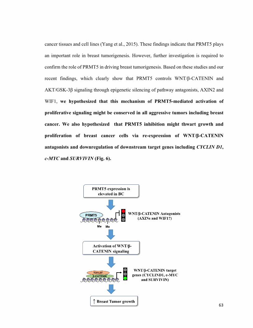
63
cancer tissues and cell lines (Yang et al., 2015). These findings indicate that PRMT5 plays
an important role in breast tumorigenesis. However, further investigation is required to
confirm the role of PRMT5 in driving breast tumorigenesis. Based on these studies and our
recent findings, which clearly show that PRMT5 controls WNT/β-CATENIN and
AKT/GSK-3β signaling through epigenetic silencing of pathway antagonists, AXIN2 and
WIF1, we hypothesized that this mechanism of PRMT5-mediated activation of
proliferative signaling might be conserved in all aggressive tumors including breast
cancer. We also hypothesized that PRMT5 inhibition might thwart growth and
proliferation of breast cancer cells via re-expression of WNT/β-CATENIN
antagonists and downregulation of downstream target genes including CYCLIN D1,
c-MYC and SURVIVIN (Fig. 6).
64
Figure 6: Working hypothesis
PRMT5 expression is elevated in breast cancer and that it suppresses the transcription of
pathway antagonists, AXIN2 and WIF1 by directly binding to their promoter region and
inducing H3R8 and H4R3 symmetrical methylation. As a result, transcription of WNT/β-
CATENIN downstream target genes such as CYCLIN D1, c-MYC and SURVIVIN is
enhanced, which in turn promotes the growth of breast cancer cells.
65
CHAPTER 3. MATERIALS AND METHODS
3.1 Cell culture
MCF7 breast cancer cells, which were originally isolated from metastatic pleural effusion
of an adenocarcinoma patient and express the estrogen receptor, were purchased from
ATCC (ATCC HTB-22TM). HCC1937 triple negative breast cancer cells, which were
isolated from the mammary duct of a grade 3 primary ductal carcinoma patient, were
purchased from ATCC (ATCC CRL-2336TM). BT549 triple negative breast cancer cells
were originally isolated from the mammary gland of a ductal carcinoma patient, and were
also purchased from ATCC (ATCC HTB-122TM).
Human mammary epithelial cells (HMECs) were grown in mammary epithelial cell basal
medium (American type culture collection) supplemented with rH-insulin (5 µg/ml), L-
Glutamine (6 mM), Epinephrine (1 µM), Apo-transferrin (5 µg/ml), rH-TGF-α (5 ng/ml),
pituitary extract (0.4%) and hydrocortisone (100 ng/ml). Human breast cancer cell lines
HCC1937 and BT549 cells were cultured in RPMI 1640 medium (Gibco, Life
Technologies Inc.) supplemented with 10% fetal bovine serum (FBS). BT549 cells were
supplemented with insulin (0.023 U/ml). MCF7 cells were cultured in DMEM medium
(Gibco, Life Technologies Inc.) supplemented with 10% FBS. All the cells were grown at
37°C, in a humidified CO2 incubator supplied with 5% CO2.
3.2 Real Time PCR
Total RNA was extracted using TRIzol reagent (InvitogenTM). Briefly, TNBC cells from
66
a T75 flask were removed using 750 µl of TRIzol reagent, transferred to an eppendorf tube
and incubated at room temperature for 5 min. Next, 300 µl of chloroform was added and
samples were mixed vigorously, and incubated at room temperature for 10 min. Samples
were then centrifuged at 4°C for 15 min before the upper layer containing RNA was
transferred to a new eppendorf tube containing 500 µl of isopropanol. After incubation at
room temperature for 10 min, the reactions were centrifuged at 4°C for 15 min. The RNA
pellet was rinsed with 75% ethanol, air-dried before resuspension in 20 µl of nuclease-free
water. Next, mRNA was converted into cDNA using High-Capacity cDNA Reverse
Transcription Kit as per manufacturer’s instructions (Applied BiosystemsTM). Briefly, 2 µg
of total RNA was reverse transcribed in a 20-µl reaction mixture containing 2.5 µM random
primers, 100 mM dNTP Mix, and Taqman reverse transcription reagents. The reverse
transcription was carried out by first incubating the reaction mixture at 25°C for 10 min,
which was followed by an incubation at 37°C for 2 hrs. Finally, the reaction was terminated
by increasing the temperature to 85°C for 5 min, followed by incubation at 4°C. The cDNA
synthesized was stored at -20°C until further use. To measure the mRNA levels of WNT
target genes and antagonists, real time PCR was carried out in a 20-µl reaction containing
2 µl of cDNA, 10 µl of 2X TaqManTM Universal PCR Master Mix, 1.6 µl each of the reverse
and forward primers (20 picomole/µl), 0.2 µl of Universal probe, and 6.2 µl of RNase free
water. The primer and probe sets used are listed in Table 2. β-ACTIN was used as an
internal control to normalize expression of tested genes.
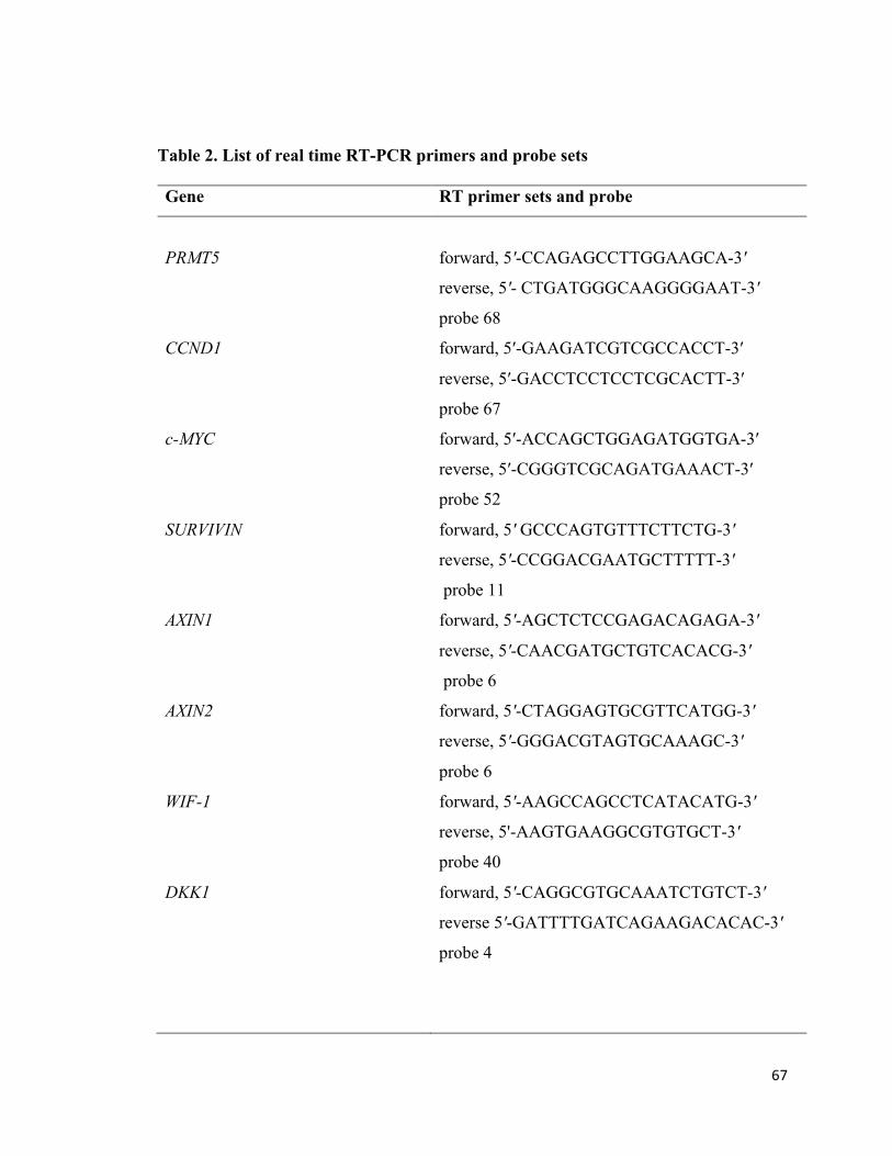
67
Table 2. List of real time RT-PCR primers and probe sets Gene RT primer sets and probe
PRMT5
forward, 5'-CCAGAGCCTTGGAAGCA-3'
reverse, 5'- CTGATGGGCAAGGGGAAT-3'
probe 68
CCND1 forward, 5′-GAAGATCGTCGCCACCT-3′
reverse, 5′-GACCTCCTCCTCGCACTT-3′
probe 67
c-MYC forward, 5′-ACCAGCTGGAGATGGTGA-3′
reverse, 5′-CGGGTCGCAGATGAAACT-3′
probe 52
SURVIVIN forward, 5' GCCCAGTGTTTCTTCTG-3'
reverse, 5'-CCGGACGAATGCTTTTT-3'
probe 11
AXIN1 forward, 5'-AGCTCTCCGAGACAGAGA-3'
reverse, 5'-CAACGATGCTGTCACACG-3'
probe 6
AXIN2 forward, 5'-CTAGGAGTGCGTTCATGG-3'
reverse, 5'-GGGACGTAGTGCAAAGC-3'
probe 6
WIF-1 forward, 5'-AAGCCAGCCTCATACATG-3'
reverse, 5'-AAGTGAAGGCGTGTGCT-3'
probe 40
DKK1 forward, 5'-CAGGCGTGCAAATCTGTCT-3'
reverse 5'-GATTTTGATCAGAAGACACAC-3'
probe 4
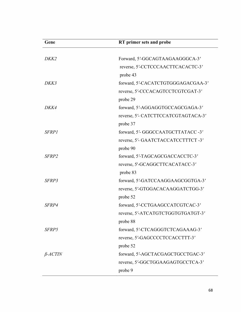
68
Gene RT primer sets and probe
DKK2
Forward, 5'-GGCAGTAAGAAGGGCA-3'
reverse, 5'-CCTCCCAACTTCACACTC-3'
probe 43
DKK3 forward, 5'-CACATCTGTGGGAGACGAA-3'
reverse, 5'-CCCACAGTCCTCGTCGAT-3'
probe 29
DKK4 forward, 5'-AGGAGGTGCCAGCGAGA-3'
reverse, 5'- CATCTTCCATCGTAGTACA-3'
probe 37
SFRP1 forward, 5'- GGGCCAATGCTTATACC -3'
reverse, 5'- GAATCTACCATCCTTTCT -3'
probe 90
SFRP2 forward, 5'-TAGCAGCGACCACCTC-3'
reverse, 5'-GCAGGCTTCACATACC-3'
probe 83
SFRP3 forward, 5'-GATCCAAGGAAGCGGTGA-3'
reverse, 5'-GTGGACACAAGGATCTGG-3'
probe 52
SFRP4 forward, 5'-CCTGAAGCCATCGTCAC-3'
reverse, 5'-ATCATGTCTGGTGTGATGT-3'
probe 88
SFRP5 forward, 5'-CTCAGGGTCTCAGAAAG-3'
reverse, 5'-GAGCCCCTCCACCTTT-3'
probe 52
β-ACTIN forward, 5'-AGCTACGAGCTGCCTGAC-3'
reverse, 5'-GGCTGGAAGAGTGCCTCA-3'
probe 9

69
3.3 Western blot analysis
Whole cell extracts were prepared in radioimmune precipitation assay (RIPA) buffer (50
mM Tris-HCl [pH 7.5], 150 mM NaCl, 1% NP-40, 0.1% SDS, 0.5% sodium deoxycholate,
0.5 mM DTT, 0.5 mM PMSF and 2.5 mM Roche protease inhibitor cocktail). The extracts
were subjected to Western blot analysis as described previously (Chung et al., 2013).
Briefly, 20 to 40 µg of total protein were separated using 7-12% SDS-PAGE, and
transferred to PVDF membrane. The membrane containing transferred proteins was
blocked by incubating with 5% BSA containing 0.05% Tween-20 and incubated overnight
at 4°C with primary antibody to detect CYCLIN D1 (Abcam, ab134175), c-MYC (Abcam,
ab62928), SURVIVIN (Abcam, ab76424), α-TUBULIN (Abcam, ab4074), DKK1
(Abcam, ab109416), DKK3 (Abcam, ab186409), β-ACTIN (Cell Signaling Technology,
4970), CYCLIN D3 (Cell Signaling Technology, DCS22) and PRMT5 (Thermo Fisher,
MA1-25470). After incubation with primary antibody, the membrane was treated with
HRP-conjugated goat anti-mouse (Amersham Biosciences, NA931) or anti-rabbit
(Amersham Biosciences, NA934V) secondary antibody. Next, proteins were visualized
using the ECL detection kit (Amersham, RPN2209) in a Western blot imager (Flurochem
E system, proteinsimple, California, USA).
3.4 Chromatin immunoprecipitation (ChIP) assay
Chromatin immunoprecipitation was carried out as described previously (Chung et al.,
2013). Cross-linked chromatin was resuspended in ChIP lysis buffer (100 mM Tris-HCl
[pH 8.6], 15 mM NaCl, 60 mM KCl, 1 mM CaCl2, 3 mM MgCl2) supplemented with
70
protease inhibitors, Aprotinin (10 µg/ml), PMSF (100 mM), Pepstatin (2.25 µg/ml), and
Leupeptin (10 µg/ml), and fragmented using Q55 sonicator (Qsonica, USA). Sonicated
chromatin was further digested by micrococcal nuclease (MNase) (0.6 Units) treatment at
37°C for 20 min. MNase-treated chromatin was analyzed by agarose gel electrophoresis
to ensure that DNA fragment sizes did not exceed 500 bp. To evaluate PRMT5 recruitment
as well as PRMT5-induced H3R8 and H4R3 symmetric methylation marks, chromatin was
immunoprecipitated overnight at 4°C using either pre-immune or immune antibodies
against PRMT5, H3(Me2)R8 and H4(Me2)R3 in the presence of protein A-Sepharose
beads, which were pre-blocked with sheared salmon sperm DNA (0.2 mg/ml) and BSA
(0.5 mg/ml). The retained complexes were washed successively with mixed micelle buffer
(20 mM Tris-HCl [pH 8.1], 50 mM NaCl, 5 mM EDTA, 5% w/v sucrose, 0.2% Triton X-
100, 0.2% SDS), buffer 250 (50 mM HEPES [pH 7.5], 0.1% sodium deoxycholate, 250
mM NaCl, 1 mM EDTA, 0.2% Triton X-100), and wash buffer (10 mM Tris-HCl [pH 8],
150 mM LiCl, 1 mM EDTA, 0.5% sodium deoxycholate, 0.25% NP-40,). Next, chromatin
was eluted with 200 µl of elution buffer (50 mM Tris-HCl [pH 8.0], 10 mM EDTA, 1%
SDS) at 65°C for 10 min, and cross-links were reversed by incubating the samples at 65°C
overnight. Chromatin was then incubated with proteinase K (5 µg/ml), yeast tRNA (0.5
µg/ml) and TE [pH 7.6] at 37°C for 2 hrs before 50 µl of 4 M LiCl was added, and DNA
was purified by phenol and chloroform extraction. Purified DNA was resuspended in 30 µl
of Tris-EDTA (pH 8.0) and 3 µl of RNase A (10 mg/ml). To amplify promoter sequences
of PRMT5 target genes, approximately 6 µl of eluted DNA was used in a 20-µl real time
PCR reaction mixture containing the primer pairs and specific Roche Applied Science
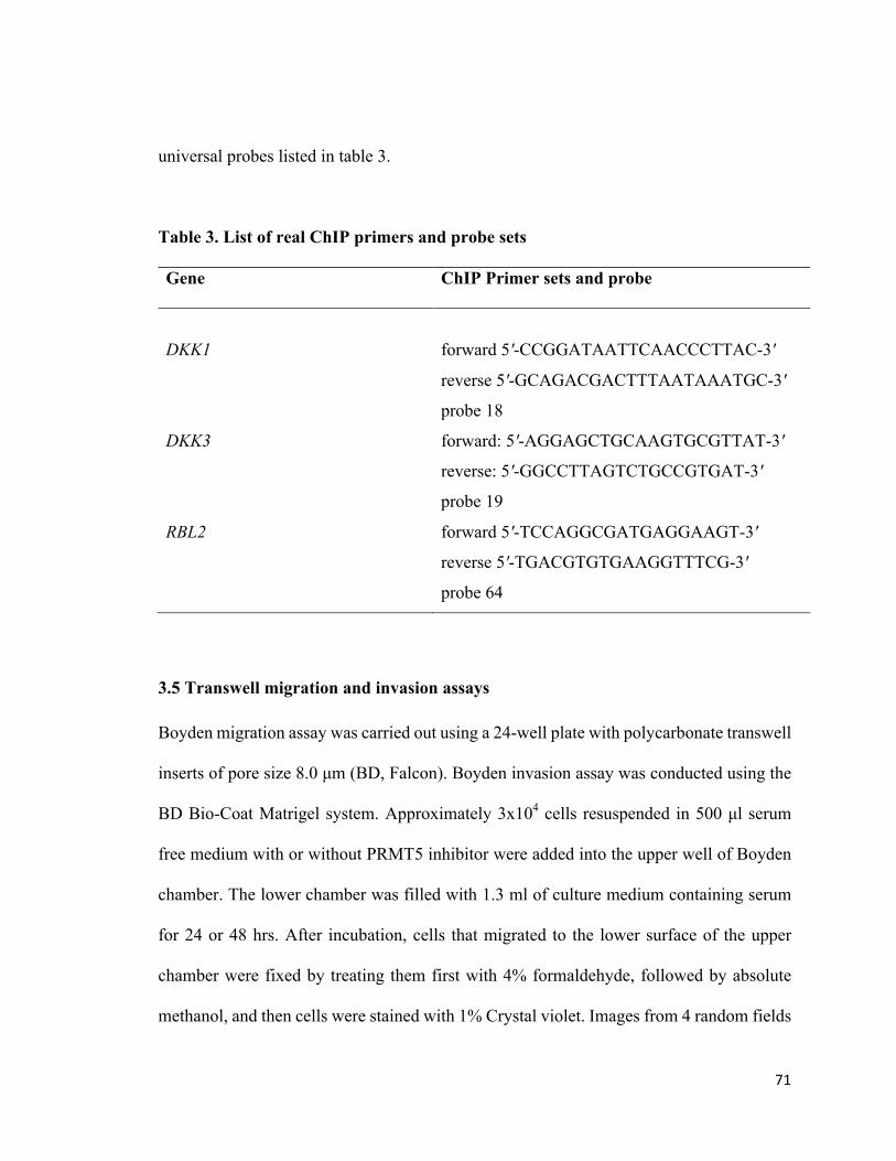
71
universal probes listed in table 3.
Table 3. List of real ChIP primers and probe sets Gene ChIP Primer sets and probe
DKK1
forward 5'-CCGGATAATTCAACCCTTAC-3'
reverse 5'-GCAGACGACTTTAATAAATGC-3'
probe 18
DKK3
forward: 5'-AGGAGCTGCAAGTGCGTTAT-3'
reverse: 5'-GGCCTTAGTCTGCCGTGAT-3'
probe 19
RBL2
forward 5'-TCCAGGCGATGAGGAAGT-3'
reverse 5'-TGACGTGTGAAGGTTTCG-3'
probe 64
3.5 Transwell migration and invasion assays
Boyden migration assay was carried out using a 24-well plate with polycarbonate transwell
inserts of pore size 8.0 µm (BD, Falcon). Boyden invasion assay was conducted using the
BD Bio-Coat Matrigel system. Approximately 3x10⁴ cells resuspended in 500 µl serum
free medium with or without PRMT5 inhibitor were added into the upper well of Boyden
chamber. The lower chamber was filled with 1.3 ml of culture medium containing serum
for 24 or 48 hrs. After incubation, cells that migrated to the lower surface of the upper
chamber were fixed by treating them first with 4% formaldehyde, followed by absolute
methanol, and then cells were stained with 1% Crystal violet. Images from 4 random fields
72
were captured using an inverted microscope, and cells were counted using ImageJ
software.
3.6 Proliferation assay
Approximately 2x10⁴ cells were seeded into a 24-well plate, and 24 hrs later, cells were
treated with or without PRMT5 inhibitor for different time intervals. Next, cells were
collected, resuspended in 20-µl solution containing equal volume of culture medium and
Trypan blue dye, and counted in order to determine the number of viable cells.
3.7 Flow Cytometry
Approximately 6x10⁴ cells were seeded in a 6-well plate prior to treatment with or without
inhibitor for 24 or 48 hrs. Next, cells were harvested, washed and resuspended in 100 µl
of 1X Annexin binding buffer containing 5 µl Annexin V-FITC and 5 µl PI, and then
incubated for 15 min in the dark at room temperature. Stained cells were analyzed with BD
LSRFortessa flow cytometer using FACS Diva software (Becton Dickinson, USA).
3.8 ELISA Assay
Levels of secreted antagonists of the WNT/β-CATENIN pathway were detected by ELISA
technique using DuoSet ELISA kit (R&D system, USA). Essentially 100 µl of capture
antibody was added to 96-well culture plate, and incubated at room temperature overnight.
Next, the antibody solution was removed, and wells were washed twice with 400 µl of
wash buffer (0.005% Tween-20 in filter sterilized 1X PBS). Wells were blocked by
73
incubating with 300 µl of blocking buffer (1% BSA in filter sterilized 1X PBS) for 1 hr at
room temperature. Blocked wells were washed twice with wash buffer before adding 100
µl of either specific standard protein or sample solution. Wells were sealed with adhesive
plastic strips, incubated at room temperature for 2 hr before washing twice with wash
buffer. Next, 100 µl of Streptavidin-HRP A was added to each well and incubated in the
dark for 20 min at room temperature. Wells were then washed, and 100 µl of Substrate
solution containing 1:1 mixture of color reagent A (H2O2) and color reagent B
(Tetramethylbenzidine) was added before incubating at room temperature in the dark for
20 min. After incubation, 50 µl of stop solution (2N H2SO4) was added, and the optical
density of each well was read at 450 nm (TECAN, Infinite 200PRO, Switzerland).
3.9 Statistical analysis
The real-time RT-PCR and ChIP experiments were performed at least two times using
different biological replicates, and the data obtained were represented as mean ± SD.
Statistical validation of the data obtained from multiple samples within different groups
was performed by two-way ANOVA analysis using GraphPad Prism 7 software, and p
values ≤ 0.05 were considered as statistically significant.
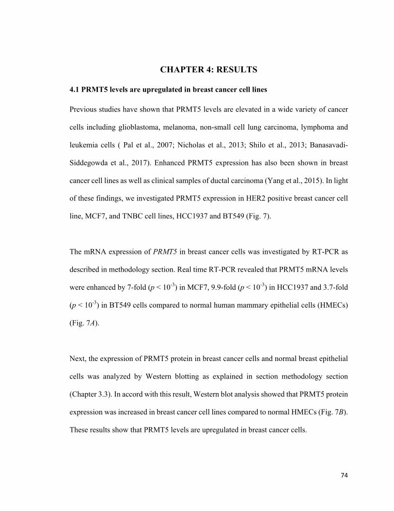
74
CHAPTER 4: RESULTS
4.1 PRMT5 levels are upregulated in breast cancer cell lines
Previous studies have shown that PRMT5 levels are elevated in a wide variety of cancer
cells including glioblastoma, melanoma, non-small cell lung carcinoma, lymphoma and
leukemia cells ( Pal et al., 2007; Nicholas et al., 2013; Shilo et al., 2013; Banasavadi-
Siddegowda et al., 2017). Enhanced PRMT5 expression has also been shown in breast
cancer cell lines as well as clinical samples of ductal carcinoma (Yang et al., 2015). In light
of these findings, we investigated PRMT5 expression in HER2 positive breast cancer cell
line, MCF7, and TNBC cell lines, HCC1937 and BT549 (Fig. 7).
The mRNA expression of PRMT5 in breast cancer cells was investigated by RT-PCR as
described in methodology section. Real time RT-PCR revealed that PRMT5 mRNA levels
were enhanced by 7-fold (p < 10-3) in MCF7, 9.9-fold (p < 10-3) in HCC1937 and 3.7-fold
(p < 10-3) in BT549 cells compared to normal human mammary epithelial cells (HMECs)
(Fig. 7A).
Next, the expression of PRMT5 protein in breast cancer cells and normal breast epithelial
cells was analyzed by Western blotting as explained in section methodology section
(Chapter 3.3). In accord with this result, Western blot analysis showed that PRMT5 protein
expression was increased in breast cancer cell lines compared to normal HMECs (Fig. 7B).
These results show that PRMT5 levels are upregulated in breast cancer cells.
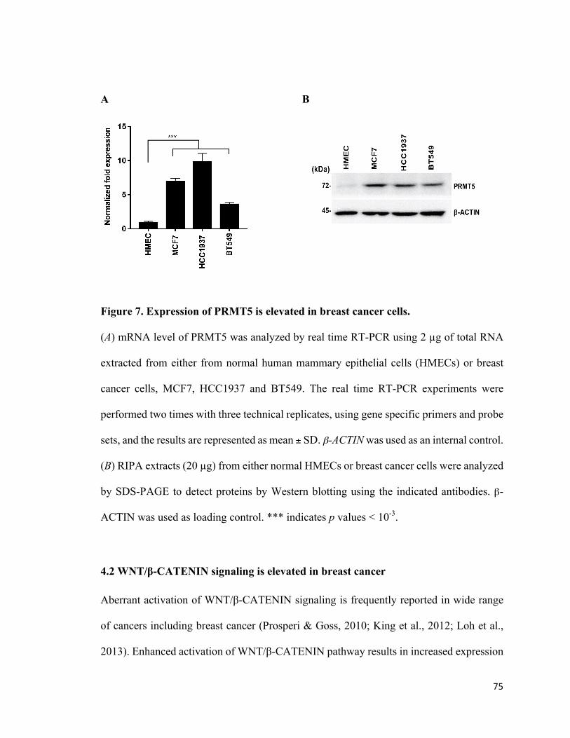
75
A B
Figure 7. Expression of PRMT5 is elevated in breast cancer cells.
(A) mRNA level of PRMT5 was analyzed by real time RT-PCR using 2 µg of total RNA
extracted from either from normal human mammary epithelial cells (HMECs) or breast
cancer cells, MCF7, HCC1937 and BT549. The real time RT-PCR experiments were
performed two times with three technical replicates, using gene specific primers and probe
sets, and the results are represented as mean ± SD. β-ACTIN was used as an internal control.
(B) RIPA extracts (20 µg) from either normal HMECs or breast cancer cells were analyzed
by SDS-PAGE to detect proteins by Western blotting using the indicated antibodies. β-
ACTIN was used as loading control. *** indicates p values < 10-3.
4.2 WNT/β-CATENIN signaling is elevated in breast cancer
Aberrant activation of WNT/β-CATENIN signaling is frequently reported in wide range
of cancers including breast cancer (Prosperi & Goss, 2010; King et al., 2012; Loh et al.,
2013). Enhanced activation of WNT/β-CATENIN pathway results in increased expression
76
of downstream target genes such as CYCLIN D1, c-MYC and SURVIVIV (Barker et al.,
2001; Kramps et al., 2002; H. Ma et al., 2005). Our recent work using aggressive
lymphoma cell lines has shown that PRMT5 promotes WNT/β-CATENIN proliferative
signaling by transcriptionally repressing expression of pathway antagonists, AXIN2 and
WIF1. Consequently, expression of WNT/β-CATENIN target genes such as CYCLIN D1,
c-MYC and SURVIVIN increases (Chung et al., 2019). To investigate whether elevated
levels of PRMT5 regulate WNT/β-CATENIN proliferative signaling in breast cancer, we
checked expression of all three WNT/β-CATENIN pathway target genes in different breast
cancer cell lines (Fig. 8).
Analysis of mRNA expression showed that CYCLIN D1 was increased by 2.5-fold (p < 10-
3) in MCF7, and 2.4-fold (p < 10-3) in HCC1937 compared to control HMECs (Fig. 8A). In
agreement with work by Lin et al. (2000), which showed previously that CYCLIN D1 is
not expressed in BT549 cells, we were unable to detect CYCLIN D1 mRNA in BT549 cells
(Fig. 8A). An initial study by Bartkova et al. (1998) investigated the abundance of D type
CYCLINs in human diploid cells and tumor cell lines, and showed that CYCLIN D3 is the
most widely expressed D-type CYCLIN in most of human diploid cells as well as cancer
cells including BT549. Therefore, we measured CYCLIN D3 mRNA levels in breast cancer
cells. We found that CYCLIN D3 mRNA levels were increased by 2.4-fold (p < 10-3) in
MCF7, 2-fold (p < 10-3) in HCC1937 cells, and 1.8-fold (p < 10-3) in BT549 cells (Fig. 8A).
Furthermore, we found that c-MYC mRNA levels were elevated by 4.3 to 6-fold (p < 10-3)
in breast cancer cells. Similarly, SURVIVIN mRNA was enhanced 12.9 to 49.3-fold (p <
10-3) in all three breast cancer cell lines compared to normal HMECs (Fig. 8A).
77
A B
Figure 8. Expression of WNT/β-CATENIN target genes is elevated in breast cancer
cells
(A) mRNA levels of WNT/β-CATENIN target genes including CYCLIN D1, CYCLIN D3,
c-MYC and SURVIVIN were analyzed by real time RT-PCR using 2 µg of total RNA
extracted either from normal human mammary epithelial cells (HMECs) or breast cancer
cells, MCF7, HCC1937 and BT549. The real time RT-PCR experiments were performed
two times with three technical replicates, using gene specific primers and probe sets, and
the results are represented as mean ± SD. β-ACTIN was used as an internal control. (B)
RIPA extracts (20 µg) from either normal HMECs or breast cancer cells were analyzed by
SDS-PAGE to detect proteins by Western blotting using the indicated antibodies. β-
ACTIN was used as loading control. *** indicates p values < 10-3.
Having found that mRNA levels of WNT/β-CATENIN target genes are increased in
transformed breast cancer cell lines, we checked c-MYC, SURVIVIN, and CYCLIN D1
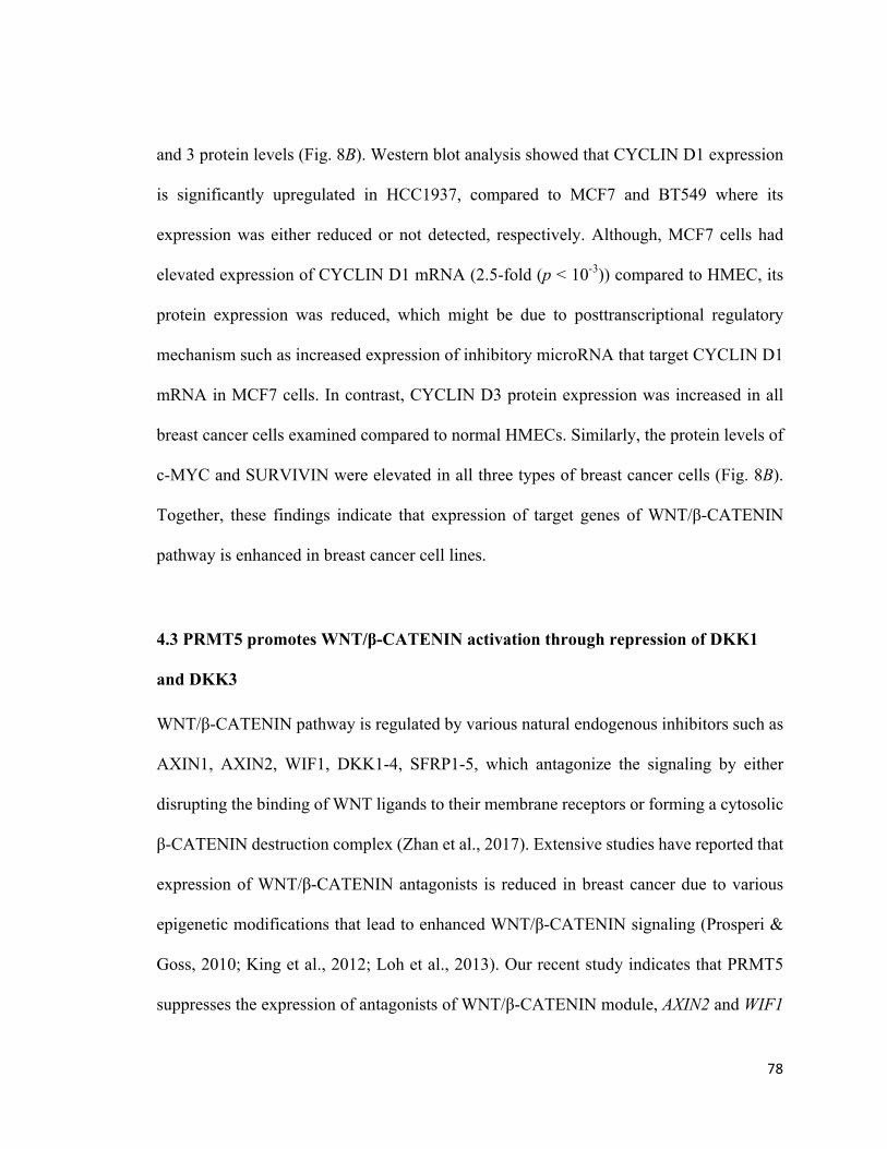
78
and 3 protein levels (Fig. 8B). Western blot analysis showed that CYCLIN D1 expression
is significantly upregulated in HCC1937, compared to MCF7 and BT549 where its
expression was either reduced or not detected, respectively. Although, MCF7 cells had
elevated expression of CYCLIN D1 mRNA (2.5-fold (p < 10-3)) compared to HMEC, its
protein expression was reduced, which might be due to posttranscriptional regulatory
mechanism such as increased expression of inhibitory microRNA that target CYCLIN D1
mRNA in MCF7 cells. In contrast, CYCLIN D3 protein expression was increased in all
breast cancer cells examined compared to normal HMECs. Similarly, the protein levels of
c-MYC and SURVIVIN were elevated in all three types of breast cancer cells (Fig. 8B).
Together, these findings indicate that expression of target genes of WNT/β-CATENIN
pathway is enhanced in breast cancer cell lines.
4.3 PRMT5 promotes WNT/β-CATENIN activation through repression of DKK1
and DKK3
WNT/β-CATENIN pathway is regulated by various natural endogenous inhibitors such as
AXIN1, AXIN2, WIF1, DKK1-4, SFRP1-5, which antagonize the signaling by either
disrupting the binding of WNT ligands to their membrane receptors or forming a cytosolic
β-CATENIN destruction complex (Zhan et al., 2017). Extensive studies have reported that
expression of WNT/β-CATENIN antagonists is reduced in breast cancer due to various
epigenetic modifications that lead to enhanced WNT/β-CATENIN signaling (Prosperi &
Goss, 2010; King et al., 2012; Loh et al., 2013). Our recent study indicates that PRMT5
suppresses the expression of antagonists of WNT/β-CATENIN module, AXIN2 and WIF1
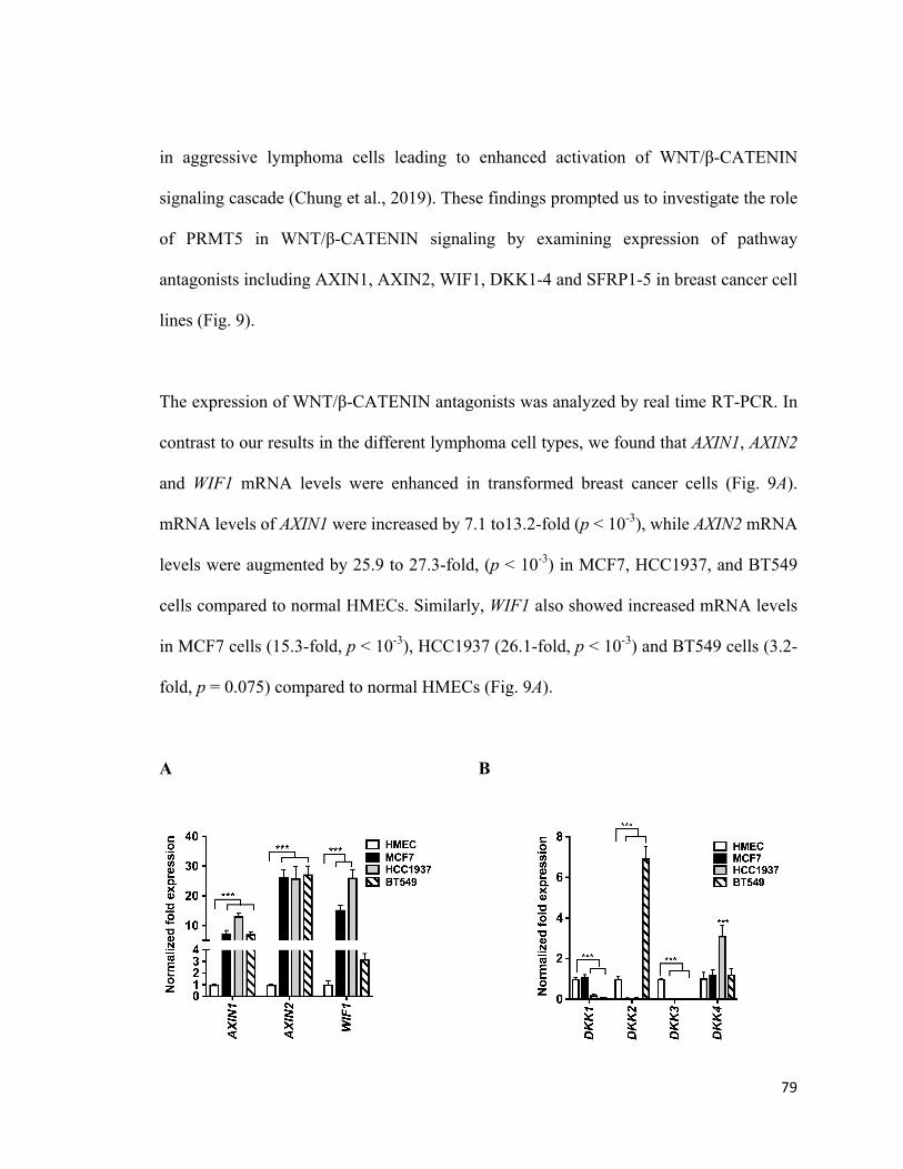
79
in aggressive lymphoma cells leading to enhanced activation of WNT/β-CATENIN
signaling cascade (Chung et al., 2019). These findings prompted us to investigate the role
of PRMT5 in WNT/β-CATENIN signaling by examining expression of pathway
antagonists including AXIN1, AXIN2, WIF1, DKK1-4 and SFRP1-5 in breast cancer cell
lines (Fig. 9).
The expression of WNT/β-CATENIN antagonists was analyzed by real time RT-PCR. In
contrast to our results in the different lymphoma cell types, we found that AXIN1, AXIN2
and WIF1 mRNA levels were enhanced in transformed breast cancer cells (Fig. 9A).
mRNA levels of AXIN1 were increased by 7.1 to13.2-fold (p < 10-3), while AXIN2 mRNA
levels were augmented by 25.9 to 27.3-fold, (p < 10-3) in MCF7, HCC1937, and BT549
cells compared to normal HMECs. Similarly, WIF1 also showed increased mRNA levels
in MCF7 cells (15.3-fold, p < 10-3), HCC1937 (26.1-fold, p < 10-3) and BT549 cells (3.2-
fold, p = 0.075) compared to normal HMECs (Fig. 9A).
A B
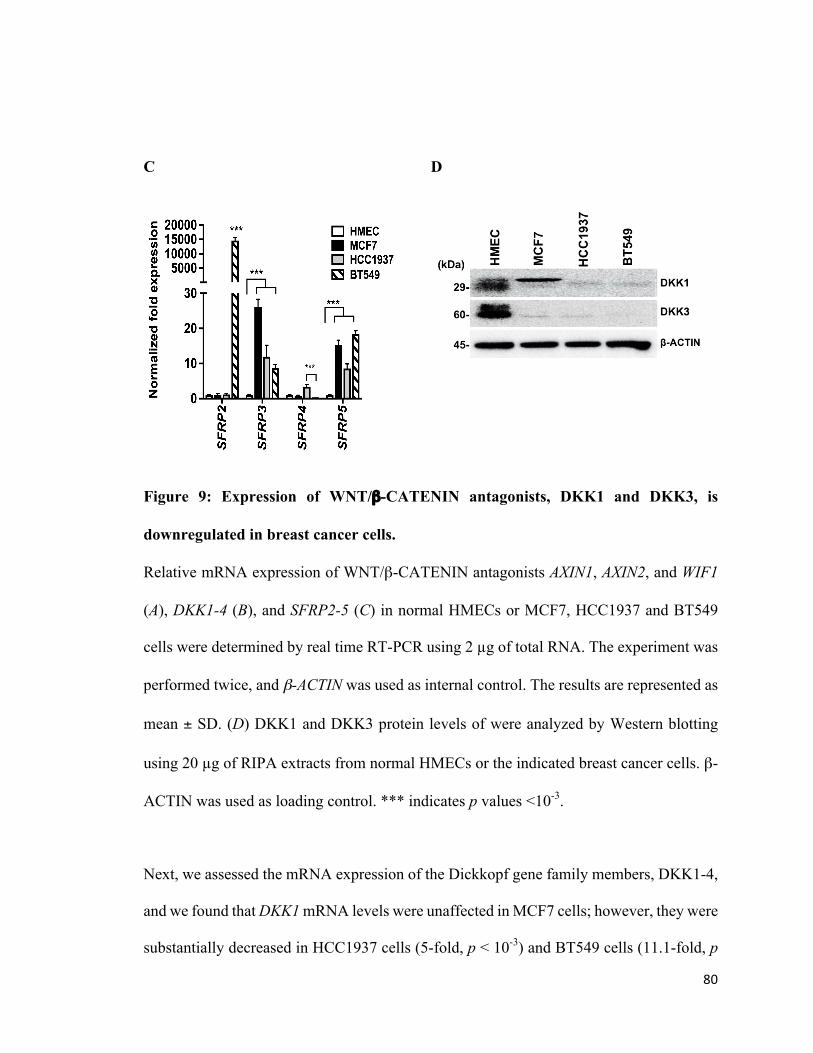
80
C D
Figure 9: Expression of WNT/β-CATENIN antagonists, DKK1 and DKK3, is
downregulated in breast cancer cells.
Relative mRNA expression of WNT/β-CATENIN antagonists AXIN1, AXIN2, and WIF1
(A), DKK1-4 (B), and SFRP2-5 (C) in normal HMECs or MCF7, HCC1937 and BT549
cells were determined by real time RT-PCR using 2 µg of total RNA. The experiment was
performed twice, and β-ACTIN was used as internal control. The results are represented as
mean ± SD. (D) DKK1 and DKK3 protein levels of were analyzed by Western blotting
using 20 µg of RIPA extracts from normal HMECs or the indicated breast cancer cells. β-
ACTIN was used as loading control. *** indicates p values <10-3.
Next, we assessed the mRNA expression of the Dickkopf gene family members, DKK1-4,
and we found that DKK1 mRNA levels were unaffected in MCF7 cells; however, they were
substantially decreased in HCC1937 cells (5-fold, p < 10-3) and BT549 cells (11.1-fold, p
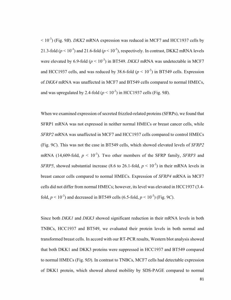
81
< 10-3) (Fig. 9B). DKK2 mRNA expression was reduced in MCF7 and HCC1937 cells by
21.3-fold (p < 10-3) and 21.6-fold (p < 10-3), respectively. In contrast, DKK2 mRNA levels
were elevated by 6.9-fold (p < 10-3) in BT549. DKK3 mRNA was undetectable in MCF7
and HCC1937 cells, and was reduced by 38.6-fold (p < 10-3) in BT549 cells. Expression
of DKK4 mRNA was unaffected in MCF7 and BT549 cells compared to normal HMECs,
and was upregulated by 2.4-fold (p < 10-3) in HCC1937 cells (Fig. 9B).
When we examined expression of secreted frizzled-related proteins (SFRPs), we found that
SFRP1 mRNA was not expressed in neither normal HMECs or breast cancer cells, while
SFRP2 mRNA was unaffected in MCF7 and HCC1937 cells compared to control HMECs
(Fig. 9C). This was not the case in BT549 cells, which showed elevated levels of SFRP2
mRNA (14,609-fold, p < 10-3). Two other members of the SFRP family, SFRP3 and
SFRP5, showed substantial increase (8.6 to 26.1-fold, p < 10-3) in their mRNA levels in
breast cancer cells compared to normal HMECs. Expression of SFRP4 mRNA in MCF7
cells did not differ from normal HMECs; however, its level was elevated in HCC1937 (3.4-
fold, p < 10-3) and decreased in BT549 cells (6.5-fold, p < 10-3) (Fig. 9C).
Since both DKK1 and DKK3 showed significant reduction in their mRNA levels in both
TNBCs, HCC1937 and BT549, we evaluated their protein levels in both normal and
transformed breast cells. In accord with our RT-PCR results, Western blot analysis showed
that both DKK1 and DKK3 proteins were suppressed in HCC1937 and BT549 compared
to normal HMECs (Fig. 9D). In contrast to TNBCs, MCF7 cells had detectable expression
of DKK1 protein, which showed altered mobility by SDS-PAGE compared to normal
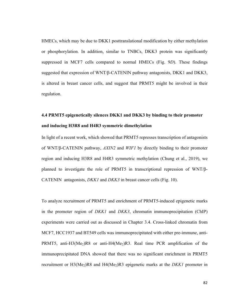
82
HMECs, which may be due to DKK1 posttranslational modification by either methylation
or phosphorylation. In addition, similar to TNBCs, DKK3 protein was significantly
suppressed in MCF7 cells compared to normal HMECs (Fig. 9D). These findings
suggested that expression of WNT/β-CATENIN pathway antagonists, DKK1 and DKK3,
is altered in breast cancer cells, and suggest that PRMT5 might be involved in their
regulation.
4.4 PRMT5 epigenetically silences DKK1 and DKK3 by binding to their promoter
and inducing H3R8 and H4R3 symmetric dimethylation
In light of a recent work, which showed that PRMT5 represses transcription of antagonists
of WNT/β-CATENIN pathway, AXIN2 and WIF1 by directly binding to their promoter
region and inducing H3R8 and H4R3 symmetric methylation (Chung et al., 2019), we
planned to investigate the role of PRMT5 in transcriptional repression of WNT/β-
CATENIN antagonists, DKK1 and DKK3 in breast cancer cells (Fig. 10).
To analyze recruitment of PRMT5 and enrichment of PRMT5-induced epigenetic marks
in the promoter region of DKK1 and DKK3, chromatin immunoprecipitation (ChIP)
experiments were carried out as discussed in Chapter 3.4. Cross-linked chromatin from
MCF7, HCC1937 and BT549 cells was immunoprecipitated with either pre-immune, anti-
PRMT5, anti-H3(Me2)R8 or anti-H4(Me2)R3. Real time PCR amplification of the
immunoprecipitated DNA showed that there was no significant enrichment in PRMT5
recruitment or H3(Me2)R8 and H4(Me2)R3 epigenetic marks at the DKK1 promoter in
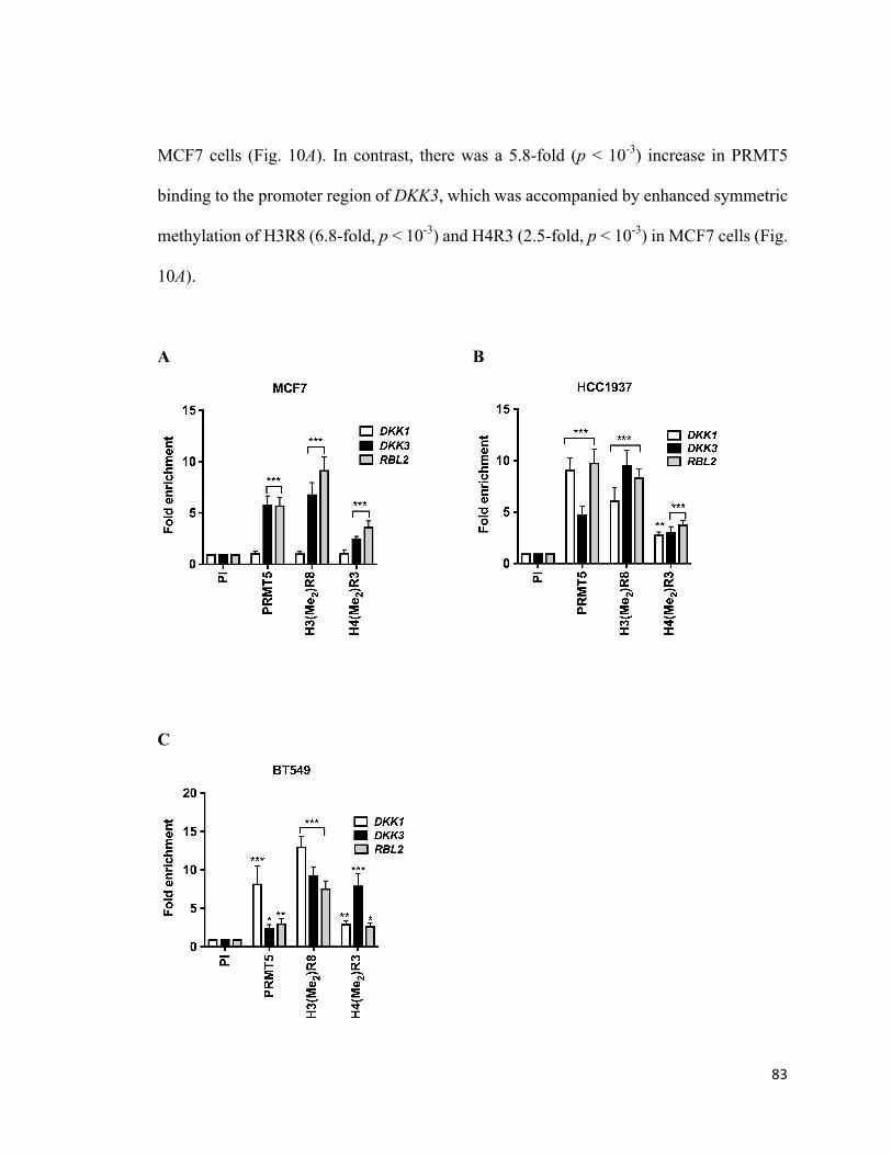
83
MCF7 cells (Fig. 10A). In contrast, there was a 5.8-fold (p < 10-3) increase in PRMT5
binding to the promoter region of DKK3, which was accompanied by enhanced symmetric
methylation of H3R8 (6.8-fold, p < 10-3) and H4R3 (2.5-fold, p < 10-3) in MCF7 cells (Fig.
10A).
A B
C
84
Figure 10: PRMT5 epigenetically suppresses expression of WNT/β-CATENIN
antagonists, DKK1 and DKK3 in breast cancer cells.
ChIP assays were conducted using cross-linked chromatin from either MCF7 (A),
HCC1937 (B) or BT549 (C) cells. Immunoprecipitation of nucleoprotein complexes was
performed using pre-immune (PI), anti-PRMT5, anti-H3(Me2)R8 or anti-H4(Me2)R3
antibodies, and the purified DNA was used to detect the promoter sequences of DKK1 and
DKK3 by real time PCR. ChIP assays were repeated two times with three technical
replicates. The results are represented as mean ± SD. *** indicates p values < 10-3, **
indicates p values < 10-2 and * indicates p value < 10-1.
ChIP assay in HCC1937 cells showed enhanced PRMT5 recruitment (9.2-fold, p < 10-3)
and elevated methylation of H3R8 (6.1-fold, p < 10-3) and H4R3 (2.8-fold, p = 0.001) at
the DKK1 promoter (Fig. 10B). Similarly, PRMT5 recruitment and enrichment of its
epigenetic marks were enhanced by 3.1 to 9.6-fold, (p < 10-3) in the promoter region of
DKK3 (Fig. 10B). PRMT5 recruitment in the promoter region of DKK1 was enriched by
8.2-fold (p < 10-3), whereas PRMT5 binding to the promoter region of DKK3 was increased
by 2.5-fold (p = 0.038) in BT549 cells (Fig. 10C). Consistent with these results, elevated
levels of H3(Me2)R8 (13.1-fold, p < 10-3) and H4(Me2)R3 (3-fold, p = 0.003) were detected
in the promoter region of DKK1. Furthermore, symmetric methylation of H3R8 (9.3-fold,
p < 10-3) and H4R3 (8-fold, p < 10-3) was also detected in the promoter region of DKK3
(Fig. 10C). These results indicate that PRMT5 regulates WNT/β-CATENIN proliferative
signaling through epigenetic transcriptional repression of pathway antagonists, DKK1 and

85
DKK3, in breast cancer cell lines.
4.5 PRMT5 inhibition reduces viability of breast cancer cells
Elevated PRMT5 activity promotes cell proliferation in different types of cancers, whereas
its inhibition is shown to have potential antitumor effect. Previous study by our group has
shown that PRMT5 inhibition using first class small molecule inhibitor, CMP5, reduces
the viability of diffuse large B-cell lymphoma (DLBCL) cells (LC50 value of 20-30 µM)
(Alinari et al., 2015) in vitro. Having found that PRMT5 levels are upregulated in breast
cancer cells, we planned to investigate the impact of PRMT5 inhibition on proliferation of
breast cancer cells in vitro (Fig. 11).
To assess the effect of PRMT5 inhibition on viability of breast cancer cells, we incubated
HMEC, MCF7, HCC1937 and BT549 cells with increasing amounts of CMP5 (2, 4, 8 and
16 µM) for either 24, 48 or 72 hrs. Trypan blue dye exclusion indicated that CMP5 had
limited cytotoxicity on HMECs even at higher concentrations at any given time intervals.
(Fig. 11A). Furthermore, treatment with lower concentrations of CMP5 (2 and 4 µM) did
not reduce breast cancer cell viability significantly at any of the tested time intervals (Fig
11). However, treatment with 8 µM of CMP5 caused a reduction in MCF7 cell viability
by 11.4% (p < 10-3), 39% (p < 10-3) and 61.2% (p < 10-3) after 24, 48 and 72 hrs,
respectively, compared to control DMSO treated cells (Fig. 11B). Treatment with 16 µM
of CMP5 showed significant decrease in viability by 18% (p < 10-3), 46.9% (p < 10-3), and
69.1% (p < 10-3) after 24, 48 and 72 hrs, respectively. Based on these results, we found that
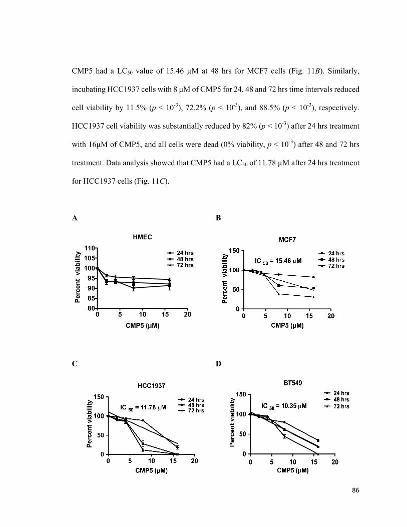
86
CMP5 had a LC50 value of 15.46 µM at 48 hrs for MCF7 cells (Fig. 11B). Similarly,
incubating HCC1937 cells with 8 µM of CMP5 for 24, 48 and 72 hrs time intervals reduced
cell viability by 11.5% (p < 10-3), 72.2% (p < 10-3), and 88.5% (p < 10-3), respectively.
HCC1937 cell viability was substantially reduced by 82% (p < 10-3) after 24 hrs treatment
with 16µM of CMP5, and all cells were dead (0% viability, p < 10-3) after 48 and 72 hrs
treatment. Data analysis showed that CMP5 had a LC50 of 11.78 µM after 24 hrs treatment
for HCC1937 cells (Fig. 11C).
A B
C D
0 5 10 15 2080859095
100105110
CMP5 (µM)
Perc
ent
viab
ility
HMEC
24 hrs48 hrs72 hrs
87
Figure 11. PRMT5 inhibition reduces the viability of breast cancer cells
Normal HMEC (A), MCF7 (B), HCC1937 (C) and BT549 (D) were incubated with
different concentrations of CMP5 for 24, 48 and 72 hrs. Cell viability was measured by
Trypan blue dye exclusion assay.
Treating BT549 cells with 8 µM of CMP5 reduced their viability by 20% (p < 10-3), 38%
(p < 10-3), and 55.6% (p < 10-3) after 24, 48 and 72hrs post-treatment, respectively. In
addition, treatment with 16 µM of CMP5 reduced viability of BT549 cells by 65.5% (p <
10-3), and 82% (p < 10-3) after 24 and 48 hrs, respectively. Treatment for 72 hrs with 16µM
of CMP5 resulted in complete loss of cell viability. When we plotted the results, we found
that CMP5 had a LC50 of 10.35 µM at 48 hrs for BT549 cells (Fig. 11D). Taken together,
these findings indicate that PRMT5 inhibition selectively reduces the proliferation of breast
tumor cells that overexpress PRMT5 without inducing any significant toxicity to normal
breast epithelial cells.
4.6 Inhibition of PRMT5 derepresses DKK1 and DKK3 in BT549 cells
Our recent study showed that inhibiting PRMT5 either using specific small molecule
inhibitor, CMP5, or shRNA induces transcriptional transcriptional derepression of pathway
antagonists AXIN2 and WIF1 in lymphoma cells. Based on this study, and our results,
which indicate that PRMT5 protein levels are inversely correlated with WNT/β-CATENIN
antagonists, we wanted to assess the effect of inhibition of PRMT5 on expression of DKK1
and DKK3 in TNBCs (Fig. 12).
88
HCC1937 and BT549 cells were treated with increasing concentrations of CMP5 for 24 or
48 hrs, respectively, and total RNA was analyzed. Real time RT-PCR data indicated that
transcription of DKK1 was induced 4 to 4.3-fold (p < 10-2) at lower concentrations of
CMP5 (3 µM to 9 µM), and was significantly upregulated by 24-fold (p < 10-3) at 12 µM
in BT549 cells (Fig. 12A). Similarly, DKK3 mRNA levels were upregulated by 5.6-fold (p
< 10-3) in the presence of 12 µM of CMP5 in BT549 cells (Fig. 12A). The mRNA
expression of DKK1 and DKK3 were unaffected in HCC1937 cells (Fig. 12B), implying
that alternative mechanisms might be involved in their regulation. As control, the
expression of PRMT5 mRNA was also analyzed, and were found to be unaffected in the
presence of CMP5 (Fig. 12A and B). To determine the biological significance of DKK1
and DKK3 transcriptional derepression, we measured the levels of DKK1 and DKK3
proteins in BT549 cells treated with CMP5 (Fig. 12C). ELISA analysis showed that both
DKK1 and DKK3 were derepressed by 2 and 1.4-fold (p < 10-3), respectively (Fig. 12C).
Consistent with the real time RT-PCR results, levels of DKK1 and DKK3 proteins were
unchanged in HCC1937 cells (Fig. 12D).
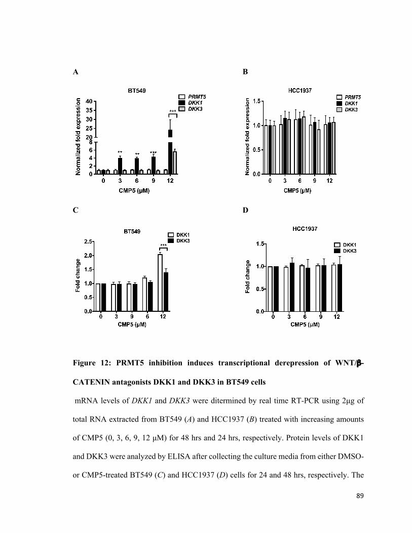
89
A B
C D
Figure 12: PRMT5 inhibition induces transcriptional derepression of WNT/β-
CATENIN antagonists DKK1 and DKK3 in BT549 cells
mRNA levels of DKK1 and DKK3 were ditermined by real time RT-PCR using 2µg of
total RNA extracted from BT549 (A) and HCC1937 (B) treated with increasing amounts
of CMP5 (0, 3, 6, 9, 12 µM) for 48 hrs and 24 hrs, respectively. Protein levels of DKK1
and DKK3 were analyzed by ELISA after collecting the culture media from either DMSO-
or CMP5-treated BT549 (C) and HCC1937 (D) cells for 24 and 48 hrs, respectively. The
90
results are represented as mean ± SD. *** indicates p values < 10-3 and ** indicates p
values < 10-2.
4.7 Inhibition of PRMT5 in combination with HDACs and DNMT3A derepresses
DKK1 and DKK3 in HCC1937 cells
Since PRMT5 inhibition could not restore expression of DKK1 and DKK3 in HCC1937
cells (Fig. 12B and D), we investigated other epigenetic mechanisms that could be involved
in their transcriptional regulation. PRMT5 has previously been shown to work in concert
with DNA methyltransferase 3A (DNMT3A) ( Zhao et al., 2009; Liu et al., 2018).
Therefore, we reasoned that PRMT5 might be promoting DNA methylation of DKK1 and
DKK3 promoters in HCC1937 cells (Fig. 13).
To address whether DNA methylation is involved in transcriptional regulation of DKK1
and DKK3, we treated HCC1937 cells with the DNA methyltransferase inhibitor, 5-
Azacytidine (5-Aza). HCC1937 cells were incubated with increasing amounts of 5-Aza
for either 24, 48 or 72 hrs, and viability was determined by Trypan blue dye exclusion
assay, which showed that even a higher concentration of 5-Aza (24 µM) does not have
significant cytotoxicity on HCC1937 cells (Fig. 13A). Next, we examined the effect of 5-
Aza (24 µM) alone or in combination with suboptimal amount of CMP5 (9 µM), on
transcriptional derepression of DKK1 and DKK3 in HCC1937 cells. Treatment of
HCC1937 cells with 24 µM of 5-Azacytidine for 48 hrs did not impact transcription of
DKK1 (1.75-fold, p < 10-3) and DKK3 (1.5-fold, p < 10-3). Furthermore, sequential
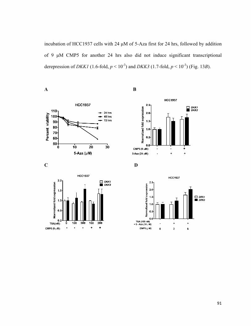
91
incubation of HCC1937 cells with 24 µM of 5-Aza first for 24 hrs, followed by addition
of 9 µM CMP5 for another 24 hrs also did not induce significant transcriptional
derepression of DKK1 (1.6-fold, p < 10-3) and DKK3 (1.7-fold, p < 10-3) (Fig. 13B).
A B
C D
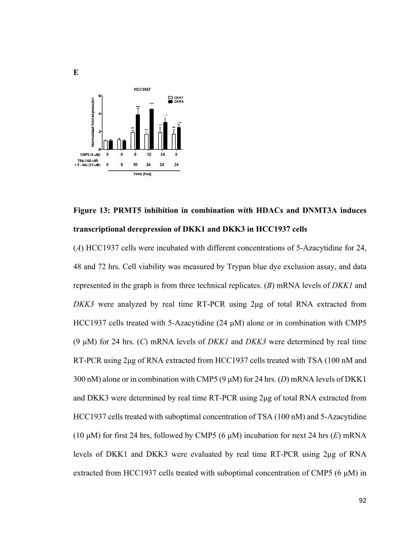
92
E
Figure 13: PRMT5 inhibition in combination with HDACs and DNMT3A induces
transcriptional derepression of DKK1 and DKK3 in HCC1937 cells
(A) HCC1937 cells were incubated with different concentrations of 5-Azacytidine for 24,
48 and 72 hrs. Cell viability was measured by Trypan blue dye exclusion assay, and data
represented in the graph is from three technical replicates. (B) mRNA levels of DKK1 and
DKK3 were analyzed by real time RT-PCR using 2µg of total RNA extracted from
HCC1937 cells treated with 5-Azacytidine (24 µM) alone or in combination with CMP5
(9 µM) for 24 hrs. (C) mRNA levels of DKK1 and DKK3 were determined by real time
RT-PCR using 2µg of RNA extracted from HCC1937 cells treated with TSA (100 nM and
300 nM) alone or in combination with CMP5 (9 µM) for 24 hrs. (D) mRNA levels of DKK1
and DKK3 were determined by real time RT-PCR using 2µg of total RNA extracted from
HCC1937 cells treated with suboptimal concentration of TSA (100 nM) and 5-Azacytidine
(10 µM) for first 24 hrs, followed by CMP5 (6 µM) incubation for next 24 hrs (E) mRNA
levels of DKK1 and DKK3 were evaluated by real time RT-PCR using 2µg of RNA
extracted from HCC1937 cells treated with suboptimal concentration of CMP5 (6 µM) in
93
combination with TSA (100 nM) and 5-Azacytidine (10 µM) for the indicated time
intervals. All the experiments were repeated two times with three technical replicates. The
results are represented as mean ± SD. *** indicates p values < 10-3 and ** indicates p
values < 10-2.
Deacetylation of histone lysine residues by histone deacetylase enzymes (HDAC) has been
shown to be essential for transcriptional silencing of genes (Gallinari et al., 2007). An
earlier study by our group showed that PRMT5 can associate with mSin3A/histone
deacetylase 2 (HDAC2) to form a transcriptional repression complex with the BRG1-based
hSWI/SNF chromatin remodelers (Pal et al., 2004). In addition, we have previously shown
that PRMT5 associates with an NF-κB transcriptional repressor complex containing
HDAC3, which promotes deactylation of H3K14 lysine, and triggers gene silencing in
lymphoma cells (Alinari et al., 2015). To investigate whether histone deacetylation is
involved in transcriptional repression of WNT antagonists, DKK1 and DKK3, we treated
HCC1937 cells with Trichostatin A (TSA). Real time RT-PCR analysis showed that
treatment with either 100 nM or 300 nM of TSA for 24 hrs had no effect on DKK1 and
DKK3 transcription in HCC1937 cells. Similarly, combinatorial treatment with 9 µM of
CMP5 and either 100 or 300 nM of TSA for 24 hrs also failed to induce transcriptional
activation of WNT antagonists, DKK1 and DKK3 in these cells (Fig. 13C).
Previous studies have shown that simultaneous inhibition of histone deacetylase enzyme
and DNA methyltransferase enzyme results in restoration of epigenetically silenced genes
94
(Bartoli et al., 2003; Tang et al., 2006). Therefore, we investigated the effect of
combinatorial treatment of TSA and 5-Aza in inducing transcriptional derepression of
DKK1 and DKK3 in HCC1937 cells. We incubated HCC1937 cells with a combination of
suboptimal doses of TSA (100 nM) and 5-Aza (10 µM) for 24 hrs (Fig. 13E). Real time
RT-PCR data showed that this treatment increased mRNA level of DKK1 and DKK3 by
1.8-fold (p=0.006) and 2.5-fold (p < 10-3), respectively (Fig. 13E). Since histone lysine
deacetylation, histone arginine methylation, and DNA methylation can all work together
to induce gene silencing, we hypothesized that combinatorial treatment with inhibitors
against HDAC, PRMT5 and DNMT3A might enhance transcriptional derepression of
DKK1 and DKK3 in HCC1937 cells as compared to treatment with CMP5 in the presence
of either TSA or 5-Aza. First, we treated HCC1937 cells with 100 nM of TSA and 10 µM
of 5-Aza for 24 hrs, followed by incubation with 9 µM of CMP5 for an additional 24 hrs.
The results of this study showed that mRNA level of DKK1 and DKK3 by 1.6-fold and 2-
fold, respectively (Fig. 13D), suggesting that combinational treatment with all three
inhibitors was not able to induce further expression of DKK1 and DKK3 in these cells.
Since, PRMT5-induced H4R3 histone methylation is known to direct binding of
DNMT3A, and induce gene silencing (Zhao et al., 2009), we tested the effect of an initial
treatment with CMP5, followed by addition of TSA and 5-Aza in HCC1937 cells. We
treated HCC1937 cells with 6 µM of CMP5 for 4 hrs, and then added 100 nM of TSA and
10 µM of 5-Aza for an additional 8 hrs. Results from this experiment showed that DKK1
and DKK3 expression was not affected (Fig. 13E). Next, we treated HCC1937 cells with
CMP5 (6 µM) for 8 hrs, followed by an incubation with TSA (100 nM) and 5-Aza (10 µM)
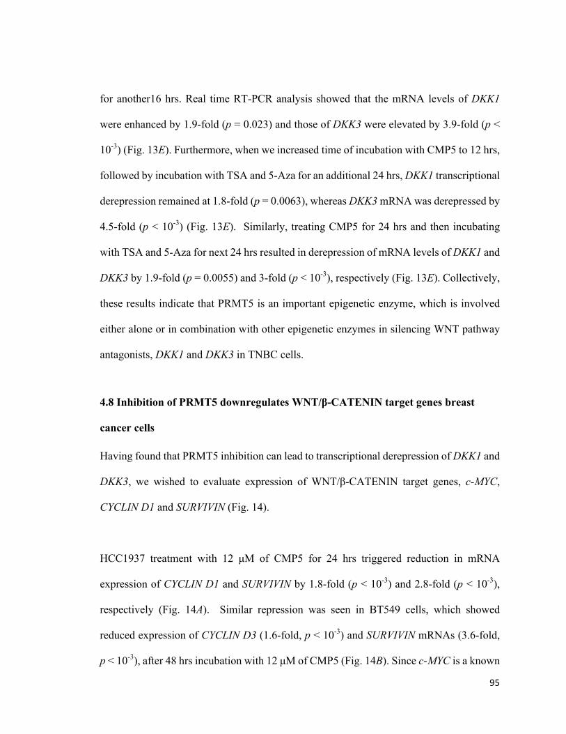
95
for another16 hrs. Real time RT-PCR analysis showed that the mRNA levels of DKK1
were enhanced by 1.9-fold (p = 0.023) and those of DKK3 were elevated by 3.9-fold (p <
10-3) (Fig. 13E). Furthermore, when we increased time of incubation with CMP5 to 12 hrs,
followed by incubation with TSA and 5-Aza for an additional 24 hrs, DKK1 transcriptional
derepression remained at 1.8-fold (p = 0.0063), whereas DKK3 mRNA was derepressed by
4.5-fold (p < 10-3) (Fig. 13E). Similarly, treating CMP5 for 24 hrs and then incubating
with TSA and 5-Aza for next 24 hrs resulted in derepression of mRNA levels of DKK1 and
DKK3 by 1.9-fold (p = 0.0055) and 3-fold (p < 10-3), respectively (Fig. 13E). Collectively,
these results indicate that PRMT5 is an important epigenetic enzyme, which is involved
either alone or in combination with other epigenetic enzymes in silencing WNT pathway
antagonists, DKK1 and DKK3 in TNBC cells.
4.8 Inhibition of PRMT5 downregulates WNT/β-CATENIN target genes breast
cancer cells
Having found that PRMT5 inhibition can lead to transcriptional derepression of DKK1 and
DKK3, we wished to evaluate expression of WNT/β-CATENIN target genes, c-MYC,
CYCLIN D1 and SURVIVIN (Fig. 14).
HCC1937 treatment with 12 µM of CMP5 for 24 hrs triggered reduction in mRNA
expression of CYCLIN D1 and SURVIVIN by 1.8-fold (p < 10-3) and 2.8-fold (p < 10-3),
respectively (Fig. 14A). Similar repression was seen in BT549 cells, which showed
reduced expression of CYCLIN D3 (1.6-fold, p < 10-3) and SURVIVIN mRNAs (3.6-fold,
p < 10-3), after 48 hrs incubation with 12 µM of CMP5 (Fig. 14B). Since c-MYC is a known
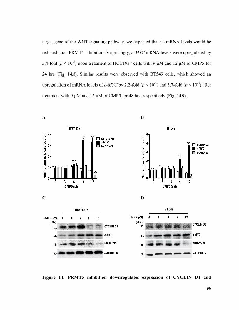
96
target gene of the WNT signaling pathway, we expected that its mRNA levels would be
reduced upon PRMT5 inhibition. Surprisingly, c-MYC mRNA levels were upregulated by
3.4-fold (p < 10-3) upon treatment of HCC1937 cells with 9 µM and 12 µM of CMP5 for
24 hrs (Fig. 14A). Similar results were observed with BT549 cells, which showed an
upregulation of mRNA levels of c-MYC by 2.2-fold (p < 10-3) and 3.7-fold (p < 10-3) after
treatment with 9 µM and 12 µM of CMP5 for 48 hrs, respectively (Fig. 14B).
A B
C D
Figure 14: PRMT5 inhibition downregulates expression of CYCLIN D1 and
97
SURVIVIN in TNBC cells.
HCC1937 (A) and BT549 (B) TNBC cells were treated with increasing amounts of CMP5
(0, 3, 6, 9, 12 µM) and total RNA was extracted 24 hrs and 48 hrs post-treatment,
respectively. Expression of WNT/β-CATENIN target genes were analyzed by real time
RT-PCR using gene specific primers and probe sets. The experiment was repeated two
times with three technical replicates and β-ACTIN was used as an internal control. The data
shown in the graph represent the mean for each concentration ± SD. Approximately 20 µg
of RIPA extracts from either treated or non-treated HCC1937 (C) and BT549 (D) cells
were analyzed by immunoblotting using the indicated antibodies. ⍺-TUBULIN was
detected to show equal loading. *** indicates p values < 10-3, and ** indicates p value <
10-2.
Next, we examined the impact of PRMT5 inhibition on protein expression of WNT/β-
CATENIN target genes in TNBCs. In accord with the RT-PCR data, CYCLIN D1 and
SURVIVIN protein levels decreased in HCC1937 cells after treatment with either 9 or 12
µM of CMP5 for 24 hrs (Fig. 14C). BT549 cells also showed significant reduction in
CYCLIN D3 and SURVIVIN protein expression after 48 hrs incubation with 12 µM of
CMP5 (Fig. 14D). In stark contrast, the levels of c-MYC protein showed an increase with
either 9 or 12µM of CMP5 treatment in both TNBCs (Fig. 14C and D). These results
indicate that through its ability to regulate WNT/β-CATENIN signaling, PRMT5 controls
expression of key survival and cell cycle regulators including CYCLIN D1, CYCLIN D3
and SURVIVIN in TNBC cells.
98
4.9 PRMT5 inhibition alters its recruitment and H3R8 and H4R3 symmetric
methylation in the promoter region of DKK1 and DKK3
In our recent study, we have seen that CMP5 treatment reduces PRMT5 recruitment as well
as PRMT5-induced epigenetic marks on the promoter region of WNT pathway antagonists,
AXIN2 and WIF1 in lymphoma (Chung et al., 2019). In light of these findings, we
investigated the impact of inhibition of PRMT5 on recruitment of PRMT5 as well as
methylation of H3R8 and H4R3 in the promoter regions of DKK1 and DKK3 in TNBCs
(Fig. 15)
Treatment of BT549 with PRMT5 inhibitor resulted in DKK1 and DKK3 transcriptional
derepression (Fig. 12A). Therefore, we conducted ChIP experiments to monitor PRMT5
recruitment as well as methylation of H3R8 and H4R3 in the promoter region of DKK1
and DKK3 in the presence and absence of CMP5 (Fig. 15). ChIP analysis revealed that
PRMT5 recruitment was increased by 7.4-fold (p < 10-3) and 2.3-fold (p < 10-3) in the
promoter regions of DKK1 and DKK3, respectively, in control DMSO-treated BT549 cells.
Consistent with these results, symmetric methylation of H3R8 was enriched by 9.6 (p < 10-
3) and 7.1-fold (p < 10-3) in the promoter of DKK1 and DKK3, respectively. Similarly,
methylation of H4R3 histones in the promoter region of DKK1 and DKK3 was increased
by 2.6 to 6.3-fold (p < 10-3). When BT549 cells were treated with CMP5, there was a 3.4-
fold (p < 10-3) decrease in PRMT5, which was accompanied by a 2.6-fold (p < 10-3) and
1.5-fold (p = 0.072) reduced methylation of H3R8 and H4R3, respectively in the DKK1
promoter (Fig. 15A and B). In addition, BT549 cells showed that CMP5 treatment resulted
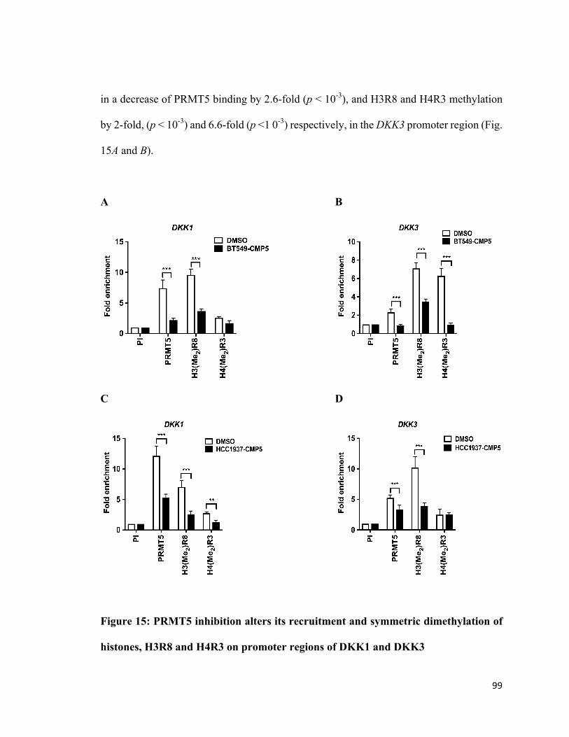
99
in a decrease of PRMT5 binding by 2.6-fold (p < 10-3), and H3R8 and H4R3 methylation
by 2-fold, (p < 10-3) and 6.6-fold (p <1 0-3) respectively, in the DKK3 promoter region (Fig.
15A and B).
A B
C D
Figure 15: PRMT5 inhibition alters its recruitment and symmetric dimethylation of
histones, H3R8 and H4R3 on promoter regions of DKK1 and DKK3
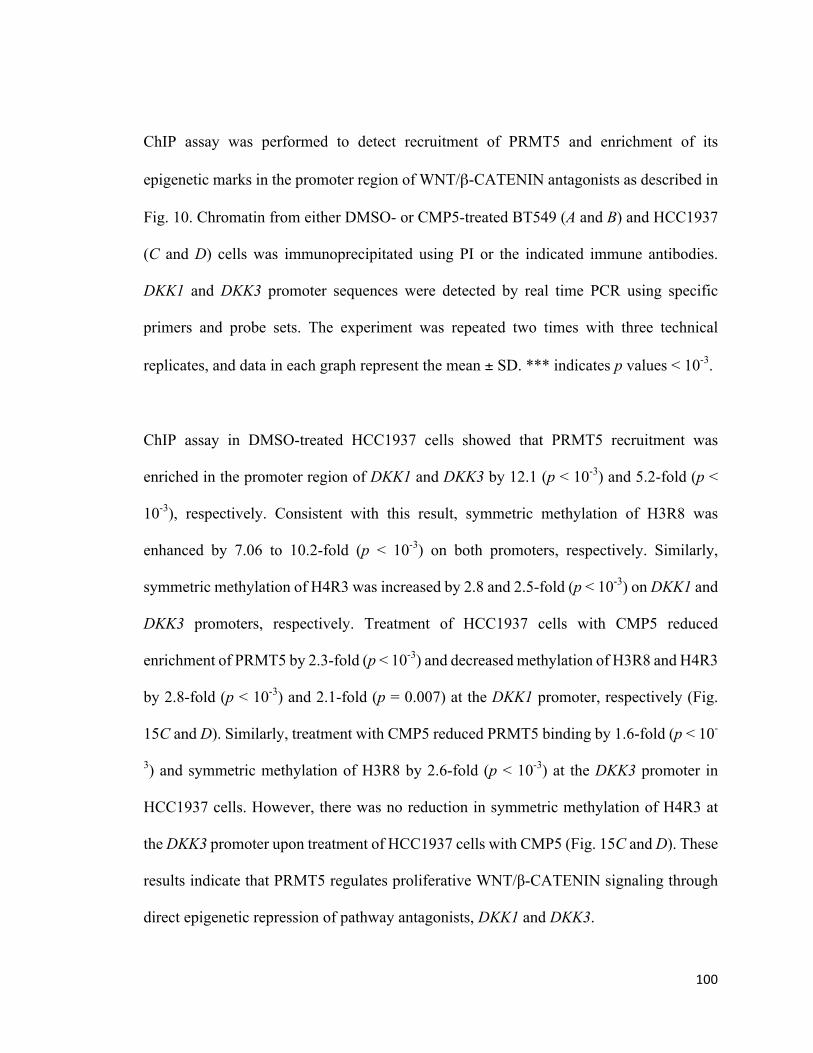
100
ChIP assay was performed to detect recruitment of PRMT5 and enrichment of its
epigenetic marks in the promoter region of WNT/β-CATENIN antagonists as described in
Fig. 10. Chromatin from either DMSO- or CMP5-treated BT549 (A and B) and HCC1937
(C and D) cells was immunoprecipitated using PI or the indicated immune antibodies.
DKK1 and DKK3 promoter sequences were detected by real time PCR using specific
primers and probe sets. The experiment was repeated two times with three technical
replicates, and data in each graph represent the mean ± SD. *** indicates p values < 10-3.
ChIP assay in DMSO-treated HCC1937 cells showed that PRMT5 recruitment was
enriched in the promoter region of DKK1 and DKK3 by 12.1 (p < 10-3) and 5.2-fold (p <
10-3), respectively. Consistent with this result, symmetric methylation of H3R8 was
enhanced by 7.06 to 10.2-fold (p < 10-3) on both promoters, respectively. Similarly,
symmetric methylation of H4R3 was increased by 2.8 and 2.5-fold (p < 10-3) on DKK1 and
DKK3 promoters, respectively. Treatment of HCC1937 cells with CMP5 reduced
enrichment of PRMT5 by 2.3-fold (p < 10-3) and decreased methylation of H3R8 and H4R3
by 2.8-fold (p < 10-3) and 2.1-fold (p = 0.007) at the DKK1 promoter, respectively (Fig.
15C and D). Similarly, treatment with CMP5 reduced PRMT5 binding by 1.6-fold (p < 10-
3) and symmetric methylation of H3R8 by 2.6-fold (p < 10-3) at the DKK3 promoter in
HCC1937 cells. However, there was no reduction in symmetric methylation of H4R3 at
the DKK3 promoter upon treatment of HCC1937 cells with CMP5 (Fig. 15C and D). These
results indicate that PRMT5 regulates proliferative WNT/β-CATENIN signaling through
direct epigenetic repression of pathway antagonists, DKK1 and DKK3.
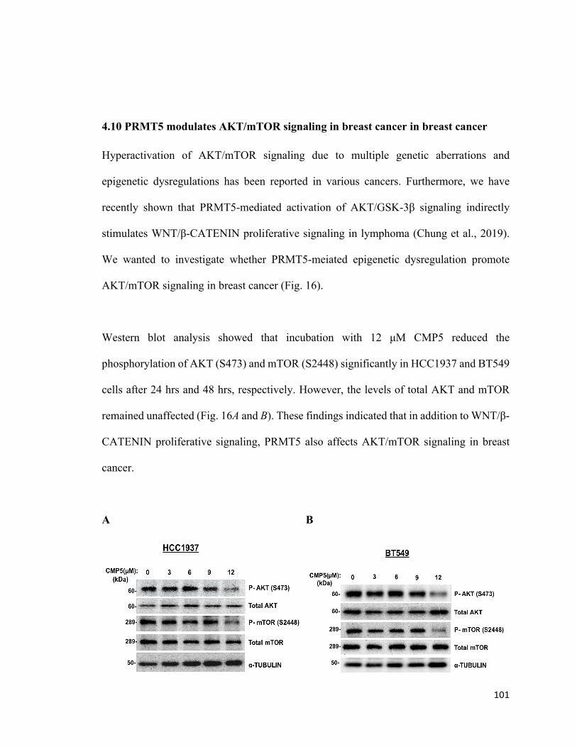
101
4.10 PRMT5 modulates AKT/mTOR signaling in breast cancer in breast cancer
Hyperactivation of AKT/mTOR signaling due to multiple genetic aberrations and
epigenetic dysregulations has been reported in various cancers. Furthermore, we have
recently shown that PRMT5-mediated activation of AKT/GSK-3β signaling indirectly
stimulates WNT/β-CATENIN proliferative signaling in lymphoma (Chung et al., 2019).
We wanted to investigate whether PRMT5-meiated epigenetic dysregulation promote
AKT/mTOR signaling in breast cancer (Fig. 16).
Western blot analysis showed that incubation with 12 µM CMP5 reduced the
phosphorylation of AKT (S473) and mTOR (S2448) significantly in HCC1937 and BT549
cells after 24 hrs and 48 hrs, respectively. However, the levels of total AKT and mTOR
remained unaffected (Fig. 16A and B). These findings indicated that in addition to WNT/β-
CATENIN proliferative signaling, PRMT5 also affects AKT/mTOR signaling in breast
cancer.
A B
102
Figure 16: PRMT5 inhibition downregulates AKT/mTOR signaling in breast cancer
RIPA extracts (20 µg) from HCC1937 (A) and BT549 (B) cells treated with PRMT5
inhibitor, CMP5 (0, 3, 6, 9, 12 µM) for 24 hrs and 48 hrs, respectively were analyzed by
Western blotting to evaluate the levels of phospho-AKT (S473) and total AKT as well as
phospho-mTOR (S2448) and total mTOR. α-TUBULIN was detected to represent equal
loading.
4.11 PRMT5 is required for migration and invasion of TNBC cells
Enhanced migration is an intrinsic property of cancer cells, which also helps metastasis, a
hallmark of triple negative breast cancers. Having found that CMP5 inhibits the
proliferation of breast cancer cells we planned to discover the effect of PRMT5 inhibition
on breast cancer cell migration and invasion in vitro by migration assay and invasion assay,
respectively as described in methodology section (Fig. 17).
Treatment of MCF7, HCC1937 and BT549 with CMP5 at their respective LC50 dose,
showed that migration of MCF7 and HCC1937 cells was completely inhibited compared
to control DMSO-treated cells. However, migration of BT549 cells was reduced by 58.6%
(p < 10-3) (Fig. 17A and B). To assess if PRMT5 inhibition can impact invasiveness of
breast cancer cells, we utilized Boyden invasion assay. Our findings showed that PRMT5
inhibition using the respective LC50 completely abolished invasion by MCF7, HCC1937
and BT549 (Fig. 17C and D).
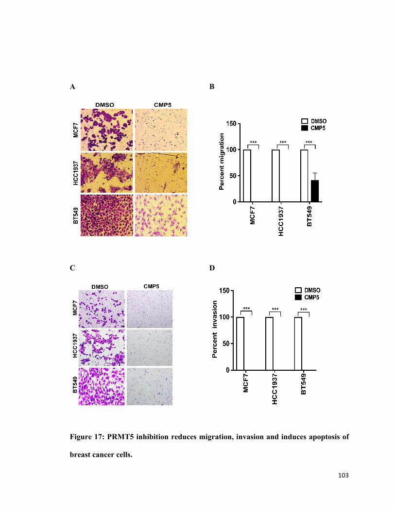
103
A B
C D
Figure 17: PRMT5 inhibition reduces migration, invasion and induces apoptosis of
breast cancer cells.
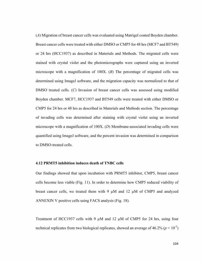
104
(A) Migration of breast cancer cells was evaluated using Matrigel coated Boyden chamber.
Breast cancer cells were treated with either DMSO or CMP5 for 48 hrs (MCF7 and BT549)
or 24 hrs (HCC1937) as described in Materials and Methods. The migrated cells were
stained with crystal violet and the photomicrographs were captured using an inverted
microscope with a magnification of 100X. (B) The percentage of migrated cells was
determined using ImageJ software, and the migration capacity was normalized to that of
DMSO treated cells. (C) Invasion of breast cancer cells was assessed using modified
Boyden chamber. MCF7, HCC1937 and BT549 cells were treated with either DMSO or
CMP5 for 24 hrs or 48 hrs as described in Materials and Methods section. The percentage
of invading cells was determined after staining with crystal violet using an inverted
microscope with a magnification of 100X. (D) Membrane-associated invading cells were
quantified using ImageJ software, and the percent invasion was determined in comparison
to DMSO-treated cells.
4.12 PRMT5 inhibition induces death of TNBC cells
Our findings showed that upon incubation with PRMT5 inhibitor, CMP5, breast cancer
cells become less viable (Fig. 11). In order to determine how CMP5 reduced viability of
breast cancer cells, we treated them with 9 µM and 12 µM of CMP5 and analyzed
ANNEXIN V positive cells using FACS analysis (Fig. 18).
Treatment of HCC1937 cells with 9 µM and 12 µM of CMP5 for 24 hrs, using four
technical replicates from two biological replicates, showed an average of 46.2% (p < 10-3)
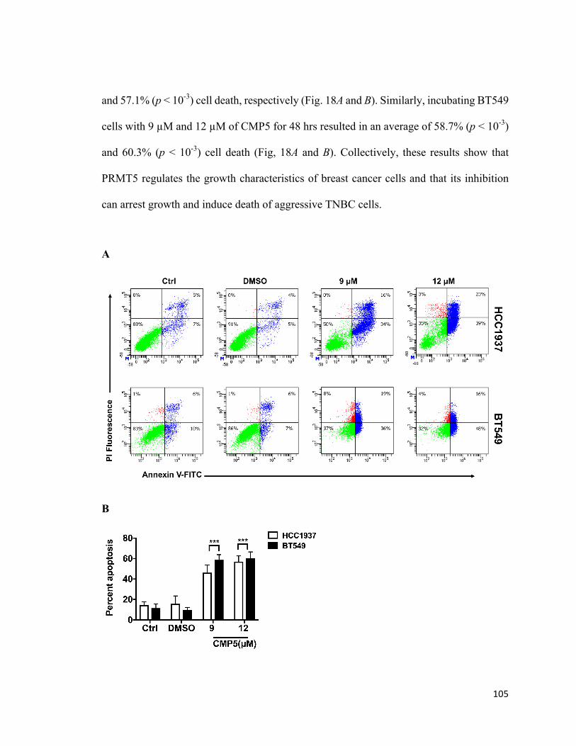
105
and 57.1% (p < 10-3) cell death, respectively (Fig. 18A and B). Similarly, incubating BT549
cells with 9 µM and 12 µM of CMP5 for 48 hrs resulted in an average of 58.7% (p < 10-3)
and 60.3% (p < 10-3) cell death (Fig, 18A and B). Collectively, these results show that
PRMT5 regulates the growth characteristics of breast cancer cells and that its inhibition
can arrest growth and induce death of aggressive TNBC cells.
A
B
106
Figure 18. PRMT5 inhibition induces apoptosis of breast cancer cells
(A) HCC1937 and BT549 cells were treated with different concentrations of CMP5 as
indicated, and the number of dead cells was measured by FACS analysis after staining cells
with ANNEXIN V and propidium iodide (PI). (B) The average result from the two
biological replicates used in E is represented as mean ± SD. All experiments were carried
out twice in duplicates, and the data is represented as mean ± SD. *** indicates p values <
10-3.
107
CHAPTER 5: DISCUSSION
Studies on epigenetic modifications that regulate proliferative and death signaling
pathways have become center stage for therapeutic intervention. PRMT5 is now considered
a key epigenetic modifying enzyme that governs major cellular process including cell
proliferation and apoptosis. We have recently reported that enhanced expression of PRMT5
upregulates WNT/β-CATENIN proliferative signaling by transcriptionally repressing
pathway antagonists, AXIN2 and WIF1, in three different types of non-Hodgkin’s
lymphoma cells (Chung et al., 2019). In the current study, we show that PRMT5 levels are
upregulated in different types of breast cancer cell lines compared to normal human
mammary epithelial cells. Our investigation revealed that PRMT5 promotes growth of
breast cancer cells in vitro by promoting WNT/β-CATENIN signaling via suppression of
expression of WNT antagonists, DKK1 and DKK3, thereby leading to enhanced expression
of WNT/β-CATENIN target genes including c-MYC, CYCLIN D1 and SURVIVIN. More
specifically, our experiments show that PRMT5 inhibition using the specific inhibitor,
CMP5, in breast cancer cells reduces expression of WNT/β-CATENIN target genes,
CYCLIN D1 and SURVIVIN, and induces expression of pathway antagonists, DKK1 and
DKK3 (Fig. 19). Our findings also show significant reduction in cell proliferation,
migration and invasion in response to PRMT5 inhibition. These changes in the growth
characteristics of breast cancer cells are also accompanied by induced cell death.
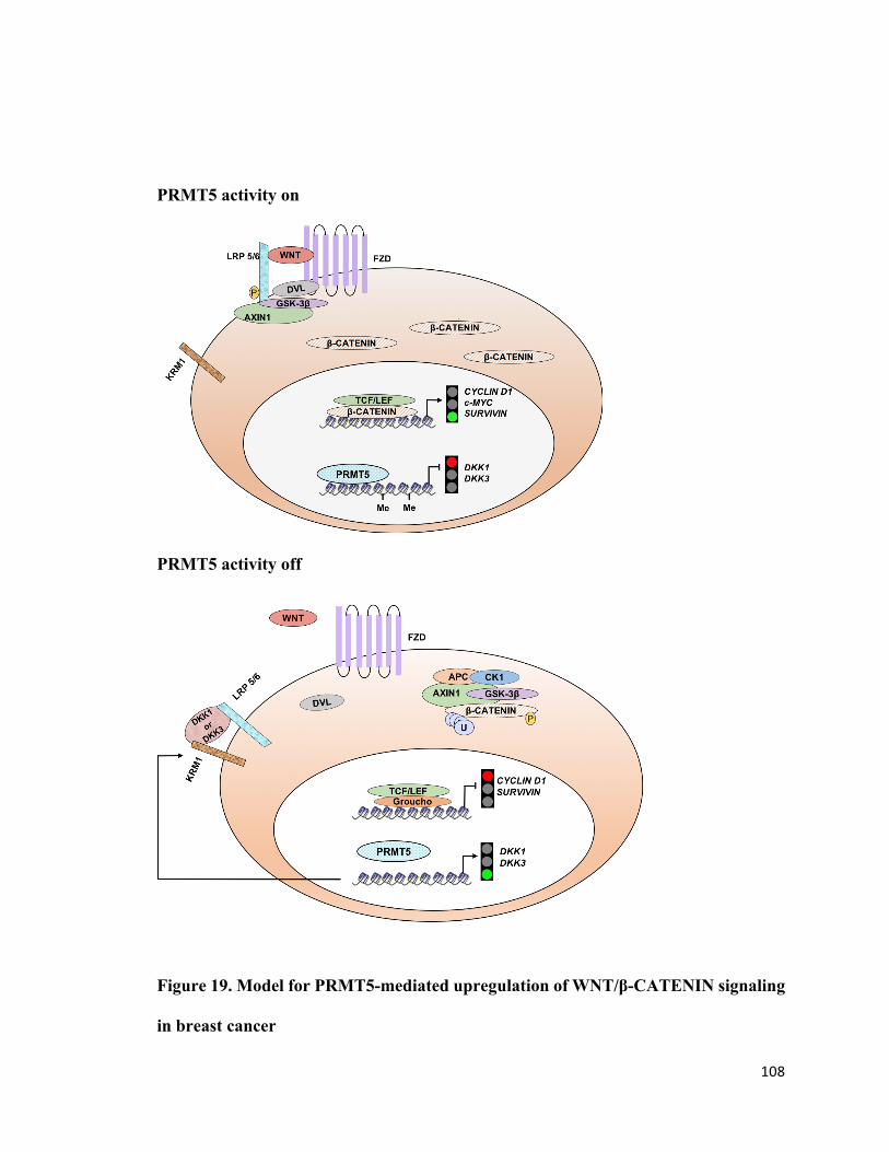
108
PRMT5 activity on
PRMT5 activity off
Figure 19. Model for PRMT5-mediated upregulation of WNT/β-CATENIN signaling
in breast cancer
109
PRMT5 is recruited to the promoter region of WNT/β-CATENIN pathway antagonists,
DKK1 and DKK3, and methylates H3R8 and H4R3 leading to their transcriptional
repression. In the absence of DKK1 and DKK3, WNT ligand binds to its receptor, Frizzled
receptor (FZD), which complexes with LRP5/6 co-receptor. Ligand bound Frizzled
receptor recruits Dishevelled (DVL) and promotes binding of GSK-3β, which
phosphorylates LRP5/6 and favors AXIN1 recruitment. Consequently, the cytosolic
destruction complex formation and β-CATENIN phosphorylation are prevented, which in
turn increases cytosolic β-CATENIN level. Accumulated β-CATENIN translocates to the
nucleus, where it complexes with TCF/LEF and activates transcription of target genes of
WNT signaling, CYCLIN D1, c-MYC and SURVIVIN (PRMT5 On). PRMT5 inhibition
results in transcriptional derepression of DKK1 and DKK3, which are excreted and bind to
LRP5/6 through interaction with KREMEN1 (KRM1), thereby form a ternary complex that
is endocytosed and degraded. In the absence of LRP5/6, WNT signaling is inhibited,
resulting in β-CATENIN degradation. During β-CATENIN unavailability, TCF and LEF
associate with transcriptional co-repressors, Groucho to suppress transcription of target
genes of WNT pathway, CYCLIN D1 and SURVIVIN (PRMT5 Off). These molecular
changes are associated with reduced proliferation, migration and invasion, and increased
death of breast cancer cells.
PRMT5 is associated with abnormal proliferation of breast cancer cells, and it was shown
that PRMT5 promotes growth of MCF7 cells by increasing expression of the translation
factor, eIF4E (Scoumanne et al., 2009). In another study, elevated levels of PRMT5 were
shown to reduce the tumor-suppressor activity of PDCD4 in primary breast cancer tumors
110
by methylating its N-terminal arginine residues, resulting in reduced patient survival
(Powers et al., 2011) . A more recent study indicated that PRMT5 levels are upregulated
in MCF7, MDA-MB-231 and MCF-10A cell lines as well as in clinical samples of ductal
carcinoma (Yang et al., 2015). In line with these findings, our investigation showed that
PRMT5 levels are upregulated in three different types of breast cancer cell lines including
MCF7, HCC1937 and BT549 compared to normal human mammary epithelial cells (Fig.
7). Dysregulation of WNT/β-CATENIN signaling in breast cancer serves as a major
oncogenic driver, and its upregulation has been implicated in many aggressive and invasive
breast cancer types, and correlates with acquired drug resistance (Prosperi & Goss, 2010;
King et al., 2012; Loh et al., 2013). Aberrant activation of WNT/β-CATENIN signaling
leads to enhanced expression of downstream target genes. Our study showed that levels of
WNT/β-CATENIN target genes CYCLIN D1, c-MYC and SURVIVIN are elevated in breast
cancer cells (Fig. 8).
Epigenetic silencing of WNT/β-CATENIN antagonists is frequently observed in breast
cancer, and reduced expression of APC, CDH1, SFRP1 and SFRP2 due to promoter
hypermethylation has been reported in many breast cancer samples (Klarmann et al., 2008).
More strikingly, WIF1 promoter hypermethylation has been frequently observed in breast
cancer, and also serves as an early trigger in the development of hereditary breast cancers
(Ai et al., 2006; Alvarez et al., 2013). A previous study indicated that WIF1 expression is
significantly repressed in breast cancer stem cells, which in turn promotes their self-
renewal (Wang et al., 2015). AXIN1 mRNA levels were also found to be repressed in MDA-
MB-231 cells and many breast cancer clinical samples ( Zhang et al., 2012). Furthermore,
111
DKK1 and DKK3 promoter hypermethylation has been documented in many breast cancer
samples (Suzuki et al., 2008; Xiang et al., 2013). We have examined expression of a panel
of WNT/β-CATENIN pathway antagonists in breast cancer cell lines, and found that
expression of DKK1 and DKK3 is significantly reduced in triple negative breast cancer
cell lines (Fig. 9). In light of our findings, which showed that PRMT5 is overexpressed in
breast cancer cells, we checked if it was involved in inducing transcriptional repression of
DKK1 and DKK3 in breast cancer. ChIP analysis showed that PRMT5 binds to the
promoter region of WNT antagonists, DKK1 and DKK3, and induces symmetric
methylation of H3R8 and H4R3, thereby causing their transcriptional repression (Fig. 10).
To evaluate the contribution of PRMT5 to transcriptional regulation of DKK1 and DKK3,
we inhibited its activity and monitored expression of both WNT/β-CATENIN antagonists.
We found that PRMT5 inhibition brings about DKK1 and DKK3 transcriptional
derepression, which correlates with reduced expression of WNT/β-CATENIN target genes
CYCLIN D1 and SURVIVIN in TNBCs (Fig. 12 and 14). However, c-MYC showed
enhanced expression in response to PRMT5 inhibition. A prior study by Evan et al. (1992)
showed that when expressed in the absence of proliferation signals, c-MYC induces cell
death. In agreement with this result, a later study by Murphy et al. (2008) showed that
distinct c-MYC expression levels regulate proliferation and apoptosis. While low levels of
c-MYC protein promote cell proliferation, its elevated expression activates the ARF/p53
tumor suppressor pathway, which leads to cell death (Murphy et al., 2008). In our study,
we found that PRMT5 inhibition reduces viability of breast cancer cells and induces their
death (Fig. 11 and 18). Therefore, it is conceivable that CMP5 treatment induces breast
cancer cell death through decreased expression of CYCLIN D1 and SURVIVIN, and
112
enhanced c-MYC expression. Further studies focusing on the mechanism by which c-MYC
induction is achieved are needed.
We have shown that PRMT5 epigenetically induces transcriptional repression of DKK1
and DKK3 in TNBCs, and that its inhibition restores expression of both WNT/β-CATENIN
antagonists in BT549 cells. Surprisingly, CMP5 treatment did not induce transcriptional
derepression of DKK1 and DKK3 in HCC1937 cells, indicating that there are other
epigenetic modifications involved in their regulation (Fig. 12B). The fact that PRMT5 is
known to induce transcriptional repression through interaction with DNMT3A (Liu et al.,
2018), we thought that both PRMT5 and DNMT3A might be playing a similar role in
silencing DKK1 and DKK3 in HCC1937 cells. However, treatment of HCC1937 cells with
the DNA methyltransferase inhibitor, 5-Azacytidine either alone or in combination with
CMP5 did not trigger DKK1 and DKK3 derepression more than 1.6 to1.7-fold (Fig. 13B).
PRMT5 is also known to associate with histone deacetylases 2 and 3 in large multisubunit
transcriptional repressor complexes ( Pal et al., 2003; Pal et al., 2004; Alinari et al., 2015)
Therefore, we reasoned that reduced expression of DKK1 and DKK3 could be due to
enhanced hypoacetylation of DKK1 and DKK3 promoter histones in HCC1937 cells.
Treatment of HCC1937 cells with TSA alone or in combination with CMP5 did not induce
DKK1 and DKK3 transcriptional derepression (Fig. 13C). However, combinatorial
treatment of HCC1937 cells with CMP5, 5-Aza and TSA resulted in increased
transcriptional derepression of DKK1 and DKK3, indicating that histone lysine
deacetylation, histone arginine methylation and DNA methylation work in concert to
epigenetically silence DKK1 and DKK3 (Fig. 13E).
113
To further understand the role of PRMT5 in regulating other pathways that promote breast
cancer growth, we investigated the effect of major growth promoting signaling cascade,
PI3K/AKT/mTOR pathway in breast cancer. The PI3K/AKT/mTOR signaling cascade
regulates the gene expression program that regulate cell growth and survival. Aberrant
activation of PI3K/AKT/mTOR pathway is frequently associated with many cancer types
including breast cancer. External cues such as growth factors can activate Phosphoinositide
3-kinase (PI3K) resulting in phosphorylation of AKT at T308 and S473 by PDK1 and
mTORC2, respectively. Phosphorylated AKT can activate several downstream substrates,
including TSC2 (Tuberous Sclerosis Complex 2), and prevents the formation of TSC1-
mTORC1 kinase complex, resulting in activation of mTORC1. Activated mTORC1
phosphorylates its downstream target proteins including the eukaryotic translation
initiation factor 4E binding protein 1 (4E-BP1) at its multiple sites including Ser6,
Thr37/46, and Thr70. Phosphorylation of 4E-BP1 results in releasing of eukaryotic
initiation translation factor 4E (eIF4E) from 4E-BP1- eIF4E complex. eIF4E can then form
an active eIF4F complex, resulting in stimulation of cap-dependent translation. mTORC1
is also a direct target of activated AKT which phosphorylates at Ser 2448 (Sekulic et al.,
2000). An initial study by Wei et al. (2012) showed that PRMT5 overexpression elevates
phosphorylation of PI3K p85 (Tyr458) and AKT (Ser308 and Ser473) and reduces
phosphorylation of negative regulator of AKT, PTEN (Ser380). Consequently, the
phosphorylation of AKT downstream effector, mTOR (Ser 2448), and expression of eIF4E
was elevated, indicating that PRMT5 can activate PI3K/AKT/mTOR signaling. In
114
addition, a recent study by Zhang et al. (2019) showed that PRMT5 can directly interact
with AKT and promote its phosphorylation at Thr308 and Ser473 in lung cancer cells. In
line with these findings, the current study showed tha inhibition of PRMT5 reduces
phosphorylation of AKT and mTOR at Ser 473 and Ser 2448 respectively in both
HCC1937 and BT549 cells (Fig. 16), further confirming the role of PRMT5 in AKT/mTOR
pathway.
Metastasis is a common characteristics of all aggressive breast tumors, and contributes to
poor prognosis and increased mortality (Redig & McAllister, 2013). A previous study by
Dey et al. (2013) showed that enhanced activation of WNT/β-CATENIN signaling is
positively correlated with increased metastatic potential and overall worse prognosis of
breast cancer patients, highlighting the role of WNT/β-CATENIN activation in breast
cancer metastasis. Furthermore, inhibition of WNT/β-CATENIN activation has been
shown to reduce proliferation and migration of BT549 cells, and induce their death in vitro
(Bilir et al., 2013). Since our study showed that PRMT5 inhibition reduces WNT/β-
CATENIN signaling in breast cancer cells, we examined the effect of PRMT5 inhibition
on migration and invasion of breast cancer cells. Our results showed significant reduction
in breast cancer cell migration and invasion in response to PRMT5 inhibition (Fig. 17A-
D). Our results also showed that PRMT5 inhibition using CMP5 LC50 concentration
induced death of TNBCs as measured by Annexin V/PI staining (Fig. 18Aand B).
Taken together, our work shows that PRMT5 levels are upregulated in breast cancer cell
115
lines, and that it is involved in epigenetic silencing of a different set of WNT antagonists
compared to lymphoma cells. In aggressive lymphomas, PRMT5 epigenetically inhibits
expression of WNT antagonists, AXIN2 and WIF1 (Chung et al., 2019). AXIN2 serve as
a scaffolding protein in the cytosolic destruction complex, which induces β-CATENIN
degradation (Hinoi et al., 2000), whereas, WIF1 inactivates WNT/β-CATENIN signaling
by directly binding to WNT ligand and inhibiting its interaction with the FZD receptor
(Hsieh et al., 1999; Malinauskas et al., 2011). In breast cancer cells, PRMT5 suppresses
expression of DKK1, which antagonizes WNT/β-CATENIN signaling by binding to its
cognate membrane receptor, KREMEN2, and promoting cell surface removal of LRP5/6
by endocytosis, thereby preventing dimerization of WNT-bound FZD with LRP5/6 co-
receptor (Mao et al., 2001; Mao et al., 2002). DKK3 is also epigenetically suppressed by
PRMT5; however, the mechanism by which DKK3 down-regulates WNT signaling
remains unclear. Regardless, it appears that PRMT5 targets various WNT/β-CATENIN
pathway antagonists that operate at different levels, highlighting the complexity and
diversity of its mechanism of action in the different cell types. The net outcome of PRMT5-
induced epigenetic silencing is to promote growth and survival of cancer cell, which
renders its ideal for therapeutic intervention, because its selective inhibition is marked by
decreased expression of pro-survival proteins, CYCLIN D1 and SURVIVIN. These
molecular changes trigger reduced proliferation, migration, and invasion, and increased
cell death, which are all desired attributes for killing tumor cells.
116
CHAPTER 6: CONCLUSION AND FUTURE WORK
CONCLUSION
Protein arginine methyltransferase 5 (PRMT5) activity is dysregulated in many aggressive
cancers and its enhanced levels are associated with increased tumor growth and survival.
The current study was focused on investigating the role of PRMT5 in promoting breast
tumorigenesis. The results of the study showed that PRMT5 is overexpressed in breast
cancer cell lines of varying aggressiveness, and that it promotes their growth by activating
WNT/β-CATENIN proliferative signaling through epigenetic silencing of pathway
antagonists, DKK1 and DKK3, leading to enhanced expression of c-MYC, CYCLIN D1 and
SURVIVIN. Through chromatin immunoprecipitation (ChIP) studies, it was confirmed that
PRMT5 binds to the promoter region of WNT antagonists, DKK1 and DKK3, and induces
symmetric dimethylation of H3R8 and H4R3 histones. Furthermore, PRMT5 inhibition
using a specific small molecule inhibitor, compound 5 (CMP5), reduced PRMT5
recruitment as well as methylation of H3R8 and H4R3 histones in the promoter regions of
DKK1 and DKK3, which consequently results in reduced expression CYCLIN D1 and
SURVIVIN. In addition, CMP5 treatment either alone or in combination with 5-Azacytidine
and Trichostatin A restored expression of DKK1 and DKK3 in TNBCs. PRMT5 inhibition
also altered the growth characteristics of TNBC cells including proliferation, migration and
invasion, and induced their death.
117
PROSPECTIVE
The current study compared the expression of PRMT5, WNT/β-CATENIN downstream
target genes and pathway antagonists in three different breast cancer cell lines versus
normal mammary epithelial cells (HMECs). The results of the study indicated that there is
positive correlation between PRMT5 expression and that of WNT/β-CATENIN target
genes, CYCLIN D1, c-MYC and SURVIVIN, and that there is an inverse correlation between
PRMT5 and WNT/β-CATENIN antagonists, DKK1 and DKK3, expression in breast cancer
cells (Chapter 4.1, 4.2 and 4.3). Since breast cancer is a heterogeneous disease, it is
important to screen more breast cancer cell lines as well as clinical samples to determine
and validate the biological relevance of our findings. In lymphoma, PRMT5 levels are
increased due to increased translation and reduced expression of miRs 92 and 96 (Pal et
al., 2007). However, the current study revealed that expression of PRMT5 is elevated due
to enhanced gene transcription in breast cancer. The mechanism by which PRMT5
transcription is increased in breast cancer is at present unknown, and more work is required
to address this question. Hence, we plan to perform ChIP experiments using antibodies
against transcriptional activation marks including H3K9ac, H3K27ac and H3K4me3 to
decipher the mechanism of transcriptional activation of PRMT5 in breast cancer.
In the current study it has been demonstrated that PRMT5 inhibition reduces proliferation,
migration and invasion of breast cancer cells and induces their death in vitro (Chapter 4.5,
4.11 and 4.12). Therefore, it is important to characterize the role of PRMT5 overexpression
and impact of its inhibition in vivo using TNBC xenografts in SCID mice. This would
118
further validate the therapeutic significance of targeting PRMT5 for breast cancer
treatment.
The mechanism by which PRMT5 activates WNT/β-CATENIN signaling in breast cancer
was investigated in the current study, which showed that PRM5 epigenetically suppresses
expression of WNT pathway antagonists, DKK1 and DKK3, by binding to their promoters
and inducing symmetric methylation of H3R8 and H4R3 histones (Chapter 4.3 and 4.4).
DKKs antagonize WNT/β-CATENIN signaling by binding to the membrane receptor,
KREMEN1, and promoting cell surface removal of LRP5/6 by endocytosis, thereby
preventing dimerization of WNT-bound FZD with LRP5/6 co-receptor (Mao et al., 2002;
Mao et al., 2001). Hence, it would be interesting to design peptide mimicks corresponding
to the biologically active DKK1 and DKK3 domains, and to test their efficacy in reducing
breast cancer growth and metastasis. We also plan to investigate the impact of stable re-
expression of DKK1 and DKK3 using tet-inducible lentiviral expression vector on WNT/β-
CATENIN signaling and to assess their influence on the growth characteristics of TNBCs
in vitro and in vivo.
PRMT5 inhibition also resulted in reduced expression of WNT/β-CATENIN downstream
target genes, CYCLIN D1 and SURVIVIN. However, c-MYC expression was increased in
both HCC1937 and BT549 cells. It would be interesting to study the mechanism by which
PRMT5 regulates c-MYC expression in breast cancer cells. Furthermore, we plan to
conduct differential gene expression studies using RNA-Seq analysis before and after
119
CMP5 treatment. We also plan to conduct proteomic analysis to investigate the differential
expression of proteins before and after CMP5 treatment in TNBCs. These studies will help
us identify PRMT5-regulated networks and pathways involved in breast cancer
tumorigenesis.
120
REFERENCES
Aggarwal, P., Vaites, L. P., Kim, J. K., Mellert, H., Gurung, B., Nakagawa, H., Herlyn,
M., Hua, X., Rustgi, A. K., McMahon, S. B., & Diehl, J. A. (2010). Nuclear cyclin D1/CDK4 kinase regulates CUL4 expression and triggers neoplastic growth via activation of the PRMT5 methyltransferase. Cancer Cell, 18(4), 329-340.
Aguilera, O., Fraga, M. F., Ballestar, E., Paz, M. F., Herranz, M., Espada, J., Garcia, J.M.,
Munoz, A., & Gonzalez-Sancho, J. M. (2006). Epigenetic inactivation of the Wnt antagonist DICKKOPF-1 (DKK-1) gene in human colorectal cancer. Oncogene, 25(29), 4116-4121.
Ai, L., Tao, Q., Zhong, S., Fields, C. R., Kim, W. J., Lee, M. W., Cui, Y., Brown, K. D.,
& Robertson, K. D. (2006). Inactivation of Wnt inhibitory factor-1 (WIF1) expression by epigenetic silencing is a common event in breast cancer. Carcinogenesis, 27(7), 1341-1348.
Alinari, L., Mahasenan, K. V., Yan, F., Karkhanis, V., Chung, J. H., Smith, E. M., Quinion,
C., Smith, P.L., Kim, L., Patton, J. T., Lapalombella, R., Yu, B., Roy, S., De Leo, A., Pileri, S., Agostinelli, C., Ayers, L., Bradner, J. E., Chen-Kiang, S., Elemento, O., Motiwala, T., Majumdar, S., Byrd, J. C., Jacob, S., Sif, S., Li, C., & Baiocchi, R. A. (2015). Selective inhibition of protein arginine methyltransferase 5 blocks initiation and maintenance of B-cell transformation. Blood, 125(16), 2530-2543.
Altieri, D. C. (2003). Survivin, versatile modulation of cell division and apoptosis in
cancer. Oncogene, 22(53), 8581-8589. Alvarez, C., Tapia, T., Cornejo, V., Fernandez, W., Munoz, A., Camus, M., Alvarez, M.,
Devoto, L., & Carvallo, P. (2013). Silencing of tumor suppressor genes RASSF1A, SLIT2, and WIF1 by promoter hypermethylation in hereditary breast cancer. Mol Carcinog, 52(6), 475-487.
Amente, S., Napolitano, G., Licciardo, P., Monti, M., Pucci, P., Lania, L., & Majello, B.
(2005). Identification of proteins interacting with the RNAPII FCP1 phosphatase: FCP1 forms a complex with arginine methyltransferase PRMT5 and it is a substrate for PRMT5-mediated methylation. FEBS Lett, 579(3), 683-689.
Amit, S., Hatzubai, A., Birman, Y., Andersen, J. S., Ben-Shushan, E., Mann, M., Ben-
Neriah, Y., & Alkalay, I. (2002). Axin-mediated CKI phosphorylation of beta-catenin at Ser 45: a molecular switch for the Wnt pathway. Genes Dev, 16(9), 1066-1076.
Anders, L., Ke, N., Hydbring, P., Choi, Y. J., Widlund, H. R., Chick, J. M., Zhai, H., Vidal,
121
M., Gygi, S. P., Braun, P., & Sicinski, P. (2011). A systematic screen for CDK4/6 substrates links FOXM1 phosphorylation to senescence suppression in cancer cells. Cancer cell, 20(5), 620-634.
Andreu-Perez, P., Esteve-Puig, R., de Torre-Minguela, C., Lopez-Fauqued, M., Bech-
Serra, J. J., Tenbaum, S., Gracia-Trevijano, E. R., Canals, F., Merlino, G., Avila, M. A., & Recio, J. A. (2011). Protein arginine methyltransferase 5 regulates ERK1/2 signal transduction amplitude and cell fate through CRAF. Sci Signal, 4(190), ra58.
Antonysamy, S., Bonday, Z., Campbell, R. M., Doyle, B., Druzina, Z., Gheyi, T., Han, B.,
Jungheim, L. N., Qian, Y., Rauch, C., Russel, M., Sauder, J. M., Wasserman, S. R., Weichert, K., Willard, F,. S., Zhang, A., & Emtage, S. (2012). Crystal structure of the human PRMT5:MEP50 complex. Proc Natl Acad Sci U S A, 109(44), 17960-17965.
Baehs, S., Herbst, A., Thieme, S. E., Perschl, C., Behrens, A., Scheel, S., Jung, A.,
Barbletz, T., Göke, B., Blum, H., & Kolligs, F. T. (2009). Dickkopf-4 is frequently down-regulated and inhibits growth of colorectal cancer cells. Cancer Letters, 276(2), 152-159.
Bafico, A., Gazit, A., Pramila, T., Finch, P. W., Yaniv, A., & Aaronson, S. A. (1999).
Interaction of frizzled related protein (FRP) with Wnt ligands and the frizzled receptor suggests alternative mechanisms for FRP inhibition of Wnt signaling. J Biol Chem, 274(23), 16180-16187.
Baldin, V., Lukas, J., Marcote, M. J., Pagano, M., & Draetta, G. (1993). Cyclin D1 is a
nuclear protein required for cell cycle progression in G1. Genes Dev, 7(5), 812-821.
Banasavadi-Siddegowda, Y. K., Russell, L., Frair, E., Karkhanis, V. A., Relation, T., Yoo,
J. Y., Zhang, J., Sif, S., Imitola, J., Baiocchi, R., & Kaur, B. (2017). PRMT5-PTEN molecular pathway regulates senescence and self-renewal of primary glioblastoma neurosphere cells. Oncogene, 36(2), 263-274.
Bandyopadhyay, S., Harris, D. P., Adams, G. N., Lause, G. E., McHugh, A., Tillmaand, E.
G., Money, A., Willard, B., Fox, P. L., & DiCorleto, P. E. (2012). HOXA9 methylation by PRMT5 is essential for endothelial cell expression of leukocyte adhesion molecules. Molecular and cellular biology, 32(7), 1202-1213.
Bao, X., Zhao, S., Liu, T., Liu, Y., Liu, Y., & Yang, X. (2013). Overexpression of PRMT5
promotes tumor cell growth and is associated with poor disease prognosis in epithelial ovarian cancer. J Histochem Cytochem, 61(3), 206-217.
Barker, N. (2008). The Canonical Wnt/β-Catenin Signalling Pathway. In E. Vincan (Ed.),
122
Wnt Signaling (Vol. 468, pp. 5-15): Humana Press. Barker, N., Hurlstone, A., Musisi, H., Miles, A., Bienz, M., & Clevers, H. (2001). The
chromatin remodelling factor Brg-1 interacts with beta-catenin to promote target gene activation. Embo j, 20(17), 4935-4943.
Bartkova, J., Lukas, J., Strauss, M., & Bartek, J. (1998). Cyclin D3: requirement for G1/S
transition and high abundance in quiescent tissues suggest a dual role in proliferation and differentiation. Oncogene, 17(8), 1027-1037.
Bartoli, A., Fettucciari, K., Fetriconi, I., Rosati, E., Ianni, M. D., Tabilio, A., Delfino, D.
V., Rossi, R., & Marconi, P. (2003). Effect of trichostatin a and 5′-azacytidine on transgene reactivation in U937 transduced cells. Pharmacological Research, 48(1), 111-118.
Bazzi, H., Fantauzzo, K. A., Richardson, G. D., Jahoda, C. A. B., & Christiano, A. M.
(2007). The Wnt inhibitor, Dickkopf 4, is induced by canonical Wnt signaling during ectodermal appendage morphogenesis. Developmental Biology, 305(2), 498-507.
Behrens, J., Jerchow, B. A., Wurtele, M., Grimm, J., Asbrand, C., Wirtz, R., Kühl, M.,
Weldich, D., & Birchmeier, W. (1998). Functional interaction of an axin homolog, conductin, with beta-catenin, APC, and GSK3beta. Science, 280(5363), 596-599.
Bezzi, M., Teo, S. X., Muller, J., Mok, W. C., Sahu, S. K., Vardy, L. A., Bonday, Z. Q., &
Guccione, E. (2013). Regulation of constitutive and alternative splicing by PRMT5 reveals a role for Mdm4 pre-mRNA in sensing defects in the spliceosomal machinery. Genes Dev, 27(17), 1903-1916.
Bhanot, P., Brink, M., Samos, C. H., Hsieh, J. C., Wang, Y., Macke, J. P., Andrew, D.,
Nathans, J., & Nusse, R. (1996). A new member of the frizzled family from Drosophila functions as a Wingless receptor. Nature, 382(6588), 225-230.
Bilir, B., Kucuk, O., & Moreno, C. S. (2013). Wnt signaling blockage inhibits cell
proliferation and migration, and induces apoptosis in triple-negative breast cancer cells. Journal of translational medicine, 11, 280-280.
Bowman, G. D., & Poirier, M. G. (2015). Post-translational modifications of histones that
influence nucleosome dynamics. Chemical reviews, 115(6), 2274-2295. doi:10.1021/cr500350
Brahms, H., Meheus, L., de Brabandere, V., Fischer, U., & Luhrmann, R. (2001).
Symmetrical dimethylation of arginine residues in spliceosomal Sm protein B/B' and the Sm-like protein LSm4, and their interaction with the SMN protein. Rna, 7(11), 1531-1542.
123
Branscombe, T. L., Frankel, A., Lee, J. H., Cook, J. R., Yang, Z., Pestka, S., & Clarke, S.
(2001). PRMT5 (Janus kinase-binding protein 1) catalyzes the formation of symmetric dimethylarginine residues in proteins. J Biol Chem, 276(35), 32971-32976.
Brenner, C., Deplus, R., Didelot, C., Loriot, A., Vire, E., De Smet, C., Gutierrez, A.,
Danvoi, D., Bernards, D., Boon, T., Pelicci, P. G., Amati, B., Kouzarides, T., de Launoti, Y., Di Croce, L., & Fuks, F. (2005). Myc represses transcription through recruitment of DNA methyltransferase corepressor. Embo j, 24(2), 336-346.
Brott, B. K., & Sokol, S. Y. (2002). Regulation of Wnt/LRP Signaling by Distinct Domains
of Dickkopf Proteins. Molecular and Cellular Biology, 22(17), 6100. Brunner, E., Peter, O., Schweizer, L., & Basler, K. (1997). pangolin encodes a Lef-1
homologue that acts downstream of Armadillo to transduce the Wingless signal in Drosophila. Nature, 385(6619), 829-833.
Bruns, A. F., Grothe, C., & Claus, P. (2009). Fibroblast growth factor 2 (FGF-2) is a novel
substrate for arginine methylation by PRMT5. Biol Chem, 390(1), 59-65. Bu, X. M., Zhao, C. H., Zhang, N., Gao, F., Lin, S., & Dai, X. W. (2008). Hypermethylation
and aberrant expression of secreted frizzled-related protein genes in pancreatic cancer. World J Gastroenterol, 14(21), 3421-3424.
Burgos, E. S., Wilczek, C., Onikubo, T., Bonanno, J. B., Jansong, J., Reimer, U., &
Shechter, D. (2015). Histone H2A and H4 N-terminal tails are positioned by the MEP50 WD repeat protein for efficient methylation by the PRMT5 arginine methyltransferase. J Biol Chem, 290(15), 9674-9689.
Caceres, T. B., Thakur, A., Price, O. M., Ippolito, N., Li, J., Qu, J., Acevedo, O., & Hevel,
J. M. (2018). Phe71 in Type III Trypanosomal Protein Arginine Methyltransferase 7 (TbPRMT7) Restricts the Enzyme to Monomethylation. Biochemistry (57), 1349-1359
Cai, W., Xu, Y., Yin, J., Zuo, W., & Su, Z. (2019). miR-552-5p facilitates osteosarcoma
cell proliferation and metastasis by targeting WIF1. Experimental and therapeutic medicine, 17(5), 3781-3788.
Calabretta, S., Vogel, G., Yu, Z., Choquet, K., Darbelli, L., Nicholson, T. B., Kleinman,
C. L., & Richard, S. J. b. (2018). Loss of PRMT5 promotes PDGFRα degradation during oligodendrocyte differentiation and myelination. 252056.
Caricasole, A., Ferraro, T., Iacovelli, L., Barletta, E., Caruso, A., Melchiorri, D.,
Terstappen, G. C., & Nicoletti, F. (2003). Functional characterization of WNT7A
124
signaling in PC12 cells: interaction with A FZD5 x LRP6 receptor complex and modulation by Dickkopf proteins. J Biol Chem, 278(39), 37024-37031.
Carmon, K. S., & Loose, D. S. (2008). Secreted frizzled-related protein 4 regulates two
Wnt7a signaling pathways and inhibits proliferation in endometrial cancer cells. Mol Cancer Res, 6(6), 1017-1028.
Cavallo, R. A., Cox, R. T., Moline, M. M., Roose, J., Polevoy, G. A., Clevers, H., Peifer,
M., & Bejsovec, A. (1998). Drosophila Tcf and Groucho interact to repress Wingless signalling activity. Nature, 395(6702), 604-608.
Chen, G., Fernandez, J., Mische, S., & Courey, A. J. (1999). A functional interaction
between the histone deacetylase Rpd3 and the corepressor groucho in Drosophila development. Genes Dev, 13(17), 2218-2230.
Chen, H. Y., Lang, Y. D., Lin, H. N., Liu, Y.-R., Liao, C. C., Nana, A. W., Yen, Y., &
Chen, R. H. (2019). miR-103/107 prolong Wnt/β-catenin signaling and colorectal cancer stemness by targeting Axin2. Scientific Reports, 9(1), 9687.
Chen, L., Li, M., Li, Q., Wang, C. J., & Xie, S. Q. (2013). DKK1 promotes hepatocellular
carcinoma cell migration and invasion through β-catenin/MMP7 signaling pathway. Molecular Cancer, 12(1), 157.
Chen, M., Qu, X., Zhang, Z., Wu, H., Qin, X., Li, F., Liu, Z., Tian, L., Miao, J., & Shu, W.
(2016). Cross-talk between Arg methylation and Ser phosphorylation modulates apoptosis signal-regulating kinase 1 activation in endothelial cells. Mol Biol Cell, 27(8), 1358-1366.
Chen, X., Zhong, L., Li, X., Liu, W., Zhao, Y., & Li, J. (2019). Down-regulation of
microRNA-31-5p inhibits proliferation and invasion of osteosarcoma cells through Wnt/β-catenin signaling pathway by enhancing AXIN1. Experimental and Molecular Pathology, 108, 32-41.
Chen, Y., Whetstone, H. C., Lin, A. C., Nadesan, P., Wei, Q., Poon, R., & Alman, B. A.
(2007). Beta-catenin signaling plays a disparate role in different phases of fracture repair: implications for therapy to improve bone healing. PLoS Med, 4(7), e249.
Chiang, K., Zielinska, A. E., Shaaban, A. M., Sanchez-Bailon, M. P., Jarrold, J., Clarke, T.
L., Zhang, J., Francis, A., Jones, L. J., Smith, S., Barbash, O., Guccione, E., Farnie, G., Smalley, M. J., & Davies, C. C. (2017). PRMT5 Is a Critical Regulator of Breast Cancer Stem Cell Function via Histone Methylation and FOXP1 Expression. Cell Rep, 21(12), 3498-3513.
Chittka, A., Nitarska, J., Grazini, U., & Richardson, W. D. (2012). Transcription factor
positive regulatory domain 4 (PRDM4) recruits protein arginine methyltransferase

125
5 (PRMT5) to mediate histone arginine methylation and control neural stem cell proliferation and differentiation. The Journal of biological chemistry, 287(51), 42995-43006.
Cho, E.-C., Zheng, S., Munro, S., Liu, G., Carr, S. M., Moehlenbrink, J., Lu, Y. C.,
Stimson, L., Khan, O., Konietzny, R., McGouran, J., Coutts, A. S., Kessler, B., Kerr, D. J., & Thangue, N. B. L. (2012). Arginine methylation controls growth regulation by E2F-1. The EMBO journal, 31(7), 1785-1797.
Cho, Y. K., Kang, Y. M., Lee, S. E., Lee, Y. L., Seol, S. M., Lee, W. J., Park, J. Y., & Jung,
C. H. (2018). Effect of SFRP5 (Secreted Frizzled-Related Protein 5) on the WNT5A (Wingless-Type Family Member 5A)-Induced Endothelial Dysfunction and Its Relevance With Arterial Stiffness in Human Subjects. Arteriosclerosis, thrombosis, and vascular biology, 38(6), 1358-1367.
Chung, J., Karkhanis, V., Baiocchi, R. A., & Sif, S. (2019). Protein Arginine
Methyltransferase 5 (PRMT5) Promotes Survival of Lymphoma Cells via Activation of WNT/beta-CATENIN and AKT/GSK3beta Proliferative Signaling. J Biol Chem.
Chung, J., Karkhanis, V., Tae, S., Yan, F., Smith, P., Ayers, L. W., Angostinelli, C., Pileri,
S., Baiocchi, R. A., & Sif, S. (2013). Protein Arginine Methyltransferase 5 (PRMT5) Inhibition Induces Lymphoma Cell Death through Reactivation of the Retinoblastoma Tumor Suppressor Pathway and Polycomb Repressor Complex 2 (PRC2) Silencing. Journal of Biological Chemistry, 288(49), 35534-35547.
Chung, M. T., Lai, H. C., Sytwu, H. K., Yan, M. D., Shih, Y. L., Chang, C. C., Yu, M. H.,
Liu, H. S., Chu, D. W., & Lin, Y. W. (2009). SFRP1 and SFRP2 suppress the transformation and invasion abilities of cervical cancer cells through Wnt signal pathway. Gynecol Oncol, 112(3), 646-653.
Chung, M. T., Sytwu, H. K., Yan, M. D., Shih, Y. L., Chang, C. C., Yu, M. H., Chu, T. Y.,
Lai, H. C. & Lin, Y. W. (2009). Promoter methylation of SFRPs gene family in cervical cancer. Gynecol Oncol, 112(2), 301-306.
Clevers, H. (2006). Wnt/beta-catenin signaling in development and disease. Cell, 127(3),
469-480. Clevers, H., & Nusse, R. (2012). Wnt/beta-catenin signaling and disease. Cell, 149(6),
1192-1205. Cliffe, A., Hamada, F., & Bienz, M. (2003). A role of Dishevelled in relocating Axin to
the plasma membrane during wingless signaling. Curr Biol, 13(11), 960-966. Cowling, V. H., & Cole, M. D. (2007). E-cadherin repression contributes to c-Myc-induced
126
epithelial cell transformation. Oncogene, 26(24), 3582-3586. Cowling, V. H., Cruz, C. M., Chodosh, L. A., & Cole, M. D. (2007). c-Myc Transforms
Human Mammary Epithelial Cells through Repression of the Wnt Inhibitors DKK1 and SFRP1. Molecular and Cellular Biology, 27(14), 5135.
Cruciat, C. M., & Niehrs, C. (2013). Secreted and transmembrane wnt inhibitors and
activators. Cold Spring Harb Perspect Biol, 5(3), a015081. doi:10.1101/cshperspect.a015081
Dang, C. V., O'Donnell, K. A., Zeller, K. I., Nguyen, T., Osthus, R. C., & Li, F. (2006).
The c-Myc target gene network. Semin Cancer Biol, 16(4), 253-264. Davidson, K. C., Adams, A. M., Goodson, J. M., McDonald, C. E., Potter, J. C., Berndt, J.
D., Biechele, T. L., Taylor, R. J., & Moon, R. T. (2012). Wnt/beta-catenin signaling promotes differentiation, not self-renewal, of human embryonic stem cells and is repressed by Oct4. Proc Natl Acad Sci U S A, 109(12), 4485-4490.
De Brouwer, S., Mestdagh, P., Lambertz, I., Pattyn, F., De Paepe, A., Westermann, F.,
Schroeder, C., Schulte, J. H., Schramm, A., De Preter, K., Vandesompele, J., & Speleman, F. (2012). Dickkopf-3 is regulated by the MYCN-induced miR-17-92 cluster in neuroblastoma. 130(11), 2591-2598.
Deng, X., Hou, C., Wang, H., Liang, T., & Zhu, L. (2017). Hypermethylation of WIF1 and
its inhibitory role in the tumor growth of endometrial adenocarcinoma. Mol Med Rep, 16(5), 7497-7503.
Deng, X., Shao, G., Zhang, H. T., Li, C., Zhang, D., Cheng, L., Elzey, B. D., Pili, R.,
Ratliff, T. L., Huang, J., & Hu, C. D. (2017). Protein arginine methyltransferase 5 functions as an epigenetic activator of the androgen receptor to promote prostate cancer cell growth. Oncogene, 36(9), 1223-1231.
Dey, N., Barwick, B. G., Moreno, C. S., Ordanic-Kodani, M., Chen, Z., Oprea-Ilies, G.,
Tang, W., Catzavelos, C., Kerstann, K. F., Sledge, G. W. Jr., Abramovitz, M., Bouzyk, M., De, P., & Leyland-Jones, B. R. (2013). Wnt signaling in triple negative breast cancer is associated with metastasis. BMC Cancer, 13, 537.
di Caprio, R., Ciano, M., Montano, G., Costanzo, P., & Cesaro, E. (2015). KAP1 is a Novel
Substrate for the Arginine Methyltransferase PRMT5. Biology (Basel), 4(1), 41-49. Donepudi, M. S., Kondapalli, K., Amos, S. J., & Venkanteshan, P. (2014). Breast cancer
statistics and markers. J Cancer Res Ther, 10(3), 506-511. Eckert, D., Biermann, K., Nettersheim, D., Gillis, A. J. M., Steger, K., Jäck, H. M., Müller,
A. M., Looijenga, L. H., & Schorle, H. (2008). Expression of BLIMP1/PRMT5 and

127
concurrent histone H2A/H4 arginine 3 dimethylation in fetal germ cells, CIS/IGCNU and germ cell tumors. BMC Developmental Biology, 8, 106-106.
Evan, G. I., Wyllie, A. H., Gilbert, C. S., Littlewood, T. D., Land, H., Brooks, M., Waters,
C. M., Penn, L. Z., & Hancock, D. C. (1992). Induction of apoptosis in fibroblasts by c-myc protein. Cell, 69(1), 119-128.
Fatima, S., Lee, N. P., Tsang, F. H., Kolligs, F. T., Ng, I. O. L., Poon, R. T. P., Fan, S. T.,
& Luk, J. M. (2012). Dickkopf 4 (DKK4) acts on Wnt/β-catenin pathway by influencing β-catenin in hepatocellular carcinoma. Oncogene, 31(38), 4233-4244.
Fay, M. M., Clegg, J. M., Uchida, K. A., Powers, M. A., & Ullman, K. S. (2014). Enhanced
arginine methylation of programmed cell death 4 protein during nutrient deprivation promotes tumor cell viability. J Biol Chem, 289(25), 17541-17552.
Fedi, P., Bafico, A., Nieto Soria, A., Burgess, W. H., Miki, T., Bottaro, D. P., Kraus, M.
H., & Aaronson, S. A. (1999). Isolation and biochemical characterization of the human Dkk-1 homologue, a novel inhibitor of mammalian Wnt signaling. J Biol Chem, 274(27), 19465-19472.
Feng, G. J., Cotta, W., Wei, X. Q., Poetz, O., Evans, R., Jarde, T., Reed, K., Meniel, V.,
Williams, G. T., Clarke, A. R., & Dale, T. C. (2012). Conditional disruption of Axin1 leads to development of liver tumors in mice. Gastroenterology, 143(6), 1650-1659.
Feng, Y., Zhang, Y., Wei, X., & Zhang, Q. (2019). Correlations of DKK1 with
pathogenesis and prognosis of human multiple myeloma. Cancer Biomark, 24(2), 195-201.
Fernández, J. G., Rodríguez, D. A., Valenzuela, M., Calderon, C., Urzúa, U., Munroe, D.,
Rosas, C., Lemus, D., Díaz, N., Wright, M. C., Leyton, L., Tapia, J. C., & Quest, A. F. (2014). Survivin expression promotes VEGF-induced tumor angiogenesis via PI3K/Akt enhanced β-catenin/Tcf-Lef dependent transcription. Molecular cancer, 13, 209-209.
Fevr, T., Robine, S., Louvard, D., & Huelsken, J. (2007). Wnt/beta-catenin is essential for
intestinal homeostasis and maintenance of intestinal stem cells. Mol Cell Biol, 27(21), 7551-7559.
Frank, S. R., Parisi, T., Taubert, S., Fernandez, P., Fuchs, M., Chan, H. M., Livingston, D.
M., & Amati, B. (2003). MYC recruits the TIP60 histone acetyltransferase complex to chromatin. EMBO reports, 4(6), 575-580.
Friesen, W. J., Paushkin, S., Wyce, A., Massenet, S., Pesiridis, G. S., Van Duyne, G.,
Rappsilber, J., Mann, M., & Dreyfuss, G. (2001). The methylosome, a 20S complex
128
containing JBP1 and pICln, produces dimethylarginine-modified Sm proteins. Mol Cell Biol, 21(24), 8289-8300.
Galli, L. M., Barnes, T., Cheng, T., Acosta, L., Anglade, A., Willert, K., Nusse, R., &
Burrus, L. W. (2006). Differential inhibition of Wnt-3a by Sfrp-1, Sfrp-2, and Sfrp-3. Developmental dynamics : an official publication of the American Association of Anatomists, 235(3), 681-690.
Gallinari, P., Marco, S. D., Jones, P., Pallaoro, M., & Steinkühler, C. (2007). HDACs,
histone deacetylation and gene transcription: from molecular biology to cancer therapeutics. Cell Research, 17(3), 195-211.
Gao, C., Xiao, G., & Hu, J. (2014). Regulation of Wnt/beta-catenin signaling by
posttranslational modifications. Cell Biosci, 4(1), 13. Giodini, A., Kallio, M. J., Wall, N. R., Gorbsky, G. J., Tognin, S., Marchisio, P. C.,
Symons, M., & Altieri, D. C. (2002). Regulation of microtubule stability and mitotic progression by survivin. Cancer Res, 62(9), 2462-2467.
Gkountela, S., Li, Z., Chin, C. J., Lee, S. A., & Clark, A. T. (2014). PRMT5 is required for
human embryonic stem cell proliferation but not pluripotency. Stem Cell Rev, 10(2), 230-239.
Glinka, A., Wu, W., Delius, H., Monaghan, A. P., Blumenstock, C., & Niehrs, C. (1998).
Dickkopf-1 is a member of a new family of secreted proteins and functions in head induction. Nature, 391(6665), 357-362.
Gluecksohn-Schoenheimer, S. (1949). The effects of a lethal mutation responsible for
duplications and twinning in mouse embryos. Journal of Experimental Zoology, 110(1), 47-76.
González-Sancho, J. M., Aguilera, O., García, J. M., Pendás-Franco, N., Peña, C., Cal, S.,
Gracía de Herreros, A., Bonilla, F., & Muñoz, A. (2005). The Wnt antagonist DICKKOPF-1 gene is a downstream target of β-catenin/TCF and is downregulated in human colon cancer. Oncogene, 24(6), 1098-1103.
Guccione, E., & Richard, S. (2019). The regulation, functions and clinical relevance of
arginine methylation. Nature Reviews Molecular Cell Biology. Guderian, G., Peter, C., Wiesner, J., Sickmann, A., Schulze-Osthoff, K., Fischer, U., &
Grimmler, M. (2011). RioK1, a new interactor of protein arginine methyltransferase 5 (PRMT5), competes with pICln for binding and modulates PRMT5 complex composition and substrate specificity. J Biol Chem, 286(3), 1976-1986.
129
Guo, S., & Bao, S. (2010). srGAP2 arginine methylation regulates cell migration and cell spreading through promoting dimerization. J Biol Chem, 285(45), 35133-35141.
Guo, Z., Zheng, L., Xu, H., Dai, H., Zhou, M., Pascua, M. R., Chen, Q. M., & Shen, B.
(2010). Methylation of FEN1 suppresses nearby phosphorylation and facilitates PCNA binding. Nat Chem Biol, 6(10), 766-773.
Hagen, T., Di Daniel, E., Culbert, A. A., & Reith, A. D. (2002). Expression and
characterization of GSK-3 mutants and their effect on beta-catenin phosphorylation in intact cells. J Biol Chem, 277(26), 23330-23335.
Han, C., Gu, H., Wang, J., Lu, W., Mei, Y., & Wu, M. (2013). Regulation of L-threonine
dehydrogenase in somatic cell reprogramming. Stem Cells, 31(5), 953-965. Han, S. X., Zhou, X., Sui, X., He, C. C., Cai, M. J., Ma, J.L., Zhang, Y. Y., Zhou, C. Y.,
Ma, C. X., Varela-Ramirez, A., & Zhu, Q. (2015). Serum dickkopf-1 is a novel serological biomarker for the diagnosis and prognosis of pancreatic cancer. 6(23).
Han, X., Li, R., Zhang, W., Yang, X., Wheeler, C. G., Friedman, G. K., Province, P., Ding,
Q., You, Z., Fathallah-Shaykh, H. M., Gillespie, G. Y., Zhao, X., King, P. H., & Nabors, L. B. (2014). Expression of PRMT5 correlates with malignant grade in gliomas and plays a pivotal role in tumor growth in vitro. Journal of neuro-oncology, 118(1), 61-72.
Han, X., Saiyin, H., Zhao, J., Fang, Y., Rong, Y., Shi, C., Lou, W., & Kuang, T. (2017).
Overexpression of miR-135b-5p promotes unfavorable clinical characteristics and poor prognosis via the repression of SFRP4 in pancreatic cancer. Oncotarget, 8(37), 62195-62207.
Harris, D. P., Chandrasekharan, U. M., Bandyopadhyay, S., Willard, B., & DiCorleto, P.
E. (2016). PRMT5-Mediated Methylation of NF-kappaB p65 at Arg174 Is Required for Endothelial CXCL11 Gene Induction in Response to TNF-alpha and IFN-gamma Costimulation. PLoS One, 11(2), e0148905.
He, T. C., Sparks, A. B., Rago, C., Hermeking, H., Zawel, L., da Costa, L. T., Morin, P. J.,
Voglestein, B., & Kinzler, K. W. (1998). Identification of c-MYC as a target of the APC pathway. Science, 281(5382), 1509-1512.
He, W., Ma, X., Yang, X., Zhao, Y., Qiu, J., & Hang, H. (2011). A role for the arginine
methylation of Rad9 in checkpoint control and cellular sensitivity to DNA damage. Nucleic Acids Res, 39(11), 4719-4727.
Hinoi, T., Yamamoto, H., Kishida, M., Takada, S., Kishida, S., & Kikuchi, A. (2000).
Complex formation of adenomatous polyposis coli gene product and axin facilitates glycogen synthase kinase-3 beta-dependent phosphorylation of beta-catenin and
130
down-regulates beta-catenin. J Biol Chem, 275(44), 34399-34406. Hirata, H., Hinoda, Y., Majid, S., Chen, Y., Zaman, M. S., Ueno, K., Nakajima, K.,
Tabatabi, Z. L., Ishi, N., & Dahiya, R. (2011). DICKKOPF-4 activates the noncanonical c-Jun-NH2 kinase signaling pathway while inhibiting the Wnt-canonical pathway in human renal cell carcinoma. Cancer, 117(8), 1649-1660.
Hirata, H., Hinoda, Y., Nakajima, K., Kawamoto, K., Kikuno, N., Kawakami, K.,
Yamamura, S., Ueno, K., Majid, S., Saini, S., Ishi, N., & Dahiya, R. (2009). Wnt antagonist gene DKK2 is epigenetically silenced and inhibits renal cancer progression through apoptotic and cell cycle pathways. Clin Cancer Res, 15(18), 5678-5687.
Horvath, L. G., Lelliott, J. E., Kench, J. G., Lee, C. S., Williams, E. D., Saunders, D. N.,
Grygiel, J. J., Sutherland, R. L., & Henshall, S. M. (2007). Secreted frizzled-related protein 4 inhibits proliferation and metastatic potential in prostate cancer. Prostate, 67(10), 1081-1090.
Hsieh, J. C., Kodjabachian, L., Rebbert, M. L., Rattner, A., Smallwood, P. M., Samos, C.
H., Nusse, R., Dawid, I. B., & Nathans, J. (1999). A new secreted protein that binds to Wnt proteins and inhibits their activities. Nature, 398(6726), 431-436.
Hu, D., Gur, M., Zhou, Z., Gamper, A., Hung, M. C., Fujita, N., Lan, L., Bahar, I., & Wan,
Y. (2015). Interplay between arginine methylation and ubiquitylation regulates KLF4-mediated genome stability and carcinogenesis. Nat Commun, 6, 8419.
Hu, J., McCall, C. M., Ohta, T., & Xiong, Y. (2004). Targeted ubiquitination of CDT1 by
the DDB1-CUL4A-ROC1 ligase in response to DNA damage. Nat Cell Biol, 6(10), 1003-1009.
Huang, J., Vogel, G., Yu, Z., Almazan, G., & Richard, S. (2011). Type II arginine
methyltransferase PRMT5 regulates gene expression of inhibitors of differentiation/DNA binding Id2 and Id4 during glial cell differentiation. J Biol Chem, 286(52), 44424-44432.
Ito, T., Shiraki, K., Sugimoto, K., Yamanaka, T., Fujikawa, K., Ito, M., Takase, K.,
Moriyama, M., Kawano, H., Hayashida, M., Nakano, T., & Suzuki, A. (2000). Survivin promotes cell proliferation in human hepatocellular carcinoma. Hepatology, 31(5), 1080-1085.
Jansson, M., Durant, S. T., Cho, E. C., Sheahan, S., Edelmann, M., Kessler, B., & La
Thangue, N. B. (2008). Arginine methylation regulates the p53 response. Nat Cell Biol, 10(12), 1431-1439.
Jho, E. H., Lomvardas, S., & Costantini, F. (1999). A GSK3β Phosphorylation Site in Axin
131
Modulates Interaction with β-Catenin and Tcf-Mediated Gene Expression. Biochemical and Biophysical Research Communications, 266(1), 28-35.
Jia, X., Li, N., Peng, C., Deng, Y., Wang, J., Deng, M., Lu, M., Yin, J., Zheng, G., Liu, H.,
& He, Z. (2016). miR-493 mediated DKK1 down-regulation confers proliferation, invasion and chemo-resistance in gastric cancer cells. Oncotarget, 7(6), 7044-7054.
Jin, Z., Tamura, G., Tsuchiya, T., Sakata, K., Kashiwaba, M., Osakabe, M., & Motoyama,
T. (2001). Adenomatous polyposis coli (APC) gene promoter hypermethylation in primary breast cancers. Br J Cancer, 85(1), 69-73.
Jitariu, A.-A., Cîmpean, A. M., Ribatti, D., & Raica, M. (2017). Triple negative breast
cancer: the kiss of death. Oncotarget, 8(28), 46652-46662. Kagey, M. H., & He, X. (2017). Rationale for targeting the Wnt signalling modulator
Dickkopf-1 for oncology. British journal of pharmacology, 174(24), 4637-4650. Kahn, M. (2014). Can we safely target the WNT pathway? Nat Rev Drug Discov, 13(7),
513-532. Kanamaluru, D., Xiao, Z., Fang, S., Choi, S. E., Kim, D. H., Veenstra, T. D., & Kemper,
J. K. (2011). Arginine methylation by PRMT5 at a naturally occurring mutation site is critical for liver metabolic regulation by small heterodimer partner. Mol Cell Biol, 31(7), 1540-1550.
Karve, T. M., & Cheema, A. K. (2011). Small changes huge impact: the role of protein
posttranslational modifications in cellular homeostasis and disease. J Amino Acids, 2011, 207691.
Kawano, Y., & Kypta, R. (2003). Secreted antagonists of the Wnt signalling pathway.
Journal of Cell Science, 116(13), 2627. Kimelman, D., & Xu, W. (2006). beta-catenin destruction complex: insights and questions
from a structural perspective. Oncogene, 25(57), 7482-7491. King, T. D., Suto, M. J., & Li, Y. (2012). The Wnt/beta-catenin signaling pathway: a
potential therapeutic target in the treatment of triple negative breast cancer. J Cell Biochem, 113(1), 13-18.
Klarmann, G. J., Decker, A., & Farrar, W. L. (2008). Epigenetic gene silencing in the Wnt
pathway in breast cancer. Epigenetics, 3(2), 59-63. Koch, A., Weber, N., Waha, A., Hartmann, W., Denkhaus, D., Behrens, J., Birchmeire,
W., von Schweinitz, D., & Pietsch, T. (2004). Mutations and elevated transcriptional activity of conductin (AXIN2) in hepatoblastomas. J Pathol, 204(5),
132
546-554. Koinuma, K., Yamashita, Y., Liu, W., Hatanaka, H., Kurashina, K., Wada, T., Takada, S.,
Kaneda, R., Choi, Y. L., Fujiwara, S. I., Miyakura, Y., Nagai, H., & Mano, H. (2006). Epigenetic silencing of AXIN2 in colorectal carcinoma with microsatellite instability. Oncogene, 25(1), 139-146.
Kramps, T., Peter, O., Brunner, E., Nellen, D., Froesch, B., Chatterjee, S., Murone, M.,
Züllig, S., & Basler, K. (2002). Wnt/wingless signaling requires BCL9/legless-mediated recruitment of pygopus to the nuclear beta-catenin-TCF complex. Cell, 109(1), 47-60.
Krupnik, V. E., Sharp, J. D., Jiang, C., Robison, K., Chickering, T. W., Amaravadi, L.,
Brown, D. E., Guyot, D., Mays, G., Leiby, K., Chang, B., Duong, T., Goodearl, A. D., Gearing, D. P., Sokol, S. Y., & McCarthy, S. A. (1999). Functional and structural diversity of the human Dickkopf gene family. Gene, 238(2), 301-313.
Kwak, Y. T., Guo, J., Prajapati, S., Park, K. J., Surabhi, R. M., Miller, B., Gehrig, P., &
Gaynor, R. B. (2003). Methylation of SPT5 regulates its interaction with RNA polymerase II and transcriptional elongation properties. Mol Cell, 11(4), 1055-1066.
Lambiv, W. L., Vassallo, I., Delorenzi, M., Shay, T., Diserens, A. C., Misra, A., Feuerstein,
B., Murat, A., Migliavacca, E., Hamou, M. F., Sciuscio, D., Burger, R., Domany, E., Stupp, R., & Hegi, M. E. (2011). The Wnt inhibitory factor 1 (WIF1) is targeted in glioblastoma and has a tumor suppressing function potentially by induction of senescence. Neuro Oncol, 13(7), 736-747.
Lan, Y., Ryan, R. C., Zhang, Z., Bullard, S. A., Bush, J. O., Maltby, K. M., Lidral, A. C.,
& Jiang, R. (2006). Expression of Wnt9b and activation of canonical Wnt signaling during midfacial morphogenesis in mice. Dev Dyn, 235(5), 1448-1454.
Latres, E., Chiaur, D. S., & Pagano, M. (1999). The human F box protein beta-Trcp
associates with the Cul1/Skp1 complex and regulates the stability of beta-catenin. Oncogene, 18(4), 849-854.
LeBlanc, S. E., Konda, S., Wu, Q., Hu, Y. J., Oslowski, C. M., Sif, S., & Imbalzano, A. N.
(2012). Protein arginine methyltransferase 5 (Prmt5) promotes gene expression of peroxisome proliferator-activated receptor gamma2 (PPARgamma2) and its target genes during adipogenesis. Mol Endocrinol, 26(4), 583-597.
Lee, E. J., Yoo, Y. N., Park, K., Kang, H., Kim, C. J., Lee, Y. Y., Choi, C. H., Kim, T. J.,
Kim, B. G., Bae, D. S., & Lee, J. H. (2008). Dkk3, down-regulated gene by epigenetic alteration in cervical cancer could be therapeutic candidate targeting beta-catenin. Clinical Cancer Research, 14(19 Supplement), B53-B53.
133
Leung, J. Y., Kolligs, F. T., Wu, R., Zhai, Y., Kuick, R., Hanash, S., Cho, K. R., & Fearon,
E. R. (2002). Activation of AXIN2 expression by beta-catenin-T cell factor. A feedback repressor pathway regulating Wnt signaling. J Biol Chem, 277(24), 21657-21665.
Leyns, L., Bouwmeester, T., Kim, S. H., Piccolo, S., & De Robertis, E. M. (1997). Frzb-1
is a secreted antagonist of Wnt signaling expressed in the Spemann organizer. Cell, 88(6), 747-756.
Li, Q., Shen, K., Zhao, Y., He, X., Ma, C., Wang, L., Wang, B., Liu, J., & Ma, J. (2013).
MicroRNA-222 promotes tumorigenesis via targeting DKK2 and activating the Wnt/β-catenin signaling pathway. FEBS Letters, 587(12), 1742-1748.
Li, T., Lai, Q., Wang, S., Cai, J., Xiao, Z., Deng, D., He, L., Jiao, H., Ye, Y., Liang, L.,
Ding, Y., & Liao, W. (2016). MicroRNA-224 sustains Wnt/β-catenin signaling and promotes aggressive phenotype of colorectal cancer. Journal of Experimental & Clinical Cancer Research, 35(1), 21.
Lin, K., Wang, S., Julius, M. A., Kitajewski, J., Moos, M., Jr., & Luyten, F. P. (1997). The
cysteine-rich frizzled domain of Frzb-1 is required and sufficient for modulation of Wnt signaling. Proceedings of the National Academy of Sciences of the United States of America, 94(21), 11196-11200.
Lin, K., Wang, S., Julius, M. A., Kitajewski, J., Moos, M., & Luyten, F. P. (1997). The
cysteine-rich frizzled domain of Frzb-1 is required and sufficient for modulation of Wnt signaling. Proceedings of the National Academy of Sciences, 94(21), 11196.
Lin, S. Y., Xia, W., Wang, J. C., Kwong, K. Y., Spohn, B., Wen, Y., Pestell, R. G., &
Hung, M. C. (2000). Beta-catenin, a novel prognostic marker for breast cancer: its roles in cyclin D1 expression and cancer progression. Proc Natl Acad Sci U S A, 97(8), 4262-4266.
Lin, Y. W., Shih, Y. L., Lien, G. S., Suk, F. M., Hsieh, C. B., & Yan, M. D. (2014).
Promoter methylation of SFRP3 is frequent in hepatocellular carcinoma. Dis Markers, 2014, 351863.
Liu, F., Cheng, G., Hamard, P. J., Greenblatt, S., Wang, L., Man, N., Perna, F., Xu, H.,
Tadi, M., Luciani, L., & Nimer, S. D. (2015). Arginine methyltransferase PRMT5 is essential for sustaining normal adult hematopoiesis. J Clin Invest, 125(9), 3532-3544.
Liu, F., Zhao, X., Perna, F., Wang, L., Koppikar, P., Abdel-Wahab, O., Harr, M. W.,
Levine, R. L., Xu, H., Tefferi, A., Deblasio, A., Hatlen, M., Menendez, S., & Nimer, S. D. (2011). JAK2V617F-mediated phosphorylation of PRMT5
134
downregulates its methyltransferase activity and promotes myeloproliferation. Cancer Cell, 19(2), 283-294.
Liu, L., Zhao, X., Zhao, L., Li, J., Yang, H., Zhu, Z., Liu, J., & Huang, G. (2016). Arginine
Methylation of SREBP1a via PRMT5 Promotes De Novo Lipogenesis and Tumor Growth. Cancer Res.
Liu, Q., Zhou, Y. X., Hui, W., Li, Q. X., Wu, M., & Ma, Y. Q. (2019). The Silencing of
SFRP2 Expression in ESCC Is Due to Methylation of the Gene Promoter. Technol Cancer Res Treat, 18, 1533033819877977.
Liu, S., Wang, Z., Liu, Z., Shi, S., Zhang, Z., Zhang, J., & Lin, H. (2018). miR-221/222
activate the Wnt/β-catenin signaling to promote triple-negative breast cancer. Journal of Molecular Cell Biology, 10(4), 302-315.
Liu, W., Li, L., Li, G., Garritano, F., Shanske, A., & Frenz, D. A. (2008). Coordinated
molecular control of otic capsule differentiation: functional role of Wnt5a signaling and opposition by sfrp3 activity. Growth Factors, 26(6), 343-354.
Liu, X., Zhang, J., Liu, L., Jiang, Y., Ji, J., Yan, R., Zhu, Z., & Yu, Y. (2018). Protein
arginine methyltransferase 5-mediated epigenetic silencing of IRX1 contributes to tumorigenicity and metastasis of gastric cancer. Biochim Biophys Acta Mol Basis Dis, 1864(9 Pt B), 2835-2844.
Liu, X. F., Li, X. Y., Zheng, P. S., & Yang, W. T. (2018). DAX1 promotes cervical cancer
cell growth and tumorigenicity through activation of Wnt/β-catenin pathway via GSK3β. Cell death & disease, 9(3), 339-339.
Logan, C. Y., & Nusse, R. (2004). The Wnt signaling pathway in development and disease.
Annu Rev Cell Dev Biol, 20, 781-810. Loh, Y. N., Hedditch, E. L., Baker, L. A., Jary, E., Ward, R. L., & Ford, C. E. (2013). The
Wnt signalling pathway is upregulated in an in vitro model of acquired tamoxifen resistant breast cancer. BMC Cancer, 13, 174.
Lundberg, A. S., & Weinberg, R. A. (1998). Functional inactivation of the retinoblastoma
protein requires sequential modification by at least two distinct cyclin-cdk complexes. Mol Cell Biol, 18(2), 753-761.
Luscher, B., & Eisenman, R. N. (1990). New light on Myc and Myb. Part I. Myc. Genes
Dev, 4(12a), 2025-2035. Ma, H., Nguyen, C., Lee, K. S., & Kahn, M. (2005). Differential roles for the coactivators
CBP and p300 on TCF/beta-catenin-mediated survivin gene expression. Oncogene, 24(22), 3619-3631.
135
Ma, L., Young, J., Prabhala, H., Pan, E., Mestdagh, P., Muth, D., Teruya-Feldstein, J.,
Reinhardt, F., Onder, T. T., Vatsalyan, S., Westermann, F., Speleman, F., Vandesompele, J., & Weinberg, R. A. (2010). miR-9, a MYC/MYCN-activated microRNA, regulates E-cadherin and cancer metastasis. Nature cell biology, 12(3), 247-256.
Malinauskas, T., Aricescu, A. R., Lu, W., Siebold, C., & Jones, E. Y. (2011). Modular
mechanism of Wnt signaling inhibition by Wnt inhibitory factor 1. Nat Struct Mol Biol, 18(8), 886-893.
Mao, B., & Niehrs, C. (2003). Kremen2 modulates Dickkopf2 activity during Wnt/LRP6
signaling. Gene, 302(1-2), 179-183. Mao, B., Wu, W., Davidson, G., Marhold, J., Li, M., Mechler, B. M., Delius, H., Hoppe,
D., Stannek, P., Walter, C., Glinka, A., & Niehrs, C. (2002). Kremen proteins are Dickkopf receptors that regulate Wnt/beta-catenin signalling. Nature, 417(6889), 664-667.
Mao, B., Wu, W., Li, Y., Hoppe, D., Stannek, P., Glinka, A., & Niehrs, C. (2001). LDL-
receptor-related protein 6 is a receptor for Dickkopf proteins. Nature, 411(6835), 321-325. doi:10.1038/35077108
Martin, G., Ostareck-Lederer, A., Chari, A., Neuenkirchen, N., Dettwiler, S., Blank, D.,
Rüegsegger, U., Fiscer, U., & Keller, W. (2010). Arginine methylation in subunits of mammalian pre-mRNA cleavage factor I. Rna, 16(8), 1646-1659.
Mazieres, J., He, B., You, L., Xu, Z., Lee, A. Y., Mikami, I., Reguart, N., Rosell, R.,
McCormick, F., & Jablons, D. M. (2004). Wnt inhibitory factor-1 is silenced by promoter hypermethylation in human lung cancer. Cancer Res, 64(14), 4717-4720.
McKenzie, J. A., Liu, T., Jung, J. Y., Jones, B. B., Ekiz, H. A., Welm, A. L., & Grossman,
D. (2013). Survivin promotion of melanoma metastasis requires upregulation of α5 integrin. Carcinogenesis, 34(9), 2137-2144.
Meyer, N., & Penn, L. Z. (2008). Reflecting on 25 years with MYC. Nat Rev Cancer, 8(12),
976-990. Miao, N., Bian, S., Lee, T., Mubarak, T., Huang, S., Wen, Z., Hussain, G., & Sun, T.
(2018). Opposite Roles of Wnt7a and Sfrp1 in Modulating Proper Development of Neural Progenitors in the Mouse Cerebral Cortex. Frontiers in molecular neuroscience, 11, 247-247.
Mohammadpour, H., Pourfathollah, A. A., Nikougoftar Zarif, M., & Khalili, S. (2016).
Key role of Dkk3 protein in inhibition of cancer cell proliferation: An in silico
136
identification. Journal of Theoretical Biology, 393, 98-104. Morettin, A., Baldwin, R. M., & Cote, J. (2015). Arginine methyltransferases as novel
therapeutic targets for breast cancer. Mutagenesis, 30(2), 177-189. Morton, J. P., & Sansom, O. J. (2013). MYC-y mice: from tumour initiation to therapeutic
targeting of endogenous MYC. Molecular oncology, 7(2), 248-258. Mounir, Z., Korn, J. M., Westerling, T., Lin, F., Kirby, C. A., Schirle, M., et al. (2016).
ERG signaling in prostate cancer is driven through PRMT5-dependent methylation of the androgen receptor. Elife, 5.
Mu, J., Hui, T., Shao, B., Li, L., Du, Z., Lu, L., Ye, L., Li, S., Li, Q., Xiao, Q., Qiu, Z.,
Zhang, Y., Fan, J., Ren, G., Tao, Q., & Xiang, T. (2017). Dickkopf-related protein 2 induces G0/G1 arrest and apoptosis through suppressing Wnt/β-catenin signaling and is frequently methylated in breast cancer. Oncotarget, 8(24), 39443-39459.
Mukherjee, N., Bhattacharya, N., Alam, N., Roy, A., Roychoudhury, S., & Panda, C. K.
(2012). Subtype-specific alterations of the Wnt signaling pathway in breast cancer: clinical and prognostic significance. Cancer Sci, 103(2), 210-220.
Mukherjee, S., & Conrad, S. E. (2005). c-Myc suppresses p21WAF1/CIP1 expression
during estrogen signaling and antiestrogen resistance in human breast cancer cells. The Journal of biological chemistry, 280(18), 17617-17625.
Murphy, D. J., Junttila, M. R., Pouyet, L., Karnezis, A., Shchors, K., Bui, D. A., Brown-
Swigart, L., Jhonson, L., & Evan, G. I. (2008). Distinct thresholds govern Myc's biological output in vivo. Cancer Cell, 14(6), 447-457.
Naghibalhossaini, F., Zamani, M., Mokarram, P., Khalili, I., Rasti, M., & Mostafavi-Pour,
Z. (2012). Epigenetic and genetic analysis of WNT signaling pathway in sporadic colorectal cancer patients from Iran. Mol Biol Rep, 39(5), 6171-6178.
Nakajima, K., Inagawa, M., Uchida, C., Okada, K., Tane, S., Kojima, M., Kubota, M.,
Noda, M., Ogawa, S., Shirato, H., Sato, M., Suzuki-Migishima, R., Hino, T., Satoh, Y., Kitagawa, M., & Takeuchi, T. (2011). Coordinated regulation of differentiation and proliferation of embryonic cardiomyocytes by a jumonji (Jarid2)-cyclin D1 pathway. Development, 138(9), 1771-1782.
Nakamura, R. E., Hunter, D. D., Yi, H., Brunken, W. J., & Hackam, A. S. (2007).
Identification of two novel activities of the Wnt signaling regulator Dickkopf 3 and characterization of its expression in the mouse retina. BMC Cell Biol, 8, 52.
Nakamura, T., Hamada, F., Ishidate, T., Anai, K., Kawahara, K., Toyoshima, K., &
Akiyama, T. (1998). Axin, an inhibitor of the Wnt signalling pathway, interacts
137
with beta-catenin, GSK-3beta and APC and reduces the beta-catenin level. Genes Cells, 3(6), 395-403.
Nicholas, C., Yang, J., Peters, S. B., Bill, M. A., Baiocchi, R. A., Yan, F., Sif, S., Tae, S.,
Gaudio, E., Wu, X., Grever, m. R., Young, G. S., & Lesinski, G. B. (2013). PRMT5 Is Upregulated in Malignant and Metastatic Melanoma and Regulates Expression of MITF and p27(Kip1). PLoS ONE, 8(9), e74710.
Nishida, N., Fukuda, Y., Komeda, T., Kita, R., Sando, T., Furukawa, M., Amenomori, M.,
Shibagaki, I., Nakao, K., Ikenaga, M., et al. (1994). Amplification and overexpression of the cyclin D1 gene in aggressive human hepatocellular carcinoma. Cancer Res, 54(12), 3107-3110.
Nusse, R., & Varmus, H. E. (1982). Many tumors induced by the mouse mammary tumor
virus contain a provirus integrated in the same region of the host genome. Cell, 31(1), 99-109.
Pal, S., Baiocchi, R. A., Byrd, J. C., Grever, M. R., Jacob, S. T., & Sif, S. (2007). Low
levels of miR-92b/96 induce PRMT5 translation and H3R8/H4R3 methylation in mantle cell lymphoma. Embo j, 26(15), 3558-3569.
Pal, S., Vishwanath, S. N., Erdjument-Bromage, H., Tempst, P., & Sif, S. (2004). Human
SWI/SNF-associated PRMT5 methylates histone H3 arginine 8 and negatively regulates expression of ST7 and NM23 tumor suppressor genes. Mol Cell Biol, 24(21), 9630-9645.
Pal, S., Yun, R., Datta, A., Lacomis, L., Erdjument-Bromage, H., Kumar, J., Tempst, P., &
Sif, S. (2003). mSin3A/histone deacetylase 2- and PRMT5-containing Brg1 complex is involved in transcriptional repression of the Myc target gene cad. Molecular and cellular biology, 23(21), 7475-7487.
Park, J. H., Szemes, M., Vieira, G. C., Melegh, Z., Malik, S., Heesom, K. J., Von Wallwitz-
Freitas, L., Greenhough, A., Brown, K. W., Zheng, Y. G., Catchpoole, D., Deery, M. J., & Malik, K. (2015). Protein arginine methyltransferase 5 is a key regulator of the MYCN oncoprotein in neuroblastoma cells. Mol Oncol, 9(3), 617-627.
Pendas-Franco, N., Garcia, J. M., Pena, C., Valle, N., Palmer, H. G., Heinaniemi, M.,
Carlberg, C., Jiménez, B., Bonilla, F., Muñoz, A., & González-Sancho, J. M. (2008). DICKKOPF-4 is induced by TCF/beta-catenin and upregulated in human colon cancer, promotes tumour cell invasion and angiogenesis and is repressed by 1alpha,25-dihydroxyvitamin D3. Oncogene, 27(32), 4467-4477.
Perou, C. M. (2010). Molecular stratification of triple-negative breast cancers. The
oncologist, 15 Suppl 5, 39-48.
138
Person, A. D., Garriock, R. J., Krieg, P. A., Runyan, R. B., & Klewer, S. E. (2005). Frzb modulates Wnt-9a-mediated beta-catenin signaling during avian atrioventricular cardiac cushion development. Developmental biology, 278(1), 35-48.
Perumal, V., Dharmarajan, A. M., & Fox, S. A. (2017). The Wnt regulator SFRP4 inhibits
mesothelioma cell proliferation, migration, and antagonizes Wnt3a via its netrin-like domain. Int J Oncol, 51(1), 362-368.
Pesiridis, G. S., Diamond, E., & Van Duyne, G. D. (2009). Role of pICLn in methylation
of Sm proteins by PRMT5. J Biol Chem, 284(32), 21347-21359. Petersen, C. P., & Reddien, P. W. (2009). Wnt signaling and the polarity of the primary
body axis. Cell, 139(6), 1056-1068. Picco, G., Petti, C., Centonze, A., Torchiaro, E., Crisafulli, G., Novara, L., Acquaviva, A.,
Bardelli, A., & Medico, E. (2017). Loss of AXIN1 drives acquired resistance to WNT pathway blockade in colorectal cancer cells carrying RSPO3 fusions. EMBO Molecular Medicine, 9(3), 293-303.
Pollack, B. P., Kotenko, S. V., He, W., Izotova, L. S., Barnoski, B. L., & Pestka, S. (1999).
The human homologue of the yeast proteins Skb1 and Hsl7p interacts with Jak kinases and contains protein methyltransferase activity. J Biol Chem, 274(44), 31531-31542.
Powers, M. A., Fay, M. M., Factor, R. E., Welm, A. L., & Ullman, K. S. (2011). Protein
arginine methyltransferase 5 accelerates tumor growth by arginine methylation of the tumor suppressor programmed cell death 4. Cancer Res, 71(16), 5579-5587.
Prall, O. W., Rogan, E. M., & Sutherland, R. L. (1998). Estrogen regulation of cell cycle
progression in breast cancer cells. The Journal of steroid biochemistry and molecular biology, 65(1-6), 169-174.
Pronobis, M. I., Rusan, N. M., & Peifer, M. (2015). A novel GSK3-regulated APC:Axin
interaction regulates Wnt signaling by driving a catalytic cycle of efficient betacatenin destruction. Elife, 4, e08022.
Prosperi, J. R., & Goss, K. H. (2010). A Wnt-ow of opportunity: targeting the Wnt/beta-
catenin pathway in breast cancer. Curr Drug Targets, 11(9), 1074-1088. Qi, J., Zhu, Y. Q., Luo, J., & Tao, W. H. (2006). Hypermethylation and expression
regulation of secreted frizzled-related protein genes in colorectal tumor. World J Gastroenterol, 12(44), 7113-7117.
Qu, Y., Ray, P. S., Li, J., Cai, Q., Bagaria, S. P., Moran, C., Sim, M. S., Zhang, J., Turner,
R. R., Zhu, Z., Cui, X., & Liu, B. (2013). High levels of secreted frizzled-related
139
protein 1 correlate with poor prognosis and promote tumourigenesis in gastric cancer. Eur J Cancer, 49(17), 3718-3728.
Rastetter, R. H., Blomacher, M., Drebber, U., Marko, M., Behrens, J., Solga, R., Hojelli,
S., Bhattacharya, K., Wunderlich, F. T., Odenthal, M., Zeimann, A., Eichinger, L., & Clemen, C. S. (2015). Coronin 2A (CRN5) expression is associated with colorectal adenoma-adenocarcinoma sequence and oncogenic signalling. BMC Cancer, 15, 638.
Rattner, A., Hsieh, J. C., Smallwood, P. M., Gilbert, D. J., Copeland, N. G., Jenkins, N. A.,
& Nathans, J. (1997). A family of secreted proteins contains homology to the cysteine-rich ligand-binding domain of frizzled receptors. Proc Natl Acad Sci U S A, 94(7), 2859-2863.
Redig, A. J., & McAllister, S. S. (2013). Breast cancer as a systemic disease: a view of
metastasis. Journal of internal medicine, 274(2), 113-126. Reguart, N., He, B., Xu, Z., You, L., Lee, A. Y., Mazieres, J., Mikami, I., Batra, S., Rosell,
R., McCormick, F., & Jablons, D. M. (2004). Cloning and characterization of the promoter of human Wnt inhibitory factor-1. Biochem Biophys Res Commun, 323(1), 229-234.
Ren, J., Wang, Y., Liang, Y., Zhang, Y., Bao, S., & Xu, Z. (2010). Methylation of
ribosomal protein S10 by protein-arginine methyltransferase 5 regulates ribosome biogenesis. J Biol Chem, 285(17), 12695-12705.
Rho, J., Choi, S., Seong, Y. R., Cho, W. K., Kim, S. H., & Im, D. S. (2001). Prmt5, which
forms distinct homo-oligomers, is a member of the protein-arginine methyltransferase family. J Biol Chem, 276(14), 11393-11401.
Rijsewijk, F., Schuermann, M., Wagenaar, E., Parren, P., Weigel, D., & Nusse, R. (1987).
The Drosophila homolog of the mouse mammary oncogene int-1 is identical to the segment polarity gene wingless. Cell, 50(4), 649-657.
Rousseau, A., Lim, M. S., Lin, Z., & Jordan, R. C. (2001). Frequent cyclin D1 gene
amplification and protein overexpression in oral epithelial dysplasias. Oral Oncol, 37(3), 268-275.
Salahshor, S., & Woodgett, J. R. (2005). The links between axin and carcinogenesis.
Journal of clinical pathology, 58(3), 225-236. Satoh, S., Daigo, Y., Furukawa, Y., Kato, T., Miwa, N., Nishiwaki, T., Kawasoe, T.,
Ishiguro, H., Fujita, M., Tokino, T., Sasaki, Y., Imaoka, S., Murata, M., Shimano, T.,Yamaoka, Y., & Nakamura, Y. (2000). AXIN1 mutations in hepatocellular carcinomas, and growth suppression in cancer cells by virus-mediated transfer of
140
AXIN1. Nat Genet, 24(3), 245-250. Scaglione, A., Patzig, J., Liang, J., Frawley, R., Bok, J., Mela, A., Yattah, C., Zhang, J.,
Teo, S. X., Zhou, T., Chen, S., Bernstein, E., Canoll, P., Guccione, E., & Casaccia, P. (2018). PRMT5-mediated regulation of developmental myelination. Nat Commun, 9(1), 2840.
Schiefer, L., Visweswaran, M., Perumal, V., Arfuso, F., Groth, D., Newsholme, P.,
Warrier, S., & Dharmarajan, A. (2014). Epigenetic regulation of the secreted frizzled-related protein family in human glioblastoma multiforme. Cancer Gene Ther, 21(7), 297-303.
Schlensog, M., Magnus, L., Heide, T., Eschenbruch, J., Steib, F., Tator, M., Kloten, V.,
Rose, M., Noetzel, E., Gaisa, N. T., Knüchel, R., & Dahl, E. (2018). Epigenetic loss of putative tumor suppressor SFRP3 correlates with poor prognosis of lung adenocarcinoma patients. Epigenetics, 13(3), 214-227.
Scoumanne, A., Zhang, J., & Chen, X. (2009). PRMT5 is required for cell-cycle
progression and p53 tumor suppressor function. Nucleic Acids Res, 37(15), 4965-4976.
Sekulic A, Hudson CC, Homme JL, Yin P, Otterness DM, Karnitz LM, et al. (2006). A
direct linkage between the phosphoinositide 3-kinase-AKT signaling pathway and the mammalian target of rapamycin in mitogen-stimulated and transformed cells. Cancer Res. 60(13):3504-13.
Sewify, E. M., Afifi, O. A., Mosad, E., Zaki, A. H., & El Gammal, S. A. (2014). Cyclin
D1 amplification in multiple myeloma is associated with multidrug resistance expression. Clin Lymphoma Myeloma Leuk, 14(3), 215-222.
Shailesh, H., Zakaria, Z. Z., Baiocchi, R., & Sif, S. (2018). Protein arginine
methyltransferase 5 (PRMT5) dysregulation in cancer. Oncotarget, 9(94), 36705-36718.
Shilo, K., Wu, X., Sharma, S., Welliver, M., Duan, W., Villalona-Calero, M., Fukoka, J.,
Sif, S., Baiocchi, R., Hitchcock, C. L., Zho, W., & Otterson, G. A. (2013). Cellular localization of protein arginine methyltransferase-5 correlates with grade of lung tumors. Diagnostic pathology, 8, 201-201.
Shirato, H., Ogawa, S., Nakajima, K., Inagawa, M., Kojima, M., Tachibana, M., Shinkai,
Y., & Takeuchi, T. (2009). A jumonji (Jarid2) protein complex represses cyclin D1 expression by methylation of histone H3-K9. J Biol Chem, 284(2), 733-739.
Shtutman, M., Zhurinsky, J., Simcha, I., Albanese, C., D’Amico, M., Pestell, R., & Ben-
Ze’ev, A. (1999). The cyclin D1 gene is a target of the β-catenin/LEF-1 pathway.
141
Proceedings of the National Academy of Sciences, 96(10), 5522-5527. Shulewitz, M., Soloviev, I., Wu, T., Koeppen, H., Polakis, P., & Sakanaka, C. (2006).
Repressor roles for TCF-4 and Sfrp1 in Wnt signaling in breast cancer. Oncogene, 25(31), 4361-4369.
Spiegelman, V. S., Slaga, T. J., Pagano, M., Minamoto, T., Ronai, Z., & Fuchs, S. Y.
(2000). Wnt/beta-catenin signaling induces the expression and activity of betaTrCP ubiquitin ligase receptor. Mol Cell, 5(5), 877-882.
Su, H. Y., Lai, H. C., Lin, Y. W., Liu, C. Y., Chen, C. K., Chou, Y. C., Lin, S. P., Lin, W.
C., Lee, H. Y., & Yu, M. H. (2010). Epigenetic silencing of SFRP5 is related to malignant phenotype and chemoresistance of ovarian cancer through Wnt signaling pathway. International journal of cancer, 127(3), 555-567.
Sun, Y., Zhu, D., Chen, F., Qian, M., Wei, H., Chen, W., & Xu, J. (2016). SFRP2 augments
WNT16B signaling to promote therapeutic resistance in the damaged tumor microenvironment. Oncogene, 35(33), 4321-4334.
Suzuki, H., Toyota, M., Carraway, H., Gabrielson, E., Ohmura, T., Fujikane, T.,
Nishikawa, N., Sogabe, Y., Nojima, M., Sonoda, T., Mori, M., Hirata, K., Imai, K., Shinoumara, Y., Baylin, S. B., & Tokino, T. (2008). Frequent epigenetic inactivation of Wnt antagonist genes in breast cancer. British journal of cancer, 98(6), 1147-1156.
Suzuki, H., Watkins, D. N., Jair, K. W., Schuebel, K. E., Markowitz, S. D., Chen, W. D.,
Pretlow, P. T., Yang, B., Akiyama, Y., Van Engeland, M., Toyota, M., Tokino, T., Hinoda, Y., Imai, K., Herman, J. G., & Baylin, S. B. (2004). Epigenetic inactivation of SFRP genes allows constitutive WNT signaling in colorectal cancer. Nat Genet, 36(4), 417-422.
Tamai, K., Semenov, M., Kato, Y., Spokony, R., Liu, C., Katsuyama, Y., Hess, F., Saint-
Jeannet, J. P., & He, X. (2000). LDL-receptor-related proteins in Wnt signal transduction. Nature, 407(6803), 530-535.
Tamm, I., Wang, Y., Sausville, E., Scudiero, D. A., Vigna, N., Oltersdorf, T., & Reed, J.
C. (1998). IAP-family protein survivin inhibits caspase activity and apoptosis induced by Fas (CD95), Bax, caspases, and anticancer drugs. Cancer Res, 58(23), 5315-5320.
Tan, C. P., & Nakielny, S. (2006). Control of the DNA methylation system component
MBD2 by protein arginine methylation. Mol Cell Biol, 26(19), 7224-7235. Tan, M., Wu, J., & Cai, Y. (2013). Suppression of Wnt signaling by the miR-29 family is
mediated by demethylation of WIF-1 in non-small-cell lung cancer. Biochem
142
Biophys Res Commun, 438(4), 673-679. Tanaka, K., Kitagawa, Y., & Kadowaki, T. (2002). Drosophila segment polarity gene
product porcupine stimulates the posttranslational N-glycosylation of wingless in the endoplasmic reticulum. J Biol Chem, 277(15), 12816-12823.
Tang, D., Sivko, G. S., & DeWille, J. W. (2006). Promoter methylation reduces
C/EBPdelta (CEBPD) gene expression in the SUM-52PE human breast cancer cell line and in primary breast tumors. Breast cancer research and treatment, 95(2), 161-170.
Tao, X., Yang, X., Wu, K., Yang, L., Huang, Y., Jin, Q., & Chen, S. (2019). miR-629–5p
promotes growth and metastasis of hepatocellular carcinoma by activating β-catenin. Experimental Cell Research, 380(2), 124-130.
Tee, W.-W., Pardo, M., Theunissen, T. W., Yu, L., Choudhary, J. S., Hajkova, P., & Surani,
M. A. (2010). Prmt5 is essential for early mouse development and acts in the cytoplasm to maintain ES cell pluripotency. Genes & development, 24(24), 2772-2777.
Tetsu, O., & McCormick, F. (1999). Beta-catenin regulates expression of cyclin D1 in
colon carcinoma cells. Nature, 398(6726), 422-426. Toyoda, M., Shirato, H., Nakajima, K., Kojima, M., Takahashi, M., Kubota, M., Suzuki-
Migishima, R., Motegi, Y., Yokoyama, M., & Takeuchi, T. (2003). jumonji downregulates cardiac cell proliferation by repressing cyclin D1 expression. Dev Cell, 5(1), 85-97.
Tsai, W. W., Niessen, S., Goebel, N., Yates, J. R., Guccione, E., & Montminy, M. (2013).
PRMT5 modulates the metabolic response to fasting signals. Proceedings of the National Academy of Sciences, 110(22), 8870-8875.
Tsai, W. C., Gayatri, S., Reineke, L. C., Sbardella, G., Bedford, M. T., & Lloyd, R. E.
(2016). Arginine demethylation of G3BP1 promotes stress granule assembly. J Biol Chem.
Tsutsui, T., Fukasawa, R., Shinmyouzu, K., Nakagawa, R., Tobe, K., Tanaka, A., &
Ohkuma, Y. (2013). Mediator complex recruits epigenetic regulators via its two cyclin-dependent kinase subunits to repress transcription of immune response genes. J Biol Chem, 288(29), 20955-20965.
Ueno, K., Hirata, H., Majid, S., Chen, Y., Zaman, M. S., Tabatabai, Z. L., Hinoda, Y., &
Dahiya, R. (2011). Wnt antagonist DICKKOPF-3 (Dkk-3) induces apoptosis in human renal cell carcinoma. Molecular Carcinogenesis, 50(6), 449-457.
143
Vagin, V. V., Wohlschlegel, J., Qu, J., Jonsson, Z., Huang, X., Chuma, S., Girard, A., Sachidandam, R., Hanon, G. J., & Aravin, A. A. (2009). Proteomic analysis of murine Piwi proteins reveals a role for arginine methylation in specifying interaction with Tudor family members. Genes & Development, 23(15), 1749-1762.
Veeck, J., & Dahl, E. (2012). Targeting the Wnt pathway in cancer: the emerging role of
Dickkopf-3. Biochim Biophys Acta, 1825(1), 18-28. Veeck, J., Geisler, C., Noetzel, E., Alkaya, S., Hartmann, A., Knüchel, R., & Dahl, E.
(2008). Epigenetic inactivation of the secreted frizzled-related protein-5 (SFRP5) gene in human breast cancer is associated with unfavorable prognosis. Carcinogenesis, 29(5), 991-998.
Veeck, J., Noetzel, E., Bektas, N., Jost, E., Hartmann, A., Knüchel, R., & Dahl, E. (2008).
Promoter hypermethylation of the SFRP2 gene is a high-frequent alteration and tumor-specific epigenetic marker in human breast cancer. Molecular Cancer, 7(1), 83.
von Marschall, Z., & Fisher, L. W. (2010). Secreted Frizzled-related protein-2 (sFRP2)
augments canonical Wnt3a-induced signaling. Biochem Biophys Res Commun, 400(3), 299-304.
Wang, L., Pal, S., & Sif, S. (2008). Protein arginine methyltransferase 5 suppresses the
transcription of the RB family of tumor suppressors in leukemia and lymphoma cells. Mol Cell Biol, 28(20), 6262-6277.
Wang, N., Wang, Z., Wang, Y., Xie, X., Shen, J., Peng, C., You, J., Peng, F., Tang, H.,
Guan, X., & Chen, J. (2015). Dietary compound isoliquiritigenin prevents mammary carcinogenesis by inhibiting breast cancer stem cells through WIF1 demethylation. Oncotarget, 6(12), 9854-9876.
Wang, S., Wei, H., & Zhang, S. (2017). Dickkopf-4 is frequently overexpressed in
epithelial ovarian carcinoma and promotes tumor invasion. BMC Cancer, 17(1), 455.
Wang, Y., Thakur, A., Sun, Y., Wu, J., Biliran, H., Bollig, A., & Liao, D. J. (2007).
Synergistic Effect of Cyclin D1 and c-Myc Leads to More Aggressive and Invasive Mammary Tumors in Severe Combined Immunodeficient Mice. Cancer research, 67(8), 3698-3707.
Wang, Y., Zhu, T., Li, Q., Liu, C., Han, F., Chen, M., Zhang, L., Cui, X., Qin, Y., Bao, S.,
& Gao, F. (2015). Prmt5 is required for germ cell survival during spermatogenesis in mice. Sci Rep, 5, 11031.
144
Wei, H., Wang, B., Miyagi, M., She, Y., Gopalan, B., Huang, D. B., Ghosh, G., Stark, G. R., & Lu, T. (2013). PRMT5 dimethylates R30 of the p65 subunit to activate NF-κB. Proceedings of the National Academy of Sciences of the United States of America, 110(33), 13516-13521.
Wei, T. Y., Juan, C. C., Hisa, J. Y., Su, L. J., Lee, Y. C., Chou, H. Y., Chen, J. M., Wu, Y.
C., Chiu, S. C., Hsu, C. P., Liu, K. L., & Yu, C. T. (2012). Protein arginine methyltransferase 5 is a potential oncoprotein that upregulates G1 cyclins/cyclin-dependent kinases and the phosphoinositide 3-kinase/AKT signaling cascade. Cancer Sci, 103(9), 1640-1650.
Wei, Z., Zhong, M., Guo, Y., Wang, Y., Ren, M., & Wang, Z. (2013). Expression of β-
catenin and AXIN2 in ameloblastomas. Contemporary oncology (Poznan, Poland), 17(3), 250-256.
Wissmann, C., Wild, P. J., Kaiser, S., Roepcke, S., Stoehr, R., Woenckhaus, M.,
Kristiansen, G., Hsieh, J. C., Hofstaedeter, F., Hartmann, A., Knuechel, R., Rosenthal, A., & Pilarsky, C. (2003). WIF1, a component of the Wnt pathway, is down-regulated in prostate, breast, lung, and bladder cancer. J Pathol, 201(2), 204-212.
Wong, H. C., Bourdelas, A., Krauss, A., Lee, H. J., Shao, Y., Wu, D., Mlodzik, M., Shi,
D. L., & Zheng, J. (2003). Direct binding of the PDZ domain of Dishevelled to a conserved internal sequence in the C-terminal region of Frizzled. Mol Cell, 12(5), 1251-1260.
Wu, T. F., Yao, Y. L., Lai, I. L., Lai, C. C., Lin, P. L., & Yang, W. M. (2015). Loading of
PAX3 to Mitotic Chromosomes Is Mediated by Arginine Methylation and Associated with Waardenburg Syndrome. J Biol Chem, 290(33), 20556-20564.
Wu, W., Glinka, A., Delius, H., & Niehrs, C. (2000). Mutual antagonism between
dickkopf1 and dickkopf2 regulates Wnt/beta-catenin signalling. Curr Biol, 10(24), 1611-1614.
Wu, Y., Wang, Z., Zhang, J., & Ling, R. (2017). Elevated expression of protein arginine
methyltransferase 5 predicts the poor prognosis of breast cancer. Tumor Biology, 39(4), 1010428317695917.
Wu, Z. Q., Brabletz, T., Fearon, E., Willis, A. L., Hu, C. Y., Li, X. Y., & Weiss, S. J.
(2012). Canonical Wnt suppressor, Axin2, promotes colon carcinoma oncogenic activity. Proc Natl Acad Sci U S A, 109(28), 11312-11317.
Xiang, T., Li, L., Yin, X., Zhong, L., Peng, W., Qiu, Z., Ren, G., & Tao, Q. (2013).
Epigenetic silencing of the WNT antagonist Dickkopf 3 disrupts normal Wnt/beta-catenin signalling and apoptosis regulation in breast cancer cells. J Cell Mol Med,
145
17(10), 1236-1246. Xu, H. T., Wang, L., Lin, D., Liu, Y., Liu, N., Yuan, X. M., & Wang, E. H. (2006).
Abnormal beta-catenin and reduced axin expression are associated with poor differentiation and progression in non-small cell lung cancer. Am J Clin Pathol, 125(4), 534-541.
Xu, J., Sadahira, T., Kinoshita, R., Li, S. A., Huang, P., Wada, K., Araki, M., Ochiai, K.,
Noguchi, H., Sakaguchi, M., Nasu, Y., & Watanabe, M. (2017). Exogenous DKK-3/REIC inhibits Wnt/beta-catenin signaling and cell proliferation in human kidney cancer KPK1. Oncol Lett, 14(5), 5638-5642.
Yang, F., Wang, J., Ren, H. Y., Jin, J., Wang, A. L., Sun, L. L., Diao, K. X., Wang, E. H.,
& Mi, X. Y. (2015). Proliferative role of TRAF4 in breast cancer by upregulating PRMT5 nuclear expression. Tumour Biol, 36(8), 5901-5911.
Yue, W., Sun, Q., Dacic, S., Landreneau, R. J., Siegfried, J. M., Yu, J., & Zhang, L. (2008).
Downregulation of Dkk3 activates beta-catenin/TCF-4 signaling in lung cancer. Carcinogenesis, 29(1), 84-92.
Zeng, L., Fagotto, F., Zhang, T., Hsu, W., Vasicek, T. J., Perry, W. L., 3rd, Lee, J. J.,
Tilghman, S. M., Gumbiner, B. M., & Costantini, F. (1997). The mouse Fused locus encodes Axin, an inhibitor of the Wnt signaling pathway that regulates embryonic axis formation. Cell, 90(1), 181-192.
Zeng, X., Tamai, K., Doble, B., Li, S., Huang, H., Habas, R., Okamura, H., & He, X.
(2005). A dual-kinase mechanism for Wnt co-receptor phosphorylation and activation. Nature, 438(7069), 873-877.
Zhan, T., Rindtorff, N., & Boutros, M. (2017). Wnt signaling in cancer. Oncogene, 36(11),
1461-1473. Zhang, B., Dong, S., Zhu, R., Hu, C., Hou, J., Li, Y., Zhao, Q., Shao, X., Bu, Q., Li, H.,
Wu, Y., Cen, X., & Zhao, Y. (2015). Targeting protein arginine methyltransferase 5 inhibits colorectal cancer growth by decreasing arginine methylation of eIF4E and FGFR3. Oncotarget; Vol 6, No 26.
Zhang, H., Sun, D., Qiu, J., & Yao, L. (2019). SFRP1 inhibited the epithelial ovarian cancer
through inhibiting Wnt/beta-catenin signaling. Acta Biochim Pol, 66(4), 393-400. Zhang, S., Ma, Y., Hu, X., Zheng, Y., and Chen, X. (2019) Targeting PRMT5/Akt
signalling axis prevents human lung cancer cell growth. J Cell Mol Med 23, 1333-1342
Zhang, K., Watanabe, M., Kashiwakura, Y., Li, S. A., Edamura, K., Huang, P., Yamaguchi,
146
K., Nasu, Y., Kobayashi, Y., Sakaguchi, M., Ochiai, K., Yamada, H., Takei, K., Ueki, H., Huh, N. H., Li, M., Kaku, H., Na, Y & Kumon, H. (2010). Expression pattern of REIC/Dkk-3 in various cell types and the implications of the soluble form in prostatic acinar development. Int J Oncol, 37(6), 1495-1501.
Zhang, T., Otevrel, T., Gao, Z., Gao, Z., Ehrlich, S. M., Fields, J. Z., & Boman, B. M.
(2001). Evidence that APC regulates survivin expression: a possible mechanism contributing to the stem cell origin of colon cancer. Cancer Res, 61(24), 8664-8667.
Zhang, X., Farrell, A. S., Daniel, C. J., Arnold, H., Scanlan, C., Laraway, B. J., Janghorban,
M., Lum, L., Chen, D., Troxell, M., & Sears, R. (2012). Mechanistic insight into Myc stabilization in breast cancer involving aberrant Axin1 expression. Proc Natl Acad Sci U S A, 109(8), 2790-2795.
Zhao, D. Y., Gish, G., Braunschweig, U., Li, Y., Ni, Z., Schmitges, F. W., Zhoong, G.,
Liu, K., Li, W., Moffat, J., Pawson, T. J., Blencowe, B. J., & Greenblatt, J. F. (2016). SMN and symmetric arginine dimethylation of RNA polymerase II C-terminal domain control termination. Nature, 529(7584), 48-53.
Zhao, Q., Rank, G., Tan, Y. T., Li, H., Moritz, R. L., Simpson, R. J., Cerruti, L., Curtis, D.
J., Patel, D. J., Allis, C. D., Cunningham, J. M., & Jane, S. M. (2009). PRMT5-mediated methylation of histone H4R3 recruits DNMT3A, coupling histone and DNA methylation in gene silencing. Nat Struct Mol Biol, 16(3), 304-311.
Zheng, S., Moehlenbrink, J., Lu, Y.-C., Zalmas, L.-P., Sagum, C. A., Carr, S., McGouran,
J. F., Alexaner, L., Fedorov, O., Munro, S., Kessler, B., Bedford, M. T., Yu, Q, & La Thangue, N. B. (2013). Arginine methylation-dependent reader-writer interplay governs growth control by E2F-1. Molecular cell, 52(1), 37-51.
Zhou, Z., Sun, X., Zou, Z., Sun, L., Zhang, T., Guo, S., Wen, Y., Liu, L., Wang, Y., Qin,
J., Li, L., Gong, W., & Bao, S. (2010). PRMT5 regulates Golgi apparatus structure through methylation of the golgin GM130. Cell Res, 20(9), 1023-1033.
Zhu, J., Zhang, S., Gu, L., & Di, W. (2012). Epigenetic silencing of DKK2 and Wnt signal
pathway components in human ovarian carcinoma. Carcinogenesis, 33(12), 2334-2343.