Embed Size (px)
Citation preview
This work has been submitted to NECTAR, the
Northampton Electronic Collection of Theses and Research.
http://nectar.northampton.ac.uk/2131/ Creator(s): Beeson, P., Phillips, C., Corr, S. and Ribbans, W. Title: Hallux rigidus: A cross-sectional study to evaluate clinical parameters. Date: 2009 Originally published in: The Foot Example citation: Beeson, P., Phillips, C., Corr, S. and Ribbans, W. (2009) Hallux rigidus: A cross-sectional study to evaluate clinical parameters. The Foot. 19(2), pp. 80-92. 0958-2592. Version of item: Final draft, post peer review PUBLISHER’S NOTICE: this is the author’s version of a work that was accepted for publication in The Foot. Changes resulting from the publishing process, such as peer review, editing, corrections, structural formatting, and other quality control mechanisms may not be reflected in this document. Changes may have been made to this work since it was submitted for publication. A definitive version was subsequently published in The Foot Volume 19, Issue 2 (2009). DOI: 10.1016/j.foot.2008.12.001
1
Hallux rigidus: A cross-sectional study to evaluate clinical parameters
P. Beeson MSc, BSc(Hons), FCPodMed C. Phillips PhD, BSc(Hons) S. Corr PhD, MPhil, DipCOT WJ. Ribbans PhD, FRCS Orth, FFSEM Division of Podiatry, School of Health, The University of Northampton Corresponding author: Mr Paul Beeson Senior Lecturer School of Health The University of Northampton Park Campus, Boughton Green Road Northampton NN2 7AL England Tel: 01604 735500 Email: [email protected] Sources in the form of grants - None
2
ABSTRACT
Background: Hallux rigidus (HR) is a common condition with history and
physical examination used to help evaluate pathology, grade clinical changes
and to inform treatment.
Method: A cross-sectional study was undertaken to evaluate the
demographics of and clinical parameters encountered in HR. In 110 subjects
(180 feet) aged 18-70 yrs (mean 52 yrs) a standardized history and physical
examination was undertaken. Clinical parameters associated with HR were
evaluated. The Foot Health Status Questionnaire (FHSQ) was used to measure
health-related quality-of-life dimensions.
Results: Seventy (64%) subjects had bilateral HR and 73 (66%) were female.
Mean HR onset was 44 (14-68 yrs) years and median HR duration 6 years (1-
33 yrs). A history of 1st MTPJ trauma presented in 22% of subjects; 74% of
whom had unilateral HR. Eighty-four (47%) feet had pes planus based on a
positive Foot Posture Index. A correlation between pes planus and 1st MTPJ
pain was found (r= 0.84, p=0.05). In 74% of feet, hallux abductus
interphalangeus angle (HAI°) was greater than normal (<10°). A correlation
between HAI and reduced 1st MTPJ ROM was found (r= 0.92, p=0.05). Second
toe length was the same as the hallux in 111 feet (62%). A correlation
between valgus hallucal rotation and 1st MTP joint pain in HR was found (r=
.78, p= .05). A positive relationship was found between 2nd toe length and 1st
MTPJ pain (p= 0.001<0.05). A correlation between hallucal IPJ
hyperextension and 1st MTPJ pain was found (r= 0.78, p=0.01). A positive
relationship was found between lesser MTPJ pain and supination at propulsion
(p<0.001). There was no evidence of Achilles tendon contracture. The FHSQ
results concur with clinical findings.
Conclusions: HR was associated with female gender, bilateral involvement,
older age groups, increased HAI°, 2nd toe length similar to hallux, hallucal IPJ
hyperextension, lesser MTP joint pain, flat foot and certain gait alterations. HR
was not associated with Achilles tendon tightness or footwear. The content
validity of clinical parameters of HR needs to be established by formal
research prior to their inclusion in a classification of HR.
KEY WORDS: Hallux rigidus, Clinical, Parameters, History, Physical examination

3
INTRODUCTION
Hallux rigidus is a term used to describe symptoms commonly associated with
degenerative arthritis of the 1st metatarsophalangeal (MTP) joint. HR is
associated with painful, progressive loss of dorsiflexion, and proliferative bone
response, leading to increased bulk of the joint. HR is a common 1st MTP joint
problem, second only in incidence to hallux valgus1. Symptoms associated
with HR were initially reported by Davies-Colley 18872, although Cotterill3 is
credited with proposing the term hallux rigidus. The pathological changes of
the 1st MTP joint and metatarso-sesamoid compartment in HR differs from
hallux valgus4 due to its different biomechanical properties5. Since Davies-
Colley’s description in 1887 numerous authors have reported on the clinical
parameters of HR6-15. Symptoms and objective information from history and
physical examination of HR are well documented (Table 1). There is, however,
conflicting information on demographics and clinical evaluation, as well as
widespread disagreement on certain clinical parameters (Table 2).
Table 1: Documented findings
History Examination
Pain with joint motion 13, 15-18. Restricted joint motion6, 13, 23-25.
Intolerance of footwear 13, 21, 22. Increased joint size13, 20, 26; soft tissue
swelling19, 20.
Everted or supinated gait16, 18, 20, 27-30.
Table 2: Disputed findings of HR
Demographic data Clinical data
Family history
- Early onset of disease 9.
- Great toe problems13.
Age of onset
- Adult13, 19, 20, 31-34;
- Adolescent6, 23, 27, 35, 36.
Presentation
- Unilteral9, 20, 37; Bilateral13, 17, 38.
Functional hallux limitus
- Supports concept28, 42-44.
- Questions concept13, 45.
Arch
- Pes planus3, 6, 23, 40, 46-50.
- Normal51.
Achilles tendon
- Contracture23, 52.
4
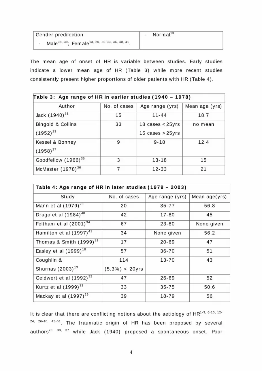
Gender predilection
- Male38, 39; Female13, 20, 30-33, 36, 40, 41.
- Normal13.
The mean age of onset of HR is variable between studies. Early studies
indicate a lower mean age of HR (Table 3) while more recent studies
consistently present higher proportions of older patients with HR (Table 4).
Table 3: Age range of HR in earlier studies (1940 – 1978)
Author No. of cases Age range (yrs) Mean age (yrs)
Jack (1940)51 15 11-44 18.7
Bingold & Collins
(1952)23
33 18 cases <25yrs
15 cases >25yrs
no mean
Kessel & Bonney
(1958)27
9 9-18 12.4
Goodfellow (1966)35 3 13-18 15
McMaster (1978)36 7 12-33 21
Table 4: Age range of HR in later studies (1979 – 2003)
Study No. of cases Age range (yrs) Mean age(yrs)
Mann et al (1979)20 20 35-77 56.8
Drago et al (1984)40 42 17-80 45
Feltham et al (2001)34 67 23-80 None given
Hamilton et al (1997)41 34 None given 56.2
Thomas & Smith (1999)31 17 20-69 47
Easley et al (1999)18 57 36-70 51
Coughlin &
Shurnas (2003)13
114
(5.3%) < 20yrs
13-70 43
Geldwert et al (1992)32 47 26-69 52
Kurtz et al (1999)33 33 35-75 50.6
Mackay et al (1997)19 39 18-79 56
It is clear that there are conflicting notions about the aetiology of HR1-3, 6-10, 12-
24, 26-40, 43-51. The traumatic origin of HR has been proposed by several
authors20, 38, 37 while Jack (1940) proposed a spontaneous onset. Poor
5
footwear2, 23, ankle equinus23, pes planus3, 6, 23, 26, 40, 46-51 and functional hallux
limitus28-42-45 has also been cited.
A number of complaints can be associated with HR. These include generalized
foot pain, 1st MTP joint or metatarsosesamoid joint pain, 1st MTP joint
stiffness, locking and spasm/ cramp. In some cases, significant synovitis may
accompany these complaints. Variability of the severity and location of 1st MTP
joint pain may be dependent upon a number of factors including lifestyle and
activity levels. In the early stages, discomfort predominates at the dorsal
aspect of the joint and becomes more diffuse with the progression of the
disease. Other complaints including metatarsalgia (due to a compensatory
increase in weight bearing to unload the 1st ray during gait), inability to rise
up on toes and altered gait have been documented1, 13, 14, 20, 21.
This study aims to identify the demographics and key clinical parameters
associated with a group of subjects with HR. The methodological process used
and its impact on the accuracy of severity and grading of HR will be
discussed.
6
METHODOLOGY
An observational, cross-sectional study was undertaken. This involved a
quantification of specific variables applied to a sample of subjects with varying
severity. It was undertaken to evaluate clinical parameters in 110 HR subjects
(180 feet) aged 18-70 years with varying degrees of restricted 1st MTP joint
dorsiflexion <65° (measured with a standard full-circle plastic goniometer,
calibrated to 1º increments) with either pain, deformity or both. Ethical
approval (Leicestershire, Northants, Rutland) was obtained, subjects gave
informed consent and a pilot study was undertaken.
Careful preliminary examination of subjects’ clinical notes was undertaken to
remove those possessing criteria of exclusion (Table 5). An invitation letter
and study information sheet was sent to suitable subjects giving them time
for consideration prior to inclusion in the study.
Detailed exclusion criteria were reviewed at the time of data collection.
Table 5: Exclusion criteria
Hallux valgus-rigidus (intermetatarsal angle > 12º).
Severe multiple forefoot deformities.
Significant trauma sustained to foot/ leg in previous 12 months.
Neuropathy.
1st ray/ forefoot surgery (including digital/ excluding soft tissue).
Morton’s neuroma affecting any inter-metatarsal space.
Septic arthritis of 1st MTP joint.
Inflammatory arthritides.
Neuromuscular disorders.
Insulin dependent Diabetes Mellitus.
Hypermobility syndromes.
Long-term steroid use.
History of severe peripheral vascular disease.
Metabolic bone disease.

7
A standardized questionnaire and examination were used. Subjects were
questioned about their history including the following: family history of great
toe problems, age of onset (denoted by 1st MTP joint deformity or restriction/
pain), duration of pain or symptoms (including stiffness, locking,
spasm/cramp), variability of pain, factors aggravating symptoms, factors
providing relief of symptoms, affect on activity levels and types of activities
restricted, contribution of occupation to HR and footwear restrictions. The
body mass index (BMI) for each subject was documented to determine its
effect on the clinical parameters. Repetitive 1st MTP joint trauma can result in
joint damage precipitating HR. The subject’s type and frequency of sporting
activities was documented. The association of 1st MTP joint OA (HR) and OA at
other sites was documented.
A validated questionnaire (FHSQ)59, 60 was also completed by each subject and
used to measure health-related quality-of-life dimensions: foot pain, physical
function, appearance, footwear and general perceptions of foot health.
The physical examination included inspection of the foot non-weight bearing
and weight-bearing. Both feet were examined (exclusion criteria permitting).
The following clinical data was obtained: Rearfoot position in stance was
evaluated using the Foot Posture Index (FPI)53. The FPI was used to quantify
the degree to which the foot was pronated, supinated or in a neutral
position53. Six foot parameters (3 rearfoot and 3 forefoot) were evaluated for
each subject. Each parameter was graded as described by Redmond et al53.
Final aggregate scores were applied to categorize type of foot posture.
Location, magnitude and timing of 1st MTP joint pain were assessed. Passive
1st MTP joint range of motion ROM was measured using the method described
by Ronconi et al54 and Greene & Heckman55. The total ROM (dorsiflexion &
plantarflexion) was used to calculate reduction in motion and compared with
normal values55. Active 1st MTP joint dorsiflexion was measured in a static
weight bearing position. Subjects were asked to push up onto the ball of the
foot (avoiding supinating) to obtain maximum 1st MTP joint dorsiflexion.
Subjects’ ability to rise up on toes without supinating was also evaluated.
Hallucal IPJ pain (using system described by Coughlin22) was assessed.
Frontal plane hallucal position was determined by comparing the angle of the
hallucal nail plate with the ground. Sagittal plane position (hallucal IPJ
hyperextension) was measured weight bearing with a goniometer using the

8
medial mid-axial line of the proximal and distal phalanges as reference points.
Transverse plane deformity of the hallucal IPJ (hallux abductus
interphalangeus) was measured with a goniometer using the dorsal mid-axial
line of proximal and distal phalanges as reference points. Hallucal flexor
power was measured by assessing the ability of the hallux to prevent a piece
of paper from being pulled away from under it during static stance.
The location of callosities, lesser toe deformities, comparison of 2nd toe length
with hallux and lesser MTP joint pain were documented. Ankle joint
dorsiflexion was measured with a standard plastic full-circle goniometer
(calibrated to 1 degree increments) using the technique described by
Silfverskiold56 (knee extended and flexed position). The foot was held with the
talonavicular joint reduced to eliminate transverse tarsal or subtalar motion57,
58. The fibula and plantar-lateral border of the foot were used as landmarks
(Figure 1). A right angle was considered to be neutral position. A brief
subjective assessment of the subjects’ gait at propulsion (by 1st author) was
undertaken (Table 6).
Figure 1: Measurement of ankle joint dorsiflexion
Table 6: Observed gait parameters
Propulsion
Mid-tarsal joint pronation
Supination
Delayed heel lift
Vertical toe-off
Ab/Adductory twist at toe-off
Knee flexion
Inability to push through ground at toe-off

9
RESULTS
Descriptive and comparative statistical analyses were performed using SPSS
for Windows version 15.0) (SPSS Inc., 233 S. Wacker Drive, Chicago, IL
60606, USA). Standard chi-square analysis (x2) was performed on categorical
data. Pearson and binary correlation coefficients were used to evaluate the
non-continuous data. Differences were considered to be significant when the P
value was <0.05.
Demographic data
The findings of the current study demonstrate that HR was associated with
increased female prevalence, bilateral involvement, and older age of subjects
at onset (Tables 7, 8, 9). These findings concur with those of previous
research13. Few subjects in the current study had adolescent onset. It is
recognised that this may be influenced by the minimum age of subjects (18
years) used and the fact that subjects were only taken from an adult
orthopaedic clinic.
The mean age of onset of symptoms (1st MTP joint deformity or restriction/
pain) was 44 years. This is eleven years prior to the median age of
presentation at a foot and ankle clinic (55 yrs) and supports the concept that
this condition may be one of insidious development. Foot biomechanics,
footwear type and activity levels may have some bearing on the development
of symptoms and subsequent progression of disease. Overall subjects were
marginally overweight (>25 Kg/m2 = overweight), indicated by a mean BMI of
25.93 Kg/m2 (19.53-37.26) but with no gender difference for this variable
(male: 26.48, female: 25.70).
In the current study there was a pronounced difference between gender;
more females presented with HR (Table 7), the mean age of HR onset was
less in females (43 years) than males (51 years) and the ratio of females to
males was greater in the younger age groups (Table 8).
The mean age of HR onset in the bilateral group was 50 years and unilateral
group 53 years. Bilateral foot involvement was similar between genders (62%
females, 68% males).
10
Table 7. Sample characteristics
Subjects
(feet)
Gender
Female Male
Age (yrs)
Mean Median
(range)
Age of onset
Mean (range)
Duration of
symptoms
yrs (range)
110
(180)
73
66%
37
34%
52
(23-70)
55 44 (14-68) 6 (1-33)
Table 8. Age groups
Years 18-30 31-40 41-50 51-60 61-70
% 5.7 10.7 18.6 37.1 27.9
F:M ratio 7:1 7:1 1:1 2:1 3:1
History data
No statistically significant association was found between HR and a history of
trauma (p=0.1). Trauma history was only found in a small proportion of
subjects and was more common in those with unilateral HR (Table 9).
Table 9: Foot involvement
Bilateral
(subjects) %
Unilateral
(subjects) %
Trauma history
(feet) %
(70) 64% (40) 36%
L (18) 45%
R (22) 55%
(39) 22%
Unilateral 74%
Bilateral 26%
Onset of HR was reported to be insidious in 86 (78%) of subjects and acute in
24 (22%) subjects. 1st MTP joint pain (within the last 6 months) was reported
to be severe in 26 (23.6%) subjects, moderate in 42 (38.2%), mild in 22
(20%) and not present in 20 (18.2%) of subjects. Historical categorical
findings are presented in Table 10A and 10B. Subjects stated that footwear
contributed to the development of HR in 23% of cases, however, pain in the
1st MTP joint was found to be associated with footwear on most days in 40
patients (36%). Short, tight, loose fitting, high-heeled and new footwear was

11
found to aggravate symptoms of HR. Occupation contributed to HR in 29% of
subjects and other factors found to aggravate HR are outlined in the
discussion. There was no statistically significant correlation between HR and
footwear or occupation (p>0.1).
Table 10A. Categorical history findings (Based on 110 subjects or 180 feet)
Parameters
Count (%)
Never Rarely Some days Most days Everyday
Activity levels
restricted by HR
8
(7)
16
(14.5)
17
(15.5)
35
(31.8)
33
(30)
Footwear contributing
to 1st MTPJ pain
4
(3.6)
20
(18.1)
25
(22.7)
40
(36.3)
21
(19.3)
Variability of 1st MTPJ
pain
6
(5.4)
8
(7.2)
37
(33.6)
42
(38.3)
17
(15.4)
1st MTPJ pain on
movement
9
(8.1)
2
(1.8)
24
(21.8)
35
(31.8)
40
(36.3)
1st MTPJ pain at rest 42
(38.1)
14
(12.7)
32
(29)
16
(14.5)
6
(5.4)
Presence of 1st MTPJ
stiffness
15
(13.6)
10
(9)
26
(23.6)
36
(32.7)
23
(20.9)
Morning 1st MTPJ
stiffness only
38
(34.5)
7
(6.3)
15
(13.7)
29
(26.4)
21
(19.1)
Evening 1st MTPJ
stiffness only
31
(28.1)
14
(12.2)
24
(21.8)
28
(25.4)
13
(11.8)
1st MTPJ stiffness all
day
39
(35.4)
11
(10)
23
(20.9)
21
(19)
16
(14.5)
1st MTPJ spasm/
cramp
50
(45.4)
18
(16.3)
32
(29)
9
(8.3)
1
(0.9)
Locking of 1st MTPJ 70
(63.6)
13
(11.8)
23
(20.9)
3
(2.7)
1
(0.9)
Ability to rise up on
toes
23
(20.9)
24
(21.8)
20
(18.1)
21
(19)
22
(20)
Lesser MTPJ pain 111
(61.6)
16
(8.8)
33
(18.3)
12
(6.6)
8
(4.4)

12
Change in walking
pattern
11
(10)
13
(11.8)
29
(26.1)
21
(19)
36
(32.7)
Ability to push off
through ground
21
(11.6)
36
(20)
50
(27.7)
22
(12.2)
51
(28.3)
Roll out during
propulsion
45
(25)
24
(13.3)
38
(21.1)
31
(17.2)
42
(23.3)
Table 10B. Categorical history findings (Based on 110 subjects)
Parameters
Count (%)
None CLO GS GS
+ C
Gels P B V A CD CC
Drugs used
for 1st
MTPJ pain
51
(46)
7
(6)
13
(12)
12
(11)
1
(1)
4
(3.5)
11
(10)
5
(4.5)
2
(1.8)
2
(1.8)
2
(1.8)
Legend: CLO= Cod liver oil, GS= Glucosamine Sulphate, C= Chondroitin,
Gels = topical non-steroidals, P= Paracetamol, B= Brufen, V= Volterol, A=
Arthrotec, CD= Co-dydramol, CC= Co-codamol.
Clinical data
Table 11 shows mean clinical findings. The confidence interval (CI) illustrates
the range of measures drawn from the study sample.
Table 11. Mean Clinical findings (Based on 180 feet)
Parameters
(counts*)
Mean ± SD 95% CI
Lower Upper
Range
Passive 1st MTPJ ROM
- Dorsiflexion
- Plantar flexion
Active 1st MTPJ ROM
- Dorsiflexion
Ankle joint equinus
- Knee extended
- Knee flexed
41°
15°
58°
10°
13°
19°
5°
19°
2°
3°
37
11
53
8°
12°
43
17
60
10°
15°
0-82°
0-25°
0-90°
5°-17°
8°-25°

13
Legend: *= nominal data, SD= Standard deviation, ROM= range of motion
The hallucal position (frontal plane) was rectus in 91 (50.5%) feet, valgus in
75 (41.6%) feet and varus in 13 (7.2%) feet. Hallucal flexor power was weak
in 10 (5.5%) feet, medium in 20 (11.1%) feet and strong in 150 (83.3%)
feet. The length of the 2nd toe compared with the hallux was found to be
longer in 54 (30%) feet, the same length as the hallux in 111 (61.6%) feet
and shorter than the hallux in 15 (8.3%). During passive 1st MTP joint
dorsiflexion pain occurred at the end-of-range in 29 (26.3%) subjects, mid-
range in 41 (37.2%) subjects, beginning in 35 (31.8%) subjects and all-of-
range in 5 (4.5%) subjects.
Osteoarthritis was present in joints other than the 1st MTP joint in 32 (29.1%)
subjects; hips were affected in 14 (12.7%) subjects, knees in 40 (36.3%)
subjects and finger joints in 56 (50.9%) subjects.
Tables 12A and 12B show categorical clinical findings.
Table 12A. Categorical clinical findings (Based on 110 subjects or 180 feet*)
Parameters
Count (%)
Normal Delayed
heel lift
Supination Vertical
toe-off
Abductory
twist
Knee
flexion
Gait at
propulsion
37
(20.5)
50
(27.7)
68
(37.7)
11
(6.1)
12
(6.6)
2
(1.1)
None Hallux IPJ 2nd MTPJ 3rd MTPJ 5th MTPJ 1st MH
Location of
callosities*
58
(32.2)
67
(37.2)
18
(10)
10
(5.5)
18
(10)
9
(5)
Severely
supinated
Supinated Neutral Pronated Severely
pronated
Foot Posture
Index*
6
(3.3)
12
(6.6)
78
(43.3)
64
(35.5)
20
(11.1)
None Hammer Claw Mallet AV
Lesser toe
deformities*
9
(5)
13
(7.2)
77
(42.7)
18
(10)
63
(35)
14
< 20° DF < 15° DF < 10° DF < 5° DF < 0° DF
Ankle joint (AJ)
equinus*
5
(2.7)
58
(32.2)
107
(59.4)
10
(5.5)
0
(0)
Absent Mild
> 5°
Moderate
> 10°
Severe
> 15°
Hallucal IPJ
hyperextension*
60
(33.3)
66
(36.6)
46
(25.5)
8
(4.4)
HAI° * 51
(28.3)
50
(27.7)
57
(31.6)
22
(12.2)
Hallucal IPJ
pain*
144
(80)
18
(10)
16
(8.8)
2
(1.2)
Legend: HAI°= Hallux abductus interphalangeus angle, IPJ= interphalangeal
joint, DF= dorsiflexion, MT= metatarsal head, AV= adducto-varus.
Table 12B. Categorical clinical findings (Based on 180 feet*)
Parameters Count Percentage
Location of HR pain*
- Dorsal bump (DB)
- 1st MTP joint
- Sesamoids
- Proximal phalanx (PP)
- PP+ DC/ EHL
- DB + 1st MTP joint
- DB + PP
- DB + DC/ EHL
- DB + sesamoids
- DB + joint + sesamoids
- DB + DC/ EHL + sesamoids
- Joint + DC/ EHL
- Joint + PP
75
21
10
4
3
12
9
2
13
7
11
11
2
41.6
11.6
5.5
2.2
1.6
6.6
5
1.1
7.2
3.8
6.1
6.1
1.1

15
Legend: EHL= Extensor hallucis longus, DC= Dorsal capsule, ROM= range of
motion.
Foot Health Status Questionnaire
Questions relating to foot pain and physical function were assessed during the
last week whereas perceptions of foot health were assessed during the last
month (Table 13). General health was rated as very good by 90 (88%)
subjects, fair by 18 (16%) subjects and poor by 2 (2%) subjects. Severe foot
pain was experienced by 41 (37%) subjects, moderate pain by 22 (20%),
mild pain by 20 (18%), very mild by 9 (10%) and 17 (15%) experienced no
pain. Condition of feet was rated as excellent by 4 (4%) subjects, very good
by 18 (16%) subjects, good by 58 (53%) subjects, fair by 20 (18%) subjects
and poor by 10 (9%) subjects. Overall foot health was rated as excellent by 5
(5%) subjects, very good by 17 (15%) subjects, good by 58 (53%) subjects,
fair by 20 (18%) subjects and poor by 10 (9%) subjects. The amount of time
that foot pain affected subjects emotionally was rated as no time at all by 10
(9%) subjects, a small amount of time 25 (23%) subjects, moderate amount
of time 53 (48%) subjects, quite a bit of time 17 (15%) subjects and all of
the time 5 (5%) subjects.
Table 13. Foot Health Status Questionnaire (110 questionnaires)
FOOT PAIN
Count (%)
Never Occasionally Often Very often Always
Frequency of
foot pain
6
(5)
9
(10)
30
(27)
57
(52)
7
(6)
Frequency of
aching feet
6
(5)
18
(16)
25
(23)
51
(46)
10
(9)
Frequency of
sharp pains
25
(23)
53
(48)
22
(20)
6
(5)
4
(4)
PHYSICAL
FUNCTION
Not at all Slightly Moderately Quite a bit Extremely
Feet limit 15 22 33 25 15

16
work activity (14) (20) (30) (22) (14)
Feet limit
type of work
65
(59)
25
(23)
6
(5)
2
(2)
2
(2)
Foot health
limits walking
13
(12)
22
(20)
35
(32)
25
(22)
15
(14)
Feet limit
climbing stairs
9
(10)
22
(20)
37
(34)
21
(19)
19
(17)
FOOTWEAR Strongly
agree
Agree (A) Neither A
or D
Disagree
(D)
Strongly
disagree
Hard to find
comfy shoes
9
(8)
11
(10)
27
(25)
50
(45)
13
(12)
Hard to find
shoes to fit
11
(10)
9
(8)
27
(25)
50
(45)
13
(12)
Limited in
shoes worn
13
(12
50
(45)
27
(25)
9
(8)
11
(10)
PERCEPTIONS
FOOT HEALTH
All the
time
Most of the
time
Some of
the time
Little of
the time
None of
the time
Did foot
problems tire
10
(9)
25
(23)
53
(48)
17
(15)
5
(5)
Did you have
lots of energy
5
(5)
18
(16)
52
(47)
26
(24)
9
(8)
Did you feel
worn out
9
(8)
25
(23)
51
(46)
17
(16)
8
(7)
Did you feel
full of life
5
(5)
18
(16)
52
(47)
26
(24)
9
(8)

17
DISCUSSION
A number of findings are commonly reported in patients with HR, these
include pain on 1st MTP joint motion (particularly dorsiflexion)13, 15- 18,
restriction of 1st MTP joint motion6, 13, 23-25, joint enlargement with dorsal bony
proliferation13, 20, 26, intolerance of constricting footwear13, 21, 22, inability to
raise up on toes and a modified gait13, 16, 18, 20, 27-30, 61.These were verified in
the current study and will be discussed alongside other demographic, history
and clinical factors.
Demographics & history findings
Family History
Bonney and MacNab9 reported that patients with a positive family history (FH)
of great toe arthritis had an earlier onset of disease and Coughlin & Shurnas 13
found an association between HR and a positive FH of great toe problems in
almost two-thirds of patients. What is not clear is how many of these were in
fact hallux valgus (HV). In the current study 24% reported a positive FH, but
they could not differentiate between HR and HV. Future HR studies may need
to consider a properly controlled family study before a positive FH is
concluded.
Age of onset
Much has been written about the age of HR onset but not all authors are in
agreement (Table 2). Some early studies (Table 3) state that HR starts
spontaneously in childhood or adolescence35, 51 while others suggest it is
categorized as either primary (adolescent) or secondary (adult)6. Few studies
have reported on adolescent patients with HR5, 23, 27, 35, 36. In reviewing studies
that report on age,13, 18-20, 31-34, 40, 41, 62 the mean age at onset was 51 years.
The mean age at onset in the current study was 44 (14-68) years; only 3
subjects developed symptoms at an age of less than 18 years. Given the
small number of adolescent subjects with HR reported by our study and
others13, 18-20, 31-34, 40, 41, 62 and the fact that pathological specimens from both
adults and adolescent patients with HR were found to be consistent with
18
degenerative arthritis13, 23 it is concluded that artificially dividing patients into
primary and secondary categories is unnecessary.
Gender Predilection
Gould et al38 and Hattrup & Johnson39 both found a male predilection to HR.
Gould et al38 reported that 64% of HR patients were males and that gender
predilection depended upon age. Their results were only based on
15,000/45,000 returned questionnaires sent to shoe shops, where briefed
shoe fitters, asked and marked the questions. No clinical examination was
undertaken. The findings were then projected into the total United States
population (186 million at the time).
In complete contrast, virtually all recent HR studies (predominantly surgical
intervention)13, 20, 30, 31, 33, 36, 40, 41 show a higher female predilection (62%), a
percentage comparable to the current study (66%).
This female predilection to HR may not be due to biological differences but to
social and cultural factors that result in women wearing footwear that
aggravate a predisposition to develop HR or aggravate pain in deformities of
similar magnitude. Coughlin & Shurnas13, in a self-selected review of 18 post-
surgery HR studies, found that 62% of females were affected by HR, a finding
similar to their own results (63%) and concluded that there was an
association between HR and female gender. In addition they found that
females were more commonly affected in all age groups, a finding comparable
with the current study (Table 8). However, this finding may only reflect the
higher number of females receiving surgical treatment for HR, but not the
true male/female incidence in the general population, who have the condition
but have not as yet, had surgical intervention. The current study shows a
much higher ratio of females in the younger age groups (Table 8): Is this
because 18-40 year-old females are more likely to wear inappropriate
footwear?
Body mass index (BMI)
It was considered that an increased BMI may predispose subjects towards HR
and contribute towards levels of pain experienced. In the current study
subjects were only marginally overweight, indicated by a mean BMI of 25.93
19
Kg/m2 (19.53-37.26) and there was no gender difference (male: 26.48,
female: 25.70). BMI was not considered to be a predisposing factor for HR.
Bilateral involvement
Unilateral HR has been reported by some authors9, 20, 37. Drago et al40 reported
increased unilateral involvement in females, but presented no demographic
data to support this. In the current study unilateral involvement presented in
40 (36%) subjects (equal numbers of left or right feet); 38% were female
(Table 9). Other studies report bilateral HR13, 17, 38 or bilateral presentation
with unilateral symptoms. In this study bilateral involvement presented in 70
(64%) subjects (Table 9), which may reflect the predominance of older
subjects (Table 7) rather than the true incidence as, with the passage of time,
a higher percentage of patients are likely to exhibit bilateral disease. It may
also reflect the type of clinic (surgical) from which subjects were taken. In the
current study analysis was undertaken at the point of referral. Coughlin &
Shurnas13 found bilateral HR at final follow-up (79%) compared to 19% at
initial examination.
In the current study a history of trauma was common in subjects who
developed unilateral HR (positive trauma history in 22% study sample; 74%
of whom had unilateral involvement) (Table 9). No association between HR as
a whole and a history of trauma (p = 0.1) was found. These findings concur
with that of other researchers13. A statistically significant association between
unilateral HR and trauma (p < 0.05) was found.
A small proportion of unilateral HR subjects had the asymptomatic foot
examined. In these cases it was apparent that differences between the feet
existed and that they may result in different biomechanical function of the 1st
MTP joint. Although these findings suggest a trend the numbers of subjects
where such a comparison was possible was too small to enable definitive
conclusions to be drawn. Further research in this area is warranted.
Footwear
Poor footwear has been implicated in the development of HR for many
decades. Davis-Colley2 first proposed a link in 1887. Bingold & Collins23 and
DuVries63 cited footwear that is too short, Lorimer et al64 footwear that is too
loosely fitting and Cracchiolo65 footwear that causes hyperextension of the
20
great toe as a cause of HR. Some authors reported that patients with HR were
intolerant to footwear13, 21, 22. Unfortunately, the vast majority of ‘evidence’
over the years has been anecdotal. The few studies that addressed the issue,
found that the association between footwear and HR was not statistically
significant13, 66. Sim-Fook & Hodgson66 examined 118 shod and 107 unshod
Chinese subjects. Only 17% of those wearing footwear and 10.3% not
wearing footwear, were affected by HR. There was a marked gender bias in
that 84% of the unshod were female and 67% of the shod were male.
Coughlin & Shurnas13 found that 16% of patients considered their footwear to
be a contributory cause of their HR. They found no statistically significant
correlation between footwear and HR to confirm this (r =0.08, p>0.1)13.
In the current study only 23% subjects considered their footwear a
contributory cause of their HR. However, the frequency of 1st MTP joint pain in
HR associated with footwear was found to affect 36% of subjects on most
days (Table 10A). The most common types of footwear restrictions reported
by females were high heeled shoes (31%) probably because the 1st MTP joint
is held in an extended position during gait. Slip-on shoes (16%) and
Wellington boots (3%) may cause FHB overuse to maintain stability and
subsequent sesamoid pain. In 14% of subjects dress shoes were found to
compress the forefoot, this may alter 1st MTP joint biomechanics. Flat shoes
(5%) may increase the requirement for dorsiflexion at propulsion. Shoes with
a seam over 1st MTP joint (3%) rub the joint especially if dorsal osteophytes
are present and can compress the dorsomedial cutaneous nerve resulting in
dysesthesia or numbness along the medial border of the hallux. Walking
boots (2%) and new shoes (1%) only contributed to HR in a few cases. No
footwear restrictions were reported in a quarter of subjects, most of which
were males.
Factors aggravating HR
In the current study subjects reported a number of factors responsible for
aggravating the symptoms of HR. Whilst footwear (23%) was the most
common other factors were also reported: cold/damp weather (11%), walking
on uneven terrain (10%) or for long distances (9.5%), normal walking
(8.2%), running (6.4%), descending stairs (6.4%), stubbing HR toe joint
(4.6%), not wearing insoles (4.6%), kneeling (4.5%), driving for long periods
21
(3.6%), standing for long periods (2.8%), weight of bed covers (2.7%),
increased body weight (0.9%). Subjects reported that prolonged activity while
barefoot or in soft-soled shoes was often difficult. Only 1.8% of subjects
reported that no factors aggravated their HR. Factors aggravating HR are
likely to be idiosyncratic, influenced by lifestyle and general health.
Relief of HR symptoms
Subjects reported strategies responsible for immediate relief of HR symptoms.
Sitting (23.6%), removal of footwear (23%), wearing of insoles with trainers
(9%) and use of painkillers (5.7%) were the most common. It is interesting
that so few subjects opted to use painkillers although this is reflected in the
small number of subjects reporting severe 1st MTP joint pain (23.6%). A
strong correlation between the use of painkillers and symptoms in these
particular subjects was found (r= .82, p= 0.05). Other strategies reported
included: 1st MTP joint distraction (5%), immersing joint in warm water
(4.3%), use of flat stiff soled shoes (3.5%), modified gait (walking on outer
border of foot) (3.4%), foot exercises (3%), massaging joint (2.9%), walking
on flat surfaces (2.6%) and use of non-steroidal gel (1%). In subjects with
well advanced disease no measure would obtain immediate pain relief (13%).
Subjects presented with a wide range of HR pathology and symptoms but the
majority took either no pain medication (46%) or over the counter drugs
(44%) whilst a few (11%) took prescription only medicines (Table 10B).
Restriction of activity levels
1st MTP joint pain in HR was found to restrict activity levels in subjects on
most days (31.8%) (Table 10A) and 30% of subjects were moderately
affected in their activities (Table 13). The types of activities restricted by HR
included: running, long walks (particularly hill walking), walking on uneven
surfaces, dancing, multidirectional sports and aerobic exercise. Predominantly
activities requiring a forced excursion of the 1st MTP joint in the sagittal
and/or frontal plane may precipitate pain. Transverse plane movement
however, is resisted because of increased transverse plane stability promoted
by bony changes in HR.
Occupation

22
In the current study 42% of subjects lead an active occupation but only 29%
considered that their occupation contributed to HR. This concurs with the
FHSQ data (30% of subjects reported being affected at work by their HR) and
that of other studies who found no statistically significant correlation between
HR and occupation (r= .08, p>.1)13.
In the current study, just over one-quarter (27%) of subjects were retired,
which may influence their activity levels and subsequent HR pain. In
retirement some subjects may be more active while others may be less active
because of ill health. This factor has not been considered in other studies.
1st MTP joint symptoms
In the current study subjects reported moderate (38.2%) and severe (23.6%)
1st MTP joint pain within the last 6 months and only 18% of subjects reported
no pain (Table 10A). A painful 1st MTP joint was reported for 67% of waking
hours on movement and variable on most days for 38% of subjects (Table
10A). Some subjects (29%) presented with pain at rest (Table 10A).
Subjects were asked to grade their 1st MTP joint stiffness and indicate the
period during the day when they experienced joint stiffness. This was graded
on a continuum between zero and ten (0= no stiffness, 10 = unable to move).
In the current study 86% of subjects reported 1st MTP joint stiffness (Table
10A) and if variable, at its worst, 45% were graded as 5 out of 10. Only 20%
of subjects reported no 1st MTP joint stiffness. There was a strong correlation
between 1st MTP joint pain and stiffness (r= .79, p= .01) but this was not
statistically significant. Morning stiffness was reported in 66% of subjects;
evening stiffness in 71% and 64% had 1st MTP joint stiffness throughout the
day (Table 10A).
Locking of the 1st MTP joint was reported in 36% subjects but was variable
and short lasting in nature. More commonly 55% of subjects experienced
cramp/ spasm of the 1st MTP joint and hallux (Table 10A) a consequence of
capsulitis and FHL/ FHB tenosynovitis. Subjects reported 1st MTP joint
symptoms to be worse during the heel-rise and propulsion phases of gait.
Subject’s perception of their gait
In the current study 90% of subjects considered that their walking pattern
had changed during the development of their HR, of which 33% considered

23
that this change affected them everyday (Table 10A). Only 51 (28%) of feet
were able to push through the ground at propulsion everyday, the remainder
were affected to varying degrees of severity (Table 10A) and 135 feet (75%)
rolled outwards during propulsion. The differences in frequency for each of the
above variables of gait are outlined in Table 12A.
Presence of OA in other joints
An association between radiological foot OA and radiological OA at other sites
has been reported67. In the current clinical study 29% of subjects (mainly
females) with HR (1st MTP joint OA) reported OA in other joints. This was
found to be most common in finger joints (51%). Whilst these findings
indicate a relationship between the parameters this is not necessarily a causal
relationship. Future epidemiological studies would be useful to determine
whether a systemic aetiology is involved in the development of HR and
provide an enhanced ability to describe the respective influences of
mechanical and systemic factors in the development of this condition.
Sport
It is recognized that certain sports impart 1st MTP joint trauma and may be
responsible for precipitating HR development whilst their frequency may
exacerbate symptoms. In the current study 69% of subjects reported
undertaking a range of sports (football, rugby, tennis, golf, badminton, rock
climbing, running, walking, horse riding, yoga, aerobics and swimming) of
variable frequency (1-5 times per week) prior to HR onset.
Foot Health Status Questionnaire (FHSQ)
The FHSQ evaluated health related quality-of-life dimensions of foot pain,
physical function/ appearance, footwear and general perceptions of foot
health. Findings from the FHSQ (Table 13) broadly concur with the history and
physical results of the current study (Tables 10-12). The severity (37% -
severe) and frequency (42% - very often) of foot pain documented was
greater than that verbally reported (clinical study). This may be because the
FHSQ data related to foot pain within the previous week rather than the last
six months (clinical study). Interestingly the frequency of foot pain was found
to vary (52% -very often) more often in the short term (one week) than over

24
a longer period of six months (38% most days). Some subjects reported that
their 1st MTP joint pain made them feel tired and worn out and that their pain
appeared to affect them both physically and emotionally (Table 13).
The restrictions of physical function documented by subjects were related to
similar activities as those found in the clinical component of the study
(aggravating factors). Subjects reported that although it was possible to find
footwear to fit their feet and which does not hurt their feet the number and
type of footwear was limited (Table 13). A number of subjects (particularly
females) were not happy with the appearance of their feet because of the
enlarged 1st MTP joint/s and considered that this factor as well as joint pain
limited them in their choice of footwear. The majority of subjects reported
their general health as good except for two subjects who were restricted by
heart disease (angina). It was interesting to note that the subject’s perception
of their general foot health was good (apart from 1st MTP joint) but many felt
that their 1st MTP joint/s pathology limited them in vigorous physical and
social activities and were concerned about the impact this may have on their
long term general health.
Clinical findings
Factrs thought to contribute to development of HR
Pes Planus
Pes planus as a cause of HR has been implicated by a number of authors3, 6, 23,
26, 40, 46-51, 61 with the understanding that excessive foot pronation results in
increased plantar fascia tension and increased dorsiflexion force under the 1st
metatarsal head, and thus a reduced ability of the hallux to dorsiflex. No
demographic data were reported in any of these studies to substantiate the
notion that pes planus is a cause of HR.
Jack51 assessed foot posture by observing the weight-bearing arch of the foot
but no criteria were documented to quantify this. Jack51 considered an
association between pes planus and HR but was unclear which comes first or
whether the two develop pari passu. Coughlin & Shurnas13 assessed foot
posture using a Harris Beath mat to measure arch height or excess heel
valgus. Only 11% of their patients had pes planus. Their results were similar
to those of Harris & Beath57 (15%) who examined 3619 normal military
25
recruits. The Harris & Beath mat has not been tested for reliability and validity
and it was considered that the results of Coughlin & Shurnas which were
based on previous studies were not reliable or conclusive.
Scherer68 suggested that calcaneal eversion can theoretically limit 1st MTP
joint motion. Harradine & Bevan69 appeared to validate this conjecture by
examining the effect of static rearfoot eversion (using 3º, 5º and 8º valgus
wedges in a standard shoe) on 1st MTP joint ROM. A reduced joint ROM with
increasing calcaneal eversion was found. This artificially replicated three
magnitudes of pronation; therefore the findings may not be representative of
the full continuum of foot pronation seen in the general population.
Mahiquez70 examined the relationship between rearfoot valgus and 1st MTP
joint OA and found 23% of subjects more likely to develop 1st MTP joint OA
with hindfoot valgus. Halstead et al71 found patients with 1st MTP joint OA
demonstrated higher medial forefoot pressures and more pronated foot
postures. Grady et al72 retrospectively analysed 772 HR patients and found
5.5% had aetiologies of both trauma and excessive pronation while 21.7%
had excess pronation only. The measurement criteria included excess
pronation at mid-stance/toe-off and Kite’s angle >45º.
Payne & Dananberg73 contend that blockade of 1st MTP joint sagittal plane
motion (sagittal plane facilitation theory) produces compensation within other
planes. Compensatory subtalar and mid-tarsal joint pronation (frontal plane)
with forefoot abduction (transverse plane) can ensue, producing flatfoot in
some HR patients. Whilst the above studies provide interesting theories
linking pes planus with HR none use a validated tool to quantify foot posture.
In the current study the Foot Posture Index (FPI)53 was used to quantify the
degree of pronation or supination. This is a valid, reliable and objective
measure of foot function53. The FPI quantifies foot posture in a relaxed stance
position, requiring no manipulation of the foot, marking of lines or
measurement with instrumentation. Thus the controversial issues relating to
goniometer assessment and validity of neutral subtalar joint positioning are
avoided74, 75. In the current study 84 (47%) feet had pes planus (11% of
which were severely pronated). The remaining 78 (43%) feet had a normal
foot posture and 20 (10%) feet had a high arched supinated foot (Table 12A).
A strong correlation between a pronated (pes planus) foot and 1st MTP joint
pain was found (r= .84, p= .05). It is theorized that in a pes planus foot
26
forefoot hypermobility at propulsion may promote 1st MTP joint instability,
increasing ROM and pain. A correlation between increased 1st MTP joint ROM
and pronated feet (r= .72, p=0.1) support this concept although this was not
statistically significant. Whilst these findings indicate a relationship between
the parameters this is not necessarily causal.
Functional hallux limitus
Functional hallux limitus (FHLim) is defined as reduced 1st MTP joint
dorsiflexion on foot loading compared with passive non-weight-bearing and
has been proposed as a cause of HR28,42-45. None of these authors mention
ankle joint position when assessing for FHLim. If the ankle is plantarflexed
when passive 1st MTP joint dorsiflexion is tested then hallux dorsiflexion is
likely to increase as the flexor hallucis longus (FHL) is taken off stretch15.
Coughlin & Shurnas76 also question the concept of FHLim as reported in the
literature, which remains theoretical conjecture and a subjective diagnosis45,
conceived to explain abnormalities seen on in-shoe pressure readings and
visual gait assessments68. Coughlin & Shurnas13 hypothesized that FHLim may
represent the residual elevatus occasionally noted on dorsiflexion stress x-
rays of patients with severe HR. In early stage HR FHLim may be a
consequence of tenosynovitis of the FHL tendon which limits its excursion and
subsequently that of 1st MTP joint dorsiflexion on foot loading15. The findings
of the current study concur with those of other authors13, 15.
2nd toe length
Three foot types are seen in the general population. Square foot (hallux and
2nd toe equal length), Morton’s or Greek foot (hallux shorter than 2nd toe) and
Egyptian foot (hallux longer than 2nd toe). Ogilvie-Harris et al77 assessed 2nd
toe length in ballet dancers and found a correlation between HR and a longer
2nd toe78. One clinician examined 59 dancers (34 female & 25 male)
comparing them to a randomly selected control group of 60 subjects (30
female, 30 male). The authors defined any difference in length of <2mm as
not significant recording it as normal. No radiographic evidence was used and
the lack of this may have influenced the outcome of results. They reported
that 40% male and 27% female ballet dancers had a long 2nd toe compared to
the hallux. In the control group (in which there was no HR), 60% of males
27
and 43% of females had a longer 2nd toe. The authors concluded that 44% of
ballet dancers, with a long 2nd toe, had bilateral HR but failed to elicit the
related pathomechanics.
The current study does not concur with that of Olgilvie-Harris et al77; 54 feet
(30%) had a long 2nd toe while 111 feet (62%) had a 2nd toe the same length
as the hallux and 15 (8%) a 2nd toe shorter than the hallux. Chi-square
analysis of 2nd toe length and 1st MTP joint pain revealed a significant finding
(p=0.001<0.05). In a radiographic study of the same subjects the proximal
phalanx was found to be longer than the distal phalanx79. The overall length
of the hallux may be a factor contributing to HR and is supported by others
who have compared HR with non-HR subjects and found a longer hallux in the
HR group80. Whilst these findings indicate a relationship between the
parameters this is not necessarily a causal relationship.
Factors used as markers of severity
Increased joint size and soft tissue swelling
Increased 1st MTP joint size in HR has been documented13, 20, 26. This is related
to the presence of osteophytes, joint distension secondary to synovitis and
may provide an indirect clinical measure of joint damage. Soft-tissue swelling
of the 1st MTP joint has also been reported19, 20 and may be related to a dorsal
prominence that becomes painful from constant rubbing against the shoe,
capsulitis and EHL tenosynovitis resulting from stretching of soft tissues over
dorsal osteophytes. This may be the reason why HR subjects sometimes
complain of pain on hallucal plantarflexion. In the current study it was
observed that the magnitude of joint size increased with the severity and
duration of HR.
Pain with 1st MTP joint motion
Some studies have documented pain during 1st MTP joint motion13, 15-18. In the
current study subjects reported pain during passive ROM. The timing of this
pain during joint movement was documented in an attempt to quantify the
severity of HR (joint damage). Twenty-six percent of subjects reported end-
of-range pain suggestive of minimal joint damage, 69% of subjects reported
pain at the beginning or mid-range pain accounting for mild to moderate joint

28
damage and 4.5% reported all-of-range joint pain representing severe joint
damage. This reflected the range of severity of HR within the subjects.
Interestingly a strong correlation between 1st MTP joint pain and increased 1st
MTP joint ROM at propulsion was found (r= .84, p= .01) but this was not
statistically significant. This may explain why in a damaged 1st MTP joint
where there is still free and unrestricted joint motion pain is often likely
whereas, in an ankylosed 1st MTP joint where movement is restricted pain is
less likely. In feet with restricted 1st MTP joint ROM pain may present in other
areas (lateral forefoot) due to the compensation imposed by the restricted
joint motion. During active ROM subjects reported pain primarily during heel
lift and propulsion where 1st MTP joint dorsiflexion was required. In the
current study subjects reported that by altering their gait pattern they could
modify the severity and timing of symptoms.
Variability of 1st MTP joint pain
The natural history and symptoms of HR can vary from day-to-day and are
influenced by numerous aggravating or relieving factors. In some cases, the
condition takes a relatively benign course and in others symptoms are more
persistent81. In the current study 87% of subjects were found to have daily
variability of joint pain (Table 10A). It is concluded that the variability of joint
pain is multifactorial and may include factors such as lifestyle, health,
footwear and others (see aggravating factors). Whilst occupation does not
appear to play a role in the development of HR is may be responsible for its
variability.
Location of HR pain
Subjects presented with primarily dorsal bump pain (42%) (particularly early
in the condition), 1st MTP joint pain (12%), sesamoid pain (9%), proximal
phalanx pain (2%) or a combination of locations around the 1st MTP joint
(Table 12B). Sesamoid pain appeared to be more common in established HR.
Restricted joint motion
Studies have documented restricted 1st MTP joint motion in HR (especially
dorsiflexion)6, 13, 23, 24. The current study concurs with these findings (Table
11). Halstead et al71 demonstrated no association between measurements
29
obtained at the 1st MTP joint during static stance and maximum dorsiflexion
during walking. In view of the different modes of compensation for HR during
gait further research to compare 1st MTP joint motion in static stance and
during walking would be valuable and may help inform treatment.
Passive versus active 1st MTP joint ROM
Overall passive 1st MTP joint ROM was reduced as expected. Mean dorsiflexion
41° (0-82°) was below the normal range (65°-90°)21 and plantarflexion was
also reduced, mean 15° (0-25°) (Table 11).
Active 1st MTP joint dorsiflexion increased with weight-bearing. Mean active
dorsiflexion 58° (0-90°) (Table 11) was greater than mean passive
dorsiflexion, this may be a result of body weight and forward momentum
increasing available joint dorsiflexion however, this was still well below the
normal range (65°-90°). Also, possibly because of joint pain, some subjects
supinated their foot during movement reducing the need for as much
dorsiflexion. These findings concur with a radiological study in which a mean
hallux equinus angle of 11° was found during stance, this is outside the
normal range (16°-18°)79.
Both bone (including joint) and soft tissue changes associated with HR are
responsible for a reduced joint ROM (particularly dorsiflexion). The dorsal
capsule and EHL can become stretched and inflamed by dorsal osteophytes
causing pain and may contribute to limited plantarflexion in HR.
Hallux abductus interphalangeus (HAI)
In the current study 129 feet (72%) presented with HAI (Table 12A). A
moderate degree (> 10°) of HAI (transverse plane) was present in 57 feet
(32%). In 79 feet (44%) the HAI° was greater than normal (where normal
<10°). Strong correlations were found between HAI and 1st MTP joint pain (r
= .82, p = .03) which was not significant and HAI and reduced 1st MTP joint
ROM (r = .92, p = .05). It is hypothesized that the presence of HAI indicates
a more progressive HR process and that with increased 1st MTP joint damage
the 1st metatarsal head becomes flatter and more resistant to transverse
plane movement, thus predisposing to an increased HAI.
Factors associated with or secondary to HR
30
Ability to rise up on toes
In HR if 1st MTP joint dorsiflexion is restricted or painful then subjects may
avoid forced dorsiflexion of the joint imparted by rising up on their toes. In
the current study 21% of subjects were unable to undertake this manoeuvre,
the reminder could perform the task to varying degrees (Table 10A). A weak
correlation between 1st MTP joint pain and ability to rise up on toes was found
(r= .40, p=.03). Subjects can still perform this manoeuvre by supinating their
foot.
Hallucal position (frontal plane)
Medial 1st ray deviation, increased 1/2 intermetatarsal angle and lateral
deviation of the hallux may alter the pull of abductor hallucis causing it to
rotate the hallux into valgus. Valgus hallucal rotation is normally associated
with hallux valgus but may present in HR (Figure 2) where it may influence
sagittal plane motion at the 1st MTP joint.
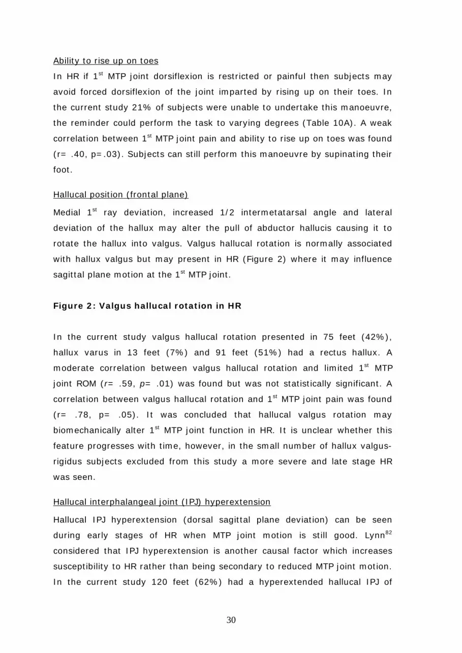
Figure 2: Valgus hallucal rotation in HR
In the current study valgus hallucal rotation presented in 75 feet (42%),
hallux varus in 13 feet (7%) and 91 feet (51%) had a rectus hallux. A
moderate correlation between valgus hallucal rotation and limited 1st MTP
joint ROM (r= .59, p= .01) was found but was not statistically significant. A
correlation between valgus hallucal rotation and 1st MTP joint pain was found
(r= .78, p= .05). It was concluded that hallucal valgus rotation may
biomechanically alter 1st MTP joint function in HR. It is unclear whether this
feature progresses with time, however, in the small number of hallux valgus-
rigidus subjects excluded from this study a more severe and late stage HR
was seen.
Hallucal interphalangeal joint (IPJ) hyperextension
Hallucal IPJ hyperextension (dorsal sagittal plane deviation) can be seen
during early stages of HR when MTP joint motion is still good. Lynn82
considered that IPJ hyperextension is another causal factor which increases
susceptibility to HR rather than being secondary to reduced MTP joint motion.
In the current study 120 feet (62%) had a hyperextended hallucal IPJ of
31
varying degrees of severity and 30% of these were greater than 10° (Table
12A). A correlation was found between hallucal IPJ hyperextension and 1st
MTP joint pain (r = .78, p = .01) but this was not statistically significant. This
relationship is not necessarily causal. In the current study the degree of
hallucal IPJ hyperextension did not appear to increase with increasing severity
of HR.
Hallucal interphalangeal joint (IPJ) pain
As sagittal plane restriction of the 1st MTP joint can result in compensatory
transverse and/or sagittal plane deformity of the hallucal IPJ it was assumed
that hallucal IPJ pain may develop. In the current study a painful IPJ was
reported in 36 subjects (20%) and only 18 (10%) of these had moderate to
severe pain (Table 12A). Chi-square analysis of hallucal IPJ pain and 1st MTP
joint pain revealed no significant finding (0.24>p>0.05). In this group of
subject’s hallucal IPJ pain was not considered to be a feature associated with
HR.
Hallucal flexor function
Tenosynovitis of the hallucal flexor tendons in HR may influence hallucal
purchase power. The ability of the hallux to prevent a piece of paper from
being pulled away from under it during static stance was not found to be
impaired in HR where 150 (83%) feet had a strong (not moveable) response.
Location of plantar callosities
These were related to abnormalities of gait (pronation and inverted step).
Increasing severity of 1st MTP joint pain results in more supinatory
compensation and subsequently more laterally placed callosities. In 67 (37%)
feet callus presented over the plantar medial hallucal IPJ. This may be related
to 47% of subjects who presented with a pronated gait in which there is likely
to be increased hallucal IPJ propulsion. Of the remaining 46 (26%) feet callus
was located under the lateral metatarsal heads (2nd- 5th), 9 (5%) feet under
the 1st metatarsal head and 58 (32%) feet presented with no callosities (Table
12A). The severity of callosities may be influenced by lifestyle and activity
levels.
32
Lesser toe position
Coughlin83 and Roukis et al1 noted that the medial angulation of the 2nd toe
can result from compensation during gait. In an attempt to provide medial
column stability the flexor digitorum longus (FDL) muscle contracts. A
“windswept” appearance to the entire forefoot, rather than just the 2nd toe
may result. In the current study lesser toe clawing in 77 (43%) feet,
medialisation (adduct-varus) of 3rd-5th toes in 63 (35%) feet, and other toe
deformities presented (Table 12A).
Ankle equinus
A recent study concludes that gastrocnemius contracture plays a vital
biomechanical role in chronic foot problems58. Bingold & Collins23 suggested
an association between Achilles tendon contracture and HR. Isolated
gastrocnemius tightness has been reported in up to 24% of “normal” patients
when defined as less than 5º dorsiflexion with the knee fully extended and the
condition was implicated in the pathogenesis of midfoot, hind foot, and
forefoot pathology52 although it was unclear how many patients had HR.
Coughlin & Shurnas13 found no association between Achilles tendon tightness
and HR. They defined gastrocnemius contracture as < 0º dorsiflexion when
the knee was fully extended with the foot in neutral. Only 3.5% of their
subjects had 5º or less of dorsiflexion although the study had no control
group with which to correlate the results. In the current study 10 (5.5%) feet
had 5° or less dorsiflexion with the knee fully extended and foot held in
neutral (to eliminate subtalar & midtarsal joint involvement) (Table 12A). No
subject had an Achilles tendon contracture <0°. The mean ankle dorsiflexion
with the knee extended was 9° (5°-17°) this increased to 13° (8°-25°) with
the knee flexed (Table 11). It is concluded that ankle equinus secondary to
Achilles tendon tightness is not associated with HR.
Lesser metatarsal overload
Supinated gait in response to restricted 1st MTP joint motion can cause
overload and pain in the region of the lesser metatarsal heads45. In the
current study lesser MTP joint pain (transfer metatarsalgia) was in reported in
69 (38%) feet with varying degrees of frequency (Table 10A). A strong
correlation between lesser MTP joint pain and a change in walking pattern (r=
33
.80, p= .05) was found. Chi-square analysis of lesser MTP joint pain and
supination at propulsion revealed a statistically significant finding (p<0.001).
It is hypothesized that 1st MTP joint restriction/ pain is responsible for altered
forefoot loading and subsequent metatarsalgia experienced (Table 10A). This
is supported by gait modifications found within the same proportion of
subjects where 68 (37%) feet were held in supination at propulsion (Table
12A). As other gait modifications are associated with HR not all subjects will
complain of forefoot pain.
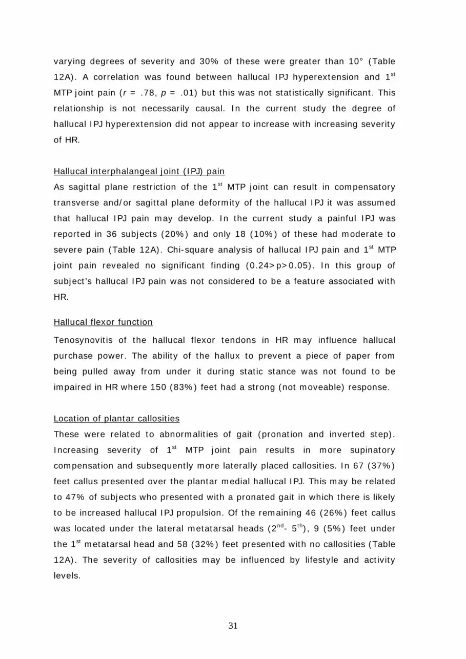
Altered gait
It is documented that gait in HR may become increasingly antalgic as the MTP
joint stiffens resulting in an everted13, 16, 18, 20, 27, 30 or supinated13, 51 position of
the foot. The sagittal plane facilitation theory42, 73 supports this and describes
five forms of compensation for sagittal plane blockade in HR:
1. Delayed heel lift
The mid-tarsal joint is the closest to the 1st MTP joint which allows sagittal
plane motion. This is seen as delayed heel lift with late midstance pronation
(Figure3) and navicular adduction/plantarflexion.
Figure 3: Delayed heel lift - mid-tarsal joint pronation/ eversion
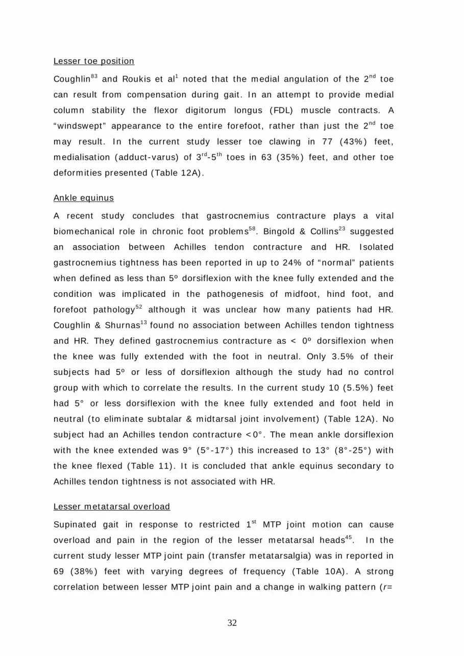
2. Vertical toe-off
Denotes continuation of delayed heel-lift where foot is lifted vertically off the
ground. An apropulsive, laborious, slow gait can present where there is a lack
of heel-off by the time of the contra-lateral heel contact (Figure 4).
Figure 4: Vertical toe-off
3. Inverted step
Patients with increasingly severe HR supinate their foot during gait13 to avoid
extending the 1st MTP joint and propulse from the lateral four toes (Figure 5).
Weight-flow is directed to the lateral column and fails to shift medially to the
first web space prior to heel lift. Lateral shoe wear results despite excessive
foot pronation. This explains the paradox of a flexible pronated foot with
lateral forefoot shoe wear and bulging. Dynamic in-shoe pressure
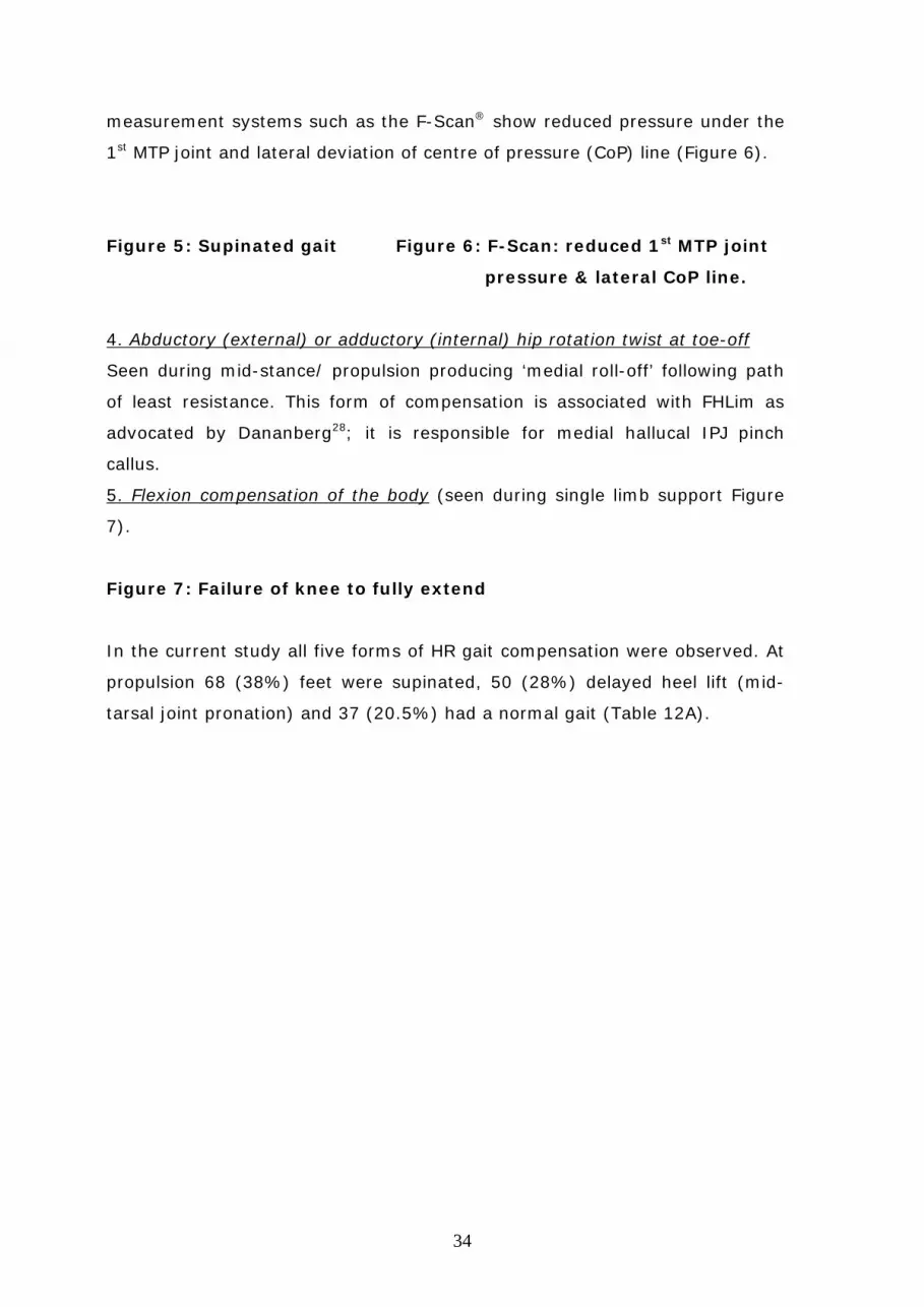
34
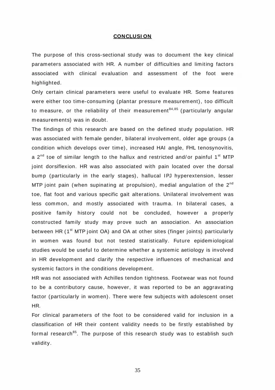
measurement systems such as the F-Scan® show reduced pressure under the
1st MTP joint and lateral deviation of centre of pressure (CoP) line (Figure 6).
Figure 5: Supinated gait Figure 6: F-Scan: reduced 1st MTP joint
pressure & lateral CoP line.
4. Abductory (external) or adductory (internal) hip rotation twist at toe-off
Seen during mid-stance/ propulsion producing ‘medial roll-off’ following path
of least resistance. This form of compensation is associated with FHLim as
advocated by Dananberg28; it is responsible for medial hallucal IPJ pinch
callus.
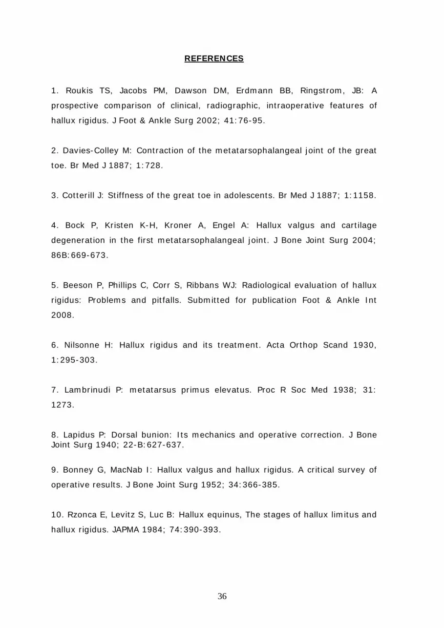
5. Flexion compensation of the body (seen during single limb support Figure
7).
Figure 7: Failure of knee to fully extend
In the current study all five forms of HR gait compensation were observed. At
propulsion 68 (38%) feet were supinated, 50 (28%) delayed heel lift (mid-
tarsal joint pronation) and 37 (20.5%) had a normal gait (Table 12A).
35
CONCLUSION
The purpose of this cross-sectional study was to document the key clinical
parameters associated with HR. A number of difficulties and limiting factors
associated with clinical evaluation and assessment of the foot were
highlighted.
Only certain clinical parameters were useful to evaluate HR. Some features
were either too time-consuming (plantar pressure measurement), too difficult
to measure, or the reliability of their measurement84,85 (particularly angular
measurements) was in doubt.
The findings of this research are based on the defined study population. HR
was associated with female gender, bilateral involvement, older age groups (a
condition which develops over time), increased HAI angle, FHL tenosynovitis,
a 2nd toe of similar length to the hallux and restricted and/or painful 1st MTP
joint dorsiflexion. HR was also associated with pain located over the dorsal
bump (particularly in the early stages), hallucal IPJ hyperextension, lesser
MTP joint pain (when supinating at propulsion), medial angulation of the 2nd
toe, flat foot and various specific gait alterations. Unilateral involvement was
less common, and mostly associated with trauma. In bilateral cases, a
positive family history could not be concluded, however a properly
constructed family study may prove such an association. An association
between HR (1st MTP joint OA) and OA at other sites (finger joints) particularly
in women was found but not tested statistically. Future epidemiological
studies would be useful to determine whether a systemic aetiology is involved
in HR development and clarify the respective influences of mechanical and
systemic factors in the conditions development.
HR was not associated with Achilles tendon tightness. Footwear was not found
to be a contributory cause, however, it was reported to be an aggravating
factor (particularly in women). There were few subjects with adolescent onset
HR.
For clinical parameters of the foot to be considered valid for inclusion in a
classification of HR their content validity needs to be firstly established by
formal research85. The purpose of this research study was to establish such
validity.
36
REFERENCES
1. Roukis TS, Jacobs PM, Dawson DM, Erdmann BB, Ringstrom, JB: A
prospective comparison of clinical, radiographic, intraoperative features of
hallux rigidus. J Foot & Ankle Surg 2002; 41:76-95.
2. Davies-Colley M: Contraction of the metatarsophalangeal joint of the great
toe. Br Med J 1887; 1:728.
3. Cotterill J: Stiffness of the great toe in adolescents. Br Med J 1887; 1:1158.
4. Bock P, Kristen K-H, Kroner A, Engel A: Hallux valgus and cartilage
degeneration in the first metatarsophalangeal joint. J Bone Joint Surg 2004;
86B:669-673.
5. Beeson P, Phillips C, Corr S, Ribbans WJ: Radiological evaluation of hallux
rigidus: Problems and pitfalls. Submitted for publication Foot & Ankle Int
2008.
6. Nilsonne H: Hallux rigidus and its treatment. Acta Orthop Scand 1930,
1:295-303.
7. Lambrinudi P: metatarsus primus elevatus. Proc R Soc Med 1938; 31:
1273.
8. Lapidus P: Dorsal bunion: Its mechanics and operative correction. J Bone Joint Surg 1940; 22-B:627-637.
9. Bonney G, MacNab I: Hallux valgus and hallux rigidus. A critical survey of
operative results. J Bone Joint Surg 1952; 34:366-385.
10. Rzonca E, Levitz S, Luc B: Hallux equinus, The stages of hallux limitus and
hallux rigidus. JAPMA 1984; 74:390-393.

37
11. Regnauld, B: Disorders of the great toe. In: Elson, R. The Foot:
pathology, aetiology, seminology, clinical investigation and treatment. New
York. Springer-Verlag. 1986; 269-281, 344-349.
12. Durrant MN, Siepert KK: Role of soft tissue structures as an etiology of
hallux limitus. JAPMA 1993; 83:173-180.
13. Coughlin MJ, Shurnas PS: Hallux rigidus: Dermographics, etiology and
radiographic assessment. Foot & Ankle Int 2003a; 24:731-743.
14. Vanore JV, Christensen JC, Kravitz SR, Schuberth JM, Thomas JL, Weil LS
et al: Clinical practice guideline first metatarsophalangeal joint disorders Panel
of the American College of Foot & Ankle Surgeons. Diagnosis and treatment of
first metatarsophalangeal joint disorders. Section 2: Hallux rigidus. J Foot
Ankle Surg 2003; 42:124-136.
15. Michelson J, Dunn L: Tenosynovitis of the flexor hallucis longus: A clinical
study of the spectrum of presentation and treatment. Foot & Ankle Int 2005;
26: 291-303.
16. Mann R, Clanton T: Hallux rigidus: Treatment by cheilectomy. J Bone Joint
Surg 1988; 70-A:400-406.
17. Shereff M, Baumhauser J: Hallux rigidus and osteoarthritis of the first
metatarsophalangeal joint: instructional course lecture. Current concepts
review. J Bone Joint Surg 1988; 80-A:898-909.
18. Easley M, Davis W, Anderson R: Intermediate to long-term follow-up of
medial-approach dorsal cheilectomy for hallux rigidus. Foot & Ankle Int 1999;
20:147-152.
19. Mackay D, Blyth M, Rymaszewaski L: The role of cheilectomy in the
treatment of hallux rigidus. J Foot & Ankle Surg 1997; 36:337-340.
38
20. Mann RA, Coughlin MJ, DuVries HL: Hallux rigidus: a review of the
literature and a method of treatment. Clin Orthop 1979, 142:57-63.
21. Camasta, CA: Hallux limitus and hallux rigidus. Clinics Podiatric Medicine
& Surgery 1996; 13: 431-437.
22. Coughlin M: Arthritides. In: Coughlin MJ, Mann RA eds: Surgery of the
Foot and Ankle 7th Ed. St Louis, Mosby 1999, 605-650.
23. Bingold A, Collins D: Hallux rigidus. J Bone Joint Surg 1950; 32-B:214-
222.
24. Smith R, Katchis S, Ayson L: Outcomes in hallux rigidus treated non-
operatively: a long-term follow-up study. Foot & Ankle Int 2000; 21:906-913.
25. Kilmartin TE: Metatarsal osteotomy for hallux rigidus. An outcome study
of three different osteotomy techniques compared with Keller’s excisional
arthroplasty. Br J Pod 2000; 3:95-101.
26. Giannestras N: Hallux rigidus. In : Giannestras N ed: Foot Disorders:
Medical and Surgical Management. Philadelphia, Lea & Febiger, 1973:400-
402.
27. Kessell L, Bonney G: Hallux rigidus in the adolescent. J Bone Joint Surg
1958; 40-B: 668-673.
28. Dananberg HJ: Gait style as an etiology to chronic postural pain. Part I:
Functional hallux limitus. JAPMA 1993a, 83:433-441.
29. Payne CB, Dananberg HJ: Sagittal plane facilitation of the foot. AJPM
1997, 31:7-11.
30. Mulier T, Steenwerckx A, Thienpont E, et al: Results after cheilectomy in
athletes with hallux rigidus. Foot & Ankle Int 1999; 20: 232-237.

39
31. Thomas P, Smith R: Proximal phalanx osteotomy for the surgical
treatment of hallux rigidus. Foot & Ankle Int. 1999; 20:3-12.
32. Geldwert J, Rocj G, McGrath M, Mancuso J: Cheilectomy: still a useful
technique for grade I and grade II hallux limitus/ rigidus. J Foot Surg 1992;
31:154-159.
33. Kurtz D, Harrill J, Kaczander B, Solomon M: The Valenti procedure for
hallux limitus: a long–term follow-up and analysis. J Foot & Ankle Surg 1999;
38:123-130.
34. Feltham G, Hanks S, Marcus R: Age-based outcomes of cheilectomy for
the treatment of hallux rigidus. Foot Ankle Int 2001; 22:192-197.
35. Goodfellow J: Etiology of hallux rigidus. Proc R Soc Med 1966; 59:821-
824.
36. McMaster MJ: The pathogenesis of hallux rigidus. J Bone Joint Surg 1978,
60B:82-87.
37. Bryant A, Tinley P, Singer K: A comparison of radiographic measurements
in normal, hallux valgus and hallux limitus feet. J Foot & Ankle Surg 2000;
39:39-43.
38. Gould N: Hallux rigidus: cheilectomy or implant? Foot & Ankle Int 1981;
1:315-320.
39. Hattrup S, Johnson K: Subjective results of hallux rigidus following
treatment with cheilectomy. Clin Orthop 1988; 226:182-191.
40. Drago JJ, Oloff L, Jacobs AM: A comprehensive review of hallux limitus. J
Foot & Ankle Surg 1984, 23:213-220.
41. Hamilton W, O’Malley M, Thompson F, Kovatis P: Capsular interposition
arthroplasty for severe hallux rigidus. Foot & Ankle Int 1997; 18:68-70.
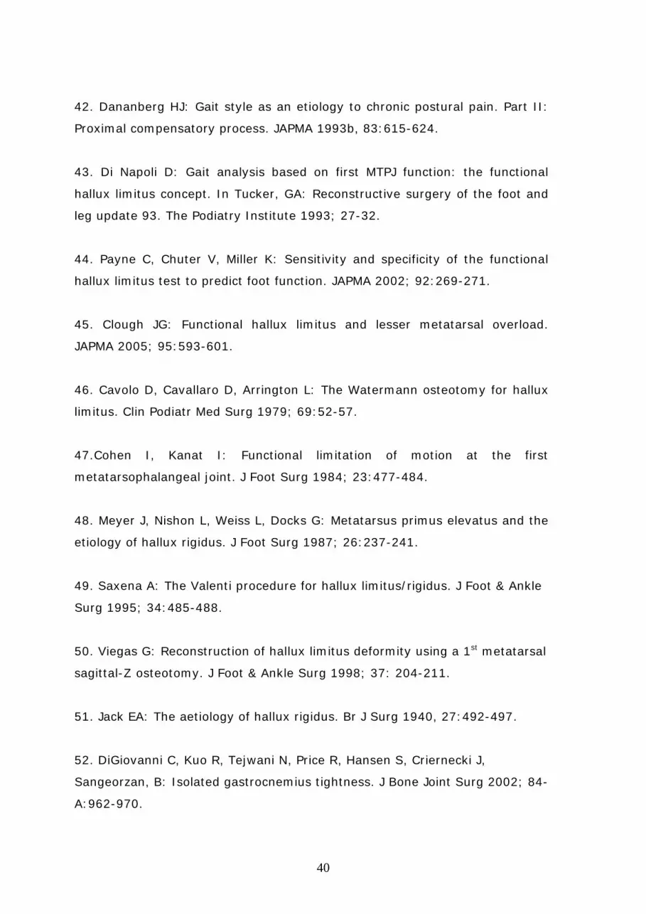
40
42. Dananberg HJ: Gait style as an etiology to chronic postural pain. Part II:
Proximal compensatory process. JAPMA 1993b, 83:615-624.
43. Di Napoli D: Gait analysis based on first MTPJ function: the functional
hallux limitus concept. In Tucker, GA: Reconstructive surgery of the foot and
leg update 93. The Podiatry Institute 1993; 27-32.
44. Payne C, Chuter V, Miller K: Sensitivity and specificity of the functional
hallux limitus test to predict foot function. JAPMA 2002; 92:269-271.
45. Clough JG: Functional hallux limitus and lesser metatarsal overload.
JAPMA 2005; 95:593-601.
46. Cavolo D, Cavallaro D, Arrington L: The Watermann osteotomy for hallux
limitus. Clin Podiatr Med Surg 1979; 69:52-57.
47.Cohen I, Kanat I: Functional limitation of motion at the first
metatarsophalangeal joint. J Foot Surg 1984; 23:477-484.
48. Meyer J, Nishon L, Weiss L, Docks G: Metatarsus primus elevatus and the
etiology of hallux rigidus. J Foot Surg 1987; 26:237-241.
49. Saxena A: The Valenti procedure for hallux limitus/rigidus. J Foot & Ankle
Surg 1995; 34:485-488.
50. Viegas G: Reconstruction of hallux limitus deformity using a 1st metatarsal
sagittal-Z osteotomy. J Foot & Ankle Surg 1998; 37: 204-211.
51. Jack EA: The aetiology of hallux rigidus. Br J Surg 1940, 27:492-497.
52. DiGiovanni C, Kuo R, Tejwani N, Price R, Hansen S, Criernecki J,
Sangeorzan, B: Isolated gastrocnemius tightness. J Bone Joint Surg 2002; 84-
A:962-970.

41
53. Redmond A, Crosbie J, Ouvrier RA: Development and validation of a novel
rating system for scoring standing foot posture: The Foot Posture Index.
Clinical Biomechanics 2005; 21: 89-98.
54. Ronconi P, Monachino P, Baleanu P, Favilli G: Distal oblique osteotomy of
the first metatarsal for the correction of hallux limitus and rigidus deformity. J
Foot & Ankle Surg 2000; 39:154-160.
55. Greene WB, Hecknam JD:The Clinical Measurement of Joint Motion.
American Academy of Orthopedic Surgeons. 1994, 29-31.
56. Silfverskiold N: Reduction of the two-joints muscles of the leg in one joint
muscles in spastic conditions. Acta Chir Scand 1924; 56:315-330.
57. Harris R, Beath T: Hypermobile flatfoot with short tendo-achilles. J Bone
Joint Surg 1948; 30-A:116-138.
58. Grebing, BR; Coughlin, MJ: Evaluation of Morton’s theory of 2nd metatarsal
hypertrophy. J Bone Joint Surg 2004; 86A:1376-1385.
59. Bennett PJ, Patterson C, Wearing S, Baglioni T: Development and
validation of a questionnaire designed to measure foot health status.
JAPMA 1998; 88:419-428.
60. Bennett PJ, Patterson C: The Foot Health Status questionnaire (FHSQ):
A new instrument for measuring outcomes of footcare. Aust J Podiatr Med
1998; 32: 87-92.
61. Feldman R, Hutter J, Lapow L, Pour B: Cheilectomy and hallux rigidus. J
Foot Surg 1983; 22:170-174.
62. Pontell D, Gudas C: Retrospective analysis of surgical treatment of hallux
limitus/rigidus: clinical and radiographic follow-up of hinged, silastic implant
arthroplasty and cheilectomy. J Foot Surg 1988; 27:503-510.

42
63. DuVries H: Static deformities. In DuVries H ed: Surgery of the Foot. St
Louis, Mosby 1959; 392-398.
64. Lorimer D, French G, O’Donnell, M, Burrow, G: Neale’s Disorders of the
Foot. Edinburgh, Churchill Livingstone. 2002, 193.
65. Cracchiolo A, Weltmer JB, Lian G et al: Arthroplasty of the first
metatarsophalangeal joint with double-stem silicone implant. J Bone Joint
Surg 1992; 74A:552-563.
66. Sim-Fook L, Hodgson AR: A comparison of foot forms among the non-
shoe and shoe waering Chinese population. J Bone Joint Surg 1958; 40-A:
1058-1062.
67. Wilder FV, Barrett, JP, Farina, EJ: The association of radiographic foot
osteoarthritis and radiographic osteoarthritis at other sites. Osteoarthritis &
Cartilage 2005; 13:211-215.
68. Scherer PR: Heel spur syndrome: Pathomechanics and nonsurgical
treatment. JAPMA 1991; 81: 68-72.
69. Harradine PD, Bevan LS: The effect on rearfoot eversion on maximal
hallux dorsiflexion: a preliminary study. JAPMA 2000; 90:390-393.
70. Mahiquez, MY; Wilder, FV; Stephens, HM: Positive hindfoot valgus and
osteoarthritis of the first metatarsophalangeal joint. Foot & Ankle Int 2006;
27: 1055-1059.
71. Halstead J, Turner DE, Redmond AC: The relationship between hallux
dorsiflexion and ankle joint complex frontal plane kinematics: A preliminary
study. Clinical Biomechanics 2005; 20: 526-531.
72. Grady JF, Axe TM, Zager EJ, et al :A retrospective analysis of 772 patients
with hallux limitus. JAPMA 2002; 92:102-108.
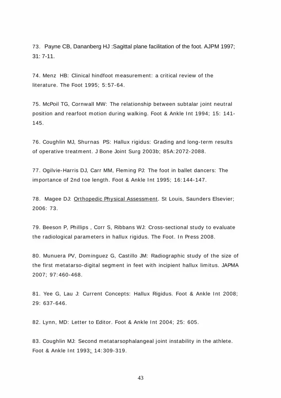
43
73. Payne CB, Dananberg HJ :Sagittal plane facilitation of the foot. AJPM 1997;
31: 7-11.
74. Menz HB: Clinical hindfoot measurement: a critical review of the
literature. The Foot 1995; 5:57-64.
75. McPoil TG, Cornwall MW: The relationship between subtalar joint neutral
position and rearfoot motion during walking. Foot & Ankle Int 1994; 15: 141-
145.
76. Coughlin MJ, Shurnas PS: Hallux rigidus: Grading and long-term results
of operative treatment. J Bone Joint Surg 2003b; 85A:2072-2088.
77. Ogilvie-Harris DJ, Carr MM, Fleming PJ: The foot in ballet dancers: The
importance of 2nd toe length. Foot & Ankle Int 1995; 16:144-147.
78. Magee DJ: Orthopedic Physical Assessment. St Louis, Saunders Elsevier;
2006: 73.
79. Beeson P, Phillips , Corr S, Ribbans WJ: Cross-sectional study to evaluate
the radiological parameters in hallux rigidus. The Foot. In Press 2008.
80. Munuera PV, Dominguez G, Castillo JM: Radiographic study of the size of
the first metatarso-digital segment in feet with incipient hallux limitus. JAPMA
2007; 97:460-468.
81. Yee G, Lau J: Current Concepts: Hallux Rigidus. Foot & Ankle Int 2008;
29: 637-646.
82. Lynn, MD: Letter to Editor. Foot & Ankle Int 2004; 25: 605.
83. Coughlin MJ: Second metatarsophalangeal joint instability in the athlete.
Foot & Ankle Int 1993; 14:309-319.

44
84. Coughlin MJ, Freund E: The reliability of angular measurements in hallux
valgus deformities. Foot & Ankle Int 2001; 22: 369-379.
85. Beeson P, Phillips C, Corr S, Ribbans WJ: Classification systems for hallux
rigidus: A review of the literature. Foot & Ankle Int 2008; 29:407-414.
45
Captions to figures
Figure 1: Measurement of ankle joint dorsiflexion
Figure 2: Valgus hallucal rotation in HR
Figure 3: Delayed heel-lift – mid-tarsal joint pronation/ eversion
Figure 4: Vertical toe-off
Figure 5: Supinated gait
Figure 6: F-Scan shows reduced 1st MTP joint pressure & lateral CoP
line
Figure 7: Failure of knee to fully extend

![[Effect of physiotherapy on the functional improvement after hallux valgus surgery - a prospective pedobarographic study]](https://img.dokumen.tips/doc/110x75/634e13d8d38be601b8060bb2/effect-of-physiotherapy-on-the-functional-improvement-after-hallux-valgus-surgery.jpg)


