Embed Size (px)
Citation preview
Growth Factors and Oncogenes as Targets in Melanoma: Lost inTranslation?
Lawrence Kwonga, Lynda China,b,c, and Stephan N. Wagner, MDd
a Department of Medical Oncology, Dana-Farber Cancer Institute, Boston, MA 02115
b Melanoma Program, Dana-Farber Cancer Institute, Boston, MA 02115
c Department of Dermatology, Harvard Medical School, Boston, MA 02115
d Associate Professor of Medicine and Director, Section Dermatooncology-Molecular Medicine of theDivision of Immunology, Allergy and Infectious Diseases, Department of Dermatology, Medical Universityof Vienna, Austria
AbstractIf untreated at early stages, melanoma becomes a highly aggressive cancer with rapid metastasis todistant sites. Although cell biologic analyses have uncovered a multitude of signaling pathwaysinvolved in melanoma genesis and progression – including the MAPK, PI3K, and FAK pathways –efficacious therapies that target these cellular components have remained elusive. Genome-widetechnologies such as microarray chips and array comparative genomic hybridization have generatedgenetic information that can identify cellular mechanisms critical for the induction and maintainenceof the malignant phenotype. Thus, such data can guide the choice of a biologically relevant drug.However, these techniques have also identified melanoma as a genetically and biologically highlyheterogeneous disease that likely requires individually tailored therapies based on the patient¹sindividual genetic and biologic alterations. In addition, these techniques have generated a large bodyof data on candidate melanoma genes that await extensive functional validation to separate so called“driver” from “passenger” events. In this review, we cover several advances in melanomatherapeutics and their current limitations as well as emerging genomic, proteomic, and epigeneticstrategies for the identification of critical cellular dependencies that may be tractable to therapeutictargeting.
Keywordsmelanoma; growth factors; oncogenes; therapy; melanoma genome
THE DISEASEMelanoma arises from melanocytes, specialized pigmented cells that reside in the skin, at thechoroidal layer of the eye, the gastrointestinal and genitourethral mucosal surfaces and themeninges. Melanocytes produce melanins, the pigments responsible for skin and hair color. In
Corresponding author for proof and reprints: Stephan N. Wagner, MD, Division of Immunology, Allergy and Infectious Diseases,Department of Dermatology, Medical University of Vienna, Währinger Gürtel 18-20, A-1090 Vienna, AUSTRIA, ++43-1-40400-7915,++43-1-40400-7574 (fax), [email protected] (e-mail).Publisher's Disclaimer: This is a PDF file of an unedited manuscript that has been accepted for publication. As a service to our customerswe are providing this early version of the manuscript. The manuscript will undergo copyediting, typesetting, and review of the resultingproof before it is published in its final citable form. Please note that during the production process errors may be discovered which couldaffect the content, and all legal disclaimers that apply to the journal pertain.
NIH Public AccessAuthor ManuscriptAdv Dermatol. Author manuscript; available in PMC 2008 December 17.
Published in final edited form as:Adv Dermatol. 2007 ; 23: 99–129. doi:10.1016/j.yadr.2007.07.015.
NIH
-PA Author Manuscript
NIH
-PA Author Manuscript
NIH
-PA Author Manuscript
the skin, melanocytes reside between keratinocytes in the basal layer of the epidermis and inthe hair follicles and produce melanin to a variety of direct and indirect stimuli such asultraviolet (UV) radiation. Thereby, melanocytes fulfill a key role in protection of our wholebody from environmental stress factors such as damaging UV radiation, which can induce skincancer.
There are an estimated 2–3 million cases of newly diagnosed skin cancers across the worldeach year and melanoma accounts for around 132,000 cases of these (World HealthOrganization, http://www.who.int/uv/faq/skincancer/en/index1.html). The incidence rates forcutaneous melanoma have risen faster than those of any other malignancy in the Caucasianpopulation over the last 30 years. From 1976 to 2003, the Central Malignant MelanomaRegistry of Germany has documented a 3-fold increase in the incidence of cutaneousmelanoma, reaching 10.3 and 13.3 newly diagnosed melanomas for males and females,respectively, per 100,000 people per year 1. In the USA incidence rates are 17.2 (males) and12.1 (females) per 100,000 population 2, highest incidence rates are observed in Australia with38.5 (males) and 29.5 (females) per 100,000 people (Globocan 2002 database,http://www-dep.iarc.fr/globocan/downloads.html).
Although melanoma accounts for only 4% of all skin cancers, it is responsible for 80% ofdeaths from skin cancer. Nowadays, timely diagnosis and treatment of melanoma during theearliest stages of its evolution is of critical importance to patient survival. Whereas surgicalresection is curative in most patients with early in situ and radial growth phase melanomas(and around 80% of melanomas can be treated this way), only 14% of patients presenting withlocoregional and/or distant metastasis survive for 5 years, despite all therapeutic efforts 3.
ENVIRONMENT AND PREVENTION OF THE DISEASEIn view of the epidemiological, the clinical, and the emerging experimental evidence linkingmelanoma incidence to UV exposure and skin phototype, primary (sun protection) andsecondary (early detection) prevention strategies have been key areas to efforts for reductionof disease incidence and severity in the last centuries.
In Queensland, Australia where secondary prevention started in the 1960s and primaryprevention in the 1980s, Coory et al. found sustained increases in age-standardized incidence,but stabilization of mortality rates, with a shift to detection of more early (in situ) melanomas4, 5. Similar results have been reported from the Central Malignant Melanoma Registry ofGermany with a decrease of age-standardized mortality accompanied by a shift towards thedetection of thin melanomas in patients younger than 70 years 1. These data are being regardedas reflecting the success of earlier detection of melanoma and the authors conclude thatcontinuation of secondary preventive strategies is warranted.
In contrast, strategies to primary prevention (sun protection) in Queensland have shown at bestsome tentative signs that some younger-age groups may be experiencing trend improvements5. Similar evidence comes from the USA, where sun protection campaigns have failed tosubstantially reduce melanoma incidence 6. These results may also be reflected byepidemiologic studies that identify UV radiation-exposure, as compared to other risk factorsfor developing melanoma, as a surprisingly modest one (1.7-fold) in an unselected population7. However, the effect of UV radiation is governed by variations (polymorphisms) in particulargenes that affect both the defensive response of skin (particularly its impact on cutaneousimmune function) and the risk of developing melanoma, e.g. by induction of growth factorsand reactive oxygen species (ROS) 8, 9. At the molecular level, exposure to UV radiationincreases pigmentation through release of α-melanocyte-stimulating hormone (α-MSH), whichinteracts with its receptor melanocortin receptor 1 (MCR1) to induce the expression of enzymesproducing melanin on the surface of melanocytes. Pigmentation as a response to UV radiation
Kwong et al. Page 2
Adv Dermatol. Author manuscript; available in PMC 2008 December 17.
NIH
-PA Author Manuscript
NIH
-PA Author Manuscript
NIH
-PA Author Manuscript
10 is modulated by polymorphisms of MCR1, which functionally reduce the activity of thisreceptor 11 (as in light-skinned and red-headed individuals with skin phototype I 10) and aresignificantly associated with an increased risk for melanoma 12.
CLINICAL AND MOLECULAR PATHOLOGY OF CUTANEOUS MELANOMACutaneous melanoma presents clinically with four different major subtypes 13. By far the mostcommon form (around 75%) is superficial spreading melanoma. It presents clinically as amacule or variably raised plaque with distinct irregular coloration. It is the third most commoncancer in young people in the USA and accounts for around 50% of melanoma cases in non-Caucasians 9, 14. In contrast, the second most common form (around 15%) of melanoma isnodular melanoma, which constitutes a raised nodule, with or without a macule in thebackground. Acral lentiginous melanoma (ALM) is usually found on the palms, the soles andthe nail beds. It accounts for about 5% of melanomas in the Caucasian and for about 50% inthe non-Caucasian population. Lentigo maligna (around 5%) is generally a flat maculeoccurring at chronically sun-exposed body sites of the elderly and is associated with the lifetimedose of UV radiation received.
The Clark model of the progression of melanoma emphasizes the stepwise transformation ofmelanocytes to melanoma. The model depicts the proliferation of melanocytes in the processof forming melanocytic nevi and the subsequent development of hyperplasia, dysplasia,invasion and metastasis 13. According to Clark’s model, the first phenotypic change inmelanocytes is the development of benign nevi. Histologically, such lesions have an increasednumber of nested melanocytes either restricted to the epidermis along the basal layer (junctionalnevus), to the dermis (dermal nevus), or to both (compound nevus).
The next step toward melanoma is the occurrence of cytological atypia such as aberrant growthand dysplastic cells, which are characteristic for so called dysplastic nevi. These lesions mayoccur either at a new location or in a preexisting nevus. Nevi are generally benign, but canprogress rarely into radial growth phase (RGP) melanoma 13. RGP presents with a completely(in situ) or a predominantly intraepidermal proliferation of melanocytes with invasion of singlecells or nests into the papillary dermis. RGP cells cannot form colonies in soft agar in vitro.
RGP cells can progress to vertical growth phase (VGP) melanoma, which is either confined tothe papillary dermis with development of a tumor nodule or extends deeply into the dermisand/or the subcutaneous fat. These cells have the ability to form colonies in soft agar in vitroand tumor nodules when implanted into nude mice and have acquired metastatic potential. Thedistinction between RGP and VGP is the single most important pathologic observation inmelanoma as most RGP melanomas are curable by surgical resection only, whereas VGPmelanomas have the capacity to metastasize.
Not all melanomas strictly pass through each of these individual phases, but can directlydevelop from melanocytes and progress to metastatic melanoma 15.
THERAPY OF MALIGNANT MELANOMA AND ITS LIMITATIONSSurgical excision is often curative in patients with thin primary tumors. However, the majorityof patients with deeply infiltrating primary melanomas or tumors that metastasize to regionalnodes will develop distant metastases later on.
Conventional TherapeuticsOver the last four decades only little, if any, progress has been made in the systemic treatmentof metastatic melanoma. The alkylating agent dacarbazine (DTIC-Dome, Bayer, West Haven,
Kwong et al. Page 3
Adv Dermatol. Author manuscript; available in PMC 2008 December 17.
NIH
-PA Author Manuscript
NIH
-PA Author Manuscript
NIH
-PA Author Manuscript
CT) has been approved by the Food and Drug Administration (FDA) in the 1970s and isconsidered to be the reference single agent for advanced disease. DTIC has been reported toinduce objective clinical responses in about 15 to 20% of patients in older trials, but withresponse rates of below 10% in recent multicenter trials 16, 17. Although complete clinicalresponses have been reported, most responses are partial and last for around 5–7 months 16,17.
Interleukin-2 (IL-2) was approved by the FDA in 1998 based on a 6% complete response ratein phase II study data set of 270 patients 18. If induced, clinical responses may be remarkablysustained (31 patients of 270 are still alive at a 10 years follow-up) 19. In the absence of anyphase III data demonstrating a clinical benefit of any dose of IL-2 in metastatic melanoma andwith regard to its high and sometimes unacceptable toxicity, it is unlikely that IL-2 will beapproved in Europe, particularly as long as it is not known which subgroup of patients willrespond.
Targeted TherapyEven though many if not most of the current effective therapies in cancer are targeted (e.g.taxanes are targeted to beta-tubulin, the fluoropyrimidines to thymidylate syntethase, anddoxorubicin to topoisomerase-II), “targeted therapy” has become a well-worn password inclinical oncology today. This is the consequence of the remarkable efficacy of novel targetedagents such as small inhibitory molecules and antibodies in other cancers and led to aremarkable shift in the investigational paradigm for melanoma.
The concept of “oncogene addiction” argues that cancer cells rely more heavily onhyperactivated intracellular signaling pathways than do normal cells, and therefore relycritically on activated oncogenes that drive those pathways (reviewed in 21). Not all oncogenesare tractable to therapeutic targeting, but intracellular enzymes such as kinases, proteases andphosphatases and extracellular antigens such as cell surface receptors are prime targets. In thefollowing paragraph, we want to give three prominent examples of targeted therapies that havebeen evaluated in advanced melanoma patients.
Based on the observation that activating mutations of a serine/threonine-specific protein kinaseBRAF occur in a high proportion of melanomas (for more details see later) clinical trials inadvanced melanoma patients have been initiated with sorafenib, a tyrosine kinase inhibitor thattargets mutant and wild-type BRAF, vascular endothelial growth-factor receptors (VEGF-R)-2and -3, c-KIT, and platelet-derived growth factor receptor (PDGF-R)-β. Whereas sorafenibwas well tolerated, it had only little or no antitumor activity 22.
It is also known that melanoma expresses a number of growth factor receptors at the cell surfaceand that ligands for these receptors may be present in the tumor milieu 23. Imatinib mesylateis an oral tyrosine kinase inhibitor that targets BCR-ABL, c-KIT, PDGF-R-α and -β. Asactivating mutations and gene amplifications of c-KIT 24, as well as signal transduction throughPDGF-R-α and -β have been described in melanoma, clinical trials have been initiated withimatinib mesylate in metastatic melanoma patients. Patients experienced significant toxicity,but no objective clinical responses 25, 26.
Another trial targeted epidermal growth factor receptor (EGFR), a type 1 receptor tyrosinekinase involved in cellular differentiation and proliferation of cancer cells including melanoma27, 28. A phase II trial in metastatic melanoma patients with erlotinib, a small molecule EGFRkinase inhibitor, revealed mild toxicity, but no objective responses 29.
All in all, no agent or combination of agents has been shown to have an impact on survival inpatients suffering from metastatic melanoma and thus, no single agent can be regarded as
Kwong et al. Page 4
Adv Dermatol. Author manuscript; available in PMC 2008 December 17.
NIH
-PA Author Manuscript
NIH
-PA Author Manuscript
NIH
-PA Author Manuscript
standard of care. The intractability of advanced melanoma painfully illustrates how much westill have to learn about the biology of this disease and the molecular changes associated with,or better, resulting in progression, metastasis and resistance to therapy. In the past, too manyclinical trials have been conducted with a poor understanding of the mechanisms of action ofthe involved compounds and without an adequate consideration or knowledge of the biologyof the disease. In other words, testing a targeted therapy when we don’t know the expressionpattern and don’t understand the functional consequences of the expression of a target moleculein a disease-specific cellular context is a low-yield clinical research strategy.
THE KEY QUESTIONSThe etiology of transformation and progression of melanocytes to melanoma is not wellunderstood. The most critical questions to melanoma cell biology that have to be answered are:(1) which genetic alterations are responsible for development, progression and maintenanceof the established disease? (2) what genetic events underlie the propensity for metastasis andtreatment resistance? (3) finally, what maintenance-essential biological or molecular signalingpathways/networks might prove amenable to preventive and/or therapeutic intervention inman?
Many studies conducted over the last decades on benign and malignant melanocytic lesions aswell as melanoma cell lines have implicated numerous genes in melanoma development andprogression. Here, we will focus on (1) validated genetic events in cell signaling pathways andgrowth factors, which include predisposing or somatic structural alterations in melanomaspecimens on the DNA level, such as translocation, amplification/deletion and point mutations30 and (2) how functional genomic information can contribute to the identification andvalidation of new candidate genes.
MELANOMA-RELEVANT CELL SIGNALING PATHWAYSRAS, RAF and Other Activators of MAP Kinase Signaling
The RAS/RAF/MEK/ERK mitogen-activated protein (MAP) kinase pathway (Fig. 1) has beenmost directly linked to the development of melanoma due to its growth-promoting activities.Since self-sufficiency in growth signaling is a requisite capability acquired by all cancer cells31, hyperactive extracellular signal-regulated kinases (ERKs) are common in many humancancers, including melanoma 32, 33. In melanocytes, this pathway can be activated by growthfactors such as stem cell factor (SCF), fibroblast growth factor (FGF) and hepatocyte growthfactor/scatter factor (HGF) 34, but individually these growth factors induce only weak ortransient ERK activation. A sustained hyperactive state can theoretically be achieved byactivating mutations of any signaling mediator upstream of ERKs and there are clear tumor-type specific patterns of mutational activation. The small G protein RAS is perhaps the mostfrequently activated component of this signaling cascade with a reported incidence of 15 to30% in all human cancers 35. In the absence of gain-of-function mutations in RAS genes,BRAF alone is responsible for coupling RAS signals to MEK. However, as soon as melanomacells acquire a mutation in RAS, cells switch their signaling from BRAF to CRAF which isaccompanied by disruption of cAMP signaling, a prerequisite for CRAF signaling to MEK36.
BRAF is also commonly targeted in human cancers with an overall mutation frequency of 7%37, but a reported mutation frequency of up to 70% in metastatic melanoma 38–44. ERKactivation can induce transcription of genes involved in melanoma cell proliferation (e.g.FGF-2, IL-8, and HIF-1a), actin organization and cell motility (e.g. PREX1, COTL1),angiogenesis (e.g. angiomotin-like 1), metastasis (TWIST1), and immune response (e.g.CD58, CD200) 45. However, ERK activation can also regulate differentiation, senescence and
Kwong et al. Page 5
Adv Dermatol. Author manuscript; available in PMC 2008 December 17.
NIH
-PA Author Manuscript
NIH
-PA Author Manuscript
NIH
-PA Author Manuscript
survival. Therefore, genotype-phenotype correlation must take into account consequencesother than growth promotion by activating mutations in components of ERK signaling.
The RAS Family of Proto-Oncogenes: H-, N- and K-RASIn contrast to other solid tumors, activating mutations of RAS are not detected with highfrequency in melanoma, ranging from low to 10–15% incidence 46 (reviewed in 47). N-RASis the most frequent RAS family member targeted in the melanocyte lineage, with activatingmutations in as many as 81% of congenital nevi 48, up to 33% of primary and 26% of metastaticmelanoma samples 49. Activating N-RAS mutations have been correlated with nodular lesionsand sun exposure 50, 51. Interestingly, N-RAS mutations are rarely found in dysplastic nevi50, 52, 53, which may imply their distinct evolutionary path to melanoma. H-RAS activationhas occasionally been detected in melanoma, albeit more commonly associated with Spitz nevi,based on amplification of its genomic locus on 11p and oncogenic point mutations 54. K-RAS mutations have not been described in human melanocytic lesions.
These mutation patterns point to distinct biological activities of the different RAS familymembers in melanocyte biology. The phenotypic impact of activated H-RAS versus N-RAStransgenic mice has reinforced this view. Specifically, an activated H-RAS transgene, togetherwith inactivating mutations in Ink4a, Arf and/or p53 promotes development of non-metastaticmelanomas 55–57. In contrast, when targeted to the melanocytic compartment, an activatedN-RAS transgene and Ink4a/Arf deficiency drives cutaneous melanomas with high penetranceand short latency, as well as metastatic spread to lymph nodes and other distal sites (e.g. lungand liver) in a third of the cases 58.
BRAF, a Potent Activator of ERK Protein KinasesThe most commonly mutated member of the RAS/RAF/MEK/ERK pathway is BRAF, one theof the three RAF genes (together with ARAF and CRAF). Since its discovery through a genome-wide cancer re-sequencing effort 37, mutations in BRAF have been detected in a variety oftumor types, with the highest incidence in melanoma (ranging from 27 to 70%) 38–44.BRAF mutations occur particularly in melanomas at body sites with intermittent UV exposure59 and are associated with the occurrence of germline variants of the MC1R gene 60. The pointmutations cluster in specific regions of biochemical importance, with the predominantmelanoma mutation being a single phosphomimetic substitution in the kinase activationdomain (V600E), which confers constitutive activation 61. BRAFV600E stimulates constitutiveERK signaling and directly and indirectly regulates expression and function of several genescritical to proliferation and survival of melanoma cells. These include transcription factorsmicrophthalmia-associated transcription factor (MITF) 62, NF-kB 63, and the cell cycleregulators Cyclin D1 64, p16INK4A 65, and p27Kip166.
An intriguing observation is that BRAF mutations are also common in benign and dysplasticnevi 39, 42, 67, 68 suggesting a role in the earliest stages of neoplasia. It is notable, however,that most nevi remain growth-arrested for their whole lifetime and only rarely progress intomelanoma. This raises the possibility that BRAFV600E-induced checkpoint mechanisms existoperating to constrain malignant transformation. Indeed, a recent study showed that humancongenital nevi are positive for both p16INK4A and senescence-associated acidic beta-galactosidase (SA-beta-Gal), the classical senescence-associated marker 65. Furthermore,BRAFV600E expression alone is not sufficient to transform human melanocytes 69, but able totransform p16INK4A-deficient murine melanocytes 70. Thus, BRAFV600E alone seems not tobe sufficient to induce melanoma, but induces cell cycle arrest with concomitant induction ofboth p16INK4A and markers of senescence 65. This phenomenon is called oncogene-inducedsenescence (OIS), a mechanisms known to constrain progression of early premalignant lesions71. Together, these results argue for a model whereby p16INK4A serves as a brake to
Kwong et al. Page 6
Adv Dermatol. Author manuscript; available in PMC 2008 December 17.
NIH
-PA Author Manuscript
NIH
-PA Author Manuscript
NIH
-PA Author Manuscript
BRAFV600E-triggered melanocyte proliferation and p16INK4A pathway inactivation is requiredfor progression to melanoma. Notably, in melanocytic nevi expression of p16INK4A is not in100% concordance with SA-beta-Gal positivity, suggesting the presence of a non-p16INK4A
dependent pathway in mediating BRAF-induced OIS 65. Therefore, it seems likely that OISwill lead to the discovery of another tumor suppressor whose importance in melanoma mayrival that of p16INK4A.
The notion that BRAFV600E is not sufficient for transformation of melanocytes has also beendemonstrated in other model systems. In zebrafish, it has been shown that BRAF activationleads only to development of benign nevi, while progression to frank melanoma requires p53deficiency 72. Similarly, BRAFV600E mutation alone in TERT-immortalized RB-p53 mutanthuman melanocytes was found to produce only junctional nevi in the human/mouse skin graft,in contrast to activated NRAS or PI3K p110a mutants which generated invasive melanomalesions 73. These biological outcomes indicate distinct roles for NRAS and BRAF activationin melanoma development. The mutually exclusive occurrence of either activated NRAS or B-RAF alleles in melanoma and other tumor types 37, 74, 75 may argue for some functionaloverlap of NRAS and BRAF activation, but may also be the result of a synthetic lethalitybetween NRAS and BRAF activation at the single cell level 45.
PTEN, a Negative Regulator of the Phosphatidylinositol 3-Kinase (PI3K)-AKT PathwayAnother pathway promoting cell growth and survival in melanoma is the phosphoinositide-3-OH kinase (PI3K)-AKT pathway (Fig. 1). Phosphoinositides are membrane lipids that areconverted to phosphatidylinositol phosphate (PIP3) second messengers throughhyperphosphorylation by one of the PI3K family members 76. Integrins, extracellular matrixcomponents such as fibronectin, and established growth factors, such as HGF and insulin-likegrowth factor (IGF)-1, act through this signaling pathway 77–79. In the presence of growthfactor signaling, the intracellular level of PIP3 rises leading to phosphorylation of AKT, whichis known to promote cell cycle progression and to inhibit apoptosis, and whose expression inits phosphorylated form is correlated adversely with patient survival 80. PIP3 secondmessengers are negatively regulated by the lipid and protein phosphatase phosphate and tensinhomologue (PTEN) and inactivation of PTEN results in accumulation of PIP3, AKThyperphosphorylation, and induction of expression of genes involved in enhanced cell survival/proliferation, tumor growth and metastasis such as the cell cycle promoting kinase CyclinD3 81 and the glycophosphoprotein Osteopontin 82
Unlike the MAP kinase pathway, genetic alterations specifically targeting components of thissignaling cascade do not occur at high frequency in melanoma 83. Of those that do occur, thebest-known culprit is the PTEN tumor suppressor. PTEN resides on chromosome 10q, a regionknown to sustain LOH in many human cancers, including melanoma 84, 85. Allelic loss oraltered expression of PTEN occurs in 20% and 40% of melanoma tumors, respectively 74,86–88, although somatic point mutations and homozygous deletions are rarely observed.Functionally, ectopic expression of PTEN in PTEN-deficient melanoma cells can abolishphospho-AKT activity, induce apoptosis, and suppress growth, tumorigenicity and metastasis89–91; reviewed in 79. Correspondingly, germline or somatic inactivation of Pten in the mousestrongly promotes tumor phenotypes in multiple cell lineages 92–95 including melanoma 96.
Most recently, additional ways of inactivation of PTEN activity have been described. PTENcan be inactivated by poly-ubiquitilation through NEDD4-1, which leads to PTEN degradationin the cytoplasm 97. By contrast, mono-ubiquitylated PTEN localizes to the cell nucleus 97,where it antagonizes the (PI3K)-AKT survival pathway and maintains chromosomal stabilitythrough physical interaction with centromeres and control of DNA repair 98. Whereas PTENinactivation through NEDD4-1 alone is not sufficient to induce tumors, it significantlyaugments the efficiency of Ras-mediated transformation of mouse fibroblasts in the presence
Kwong et al. Page 7
Adv Dermatol. Author manuscript; available in PMC 2008 December 17.
NIH
-PA Author Manuscript
NIH
-PA Author Manuscript
NIH
-PA Author Manuscript
of p53 deficiency 99. This observation underlines the importance of the simultaneous activationof ERK and PI3K signaling for tumor induction, a phenomenon similarly important formelanoma development. In three-dimensional melanoma cultures as well as in the transgenicTPRas melanoma mouse model both signaling pathways must be inhibited to suppress cellgrowth 100, 101. In melanoma specimens, NRAS and PTEN mutations are mutually exclusive,but BRAF and PTEN mutations have been shown to coincide in about 20% of cases 59, 102.
In line with the experimental evidence supporting a melanoma suppressive role of PTEN,constitutive activation of AKT has been shown to be a potent inducer of melanocytetransformation 73 and progression of RGP into VGP melanoma in vivo 103. In addition, DNAcopy gain involving the AKT3 locus has recently been described in melanoma, and selectiveAKT3 activation may characterize 40–60% of sporadic tumors 104. However, the complexityof this signaling cascade has not been fully understood till today. Recent data have suggestedthat activation of different AKT isotypes may elicit distinct effects on cell proliferation andsurvival. For example, one report found that targeted deletion of AKT3, whose expressioncorrelated most strongly with melanoma tumor progression amongst the three AKT isotypes,triggered apoptotic signaling 104. On the other hand, AKT1 activation was found to inhibitthe migration and invasion of certain cancer cell lines 105, including MDA-MB435, a linepreviously believed to derive from breast cancer but subsequently shown throughtranscriptional and SNP array profiling studies to be a melanoma cell line 69, 106. Thus,although the PI3K/AKT pathway clearly demonstrates enhanced activity in many melanomas,the extent to which this constitutes a critical melanoma dependency remains unresolved.
Activation of Receptor Tyrosine KinasesConsidering the prominent roles of RTKs in transmitting extracellular signals to intracellulareffectors, and the importance of homotypic and heterotypic cell-cell interactions in cancers, itis not surprising that almost all of the direct signaling components of RTKs have beenimplicated in the development of human melanoma (Fig. 1). Several RTKs map to knownregions of recurrent DNA copy number gain or amplification. Moreover, considering theexample of c-KIT (see below), it is expected that systematic re-sequencing efforts will identifyactivating mutations in these and other RTKs in melanomas.
The c-KIT gene encodes a RTK that serves as the receptor for stem cell factor (SCF). Theregulation of the KIT pathway is complex and tightly regulated. A number of isoforms areknown for the receptor and its ligand, and the ligand can interact with the receptor in bothsoluble and membrane-bound forms 107. Binding of soluble SCF leads to KIT receptoractivation, internalization and degradation, whereas binding of membrane-bound SCF leads toprolonged KIT activation. The KIT receptor can interact with multiple downstream signalingpathways including RAS/RAF/MEK/ERK, PI3K/AKT, phospholipase C, and the SRC family.The mechanisms underlying the differential activation of these pathways are not understoodtill today. In melanocytes, KIT plays a critical role in migration, survival, proliferation anddifferentiation. Mice deficient in Kit activation loose melanocytes 108 and KIT inhibitionappears to drive melanocyte cell loss in human skin 109, indicating that KIT is a critical survivalfactor for melanocytes. KIT is also responsible for melanocyte cell proliferation. In vitro, KITactivation induces proliferation of cells of the melanocytic lineage 110 and transgenic miceoverexpressing the membrane-bound form of SCF in the epidermis develop melanocytichyperplasia 111. Most recently, the D814Y activating KIT mutation has shown to induce PI3Ksignaling and migration of melanocytes 112.
Numerous immunohistochemical studies have linked progressive loss of c-KIT expressionwith the transition from benign to primary and metastatic melanomas 113–115. Reconstitutionof c-KIT in metastatic melanoma cells apparently conferred sensitivity to SCF-inducedapoptosis in vitro 116. Thus, at first glance, KIT does not fit the profile of a RTK targeted for
Kwong et al. Page 8
Adv Dermatol. Author manuscript; available in PMC 2008 December 17.
NIH
-PA Author Manuscript
NIH
-PA Author Manuscript
NIH
-PA Author Manuscript
activation in melanoma. However, a recurrent L576P mutation in c-KIT has recently beenreported in melanoma. Among 153 cases examined, Holden and colleagues identified fourmetastatic melanomas with robust expression of c-KIT on IHC. High-resolution ampliconmelting analyses followed by direct DNA sequencing revealed that three of them harbored aL576P mutation with selective loss of the normal allele 117, 118. L576P is a known GIST-associated mutation that maps to the 5′ juxtamembrane domain where most activating KITmutations cluster 119, 120. Most recently, Bastian and colleagues reported in a chort of 102primary melanoma samples on the presence of activating KIT mutations and geneamplifications at a frequency of 28 to 39% in particular subtypes of melanomas, i.e. acralmelanoma, mucosal melanoma, and melanomas from chronically sun-damaged skin. 69% ofthe identified KIT mutations were predicted to affect the 5′ juxta-membrane domain and 19%the kinase domain 24. The example of the EGFR mutational status as a predictor for therapeuticresponses in NSCLC 121, 122 suggests the possibility of identifying a melanoma patientsubpopulation that will respond to the c-KIT inhibitor imatinib mesylate based on c-KITmutational status.
Activation of EGFR by its ligands EGF, transforming growth factor (TGF)-α, amphiregulinand heparin-binding EGF (HBEGF) has been shown to activate several downstream signalingpathways such as the MAPK pathways, the PI3K-AKT pathway, the stress-activated proteinkinase C and the Janus kinase (Jak)- signal transducer and activator of transcription 16 pathwayand thus critically regulates cell differentiation, proliferation, survival, and migration 123. Latestage melanomas often exhibit EGFR over-expression in association with increased copies ofchromosome 7 124–126. Enforced activation of EGFR has been associated in metastaticprogression in a cell-based study 127, 128. However, unlike glioblastomas or lungadenocarcinoma 129, 130, focal amplification and/or mutation of EGFR have not been reportedin melanoma. The non-focal nature of chromosome 7 gains in melanoma renders it impossibleto assign EGFR as a target of such genomic alterations. In an inducible HRAS-driven mousemelanoma model 131, transcriptome analysis revealed the existence of a RAS-dependentEGFR signaling loop mediated through upregulation of EGF family ligands (e.g. amphiregulinand epiregulin) 132. This EGFR signaling pathway provides important survival signalsinvolving PI3K-dependent activation of AKT, as sustained EGFR activity is able to prolongviability of established melanoma upon inactivation of RAS. Conversely, inactivation bydominant negative EGFR abolishes tumorigenicity of RAS-driven melanoma cells, consistentwith observations in other cell systems (fibroblasts, keratinocytes, and intestinal epithelialcells) that autocrine EGFR signaling is required for transformation by activated RAS 133–135. Thus, in addition to providing experimental evidence that EGFR activation is biologicallyrelevant, the above-mentioned study in the inducible model also points out the possibility thatEGFR or its ligands may constitute alternative point(s) of therapeutic intervention in RAS-activated melanoma. It should be mentioned that the contribution of EGFR signaling tomelanoma development and possibly progression is evolutionarily conserved, as activatingmutations in the EGFR homologue, Xmrk, increase melanoma susceptibility in Xiphophorusfish 136–138; reviewed in 139. It therefore remains possible that similar activating mutationsexist in human melanoma, although systematic re-sequencing of large cohorts of melanomasfrom different ethnic and/or molecular subclasses will be required to uncover such examples.
The RTK c-MET is normally expressed on epithelial cells and melanocytes 140 and is activatedby binding of its ligand, HGF, through a number of downstream signaling pathways includingRAS/RAF/MEK/ERK, PI3K, phospolipase c-g and STAT 47, 141. c-MET is a multifacetedregulator of growth, motility and invasion in a number of cell lineages. Whereas RAS/RAF/MEK/ERK signaling may be responsible for proliferation, PI3K signaling is required forscattering and these two pathways in combination with STAT signaling may be important inmorphogenesis 142–144. Although MET is normally activated in a paracrine manner, autocrineactivation of HGF–MET has been described in melanoma progression 145; reviewed in 146.
Kwong et al. Page 9
Adv Dermatol. Author manuscript; available in PMC 2008 December 17.
NIH
-PA Author Manuscript
NIH
-PA Author Manuscript
NIH
-PA Author Manuscript
Accordingly, increased c-MET expression has been observed in metastatic melanoma 147, andcopy number gain of the c-MET locus at 7q33 - qter seems to be a late event in melanomaprogression 148. However, similar to EGFR above, neither focal MET amplifications noractivating MET point mutations have been detected in melanoma, although both have beenobserved in other human cancers 149–152. However, several lines of experimental andfunctional evidence support a causal role for MET signaling in human melanoma. For example,in explant models, it has been shown that elevated c-Met expression or Met receptor tyrosinekinase activity may correlate with metastasis 153. In genetically engineered models,constitutive and ubiquitous HGF expression establishes an autocrine loop with c-Met, leadingto step-wise development and progression of cutaneous and metastatic melanomas, whichcooperates with UVB, Ink4a/Arf deficiency, and activated Cdk4 154–156. Correspondingly,while enforced expression of c-Met in melanocytes provides only weak cancer-initiatingactivity, this mutation drives the development of metastatic disease, and such tumor lesionsshow concomitant activation of HGF and establishment of HGF-Met signaling loop (LC,unpublished observations). Finally, c-MET was recently shown to be a direct transcriptionaltarget of MITF 157, the melanocytic lineage transcription factor that can be activated by focalamplification in melanoma (see below).
THE MELANOMA GENOMEIn probing the cancer genome for novel targets, technological advances have enabled the rapidproduction of genome-wide datasets informative for changes in gene expression, DNA copynumber, and loss of heterozygosity. Such assays have revealed in melanomas an increasinglycomplex pantheon of genetic aberrations beyond the established ones described above. Noveltumor suppressors and oncogenes are continuing to be described, but many validation stepsare required before any gene can be considered bona fide therapeutic targets. Effects on tumorinitiation, growth, or invasion must be experimentally demonstrable both in vitro and in vivo.
Typically, experiments in cell culture make use of overexpression vectors, knockdown vectorssuch as siRNA or shRNA, drug inhibitors, or competitive antibodies to modulate geneexpression or product levels. Cells can then be assayed for their ability to form colonies in softagar, indicative of anchorage-independent growth and loss of contact inhibition. Growth curvescan be measured, and invasion can be tested in Boyden chambers, which simulate the initialsteps of metastatic invasion through the extracellular matrix. Three- dimensional cultures,including organotypic skin rafts and collagen matrices, may better portray the conditions ofthe in vivo environment. Culture preparations can also be injected subcutaneously into athymicmice, and the incidence, growth, and invasiveness of the resulting xenograft tumor isconsidered more clinically relevant than cell culture. Tail or portal vein injections and theobservation of subsequent metastasis formation can measure the intravasation and seedingsteps. Finally, genetically engineered mouse models can provide strong evidence for a gene’srole in melanoma, as cell type-specific gene knockouts, knockins, and transgenics canrecapitulate human mutations in the context of established mouse melanoma models (seeabove). A number of studies employing genome-wide assays are catalogued in Table 1, alongwith the extent of candidate gene validation performed in each.
As can be seen, the majority of gene discovery studies have utilized gene expression arrays.For example, comparisons of metastatic and non-metastatic cell lines, the latter with anengineered extra copy of chromosome 6, identified CRSP3 and TXNIP as correlated with thesuppression of metastasis 158. Although overexpression of either gene did not affect tumorgrowth in vitro or in xenografts, they significantly and independently suppressed the formationof metastases to the lung when the cells were injected either subcutaneously or intravenously.Preliminary evidence suggested that both genes operate through the KISS1 (Kisspeptin-1)pathway, which may affect cell motility 159. Others 160 used a similar set of cell lines with
Kwong et al. Page 10
Adv Dermatol. Author manuscript; available in PMC 2008 December 17.
NIH
-PA Author Manuscript
NIH
-PA Author Manuscript
NIH
-PA Author Manuscript
or without an extra chromosome 6, with the latter displaying anchorage-independent growthin soft agar and a downregulation of the connexin gene CX43. Overexpression of CX43significantly suppressed the soft agar colony formation. As a final example, tenascin andfibronectin were upregulated in progressed tumors, and their co-localization confirmed by IHC161. Overexpression of either gene conferred growth and invasive behavior in three-dimensional collagen gels.
Combinations of genome-wide technologies – expression microarrays, array comparativegenomic hybridization, and SNP microarrays – can together define a genomic atlas of recurrentDNA and gene regulatory aberrations. As the accumulation of genome-wide data continues tooutpace the technology to validate individual genes, basic cancer research is faced with a needto isolate bona fide therapeutic targets. The brief survey of articles in Table 1 indicates that weare faced with a wealth of candidate melanoma modifiers, each with a long experimentalprocess ahead of it (it should be noted, however, that many of the studies with a smaller extentof validation achieved goals other than gene discovery.) The extents to which these listed genestruly control melanoma behavior and thus represent promising therapeutic targets await furthertesting, as illustrated by the examples of NEDD9 (neural precursor cell expressed,developmentally down-regulated 9) and MITF below.
NEDD9The power of array technology further benefits from comparisons among species, with theevolutionary conservation of genetic pathways providing a means to highlight significantchanges; this is evidenced by the novel cross-species identification of Nedd9 as a regulator ofmelanoma metastasis. The Ink4a/Arf−/−, inducible H-ras mouse melanoma model 55, 131provided a tractable means by which to create isogenic metastatic and non-metastatic tumors.Selective pressure for “escaper” tumors no longer dependent on the doxycycline-induced H-ras signal was generated by alternating the signal over defined time periods. Two such linesproved metastatic to distant sites in xenografts. DNA copy number profiles of these lines anda non-metastatic sister line were compared by aCGH, pinpointing an 850kb common site ofamplification on chromosome 13. Only one gene in this region, Nedd9, showed a significantupregulation in mouse melanomas but not in normal melanocytes. The syntenic human region,6p24-25, undergoes copy number gain in 36% of human metastatic, but not non-metastatic,melanomas 124, 162, indicating a common route of genetic modification between human andmouse. Indeed, human melanoma tissue microarrays (TMAs) revealed a significant correlationof Nedd9 protein levels with tumor progression.
Illustrating the power of candidate gene validation, Nedd9 showed metastasis-modulatingactivity at multiple levels. In Boyden chamber assays, overexpression of Nedd9 enhanced theinvasiveness and growth of non-metastatic Ink4a/Arf−/− cell lines expressing activated H-Rasor B-Raf, while knockdown by shRNA inhibited invasiveness in the original metastatic escaperline. Demonstrating this bidirectional behavior revealed the dependency of the cell line onNedd9 for invasive potential. Nedd9 also conferred a capacity to metastasize to distant siteswhen overexpressing cells were injected subcutaneously into SCID mice, providing moreclinically relevant in vivo evidence. Finally, the mechanism of Nedd9 action was dissectedwith biochemical and genetic assays, which showed focal adhesion kinase (Fak) to be a criticalmediator of Nedd9-induced in vitro invasion, formation of dynamic focal contacts, andattachment to matrigel. These properties are considered hallmarks of cell motility and acapacity for extravasation into the bloodstream.
The identification of Nedd9 as a cross-species melanoma metastasis gene reinforced two majorconcepts: that the fundamental properties of cancer genetics are conserved and thus highlightedbetween mouse and human and that modulation of a single genetic pathway can greatly alter
Kwong et al. Page 11
Adv Dermatol. Author manuscript; available in PMC 2008 December 17.
NIH
-PA Author Manuscript
NIH
-PA Author Manuscript
NIH
-PA Author Manuscript
tumor fate. The ongoing research into creating and testing mice genetically engineered to over-or underexpress Nedd9 moves it closer to a thorough conception for its clinical translation.
MITFThe identification of MITF as a critical melanoma survival gene took a cross-tissue approach,wherein the NCI-60 cell line panel representing nine tumor types was subjected to both geneexpression and SNP array analysis 69. A recurrent gain of 3p13-14 significantly segregatedmelanoma from other tumor classes and was confirmed by quantitative PCR (qPCR). Thecombined expression and qPCR data isolated MITF as the only gene in the region with maximalamplification and overexpression. Fluorescence in situ hybridization andimmunohistochemistry on melanoma TMAs revealed a correlation of increased MITF genedosage and protein levels with malignancy and decreased survival. Correlation, however, doesnot discriminate between MITF upregulation as a cause or bystander effect of malignanttransformation. Therefore, the effect of exogenous MITF was determined in humanmelanocytes engineered to be immortalized but non-transformed. Only when MITF wasexpressed in a melanoma-relevant signaling context (i.e. activated BRAF) cells were able togrow in the absence of otherwise essential media factors and able to form colonies in soft agar,both results suggestive of full transformation. Conversely, inhibition of MITF in cell linesshowing 3p13-14 amplification reduced growth and survival and conferred sensitivity tocertain anticancer drugs. This careful validation of MITF in human cells sets the stage for invivo mouse studies.
By comparing different tumor types, genetic changes specific to the relevant lineages werehighlighted. The critical role of MITF in both normal melanocyte development and melanomasurvival was therefore suggested to identify, along with androgen receptor, a novel class ofoncogenes termed “lineage addiction” oncogenes 163. The tumor may “hijack” extant lineagesurvival mechanisms in the presence of selective pressures; indeed, activated BRAF is knownto target MITF for proteolytic degradation, which may select for refractory cellular variantswith amplified MITF. Tellingly, MITF gene disruption leads to coat color graying in mice164, 165 and pigmentation and hearing defects (melanocytes play a role in cochleardevelopment) in humans, termed Waardenburg Syndrome Type 2A 166. The demonstrationthat a gene involved specifically in melanocyte maintenance and differentiation is dysregulatedin melanomas opens the possibility that anticancer drugs can be targeted not only to specificcellular pathways, but also to specific cell types.
Epigenetics, miRNAs, and proteomicsEven with a combination of the three main avenues of genome-wide interrogation – geneexpression, DNA copy number, and LOH – a full picture of the changes that take place intumorigenesis is lacking. The intracellular milieu has many levels of regulation, both at theDNA and protein levels. Thus, a number of other large-scale technologies will build towardsa more comprehensive view of melanoma: epigenetic profiling, screening for non-coding butfunctional DNA, and proteomic profiling (Fig 2).
Epigenetic studies in cancer have focused on the role of both DNA methylation and histonemodifications to understand how genes can be aberrantly regulated in the absence of disruptivemutations. The methylation of cytosines within CpG islands upstream of gene promoters resultsin gene silencing, while methylation at other sites may activate genes, such as at thedifferentially methylated region of Igf2 (insulin-like growth factor 2) 167. In conjunction, the“histone code” represents a complex combination of modifications of specific histone tailresidues, with certain methylation and acetylation (and, perhaps to a lesser extent,phosphorylation, ubiquitination, and sumoylation) patterns governing chromatin structure andhence gene expression. Though the interaction of DNA and histone modifications continues
Kwong et al. Page 12
Adv Dermatol. Author manuscript; available in PMC 2008 December 17.
NIH
-PA Author Manuscript
NIH
-PA Author Manuscript
NIH
-PA Author Manuscript
to be clarified, recent studies suggest that silencing via histone methylation may supercede thecytosine methylation status 168.
Hence, genome-wide epigenetic scans can be either DNA-specific – methylation-specificdigital karyotyping 169, restriction landmark genome scanning 170, or ChIP-Chip 171 – orhistone-specific, such as applying expression arrays to cells treated with histone deacetylase(HDAC) inhibitors 172. One study made use of the DNA methylation inhibitor 5-Aza-2′-deoxycytidine to compare treated and untreated melanoma cell lines 173. The genes SYN andHOXB13 were found to be significantly re-expressed upon treatment, and were also reducedin untreated cell lines compared to normal melanocytes. Overexpression of each geneindividually reduced in vitro proliferation and in vivo xenograft tumor size, indicating a tumorsuppressor role for both genes. Other current evidence points to a pivotal role for the epigeneticregulation of specific genes in melanoma progression: focal comparisons of primary andmetastatic tumor specimens have confirmed differential methylation statuses of peroxiredoxin2 174, skeletrophin 175, and estrogen receptor (ER)-α 176. Intriguingly, ER-α 176 and PTEN177 methylation are increased in DNA circulating in the serum of patients with metastaticdisease, suggesting that they may be useful as clinical indicators. Furthermore, HDACinhibitors show some initial promise in cell lines and xenografts 178, supporting globalepigenetic changes as relevant for tumor maintenance.
Long referred to as “junk DNA,” certain non-coding genetic elements have also recently takena prominent spot in gene regulation. Of particular interest are miRNAs, which are short,21-23bp RNAs that bind to homologous mRNA segments and mediate the translationalblockage or argonaute-directed degradation of gene transcripts as well as initiate epigeneticgene silencing modifications. Multi-platform assays have identified an enrichment for miRNAsin various solid cancers that target known tumor suppressors and oncogenes 179. The samegroup discovered that miR155 can function as an oncogene in B cell lineages, with Eμ-directedoverexpression resulting in the development of leukemia and lymphoma 180. WhethermiRNAs play a significant functional role in melanoma remains to be seen, though initialstudies on different tumor types demonstrate that melanoma can be differentiated from othertumor types based solely on miRNA expression signatures 181 and that each of 243/283miRNAs exhibited copy number changes in at least seven of 45 melanoma cell lines 182.
Finally, gene expression levels do not necessarily correlate with protein levels, due toposttranslational modifications undetectable by gene-centric assays. Comprehensive analysisof the tumor proteome is a technically challenging process, requiring the use of low-throughputbiochemical isolation techniques (2D-gel electrophoresis, protein chips) and specializedmachinery for protein identification and quantification (MALDI-TOF). Nevertheless, suchanalysis of a mouse melanoma xenograft model identified associations of increased VEGF andcathepsin D levels with progression 183. Proteomics also provides a plausible method for non-invasive prognostic tests: the MALDI-TOF analysis of patient sera identified a proteinspectrum that could retrospectively classify 55 progressive and non-progressive stage IIItumors with 80% specificity 184. Such proteomic data awaits predictive testing. Continuingtechnology improvements will necessitate larger scale studies towards a more completeunderstanding of the melanoma cell.
STRATEGIES FOR TRANSLATION OF GENETIC INFORMATION INTOMELANOMA THERAPY
All in all, melanoma must be regarded as a genetically and biologically heterogeneous disease.This heterogeneity is exemplified in the clinics by the differential and until today unpredictableresponse to therapy. Rational drug design has significantly changed the daily practice of clinicaloncology through introduction of small molecule inhibitors and antibodies, e.g. against
Kwong et al. Page 13
Adv Dermatol. Author manuscript; available in PMC 2008 December 17.
NIH
-PA Author Manuscript
NIH
-PA Author Manuscript
NIH
-PA Author Manuscript
overexpression of HER2/NEU in breast cancer, activating c-KIT mutations in gastrointestinalstromal tumors, and EGFR mutations and overexpression in non-small cell lung cancer. Whatcould be the prospects for the paradigm of genetic targets to melanoma?
Due to the high incidence of activating mutations in genes of the RAS/RAF/MEK/ERKsignaling pathway, BRAF targeting has been predicted as a promising therapeutic strategy formelanoma, but failed to accomplish meaningful clinical activity 22. A possible explanation forthis could be that the agent itself may be insufficiently potent. However, with a more completeunderstanding of the genetic and functional data of this pathway -as outlined above- we favorexplanations suggested by genetic and functional data: (1) BRAFV600E alone is not sufficientto cause melanoma, but induces cell cycle arrest with concomitant induction of oncogeneinduced senescence, (2) BRAF activity is modulated by the genetic background at the singlecell level, e.g. the presence or absence of control mechanisms such as p16INK4A, (3) RAS/RAF/MEK/ERK signaling switches from BRAF to CRAF signaling upon the acquisition ofRAS mutation, (4) pathway redundancy or digression may occur: in contrast to targetssuccessfully targeted in other cancers, the most frequent mutation of the RAS/RAF/MEK/ERKsignaling pathway lies several steps downstream of the initial receptor-ligand interaction, thusfavoring the recruitment of alternative pathways of cell signaling, (5) the development ofresistance mechanisms: several melanoma cell lines survive MAPK inhibition by expressionof the antiapoptotic factors Mcl-1, Bcl-xL and Bcl-2 and suppression of tumor suppressor p53through ROS 185.
Unlike the RAS/RAF/MEK/ERK pathway, genetic alterations specifically affectingcomponents of other established signaling cascades do occur only in a small proportion ofmelanomas. This complex and heterogeneous genetic and biological background significantlychallenges the identification of targets for drug development and should preclude theunselected enrollment of melanoma patients into clinical trials. In this context, strategiestowards a genetic-based patient selection and individualized therapy have to be developed. Inthe last few years, we have been witnessing the establishment of first genotype-phenotypecorrelations. Examples are the reported association of activating N-RAS mutations with nodularlesions and sun exposure 50, 51, the occurrence of BRAF mutations particularly in melanomasfrom body sites with intermittent UV exposure 59 and their correlation with germline variantsof the MC1R gene 60, the presence of CDK4 amplifications preferentially in acral and mucosalmelanomas 59, the presence of Cyclin D1 amplifications particularly in melanomas from skinwith chronic sun damage 59, and the presence of activating KIT mutations and geneamplifications in acral melanoma, mucosal melanoma, and melanomas from chronically sun-damaged skin 24.
However, these correlations can provide only a rough estimate on the presence of certain typesof genetic alterations and are far from being predictive for the most appropriate avenue ofpersonalized treatment. In this regard, individualized genomic information can providesignificant contributions.
OUTLOOK: INDIVIDUAL GENETIC TYPING FOR GENOTYPE-PHENOTYPECORRELATIONS
Success in identifying gene expression signatures predictive of survival in breast and othersolid cancers 186, 187 establishes a real possibility that the molecular profiling of melanomacan inform clinical decisions. Recent descriptive, retrospective observations of gene expressionin metastatic versus non metastatic tumors 188–190 provide a tantalizing glimpse into whatthese signatures may look like, but they must be functionally validated in order to have trueclinical relevance.
Kwong et al. Page 14
Adv Dermatol. Author manuscript; available in PMC 2008 December 17.
NIH
-PA Author Manuscript
NIH
-PA Author Manuscript
NIH
-PA Author Manuscript
Indeed, the ability to classify melanomas by particular genomic signatures suggests such apredictive capacity. For example, CGH profiling could distinguish among 126 acral, musocal,and chronically and non-chronically sun-damaged primary skin melanomas with 70% accuracy59. BRAF and NRAS mutations were significantly associated with the non-chronically sundamaged subtype, suggesting that knowledge of common gene mutations alone could providea degree of classifying information. In fact, a microarray analysis of cell lines with and withoutCDKN2A deletions, coupled with confirmatory RT-PCR on 14 cell lines, identified eight genesconsistently differing in expression between the two classes 191. Conversely, although twostudies supported an expression profile difference between NRAS and BRAF mutant tumors192, 193, others employing stricter statistical parameters did not 188, 194. Overall, genome-wide profiling may provide more specific classification than single-gene sequencing.
In the farther future, the feasibility of personal whole-genome sequencing comes ever closeras sequencing costs continue to drop and novel techniques continue to be rapidly developed.Recent initiatives to exhaustively sequence annotated genes in breast and colon cancers haveunearthed only a fractional minority of putatively functional aberrations 195. Given the richdiversity of cellular regulation illustrated above, any future clinically-oriented sequencingefforts may need a massive supporting network of data to have true prognostic value.
AcknowledgementsThis work was supported by grants to SNW (Grant No. APP19722FW from the FWF - Fonds zur Förderung derwissenschaftlichen Forschung) and LC (NIH RO1 CA93947; UO1 CA84313 and P50 CA93683).
BIBLIOGRAPHY1. Lasithiotakis KG, Leiter U, Gorkievicz R, et al. The incidence and mortality of cutaneous melanoma
in Southern Germany: trends by anatomic site and pathologic characteristics, 1976 to 2003. CancerSep 15;2006 107(6):1331–1339. [PubMed: 16909413]
2. Giblin AV, Thomas JM. Incidence, mortality and survival in cutaneous melanoma. J Plast ReconstrAesthet Surg 2007;60(1):32–40. [PubMed: 17126264]
3. Balch CM, Buzaid AC, Soong SJ, et al. Final version of the American Joint Committee on Cancerstaging system for cutaneous melanoma. J Clin Oncol Aug 15;2001 19(16):3635–3648. [PubMed:11504745]
4. Baade P, Coory M. Trends in melanoma mortality in Australia: 1950–2002 and their implications formelanoma control. Aust N Z J Public Health Aug;2005 29(4):383–386. [PubMed: 16222938]
5. Coory M, Baade P, Aitken J, Smithers M, McLeod GR, Ring I. Trends for in situ and invasive melanomain Queensland, Australia, 1982–2002. Cancer Causes Control Feb;2006 17(1):21–27. [PubMed:16411049]
6. Weinstock MA. Sun protection and early detection: do we have the balance right? 6th World Congresson Melanoma. 2005abstract 114
7. Berwick M, Wiggins C. The current epidemiology of cutaneous malignant melanoma. Front Biosci2006;11:1244–1254. [PubMed: 16368510]
8. Thompson JF, Scolyer RA, Kefford RF. Cutaneous melanoma. Lancet Feb 19–25;2005 365(9460):687–701. [PubMed: 15721476]
9. Gilchrest BA, Eller MS, Geller AC, Yaar M. The pathogenesis of melanoma induced by ultravioletradiation. N Engl J Med Apr 29;1999 340(17):1341–1348. [PubMed: 10219070]
10. Valverde P, Healy E, Jackson I, Rees JL, Thody AJ. Variants of the melanocyte-stimulating hormonereceptor gene are associated with red hair and fair skin in humans. Nat Genet Nov;1995 11(3):328–330. [PubMed: 7581459]
11. Frandberg PA, Doufexis M, Kapas S, Chhajlani V. Human pigmentation phenotype: a point mutationgenerates nonfunctional MSH receptor. Biochem Biophys Res Commun Apr 17;1998 245(2):490–492. [PubMed: 9571181]
Kwong et al. Page 15
Adv Dermatol. Author manuscript; available in PMC 2008 December 17.
NIH
-PA Author Manuscript
NIH
-PA Author Manuscript
NIH
-PA Author Manuscript
12. Kennedy C, ter Huurne J, Berkhout M, et al. Melanocortin 1 receptor (MC1R) gene variants areassociated with an increased risk for cutaneous melanoma which is largely independent of skin typeand hair color. J Invest Dermatol Aug;2001 117(2):294–300. [PubMed: 11511307]
13. Clark WH Jr, Elder DE, Guerry Dt, Epstein MN, Greene MH, Van Horn M. A study of tumorprogression: the precursor lesions of superficial spreading and nodular melanoma. Hum Pathol Dec;1984 15(12):1147–1165. [PubMed: 6500548]
14. Ishihara K, Saida T, Yamamoto A. Updated statistical data for malignant melanoma in Japan. Int JClin Oncol Jun;2001 6(3):109–116. [PubMed: 11706778]
15. Miller AJ, Mihm MC Jr. Melanoma. N Engl J Med Jul 6;2006 355(1):51–65. [PubMed: 16822996]16. Middleton MR, Grob JJ, Aaronson N, et al. Randomized phase III study of temozolomide versus
dacarbazine in the treatment of patients with advanced metastatic malignant melanoma. J Clin OncolJan;2000 18(1):158–166. [PubMed: 10623706]
17. Bedikian AY, Millward M, Pehamberger H, et al. Bcl-2 antisense (oblimersen sodium) plusdacarbazine in patients with advanced melanoma: the Oblimersen Melanoma Study Group. J ClinOncol Oct 10;2006 24(29):4738–4745. [PubMed: 16966688]
18. Atkins MB, Lotze MT, Dutcher JP, et al. High-dose recombinant interleukin 2 therapy for patientswith metastatic melanoma: analysis of 270 patients treated between 1985 and 1993. J Clin Oncol Jul;1999 17(7):2105–2116. [PubMed: 10561265]
19. Atkins MB, Kunkel L, Sznol M, Rosenberg SA. High-dose recombinant interleukin-2 therapy inpatients with metastatic melanoma: long-term survival update. Cancer J Sci Am Feb;2000 6(Suppl1):S11–14. [PubMed: 10685652]
20. Rosenberg SA, Yang JC, Restifo NP. Cancer immunotherapy: moving beyond current vaccines. NatMed Sep;2004 10(9):909–915. [PubMed: 15340416]
21. Weinstein IB, Joe AK. Mechanisms of disease: Oncogene addiction--a rationale for moleculartargeting in cancer therapy. Nat Clin Pract Oncol Aug;2006 3(8):448–457. [PubMed: 16894390]
22. Eisen T, Ahmad T, Flaherty KT, et al. Sorafenib in advanced melanoma: a Phase II randomiseddiscontinuation trial analysis. Br J Cancer Sep 4;2006 95(5):581–586. [PubMed: 16880785]
23. Giehl KA, Nagele U, Volkenandt M, Berking C. Protein expression of melanocyte growth factors(bFGF, SCF) and their receptors (FGFR-1, c-kit) in nevi and melanoma. J Cutan Pathol Jan;2007 34(1):7–14. [PubMed: 17214848]
24. Curtin JA, Busam K, Pinkel D, Bastian BC. Somatic activation of KIT in distinct subtypes ofmelanoma. J Clin Oncol Sep 10;2006 24(26):4340–4346. [PubMed: 16908931]
25. Ugurel S, Hildenbrand R, Zimpfer A, et al. Lack of clinical efficacy of imatinib in metastaticmelanoma. Br J Cancer Apr 25;2005 92(8):1398–1405. [PubMed: 15846297]
26. Wyman K, Atkins MB, Prieto V, et al. Multicenter Phase II trial of high-dose imatinib mesylate inmetastatic melanoma: significant toxicity with no clinical efficacy. Cancer May 1;2006 106(9):2005–2011. [PubMed: 16565971]
27. Ellis DL, King LE Jr, Nanney LB. Increased epidermal growth factor receptors in melanocytic lesions.J Am Acad Dermatol Oct;1992 27(4):539–546. [PubMed: 1383295]
28. Sparrow LE, Heenan PJ. Differential expression of epidermal growth factor receptor in melanocytictumours demonstrated by immunohistochemistry and mRNA in situ hybridization. Australas JDermatol Feb;1999 40(1):19–24. [PubMed: 10098284]
29. Wyman, K.; Kelley, M.; Puzanov, I., et al. Phase II study of erlotinib given daily for patients withmetastatic melanoma (MM). J Clin Oncol; 2006 ASCO Annual Meeting Proceedings Part I; June 20Supplement; 2006. p. 18002 (abstract)
30. Futreal PA, Coin L, Marshall M, et al. A census of human cancer genes. Nat Rev Cancer Mar;20044(3):177–183. [PubMed: 14993899]
31. Hanahan D, Weinberg RA. The hallmarks of cancer. Cell Jan 7;2000 100(1):57–70. [PubMed:10647931]
32. Takata M, Goto Y, Ichii N, et al. Constitutive activation of the mitogen-activated protein kinasesignaling pathway in acral melanomas. J Invest Dermatol Aug;2005 125(2):318–322. [PubMed:16098043]
33. Zhuang L, Lee CS, Scolyer RA, et al. Activation of the extracellular signal regulated kinase (ERK)pathway in human melanoma. J Clin Pathol Nov;2005 58(11):1163–1169. [PubMed: 16254105]
Kwong et al. Page 16
Adv Dermatol. Author manuscript; available in PMC 2008 December 17.
NIH
-PA Author Manuscript
NIH
-PA Author Manuscript
NIH
-PA Author Manuscript
34. Bohm M, Moellmann G, Cheng E, et al. Identification of p90RSK as the probable CREB-Ser133kinase in human melanocytes. Cell Growth Differ Mar;1995 6(3):291–302. [PubMed: 7540859]
35. Bos JL. ras oncogenes in human cancer: a review. Cancer Res Sep 1;1989 49(17):4682–4689.[PubMed: 2547513]
36. Dumaz N, Hayward R, Martin J, et al. In Melanoma, RAS Mutations Are Accompanied by SwitchingSignaling from BRAF to CRAF and Disrupted Cyclic AMP Signaling. Cancer Res Oct 1;2006 66(19):9483–9491. [PubMed: 17018604]
37. Davies H, Bignell GR, Cox C, et al. Mutations of the BRAF gene in human cancer. Nature Jun 27;2002417(6892):949–954. [PubMed: 12068308]
38. Maldonado JL, Fridlyand J, Patel H, et al. Determinants of BRAF mutations in primary melanomas.J Natl Cancer Inst Dec 17;2003 95(24):1878–1890. [PubMed: 14679157]
39. Pollock PM, Harper UL, Hansen KS, et al. High frequency of BRAF mutations in nevi. Nat GenetJan;2003 33(1):19–20. [PubMed: 12447372]
40. Uribe P, Wistuba II, Gonzalez S. BRAF mutation: a frequent event in benign, atypical, and malignantmelanocytic lesions of the skin. Am J Dermatopathol Oct;2003 25(5):365–370. [PubMed: 14501284]
41. Daniotti M, Oggionni M, Ranzani T, et al. BRAF alterations are associated with complex mutationalprofiles in malignant melanoma. Oncogene Aug 5;2004 23(35):5968–5977. [PubMed: 15195137]
42. Kumar R, Angelini S, Snellman E, Hemminki K. BRAF mutations are common somatic events inmelanocytic nevi. J Invest Dermatol Feb;2004 122(2):342–348. [PubMed: 15009715]
43. Shinozaki M, Fujimoto A, Morton DL, Hoon DS. Incidence of BRAF oncogene mutation and clinicalrelevance for primary cutaneous melanomas. Clin Cancer Res Mar 1;2004 10(5):1753–1757.[PubMed: 15014028]
44. Libra M, Malaponte G, Navolanic PM, et al. Analysis of BRAF mutation in primary and metastaticmelanoma. Cell Cycle Oct;2005 4(10):1382–1384. [PubMed: 16096377]
45. Petti C, Molla A, Vegetti C, Ferrone S, Anichini A, Sensi M. Coexpression of NRASQ61R andBRAFV600E in human melanoma cells activates senescence and increases susceptibility to cell-mediated cytotoxicity. Cancer Res Jul 1;2006 66(13):6503–6511. [PubMed: 16818621]
46. Wagner SN, Ockenfels HM, Wagner C, Hofler H, Goos M. Ras gene mutations: a rare event innonmetastatic primary malignant melanoma. J Invest Dermatol May;1995 104(5):868–871.[PubMed: 7738369]
47. Chin L, Merlino G, DePinho RA. Malignant melanoma: modern black plague and genetic black box.Genes Dev Nov 15;1998 12(22):3467–3481. [PubMed: 9832500]
48. Bauer J, Curtin JA, Pinkel D, Bastian BC. Congenital melanocytic nevi frequently harbor NRASmutations but no BRAF mutations. J Invest Dermatol Jan;2007 127(1):179–182. [PubMed:16888631]
49. Demunter A, Stas M, Degreef H, De Wolf-Peeters C, van den Oord JJ. Analysis of N- and K-rasmutations in the distinctive tumor progression phases of melanoma. J Invest Dermatol Dec;2001 117(6):1483–1489. [PubMed: 11886512]
50. Jafari M, Papp T, Kirchner S, et al. Analysis of ras mutations in human melanocytic lesions: activationof the ras gene seems to be associated with the nodular type of human malignant melanoma. J CancerRes Clin Oncol 1995;121(1):23–30. [PubMed: 7860615]
51. van Elsas A, Zerp SF, van der Flier S, et al. Relevance of ultraviolet-induced N-ras oncogene pointmutations in development of primary human cutaneous melanoma. Am J Pathol Sep;1996 149(3):883–893. [PubMed: 8780392]
52. Albino AP, Nanus DM, Mentle IR, et al. Analysis of ras oncogenes in malignant melanoma andprecursor lesions: correlation of point mutations with differentiation phenotype. Oncogene Nov;19894(11):1363–1374. [PubMed: 2682463]
53. Papp T, Pemsel H, Zimmermann R, Bastrop R, Weiss DG, Schiffmann D. Mutational analysis of theN-ras, p53, p16INK4a, CDK4, and MC1R genes in human congenital melanocytic naevi. J MedGenet Aug;1999 36(8):610–614. [PubMed: 10465111]
54. Bastian BC, LeBoit PE, Pinkel D. Mutations and copy number increase of HRAS in Spitz nevi withdistinctive histopathological features. Am J Pathol Sep;2000 157(3):967–972. [PubMed: 10980135]
55. Chin L, Pomerantz J, Polsky D, et al. Cooperative effects of INK4a and ras in melanoma susceptibilityin vivo. Genes Dev Nov 1;1997 11(21):2822–2834. [PubMed: 9353252]
Kwong et al. Page 17
Adv Dermatol. Author manuscript; available in PMC 2008 December 17.
NIH
-PA Author Manuscript
NIH
-PA Author Manuscript
NIH
-PA Author Manuscript
56. Bardeesy N, Bastian BC, Hezel A, Pinkel D, DePinho RA, Chin L. Dual inactivation of RB and p53pathways in RAS-induced melanomas. Mol Cell Biol Mar;2001 21(6):2144–2153. [PubMed:11238948]
57. Sharpless NE, Kannan K, Xu J, Bosenberg MW, Chin L. Both products of the mouse Ink4a/Arf locussuppress melanoma formation in vivo. Oncogene Aug 7;2003 22(32):5055–5059. [PubMed:12902988]
58. Ackermann J, Frutschi M, Kaloulis K, McKee T, Trumpp A, Beermann F. Metastasizing melanomaformation caused by expression of activated N-RasQ61K on an INK4a-deficient background. CancerRes May 15;2005 65(10):4005–4011. [PubMed: 15899789]
59. Curtin JA, Fridlyand J, Kageshita T, et al. Distinct sets of genetic alterations in melanoma. N Engl JMed Nov 17;2005 353(20):2135–2147. [PubMed: 16291983]
60. Landi MT, Bauer J, Pfeiffer RM, et al. MC1R germline variants confer risk for BRAF-mutantmelanoma. Science Jul 28;2006 313(5786):521–522. [PubMed: 16809487]
61. Garnett MJ, Marais R. Guilty as charged: B-RAF is a human oncogene. Cancer Cell Oct;2004 6(4):313–319. [PubMed: 15488754]
62. Wellbrock C, Marais R. Elevated expression of MITF counteracts B-RAF-stimulated melanocyte andmelanoma cell proliferation. J Cell Biol Aug 29;2005 170(5):703–708. [PubMed: 16129781]
63. Liu J, Suresh Kumar KG, Yu D, et al. Oncogenic BRAF regulates beta-Trcp expression and NF-kappaB activity in human melanoma cells. Oncogene Mar 22;2007 26(13):1954–1958. [PubMed:17001349]
64. Bhatt KV, Spofford LS, Aram G, McMullen M, Pumiglia K, Aplin AE. Adhesion control of cyclinD1 and p27Kip1 levels is deregulated in melanoma cells through BRAF-MEK-ERK signaling.Oncogene May 12;2005 24(21):3459–3471. [PubMed: 15735667]
65. Michaloglou C, Vredeveld LC, Soengas MS, et al. BRAFE600-associated senescence-like cell cyclearrest of human naevi. Nature Aug 4;2005 436(7051):720–724. [PubMed: 16079850]
66. Bhatt KV, Hu R, Spofford LS, Aplin AE. Mutant B-RAF signaling and cyclin D1 regulate Cks1/S-phase kinase-associated protein 2-mediated degradation of p27Kip1 in human melanoma cells.Oncogene Feb 15;2007 26(7):1056–1066. [PubMed: 16924241]
67. Saldanha G, Purnell D, Fletcher A, Potter L, Gillies A, Pringle JH. High BRAF mutation frequencydoes not characterize all melanocytic tumor types. Int J Cancer Sep 20;2004 111(5):705–710.[PubMed: 15252839]
68. Yazdi AS, Palmedo G, Flaig MJ, et al. Mutations of the BRAF gene in benign and malignantmelanocytic lesions. J Invest Dermatol Nov;2003 121(5):1160–1162. [PubMed: 14708620]
69. Garraway LA, Widlund HR, Rubin MA, et al. Integrative genomic analyses identify MITF as a lineagesurvival oncogene amplified in malignant melanoma. Nature Jul 7;2005 436(7047):117–122.[PubMed: 16001072]
70. Wellbrock C, Ogilvie L, Hedley D, et al. V599EB-RAF is an oncogene in melanocytes. Cancer ResApr 1;2004 64(7):2338–2342. [PubMed: 15059882]
71. Sharpless NE, DePinho RA. Cancer: crime and punishment. Nature Aug 4;2005 436(7051):636–637.[PubMed: 16079829]
72. Patton EE, Widlund HR, Kutok JL, et al. BRAF mutations are sufficient to promote nevi formationand cooperate with p53 in the genesis of melanoma. Curr Biol Feb 8;2005 15(3):249–254. [PubMed:15694309]
73. Chudnovsky Y, Adams AE, Robbins PB, Lin Q, Khavari PA. Use of human tissue to assess theoncogenic activity of melanoma-associated mutations. Nat Genet Jul;2005 37(7):745–749. [PubMed:15951821]
74. Goel VK, Lazar AJ, Warneke CL, Redston MS, Haluska FG. Examination of mutations in BRAF,NRAS, and PTEN in primary cutaneous melanoma. J Invest Dermatol Jan;2006 126(1):154–160.[PubMed: 16417231]
75. Rajagopalan H, Bardelli A, Lengauer C, Kinzler KW, Vogelstein B, Velculescu VE. Tumorigenesis:RAF/RAS oncogenes and mismatch-repair status. Nature Aug 29;2002 418(6901):934. [PubMed:12198537]
76. Shaw RJ, Cantley LC. Ras, PI(3)K and mTOR signalling controls tumour cell growth. Nature May25;2006 441(7092):424–430. [PubMed: 16724053]
Kwong et al. Page 18
Adv Dermatol. Author manuscript; available in PMC 2008 December 17.
NIH
-PA Author Manuscript
NIH
-PA Author Manuscript
NIH
-PA Author Manuscript
77. Boisvert-Adamo K, Aplin AE. B-RAF and PI-3 kinase signaling protect melanoma cells from anoikis.Oncogene Aug 10;2006 25(35):4848–4856. [PubMed: 16547495]
78. Meier F, Schittek B, Busch S, et al. The RAS/RAF/MEK/ERK and PI3K/AKT signaling pathwayspresent molecular targets for the effective treatment of advanced melanoma. Front Biosci2005;10:2986–3001. [PubMed: 15970553]
79. Robertson GP. Functional and therapeutic significance of Akt deregulation in malignant melanoma.Cancer Metastasis Rev Jun;2005 24(2):273–285. [PubMed: 15986137]
80. Dai DL, Martinka M, Li G. Prognostic significance of activated Akt expression in melanoma: aclinicopathologic study of 292 cases. J Clin Oncol Mar 1;2005 23(7):1473–1482. [PubMed:15735123]
81. Spofford LS, Abel EV, Boisvert-Adamo K, Aplin AE. Cyclin D3 expression in melanoma cells isregulated by adhesion-dependent phosphatidylinositol 3-kinase signaling and contributes to G1-Sprogression. J Biol Chem Sep 1;2006 281(35):25644–25651. [PubMed: 16815849]
82. Packer L, Pavey S, Parker A, et al. Osteopontin is a downstream effector of the PI3-kinase pathwayin melanomas that is inversely correlated with functional PTEN. Carcinogenesis Sep;2006 27(9):1778–1786. [PubMed: 16571650]
83. Curtin JA, Stark MS, Pinkel D, Hayward NK, Bastian BC. PI3-kinase subunits are infrequent somatictargets in melanoma. J Invest Dermatol Jul;2006 126(7):1660–1663. [PubMed: 16614723]
84. Bastian BC. Understanding the progression of melanocytic neoplasia using genomic analysis: fromfields to cancer. Oncogene May 19;2003 22(20):3081–3086. [PubMed: 12789284]
85. Wu H, Goel V, Haluska FG. PTEN signaling pathways in melanoma. Oncogene May 19;2003 22(20):3113–3122. [PubMed: 12789288]
86. Mikhail M, Velazquez E, Shapiro R, et al. PTEN expression in melanoma: relationship with patientsurvival, Bcl-2 expression, and proliferation. Clin Cancer Res Jul 15;2005 11(14):5153–5157.[PubMed: 16033830]
87. Pollock PM, Walker GJ, Glendening JM, et al. PTEN inactivation is rare in melanoma tumours butoccurs frequently in melanoma cell lines. Melanoma Res Dec;2002 12(6):565–575. [PubMed:12459646]
88. Slipicevic A, Holm R, Nguyen MT, Bohler PJ, Davidson B, Florenes VA. Expression of activatedAkt and PTEN in malignant melanomas: relationship with clinical outcome. Am J Clin Pathol Oct;2005 124(4):528–536. [PubMed: 16146807]
89. Robertson GP, Furnari FB, Miele ME, et al. In vitro loss of heterozygosity targets the PTEN/MMAC1gene in melanoma. Proc Natl Acad Sci U S A Aug 4;1998 95(16):9418–9423. [PubMed: 9689095]
90. Stahl JM, Cheung M, Sharma A, Trivedi NR, Shanmugam S, Robertson GP. Loss of PTEN promotestumor development in malignant melanoma. Cancer Res Jun 1;2003 63(11):2881–2890. [PubMed:12782594]
91. Stewart AL, Mhashilkar AM, Yang XH, et al. PI3 kinase blockade by Ad-PTEN inhibits invasionand induces apoptosis in RGP and metastatic melanoma cells. Mol Med Aug;2002 8(8):451–461.[PubMed: 12435856]
92. Di Cristofano A, Pesce B, Cordon-Cardo C, Pandolfi PP. Pten is essential for embryonic developmentand tumour suppression. Nat Genet Aug;1998 19(4):348–355. [PubMed: 9697695]
93. Ma X, Ziel-van der Made AC, Autar B, et al. Targeted biallelic inactivation of Pten in the mouseprostate leads to prostate cancer accompanied by increased epithelial cell proliferation but not byreduced apoptosis. Cancer Res Jul 1;2005 65(13):5730–5739. [PubMed: 15994948]
94. Podsypanina K, Ellenson LH, Nemes A, et al. Mutation of Pten/Mmac1 in mice causes neoplasia inmultiple organ systems. Proc Natl Acad Sci U S A Feb 16;1999 96(4):1563–1568. [PubMed:9990064]
95. Stambolic V, Suzuki A, de la Pompa JL, et al. Negative regulation of PKB/Akt-dependent cell survivalby the tumor suppressor PTEN. Cell Oct 2;1998 95(1):29–39. [PubMed: 9778245]
96. You MJ, Castrillon DH, Bastian BC, et al. Genetic analysis of Pten and Ink4a/Arf interactions in thesuppression of tumorigenesis in mice. Proc Natl Acad Sci U S A Feb 5;2002 99(3):1455–1460.[PubMed: 11818530]
97. Trotman LC, Wang X, Alimonti A, et al. Ubiquitination regulates PTEN nuclear import and tumorsuppression. Cell Jan 12;2007 128(1):141–156. [PubMed: 17218261]
Kwong et al. Page 19
Adv Dermatol. Author manuscript; available in PMC 2008 December 17.
NIH
-PA Author Manuscript
NIH
-PA Author Manuscript
NIH
-PA Author Manuscript
98. Shen WH, Balajee AS, Wang J, et al. Essential role for nuclear PTEN in maintaining chromosomalintegrity. Cell Jan 12;2007 128(1):157–170. [PubMed: 17218262]
99. Wang X, Trotman LC, Koppie T, et al. NEDD4-1 is a proto-oncogenic ubiquitin ligase for PTEN.Cell Jan 12;2007 128(1):129–139. [PubMed: 17218260]
100. Bedogni B, Welford SM, Kwan AC, Ranger-Moore J, Saboda K, Powell MB. Inhibition ofphosphatidylinositol-3-kinase and mitogen-activated protein kinase kinase 1/2 prevents melanomadevelopment and promotes melanoma regression in the transgenic TPRas mouse model. Mol CancerTher Dec;2006 5(12):3071–3077. [PubMed: 17172409]
101. Smalley KS, Haass NK, Brafford PA, Lioni M, Flaherty KT, Herlyn M. Multiple signaling pathwaysmust be targeted to overcome drug resistance in cell lines derived from melanoma metastases. MolCancer Ther May;2006 5(5):1136–1144. [PubMed: 16731745]
102. Tsao H, Goel V, Wu H, Yang G, Haluska FG. Genetic interaction between NRAS and BRAFmutations and PTEN/MMAC1 inactivation in melanoma. J Invest Dermatol Feb;2004 122(2):337–341. [PubMed: 15009714]
103. Govindarajan B, Sligh JE, Vincent BJ, et al. Overexpression of Akt converts radial growth melanomato vertical growth melanoma. J Clin Invest Mar;2007 117(3):719–729. [PubMed: 17318262]
104. Stahl JM, Sharma A, Cheung M, et al. Deregulated Akt3 activity promotes development of malignantmelanoma. Cancer Res Oct 1;2004 64(19):7002–7010. [PubMed: 15466193]
105. Yoeli-Lerner M, Yiu GK, Rabinovitz I, Erhardt P, Jauliac S, Toker A. Akt blocks breast cancer cellmotility and invasion through the transcription factor NFAT. Mol Cell Nov 23;2005 20(4):539–550. [PubMed: 16307918]
106. Ross P, Hall L, Haff LA. Quantitative approach to single-nucleotide polymorphism analysis usingMALDI-TOF mass spectrometry. Biotechniques Sep;2000 29(3):620–626. 628–629. [PubMed:10997276]
107. Ronnstrand L. Signal transduction via the stem cell factor receptor/c-Kit. Cell Mol Life Sci Oct;2004 61(19–20):2535–2548. [PubMed: 15526160]
108. Yoshida H, Kunisada T, Kusakabe M, Nishikawa S, Nishikawa SI. Distinct stages of melanocytedifferentiation revealed by anlaysis of nonuniform pigmentation patterns. Development Apr;1996122(4):1207–1214. [PubMed: 8620847]
109. Grichnik JM, Burch JA, Burchette J, Shea CR. The SCF/KIT pathway plays a critical role in thecontrol of normal human melanocyte homeostasis. J Invest Dermatol Aug;1998 111(2):233–238.[PubMed: 9699723]
110. Sviderskaya EV, Wakeling WF, Bennett DC. A cloned, immortal line of murine melanoblastsinducible to differentiate to melanocytes. Development May;1995 121(5):1547–1557. [PubMed:7540532]
111. Kunisada T, Lu SZ, Yoshida H, et al. Murine cutaneous mastocytosis and epidermal melanocytosisinduced by keratinocyte expression of transgenic stem cell factor. J Exp Med May 18;1998 187(10):1565–1573. [PubMed: 9584135]
112. Alexeev V, Yoon K. Distinctive role of the cKit receptor tyrosine kinase signaling in mammalianmelanocytes. J Invest Dermatol May;2006 126(5):1102–1110. [PubMed: 16410786]
113. Isabel Zhu Y, Fitzpatrick JE. Expression of c-kit (CD117) in Spitz nevus and malignant melanoma.J Cutan Pathol Jan;2006 33(1):33–37. [PubMed: 16441409]
114. Montone KT, van Belle P, Elenitsas R, Elder DE. Proto-oncogene c-kit expression in malignantmelanoma: protein loss with tumor progression. Mod Pathol Sep;1997 10(9):939–944. [PubMed:9310959]
115. Shen SS, Zhang PS, Eton O, Prieto VG. Analysis of protein tyrosine kinase expression in melanocyticlesions by tissue array. J Cutan Pathol Oct;2003 30(9):539–547. [PubMed: 14507401]
116. Huang S, Luca M, Gutman M, et al. Enforced c-KIT expression renders highly metastatic humanmelanoma cells susceptible to stem cell factor-induced apoptosis and inhibits their tumorigenic andmetastatic potential. Oncogene Dec 5;1996 13(11):2339–2347. [PubMed: 8957075]
117. Willmore-Payne C, Holden JA, Tripp S, Layfield LJ. Human malignant melanoma: detection ofBRAF- and c-kit-activating mutations by high-resolution amplicon melting analysis. Hum PatholMay;2005 36(5):486–493. [PubMed: 15948115]
Kwong et al. Page 20
Adv Dermatol. Author manuscript; available in PMC 2008 December 17.
NIH
-PA Author Manuscript
NIH
-PA Author Manuscript
NIH
-PA Author Manuscript
118. Willmore-Payne C, Holden JA, Hirschowitz S, Layfield LJ. BRAF and c-kit gene copy number inmutation-positive malignant melanoma. Human Pathology. 2006in press
119. Fukuda R, Hamamoto N, Uchida Y, et al. Gastrointestinal stromal tumor with a novel mutation ofKIT proto-oncogene. Intern Med Apr;2001 40(4):301–303. [PubMed: 11334388]
120. Nakahara M, Isozaki K, Hirota S, et al. A novel gain-of-function mutation of c-kit gene ingastrointestinal stromal tumors. Gastroenterology Nov;1998 115(5):1090–1095. [PubMed:9797363]
121. Lynch TJ, Bell DW, Sordella R, et al. Activating mutations in the epidermal growth factor receptorunderlying responsiveness of non-small-cell lung cancer to gefitinib. N Engl J Med May 20;2004350(21):2129–2139. [PubMed: 15118073]
122. Paez JG, Janne PA, Lee JC, et al. EGFR mutations in lung cancer: correlation with clinical responseto gefitinib therapy. Science Jun 4;2004 304(5676):1497–1500. [PubMed: 15118125]
123. Jost M, Kari C, Rodeck U. The EGF receptor - an essential regulator of multiple epidermal functions.Eur J Dermatol Oct-Nov;2000 10(7):505–510. [PubMed: 11056418]
124. Bastian BC, LeBoit PE, Hamm H, Brocker EB, Pinkel D. Chromosomal gains and losses in primarycutaneous melanomas detected by comparative genomic hybridization. Cancer Res May 15;199858(10):2170–2175. [PubMed: 9605762]
125. Koprowski H, Herlyn M, Balaban G, Parmiter A, Ross A, Nowell P. Expression of the receptor forepidermal growth factor correlates with increased dosage of chromosome 7 in malignant melanoma.Somat Cell Mol Genet May;1985 11(3):297–302. [PubMed: 2988138]
126. Udart M, Utikal J, Krahn GM, Peter RU. Chromosome 7 aneusomy. A marker for metastaticmelanoma? Expression of the epidermal growth factor receptor gene and chromosome 7 aneusomyin nevi, primary malignant melanomas and metastases. Neoplasia May-Jun;2001 3(3):245–254.[PubMed: 11494118]
127. de Wit PE, Moretti S, Koenders PG, et al. Increasing epidermal growth factor receptor expressionin human melanocytic tumor progression. J Invest Dermatol Aug;1992 99(2):168–173. [PubMed:1629628]
128. Huang TS, Rauth S, Das Gupta TK. Overexpression of EGF receptor is associated with spontaneousmetastases of a human melanoma cell line in nude mice. Anticancer Res Nov–Dec;1996 16(6B):3557–3563. [PubMed: 9042221]
129. Maher EA, Furnari FB, Bachoo RM, et al. Malignant glioma: genetics and biology of a grave matter.Genes Dev Jun 1;2001 15(11):1311–1333. [PubMed: 11390353]
130. Sihto H, Puputti M, Pulli L, et al. Epidermal growth factor receptor domain II, IV, and kinase domainmutations in human solid tumors. J Mol Med Dec;2005 83(12):976–983. [PubMed: 16133419]
131. Chin L, Tam A, Pomerantz J, et al. Essential role for oncogenic Ras in tumour maintenance. NatureJul 29;1999 400(6743):468–472. [PubMed: 10440378]
132. Bardeesy N, Kim M, Xu J, et al. Role of epidermal growth factor receptor signaling in RAS-drivenmelanoma. Mol Cell Biol May;2005 25(10):4176–4188. [PubMed: 15870287]
133. Dlugosz AA, Hansen L, Cheng C, et al. Targeted disruption of the epidermal growth factor receptorimpairs growth of squamous papillomas expressing the v-ras(Ha) oncogene but does not block invitro keratinocyte responses to oncogenic ras. Cancer Res 1997;57(15):3180–3188. [PubMed:9242447]
134. Gangarosa LM, Sizemore N, Graves-Deal R, Oldham SM, Der CJ, Coffey RJ. A raf-independentepidermal growth factor receptor autocrine loop is necessary for Ras transformation of rat intestinalepithelial cells. J Biol Chem 1997;272(30):18926–18931. [PubMed: 9228072]
135. Sibilia M, Fleischmann A, Behrens A, et al. The EGF receptor provides an essential survival signalfor SOS- dependent skin tumor development. Cell 2000;102(2):211–220. [PubMed: 10943841]
136. Wittbrodt J, Lammers R, Malitschek B, Ullrich A, Schartl M. The Xmrk receptor tyrosine kinase isactivated in Xiphophorus malignant melanoma. Embo J Nov;1992 11(11):4239–4246. [PubMed:1327761]
137. Winnemoeller D, Wellbrock C, Schartl M. Activating mutations in the extracellular domain of themelanoma inducing receptor Xmrk are tumorigenic in vivo. Int J Cancer Dec 10;2005 117(5):723–729. [PubMed: 15957173]
Kwong et al. Page 21
Adv Dermatol. Author manuscript; available in PMC 2008 December 17.
NIH
-PA Author Manuscript
NIH
-PA Author Manuscript
NIH
-PA Author Manuscript
138. Meierjohann S, Wende E, Kraiss A, Wellbrock C, Schartl M. The oncogenic epidermal growth factorreceptor variant Xiphophorus melanoma receptor kinase induces motility in melanocytes bymodulation of focal adhesions. Cancer Res Mar 15;2006 66(6):3145–3152. [PubMed: 16540665]
139. Bardeesy N, Wong KK, DePinho RA, Chin L. Animal models of melanoma: recent advances andfuture prospects. Adv Cancer Res 2000;79:123–156. [PubMed: 10818679]
140. Bottaro DP, Rubin JS, Faletto DL, et al. Identification of the hepatocyte growth factor receptor asthe c- met proto-oncogene product. Science 1991;251:802–804. [PubMed: 1846706]
141. Birchmeier C, Birchmeier W, Gherardi E, Vande Woude GF. Met, metastasis, motility and more.Nat Rev Mol Cell Biol Dec;2003 4(12):915–925. [PubMed: 14685170]
142. Bardelli A, Longati P, Gramaglia D, Stella MC, Comoglio PM. Gab1 coupling to the HGF/Metreceptor multifunctional docking site requires binding of Grb2 and correlates with the transformingpotential. Oncogene Dec 18;1997 15(25):3103–3111. [PubMed: 9444958]
143. Fixman ED, Fournier TM, Kamikura DM, Naujokas MA, Park M. Pathways downstream of Shcand Grb2 are required for cell transformation by the tpr-Met oncoprotein. J Biol Chem May 31;1996271(22):13116–13122. [PubMed: 8662733]
144. Ponzetto C, Bardelli A, Zhen Z, et al. A multifunctional docking site mediates signaling andtransformation by the hepatocyte growth factor/scatter factor receptor family. Cell Apr 22;1994 77(2):261–271. [PubMed: 7513258]
145. Li G, Schaider H, Satyamoorthy K, Hanakawa Y, Hashimoto K, Herlyn M. Downregulation of E-cadherin and Desmoglein 1 by autocrine hepatocyte growth factor during melanoma development.Oncogene Dec 6;2001 20(56):8125–8135. [PubMed: 11781826]
146. Vande Woude GF, Jeffers M, Cortner J, Alvord G, Tsarfaty I, Resau J. Met-HGF/SF: tumorigenesis,invasion and metastasis. Ciba Found Symp 1997;212:119–130. [PubMed: 9524767]discussion130–112, 148–154
147. Natali PG, Nicotra MR, Di Renzo MF, et al. Expression of the c-Met/HGF receptor in humanmelanocytic neoplasms: demonstration of the relationship to malignant melanoma tumourprogression. British Journal of Cancer 1993;68:746–750. [PubMed: 8104462]
148. Wiltshire RN, Duray P, Bittner ML, et al. Direct visualization of the clonal progression of primarycutaneous melanoma: application of tissue microdissection and comparative genomic hybridization.Cancer Res Sep 15;1995 55(18):3954–3957. [PubMed: 7664261]
149. Jeffers M, Schmidt L, Nakaigawa N, et al. Activating mutations for the met tyrosine kinase receptorin human cancer. Proc Natl Acad Sci U S A Oct 14;1997 94(21):11445–11450. [PubMed: 9326629]
150. Schmidt L, Duh FM, Chen F, et al. Germline and somatic mutations in the tyrosine kinase domainof the MET proto-oncogene in papillary renal carcinomas. Nat Genet May;1997 16(1):68–73.[PubMed: 9140397]
151. Kong-Beltran M, Seshagiri S, Zha J, et al. Somatic mutations lead to an oncogenic deletion of metin lung cancer. Cancer Res Jan 1;2006 66(1):283–289. [PubMed: 16397241]
152. Smolen GA, Sordella R, Muir B, et al. Amplification of MET may identify a subset of cancers withextreme sensitivity to the selective tyrosine kinase inhibitor PHA-665752. Proc Natl Acad Sci U SA Feb 14;2006 103(7):2316–2321. [PubMed: 16461907]
153. Rusciano D, Lorenzoni P, Burger MM. Expression of constitutively activated hepatocyte growthfactor/scatter factor receptor (c-met) in B16 melanoma cells selected for enhanced livercolonization. Oncogene 1995;11:1979–1987. [PubMed: 7478516]
154. Noonan FP, Recio JA, Takayama H, et al. Neonatal sunburn and melanoma in mice. Nature Sep20;2001 413(6853):271–272. [PubMed: 11565020]
155. Otsuka T, Takayama H, Sharp R, et al. c-Met autocrine activation induces development of malignantmelanoma and acquisition of the metastatic phenotype. Cancer Res Nov 15;1998 58(22):5157–5167. [PubMed: 9823327]
156. Recio JA, Noonan FP, Takayama H, et al. Ink4a/arf deficiency promotes ultraviolet radiation-induced melanomagenesis. Cancer Res Nov 15;2002 62(22):6724–6730. [PubMed: 12438273]
157. McGill GG, Haq R, Nishimura EK, Fisher DE. c-met expression is regulated by mitf in themelanocyte lineage. J Biol Chem. Feb 2;2006
Kwong et al. Page 22
Adv Dermatol. Author manuscript; available in PMC 2008 December 17.
NIH
-PA Author Manuscript
NIH
-PA Author Manuscript
NIH
-PA Author Manuscript
158. Goldberg SF, Miele ME, Hatta N, et al. Melanoma Metastasis Suppression by Chromosome 6:Evidence for a Pathway Regulated by CRSP3 and TXNIP. Cancer Res January 15, 2003;2003 63(2):432–440. [PubMed: 12543799]
159. Lee J-H, Miele ME, Hicks DJ, et al. KiSS-1, a Novel Human Malignant Melanoma Metastasis-Suppressor Gene. J Natl Cancer Inst December 4, 1996;1996 88(23):1731–1737. [PubMed:8944003]
160. Su YA, Bittner ML, Chen Y, et al. Identification of tumor-suppressor genes using human melanomacell lines UACC903, UACC903(+6), and SRS3 by comparison of expression profiles. Mol CarcinogJun;2000 28(2):119–127. [PubMed: 10900469]
161. Kaariainen E, Nummela P, Soikkeli J, et al. Switch to an invasive growth phase in melanoma isassociated with tenascin-C, fibronectin, and procollagen-I forming specific channel structures forinvasion. J Pathol Oct;2006 210(2):181–191. [PubMed: 16924594]
162. Namiki T, Yanagawa S, Izumo T, et al. Genomic alterations in primary cutaneous melanomasdetected by metaphase comparative genomic hybridization with laser capture or manualmicrodissection: 6p gains may predict poor outcome. Cancer Genet Cytogenet Feb;2005 157(1):1–11. [PubMed: 15676140]
163. Garraway LA, Weir BA, Zhao X, et al. Lineage Addiction” in Human Cancer: Lessons fromIntegrated Genomics. Cold Spring Harbor Symposia on Quantitative Biology 2005;70(1):25–34.
164. Hemesath TJ, Steingrimsson E, McGill G, et al. microphthalmia, a critical factor in melanocytedevelopment, defines a discrete transcription factor family. Genes Dev Nov 15;1994 8(22):2770–2780. [PubMed: 7958932]
165. Steingrimsson E, Moore KJ, Lamoreux ML, et al. Molecular basis of mouse microphthalmia (mi)mutations helps explain their developmental and phenotypic consequences. Nat Genet Nov;1994 8(3):256–263. [PubMed: 7874168]
166. Price ER, Fisher DE. Sensorineural Deafness and Pigmentation Genes: Melanocytes and the MitfTranscriptional Network. Neuron 2001;30(1):15–18. [PubMed: 11343641]
167. Ferguson-Smith AC, Sasaki H, Cattanach BM, Surani MA. Parental-origin-specific epigeneticmodification of the mouse H19 gene. Nature 1993;362(6422):751–755. [PubMed: 8469285]
168. Stransky N, Vallot C, Reyal F, et al. Regional copy number-independent deregulation of transcriptionin cancer. Nat Genet 2006;38(12):1386–1396. [PubMed: 17099711]
169. Hu M, Yao J, Cai L, et al. Distinct epigenetic changes in the stromal cells of breast cancers. NatGenet 2005;37(8):899–905. [PubMed: 16007089]
170. Hatada I, Hayashizaki Y, Hirotsune S, Komatsubara H, Mukai T. A Genomic Scanning Method forHigher Organisms Using Restriction Sites as Landmarks. PNAS November 1, 1991;1991 88(21):9523–9527. [PubMed: 1946366]
171. Ballestar E, Paz MF, Valle L, et al. Methyl-CpG binding proteins identify novel sites of epigeneticinactivation in human cancer. Embo J Dec 1;2003 22(23):6335–6345. [PubMed: 14633992]
172. Shi H, Wei SH, Leu Y-W, et al. Triple Analysis of the Cancer Epigenome: An Integrated MicroarraySystem for Assessing Gene Expression, DNA Methylation, and Histone Acetylation. Cancer ResMay 1, 2003;2003 63(9):2164–2171. [PubMed: 12727835]
173. Muthusamy V, Duraisamy S, Bradbury CM, et al. Epigenetic Silencing of Novel Tumor Suppressorsin Malignant Melanoma. Cancer Res December 1, 2006;2006 66(23):11187–11193. [PubMed:17145863]
174. Furuta J, Nobeyama Y, Umebayashi Y, Otsuka F, Kikuchi K, Ushijima T. Silencing of Peroxiredoxin2 and Aberrant Methylation of 33 CpG Islands in Putative Promoter Regions in Human MalignantMelanomas. Cancer Res June 15, 2006;2006 66(12):6080–6086. [PubMed: 16778180]
175. Takeuchi T, Adachi Y, Sonobe H, Furihata M, Ohtsuki Y. A ubiquitin ligase, skeletrophin, is anegative regulator of melanoma invasion. Oncogene 2006;25(53):7059–7069. [PubMed:16715130]
176. Mori T, Martinez SR, O’Day SJ, et al. Estrogen Receptor-{alpha} Methylation Predicts MelanomaProgression. Cancer Res July 1, 2006;2006 66(13):6692–6698. [PubMed: 16818643]
177. Mirmohammadsadegh A, Marini A, Nambiar S, et al. Epigenetic Silencing of the PTEN Gene inMelanoma. Cancer Res July 1, 2006;2006 66(13):6546–6552. [PubMed: 16818626]
Kwong et al. Page 23
Adv Dermatol. Author manuscript; available in PMC 2008 December 17.
NIH
-PA Author Manuscript
NIH
-PA Author Manuscript
NIH
-PA Author Manuscript
178. Kato Y, Salumbides BC, Wang X-F, et al. Antitumor effect of the histone deacetylase inhibitorLAQ824 in combination with 13-cis-retinoic acid in human malignant melanoma. Mol Cancer TherJanuary 1, 2007;2007 6(1):70–81. [PubMed: 17237267]
179. Volinia S, Calin GA, Liu C-G, et al. A microRNA expression signature of human solid tumorsdefines cancer gene targets. PNAS February 14, 2006;2006 103(7):2257–2261. [PubMed:16461460]
180. Costinean S, Zanesi N, Pekarsky Y, et al. Pre-B cell proliferation and lymphoblastic leukemia/high-grade lymphoma in E{micro}-miR155 transgenic mice. PNAS May 2, 2006;2006 103(18):7024–7029. [PubMed: 16641092]
181. Gaur A, Jewell DA, Liang Y, et al. Characterization of MicroRNA Expression Levels and TheirBiological Correlates in Human Cancer Cell Lines. Cancer Res March 15, 2007;2007 67(6):2456–2468. [PubMed: 17363563]
182. Zhang L, Huang J, Yang N, et al. microRNAs exhibit high frequency genomic alterations in humancancer. PNAS June 13, 2006;2006 103(24):9136–9141. [PubMed: 16754881]
183. Culp WD, Neal R, Massey R, Egevad L, Pisa P, Garland D. Proteomic Analysis of TumorEstablishment and Growth in the B16-F10 Mouse Melanoma Model. J Proteome Res 2006;5(6):1332–1343. [PubMed: 16739985]
184. Mian S, Ugurel S, Parkinson E, et al. Serum Proteomic Fingerprinting Discriminates BetweenClinical Stages and Predicts Disease Progression in Melanoma Patients. J Clin Oncol August 1,2005;2005 23(22):5088–5093. [PubMed: 16051955]
185. Verhaegen M, Bauer JA, Martin de la Vega C, et al. A novel BH3 mimetic reveals a mitogen-activated protein kinase-dependent mechanism of melanoma cell death controlled by p53 andreactive oxygen species. Cancer Res Dec 1;2006 66(23):11348–11359. [PubMed: 17145881]
186. Minn AJ, Gupta GP, Siegel PM, et al. Genes that mediate breast cancer metastasis to lung. Nature2005;436(7050):518–524. [PubMed: 16049480]
187. Ramaswamy S, Ross KN, Lander ES, Golub TR. A molecular signature of metastasis in primarysolid tumors. Nat Genet 2003;33(1):49–54. [PubMed: 12469122]
188. Hoek KS, Schlegel NC, Brafford P, et al. Metastatic potential of melanomas defined by specificgene expression profiles with no BRAF signature. Pigment Cell Res Aug;2006 19(4):290–302.[PubMed: 16827748]
189. Jaeger J, Koczan D, Thiesen HJ, et al. Gene expression signatures for tumor progression, tumorsubtype, and tumor thickness in laser-microdissected melanoma tissues. Clin Cancer Res Feb1;2007 13(3):806–815. [PubMed: 17289871]
190. Winnepenninckx V, Lazar V, Michiels S, et al. Gene expression profiling of primary cutaneousmelanoma and clinical outcome. J Natl Cancer Inst Apr 5;2006 98(7):472–482. [PubMed:16595783]
191. Bloethner S, Hemminki K, Thirumaran RK, et al. Differences in global gene expression in melanomacell lines with and without homozygous deletion of the CDKN2A locus genes. Melanoma Res Aug;2006 16(4):297–307. [PubMed: 16845325]
192. Bloethner S, Chen B, Hemminki K, et al. Effect of common B-RAF and N-RAS mutations on globalgene expression in melanoma cell lines. Carcinogenesis Jul;2005 26(7):1224–1232. [PubMed:15760917]
193. Pavey S, Johansson P, Packer L, et al. Microarray expression profiling in melanoma reveals a BRAFmutation signature. Oncogene May 20;2004 23(23):4060–4067. [PubMed: 15048078]
194. Shields JM, Thomas NE, Cregger M, et al. Lack of extracellular signal-regulated kinase mitogen-activated protein kinase signaling shows a new type of melanoma. Cancer Res Feb 15;2007 67(4):1502–1512. [PubMed: 17308088]
195. Sjoblom T, Jones S, Wood LD, et al. The Consensus Coding Sequences of Human Breast andColorectal Cancers. Science October 13, 2006;2006 314(5797):268–274. [PubMed: 16959974]
Kwong et al. Page 24
Adv Dermatol. Author manuscript; available in PMC 2008 December 17.
NIH
-PA Author Manuscript
NIH
-PA Author Manuscript
NIH
-PA Author Manuscript
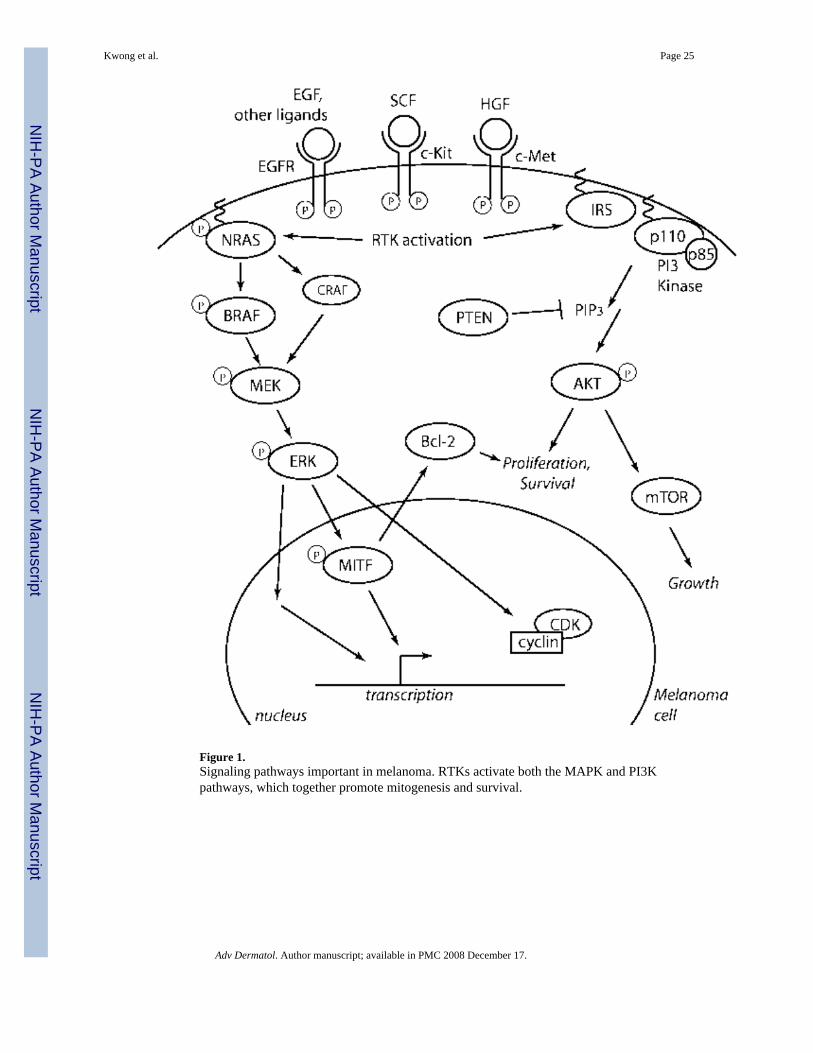
Figure 1.Signaling pathways important in melanoma. RTKs activate both the MAPK and PI3Kpathways, which together promote mitogenesis and survival.
Kwong et al. Page 25
Adv Dermatol. Author manuscript; available in PMC 2008 December 17.
NIH
-PA Author Manuscript
NIH
-PA Author Manuscript
NIH
-PA Author Manuscript
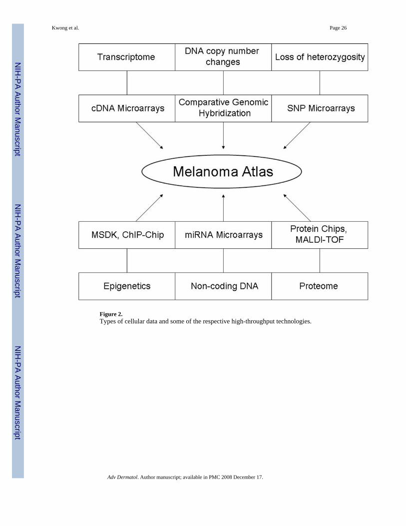
Figure 2.Types of cellular data and some of the respective high-throughput technologies.
Kwong et al. Page 26
Adv Dermatol. Author manuscript; available in PMC 2008 December 17.
NIH
-PA Author Manuscript
NIH
-PA Author Manuscript
NIH
-PA Author Manuscript
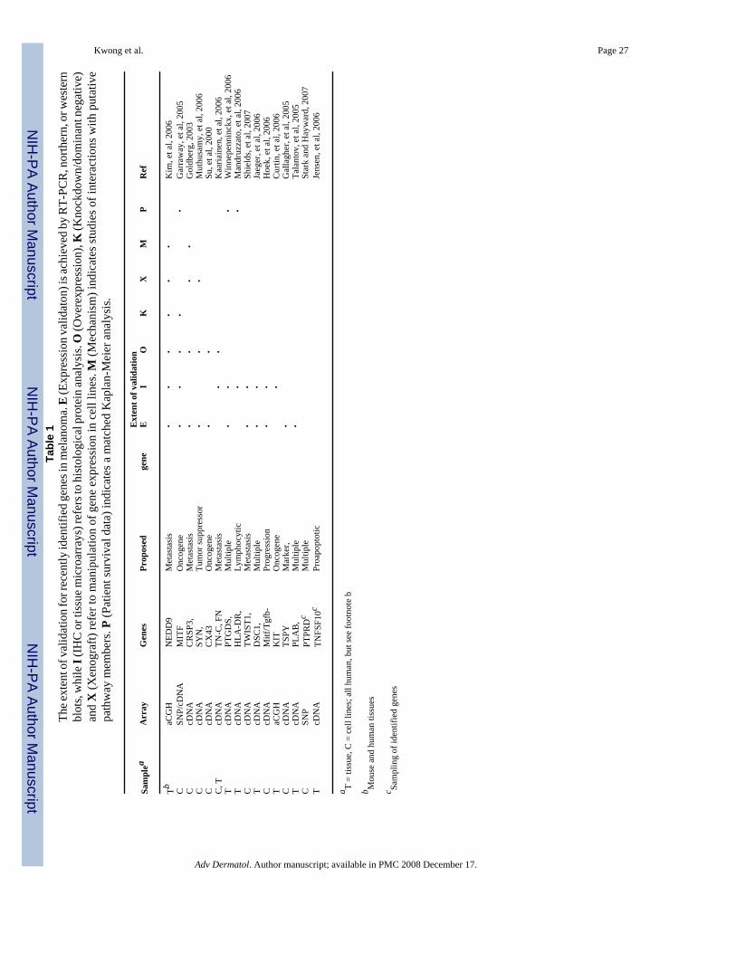
NIH
-PA Author Manuscript
NIH
-PA Author Manuscript
NIH
-PA Author Manuscript
Kwong et al. Page 27Ta
ble
1Th
e ext
ent o
f val
idat
ion
for r
ecen
tly id
entif
ied
gene
s in
mel
anom
a. E
(Exp
ress
ion
valid
aton
) is a
chie
ved
by R
T-PC
R, n
orth
ern,
or w
este
rnbl
ots,
whi
le I
(IH
C o
r tis
sue m
icro
arra
ys) r
efer
s to
hist
olog
ical
pro
tein
anal
ysis
. O (O
vere
xpre
ssio
n), K
(Kno
ckdo
wn/
dom
inan
t neg
ativ
e)an
d X
(Xen
ogra
ft) re
fer t
o m
anip
ulat
ion
of g
ene
expr
essi
on in
cel
l lin
es. M
(Mec
hani
sm) i
ndic
ates
stud
ies o
f int
erac
tions
with
put
ativ
epa
thw
ay m
embe
rs. P
(Pat
ient
surv
ival
dat
a) in
dica
tes a
mat
ched
Kap
lan-
Mei
er a
naly
sis.
Ext
ent o
f val
idat
ion
Sam
plea
Arr
ayG
enes
Prop
osed
gene
EI
OK
XM
PR
ef
TbaC
GH
NED
D9
Met
asta
sis
••
••
••
Kim
, et a
l, 20
06C
SNP/
cDN
AM
ITF
Onc
ogen
e•
••
••
Gar
raw
ay, e
t al,
2005
CcD
NA
CR
SP3,
Met
asta
sis
••
••
Gol
dber
g, 2
003
CcD
NA
SYN
,Tu
mor
supp
ress
or•
••
Mut
husa
my,
et a
l, 20
06C
cDN
AC
X43
Onc
ogen
e•
•Su
, et a
l, 20
00C
, TcD
NA
TN-C
, FN
Met
asta
sis
••
Kaa
riain
en, e
t al,
2006
TcD
NA
PTG
DS,
Mul
tiple
••
•W
inne
penn
inck
x, e
t al,
2006
TcD
NA
HLA
-DR
,Ly
mph
ocyt
ic•
•M
andr
uzza
to, e
t al,
2006
CcD
NA
TWIS
T1,
Met
asta
sis
••
Shie
lds,
et a
l, 20
07T
cDN
AD
SC1,
Mul
tiple
••
Jaeg
er, e
t al,
2006
CcD
NA
Mitf
/Tgf
b-Pr
ogre
ssio
n•
•H
oek,
et a
l, 20
06T
aCG
HK
ITO
ncog
ene
•C
urtin
, et a
l, 20
06C
cDN
ATS
PYM
arke
r,•
Gal
lagh
er, e
t al,
2005
TcD
NA
PLA
B,
Mul
tiple
•Ta
lant
ov, e
t al,
2005
CSN
PPT
PRD
cM
ultip
leSt
ark
and
Hay
war
d, 2
007
TcD
NA
TNFS
F10c
Proa
popt
otic
Jens
en, e
t al,
2006
a T =
tissu
e, C
= c
ell l
ines
; all
hum
an, b
ut se
e fo
otno
te b
b Mou
se a
nd h
uman
tiss
ues
c Sam
plin
g of
iden
tifie
d ge
nes
Adv Dermatol. Author manuscript; available in PMC 2008 December 17.

![What Was Lost' [poem]](https://img.dokumen.tips/doc/110x75/6352c4e041bc6573100e373f/what-was-lost-poem.jpg)


