Embed Size (px)
Citation preview
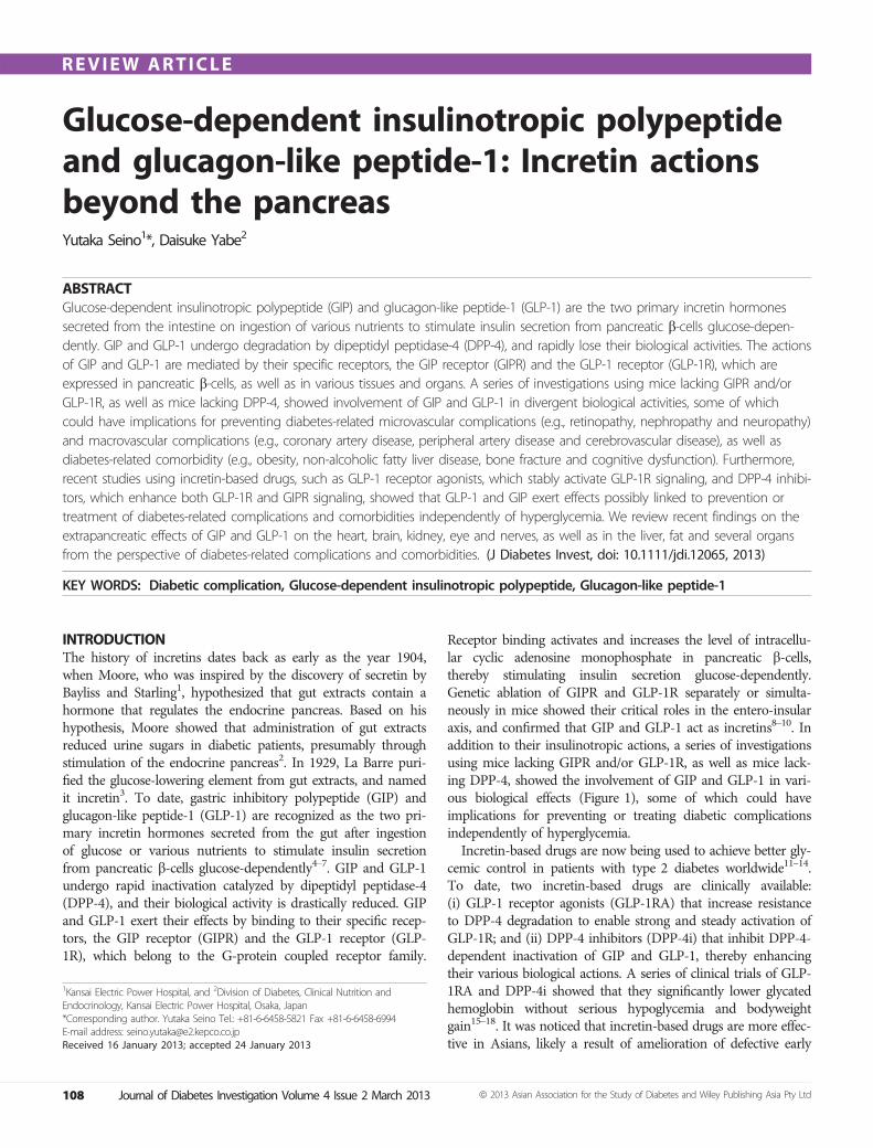
Glucose-dependent insulinotropic polypeptideand glucagon-like peptide-1: Incretin actionsbeyond the pancreasYutaka Seino1*, Daisuke Yabe2
ABSTRACTGlucose-dependent insulinotropic polypeptide (GIP) and glucagon-like peptide-1 (GLP-1) are the two primary incretin hormonessecreted from the intestine on ingestion of various nutrients to stimulate insulin secretion from pancreatic b-cells glucose-depen-dently. GIP and GLP-1 undergo degradation by dipeptidyl peptidase-4 (DPP-4), and rapidly lose their biological activities. The actionsof GIP and GLP-1 are mediated by their specific receptors, the GIP receptor (GIPR) and the GLP-1 receptor (GLP-1R), which areexpressed in pancreatic b-cells, as well as in various tissues and organs. A series of investigations using mice lacking GIPR and/orGLP-1R, as well as mice lacking DPP-4, showed involvement of GIP and GLP-1 in divergent biological activities, some of whichcould have implications for preventing diabetes-related microvascular complications (e.g., retinopathy, nephropathy and neuropathy)and macrovascular complications (e.g., coronary artery disease, peripheral artery disease and cerebrovascular disease), as well asdiabetes-related comorbidity (e.g., obesity, non-alcoholic fatty liver disease, bone fracture and cognitive dysfunction). Furthermore,recent studies using incretin-based drugs, such as GLP-1 receptor agonists, which stably activate GLP-1R signaling, and DPP-4 inhibi-tors, which enhance both GLP-1R and GIPR signaling, showed that GLP-1 and GIP exert effects possibly linked to prevention ortreatment of diabetes-related complications and comorbidities independently of hyperglycemia. We review recent findings on theextrapancreatic effects of GIP and GLP-1 on the heart, brain, kidney, eye and nerves, as well as in the liver, fat and several organsfrom the perspective of diabetes-related complications and comorbidities. (J Diabetes Invest, doi: 10.1111/jdi.12065, 2013)
KEY WORDS: Diabetic complication, Glucose-dependent insulinotropic polypeptide, Glucagon-like peptide-1
INTRODUCTIONThe history of incretins dates back as early as the year 1904,when Moore, who was inspired by the discovery of secretin byBayliss and Starling1, hypothesized that gut extracts contain ahormone that regulates the endocrine pancreas. Based on hishypothesis, Moore showed that administration of gut extractsreduced urine sugars in diabetic patients, presumably throughstimulation of the endocrine pancreas2. In 1929, La Barre puri-fied the glucose-lowering element from gut extracts, and namedit incretin3. To date, gastric inhibitory polypeptide (GIP) andglucagon-like peptide-1 (GLP-1) are recognized as the two pri-mary incretin hormones secreted from the gut after ingestionof glucose or various nutrients to stimulate insulin secretionfrom pancreatic b-cells glucose-dependently4–7. GIP and GLP-1undergo rapid inactivation catalyzed by dipeptidyl peptidase-4(DPP-4), and their biological activity is drastically reduced. GIPand GLP-1 exert their effects by binding to their specific recep-tors, the GIP receptor (GIPR) and the GLP-1 receptor (GLP-1R), which belong to the G-protein coupled receptor family.
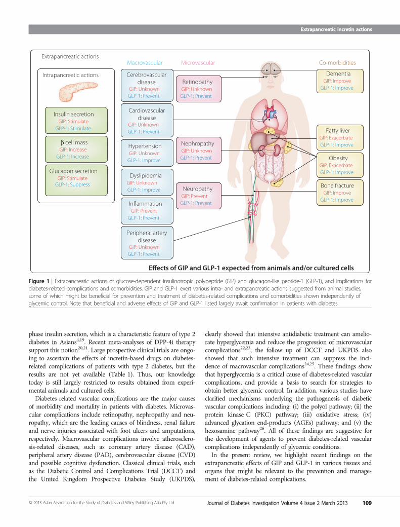
Receptor binding activates and increases the level of intracellu-lar cyclic adenosine monophosphate in pancreatic b-cells,thereby stimulating insulin secretion glucose-dependently.Genetic ablation of GIPR and GLP-1R separately or simulta-neously in mice showed their critical roles in the entero-insularaxis, and confirmed that GIP and GLP-1 act as incretins8–10. Inaddition to their insulinotropic actions, a series of investigationsusing mice lacking GIPR and/or GLP-1R, as well as mice lack-ing DPP-4, showed the involvement of GIP and GLP-1 in vari-ous biological effects (Figure 1), some of which could haveimplications for preventing or treating diabetic complicationsindependently of hyperglycemia.Incretin-based drugs are now being used to achieve better gly-
cemic control in patients with type 2 diabetes worldwide11–14.To date, two incretin-based drugs are clinically available:(i) GLP-1 receptor agonists (GLP-1RA) that increase resistanceto DPP-4 degradation to enable strong and steady activation ofGLP-1R; and (ii) DPP-4 inhibitors (DPP-4i) that inhibit DPP-4-dependent inactivation of GIP and GLP-1, thereby enhancingtheir various biological actions. A series of clinical trials of GLP-1RA and DPP-4i showed that they significantly lower glycatedhemoglobin without serious hypoglycemia and bodyweightgain15–18. It was noticed that incretin-based drugs are more effec-tive in Asians, likely a result of amelioration of defective early
1Kansai Electric Power Hospital, and 2Division of Diabetes, Clinical Nutrition andEndocrinology, Kansai Electric Power Hospital, Osaka, Japan*Corresponding author. Yutaka Seino Tel.: +81-6-6458-5821 Fax +81-6-6458-6994E-mail address: [email protected] 16 January 2013; accepted 24 January 2013
108 Journal of Diabetes Investigation Volume 4 Issue 2 March 2013 ª 2013 Asian Association for the Study of Diabetes and Wiley Publishing Asia Pty Ltd
REVIEW ARTICLE
phase insulin secretion, which is a characteristic feature of type 2diabetes in Asians4,19. Recent meta-analyses of DPP-4i therapysupport this notion20,21. Large prospective clinical trials are ongo-ing to ascertain the effects of incretin-based drugs on diabetes-related complications of patients with type 2 diabetes, but theresults are not yet available (Table 1). Thus, our knowledgetoday is still largely restricted to results obtained from experi-mental animals and cultured cells.Diabetes-related vascular complications are the major causes
of morbidity and mortality in patients with diabetes. Microvas-cular complications include retinopathy, nephropathy and neu-ropathy, which are the leading causes of blindness, renal failureand nerve injuries associated with foot ulcers and amputations,respectively. Macrovascular complications involve atherosclero-sis-related diseases, such as coronary artery disease (CAD),peripheral artery disease (PAD), cerebrovascular disease (CVD)and possible cognitive dysfunction. Classical clinical trials, suchas the Diabetic Control and Complications Trial (DCCT) andthe United Kingdom Prospective Diabetes Study (UKPDS),
clearly showed that intensive antidiabetic treatment can amelio-rate hyperglycemia and reduce the progression of microvascularcomplications22,23; the follow up of DCCT and UKPDS alsoshowed that such intensive treatment can suppress the inci-dence of macrovascular complications24,25. These findings showthat hyperglycemia is a critical cause of diabetes-related vascularcomplications, and provide a basis to search for strategies toobtain better glycemic control. In addition, various studies haveclarified mechanisms underlying the pathogenesis of diabeticvascular complications including: (i) the polyol pathway; (ii) theprotein kinase C (PKC) pathway; (iii) oxidative stress; (iv)advanced glycation end-products (AGEs) pathway; and (v) thehexosamine pathway26. All of these findings are suggestive forthe development of agents to prevent diabetes-related vascularcomplications independently of glycemic conditions.In the present review, we highlight recent findings on the
extrapancreatic effects of GIP and GLP-1 in various tissues andorgans that might be relevant to the prevention and manage-ment of diabetes-related complications.
Cerebrovasculardisease
GIP: UnknownGLP-1: Prevent
Macrovascular
Insulin secretionGIP: Stimulate
GLP-1: Stimulate
β cell massGIP: Increase
GLP-1: Increase
Glucagon secretionGIP: Stimulate
GLP-1: Suppress
Intrapancreatic actions
Extrapancreatic actionsCo-morbidities
Effects of GIP and GLP-1 expected from animals and/or cultured cells
DementiaGIP: Improve
GLP-1: Improve
Fatty liverGIP: ExacerbateGLP-1: Improve
ObesityGIP: ExacerbateGLP-1: Improve
Cardiovasculardisease
GIP: UnknownGLP-1: Prevent
Microvascular
HypertensionGIP: Unknown
GLP-1: Improve
DyslipidemiaGIP: UnknownGLP-1: Improve
InflammationGIP: Prevent
GLP-1: Prevent
RetinopathyGIP: Unknown
GLP-1: Prevent
NephropathyGIP: Unknown
GLP-1: Prevent
Peripheral arterydisease
GIP: UnknownGLP-1: Prevent
Bone fractureGIP: Improve
GLP-1: Improve
NeuropathyGIP: Prevent
GLP-1: Prevent
Figure 1 | Extrapancreatic actions of glucose-dependent insulinotropic polypeptide (GIP) and glucagon-like peptide-1 (GLP-1), and implications fordiabetes-related complications and comorbidities. GIP and GLP-1 exert various intra- and extrapancreatic actions suggested from animal studies,some of which might be beneficial for prevention and treatment of diabetes-related complications and comorbidities shown independently ofglycemic control. Note that beneficial and adverse effects of GIP and GLP-1 listed largely await confirmation in patients with diabetes.
ª 2013 Asian Association for the Study of Diabetes and Wiley Publishing Asia Pty Ltd Journal of Diabetes Investigation Volume 4 Issue 2 March 2013 109
Extrapancreatic incretin actions
DIABETES-RELATED MACROVASCULARCOMPLICATIONSSeveral studies showing the effects of GLP-1 and GIP onmacrovascular function have recently been published. Wesummarize the effects of GLP-1 and GIP that might be ofsignificance for treatment of macrovascular complications, suchas CAD, PAD and CVD, in patients with diabetes.
Effects on AtherosclerosisAtherosclerosis constitutes the underlying pathological lesion inthe clinical entities CAD, CVD and PAD. The inflammatoryresponses induced by adhesion of monocytes to the vascular wallhave been known to play important roles in the early stages ofthe development of atherosclerosis. GLP-1 was shown to signifi-cantly inhibit macrophage infiltration and atherosclerosis devel-opment in normal and diabetic apolipoprotein E-deficient mice(Figure 2)27–29, whereas a recent study using the long-actingGLP-1RA taspoglutide failed to reproduce attenuation of
atherosclerosis by GLP-1R activation30. Exendin-4 signifi-cantly inhibited monocyte adhesion to the vascular endothelialcells by reducing production of intercellular adhesion molecule(ICAM)-129. Liraglutide also attenuated the expressions ofvascular adhesion molecules (VAM) and ICAM-1 in human vas-cular endothelial cells, thereby suppressing development of ath-erosclerosis31,32. In patients with diabetes, it has been shown thatexenatide suppressed markers for inflammation, high sensitiveC-reactive protein and monocyte chemo-attractant protein-1(MCP-1), oxidative stress, and prostaglandin F2a33, and that ex-enatide improved induced endothelial dysfunction34,35.Although GLP-1R expression in mouse macrophages is still
controversial30, it has been shown that exendin-4 somehowinhibits lipopolysaccharide (LPS)-induced production of theinflammatory cytokine tumor necrosis factor (TNF)-a andMCP-1 from isolated mouse macrophages29. Exendin-4suppressed the nuclear translocation of p65, a component ofnuclear factor (NF)-jB, and this effect was reversed by both
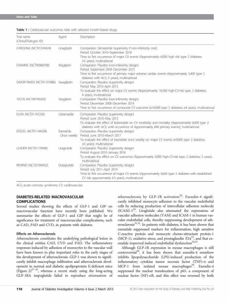
Table 1 | Cardiovascular outcomes trials with selected incretin-based drugs.
Trial name(ClinicalTrials.gov ID)
Agent Description
CAROLINA (NCT01243424) Linagliptin Comparator: Glimepiride (superiority if non-inferiority met)Period: October 2010–September 2018Time to first occurrence of major CV events (Approximately 6,000 high risk type 2 diabetes;>5 years), multinational
EXAMINE (NCT00968708) Alogliptin Comparator: Placebo (non-inferiority design)Period: September 2009–December 2013Time to first occurrence of primary major adverse cardiac events (Approximately 5,400 type 2diabetes with ACS; 5 years), multinational
SAVOR-TIMI53 (NCT01107886) Saxagliptin Comparator: Placebo (superiority design)Period: May 2010–April 2013To evaluate the effect on major CV events (Approximately 16,500 high-CV-risk type 2 diabetes;4 years), multinational
TECOS (NCT00790205) Sitagliptin Comparator: Placebo (non-inferiority design)Period: December 2008–December 2014Time to first occurrence of composite CV outcome (>14,000 type 2 diabetes; >4 years), multinational
ELIXA (NCT01147250) Lixisenatide Comparator: Placebo (superiority design)Period: June 2010–May 2013To evaluate the effect of lixisenatide on CV morbidity and mortality (Apporximately 6,000 type 2diabetes with ACS; until occurrence of Apporximately 840 primary events), multinational
EXSCEL (NCT01144338) ExenatideOnce weekly
Comparator: Placebo (superiority design)Period: June 2010–March 2017To evaluate the effect of exenatide once weekly on major CV events (>9,000 type 2 diabetes;>5 years), multinational
LEADER (NCT01179048) Liraglutide Comparator: Placebo (superiority design)Period: August 2010–January 2016To evaluate the effect on CV outcomes (Apporximately 9,000 high-CV-risk type 2 diabetes; 5 years),multinational
REWIND (NCT01394952) Dulaglutide Comparator: Placebo (superiority design)Period: July 2011–April 2019Time to first occurrence of major CV events (Approximately 9,600 type 2 diabetes with establishedCV risk; approximately 6.5 years), multinational
ACS, acute coronary syndrome; CV, cardiovascular.
110 Journal of Diabetes Investigation Volume 4 Issue 2 March 2013 ª 2013 Asian Association for the Study of Diabetes and Wiley Publishing Asia Pty Ltd
Seino and Yabe
MDL-12330A, a cyclic adenosine monophosphate (cAMP)inhibitor, and PKI14-22, a protein kinase A (PKA)-specificinhibitor. Therefore, it was suggested that these actions ofexendin-4 are likely to be mediated through a GLP-1R/PKApathway in mouse macrophages29. It also has been shown thatGLP-1 activates human macrophages through signal transduc-ers and activator of transcription (STAT3) activation36.In addition to its effects on macrophages, it has been shown
that GLP-1 exerts direct effects on vascular endothelial cells(Figure 2). GLP-1R activation directly activated endothelialnitric oxide synthase (eNOS) in human umbilical veinendothelial cells (HUVECs)37 and the aortic endothelium31.GLP-1 increased production of nitric oxide (NO), resulting inan increase of the microvascular blood flow in the blood38. Itwas reported that NO production was mediated by thepoly(adenosine diphosphate-ribose) polymerase pathway39.GLP-1 also promoted proliferation and differentiation of endo-thelial progenitor cells by upregulating vascular endothelialgrowth factor (VEGF) expression40. Incubation of human coro-nary artery endothelial cells (HCAECs) with exendin-4 resultedin a dose-dependent increase in DNA synthesis and anincreased cell number associated with enhanced eNOS andv-akt murine thymoma viral oncogene homolog 1 (Akt1) acti-vation that were abolished by a GLP-1R antagonist41; Exendin-4 directly improves endothelial dysfunction in isolated aortas42.Liraglutide also inhibited the NF-jB pathway and suppressedapoptosis of HUVECs43. There is a report that GLP-1 also
inhibited AGE-induced apoptosis in HUVECs44. Takentogether, it appears that GLP-1 can exert anti-atherogeniceffects through various mechanisms.GIP was also shown to significantly inhibit macrophage infil-
tration and atherosclerosis development in normal and diabeticapolipoprotein E-deficient mice27,28. It was recently found thatGIP inhibits AGEs-enhanced production of reactive oxygenspecies (ROS), and expression of VAM-1 and PAI-1 throughthe GIPR/Epac pathway45. These findings show that GLP-1and GIP can exert anti-atherosclerosis effects, and might haveimplications in preventing macrovascular complications.Consistent with the anti-atherosclerotic effects of GLP-1 and
GIP, DPP-4i des-fluor-sitagliptin was also shown to inhibitthe production of inflammatory cytokines, such as TNF-a,interleukin (IL)-6, IL-1b and MCP-1, as well as that of VACM-1and ICAM-1, and to increase endothelial NO production andsuppress the NF-jB pathway in experimental animals46.Furthermore, they showed that DPP-4 inhibition increases cir-culating endothelial precursor cells (EPCs), which exert vaso-protective effects46. Similar observations were made withvarious DPP-4i including sitagliptin47, alogliptin47,48 and vildag-liptin49,50. Such anti-atherosclerotic effects were also observed intype 2 diabetic patients treated with sitagliptin51–53.
Effect on Cardiac FunctionStudies in experimental animals have shown beneficial roles ofGLP-1 or GLP-1RA in CAD (Figure 2). In pigs, exendin-4
INFLAMMATION
LIPIDMETABOLISM
BLOOD PRESSURE
BrainROS production ↓
Apoptosis ↓
IntestineLipid absorption ↓
LiverFatty acid synthesis ↓ Fatty acid oxidation ↑
Endothelial cellsNO production ↑
Proliferation ↑ Apoptosis ↓
HeartApoptosis ↓
Glucose uptake ↑ NO production ↑
MacrophagesInflammatory cytokines ↓
Infiltration ↓ Foaming ↓
GLP-1
KidneySodium excretion ↑
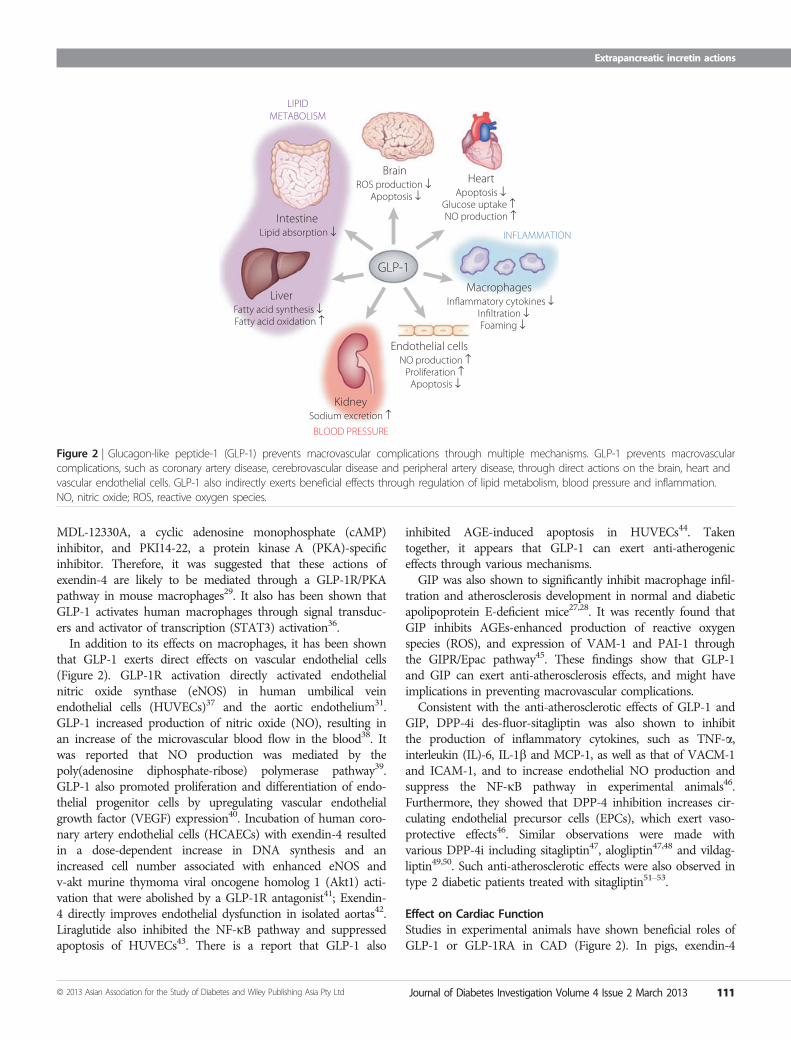
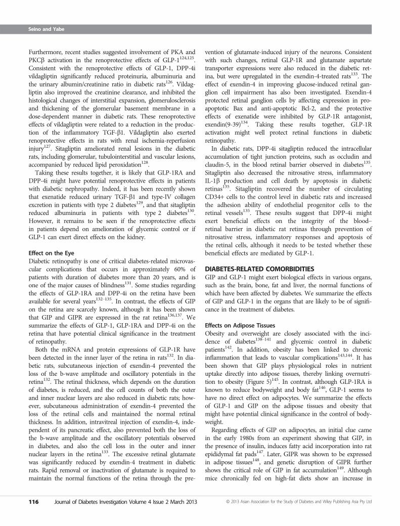
Figure 2 | Glucagon-like peptide-1 (GLP-1) prevents macrovascular complications through multiple mechanisms. GLP-1 prevents macrovascularcomplications, such as coronary artery disease, cerebrovascular disease and peripheral artery disease, through direct actions on the brain, heart andvascular endothelial cells. GLP-1 also indirectly exerts beneficial effects through regulation of lipid metabolism, blood pressure and inflammation.NO, nitric oxide; ROS, reactive oxygen species.
ª 2013 Asian Association for the Study of Diabetes and Wiley Publishing Asia Pty Ltd Journal of Diabetes Investigation Volume 4 Issue 2 March 2013 111
Extrapancreatic incretin actions
administration significantly reduced infarct size and improvedwall motion54. In mice, liraglutide administration showed simi-lar cardioprotective effects with activation of cardioprotectivegenes55. In rabbits, administration of GLP-1 fused to humantransferrin significantly reduced infarct size and improved thewall motion and ejection fraction after myocardial ischemia/reperfusion injury56. Like GLP-1RA, native GLP-1 also showeda cardioprotective function in rats with myocardial ischemia/reperfusion injury, partly by suppressing activation of peripheralneutrophil57. Another study on dogs with myocardial ischemiasuggested that GLP-1 exerted a cardioprotective role by aug-menting glucose uptake58. This is consistent with a recent studyshowing that the long-acting GLP-1RA albiglutide enhancedmyocardial glucose uptake and promoted a shift toward a moreenergetically favorable substrate metabolism by increasing bothglucose and lactate oxidation, thereby reducing infarct size andimproving cardiac function59. Despite recent debates on GLP-1R expression in mouse hearts, the earlier study showed thatGLP-1R activation increases cAMP production and suppressescaspase-3 activation in cardiomyocytes, thereby preventingapoptosis of cardiomyocytes55. Another study showed thatGLP-1R activation increases cAMP and phosphorylation of Aktand extracellular signal-regulated kinase (ERK), regulators ofgrowth and glucose metabolism in cardiomyocytes60. Impor-tantly, it was shown that although GLP-1 increased cAMPlevels in cardiomyocytes, the increased cAMP did not correlatewith the intracellular calcium concentration and subsequentcardiomyocyte contractility61. It is interesting to note thatGLP-1 was previously shown to expert cardioprotectiveeffects mediated through both GLP-1R-dependent andGLP-1R-independent mechanisms62. It was suggested thatGLP-1R-independent cardioprotective actions of GLP-1 aremediated by GLP-1(9-36)amide, the primary GLP-1 metabolitein vivo, because GLP-1(9-36)amide exerts cardioprotectiveactions in GLP-1R-deficient mice60. However, the molecularmechanisms underlying the GLP-1R-independent cardioprotec-tive actions of GLP-1(9-36)amide are largely unknown. Little isknown of the effects of GIP on the heart.In a clinical study, it was reported that GLP-1 significantly
improved left ventricle ejection fraction, global wall motionindices, and regional wall motion indices in patients with acutemyocardial infarction and severe systolic dysfunction63. Morerecently, it was reported that GLP-1 infusion protects the heartfrom ischemic left ventricle dysfunction induced by dobutaminestress in CAD patients64. Furthermore, it was shown that exe-natide reduces reperfusion injury and final infarct size inpatients with ST-segment elevated myocardial infarction65,66.Studies in experimental animals showed that DPP-4 inhibi-
tion also results in similar cardioprotective effects67–72. It hasbeen shown that the DPP-4i sitagliptin increased the intracellu-lar cAMP and PKA activity, and that H-89, a potent selectivePKA inhibitor, completely blocked the effect of sitagliptin inreducing the size of myocardial infarct, suggesting involvementof the GLP-1R/cAMP/PKA pathway70. However, a recent study
also suggested that the cardioprotective effects of DPP-4i aremediated not only by GLP-1, but also by other bioactive pep-tides, such as stromal-derived factor-1a51,73,74.It was also suggested that GLP-1R activation might exert ben-
eficial effects in heart failure. In a dog model of pacing-inducedheart failure, GLP-1 increased glucose uptake, thereby increasingleft ventricular function75. In the spontaneously hypertensiveand heart failure-prone rat, GLP-1 infusion improved survivaland preserved left ventricular function with reduced apoptosisof cardiomyocytes76. In a rat model of chronic heart failure withpermanent occlusion of the left anterior descending artery, infu-sion of GLP-1 or GLP-1RA enhanced left ventricular function,reducing left ventricular remodeling and improving survival77.In humans, infusion of GLP-1 or GLP-1RA in patients withheart failure improved left ventricular ejection faction78,79. Fur-thermore, it has been shown that DPP-4i improved left ventric-ular function in experimental models of heart failure71,80.Although further studies of the underlying mechanisms arerequired, these results together strongly suggest clinical implica-tions for GLP-1RA in treatment of heart failure.
Effect on Cerebrovascular FunctionNeuroprotective effects of GLP-1RA after cerebral ischemiain non-diabetic and diabetic animals have been described(Figure 2)81–83. Intravenous injection of exendin-4 after cerebralischemia reduced the infarct size and neurological deficitsinduced by reperfusion after occlusion of the middle cerebralartery in a mouse model of acute cerebral infarction81,83. Exen-din-4 also attenuated the oxidative stress and reduced neuronalcell death after reperfusion in this focal ischemia model. Asexendin-4 injection has been associated with increased intracel-lular cAMP levels81, this compound likely exerts its neuropro-tective effect through a cAMP activation pathway. It thereforeappears that GLP-1RA might be potentially useful in the treat-ment of cerebral infarction. No study has been carried out onthe neuroprotective effects of GIP after cerebral ischemia,although it has been shown that GIPR-deficient mice showedimpaired learning, synaptic plasticity and neurogenesis84, andthat GIP and GIPR agonist enhance long-term potentiation(LTP) and neurogenesis85,86. Consistent with the neuroprotec-tive effects of GLP-1RA after cerebral ischemia, the DPP-4ilinagliptin was found to reduce ischemic brain damage indiabetic mice87. In the same study, sulfonylurea glimepiridelowered glucose levels, but did not show similar neuroprotectiveeffects, suggesting that the neuroptotective effects are indepen-dent of glycemic control and presumably mediated by GLP-187.
Effect on Blood PressureHypertension plays a critical role in the development of macro-vascular complications. Chronic infusion of GLP-1 reduced theincidence of hypertension, and prevented cardiac hypertrophyand fibrosis in salt-sensitive Dahl rats88. In this model ofhypertension, GLP-1 also reduced urinary albumin excre-tion, increased urinary sodium excretion and improved
112 Journal of Diabetes Investigation Volume 4 Issue 2 March 2013 ª 2013 Asian Association for the Study of Diabetes and Wiley Publishing Asia Pty Ltd
Seino and Yabe
histopathological abnormalities, such as glomerulosclerosis andtubular necrosis (Figure 2)88. In salt-sensitive obese db/db miceand angiotensin II-infused C57BL/6J mice, exendin-4 preventedthe onset of hypertension and increased the urinary sodiumexcretion89. Similarly, infusion of GLP-1RA AC3174 also atten-uated hypertension and the histopathological changes associatedwith the renal dysfunction in the Dahl rats90. Administration ofDPP-4i sitagliptin also reduced blood pressure in spontaneouslyhypertensive rats by decreasing expression of Na+/H+ exchangerisoform 3 in microvilli membranes of the proximal renaltubule, thereby increasing the urinary sodium excretion and theurinary volume, and reducing blood pressure91. Furthermore,GLP-1RA and DPP-4i also ameliorates endothelial dysfunc-tion92.Antihypertensive effects of GLP-1 have consistently been
shown in several clinical trials. In the trials to evaluate efficacyand safety of GLP-1RA liraglutide carried out in type 2 diabeticpatients, liraglutide administration was found to decrease sys-tolic blood pressure by 2–6 mmHg from baseline in26 weeks93. In addition, measurement of flow-mediated vasodi-latation (FMD) of the brachial artery as a measure of endothe-lial function in patients with type 2 diabetes after 16-weekexenatide treatment showed a significantly higher value ofFMD in the exenatide-treated group compared with that in theglimepiride-treated group94. This result shows that exenatideexerts vasodilatory action and might reduce blood pressure.Increase of urinary sodium excretion and urinary volume are
well-known effects of GLP-1 and GLP-1RA in rodents. In rats,intracerebroventricular injection of GLP-1 was found to exertmarked natriuretic and diuretic effects mediated by GLP-1Rthat were blocked by treatment with GLP-1R antagonist exen-din(9-39)95. GLP-1 was expressed in porcine proximal tubularcells isolated from kidneys, and inhibited sodium reabsorp-tion96. It is therefore likely that the marked diuretic effect ofGLP-1 is mediated by direct regulation of sodium reabsorptionin kidney proximal tubules, as well as through hypothalamicGLP-1R. Clinically, the effects of GLP-1 infusion on urinarysodium excretion, urinary output and the glomerular filtrationrate after an intravenous administration of salt load were inves-tigated in obese men; GLP-1 was found to significantly enhancethe urinary sodium excretion, H+ secretion and glomerularhyperfiltration in obese men97.Although no report has shown an association of GIP with
blood pressure, DPP-4i sitagliptin attenuated elevation of bloodpressure in spontaneously hypertensive rats91,92. It also has beenreported that DPP-4i reduces blood pressure98,99, which supportsthe notion that GLP-1 regulates blood pressure in humans.
Effect on DyslipidemiaDyslipidemia plays a critical role in the development of macro-vascular complications. It has been shown that GLP-1 amelio-rates dyslipidemia in experimental animals, as well as inhumans, whereas little is known on the effects of GIP on lipidmetabolisms.
GLP-1 infusion reduced apolipoprotein B-48 production andtriglycerides absorption (Figure 2)100. These effects were repro-duced in mice and hamsters infused with exendin-4, whichacutely decreased postprandial serum triacylglycerol and apoli-poprotein B-48 GLP-1R-dependently101. These effects wereobserved even if exendin-4 was given 1 h after fat ingestion,showing that the effects on postprandial lipid metabolism werenot related to delayed gastric emptying101. Secretion of apolipo-protein B-48 was significantly reduced from hamster primaryenterocytes treated by enendin-4101, suggesting that GLP-1Ractivation expressed on enterocytes controls secretion ofchylomicron. GLP-1 controls hepatic lipid metabolism. GLP-1RA markedly reduced hepatic lipid content by suppressinggenes involved in fatty acid synthesis (e.g., sterol-regulatory ele-ment binding protein-1c, fatty acid synthase and steroyl CoAdesaturase-1) and enhancing expression genes regulating fattyacid oxidation (e.g., acyl-coenzyme A oxidase and carnitine pal-mitoyltransferase 1a)30,102,103. Mechanisms regulating expressionof genes involved in lipid metabolism by GLP-1RA are largelyunknown, inasmuch as the presence of hepatic GLP-1R expres-sion is still controversial. Nevertheless, both GLP-1 and GLP-1RA clearly ameliorate dyslipidemia in experimental animals,suggesting clinical implications in patients with dyslipidemia.GLP-1 infusion inhibited the postprandial elevation of trigly-
cerides and free fatty acids in healthy human subjects104. A sin-gle subcutaneous injection of exenatide in patients with newlydiagnosed type 2 diabetes also showed marked reduction inpostprandial triacylglycerol, as well as in apolipoprotein B-48105.Although these effects of GLP-1 or GLP-1RA on triglyceridesand free fatty acids could be partly a result of delayed gastricemptying, these results clearly show that GLP-1R activationameliorates postprandial lipidemia. It has also been shown thatDPP-4i vildagliptin and sitagliptin suppressed postprandialelevation of triglycerides and apolipoprotein B-48 in patientswith type 2 diabetes106,107. As DPP-4i shows little effect ongastric emptying108, the effects of DPP-4i on postprandiallipidemia might be largely mediated by inhibition of intestinallipid absorption. It is noteworthy that DPP-4i ameliorates dysli-pidemia in patients with type 2 diabetes109, which, althoughsmall, could also contribute to the prevention of macrovascularcomplications.
DIABETES-RELATED MICROVASCULARCOMPLICATIONSGIPR and GLP-1R are expressed in the peripheral and centralnervous system, the eyes, and the kidney, suggesting the possi-bility of some effects in these organs. We summarize the effectsof GIP and GLP-1 in those organs that are likely to be ofsignificance in the treatment of the microvascular complications(i.e., retinopathy, nephropathy and neuropathy) of diabetes.
Effect on the Peripheral Nervous SystemMost recognized diabetes-related neurological complicationsinvolve the peripheral nervous system110. Several studies have
ª 2013 Asian Association for the Study of Diabetes and Wiley Publishing Asia Pty Ltd Journal of Diabetes Investigation Volume 4 Issue 2 March 2013 113
Extrapancreatic incretin actions
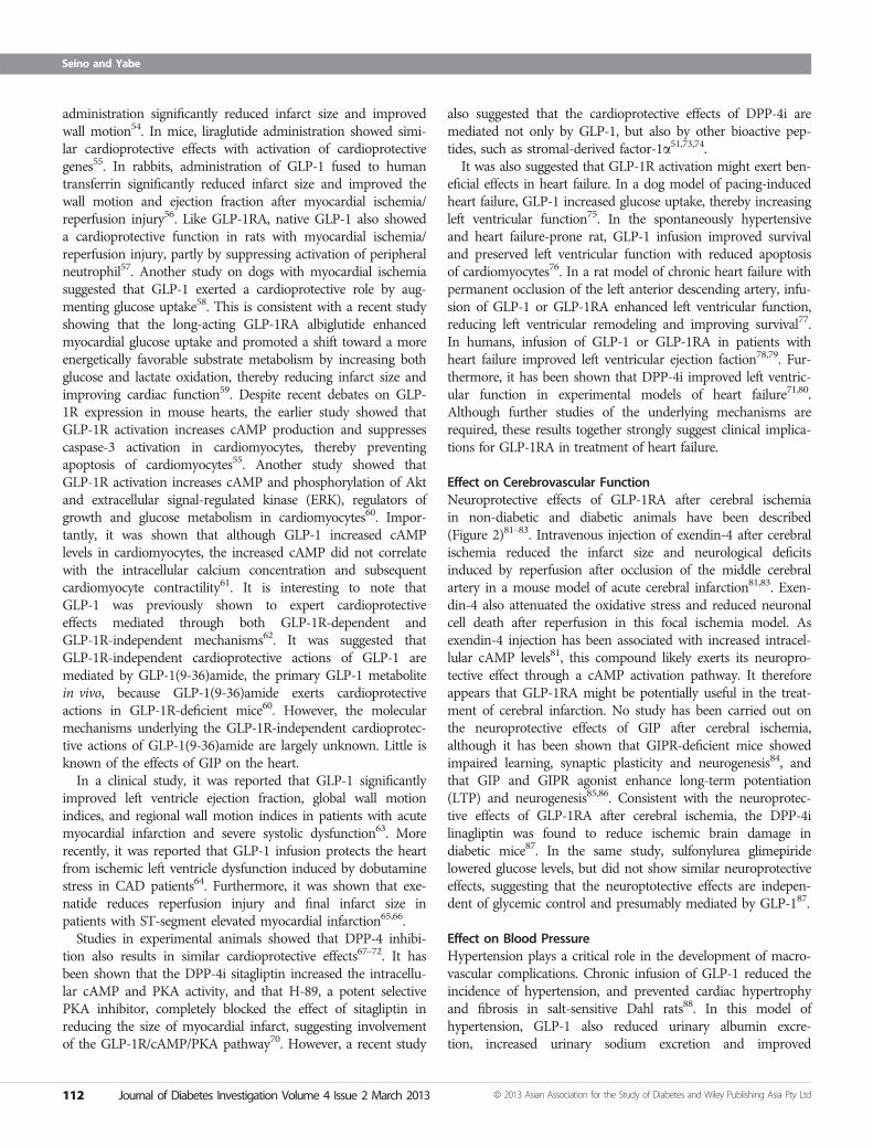
shown beneficial effects of GLP-1 and GIP on the peripheralnervous system that might have therapeutic implications forthe treatment of diabetic neuropathy.GLP-1R has been found to be expressed in the lumbar dorsal
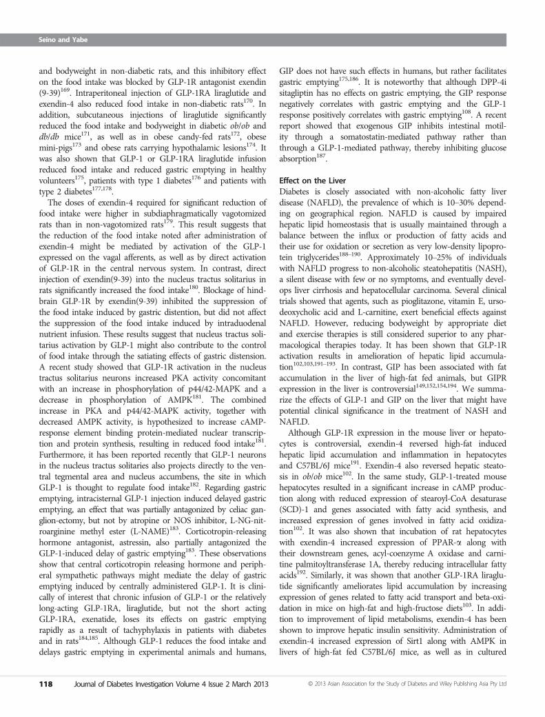
root ganglion neurons (DRG) of C57BL6/J mice111. GLP-1 andGLP-1RA exendin-4 significantly promoted the neurite out-growth of DRG neurons (Figure 3). Exendin-4 attenuated thehypoalgesia, and delayed motor and sensory nerve conductionvelocity in diabetic mice112. In the same study, exendin-4 alsoameliorated the decrease in intra-epidermal nerve fiber densitiesin the sole skins of the diabetic mice. These findings suggestthat exendin-4 might ameliorate the severity of diabetic poly-neuropathy through exerting direct actions on the DRG neu-rons and their axons. GLP-1R was also found to be expressedin the sciatic nerve112. GLP-1RA significantly increased thephosphorylated ERK 1/2 levels in the sciatic nerves of diabeticrats, showing GLP-1R activity in this diabetic tissue. Exendin-4exerted no effect on the blood sugar or insulin levels in diabeticmice, and also had no effect on paw thermal response latenciesin these mice, but attenuated the reductions of motor nerveconduction velocity and paw intra-epidermal fiber density seenin diabetic mice. Thus, GLP-1R-mediated ERK-signaling in thesciatic nerve of diabetic rodents might protect the large-motorfiber functions and small C fiber structure by a mechanismindependent of glycemic control113. GLP-1R has been found tobe expressed in the skin. Exendin-4 treatment reduced theincrease in the current perception threshold seen in diabetic
rats. The decrease in the size of myelinated fibers or in theaxon/fiber area ratio in the sciatic nerve and loss of the intra-epidermal nerve fibers in the skin of diabetic rats were alsoameliorated by exendin-4 treatment. Thus, exendin-4 mightprevent the peripheral nerve degeneration seen in diabetic rats,suggesting that GLP-1RA might be useful in the treatment ofperipheral neuropathy112. Pyridoxine-induced peripheral neu-ropathy is characterized by sensory nerve conduction deficitsassociated with disturbances of the nerve fiber geometry andaxonal atrophy. In an evaluation carried out using behavioraland morphometric techniques, GLP-1 and GLP-1RA exendin-4were found to improve pyridoxine-induced sensory peripheralneuropathy in rats114. Based on these findings, it has beensuggested that GLP-1RA might be useful in the treatment ofdiabetic neuropathy.Expressions of GIP and GIPR were enhanced after sciatic
nerve crush injury in DRG, spinal cord and nerve fragments ofrats, suggesting involvement of GIP/GIPR in axonal regenera-tion. Indeed, GIPR-deficient mice show impaired axonal regen-eration115. Thus, GIPR activation might have therapeuticimplications for the treatment of diabetic neuropathy.Consistent with the beneficial effects of GIP and GLP-1 on
the peripheral nervous system, DPP-4i vildagliptin analogPKF275-055 partially counteracted the nerve conduction veloc-ity deficit observed in diabetic rats116. Diabetic rats developedmechanical hyperalgesia and showed significantly longer ther-mal response latencies117. PKF275-055 induced recovery of the
100 µm
Neurite outgrowth
(a)
(c) (d)
(b)
Ganglions140
120
100
80
60
40
20
0
6000
5000
4000
3000
Leng
th (µ
m/c
ell)
Join
t (nu
mbe
r/ce
ll)
2000
1000
0CON CON0.1 1 10 100 GLP-1
10 nmol/LEx4 (nmol/L)0.1 1 10 100 GLP-1
10 nmol/LEx4 (nmol/L)
100 µm
Figure 3 | Glucagon-like peptide-1 (GLP-1) receptor activation promotes neurite outgrowth of the dorsal root ganglion. Representative fluorescencemicrograph of dorsal root ganglion neurons cultured in the (a) absence or (b) presence of GLP-1. GLP-1 or GLP-1 receptor agonist exendin-4 (Ex4)increased the total neurite length (c) and joint number (d) of neurites. CON, control. Reproduced from Himeno et al.111, with permission from theAmerican Diabetes Association © 2011.
114 Journal of Diabetes Investigation Volume 4 Issue 2 March 2013 ª 2013 Asian Association for the Study of Diabetes and Wiley Publishing Asia Pty Ltd
Seino and Yabe
mechanical sensitivity thresholds by approximately 50% andprogressively improved the alterations in the thermal respon-siveness. DPP-4i is therefore likely to have a potential thera-peutic effect in the treatment of diabetic neuropathy.Vildagliptin was found to protect against nerve fiber loss indiabetic animals117. The decrease in intra-epidermal nerve fiberdensity in diabetic rats was significantly inhibited by vildaglip-tin treatment117. Based on these results, it is suggested thatDPP-4i might prevent peripheral nerve degeneration indiabetic animals and might be useful in treatment of peri-pheral neuropathy.
Effect on the KidneyDiabetic nephropathy is a critical diabetes-related microvascularcomplication that is a major cause of renal failure118,119.Recently, many studies have been carried out to investigate theeffects of GLP-1, GLP-1RA and DPP-4i on kidney dysfunc-tions, but little is known about the effect of GIP on kidney. Wereview the effects of GLP-1, GLP-1RA and DPP-4i on the kid-ney functions that might be of significance in the treatment ofdiabetic nephropathy.GLP-1R messenger ribonucleic acid (mRNA) was detected in
rat glomeruli and glomerular endothelial cells, as well as inhuman monocytes and macrophages120. In diabetic db/db mice,
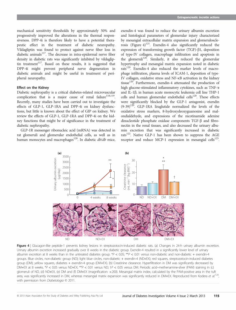
exendin-4 was found to reduce the urinary albumin excretionand histological parameters of glomerular injury characterizedby mesangial extracellular matrix expansion and glomeruloscle-rosis (Figure 4)121. Exendin-4 also significantly reduced theexpression of transforming growth factor (TGF)-b1, depositionof type-IV collagen, macrophage infiltration and apoptosis inthe glomeruli120. Similarly, it also reduced the glomerularhypertrophy and mesangial matrix expansion noted in diabeticrats120. Exendin-4 also reduced the marker levels of macro-phage infiltration, plasma levels of ICAM-1, deposition of type-IV collagen, oxidative stress and NF-jB activation in the kidneytissue120. Furthermore, exendin-4 attenuated the production ofhigh glucose-stimulated inflammatory cytokines, such as TNF-aand IL-1b, in human acute monocytic leukemia cell line THP-1cells and human glomerular endothelial cells120. These effectswere significantly blocked by the GLP-1 antagonist, exendin(9-39)120. GLP-1RA liraglutide normalized the levels of theoxidative stress markers, 8-hydroxydeoxyguanosine and mal-ondialdehyde, and expressions of the nicotinamide adeninedinucleotide phosphate oxidase components TGF-b and fibro-nectin in the renal tissues, and also decreased the urinary albu-min excretion that was significantly increased in diabeticrats122. Native GLP-1 has been shown to suppress the AGEreceptor and reduce MCP-1 expression in mesangial cells123.
** ***
†
30
20
Cre
atin
ine
clea
ranc
e(m
L m
in–1
[kg
BW]–1
)
10
0ND ND+EX DM DM+EX
*
3000
3500(a) (b)
(c) (d) (e) (f)
Urin
ary
albu
min
exc
retio
n (µ
g/da
y)
2500
2000
1500
1000
500
00
ND ND+EX DM DM+EX
4 weeks 8 weeks
Figure 4 | Glucagon-like peptide-1 prevents kidney lesions in streptozotocin-induced diabetic rats. (a) Changes in 24-h urinary albumin excretion.Urinary albumin excretion increased gradually over 8 weeks in the diabetic group. Exendin-4 resulted in a significantly lower level of urinaryalbumin excretion at 8 weeks than in the untreated diabetes group. *P < 0.05; **P < 0.01 versus non-diabetic and non-diabetic + exendin-4groups. Blue circles, non-diabetic group (ND); light blue circles, non-diabetic + exendin-4 (ND+EX); red squares, streptozotocin-induced diabetesgroup (DM); yellow squares, diabetes + exendin-4 group (DM+EX). (b) Creatinine clearance. Hyperfiltration in DM was significantly decreased byDM+EX at 8 weeks. *P < 0.05 versus ND+EX; **P < 0.01 versus ND; †P < 0.05 versus DM. Periodic acid–methenamine-silver (PAM) staining in (c)glomeruli of ND, (d) ND+EX, (e) DM and (f) DM+EX (magnification: 9200). Mesangial matrix index, calculated by the PAM-positive area in the tuftarea, was significantly increased in DM, whereas mesangial matrix expansion was significantly reduced in DM+EX. Reproduced from Kodera et al.120,with permission from Diabetologia © 2011.
ª 2013 Asian Association for the Study of Diabetes and Wiley Publishing Asia Pty Ltd Journal of Diabetes Investigation Volume 4 Issue 2 March 2013 115
Extrapancreatic incretin actions
Furthermore, recent studies suggested involvement of PKA andPKCb activation in the renoprotective effects of GLP-1124,125.Consistent with the renoprotective effects of GLP-1, DPP-4ivildagliptin significantly reduced proteinuria, albuminuria andthe urinary albumin/creatinine ratio in diabetic rats126. Vildag-liptin also improved the creatinine clearance, and inhibited thehistological changes of interstitial expansion, glomerulosclerosisand thickening of the glomerular basement membrane in adose-dependent manner in diabetic rats. These renoprotectiveeffects of vildagliptin were related to a reduction in the produc-tion of the inflammatory TGF-b1. Vildagliptin also exertedrenoprotective effects in rats with renal ischemia-reperfusioninjury127. Sitagliptin ameliorated renal lesions in the diabeticrats, including glomerular, tubulointerstitial and vascular lesions,accompanied by reduced lipid peroxidation128.Taking these results together, it is likely that GLP-1RA and
DPP-4i might have potential renoprotective effects in patientswith diabetic nephropathy. Indeed, it has been recently shownthat exenatide reduced urinary TGF-b1 and type-IV collagenexcretion in patients with type 2 diabetes129, and that sitagliptinreduced albuminuria in patients with type 2 diabetes130.However, it remains to be seen if the renoprotective effectsin patients depend on amelioration of glycemic control or ifGLP-1 can exert direct effects on the kidney.
Effect on the EyeDiabetic retinopathy is one of critical diabetes-related microvas-cular complications that occurs in approximately 60% ofpatients with duration of diabetes more than 20 years, and isone of the major causes of blindness131. Some studies regardingthe effects of GLP-1RA and DPP-4i on the retina have beenavailable for several years132–135. In contrast, the effects of GIPon the retina are scarcely known, although it has been shownthat GIP and GIPR are expressed in the rat retina136,137. Wesummarize the effects of GLP-1, GLP-1RA and DPP-4i on theretina that have potential clinical significance in the treatmentof retinopathy.Both the mRNA and protein expressions of GLP-1R have
been detected in the inner layer of the retina in rats132. In dia-betic rats, subcutaneous injection of exendin-4 prevented theloss of the b-wave amplitude and oscillatory potentials in theretina132. The retinal thickness, which depends on the durationof diabetes, is reduced, and the cell counts of both the outerand inner nuclear layers are also reduced in diabetic rats; how-ever, subcutaneous administration of exendin-4 prevented theloss of the retinal cells and maintained the normal retinalthickness. In addition, intravitreal injection of exendin-4, inde-pendent of its pancreatic effect, also prevented both the loss ofthe b-wave amplitude and the oscillatory potentials observedin diabetes, and also the cell loss in the outer and innernuclear layers in the retina133. The excessive retinal glutamatewas significantly reduced by exendin-4 treatment in diabeticrats. Rapid removal or inactivation of glutamate is required tomaintain the normal functions of the retina through the pre-
vention of glutamate-induced injury of the neurons. Consistentwith such changes, retinal GLP-1R and glutamate aspartatetransporter expressions were also reduced in the diabetic ret-ina, but were upregulated in the exendin-4-treated rats133. Theeffect of exendin-4 in improving glucose-induced retinal gan-glion cell impairment has also been investigated. Exendin-4protected retinal ganglion cells by affecting expression in pro-apoptotic Bax and anti-apoptotic Bcl-2, and the protectiveeffects of exenatide were inhibited by GLP-1R antagonist,exendin(9-39)134. Taking these results together, GLP-1Ractivation might well protect retinal functions in diabeticretinopathy.In diabetic rats, DPP-4i sitagliptin reduced the intracellular
accumulation of tight junction proteins, such as occludin andclaudin-5, in the blood retinal barrier observed in diabetes135.Sitagliptin also decreased the nitrosative stress, inflammatoryIL-1b production and cell death by apoptosis in diabeticretinas135. Sitagliptin recovered the number of circulatingCD34+ cells to the control level in diabetic rats and increasedthe adhesion ability of endothelial progenitor cells to theretinal vessels135. These results suggest that DPP-4i mightexert beneficial effects on the integrity of the blood–retinal barrier in diabetic rat retinas through prevention ofnitrosative stress, inflammatory responses and apoptosis ofthe retinal cells, although it needs to be tested whether thesebeneficial effects are mediated by GLP-1.
DIABETES-RELATED COMORBIDITIESGIP and GLP-1 might exert biological effects in various organs,such as the brain, bone, fat and liver, the normal functions ofwhich have been affected by diabetes. We summarize the effectsof GIP and GLP-1 in the organs that are likely to be of signifi-cance in the treatment of diabetes.
Effects on Adipose TissuesObesity and overweight are closely associated with the inci-dence of diabetes138–141 and glycemic control in diabeticpatients142. In addition, obesity has been linked to chronicinflammation that leads to vascular complications143,144. It hasbeen shown that GIP plays physiological roles in nutrientuptake directly into adipose tissues, thereby linking overnutri-tion to obesity (Figure 5)145. In contrast, although GLP-1RA isknown to reduce bodyweight and body fat146, GLP-1 seems tohave no direct effect on adipocytes. We summarize the effectsof GLP-1 and GIP on the adipose tissues and obesity thatmight have potential clinical significance in the control of body-weight.Regarding effects of GIP on adipocytes, an initial clue came
in the early 1980s from an experiment showing that GIP, inthe presence of insulin, induces fatty acid incorporation into ratepididymal fat pads147. Later, GIPR was shown to be expressedin adipose tissues148, and genetic disruption of GIPR furthershows the critical role of GIP in fat accumulation149. Althoughmice chronically fed on high-fat diets show an increase in
116 Journal of Diabetes Investigation Volume 4 Issue 2 March 2013 ª 2013 Asian Association for the Study of Diabetes and Wiley Publishing Asia Pty Ltd
Seino and Yabe
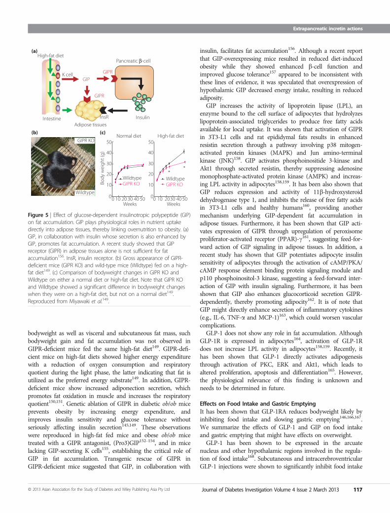
bodyweight as well as visceral and subcutaneous fat mass, suchbodyweight gain and fat accumulation was not observed inGIPR-deficient mice fed the same high-fat diet149. GIPR-defi-cient mice on high-fat diets showed higher energy expenditurewith a reduction of oxygen consumption and respiratoryquotient during the light phase, the latter indicating that fat isutilized as the preferred energy substrate149. In addition, GIPR-deficient mice show increased adiponection secretion, whichpromotes fat oxidation in muscle and increases the respiratoryquotient150,151. Genetic ablation of GIPR in diabetic ob/ob miceprevents obesity by increasing energy expenditure, andimproves insulin sensitivity and glucose tolerance withoutseriously affecting insulin secretion145,149. These observationswere reproduced in high-fat fed mice and obese ob/ob micetreated with a GIPR antagonist, (Pro3)GIP152–154, and in micelacking GIP-secreting K cells155, establishing the critical role ofGIP in fat accumulation. Transgenic rescue of GIPR inGIPR-deficient mice suggested that GIP, in collaboration with
insulin, facilitates fat accumulation156. Although a recent reportthat GIP-overexpressing mice resulted in reduced diet-inducedobesity while they showed enhanced b-cell function andimproved glucose tolerance157 appeared to be inconsistent withthese lines of evidence, it was speculated that overexpression ofhypothalamic GIP decreased energy intake, resulting in reducedadiposity.GIP increases the activity of lipoprotein lipase (LPL), an
enzyme bound to the cell surface of adipocytes that hydrolyzeslipoprotein-associated triglycerides to produce free fatty acidsavailable for local uptake. It was shown that activation of GIPRin 3T3-L1 cells and rat epididymal fats results in enhancedresistin secretion through a pathway involving p38 mitogen-activated protein kinases (MAPK) and Jun amino-terminalkinase (JNK)158. GIP activates phosphoinositide 3-kinase andAkt1 through secreted resistin, thereby suppressing adenosinemonophosphate-activated protein kinase (AMPK) and increas-ing LPL activity in adipocytes158,159. It has been also shown thatGIP reduces expression and activity of 11b-hydroxysteroiddehydrogenase type 1, and inhibits the release of free fatty acidsin 3T3-L1 cells and healthy humans160, providing anothermechanism underlying GIP-dependent fat accumulation inadipose tissues. Furthermore, it has been shown that GIP acti-vates expression of GIPR through upregulation of peroxisomeproliferator-activated receptor (PPAR)-c161, suggesting feed-for-ward action of GIP signaling in adipose tissues. In addition, arecent study has shown that GIP potentiates adipocyte insulinsensitivity of adipocytes through the activation of cAMP/PKA/cAMP response element binding protein signaling module andp110 phosphoinositol-3 kinase, suggesting a feed-forward inter-action of GIP with insulin signaling. Furthermore, it has beenshown that GIP also enhances glucocorticoid secretion GIPR-dependently, thereby promoting adipocity162. It is of note thatGIP might directly enhance secretion of inflammatory cytokines(e.g., IL-6, TNF-a and MCP-1)163, which could worsen vascularcomplications.GLP-1 does not show any role in fat accumulation. Although
GLP-1R is expressed in adipocytes164, activation of GLP-1Rdoes not increase LPL activity in adipocytes158,159. Recently, ithas been shown that GLP-1 directly activates adipogenesisthrough activation of PKC, ERK and Akt1, which leads toaltered proliferation, apoptosis and differentiation165. However,the physiological relevance of this finding is unknown andneeds to be determined in future.
Effects on Food Intake and Gastric EmptyingIt has been shown that GLP-1RA reduces bodyweight likely byinhibiting food intake and slowing gastric emptying146,166,167.We summarize the effects of GLP-1 and GIP on food intakeand gastric emptying that might have effects on overweight.GLP-1 has been shown to be expressed in the arcuate
nucleus and other hypothalamic regions involved in the regula-tion of food intake168. Subcutaneous and intracerebroventricularGLP-1 injections were shown to significantly inhibit food intake
High-fat diet(a)
(b) (c)
K cellGIP
GIPR
Pancreatic β-cell
InsulinInsR
Adipose tissues
Body
wei
ght (
g)
**
50
50Weeks
40
40
30
30
20
20
Wildtype
Normal diet High-fat diet
GIPR KOWildtypeGIPR KO10
100
50
40
30
20
10
00 50Weeks
403020100
Intestine
GIPR KO
Wildtype
GIPR
Figure 5 | Effect of glucose-dependent insulinotropic polypeptide (GIP)on fat accumulation. GIP plays physiological roles in nutrient uptakedirectly into adipose tissues, thereby linking overnutrition to obesity. (a)GIP, in collaboration with insulin whose secretion is also enhanced byGIP, promotes fat accumulation. A recent study showed that GIPreceptor (GIPR) in adipose tissues alone is not sufficient for fataccumulation156. InsR, insulin receptor. (b) Gross appearance of GIPR-deficient mice (GIPR KO) and wild-type mice (Wildtype) fed on a high-fat diet149. (c) Comparison of bodyweight changes in GIPR KO andWildtype on either a normal diet or high-fat diet. Note that GIPR KOand Wildtype showed a significant difference in bodyweight changeswhen they were on a high-fat diet, but not on a normal diet149.Reproduced from Miyawaki et al.149.
ª 2013 Asian Association for the Study of Diabetes and Wiley Publishing Asia Pty Ltd Journal of Diabetes Investigation Volume 4 Issue 2 March 2013 117
Extrapancreatic incretin actions
and bodyweight in non-diabetic rats, and this inhibitory effecton the food intake was blocked by GLP-1R antagonist exendin(9-39)169. Intraperitoneal injection of GLP-1RA liraglutide andexendin-4 also reduced food intake in non-diabetic rats170. Inaddition, subcutaneous injections of liraglutide significantlyreduced the food intake and bodyweight in diabetic ob/ob anddb/db mice171, as well as in obese candy-fed rats172, obesemini-pigs173 and obese rats carrying hypothalamic lesions174. Itwas also shown that GLP-1 or GLP-1RA liraglutide infusionreduced food intake and reduced gastric emptying in healthyvolunteers175, patients with type 1 diabetes176 and patients withtype 2 diabetes177,178.The doses of exendin-4 required for significant reduction of
food intake were higher in subdiaphragmatically vagotomizedrats than in non-vagotomized rats179. This result suggests thatthe reduction of the food intake noted after administration ofexendin-4 might be mediated by activation of the GLP-1expressed on the vagal afferents, as well as by direct activationof GLP-1R in the central nervous system. In contrast, directinjection of exendin(9-39) into the nucleus tractus solitarius inrats significantly increased the food intake180. Blockage of hind-brain GLP-1R by exendin(9-39) inhibited the suppression ofthe food intake induced by gastric distention, but did not affectthe suppression of the food intake induced by intraduodenalnutrient infusion. These results suggest that nucleus tractus soli-tarius activation by GLP-1 might also contribute to the controlof food intake through the satiating effects of gastric distension.A recent study showed that GLP-1R activation in the nucleustractus solitarius neurons increased PKA activity concomitantwith an increase in phosphorylation of p44/42-MAPK and adecrease in phosphorylation of AMPK181. The combinedincrease in PKA and p44/42-MAPK activity, together withdecreased AMPK activity, is hypothesized to increase cAMP-response element binding protein-mediated nuclear transcrip-tion and protein synthesis, resulting in reduced food intake181.Furthermore, it has been reported recently that GLP-1 neuronsin the nucleus tractus solitaries also projects directly to the ven-tral tegmental area and nucleus accumbens, the site in whichGLP-1 is thought to regulate food intake182. Regarding gastricemptying, intracisternal GLP-1 injection induced delayed gastricemptying, an effect that was partially antagonized by celiac gan-glion-ectomy, but not by atropine or NOS inhibitor, L-NG-nit-roarginine methyl ester (L-NAME)183. Corticotropin-releasinghormone antagonist, astressin, also partially antagonized theGLP-1-induced delay of gastric emptying183. These observationsshow that central corticotropin releasing hormone and periph-eral sympathetic pathways might mediate the delay of gastricemptying induced by centrally administered GLP-1. It is clini-cally of interest that chronic infusion of GLP-1 or the relativelylong-acting GLP-1RA, liraglutide, but not the short actingGLP-1RA, exenatide, loses its effects on gastric emptyingrapidly as a result of tachyphylaxis in patients with diabetesand in rats184,185. Although GLP-1 reduces the food intake anddelays gastric emptying in experimental animals and humans,
GIP does not have such effects in humans, but rather facilitatesgastric emptying175,186. It is noteworthy that although DPP-4isitagliptin has no effects on gastric emptying, the GIP responsenegatively correlates with gastric emptying and the GLP-1response positively correlates with gastric emptying108. A recentreport showed that exogenous GIP inhibits intestinal motil-ity through a somatostatin-mediated pathway rather thanthrough a GLP-1-mediated pathway, thereby inhibiting glucoseabsorption187.
Effect on the LiverDiabetes is closely associated with non-alcoholic fatty liverdisease (NAFLD), the prevalence of which is 10–30% depend-ing on geographical region. NAFLD is caused by impairedhepatic lipid homeostasis that is usually maintained through abalance between the influx or production of fatty acids andtheir use for oxidation or secretion as very low-density lipopro-tein triglycerides188–190. Approximately 10–25% of individualswith NAFLD progress to non-alcoholic steatohepatitis (NASH),a silent disease with few or no symptoms, and eventually devel-ops liver cirrhosis and hepatocellular carcinoma. Several clinicaltrials showed that agents, such as pioglitazone, vitamin E, urso-deoxycholic acid and L-carnitine, exert beneficial effects againstNAFLD. However, reducing bodyweight by appropriate dietand exercise therapies is still considered superior to any phar-macological therapies today. It has been shown that GLP-1Ractivation results in amelioration of hepatic lipid accumula-tion102,103,191–193. In contrast, GIP has been associated with fataccumulation in the liver of high-fat fed animals, but GIPRexpression in the liver is controversial149,152,154,194. We summa-rize the effects of GLP-1 and GIP on the liver that might havepotential clinical significance in the treatment of NASH andNAFLD.Although GLP-1R expression in the mouse liver or hepato-
cytes is controversial, exendin-4 reversed high-fat inducedhepatic lipid accumulation and inflammation in hepatocytesand C57BL/6J mice191. Exendin-4 also reversed hepatic steato-sis in ob/ob mice102. In the same study, GLP-1-treated mousehepatocytes resulted in a significant increase in cAMP produc-tion along with reduced expression of stearoyl-CoA desaturase(SCD)-1 and genes associated with fatty acid synthesis, andincreased expression of genes involved in fatty acid oxidiza-tion102. It was also shown that incubation of rat hepatocyteswith exendin-4 increased expression of PPAR-a along withtheir downstream genes, acyl-coenzyme A oxidase and carni-tine palmitoyltransferase 1A, thereby reducing intracellular fattyacids192. Similarly, it was shown that another GLP-1RA liraglu-tide significantly ameliorates lipid accumulation by increasingexpression of genes related to fatty acid transport and beta-oxi-dation in mice on high-fat and high-fructose diets103. In addi-tion to improvement of lipid metabolisms, exendin-4 has beenshown to improve hepatic insulin sensitivity. Administration ofexendin-4 increased expression of Sirt1 along with AMPK inlivers of high-fat fed C57BL/6J mice, as well as in cultured
118 Journal of Diabetes Investigation Volume 4 Issue 2 March 2013 ª 2013 Asian Association for the Study of Diabetes and Wiley Publishing Asia Pty Ltd
Seino and Yabe
hepatic cell lines, HepG2 and Huh7 cells, suggesting involve-ment of GLP-1 signaling not only in lipid metabolism, but alsoin glucose metabolism in the liver. Another study showed thatincubation of rat hepatocytes with exendin-4 increased expres-sion of PPAR-c, which exerted its insulin-sensitizing action byreducing JNK phosphorylation, and increased phosphorylationof Akt1 and AMPK192. Similarly, it was shown that exendin-4increased the phosphorylation of Akt1 and PKC-f in HepG2and Huh7 cells195. A recent study also showed that exendin-4activates glucokinase and increases hepatic glycogen contentsin streptozotocin (STZ)-treated C57BL/6J mice independentlyof insulin196. Collectively, GLP-1RA is supposed to ameliorateinsulin sensitivity and regulate glucose metabolism in the liver.Another effect of GLP-1RA in the liver includes protectionagainst hepatocellular injuries. It was shown that exendin-4exerts a protective role in the steatoic liver by inhibiting celldeath through upregulation of genes associated with autophagy,thereby reducing endoplasmic reticulum (ER) stress-relatedapoptosis in human hepatocytes treated with fatty acids, as wellas in mice fed a high-fat diet193. Furthermore, exendin-4administration in STZ-treated diabetic mice showed that exen-din-4 suppresses hepatocyte injury by decreasing proliferationof hepatocytes197, which might have implications in treat-ment of NAFLD. Indeed, it was shown that exenatide treat-ment ameliorates hepatic biomarkers in patients with type 2diabetes treated for at least 3 years198. Whether the liver orhepatocytes in humans express GLP-1R needs to be addressedcautiously, as it is possible that effects of GLP-1 in the liverare partly mediated through the nervous system, as evidencedin mice.In contrast to GLP-1, GIP was associated with fat accumula-
tion in the livers of high-fat fed animals, but GIPR expressionin the liver is still controversial. It was shown that GIP anta-gonist (Pro3)GIP not only reduces bodyweight, but also ame-liorates fat accumulation in the livers of high-fat fed mice152,154
and ob/ob mice153. These findings were further established byrecent a investigation in high-fat fed mice showing that activeimmunization against (Pro3)GIP resulted in GIP antibody pro-duction and significant reduction of liver triglyceride alongwith reduction in blood glucose levels194. These lines of evi-dence suggest that GIP antagonism might be beneficial as treat-ment for NASH and NAFLD. However, this was challenged bya recent report that GIP-overexpressing mice resulted inreduced diet-induced obesity and amelioration of liver steatosis,whereas they showed enhanced b-cell function and improvedglucose tolerance157. Although this finding by McIntoshet al.157 was unexpected from the results on GIPR-deficientmice that also showed reduced diet-induced obesity, they spec-ulated that overexpression of hypothalamic GIP decreasedenergy intake, resulting in reduced adiposity157. Further studiesare required to develop GIP-related therapies for obesity andNAFLD.Effects of DPP-4 inhibition have been investigated in mice
and humans. DPP-4 inhibition in diet-induced diabetic glucoki-
nase+/- mice suppressed hepatic inflammation and reducedexpression of sterol-regulatory binding protein (SREBP)-1c,SCD-1 and fatty acid synthase (FAS), and upregulation ofPPAR-a, thereby ameliorating hepatic steatosis199. Similarly,DPP-4 inhibition improves insulin sensitivity and hepatic stea-tosis in diet-induced obese C57BL/6 mice with reduced hepaticexpression of SREBP-1c, SCD-1 and FAS200. Furthermore, itwas shown that DPP-4i sitagliptin ameliorates hepatic biomar-kers in type 2 diabetic patients with NAFLD treated for4 months201. In addition, sitagliptin was shown to ameliorateliver enzymes and hepatocyte ballooning in type 2 diabeticpatients with NASH202. Taken together, these results suggestclinical implications of DPP-4i in treatment for NASH andNAFLD.
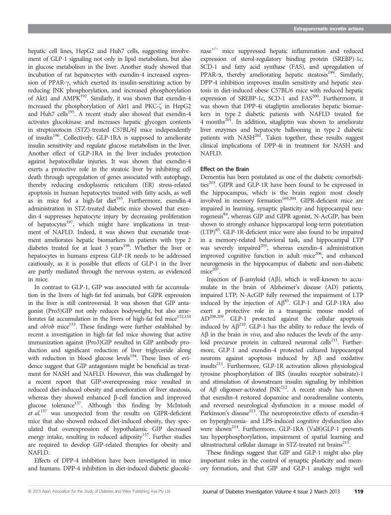
Effect on the BrainDementia has been postulated as one of the diabetic comorbidi-ties203. GIPR and GLP-1R have been found to be expressed inthe hippocampus, which is the brain region most closelyinvolved in memory formation169,204. GIPR-deficient mice areimpaired in learning, synaptic plasticity and hippocampal neu-rogenesis84, whereas GIP and GIPR agonist, N-AcGIP, has beenshown to strongly enhance hippocampal long-term potentiation(LTP)85. GLP-1R-deficient mice were also found to be impairedin a memory-related behavioral task, and hippocampal LTPwas severely impaired205, whereas exendin-4 administrationimproved cognitive function in adult mice206, and enhancedneurogenesis in the hippocampus of diabetic and non-diabeticmice207.Injection of b-amyloid (Ab), which is well-known to accu-
mulate in the brain of Alzheimer’s disease (AD) patients,impaired LTP; N-AcGIP fully reversed the impairment of LTPinduced by the injection of Ab85. GLP-1 and GLP-1RA alsoexert a protective role in a transgenic mouse model ofAD208,209. GLP-1 protected against the cellular apoptosisinduced by Ab210. GLP-1 has the ability to reduce the levels ofAb in the brain in vivo, and also reduces the levels of the amy-loid precursor protein in cultured neuronal cells211. Further-more, GLP-1 and exendin-4 protected cultured hippocampalneurons against apoptosis induced by Ab and oxidativeinsults211. Furthermore, GLP-1R activation allows physiologicaltyrosine phosphorylation of IRS (insulin receptor substrate)-1and stimulation of downstream insulin signaling by inhibitionof Ab oligomer-activated JNK212. A recent study has shownthat exendin-4 restored dopamine and noradrenaline contents,and reversed neurological dysfunction in a mouse model ofParkinson’s disease213. The neuroprotective effects of exendin-4on hyperglycemia- and LPS-induced cognitive dysfunction alsowere shown214. Furthermore, GLP-1RA (Val8)GLP-1 preventstau hyperphosphorylation, impairment of spatial learning andultrastructural cellular damage in STZ-treated rat brains215.These findings suggest that GIP and GLP-1 might also play
important roles in the control of synaptic plasticity and mem-ory formation, and that GIP and GLP-1 analogs might well
ª 2013 Asian Association for the Study of Diabetes and Wiley Publishing Asia Pty Ltd Journal of Diabetes Investigation Volume 4 Issue 2 March 2013 119
Extrapancreatic incretin actions
inhibit impairment of memory formation in patients with AD,as well as other cognitive deficits in neurodegenerative disor-ders.
Effect on the BoneBone fracture, which is directly related to decreased mobilityand compromised quality of life (QOL), is associated with dia-betes216. It has been reported that GIP promotes bone forma-tion, whereas GLP-1 inhibits bone resorption (Figure 6)217,218.Thus, both GIP and GLP-1 could play important roles in thereduction of the bone fracture risk in diabetic patients.GIPR has been shown to be present in osteoclasts. GIP
inhibits the osteoclast resorptive activity in organ culture sys-tems and the resorptive activity of mature osteoclasts219. Thebone formation parameters in GIPR-deficient mice were signifi-cantly lower than those in wild-type mice, and the number ofosteoclasts was significantly increased in GIPR-deficient mice,indicative of high-turnover osteoporosis220. In addition, GIPsuppressed the apoptosis of osteoblasts in vitro, suggesting thatGIP stimulates bone formation through inhibition of osteoblastapoptosis220. Furthermore, GIP transgenic mice showed a sig-nificant increase in the markers of bone formation and adecrease in the markers of bone resorption, and also a signifi-cant increase of the bone mass221,222. From these findings inGIPR-deficient mice and GIP-transgenic mice, it is suggestedthat GIP inhibits bone resorption and stimulates bone forma-tion, and that excess signaling through the GIPR results in thegain of bone mass. In addition, the plasma calcium concentra-tion after meal ingestion in GIPR-deficient mice was increased,and GIP might play an important role in the stimulation ofcalcium deposition in the bone223.
GLP-1R-decifient mice showed cortical osteopenia, bone fra-gility and an increase in the markers of bone resorption andosteoclast numbers224. GLP-1R-decifient mice also showedreduced calcitonin gene expression in the thyroid, whereasexendin-4 increased calcitonin expression. GLP-1 did not showany direct effect on the osteoblasts or osteoclasts, although itappears to have an essential role in endogenous GLP-1R signal-ing in bone resorption through activation of the calcitonin-dependent pathway, which in turn, plays an important role inbone formation224. In the analysis of bone structure in STZ-induced diabetic rats and fructose-induced insulin resistancerats, GLP-1 significantly reduced the trabecular separation225,whereas exendin-4 induced a significant decrease in trabecularseparation, and increase in trabecular thickness and trabecularspacing. It also has been also shown that GLP-1 and exendin-4reversed hyprelipidic-related osteopenia226. Exendin-4 normal-ized the LDL-receptor-related protein 5 (an activator of thewindless type [Wnt] pathway)/Dickkopf-related protein 1 (ablocker of LDL-receptor-related protein 5) ratio in diabetic ratsand insulin resistant rats227. This finding suggests that exendin-4 might induce the bone formation in diabetic and insulinresistant rats through the Wnt signaling pathway227. Consistentwith the effects of GIP and GLP-1 on bone metabolism, DPP-4i sitagliptin significantly improved vertebral volumetric bonemineral density and trabecular architecture in female mice228.Although these lines of evidence suggest an association of
GIP and GLP-1 with bone metabolism in humans, the effectsof GIP and GLP-1 on human bone turnover are largelyunknown. A meta-analysis of 28 clinical trials of treatment withDPP-4i (i.e., vildagliptin, sitagliptin, saxagliptin, alogliptin, linag-liptin and dutogliptin for at least 24 weeks) in patients withtype 2 diabetes showed reduced the risk of bone fractures inpatients treated with DPP-4i229. However, 44-week exenatidetreatment did not affect the bone mineral density or serummarkers of bone homeostasis (i.e., serum alkaline phosphatase,calcium and phosphate), as compared with insulin glargine, inpatients with type 2 diabetes230. As mentioned, GLP-1 actionon the bone is presumably mediated through calcitonin. A ser-ies of clinical trials on liraglutide showed few changes in serumcalcitonin levels in patients with type 2 diabetes, suggesting thatGLP-1 might not play a role in human bone metabolism.Regarding GIP, it has been previously reported that postpran-dial reduction of bone resorption was not mediated by GIP,but by GLP-2, another intestinal hormone secreted simulta-neously with GLP-1231. However, they investigated the effectsof subcutaneous single injections of native GIP that should berapidly inactivated by DPP-4 before it reached the bones. Thus,further investigations are definitely required to understand GIPand GLP-1 actions on bone metabolism in humans.
Effects on Thyroid and TumorigenesisStudies in experimental animals, including toxicology studies, didnot show that incretins had an effect on tumorigenesis. However,it has been recently reported that GLP-1RA liraglutide and exe-
Nutrients
GIP
GLP-1
K cell
L cellIntestine
ThyroidC cells
BoneOsteoclasts
Osteoblasts
Calcitonin
Boneformation ↑
Boneresorption ↓
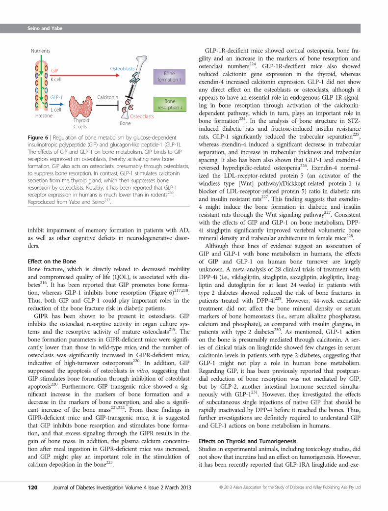
Figure 6 | Regulation of bone metabolism by glucose-dependentinsulinotropic polypeptide (GIP) and glucagon-like peptide-1 (GLP-1).The effects of GIP and GLP-1 on bone metabolism. GIP binds to GIPreceptors expressed on osteoblasts, thereby activating new boneformation. GIP also acts on osteoclasts, presumably through osteoblasts,to suppress bone resorption. In contrast, GLP-1 stimulates calcitoninsecretion from the thyroid gland, which then suppresses boneresorption by osteoclasts. Notably, it has been reported that GLP-1receptor expression in humans is much lower than in rodents240.Reproduced from Yabe and Seino217.
120 Journal of Diabetes Investigation Volume 4 Issue 2 March 2013 ª 2013 Asian Association for the Study of Diabetes and Wiley Publishing Asia Pty Ltd
Seino and Yabe
natide were associated with benign and malignant thyroidtumors in rats and mice232. GLP-1R was found to be expressedin normal rat thyroid, as well as medullary thyroid carcinomacells233. GLP-1 and GLP-1RA stimulate expression and secretionof calcitonin, a clinical biomarker for C cell diseases, such asmedullary thyroid carcinoma234, in a dose-dependent manneralong with production of cAMP in rodent C cell lines232,233,235
and rodents232,236. However, it was reported that GLP-1R expres-sion was low in a human C cell line, and that liraglutideenhances little calcitonin secretion or C cell proliferation in pri-mates232. Furthermore, clinical trials of GLP-1RA, such as lira-glutide, did not report elevation of calcitonin237,238. Thus,although the preclinical studies in rodents show a link betweenGLP-1 and thyroid tumors, the relevance of these findings forhumans is still unclear. In addition to the marked differences inthe way that rodent and human C cells respond to GLP-1, spon-taneous C cell tumors are frequently seen in rats, whereas med-ullary thyroid cancer in humans is rare. Nevertheless, a recentstudy indicates expression of GLP-1R in neoplastic and hyper-plastic lesions of thyroid C cells239. Thus, long-term observa-tional studies are required to monitor the effects of sustainedGLP-1R signaling over the long term on the human thyroid.
CONCLUSIONBiological processes regulated by the incretin hormones, GIPand GLP-1, are much broader than previously expected. Theirinsulinotropic actions are applied in the development of incre-tin-based therapies, DPP-4i and GLP-1RA that exert glucoselowering effects, thereby suppressing diabetes-related complica-tions in patients with type 2 diabetes. In addition, it is conceiv-able that extrapancreatic function of GIP and GLP-1 might beexploited to prevent onset and progression of diabetes-relatedcomplications independently of glycemic control. A series ofexperimental results obtained so far suggest that diabetes-relatedmicrovascular complications (e.g., retinopathy, nephropathy andneuropathy) and macrovascular complications (e.g., CAD, CVDand PAD) are directly affected by incretin-based therapies. Fur-thermore, diabetes-related comorbidities, such as cognitive dys-function, obesity, fatty liver and bone fracture, might be alsoameliorated by incretin-based therapies. Clinical trials with ade-quately powered, prospective, controlled relevant end-points willclarify, in future, the effects of incretin-based drugs on diabetes-related complications.
ACKNOWLEDGEMENTSThe authors thank current and former colleagues in the labora-tory of Yutaka Seino, and apologize for citing only part of therelevant work in this field due to limited space, and areindebted to many authors for their contributions. YS reportsreceiving consulting and/or speaker fees from Eli Lilly, MSD,Novartis, Novo Nordisk, Sanofi-Aventis and Takeda. DYreports receiving speaker fees from Eli Lilly, MSD, Sanofi-Aventis, Novo Nordisk and Takeda.
REFERENCES1. Bayliss WM, Starling EH. The mechanism of pancreatic
secretion. J Physiol 1902; 28: 325–353.2. Moore B. On the treatment of Diabetus mellitus by acid
extract of Duodenal Mucous Membrane. Biochem J 1906;1: 28–38.
3. Zunz E, La Barre J. Contributiona a l’etude des variationsphysiologiques de la secretion interne du pancreas.relations entre les secretions externe et interne dupancreas. Arch Int Physiol Biochim 1929; 31: 20–44.
4. Seino Y, Fukushima M, Yabe D. GIP and GLP-1, the twoincretin hormones: similarities and differences. J DiabetesInvest 2010; 1: 9–23.
5. Drucker DJ. The biology of incretin hormones. Cell Metab2006; 3: 153–165.
6. Holst JJ. The physiology of glucagon-like peptide 1. PhysiolRev 2007; 87: 1409–1439.
7. Nauck MA, Baller B, Meier JJ. Gastric inhibitory polypeptideand glucagon-like peptide-1 in the pathogenesis of type 2diabetes. Diabetes 2004; 53(Suppl 3): S190–S196.
8. Hansotia T, Baggio LL, Delmeire D, et al. Double incretinreceptor knockout (DIRKO) mice reveal an essential rolefor the enteroinsular axis in transducing theglucoregulatory actions of DPP-IV inhibitors. Diabetes 2004;53: 1326–1335.
9. Miyawaki K, Yamada Y, Yano H, et al. Glucose intolerancecaused by a defect in the entero-insular axis: a study ingastric inhibitory polypeptide receptor knockout mice. ProcNatl Acad Sci USA 1999; 96: 14843–14847.
10. Scrocchi LA, Brown TJ, MaClusky N, et al. Glucoseintolerance but normal satiety in mice with a nullmutation in the glucagon-like peptide 1 receptor gene.Nat Med 1996; 2: 1254–1258.
11. Nauck MA. Incretin-based therapies for type 2 diabetesmellitus: properties, functions, and clinical implications. AmJ Med 2011; 124: S3–S18.
12. Lovshin JA, Drucker DJ. Incretin-based therapies for type 2diabetes mellitus. Nat Rev Endocrinol 2009; 5: 262–269.
13. Dicker D. DPP-4 inhibitors: impact on glycemic control andcardiovascular risk factors. Diabetes Care 2011; 34(Suppl 2):S276–S278.
14. Deacon CF. Dipeptidyl peptidase-4 inhibitors in thetreatment of type 2 diabetes: a comparative review.Diabetes Obes Metab 2011; 13: 7–18.
15. Amori RE, Lau J, Pittas AG. Efficacy and safety of incretintherapy in type 2 diabetes: systematic review and meta-analysis. JAMA 2007; 298: 194–206.
16. Monami M, Iacomelli I, Marchionni N, et al. Dipeptydilpeptidase-4 inhibitors in type 2 diabetes: a meta-analysisof randomized clinical trials. Nutr Metab Cardiovasc Dis2010; 20: 224–235.
17. Monami M, Marchionni N, Mannucci E. Glucagon-likepeptide-1 receptor agonists in type 2 diabetes: a meta-
ª 2013 Asian Association for the Study of Diabetes and Wiley Publishing Asia Pty Ltd Journal of Diabetes Investigation Volume 4 Issue 2 March 2013 121
Extrapancreatic incretin actions
analysis of randomized clinical trials. Eur J Endocrinol 2009;160: 909–917.
18. Nauck MA, Vardarli I. Comparative evaluation of incretin-based antidiabetic medications and alternative therapies tobe added to metformin in the case of monotherapyfailure. J Diabetes Invest 2010; 1: 24–36.
19. Fukushima M, Suzuki H, Seino Y. Insulin secretion capacityin the development from normal glucose tolerance totype 2 diabetes. Diabetes Res Clin Pract 2004; 66S:S37–S44.
20. Park H, Park C, Kim Y, et al. Efficacy and safety ofdipeptidyl peptidase-4 inhibitors in type 2 diabetes: meta-analysis. Ann Pharmacother 2012; 46: 1453–1469.
21. Kim YG, Hahn S, Oh TJ, et al. Differences in the glucose-lowering efficacy of dipeptidyl peptidase-4 inhibitorsbetween Asians and Non-Asians: a systematic review andmeta-analysis. Diabetologia 2013; doi: 10.1007/s00125-012-2827-3.
22. UK Prospective Diabetes Study (UKPDS) Group. Intensiveblood-glucose control with sulphonylureas or insulincompared with conventional treatment and risk ofcomplications in patients with type 2 diabetes (UKPDS 33).Lancet 1998;352: 837–853.
23. The Diabetes Control and Complications Trial ResearchGroup. The effect of intensive treatment of diabetes onthe development and progression of long-termcomplications in insulin-dependent diabetes mellitus.N Engl J Med 1993;329: 977–986.
24. Nathan DM, Cleary PA, Backlund JY, et al. Intensivediabetes treatment and cardiovascular disease in patientswith type 1 diabetes. N Engl J Med 2005; 353: 2643–2653.
25. Holman RR, Paul SK, Bethel MA, et al. 10-year follow-up ofintensive glucose control in type 2 diabetes. N Engl J Med2008; 359: 1577–1589.
26. Kitada M, Zhang Z, Mima A, et al. Molecular mechanismsof diabetic vascular complications. J Diabetes Invest 2010;1: 77–89.
27. Nagashima M, Watanabe T, Terasaki M, et al. Nativeincretins prevent the development of atheroscleroticlesions in apolipoprotein E knockout mice. Diabetologia2011; 54: 2649–2659.
28. Nogi Y, Nagashima M, Terasaki M, et al. Glucose-dependentinsulinotropic polypeptide prevents the progression ofmacrophage-driven atherosclerosis in diabeticapolipoprotein E-null mice. PLoS ONE 2012; 7: e35683.
29. Arakawa M, Mita T, Azuma K, et al. Inhibition of monocyteadhesion to endothelial cells and attenuation ofatherosclerotic lesion by a glucagon-like peptide-1receptor agonist, exendin-4. Diabetes 2010; 59: 1030–1037.
30. Panjwani N, Mulvihill EE, Longuet C, et al. GLP-1 receptoractivation indirectly reduces hepatic lipid accumulationbut does not attenuate development of atherosclerosisin diabetic male ApoE-/- mice. Endocrinology 2013; 154:127–139.
31. Gaspari T, Liu H, Welungoda I, et al. A GLP-1 receptoragonist liraglutide inhibits endothelial cell dysfunctionand vascular adhesion molecule expression in anApoE-/- mouse model. Diab Vasc Dis Res 2011; 8:117–124.
32. Liu H, Dear AE, Knudsen LB, et al. A long-acting glucagon-like peptide-1 analogue attenuates induction ofplasminogen activator inhibitor type-1 and vascularadhesion molecules. J Endocrinol 2009; 201: 59–66.
33. Wu JD, Xu XH, Zhu J, et al. Effect of exenatide oninflammatory and oxidative stress markers in patients withtype 2 diabetes mellitus. Diabetes Technol Ther 2011; 13:143–148.
34. Ha SJ, Kim W, Woo JS, et al. Preventive effects ofexenatide on endothelial dysfunction induced byischemia-reperfusion injury via KATP channels. ArteriosclerThromb Vasc Biol 2012; 32: 474–480.
35. Koska J, Schwartz EA, Mullin MP, et al. Improvement ofpostprandial endothelial function after a single dose ofexenatide in individuals with impaired glucose toleranceand recent-onset type 2 diabetes. Diabetes Care 2010; 33:1028–1030.
36. Shiraishi D, Fujiwara Y, Komohara Y, et al. Glucagon-likepeptide-1 (GLP-1) induces M2 polarization of humanmacrophages via STAT3 activation. Biochem Biophys ResCommun 2012; 425: 304–308.
37. Ding L, Zhang J. Glucagon-like peptide-1 activatesendothelial nitric oxide synthase in human umbilical veinendothelial cells. Acta Pharmacol Sin 2012; 33: 75–81.
38. Chai W, Dong Z, Wang N, et al. Glucagon-like peptide 1recruits microvasculature and increases glucose use inmuscle via a nitric oxide-dependent mechanism. Diabetes2012; 61: 888–896.
39. Liu FQ, Zhang XL, Gong L, et al. Glucagon-like peptide 1protects microvascular endothelial cells by inactivating thePARP-1/iNOS/NO pathway. Mol Cell Endocrinol 2011; 339:25–33.
40. Xiao-Yun X, Zhao-Hui M, Ke C, et al. Glucagon-like peptide-1 improves proliferation and differentiation of endothelialprogenitor cells via upregulating VEGF generation. Med SciMonit 2011;17: BR35–BR41.
41. Erdogdu O, Nathanson D, Sjoholm A, et al. Exendin-4stimulates proliferation of human coronary arteryendothelial cells through eNOS-, PKA- and PI3K/Akt-dependent pathways and requires GLP-1 receptor. Mol CellEndocrinol 2010; 325: 26–35.
42. Han L, Yu Y, Sun X, et al. Exendin-4 directly improvesendothelial dysfunction in isolated aortas from obese ratsthrough the cAMP or AMPK-eNOS pathways. Diabetes ResClin Pract 2012; 97: 453–460.
43. Shiraki A, Oyama J, Komoda H, et al. The glucagon-likepeptide 1 analog liraglutide reduces TNF-alpha-inducedoxidative stress and inflammation in endothelial cells.Atherosclerosis 2012; 221: 375–382.
122 Journal of Diabetes Investigation Volume 4 Issue 2 March 2013 ª 2013 Asian Association for the Study of Diabetes and Wiley Publishing Asia Pty Ltd
Seino and Yabe
44. Zhan Y, Sun HL, Chen H, et al. Glucagon-like peptide-1(GLP-1) protects vascular endothelial cells againstadvanced glycation end products (AGEs)-inducedapoptosis. Med Sci Monit 2012; 18: BR286–BR291.
45. Ojima A, Matsui T, Maeda S, et al. Glucose-dependentinsulinotropic polypeptide (GIP) inhibits signaling pathwaysof advanced glycation end products (AGEs) in endothelialcells via its antioxidative properties. Horm Metab Res 2012;44: 501–505.
46. Matsubara J, Sugiyama S, Sugamura K, et al. A dipeptidylpeptidase-4 inhibitor, des-fluoro-sitagliptin, improvesendothelial function and reduces atherosclerotic lesionformation in apolipoprotein E-deficient mice. J Am CollCardiol 2012; 59: 265–276.
47. Shah Z, Kampfrath T, Deiuliis JA, et al. Long-termdipeptidyl-peptidase 4 inhibition reducesatherosclerosis and inflammation via effects onmonocyte recruitment and chemotaxis. Circulation2011; 124: 2338–2349.
48. Ta NN, Schuyler CA, Li Y, et al. DPP-4 (CD26) inhibitoralogliptin inhibits atherosclerosis in diabetic apolipoproteinE-deficient mice. J Cardiovasc Pharmacol 2011; 58:157–166.
49. Terasaki M, Nagashima M, Watanabe T, et al. Effects ofPKF275-055, a dipeptidyl peptidase-4 inhibitor, on thedevelopment of atherosclerotic lesions in apolipoproteinE-null mice. Metabolism 2012; 61: 974–977.
50. Maeda S, Matsui T, Yamagishi S. Vildagliptininhibits oxidative stress and vascular damage instreptozotocin-induced diabetic rats. Int J Cardiol 2012;158: 171–173.
51. Fadini GP, Boscaro E, Albiero M, et al. The oral dipeptidylpeptidase-4 inhibitor sitagliptin increases circulatingendothelial progenitor cells in patients with type 2diabetes: possible role of stromal-derived factor-1alpha.Diabetes Care 2010; 33: 1607–1609.
52. Makdissi A, Ghanim H, Vora M, et al. Sitagliptin exerts anantinflammatory action. J Clin Endocrinol Metab 2012; 97:3333–3341.
53. Satoh-Asahara N, Sasaki Y, Wada H, et al. A dipeptidylpeptidase-4 inhibitor, sitagliptin, exerts anti-inflammatoryeffects in type 2 diabetic patients. Metabolism 2012; 62:347–351.
54. Timmers L, Henriques JP, de Kleijn DP, et al. Exenatidereduces infarct size and improves cardiac function in aporcine model of ischemia and reperfusion injury. J AmColl Cardiol 2009; 53: 501–510.
55. Noyan-Ashraf MH, Momen MA, Ban K, et al. GLP-1Ragonist liraglutide activates cytoprotective pathways andimproves outcomes after experimental myocardialinfarction in mice. Diabetes 2009; 58: 975–983.
56. Matsubara M, Kanemoto S, Leshnower BG, et al. Singledose GLP-1-Tf ameliorates myocardial ischemia/reperfusioninjury. J Surg Res 2011; 165: 38–45.
57. Dokken BB, La Bonte LR, Davis-Gorman G, et al.Glucagon-like peptide-1 (GLP-1), immediately prior toreperfusion, decreases neutrophil activation and reducesmyocardial infarct size in rodents. Horm Metab Res 2011;43: 300–305.
58. Moberly SP, Berwick ZC, Kohr M, et al. Intracoronaryglucagon-like peptide 1 preferentially augments glucoseuptake in ischemic myocardium independent of changesin coronary flow. Exp Biol Med (Maywood) 2012; 237:334–342.
59. Bao W, Aravindhan K, Alsaid H, et al. Albiglutide, a longlasting glucagon-like peptide-1 analog, protects the ratheart against ischemia/reperfusion injury: evidence forimproving cardiac metabolic efficiency. PLoS ONE 2011;6: e23570.
60. Ban K, Kim KH, Cho CK, et al. Glucagon-like peptide(GLP)-1(9-36)amide-mediated cytoprotection is blocked byexendin(9-39) yet does not require the known GLP-1receptor. Endocrinology 2010; 151: 1520–1531.
61. Vila Petroff MG, Egan JM, Wang X, et al. Glucagon-likepeptide-1 increases cAMP but fails to augment contractionin adult rat cardiac myocytes. Circ Res 2001; 89: 445–452.
62. Ban K, Noyan-Ashraf MH, Hoefer J, et al. Cardioprotectiveand vasodilatory actions of glucagon-like peptide 1 receptorare mediated through both glucagon-like peptide 1receptor-dependent and -independent pathways.Circulation 2008; 117: 2340–2350.
63. Nikolaidis LA, Mankad S, Sokos GG, et al. Effects ofglucagon-like peptide-1 in patients with acute myocardialinfarction and left ventricular dysfunction after successfulreperfusion. Circulation 2004; 109: 962–965.
64. Read PA, Khan FZ, Dutka DP. Cardioprotection againstischaemia induced by dobutamine stress using glucagon-like peptide-1 in patients with coronary artery disease.Heart 2011; 98: 408–413.
65. Lonborg J, Kelbaek H, Vejlstrup N, et al. Exenatide reducesfinal infarct size in patients with ST-segment-elevationmyocardial infarction and short-duration of ischemia. CircCardiovasc Interv 2012; 5: 288–295.
66. Lonborg J, Vejlstrup N, Kelbaek H, et al. Exenatide reducesreperfusion injury in patients with ST-segment elevationmyocardial infarction. Eur Heart J 2012; 33: 1491–1499.
67. Huisamen B, Genis A, Marais E, et al. Pre-treatmentwith a DPP-4 inhibitor is infarct sparing in heartsfrom obese, pre-diabetic rats. Cardiovasc Drugs Ther2011; 25: 13–20.
68. Sauve M, Ban K, Momen MA, et al. Genetic deletion orpharmacological inhibition of dipeptidyl peptidase-4improves cardiovascular outcomes after myocardialinfarction in mice. Diabetes 2010; 59: 1063–1073.
69. Hocher B, Sharkovska Y, Mark M, et al. The novel DPP-4inhibitors linagliptin and BI 14361 reduce infarct size aftermyocardial ischemia/reperfusion in rats. Int J Cardiol 2012;doi:10.1016/j.ijcard.2011.12.007.
Extrapancreatic incretin actions
ª 2013 Asian Association for the Study of Diabetes and Wiley Publishing Asia Pty Ltd Journal of Diabetes Investigation Volume 4 Issue 2 March 2013 123
70. Ye Y, Keyes KT, Zhang C, et al. The myocardial infarctsize-limiting effect of sitagliptin is PKA-dependent, whereasthe protective effect of pioglitazone is partially dependenton PKA. Am J Physiol Heart Circ Physiol 2010; 298:H1454–H1465.
71. Yin M, Sillje HH, Meissner M, et al. Early and late effects ofthe DPP-4 inhibitor vildagliptin in a rat model of post-myocardial infarction heart failure. Cardiovasc Diabetol2011; 10: 85.
72. Lenski M, Kazakov A, Marx N, et al. Effects of DPP-4inhibition on cardiac metabolism and function in mice.J Mol Cell Cardiol 2011; 51: 906–918.
73. Huber BC, Brunner S, Segeth A, et al. Parathyroid hormoneis a DPP-IV inhibitor and increases SDF-1-driven homing ofCXCR4(+) stem cells into the ischaemic heart. CardiovascRes 2011; 90: 529–537.
74. Connelly K, Zhang Y, Advani A, et al. DPP-4 inhibitionattenuates cardiac dysfunction and adverse remodellingfollowing myocardial infarction in rats with experimentaldiabetes. Cardiovasc Ther 2012; doi: 10.1111/1755-5922.12005.
75. Nikolaidis LA, Elahi D, Hentosz T, et al. Recombinantglucagon-like peptide-1 increases myocardial glucoseuptake and improves left ventricular performance inconscious dogs with pacing-induced dilatedcardiomyopathy. Circulation 2004; 110: 955–961.
76. Poornima I, Brown SB, Bhashyam S, et al. Chronicglucagon-like peptide-1 infusion sustains left ventricularsystolic function and prolongs survival in thespontaneously hypertensive, heart failure-prone rat.Circ Heart Fail 2008; 1: 153–160.
77. Liu Q, Anderson C, Broyde A, et al. Glucagon-likepeptide-1 and the exenatide analogue AC3174 improvecardiac function, cardiac remodeling, and survival in ratswith chronic heart failure. Cardiovasc Diabetol 2010;9: 76.
78. Sokos GG, Nikolaidis LA, Mankad S, et al. Glucagon-likepeptide-1 infusion improves left ventricular ejectionfraction and functional status in patients with chronicheart failure. J Card Fail 2006; 12: 694–699.
79. Nathanson D, Ullman B, Lofstrom U, et al. Effects ofintravenous exenatide in type 2 diabetic patients withcongestive heart failure: a double-blind, randomisedcontrolled clinical trial of efficacy and safety. Diabetologia2012; 55: 926–935.
80. Gomez N, Touihri K, Matheeussen V, et al. Dipeptidylpeptidase IV inhibition improves cardiorenal function inoverpacing-induced heart failure. Eur J Heart Fail 2012;14: 14–21.
81. Teramoto S, Miyamoto N, Yatomi K, et al. Exendin-4, aglucagon-like peptide-1 receptor agonist, providesneuroprotection in mice transient focal cerebralischemia. J Cereb Blood Flow Metab 2011; 31: 1696–1705.
82. Lee CH, Yan B, Yoo KY, et al. Ischemia-induced changes inglucagon-like peptide-1 receptor and neuroprotectiveeffect of its agonist, exendin-4, in experimental transientcerebral ischemia. J Neurosci Res 2011; 89: 1103–1113.
83. Darsalia V, Mansouri S, Ortsater H, et al. Glucagon-likepeptide-1 receptor activation reduces ischaemic braindamage following stroke in Type 2 diabetic rats. Clin Sci(Lond) 2012; 122: 473–483.
84. Faivre E, Gault VA, Thorens B, et al. Glucose-dependentinsulinotropic polypeptide receptor knockout mice areimpaired in learning, synaptic plasticity, and neurogenesis.J Neurophysiol 2011; 105: 1574–1580.
85. Gault VA, Holscher C. Protease-resistant glucose-dependentinsulinotropic polypeptide agonists facilitate hippocampalLTP and reverse the impairment of LTP induced by beta-amyloid. J Neurophysiol 2008; 99: 1590–1595.
86. Faivre E, Hamilton A, Holscher C. Effects of acute andchronic administration of GIP analogues on cognition,synaptic plasticity and neurogenesis in mice. Eur JPharmacol 2012; 674: 294–306.
87. Darsalia V, Ortsater H, Olverling A, et al. The DPP-4inhibitor linagliptin counteracts stroke in the normal anddiabetic mouse brain: a comparison with glimepiride.Diabetes 2012; doi: 10.2337/db12-0988.
88. Yu M, Moreno C, Hoagland KM, et al. Antihypertensiveeffect of glucagon-like peptide 1 in Dahl salt-sensitive rats.J Hypertens 2003; 21: 1125–1135.
89. Hirata K, Kume S, Araki S, et al. Exendin-4 has an anti-hypertensive effect in salt-sensitive mice model. BiochemBiophys Res Commun 2009; 380: 44–49.
90. Liu Q, Adams L, Broyde A, et al. The exenatide analogueAC3174 attenuates hypertension, insulin resistance, andrenal dysfunction in Dahl salt-sensitive rats. CardiovascDiabetol 2010; 9: 32.
91. Pacheco BP, Crajoinas RO, Couto GK, et al. Dipeptidylpeptidase IV inhibition attenuates blood pressure rising inyoung spontaneously hypertensive rats. J Hypertens 2011;29: 520–528.
92. Liu L, Liu J, Wong WT, et al. Dipeptidyl peptidase 4inhibitor sitagliptin protects endothelial function inhypertension through a glucagon-like peptide 1-dependent mechanism. Hypertension 2012; 60: 833–841.
93. McGill JB. Liraglutide: effects beyond glycaemic control indiabetes treatment. Int J Clin Pract Suppl 2010; 167: 28–34.
94. Irace C, De Luca S, Shehaj E, et al. Exenatide improvesendothelial function assessed by flow mediated dilationtechnique in subjects with type 2 diabetes: results froman observational research. Diab Vasc Dis Res 2013;10: 72–77.
95. Tang-Christensen M, Larsen PJ, Goke R, et al. Centraladministration of GLP-1-(7–36) amide inhibits food andwater intake in rats. Am J Physiol 1996; 271: R848–R856.
96. Schafer SA, Tschritter O, Machicao F, et al. Impairedglucagon-like peptide-1-induced insulin secretion in carriers
Seino and Yabe
124 Journal of Diabetes Investigation Volume 4 Issue 2 March 2013 ª 2013 Asian Association for the Study of Diabetes and Wiley Publishing Asia Pty Ltd
of transcription factor 7-like 2 (TCF7L2) genepolymorphisms. Diabetologia 2007; 50: 2443–2450.
97. Gutzwiller JP, Tschopp S, Bock A, et al. Glucagon-likepeptide 1 induces natriuresis in healthy subjects and ininsulin-resistant obese men. J Clin Endocrinol Metab 2004;89: 3055–3061.
98. Kubota A, Maeda H, Kanamori A, et al. Pleiotropic effectsof sitagliptin in the treatment of type 2 diabetes mellituspatients. J Clin Med Res 2012; 4: 309–313.
99. Ogawa S, Ishiki M, Nako K, et al. Sitagliptin, a dipeptidylpeptidase-4 inhibitor, decreases systolic blood pressure inJapanese hypertensive patients with type 2 diabetes.Tohoku J Exp Med 2011; 223: 133–135.
100. Qin X, Shen H, Liu M, et al. GLP-1 reduces intestinal lymphflow, triglyceride absorption, and apolipoproteinproduction in rats. Am J Physiol Gastrointest Liver Physiol2005; 288: G943–G949.
101. Hsieh J, Longuet C, Baker CL, et al. The glucagon-likepeptide 1 receptor is essential for postprandial lipoproteinsynthesis and secretion in hamsters and mice. Diabetologia2010; 53: 552–561.
102. Ding X, Saxena NK, Lin S, et al. Exendin-4, a glucagon-likeprotein-1 (GLP-1) receptor agonist, reverses hepaticsteatosis in ob/ob mice. Hepatology 2006; 43: 173–181.
103. Mells JE, Fu PP, Sharma S, et al. Glp-1 analog, liraglutide,ameliorates hepatic steatosis and cardiac hypertrophy inC57BL/6J mice fed a Western diet. Am J PhysiolGastrointest Liver Physiol 2012; 302: G225–G235.
104. Meier JJ, Gethmann A, Gotze O, et al. Glucagon-likepeptide 1 abolishes the postprandial rise in triglycerideconcentrations and lowers levels of non-esterified fattyacids in humans. Diabetologia 2006; 49: 452–458.
105. Schwartz EA, Koska J, Mullin MP, et al. Exenatidesuppresses postprandial elevations in lipids andlipoproteins in individuals with impaired glucose toleranceand recent onset type 2 diabetes mellitus. Atherosclerosis2010; 212: 217–222.
106. Matikainen N, Manttari S, Schweizer A, et al. Vildagliptintherapy reduces postprandial intestinal triglyceride-richlipoprotein particles in patients with type 2 diabetes.Diabetologia 2006; 49: 2049–2057.
107. Tremblay AJ, Lamarche B, Deacon CF, et al. Effect ofsitagliptin therapy on postprandial lipoprotein levels inpatients with type 2 diabetes. Diabetes Obes Metab 2011;13: 366–373.
108. Stevens JE, Horowitz M, Deacon CF, et al. The effects ofsitagliptin on gastric emptying in healthy humans – arandomised, controlled study. Aliment Pharmacol Ther2012; 36: 379–390.
109. Monami M, Lamanna C, Desideri CM, et al. DPP-4inhibitors and lipids: systematic review and meta-analysis.Adv Ther 2012; 29: 14–25.
110. Freeman R. The Nervous System and Diabetes. In: KahnCR, Weir GC, King GL, et al. (eds). Joslin’s Diabetes Mellitus.
14th edn. Lippincott Williams and Wilkins, Boston, 2005:951–974.
111. Himeno T, Kamiya H, Naruse K, et al. Beneficial effects ofexendin-4 on experimental polyneuropathy in diabeticmice. Diabetes 2011; 60: 2397–2406.
112. Liu WJ, Jin HY, Lee KA, et al. Neuroprotective effect of theglucagon-like peptide-1 receptor agonist, syntheticexendin-4, in streptozotocin-induced diabetic rats. Br JPharmacol 2011; 164: 1410–1420.
113. Jolivalt CG, Fineman M, Deacon CF, et al. GLP-1 signalsvia ERK in peripheral nerve and prevents nervedysfunction in diabetic mice. Diabetes Obes Metab 2011;13: 990–1000.
114. Perry T, Holloway HW, Weerasuriya A, et al. Evidence ofGLP-1-mediated neuroprotection in an animal model ofpyridoxine-induced peripheral sensory neuropathy. ExpNeurol 2007; 203: 293–301.
115. Buhren BA, Gasis M, Thorens B, et al. Glucose-dependentinsulinotropic polypeptide (GIP) and its receptor (GIPR):cellular localization, lesion-affected expression, andimpaired regenerative axonal growth. J Neurosci Res 2009;87: 1858–1870.
116. Bianchi R, Cervellini I, Porretta-Serapiglia C, et al. Beneficialeffects of PKF275-055, a novel, selective, orally bioavailable,long-acting dipeptidyl peptidase IV inhibitor instreptozotocin-induced diabetic peripheral neuropathy.J Pharmacol Exp Ther 2012; 340: 64–72.
117. Jin HY, Liu WJ, Park JH, et al. Effect of dipeptidylpeptidase-IV (DPP-IV) inhibitor (Vildagliptin) on peripheralnerves in streptozotocin-induced diabetic rats. Arch MedRes 2009; 40: 536–544.
118. Koya D, Araki S, Haneda M. Therapeutic management ofdiabetic kidney disease. J Diabetes Invest 2011; 2: 248–254.
119. Van Buren PN, Toto R. Current update in themanagement of diabetic nephropathy. Curr Diabetes Rev2013; 9: 62–77.
120. Kodera R, Shikata K, Kataoka HU, et al. Glucagon-likepeptide-1 receptor agonist ameliorates renal injurythrough its anti-inflammatory action without loweringblood glucose level in a rat model of type 1 diabetes.Diabetologia 2011; 54: 965–978.
121. Park CW, Kim HW, Ko SH, et al. Long-term treatment ofglucagon-like peptide-1 analog exendin-4 amelioratesdiabetic nephropathy through improving metabolicanomalies in db/db mice. J Am Soc Nephrol 2007;18: 1227–1238.
122. Hendarto H, Inoguchi T, Maeda Y, et al. GLP-1 analogliraglutide protects against oxidative stress andalbuminuria in streptozotocin-induced diabetic rats viaprotein kinase A-mediated inhibition of renal NAD(P)Hoxidases. Metabolism 2012; 61: 1422–1434.
123. Ishibashi Y, Nishino Y, Matsui T, et al. Glucagon-likepeptide-1 suppresses advanced glycation end product-induced monocyte chemoattractant protein-1 expression
Extrapancreatic incretin actions
ª 2013 Asian Association for the Study of Diabetes and Wiley Publishing Asia Pty Ltd Journal of Diabetes Investigation Volume 4 Issue 2 March 2013 125
in mesangial cells by reducing advanced glycation endproduct receptor level. Metabolism 2011; 60: 1271–1277.
124. Ishibashi Y, Matsui T, Ojima A, et al. Glucagon-likepeptide-1 inhibits angiotensin II-induced mesangial celldamage via protein kinase A. Microvasc Res 2013; 182:132–141.
125. Mima A, Hiraoka-Yamomoto J, Li Q, et al. Protective effectsof GLP-1 on glomerular endothelium and its inhibition byPKCbeta activation in diabetes. Diabetes 2012; 61: 2967–2979.
126. Liu WJ, Xie SH, Liu YN, et al. Dipeptidyl peptidase IVinhibitor attenuates kidney injury in streptozotocin-induceddiabetic rats. J Pharmacol Exp Ther 2012; 340: 248–255.
127. Glorie LL, Verhulst A, Matheeussen V, et al. DPP4inhibition improves functional outcome after renalischemia-reperfusion injury. Am J Physiol Renal Physiol2012; 303: F681–F688.
128. Mega C, de Lemos ET, Vala H, et al. Diabetic nephropathyamelioration by a low-dose sitagliptin in an animal modelof type 2 diabetes (Zucker diabetic fatty rat). Exp DiabetesRes 2011; 2011: 162092.
129. Zhang H, Zhang X, Hu C, et al. Exenatide reduces urinarytransforming growth factor-beta(1) and type IV collagenexcretion in patients with type 2 diabetes andmicroalbuminuria. Kidney Blood Press Res 2012; 35:483–488.
130. Hattori S. Sitagliptin reduces albuminuria in patients withtype 2 diabetes. Endocr J 2011; 58: 69–73.
131. Fong DS, Aiello LP, Ferris FL III, et al. Diabetic retinopathy.Diabetes Care 2004; 27: 2540–2553.
132. Zhang Y, Wang Q, Zhang J, et al. Protection of exendin-4analogue in early experimental diabetic retinopathy.Graefes Arch Clin Exp Ophthalmol 2009; 247: 699–706.
133. Zhang Y, Zhang J, Wang Q, et al. Intravitreal injection ofexendin-4 analogue protects retinal cells in early diabeticrats. Invest Ophthalmol Vis Sci 2011; 52: 278–285.
134. Fu Z, Kuang H-Y, Hao M, et al. Protection of exenatide forretinal ganglion cells with different glucose concentrations.Peptides 2012; 37: 25–31.
135. Goncalves A, Leal E, Paiva A, et al. Protective effects of thedipeptidyl peptidase IV inhibitor sitagliptin in the blood-retinal barrier in a type 2 diabetes animal model. DiabetesObes Metab 2012; 14: 454–463.
136. Cho GJ, Ryu S, Kim YH, et al. Upregulation of glucose-dependent insulinotropic polypeptide and its receptor inthe retina of streptozotocin-induced diabetic rats. Curr EyeRes 2002; 25: 381–388.
137. Mathis U, Schaeffel F. Glucagon-related peptides in themouse retina and the effects of deprivation of form vision.Graefes Arch Clin Exp Ophthalmol 2007; 245: 267–275.
138. Holbrook TL, Barrett-Connor E, Wingard DL. Theassociation of lifetime weight and weight control patternswith diabetes among men and women in an adultcommunity. Int J Obes 1989; 13: 723–729.
139. Knowler WC, Pettitt DJ, Savage PJ, et al. Diabetesincidence in Pima indians: contributions of obesity andparental diabetes. Am J Epidemiol 1981; 113: 144–156.
140. Knowler WC, Barrett-Connor E, Fowler SE, et al. Reductionin the incidence of type 2 diabetes with lifestyleintervention or metformin. N Engl J Med 2002; 346:393–403.
141. Pan XR, Li GW, Hu YH, et al. Effects of diet and exercise inpreventing NIDDM in people with impaired glucosetolerance. The Da Qing IGT and Diabetes Study. DiabetesCare 1997; 20: 537–544.
142. Wing RR, Koeske R, Epstein LH, et al. Long-term effects ofmodest weight loss in type II diabetic patients. Arch InternMed 1987; 147: 1749–1753.
143. Romeo GR, Lee J, Shoelson SE. Metabolic syndrome,insulin resistance, and roles of inflammation–mechanismsand therapeutic targets. Arterioscler Thromb Vasc Biol 2012;32: 1771–1776.
144. Taube A, Schlich R, Sell H, et al. Inflammation andmetabolic dysfunction: links to cardiovascular diseases. AmJ Physiol Heart Circ Physiol 2012; 302: H2148–H2165.
145. Yamada Y, Miyawaki K, Tsukiyama K, et al. Pancreatic andextrapancreatic effects of gastric inhibitory polypeptide.Diabetes 2006; 55: S88–S91.
146. Vilsboll T, Christensen M, Junker AE, et al. Effects ofglucagon-like peptide-1 receptor agonists on weight loss:systematic review and meta-analyses of randomisedcontrolled trials. BMJ 2012; 344: d7771.
147. Beck B, Max JP. Gastric inhibitory polypeptideenhancement of the insulin effect on fatty acidincorporation into adipose tissue in the rat. Regul Pept1983; 7: 3–8.
148. Yip RG, Boylan MO, Kieffer TJ, et al. Functional GIPreceptors are present on adipocytes. Endocrinology 1998;139: 4004–4007.
149. Miyawaki K, Yamada Y, Ban N, et al. Inhibition of gastricinhibitory polypeptide signaling prevents obesity. Nat Med2002; 8: 738–742.
150. Naitoh R, Miyawaki K, Harada N, et al. Inhibition of GIPsignaling modulates adiponectin levels under high-fat dietin mice. Biochem Biophys Res Commun 2008; 376: 21–25.
151. Zhou H, Yamada Y, Tsukiyama K, et al. Gastric inhibitorypolypeptide modulates adiposity and fat oxidation underdiminished insulin action. Biochem Biophys Res Commun2005; 335: 937–942.
152. Gault VA, McClean PL, Cassidy RS, et al. Chemical gastricinhibitory polypeptide receptor antagonism protectsagainst obesity, insulin resistance, glucose intolerance andassociated disturbances in mice fed high-fat and cafeteriadiets. Diabetologia 2007; 50: 1752–1762.
153. Irwin N, McClean PL, O’Harte FP, et al. Early administrationof the glucose-dependent insulinotropic polypeptidereceptor antagonist (Pro3)GIP prevents the developmentof diabetes and related metabolic abnormalities associated
Seino and Yabe
126 Journal of Diabetes Investigation Volume 4 Issue 2 March 2013 ª 2013 Asian Association for the Study of Diabetes and Wiley Publishing Asia Pty Ltd
with genetically inherited obesity in ob/ob mice.Diabetologia 2007; 50: 1532–1540.
154. McClean PL, Irwin N, Cassidy RS, et al. GIP receptorantagonism reverses obesity, insulin resistance, andassociated metabolic disturbances induced in mice byprolonged consumption of high-fat diet. Am J PhysiolEndocrinol Metab 2007; 293: E1746–E1755.
155. Althage MC, Ford EL, Wang S, et al. Targeted ablation ofglucose-dependent insulinotropic polypeptide-producingcells in transgenic mice reduces obesity and insulinresistance induced by a high fat diet. J Biol Chem 2008;283: 18365–18376.
156. Ugleholdt R, Pedersen J, Bassi MR, et al. Transgenic rescueof adipocyte glucose-dependent insulinotropic polypeptidereceptor expression restores high fat diet-induced bodyweight gain. J Biol Chem 2011; 286: 44632–44645.
157. Kim SJ, Nian C, Karunakaran S, et al. GIP-overexpressingmice demonstrate reduced diet-induced obesity andsteatosis, and improved glucose homeostasis. PLoS ONE2012; 7: e40156.
158. Kim SJ, Nian C, McIntosh CH. Resistin is a key mediator ofglucose-dependent insulinotropic polypeptide (GIP)stimulation of lipoprotein lipase (LPL) activity in adipocytes.J Biol Chem 2007; 282: 34139–34147.
159. Kim SJ, Nian C, McIntosh CH. Activation of lipoproteinlipase by glucose-dependent insulinotropic polypeptide inadipocytes. A role for a protein kinase B, LKB1, and AMP-activated protein kinase cascade. J Biol Chem 2007; 282:8557–8567.
160. Gogebakan O, Andres J, Biedasek K, et al. Glucose-dependent insulinotropic polypeptide reduces fat-specificexpression and activity of 11beta-hydroxysteroiddehydrogenase type 1 and inhibits release of free fattyacids. Diabetes 2012; 61: 292–300.
161. Kim SJ, Nian C, McIntosh CH. Adipocyte expression of theglucose-dependent insulinotropic polypeptide receptorinvolves gene regulation by PPARgamma and histoneacetylation. J Lipid Res 2011; 52: 759–770.
162. Bates HE, Campbell JE, Ussher JR, et al. Gipr is essential foradrenocortical steroidogenesis; however, corticosteronedeficiency does not mediate the favorable metabolicphenotype of Gipr(-/-) mice. Diabetes 2012; 61: 40–48.
163. Nie Y, Ma RC, Chan JC, et al. Glucose-dependentinsulinotropic peptide impairs insulin signaling viainducing adipocyte inflammation in glucose-dependentinsulinotropic peptide receptor-overexpressing adipocytes.FASEB J 2012; 26: 2383–2393.
164. Montrose-Rafizadeh C, Yang H, Wang Y, et al. Novel signaltransduction and peptide specificity of glucagon-likepeptide receptor in 3T3-L1 adipocytes. J Cell Physiol 1997;172: 275–283.
165. Challa TD, Beaton N, Arnold M, et al. Regulation ofadipocyte formation by GLP-1/GLP-1R signaling. J BiolChem 2012; 287: 6421–6430.
166. Halford JC, Boyland EJ, Blundell JE, et al. Pharmacologicalmanagement of appetite expression in obesity. Nat RevEndocrinol 2010; 6: 255–269.
167. Hellstrom PM. Glucagon-like peptide-1 gastrointestinalregulatory role in metabolism and motility. Vitam Horm2010; 84: 319–329.
168. Goke R, Larsen PJ, Mikkelsen JD, et al. Distribution of GLP-1 binding sites in the rat brain: evidence that exendin-4 isa ligand of brain GLP-1 binding sites. Eur J Neurosci 1995;7: 2294–2300.
169. Turton MD, O’Shea D, Gunn I, et al. A role for glucagon-like peptide-1 in the central regulation of feeding. Nature1996; 379: 69–72.
170. McKay NJ, Kanoski SE, Hayes MR, et al. Glucagon-likepeptide-1 receptor agonists suppress water intakeindependent of effects on food intake. Am J Physiol RegulIntegr Comp Physiol 2011; 301: R1755–R1764.
171. Rolin B, Larsen MO, Gotfredsen CF, et al. The long-actingGLP-1 derivative NN2211 ameliorates glycemia andincreases beta-cell mass in diabetic mice. Am J PhysiolEndocrinol Metab 2002; 283: E745–E752.
172. Raun K, von Voss P, Gotfredsen CF, et al. Liraglutide, along-acting glucagon-like peptide-1 analog, reduces bodyweight and food intake in obese candy-fed rats, whereasa dipeptidyl peptidase-IV inhibitor, vildagliptin, does not.Diabetes 2007; 56: 8–15.
173. Raun K, von Voss P, Knudsen LB. Liraglutide, a once-dailyhuman glucagon-like peptide-1 analog, minimizes foodintake in severely obese minipigs. Obesity (Silver Spring)2007; 15: 1710–1716.
174. Elfers CT, Simmons JH, Roth CL. Glucagon-like peptide-1agonist exendin-4 leads to reduction of weight and caloricintake in a rat model of hypothalamic obesity. Horm ResPaediatr 2012; 78: 47–53.
175. Edholm T, Degerblad M, Gryback P, et al. Differentialincretin effects of GIP and GLP-1 on gastric emptying,appetite, and insulin-glucose homeostasis.Neurogastroenterol Motil 2010; 22: 1191–e315.
176. Asmar M, Bache M, Knop FK, et al. Do the actions ofglucagon-like peptide-1 on gastric emptying, appetite, andfood intake involve release of amylin in humans? J ClinEndocrinol Metab 2010; 95: 2367–2375.
177. Horowitz M, Flint A, Jones KL, et al. Effect of the once-daily human GLP-1 analogue liraglutide on appetite,energy intake, energy expenditure and gastric emptying intype 2 diabetes. Diabetes Res Clin Pract 2012; 97: 258–266.
178. Willms B, Werner J, Holst JJ, et al. Gastric emptying,glucose responses, and insulin secretion after a liquid testmeal: effects of exogenous glucagon-like peptide-1 (GLP-1)-(7–36) amide in type 2 (noninsulin-dependent) diabeticpatients. J Clin Endocrinol Metab 1996; 81: 327–332.
179. Kanoski SE, Fortin SM, Arnold M, et al. Peripheral andcentral GLP-1 receptor populations mediate the anorecticeffects of peripherally administered GLP-1 receptor
Extrapancreatic incretin actions
ª 2013 Asian Association for the Study of Diabetes and Wiley Publishing Asia Pty Ltd Journal of Diabetes Investigation Volume 4 Issue 2 March 2013 127
agonists, liraglutide and exendin-4. Endocrinology 2011;152: 3103–3112.
180. Hayes MR, Bradley L, Grill HJ. Endogenous hindbrainglucagon-like peptide-1 receptor activation contributes tothe control of food intake by mediating gastric satiationsignaling. Endocrinology 2009; 150: 2654–2659.
181. Hayes MR, Leichner TM, Zhao S, et al. Intracellular signalsmediating the food intake-suppressive effects of hindbrainglucagon-like peptide-1 receptor activation. Cell Metab2011; 13: 320–330.
182. Alhadeff AL, Rupprecht LE, Hayes MR. GLP-1 neurons inthe nucleus of the solitary tract project directly to theventral tegmental area and nucleus accumbens to controlfor food intake. Endocrinology 2012; 153: 647–658.
183. Nakade Y, Tsukamoto K, Pappas TN, et al. Centralglucagon like peptide-1 delays solid gastric emptying viacentral CRF and peripheral sympathetic pathway in rats.Brain Res 2006; 1111: 117–121.
184. Nauck MA, Kemmeries G, Holst JJ, et al. Rapidtachyphylaxis of the glucagon-like peptide 1-induceddeceleration of gastric emptying in humans. Diabetes2011; 60: 1561–1565.
185. Jelsing J, Vrang N, Hansen G, et al. Liraglutide: short-livedeffect on gastric emptying – long lasting effects on bodyweight. Diabetes Obes Metab 2012; 14: 531–538.
186. Meier JJ, Goetze O, Anstipp J, et al. Gastric inhibitorypolypeptide does not inhibit gastric emptying inhumans. Am J Physiol Endocrinol Metab 2004; 286:E621–E625.
187. Ogawa E, Hosokawa M, Harada N, et al. The effect ofgastric inhibitory polypeptide on intestinal glucoseabsorption and intestinal motility in mice. Biochem BiophysRes Commun 2011; 404: 115–120.
188. Milic S, Stimac D. Nonalcoholic fatty liver disease/steatohepatitis: epidemiology, pathogenesis, clinicalpresentation and treatment. Dig Dis 2012; 30: 158–162.
189. Cusi K. Role of obesity and lipotoxicity in thedevelopment of nonalcoholic steatohepatitis:pathophysiology and clinical implications. Gastroenterology2012; 142: 711–725.e6.
190. Schattenberg JM, Schuppan D. Nonalcoholicsteatohepatitis: the therapeutic challenge of a globalepidemic. Curr Opin Lipidol 2011; 22: 479–488.
191. Lee J, Hong SW, Chae SW, et al. Exendin-4 improvessteatohepatitis by increasing Sirt1 expression in high-fatdiet-induced obese C57BL/6J mice. PLoS ONE 2012; 7:e31394.
192. Svegliati-Baroni G, Saccomanno S, Rychlicki C, et al.Glucagon-like peptide-1 receptor activation stimulateshepatic lipid oxidation and restores hepatic signallingalteration induced by a high-fat diet in nonalcoholicsteatohepatitis. Liver Int 2011; 31: 1285–1297.
193. Sharma S, Mells JE, Fu PP, et al. GLP-1 analogs reducehepatocyte steatosis and improve survival by enhancing
the unfolded protein response and promotingmacroautophagy. PLoS ONE 2011; 6: e25269.
194. Montgomery IA, Irwin N, Flatt PR. Active immunizationagainst (Pro(3))GIP improves metabolic status in high-fat-fed mice. Diabetes Obes Metab 2010; 12: 744–751.
195. Gupta NA, Mells J, Dunham RM, et al. Glucagon-likepeptide-1 receptor is present on human hepatocytes andhas a direct role in decreasing hepatic steatosis in vitro bymodulating elements of the insulin signaling pathway.Hepatology 2010; 51: 1584–1592.
196. Dhanesha N, Joharapurkar A, Shah G, et al. Exendin-4reduces glycemia by increasing liver glucokinase activity:an insulin independent effect. Pharmacol Rep 2012; 64:140–149.
197. Gezginci-Oktayoglu S, Sacan O, Yanardag R, et al. Exendin-4 improves hepatocyte injury by decreasing proliferationthrough blocking NGF/TrkA in diabetic mice. Peptides2011; 32: 223–231.
198. Klonoff DC, Buse JB, Nielsen LL, et al. Exenatide effects ondiabetes, obesity, cardiovascular risk factors and hepaticbiomarkers in patients with type 2 diabetes treated for atleast 3 years. Curr Med Res Opin 2008; 24: 275–286.
199. Shirakawa J, Fujii H, Ohnuma K, et al. Diet-inducedadipose tissue inflammation and liver steatosis areprevented by DPP-4 inhibition in diabetic mice. Diabetes2011; 60: 1246–1257.
200. Kern M, Kloting N, Niessen HG, et al. Linagliptin improvesinsulin sensitivity and hepatic steatosis in diet-inducedobesity. PLoS ONE 2012; 7: e38744.
201. Iwasaki T, Yoneda M, Inamori M, et al. Sitagliptin as anovel treatment agent for non-alcoholic Fatty liver diseasepatients with type 2 diabetes mellitus.Hepatogastroenterology 2011; 58: 2103–2105.
202. Yilmaz Y, Yonal O, Deyneli O, et al. Effects of sitagliptin indiabetic patients with nonalcoholic steatohepatitis. ActaGastroenterol Belg 2012; 75: 240–244.
203. Kawamura T, Umemura T, Hotta N. Cognitive impairmentin diabetic patients: Can diabetic control prevent cognitivedecline? J Diabetes Invest 2012; 3: 413–423.
204. Nyberg J, Anderson MF, Meister B, et al. Glucose-dependent insulinotropic polypeptide is expressed in adulthippocampus and induces progenitor cell proliferation.J Neurosci 2005; 25: 1816–1825.
205. Abbas T, Faivre E, Holscher C. Impairment of synapticplasticity and memory formation in GLP-1 receptor KOmice: Interaction between type 2 diabetes and Alzheimer’sdisease. Behav Brain Res 2009; 205: 265–271.
206. Isacson R, Nielsen E, Dannaeus K, et al. The glucagon-like peptide 1 receptor agonist exendin-4 improvesreference memory performance and decreasesimmobility in the forced swim test. Eur J Pharmacol2011; 650: 249–255.
207. Li H, Lee CH, Yoo KY, et al. Chronic treatment of exendin-4 affects cell proliferation and neuroblast differentiation in
Seino and Yabe
128 Journal of Diabetes Investigation Volume 4 Issue 2 March 2013 ª 2013 Asian Association for the Study of Diabetes and Wiley Publishing Asia Pty Ltd
the adult mouse hippocampal dentate gyrus. Neurosci Lett2010; 486: 38–42.
208. McClean PL, Parthsarathy V, Faivre E, et al. The diabetesdrug liraglutide prevents degenerative processes in amouse model of Alzheimer’s disease. J Neurosci 2011;31: 6587–6594.
209. Klinge PM, Harmening K, Miller MC, et al. Encapsulatednative and glucagon-like peptide-1 transfected humanmesenchymal stem cells in a transgenic mouse model ofAlzheimer’s disease. Neurosci Lett 2011; 497: 6–10.
210. Qin Z, Sun Z, Huang J, et al. Mutated recombinant humanglucagon-like peptide-1 protects SH-SY5Y cells fromapoptosis induced by amyloid-beta peptide (1–42).Neurosci Lett 2008; 444: 217–221.
211. Perry T, Lahiri DK, Sambamurti K, et al. Glucagon-likepeptide-1 decreases endogenous amyloid-beta peptide(Abeta) levels and protects hippocampal neurons fromdeath induced by Abeta and iron. J Neurosci Res 2003; 72:603–612.
212. Bomfim TR, Forny-Germano L, Sathler LB, et al. Ananti-diabetes agent protects the mouse brain fromdefective insulin signaling caused by Alzheimer’s disease-associated Abeta oligomers. J Clin Invest 2012; 122: 1339–1353.
213. Rampersaud N, Harkavyi A, Giordano G, et al. Exendin-4reverses biochemical and behavioral deficits in apre-motor rodent model of Parkinson’s disease withcombined noradrenergic and serotonergic lesions.Neuropeptides 2012; 46: 183–193.
214. Huang HJ, Chen YH, Liang KC, et al. Exendin-4 protectedagainst cognitive dysfunction in hyperglycemic micereceiving an intrahippocampal lipopolysaccharide injection.PLoS ONE 2012; 7: e39656.
215. Li L, Zhang ZF, Holscher C, et al. (Val(8)) glucagon-likepeptide-1 prevents tau hyperphosphorylation, impairmentof spatial learning and ultra-structural cellular damageinduced by streptozotocin in rat brains. Eur J Pharmacol2012; 674: 280–286.
216. Hamann C, Kirschner S, Gunther KP, et al. Bone, sweetbone-osteoporotic fractures in diabetes mellitus. Nat RevEndocrinol 2012; 8: 297–305.
217. Yabe D, Seino Y. DPP-4 inhibitors and prevention of bonefractures: effects beyond glyemic control. J Diabetes Invest2012; 3: 347–348.
218. Dicembrini I, Mannucci E, Rotella CM. Bone: incretinhormones perceiver or receiver? Exp Diabetes Res 2012;2012: 519784.
219. Zhong Q, Itokawa T, Sridhar S, et al. Effects of glucose-dependent insulinotropic peptide on osteoclast function.Am J Physiol Endocrinol Metab 2007; 292: E543–E548.
220. Tsukiyama K, Yamada Y, Yamada C, et al. Gastric inhibitorypolypeptide as an endogenous factor promoting newbone formation after food ingestion. Mol Endocrinol 2006;20: 1644–1651.
221. Xie D, Zhong Q, Ding KH, et al. Glucose-dependentinsulinotropic peptide-overexpressing transgenic micehave increased bone mass. Bone 2007; 40: 1352–1360.
222. Ding KH, Shi XM, Zhong Q, et al. Impact of glucose-dependent insulinotropic peptide on age-induced boneloss. J Bone Miner Res 2008; 23: 536–543.
223. Bollag RJ, Zhong Q, Ding KH, et al. Glucose-dependentinsulinotropic peptide is an integrative hormone withosteotropic effects. Mol Cell Endocrinol 2001; 177: 35–41.
224. Yamada C, Yamada Y, Tsukiyama K, et al. The murineglucagon-like peptide-1 receptor is essential for control ofbone resorption. Endocrinology 2008; 149: 574–579.
225. Nuche-Berenguer B, Moreno P, Esbrit P, et al. Effect ofGLP-1 treatment on bone turnover in normal, type 2diabetic, and insulin-resistant states. Calcif Tissue Int 2009;84: 453–461.
226. Nuche-Berenguer B, Lozano D, Gutierrez-Rojas I, et al. GLP-1 and exendin-4 can reverse hyperlipidic-relatedosteopenia. J Endocrinol 2011; 209: 203–210.
227. Nuche-Berenguer B, Moreno P, Portal-Nunez S, et al.Exendin-4 exerts osteogenic actions in insulin-resistant andtype 2 diabetic states. Regul Pept 2010; 159: 61–66.
228. Kyle KA, Willett TL, Baggio LL, et al. Differential effects ofPPAR-{gamma} activation versus chemical or geneticreduction of DPP-4 activity on bone quality in mice.Endocrinology 2011; 152: 457–467.
229. Monami M, Dicembrini I, Antenore A, et al. Dipeptidylpeptidase-4 inhibitors and bone fractures: a meta-analysisof randomized clinical trials. Diabetes Care 2011; 34:2474–2476.
230. Bunck MC, Eliasson B, Corner A, et al. Exenatide treatmentdid not affect bone mineral density despite body weightreduction in patients with type 2 diabetes. Diabetes ObesMetab 2011; 13: 374–377.
231. Henriksen DB, Alexandersen P, Bjarnason NH, et al. Role ofgastrointestinal hormones in postprandial reduction ofbone resorption. J Bone Miner Res 2003; 18: 2180–2189.
232. Bjerre Knudsen L, Madsen LW, Andersen S, et al.Glucagon-like Peptide-1 receptor agonists activate rodentthyroid C-cells causing calcitonin release and C-cellproliferation. Endocrinology 2010; 151: 1473–1486.
233. Crespel A, De Boisvilliers F, Gros L, et al. Effects ofglucagon and glucagon-like peptide-1-(7–36) amide on Ccells from rat thyroid and medullary thyroid carcinomaCA-77 cell line. Endocrinology 1996; 137: 3674–3680.
234. Costante G, Meringolo D, Durante C, et al. Predictive valueof serum calcitonin levels for preoperative diagnosis ofmedullary thyroid carcinoma in a cohort of 5817consecutive patients with thyroid nodules. J Clin EndocrinolMetab 2007; 92: 450–455.
235. Lamari Y, Boissard C, Moukhtar MS, et al. Expression ofglucagon-like peptide 1 receptor in a murine C cell line:regulation of calcitonin gene by glucagon-like peptide 1.FEBS Lett 1996; 393: 248–252.
Extrapancreatic incretin actions
ª 2013 Asian Association for the Study of Diabetes and Wiley Publishing Asia Pty Ltd Journal of Diabetes Investigation Volume 4 Issue 2 March 2013 129
236. Madsen LW, Knauf JA, Gotfredsen C, et al. GLP-1receptor agonists and the thyroid: C-cell effects inmice are mediated via the GLP-1 receptor and notassociated with RET activation. Endocrinology 2012; 153:1538–1547.
237. Yabe D, Seino Y. Liraglutide in adults with type 2 diabetes:global perspective on safety, efficacy and patientpreference. Clin Med Insights Endocrinol Diabetes 2011;4: 47–62.
238. Hegedus L, Moses AC, Zdravkovic M, et al. GLP-1 andcalcitonin concentration in humans: lack of evidence of
calcitonin release from sequential screening in over 5000subjects with type 2 diabetes or nondiabetic obesesubjects treated with the human GLP-1 analog, liraglutide.J Clin Endocrinol Metab 2011; 96: 853–860.
239. Gier B, Butler PC, Lai CK, et al. Glucagon like peptide-1receptor expression in the human thyroid gland. J ClinEndocrinol Metab 2012; 97: 121–131.
240. Waser B, Beetschen K, Pellegata NS, et al. Incretinreceptors in non-neoplastic and neoplastic thyroid C cellsin rodents and humans: relevance for incretin-baseddiabetes therapy. Neuroendocrinology 2011; 94: 291–301.
Seino and Yabe
130 Journal of Diabetes Investigation Volume 4 Issue 2 March 2013 ª 2013 Asian Association for the Study of Diabetes and Wiley Publishing Asia Pty Ltd


![NMR Solution Structure of the Glucagon Antagonist [desHis 1 , desPhe 6 , Glu 9 ]Glucagon Amide in the Presence of Perdeuterated Dodecylphosphocholine Micelles † , ‡](https://img.dokumen.tips/doc/110x75/63471c6a35661330610f6983/nmr-solution-structure-of-the-glucagon-antagonist-deshis-1-desphe-6-glu-9-glucagon.jpg)