Embed Size (px)
Citation preview
~ 506 ~
Journal of Entomology and Zoology Studies 2014; 3(3): 506-515 ISSN 2320-7078 JEZS 2014; 3(3): 506-515 © 2014 JEZS www.entomoljournal.com Received: 25-04-2015 Accepted: 29-05-2015 Sharma RK Reproductive Physiology Laboratory, Kurukshetra University, Kurukshetra-136119. Bhat RA Reproductive Physiology Laboratory, Kurukshetra University, Kurukshetra-136119. Goyal AK Reproductive Physiology Laboratory, Kurukshetra University, Kurukshetra-136119. Bhardwaj JK Reproductive Physiology Laboratory, Kurukshetra University, Kurukshetra-136119. Correspondence: R.K. Sharma, Reproductive Physiology Laboratory, Department of Zoology, Kurukshetra University, Kurukshetra-136119
Germ Cells Apoptosis during Spermatogenesis in Mammals
Sharma RK, Bhat RA, Goyal AK, Bhardwaj JK
Abstract Cellular apoptosis is one of the prominent processes observed in testicular development and normal spermatogenesis. This is essential because mammalian spermatogenesis is a complex process that requires precise homeostasis of different cell types. Sertoli cells, which tightly regulate germ cell proliferation and differentiation, are implicated in the control of germ cell apoptosis. A lot of information is available on different cellular (morphological including ultra-structure), molecular, endocrine, biochemical and genetic regulation of germ cell apoptosis in testis that has inspired researchers and biologists to explore greater details in the intricacies of this finely tuned phenomenon. Various endocrine, exocrine factors, toxins, drugs, metabolites and peptides triggering the cascade of events leading to activation of specific genes and their up and down interplay has been discussed in detail. The important aspect of this review is that detailed morphology of testicular tissue and apoptotic cells has been analyzed by light as well as by electron microscopy. The significance of apoptosis in germ cell differentiation and screening vis-à-vis various selection criterions have been analyzed to specify the positive and negative selective pressures (environmental, nutritional) towards evolving reproductive success. Keywords: Apoptosis, Germ cell, Spermatogenesis, Sertoli cells, Aging. 1. Introduction The term ‘programmed cell death’ was originally used to describe the coordinated series of events leading to cell demise during development (Lockshin and Williams, 1965) [53]. The term ‘apoptosis’, coined by Kerr and colleagues (1972) [49] to refer to a morphologically distinct form of cell death, that plays a major role during the normal development and homeostasis of multicellular organisms (Kerr et al., 1972) [49]. This mode of cell death is a tightly regulated series of energy-dependent molecular and biochemical events orchestrated by a genetic programme. Apoptosis is an evolutionary highly conserved process that plays a major role in normal development and homeostasis of multicellular organisms (Hikim and Swerdloff, 1999) [50]. It is a prominent force in sculpting body parts, deleted unneeded portions, maintaining tissue homeostasis. This important phenomenon of cell death removes unwanted and potentially dangerous cells, such as self-reactive lymphocytes, tumor and virus infected cells (Jacobson et al., 1997) [44]. Apoptosis is also having some pathogenic role in diverse dreadful human diseases which include cancer, acquired immunodeficiency syndrome, neurodegenerative diseases like Parkinson’s disease and cardiomyopathy (Thompson, 1995; Hannun, 1997) [96, 37]. The process of apoptosis has become one of the most intensively studied topics in biological sciences in recent years. Much research is being carried to understand the regulation of apoptosis in various extra gonadal cell systems (Majno and Joris, 1995; White1996; Nagata, 1997) [58, 100, 68] and in the ovary (Hseuh et al., 1994) [42]. Although the understanding the mechanisms that control germ cell death is evident, male infertility treatment requires further investigation. Apoptosis is a well characterized mechanism for removing redundant cells. The characterization of several apoptotic events as critical signals in damaged cells symbolizes a key to advance the understanding about molecular aspects of male infertility. Nowadays apoptosis has become one of the most interesting research areas in bio medicinal sciences. A lot of research is going on to unfold the molecular mechanism of the apoptosis, and it is reported that about 13000 papers are published on this important cellular phenomenon every year. It is difficult to comprehend such huge number of research papers; it is therefore an attempt is made to compile this vast information in the form of a review. Being aware of the necessary shortfalls of the present reviews, attempt is made to project an overview about the mechanism and the research conducted on the testicular apoptosis. A wide range of environmental, nutritional, endocrine, xenobiotic and
~ 507 ~
Journal of Entomology and Zoology Studies
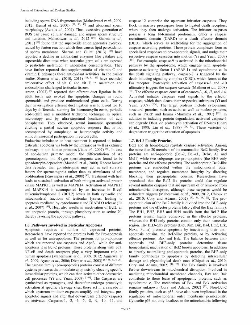
drugs influencing gonadal cell apoptosis have been discussed to provide new understanding of the molecular processes underlying cell demise as well as better treatment protocols for the management of pathological conditions involving excessive gonadal cell degeneration in mammalian testis. Among all the cell types present in the body of mammals, the germ cells are very important as they determine the fertility potential of an individual and ensure continuity of its own type. Failure of the germ cell to survive during the development may lead defective or no gamete production and hence can lead to infertility. In mammals it has been established that extensive loss of female and male germ cells occurs during embryonic and fetal stages. Apoptosis is frequently observed in early testicular development and is reported that about 25-75% of the sperm yield are lost during normal process of spermatogenesis (Oakberg, 1956; Huckins, 1978; De Rooij and Lok, 1987) [70, 43, 25]. It has been documented that the germ cell death is due to apoptotic process, however till date the understanding of the molecular mechanisms of germ cell death during testicular development is limited. Therefore it is very important to study cellular and molecular mechanisms of testicular cell apoptosis, so as to determine full reproductive output for both in vivo and in vitro studies. Apoptosis is frequently observed during the early testicular development (Coucouvanis, 1993) [23]. Moreover, the exact molecular mechanism responsible for the induction of germ cell death has not been identified, except for the involvement of androgen in the prevention of germ cell apoptosis in the adult rat testis (Henriksen et al., 1995) [39]. On the basis of quantification of DNA fragmentation, a gradual increase in apoptosis has been reported in the testicular cells of juvenile (16 to 18 day old) rats as compared with neonatal animals, followed by a decrease in adult animals (Billig et al., 1995) [10]. Berensztein et al., (2002) [9] have reported the apoptosis and proliferation of human testicular somatic and germ cell during prepuberty (Berenstein et al., 2002) [9]. They have measured the relative number of testicular cells in apoptosis and in active proliferation by calculating PI/AI ratio, in the seminiferous cords and in the interstitium, at different age periods of prepubertal testicular development and concluded that in normal subjects, vigorous growth of the testis during the newborn period is mainly mediated by decreased apoptosis and this change takes place before the testosterone peak of the postnatal gonadal activation of first trimester of life (Berenstein et al., 2002) [9]. 1.1. Scope and objectives of the Review The study of the cell death in the testis is of great relevance for the betterment of reproductive health. This review provides an overview of apoptotic process in the testis under following headings. 1) First it is important to know the process of spermatogenesis and germ cell apoptosis. 2). Germ cell apoptosis during ageing and its effects on reproduction. 3). Effects of different toxins on testicular cells. 4). Endocrine regulation of apoptosis. 5). Pathways involved in testicular apoptosis. 1.2. Spermatogenesis and apoptosis in mammals The process of spermatogenesis is started with the proliferation and mitosis of spermatogonia to form primary spermatocytes in mammals which after this undergo meiosis to form round spermatids and get differentiated into mature sperms (Fig. 1, 2 and 3). During this Sertoli cells support the
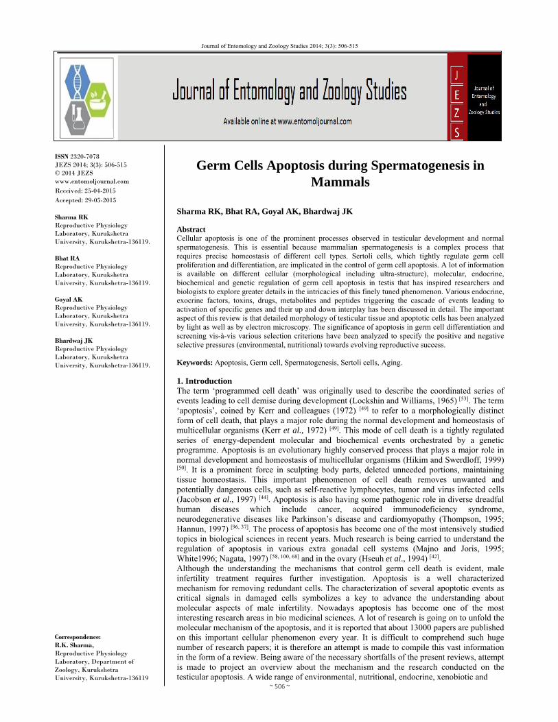
process of spermatogenesis by providing appropriate environment for the development of the germ cells in the presence of balanced hormonal support and nutrition. A number of studies have demonstrated that in most mammals the process of apoptosis is dependent on different apoptotic signaling machinery which includes Bcl-2 proteins, Fas, and caspases. During the thorough process of spermatogenesis, apoptosis occurs spontaneously for the development of normal mature spermatozoa, and for the elimination of excess or for the removal of abnormal germ cells (Rodriguez, 1997) [76].
Fig. 1 and 2: Light micrograph showing the basement membrane seminiferous epithelium (SE) possessed the germs (meiotic
Spermatogonia, Ms) and Sertoli cells (S). Note the interstitial tissue compartment comprised of connective tissue, circular Leydig cells
(L) and myoid cells (m). Next figure showing the various Sertoli cells (S), lying near the basal lamina (BL), one with 2 or 3 internuclear
clefts, two Sertoli cell having finger like projections (Sharma RK, Un publish Work).
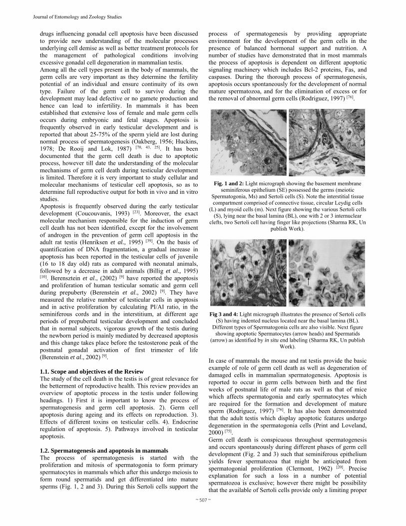
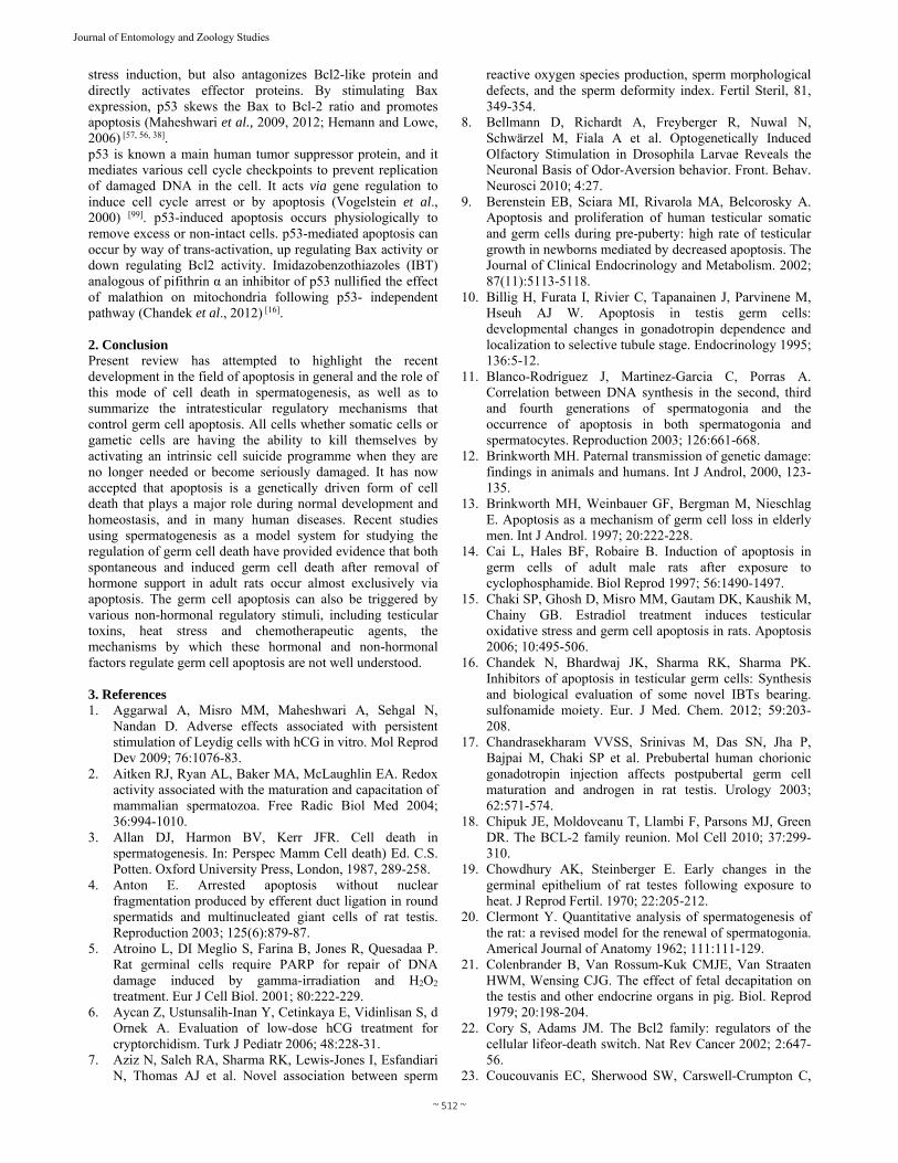
Fig 3 and 4: Light micrograph illustrates the presence of Sertoli cells (S) having indented nucleus located near the basal lamina (BL).
Different types of Spermatogonia cells are also visible. Next figure showing apoptotic Spermatocytes (arrow heads) and Spermatids
(arrow) as identified by in situ end labeling (Sharma RK, Un publish Work).
In case of mammals the mouse and rat testis provide the basic example of role of germ cell death as well as degeneration of damaged cells in mammalian spermatogenesis. Apoptosis is reported to occur in germ cells between birth and the first weeks of postnatal life of male rats as well as that of mice which affects spermatogonia and early spermatocytes which are required for the formation and development of mature sperm (Rodriguez, 1997) [76]. It has also been demonstrated that the adult testis which display apoptotic features undergo degeneration in the spermatogonia cells (Print and Loveland, 2000) [75]. Germ cell death is conspicuous throughout spermatogenesis and occurs spontaneously during different phases of germ cell development (Fig. 2 and 3) such that seminiferous epithelium yields fewer spermatozoa that might be anticipated from spermatogonial proliferation (Clermont, 1962) [20]. Precise explanation for such a loss in a number of potential spermatozoa is exclusive; however there might be possibility that the available of Sertoli cells provide only a limiting proper

~ 508 ~
Journal of Entomology and Zoology Studies
environment to sustain a number of germ cells which are not supported by the Sertoli cells. However studies demonstrated that the germ cell survival is regulated by many hormones such as FSH, playing role in testicular size and normal numbers of germ cells, LH, hCG and testosterone are important for maturation of the testis and fertility (Singh and Handelsman, 1995) [87]. Sinha Hikim et al., (1997a) [90] studied a significant increase in germ cell death following treatment with a GnRH antagonist that eliminates secretion of gonadotropins and testosterone, while on the other hand inhibition of apoptosis can be brought by hormone treatment. Apoptosis has been reported to be underlying mechanism of germ cell death during normal spermatogenesis in various mammals, including rats (Sinha et al., 1995, 1997a, 1997b; Maheshwar et al., 2009; 2012) [89, 90, 91, 56, 1, 57] hamsters (Lue et al., 1997) [54], humans (Sinha et al., 1998) [92] and goats (Sharma et al., 2011a, 2011b) [83, 65]. It was found that in rats and hamsters, spontaneous apoptosis takes place in a few differentiating spermatogonia and spermatocytes during their meiotic divisions. In mice, spontaneous apoptosis was commonly observed during spermatocytes including the dividing spermatocytes, and less frequently in spermatogonia and seldom in spermatids (Maheshwar et al., 2009, 2011; Sharma et al., 2011a, 2011b) [1, 55, 83, 85]. A number of studies have been carried on germ cell degeneration and being published on morphometric analysis of perfusion-fixed testis which have indicated that the loss of germ cells in the rodent testis is greatest during the mitosis of type A2, A3, and A4 spermatogonia and during the meiotic division (Kerr, 1932). No degeneration was reported in type A spermatogonia, or in intermediate or type B spermatogonia (Kerr, 1932). While on basis of morphological evidences, it has been suggested that cell death of spermatogonia during normal spermatogenesis takes place through the apoptotic mechanism, the spermatocytes and spermatids, in contrast undergo necrosis (Allan et al., 1987) [3]. Apoptosis has been frequently observed during normal spermatogenesis in the same types of cells and stages in which germ cell degeneration was previously observed (Billig et al., 1995) [10], indicating that germ cell degeneration is in fact apoptotic cell death. Sinha Hikim et al., (1998) [92] have examined the involvement of apoptosis in spontaneous loss of germ cells in men and revealed an evidence for ethnic differences in the inherent susceptibility of germ cells to apoptosis. During their investigation, testicular sections of 14 subjects have been found to exhibit spontaneous occurrence of germ cell apoptosis involving spermatogonia, spermatocytes and spermatids. The incidence of spermatogonial as well as spermatid apoptosis was higher in Chinese than Caucasian men and a higher incidence of spermatocyte apoptosis was also noted for Chinese as compared to Caucasian men, but here difference was not statistically significant. Blanco-Rodriguez et al., (2003) [11] have revealed a clear correlation between the occurrence of DNA replication in the second, third and fourth generation of spermatogonia and most physiological apoptosis taking place in both spermatogonia and spermatocytes in three different mammalian disorders (Rodntia, Lagomorpha and Carnivora). During their study, pairs of immunperoxidase-stained adjacent testis sections from rats, mice, rabbits and cats were either BrdU labeled or DNA 3’ end labeled and compared to analyze both the events. Sharma et al., (2011) [53, 83, 85] have demonstrated in situ 3’ end labeling (TUNEL) of fragmented apoptotic DNA in goat testicular sections (Fig. 4). Ultrastructurally apoptotic germ
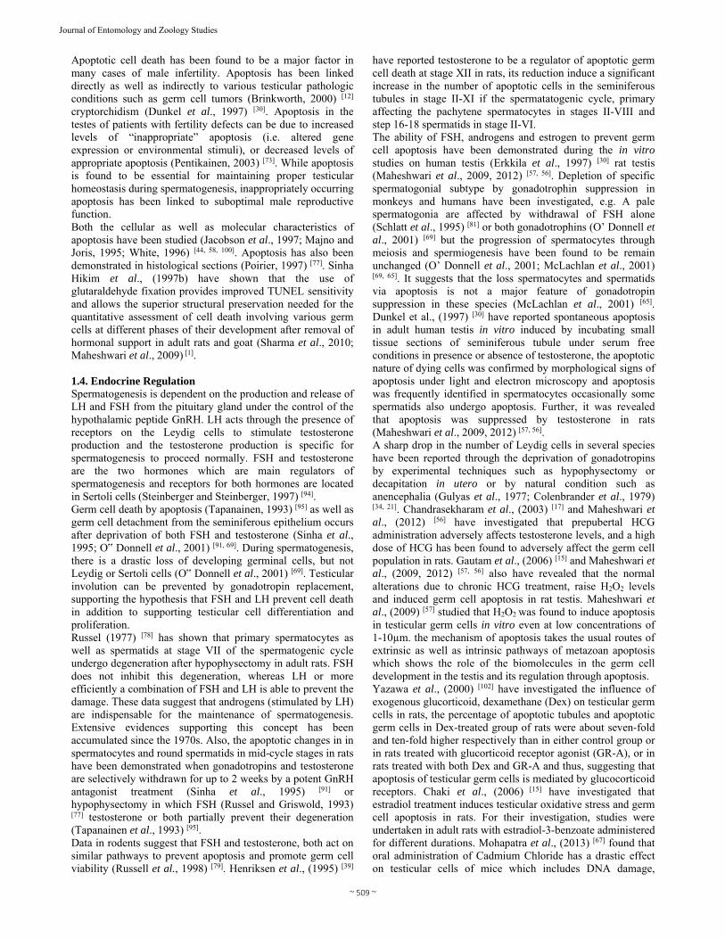
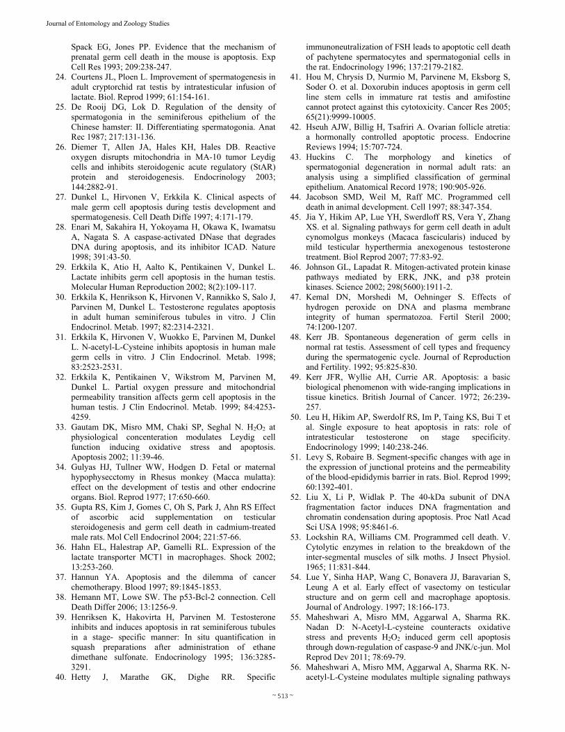
cells of goat testis revealed compaction and segregation of chromatin material, condensation of cytoplasm and fragmentation of membrane bound apoptotic bodies (Fig 5 to 8). In some germ cells (spermatogonia and spermatogonium, increased number of nuclear pores, undulations of nuclear membrane and pinching off of the nuclear fragment (Fig 5 to 8) (Sharma et al., 2011) [53, 83, 85].
Fig 5 and 6: Electron microphotograph showing normal spermatids with acrosome formation (star), and cresent-like apoptotic spermatid (arrow). Next figure showing typical apoptotic cell at advanced stage
of apoptosis (Sharma RK, Unpublish Work).
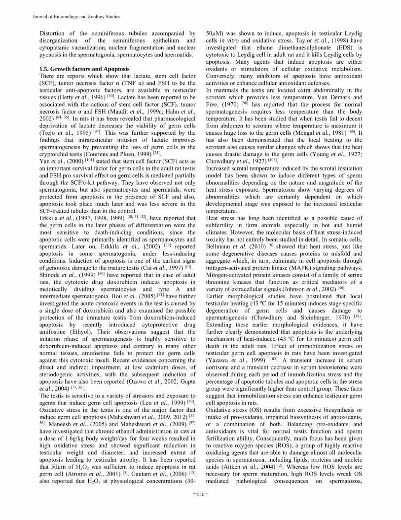
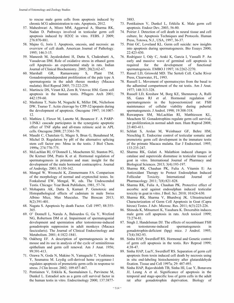
Fig 7 and 8: Transmission electronic micrograph of testicular tissue showing Sertoli cell nucleus, spermatogonium, spermatocytes and
spermatids with intracytoplasmic vacuolization (V), disorganization, chromolysis, slight margination and condensation of chromatin
material (Cd) and slight disruption of nuclear membrane (Arrow) in the spermatocyots & next figure showing Spermatocytes (Spc) with ruptured nuclear membrane (arrow) and condensed (Cd) chromatin material. Marginated chromatin material in spermatocytes (Sc) are
visible. (Sharma RK, Unpublish Work). 1.3. Germ Cell Apoptosis during Ageing Both spermatogenesis as and steroidogenesis decrease with age and it has been shown that apoptosis increases with age leading an accelerated germ cell loss (Levy and Robaire, 1999) [99]. These changes were related to the fall in androgen levels and to the increase in oxidative stress in the testicular tissue. It has been already reported that apoptosis has a very important role in germ cell degeneration in the aged testis during spermatogenesis (Brinkworth et al., 1997) [13]. Martincic et al., (2001) [61] revealed that improperly activated apoptosis in the testis can cause infertility or even cancer. They reported that increase in rate of apoptosis occurs in infertile male patients. Martincic et al., (2001) [61] studied that apoptosis in testis depletes the excess germ cells and removes the abnormal spermatozoa cells during spermatogenesis. It has been reported about 75% of germ cells are eliminated by apoptosis before they become mature. Apoptotic activity is thus important in maintaining the population control of the germ cells. Apoptosis is either developmentally regulated in response to a specific stimuli, like deprivation of survival factors, exposure to ionizing radiation and chemotherapeutic drugs or activation by various death factors or can be induced in response to cell injury or stress (Maheshwari et al., 2011, 2012) [55, 56].
~ 509 ~
Journal of Entomology and Zoology Studies
Apoptotic cell death has been found to be a major factor in many cases of male infertility. Apoptosis has been linked directly as well as indirectly to various testicular pathologic conditions such as germ cell tumors (Brinkworth, 2000) [12] cryptorchidism (Dunkel et al., 1997) [30]. Apoptosis in the testes of patients with fertility defects can be due to increased levels of “inappropriate” apoptosis (i.e. altered gene expression or environmental stimuli), or decreased levels of appropriate apoptosis (Pentikainen, 2003) [73]. While apoptosis is found to be essential for maintaining proper testicular homeostasis during spermatogenesis, inappropriately occurring apoptosis has been linked to suboptimal male reproductive function. Both the cellular as well as molecular characteristics of apoptosis have been studied (Jacobson et al., 1997; Majno and Joris, 1995; White, 1996) [44, 58, 100]. Apoptosis has also been demonstrated in histological sections (Poirier, 1997) [77]. Sinha Hikim et al., (1997b) have shown that the use of glutaraldehyde fixation provides improved TUNEL sensitivity and allows the superior structural preservation needed for the quantitative assessment of cell death involving various germ cells at different phases of their development after removal of hormonal support in adult rats and goat (Sharma et al., 2010; Maheshwari et al., 2009) [1]. 1.4. Endocrine Regulation Spermatogenesis is dependent on the production and release of LH and FSH from the pituitary gland under the control of the hypothalamic peptide GnRH. LH acts through the presence of receptors on the Leydig cells to stimulate testosterone production and the testosterone production is specific for spermatogenesis to proceed normally. FSH and testosterone are the two hormones which are main regulators of spermatogenesis and receptors for both hormones are located in Sertoli cells (Steinberger and Steinberger, 1997) [94]. Germ cell death by apoptosis (Tapanainen, 1993) [95] as well as germ cell detachment from the seminiferous epithelium occurs after deprivation of both FSH and testosterone (Sinha et al., 1995; O‟ Donnell et al., 2001) [91, 69]. During spermatogenesis, there is a drastic loss of developing germinal cells, but not Leydig or Sertoli cells (O‟ Donnell et al., 2001) [69]. Testicular involution can be prevented by gonadotropin replacement, supporting the hypothesis that FSH and LH prevent cell death in addition to supporting testicular cell differentiation and proliferation. Russel (1977) [78] has shown that primary spermatocytes as well as spermatids at stage VII of the spermatogenic cycle undergo degeneration after hypophysectomy in adult rats. FSH does not inhibit this degeneration, whereas LH or more efficiently a combination of FSH and LH is able to prevent the damage. These data suggest that androgens (stimulated by LH) are indispensable for the maintenance of spermatogenesis. Extensive evidences supporting this concept has been accumulated since the 1970s. Also, the apoptotic changes in in spermatocytes and round spermatids in mid-cycle stages in rats have been demonstrated when gonadotropins and testosterone are selectively withdrawn for up to 2 weeks by a potent GnRH antagonist treatment (Sinha et al., 1995) [91] or hypophysectomy in which FSH (Russel and Griswold, 1993)
[77] testosterone or both partially prevent their degeneration (Tapanainen et al., 1993) [95]. Data in rodents suggest that FSH and testosterone, both act on similar pathways to prevent apoptosis and promote germ cell viability (Russell et al., 1998) [79]. Henriksen et al., (1995) [39]
have reported testosterone to be a regulator of apoptotic germ cell death at stage XII in rats, its reduction induce a significant increase in the number of apoptotic cells in the seminiferous tubules in stage II-XI if the spermatatogenic cycle, primary affecting the pachytene spermatocytes in stages II-VIII and step 16-18 spermatids in stage II-VI. The ability of FSH, androgens and estrogen to prevent germ cell apoptosis have been demonstrated during the in vitro studies on human testis (Erkkila et al., 1997) [30] rat testis (Maheshwari et al., 2009, 2012) [57, 56]. Depletion of specific spermatogonial subtype by gonadotrophin suppression in monkeys and humans have been investigated, e.g. A pale spermatogonia are affected by withdrawal of FSH alone (Schlatt et al., 1995) [81] or both gonadotrophins (O’ Donnell et al., 2001) [69] but the progression of spermatocytes through meiosis and spermiogenesis have been found to be remain unchanged (O’ Donnell et al., 2001; McLachlan et al., 2001) [69, 65]. It suggests that the loss spermatocytes and spermatids via apoptosis is not a major feature of gonadotropin suppression in these species (McLachlan et al., 2001) [65]. Dunkel et al., (1997) [30] have reported spontaneous apoptosis in adult human testis in vitro induced by incubating small tissue sections of seminiferous tubule under serum free conditions in presence or absence of testosterone, the apoptotic nature of dying cells was confirmed by morphological signs of apoptosis under light and electron microscopy and apoptosis was frequently identified in spermatocytes occasionally some spermatids also undergo apoptosis. Further, it was revealed that apoptosis was suppressed by testosterone in rats (Maheshwari et al., 2009, 2012) [57, 56]. A sharp drop in the number of Leydig cells in several species have been reported through the deprivation of gonadotropins by experimental techniques such as hypophysectomy or decapitation in utero or by natural condition such as anencephalia (Gulyas et al., 1977; Colenbrander et al., 1979) [34, 21]. Chandrasekharam et al., (2003) [17] and Maheshwari et al., (2012) [56] have investigated that prepubertal HCG administration adversely affects testosterone levels, and a high dose of HCG has been found to adversely affect the germ cell population in rats. Gautam et al., (2006) [15] and Maheshwari et al., (2009, 2012) [57, 56] also have revealed that the normal alterations due to chronic HCG treatment, raise H2O2 levels and induced germ cell apoptosis in rat testis. Maheshwari et al., (2009) [57] studied that H2O2
was found to induce apoptosis in testicular germ cells in vitro even at low concentrations of 1-10µm. the mechanism of apoptosis takes the usual routes of extrinsic as well as intrinsic pathways of metazoan apoptosis which shows the role of the biomolecules in the germ cell development in the testis and its regulation through apoptosis. Yazawa et al., (2000) [102] have investigated the influence of exogenous glucorticoid, dexamethane (Dex) on testicular germ cells in rats, the percentage of apoptotic tubules and apoptotic germ cells in Dex-treated group of rats were about seven-fold and ten-fold higher respectively than in either control group or in rats treated with glucorticoid receptor agonist (GR-A), or in rats treated with both Dex and GR-A and thus, suggesting that apoptosis of testicular germ cells is mediated by glucocorticoid receptors. Chaki et al., (2006) [15] have investigated that estradiol treatment induces testicular oxidative stress and germ cell apoptosis in rats. For their investigation, studies were undertaken in adult rats with estradiol-3-benzoate administered for different durations. Mohapatra et al., (2013) [67] found that oral administration of Cadmium Chloride has a drastic effect on testicular cells of mice which includes DNA damage,
~ 510 ~
Journal of Entomology and Zoology Studies
Distortion of the seminiferous tubules accompanied by disorganization of the seminiferous epithelium and cytoplasmic vacuolization, nuclear fragmentation and nuclear pycnosis in the spermatogonia, spermatocytes and spermatids. 1.5. Growth factors and Apoptosis There are reports which show that lactate, stem cell factor (SCF), tumor necrosis factor α (TNF α) and FSH to be the testicular anti-apoptotic factors, are available in testicular tissues (Hetty et al., 1996) [40]. Lactate has been reported to be associated with the actions of stem cell factor (SCF), tumor necrosis factor α and FSH (Maudit et al., 1999a; Hahn et al., 2002) [64, 36]. In rats it has been revealed that pharmacological deprivation of lactate decreases the viability of germ cells (Trejo et al., 1995) [97]. This was further supported by the findings that intraresticular infusion of lactate improves spermatogenesis by preventing the loss of germ cells in the cryptorchid testis (Courtens and Ploen, 1999) [24]. Yan et al., (2000) [101] stated that stem cell factor (SCF) acts as an important survival factor for germ cells in the adult rat testis and FSH pro-survival effect on germ cells is mediated partially through the SCF/c-kit pathway. They have observed not only spermatogonia, but also spermatocytes and spermatids, were protected from apoptosis in the presence of SCF and also, apoptosis took place much later and was less severe in the SCF-treated tubules than in the control. Erkkila et al., (1997, 1998, 1999) [30, 31, 32], have reported that the germ cells in the later phases of differentiation were the most sensitive to death-inducing conditions, since the apoptotic cells were primarily identified as spermatocytes and spermatids. Later on, Erkkila et al., (2002) [29] reported apoptosis in some spermatogonia, under less-inducing conditions. Induction of apoptosis is one of the earliest signs of genotoxic damage to the mature testis (Cai et al., 1997) [14]. Shinoda et al., (1999) [86] have reported that in case of adult rats, the cytotoxic drug doxorubicin induces apoptosis in meiotically dividing spermatocytes and type A and intermediate spermatogonia. Hou et al., (2005) [41] have further investigated the acute cytotoxic events in the test is caused by a single dose of doxorubicin and also examined the possible protection of the immature testis from doxorubicin-induced apoptosis by recently introduced cytoprotective drug amifostine (Ethyol). Their observations suggest that the initation phase of spermatogenesis is highly sensitive to doxorubicin-induced apoptosis and contrary to many other normal tissues, amofostine fails to protect the germ cells against this cytotoxic insult. Recent evidences concerning the direct and indirect impairment, at low cadmium doses, of steriodogenic activities, with the subsequent induction of apoptosis have also been reported (Ozawa et al., 2002; Gupta et al., 2004) [71, 35]. The testis is sensitive to a variety of stressors and exposure to agents that induce germ cell apoptosis (Leu et al., 1999) [50]. Oxidative stress in the testis is one of the major factor that induce germ cell apoptosis (Maheshwari et al., 2009, 2012) [57,
56]. Maneesh et al., (2005) and Maheshwari et al., (2009) [57] have investigated that chronic ethanol administration in rats at a dose of 1.6g/kg body weight/day for four weeks resulted in high oxidative stress and showed significant reduction in testicular weight and diameter; and increased extent of apoptosis leading to testicular atrophy. It has been reported that 50µm of H2O2 was sufficient to induce apoptosis in rat germ cell (Atroino et al., 2001) [5]. Gautam et al., (2006) [15] also reported that H2O2 at physiological concentrations (30-
50µM) was shown to induce, apoptosis in testicular Leydig cells in vitro and oxidative stress. Taylor et al., (1998) have investigated that ethane dimethanesulphonate (EDS) is cytotoxic to Leydig cell in adult rat and it kills Leydig cells by apoptosis. Many agents that induce apoptosis are either oxidants or stimulators of cellular oxidative metabolism. Conversely, many inhibitors of apoptosis have antioxidant activities or enhance cellular antioxidant defenses. In mammals the testis are located extra abdominally in the scrotam which provides less temperature. Van Demark and Free, (1970) [98] has reported that the process for normal spermatogenesis requires less temperature than the body temperature. It has been studied that when testis fail to decent from abdomen to scrotam where temperature is maximum it causes huge loss to the germ cells (Mengal et al., 1981) [66]. It has also been demonstrated that the local heating to the scrotam also causes similar changes which shows that the heat causes drastic damage to the germ cells (Young et al., 1927; Chowdhury et al., 1927) [105]. Increased scrotal temperature induced by the scrotal insulation model has been shown to induce different types of sperm abnormalities depending on the nature and magnitude of the heat stress exposure. Spermatozoa show varying degrees of abnormalities which are certainly dependent on which developmental stage was exposed to the increased testicular temperature. Heat stress has long been identified as a possible cause of subfertility in farm animals especially in hot and humid climates. However, the molecular basis of heat stress-induced toxicity has not entirely been studied in detail. In somatic cells, Bellmann et al. (2010) [8] showed that heat stress, just like some degenerative diseases causes proteins to misfold and aggregate which, in turn, culminate in cell apoptosis through mitogen-activated protein kinase (MAPK) signaling pathways. Mitogen-activated protein kinases consist of a family of serine threonine kinases that function as critical mediators of a variety of extracellular signals (Johnson et al., 2002) [46]. Earlier morphological studies have postulated that local testicular heating (43 ºC for 15 minutes) induces stage specific degeneration of germ cells and causes damage to spermatogenesis (Chowdhury and Steinberger, 1970) [19]. Extending these earlier morphological evidences, it have further clearly demonstrated that apoptosis is the underlying mechanism of heat-induced (43 ºC for 15 minutes) germ cell death in the adult rats. Effect of immobilization stress on testicular germ cell apoptosis in rats have been investigated (Yazawa et al., 1999) [103]. A transient increase in serum cortisone and a transient decrease in serum testosterone were observed during each period of immobilization stress and the percentage of apoptotic tubules and apoptotic cells in the stress group were significantly higher than control group. These facts suggest that immobilization stress can enhance testicular germ cell apoptosis in rats. Oxidative stress (OS) results from excessive biosynthesis or intake of pro-oxidants, impaired biosynthesis of antioxidants, or a combination of both. Balancing pro-oxidants and antioxidants is vital for normal testis function and sperm fertilization ability. Consequently, much focus has been given to reactive oxygen species (ROS), a group of highly reactive oxidizing agents that are able to damage almost all molecular species in spermatozoa, including lipids, proteins and nucleic acids (Aitken et al., 2004) [2]. Whereas low ROS levels are necessary for sperm maturation, high ROS levels wreak OS mediated pathological consequences on spermatozoa,
~ 511 ~
Journal of Entomology and Zoology Studies
including sperm DNA fragmentation (Maheshwari et al., 2009, 2012; Kemal et al., 2000) [57, 56, 47] and abnormal sperm morphology (Aziz et al., 2004). Thus, excessive generation of ROS can cause cellular damage, and impair sperm structure and function. Maheshwari et al., 2012 [56], Sharma et al., (2013) [82] found that the superoxide anion generates hydroxyl radical by fenton reaction which thus causes lipid peroxidation of sperm membrane. Sharma and Gulati (2013) [82] have reported a decline in antioxidant enzymes like catalase and superoxide dismutase when testicular germ cells are exposed to pestictide melathion at nanomolar concenteration. They have further reported that supplementatio of vitamin C and vitamin E enhances these antioxidant activities. In the earlier studies Sharma et al., (2010, 2011) [84, 83, 85] have recorded amleorative effect of vit C and vit E in malathion and endosulphan challenged testicular tissues. Anton, (2003) [4] reported that efferent duct ligation in the adult testis rats evoked the apoptotic changes in round spermatids and produce multinucleated giant cells. During their investigation efferent duct ligation was followed for 10 days by differential staining for haemotoxylin-eosin, periodic acid-Schiff and a modified trichrome technique in optical microscopy and by ultra-structural localization of acid phosphatase. They observed, round immature spermatids eliciting a partial nuclear apoptotic response that is not accompanied by autophagic or heterophagic activity and without lysosomal participation in Sertoli cells. Endocrine imbalance or heat treatment is reported to induce testicular apoptosis via both by the intrinsic as well as extrinsic pathways in non-human primates (Jia et al., 2007) [45]. In case of non-human primate model, the differentiation of Ap spermatogonia into B-type spermatogonia was found to be gonadotropin-dependent (Marshall et al., 2000). Recent human data revealed that gonadotropins may act as cell survival factors for spermatogonia rather than as stimulators of cell proliferation (Ruwanpura et al., 2008) [80]. Treatment with heat leads to sustained activation of both mitogen-activated protein kinase MAPK13 as well as MAPK14. Activation of MAPK13 and MAPK14 is accompanied by an increase in B-cell leukemia/lymphoma 2 (BCL2) levels in both cytosolic and mitochondrial fractions of testicular lysates, leading to apoptosis mediated by cytochrome c and DIABLO release (Jia et al., 2007) [45]. Heat also results in inactivation of Bcl2, an anti-apoptotic protein, through phosphorylation at serine 70, thereby favoring the apoptotic pathway. 1.6. Pathways Involved in Testicular Apoptosis Apoptosis requires a number of expressed proteins. Researchers have reported the proteins both for Pro-apoptosis as well as for anti-apoptosis. The proteins for pro-apoptosis which are reported are caspases and Apaf-1 while for anti-apoptosis it is Bcl-2 proteins. These proteins along with p53, NF-κB and death receptors play a very important role in human apoptosis (Maheshwari et al., 2009, 2012; Aggarwal et al., 2009; Aycan et al., 2006; Diemer et al., 2003) [57, 56, 57, 6, 26]. The caspase family (pro-apoptotic) proteins consist of different cysteine proteases that modulate apoptosis by cleaving specific intracellular proteins, which can then activate other destructive cell processes (Yi and Yuan, 2009) [104]. The Caspases are synthesized as zymogens, and thereafter undergo proteolytic activation at specific cleavage sites, these act in a cascade in which upstream initiator caspases amplify and integrate pro-apoptotic signals and after that downstream effector caspases are activated. Caspases-1, -2, -4, -5, -8, -9, -10, -11, and
caspase-12 comprise the upstream initiatior caspases. They flock in inactive procaspase form to ligated death receptors, where they then undergo activation. The initiator caspases possess a long N-terminal prodomain, either a caspase recruitment domain (CARD) or a death effector domain (DED), which serves as scaffolding for the aggregation of caspase activating proteins. These protein complexes form as specialized responses to pro-apoptotic signals, and nudge their respective caspase cascades into motion (Yi and Yuan, 2009) [104]. For example, caspase-9 is activated in the mitochondrial pathway by the apoptosome, which engages with apoptotic protease-activating factor-1 (APAF-1) and cytochrome c. In the death signaling pathway, caspase-8 is triggered by the death inducing signaling complex (DISC), which forms at the Fas receptor. Proteolytic activation of caspases-8 and -10 ultimately triggers the caspase cascade (Mathieu et al., 2008) [63]. The effector caspases consist of capsases-3, -6, -7, and -14. Activated initiator caspases send signals to the effector caspases, which then cleave their respective substrates (Yi and Yuan, 2009) [104]. The target proteins include cytoplasmic structural proteins, such as actin, as well as nuclear proteins, such as PARP and lamins (Mashima et al., 1997) [62]. In addition to inducing protein degradation, activated caspase-3 can degrade DNA via proteolytic activation of DNases (Enari et al., 1998; Liu et al., 1998) [28, 52]. These varieties of degradation trigger the execution of apoptosis. 1.7. Bcl-2 Family Proteins Bcl2 and its homologues regulate caspase activation. Among the more than 20 members of the mammalian Bcl2 family, five proteins are anti-apoptotic (Bcl-2, Bcl-xL, Bclw, A1 and Mcl1) while two subgroups are pro-apoptotic (the BH3-only proteins and the effector proteins). The antiapoptotic Bcl2-like proteins are embedded within the mitochondrial outer membrane, and regulate membrane integrity by directing blocking their proapoptotic cousins. Researchers have speculated that the Bcl2-like proteins could also regulate several initiator caspases that are upstream of or removed from mitochondrial disruption, although these caspases would be redundant triggers (Maheshwari et al., 2009, 2012, Chipuk et al., 2010; Cory and Adams, 2002) [57, 56, 18, 22]. The pro-apoptotic clan of the Bcl2 family is divided into the BH3-only proteins and the effector proteins (also called the Bax family). The BH1, BH2, BH3 and BH4 motifs from the Bcl-2 like proteins remain highly conserved in the effector proteins, whereas the BH3-only proteins contain only their namesake region. The BH3-only proteins (Bid, Bim, Bik, Bad, Bmf, Hrk, Noxa, Puma) promote apoptosis by inactivating their anti-apoptosis cousins, the Bcl2-like proteins, or by activating effector proteins, Bax and Bak. The balance between anti-apoptosis and BH3-only proteins determine tissue homeostasis; inactivation of Bcl2 boosts apoptosis. In addition to directly neutralizing anti-apoptotic proteins, the BH3-only family contributes to apoptosis by detecting intracellular damage and physiological death cues (Chipuk et al., 2010; Cory and Adams, 2002) [18, 22]. The Bax family is involved further downstream in mitochondrial disruption. Involved in mediating mitochondrial membrane channels, Bax and Bak contribute to there lease of apoptogenic proteins, such as cytochrome c. The mechanism of Bax and Bak activation remains unknown (Cory and Adams, 2002) [22]. Non-Bcl-2 family proteins, such as p53, have also been implicated in the regulation of mitochondrial outer membrane permeability. Cytosolic p53 not only localizes to the mitochondria following
~ 512 ~
Journal of Entomology and Zoology Studies
stress induction, but also antagonizes Bcl2-like protein and directly activates effector proteins. By stimulating Bax expression, p53 skews the Bax to Bcl-2 ratio and promotes apoptosis (Maheshwari et al., 2009, 2012; Hemann and Lowe, 2006) [57, 56, 38]. p53 is known a main human tumor suppressor protein, and it mediates various cell cycle checkpoints to prevent replication of damaged DNA in the cell. It acts via gene regulation to induce cell cycle arrest or by apoptosis (Vogelstein et al., 2000) [99]. p53-induced apoptosis occurs physiologically to remove excess or non-intact cells. p53-mediated apoptosis can occur by way of trans-activation, up regulating Bax activity or down regulating Bcl2 activity. Imidazobenzothiazoles (IBT) analogous of pifithrin α an inhibitor of p53 nullified the effect of malathion on mitochondria following p53- independent pathway (Chandek et al., 2012) [16]. 2. Conclusion Present review has attempted to highlight the recent development in the field of apoptosis in general and the role of this mode of cell death in spermatogenesis, as well as to summarize the intratesticular regulatory mechanisms that control germ cell apoptosis. All cells whether somatic cells or gametic cells are having the ability to kill themselves by activating an intrinsic cell suicide programme when they are no longer needed or become seriously damaged. It has now accepted that apoptosis is a genetically driven form of cell death that plays a major role during normal development and homeostasis, and in many human diseases. Recent studies using spermatogenesis as a model system for studying the regulation of germ cell death have provided evidence that both spontaneous and induced germ cell death after removal of hormone support in adult rats occur almost exclusively via apoptosis. The germ cell apoptosis can also be triggered by various non-hormonal regulatory stimuli, including testicular toxins, heat stress and chemotherapeutic agents, the mechanisms by which these hormonal and non-hormonal factors regulate germ cell apoptosis are not well understood. 3. References 1. Aggarwal A, Misro MM, Maheshwari A, Sehgal N,
Nandan D. Adverse effects associated with persistent stimulation of Leydig cells with hCG in vitro. Mol Reprod Dev 2009; 76:1076-83.
2. Aitken RJ, Ryan AL, Baker MA, McLaughlin EA. Redox activity associated with the maturation and capacitation of mammalian spermatozoa. Free Radic Biol Med 2004; 36:994-1010.
3. Allan DJ, Harmon BV, Kerr JFR. Cell death in spermatogenesis. In: Perspec Mamm Cell death) Ed. C.S. Potten. Oxford University Press, London, 1987, 289-258.
4. Anton E. Arrested apoptosis without nuclear fragmentation produced by efferent duct ligation in round spermatids and multinucleated giant cells of rat testis. Reproduction 2003; 125(6):879-87.
5. Atroino L, DI Meglio S, Farina B, Jones R, Quesadaa P. Rat germinal cells require PARP for repair of DNA damage induced by gamma-irradiation and H2O2 treatment. Eur J Cell Biol. 2001; 80:222-229.
6. Aycan Z, Ustunsalih-Inan Y, Cetinkaya E, Vidinlisan S, d Ornek A. Evaluation of low-dose hCG treatment for cryptorchidism. Turk J Pediatr 2006; 48:228-31.
7. Aziz N, Saleh RA, Sharma RK, Lewis-Jones I, Esfandiari N, Thomas AJ et al. Novel association between sperm
reactive oxygen species production, sperm morphological defects, and the sperm deformity index. Fertil Steril, 81, 349-354.
8. Bellmann D, Richardt A, Freyberger R, Nuwal N, Schwärzel M, Fiala A et al. Optogenetically Induced Olfactory Stimulation in Drosophila Larvae Reveals the Neuronal Basis of Odor-Aversion behavior. Front. Behav. Neurosci 2010; 4:27.
9. Berenstein EB, Sciara MI, Rivarola MA, Belcorosky A. Apoptosis and proliferation of human testicular somatic and germ cells during pre-puberty: high rate of testicular growth in newborns mediated by decreased apoptosis. The Journal of Clinical Endocrinology and Metabolism. 2002; 87(11):5113-5118.
10. Billig H, Furata I, Rivier C, Tapanainen J, Parvinene M, Hseuh AJ W. Apoptosis in testis germ cells: developmental changes in gonadotropin dependence and localization to selective tubule stage. Endocrinology 1995; 136:5-12.
11. Blanco-Rodriguez J, Martinez-Garcia C, Porras A. Correlation between DNA synthesis in the second, third and fourth generations of spermatogonia and the occurrence of apoptosis in both spermatogonia and spermatocytes. Reproduction 2003; 126:661-668.
12. Brinkworth MH. Paternal transmission of genetic damage: findings in animals and humans. Int J Androl, 2000, 123-135.
13. Brinkworth MH, Weinbauer GF, Bergman M, Nieschlag E. Apoptosis as a mechanism of germ cell loss in elderly men. Int J Androl. 1997; 20:222-228.
14. Cai L, Hales BF, Robaire B. Induction of apoptosis in germ cells of adult male rats after exposure to cyclophosphamide. Biol Reprod 1997; 56:1490-1497.
15. Chaki SP, Ghosh D, Misro MM, Gautam DK, Kaushik M, Chainy GB. Estradiol treatment induces testicular oxidative stress and germ cell apoptosis in rats. Apoptosis 2006; 10:495-506.
16. Chandek N, Bhardwaj JK, Sharma RK, Sharma PK. Inhibitors of apoptosis in testicular germ cells: Synthesis and biological evaluation of some novel IBTs bearing. sulfonamide moiety. Eur. J Med. Chem. 2012; 59:203-208.
17. Chandrasekharam VVSS, Srinivas M, Das SN, Jha P, Bajpai M, Chaki SP et al. Prebubertal human chorionic gonadotropin injection affects postpubertal germ cell maturation and androgen in rat testis. Urology 2003; 62:571-574.
18. Chipuk JE, Moldoveanu T, Llambi F, Parsons MJ, Green DR. The BCL-2 family reunion. Mol Cell 2010; 37:299-310.
19. Chowdhury AK, Steinberger E. Early changes in the germinal epithelium of rat testes following exposure to heat. J Reprod Fertil. 1970; 22:205-212.
20. Clermont Y. Quantitative analysis of spermatogenesis of the rat: a revised model for the renewal of spermatogonia. Americal Journal of Anatomy 1962; 111:111-129.
21. Colenbrander B, Van Rossum-Kuk CMJE, Van Straaten HWM, Wensing CJG. The effect of fetal decapitation on the testis and other endocrine organs in pig. Biol. Reprod 1979; 20:198-204.
22. Cory S, Adams JM. The Bcl2 family: regulators of the cellular lifeor-death switch. Nat Rev Cancer 2002; 2:647-56.
23. Coucouvanis EC, Sherwood SW, Carswell-Crumpton C,
~ 513 ~
Journal of Entomology and Zoology Studies
Spack EG, Jones PP. Evidence that the mechanism of prenatal germ cell death in the mouse is apoptosis. Exp Cell Res 1993; 209:238-247.
24. Courtens JL, Ploen L. Improvement of spermatogenesis in adult cryptorchid rat testis by intratesticular infusion of lactate. Biol. Reprod 1999; 61:154-161.
25. De Rooij DG, Lok D. Regulation of the density of spermatogonia in the seminiferous epithelium of the Chinese hamster: II. Differentiating spermatogonia. Anat Rec 1987; 217:131-136.
26. Diemer T, Allen JA, Hales KH, Hales DB. Reactive oxygen disrupts mitochondria in MA-10 tumor Leydig cells and inhibits steroidogenic acute regulatory (StAR) protein and steroidogenesis. Endocrinology 2003; 144:2882-91.
27. Dunkel L, Hirvonen V, Erkkila K. Clinical aspects of male germ cell apoptosis during testis development and spermatogenesis. Cell Death Diffe 1997; 4:171-179.
28. Enari M, Sakahira H, Yokoyama H, Okawa K, Iwamatsu A, Nagata S. A caspase-activated DNase that degrades DNA during apoptosis, and its inhibitor ICAD. Nature 1998; 391:43-50.
29. Erkkila K, Atio H, Aalto K, Pentikainen V, Dunkel L. Lactate inhibits germ cell apoptosis in the human testis. Molecular Human Reproduction 2002; 8(2):109-117.
30. Erkkila K, Henrikson K, Hirvonen V, Rannikko S, Salo J, Parvinen M, Dunkel L. Testosterone regulates apoptosis in adult human seminiferous tubules in vitro. J Clin Endocrinol. Metab. 1997; 82:2314-2321.
31. Erkkila K, Hirvonen V, Wuokko E, Parvinen M, Dunkel L. N-acetyl-L-Cysteine inhibits apoptosis in human male germ cells in vitro. J Clin Endocrinol. Metab. 1998; 83:2523-2531.
32. Erkkila K, Pentikainen V, Wikstrom M, Parvinen M, Dunkel L. Partial oxygen pressure and mitochondrial permeability transition affects germ cell apoptosis in the human testis. J Clin Endocrinol. Metab. 1999; 84:4253-4259.
33. Gautam DK, Misro MM, Chaki SP, Seghal N. H2O2 at physiological concenteration modulates Leydig cell function inducing oxidative stress and apoptosis. Apoptosis 2002; 11:39-46.
34. Gulyas HJ, Tullner WW, Hodgen D. Fetal or maternal hypophysecctomy in Rhesus monkey (Macca mulatta): effect on the development of testis and other endocrine organs. Biol. Reprod 1977; 17:650-660.
35. Gupta RS, Kim J, Gomes C, Oh S, Park J, Ahn RS Effect of ascorbic acid supplementation on testicular steroidogenesis and germ cell death in cadmium-treated male rats. Mol Cell Endocrinol 2004; 221:57-66.
36. Hahn EL, Halestrap AP, Gamelli RL. Expression of the lactate transporter MCT1 in macrophages. Shock 2002; 13:253-260.
37. Hannun YA. Apoptosis and the dilemma of cancer chemotherapy. Blood 1997; 89:1845-1853.
38. Hemann MT, Lowe SW. The p53-Bcl-2 connection. Cell Death Differ 2006; 13:1256-9.
39. Henriksen K, Hakovirta H, Parvinen M. Testosterone inhibits and induces apoptosis in rat seminiferous tubules in a stage- specific manner: In situ quantification in squash preparations after administration of ethane dimethane sulfonate. Endocrinology 1995; 136:3285-3291.
40. Hetty J, Marathe GK, Dighe RR. Specific
immunoneutralization of FSH leads to apoptotic cell death of pachytene spermatocytes and spermatogonial cells in the rat. Endocrinology 1996; 137:2179-2182.
41. Hou M, Chrysis D, Nurmio M, Parvinene M, Eksborg S, Soder O. et al. Doxorubin induces apoptosis in germ cell line stem cells in immature rat testis and amifostine cannot protect against this cytotoxicity. Cancer Res 2005; 65(21):9999-10005.
42. Hseuh AJW, Billig H, Tsafriri A. Ovarian follicle atretia: a hormonally controlled apoptotic process. Endocrine Reviews 1994; 15:707-724.
43. Huckins C. The morphology and kinetics of spermatogonial degeneration in normal adult rats: an analysis using a simplified classification of germinal epithelium. Anatomical Record 1978; 190:905-926.
44. Jacobson SMD, Weil M, Raff MC. Programmed cell death in animal development. Cell 1997; 88:347-354.
45. Jia Y, Hikim AP, Lue YH, Swerdloff RS, Vera Y, Zhang XS. et al. Signaling pathways for germ cell death in adult cynomolgus monkeys (Macaca fascicularis) induced by mild testicular hyperthermia anexogenous testosterone treatment. Biol Reprod 2007; 77:83-92.
46. Johnson GL, Lapadat R. Mitogen-activated protein kinase pathways mediated by ERK, JNK, and p38 protein kinases. Science 2002; 298(5600):1911-2.
47. Kemal DN, Morshedi M, Oehninger S. Effects of hydrogen peroxide on DNA and plasma membrane integrity of human spermatozoa. Fertil Steril 2000; 74:1200-1207.
48. Kerr JB. Spontaneous degeneration of germ cells in normal rat testis. Assessment of cell types and frequency during the spermatogenic cycle. Journal of Reproduction and Fertility. 1992; 95:825-830.
49. Kerr JFR, Wyllie AH, Currie AR. Apoptosis: a basic biological phenomenon with wide-ranging implications in tissue kinetics. British Journal of Cancer. 1972; 26:239-257.
50. Leu H, Hikim AP, Swerdolf RS, Im P, Taing KS, Bui T et al. Single exposure to heat apoptosis in rats: role of intratesticular testosterone on stage specificity. Endocrinology 1999; 140:238-246.
51. Levy S, Robaire B. Segment-specific changes with age in the expression of junctional proteins and the permeability of the blood-epididymis barrier in rats. Biol. Reprod 1999; 60:1392-401.
52. Liu X, Li P, Widlak P. The 40-kDa subunit of DNA fragmentation factor induces DNA fragmentation and chromatin condensation during apoptosis. Proc Natl Acad Sci USA 1998; 95:8461-6.
53. Lockshin RA, Williams CM. Programmed cell death. V. Cytolytic enzymes in relation to the breakdown of the inter-segmental muscles of silk moths. J Insect Physiol. 1965; 11:831-844.
54. Lue Y, Sinha HAP, Wang C, Bonavera JJ, Baravarian S, Leung A et al. Early effect of vasectomy on testicular structure and on germ cell and macrophage apoptosis. Journal of Andrology. 1997; 18:166-173.
55. Maheshwari A, Misro MM, Aggarwal A, Sharma RK. Nadan D: N-Acetyl-L-cysteine counteracts oxidative stress and prevents H2O2 induced germ cell apoptosis through down-regulation of caspase-9 and JNK/c-jun. Mol Reprod Dev 2011; 78:69-79.
56. Maheshwari A, Misro MM, Aggarwal A, Sharma RK. N-acetyl-L-Cysteine modulates multiple signaling pathways
~ 514 ~
Journal of Entomology and Zoology Studies
to rescue male germ cells from apoptosis induced by chronic hCG administration to rats. Apoptosis, 2012.
57. Maheshwari A, Misro MM, Aggarwal A, Sharma RK, Nadan D. Pathways involved in testicular germ cell apoptosis induced by H2O2 in vitro. FEBS. J 2009; 276:870-881.
58. Majno G, Joris I. Apoptosis, oncosis, and necrosis: an overview of cell death. American Journal of Pathology. 1995; 146:3-15.
59. Maneesh M, Jayaleskshmi H, Dutta S, Chakrabarti A, Vasudevan DM. Role of oxidative stress in ethanol germ cell Apoptosis- an experimental study in rats. Indian Journal of Clinical Biochemistry. 2005; 20(2):62-67.
60. Marshall GR, Ramaswamy S, Plant TM. Gonadotropinindependent proliferation of the pale type A spermatogonia in the adult rhesus monkey (Macaca mulatta). Biol Reprod 2005; 73:222-229.
61. Martincic DS, Virant KI, Zorn B, Vrtovec HM. Germ cell apoptosis in the human testis. Pflugers Arch 2001; 442:159-60.
62. Mashima T, Naito M, Noguchi K, Miller DK, Nicholson DW, Tsuruo T. Actin cleavage by CPP-32/apopain during the development of apoptosis. Oncogene 1997; 14:1007-12.
63. Mathieu J, Flexor M, Lanotte M, Besancoz F. A PARP-1/JNK1 cascade participates in the synergistic apoptotic effect of TNF alpha and all-trans retinoic acid in APL cells. Oncogene 2008; 27:3361-70.
64. Maudit C, Chatelain G, Magre S, Brun G, Benahmed M, Michel D. Regulation by pH of the alternative splicing stem cell factor pre- Mrna in the testis. J Biol Chem. 1999a; 274:770-775.
65. McLachlan RI, O’Donnell L, Meachemm SJ, Stanton PG, De Kretser DM, Pratis K et al. Hormonal regulation of spermatogenesis in primates and man: insight for the development of the male hormonal contraceptive. Journal of Androlgy. 2001; 8:150-162.
66. Mengal W, Wronechi K, Zimmermann FA. Comparison of the morphology of normal and cryptorchid testes. In: Fonkalsrud EW, Mengal, W, eds. The Undescended Testis. Chicago: Year Book Publishers, 1981, 57-74.
67. Mohapatra AK, Datta S, Kumari P. Genotoxic and Histopathological effects of cadmium in male Swiss Albino Mice, Mus Musculus. The Bioscan 2013; 8(2):391-401.
68. Nagata S. Apoptosis by death Factor. Cell 1997; 88:355-365.
69. O’ Donnell L, Narula A, Balourdos G, Gu Y, Wreford NG, Robertson DM et al. Impairment of spermatogonial development and spermiation after testosterone-induced gonadotropin suppression in adult monkeys (Macaca fascicularis). The Journal of Clinical Endocrinology and Metabolism. 2001; 4:1822-1841.
70. Oakberg EF. A description of spermatogenesis in the mouse and its use in analysis of the cycle of seminiferous epithelium and germ cell renewal. Am J Anat. 1956; 99:391-413.
71. Ozawa N, Goda N, Makino N, Yamaguchi T, Yoshimora Y, Seumatsu M. Leydig cell-derived heme oxygenase-1 regulates apoptosis of premeiotic germ cells in response to stress. J Clin Invest. 2002; 109:457-467.
72. Pentiainen V, Erkkila K, Suomalainen L, Parvinene M, Dunkel L. Estradiol acts as a germ cell survival factor in the human testis in vitro. Endocrinology 2000; 137:3877-
3883. 73. Pentikainen V, Dunkel L, Erkkila K. Male germ cell
apoptosis. Endocr Dev, 2003, 56-80. 74. Poirier J. Detection of cell death in neural tissue and cell
culture, In: Apoptosis Techniques and Protocols. Human Press, Totowa, N.J., USA, 1997, 36-37.
75. Print GC, Loveland KL. Germ cell suicide: new insights into apoptosis during spermatogenesis. Bio Essays 2000; 22:423-430.
76. Rodriguez I, Ody C, Araki K, Garcia I, Vassalli P. An early and massive wave of germinal cell apoptosis is required for the development of functional spermatogenesis. EMBO J 1997; 16:2262-2270.
77. Russel LD, Griswold MD. The Sertoli Cell. Cache River Press, Clearwater, FL, 1993.
78. Russell L. Movement of spermatocytes from the basal to the adluminal compartment of the rat testis. Am J Anat. 1977; 148:313-328.
79. Russell LD, Kreshaw M, Borg KE, Shennaway A, Rulli SS, Gates RJ et al. Hormonal regulation of spermatogenesis in the hyposectomized rat: FSH maintenance of cellular viability during pubertal spermatogenesis. J Androl. 1998; 19:308-318.
80. Ruwanpura SM, McLachlan RI, Matthiesson KL, Meachem SJ. Gonadotrophins regulate germ cell survival, not proliferation,in normal adult men. Hum Reprod 2008; 23:403-411.
81. Schlatt S, Arslan M, Weinbauer GF, Behre HM, Nieschlag E. Endocrine control of testicular somatic and premeiotic germ cell development in the immature testis of the primate Macaca mulatta. Eur J Endocrinol. 1995; 133:235-247.
82. Sharma RK, Gulati A. Malathion induced changes in catalase and superoxide dismutase in testicular tissues of goat in vitro. International Journal of Pharmacy and Biological Sciences. 2013; 3(4):193-197.
83. Sharma RK, Chauhan PK, Fulia A. Vitamin E: An Antioxidant Therapy to Protect Endosulphan Induced Follicular Toxicity. International Journal of Pharmacology. 2011; 7(8):821-828.
84. Sharma RK, Fulia A, Chauhan PK. Protective effect of ascorbic acid against endosulphan induced testicular toxicity in goat in vitro. J Boil. Sci. 2010; 10:624-630.
85. Sharma RK, Sharma V, Bhardwaj JK. Ultrastructural Characterization of Germ Cell Apoptosis in Goat (Capra hircus) Testes. J Adv. Microsc. Res. 2011; 6(3):223-226.
86. Shinoda K, Mitsumori K, Yasuhara K. Doxorubin induces male germ cell apoptosis in rats. Arch toxicol 1999; 73:274-81.
87. Singh J, Handelsman DJ. The effects of recombinant FSH on testosterone-induced spermatogenesis in gonadotrophin-deficient (hpg) mice. J Androl. 1995; 17(4):382-93.
88. Sinha HAP, Swerdloff RS. Hormonal and Genetic control of germ cell apoptosis in the testis. Rev Reprod 1999; 4:38-47.
89. Sinha HAP, LueY, Swerdloff RS. Separation of germ cell apoptosis from toxin induced cell death by necrosis using in situ end-labeling histochemistry after glutaraldehyde fixation. Tissue and Cell 1997a; 29: 487-493.
90. Sinha HAP, Rajavshishth TB, Sinha HI, Lue Y, Bonavera JJ, Leung A et al. Significance of apoptosis in the temporal and stage-specific loss of germ cells in the adult rat after gonadotrophin deprivation. Biology of
~ 515 ~
Journal of Entomology and Zoology Studies
Reproduction 1997a; 57:1193-1201. 91. Sinha HAP, Wang C, Leung A, Swerdloff RS.
Involvement of apoptosis in the induction of germ cell degeneration in adult rats after gonadotropin-releasing hormone antagonist treatment. Endocrinology 1995; 136:2770-2775.
92. Sinha HAP, Wang C, Lue Y, Johnson L, Wang XH, Swerdloff RS. Spontaneous germ cell apoptosis in humans: evidence for ethnic differences in the susceptility of germ cells to programme cell death. Journal of Clinical Endocrinology and Metabolism. 1998; 83:152-156.
93. Statelmann C, Lassman H. Detection of apoptosis in tissue sections. Cell and Tissue Research 2000; 301:19-31.
94. Steinberger A, Steinberger E. The Sertoli cell. In “The Testis” (A.D. Johnson, W.R. Gomes and R.L. Van Demark, ed.). Academic Press, New York 1997; 4:371-400.
95. Tapanainen JS, Tilly JL, Vihko KK, Hseuh AJ. Hormonal control of apoptotic cell death in the testis: gonadotropins and androgens as testicular cell survival factors. Mol Endocrinol 1993; 7:643-650.
96. Thompson CB. Apoptosis in the pathogenesis and treatment of disease. Science 1995; 267:1456-1462.
97. Trejo R, Valadez-Salazar A, Delhumeau G. Effects of quercetin on rat testis aerobic glycolysis. Can. J Physiol. Pharmacol. 1995; 73: 1605-1615.
98. Van Demark NL, Free MJ. Temperature effects. In: Johnson, A.A, Gomez, W.R., van Demark, N.L. (eds.), The Testis, New York: Academic Press 1970; (3):233-297.
99. Vogelstein B, Lane D, Levine AJ. Surfing the p53 network. Nature 2000; 408:307-10.
100. White E. Life, death, and the pursuit of apoptosis. Genes and Development 1996; 10:1-15.
101. Yan W, Suominene J, Toppari J. Stem cell factor protects germ cells from apoptosis in vitro. Journal of Cell Science. 2000; 113:161-168.
102. Yazawa H, Sasagawa I, Nakada T. Apoptosis in testicular germ cells induced by exogenous glucocorticoid in rats. Human Reproduction 2000; 9:1917-1920
103. Yazawa H, Sasagawa I, Ishigooka M, Nakada T. Effect of immobilization stress on testicular germ cell apoptosis in rats. Human Reproduction 1999; 7:1806-1810.
104. Yi CH, Yuan J. The Jekyll and Hyde functions of caspases. Dev Cell 2009; 16:21-34.
105. Young WC. The influence of high temperature on the guinea-pig testis: histological changes and effects on reproduction. J Exp Zool. 1927; 49:459-499.


