Embed Size (px)
Citation preview
HAL Id: tel-00747077https://tel.archives-ouvertes.fr/tel-00747077
Submitted on 30 Oct 2012
HAL is a multi-disciplinary open accessarchive for the deposit and dissemination of sci-entific research documents, whether they are pub-lished or not. The documents may come fromteaching and research institutions in France orabroad, or from public or private research centers.
L’archive ouverte pluridisciplinaire HAL, estdestinée au dépôt et à la diffusion de documentsscientifiques de niveau recherche, publiés ou non,émanant des établissements d’enseignement et derecherche français ou étrangers, des laboratoirespublics ou privés.
Geochemical and mineralogical analysis of Marsanalogue materials and the creation of the International
Space Analogue Rock Store (ISAR)Nicolas Bost
To cite this version:Nicolas Bost. Geochemical and mineralogical analysis of Mars analogue materials and the creationof the International Space Analogue Rock Store (ISAR). Geochemistry. Université d’Orléans, 2012.English. �tel-00747077�
UNIVERSITÉ D’ORLÉANS
ÉCOLE DOCTORALE SCIENCES ET TECHNOLOGIES
CENTRE DE BIOPHYSIQUE MOLECULAIRE
INSTITUT DES SCIENCES DE LA TERRE D’ORLEANS
THÈSE présentée par :
Nicolas BOST
Soutenue le : 21 juin 2012
pour obtenir le grade de : Docteur de l’université d’Orléans
Discipline : Géoscience et Environnement
Geochemical and mineralogical analysis of Mars
analogue materials and the creation of the
International Space Analogue Rock Store (ISAR)
THÈSE dirigée par : Dr. Frances WESTALL Directrice de recherche, CNRS, CBM, Orléans Dr. Claire RAMBOZ Chargée de recherche, CNRS, ISTO, Orléans
RAPPORTEURS :
Pr. Hap McSWEEN Distinguished Professor, Univ. de Tennessee (USA) Pr. Fernando RULL-PEREZ Professeur, Université de Valladolid (Espagne)
____________________________________________________________________ JURY :
Pr. Michel FAURE Professeur, Université d’Orléans, Président Dr. Frances WESTALL Directrice de recherche, CNRS, CBM, Orléans Dr. Claire RAMBOZ Chargée de recherche, CNRS, ISTO, Orléans Pr. Hap McSWEEN Distinguished Professeur, Univ. de Tennessee Pr. Fernando RULL-PEREZ Professeur, Université de Valladolid M. Michel VISO Responsable de la Thématique Exobiologie, Centre National d’Etudes Spatiales Dr. Jorge VAGO ExoMars Project Scientist, Agence Spatiale Européenne

i
Remerciements
Je souhaite avant toute chose remercier Jean-Claude Beloeil, ancien
Directeur du Centre de Biophysique Moléculaire ; Eva Jakab-Toth, actuelle
Directrice; Ary Bruand, ancien Directeur de l’Institut des Sciences de la
Terre et Bruno Scaillet, actuel Directeur, pour m’avoir permis d’effectuer ce
travail de recherche au sein des deux laboratoires dans des conditions
optimales.
Je souhaite bien évidement remercier mes deux directrices de thèse,
Frances Westall et Claire Ramboz sans qui rien n’aurait été possible. Elles
m’ont formé au travail de recherche et transmis leurs connaissances avec
passion. Je remercie tout spécialement Frances Westall qui m’a initié à une
autre géologie, en m’emmenant à la découverte des roches très anciennes et
même d’une autre planète. Je la remercie pour m’avoir « jetée » dans le
grand bain de la science en me permettant de participer à de nombreux
congrès internationaux où j’ai pu m’exprimer dans un parfait franglais.
Enfin, je remercie mes directrices de thèse pour m’avoir laissé une grande
autonomie tout au long de ces trois dernières années.
Cette thèse n’aurait pas pu être ce qu’elle est sans le soutien
indéfectible de Frédéric Foucher, qui m’a formé à la spectrométrie Raman
mais aussi à rédiger de façon claire et précise mes travaux scientifiques.
Je remercie les membres du jury ; Hap McSween et Fernando Rull-
Perez qui m’ont fait l’honneur de rapporter mon travail de thèse, Michel
Faure, Jorge Vago et Michel Viso qui ont accepté d’examiner ce travail. Je
souhaite aussi remercier Michel Viso pour ses remarques et ses conseils qui
m’ont permis de mener à bien ce travail de thèse.
Je souhaite bien évidement remercier les membres de l’équipe
d’Exobiologie du CBM : Marylène Bertrand, Annie Chabin et André Brack
qui m’ont accueilli et m’ont enrichi d’un savoir qui m’a toujours échappé : la
chimie !
Je souhaite aussi remercier tous les gens qui m’ont aidé à faire des
analyses sur les nombreux instruments que j’ai utilisés, et qui ont participé de
près ou de loin à ces travaux de recherche: Fabrice Gaillard, Olivier Rouer,
Ida Di Carlo, Phillipe Penhoud, Sabine Petit, Alain Meunier, Claude
Fontaine et Iris Fleischer
ii
Je remercie également l’ensemble des accompagnants administratifs
et particulièrement Patricia Legland, qui a jonglé avec mes nombreux ordres
de mission.
Enfin, je remercie, mes camarades, Damien, Nicolas, Mathieu,
Antoine, Mickaël, Anaëlle, pour les bons moments et les bonnes tranches de
rire de ces trois dernières années. Je leur suis reconnaissant d’avoir pu
mettre en place avec eux l’échelle géologique de Meymac !
Je souhaite remercier Caroline pour son soutien et ses relectures.
Pour terminer, je souhaite évidement remercier ma grand-mère,
Jacqueline Clavaud, mes parents, Evelyne et Pierre Bost, qui m’ont toujours
soutenu pendant mes études, qui, avec la fin de ce travail de thèse, touchent à
leurs fins. Enfin, j’ai une pensée pour tous les êtres chers qui nous ont quittés
pendant ces trois dernières années.
Et comme rien ne peut être réalisé sans support financier, je remercie
sincèrement le Centre National d’Etudes Spatiales (CNES) et la Région
Centre pour leur contribution.
iii
Foreword The official language for theses in France is French under the French
law 94-665 of 4 August 1994 on the use the French language. A letter from
the chief of Research and Technology of the 10 April 1995 states: "The
awarding of a doctorate to a French national is that it be written and defended
in French [...]". Thus, unless otherwise accepted by the University of Orleans,
all theses presented to the Ecole Doctorale Science et Technologie must be
written in French. This thesis has been written in English because it is tightly related to
ongoing and future space missions flown by the European Space Agency
(ESA) and the National Aeronautics Space Administration (NASA) and is
therefore of high international interest. Thus, in order for the information
produced in the course of the thesis project to be available for those working
on the Mars missions for which this study was undertaken, this thesis has been
written in English with the permission of the University of Orléans. Also, in
accordance with the requirements of the University of Orleans, each chapter
of this manuscript is followed by a French summary. In addition, all figures
have captions in both English and French.
Avant Propos La langue de rédaction de la thèse est le français. Ceci est
l'obligation liée à la loi 94-665 du 4 août 1994 relative à l'emploi de la
langue française. Une lettre du directeur général de la Recherche et de la
Technologie du 10 avril 1995 précise: « La thèse conduisant à la délivrance
d'un diplôme national français, il est de règle qu'elle soit rédigée et soutenue
en français [...] ». Donc sauf accord ponctuel des conseils de l'université
d'Orléans, les thèses présentées dans le cadre de l'Ecole Doctorale Science et
Technologie doivent être rédigées en Français.
Cette thèse est rédigée en Anglais car elle est associée aux futures
missions spatiales de l'Agence Spatiale Européenne (ESA) et de la National
Aeronautics Space Administration (NASA) dans un contexte international fort.
Afin de permettre aux différents acteurs des missions d’utiliser ce travail,
cette thèse est rédigée en Anglais avec la permission de l’Université
d’Orléans. Conformément aux exigences de l'université d'Orléans, chaque
chapitre de ce manuscrit est suivi d'un résumé en français. De plus l'ensemble
des figures sont doubles légendées en Anglais et en Français.

iv
7
Table of Contents
Table of Contents ................................................................................................. 7
INTRODUCTION ....................................................................................................... 9
CHAPTER I THE INTERNATIONAL SPACE ANALOGUE ROCKSTORE ......................... 19
I.1 The Project .................................................................................................... 20 I.2. Which analyses? ........................................................................................... 20 I.3. Nomenclature and relationship between samples ....................................... 28 I.4 The ISAR database......................................................................................... 29
CHAPTER II MARS ................................................................................................. 35
II.1. Geological setting ....................................................................................... 35 II.2 Life on Mars.................................................................................................. 43 II.3. Earth versus Mars ....................................................................................... 49
CHAPTER III SELECTION AND ANALYSIS OF THE FIRST GROUP OF MARS-ANALOGUE SAMPLES ....................................................................................................... 57
III.1. Analogue sample selection......................................................................... 58 III.2 Sample description ...................................................................................... 63 III.3. Results and discussion ................................................................................ 74 III.4. Mineral selection ........................................................................................ 89 III.5 Conclusion ................................................................................................... 92
CHAPTER IV A NEW MARS ANALOGUE SITE IN CYPRUS......................................... 99
IV.1 Introduction ................................................................................................ 99 IV.2. Location ................................................................................................... 100 IV.3. Geological Background ............................................................................ 100 IV.4. Sampling .................................................................................................. 104 IV.5. Methods ................................................................................................... 106 IV.6. Results ...................................................................................................... 106 IV.7. Discussion................................................................................................. 123 IV.8 Conclusion ................................................................................................. 129
CHAPTER V SYNTHESIS OF BASALTS ANALOGUE TO GUSEV CRATER ................... 137
V.1 Introduction ............................................................................................... 137 V.2 Materials and Methods .............................................................................. 140 V.3 Results ........................................................................................................ 146 V.4 Discussion................................................................................................... 153 V.5 Conclusion .................................................................................................. 156
Table of Content
8
CONCLUSIONS ...................................................................................................... 163
REFERENCES ......................................................................................................... 169
SCIENTIFIC PRODUCTION ..................................................................................... 185
APPENDICES ......................................................................................................... 191
APPENDICE A THE CATHODOLUMINESCENCE INSTRUMENT ................................ 193
A.1. Physical principle. ..................................................................................... 193 A.2 Instrument optimization ............................................................................ 194 A.3 Cathodoluminescence imagery test .......................................................... 195 A.5. Conclusions ............................................................................................... 202
APPENDICE B EXAMPLE OF DETAILED SAMPLES CHARACTERIZATION FOR THE ISAR: SAMPLES FROM SVALBARD ......................................................................... 203
B.1. Geological Background ............................................................................. 203 B.2 : 09SV15 analysis ....................................................................................... 205 B.3: 09SV05 analysis ........................................................................................ 209
APPENDICE C: GEOCHEMICAL DATA .................................................................... 215
Introduction
9
Introduction
Mars has been observed and studied for a long time; the Egyptians
observed Hor-Desher and the Babylonians venerated Nirgal. Since the
1970’s, this interest for the red planet was particularly driven by the hope of
finding extraterrestrial life. The exploration of the Martian surface and of its
geology really began in the second part of the twentieth century with the
conquest of space. The first probe to Mars, Marsnick, was launched by the
Russian Soviet Union (USSR) in 1960 but never arrived. In 1964, the
American probe Mariner 4 was thus the first to observe the surface of Mars. It
also measured the atmospheric pressure and it was concluded that liquid water
could not exist on the surface of Mars. Since liquid water is essential for life,
this finding dashed hopes of discovering Martian organisms. Since then, more
than 40 missions have been launched to Mars with only 50% success. The
most important missions dedicated to the search for life and to Martian
geology were Vikings 1 and 2 in 1975, sent by the American National
Aeronautics and Space Administration (NASA), the Mars Exploration Rovers
(MER) with Spirit and Opportunity that have been in operation since 2003
(NASA), Mars Express also in operation since 2004, sent by the European
Space Agency (ESA), Mars Reconaissance Orbiter in operation since 2006
(NASA), and Phoenix in 2008 (NASA). These missions provided extensive
information about the composition and temperature of the atmosphere and the
composition and mineralogy of outcropping rocks. To date, no evidence of
past or present life has been detected and the question of whether life ever
arose on the planet is still open. The last NASA mission Mars Science
Laboratory (MSL), with the rover Curiosity, was launched the 26th November
2011. The spacecraft should land on Mars in early August 2012. More details
about the Mars exploration by space probes is presented in insert 1. The
Introduction
10
different abbreviations concerning the space craft used in this manuscript are
also described in insert 1.
The European Space Agency exploration of Mars
In 2001, ESA initiated the Aurora programme, a long-term plan for
robotic and human exploration of the solar system, in particular of Mars, and
for the search for extraterrestrial life. The first step of this exploration is the
ExoMars mission. ExoMars had been redefined several times since the
beginning of the project, in particular for financial reasons. In its present
version, the mission will be financed jointly by ESA and Roscosmos, the
Russian space agency. This 2018 mission to Mars has a number of scientific
objectives, which are listed below by order of priority (European Space
Agency, 2010):
(1) the search for possible biosignatures of past or present life;
(2) the characterisation of water and its geochemical distribution as a
function of depth in the shallow subsurface;
(3) the study of the surface environment;
(4) the investigation of the planet's subsurface and deep interior in order
to better understand the evolution and habitability of Mars;
(5) the achievement of incremental steps ultimately culminating in a
sample return flight.
The first part of the mission will be the launch of the Trace Gas
Orbiter (TGO) and of the Descent and Landing Demonstrator Module (EDL)
in 2016 (Fig. 0.1). The TGO objective is to analyse the atmospheric
composition, in particular the presence of methane that may possibly be
associated with life processes. Methane was detected by Mumma et al. (2009),
with observations made from Earth but they have been subject to criticism
(e.g. Lefèvre and Forget, 2009). This probe will also act as a telecom relay for
the 2018 in situ mission.
Introduction
11
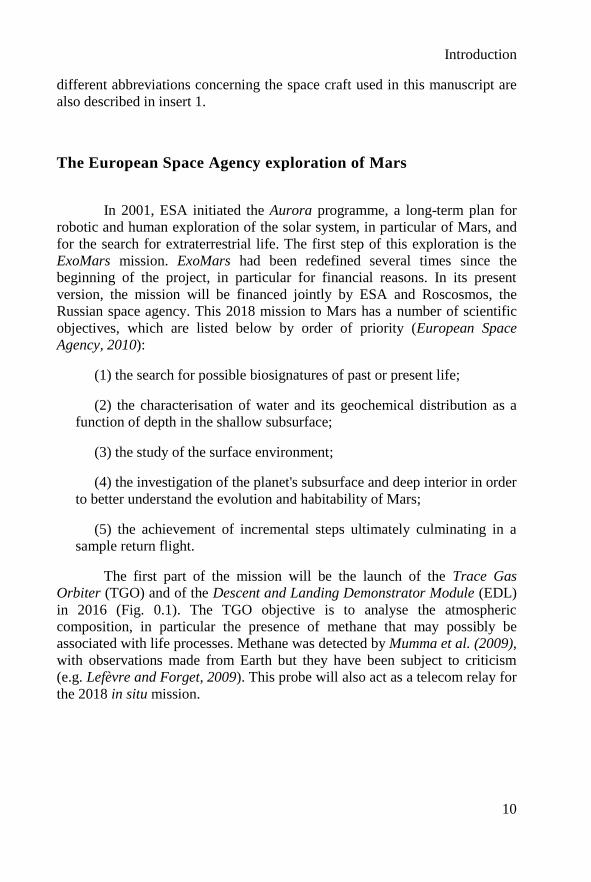
Insert 1: Martian space missions.
More than 40 missions have been launched to explore Mars. After
Viking 1 and 2, launched by the NASA in 1975, scientists had to wait a long time
for new observations of the geology of Mars. In this insert, I list previous missions and instrumentation used to study the Martian geology and the data of
which are the basis of this thesis. The missions to Mars consist of two types:
orbital and surface missions.
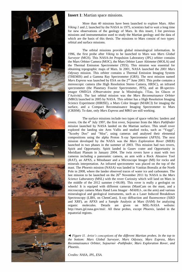
The orbital missions provide global mineralogical information. In 1996, the first probe after Viking to be launched to Mars was Mars Global
Surveyor (MGS). This NASA-Jet Propulsion Laboratory (JPL) orbiter contains
the Mars Orbiter Camera (MOC), the Mars Orbiter Laser Altimeter (MOLA) and the Thermal Emission Spectrometer (TES). This mission was essential for
obtaining topographic maps of Mars. In 2001, NASA launched the 2001 Mars
Odyssey mission. This orbiter contains a Thermal Emission Imaging System (THEMIS) and a Gamma Ray Spectrometer (GRS). The next mission named
Mars Express was launched by ESA on the 2nd June 2003. This probe contains a
stereoscopic camera (the High Resolution Stereo Camera, HRSC), an infrared spectrometer (the Planetary Fourier Spectrometer, PFS), and an IR-spectro-
imager OMEGA (Observatoire pour la Minéralogie, l’Eau, les Glaces et
l’Activité). The last orbital mission was the Mars Reconnaissance Orbiter (MRO) launched in 2005 by NASA. This orbiter has a High Resolution Imaging
Science Experiment (HIRISE), a Mars Color Imager (MARCI) for imaging the
surface, and a Compact Reconnaissance Imaging Spectrometer to Mars (CRISM). To date, only Mars Express and MRO are still working.
The surface missions include two types of space vehicles: landers and
rovers. On the 4th July 1997, the first rover, Sojourner from the Mars Pathfinder
mission launched by NASA landed on the Martian surface. This small rover explored the landing site Ares Vallis and studied rocks, such as “Yoggi”,
”Scooby Doo” and “Moe”, using cameras and analysed their elemental
compositions using the alpha Proton X-ray Spectrometer (APXS). The next mission developed by the NASA was the Mars Exploration Rover (MER)
launched in two phases in the summer of 2003. This mission had two rovers,
Spirit and Opportunity. Spirit landed in Gusev crater and Opportunity in Meridiani Planum in January 2004. The twin rovers have a mast with two
cameras including a panoramic camera, an arm with a Rock Abrasive Tool
(RAT), an APXS, a Mössbauer and a Microscope Imager (MI) for rocks and minerals interpretation. An infrared spectrometer was placed on the top of the
mast. The Phoenix mission (NASA) was landed in Vastitas Borealis at the North
Pole in 2008, where the lander observed traces of water ice and carbonates. The last mission to be launched on the 26th November 2011 by NASA is the Mars
Science Laboratory (MSL) with the rover Curiosity which will land on Mars in the middle of the 2012 summer (~06.08). This rover is really a geologist on
wheels! It is equiped with different cameras (MastCam on the mast, and a
microscopic camera Mars Hand Lens Imager –MAHLI-, on the arm) and various mineralogical and geological instruments, such as a Laser Induced Breakdown
Spectroscopy (LIBS, on ChemCam), X-ray diffraction and fluorescence (XRD
and XRF), an APXS and a Sample Analysis at Mars (SAM) for analyzing organic molecules. Details are given on MSL-NASA website:
http://mars.jpl.nasa.gov/msl/. All these probes, except Phoenix, landed in the
equatorial regions.
◄ Figure I1. Artist’s conceptions of the different Martian probes. In the top to
the bottom: Mars Global Surveyor, Mars Odyssey, Mars Express, Mars
Reconnaissance Orbiter, Sojourner -Pathfinder, Mars Exploration Rover, and Phoenix.
Credits: NASA, JPL, ESA.
Introduction
12
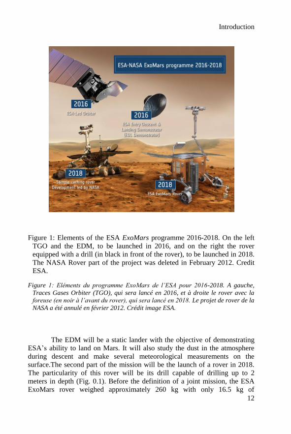
Figure 1: Elements of the ESA ExoMars programme 2016-2018. On the left
TGO and the EDM, to be launched in 2016, and on the right the rover
equipped with a drill (in black in front of the rover), to be launched in 2018.
The NASA Rover part of the project was deleted in February 2012. Credit
ESA.
Figure 1: Eléments du programme ExoMars de l’ESA pour 2016-2018. A gauche,
Traces Gases Orbiter (TGO), qui sera lancé en 2016, et à droite le rover avec la
foreuse (en noir à l’avant du rover), qui sera lancé en 2018. Le projet de rover de la
NASA a été annulé en février 2012. Crédit image ESA.
The EDM will be a static lander with the objective of demonstrating
ESA’s ability to land on Mars. It will also study the dust in the atmosphere
during descent and make several meteorological measurements on the
surface.The second part of the mission will be the launch of a rover in 2018.
The particularity of this rover will be its drill capable of drilling up to 2
meters in depth (Fig. 0.1). Before the definition of a joint mission, the ESA
ExoMars rover weighed approximately 260 kg with only 16.5 kg of
Introduction
13
instruments: the Pasteur payload, consisting of nine instruments located inside
an analytical laboratory as well as outside the rover. As of writing, although it
is planned to add new Russian instruments, all the Pasteur instruments will be
maintained in the new joint mission. Since submission of the thesis, two of the
ExoMars instruments have been deleted, the Mars-XRD and the Life Marker
Chip owing to weight constraints.
The creation of a collection of Mars analogue rocks and minerals which is the
main objective of this thesis will also take into account the Mars Science
Laboratory mission (MSL, NASA), as well as the ExoMars mission.
Creating a rock library for the exploration of Mars
The in situ exploration of planets and other extraterrestrial bodies is
limited by the weight and energy consumption of the scientific payload. This
intrinsic characteristic of space missions necessitates extensive instrument
preparation, including instrument calibration. In order to be relevant,
instrument calibration should ideally use material representative of the object
to be explored. The Mars 2018 mission will not escape this important phase.
However, no sample has yet been returned from Mars and the only samples
from Mars in hand are 85 kg of Martian meteorites. Obviously, due to the
scarcity of these samples, it is not realistic to use these rocks to calibrate the
instruments. Also, meteorites are not necessarily representative of the rocks
that will be analyzed at the landing site.
My doctoral project was undertaken in the Exobiology Group of the
Centre de Biophysique Moléculaire (CBM), in Orléans (France), and forms
part of the activities of the Observatoire des Sciences de l’Univers en Région
Centre (OSUC). This group was founded by André Brack and is regonized for
its expertise in Astrobiology since 1990. The Exobiology Group, and in
particular its actual director, Frances Westall, is widely involved in the
definition of the new payload and the development of the instruments
(CLUPI, PanCam and MicrOmega in particular) for ExoMars.The goal of my
work was thus to create a collection of Mars analogue rocks and minerals in
order to provide suitable samples for the calibration of the Mars 2018 mission
instruments. These same samples are also being used for CheMin and SAM
on MSL. This study forms part of a larger project of space analogue rock
collection in Orléans, called ISAR (for International Space Analogue
Rockstore) and is based on the previous GSPARC (Geological Specimen
Archive) collection developed by Derek Pullan in the course of his PhD thesis
(Pullan, 2008).
Introduction
14
The technical objective of this thesis is to offer to the Martian
community a collection of fully characterized Mars analogue rocks and
minerals, based on a comprehensive and a sound scientific analysis of Martian
geology (e.g. volcanic, hydrothermal, alteration processes…). The main
question is to define the criteria for the selection of analogues, in this case
their similarity to Martian materials, thus optimizing preparation future space
missions.
To begin with, an explanation of the collection project, the
characterization methodology, and the International Space Analogue
Rockstore (ISAR) database and website, which will be presented in the first
chapter. I will also present here the instruments used to characterize the rocks
in this collection, and also the scientific constraints. In the second chapter, the
Martian geology and habitability conditions will be described. This
information is essential for a scientific approach to build up the collection.
Chapter three explains the choice of the samples selected for the first set of
analogues and will give details of their geological characteristics in terms of
structures, textures, mineralogy and elemental compositions. In this section, I
have essentially concentrated on the diversity of rocks and some of the
minerals observed in the Martian surface. In Appendix B, an example of
detailed sample characterization is proposed, all the results are also available
in a CD (3th cover page). The use of analogue terrestrial field sites is also
pertinent to Martian exploration. For the reasons outlined in Chapter IV, I
proposed a new site in Cyprus, where hydrothermal, metasomatic and
meteoric water alteration of basalts has geological characteristics relevant for
early Mars. I can therefore offer a comprehensive study of a suite of rock
samples that come from one locality. This approach allows more individual
samples from a specifically geologically relevant site to be collected, thus
enabling study of context and direct relationships between samples that can
help understand past environments on Mars. In the course of my studies, it
became however apparent that there are limitations in the analogy between
terrestrial and Martian rocks, specifically with respect to basalt compositions
and their Fe- and Mg-contents. I therefore prepared artificial Martian basalts
based on the data provided by the Spirit rover in Gusev Crater. This
preparation is discussed and explained in the fifth chapter. Finally, in
conclusion, I show the characterization of the analogue samples using some of
the ExoMars instruments. I also present the results of a blind test carried out
to check the capability of ExoMars instruments to identify and characterize a
rock. I conclude with future perspectives for this kind of study that includes a
choice of other samples to complete the ISAR collection and possible
artificial weathering experiments to compare with Cyprus samples.

Introduction
15
One of the objectives of my doctoral studies was also the
development of a future flight instrument, cathodoluminescence in
spectroscopy and imagery mode. For the purposes of readability and clarity of
this manuscript, the description of this study will be presented in Appendice
A.

Introduction
16
Introduction
17
Introduction (Résumé Français)
Mars est une planète observée et étudiée depuis très longtemps. A partir des
années 80, l’étude de Mars se focalise principalement sur la recherche d’une vie
extraterrestre. Depuis 1960, une longue série de sondes spatiales a été lancée pour
mieux comprendre Mars et peut être y trouver la vie ou des traces d’une vie ancienne.
Cette exploration débute sur un échec avec Marsnick (Russie, 1960). Les Etats-Unis,
plus chanceux, avec les sondes Viking 1 et 2 lancées en 1975 (NASA), vont être les
premiers à se poser sur Mars. Cette réussite sera suivie par l’envoi de rovers, en
particulier, des Mars Exploration Rover (MER) avec Spirit et Opportunity, en 2003
(NASA) et Mars Express en 2004 par l’Agence Spatiale Européenne (ESA). Le 26
novembre 2011, la mission Mars Science Laboratory (MSL, NASA) avec son
astromobile Curiosity a été lancée pour étudier la géologie de Mars.
Depuis 2001, l’ESA développe son propre programme d’exploration du
système solaire : le programme Aurora. En collaboration avec l’agence spatiale
Russe (Roscosmos), l’ESA propose deux missions d’exploration de Mars dont les
objectifs sont de chercher une vie actuelle ou passée, d’étudier les caractéristiques
géochimiques (en particulier l’eau), de mieux comprendre les conditions
d’habitabilité de Mars et de préparer un retour d’échantillons martiens sur Terre. La
première partie de la mission est la sonde Trace Gase Orbiter (TGO, Fig.1) avec un
module de descente atmosphérique (EDL). Le but est d’étudier la composition de
l’atmosphère martienne. Son lancement est prévu pour 2016. La seconde partie de la
mission, qui doit être lancée en 2018, contient un véhicule appelée Pasteur (Fig. 1).
L’objectif principal de ma thèse est de créer une collection de roches pour
préparer les futures missions martiennes. En effet, l’exploration in situ des planètes et
des corps célestes est limitée par le poids et l’énergie utile aux instruments
scientifiques embarqués. Pour la calibration, il est important d’avoir à disposition
une collection d’échantillons parfaitement caractérisés. Nous ne pouvons pas utiliser
les météorites car elles sont trop précieuses et parfois, non pertinentes. Il est donc
nécessaire de faire un travail de sélection de roches terrestres. Ce travail s’inscrit
dans le projet plus vaste de l’International Space Analogue Rockstore (ISAR) qui ne
se limite pas à Mars. La collection a pour objectif de proposer des échantillons
analogues à une large gamme de corps du système solaire (planètes, astéroïdes, …).
Le premier chapitre sera dédié au projet de l’ISAR, avec la présentation de
la méthodologie utilisée et du site web. Le deuxième chapitre s’intéressera à la
géologie de Mars. Le troisième chapitre présentera la première sélection de roches
analogues. Le quatrième chapitre proposera un site analogue de Mars à Chypre.
Enfin, le cinquième chapitre décrira la fabrication de basaltes analogues de Mars.
Ce travail de thèse avait également pour objectif de développer un futur
instrument spatial, d’imagerie et de spectroscopie de cathodoluminescence. Dans un
souci d’unité et de clarté ce travail est présenté en annexe A

Introduction
18
Chapter I: The International Space Analogue Rockstore
19
Chapter I
The International Space Analogue Rockstore
This chapter presents the International Space Analogue Rockstore
(ISAR) and its associated online database and website, the starting point of
this project. The ISAR is a collection of well-characterized rocks and minerals
that are analogues of extraterrestrial bodies and that are intended to be used in
the testing and calibration of space instrumentation. The ISAR is inspired by
and is a continuation of a project first initiated in 2002 by Derek Pullan during
his thesis with the Geological Specimen Archive, or GSPARC (Pullan, 2008)
concerning the creation of a rock collection for calibration of instruments. The
GSPARC collection contained “generic” samples sorted by colour, texture,
fabric, composition, physical state and size, some of which were analogues of
space bodies, such as Mars or the Moon. In contrast, the ISAR is
characterized by the fact it contains rocks and minerals that are specifically
compositionally similar to those of space bodies in terms of their mineralogy
and geochemistry. I need to build up a new database, because the on-line
publishing of the data could not be made in an easy way. As described in this
chapter, the naming was complicated and did not allow to keep links between
samples. Finally, to develop our own site was simpler for the hosting at the
CNRS in Orléans (internet safety or server of storage of its data for example)
Chapter I: The International Space Analogue Rockstore
20
I.1 The Project
The availability of planetary materials for study in the laboratory is
very limited. For instance, there are only 611 known meteorites from Mars and
only ~500 kg of Moon samples returned during the Apollo and Lunokhot
programs (1969-1972). Analysis of these precious samples is particularly
problematic if the sample preparation method used is destructive. Moreover, it
is unrealistic to use these samples to calibrate and test future space
instruments because of the amount of material that would be needed.
Furthermore, most Martian meteorites are not compositionally representative
of their parent bodies because they are mostly cumulates. The ISAR collection
will thus be mainly composed of terrestrial rocks that can be used in
calibration exercises. In addition, the rockstore includes an online database
where all the textural, structural and compositional characteristics of the rocks
are described.
My thesis corresponds to the beginning of this project. Although I
have participated in the development of the project and the creation of the
database and of the associated website, the main aim of my project is the
analysis and study of the ISAR Mars analogue rocks and minerals. The choice
of Mars was motivated by the future robotic space missions, in particular the
ExoMars-2018 mission as well as MSL described in introduction. These
missions also drove the choice of the analytical methods used for the sample
characterization.
I.2. Which analyses?
I.2.1 Methodological approach
My methodological approach to the analysis of Mars analogue rocks
is essentially based on the investigational strategy of the ExoMars-2018
mission (European Space Agency, 2010) and its Pasteur payload. The rover
will be equipped with a panoramic camera (PanCam2) on the mast to make
high-quality 3D imaging. These views will provide information about the
geomorphological and global context. A High Resolution Camera (HRC) also
on the mast will obtain high quality images of interesting geological targets.
1 All the Martian meteorites are referenced in the following web site: http://www.imca.cc/mars/Martian-
meteorites-list.htm. 2 http://exploration.esa.int/science-e/www/object/index.cfm?fobjectid=45103&fbodylongid=2127
Chapter I: The International Space Analogue Rockstore
21
On the rover, a UHF Radar named WISDOM3 (for Water Ice and Subsurface
Deposit Observation on Mars) will scan the subsurface down to two or three
meters. A CLose-UP Imager (CLUPI) placed near the head of the Pasteur drill
will be able to observe rock outcrops, the soils, drill fines, and the drill cores
before they are ground up for analysis by instruments in the enclosed
laboratory. CLUPI is a true colour camera with a very high resolution (7 µm
per pixel at 20 cm). The drill will take samples from up to two meters in
depth. This will allow analysis of unweathered samples protected from UV
radiation and oxidation (Kminek and Bada, 2006) in the hopes of detecting
hypothetical organic traces of life. The drill will include a miniaturised
camera and an infrared (IR) spectrometer named MA_MISS4 for borehole
investigations. The drill will deliver the sample to a crusher in order to obtain
a powder with a grain size distribution smaller than 250 µm. The powdered
samples will be deposited by a dosing station into a carousel and distributed to
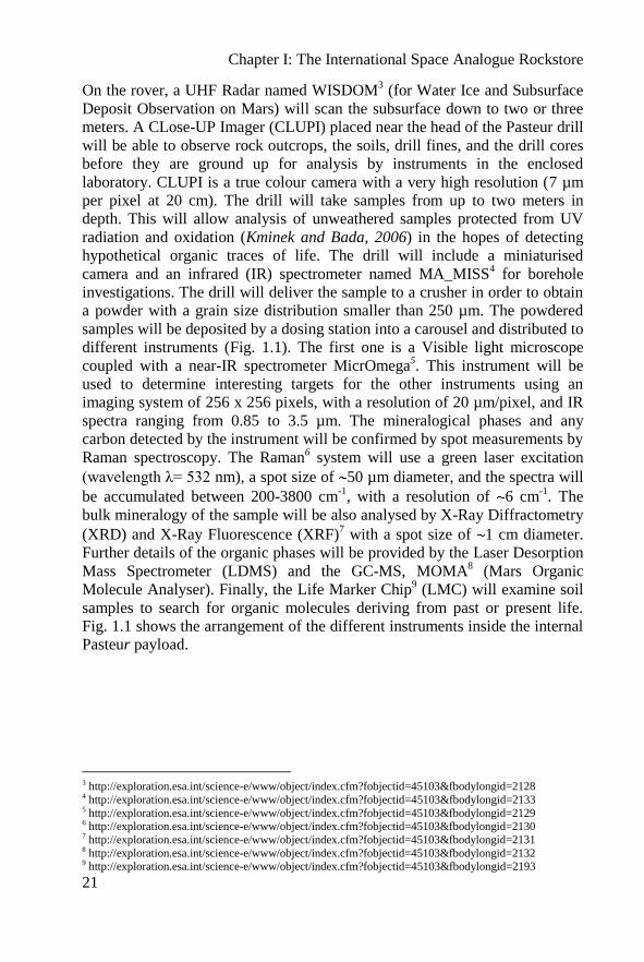
different instruments (Fig. 1.1). The first one is a Visible light microscope
coupled with a near-IR spectrometer MicrOmega5. This instrument will be
used to determine interesting targets for the other instruments using an
imaging system of 256 x 256 pixels, with a resolution of 20 µm/pixel, and IR
spectra ranging from 0.85 to 3.5 µm. The mineralogical phases and any
carbon detected by the instrument will be confirmed by spot measurements by
Raman spectroscopy. The Raman6 system will use a green laser excitation
(wavelength λ= 532 nm), a spot size of 50 µm diameter, and the spectra will
be accumulated between 200-3800 cm-1
, with a resolution of 6 cm-1
. The
bulk mineralogy of the sample will be also analysed by X-Ray Diffractometry
(XRD) and X-Ray Fluorescence (XRF)7 with a spot size of 1 cm diameter.
Further details of the organic phases will be provided by the Laser Desorption
Mass Spectrometer (LDMS) and the GC-MS, MOMA8 (Mars Organic
Molecule Analyser). Finally, the Life Marker Chip9 (LMC) will examine soil
samples to search for organic molecules deriving from past or present life.
Fig. 1.1 shows the arrangement of the different instruments inside the internal
Pasteur payload.
3 http://exploration.esa.int/science-e/www/object/index.cfm?fobjectid=45103&fbodylongid=2128 4 http://exploration.esa.int/science-e/www/object/index.cfm?fobjectid=45103&fbodylongid=2133 5 http://exploration.esa.int/science-e/www/object/index.cfm?fobjectid=45103&fbodylongid=2129 6 http://exploration.esa.int/science-e/www/object/index.cfm?fobjectid=45103&fbodylongid=2130 7 http://exploration.esa.int/science-e/www/object/index.cfm?fobjectid=45103&fbodylongid=2131 8 http://exploration.esa.int/science-e/www/object/index.cfm?fobjectid=45103&fbodylongid=2132 9 http://exploration.esa.int/science-e/www/object/index.cfm?fobjectid=45103&fbodylongid=2193
Chapter I: The International Space Analogue Rockstore
22
Figure 1.1: The Pasteur internal payload showing the different instruments.
Modified from ESA.
Figure 1.1: Schéma des différents instruments internes du rover Pasteur. Modifié
d’après l’ESA.
Presently, discussions about the Russian participation in the mission
may involve additional Russian instruments that migth include an arm
equipped with a microscope and other instruments such as an Alpha Particle
X-ray Spectrometer (APXS) similar to the one of MSL, a Raman probe, a
Mössbauer ...
The instruments on Curiosity (MSL) are also of interest in the
preparation of samples for ISAR. These instruments include different
cameras: Mast Cam (multispectral true colour camera), MAHLI (mounted to
the arm on the rover to acquire microscopic images) and MARDI (descent
camera); Chemcam, including a Laser-induced breakdown spectroscopy
(LIBS); an Alpha-particle X-ray spectrometer (APXS, such as the instrument
on the MERs); and CheMin, for measured chemistry and mineralogy with
XRD and XRF. Other instruments are associated to the MSL rover such as
Radiation assessment detector (RAD); the Dynamic Albedo of Neutrons
(DAN) for measuring hydrogen or ice and water and a meteorological
package with Rover environmental monitoring station (REMS).
Chapter I: The International Space Analogue Rockstore
23
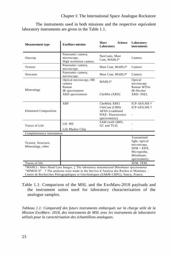
The instruments used in both missions and the respective equivalent
laboratory instruments are given in the Table 1.1.
Measurement type ExoMars mission Mars Science
Laboratory
Laboratory
instruments
Outcrop Panoramic camera, microscope,
High resolution camera.
NavCams, Mast
Cam, MAHLI* Camera
Texture Panoramic camera,
microscope. Mast Cam, MAHLI* Camera
Structure Panoramic camera,
microscope. Mast Cam, MAHLI* Camera
Mineralogy
Optical microscopy, HR
camera MAHLI*
Optical
microscopy Raman Raman WITec
IR spectrometer IR-Nicolet
XRD spectrometer CheMin (XRD) XRD- INEL
Elemental Composition
XRF CheMin( XRF) ICP-AES,MS †
CheCam (LIBS) ICP-AES,MS †
APXS (combined
PIXE- Fluorescence
spectrometry)
-
Traces of Life LD- MS
SAM (with QMS;
GC and TLS) -
Life Marker Chip -
Complementary instruments
Texture, Structure,
Mineralogy, other
Transmitted
light, optical microscopy,
SEM + EDX,
Microprobe,
Mössbauer,
spectrometry.
Traces of life SEM, TEM.
*MAHLI : Mars Hand Lens Imager, ‡ The laboratory miniaturized Mössbauer spectrometer
“MIMOS II” . † The analyses were made in the Service d’Analyse des Roches et Minéraux –
Centre de Recherches Pétrographiques et Géochimiques (SARM-CRPG), Nancy, France.
Table 1.1: Comparison of the MSL and the ExoMars-2018 payloads and
the instrument suites used for laboratory characterisation of the
analogue samples.
Tableau 1.1: Comparatif des futurs instruments embarqués sur la charge utile de la
Mission ExoMars- 2018, des instruments de MSL avec les instruments de laboratoire
utilisés pour la caractérisation des échantillons analogues.
Chapter I: The International Space Analogue Rockstore
24
I used high performance laboratory instrumentation to provide the
similar analyses to the ones provided by space instruments, as well as
analytical methods not used in space, such as optical microscopy of thin
sections, scanning electron microscopy (SEM), electron microprobe and
cathodoluminescence, and elemental analysis by Inductively Coupled Plasma
Atomic Emission and Mass Spectrometer (ICP-AES, MS). To determine the
level of oxidation of rock-forming elements, I used Mössbauer spectrometry.
I.2.2. Sample Preparation
The samples were observed and analysed in a number of formats. At
the outcrop and hand specimen scale, rough rock surfaces of freshly collected
samples and/or of sawn rock samples were used. Rock fragments exhibiting
weathered and fresh surfaces were used for Mössbauer spectrometry (MBS).
Standard polished thin sections (30 µm-thick) were prepared at CNRS-ISTO-
Orléans laboratory, for optical microscopy, Raman spectroscopy,
cathodoluminescence, SEM, energy-dispersive X-ray spectroscopy (EDX),
and electron microprobe studies. Powdered rock samples (<100 µm grain
size) were prepared for Raman spectroscopy, infrared spectroscopy (IR), X-
Ray Diffraction (XRD), and ICP analysis with an electrical and then a manual
agate mortar. Sieved powders with a grain size <250 µm were also prepared
because this will be the powder size produced by the sample preparation
system on the ExoMars-2018 mission. A flow diagram of the preparation
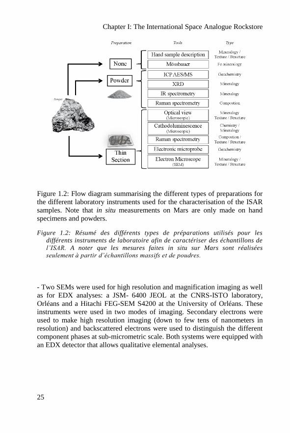
procedure is displayed in Fig. 1.2.
I.2.3. Instrumentation
- An Olympus E410 camera with characteristics similar to that of CLUPI was
used to image the outcrops and the hand samples using various objectives.
The outcrops images could be compared with the ones provided by HRC.
- An optical microscope Olympus BX51 was used to analyse the thin sections
in transmitted, reflected and polarized-analysed light to distinguish and
identify the different minerals and the possible traces of life contained in the
sample at the microscopic scale (if they are large enough).
Chapter I: The International Space Analogue Rockstore
25
Figure 1.2: Flow diagram summarising the different types of preparations for
the different laboratory instruments used for the characterisation of the ISAR
samples. Note that in situ measurements on Mars are only made on hand
specimens and powders.
Figure 1.2: Résumé des différents types de préparations utilisés pour les
différents instruments de laboratoire afin de caractériser des échantillons de
l’ISAR. A noter que les mesures faites in situ sur Mars sont réalisées
seulement à partir d’échantillons massifs et de poudres.
- Two SEMs were used for high resolution and magnification imaging as well
as for EDX analyses: a JSM- 6400 JEOL at the CNRS-ISTO laboratory,
Orléans and a Hitachi FEG-SEM S4200 at the University of Orléans. These
instruments were used in two modes of imaging. Secondary electrons were
used to make high resolution imaging (down to few tens of nanometers in
resolution) and backscattered electrons were used to distinguish the different
component phases at sub-micrometric scale. Both systems were equipped with
an EDX detector that allows qualitative elemental analyses.
Chapter I: The International Space Analogue Rockstore
26
- A WITec Alpha500 RA Raman spectrometer, CBM, Orléans, working with
a green laser (Nd:YAG frequency doubled, λ=532 nm; similar to that used for
the ExoMars-2018 mission, Rull-Pérez and Martinez-Frias, 2006), was used
to obtain mineralogical information. The method is based on the inelastic
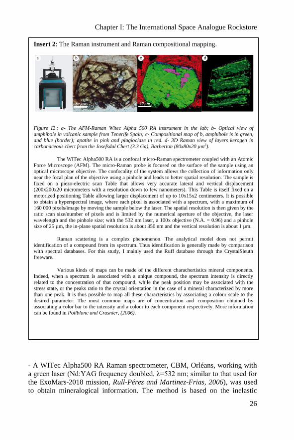
Insert 2: The Raman instrument and Raman compositional mapping.
Figure I2 : a- The AFM-Raman Witec Alpha 500 RA instrument in the lab; b- Optical view of
amphibole in volcanic sample from Tenerife Spain; c- Compositional map of b, amphibole is in green, and blue (border); apatite in pink and plagioclase in red. d- 3D Raman view of layers kerogen in
carbonaceous chert from the Josefsdal Chert (3.3 Ga), Barberton (80x80x20 µm3).
The WITec Alpha500 RA is a confocal micro-Raman spectrometer coupled with an Atomic
Force Microscope (AFM). The micro-Raman probe is focused on the surface of the sample using an optical microscope objective. The confocality of the system allows the collection of information only
near the focal plan of the objective using a pinhole and leads to better spatial resolution. The sample is
fixed on a piezo-electric scan Table that allows very accurate lateral and vertical displacement (200x200x20 micrometers with a resolution down to few nanometers). This Table is itself fixed on a
motorized positioning Table allowing larger displacement of up to 10x15x2 centimeters. It is possible
to obtain a hyperspectral image, where each pixel is associated with a spectrum, with a maximum of 160 000 pixels/image by moving the sample below the laser. The spatial resolution is then given by the
ratio scan size/number of pixels and is limited by the numerical aperture of the objective, the laser
wavelength and the pinhole size; with the 532 nm laser, a 100x objective (N.A. = 0.96) and a pinhole size of 25 µm, the in-plane spatial resolution is about 350 nm and the vertical resolution is about 1 µm.
Raman scattering is a complex phenomenon. The analytical model does not permit
identification of a compound from its spectrum. Thus identification is generally made by comparison
with spectral databases. For this study, I mainly used the Ruff database through the CrystalSleuth freeware.
Various kinds of maps can be made of the different charachertistics mineral components.
Indeed, when a spectrum is associated with a unique compound, the spectrum intensity is directly
related to the concentration of that compound, while the peak position may be associated with the stress state, or the peaks ratio to the crystal orientation in the case of a mineral characterized by more
than one peak. It is thus possible to map all these characteristics by associating a colour scale to the
desired parameter. The most common maps are of concentration and composition obtained by associating a color bar to the intensity and a colour to each component respectively. More information
can be found in Poilblanc and Crasnier, (2006).

Chapter I: The International Space Analogue Rockstore
27
Raman scattering allowing identification of a compound from the vibration of
its atomic bonds. The identification is made from the diffraction spectrum of
the scattered beam given by the CCD camera of the spectrometer, expressed
as CCD counts versus wavenumber (in cm-1
relative to the incident laser
wavenumber). The instrument used allows the sample to be scanned in order
to make compositional mapping. Each pixel in a map is associated with a
spectrum (hyperspectral data). This is a non destructive method that does not
necessitate any sample preparation. Both this instrument and optical
microscopy were fundamental techniques for the characterization of the ISAR
samples. More details on the instrument are displayed in Insert 2.
- A Nicolet IR spectrometer, 400-4000 nm, in transmitted mode at the CNRS-
ISTO laboratory, Orléans, and a Nicolet Magna IR 760 spectrometer ESP
associated with a Thermo Scientific Integrations Sphere near IR Nicolet 6700
FT-IR, at the University of Poitiers were used for mineralogical identification.
IR spectrometry is a key technique in Martian exploration; the Omega
instrument of Mars Express (ESA) and CRISM on the Mars Reconnaissance
Orbiter (NASA) have provided much information on the mineralogy of the
surface of Mars, in particular phyllosilicate mineralogy (e.g. Poulet et al.,
2005; Bibring et al.,2006; Mustard et al., 2009; Ehlmann et al., 2011;and
Chapter II). IR is highly complementary to Raman spectroscopy.
- Two X-ray instruments were used for mineral identification: an INEL
XRM3000/CPS120 X-ray diffractometer in transmitted mode, at the CNRS-
ISTO laboratory, Orléans, and a Brucker D8 Advance diffractometer in
reflected mode, at the Hydrasa laboratory, University of Poitiers. This
technique measures the lattice parameters of crystals and thus allows their
identification. This technique is mostly used on powdered samples. Powders
were analysed, respectively, for the first instrument in a 0.3 mm capillary
using a beam emitted by a Co generator and for the second in a powder paste
using a Cu generator (λCoKα = 1.78897 Å, λCuKα = 1.541838 Å).
- A laboratory version of the miniaturized Mössbauer spectrometer “MIMOS
II”, at the Johannes Gutenberg Universität-Mainz, Germany, was used to
determine the level of oxidation of minerals in the rocks. Spectra were
obtained in backscattered geometry with a field of view of 15 mm. This
instrument is part of the payload of the two Mars Exploration Rovers
(Klingelhöfer et al., 2003).
- A Cameca SX-50 electron microprobe at the ISTO-BRGM laboratory,
Orléans, was also used to determine the elemental composition of specific
minerals.
Chapter I: The International Space Analogue Rockstore
28
- Cathodoluminescence was used to study the presence of trace elements (e.g.
Mn in carbonates) and detection of vacancies in minerals (e.g. lattice defects
related to electrostatic charging phenomena during electron irradiation). This
technique also permits observation of crystal growth microstructures. The
instrument used has been developed in Orléans with the aim of developing
such an instrument for future space exploration (Thomas et al., 2009). One of
the objectives of my research was to contribute to the optimization of this
instrument. For the clarity of this manuscript, this project and the significant
advances made during the last three years are presented in Appendice A.
This list of equipment is not exhaustive but it corresponds to those mostly
used. Other types of analyses have and will be made on the ISAR samples.
I.3. Nomenclature and relationship between samples
The GSPARC system, on which the ISAR project is based, attributed
a complex name to each sample. Although this system was suitable for the
classification of a huge quantity of individual samples, it was not useful for
the ISAR collection where the link between samples and subsamples is
important. Thus, we decided to use our own nomenclature that takes into
account the heredity and the relationship between the samples, in the manner
of a family tree.
Fragmentation of the parent sample during preparations produced
many smaller pieces, most of which will be probably never studied (the
cutting wastes for instance). Moreover, the samples could be sand or gravel.
The referencing of each particle or grain would then be unnecessary. The
chosen solution has been to define two types of samples: the initial parent
samples and the sub-samples. Parent samples correspond to those taken in the
field. They may be composed of several pieces of rocks, sand, gravel, etc.
They can be seen as “a box” containing all the pieces originating from the
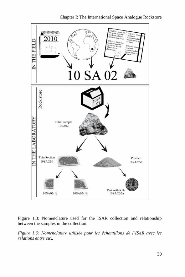
same place and taken at the same time (Fig. 1.3). The name of the initial
samples consists of two numbers for the year, two letters for the originating
country/locality and two numbers for the order of sampling during the field
trip. For example: 10SA02 is the second sample taken in 2010 in South
Africa. Some parts of the initial samples are then prepared for future analyses
in powder form or as thin sections. The prepared samples are labeled by
adding a dash and a number to the name of the initial sample, for example:
10SA02-1. The cutting waste is not referenced and is replaced back in the box
of the initial sample. Samples can also be split into subsamples. These sub-
samples are named by adding a letter at the end of the original sample
Chapter I: The International Space Analogue Rockstore
29
reference. They can themselves be further split and the additional subsamples
will be named by adding a number at the end. For example: 10SA02-1a then
10SA02-1a1… This relationship between the samples can be represented as
the family tree shown in Fig. 1.3.
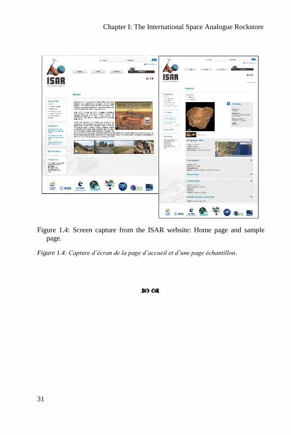
I.4 The ISAR database
The aim of the ISAR is to propose samples for calibrating space
instruments for any in situ mission. For this reason, all the sample data and
associated analyses are displayed in a web-based database: www.isar.cnrs-
orleans.fr. The website was first developed by Frédéric Foucher (CNRS-
CBM) in html and php and the associated database was developed in MySQL.
The nomenclature of the samples was also chosen in order to facilitate the
structure of this database. Finally, the website was improved in terms of
ergonomy and design by the company IdWeb10
in Bourges.
The website includes all the results provided by the different
instruments for the samples included in the ISAR collection. The web site
consists of a home page which presents the project and different pages
presenting the team, the equipment, the rockstore, and missions using the
ISAR collection (Fig. 1.4). A newspage gives the main events with which the
ISAR members have been associated or present (e.g. congresses, workshops)
and the latest team publications. The second and main part of the website is
the database, which summarizes all the analytical information obtained with
the different instruments for each sample (Fig. 1.4). After logging in, the user
can download a pdf file containing all the information and relevant figures, as
well as text files with the different spectra and other data. Finally it is possible
to borrow a sample using the lending system.
In order to choose samples, it is necessary to know the geology of
Mars. The next chapter will thus present the geology of Mars and the
possibilities of finding life or traces of fossil life on outcrops. These
considerations will allow us to make an appropriate analogue sample
selection.
10 More information’s about Idweb in this web site : http://www.idweb.fr/
Chapter I: The International Space Analogue Rockstore
30
Figure 1.3: Nomenclature used for the ISAR collection and relationship
between the samples in the collection.
Figure 1.3: Nomenclature utilisée pour les échantillons de l’ISAR avec les
relations entre eux.
Chapter I: The International Space Analogue Rockstore
31
Figure 1.4: Screen capture from the ISAR website: Home page and sample
page.
Figure 1.4: Capture d’écran de la page d’accueil et d’une page échantillon.

Chapter I: The International Space Analogue Rockstore
32
Chapter I: The International Space Analogue Rockstore
33
The ISAR (Résumé Français)
Ce chapitre a pour objectif de présenter l’ISAR, la collection internationale
de roches analogues à des objets du système solaire. Cette collection est inspirée par
le projet initié par D. Pullan pendant sa thèse en 2008, le « Geological Specimen
Archive ». En effet, il est essentiel de pouvoir disposer d’échantillons analogues pour
tester de futurs instruments de vols. Des échantillons provenant de l’espace sont
disponibles sur Terre, tels que les échantillons lunaires rapportés par les missions
Apollo et Lunokhot, ou encore les météorites martiennes. Cependant ces échantillons
sont beaucoup trop précieux pour être utilisés dans des tests avec des méthodes
destructives.
Mon travail de thèse correspond au début du projet ISAR. J’ai donc
participé au développement du projet avec la création de la base de données et du site
internet. Mais l’objectif principal est l’analyse et l’étude d’échantillons analogues de
Mars. Le choix de Mars est motivé par l’actualité des futures missions robotiques qui
s’intéressent essentiellement à Mars, en particulier MSL et le projet ExoMars.
Ce chapitre définit la stratégie d’analyse et le choix des échantillons. Les
analyses faites sur les échantillons analogues se basent essentiellement sur la future
mission ExoMars et sa charge utile : Pasteur. Ce véhicule est équipé de nombreux
instruments comme des caméras (PanCam et HRC). Un radar UHF (WISDOM).
Une« loupe » (CLUPI) fixée au sommet de la foreuse pour observer le sol et les
échantillons prélevés. La foreuse contient un petit spectromètre IR (MA_MISS). La
petite carotte prélevée par la foreuse, sera broyée puis envoyée à l’intérieur du
véhicule, dans le laboratoire scientifique (Fig. 1.1). Le laboratoire est composé de
DRX, XRF, Raman, IR, Life Marker Chip et LDMS. Ces instruments vont examiner
l’échantillon pour le caractériser d’un point de vue pétrologique et organique
(recherche de traces de vie fossile ou actuelle). Actuellement, la liste des instruments
est en cours de discussion avec l’agence spatiale Russe Roscosmos en vue d’une
collaboration pour cette mission. Pour ce travail, nous prendrons aussi en compte les
instruments de MSL. La liste des instruments que nous utiliserons est donnée dans le
Tableau 1.1.
Pour cette étude et pour la lithothèque ISAR, les échantillons sont préparés
de différentes manières : aucune préparation, en poudre ou en lame mince. Notons
que les lames minces ne sont pas des préparations utilisées par les futures missions
spatiales, mais restent la base du travail du géologue. La procédure de préparation
pour chaque instrument est résumée dans la Fig. 1.2.
Pour cette étude, nous utiliserons une large gamme d’instruments comme
l’appareil photo, le microscope optique polarisant, le MEB, les spectrométries Raman
et IR, la DRX, le Mössbauer, l’ICP-MS/OES des éléments mineurs et majeurs, la
microsonde électronique et la cathodoluminescence. Cet instrument, potentiellement
spatialisable et utilisé sur Mars fait l’objet d’une description plus précise dans
l’annexe A. Le détail de chaque instrument n’est pas repris dans ce résumé, consultez
le paragraphe I.2.
Chapter I: The International Space Analogue Rockstore
34
Nous utiliserons dans l’ensemble de cette thèse la nomenclature de
référencement des échantillons développée pour l’ISAR. Le code de l’échantillon
initial se compose de deux chiffres pour l’année (exemple 10 pour l’année 2010) suivi
de deux lettres symbolisant le pays (exemple SA pour South Africa soit Afrique du
Sud) et de deux chiffres pour l’ordre de prélèvement sur le terrain. Lorsque des
échantillons fils sont préparés (comme une lame mince ou une poudre) un chiffre est
ajouté puis une lettre puis un chiffre etc…. La nomenclature est reprise sur la Fig.
1.3.
Enfin, la base de données de l’ISAR a été développée à l’origine par
Frédéric Foucher puis améliorée parl’entreprise IdWeb et associée au maintien du
site web du projet: www.isar.cnrs-orleans.fr. Le détail des informations sur chaque
échantillon est disponible en ligne avec plusieursniveaux de droits d’accès. L’accès
sécurisé, réservé aux utilisateurs enregistrés, permet de télécharger l’ensemble des
données au format pdf. La Fig. 1.4 correspond à des captures d’écran du site web
(page d’accueil et page échantillon).
Chapter II: Mars
35
Chapter II
Mars
The red planet was named after the Roman god of war. Mars is the
fourth planet and the farthest of the telluric planets from the Sun in the Solar
System. It formed during planet accretion 4.564 billion years ago. Its orbit is
eccentric, between 1.38 and 1.66 AU and it has four seasons. Mars is a small
planet, only 1/10 of the Earth’s mass and its radius is approximately half of
the Earth’s radius (3380 km of equatorial diameter). Finally, Mars has two
small moons with irregular surfaces, Phobos and Deimos.
II.1. Geological setting
The internal structure of Mars is still unknown but orbital
measurements of its properties indicate that it has a core and possibly a
differentiated mantle (review in Dehant, 2010). However, no geological
activity has been observed on the planet today (Carr and Head, 2009). The
Martian surface is characterized by a global dichotomy: in the south,
highlands with very large craters (e.g. Hellas Planitia crater, 2,200 km in
diameter and 9.5 km of depth, it is the largest visible crater in the Solar
Chapter II: Mars
36
system) and in the north, a smoother surface which is 4 km deeper than the
southern highlands. The landscape is characterized by very large volcanoes
(e.g. Olympus Mons at 26 km altitude is the highest volcano in the solar
system), large canyons (e.g. Valles Marineris is more than 4,000 km-long,
200 km-wide and up to 7 km-deep) and numerous craters. Due to the absence
of plate tectonics, almost all impact craters have been preserved through
geological time, although it appears that craters in the northern hemisphere
have been covered by later lavas flow and sediment deposits (e.g. Mangold et
al., 2009; Le Deit et al., 2011; Dehouck et al., 2010). The preservation of
craters means that their superposition can be used in order to estimate the age
of the different regions and thus subdivide the surface into different
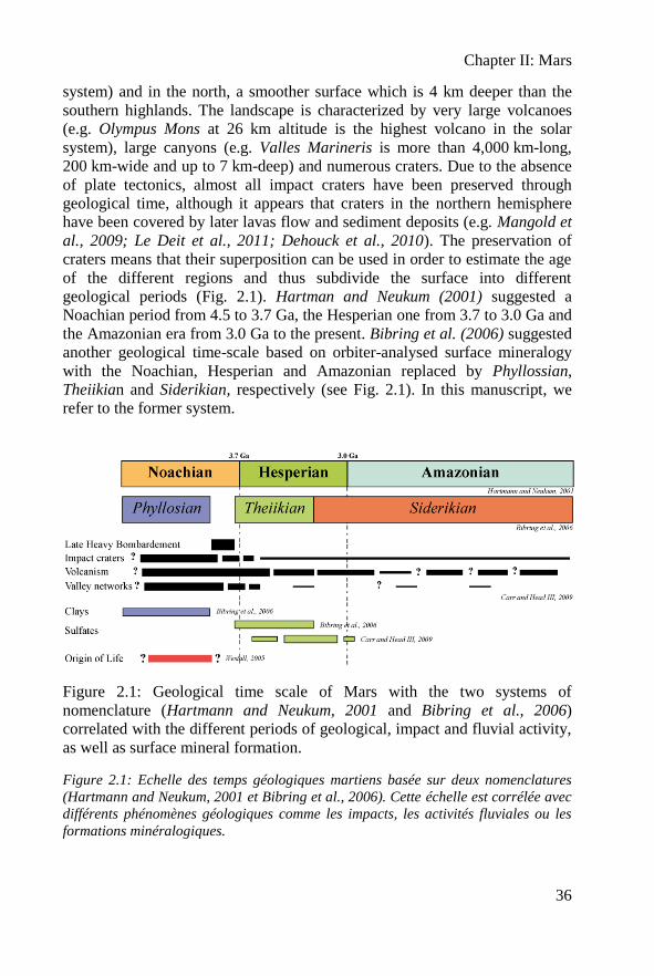
geological periods (Fig. 2.1). Hartman and Neukum (2001) suggested a
Noachian period from 4.5 to 3.7 Ga, the Hesperian one from 3.7 to 3.0 Ga and
the Amazonian era from 3.0 Ga to the present. Bibring et al. (2006) suggested
another geological time-scale based on orbiter-analysed surface mineralogy
with the Noachian, Hesperian and Amazonian replaced by Phyllossian,
Theiikian and Siderikian, respectively (see Fig. 2.1). In this manuscript, we
refer to the former system.
Figure 2.1: Geological time scale of Mars with the two systems of
nomenclature (Hartmann and Neukum, 2001 and Bibring et al., 2006)
correlated with the different periods of geological, impact and fluvial activity,
as well as surface mineral formation.
Figure 2.1: Echelle des temps géologiques martiens basée sur deux nomenclatures
(Hartmann and Neukum, 2001 et Bibring et al., 2006). Cette échelle est corrélée avec
différents phénomènes géologiques comme les impacts, les activités fluviales ou les
formations minéralogiques.
Chapter II: Mars
37
Not all the Martian surface has been observed but it seems to be
essentially composed of volcanic rocks associated with different sediments
derived from basaltic alteration and weathering. A major part of the surface is
covered by several meters of dust and detritus, named regolith. We do not
here describe and discuss the regolith. The terrain description is based on the
data from the Mars Global Surveyor (MGS), MRO and Mars Express orbiters,
the Phoenix lander and Spirit and Opportunity rovers.
II.1.1. Igneous rocks
The data of the Thermal Emission Spectrometer (TES) of MGS was
used by Bandfield (2002) to propose two major categories of surface
mineralogy. The first one consists of plagioclases, high-Ca pyroxenes, sheet
silicates/high-Si glass and hematite, suggesting a basaltic composition. The
second one also consists of plagioclase, and silicate sheets/high-Si glass but
its low content in high-Ca pyroxene is more consistent with andesite. This
interpretation was based on measurements made during the Mars Pathfinder
mission at the landing site Chryse Planitia in Ares Vallis in 1997 (Wänke et
al., 2001). However, the more Si-rich composition has been re-interpreted and
explained as being due to measurements made on the altered surface of the
basalts, since the top layer of the analyzed rocks was not removed before
analysis, as it was done later during the MER missions (Bandfield et al., 2004;
Christensen et al., 2005).
Analyses of rocks made in the Gusev Crater by Spirit indicate that,
although Martian basalts are relatively similar in composition to the terrestrial
ones, they have higher MgO (8 to 10 wt%), FeO (15 to 18 wt%) and Cl (>0.70
wt%) (Gellert et al., 2006). McSween et al. (2009) compared all data from
Martian meteorites, rovers (Mars Pathfinder and the MERs) and orbit (TES of
MGS and the Gamma Ray Spectrometer (GRS) of Mars Odyssey). In
particular they plotted all these compositions within the classical total alkali-
silica diagram (TAS) shown in Fig. 2.2a. This diagram plotting silica
composition against alkali elements (Na2O and K2O) is essential for
determining the classification of volcanic rocks. It should be noted that,
contrary to in situ analyses, basalt compositions measured from orbit
correspond to average values made over large areas all over the planet.
Moreover, analyses made in situ on the ground by the MERs were obtained
from the abraded surfaces of massive rocks that were either unaltered or less
altered than the weathered rock surfaces studied from orbit and by Mars
Pathfinder. This difference explains the offset of the MER data with respect to
the others ones (McSween et al., 2009). Most MER analyses fall within the
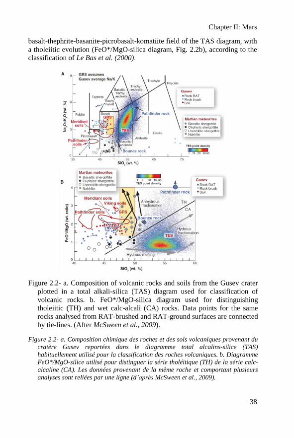
Chapter II: Mars
38
basalt-thephrite-basanite-picrobasalt-komatiite field of the TAS diagram, with
a tholeiitic evolution (FeO*/MgO-silica diagram, Fig. 2.2b), according to the
classification of Le Bas et al. (2000).
Figure 2.2- a. Composition of volcanic rocks and soils from the Gusev crater
plotted in a total alkali-silica (TAS) diagram used for classification of
volcanic rocks. b. FeO*/MgO-silica diagram used for distinguishing
tholeiitic (TH) and wet calc-alcali (CA) rocks. Data points for the same
rocks analysed from RAT-brushed and RAT-ground surfaces are connected
by tie-lines. (After McSween et al., 2009).
Figure 2.2- a. Composition chimique des roches et des sols volcaniques provenant du
cratère Gusev reportées dans le diagramme total alcalins-silice (TAS)
habituellement utilisé pour la classification des roches volcaniques. b. Diagramme
FeO*/MgO-silice utilisé pour distinguer la série tholéitique (TH) de la série calc-
alcaline (CA). Les données provenant de la même roche et comportant plusieurs
analyses sont reliées par une ligne (d’après McSween et al., 2009).
Chapter II: Mars
39
Finally, it has been shown that Martian meteorites are stony
meteorites that are not really representative of the crust (McSween et al.,
2009). They have been divided into three groups: Shergottites, Nakhlites, and
Chassignites, named after the locations where the first meteorite of each class
was discovered. They are collectively referred to as the SNC group
meteorites. Most meteorites are cumulates; Chassignites are dunites, Nakhlites
and the famous ALH84001 are clinopyroxenites, and a large part of the
Shergottites are “lherzolitic Shergottites”, a variety of peridotite. Only some
Shergottites are “basaltic Shergottites”, for example the Los Angeles
meteorite found in 1999 (Rubin et al., 2000). Nevertheless, they plot
separately from the basalts observed in Gusev crater in the TAS diagram in
Fig. 2.2a.
The basaltic composition of Adirondock, Humphrey and Mazatzal
rocks measured by Spirit in the Gusev Crater was discussed by Gellert et al.
(2004) and McSween et al. (2004). It is interpreted in terms of normative
anorthite, fayalite-forsterite, diopside, hypersthene and some chromite,
magnetite, ilmenite and apatite. On the TAS diagram of volcanic rocks, most
of the Gusev basalts fall in the silica-poor side of the basaltic field, with rare
tephrite samples overlapping the picrobasalt field (Fig. 2.2a). The
compositions of the Gusev rocks and soils, Martian meteorites, and the GRS
data, calculated by neglecting the volatile elements, also indicate that the crust
is dominated by basalts. These basalts show no evidence of fractionation and
are very primitive (McSween et al., 2009). There is some evidence of
komatiites on the Martian surface (Reyes and Christensen, 1994; Nna-
Mvondo and Martinez-Frias, 2007).
As noted above, McSween et al. (2009) suggested a different
interpretation for the andesite detected based on the TES data. Bandfield et al.
(2004) previously interpreted andesitic or basalt-andesitic compositions based
on the interpretation of a feldspar, pyroxene and quartz mineral association
(SiO2 concentrations of 62 wt%; see Fig. 2.2). However, data related to iron
oxidation were not available at that time (Taylor et al., 2008) and, moreover,
the original analyses were made on raw rock surfaces. McSween et al. (2009)
therefore, explained the high silica-content by chemical weathering of the
basalts (palagonitisation, i.e., the devitrification of a basaltic glass under the
influence of water to form palagonite). This phenomenon was probably
relatively common on early Mars, when liquid water was still present.
As cumulates, most Martian meteorites are more representative of the
Martian mantle than of its surface. On Earth, typical plutonic rocks include
granites but the evidence for granites on Mars is weak (Taylor et al., 2008).
Although SNC meteorites are mostly pryoxenites and peridotites, they do
Chapter II: Mars
40
contain interstitial acidic glasses and are characterized by isotopic
compositions that suggest the existence of components having a composition
similar to that of the terrestrial granitic continental crust (Bonin, 2011; Bonin,
2012). In the Tharsis area, rhythmically layered deposits observed on the
flanks of the giant volcanoes, including Olympus Mons, evoke alternating
basalt flows and acidic pyroclastites (Bonin, 2011). Finally, infrared spectra in
some impact craters correspond to compositions close to granites and
trondhjemites (Bandfield et al., 2004; Christensen et al., 2005).
II.1.2. Sedimentary rocks
Although less abundant than basalts, sedimentary rocks have been
detected on Mars. Most of the sediments are derived from basaltic precursors,
i.e. basaltic sands and altered basaltic detritus (Tosca et al., 2004; Ehlmann et
al., 2009). Water may be responsible for this alteration (e.g. Bibring et al.,
2006; Ehlmann et al., 2009; Viles et al., 2010; Ehlmann et al., 2010). In
particular, the orbital analyses suggest aqueous alteration of the surface
materials, first by neutral to alkaline fluids during the pre-Noachian/Noachian
period, followed by alteration under more acidic conditions from the
Hesperian onwards (Poulet et al., 2005; Bibring et al., 2006). These different
pH conditions resulted in the formation of different types of phyllosilicates,
such as neutral to alkaline Fe-Mg smectites (saponite, nontronite) during the
pre-Noachian/Noachian period and Al-rich smectites during the
Hesperian/Amazonian. It should be noted, however, that phyllosilicates can be
formed by deuteric processes during fractionation within the magmatic
chamber (Meunier al., 2010) and such an hypothesis has been proposed for
unusually thick clay deposits (Meunier et al, 2012). Ehlmann et al. (2011)
have recently reviewed the origin of clays on Mars.
Many very specific types of clays have been interpreted from orbital
measurements including chlorite, vermiculite, nontronite and smectite (review
in Ehlmann et al., 2011). I am somewhat skeptical about these detailed
interpretations because the classification of clays is already difficult on Earth
using advanced instrumentation and it is therefore even more difficult from
orbit. For this reason, I will only use the group clay name in this study.
Other types of sediments occur on Mars. Alteration and silica-
enrichment may also be induced by hydrothermal activity. In particular Ruff et
al. (2011) suggested that the opaline silica identified in laminated and cross-
bedded tephra in the Eastern Valley, between Home Plate and the Mitcheltree/
Low Ridge complex by Spirit (Squyres et al., 2008), could be interpreted as
Chapter II: Mars
41
hot spring sinter deposits associated with volcanic activity. Silica deposits are
visible on the Martian surface and form elongated dykes (Ruff et al., 2011) or
nodules (Squyres et al., 2008). Miliken et al. (2008) place their formation
during the Hesperian to Amazonian epochs.
Evaporitic deposits including jarosite, gypsum, and other hydrated
sulfates associated with hematite have been described in the high latitudes
around the polar ice cap, as well as in other locations at the Martian surface
(e.g.: in Meridiani Planum, McLennan et al., 2005; in Valles Marineris,
Margaritifer Sinus, Terra Meridiani, Gendrin et al., 2005). Small amounts ( 1
wt%) of carbonate minerals have been described from several SNC meteorites
(Gooding, 1992). In particular, Borg et al. (1999) and Treiman et al. (2002)
described carbonates in the 4.5 billion years old ALH84001 meteorite that Mc
Kay et al. (1996) and Thomas-Keptra et al. (2009) interpreted as
mineralogical indications of past life. Carbonate minerals have also been
detected from orbit in the Martian dust (<5 wt%; Bandfield et al., 2003). Mg-
rich carbonates associated with phyllosilicates and olivines were identified in
the Nili Fossae by MRO (Ehlmann et al., 2008).
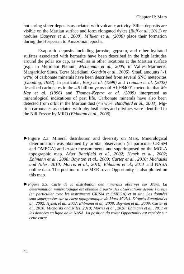
►Figure 2.3: Mineral distribution and diversity on Mars. Mineralogical
determination was obtained by orbital observation (in particular CRISM
and OMEGA) and in-situ measurements and superimposed on the MOLA
topographic map. After Bandfield et al., 2002; Hynek et al., 2002;
Ehlmann et al., 2008; Boynton et al., 2009; Carter et al., 2010; Michalski
and Niles, 2010; Morris et al., 2010; Ehlmann et al., 2011 and NASA
online data. The position of the MER rover Opportunity is also plotted on
this map.
►Figure 2.3: Carte de la distribution des minéraux observés sur Mars. La
détermination minéralogique est obtenue à partir des observations depuis l’orbite
(en particulier avec les instruments CRISM et OMEGA) et in situ. Les données
sont superposées sur la carte topographique de Mars MOLA. D’après Bandfield et
al., 2002; Hynek et al., 2002; Ehlmann et al., 2008; Boynton et al., 2009; Carter et
al., 2010; Michalski and Niles, 2010; Morris et al., 2010; Ehlmann et al., 2011 et
les données en ligne de la NASA. La position du rover Opportunity est repérée sur
cette carte.
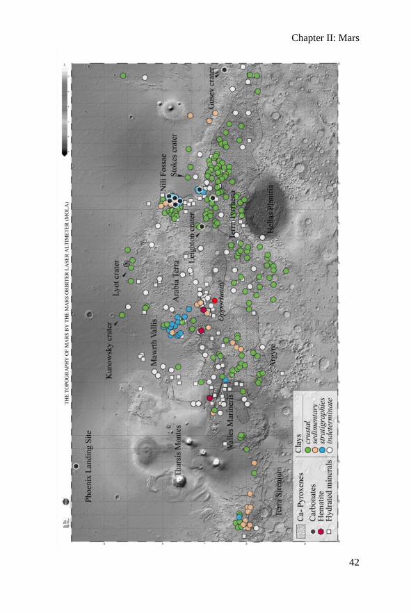
Chapter II: Mars
42
Chapter II: Mars
43
Not far from Nili Fossae, in Leighton crater, Fe- Mg- carbonates (siderite,
magnesite) associated with phyllosilicates (kaolinite group) and Fe- Mg-
bearing silicates have also been detected (Michalski and Niles, 2010). Three
to five percent calcium carbonates were also observed in situ in the soil of
Mars by the Phoenix lander (Boynton et al., 2009). Finally, the most important
evidence of carbonates was made by the Spirit rover in the end of December
2005 in very old volcaniclastic rocks (Hesperian) of the Comanche outcrop, in
the Columbia Hills of Gusev Crater (Morris et al., 2010). Using its
Mössbauer, Spirit detected approximately 26 wt% of Fe-Mg carbonates in the
rock associated with olivine (Fo0.72). Using APXS, Morris et al. (2010)
calculated the mineral composition of this Mg-Fe carbonate as:
Mc0.62Sd0.25Cc0.11Rh0.02, with Mc = magnesite; Sd= siderite; Cc= calcite and
Rh = rhodocrosite.
Figure 2.3 is a non-exhaustive overview of mineral type and locality
on Mars based on orbital measurements (essentially from Omega and CRISM)
and from the in situ measurements (the MER Spirit, Opportunity and
Phoenix).
II.2 Life on Mars
The main objective of the future Martian mission, in particular the
ExoMars mission, is to search for evidence of past or present life. But, what
kind of evidence can we expect? In this subsection, we try to give some
indications mainly based on possible terrestrial analogues of hypothetical
microbial Martian remains.
II.2.1 Mars habitability
A minimal definition of life can be stated by the dynamic interaction
between structures having a semi-permeable boundary and macromolecules
acting as catalysts and templates (Ruiz-Mirazo and Moreno, 2011).
Habitability can be simply resumed as the association the necessary
ingredients for life in a particular location (Brack, 2000; Southam and
Westall, 2007; Hoelhler and Westall, 2010), namely: liquid water; endogenic
and/or exogenic organic molecules for constructing the macromolecules
necessary for cellular life; a source of energy, in the case of Mars chemical
energy from redox reactions on reactive mineral surfaces (cf. Jakosky and
Shock, 1998); and nutrients (H, N, O, P, S, plus transition metals) obtainable
on Mars from aqueous/hydrothermal alteration of the volcanic crust. The
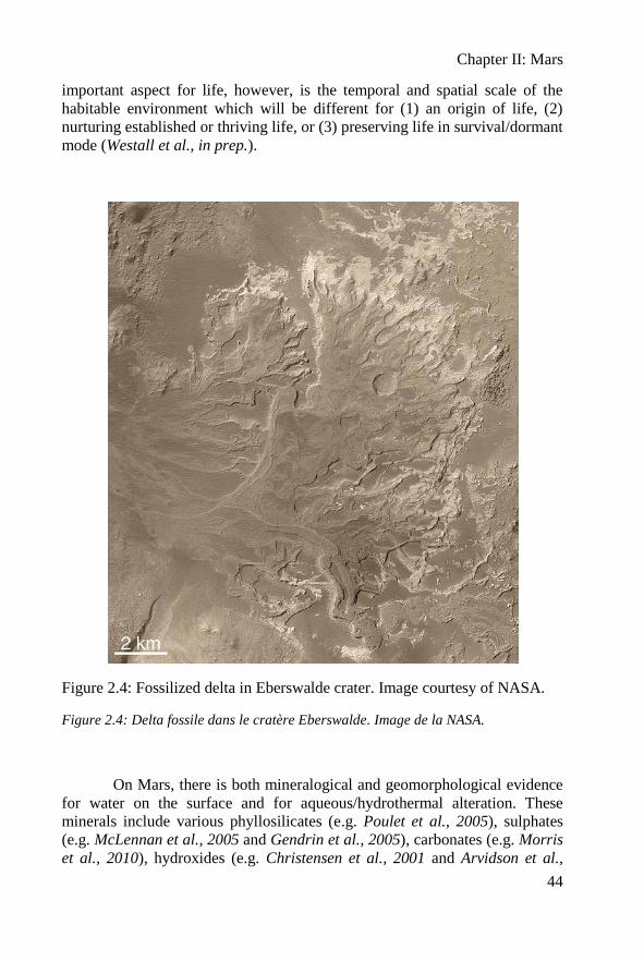
Chapter II: Mars
44
important aspect for life, however, is the temporal and spatial scale of the
habitable environment which will be different for (1) an origin of life, (2)
nurturing established or thriving life, or (3) preserving life in survival/dormant
mode (Westall et al., in prep.).
Figure 2.4: Fossilized delta in Eberswalde crater. Image courtesy of NASA.
Figure 2.4: Delta fossile dans le cratère Eberswalde. Image de la NASA.
On Mars, there is both mineralogical and geomorphological evidence
for water on the surface and for aqueous/hydrothermal alteration. These
minerals include various phyllosilicates (e.g. Poulet et al., 2005), sulphates
(e.g. McLennan et al., 2005 and Gendrin et al., 2005), carbonates (e.g. Morris
et al., 2010), hydroxides (e.g. Christensen et al., 2001 and Arvidson et al.,
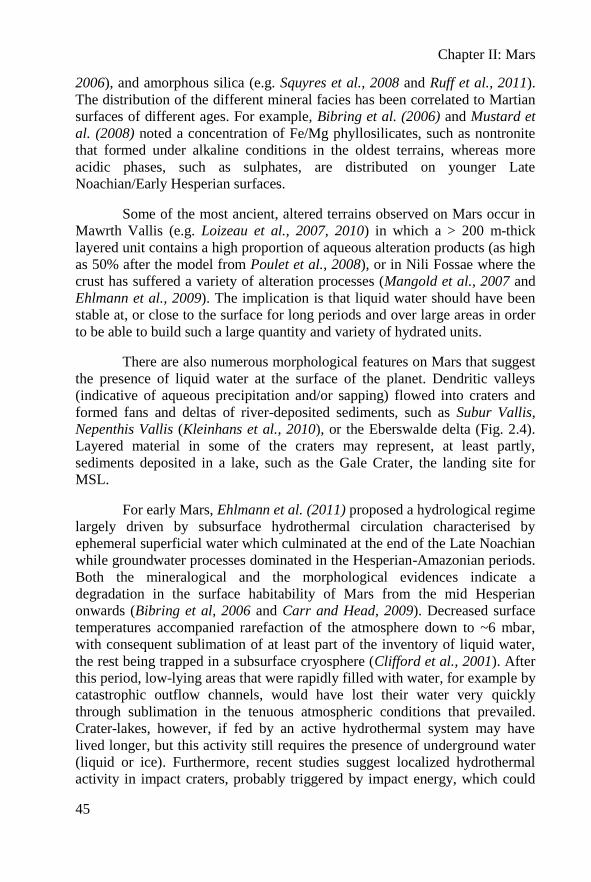
Chapter II: Mars
45
2006), and amorphous silica (e.g. Squyres et al., 2008 and Ruff et al., 2011).
The distribution of the different mineral facies has been correlated to Martian
surfaces of different ages. For example, Bibring et al. (2006) and Mustard et
al. (2008) noted a concentration of Fe/Mg phyllosilicates, such as nontronite
that formed under alkaline conditions in the oldest terrains, whereas more
acidic phases, such as sulphates, are distributed on younger Late
Noachian/Early Hesperian surfaces.
Some of the most ancient, altered terrains observed on Mars occur in
Mawrth Vallis (e.g. Loizeau et al., 2007, 2010) in which a > 200 m-thick
layered unit contains a high proportion of aqueous alteration products (as high
as 50% after the model from Poulet et al., 2008), or in Nili Fossae where the
crust has suffered a variety of alteration processes (Mangold et al., 2007 and
Ehlmann et al., 2009). The implication is that liquid water should have been
stable at, or close to the surface for long periods and over large areas in order
to be able to build such a large quantity and variety of hydrated units.
There are also numerous morphological features on Mars that suggest
the presence of liquid water at the surface of the planet. Dendritic valleys
(indicative of aqueous precipitation and/or sapping) flowed into craters and
formed fans and deltas of river-deposited sediments, such as Subur Vallis,
Nepenthis Vallis (Kleinhans et al., 2010), or the Eberswalde delta (Fig. 2.4).
Layered material in some of the craters may represent, at least partly,
sediments deposited in a lake, such as the Gale Crater, the landing site for
MSL.
For early Mars, Ehlmann et al. (2011) proposed a hydrological regime
largely driven by subsurface hydrothermal circulation characterised by
ephemeral superficial water which culminated at the end of the Late Noachian
while groundwater processes dominated in the Hesperian-Amazonian periods.
Both the mineralogical and the morphological evidences indicate a
degradation in the surface habitability of Mars from the mid Hesperian
onwards (Bibring et al, 2006 and Carr and Head, 2009). Decreased surface
temperatures accompanied rarefaction of the atmosphere down to ~6 mbar,
with consequent sublimation of at least part of the inventory of liquid water,
the rest being trapped in a subsurface cryosphere (Clifford et al., 2001). After
this period, low-lying areas that were rapidly filled with water, for example by
catastrophic outflow channels, would have lost their water very quickly
through sublimation in the tenuous atmospheric conditions that prevailed.
Crater-lakes, however, if fed by an active hydrothermal system may have
lived longer, but this activity still requires the presence of underground water
(liquid or ice). Furthermore, recent studies suggest localized hydrothermal
activity in impact craters, probably triggered by impact energy, which could
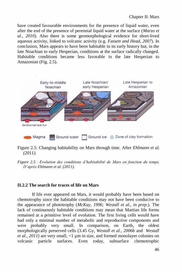
Chapter II: Mars
46
have created favourable environments for the presence of liquid water, even
after the end of the presence of perennial liquid water at the surface (Marzo et
al., 2010). Also there is some geomorphological evidence for short-lived
aqueous activity, linked to volcanic activity (e.g. Fassett and Head, 2007). In
conclusion, Mars appears to have been habitable in its early history but, in the
late Noachian to early Hesperian, conditions at the surface radically changed.
Habitable conditions became less favorable in the late Hesperian to
Amazonian (Fig. 2.5).
Figure 2.5: Changing habitability on Mars through time. After Ehlmann et al.
(2011).
Figure 2.5 : Evolution des conditions d’habitabilité de Mars en fonction du temps.
D’après Ehlmann et al. (2011).
II.2.2 The search for traces of life on Mars
If life ever appeared on Mars, it would probably have been based on
chemotrophy since the habitable conditions may not have been conducive to
the appearance of phototrophy (McKay, 1996; Westall et al., in prep.). The
lack of continuously habitable conditions may mean that Martian life forms
remained at a primitive level of evolution. The first living cells would have
had only a minimal number of metabolic and reproductive components and
were probably very small. In comparison, on Earth, the oldest
morphologically preserved cells (3.45 Gy, Westall et al., 2006b and Westall
et al., 2011) are very small, <1 µm in size, and formed monolayer colonies on
volcanic particle surfaces. Even today, subsurface chemotrophic
Chapter II: Mars
47
microorganisms are extremely small with diameters ~0.2 µm and they also
form very small mono-layer colonies that are heterogeneously distributed on
sediment particle surfaces (Ciobanu, 2012). Primitive Martian
microorganisms may have had (and may still have, if there is extant life on the
planet) similar characteristics.
Biosignatures in rocks are related to various aspects of
microorganisms: their morphology, their organic constituents, or their
metabolic processes (Westall, 1999; Westall et al., 2000; Westall and
Cavalazzi, 2011; Summons et al., 2011). Cells, cellular products, such as
extracellular polymeric substances, associations of cells, such as colonies,
microbial mats, and bio-constructions (e.g. stromatolites, bioherms…) can be
preserved through mineralization (fixation of minerals to the organic
structures). On Earth, the identification of biosignatures in very ancient rocks
is particularly controversial, not only because of the difficulty in correctly
identifying the biosignature but also in terms of demonstrating its syngenicity
with the formation of the rock (e.g. Westall and Folk, 2003; Rasmussen et al.,
2008). On Mars, although any evidence of life will be of primary importance,
whatever its age and relationship with the host rock, it will be equally
important to understand the timing of the formation and emplacement of the
biosignature in order to better interpret it. For instance, water-lain sediments
may contain biosignatures related to colonies of chemotrophic organisms that
inhabited the volcanic sediments and/or associated subaqueous hydrothermal
environments, or organic molecules fixed to phyllosilicates. Once lithified,
these same sediments and mineral deposits could host endolithic species in
cracks or intergrain spaces. Nevertheless, in all of these cases, the organisms
would be chemotrophs and the carbon and energy sources would be similar.
Note that the volcaniclastic sediments in which the Early Archaean
microfossils occur were strongly lithified by silica-saturated seawaters and by
widely pervasive hydrothermal silica. They are now termed “cherts”.
II.2.3. Possible analogue samples from Earth
Following previous considerations on Martian habitability, we can
consider that Martian life forms that could have appeared during the pre-
Noachian/Noachian period would be relatively similar to those observed in
Early Archaean sediments on Earth and that the same types of
volcaniclastic/hydrothermal sediments with similar carbonaceous remains
could be detected. To date, the oldest traces of cellular life on Earth, dating
back to 3.45 Gy, have been found in the Kitty’s Gap chert (Pilbara, Australia)
(Westall et al., 2006b and Westall et al., 2011). These traces of life are very
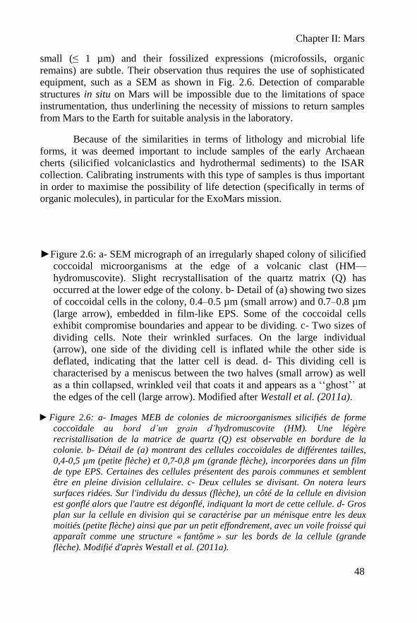
Chapter II: Mars
48
small (≤ 1 µm) and their fossilized expressions (microfossils, organic
remains) are subtle. Their observation thus requires the use of sophisticated
equipment, such as a SEM as shown in Fig. 2.6. Detection of comparable
structures in situ on Mars will be impossible due to the limitations of space
instrumentation, thus underlining the necessity of missions to return samples
from Mars to the Earth for suitable analysis in the laboratory.
Because of the similarities in terms of lithology and microbial life
forms, it was deemed important to include samples of the early Archaean
cherts (silicified volcaniclastics and hydrothermal sediments) to the ISAR
collection. Calibrating instruments with this type of samples is thus important
in order to maximise the possibility of life detection (specifically in terms of
organic molecules), in particular for the ExoMars mission.
►Figure 2.6: a- SEM micrograph of an irregularly shaped colony of silicified
coccoidal microorganisms at the edge of a volcanic clast (HM—
hydromuscovite). Slight recrystallisation of the quartz matrix (Q) has
occurred at the lower edge of the colony. b- Detail of (a) showing two sizes
of coccoidal cells in the colony, 0.4–0.5 µm (small arrow) and 0.7–0.8 µm
(large arrow), embedded in film-like EPS. Some of the coccoidal cells
exhibit compromise boundaries and appear to be dividing. c- Two sizes of
dividing cells. Note their wrinkled surfaces. On the large individual
(arrow), one side of the dividing cell is inflated while the other side is
deflated, indicating that the latter cell is dead. d- This dividing cell is
characterised by a meniscus between the two halves (small arrow) as well
as a thin collapsed, wrinkled veil that coats it and appears as a ‘‘ghost’’ at
the edges of the cell (large arrow). Modified after Westall et al. (2011a).
►Figure 2.6: a- Images MEB de colonies de microorganismes silicifiés de forme
coccoïdale au bord d’un grain d’hydromuscovite (HM). Une légère
recristallisation de la matrice de quartz (Q) est observable en bordure de la
colonie. b- Détail de (a) montrant des cellules coccoïdales de différentes tailles,
0,4-0,5 µm (petite flèche) et 0,7-0,8 µm (grande flèche), incorporées dans un film
de type EPS. Certaines des cellules présentent des parois communes et semblent
être en pleine division cellulaire. c- Deux cellules se divisant. On notera leurs
surfaces ridées. Sur l'individu du dessus (flèche), un côté de la cellule en division
est gonflé alors que l'autre est dégonflé, indiquant la mort de cette cellule. d- Gros
plan sur la cellule en division qui se caractérise par un ménisque entre les deux
moitiés (petite flèche) ainsi que par un petit effondrement, avec un voile froissé qui
apparaît comme une structure « fantôme » sur les bords de la cellule (grande
flèche). Modifié d'après Westall et al. (2011a).
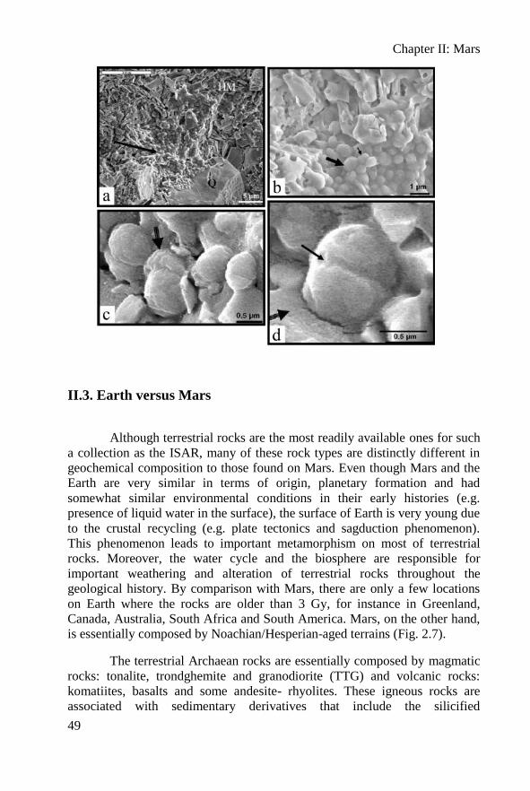
Chapter II: Mars
49
II.3. Earth versus Mars
Although terrestrial rocks are the most readily available ones for such
a collection as the ISAR, many of these rock types are distinctly different in
geochemical composition to those found on Mars. Even though Mars and the
Earth are very similar in terms of origin, planetary formation and had
somewhat similar environmental conditions in their early histories (e.g.
presence of liquid water in the surface), the surface of Earth is very young due
to the crustal recycling (e.g. plate tectonics and sagduction phenomenon).
This phenomenon leads to important metamorphism on most of terrestrial
rocks. Moreover, the water cycle and the biosphere are responsible for
important weathering and alteration of terrestrial rocks throughout the
geological history. By comparison with Mars, there are only a few locations
on Earth where the rocks are older than 3 Gy, for instance in Greenland,
Canada, Australia, South Africa and South America. Mars, on the other hand,
is essentially composed by Noachian/Hesperian-aged terrains (Fig. 2.7).
The terrestrial Archaean rocks are essentially composed by magmatic
rocks: tonalite, trondghemite and granodiorite (TTG) and volcanic rocks:
komatiites, basalts and some andesite- rhyolites. These igneous rocks are
associated with sedimentary derivatives that include the silicified
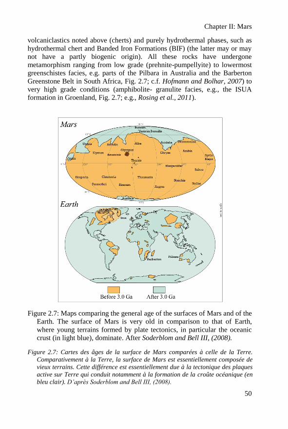
Chapter II: Mars
50
volcaniclastics noted above (cherts) and purely hydrothermal phases, such as
hydrothermal chert and Banded Iron Formations (BIF) (the latter may or may
not have a partly biogenic origin). All these rocks have undergone
metamorphism ranging from low grade (prehnite-pumpellyite) to lowermost
greenschistes facies, e.g. parts of the Pilbara in Australia and the Barberton
Greenstone Belt in South Africa, Fig. 2.7; c.f. Hofmann and Bolhar, 2007) to
very high grade conditions (amphibolite- granulite facies, e.g., the ISUA
formation in Groenland, Fig. 2.7; e.g., Rosing et al., 2011).
Figure 2.7: Maps comparing the general age of the surfaces of Mars and of the
Earth. The surface of Mars is very old in comparison to that of Earth,
where young terrains formed by plate tectonics, in particular the oceanic
crust (in light blue), dominate. After Soderblom and Bell III, (2008).
Figure 2.7: Cartes des âges de la surface de Mars comparées à celle de la Terre.
Comparativement à la Terre, la surface de Mars est essentiellement composée de
vieux terrains. Cette différence est essentiellement due à la tectonique des plaques
active sur Terre qui conduit notamment à la formation de la croûte océanique (en
bleu clair). D’après Soderblom and Bell III, (2008).
Chapter II: Mars
51
Based on these considerations, for the preliminary suite of
characterized rocks for the ISAR, I have selected a variety of terrestrial
materials as Martian analogues. The selection process and the pertinence of
this first set of samples for the ISAR will be described in the next chapter.

Chapter II: Mars
52
Chapter II: Mars
53
Mars (Résumé Français)
Mars est la quatrième planète rocheuse à partir du Soleil. Elle s’est formée
en même temps que les autres planètes telluriques : il y a 4,564 milliards d’années.
Mars est une petite planète dont la masse est seulement 1/10ème
de celle de la Terre
avec deux petites lunes, Phobos et Deimos, en orbite autour.
La structure interne de Mars est mal connue, mais il semble qu’elle possède
un noyau et un manteau différencié. Cependant, aucune activité géologique n’a été
observée en surface. La surface de Mars est caractérisée par une dichotomie globale:
au Sud, des hautes terres avec de très grands cratères (comme Hellas Planitia) et au
Nord, quatre kilomètres en-dessous des hautes terres du Sud, une surface beaucoup
plus lisse. Les plus grands volcans du système solaire (e.g. Olympus Mons), de très
grands canyons (e.g. Valles Marineris) et de nombreux cratères sont visibles à la
surface de Mars. La planète ne montrant pas d’activité tectonique, l’ensemble des
cratères est toujours visible ce qui permet de dater l’âge de la surface. Trois éons ont
été déterminés : de 4.5 à 3.7 Ga le Noachien, de 3.7 à 3.0 Ga l’Hesperien et de 3.0 à
aujourd’hui l’Amazonien. Récemment, les âges ont été redécoupés en fonction de
l’analyse minéralogique de la surface en Phyllossian, Theiikina et Siderikian (Fig.
2.1).
Ce chapitre va décrire les différentes roches observées à la surface de Mars.
Toute la surface n’a pas encore été cartographiée, mais il semble qu’elle soit
essentiellement composée de roches volcaniques associées à différents sédiments
issus de l’altération de basaltes. La surface de Mars est largement recouverte d’un
régolithe, une épaisse couche de poussière rouge. Nous ne détaillerons pas cette
formation en nous intéressant uniquement aux roches.
Les roches ignées observées à la surface de Mars sont essentiellement des
matériaux pauvres en silice, composés globalement d’olivine, de pyroxènes et de
plagioclases. Ce sont principalement des roches éruptives de type basaltique. Ces
roches sont très similaires à leurs analogues terrestres, mais présentent un fort
enrichissement en MgO (8 à 10 % poids), FeO (15 à 18% poids) et en Cl (>0,70%
poids). Le diagramme TAS (où l’échantillon est représenté en fonction de sa teneur
en SiO2 et en Na2O+K2O) et le diagramme FeO*/MgO-Silice (Fig. 2.2) reprennent
l’ensemble des données obtenues sur les roches volcaniques de Mars. Les données
TES sont obtenues à partir de l’orbite et doivent être considérées avec précaution.
Quelques observations interprètent la présence d’andésites et de roches plus riches
en silice. Faute de confirmation in situ, la question reste ouverte. Cependant il est
possible de trouver une explication par l’altération des basaltes par de l’eau liquide
qui favoriserait la formation de produits plus riches en silice. Les météorites SNC
provenant de Mars (Shergottites, Nakhilites et Chassignites) sont quant à elles plutôt
représentatives du manteau et non pas de la surface. Finalement, il n’a toujours pas
été observé précisément de granitoïdes à la surface de Mars (Bonin, 2011).
Les roches les plus abondantes après les basaltes sont les sédiments. Ces
sédiments sont essentiellement dérivés de basaltes ou de sables basaltiques. L’eau est
un des paramètres essentiels qui contrôle la formation de sédiments. Suivant les
Chapter II: Mars
54
variations de pH en surface, différents types de phyllosilicates se sont formés (ex :
saponites, nontronites). Récemment, des travaux ont montré que les phyllosilicates
pouvaient être formés sur Mars par des processus tardi-magmatiques comme le
deuterisme. L’essentiel des argiles ont été interprétées à partir des mesures
orbitales; ce sont des chlorites, vermiculites, saponites, nontronites et smectites.
D’autres types de sédiments sont présents sur Mars. D’importants niveaux de silice
ont été interprétés comme issus de l’activité hydrothermale, ou comme étant de la
silice hydratée microcristalline (opale) déposée avec des tephras. Ce sont
certainement des sources chaudes hydrothermales associées à du volcanisme datant
de l’Hesperien à l’Amazonien. Des sédiments évaporitiques composés de jarosite de
gypse et d’autres sulfates hydratés ont été décrits dans les hautes latitudes autour de
la calotte polaire et dans divers endroits à la surface de Mars. Enfin, des traces
ténues de carbonates ont été observées, principalement dans les météorites
martiennes. La météorite AHL84001 en est un exemple célèbre car elle semble
présenter des évidences de traces d’une vie passée provenant de Mars. La dernière
preuve de présence de carbonates provient de l’affleurement Comanche vue par Spirit
dans le cratère Gusev qui montre des carbonates associés à des olivines. Sur la figure
2.3 sont référencés plusieurs sites où ont été détectés ces roches et des minéraux
particulièrement intéressants pour la recherche de vie : les carbonates, les sulfates et
les minéraux hydratés.
Un des principaux objectifs des missions vers Mars est la recherche de
preuves d’une vie passée ou présente. Quel type d’évidence pouvons-nous trouver ?
L’habitabilité de Mars se résume à la capacité des organismes à trouver les
ingrédients nécessaires à leur survie, à savoir : l’eau liquide, des molécules
endogéniques et/ou exogéniques pour construire les macromolécules nécessaires à la
vie cellulaire, une source d’énergie et des nutriments (H, N, O, P, S, et les métaux de
transition). Ces deux derniers ingrédients peuvent être facilement obtenus sur Mars
par les réactions redox à la surface des minéraux, et par l’altération des roches
volcaniques, respectivement. Sur Mars, il existe maintenant de nombreuses preuves
minéralogiques et géomorphologiques de la présence d’eau liquide à la surface et
d’altérations hydrothermale ou aqueuse. La présence de minéraux hydratés comme
les phyllosilicates, les sulfates ou encore les carbonates, les hydroxydes et la silice
amorphe indiquent la présence d’eau liquide. L’observation de vallées dendritiques,
de chenaux, de delta et de sédiments de rivière (voir Fig. 2.4) montre l’écoulement
d’eau liquide en surface. D’après les datations de la surface, l’eau n’aurait coulée
sur Mars qu’au Noachien, et avec un régime hydrologique éphémère. A l’Hesperien
et à l’Amazonien, l’eau est uniquement présente en faible quantité sous la surface. En
conclusion, seule la jeune Mars était habitable, puisqu’à partir de l’Hesperien les
conditions deviennent de moins en moins favorables à la vie. L’hypothèse la plus
probable est la présence d’une vie (dans les stades les plus évolués) relativement
similaire à celle que l’on observe dans les sédiments Archéens (3.8-2.8 Ga) terrestres.
Les plus anciennes traces de vie ont été détectées dans des chailles (cherts) de Kitty’s
Gap, Pilbara, Australie et sont datées à 3.45 Ga. Ces traces fossiles sont extrêmement
petites (seulement quelques µm) et seuls des équipements tel que la microscopie à
balayage électronique (MEB) sont en mesure de les détecter (voir Fig. 2.6). Pour la
Chapter II: Mars
55
constitution de la collection de l’ISAR, il apparaît essentiel d’ajouter des échantillons
de chailles contenant ce type de fossiles.
Le choix des échantillons de la lithothèque doit se faire en prenant en compte
la géologie de Mars. La manière la plus simple pour obtenir des échantillons
analogues de Mars est de les collecter sur Terre. Cependant, les roches martiennes et
terrestres ne sont pas identiques. En effet, si Mars et la Terre sont très similaires par
leur origine et leur formation (même composition chimique, présence d’eau à la
surface, …), la surface de la Terre est beaucoup plus jeune à cause du recyclage de la
croûte (liée à la tectonique des plaques ou aux phénomènes de sagduction). Par
comparaison avec Mars, seuls quelques affleurements sur Terre sont des terrains
Archéens (e.g. Abitibi, Canada et Pilbara, Australie) tandis que Mars est
essentiellement composée de terrains Noachien et Hesperien (Fig. 2.7). De plus, les
anciennes roches terrestres ont subi un métamorphisme important qui n’a pas eu lieu
sur Mars.
A partir de ces considérations sur la géologie de Mars comparée à la Terre,
je vais présenter la première sélection de roches analogues de Mars de l’ISAR. Le
processus de sélection et la pertinence des échantillons seront décrits dans le
prochain chapitre.

Chapter II: Mars
56
Chapter III: Selection and analysis of the first group of Mars-analogue
samples
57
Chapter III
Selection and analysis of the first group of
Mars-analogue samples
In this chapter, I present and describe the selection of the first group
of Mars-analogue samples for the ISAR collection and the online data base.
The choice of materials is based on the geology of Mars as described in the
previous chapter and on the samples already available in the laboratory. The
exobiology team of the CBM has a large collection of rocks, especially
Archaean volcanic rocks, volcaniclastics and other sediments from South
Africa, Australia, Canada, Svarlbard, and Greenland, provided by Frances
Westall. Other samples were provided by Nick Arndt (ISTerre, Grenoble,
France) and Silvio Rotolo (Universita di Palermo, Italia), and were also
obtained during my own field trips to Rio Tinto (Spain), South Africa, the
Troodos Ophiolite Complex (Cyprus), and in France (French Massif Central).
This chapter will mainly focus on the selection on the samples and the
rationale for analogy to Mars. The emphasis is not on the results of all the
samples (which are presented in the Appendice B and are available on the
ISAR website), but rather to demonstrate the approach using relevant
instrumentation on a few selected samples. The petrology of each sample and
the results of the analyses will therefore be only briefly described and the
pertinence of the results will be discussed11
. I chose to present only the
detailed results obtained on samples from Svalbard as an example in
Appendice B. All the data are available in pdf file in the CD on the third cover
page (for the print format) and on the ISAR website (for the numerical
11
The content of this chapter is in review in PSS (Bost et al., in review).
Chapter III: Selection and analysis of the first group of Mars-analogue
samples
58
format). Obviously, as the ISAR is an on-going project, new samples and new
relevant techniques will be added in the future. The details of geochemical
analysis are in Appendice C.
III.1. Analogue sample selection
III.1.1 Volcanic selection
A variety of volcanic rocks forms the core of the first group of rocks
in the collection. The preliminary selection was mainly based on the elemental
composition of the Martian basalts given by McSween et al. (2009) and
displayed in Fig. 3.1. A certain number of samples with relevant compositions
were already available in the laboratory collection, e.g. from Svalbard,
Tenerife and South Africa (09SV15, 09TE08 and 07SA20 respectively).
The Svarlbard sample 09SV15 was chosen because it is associated
with dunite xenoliths (09SV02) and carbonates (09SV05) that have been
previously described as analogues of the carbonate deposits in the meteorite
ALH84001 (Treiman et al., 2002). The dunite (09SV02) was also added to the
collection as an analogue magmatic rock to the Martian mantle. Sample
09TE08 from Tenerife was chosen because it fits well with the Ca/Si vs.
Mg/Si diagram proposed by Mc Sween et al. (2009). This sample could be an
analogue of the most altered Martian basalts (Hurowitz et al., 2006). Also
note that altered volcanic rocks may sometimes contain evidence of life
associated with alteration phases (Furnes et al., 2007; Cavalazzi et al., 2011).
Sample 07SA20 from Barberton in South Africa was chosen because it is
representative of alteration processes (silicification) associated with
hydrothermal processes observed on the Early Earth. Such processes may
have also occurred on Early Mars.
However, as noted above, all the terrestrial basalts are depleted in Fe
and Mg. Moreover, sample 09SV15 has slightly higher Na2O- and K2O-
contents than those of Martian basalts, whereas sample 09TE08 from Tenerife
plots outside the limits of the TAS and FeO*/MgO-silica diagrams because of
its high Na2O- and K2O-content and high FeO*/MgO ratio.
I added one sample from Etna (09ET01) to this collection, a
“primitive basalt” that falls in the main Martian basalt field of the TAS
diagram (Fig. 3.1). This sample was provided by Sylvio Rotolo (Universita di
Palermo, Italia).
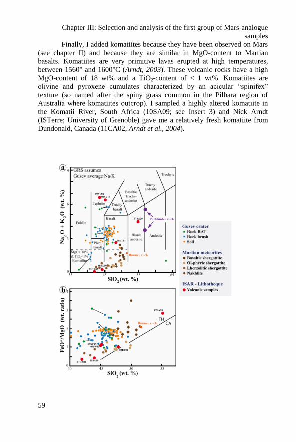
Chapter III: Selection and analysis of the first group of Mars-analogue
samples
59
Finally, I added komatiites because they have been observed on Mars
(see chapter II) and because they are similar in MgO-content to Martian
basalts. Komatiites are very primitive lavas erupted at high temperatures,
between 1560° and 1600°C (Arndt, 2003). These volcanic rocks have a high
MgO-content of 18 wt% and a TiO2-content of < 1 wt%. Komatiites are
olivine and pyroxene cumulates characterized by an acicular “spinifex”
texture (so named after the spiny grass common in the Pilbara region of
Australia where komatiites outcrop). I sampled a highly altered komatiite in
the Komatii River, South Africa (10SA09; see Insert 3) and Nick Arndt
(ISTerre; University of Grenoble) gave me a relatively fresh komatiite from
Dundonald, Canada (11CA02, Arndt et al., 2004).
Chapter III: Selection and analysis of the first group of Mars-analogue
samples
60
◄Figure 3.1- Na2O + K2O oxides and FeO*/MgO ratios versus SiO2 plots (in
wt.%; 3.1a and 3.1b, respectively). The terrestrial volcanic samples
presently included in the ISAR collection are also shown (red circles).
Bounce rock is the only sample of basaltic origin that was analysed in the
Meridiani Planum by the Opportunity rover (Rieder et al., 2004 and Zipfel
et al., 2004). The FeO*/MgO-silica diagram is used for distinguishing dry
tholeiitic (TH) and wet calc-alkaline (CA) rocks. All Martian samples are
tholeiitic.
◄Figure 3.1 Teneur en Na2O + K2O et rapport FeO*/MgO en fonction de SiO2 (en %
poids), figures 3.1a et 3.1b respectivement. Les échantillons volcaniques
terrestres de la collection ISAR sont représentés par les ronds rouges.
L’échantillon « Bounce rock » est le seul échantillon d’origine basaltique trouvé
et analysé sur Meridiani Planum par le rover Opportunity (Rieder et al., 2004 et
Zipfel et al., 2004). Le diagramme FeO*/MgO- SiO2 est utilisé pour distinguer les
série tholeiitiques (TH) des séries calco-alcalines (CA). L’ensemble des roches
martiennes est issue de la série tholeiitique.
III.1.2. Sediment selection
Some of the sedimentary analogue rocks were chosen for their
astrobiological relevance with respect to future missions to search for past
traces of life on Mars. If life ever occurred on Mars, it was probably during
the pre-Noachian/Noachian/earliest Hesperian periods, when liquid water was
still present on the Martian surface. Remains of this primitive life could
potentially be found in sediments and in altered volcanic rocks dating from
this period. On Earth, sediments dating back to the same period are very rare
due to crustal recycling. Therefore, only a few locations contain Early
Archaean age volcanic sands and silts (volcanoclastics) deposited and altered
in water. These lithologies have been largely preserved because they were
silicified by silica-rich seawater and hydrothermal fluids (Hofmann and
Bohlar, 2007). They host well preserved silicified mineralogical and organic
traces of primitive life that were probably similar to Noachian microbial
remains, if life existed on Mars (i.e. chemolithotrophs, cf. Chapter 2; Westall
et al., 2006a, 2011a and 2011b). Hydrothermal activity and siliceous fluids
related to volcanism on Mars (Ehlmann et al., 2009) could have contributed to
the preservation of Martian microorganisms (Westall et al., 2011a).
Many of the Early Archaean sediments from Australia and South
Africa have been previously investigated by Frances Westall (e.g., Westall et
Chapter III: Selection and analysis of the first group of Mars-analogue
samples
61
al., 2011a and references therein). For this study, we chose a 3.45 Ga old
silicified volcanoclastic deposit from the Pilbara craton in NW Australia
(00AU05). Early Archaean non-carbonaceous and carbonaceous hydrothermal
cherts from the Barberton Greenstone Belt, South Africa (99SA01, 99SA05)
and from the Pilbara (00AU04) were also chosen together with a Banded Iron
Formation (BIF) sample (06AU01). BIFs have not yet been discovered on
Mars but the high Fe-content of Martian rocks (15 to 18 wt%) (McSween et
al., 2009) and the detection of hydrothermal silica-rich deposits on Mars
(Squyres et al., 2008) suggest that BIFs may have deposited in aqueous basins
in the early Martian history. BIFs consist of alternating Si- and Fe-rich layers
formed in water. They were common on Earth during the Archaean and the
Proterozoic. They were deposited in basinal settings through the oxidation of
reduced Fe which was mostly introduced into the seawater by hydrothermal
fluids. The exact origin of these sediments, i.e., the mechanisms responsible
for oxidizing the Fe (possibly microbial) and for the layering
(seasonal/microbial?), is still highly debated (Posth et al., 2010). The selected
BIF for this study was a ~ 3.5 Gy old sample from the Pilbara Craton in
Australia.
As noted in the previous chapter, carbonates observed on Mars are
associated with basalt alteration phases. The carbonate-alteration of basalts in
the Comanche outcrop produced Fe-Mg-rich phases (Morris et al., 2010). The
secondary carbonates observed in the ALH84001 meteorite were also zoned
Fe and Mg carbonate phases (Borg et al., 1999; Treiman et al., 2002). Thus
similary zoned carbonates in terrestrial rocks were chosen. They were
deposited by hydrothermal exhalations/fluids (and possibly aqueous alteration
during eruption under a glacier) in a recent basalt (≈100 ky) from Svalbard.
They were collected by Frances Westall during the 2009 AMASE mission
(sample 09SV05; cf. Treiman et al., 2002; Steele et al., 2007). In order to
complete the carbonate selection, I sampled carbonates in hydrothermal veins
(stockwerk) associated with pillow basalts in the Troodos ophiolitic complex
in Cyprus (11CY04). Details concerning the Cyprus mission are described in
Chapter IV. Table 3.1 lists the samples, their origin and relevance for Mars.
►Table 3.1: List of the analogue samples characterised for the ISAR
collection and their proposed Martian equivalents.
►Tableau 3.1: Liste des échantillons analogues caractérisés pour la collection de
l’ISAR et des exemples d’équivalences martiennes.
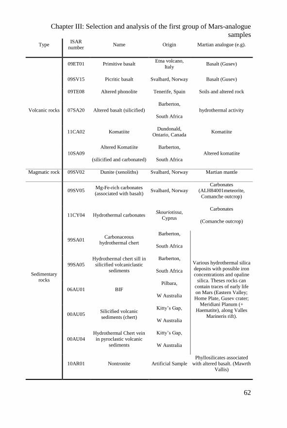
Chapter III: Selection and analysis of the first group of Mars-analogue
samples
62
Type ISAR
number Name Origin Martian analogue (e.g).
Volcanic rocks
09ET01 Primitive basalt Etna volcano,
Italy Basalt (Gusev)
09SV15 Picritic basalt Svalbard, Norway Basalt (Gusev)
09TE08 Altered phonolite Tenerife, Spain Soils and altered rock
07SA20 Altered basalt (silicified)
Barberton,
South Africa hydrothermal activity
11CA02 Komatiite Dundonald,
Ontario, Canada Komatiite
10SA09
Altered Komatiite
(silicified and carbonated)
Barberton,
South Africa
Altered komatiite
Magmatic rock 09SV02 Dunite (xenoliths) Svalbard, Norway Martian mantle
Sedimentary rocks
09SV05 Mg-Fe-rich carbonates
(associated with basalt) Svalbard, Norway
Carbonates (ALH84001meteorite,
Comanche outcrop)
11CY04 Hydrothermal carbonates Skouriotissa,
Cyprus
Carbonates
(Comanche outcrop)
99SA01 Carbonaceous
hydrothermal chert
Barberton,
South Africa
Various hydrothermal silica
deposits with possible iron concentrations and opaline
silica. Theses rocks can
contain traces of early life
on Mars (Eastern Valley;
Home Plate, Gusev crater;
Meridiani Planum (+ Haematite), along Valles
Marineris rift).
99SA05
Hydrothermal chert sill in
silicified volcaniclastic
sediments
Barberton,
South Africa
06AU01 BIF
Pilbara,
W Australia
00AU05 Silicified volcanic sediments (chert)
Kitty’s Gap,
W Australia
00AU04
Hydrothermal Chert vein
in pyroclastic volcanic
sediments
Kitty’s Gap,
W Australia
10AR01 Nontronite Artificial Sample
Phyllosilicates associated
with altered basalt. (Mawrth Vallis)
Chapter III: Selection and analysis of the first group of Mars-analogue
samples
63
III.2 Sample description
III.2.1. Volcanic rocks
III.2.1.1. 09ET01, a “primitive” basalt.
This sample comes from the Etna volcano, located on the eastern
coast of Sicily. Etna is a basaltic stratovolcano renowned for its frequent
effusive eruptions. Although the Etna lavas are compositionally picritic, the
ISAR sample comes from one particular horizon of Etna lavas characterised
by a “primitive”, non-fractionated composition. “Primitive” lavas are rare on
Earth but must have been common on Mars (e.g. Mc Sween et al., 2009). The
sample, dated at 3930 ± 60 BP (Coltelli et al., 2000), originates from a sub-
plinian eruption which created 0.183 km3 of scoria lapilli with 30 to 60 % of
microvesicularity (Coltelli et al., 2005). The magma was derived from
fractionation products of mantle-derived magmas (Kamenestsky et al., 2007)
formed in association with subduction activity beneath the Appenines
(Tonarini et al., 2001).
The sample is a microvesiculated tephra composed of olivine-rich
scoria lapilli and rare cognate lithic clasts. The hand specimen surface is
brown and an occasional orange crust around individual olivine grains is a
sign of oxidation. Large olivine grains (1-2 mm) are present (Fig. 3.2a). In
microscopic view, the sample is composed of brown (oxidized) and black
(non-oxidized) lapilli <1 cm in diameter. Mineralogically, the rock consists of
large blocky olivine (Fo91) crystals and smaller euhedral olivine crystals
associated with pyroxenes (diopside, Mg#92.5) in a basaltic glass (Fig. 3.2b).
Some small plagioclase crystals occur in the pyroxene matrix. The basalt
contains some Cr-spinel grains. The chemical composition of this sample is
close to the average Martian basalt composition (Fig. 3.1).
III.2.1.2. 09SV15, a tephritic basalt
The “tephritic-basalt” sample has the same provenance as the dunite
xenolith (09SV02) and one of the carbonate (09SV05) samples. They were
obtained from two quaternary stratovolcanoes (≈100 Ky), Sverrefjell
(N79°26,255; E013°20,486) and Sigurdjfell (N79°16,119; E013°32,235), on
the island of Svarlbard, north of Norway. The two volcanoes are very similar
in their magmatic structure and petrography. They were formed by off-ridge
Chapter III: Selection and analysis of the first group of Mars-analogue
samples
64
alkali basaltic volcanism associated with the opening of the Artic Basin and
the Greenland Sea (Skelkvåle et al., 1989). They are primitive and ultramafic
basalts containing approximately 20 % xenoliths (mantle dunites, sample
09SV02, and some gneissic crustal elements). The two volcanoes are aligned
along the Bockfjord, parallel to an important fault which defines the contact
between a major Devonian (416 to 374.5 My) graben, filled with Devonian
red-beds (molasse), and Caledonian basement rocks of the Heckla Hoek
formation. Quaternary volcanism is subactive and associated with hot springs.
(for more information see Skjelkvåle et al., 1989 and Fookes, 2008).
Sample 09SV15 is a massive and highly vesicular basalt with vesicle
sizes ranging from 1 to 5 mm. The outer surface of the hand specimen shows
some brownish grey bleaching, indicating very minor weathering. The fresh
basalt surface is dark grey. Dunite xenoliths up to 2 cm in size and some
macroscopic olivine grains (0.5-1 mm size) can be observed in the basalt (Fig.
3.2b). The mineralogy of the primitive basalt is olivine (Fo90) occurring as
phenocrysts with pyroxenes (Mg#57) and associated with plagioclase
microlites and minor glass (Fig. 3.2c). Secondary minerals include amphibole,
oxides and spinels. This sample has the particularity of containing quartz
grains, possibly a xenolithic contribution.
The rock has a chemical composition which is relevant for tephrite-type
basalts (Fig. 3.1), rich in alkali elements, on Mars. Its textural and
mineralogical characteristics, typical for this type of basalt, will be readily
identifiable using the instrumentation available for the upcoming Mars
missions.
III.2.1.3. 09TE08, altered phonolite
This is an altered “phonolitic-basalt” from the El Teide volcano
(N28°261379; E-16°594848) on the island of Tenerife in the Canaries
archipelago off western Africa. Tenerife is the exposed part of a giant
volcanic edifice that extends from the floor of the eastern central Atlantic at –
3700 m to + 3718 m at the summit of the volcano. Its subaerial history began
in the late Miocene and is associated with mafic to intermediate monogenic
cones and fissure systems along the Santiago rift to the NW and the Dorsale
rift to the NE. The volcano produces different types of magmas, especially
basalts and phonolites (Ablay and Marti, 2000).
This sample is a black massive rock with microvesicularity and some
vesicles resembling cracks. The weathered surface of the rock is brown (Fig.
3.2e). In thin section, the matrix is composed of elongated plagioclase without
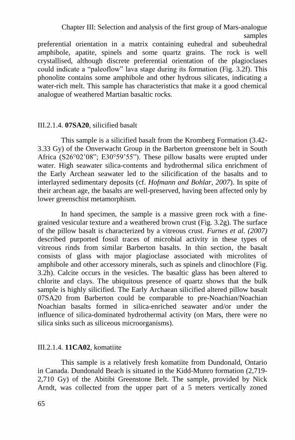
Chapter III: Selection and analysis of the first group of Mars-analogue
samples
65
preferential orientation in a matrix containing euhedral and subeuhedral
amphibole, apatite, spinels and some quartz grains. The rock is well
crystallised, although discrete preferential orientation of the plagioclases
could indicate a “paleoflow” lava stage during its formation (Fig. 3.2f). This
phonolite contains some amphibole and other hydrous silicates, indicating a
water-rich melt. This sample has characteristics that make it a good chemical
analogue of weathered Martian basaltic rocks.
III.2.1.4. 07SA20, silicified basalt
This sample is a silicified basalt from the Kromberg Formation (3.42-
3.33 Gy) of the Onverwacht Group in the Barberton greenstone belt in South
Africa (S26°02’08”; E30°59’55”). These pillow basalts were erupted under
water. High seawater silica-contents and hydrothermal silica enrichment of
the Early Archean seawater led to the silicification of the basalts and to
interlayred sedimentary deposits (cf. Hofmann and Bohlar, 2007). In spite of
their archean age, the basalts are well-preserved, having been affected only by
lower greenschist metamorphism.
In hand specimen, the sample is a massive green rock with a fine-
grained vesicular texture and a weathered brown crust (Fig. 3.2g). The surface
of the pillow basalt is characterized by a vitreous crust. Furnes et al. (2007)
described purported fossil traces of microbial activity in these types of
vitreous rinds from similar Barberton basalts. In thin section, the basalt
consists of glass with major plagioclase associated with microlites of
amphibole and other accessory minerals, such as spinels and clinochlore (Fig.
3.2h). Calcite occurs in the vesicles. The basaltic glass has been altered to
chlorite and clays. The ubiquitous presence of quartz shows that the bulk
sample is highly silicified. The Early Archaean silicified altered pillow basalt
07SA20 from Barberton could be comparable to pre-Noachian/Noachian
Noachian basalts formed in silica-enriched seawater and/or under the
influence of silica-dominated hydrothermal activity (on Mars, there were no
silica sinks such as siliceous microorganisms).
III.2.1.4. 11CA02, komatiite
This sample is a relatively fresh komatiite from Dundonald, Ontario
in Canada. Dundonald Beach is situated in the Kidd-Munro formation (2,719-
2,710 Gy) of the Abitibi Greenstone Belt. The sample, provided by Nick
Arndt, was collected from the upper part of a 5 meters vertically zoned
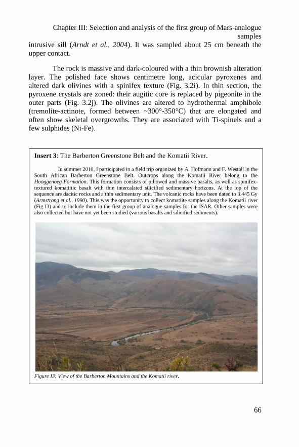
Chapter III: Selection and analysis of the first group of Mars-analogue
samples
66
intrusive sill (Arndt et al., 2004). It was sampled about 25 cm beneath the
upper contact.
The rock is massive and dark-coloured with a thin brownish alteration
layer. The polished face shows centimetre long, acicular pyroxenes and
altered dark olivines with a spinifex texture (Fig. 3.2i). In thin section, the
pyroxene crystals are zoned: their augitic core is replaced by pigeonite in the
outer parts (Fig. 3.2j). The olivines are altered to hydrothermal amphibole
(tremolite-actinote, formed between ~300°-350°C) that are elongated and
often show skeletal overgrowths. They are associated with Ti-spinels and a
few sulphides (Ni-Fe).
Insert 3: The Barberton Greenstone Belt and the Komatii River.
In summer 2010, I participated in a field trip organized by A. Hofmann and F. Westall in the South African Barberton Greenstone Belt. Outcrops along the Komatii River belong to the
Hooggenoeg Formation. This formation consists of pillowed and massive basalts, as well as spinifex-
textured komatiitic basalt with thin intercalated silicified sedimentary horizons. At the top of the sequence are dacitic rocks and a thin sedimentary unit. The volcanic rocks have been dated to 3.445 Gy
(Armstrong et al., 1990). This was the opportunity to collect komatiite samples along the Komatii river
(Fig I3) and to include them in the first group of analogue samples for the ISAR. Other samples were also collected but have not yet been studied (various basalts and silicified sediments).
Figure I3: View of the Barberton Mountains and the Komatii river.
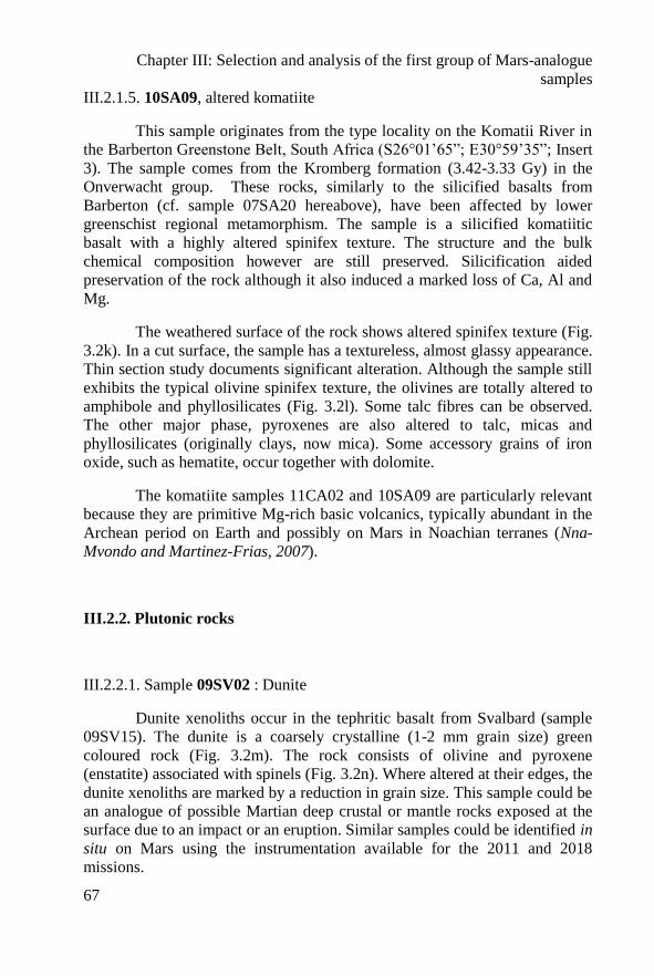
Chapter III: Selection and analysis of the first group of Mars-analogue
samples
67
III.2.1.5. 10SA09, altered komatiite
This sample originates from the type locality on the Komatii River in
the Barberton Greenstone Belt, South Africa (S26°01’65”; E30°59’35”; Insert
3). The sample comes from the Kromberg formation (3.42-3.33 Gy) in the
Onverwacht group. These rocks, similarly to the silicified basalts from
Barberton (cf. sample 07SA20 hereabove), have been affected by lower
greenschist regional metamorphism. The sample is a silicified komatiitic
basalt with a highly altered spinifex texture. The structure and the bulk
chemical composition however are still preserved. Silicification aided
preservation of the rock although it also induced a marked loss of Ca, Al and
Mg.
The weathered surface of the rock shows altered spinifex texture (Fig.
3.2k). In a cut surface, the sample has a textureless, almost glassy appearance.
Thin section study documents significant alteration. Although the sample still
exhibits the typical olivine spinifex texture, the olivines are totally altered to
amphibole and phyllosilicates (Fig. 3.2l). Some talc fibres can be observed.
The other major phase, pyroxenes are also altered to talc, micas and
phyllosilicates (originally clays, now mica). Some accessory grains of iron
oxide, such as hematite, occur together with dolomite.
The komatiite samples 11CA02 and 10SA09 are particularly relevant
because they are primitive Mg-rich basic volcanics, typically abundant in the
Archean period on Earth and possibly on Mars in Noachian terranes (Nna-
Mvondo and Martinez-Frias, 2007).
III.2.2. Plutonic rocks
III.2.2.1. Sample 09SV02 : Dunite
Dunite xenoliths occur in the tephritic basalt from Svalbard (sample
09SV15). The dunite is a coarsely crystalline (1-2 mm grain size) green
coloured rock (Fig. 3.2m). The rock consists of olivine and pyroxene
(enstatite) associated with spinels (Fig. 3.2n). Where altered at their edges, the
dunite xenoliths are marked by a reduction in grain size. This sample could be
an analogue of possible Martian deep crustal or mantle rocks exposed at the
surface due to an impact or an eruption. Similar samples could be identified in
situ on Mars using the instrumentation available for the 2011 and 2018
missions.
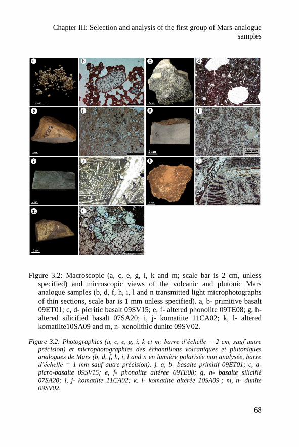
Chapter III: Selection and analysis of the first group of Mars-analogue
samples
68
Figure 3.2: Macroscopic (a, c, e, g, i, k and m; scale bar is 2 cm, unless
specified) and microscopic views of the volcanic and plutonic Mars
analogue samples (b, d, f, h, i, l and n transmitted light microphotographs
of thin sections, scale bar is 1 mm unless specified). a, b- primitive basalt
09ET01; c, d- picritic basalt 09SV15; e, f- altered phonolite 09TE08; g, h-
altered silicified basalt 07SA20; i, j- komatiite 11CA02; k, l- altered
komatiite10SA09 and m, n- xenolithic dunite 09SV02.
Figure 3.2: Photographies (a, c, e, g, i, k et m; barre d’échelle = 2 cm, sauf autre
précision) et microphotographies des échantillons volcaniques et plutoniques
analogues de Mars (b, d, f, h, i, l and n en lumière polarisée non analysée, barre
d’échelle = 1 mm sauf autre précision). ). a, b- basalte primitif 09ET01; c, d-
picro-basalte 09SV15; e, f- phonolite altérée 09TE08; g, h- basalte silicifié
07SA20; i, j- komatiite 11CA02; k, l- komatiite altérée 10SA09 ; m, n- dunite
09SV02.
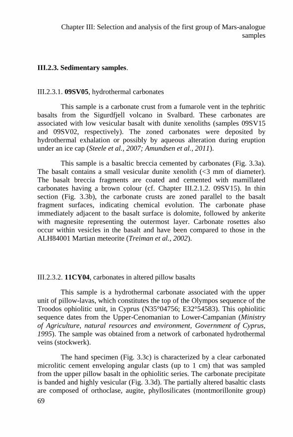
Chapter III: Selection and analysis of the first group of Mars-analogue
samples
69
III.2.3. Sedimentary samples.
III.2.3.1. 09SV05, hydrothermal carbonates
This sample is a carbonate crust from a fumarole vent in the tephritic
basalts from the Sigurdfjell volcano in Svalbard. These carbonates are
associated with low vesicular basalt with dunite xenoliths (samples 09SV15
and 09SV02, respectively). The zoned carbonates were deposited by
hydrothermal exhalation or possibly by aqueous alteration during eruption
under an ice cap (Steele et al., 2007; Amundsen et al., 2011).
This sample is a basaltic breccia cemented by carbonates (Fig. 3.3a).
The basalt contains a small vesicular dunite xenolith (<3 mm of diameter).
The basalt breccia fragments are coated and cemented with mamillated
carbonates having a brown colour (cf. Chapter III.2.1.2. 09SV15). In thin
section (Fig. 3.3b), the carbonate crusts are zoned parallel to the basalt
fragment surfaces, indicating chemical evolution. The carbonate phase
immediately adjacent to the basalt surface is dolomite, followed by ankerite
with magnesite representing the outermost layer. Carbonate rosettes also
occur within vesicles in the basalt and have been compared to those in the
ALH84001 Martian meteorite (Treiman et al., 2002).
III.2.3.2. 11CY04, carbonates in altered pillow basalts
This sample is a hydrothermal carbonate associated with the upper
unit of pillow-lavas, which constitutes the top of the Olympos sequence of the
Troodos ophiolitic unit, in Cyprus (N35°04756; E32°54583). This ophiolitic
sequence dates from the Upper-Cenomanian to Lower-Campanian (Ministry
of Agriculture, natural resources and environment, Government of Cyprus,
1995). The sample was obtained from a network of carbonated hydrothermal
veins (stockwerk).
The hand specimen (Fig. 3.3c) is characterized by a clear carbonated
microlitic cement enveloping angular clasts (up to 1 cm) that was sampled
from the upper pillow basalt in the ophiolitic series. The carbonate precipitate
is banded and highly vesicular (Fig. 3.3d). The partially altered basaltic clasts
are composed of orthoclase, augite, phyllosilicates (montmorillonite group)
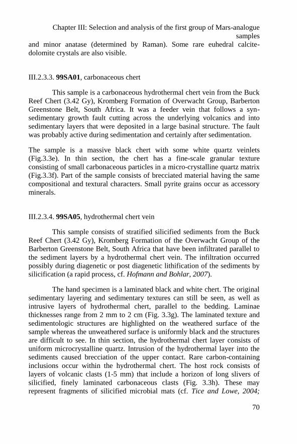
Chapter III: Selection and analysis of the first group of Mars-analogue
samples
70
and minor anatase (determined by Raman). Some rare euhedral calcite-
dolomite crystals are also visible.
III.2.3.3. 99SA01, carbonaceous chert
This sample is a carbonaceous hydrothermal chert vein from the Buck
Reef Chert (3.42 Gy), Kromberg Formation of Overwacht Group, Barberton
Greenstone Belt, South Africa. It was a feeder vein that follows a syn-
sedimentary growth fault cutting across the underlying volcanics and into
sedimentary layers that were deposited in a large basinal structure. The fault
was probably active during sedimentation and certainly after sedimentation.
The sample is a massive black chert with some white quartz veinlets
(Fig.3.3e). In thin section, the chert has a fine-scale granular texture
consisting of small carbonaceous particles in a micro-crystalline quartz matrix
(Fig.3.3f). Part of the sample consists of brecciated material having the same
compositional and textural characters. Small pyrite grains occur as accessory
minerals.
III.2.3.4. 99SA05, hydrothermal chert vein
This sample consists of stratified silicified sediments from the Buck
Reef Chert (3.42 Gy), Kromberg Formation of the Overwacht Group of the
Barberton Greenstone Belt, South Africa that have been infiltrated parallel to
the sediment layers by a hydrothermal chert vein. The infiltration occurred
possibly during diagenetic or post diagenetic lithification of the sediments by
silicification (a rapid process, cf. Hofmann and Bohlar, 2007).
The hand specimen is a laminated black and white chert. The original
sedimentary layering and sedimentary textures can still be seen, as well as
intrusive layers of hydrothermal chert, parallel to the bedding. Laminae
thicknesses range from 2 mm to 2 cm (Fig. 3.3g). The laminated texture and
sedimentologic structures are highlighted on the weathered surface of the
sample whereas the unweathered surface is uniformly black and the structures
are difficult to see. In thin section, the hydrothermal chert layer consists of
uniform microcrystalline quartz. Intrusion of the hydrothermal layer into the
sediments caused brecciation of the upper contact. Rare carbon-containing
inclusions occur within the hydrothermal chert. The host rock consists of
layers of volcanic clasts (1-5 mm) that include a horizon of long slivers of
silicified, finely laminated carbonaceous clasts (Fig. 3.3h). These may
represent fragments of silicified microbial mats (cf. Tice and Lowe, 2004;
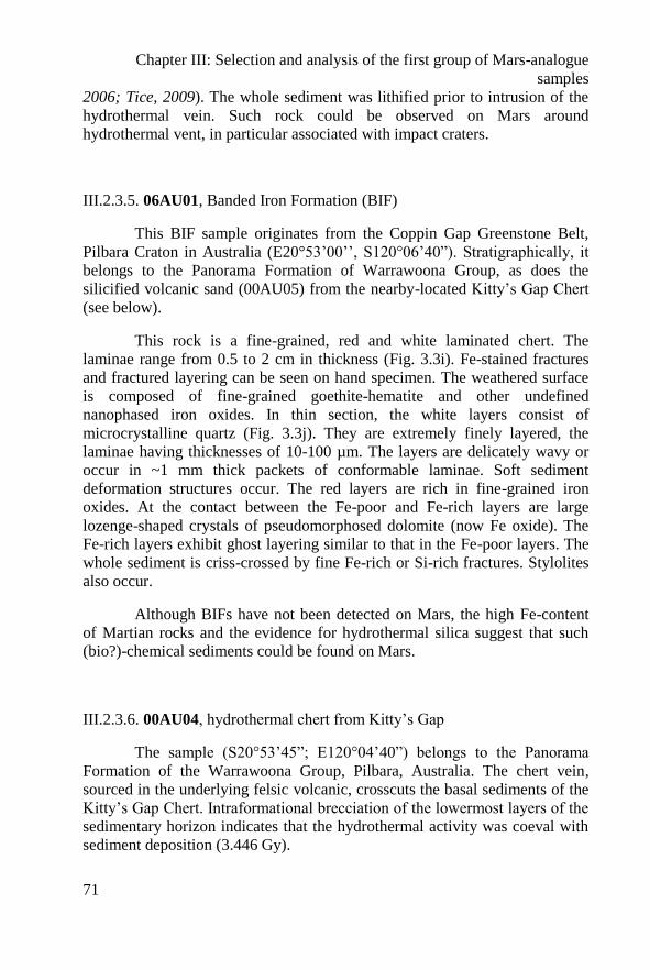
Chapter III: Selection and analysis of the first group of Mars-analogue
samples
71
2006; Tice, 2009). The whole sediment was lithified prior to intrusion of the
hydrothermal vein. Such rock could be observed on Mars around
hydrothermal vent, in particular associated with impact craters.
III.2.3.5. 06AU01, Banded Iron Formation (BIF)
This BIF sample originates from the Coppin Gap Greenstone Belt,
Pilbara Craton in Australia (E20°53’00’’, S120°06’40”). Stratigraphically, it
belongs to the Panorama Formation of Warrawoona Group, as does the
silicified volcanic sand (00AU05) from the nearby-located Kitty’s Gap Chert
(see below).
This rock is a fine-grained, red and white laminated chert. The
laminae range from 0.5 to 2 cm in thickness (Fig. 3.3i). Fe-stained fractures
and fractured layering can be seen on hand specimen. The weathered surface
is composed of fine-grained goethite-hematite and other undefined
nanophased iron oxides. In thin section, the white layers consist of
microcrystalline quartz (Fig. 3.3j). They are extremely finely layered, the
laminae having thicknesses of 10-100 µm. The layers are delicately wavy or
occur in ~1 mm thick packets of conformable laminae. Soft sediment
deformation structures occur. The red layers are rich in fine-grained iron
oxides. At the contact between the Fe-poor and Fe-rich layers are large
lozenge-shaped crystals of pseudomorphosed dolomite (now Fe oxide). The
Fe-rich layers exhibit ghost layering similar to that in the Fe-poor layers. The
whole sediment is criss-crossed by fine Fe-rich or Si-rich fractures. Stylolites
also occur.
Although BIFs have not been detected on Mars, the high Fe-content
of Martian rocks and the evidence for hydrothermal silica suggest that such
(bio?)-chemical sediments could be found on Mars.
III.2.3.6. 00AU04, hydrothermal chert from Kitty’s Gap
The sample (S20°53’45”; E120°04’40”) belongs to the Panorama
Formation of the Warrawoona Group, Pilbara, Australia. The chert vein,
sourced in the underlying felsic volcanic, crosscuts the basal sediments of the
Kitty’s Gap Chert. Intraformational brecciation of the lowermost layers of the
sedimentary horizon indicates that the hydrothermal activity was coeval with
sediment deposition (3.446 Gy).
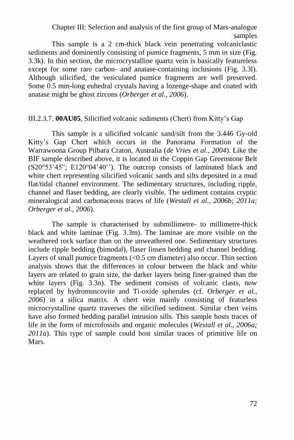
Chapter III: Selection and analysis of the first group of Mars-analogue
samples
72
This sample is a 2 cm-thick black vein penetrating volcaniclastic
sediments and dominently consisting of pumice fragments, 5 mm in size (Fig.
3.3k). In thin section, the microcrystalline quartz vein is basically featureless
except for some rare carbon- and anatase-containing inclusions (Fig. 3.3l).
Although silicified, the vesiculated pumice fragments are well preserved.
Some 0.5 mm-long euhedral crystals having a lozenge-shape and coated with
anatase might be ghost zircons (Orberger et al., 2006).
III.2.3.7. 00AU05, Silicified volcanic sediments (Chert) from Kitty’s Gap
This sample is a silicified volcanic sand/silt from the 3.446 Gy-old
Kitty’s Gap Chert which occurs in the Panorama Formation of the
Warrawoona Group Pilbara Craton, Australia (de Vries et al., 2004). Like the
BIF sample described above, it is located in the Coppin Gap Greenstone Belt
(S20°53’45”; E120°04’40’’). The outcrop consists of laminated black and
white chert representing silicified volcanic sands and silts deposited in a mud
flat/tidal channel environment. The sedimentary structures, including ripple,
channel and flaser bedding, are clearly visible. The sediment contains cryptic
mineralogical and carbonaceous traces of life (Westall et al., 2006b; 2011a;
Orberger et al., 2006).
The sample is characterised by submillimetre- to millimetre-thick
black and white laminae (Fig. 3.3m). The laminae are more visible on the
weathered rock surface than on the unweathered one. Sedimentary structures
include ripple bedding (bimodal), flaser linsen bedding and channel bedding.
Layers of small pumice fragments (<0.5 cm diameter) also occur. Thin section
analysis shows that the differences in colour between the black and white
layers are related to grain size, the darker layers being finer-grained than the
white layers (Fig. 3.3n). The sediment consists of volcanic clasts, now
replaced by hydromuscovite and Ti-oxide spherules (cf. Orberger et al.,
2006) in a silica matrix. A chert vein mainly consisting of featurless
microcrystalline quartz traverses the silicified sediment. Similar chert veins
have also formed bedding parallel intrusion sills. This sample hosts traces of
life in the form of microfossils and organic molecules (Westall et al., 2006a;
2011a). This type of sample could host similar traces of primitive life on
Mars.
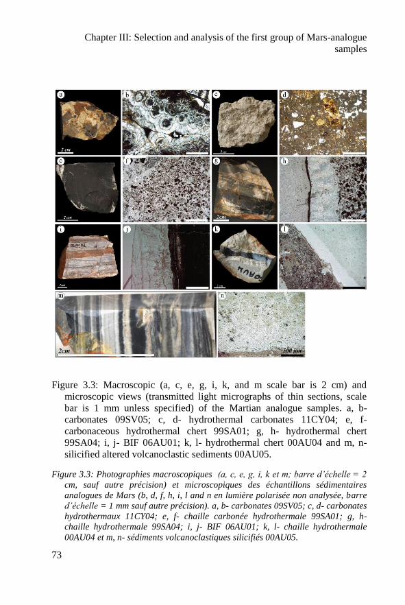
Chapter III: Selection and analysis of the first group of Mars-analogue
samples
73
Figure 3.3: Macroscopic (a, c, e, g, i, k, and m scale bar is 2 cm) and
microscopic views (transmitted light micrographs of thin sections, scale
bar is 1 mm unless specified) of the Martian analogue samples. a, b-
carbonates 09SV05; c, d- hydrothermal carbonates 11CY04; e, f-
carbonaceous hydrothermal chert 99SA01; g, h- hydrothermal chert
99SA04; i, j- BIF 06AU01; k, l- hydrothermal chert 00AU04 and m, n-
silicified altered volcanoclastic sediments 00AU05.
Figure 3.3: Photographies macroscopiques (a, c, e, g, i, k et m; barre d’échelle = 2
cm, sauf autre précision) et microscopiques des échantillons sédimentaires
analogues de Mars (b, d, f, h, i, l and n en lumière polarisée non analysée, barre
d’échelle = 1 mm sauf autre précision). a, b- carbonates 09SV05; c, d- carbonates
hydrothermaux 11CY04; e, f- chaille carbonée hydrothermale 99SA01; g, h-
chaille hydrothermale 99SA04; i, j- BIF 06AU01; k, l- chaille hydrothermale
00AU04 et m, n- sédiments volcanoclastiques silicifiés 00AU05.
Chapter III: Selection and analysis of the first group of Mars-analogue
samples
74
III.3. Results and discussion
In this section, I summarize the analyses of each sample using
laboratory instruments. The analyses were made either on the rough rock
surface, on powder preparations or on thin sections with high performance
laboratory instruments. When possible, analyses were also made on the
weathered surface. All the textural and compositional information is
accessible in the ISAR on-line database: http://www.isar.cnrs-orleans.fr.
A qualitative evaluation of the reliability of identification of structure,
texture, mineralogy or composition by the individual instruments is given in
Tables 3.2 and 3.3 and shown by the following symbles: (++) for
unambiguous identification with the instrument, (+) for probably
identification, (-) for ambiguous identification, (--) for non-identification.
There are necessarily some differences in the performances of the
instruments presently used to characterize the ISAR rocks and those that will
be used during the MSL and ExoMars missions. This study demonstrates that,
even using laboratory equipment, some common rocks on Mars (for example
volcanics) can hardly be unambiguously identify. This is all the more true if
the instruments have not been previously calibrated, demonstrating thus the
utility of the ISAR.
It is worth noting that some of the experimental settings are not
exactly the same between the laboratory and flight instruments. For example,
all the XRD analyses were made in a fine capillary with a Co generator,
whereas the XRD analyses made during the ExoMars-2018 mission analytical
laboratory will be made with a Fe50
generator (those made with the ChemMin
instrument on MSL will use a Co generator). The IR instrument in the
ExoMars rover, MicrOmega, will operate in reflection mode while our
analyses were made in transmission mode. Variations in instrumentation
settings make it all the more important to test flight instrumentation with the
same suite of samples. This study also underlines the importance of using
different instruments to facilitate rock identification. In certain circumstances,
even the absence of a signal from an instrument can aid interpretation.
The first results demonstrate the limitation of using powder
preparations for making geologically-relevant interpretations. Textural and
structural information based on macroscopic and microscopic observations, in
Chapter III: Selection and analysis of the first group of Mars-analogue
samples
75
association with mineralogy and elemental chemistry, are essential for
determining rock type and history and, hence, the relevance of the rock for
habitability and its potential for preserving signatures of life. In the
laboratory, the Raman, XRD, IR instruments give only partial data on a
powder (e.g., in the case of Raman spectroscopy, Foucher et al. 2011; 2012).
On Mars, where the resolution of the rover instruments will be less than that
of the laboratory instruments, we estimate that only about 50% of the minerals
used in our study could be undoubtedly identified. On Mars, determination of
rock type will depend heavily on the colour panoramic camera together with
the colour microscope (acting as the geologist’s hand lens). An abrasive tool
to obtain a smooth surface for observation and analysis with the microscope
will be very important for rock identification and, in the case of the ExoMars
mission, for selecting rocks relevant to the search for traces of life. In the
future, the combination of Raman spectral mapping enhanced by
complementary cathodoluminescence imagery could provide very useful
information on mineralogy, texture and structure, as shown in A
►Table 3.2. Comparison of textural, elemental and mineralogical data
obtained for the selected volcanic and magmatic rocks using different
instruments and different sample preparations (rock surface, powder, thin
section). (++) unambiguous identification, (+) probably identification, (-)
identification ambiguous, (--) identification impossible. Macro.:
Macroscopic view, Micro.: Microscopic view, P.: Powder, W.S. :
Weathered surface, T.S.: Thin section - * Raman spectrum only - The
hatched area indicates that the measurement is not applicable for a specific
instrument.
►Tableau 3.2. Comparaison de la texture, des données élémentaires et
minéralogique des échantillons volcaniques et magmatiques à partir des différents
instruments et des différentes préparations (surface brute de la roche, poudre et
lame mince). (++) identification certaine, (+) identification probable, (-)
indentification ambigüe, (--) identification impossible. Macro : observation
macroscopique, Micro: observation microscopique, P : poudre, W.S. surface
altérée, T.S. : lame mince - * : seulement spectre Raman- Les zones hachurées
indiquent que la mesure n’est pas réalisable avec l’instrument.
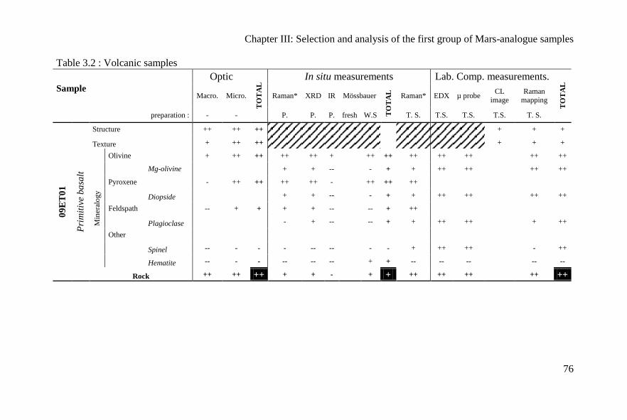
Chapter III: Selection and analysis of the first group of Mars-analogue samples
76
Table 3.2 : Volcanic samples
Sample
Optic
TO
TA
L In situ measurements Lab. Comp. measurements.
TO
TA
L
Macro. Micro. Raman* XRD IR Mössbauer
TO
TA
L
Raman* EDX µ probe CL
image
Raman
mapping
preparation : - - P. P. P. fresh W.S T. S. T.S. T.S. T.S. T. S.
09
ET
01
Pri
mit
ive
ba
salt
Structure ++ ++ ++ + + +
Texture + ++ ++
+ + +
Min
eral
ogy
Olivine + ++ ++ ++ ++ +
++ ++ ++ ++ ++
++ ++
Mg-olivine
+ + --
- + + ++ ++
++ ++
Pyroxene - ++ ++ ++ ++ -
++ ++ ++
Diopside + + --
- + + ++ ++
++ ++
Feldspath -- + + + + --
-- + ++
Plagioclase
- + --
-- + + ++ ++
+ ++
Other
Spinel -- - - - -- --
- - + ++ ++
- ++
Hematite -- - - -- -- --
+ + -- -- --
-- --
Rock ++ ++ ++ + + - + + ++ ++ ++ ++ ++
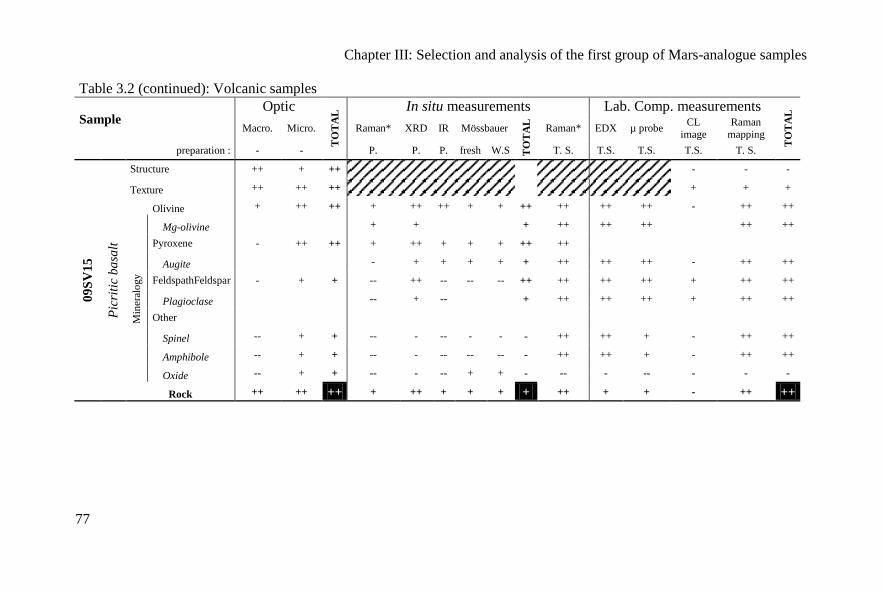
Chapter III: Selection and analysis of the first group of Mars-analogue samples
77
Table 3.2 (continued): Volcanic samples
Sample Optic
TO
TA
L In situ measurements Lab. Comp. measurements
TO
TA
L
Macro. Micro. Raman* XRD IR Mössbauer
TO
TA
L
Raman* EDX µ probe CL
image
Raman
mapping
preparation : - - P. P. P. fresh W.S T. S. T.S. T.S. T.S. T. S.
09
SV
15
Pic
riti
c b
asa
lt
Structure ++ + ++
- - -
Texture ++ ++ ++
+ + +
Olivine + ++ ++ + ++ ++ + + ++ ++ ++ ++ - ++ ++
Min
eral
ogy
Mg-olivine
+ +
+ ++ ++ ++
++ ++
Pyroxene - ++ ++ + ++ + + + ++ ++
Augite
- + + + + + ++ ++ ++ - ++ ++
FeldspathFeldspar - + + -- ++ -- -- -- ++ ++ ++ ++ + ++ ++
Plagioclase
-- + --
+ ++ ++ ++ + ++ ++
Other
Spinel -- + + -- - -- - - - ++ ++ + - ++ ++
Amphibole -- + + -- - -- -- -- - ++ ++ + - ++ ++
Oxide -- + + -- - -- + + - -- - -- - - -
Rock ++ ++ ++ + ++ + + + + ++ + + - ++ ++

Chapter III: Selection and analysis of the first group of Mars-analogue samples
78
Table 3.2 (continued): Volcanic samples
Sample Optic
TO
TA
L In situ measurements Lab. Comp. measurements
TO
TA
L
Macro. Micro. Raman* XRD IR Mössbauer
TO
TA
L
Raman* EDX µ probe CL
image
Raman
mapping
preparation : - - P. P. P. fresh W.S T. S. T.S. T.S. T.S. T. S.
09
TE
08
Alt
ered
ph
ono
lite
Structure ++ + ++
+ - -
Texture + ++ ++
+ + +
Min
eral
ogy
Feldspar -- ++ ++ ++ ++ ++ -- -- ++ ++ ++ ++
++ ++
Plagioclase + + + ++ ++
++ ++ ++ ++
++ ++
Amphibole -- ++ ++ -- -- -- + + + ++ ++ ++
++ ++
Tremolite
- - - ++ ++ ++
++ ++
Apatite -- + + -- -- -- -- -- -- ++ ++ ++
++ ++
Cl-Apatite
++ ++ ++
++ ++
Other
Spinel -- + + -- -- -- + + + -- ++ +
+ ++
Quartz -- ++ ++ -- -- -- -- -- -- ++ ++ ++
++ ++
Rock - + + - + + - - + ++ ++ ++ ++ ++
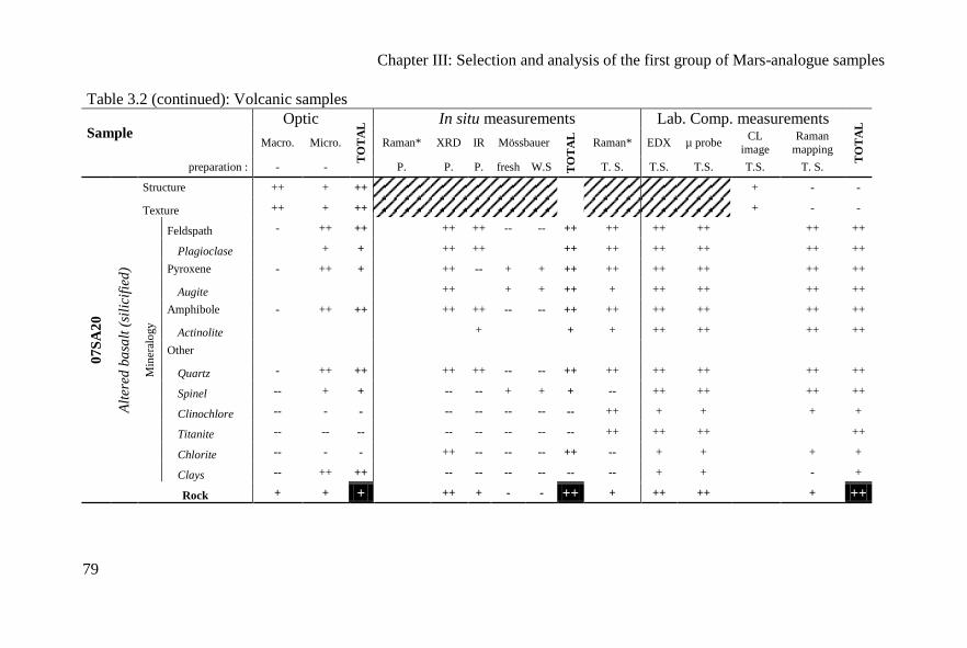
Chapter III: Selection and analysis of the first group of Mars-analogue samples
79
Table 3.2 (continued): Volcanic samples
Sample Optic
TO
TA
L In situ measurements Lab. Comp. measurements
TO
TA
L
Macro. Micro. Raman* XRD IR Mössbauer
TO
TA
L
Raman* EDX µ probe CL
image
Raman
mapping
preparation : - - P. P. P. fresh W.S T. S. T.S. T.S. T.S. T. S.
07
SA
20
Alt
ered
ba
salt
(si
lici
fied
)
Structure ++ + ++
+ - -
Texture ++ + ++
+ - -
Min
eral
ogy
Feldspath - ++ ++ ++ ++ -- -- ++ ++ ++ ++
++ ++
Plagioclase + + ++ ++
++ ++ ++ ++
++ ++
Pyroxene - ++ + ++ -- + + ++ ++ ++ ++
++ ++
Augite
++
+ + ++ + ++ ++
++ ++
Amphibole - ++ ++ ++ ++ -- -- ++ ++ ++ ++
++ ++
Actinolite
+
+ + ++ ++
++ ++
Other
Quartz - ++ ++ ++ ++ -- -- ++ ++ ++ ++
++ ++
Spinel -- + + -- -- + + + -- ++ ++
++ ++
Clinochlore -- - - -- -- -- -- -- ++ + +
+ +
Titanite -- -- -- -- -- -- -- -- ++ ++ ++
++
Chlorite -- - - ++ -- -- -- ++ -- + +
+ +
Clays -- ++ ++ -- -- -- -- -- -- + +
- +
Rock + + + ++ + - - ++ + ++ ++ + ++
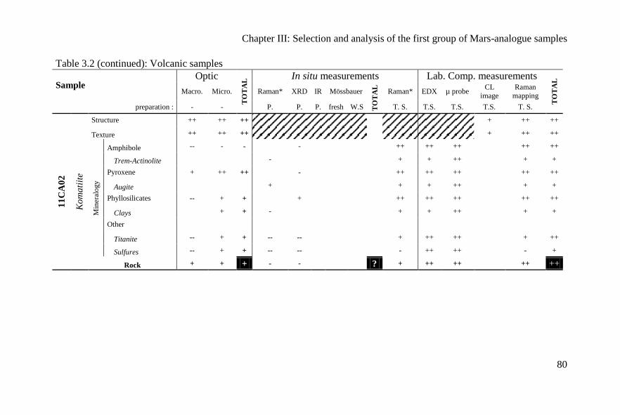
Chapter III: Selection and analysis of the first group of Mars-analogue samples
80
Table 3.2 (continued): Volcanic samples
Sample Optic
TO
TA
L In situ measurements Lab. Comp. measurements
TO
TA
L
Macro. Micro. Raman* XRD IR Mössbauer
TO
TA
L
Raman* EDX µ probe CL
image
Raman
mapping
preparation : - - P. P. P. fresh W.S T. S. T.S. T.S. T.S. T. S.
11
CA
02
Ko
ma
tiit
e
Structure ++ ++ ++ + ++ ++
Texture ++ ++ ++
+ ++ ++
Min
eral
ogy
Amphibole -- - -
-
++ ++ ++
++ ++
Trem-Actinolite
-
+ + ++
+ +
Pyroxene + ++ ++
-
++ ++ ++
++ ++
Augite
+
+ + ++
+ +
Phyllosilicates -- + +
+
++ ++ ++
++ ++
Clays + + -
+ + ++
+ +
Other
Titanite -- + + -- --
+ ++ ++
+ ++
Sulfures -- + + -- --
- ++ ++
- +
Rock + + + - - ? + ++ ++ ++ ++
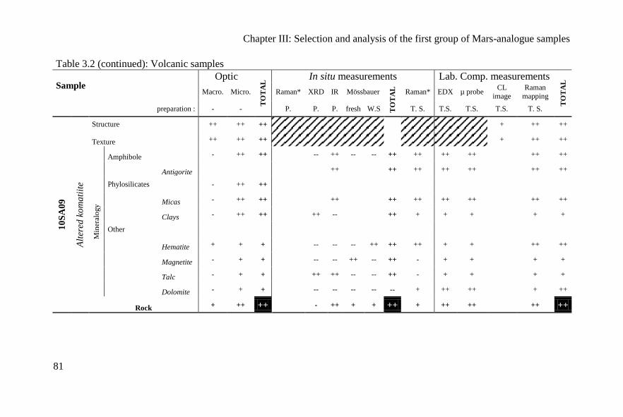
Chapter III: Selection and analysis of the first group of Mars-analogue samples
81
Table 3.2 (continued): Volcanic samples
Sample Optic
TO
TA
L In situ measurements Lab. Comp. measurements
TO
TA
L
Macro. Micro. Raman* XRD IR Mössbauer
TO
TA
L
Raman* EDX µ probe CL
image
Raman
mapping
preparation : - - P. P. P. fresh W.S T. S. T.S. T.S. T.S. T. S.
10
SA
09
Alt
ered
ko
ma
tiit
e
Structure ++ ++ ++ + ++ ++
Texture ++ ++ ++
+ ++ ++
Min
eral
ogy
Amphibole - ++ ++ -- ++ -- -- ++ ++ ++ ++
++ ++
Antigorite
++
++ ++ ++ ++
++ ++
Phylosilicates - ++ ++
Micas - ++ ++
++
++ ++ ++ ++
++ ++
Clays - ++ ++ ++ --
++ + + +
+ +
Other
Hematite + + + -- -- -- ++ ++ ++ + +
++ ++
Magnetite - + + -- -- ++ -- ++ - + +
+ +
Talc - + + ++ ++ -- -- ++ - + +
+ +
Dolomite - + + -- -- -- -- -- + ++ ++
+ ++
Rock + ++ ++ - ++ + + ++ + ++ ++ ++ ++
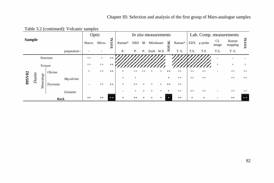
Chapter III: Selection and analysis of the first group of Mars-analogue samples
82
Table 3.2 (continued): Volcanic samples
Sample
Optic
TO
TA
L In situ measurements Lab. Comp. measurements
TO
TA
L
Macro. Micro. Raman* XRD IR Mössbauer
TO
TA
L
Raman* EDX µ probe CL
image
Raman
mapping
preparation : - - P. P. P. fresh W.S T. S. T.S. T.S. T.S. T. S.
09
SV
02
Du
nit
e
Structure ++ + ++
- - -
Texture ++ ++ ++
+ + +
Min
eral
ogy Olivine + ++ ++ + ++ ++ + + ++ ++ ++ ++ - ++ ++
Mg-olivine
+ +
+ ++ ++ ++
++ ++
Pyroxene - ++ ++ + ++ + + + ++ ++
Enstatite
- + + + + + ++ ++ ++ - ++ ++
Rock ++ ++ ++ + ++ + + + + ++ + + - ++ ++
Chapter III: Selection and analysis of the first group of Mars-analogue samples
83
►Table 3.3. Comparison of textural, elemental and mineralogical data from the
sedimentary rocks using different instruments and different sample
preparations (rock surface, powder, thin section). (++) unambiguous
identification, (+) probably identification, (-) identification ambiguous, (--)
identification impossible. Macro.: Macroscopic view, Micro.: Microscopic
view, P.: Powder, W.S. : Weathered surface, T.S.: Thin section - * only
Raman spectrum- The hatched area indicate that the measurement is not
applicable for a specific instrument.
►Tableau 3.3. Comparaison de la texture, des données élémentaires et minéralogiques
des échantillons sédimentaires à partir des différents instruments et des différentes
préparations (surface brute de la roche, poudre et lame mince). (++) identification
certaine, (+) identification probable, (-) identification ambigüe, (--) identification
impossible. Macro : observation macroscopique, Micro : observation microscopique,
P : poudre, W.S. surface altérée, T.S. : lame mince - * : seulement spectre Raman-
Les zones hachurées indiquent que la mesure n’est pas réalisable avec l’instrument.
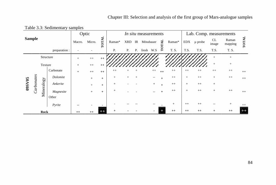
Chapter III: Selection and analysis of the first group of Mars-analogue samples
84
Table 3.3: Sedimentary samples
Sample
Optic
TO
TA
L In situ measurements Lab. Comp. measurements
TO
TA
L
Macro. Micro. Raman* XRD IR Mössbauer
TO
TA
L
Raman* EDX µ probe CL
image
Raman
mapping
preparation : - - P. P. P. fresh W.S T. S. T.S. T.S. T.S. T. S.
09
SV
05
Ca
rbo
na
tes
Structure + ++ ++
+ +
Texture + ++ ++
+ +
Min
eral
og
y
Carbonate + ++ ++ ++ + +
++ ++ ++ ++ ++ ++ ++ ++
Dolomite
+ + + + +
-- + ++ + ++ + ++ ++
Ankerite
+ + + - -
+ + ++ + ++ +
Magnesite
+ + + - -
-- + ++ + ++ + ++ ++
Other
Pyrite -- - - - -- --
-- - + ++ ++ -- + ++
Rock ++ ++ ++ + - - - + ++ ++ ++ + ++ ++
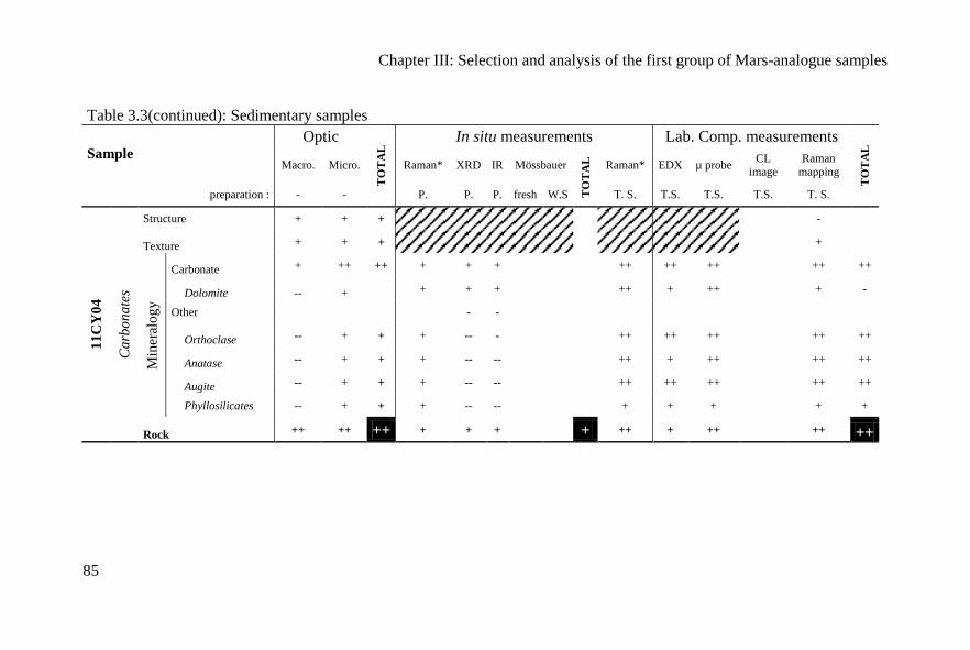
Chapter III: Selection and analysis of the first group of Mars-analogue samples
85
Table 3.3(continued): Sedimentary samples
Sample
Optic
TO
TA
L In situ measurements Lab. Comp. measurements
TO
TA
L
Macro. Micro. Raman* XRD IR Mössbauer
TO
TA
L
Raman* EDX µ probe CL
image
Raman
mapping
preparation : - - P. P. P. fresh W.S T. S. T.S. T.S. T.S. T. S.
11
CY
04
Ca
rbo
na
tes
Structure + + + -
Texture + + +
+
Min
eral
og
y
Carbonate + ++ ++ + + +
++ ++ ++
++ ++
Dolomite -- + + + +
++ + ++
+ -
Other
- -
Orthoclase -- + + + -- -
++ ++ ++
++ ++
Anatase -- + + + -- --
++ + ++
++ ++
Augite -- + + + -- --
++ ++ ++
++ ++
Phyllosilicates -- + + + -- --
+ + +
+ +
Rock ++ ++ ++ + + + + ++ + ++ ++ ++
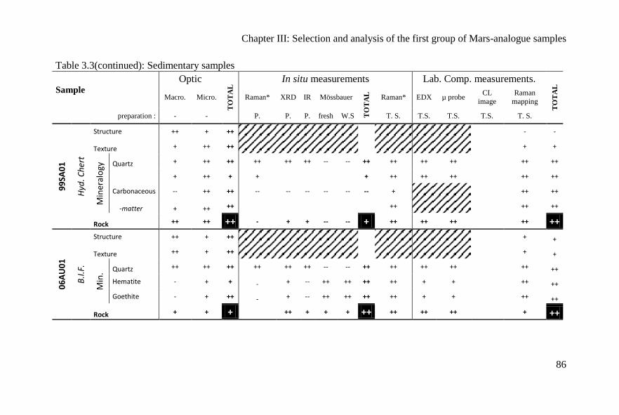
Chapter III: Selection and analysis of the first group of Mars-analogue samples
86
Table 3.3(continued): Sedimentary samples
Sample
Optic
TO
TA
L In situ measurements Lab. Comp. measurements.
TO
TA
L
Macro. Micro. Raman* XRD IR Mössbauer
TO
TA
L
Raman* EDX µ probe CL
image
Raman
mapping
preparation : - - P. P. P. fresh W.S T. S. T.S. T.S. T.S. T. S.
99
SA0
1
Hyd
. Ch
ert
Structure ++ + ++
- -
Texture + ++ ++
+ +
Min
eral
ogy
Quartz + ++ ++ ++ ++ ++ -- -- ++ ++ ++ ++
++ ++
+ ++ + +
+ ++ ++ ++
++ ++
Carbonaceous -- ++ ++ -- -- -- -- -- -- +
++ ++
-matter + ++ ++
++
++ ++
Rock ++ ++ ++ - + + -- -- + ++ ++ ++ ++ ++
06
AU
01
B.I
.F.
Structure ++ + ++
+ +
Texture ++ + ++
+ +
Min
. Quartz ++ ++ ++ ++ ++ ++ -- -- ++ ++ ++ ++
++ ++
Hematite - + + - + -- ++ ++ ++ ++ + +
++ ++
Goethite - + ++ - + -- ++ ++ ++ ++ + +
++ ++
Rock + + + ++ + + + ++ ++ ++ ++ + ++
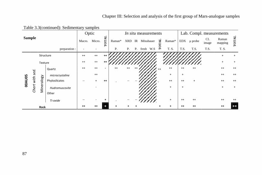
Chapter III: Selection and analysis of the first group of Mars-analogue samples
87
Table 3.3(continued): Sedimentary samples
Sample
Optic
TO
TA
L In situ measurements Lab. Compl. measurements
TO
TA
L
Macro. Micro. Raman* XRD IR Mössbauer
TO
TA
L
Raman* EDX µ probe CL
image
Raman
mapping
preparation : - - P. P. P. fresh W.S T. S. T.S. T.S. T.S. T. S.
00
AU
05
Ch
ert
wit
h s
ed.
Structure ++ ++ ++
+ +
Texture ++ ++ ++
+ +
Min
eral
ogy
Quartz ++ ++ - ++ ++ ++ ++ ++ ++ ++
++ ++
microcrystaline ++
+ +
++ ++
Phylosilicates -- + ++ - -- -- - ++ ++ +
++ ++
Hudromuscovite -
+ +
+ +
Other
Ti-oxide -- - + - -- --
+ ++ ++
++ ++
Rock ++ ++ + + + + + + ++ ++ ++ ++
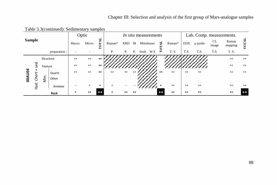
Chapter III: Selection and analysis of the first group of Mars-analogue samples
88
Table 3.3(continued): Sedimentary samples
Sample
Optic
TO
TA
L In situ measurements Lab. Comp. measurements.
TO
TA
L
Macro. Micro. Raman* XRD IR Mössbauer
TO
TA
L
Raman* EDX µ probe CL
image
Raman
mapping
preparation : - - P. P. P. fresh W.S T. S. T.S. T.S. T.S. T. S.
00
AU
04
Hyd
. Ch
ert
+ se
d.
Structure ++ ++ ++
++ ++
Texture ++ ++ ++
++ ++
Min
. Quartz ++ ++ ++ ++ ++ ++
++ ++ ++ ++
++ ++
Other
Anatase -- + + + -- --
+ ++ ++ ++
++ ++
Rock + ++ ++ + ++ ++ ++ ++ ++ ++ ++ ++

Chapter III: Selection and analysis of the first group of Mars-analogue
samples
89
III.4. Mineral selection
The minerals chosen for the ISAR collection have been identified on
Mars and include calcium carbonates (aragonite and calcite) and a Mg-
carbonate (magnesite); silicates including the nesosilicate olivine, the
sorosilicate epidote, the cyclosilicate tourmaline (schorl), the inosilicates
enstatite, wollastonite and diopside, and the phyllosilicates muscovite and
nontronite. Tectosilicates could be represented in the form of quartz from the
different cherts described above. Opal was selected as an hydrated silica
polymorph (cf. Squyres et al, 2008 and Ruff et al., 2011). A phosphate,
apatite, and the sulphates gypsum and barite were also added to the collection.
Barite has not yet been observed on Mars, but as a major terrestrial suphate, it
could be useful for calibrating instruments. Finally, I selected different Fe-
and Ti-oxides to complete this mineral collection (goethite, magnetite, rutile)
and a spinel. Lastly, we are using a laboratory-synthesised nontronite
(10AR01) as an analogue of phyllosilicates forming in neutral conditions by
aqueous alteration of basalts on Mars (Bibring et al., 2006 and Meunier et al.,
2010). Naturally-formed nontronites display a wide range of compositions.
The Table 3.4 summarizes the selection.
►Table 3.4: List of analogue minerals characterized for the ISAR collection
and calculated chemical formulae using electron microprobe analysis
(except for the artificial nontronite).
►Tableau 3.4: Liste des échantillons analogues caractérisés pour la collection de
l’ISAR avec les formules structurales obtenues par microsonde électronique (sauf
pour la nontronite artificielle).
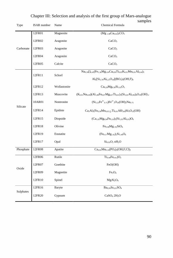
Chapter III: Selection and analysis of the first group of Mars-analogue
samples
90
Type ISAR number Name Chemical Formula
Carbonate
12FR01 Magnesite (Mg 1,98Ca0,02)2CO3
12FR02 Aragonite CaCO3
12FR03 Aragonite CaCO3
12FR04 Aragonite CaCO3
12FR05 Calcite CaCO3
Silicate
12FR11 Schorl
Na0,58[]0,42(Fe1,36Mg1,04Ca0,09Ti0,05K0,01Mn0,01Al0,44)3
Al6[Si5,78Al0,21O18](BO3)3(OH,F)4
12FR12 Wollastonite Ca0,28Mg0,58Si1,02O3
12FR13 Muscovite (K0,91Na0,09)(Al1,89Fe0,07Mg0,07Ti0,01)1(Si3,01Al0,99)4O10(OH) 2
10AR01 Nontronite (Si3.25Fe3+0.75)Fe3+
2O10(OH)2Na0.75
12FR14 Epidote Ca2Al2(Fe0,39Mn0,02☺Ti0,01Al0,68)Si3O12(OH)
12FR15 Diopside (Ca1,02Mg0,94Fe0,10)2(Si1,93Al0,03)O6
12FR18 Olivine Fe0.08Mg1.92SiO4
12FR19 Enstatite (Fe0.17Mg1.92)2Si1.94O6
12FR17 Opal Si0.95O2 nH2O
Phosphate 12FR08 Apatite Ca4.81Mn0.14(PO4)3(OH,F,Cl)1
Oxide
12FR06 Rutile Ti0.99Fe0.01)O2
12FR07 Goethite FeO(OH)
12FR09 Magnetite Fe3O4
12FR10 Spinel MgAl2O4
Sulphates
12FR16 Baryte Ba0,99Sr0,01SO4
12FR20 Gypsum CaSO4 2H2O
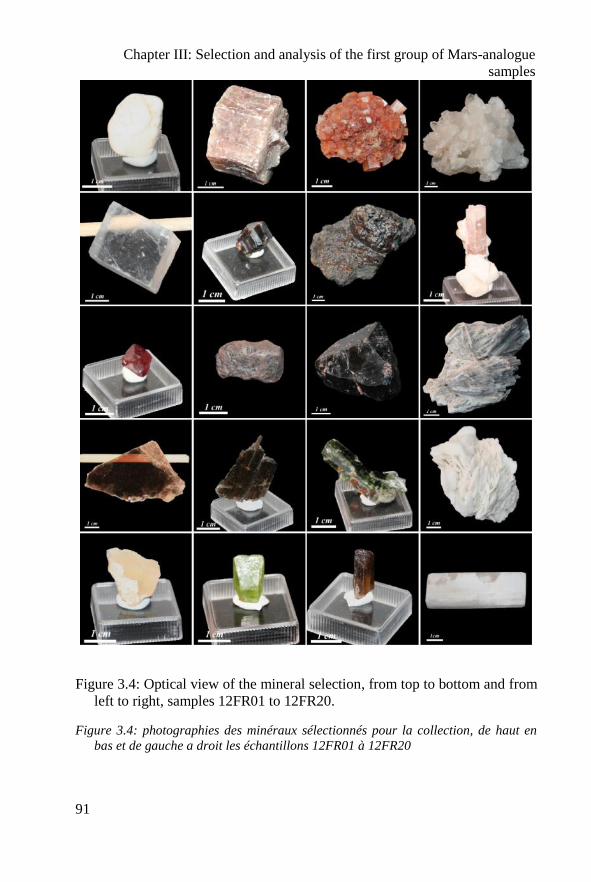
Chapter III: Selection and analysis of the first group of Mars-analogue
samples
91
Figure 3.4: Optical view of the mineral selection, from top to bottom and from
left to right, samples 12FR01 to 12FR20.
Figure 3.4: photographies des minéraux sélectionnés pour la collection, de haut en
bas et de gauche a droit les échantillons 12FR01 à 12FR20
Chapter III: Selection and analysis of the first group of Mars-analogue
samples
92
The minerals were studied without treatment and as polished sections
by Raman spectroscopy. The polished sections were also used for
SEMobservation and SEM-EDXS and electron microprobe analysis for the
elemental composition. The chemical formulae in Table 3.4 were calculated
on the basis of a mean value from 40 electronic microprobe measurements.
The pure Fe-nontronite was synthesised at 150°C in the Hydrasa
laboratory, Poitiers, by the group of Sabine Petit and Alain Meunier.
Cristallographic characterization was performed by Decarreau et al., (2008).
All details on each sample are listed in the online web site: www.isar.cnrs-
orleans.fr.
III.5 Conclusion
We have created a preliminary collection of Mars-analogue rocks and
minerals for testing space instrumentation for the upcoming in situ missions to
Mars. These materials include a variety of basalts, the most common rock
type on Mars, as well as volcanic and chemical sediments (carbonates, BIF,
cherts) and various minerals. However, the selection of terrestrial analogues
of Martian materials is limited by some major geochemical differences in the
compositions of, especially, Fe-containing rocks on the two planets. For
example, Martian basalts contain much more Fe and Mg than terrestrial
basalts. Within this limitation, the rocks we have chosen correspond as closely
as possible to Martian materials. The collection of rocks in the ISAR thus
provides a relevant selection of materials the structure, texture, and
mineralogical and geochemical compositions of which will be useful for
testing spaceflight instrumentation and will help to interpret the nature of the
rocks on the Martian surface and their potential for containing and preserving
traces of life. The relevance of the sample analogy is detailed in Table 3.5.
The analogy based on the different characteristics studied: geochemistry,
petrology, (include texture and structures) and geotechnical properties are
described in Table 3.5 using stars, 1 star to 5 stars representing weak to
excellent correspondence to Martian materials. The geotechnical property
results are based on the estimation of the resistivity of the samples and the
opportunity to find this type of sample morphology on the Martian surface.
The minerals are not included in this Table because they are not relevant for
geochemistry and petrology.
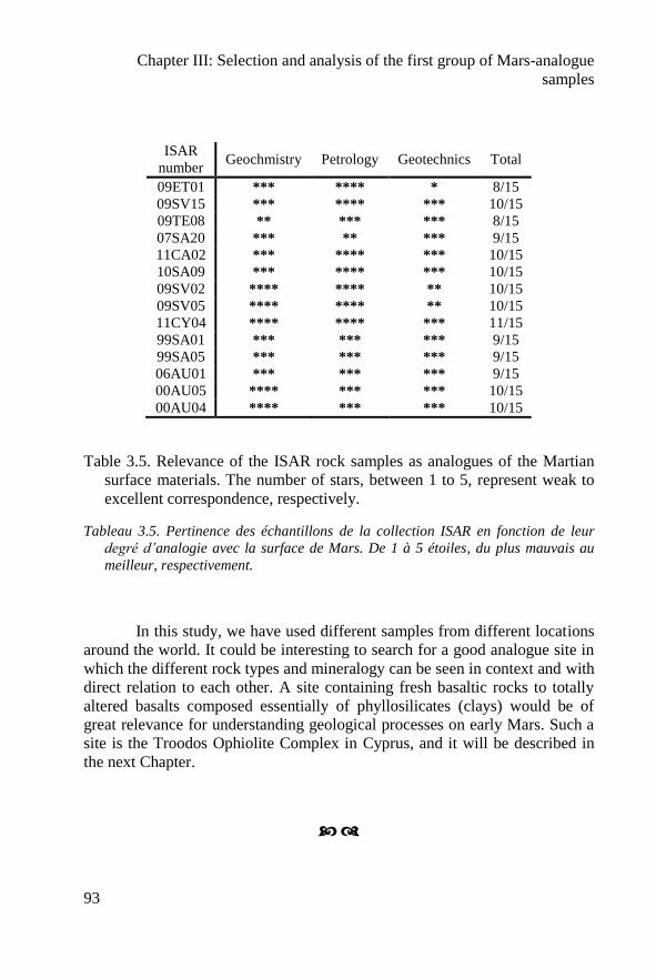
Chapter III: Selection and analysis of the first group of Mars-analogue
samples
93
ISAR
number Geochmistry Petrology Geotechnics Total
09ET01 *** **** * 8/15
09SV15 *** **** *** 10/15
09TE08 ** *** *** 8/15
07SA20 *** ** *** 9/15
11CA02 *** **** *** 10/15
10SA09 *** **** *** 10/15
09SV02 **** **** ** 10/15
09SV05 **** **** ** 10/15
11CY04 **** **** *** 11/15
99SA01 *** *** *** 9/15
99SA05 *** *** *** 9/15
06AU01 *** *** *** 9/15
00AU05 **** *** *** 10/15
00AU04 **** *** *** 10/15
Table 3.5. Relevance of the ISAR rock samples as analogues of the Martian
surface materials. The number of stars, between 1 to 5, represent weak to
excellent correspondence, respectively.
Tableau 3.5. Pertinence des échantillons de la collection ISAR en fonction de leur
degré d’analogie avec la surface de Mars. De 1 à 5 étoiles, du plus mauvais au
meilleur, respectivement.
In this study, we have used different samples from different locations
around the world. It could be interesting to search for a good analogue site in
which the different rock types and mineralogy can be seen in context and with
direct relation to each other. A site containing fresh basaltic rocks to totally
altered basalts composed essentially of phyllosilicates (clays) would be of
great relevance for understanding geological processes on early Mars. Such a
site is the Troodos Ophiolite Complex in Cyprus, and it will be described in
the next Chapter.

Chapter III: Selection and analysis of the first group of Mars-analogue
samples
94
Chapter III: Selection and analysis of the first group of Mars-analogue
samples
95
Première sélection (Résumé Français)
Ce chapitre présente le premier groupe d’échantillons sélectionnés comme
analogues de Mars. Le choix des échantillons est basé sur la géologie de Mars décrite
dans le précédent chapitre puis en sélectionnant des échantillons déjà disponibles
dans le laboratoire. En effet, l’équipe d’Exobiologie, par l’intermédiaire de F.
Westall, a déjà rassemblé une large collection de roches en particulier des roches
volcaniques et sédimentaires de l’Archéen. D’autres échantillons ont été envoyés par
des spécialistes ou recueillis lors de mes propres missions de terrain à Rio Tinto, en
Afrique du Sud, à Chypre et dans le Massif Central Français.
Ce chapitre est centré sur la sélection d’échantillons analogues de Mars.
J’ai cherché à évaluer le degré de pertinence de chaque échantillon en utilisant de
nombreuses methodes d’analyse à l’aide d’instruments de laboratoire en corrélation
avec les instruments spatialisés pour l’exploration martienne. Ce chapitre présente
brièvement les données obtenues pour chaque échantillon. Pour plus d’informations,
l’annexe B présente les résultats détaillés obtenus sur deux échantillons provenant de
Svalbard (Norvège).
Mars étant essentiellement composée de basaltes, je me suis principalement
concentré sur l’étude des basaltes. Nous avons choisi comme échantillon un basalte
de Svalbard (09SV15) car il a déjà été décrit comme analogue de la météorite
martienne ALH84001 et parce qu’il est associé à des carbonates (09SV05) et à une
dunite (09SV02). La dunite est une roche caractéristique du manteau terrestre. Nous
avons choisi un échantillon de Tenerife (09TE08) car son rapport Ca/Si vs. Mg/Si est
très similaire aux roches de Mars. De plus cet échantillon peut être représentatif de
basaltes altérés. Il ne faut pas oublier que les basaltes, au cours de leur altération,
peuvent contenir des traces de vie. Nous avons choisi l’échantillon de basalte silicifié
(07SA20) provenant de Barberton en Afrique du Sud car il est représentatif du
processus de silicification associé à de l’hydrothermalisme. Ces phénomènes sont
caractéristiques de la Terre primitive. J’ai choisi un basalte primitif de l’Etna
(09ET01) car il est géochimiquement très similaire à la moyenne des basaltes
martiens (Fig. 3.1). Enfin, j’ai ajouté deux komatiites, dont une peu altérée provenant
du Canada (11CA02) et une autre beaucoup plus alterée provenant d’Afrique du Sud
(10SA09). Ces roches semblent avoir été observées sur Mars et sont des roches
volcaniques typiques de l’Archéen (Terre).
Plusieurs types de roches sédimentaires ont été incorporés dans la première
sélection en partie en fonction de leur intérêt exobiologique, en accord avec les
objectifs des futures missions vers Mars. Comme nous l’avons vu dans le chapitre
précédent, la vie sur Mars n’a pu apparaître qu’entre le Noachien et l’Hespérien,
lorsque que l’eau liquide était présente en surface. Les roches similaires à la surface
de la Terre (datées à l’Archéen), contenant des traces fossiles sont des dépôts
volcaniques silicifiés par des fluides hydrothermaux. Cette activité hydrothermale a
déjà été suggérée sur Mars, et pourrait avoir contribué à préserver des
microorganismes. Pour cette sélection, nous avons choisi des sédiments volcaniques
silicifiés provenant de Pilbara, Australie et datés de 3.45 Ga (00AU05). Nous avons
Chapter III: Selection and analysis of the first group of Mars-analogue
samples
96
ajouté des chailles du même âge contenant ou non de la matière carbonée provenant
de Barberton, Afrique du Sud ou encore de Pilbara (99SA01, 99SA05 et 00AU04).
Pour compléter la sélection de roches siliceuses, j’ai ajouté une roche férrifère
rubanée (BIF). Ces roches n’ont pas encore été observées sur Mars mais sont très
riches en fer et y existent très probablement. Enfin, quelques carbonates sont visibles
sur Mars, j’ai donc sélectionné deux échantillons de carbonates dont l’origine est
plutôt hydrothermale et associée au volcanisme. Ces échantillons proviennent de
Svalbard (09SV05) et de Chypre (11CY04). Le Tableau 3.1 présente la liste des
échantillons étudiés dans ce chapitre. La deuxième partie de ce chapitre correspond à
la description de chaque échantillon, qui ne sera pas reprise dans ce résumé.
Cependant pour illustrer ce choix d’échantillons les Figs. 3.2 et 3.3 présentent des
photos e macroscopiques et en microscopique de chaque échantillon.
La troisième partie de ce chapitre résume pour chaque échantillon le résultat
de chaque analyse en utilisant les instruments de laboratoire. Le détail des analyses
est accessible à partir du site internet et de la base de données associée :
http://www.isar.cnrs-orleans.fr. Les Tableaux 3.2 et 3.3 présentent l’évaluation
qualitative inhérente à l’identification de la structure, texture, minéralogie et
composition pour chaque instrument et en les combinant. Cette étude montre que
même en combinant les instruments de laboratoire, donc certainement encore plus
avec les instruments de vols, les interprétations peuvent parfois être compliquées. Les
premiers résultats démontrent la limite d’interprétation en utilisant des préparations
en poudre. En effet, les textures et structures sont irrémédiablement perdues. Nous
pensons qu’avec les futurs instruments d’ExoMars, seulement 50% des minéraux
pourront être identifiés avec certitude. En effet, l’apport des caméras couleurs et du
microscope binoculaire (loupe) sera essentiel pour aider à l’identification et
l’interprétation des roches.
Finalement, nous avons sélectionné différents minéraux de bonnes qualités
chimiques et cristallographiques. La liste des minéraux est proposée dans le Tableau
3.4 et la Fig. 3.4, ainsi que leurs analyses chimiques. Ces minéraux ont été identifiés
sur Mars et sont les constituants principaux des roches. Nous avons sélectionné des
carbonates de calcium (aragonite et calcite) de magnésium (magnesite) ; différents
types de silicates : nésosilicate (olivine), sorosilicate (epidote), cyclosilicate (schorl),
inosilicates (enstatite, wollastonite et diopside) et phyllosilicates (muscovite et
nontronite). Notre sélection ne comporte pas de tectosilicates, qui peuvent être
retrouvés dans le quartz des chailles sélectionnées précédemment. Nous avons
sélectionné de l’opale, une silice hydratée. J’ai aussi ajouté un phosphate (apatite),
des sulfates (gypse et barytine). Enfin j’ai sélectionné des oxydes de fer et de titane
(goethite, magnétite et rutile). Une nontronite de synthèse a été fabriquée au
laboratoire Hydrasa (Université de Poitiers).
Nous avons créé la collection de l’ISAR avec une large variété de roches et
de minéraux afin de couvrir le plus largement possible les matériaux analogues de
Mars. Cependant, il existe de nombreuses différences entre la Terre et Mars, les
compositions chimiques ne sont pas exactement les mêmes. Par exemple, les basaltes
martiens contiennent plus de Fe et Mg que leurs analogues terrestres. Mais les
Chapter III: Selection and analysis of the first group of Mars-analogue
samples
97
conditions d’altération des roches sont aussi particulièrement différentes. Nous avons
essayé d’établir le degré d’analogie de nos échantillons avec ceux de Mars en nous
basant sur trois critères : la géochimie, la pétrologie et la géotechnique. Les
échantillons, notés de 1 à 5 étoiles, sont de plus ou moins bons analogues. Le Tableau
3.5 présente les résultats. .
J’ai cherché un site analogue à Mars où il serait possible de trouver en un
même endroit des basaltes et leurs produits d’altération obtenus dans des conditions
similaires à celles de Mars. Le prochain chapitre va présenter les résultats de l’étude
menée à Chypre qui semble être un site analogue.

Chapter III: Selection and analysis of the first group of Mars-analogue
samples
98
Chapter IV: A new Mars analogue site in Cyprus
99
Chapter IV
A new Mars analogue site in Cyprus
In this chapter, I describe a new Mars analogue site located in the
Skouriotissa mine, in North West Cyprus, from which I collected samples in
March and April 2011. Here the surfaces of outcropping ophiolitic basalts
undergo various types of chemical alteration, in particular by acidic water,
resulting in different mineralogies12
.
IV.1 Introduction
The previous chapter brought to light the difficulty of finding good
analogues of Martian rocks and soils on Earth. I therefore searched for new
terrestrial sites that are geologically more similar to the early and present Mars
outcrops, i.e. a site where the main rocks are basalts (at least the basic body) that
have been altered by the processes described in Chapter II part 2.2. However, it is
obviously impossible to find similar atmospheric conditions (i.e., without
oxygen) at the surface of the Earth. All the known analogue sites (e.g. Rio Tinto
in Spain, Amils et al., 2007, or Utah Sandstone in USA, Chan et al., 2004) focus
on present Martian conditions, and in particular on iron oxides, and are
sometimes not very similar to Martian rocks from a geological point of view. The
site of Rio Tinto in the Spanish pyrite belt, for instance, is used as an analogue
site mainly in terms of the search for present life on Mars (extremophile life,
NASA laboratory place), but the geological context is andesitic, i.e., made of
12
A part of the content of this chapter is in review in Geology (Bost et al., in review).
100
more acidic volcanic rocks that are not very representative of the Martian
surface. Moreover, these rocks are highly metamorphosed, at least to amphibolite
facies, and are thus are unlike Martian rocks.
As noted in chapter II, habitable conditions for the appearance of life
were possible during the Noachian period on Mars. With this in mind, the search
for Noachian analogue sites on Earth is more relevant (Westall et al., 2011a;
Bibring et al., 2006). At such a site, the alteration must occur under the effect of
hydrothermal and meteoric or acidic water. Thus, mines are good objectives
because they favour the production of acidic water. I therefore took advantage of
an opportunity to join a field trip to Cyprus, organized by a team from the CNRS-
ISTO-Orléans laboratory interested in epidotitisation processes in the deep
oceanic crust close to the copper mines, in particular in the Skouriotissa mine.
IV.2. Location
Cyprus is a small elongated island (~ 10 000 km2) located in the east of
the Mediterranean Sea, to the south of the Turkish Anatolia (Fig. 4.1a). The
Skouriotissa mine is located in the west of the island, south of the Morfou gulf,
between Katydata to the south and Petra to the north. The GPS position of the
open-pit is: N35.095364°; E32.887338°. The Skouriotissa mine is exploited
essentially for copper by the Hellenic copper Limited company with 3 billion
tons of proven ore reserves at a grade of 0.1 to 0.25 % of Cu (max: 2.35 % of
Cu).
IV.3. Geological Background
The Troodos ophiolite (Fig. 4.1b) has been recognized for a long time as
an uplifted slice of oceanic crust and mantle that was created through sea-floor
spreading (Gass, 1968 and Moores and Vine, 1971). Forming the central region
of the eastern Mediterranean island of Cyprus, the ophiolite displays a dome-like
structure centred on Mount Olympus (1952 m). The ophiolite formation dates
back to the upper Cretaceous. Its stratigraphy includes a mantle sequence
consisting of peridotites (harzburgites, dunites) and a serpentinite diapir exposed
at the highest elevation. Along the north slope of the ophiolite, the mantle
sequence is stratigraphically overlapped by a large gabbroic plutonic complex, a
sheeted dyke complex, extrusive lavas (Pillow lavas) and oceanic sediments (Fig.
4.1c). The pillow lavas are divided into the Upper Pillow Lavas and the Lower
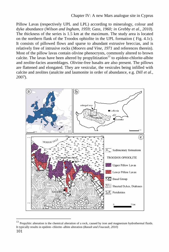
Chapter IV: A new Mars analogue site in Cyprus
101
Pillow Lavas (respectively UPL and LPL) according to mineralogy, colour and
dyke abundance (Wilson and Ingham, 1959; Gass, 1960; in Grebby et al., 2010).
The thickness of the series is 1.5 km at the maximum. The study area is located
on the northern flank of the Troodos ophiolite in the UPL formation ( Fig. 4.1c).
It consists of pillowed flows and sparse to abundant extrusive breccias, and is
relatively free of intrusive rocks (Moores and Vine, 1971 and references therein).
Most of the pillow lavas contain olivine phenocrysts, commonly altered to brown
calcite. The lavas have been altered by propylitization13
to epidote-chlorite-albite
and zeolite-facies assemblages. Olivine-free basalts are also present. The pillows
are flattened and elongated. They are vesicular, the vesicules being infilled with
calcite and zeolites (analcite and laumonite in order of abundance, e.g. Dill et al.,
2007).
13
Propylitic alteration is the chemical alteration of a rock, caused by iron and magnesium hydrothermal fluids.
It typically results in epidote–chlorite–albite alteration (Raoult and Foucault, 2010)
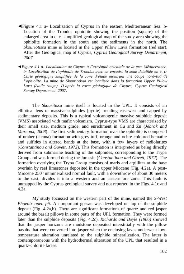
102
◄Figure 4.1 a- Localization of Cyprus in the eastern Mediterranean Sea. b-
Location of the Troodos ophiolite showing the position (square) of the
enlarged area in c. c- simplified geological map of the study area showing the
ophiolite formation in the south and the sediments in the north. The
Skouriotissa mine is located in the Upper Pillow Lava formation (red star).
After the Geological map of Cyprus, Cyprus Geological Survey Department,
2007.
◄Figure 4.1 a- Localisation de Chypre à l’extrémité orientale de la mer Méditerranée.
b- Localisation de l’ophiolite de Troodos avec en encadré la zone détaillée en c. c-
Carte géologique simplifiée de la zone d’étude montrant une coupe nord-sud de
l’ophiolite. La mine de Skouriotissa est localisée dans la formation Upper Pillow
Lava (étoile rouge). D’après la carte géologique de Chypre, Cyprus Geological
Survey Department, 2007.
The Skouritissa mine itself is located in the UPL. It consists of an
elliptical lens of massive sulphides (pyrite) trending east-west and capped by
sedimentary deposits. This is a typical volcanogenic massive sulphide deposit
(VMS) associated with mafic volcanism. Cyprus-type VMS are characterized by
their small size, medium grade, and enrichment in Cu and Zn (Jébrak and
Marcoux, 2008). The first sedimentary formation over the ophiolite is composed
of umber (sienna) formation with grey tuff, orange and ochre-coloured hematite
and sulfides in altered bands at the base, with a few layers of radiolarites
(Constantinou and Govett, 1972). This formation is interpreted as being directly
derived from submarine leaching of the sulphides, corresponding to the Trypa
Group and was formed during the Jurassic (Constantinou and Govett, 1972). The
formation overlying the Trypa Group consists of marls and argillites at the base
overlain by reef limestones deposited in the upper Miocene (Fig. 4.2a). A post-
Miocene 250° unmineralized normal fault, with a downthrow of about 30 meters
to the east, divides it into a western and an eastern ore zone. This fault is
unmapped by the Cyprus geological survey and not reported in the Figs. 4.1c and
4.2a.
My study focussed on the western part of the mine, named the S-West
Phoenix open pit. An important gossan was developed on top of the sulphide
deposit (Fig. 4.2a,b). There are significant formations of quartz and red jasper
around the basalt pillows in some parts of the UPL formation. They were formed
later than the sulphide deposits (Fig. 4.2c). Richards and Boyle (1986) showed
that the jasper horizons are mudstone deposited interstitially with the pillow
basalts that were converted into jasper when the enclosing lavas underwent low-
temperature alteration unrelated to the sulphide mineralization. The latter is
contemporaneous with the hydrothermal alteration of the UPL that resulted in a
quartz-chlorite facies.
Chapter IV: A new Mars analogue site in Cyprus
103
The second major phase of “weathering” of the pillow basalts is
associated with the mining activities. The Skouriotissa mine, in particular the
gossan, has been exploited for its copper since Roman times. The old workings
are composed of mine galleries along the massive sulphide deposit. The galleries
are largely drowned by meteoric water that has percolated into the sulphides.
This acidic water is collected and transported by gravity to the SW Phoenix pit
where it flows onto the pillow basalt lava formation (UPL) causing their
alteration. I focussed my study around this modern hydrologic system.
Figure 4.2: a- Detailed geological map of the Skouriotissa mine area. The gossan
is located in the Upper Pillow Lavas formation. Faults are not mapped here.
(After the Geological map of the Xeros-Troodos area, Cyprus Geological
Survey Department, 1969). b- View of the SW Phoenix pit with the brown
gossan at the top and the green quartz-chlorite pillow basalt formation
(hydrothermally altered) at the bottom. Note the mechanical stripper for scale
(in insert). The main massive ore deposit (main pit) is the darker lens to the
right. The study area is on the right end of the picture. c- Detail of the pillow
basalt formation. Note the presence of red jasper around the pillows. (The
hammer in the centre is 33 cm-long).
Figure 4.2: a- Carte géologique des environs de la mine de Skouriotissa. Le chapeau de
fer est localisé dans la formation Upper Pillow Lavas. Les failles ne sont pas cartées
ici. (D’après la carte géologique de Xeros-Troodos, Cyprus Geological Survey
Department, 1969). b- Vue de l’exploitation SW Phoenix avec le chapeau de fer en
marron au sommet et à la base en vert la formation de basaltes en coussins à quartz-
chlorite (altération hydrothermale). Notez la pelleteuse pour l’échelle (en insert). La
zone de sulfures massifs est la lentille sombre sur la droite. La zone d’étude est juste
à droite de l’image. c- Détail des basaltes en coussins. Notez la présence de jaspe
autour des coussins. (Le marteau mesure 33 cm de long.)
104
IV.4. Sampling
The samples were collected following the flow of acidic water on the
basaltic surface. The outcrop consists of a succession of lithological benches (~6-
4 meters high and ~1 to 4 meters width) along which the water flows, making
small grooves along the bench and creating small waterfalls between each bench.
Samples were taken in five points at different elevations along the flowing water.
The acidic water leaching from the VMS body results in a superficial iron oxide
deposit but the sampled rocks are green. Five sites were thus defined from top to
bottom (Fig. 4.3):
-Site 1, immediately adjacent to the acidic water of the mine drainage;
-Site 2, along the uppermost waterfall (Fig. 4.3). At this place the pillows
are clearly visible in the outcrop. This level corresponds to the level shown in
Figure 4.2c but without evidence of red jasper.
-Site 3, in the middle of the second large waterfall. The basalt at this
level is slightly more massive.
-Site 4, at the bottom of the old pit, where the flow excavated ~40
centimetres into the basalt formation. A few inches of red mud occurs at the
bottom of the waterfalls (Fig. 4.3), possibly the remains of a reserve of dirty
water. To date, mining activity has led to the deepening of the pit and the
creation of several levels.
-Site 5, exactly in the waterfall between the old and the new bottom of
the pit. The basalt in the recently dug base of the pit is green and has not been
altered by the acidic water but only by hydrothermal processes (quartz-chlorite
facies).
At each site, the temperature, the pH and the conductivity were measured
in situ (blue stars in Fig. 4.3). The rock samples were named 11CY08 to 11CY20
with respect to the ISAR nomenclature (cf. Chapter I). However, for more clarity,
they were renamed as a function for their position in the field. To discriminate
between natural chemical alteration due to the mining activity and the artificial
acid weathering induced by the industrial lixiviation process used to concentrate
the copper, , I sampled different parts of the waste dump that is regularly sprayed
with acid in order to further release the copper. The samples were named
11CY05 to 11CY07. For these samples there is no precise location or
information on the position in the geological levels since the samples were taken
from all over the mine.
Finally, the samples are arranged along (1) a vertical line along the
groove of flowing water: samples S1-1, S2-1, S3-1, S4-1 and S5-1, respectively
11CY16, 11CY13, 11CY09, 11CY08 and 11CY17 according to the ISAR
nomenclature; and (2) horizontally in the pillow basalt formation: samples S2-1,
S2-2 and S2-3, respectively 11CY13, 11CY14 and 11CY15; samples S3-1, S3-2,
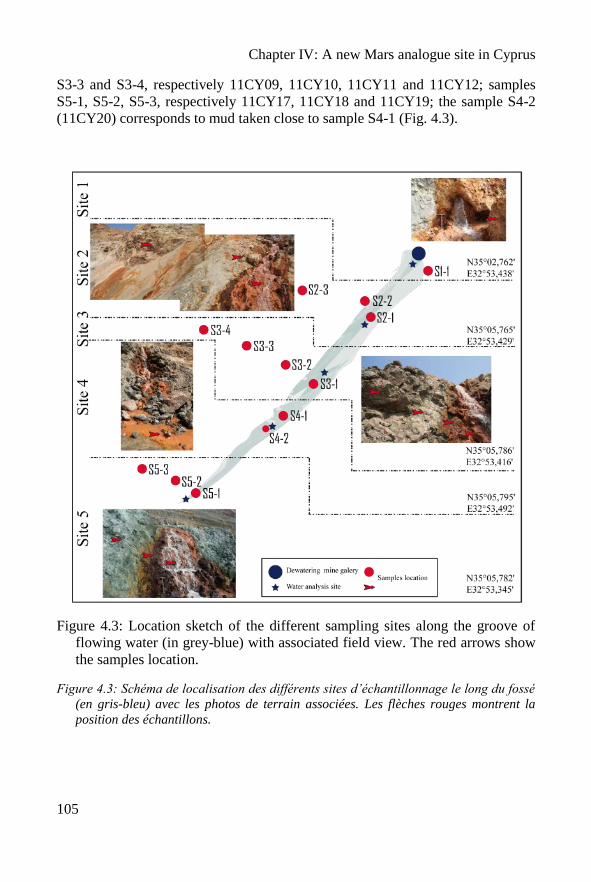
Chapter IV: A new Mars analogue site in Cyprus
105
S3-3 and S3-4, respectively 11CY09, 11CY10, 11CY11 and 11CY12; samples
S5-1, S5-2, S5-3, respectively 11CY17, 11CY18 and 11CY19; the sample S4-2
(11CY20) corresponds to mud taken close to sample S4-1 (Fig. 4.3).
Figure 4.3: Location sketch of the different sampling sites along the groove of
flowing water (in grey-blue) with associated field view. The red arrows show
the samples location.
Figure 4.3: Schéma de localisation des différents sites d’échantillonnage le long du fossé
(en gris-bleu) avec les photos de terrain associées. Les flèches rouges montrent la
position des échantillons.
106
IV.5. Methods
Fifteen samples from the groove and from the waste dump were dried
and crushed to a fine powder to make elemental analysis by ICP and
mineralogical characterization by XRD. The elemental composition of 7 samples
(S1-1, S2-1, S3-1 to S3-4 and S5-1) was determined by ICP-OES and ICP-MS
for 10 major elements and 43 minor elements respectively. The XRD
measurements were carried out in the Hydrasa Laboratory, with the help of
Claude Fontaine and Alain Meunier. Before analysis the phyllosilicates were
concentrated by suspending and settling the powders for 2 hours in de-ionised
water and then they were sonicated. The phyllosilicates were then studied in
oriented configuration after drying in the air, with ethyl-glycol vapour, and with
heat treatment at 350°C and 550°C. Polished thin sections were made at the
ISTO laboratory, but with difficulties due to the dominantly clayish composition.
Mineralogy was investigated using optical microscopy and Raman spectroscopy.
IV.6. Results
IV.6.1: Water geochemistry measurements
The results of the water analyses are displayed in Table 4.1. The
measurements were made in situ on the 30th Mars 2011.
Site 1 Site 2 Site 3 Site 4 Site 5
Temperature (°C) 17.1 17.6 19.6 19.2 19.1
pH 5.85 5.21 4.03 4.13 3.96
Conductivity (µS/cm) 1689 1696 1855 1868 2966
Table 4.1: Parameters measured along the groove of flowing water. Sites are
listed from the top to the bottom.
Tableau 4.1: Paramètres mesurés dans les eaux le long du fossé. Les sites sont classés
de haut en bas.
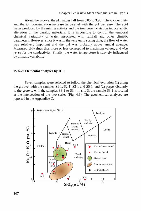
Chapter IV: A new Mars analogue site in Cyprus
107
Along the groove, the pH values fall from 5.85 to 3.96. The conductivity
and the ion concentration increase in parallel with the pH decrease. The acid
water produced by the mining activity and the iron core lixiviation induce acidic
alteration of the basaltic materials. It is impossible to control the temporal
chemical variability of water associated with rainfall and other climatic
parameters. However, since it was in the very early spring time, the flow of water
was relatively important and the pH was probably above annual average.
Measured pH-values thus more or less correspond to maximum values, and vice
versa for the conductivity. Finally, the water temperature is strongly influenced
by climatic variability.
IV.6.2: Elemental analyses by ICP
Seven samples were selected to follow the chemical evolution (1) along
the groove, with the samples S1-1, S2-1, S3-1 and S5-1, and (2) perpendicularly
to the groove, with the samples S3-1 to S3-4 in site 3; the sample S3-1 is located
at the intersection of the two series (Fig. 4.3). The geochemical analyses are
reported in the Appendice C.
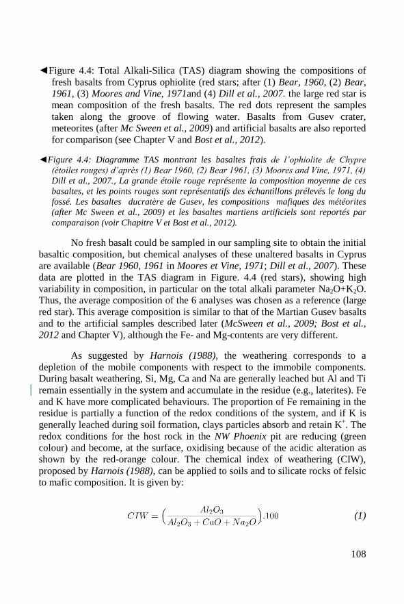
108
◄Figure 4.4: Total Alkali-Silica (TAS) diagram showing the compositions of
fresh basalts from Cyprus ophiolite (red stars; after (1) Bear, 1960, (2) Bear,
1961, (3) Moores and Vine, 1971and (4) Dill et al., 2007. the large red star is
mean composition of the fresh basalts. The red dots represent the samples
taken along the groove of flowing water. Basalts from Gusev crater,
meteorites (after Mc Sween et al., 2009) and artificial basalts are also reported
for comparison (see Chapter V and Bost et al., 2012).
◄Figure 4.4: Diagramme TAS montrant les basaltes frais de l’ophiolite de Chypre
(étoiles rouges) d’après (1) Bear 1960, (2) Bear 1961, (3) Moores and Vine, 1971, (4)
Dill et al., 2007., La grande étoile rouge représente la composition moyenne de ces
basaltes, et les points rouges sont représentatifs des échantillons prélevés le long du
fossé. Les basaltes ducratère de Gusev, les compositions mafiques des météorites
(after Mc Sween et al., 2009) et les basaltes martiens artificiels sont reportés par
comparaison (voir Chapitre V et Bost et al., 2012).
No fresh basalt could be sampled in our sampling site to obtain the initial
basaltic composition, but chemical analyses of these unaltered basalts in Cyprus
are available (Bear 1960, 1961 in Moores et Vine, 1971; Dill et al., 2007). These
data are plotted in the TAS diagram in Figure. 4.4 (red stars), showing high
variability in composition, in particular on the total alkali parameter Na2O+K2O.
Thus, the average composition of the 6 analyses was chosen as a reference (large
red star). This average composition is similar to that of the Martian Gusev basalts
and to the artificial samples described later (McSween et al., 2009; Bost et al.,
2012 and Chapter V), although the Fe- and Mg-contents are very different.
As suggested by Harnois (1988), the weathering corresponds to a
depletion of the mobile components with respect to the immobile components.
During basalt weathering, Si, Mg, Ca and Na are generally leached but Al and Ti
remain essentially in the system and accumulate in the residue (e.g., laterites). Fe
and K have more complicated behaviours. The proportion of Fe remaining in the
residue is partially a function of the redox conditions of the system, and if K is
generally leached during soil formation, clays particles absorb and retain K+. The
redox conditions for the host rock in the NW Phoenix pit are reducing (green
colour) and become, at the surface, oxidising because of the acidic alteration as
shown by the red-orange colour. The chemical index of weathering (CIW),
proposed by Harnois (1988), can be applied to soils and to silicate rocks of felsic
to mafic composition. It is given by:
(1)
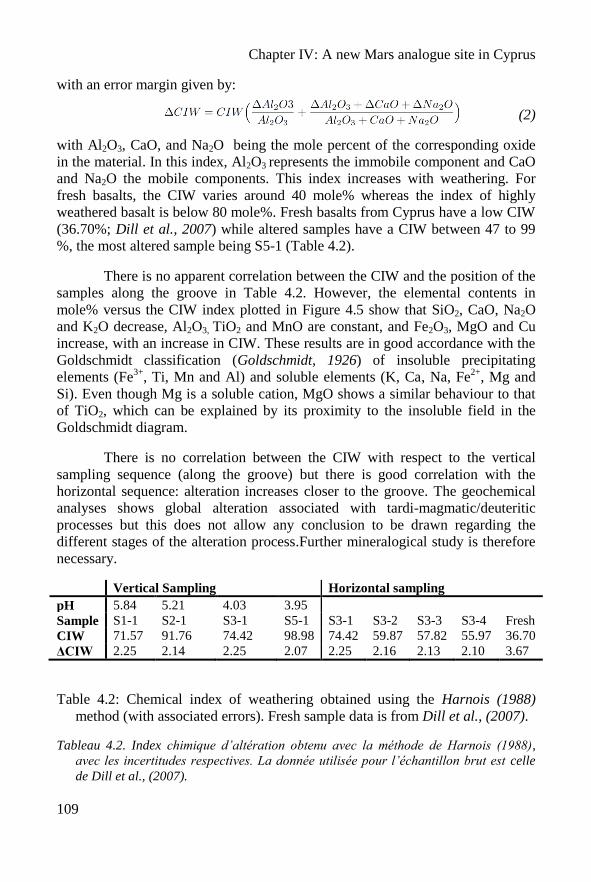
Chapter IV: A new Mars analogue site in Cyprus
109
with an error margin given by:
(2)
with Al2O3, CaO, and Na2O being the mole percent of the corresponding oxide
in the material. In this index, Al2O3 represents the immobile component and CaO
and Na2O the mobile components. This index increases with weathering. For
fresh basalts, the CIW varies around 40 mole% whereas the index of highly
weathered basalt is below 80 mole%. Fresh basalts from Cyprus have a low CIW
(36.70%; Dill et al., 2007) while altered samples have a CIW between 47 to 99
%, the most altered sample being S5-1 (Table 4.2).
There is no apparent correlation between the CIW and the position of the
samples along the groove in Table 4.2. However, the elemental contents in
mole% versus the CIW index plotted in Figure 4.5 show that SiO2, CaO, Na2O
and K2O decrease, Al2O3, TiO2 and MnO are constant, and Fe2O3, MgO and Cu
increase, with an increase in CIW. These results are in good accordance with the
Goldschmidt classification (Goldschmidt, 1926) of insoluble precipitating
elements (Fe3+
, Ti, Mn and Al) and soluble elements (K, Ca, Na, Fe2+
, Mg and
Si). Even though Mg is a soluble cation, MgO shows a similar behaviour to that
of TiO2, which can be explained by its proximity to the insoluble field in the
Goldschmidt diagram.
There is no correlation between the CIW with respect to the vertical
sampling sequence (along the groove) but there is good correlation with the
horizontal sequence: alteration increases closer to the groove. The geochemical
analyses shows global alteration associated with tardi-magmatic/deuteritic
processes but this does not allow any conclusion to be drawn regarding the
different stages of the alteration process.Further mineralogical study is therefore
necessary.
Vertical Sampling Horizontal sampling
pH 5.84 5.21 4.03 3.95
Sample S1-1 S2-1 S3-1 S5-1 S3-1 S3-2 S3-3 S3-4 Fresh
CIW 71.57 91.76 74.42 98.98 74.42 59.87 57.82 55.97 36.70
ΔCIW 2.25 2.14 2.25 2.07 2.25 2.16 2.13 2.10 3.67
Table 4.2: Chemical index of weathering obtained using the Harnois (1988)
method (with associated errors). Fresh sample data is from Dill et al., (2007).
Tableau 4.2. Index chimique d’altération obtenu avec la méthode de Harnois (1988),
avec les incertitudes respectives. La donnée utilisée pour l’échantillon brut est celle
de Dill et al., (2007).
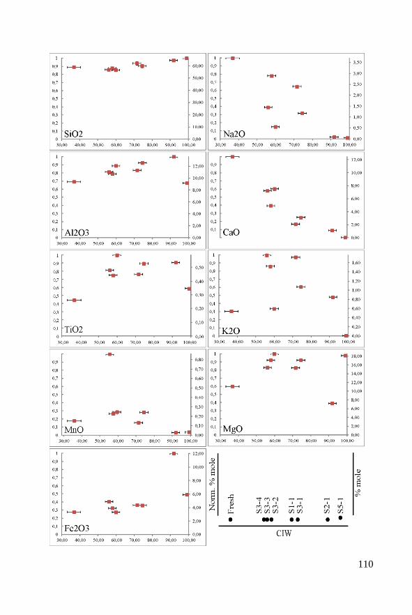
110
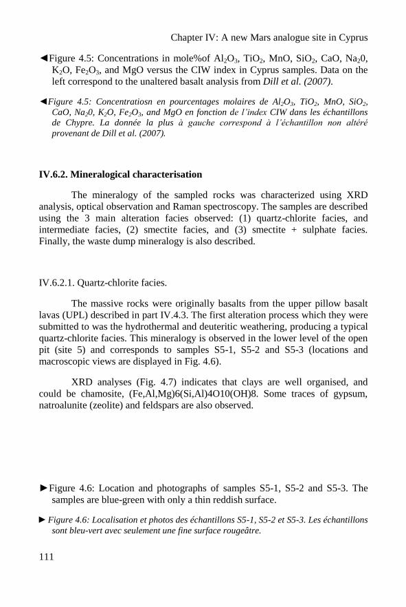
Chapter IV: A new Mars analogue site in Cyprus
111
◄Figure 4.5: Concentrations in mole%of Al2O3, TiO2, MnO, SiO2, CaO, Na20,
K2O, Fe2O3, and MgO versus the CIW index in Cyprus samples. Data on the
left correspond to the unaltered basalt analysis from Dill et al. (2007).
◄Figure 4.5: Concentratiosn en pourcentages molaires de Al2O3, TiO2, MnO, SiO2,
CaO, Na20, K2O, Fe2O3, and MgO en fonction de l’index CIW dans les échantillons
de Chypre. La donnée la plus à gauche correspond à l’échantillon non altéré
provenant de Dill et al. (2007).
IV.6.2. Mineralogical characterisation
The mineralogy of the sampled rocks was characterized using XRD
analysis, optical observation and Raman spectroscopy. The samples are described
using the 3 main alteration facies observed: (1) quartz-chlorite facies, and
intermediate facies, (2) smectite facies, and (3) smectite + sulphate facies.
Finally, the waste dump mineralogy is also described.
IV.6.2.1. Quartz-chlorite facies.
The massive rocks were originally basalts from the upper pillow basalt
lavas (UPL) described in part IV.4.3. The first alteration process which they were
submitted to was the hydrothermal and deuteritic weathering, producing a typical
quartz-chlorite facies. This mineralogy is observed in the lower level of the open
pit (site 5) and corresponds to samples S5-1, S5-2 and S5-3 (locations and
macroscopic views are displayed in Fig. 4.6).
XRD analyses (Fig. 4.7) indicates that clays are well organised, and
could be chamosite, (Fe,Al,Mg)6(Si,Al)4O10(OH)8. Some traces of gypsum,
natroalunite (zeolite) and feldspars are also observed.
►Figure 4.6: Location and photographs of samples S5-1, S5-2 and S5-3. The
samples are blue-green with only a thin reddish surface.
►Figure 4.6: Localisation et photos des échantillons S5-1, S5-2 et S5-3. Les échantillons
sont bleu-vert avec seulement une fine surface rougeâtre.
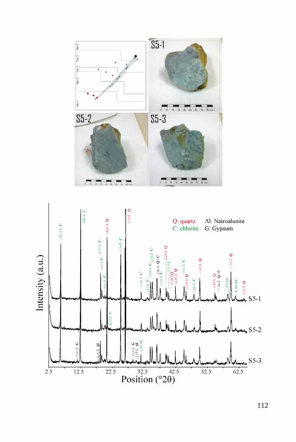
112
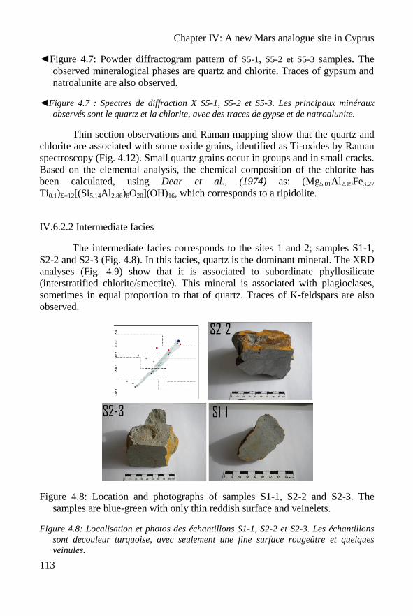
Chapter IV: A new Mars analogue site in Cyprus
113
◄Figure 4.7: Powder diffractogram pattern of S5-1, S5-2 et S5-3 samples. The
observed mineralogical phases are quartz and chlorite. Traces of gypsum and
natroalunite are also observed.
◄Figure 4.7 : Spectres de diffraction X S5-1, S5-2 et S5-3. Les principaux minéraux
observés sont le quartz et la chlorite, avec des traces de gypse et de natroalunite.
Thin section observations and Raman mapping show that the quartz and
chlorite are associated with some oxide grains, identified as Ti-oxides by Raman
spectroscopy (Fig. 4.12). Small quartz grains occur in groups and in small cracks.
Based on the elemental analysis, the chemical composition of the chlorite has
been calculated, using Dear et al., (1974) as: (Mg5.01Al2.19Fe3.27
Ti0.1)Σ=12[(Si5.14Al2.86)8O20](OH)16, which corresponds to a ripidolite.
IV.6.2.2 Intermediate facies
The intermediate facies corresponds to the sites 1 and 2; samples S1-1,
S2-2 and S2-3 (Fig. 4.8). In this facies, quartz is the dominant mineral. The XRD
analyses (Fig. 4.9) show that it is associated to subordinate phyllosilicate
(interstratified chlorite/smectite). This mineral is associated with plagioclases,
sometimes in equal proportion to that of quartz. Traces of K-feldspars are also
observed.
Figure 4.8: Location and photographs of samples S1-1, S2-2 and S2-3. The
samples are blue-green with only thin reddish surface and veinelets.
Figure 4.8: Localisation et photos des échantillons S1-1, S2-2 et S2-3. Les échantillons
sont decouleur turquoise, avec seulement une fine surface rougeâtre et quelques
veinules.
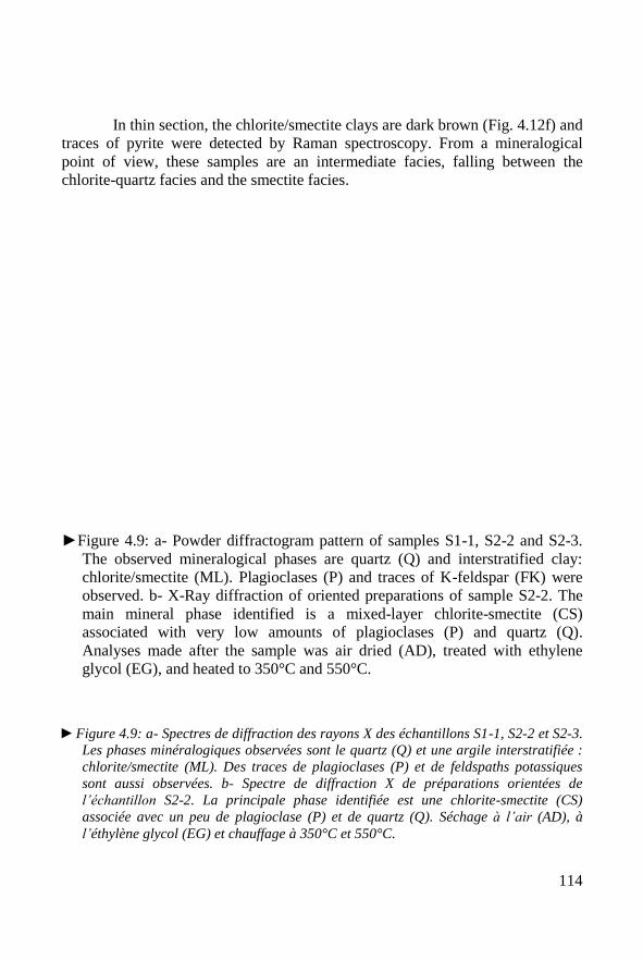
114
In thin section, the chlorite/smectite clays are dark brown (Fig. 4.12f) and
traces of pyrite were detected by Raman spectroscopy. From a mineralogical
point of view, these samples are an intermediate facies, falling between the
chlorite-quartz facies and the smectite facies.
►Figure 4.9: a- Powder diffractogram pattern of samples S1-1, S2-2 and S2-3.
The observed mineralogical phases are quartz (Q) and interstratified clay:
chlorite/smectite (ML). Plagioclases (P) and traces of K-feldspar (FK) were
observed. b- X-Ray diffraction of oriented preparations of sample S2-2. The
main mineral phase identified is a mixed-layer chlorite-smectite (CS)
associated with very low amounts of plagioclases (P) and quartz (Q).
Analyses made after the sample was air dried (AD), treated with ethylene
glycol (EG), and heated to 350°C and 550°C.
►Figure 4.9: a- Spectres de diffraction des rayons X des échantillons S1-1, S2-2 et S2-3.
Les phases minéralogiques observées sont le quartz (Q) et une argile interstratifiée :
chlorite/smectite (ML). Des traces de plagioclases (P) et de feldspaths potassiques
sont aussi observées. b- Spectre de diffraction X de préparations orientées de
l’échantillon S2-2. La principale phase identifiée est une chlorite-smectite (CS)
associée avec un peu de plagioclase (P) et de quartz (Q). Séchage à l’air (AD), à
l’éthylène glycol (EG) et chauffage à 350°C et 550°C.
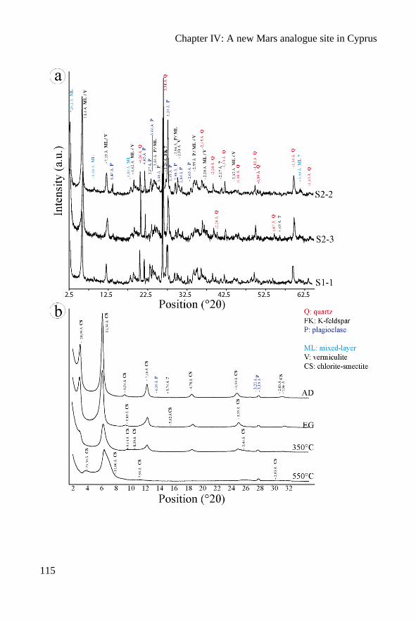
Chapter IV: A new Mars analogue site in Cyprus
115
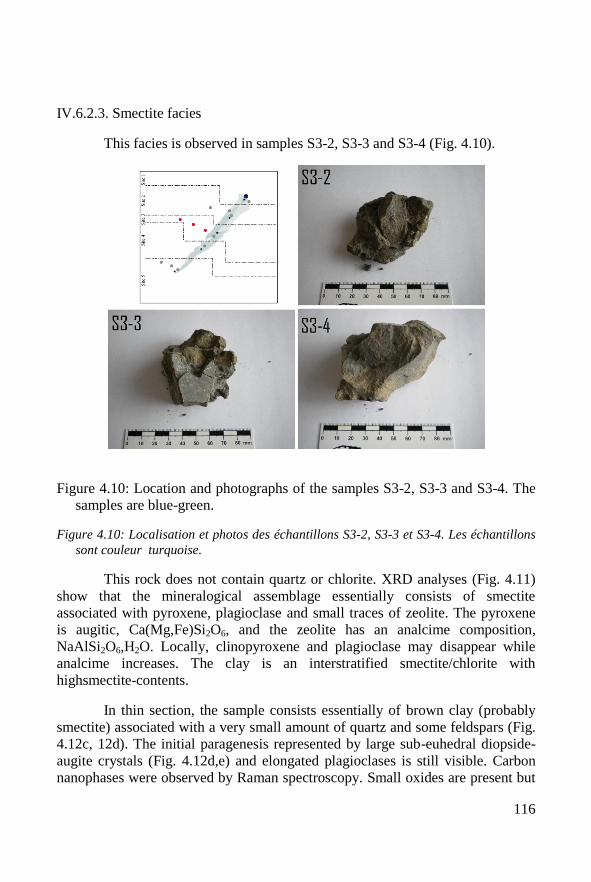
116
IV.6.2.3. Smectite facies
This facies is observed in samples S3-2, S3-3 and S3-4 (Fig. 4.10).
Figure 4.10: Location and photographs of the samples S3-2, S3-3 and S3-4. The
samples are blue-green.
Figure 4.10: Localisation et photos des échantillons S3-2, S3-3 et S3-4. Les échantillons
sont couleur turquoise.
This rock does not contain quartz or chlorite. XRD analyses (Fig. 4.11)
show that the mineralogical assemblage essentially consists of smectite
associated with pyroxene, plagioclase and small traces of zeolite. The pyroxene
is augitic, Ca(Mg,Fe)Si2O6, and the zeolite has an analcime composition,
NaAlSi2O6,H2O. Locally, clinopyroxene and plagioclase may disappear while
analcime increases. The clay is an interstratified smectite/chlorite with
highsmectite-contents.
In thin section, the sample consists essentially of brown clay (probably
smectite) associated with a very small amount of quartz and some feldspars (Fig.
4.12c, 12d). The initial paragenesis represented by large sub-euhedral diopside-
augite crystals (Fig. 4.12d,e) and elongated plagioclases is still visible. Carbon
nanophases were observed by Raman spectroscopy. Small oxides are present but
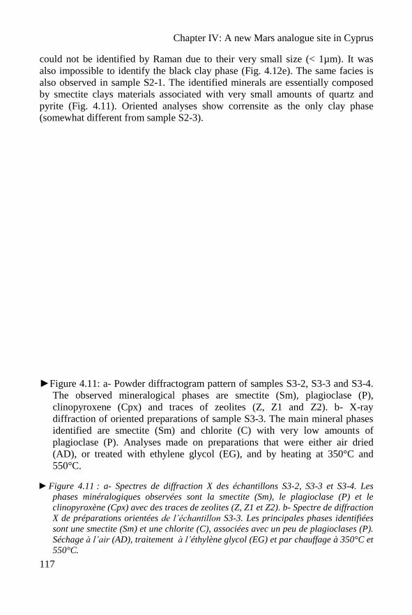
Chapter IV: A new Mars analogue site in Cyprus
117
could not be identified by Raman due to their very small size (< 1µm). It was
also impossible to identify the black clay phase (Fig. 4.12e). The same facies is
also observed in sample S2-1. The identified minerals are essentially composed
by smectite clays materials associated with very small amounts of quartz and
pyrite (Fig. 4.11). Oriented analyses show corrensite as the only clay phase
(somewhat different from sample S2-3).
►Figure 4.11: a- Powder diffractogram pattern of samples S3-2, S3-3 and S3-4.
The observed mineralogical phases are smectite (Sm), plagioclase (P),
clinopyroxene (Cpx) and traces of zeolites (Z, Z1 and Z2). b- X-ray
diffraction of oriented preparations of sample S3-3. The main mineral phases
identified are smectite (Sm) and chlorite (C) with very low amounts of
plagioclase (P). Analyses made on preparations that were either air dried
(AD), or treated with ethylene glycol (EG), and by heating at 350°C and
550°C.
►Figure 4.11 : a- Spectres de diffraction X des échantillons S3-2, S3-3 et S3-4. Les
phases minéralogiques observées sont la smectite (Sm), le plagioclase (P) et le
clinopyroxène (Cpx) avec des traces de zeolites (Z, Z1 et Z2). b- Spectre de diffraction
X de préparations orientées de l’échantillon S3-3. Les principales phases identifiées
sont une smectite (Sm) et une chlorite (C), associées avec un peu de plagioclases (P).
Séchage à l’air (AD), traitement à l’éthylène glycol (EG) et par chauffage à 350°C et
550°C.
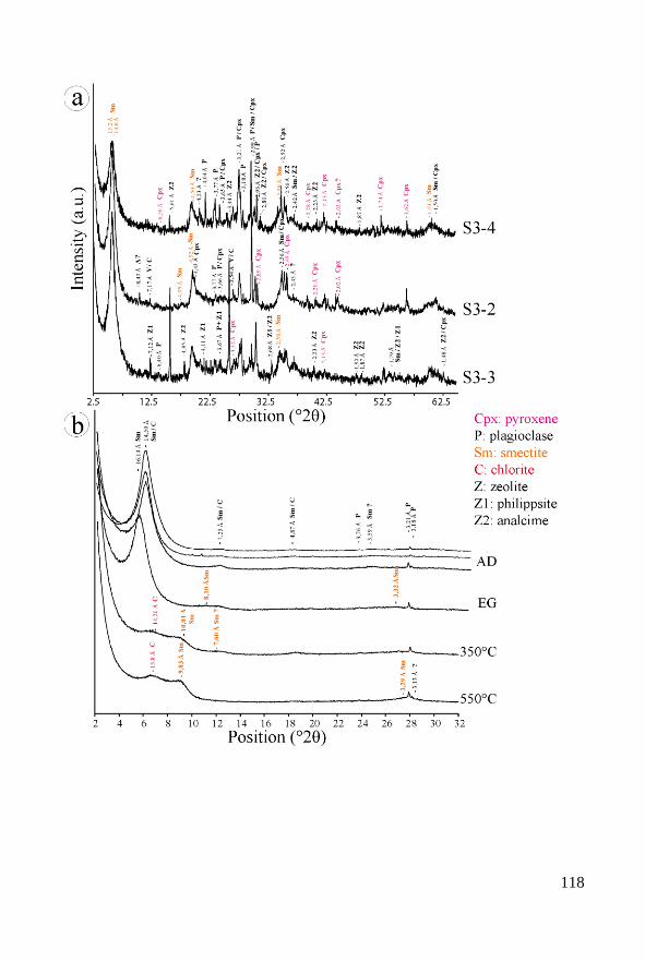
118
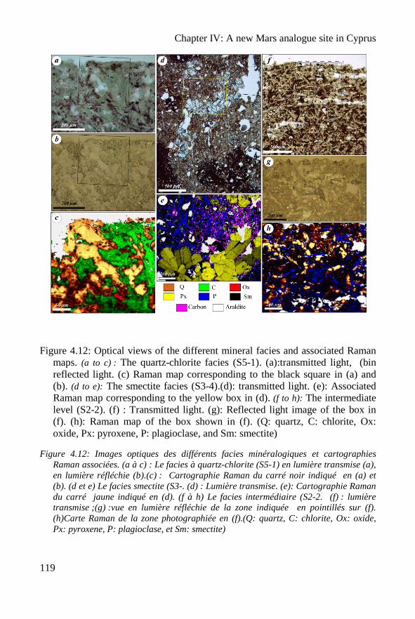
Chapter IV: A new Mars analogue site in Cyprus
119
Figure 4.12: Optical views of the different mineral facies and associated Raman
maps. (a to c) : The quartz-chlorite facies (S5-1). (a):transmitted light, (bin
reflected light. (c) Raman map corresponding to the black square in (a) and
(b). (d to e): The smectite facies (S3-4).(d): transmitted light. (e): Associated
Raman map corresponding to the yellow box in (d). (f to h): The intermediate
level (S2-2). (f) : Transmitted light. (g): Reflected light image of the box in
(f). (h): Raman map of the box shown in (f). (Q: quartz, C: chlorite, Ox:
oxide, Px: pyroxene, P: plagioclase, and Sm: smectite)
Figure 4.12: Images optiques des différents facies minéralogiques et cartographies
Raman associées. (a à c) : Le facies à quartz-chlorite (S5-1) en lumière transmise (a),
en lumière réfléchie (b).(c) : Cartographie Raman du carré noir indiqué en (a) et
(b). (d et e) Le facies smectite (S3-. (d) : Lumière transmise. (e): Cartographie Raman
du carré jaune indiqué en (d). (f à h) Le facies intermédiaire (S2-2. (f) : lumière
transmise ;(g) :vue en lumière réfléchie de la zone indiquée en pointillés sur (f).
(h)Carte Raman de la zone photographiée en (f).(Q: quartz, C: chlorite, Ox: oxide,
Px: pyroxene, P: plagioclase, et Sm: smectite)
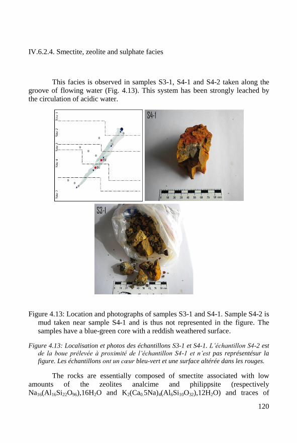
120
IV.6.2.4. Smectite, zeolite and sulphate facies
This facies is observed in samples S3-1, S4-1 and S4-2 taken along the
groove of flowing water (Fig. 4.13). This system has been strongly leached by
the circulation of acidic water.
Figure 4.13: Location and photographs of samples S3-1 and S4-1. Sample S4-2 is
mud taken near sample S4-1 and is thus not represented in the figure. The
samples have a blue-green core with a reddish weathered surface.
Figure 4.13: Localisation et photos des échantillons S3-1 et S4-1. L’échantillon S4-2 est
de la boue prélevée à proximité de l’échantillon S4-1 et n’est pas représentésur la
figure. Les échantillons ont un cœur bleu-vert et une surface altérée dans les rouges.
The rocks are essentially composed of smectite associated with low
amounts of the zeolites analcime and philippsite (respectively
Na16(Al16Si22O96),16H2O and K2(Ca0.5Na)4(Al6Si10O32),12H2O) and traces of
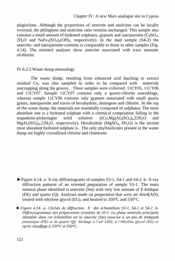
Chapter IV: A new Mars analogue site in Cyprus
121
plagioclase. Although the proportions of smectite and analcime can be locally
reversed, the philippsite and analcime ratio remains unchanged. This sample also
contains a small amount of hydrated sulphates, gypsum and natrojarosite (CaSO4,
2H2O and NaFe3(SO4)2(OH)6, respectively). In the mud sample (S4-2) the
smectite- and natrojarosite-contents is comparable to those in other samples (Fig.
4.14). The oriented analyses show smectite associated with trace amounts
ofchlorite.
IV.6.2.5 Waste dump mineralogy
The waste dump, resulting from enhanced acid leaching to extract
residual Cu, was also sampled in order to be compared with materials
outcropping along the groove. . Three samples were collected: 11CY05, 11CY06
and 11CY07. Sample 11CY07 contains only a quartz-chlorite assemblage,
whereas sample 11CY06 contains only gypsum associated with small quartz
grains, natrojarosite and traces of hexahydrite, alunogene and chlorite. At the top
of the waste dump, the materials are essentially composed of sulphates. The most
abundant one is a hydrated sulphate with a chemical composition falling in the
wupatkiite-pickeringite solid solution: ((Co,Mg)Al2(SO4)4,22H2O and
MgAl2(SO4)4,22H2O, respectively). Hexahydrite (MgSO4, 6H2O) is the second
most abundant hydrated sulphate is . The only phyllosilicates present in the waste
dump are highly crystallized chlorite and chamosite.
►Figure 4.14: a- X-ray diffractograms of samples S3-1, S4-1 and S4-2. b- X-ray
diffraction patterns of an oriented preparation of sample S3-1. The main
mineral phase identified is smectite (Sm) with very low amount of F-feldspar
(FK) and quartz (Q). Analyses made on preparation that were air dried(AD),
treated with ethylene glycol (EG), and heatied to 350°C and 550°C.
►Figure 4.14: a- Clichés de diffraction X des échantillons S3-1, S4-1 et S4-2. b-
Diffractogrammes des préparations orientées de S3-1. La phase minérale principale
identifiée dans cet échantillon est la smectite (Sm) associée à un peu de feldspath
potassique (FK) et de quartz (Q). Séchage à l’air (AD), à l’éthylène glycol (EG) et
après chauffage à 350°C et 550°C.
122
Chapter IV: A new Mars analogue site in Cyprus
123
IV.7. Discussion
IV.7.1 Mineralogy
The mineralogical observations are summarized in Table 4.3. The
massive host-rock consists of three types of mineral associations (paragenesis):
- (1) quartz + chlorite;
- (2) smectite (+ pyroxene + plagioclase);
- (3a) smectites + zeolites and (3b) smectites + zeolites + sulphates.
The intermediate facies is a mixture of the first and second facies. Hematite was
not observed in the studied area.
Paragenesis (1) corresponds to the quartz-chlorite facies typical of the
Upper Pillow Lava formation. It is associated with primary hydrothermal
alteration (Richards et al., 1989), which suggests formation by deuteritic
processes.
Paragenesis (2) corresponds to a further evolution of paragenesis (1),
especially with the transformation of interstratified primary chlorite to a mixed
chlorite/smectite, such as corrensite. This paragenesis is related to further
metasomatic alteration in low temperature conditions operating during the aging
of the Troodos extrusive sequences (Richards et al., 1989).
Indeed, the presence of fairly abundant pyroxene, a primary mineral, attests to
the basaltic origin of the material, although the main phase is smectite. This
paragenesis is similar to the smectite facies described in the Pitherokhoma mine
by Richards et al. (1989), and can also be compared to the facies (3a) described
by Alt and Honnorez (1984) in the DSDP site 417. This mineralogy is associated
to the circulation of oxygenated seawater.
Paragenesis (3) (a) and (b) are the evolution of paragenesis (2) in more
acidic conditions. The first alteration step is lixiviation. Figure 4.5 (samples S3-1
and S3-3) shows a sudden variation of the elemental composition, in particular a
decrease in Fe, which, from a mineralogical point of view, corresponds to the
precipitation of Fe-free zeolites . Moreover, the presence of zeolites can be
explained by the destabilization of pyroxene (i.e. clinopyroxene + fluid =
analcime + albite, and volcanic glass + fluid = phillipsite). Zeolites can be
124
formed by interaction of seawater-derived fluids with basalts (Alt and Honnorez,
1984).
However, the increase in acidic conditions is associated with the appearance of
sulphate minerals (gypsum and natrojarosite). It is for this reason that the study
of materials in the acid-weathered waste dump is important.
The waste dump mineralogy is typical of that associated with acidic
leaching whereby acidic water attacks the primary minerals, such as chlorite and
albite, and releases Mg, Fe, Al, Ca and Na. Thus we observe the following
sequence of increasingly less soluble salts with decreasing pH: wupatkiite-
pickeringite [(Co,Mg,Ni)Al2(SO4)4•22H2O and MgAl2(SO4)4•22H2O]
hexahydrite [MgSO4•6H2O] alunogens [Al2(SO4)3•17H2O] natrojarosite
[NaFe3(SO4)(OH)6] gypsum [CaSO4•2H2O].
Geochemical analyses confirm the mineralogical evolution suggested by
Table 4.3 . Samples S3-1 and S3-2 show a geochemical composition with a very
important decrease in Fe associated an important variation of the mineralogy. It
underlines the fact that the evolution (superimposition) of hydrothermal and
metasomatic alteration to an alteration with acidic water in rock-buffered reduced
conditions promotes the precipitation of sulphates. Complementary mineralogical
and geochemical characterization (e.g., chemical analyses of mine waste waters,
elemental analysis of minerals using electron microprobe), and a more detailed
sampling in the outcrop will be necessary to refine this interpretation. Finally, it
is possible to characterize the different mineralogical facies in the Skouriotissa
mine (Fig. 4.15).
►Table 4.3: Mineralogical composition of the different facies. From top to
bottom of the Table: (1) chlorite-quartz facies, (2a) intermediate facies, (2b)
smectite facies, (3a) smectite + zeolite facies, then (3b) smectite + zeolite+
sulphate. tr.: traces, *: samples with geochemical analysis.
►Tableau 4.3: Composition minéralogique des différents faciès. De haut en bas
du Tableau : (1) faciès à chlorite-quartz, (2a) faciès intermédiaire, (2b) faciès
à smectite, (3a) faciès à smectite + zeolite, (3b) faciès à smectite + zeolite +
sulphate. tr.: traces, *: échantillons avec des analyses géochimiques.
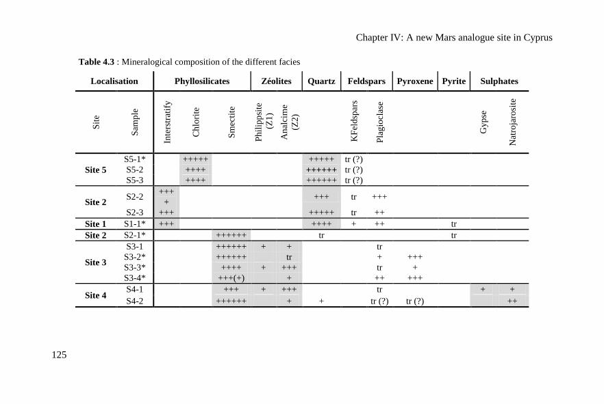
Chapter IV: A new Mars analogue site in Cyprus
125
Table 4.3 : Mineralogical composition of the different facies
Localisation Phyllosilicates Zéolites Quartz Feldspars Pyroxene Pyrite Sulphates
Sit
e
Sam
ple
Inte
rstr
atif
y
Ch
lori
te
Sm
ecti
te
Ph
ilip
psi
te
(Z
1)
An
alci
me
(Z2
)
KF
eld
spar
s
Pla
gio
clas
e
Gy
pse
Nat
roja
rosi
te
Site 5
S5-1* +++++ +++++ tr (?)
S5-2 ++++ ++++++ tr (?)
S5-3 ++++ ++++++ tr (?)
Site 2 S2-2
+++
+ +++ tr +++
S2-3 +++ +++++ tr ++
Site 1 S1-1* +++ ++++ + ++ tr
Site 2 S2-1* ++++++ tr tr
Site 3
S3-1 ++++++ + + tr
S3-2* ++++++ tr + +++
S3-3* ++++ + +++ tr +
S3-4* +++(+) + ++ +++
Site 4 S4-1 +++ + +++ tr + +
S4-2 ++++++ + + tr (?) tr (?) ++
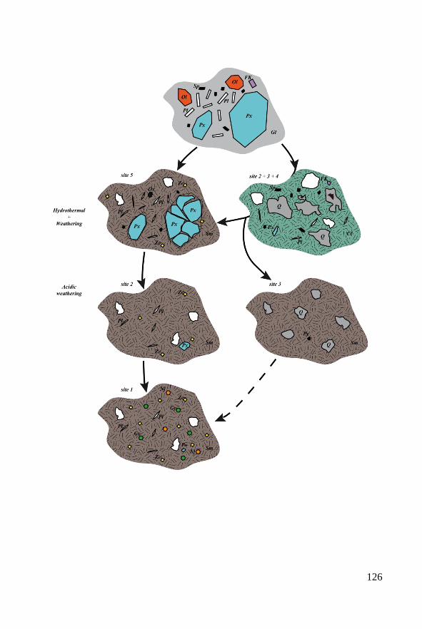
126
Chapter IV: A new Mars analogue site in Cyprus
127
◄Figure 4.15: Mineralogical evolution of the Skouriotissa basalt during
hydrothermal and acidic weathering. The horizontal exchanges are only
suggestions for which there is no evidence in thin sections. Sp : spinel (black),
FK: K-feldspar (purple), Px : pyroxene (blue), Ol : olivine (dark orange), Pl:
plagioclase (white), Gl : glass (grey), Ox : oxide, Sm :smectite, Cl: chlorite,
Nj : natrojarosite, Ze : zeolite, Gy : gypsum (green), Q: quartz (dark grey), Py:
pyrite (black). White spots are vacuoles.
◄Figure 4.15: Evolution minéralogique des basaltes de Skouriotissa pendant
l’altération hydrothermale et acide. Les échanges horizontaux sont seulement
suggérés en l’abscence de preuves en lames minces. Sp : spinelle (noir), FK:
feldsphath potassique (violet), Px : pyroxene (bleu), Ol : olivine (orange foncé), Pl:
plagioclase (blanc), Gl : verre (gris), Ox : oxyde, Sm : smectite, Cl: chlorite, Nj :
natrojarosite, Ze : zeolite, Gy : gypse (vert), Q: quartz (gris foncé), Py: pyrite
(noir).Les plages blanches sont des vacuoles.
IV.7.2. Relevance for Mars exploration
Martian basalts have been altered by different acidic processes during the
Noachian and Hesperian periods. Acid-sulphate weathering of basalts is a likely
explanation for the sulphate-rich outcrops observed by the MERs Spirit and
Opportunity (Golden et al., 2005). For example, the jarosite occurrence
(associated with other hydrated sulphates) in Eagle, Fram and Endurance
crateroutcrops in Meridiani Planum (Squyres et al., 2004; Clark et al., 2005)
could only have been formed by aqueous processes under very acidic conditions
(van Breeman, 1980 and Bigham and Nordstron, 2000 in Golden et al., 2005).
Phyllosilicates (clays) have been interpreted to be the result of aqueous
weathering (e.g. Bibring et al., 2006) but some of them may also be associated
with deuteritic processes (Ehlman et al., 2011 and Meunier et al., 2012). The
latter phenomenon corresponds to deep-seated alteration of magmatic rocks
during the later stages of their formation and is the direct result of magma
consolidation. The phyllosilicates precipitate from fluids representing the
residual liquid present at the end of the crystallization. Thus the precipitation of
phyllosilicates in these conditions can be considered as a sort of hydrothermal
phenomenon.
The rocks in the Skouriotissa mine exhibit alteration that is due to both
natural processes and human activity. Two types of alteration have occurred.
First, a pseudo-deuteritic alteration that resulted in the quartz-chlorite facies,
followed by a second weathering process (smectite facies) that is associated with
alteration by acidic waters flowing down the groove from the sulphide ore
deposit and resulting in the formation of zeolites and sulphates.
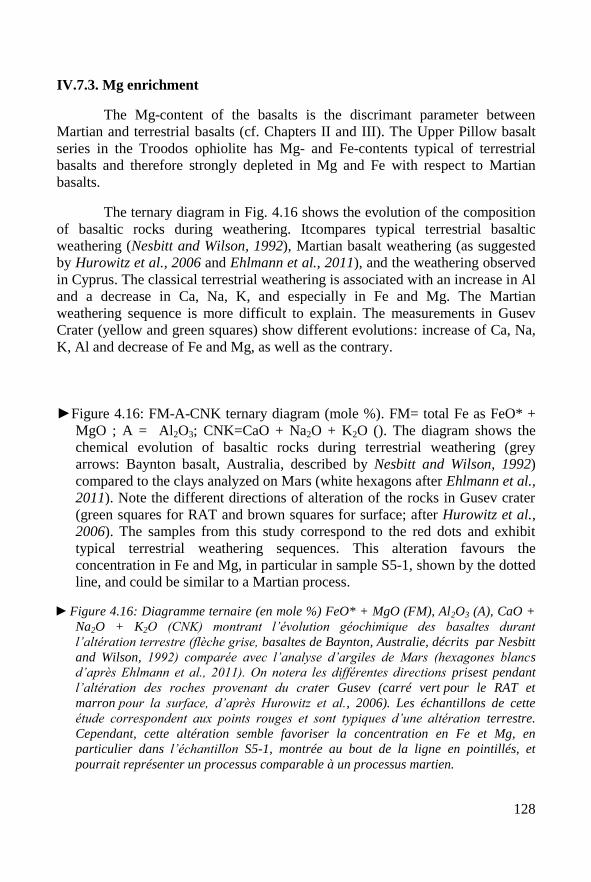
128
IV.7.3. Mg enrichment
The Mg-content of the basalts is the discrimant parameter between
Martian and terrestrial basalts (cf. Chapters II and III). The Upper Pillow basalt
series in the Troodos ophiolite has Mg- and Fe-contents typical of terrestrial
basalts and therefore strongly depleted in Mg and Fe with respect to Martian
basalts.
The ternary diagram in Fig. 4.16 shows the evolution of the composition
of basaltic rocks during weathering. Itcompares typical terrestrial basaltic
weathering (Nesbitt and Wilson, 1992), Martian basalt weathering (as suggested
by Hurowitz et al., 2006 and Ehlmann et al., 2011), and the weathering observed
in Cyprus. The classical terrestrial weathering is associated with an increase in Al
and a decrease in Ca, Na, K, and especially in Fe and Mg. The Martian
weathering sequence is more difficult to explain. The measurements in Gusev
Crater (yellow and green squares) show different evolutions: increase of Ca, Na,
K, Al and decrease of Fe and Mg, as well as the contrary.
►Figure 4.16: FM-A-CNK ternary diagram (mole %). FM= total Fe as FeO* +
MgO ; A = Al2O3; CNK=CaO + Na2O + K2O (). The diagram shows the
chemical evolution of basaltic rocks during terrestrial weathering (grey
arrows: Baynton basalt, Australia, described by Nesbitt and Wilson, 1992) compared to the clays analyzed on Mars (white hexagons after Ehlmann et al.,
2011). Note the different directions of alteration of the rocks in Gusev crater
(green squares for RAT and brown squares for surface; after Hurowitz et al.,
2006). The samples from this study correspond to the red dots and exhibit
typical terrestrial weathering sequences. This alteration favours the
concentration in Fe and Mg, in particular in sample S5-1, shown by the dotted
line, and could be similar to a Martian process.
►Figure 4.16: Diagramme ternaire (en mole %) FeO* + MgO (FM), Al2O3 (A), CaO +
Na2O + K2O (CNK) montrant l’évolution géochimique des basaltes durant
l’altération terrestre (flèche grise, basaltes de Baynton, Australie, décrits par Nesbitt
and Wilson, 1992) comparée avec l’analyse d’argiles de Mars (hexagones blancs
d’après Ehlmann et al., 2011). On notera les différentes directions prisest pendant
l’altération des roches provenant du crater Gusev (carré vert pour le RAT et
marron pour la surface, d’après Hurowitz et al., 2006). Les échantillons de cette
étude correspondent aux points rouges et sont typiques d’une altération terrestre.
Cependant, cette altération semble favoriser la concentration en Fe et Mg, en
particulier dans l’échantillon S5-1, montrée au bout de la ligne en pointillés, et
pourrait représenter un processus comparable à un processus martien.
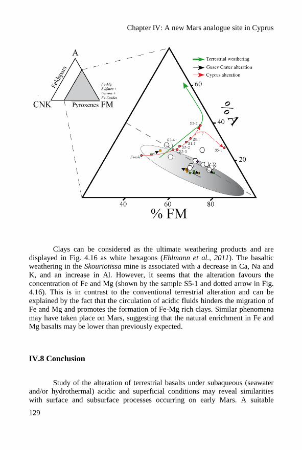
Chapter IV: A new Mars analogue site in Cyprus
129
Clays can be considered as the ultimate weathering products and are
displayed in Fig. 4.16 as white hexagons (Ehlmann et al., 2011). The basaltic
weathering in the Skouriotissa mine is associated with a decrease in Ca, Na and
K, and an increase in Al. However, it seems that the alteration favours the
concentration of Fe and Mg (shown by the sample S5-1 and dotted arrow in Fig.
4.16). This is in contrast to the conventional terrestrial alteration and can be
explained by the fact that the circulation of acidic fluids hinders the migration of
Fe and Mg and promotes the formation of Fe-Mg rich clays. Similar phenomena
may have taken place on Mars, suggesting that the natural enrichment in Fe and
Mg basalts may be lower than previously expected.
IV.8 Conclusion
Study of the alteration of terrestrial basalts under subaqueous (seawater
and/or hydrothermal) acidic and superficial conditions may reveal similarities
with surface and subsurface processes occurring on early Mars. A suitable
130
terrestrial analogue site should therefore consists of non-metamorphosed basalt,
i.e. a relatively young environment (Tosca et al., 2004; Hurrowitz et al., 2006)
that has undergone acidic alteration. On Earth, typical acidic environments are
found in sulphide-rich quarries and mine tailings in volcanogenic massive
sulphide (VMS) ore deposits. VMS deposits are widespread in the oceanic
basaltic crust (Jebrak and Marcoux, 2008) and are related to high to medium
temperature hydrothermalism at ridge crests. Primary alteration of the VMS
(Cyprus type) and associated basalts by seawater and/or hydrothermal fluids
occurs at the seafloor whereas secondary alteration occurs when these formations
are subaerially exposed as ophiolites. The Skouriotissa mine corresponds to a
massive sulphide ore deposit associated with basaltic volcanism (VMS-Cyprus
type). These environments are very good analogue sites for understanding
alteration that has taken place on the Noachian Martian surface.
However, more work is necessary in the Skouriotissa open pit in order to
confirm these preliminary observations and interpretations of the geochemical
and petrological data. For example, it would be useful to make more detailed and
regular sampling along the outcrop in order to identify changes in the
mineralogical phases in three dimensions.
The samples from Cyprus have been included in the ISAR collection.
The relevance of the samples as Mars analogues is detailed in the Table 4.4. The
analogy is based on the different characteristics studied: geochemistry, petrology
(including texture and structure) and geotechnical properties. They are evaluated
using stars: 1 to5 stars indicating respectively poor to excellent correspondance.
However, the geotechnical properties of these samples have not been measured
and therefore a value of 3 stars was attributed, based on knowledge of the
Martian surface materials.
►Table 4.4. Analogy of the samples from Cyprus with the surface of early Mars
surface; between 1 to 5, respectively bad to very good (n/a is not applicable).
►Tableau 4.4. Analogie des échantillons de Chypre avec la surface primitive de Mars ;
de 1 à 5 étoiles, du plus mauvais au meilleur respectivement (n/a pour non
applicable).
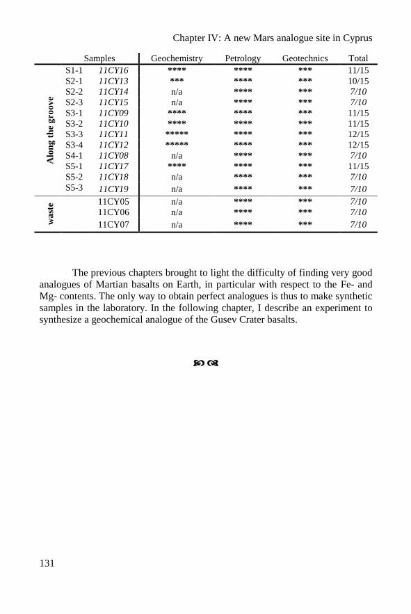
Chapter IV: A new Mars analogue site in Cyprus
131
Samples Geochemistry Petrology Geotechnics Total A
lon
g t
he
gro
ov
e
S1-1 11CY16 **** **** *** 11/15
S2-1 11CY13 *** **** *** 10/15
S2-2 11CY14 n/a **** *** 7/10
S2-3 11CY15 n/a **** *** 7/10
S3-1 11CY09 **** **** *** 11/15
S3-2 11CY10 **** **** *** 11/15
S3-3 11CY11 ***** **** *** 12/15
S3-4 11CY12 ***** **** *** 12/15
S4-1 11CY08 n/a **** *** 7/10
S5-1 11CY17 **** **** *** 11/15
S5-2 11CY18 n/a **** *** 7/10
S5-3 11CY19 n/a **** *** 7/10
wa
ste 11CY05 n/a **** *** 7/10
11CY06 n/a **** *** 7/10
11CY07 n/a **** *** 7/10
The previous chapters brought to light the difficulty of finding very good
analogues of Martian basalts on Earth, in particular with respect to the Fe- and
Mg- contents. The only way to obtain perfect analogues is thus to make synthetic
samples in the laboratory. In the following chapter, I describe an experiment to
synthesize a geochemical analogue of the Gusev Crater basalts.

132
Chapter IV: A new Mars analogue site in Cyprus
133
Chypre, un nouveau site analogue (Résumé Français)
Ce chapitre décrit un nouveau site analogue de Mars situé dans la mine de
Skouriotissa au nord-ouest de l’île de Chypre. Ce travail fait suite à ma mission de
terrain à Chypre au printemps 2011. Dans la mine, les basaltes ophiolitiques subissent
différentes altérations chimiques. En particulier avec l’interaction d’eau d’exhaure
acide, ce qui produit différentes minéralogies. Dans le chapitre précédent nous avons
montré qu’il était difficile de trouver de bons analogues de roches et de sols martiens.
Afin d’avoir des échantillons plus pertinents, il est intéressant de chercher des
échantillons géologiquement similaires à ceux de Mars primitive à actuelle et qui ont été
altérés par les processus décrits dans le Chapitre II partie 2.2. : des basaltes altérés dans
des conditions hydrothermales et par la circulation d’eaux d’exhaure acides. Des sites
analogues ont déjà été proposés (Rio Tinto en Espagne ou les calcaires de l’Utah aux
USA). Ces derniers étaient focalisés sur l’analogie à des conditions martiennes actuelles
mais pas toujours avec un point de vue géologique. Par exemple, le site de RioTinto est
particulièrement intéressant pour la recherche de vie extrêmophile, mais le contexte
géologique n’est pas comparable à celui de Mars. Les roches sont andésitiques et ont
subi un métamorphisme très important, ce qui n’est pas le cas sur Mars. Les
environnements miniers sont particulièrement intéressants car ils sont souvent associés à
la production d’eau acide. J’ai eu l’opportunité de faire une mission de terrain à Chypre,
où il était possible de visiter une mine de cuivre en environnement basaltique.
Chypre est une petite île allongée à l’extrémité orientale de la mer Méditerranée
au sud de la péninsule anatolienne (Fig. 4.1). La mine de Skouriotissa est localisée à
l’ouest de l’île, au sud du golfe de Morfou et au sud de la ville de Petra. Cette mine est
exploitée pour le cuivre par la Hellenic copper Limited.
Le site est situé dans l’ophiolite de Troodos, datés du Crétacé supérieur, qui
forme un dôme centré sur le Mont Olympus. Stratigraphiquement l’ophiolite est
composée, du cœur vers le centre, de peridotites, de serpentines, de gabbros, d’un
complexe de dyke et de laves extrusives (en coussins), l’ensemble étant couvert par des
sédiments océaniques (Fig. 4.1c). Les laves en coussins sont divisées en deux parties :
inférieure (LPL) et supérieure (UPL), d’une épaisseur maximum de 1,5 km. La mine est
située dans la partie supérieure des basaltes en coussin. Elle consiste en une lentille de
sulfures massifs associés au volcanisme mafique (Fig. 2). On parle de « Volcanogenic
massive sulphide » (VMS) de type Chypre (Jébrak et Marcoux, 2008). Ma zone d’étude
est focalisée sur la partie ouest de la mine appelée ‘S-West Phoenix open pit’. Un
important chapeau de fer s’est développé au sommet du dépôt de sulfures (Fig 4.2a,b).
Des formations de quartz et de jaspes rouges sont visibles autour des basaltes en
coussins dans quelques endroits de la formation UPL. Ces objets se sont formés à basse
température après la mise en place des minéralisations sulfurées. L’altération de
l’ensemble de l’UPL par des fluides hydrothermaux a pour résultat de transformer la
roche en un faciès à quartz-chlorite. La seconde phase majeure d’altération des basaltes
en coussins est associée aux activités minières. De nombreuses galeries recoupent les
sulfures massifs, elles collectent les eaux de météoritiques qui sont ensuite rejetées par
une exhaure acide qui se déverse dans un fossé. La formation des basaltes en coussins
134
subit une altération acide en surface au contact des eaux d’exhaure. J’ai focalisé mon
étude sur ce système.
Les échantillons ont été prélevés au niveau du fossé et autour de celui-ci en 5
points (Fig. 4.3) de haut en bas : le site 1 directement au niveau de l’exhaure ; le site 2 le
long d’une grande cascade, à un niveau où les basaltes en coussins sont bien visibles ; le
site 3 au milieu de l’affleurement, là où les basaltes sont plus massifs ; le site 4 au bas de
l’ancien carreau de mine et le site 5 au pied de la falaise où les eaux se déversent dans
un petit lac, à un niveau mis récemment en exploitation (Fig. 4.3). Au niveau de chaque
site, la température, le pH et la conductivité ont été mesurés. Les échantillons sont
nommés d’après la nomenclature de l’ISAR de 11CY08 à 11CY20. Cependant pour plus
de clarté, nous avons utilisé une nomenclature reprenant le numéro de site et l’avons
précisée sur la figure 4.3. Sept échantillons ont été analysés par ICP-OES et ICP-MS.
L’ensemble des échantillons a été étudié en diffraction des rayons X, en microscopie
optique et en spectrométrie Raman.
Les mesures de température, de pH et de conductivité sont reportées dans le
Tableau 4.1. Le long du fossé, le pH baisse et la conductivité augmente, en accord avec
la lixiviation des roches le long du fossé par des eaux acides. Les mesures ont été faites
au début du printemps, où les eaux sont abondantes, les valeurs de pH doivent être plus
faibles l’été lorsque l’eau est plus rare. Les échantillons analysés ont été placés dans le
graphique TAS (Fig 4.4), les basaltes frais sont assez proches des basaltes martiens, et
l’altération provoque une chute du Na2O et du K2O. L’indice d’altération (ou CIW)
permet d’évaluer le taux d’altération d’une roche à partir de sa composition chimique.
L’index est reporté pour chaque échantillon dans le Tableau 4.2. Une relation existe
entre l’index et l’échantillonnage vertical : le taux d’altération augmente plus
l’échantillon est proche du fossé. Les analyses géochimiques ne sont pas suffisantes pour
comprendre les différentes phases d’altération, c’est pour cela que la suite du chapitre
est dédiée à l’étude minéralogique. Différents facies ont été mis à jour : le premier est un
mélange quartz-chlorite (Fig.4.6-4.7) avec quelques traces de sulfates et d’oxydes de
titane. Le deuxième est un facies intermédiaire (Fig. 4.8-4.9) où le quartz est dominant,
associé à une argile interstratifiée de type chlorite/smectite. Ces minéraux sont associés
avec du plagioclase et quelques traces de feldspaths potassiques. Le troisième type est un
facies à smectite (Fig. 4.10-4.11). Cette roche ne contient ni quartz ni chlorite mais est
composée essentiellement de smectite associée à du pyroxène, du plagioclase et quelques
traces de zéolites. Le quatrième type est un facies à smectite, zéolite et sulfates. Il est
présent dans le fossé aux niveaux 3 et 4 (Fig. 4.13). La roche est essentiellement
composée de smectites avec de l’analcime et de la philippsite (zeolites) et quelques traces
de plagioclases. Lorsque l’altération augmente, des sulfates hydratés apparaissent, ce
sont du gypse et de la natrojarosite. Enfin l’étude des échantillons provenant des haldes,
arrosées régulièrement d’eau très acide, sont composées soit de quartz-chlorite
(échantillon non alteré), soit d’un mélange ou uniquement de sulfates (gypse,
natrojarosite) avec des traces d’hexahydrite et d’alunogènes. Le sulfate le plus présent
est un mélange wupatkiite-pickeringite.
La minéralogie du site est donc constituée de trois paragenèses principales :
(1) quartz-chlorite associées à une altération hydrothermale primaire, deuteritique.
Chapter IV: A new Mars analogue site in Cyprus
135
(2) smectite (+ pyroxènes et plagioclases), correspondant à l’évolution de la paragenèse
(1) associée à une altération metasomatique de basse température, pendant l’extrusion
de l’ophiolite. Enfin, la paragenèse (3) découpée en deux, se déroule dans des conditions
d’eaux plus acides. (3a) correspond à la lixiviation des roches avec une baisse de le
teneur en Fe. Puis l’évolution plus acide de l’eau favorise la précipitation de sulfates,
paragenèse (3b) comparable à celle ultime observée dans les haldes. Le Tableau 4.3 et la
Fig. 4.15 reprennent les différentes paragenèses observées.
Sur Mars, les basaltes ont été altérés suivant différents processus acides pendant
le Noachien et l’Hesperien. L’altération par des eaux acides explique la présence
d’affleurements riches en sulfates. Les phyllosilicates se forment aussi dans des
conditions aqueuses, et certains auteurs proposent même une relation avec des
phénomènes deuteritiques. Les altérations observées dans la mine de Skouritissa
présentent des résultats minéralogiques similaires. Ce site est intéressant pour
comprendre les phénomènes martiens.
Comme nous l’avons vu dans les précédents chapitres la teneur en Mg des
basaltes est un paramètre permettant de discriminer les basaltes martiens et terrestres.
Les basaltes de l’ophiolite de Troodos ne dérogent pas à la règle. L’altération des
échantillons chypriotes (Fig. 4.16) est très similaire à celle terrestre, mais il semble que
sur la fin la concentration en Fe+Mg augmente fortement, un peu comme il est observé
sur Mars.
L’étude de l’altération de basaltes terrestres sous l’effet de l’eau (de mer ou
hydrothermale) dans des conditions acides révèle des similarités avec les processus de
surface et de subsurface présents sur Mars. Un bon site analogue terrestre est composé
des basaltes non métamorphisés qui ont subi des altérations acides. Ce type
d’environnement peut être trouvé dans des VMS associés aux roches basaltiques du type
Chypre. La mine de Skouriotissa correspond à ce type d’environnement et semble être un
bon site analogue de Mars. Une nouvelle mission de terrain et des analyses
complémentaires seraient intéressantes pour confirmer l’analogie et regarder les
évolutions géochimiques et pétrologiques à plus petite échelle (minéraux).
Enfin, les échantillons provenant de Chypre ont été intégrés dans la collection
de l’ISAR. J’ai évalué leur pertinance pour l’exploration martienne avec des étoiles (de 1
à 5), de mauvais à bien. Les résultats sont portés dans le Tableau 4.4.
Les précédents chapitres montrent la difficulté de trouver de parfaits basaltes
analogues de Mars sur Terre, en particulier en raison des teneurs en Fe et Mg très
différentes. La seule possibilité pour obtenir des échantillons analogues parfaits est de
faire des échantillons synthétiques en laboratoire. Dans le prochain chapitre, je vais
présenter les résultats de la synthèse expérimentale de basaltes analogues des basaltes
du cratère Gusev, Mars.

136
Chapter V: Synthesis of basalts analogue to Gusev crater
137
Chapter V
Synthesis of basalts analogue to Gusev crater
The MER rover Spirit made several analyses of basalts in Gusev Crater
showing that they are chemically very different from terrestrial basalts. In
particular they are characterized by higher Mg- and Fe- contents. Thus, in order
to complete the Mars analogue samples collection for the ISAR, it appeared
pertinent to synthesize good chemical analogue basalts in laboratory, based on
the geochemical data from Spirit. In this chapter, I present the results of two
experiments that I made to make a glassy basalt produced by quench cooling (<1
hour) and a more slowly-cooled basalt (1 day). This study is in Meteoritics and
Planetary Science (Bost et al., 2012).
V.1 Introduction
Based on analyses of SNC Martian meteorites and on in situ analyses on
Mars during the Pathfinder and MER missions, the composition of Martian
basalts varies depending upon the materials and/or the analytical methods used.
In Figure 5.1, the total alkali-content (TAS) and FeO*/MgO ratios of Martian
basaltic rocks are plotted as a function of wt.% SiO2 (Figs. 5.1a and 5.1b,
respectively; Economou 2001, Gellert et al., 2006, Ming et al., 2008 and
McSween et al., 2009). The Figures 5.1a and 5.1b show that most of the in situ
measurements plot within the basaltic field whereas the SNC meteorites plot
outside it, within the komatiite field (hatched area, Fig. 5.1a). Shergottites have
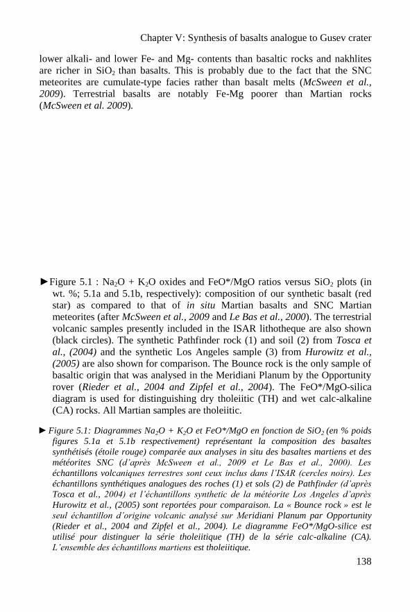
Chapter V: Synthesis of basalts analogue to Gusev crater
138
lower alkali- and lower Fe- and Mg- contents than basaltic rocks and nakhlites
are richer in SiO2 than basalts. This is probably due to the fact that the SNC
meteorites are cumulate-type facies rather than basalt melts (McSween et al.,
2009). Terrestrial basalts are notably Fe-Mg poorer than Martian rocks
(McSween et al. 2009).
►Figure 5.1 : Na2O + K2O oxides and FeO*/MgO ratios versus SiO2 plots (in
wt. %; 5.1a and 5.1b, respectively): composition of our synthetic basalt (red
star) as compared to that of in situ Martian basalts and SNC Martian
meteorites (after McSween et al., 2009 and Le Bas et al., 2000). The terrestrial
volcanic samples presently included in the ISAR lithotheque are also shown
(black circles). The synthetic Pathfinder rock (1) and soil (2) from Tosca et
al., (2004) and the synthetic Los Angeles sample (3) from Hurowitz et al.,
(2005) are also shown for comparison. The Bounce rock is the only sample of
basaltic origin that was analysed in the Meridiani Planum by the Opportunity
rover (Rieder et al., 2004 and Zipfel et al., 2004). The FeO*/MgO-silica
diagram is used for distinguishing dry tholeiitic (TH) and wet calc-alkaline
(CA) rocks. All Martian samples are tholeiitic.
►Figure 5.1: Diagrammes Na2O + K2O et FeO*/MgO en fonction de SiO2 (en % poids
figures 5.1a et 5.1b respectivement) représentant la composition des basaltes
synthétisés (étoile rouge) comparée aux analyses in situ des basaltes martiens et des
météorites SNC (d’après McSween et al., 2009 et Le Bas et al., 2000). Les
échantillons volcaniques terrestres sont ceux inclus dans l’ISAR (cercles noirs). Les
échantillons synthétiques analogues des roches (1) et sols (2) de Pathfinder (d’après
Tosca et al., 2004) et l’échantillons synthetic de la météorite Los Angeles d’après
Hurowitz et al., (2005) sont reportées pour comparaison. La « Bounce rock » est le
seul échantillon d’origine volcanic analysé sur Meridiani Planum par Opportunity
(Rieder et al., 2004 and Zipfel et al., 2004). Le diagramme FeO*/MgO-silice est
utilisé pour distinguer la série tholeiitique (TH) de la série calc-alkaline (CA).
L’ensemble des échantillons martiens est tholeiitique.
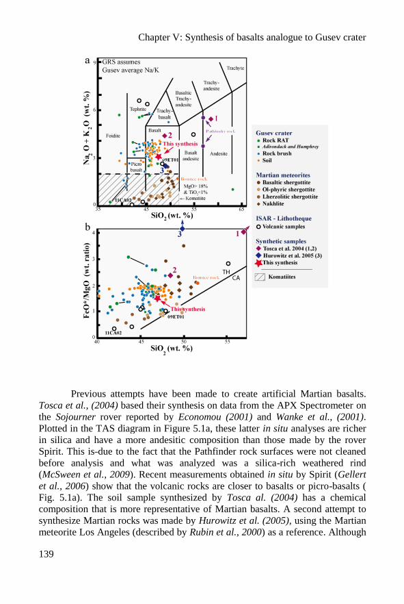
Chapter V: Synthesis of basalts analogue to Gusev crater
139
Previous attempts have been made to create artificial Martian basalts.
Tosca et al., (2004) based their synthesis on data from the APX Spectrometer on
the Sojourner rover reported by Economou (2001) and Wanke et al., (2001).
Plotted in the TAS diagram in Figure 5.1a, these latter in situ analyses are richer
in silica and have a more andesitic composition than those made by the rover
Spirit. This is due to the fact that the Pathfinder rock surfaces were not cleaned
before analysis and what was analyzed was a silica-rich weathered rind
(McSween et al., 2009). Recent measurements obtained in situ by Spirit (Gellert
et al., 2006) show that the volcanic rocks are closer to basalts or picro-basalts (
Fig. 5.1a). The soil sample synthesized by Tosca al. (2004) has a chemical
composition that is more representative of Martian basalts. A second attempt to
synthesize Martian rocks was made by Hurowitz et al. (2005), using the Martian
meteorite Los Angeles (described by Rubin et al., 2000) as a reference. Although
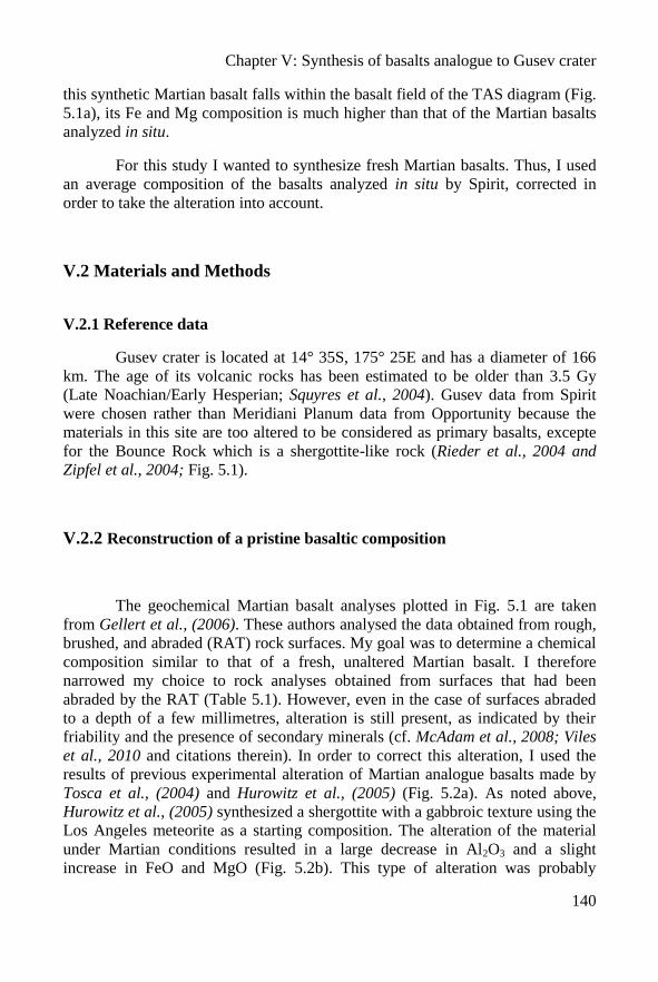
Chapter V: Synthesis of basalts analogue to Gusev crater
140
this synthetic Martian basalt falls within the basalt field of the TAS diagram (Fig.
5.1a), its Fe and Mg composition is much higher than that of the Martian basalts
analyzed in situ.
For this study I wanted to synthesize fresh Martian basalts. Thus, I used
an average composition of the basalts analyzed in situ by Spirit, corrected in
order to take the alteration into account.
V.2 Materials and Methods
V.2.1 Reference data
Gusev crater is located at 14° 35S, 175° 25E and has a diameter of 166
km. The age of its volcanic rocks has been estimated to be older than 3.5 Gy
(Late Noachian/Early Hesperian; Squyres et al., 2004). Gusev data from Spirit
were chosen rather than Meridiani Planum data from Opportunity because the
materials in this site are too altered to be considered as primary basalts, excepte
for the Bounce Rock which is a shergottite-like rock (Rieder et al., 2004 and
Zipfel et al., 2004; Fig. 5.1).
V.2.2 Reconstruction of a pristine basaltic composition
The geochemical Martian basalt analyses plotted in Fig. 5.1 are taken
from Gellert et al., (2006). These authors analysed the data obtained from rough,
brushed, and abraded (RAT) rock surfaces. My goal was to determine a chemical
composition similar to that of a fresh, unaltered Martian basalt. I therefore
narrowed my choice to rock analyses obtained from surfaces that had been
abraded by the RAT (Table 5.1). However, even in the case of surfaces abraded
to a depth of a few millimetres, alteration is still present, as indicated by their
friability and the presence of secondary minerals (cf. McAdam et al., 2008; Viles
et al., 2010 and citations therein). In order to correct this alteration, I used the
results of previous experimental alteration of Martian analogue basalts made by
Tosca et al., (2004) and Hurowitz et al., (2005) (Fig. 5.2a). As noted above,
Hurowitz et al., (2005) synthesized a shergottite with a gabbroic texture using the
Los Angeles meteorite as a starting composition. The alteration of the material
under Martian conditions resulted in a large decrease in Al2O3 and a slight
increase in FeO and MgO (Fig. 5.2b). This type of alteration was probably
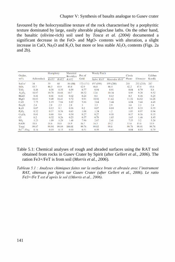
Chapter V: Synthesis of basalts analogue to Gusev crater
141
favoured by the holocrystalline texture of the rock characterised by a porphyritic
texture dominated by large, easily alterable plagioclase laths. On the other hand,
the basaltic (olivine-rich) soil used by Tosca et al. (2004) documented a
significant decrease in the FeO- and MgO- contents with alteration, a slight
increase in CaO, Na2O and K2O, but more or less stable Al2O3 contents (Figs. 2a
and 2b).
Table 5.1: Chemical analyses of rough and abraded surfaces using the RAT tool
obtained from rocks in Gusev Crater by Spirit (after Gellert et al., 2006). The
ration Fe3+/FeT is from soil (Morris et al., 2006).
Tableau 5.1 : Analyses chimiques faites sur la surface brute et abrasée avec l’instrument
RAT, obtenues par Spirit sur Gusev Crater (after Gellert et al., 2006). Le ratio
Fe3+/Fe T est d’après le sol ((Morris et al., 2006).
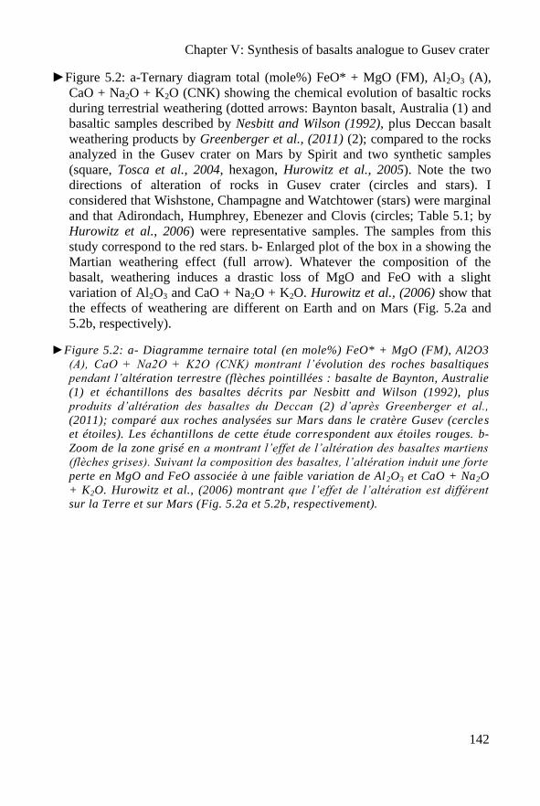
Chapter V: Synthesis of basalts analogue to Gusev crater
142
►Figure 5.2: a-Ternary diagram total (mole%) FeO* + MgO (FM), Al2O3 (A),
CaO + Na2O + K2O (CNK) showing the chemical evolution of basaltic rocks
during terrestrial weathering (dotted arrows: Baynton basalt, Australia (1) and
basaltic samples described by Nesbitt and Wilson (1992), plus Deccan basalt
weathering products by Greenberger et al., (2011) (2); compared to the rocks
analyzed in the Gusev crater on Mars by Spirit and two synthetic samples
(square, Tosca et al., 2004, hexagon, Hurowitz et al., 2005). Note the two
directions of alteration of rocks in Gusev crater (circles and stars). I
considered that Wishstone, Champagne and Watchtower (stars) were marginal
and that Adirondach, Humphrey, Ebenezer and Clovis (circles; Table 5.1; by
Hurowitz et al., 2006) were representative samples. The samples from this
study correspond to the red stars. b- Enlarged plot of the box in a showing the
Martian weathering effect (full arrow). Whatever the composition of the
basalt, weathering induces a drastic loss of MgO and FeO with a slight
variation of Al2O3 and CaO + Na2O + K2O. Hurowitz et al., (2006) show that
the effects of weathering are different on Earth and on Mars (Fig. 5.2a and
5.2b, respectively).
►Figure 5.2: a- Diagramme ternaire total (en mole%) FeO* + MgO (FM), Al2O3
(A), CaO + Na2O + K2O (CNK) montrant l’évolution des roches basaltiques
pendant l’altération terrestre (flèches pointillées : basalte de Baynton, Australie
(1) et échantillons des basaltes décrits par Nesbitt and Wilson (1992), plus
produits d’altération des basaltes du Deccan (2) d’après Greenberger et al.,
(2011); comparé aux roches analysées sur Mars dans le cratère Gusev (cercles
et étoiles). Les échantillons de cette étude correspondent aux étoiles rouges. b-
Zoom de la zone grisé en a montrant l’effet de l’altération des basaltes martiens
(flèches grises). Suivant la composition des basaltes, l’altération induit une forte
perte en MgO and FeO associée à une faible variation de Al2O3 et CaO + Na2O
+ K2O. Hurowitz et al., (2006) montrant que l’effet de l’altération est différent
sur la Terre et sur Mars (Fig. 5.2a et 5.2b, respectivement).
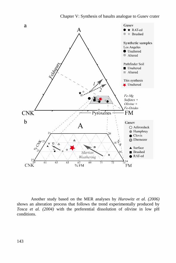
Chapter V: Synthesis of basalts analogue to Gusev crater
143
Another study based on the MER analyses by Hurowitz et al. (2006)
shows an alteration process that follows the trend experimentally produced by
Tosca et al. (2004) with the preferential dissolution of olivine in low pH
conditions.
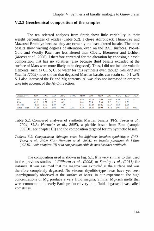
Chapter V: Synthesis of basalts analogue to Gusev crater
144
V.2.3 Geochemical composition of the samples
The ten selected analyses from Spirit show little variability in their
weight percentages of oxides (Table 5.2). I chose Adirondack, Humphrey and
Mazatzal Brooklyn because they are certainly the least altered basalts. The other
basalts show varying degrees of alteration, even on the RAT surfaces. Pot-of-
Gold and Woolly Patch are less altered than Clovis, Ebernezer and Uchben
(Morris et al., 2006). I therefore corrected for the alteration by choosing a basalt
composition that has no volatiles (also because fluid basalts extruded at the
surface of Mars were more likely to be degassed). Thus, I did not include volatile
elements, such as Cl, S, C, or water for this synthesis even though Gaillard and
Scaillet (2009) have shown that degassed Martian basalts can retain ca. 0.1 wt%
S. I also increased the Fe and Mg contents. Al was also not increased in order to
take into account of the Al2O3 reaction.
Table 5.2: Compared analyses of synthetic Martian basalts (PFS: Tosca et al.,
2004; SLA: Hurowitz et al., 2005), a picritic basalt from Etna (sample
09ET01 see chapter III) and the composition targeted for my synthetic basalt.
Tableau 5.2: Comparaison chimique entre les différents basaltes synthétiques (PFS:
Tosca et al., 2004; SLA: Hurowitz et al., 2005), un basalte picritique de l’Etna
(09ET01, voir chapitre III) et la composition cible de mes basaltes artificiels
The composition used is shown in Fig. 5.1. It is very similar to that used
in the previous studies of Filiberto et al., (2008) or Stanley et al., (2011) for
instance. It was assumed that the magma was extruded at the surface and was
therefore completely degassed. No viscous rhyolitic-type lavas have yet been
unambiguously observed at the surface of Mars. In our experiment, the high
concentrations of Mg produce a very fluid magma. Similar Mg-rich melts that
were common on the early Earth produced very thin, fluid, degassed lavas called
komatiites.
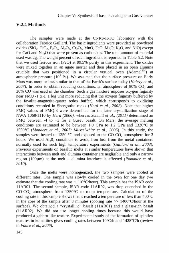
Chapter V: Synthesis of basalts analogue to Gusev crater
145
V.2.4 Methods
The samples were made at the CNRS-ISTO laboratory with the
collaboration Fabrice Gaillard. The basic ingredients were provided as powdered
oxides (SiO2, TiO2, P2O5, Al2O3, Cr2O3, MnO, FeO, MgO, K2O, and NiO) except
for CaO and Na2O that were present as carbonates. The total amount of material
used was 2g. The weight percent of each ingredient is reported in Table 5.2. Note
that we used ferrous iron (FeO) at 99.5% purity in this experiment. The oxides
were mixed together in an agate mortar and then placed in an open alumina
crucible that was positioned in a circular vertical oven (AdamelTM
) at
atmospheric pressure (105 Pa). We assumed that the surface pressure on Early
Mars was more or less similar to that of the Earth’s surface today (Halevy et al.,
2007). In order to obtain reducing conditions, an atmosphere of 80% CO2 and
20% CO was used in the chamber. Such a gas mixture imposes oxygen fugacity
near FMQ -1 (i.e. 1 log unit more reducing that the oxygen fugacity buffered by
the fayalite-magnetite-quartz redox buffer), which corresponds to oxidizing
conditions recorded in Shergottite rocks (Herd et al., 2002). Note that higher
FMQ values of FMQ-3 were determined for the later crystallization stage of
NWA 1068/1110 by Herd (2006), whereas Schmitt et al., (2011) determined an
FMQ between -4 to +3 for a Gusev basalt. On Mars, the average melting
conditions are estimated to be between 1.0 GPa to 1.2 GPa and 1320°C to
1550°C (Monders et al., 2007; Musselwhite et al., 2006). In this study, the
samples were heated to 1350 °C and exposed to the CO-CO2 atmosphere for 3
hours. We used Al203 containers to avoid iron loss from the metal containers
normally used for such high temperature experiments (Gaillard et al., 2003).
Previous experiments on basaltic melts at similar temperatures have shown that
interactions between melt and alumina container are negligible and only a narrow
region (100µm) at the melt – alumina interface is affected (Pommier et al.,
2010).
Once the melts were homogenized, the two samples were cooled at
different rates. One sample was slowly cooled in the oven for one day (we
estimate that the cooling rate was ~ 110°C/hour). This sample has the ISAR code
11AR01. The second sample, ISAR code 11AR02, was drop quenched in the
CO-CO2 atmosphere from 1350°C to room temperature. Calculation of the
cooling rate in this sample shows that it reached a temperature of less than 400°C
in the core of the sample after 8 minutes (cooling rate >> 1400°C/hour at the
surface). We obtained a “crystalline” basalt (11AR01) and a glass-rich basalt
(11AR02). We did not use longer cooling times because this would have
produced a gabbro-like texture. Experimental study of the formation of spinifex
textures in komatiites gives cooling rates between 10°C/h and 1428°C/h (review
in Faure et al., 2006).
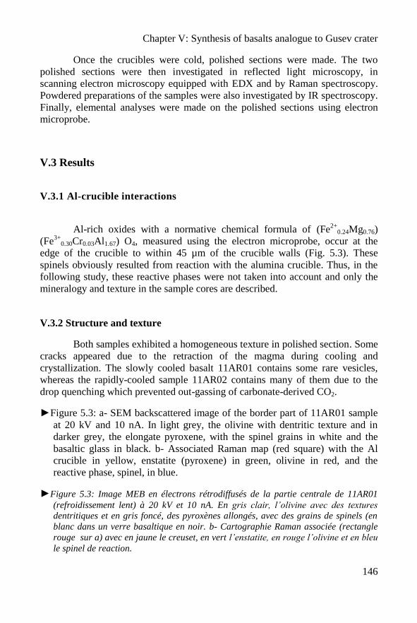
Chapter V: Synthesis of basalts analogue to Gusev crater
146
Once the crucibles were cold, polished sections were made. The two
polished sections were then investigated in reflected light microscopy, in
scanning electron microscopy equipped with EDX and by Raman spectroscopy.
Powdered preparations of the samples were also investigated by IR spectroscopy.
Finally, elemental analyses were made on the polished sections using electron
microprobe.
V.3 Results
V.3.1 Al-crucible interactions
Al-rich oxides with a normative chemical formula of (Fe2+
0.24Mg0.76)
(Fe3+
0.30Cr0.03Al1.67) O4, measured using the electron microprobe, occur at the
edge of the crucible to within 45 µm of the crucible walls (Fig. 5.3). These
spinels obviously resulted from reaction with the alumina crucible. Thus, in the
following study, these reactive phases were not taken into account and only the
mineralogy and texture in the sample cores are described.
V.3.2 Structure and texture
Both samples exhibited a homogeneous texture in polished section. Some
cracks appeared due to the retraction of the magma during cooling and
crystallization. The slowly cooled basalt 11AR01 contains some rare vesicles,
whereas the rapidly-cooled sample 11AR02 contains many of them due to the
drop quenching which prevented out-gassing of carbonate-derived CO2.
►Figure 5.3: a- SEM backscattered image of the border part of 11AR01 sample
at 20 kV and 10 nA. In light grey, the olivine with dentritic texture and in
darker grey, the elongate pyroxene, with the spinel grains in white and the
basaltic glass in black. b- Associated Raman map (red square) with the Al
crucible in yellow, enstatite (pyroxene) in green, olivine in red, and the
reactive phase, spinel, in blue.
►Figure 5.3: Image MEB en électrons rétrodiffusés de la partie centrale de 11AR01
(refroidissement lent) à 20 kV et 10 nA. En gris clair, l’olivine avec des textures
dentritiques et en gris foncé, des pyroxènes allongés, avec des grains de spinels (en
blanc dans un verre basaltique en noir. b- Cartographie Raman associée (rectangle
rouge sur a) avec en jaune le creuset, en vert l’enstatite, en rouge l’olivine et en bleu
le spinel de reaction.
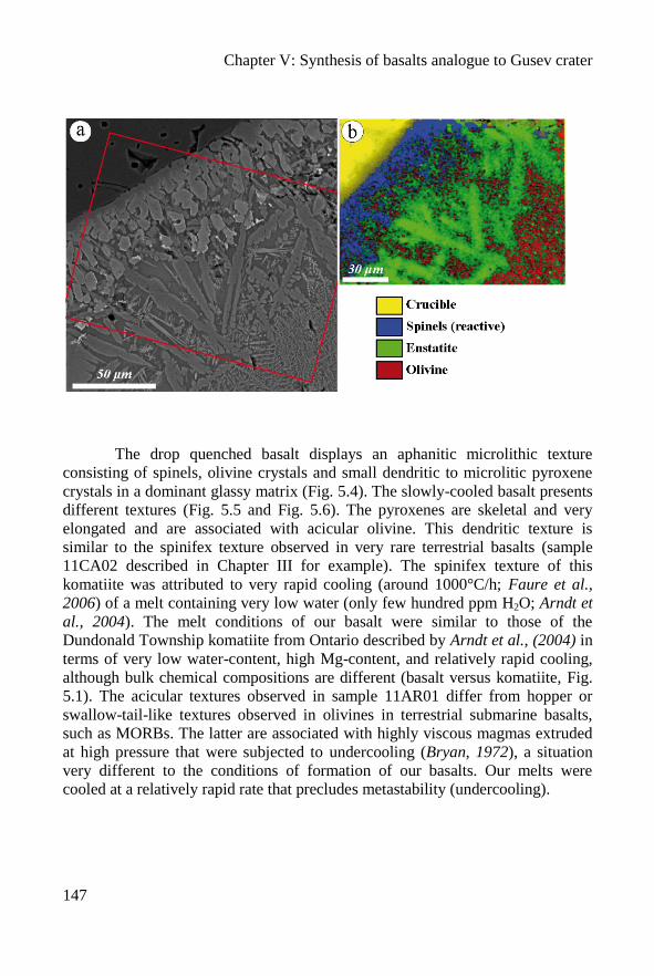
Chapter V: Synthesis of basalts analogue to Gusev crater
147
The drop quenched basalt displays an aphanitic microlithic texture
consisting of spinels, olivine crystals and small dendritic to microlitic pyroxene
crystals in a dominant glassy matrix (Fig. 5.4). The slowly-cooled basalt presents
different textures (Fig. 5.5 and Fig. 5.6). The pyroxenes are skeletal and very
elongated and are associated with acicular olivine. This dendritic texture is
similar to the spinifex texture observed in very rare terrestrial basalts (sample
11CA02 described in Chapter III for example). The spinifex texture of this
komatiite was attributed to very rapid cooling (around 1000°C/h; Faure et al.,
2006) of a melt containing very low water (only few hundred ppm H2O; Arndt et
al., 2004). The melt conditions of our basalt were similar to those of the
Dundonald Township komatiite from Ontario described by Arndt et al., (2004) in
terms of very low water-content, high Mg-content, and relatively rapid cooling,
although bulk chemical compositions are different (basalt versus komatiite, Fig.
5.1). The acicular textures observed in sample 11AR01 differ from hopper or
swallow-tail-like textures observed in olivines in terrestrial submarine basalts,
such as MORBs. The latter are associated with highly viscous magmas extruded
at high pressure that were subjected to undercooling (Bryan, 1972), a situation
very different to the conditions of formation of our basalts. Our melts were
cooled at a relatively rapid rate that precludes metastability (undercooling).
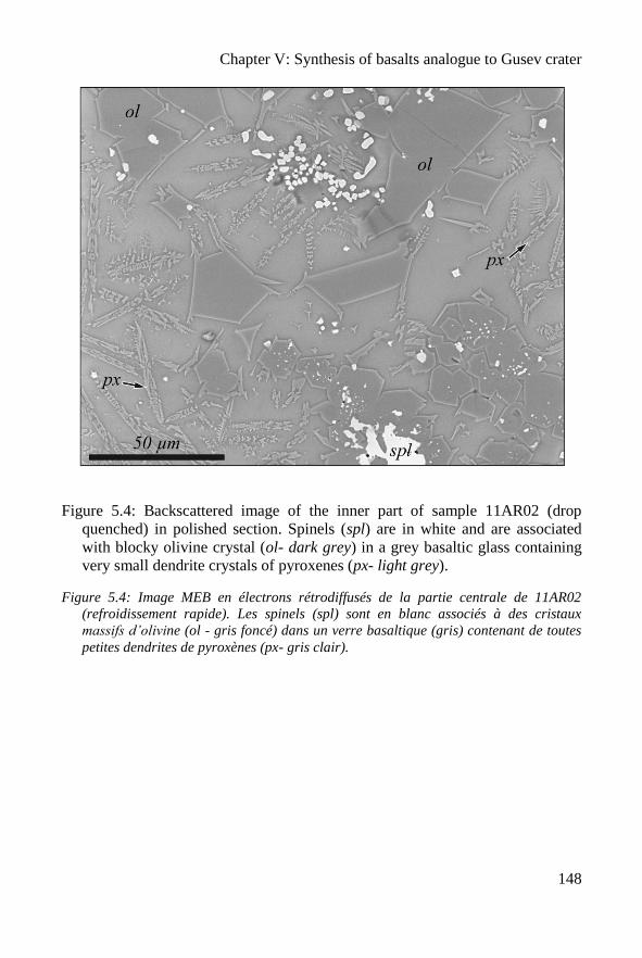
Chapter V: Synthesis of basalts analogue to Gusev crater
148
Figure 5.4: Backscattered image of the inner part of sample 11AR02 (drop
quenched) in polished section. Spinels (spl) are in white and are associated
with blocky olivine crystal (ol- dark grey) in a grey basaltic glass containing
very small dendrite crystals of pyroxenes (px- light grey).
Figure 5.4: Image MEB en électrons rétrodiffusés de la partie centrale de 11AR02
(refroidissement rapide). Les spinels (spl) sont en blanc associés à des cristaux
massifs d’olivine (ol - gris foncé) dans un verre basaltique (gris) contenant de toutes
petites dendrites de pyroxènes (px- gris clair).
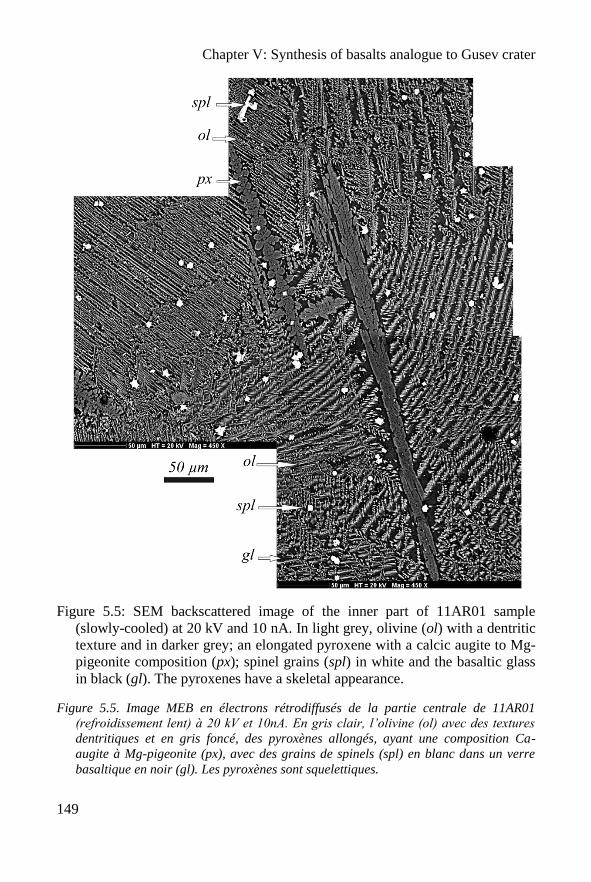
Chapter V: Synthesis of basalts analogue to Gusev crater
149
Figure 5.5: SEM backscattered image of the inner part of 11AR01 sample
(slowly-cooled) at 20 kV and 10 nA. In light grey, olivine (ol) with a dentritic
texture and in darker grey; an elongated pyroxene with a calcic augite to Mg-
pigeonite composition (px); spinel grains (spl) in white and the basaltic glass
in black (gl). The pyroxenes have a skeletal appearance.
Figure 5.5. Image MEB en électrons rétrodiffusés de la partie centrale de 11AR01
(refroidissement lent) à 20 kV et 10nA. En gris clair, l’olivine (ol) avec des textures
dentritiques et en gris foncé, des pyroxènes allongés, ayant une composition Ca-
augite à Mg-pigeonite (px), avec des grains de spinels (spl) en blanc dans un verre
basaltique en noir (gl). Les pyroxènes sont squelettiques.
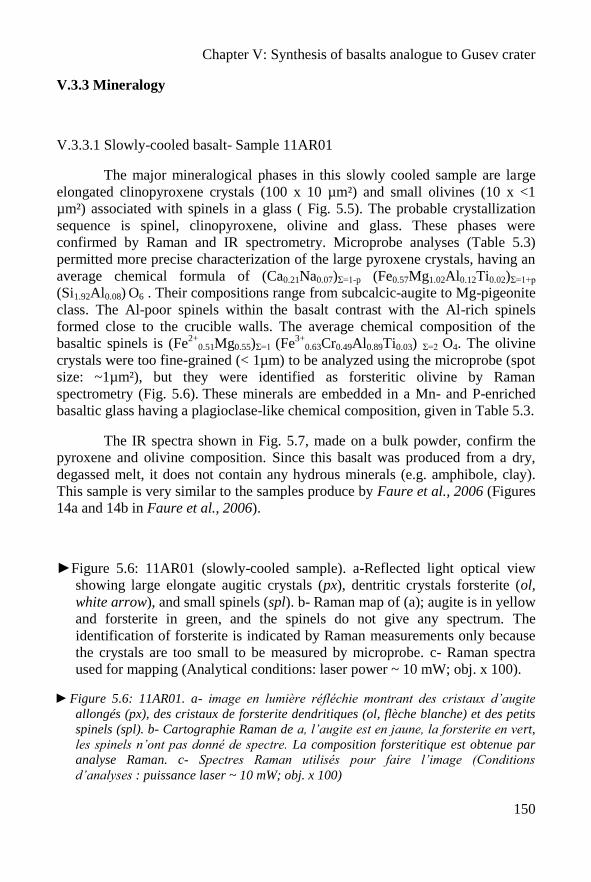
Chapter V: Synthesis of basalts analogue to Gusev crater
150
V.3.3 Mineralogy
V.3.3.1 Slowly-cooled basalt- Sample 11AR01
The major mineralogical phases in this slowly cooled sample are large
elongated clinopyroxene crystals (100 x 10 µm²) and small olivines (10 x <1
µm²) associated with spinels in a glass ( Fig. 5.5). The probable crystallization
sequence is spinel, clinopyroxene, olivine and glass. These phases were
confirmed by Raman and IR spectrometry. Microprobe analyses (Table 5.3)
permitted more precise characterization of the large pyroxene crystals, having an
average chemical formula of (Ca0.21Na0.07)Σ=1-p (Fe0.57Mg1.02Al0.12Ti0.02)Σ=1+p
(Si1.92Al0.08) O6 . Their compositions range from subcalcic-augite to Mg-pigeonite
class. The Al-poor spinels within the basalt contrast with the Al-rich spinels
formed close to the crucible walls. The average chemical composition of the
basaltic spinels is (Fe2+
0.51Mg0.55)Σ=1 (Fe3+
0.63Cr0.49Al0.89Ti0.03) Σ=2 O4. The olivine
crystals were too fine-grained (< 1µm) to be analyzed using the microprobe (spot
size: ~1µm²), but they were identified as forsteritic olivine by Raman
spectrometry (Fig. 5.6). These minerals are embedded in a Mn- and P-enriched
basaltic glass having a plagioclase-like chemical composition, given in Table 5.3.
The IR spectra shown in Fig. 5.7, made on a bulk powder, confirm the
pyroxene and olivine composition. Since this basalt was produced from a dry,
degassed melt, it does not contain any hydrous minerals (e.g. amphibole, clay).
This sample is very similar to the samples produce by Faure et al., 2006 (Figures
14a and 14b in Faure et al., 2006).
►Figure 5.6: 11AR01 (slowly-cooled sample). a-Reflected light optical view
showing large elongate augitic crystals (px), dentritic crystals forsterite (ol,
white arrow), and small spinels (spl). b- Raman map of (a); augite is in yellow
and forsterite in green, and the spinels do not give any spectrum. The
identification of forsterite is indicated by Raman measurements only because
the crystals are too small to be measured by microprobe. c- Raman spectra
used for mapping (Analytical conditions: laser power ~ 10 mW; obj. x 100).
►Figure 5.6: 11AR01. a- image en lumière réfléchie montrant des cristaux d’augite
allongés (px), des cristaux de forsterite dendritiques (ol, flèche blanche) et des petits
spinels (spl). b- Cartographie Raman de a, l’augite est en jaune, la forsterite en vert,
les spinels n’ont pas donné de spectre. La composition forsteritique est obtenue par
analyse Raman. c- Spectres Raman utilisés pour faire l’image (Conditions
d’analyses : puissance laser ~ 10 mW; obj. x 100)
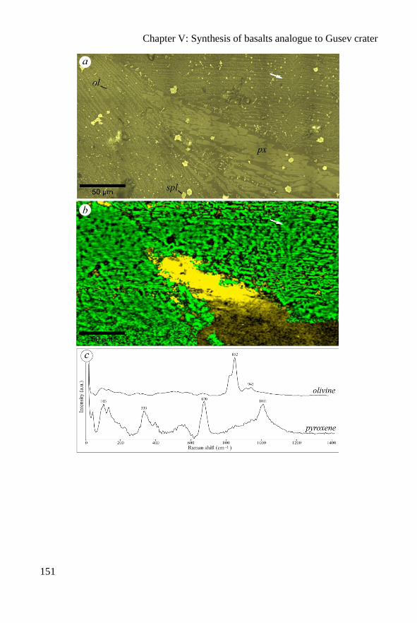
Chapter V: Synthesis of basalts analogue to Gusev crater
151
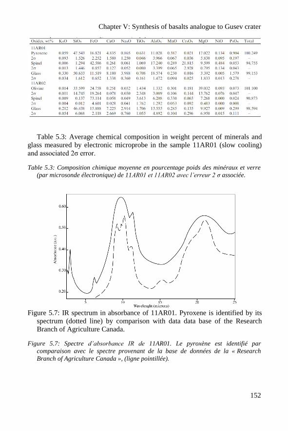
Chapter V: Synthesis of basalts analogue to Gusev crater
152
Table 5.3: Average chemical composition in weight percent of minerals and
glass measured by electronic microprobe in the sample 11AR01 (slow cooling)
and associated 2σ error.
Table 5.3: Composition chimique moyenne en pourcentage poids des minéraux et verre
(par microsonde électronique) de 11AR01 et 11AR02 avec l’erreur 2 σ associée.
Figure 5.7: IR spectrum in absorbance of 11AR01. Pyroxene is identified by its
spectrum (dotted line) by comparison with data data base of the Research
Branch of Agriculture Canada.
Figure 5.7: Spectre d’absorbance IR de 11AR01. Le pyroxène est identifié par
comparaison avec le spectre provenant de la base de données de la « Research
Branch of Agriculture Canada », (ligne pointillée).
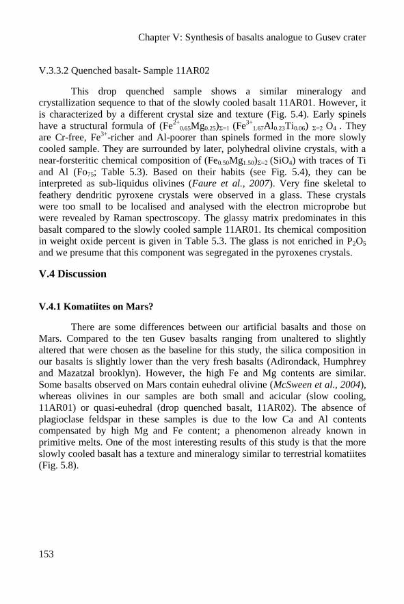
Chapter V: Synthesis of basalts analogue to Gusev crater
153
V.3.3.2 Quenched basalt- Sample 11AR02
This drop quenched sample shows a similar mineralogy and
crystallization sequence to that of the slowly cooled basalt 11AR01. However, it
is characterized by a different crystal size and texture (Fig. 5.4). Early spinels
have a structural formula of (Fe2+
0.65Mg0.25)Σ=1 (Fe3+
1.67Al0.23Ti0.06) Σ=2 O4 . They
are Cr-free, Fe3+
-richer and Al-poorer than spinels formed in the more slowly
cooled sample. They are surrounded by later, polyhedral olivine crystals, with a
near-forsteritic chemical composition of (Fe0.50Mg1.50)Σ=2 (SiO4) with traces of Ti
and Al (Fo75; Table 5.3). Based on their habits (see Fig. 5.4), they can be
interpreted as sub-liquidus olivines (Faure et al., 2007). Very fine skeletal to
feathery dendritic pyroxene crystals were observed in a glass. These crystals
were too small to be localised and analysed with the electron microprobe but
were revealed by Raman spectroscopy. The glassy matrix predominates in this
basalt compared to the slowly cooled sample 11AR01. Its chemical composition
in weight oxide percent is given in Table 5.3. The glass is not enriched in P2O5
and we presume that this component was segregated in the pyroxenes crystals.
V.4 Discussion
V.4.1 Komatiites on Mars?
There are some differences between our artificial basalts and those on
Mars. Compared to the ten Gusev basalts ranging from unaltered to slightly
altered that were chosen as the baseline for this study, the silica composition in
our basalts is slightly lower than the very fresh basalts (Adirondack, Humphrey
and Mazatzal brooklyn). However, the high Fe and Mg contents are similar.
Some basalts observed on Mars contain euhedral olivine (McSween et al., 2004),
whereas olivines in our samples are both small and acicular (slow cooling,
11AR01) or quasi-euhedral (drop quenched basalt, 11AR02). The absence of
plagioclase feldspar in these samples is due to the low Ca and Al contents
compensated by high Mg and Fe content; a phenomenon already known in
primitive melts. One of the most interesting results of this study is that the more
slowly cooled basalt has a texture and mineralogy similar to terrestrial komatiites
(Fig. 5.8).
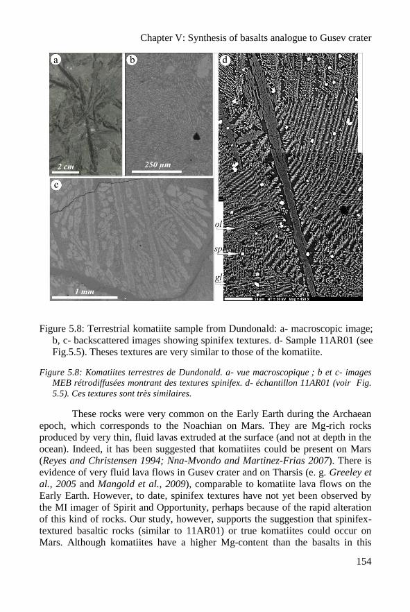
Chapter V: Synthesis of basalts analogue to Gusev crater
154
Figure 5.8: Terrestrial komatiite sample from Dundonald: a- macroscopic image;
b, c- backscattered images showing spinifex textures. d- Sample 11AR01 (see
Fig.5.5). Theses textures are very similar to those of the komatiite.
Figure 5.8: Komatiites terrestres de Dundonald. a- vue macroscopique ; b et c- images
MEB rétrodiffusées montrant des textures spinifex. d- échantillon 11AR01 (voir Fig.
5.5). Ces textures sont très similaires.
These rocks were very common on the Early Earth during the Archaean
epoch, which corresponds to the Noachian on Mars. They are Mg-rich rocks
produced by very thin, fluid lavas extruded at the surface (and not at depth in the
ocean). Indeed, it has been suggested that komatiites could be present on Mars
(Reyes and Christensen 1994; Nna-Mvondo and Martinez-Frias 2007). There is
evidence of very fluid lava flows in Gusev crater and on Tharsis (e. g. Greeley et
al., 2005 and Mangold et al., 2009), comparable to komatiite lava flows on the
Early Earth. However, to date, spinifex textures have not yet been observed by
the MI imager of Spirit and Opportunity, perhaps because of the rapid alteration
of this kind of rocks. Our study, however, supports the suggestion that spinifex-
textured basaltic rocks (similar to 11AR01) or true komatiites could occur on
Mars. Although komatiites have a higher Mg-content than the basalts in this
Chapter V: Synthesis of basalts analogue to Gusev crater
155
study (Mg ~12 wt%), we suggest that the lower Mg-content could be
compensated by a high content of Fe. Our samples contain 29.90 wt% of MgO +
FeO, which is similar to an average composition of a Barberton-type komatiite
(30 wt% of MgO + FeO; Arndt, 2003).
V.4.2 Comparison with previous studies
Our synthetic basalts differ from those previously produced by Tosca et
al., (2004) and Hurowitz et al., (2005) because both melt compositions and
experimental P-T melting conditions were different. The synthesis pressure
conditions used by Tosca et al., (2004) are comparable to ours, ~105 Pa, but we
used 1350°C whereas these authors used ~ 1120°C. The chemical composition of
the melt thus produced from the Pathfinder soil by Tosca et al., (2004) was close
to the melt we produced, but was different from the silica-rich Pathfinder rock
(Fig. 5.1). Moreover, because Tosca et al., (2004) immediately ground their
sample to a powder for the alteration experiment it is not possible to compare
their mineralogy and texture to those of our synthetic basalts. The artificial rock
produced by Hurowitz et al., 2005 was obtained at ~1135°C and at ~0.1 Pa (~1
mbar). The rock was cooled very slowly (~6°C/hour) and a “holocrystalline
basalt with aphanitic-porphyritic texture” without glass was produced, a texture
which is more similar to a gabbro than to a basalt. A further difference is that the
chemical composition of the melt synthesized by Hurowitz et al., (2005) was a
shergottite analogue of the Los Angeles meteorite. It’s very low Mg-content is
different from the Gusev crater basaltic rocks (Fig. 5.1b).
V.4.3 Implication for Early Life on Mars
The volcanics in Gusev crater are of Noachian age (Soderblom and Bell,
2008). This is the period when Mars had somewhat similar environmental
conditions to those of the Early Earth and was potentially habitable (Westall,
2005; Bibring et al., 2006; Jakosky et al., 2007; Southam et al., 2007; Westall et
al., 2011). As noted above, Fe-Mg-rich basalts and komatiite-type basalts were
common on the Early Earth. Such rocks are favourable habitats for
chemolithotrophic microorganisms that obtain their energy and nutrients from
chemical reactions on rocks surface (cf. Furnes et al., 2008). Furthermore,
alteration of these rocks would have produced Fe-Mg clays, such as nontronite
and chlorite-saponite, that could have been implicated in the concentration of
prebiotic organics and thus in various scenarios for the origin of life (Meunier et
Chapter V: Synthesis of basalts analogue to Gusev crater
156
al., 2010). Given the interpretations of such materials associated with Noachian
terrains on the surface of Mars (Poulet et al., 2005; Bibring et al., 2006; Mustard
et al., 2008; Ehlmann et al., 2009), we can extend the analogy with the Early
Earth to hypothesize the potential importance of this kind of rocks and their
alteration products for not only an origin of Martian life, but also as habitats for
possible Martian microorganisms. Both samples could be weathered by glass
alteration. The drop quenched sample contains more glass and would be rapidly
altered, whereas the spinifex texture could be used as a site of preferential
development of clays (such as nontronite) along specific surfaces (elongates
grain surfaces). This could favour the colonization of volcanic rocks by
microorganisms, in particular chemolithotrophs. For example, traces of the
activities of such organisms have been preserved in basalts and derivatives of
volcanic rocks from the Early Archaean terrains of Barberton and the Pilbara
(review in Furnes et al., 2007; Westall et al., 2006; Westall et al., 2011) as well
as in amygdales in pillow basalts (Cavalazzi et al., 2011). Moreover, typical
alteration of the mafic rocks, such as komatiites, in aqueous conditions, a high
pH and low aSi02 leads to serpentinization (this reaction produces H2, which can
serve as an important source of energy for chemosynthetic organisms, Schulte et
al., 2006). Note that biological activity is associated with serpentinization
process in hydrated mantle (Ménez et al., 2012). Furthermore, serpentine deposits
have been observed on the Martian surface (Ehlmann et al., 2010) and could be a
possible habitat for early life on Mars. Thus, if life ever appeared on Mars, it is
possible that traces of its existence could be found directly in the surfaces, cracks
and vesicles of this volcanic type of rocks. In situ missions to Mars should
examine such rocks if there is evidence that they have been associated with
water.
V.5 Conclusion
We produced experimental Fe-Mg-rich basalts based on the geochemical
data from Gusev basalts. The slowly cooled basalt present specific spinifex
textures that are very similar to those of Early Earth basalts and komatiites that
were formed in an equivalent period to the Noachian and Hesperian on Mars. Fe-
Mg-rich basalts and their alteration products are also highly relevant for the
appearance and habitability of life. Nontronite and chloritic clays could have
concentrated prebiotic organic molecules that were the building bricks of life,
whereas surfaces, cracks and vesicles in such lavas were ideal habitats for
primitive anaerobic life forms, such as chemolithotrophic microorganisms
(Furnes et al., 2007; Cavalazzi et al., 2011).
Chapter V: Synthesis of basalts analogue to Gusev crater
157
This study concludes my thesis project by adding basalt analogues to the
ISAR that are good chemical and textural analogues of Martian basalts.

Chapter V: Synthesis of basalts analogue to Gusev crater
158
Chapter V: Synthesis of basalts analogue to Gusev crater
159
Basaltes martiens artificiels (Résumé Francais)
Les analyses chimiques des basaltes du cratère Gusev par Spirit ont montré
qu’ils étaient chimiquement différents des basaltes terrestres. Cette variation se traduit
particulièrement par une teneur en Mg et Fe beaucoup plus forte pour les basaltes
martiens. Pour compléter la collection de l’ISAR, il apparaît pertinent de fabriquer en
laboratoire des basaltes analogues. Ce chapitre présente les résultats de deux synthèses,
l’une avec un refroidissement lent et l’autre avec un refroidissement rapide
(respectivement 1 jour et 1 heure).
Des études précédentes avaient déjà fabriqué des basaltes martiens artificiels
mais en se basant sur des données de la mission Pathfinder ou depuis la météorite
martienne Los Angeles. La Fig.5.1, montre que ces échantillons ne sont pas en
adéquation avec les résultats récents. Il semble particulièrement pertinent de fabriquer
des échantillons avec une composition chimique proche de celle des basaltes du cratère
Gusev. Nous avons choisi une composition chimique moyenne basée sur les analyses in-
situ des dix échantillons les moins altérés (Tableau 5.1). En effet nous voulons fabriquer
un basalte artificiel ayant une composition initiale, avant toute altération. Les basaltes
sur Gusev sont assez altérés. La Fig. 5.2 montre l’altération des basaltes terrestres et
martiens. On note que sur Mars lors de l’altération c’est principalement la teneur en Fe
et Mg qui baisse. La composition chimique des basaltes sera donc corrigée de
l’altération et ne comportera pas de volatils, considérant une mise en place à la surface
ou le magma est totalement dégazé. Le Tableau 5.2 reporte la composition chimique
utilisée et comparée aux deux précédentes expériences et à l’échantillon 09ET01.
Les échantillons ont été préparés au CNRS-ISTO avec la collaboration de
Fabrice Gaillard. Les ingrédients chimiques sont fournis en poids d’oxyde ou en
carbonates. L’échantillon final représentera 2 grammes de roche. La poudre est
mélangée dans un mortier avant d’être mise dans un creuset en alumine et placée dans
un four Adamel vertical à pression atmosphérique, à une température de 1350°c et sous
une atmosphère de CO-CO2. Lorsque les échantillons sont liquides, le premier est
refroidi lentement (une journée, 11AR01) et le deuxième est trempé (refroidissement
rapide, 11AR02). Une fois les creusets froids une section polie est préparée, ainsi que des
poudres.
Les deux échantillons présentent une texture homogène, seule une bordure de
réaction est visible entre le creuset et l’échantillon (Fig. 5.3). L’échantillon trempé
présente une texture aphanitique microlithique (Fig. 5.4). L’échantillon au
refroidissement lent présente des cristaux de pyroxène très allongées, squelettiques et des
olivines aciculaire, similaire aux textures spinifex des komatiites (Fig. 5.5 ; voir chapitre
III).
L’échantillon 11AR01 est constitué de clinopyroxènes allongés, de petites
olivines associées à des spinelles dans un verre. Les compositions chimiques de chaque
minéral sont calculées à partir des analyses microsonde (cf. Tableau 5.3), sauf pour
l’olivine qui est trop petite pour analyse. Le spectre IR de la Fig. 5.7, faite sur poudre,
Chapter V: Synthesis of basalts analogue to Gusev crater
160
confirme la présence de pyroxène et d’olivines. L’olivine est aussi identifiée au Raman
(fig. 5.6).
L’échantillon 11AR02 est constitué de la même minéralogie que l’échantillon
11AR01. Cependant les cristaux n’ont ni la même taille ni la même morphologie. De
nouveau les analyses chimiques de chaque minéral sont consignées dans le Tableau 5.3,
sauf pour les pyroxènes qui sont trop petits.
L’échantillon 11AR01 présente une texture semblable aux spinifex (Fig. 5.8).
Pouvons-nous trouver des komatiites sur Mars ? Il y a quelques différences entre nos
échantillons de synthèse et les échantillons martiens. La composition chimique en silice
est un peu supérieure aux basaltes vraiment frais (Adirondack, Humphrey et Mazatzal
brooklyn), bien que les teneurs en Fe et Mg soient similaires. Des basaltes martiens
présentent des olivines automorphes alors que dans ces échantillons elles sont petites et
aciculaires (11AR01) ou quasi-automorphes (11AR02). L’absence de feldspaths dans les
échantillons est due à la faible teneur en Ca et Al, compensée par une forte teneur en Mg
et Fe: un phénomène déjà connu dans les mélanges primitifs. Le plus intéressant est donc
la présence d’une texture et d’une minéralogie similaires à celle des komatiites. Ces
roches sont très communes à l’Archéen. Ces roches riches en Mg forment des laves très
fluides à la surface et des coulées très fines. Des komatiites ont déjà été décrites à la
surface de Mars. De plus, des preuves d’épanchement de magma très fluides en surface
sont un autre argument. Cependant, aucune texture spinifex n’a encore été révélée par le
microscope des MER, peut être à cause de l’altération rapide des ces roches. Cette étude
suggère que des roches basaltiques, ou de vraies komatiites peuvent être présentes à la
surface de Mars. Nos basaltes synthétiques sont différents de ceux des précédentes
études, puisque les compositions chimiques initiales et les paramètres de pression et de
température sont différents. Par exemple nous utilisons une température de 1350°C
comparée à 1120°C; ou encore une deuxième étude a proposé des basaltes
holocristallins avec des textures aphanitiques porphyriques sans verre qui évoque plus
un gabbro qu’un basalte.
Ce travail a aussi une implication dans la recherche de vie passée sur Mars. En
effet, les échantillons synthétisés sont analogues de roches martiennes du Noachien, une
période où la vie est possiblement apparue sur Mars. Ces roches riches en Fe et Mg sont
des habitats favorables pour des organismes chemiolithotrophes. De plus, l’altération de
ces roches produit des argiles riches en Fe et Mg comme la nontronite ou les argiles de
la famille chlorite-saponite, qui peuvent être impliquées dans la concentration des
organismes prébiotiques. Finalement, l’altération en conditions aqueuses de roches
mafiquescomme les komatiites, favorise la serpentinisation. Ce processus est souvent
associé à une importante activité biologique. Des preuves de la présence de serpentinites
ont déjà été observées sur Mars et peuvent constituer de possibles habitats pour une vie
primitive.
Nous avons produit des basaltes synthétiques riches en Fe et Mg fondés sur les
données provenant du cratère Gusev. L’échantillon refroidi lentement présente des
textures spinifex, très similaires aux komatiites typiques de la Terre primitive formées à
une époque contemporaine du Noachien martien. Ces produits et leurs altérations

Chapter V: Synthesis of basalts analogue to Gusev crater
161
semblent être pertinents pour la recherche d’environnement ayant pu abriter la vie. Ce
travail conclut mon projet de thèse et permet d’ajouter des basaltes analogues, ayant une
composition chimique et texturale analogue aux basaltes martiens, à la collection de
l’ISAR.

Chapter V: Synthesis of basalts analogue to Gusev crater
162
Conclusions
163
Conclusions
The main objective of this thesis was to follow a scientific approach
for creating a collection of rocks for testing and calibrating space
instrumentation for on-going and future space missions. For this purpose, it
was first necessary to establish a logical classification of the samples and to
develop an interface allowing scientists and engineers implicated in space
missions to obtain information on relevant samples and to be able to borrow
the samples. This was accomplished with the ISAR website. Because of the
emphasis is presently laid on Mars missions, I concentrated my efforts on the
collection of Mars analogue samples. The choice of samples was based on the
data available about the mineralogical and geochemical composition of the
Martian surface. The analogue samples were fully characterized using several
techniques relevant to space missions but using laboratory instrumentation
that allowed higher spatial resolution and accuracy. This first set of rocks is
already being used by different scientific teams working on the ExoMars
project. In December 2010, smalls slab of the ISAR samples exhibiting both
weathered and fresh surfaces were provided to Derek Pullan, University of
Leicester, for the calibration board of the PanCam system. I participated in the
ExoMars-XRD meeting with Lucia Marinageli (ExoMars-XRD PI,
International Research School of Planetary Sciences, Pescara – Italy) where I
gave her a selection of 12 samples from the ISAR to calibrate the XRD
instrument. Unfortunately, this calibration could not be completed in time for
this report. In November 2011, a number of samples were used by the Raman
team, directed by Fernando Rull (Raman PI; Universidad Asociada - Centro
de Astrobiologia, Valladolid). Samples have been sent to the MicrOmega
team to test and calibrate the instrument (Jean-Pierre Bibring, PI - F. Westall,
co-PI -, and his PhD student, Cedric Pilorget, IAS, Orsay, France). They have
also been provided to the group of Paul Mahaffy (Goddard Space Center) for
Conclusions
164
testing the SAM instrument for MSL and the GC-MS part of the MOMA
instrument for ExoMars-2018, as well as to Fred Goesmann (Max Planck
Institute, Mainz) for testing the LD-MS part of MOMA. The final
implementation of ISAR is the proposal of a blind test to the different teams
involved in ExoMars. The objective is to determine the level of expertise
accessible with the different instruments, separately and then alltogether. The
test is thus composed of two stages:
1. Two unlabelled samples were sent to the teams that accepted to
participate in the test (Raman, MicrOmega and MOMA) as <250 µm grains
size powders. Each team will send back the result of their analyses and their
first interpretation.
2. The results obtained from each instrument will be grouped together
and sent to the participants with complementary data, e.g. photography, to
help them to refine their interpretation.
The aim of this experiment is not to put the instruments in
competition but to bring out their complementarity. This test will thus help to
develop the interaction between the ExoMars teams and with the ISAR team
in order to improve the level of expertise accessible during the mission
(ability to identify a rock or to detect past traces of life for instance).
The first sample (A) is the 00AU05, the Kitty’s Gap chert from
Australia containing the oldest remains of microbial colonies. The second
sample (B) is a komatiite from the Komatii river in South Africa (10SA09).
The first stage is still in progress. The available results are displayed in the
Table 6.1.
Samples : Chert - 00AU05 Komatiite - 10SA09
ISAR Characterization : Minerals: Quartz, hydromuscovite and
anatase.
Organic molecules:
Only carbon as kerogen.
Minerals:
Quartz, amphiboles, clays,
talc, iron oxide, hematite
and dolomite.
Organic molecules:
Some
ExoMars Raman Minerals: quartz, calcite, anatase,
muscovite. carbonaceous material
Minerals: amphibole (Pargasite),
carbonaceous material
MicrOmega ? ?
MOMA ? ?
Conclusions
165
◄Table 6.1: Comparison of minerals and organic molecules observed in the
two blind test samples using ISAR instrumentation and with some of the
ExoMars instruments.
◄Tableau 6.1: Comparaison des minéraux et des molécules organiques observées
dans les deux échantillons du test à l’aveugle en utilisant les instruments de
l’ISAR et quelques uns des instruments d’ExoMars.
During the first part of my project, I had to evaluate the degree of
analogy and the pertinence of each Mars analogue sample. It was clear that
the perfect analogue sample does not exist on Earth, in particular because of
enrichment in Fe and Mg in Martian volcanics and because of the different
alteration processes. This conclusion led me to search for an area where
basaltic samples were altered by acidic fluids. Thus, in the second part of my
thesis, I suggested the Skouriotissa copper mining open pit in Cyprus as a
possible good Martian analogue site. This is a very massive sulphides deposit
(VMS) in a basaltic environment that has been altered by natural meteoric
water and acid mine runoff. I therefore sampled altered and unaltered rocks in
order to obtain mineralogical information along an alteration profile and to
study the weathering effect. I was thus able to document mineral associations
similar to those found on Noachian/early Hesperian Mars terrains and to
correlate them with specific subaqueous/subaerial and pH conditions of
alteration. This terrestrial analogue site has rock and weathering
characteristics that help to improve our understanding of geological
subsurface phenomena on Mars. It is therefore pertinent to continue the
investigation of this site and to sample it in finer detail in order to trace the
past and present physico-chemical processes, which are certainly comparable
to those taking place on Mars. The search for other analogue sites must also
be made, taking into account the new geological data coming from Mars.
The most relevant Mars analogue samples are obviously basalts.
However, the perfect chemical analogue basalt cannot exist on Earth where
basalts are depleted in Fe-Mg with respect to Martian basalts. The last part of
my project was thus dedicated to the fabrication of a Martian basalt with a
chemical composition based on the data collected in Gusev crater by Spirit.
The two synthesized samples show very interesting textures and structures.
This study increases our understanding of the geological processes taking
place on Mars (e.g. textures provide information about the cooling rate
conditions).
The ISAR collection is far from complete and it will be necessary to
add new Mars analogue rocks and minerals, such as evaporite minerals
Conclusions
166
(sulfates, chlorates or carbonates) that have been identified on the surface of
Mars (e.g. McLennan et al., 2005; Gendrin et al., 2005) and which, on Earth,
are often associated with traces of life (e.g. Barbieri and Stivaletta, 2011).
These kinds of samples have been collected in a variety of terrestrial
environments, such as the Atacama desert, in playas lakes (e.g. Morocco), or
around areas showing mining activity such as the Skouriotissa mine in
Cyprus. They will be analysed and characterized in the same way as the first
set of ISAR samples. Analogue samples from other solar system bodies, such
as the Moon and asteroids, will also be added to the collection.
Artificial weathering of the synthesized basalts, in comparison with
that of samples from natural environments (e.g. Cyprus) could be very
interesting. In order to mimic the weathering effect occurring during the
Noachian period, this alteration could be made with acid water (pH<4)
circulating on the surface of the sample. The results could thus be compared
with the “natural” processes observed in Cyprus. A second experiment carried
out under simulated Martian atmosphere and high UV irradiation, in order to
study the alteration of the surface of Mars over the last 3 Gy would also be
interesting. For astrobiological purposes, the alteration of the artificial
Martian basalt synthesized in the course of my project by lithotrophic or
photo-litotrophic microorganisms under simulated early Mars conditions
would be also very pertinent and will be undertaken in the near future. This
experiment could give important information about the interaction between
life and rocks and may, above all, be very helpful for the detection of past or
present traces of life on Mars.
Conclusions
167
Conclusion (résumé Francais)
L’objectif principal de cette thèse est d’avoir une approche scientifique afin
de créer une collection de roches pour tester et calibrer les instruments de vols des
futures missions spatiales. Pour cela, il a été nécessaire d’établir une classification
logique des échantillons et de développer une interface adaptée aux scientifiques et
ingénieurs impliqués dans les missions spatiales dans le but d’obtenir des
informations pertinentes sur les échantillons et pourvoir, le cas échéant, les
emprunter. Cet objectif est réalisé à travers le site web de l’ISAR. Puisque les
missions sont actuellement tournées vers Mars, j’ai concentré mon travail sur la
collection d’échantillons analogues de Mars. Les analogues sont intégralement
caractérisés en utilisant de nombreuses techniques pertinentes pour les missions
spatiales ; tout en utilisant des instruments de laboratoire qui permettent d’avoir de
bonnes résolutions et de la précision. Le premier lot d’échantillons a d’ores et déjà
été utilisé par différentes équipes du projet ExoMars (PanCam, ExoMars-XRD,
MicrOmega, Raman et MOMA) ; dernièrement lors de la réalisation d’un test à
l’aveugle avec les futurs instruments de vol. L’objectif est de déterminer le degré
d’expertise de chaque instrument ainsi qu’avec l’ensemble des instruments. Le test se
fait en deux étapes : 2 échantillons en poudre, ont été envoyés aux équipes
participantes (Raman, MicrOmega et MOMA) pour analyse sans aucune autre
information. Dans une deuxième étape les résultats obtenus sont regroupés et
échangés entre les équipes pour faire une deuxième itération d’interprétation.
L’objectif de l’exercice n’est pas de mettre les instruments en compétition mais de
mettre en évidence leur complémentarité. Les deux échantillons A et B sont
respectivement un chert de Kitty’s Gap (00AU05) et une komatiite d’Afrique du Sud
(10SA09). Les résultats d’analyse disponibles sont reportés dans le tableau 6.1.
Pendant la première partie de mon projet, j’ai évalué le degré d’analogie et
de pertinence de chaque échantillon analogue de Mars. Il est évidement que le parfait
échantillon analogue de Mars n’existe pas. En particulier à cause de l’enrichissement
en Fe Mg des basaltes, ou des processus d’altération différents. Cette première
conclusion m’a incité à chercher un lieu où des basaltes sont altérés par des fluides
acides. C’est pourquoi j’ai suggéré l’étude de la mine de Skouriotissa. Les basaltes y
sont altérés par des processus hydrothermaux, météoriques et acides. La minéralogie
résultante semble similaire aux observations martiennes, et ce site semble être un bon
site analogue de la Mars du Noachien – Amazonien.
Les meilleurs analogues de Mars sont sans aucun doute les basaltes.
Néanmoins des basaltes parfaitement analogues chimiquement de Mars n’existent pas
sur Terre, en particulier à cause de leur faible teneur en Fe et Mg. La dernière partie
de ma thèse a donc été dédiée à la fabrication de basaltes martiens avec une
composition chimique fondée sur les données collectées dans le cratère Gusev par
Spirit. Les deux échantillons synthétisés présentent de très intéressantes textures et
structures. Cette étude augmente la compréhension des processus géologiques qui ont
pris place sur Mars.
Conclusions
168
La collection de l’ISAR est loin d’être complète, et il est nécessaire d’ajouter
de nouvelles roches et de nouveaux minéraux analogues de Mars, comme des
évaporites.
Enfin l’altération des basaltes artificiels et la comparaison avec des
échantillons provenant d’environnements naturels (comme Chypre) peut être très
intéressant. Il serait intéressant de reproduire les conditions chimiques d’altération,
mais aussi d’essayer avec uniquement un rayonnement UV. Dans un contexte
exobiologique, faire altérer les échantillons par des microorganismes lithotrophes ou
photolitotrophes, dans des conditions simulant la Mars primitive, semble vraiment
pertinent pour mieux comprendre les interactions entre la vie et la roche, et pourrait
aider à la détection d’une vie passée ou présente sur Mars.
References
169
References
A
Ablay G. J., and Marti J., 2000. Stratigraphy, Structure, and Volcanic Evolution
of the Pico Teide-Pico Viejo Formation, Tenerife, Canary Islands. Journal of
Volcanology and Geothermal Research 103:175-208.
Alt J. C., and Honnorez J., 1984. Alteration of the upper oceanic crust, DSDP
site 417: mineralogy and chemistry. Contributions to Mineralogy and
petrology 87:149-169.
Amils R., Gonzalez-Toril E., Fernandez-Remolar D., Gomez F., Aquilera A.,
Rodriguez N., Malki M., Garcia-Moyano A., Fairen A. G., de la Fuente V.,
and Sanz J. L., 2007. Extreme environments as mars terrestrial analogs : The
Rio Tinto case. Planetary and Space Science 55:370-381.
Doi:10.1013/j.pss.2006.02.006.
Amundsen H. E. F., Benning L., Blake D. F., Fogel M., Ming D., Skidmore M.,
Steele A., and the AMASE team, 2011. Cryogenic origin for Mars analogue
carbonates in the Bockfjord volcanic complex, Svalbard (Norway), 42nd Lunar
and Planetary Conference, The Woodland Texas, Abstract # 2223.
Armstrong R. A., Compston W., De Wit M. J., and Willians I. S., 1990. The
stratigraphy of the 3.5-3.5 GA Barberton greenstone belt revisited: a single
zircon ion microprobe study. Earth and Planetary Science Letters 101:90-
106.
Arndt N. T., 2003. Komatiites, kimberlites, and boninites. Journal of
Geophysical Research 108 B6-2293. doi:10.1029/2002JB00215.
Arndt N. T., Lesher C. M., Houlé M. C., Lewin E., and Lacaze Y., 2004.
Intrusion and Crystallization of a Spinifex-Textured Komatiite Sill in
Dundonald Township, Ontario. Journal of Petrology 45:2555-2571.
Arvidson, R. E., Poulet F., Morris R. V., Bibring J.-P., Bell III J. F., Squyres S.
W., Christensen P. R., Bellucci G., Gondet B., Ehlmann B. L., Farrand W. H.,
Fergason R. L., Golombek M., Griffes J. L., Grotzinger J., Guinness E. A.,
Herkenhoff K. E., Johnson J. R., Klingelhöfer G., Langevin Y., Ming D.,
Seelos K., Sullivan R. J., Ward J. G., Wiseman S. M., and Wolffet M., 2006.
Nature and origin of the hematite-bearing plains of Terra Meridiani based on
References
170
analyses of orbital and Mars Exploration rover data sets, Journal of
Geophysical Research 111:E12S08. doi:10.1029/2006JE002728.
B
Bandfield J. L., 2002. Global mineral distributions on Mars. Journal of
Geophysical Research 107:5042. Doi:10.1029/2001je001510
Bandfield J. L., Glotch T. D., and Christensen P. R., 2003. Spectroscopic
Identification of Carbonate Minerals in the Martian Dust. Science 301:1084-
1087.
Bandfield J. L., Hamilton V. E., Christensen P. R., and McSween H. Y., 2004.
Identification of quartzofeldspathic materials on Mars. Journal of
Geophysical Research 109:E10009. Doi:10.1029/2004JE002290.
Barbieri R., and Stivaletta N., 2011. Continental evaporites and the search for
evidence of life on Mars. Geological Journal, 46: 513–524.
Barbin V., Blanc P., Klossa B., Lorin C., and Thomas R., 1999.
Cathodoluminescence: a new approach to the in situ study of martian surface
mineralogy. Symposium International: Programme d’exploration de Mars &
mission de retour d’échantillons (Paris). Resumé, CNES.Edit.
Bear L.M., 1960. Geol. Surv. Cyprus Mem. 3:1-122.
Bear L.M, 1961 (?) only reference in Moores and Vine 1971
Bibring J. -P., Langevin Y., Mustard J. F., Poulet F., Arvidson R., Gendrin A.,
Gondet B., Mangold N., Pinet P., Forget F., and the OMEGA Team, 2006.
Global Mineralogical and Aqueous Mars History Derived from
OMEGA/Mars Express Data. Science 312:400-404. Doi:
10.1126/science.1122659.
Bigham J. M., and Nordstrom D. K., 2000. Iron and aluminum hydroxysulfates
from acid sulfate waters, in Sulfate Minerals: Crystallography,
Geochemistry, and Environmental Significance , Rev. Mineral. Geochem.,
vol. 40, edited by Alpers C. N., Jambor J. L., and Nordstom D. K., pp. 351–
403, Mineral. Soc. of Am., Washington, D. C
Bonin B., 2011. Granites extra-terrestres. in Granites et granitoïdes,
Geochronique 120 : p.39.
Bonin B., 2012. Extra-terrestrial igneous granites and related rocks : a reviw of
their occurrence and petrogenesis. Lithos, in press.
Borg L. E., Connelly J. N., Nyquist L. E, Chih C.-Y., Weismann H., and Reese
Y., 1999. The age of carbonates in martian meteorite ALH84001. Science
186:90-94.
Bost N., Westall F., Gaillard F., Ramboz C., and Foucher F., 2012. Synthesis of
a spinifex-textured Basalt as an analogu to Gusev Crater basalts, Mars.
Meteoritics & planetary Science, 47: 820-831.
Bost N., Westall F., Ramboz C., Foucher F., Pullan D., Meunier A., Petit S.,
Fleischer I., Klingelhöfer G., and Vago J. L., in review. Missions to Mars:
Characterization of Mars analogue rocks for the International Space
Analogue Rockstore (ISAR). Planetary Space Science; submitted.
Bost N., Ramboz C., Westall F., Fontaine C., Foucher F., and Pilorget C., The
Skouriotissa mine: A new terrestrial analogue for mineral formation on early
Mars, Geology; submitted.
References
171
Boynton W. V., Ming D. W., Kounaves S. P., Young S. M. M., Arvidson R. E.,
Hecht M. H., Hoffman J., Niles P. B., Hamara D. K., Quinn R. C., Smith P.
H., Sutter B., Catling D. C., and Morris R. V., 2009. Evidence for Calcium
Carbonates at the Mars Phoenix Landing site. Science, 325:61-64. Doi:
10.1126/science.1172768.
Brack A., 2000. The exobiology exploration of Mars: a survey of the European
approaches. Planetary and Space Science 48:1023-1026.
Bryan W. B., 1972. Morphology of Quench Crystals in Submarine Basalts.
Journal of Geophysical Research 77:5812-5819.
C
Carter J., Poulet F., Bibring J.-P., and Murchie S., 2010. Detection of Hydrated
Silicates in Crustal Outcrops in the Northern Plains of Mars. Science
328:1682-1686.
Carr M. H., and Head J. W., 2009. Geologic history of Mars, Earth and
Planetary Science Letters 294:185-203. Doi:10.1016/j.epsl.2009.06.042.
Cavalazzi B., Westall F., Cady S. L., Barbieri R., and Foucher F., 2011. Potential
fossil endoliths in vesicular pillow basalt, Coral Patch Seamount, eastern
North Atlantic Ocean. Astrobiology 11:619-632.
Chan M. A., Beitler B., Parry W. T., Ormö J., and Komatsu G., 2004. A possible
terrestrial analogue for haematite concretions on Mars. Nature 439:731-734.
Christensen P. R., Morris R. V., Lane M. D., Bandfield J. L., and Malin M. C. ,
2001. Global mapping of Martian hematite mineral deposits: Remnants of
water-driven processes on early Mars, Journal of Geophysical Research
106:23,873–23,885. doi:10.1029/2000JE001415.
Christensen P. R., McSween H. Y., Bandfield J. L., Ruff J. L., Rogers A. D.,
Hamilton V .E., Gorelick N., Wyatt M. B., Jakosky B. M., Kieffer H. H.,
Malin M. C., and Moersch J. E., 2005. Evidence for magmatic evolution and
diversity on Mars from infrared observations. Nature 436:504-509.
Doi:10.1038/nature03639.
Ciobanu M.-C., Rabineau M., Droz L., Révillon S., Ghiglione J.-F., Dennielou B.,
Jorry S.-J., Kallmeyer J., Etoubleau J., Pignet P., Crassous P., Vandenabeele-
Trambouze O., Laugier J., Guégan M., Godfroy A., and Alain K., 2012.
Paleoenvironmental imprint on subseafloor microbial communities inWestern
Mediterranean Sea Quaternary sediments. Biogeosciences Discussion 9:253–310.
Doi:10.5194/bgd-9-253-2012.
Clark B. C., Morris R. V., McLennan S. M., Gellert R., Jolliff B., Knoll A. H.,
Squyres S. W., Lowenstein T. K., Ming D. W., Tosca N. J., Yeni A.,
Christensen P. R., Gorevank S., Brücknerd J., Calvin W., Dreibus G., Farrand
W., Klingelhoefer G., Waenke H., Zipfel J., Bell III J.F., Grotzinger J.,
McSween H.Y., and Rieder R., 2005. Chemistry and mineralogy of outcrops
at Meridiani Planum. Earth and Planetary Science Letters 240:73-94.
Doi:10.1016/j.epsl.2005.09.040.
Clifford S. M., and Parker T. J., 2001. The evolution of the Martian hydrosphere:
Implications for the fate of a primordial ocean and the current state of the
northern plains, Icarus 154, 40, doi:10.1006/icar.2001.6671.
References
172
Coltelli M., Del Carlo P., and Vezzoli, L., 2000. Stratigraphic constraints for
explosive activity in the past 100 ka at Etna Volcano, Italy, International
Journal of Earth Sciences 89:665-677, doi: 10.1007/s005310000117.
Coltelli M., Del Carlo P., Pompilio M., and Vezzoli L., 2005. Explosive eruption
of a picrite: The 3930 BP subplinian eruption of Etna volcano (Italy),
Geophysical Research Letters, 32, L23307, doi: 10.1029/2005GL024271.
Constantinou G., and Govett G. J. S., 1972. Genesis of sulphide deposits, ochre
and umber of Cyprus. Transactions of the institution of mining and
metallurgy. Section B. Applied Earth Science 81:B34-B46.
Cyprus Geological Survey Department, 1969. Geological Map of the Xeros-
Trodos Area.
Cyprus Geological Survey Department, 2007. Geological Map of cyprus.
D
Dear W. A., Howie R. A., and Zussman J., 1966. An introduction to the rock-
forming minerals. ed by Longman Group Limited .
Decarreau A., Petit S., Martin F., Farges F., Vieillard P., and Joussein E., 2008.
Hydrothermal synthesis, between 75 and 150°C, of high-charge, ferric
nontronites, Clays and Clay Minerals 56:322-327, Doi:
10.1346/CCMN.2008.0560303.
Dehant V., 2010. Mars Interior: Part 1: Structure and Activity. Communication
in ESA-Les Houches III Mars Workshop 2010.
Dehouck E., Mangold N., Le Mouélic S., Ansan V., and Poulet F., 2010.
Ismenius Cavus, Mars : A deep paleolake with phyllosilicates deposits.
Planetary and Space Science 58:941-946. Doi:10.1016/j.pss.2010.02.005.
Dill H. G., Füssl M., and Botz R., 2007. Mineralogy and (economic) geology of
zeolite-carbonate mineralization in basic igneous rocks of the Troodos
complex, Cyprus. N. Jb. Miner. Abh. 183/3:251-268.
DoCouto D., 2009. Signification géologique des datations chimiques sur
monazite. Application à la série polymétamorphique de la Sioule (Nord du
Massif Central), Mémoire Master 2, Université d’Orléans.
E
Economou T. E., 2001. Chemical analyses of Martian soil and rocks obtained by
the Pathfinder alpha proton x-ray spectrometer. Radiation Physics and
Chemistry 61:191– 197.
European Space Agency, 2010. ExoMars Science Management Plan. EXM-MS-
PL-ESA-00002, Issue 5, Rev. 4. ESA-ESTEC, Norrdwijk, 60 pp.
Ehlmann B. L., Mustard J. F., Murchie S. L., Poulet F., Bishop J. L., Brown A.
J., Calvin W.M., Clark R. N., Des Marais D. J., Milliken R. E., Roach L. H.,
Roush T. L., Swayze G. A., and Wray J. J., 2008. Orbital Identification of
Carbonates-Bearing Rocks on Mars. Science 322:1828-1832. Doi:
10.1126/science.1164759
Ehlmann B. L., Mustard J. F., Swayze G. A., Clark R. N., Bishop J. L., Poulet F.,
Des Marais D. J., Roach L. H., Milliken R. E., Wray J. J., Barnouin-Jha O.,
and Murchie S., 2009. Identification of hydrated silicate minerals on Mars
using MRO-CRISM: Geologic context near Nili Fossae and implications for
References
173
aqueous alteration. Journal of Geophysical Research 114:E00D08.
Doi:10.1029/2009JE003339.
Ehlmann B. L., Mustard J. F., and Murchie S.L., 2010. Geologic setting of
serpentine deposits on Mars, Geophysical Research Letter 37:L06201.
Doi:10.1029/2010GL042596.
Ehlmann B. L., Mustard J. F., Murchie S. L., Bibring J. -P., Meunier A.,
Fraeman A. A., and Langevin Y., 2011. Subsurface water and clay mineral
formation during the early history of Mars. Nature 479:53-60.
Doi:10.1038/nature10582.
F Fassett C. I., and Head J. W., 2007. Valley formation on martian volcanoes in
the Hesperian: Evidence for melting of summit snowpack, caldera lake
formation, drainage and erosion on Ceraunius Tholus. Icarus 189: 118-135.
Doi: 10.1016/j.icarus.2006.12.021
Faure F., Arndt N., and Libourel G., 2006. Formation of Spinifex Texture in
Komatiites: an Experimental Study. Journal of Petrology 47:1591-1610.
Doi:10.1093/petrology/egl1021.
Faure F., Schiano P., Trolliard G., Nicollet C., and Soulestin B., 2007. Textural
evolution of polyhedral olivine experiencing rapid cooling rates. Contribution
to Mineralogy and Petrology 153:405-416. Filiberto J., Treiman A. H., and Le L., 2008. Crystallization experiments on a
Gusev Adirondack basalt composition. Meteoritics & Planetary Science
43:1137–1146.
Fookes P. G., 2008. Some aspects of the geology of Svalbard. Geology Today
24:146-152
Foucault A. and Raoult J.-F., 2010. Dictionnaire de géologie. Universcience,
Dunod editor.416 p.
Foucher F., Bost N., Rüßman P., and Westall F., 2011. Effect of grain size
distribution on Raman analyses: consequences for ExoMars-C measurements.
EPSC-DPS joint meeting, Nantes, France, October 2011
Foucher F., López Reyes G. , Bost N., Rull F., Rüßmann P., and Westall F.,
2012. ☻Effect of the crushing process on Raman analyses: consequences for
the Mars 2018 mission. EGU General Assembly, Vienne, Austria, April 2012.
Furnes H., Banerjee N. R., Staudigel H., Muehlenbachs K., NMcLoughlin N., de
Wit M., and Van Kranendonk M., 2007. Comparing petrographic signatures
of bioalteration in recent to Mesoarchean pillow lavas: Tracing subsurface
life in oceanic igneous rocks. Precambrian Research 158:156-176.
doi:10.1016/j.precamres.2007.04.012.
G
Gaillard F., Pichavant M., and Scaillet B., 2003. Experimental determination of
activities of FeO and Fe2O3 components in hydrous silica melts under
oxidizing conditions. Geochimica et Cosmochimica Acta 67:4389-4409.
References
174
Gaillard F., and Scaillet B., 2009. The sulfur content of volcanic gases on Mars.
Earth and Planetary Science Letters 279:34-43.
Gass I. G., 1960. The geology and mineral resources of the Dhali area : Cyprus
Geological Survey Department Memoire 4. Gass I. G., 1968. Is the Troodos massif of Cyprus a fragment of Mesozoic ocean
crust? Nature 220:39–42.
Gellert R., Rieder R., Anderson R. C., Brückner J., Clark B. C., Dreibus G.,
Economou T., Klingelhöfer G, Lugmair G. W., Ming D. W., Squyres S. W.,
d’Uston C., Wänke H., Yen A., and Zipfel J., 2004. Chemistry of Rocks and
Soils in Gusev Crater from the Alpha Particle X-ray Spectrometer. Science
305:829-832. Doi: 10.1126/science.1099913.
Gellert R., Rieder R., Brückner J., Clark B. C., Dreibus G. , Klingelhöfer G.,
Lugmair G., Ming D. W., Wänke H., Yen A., Zipfel J., and Squyres S. W.,
2006. Alpha Particle X-Ray Spectrometer (APXS): Results from Gusev crater
and calibration report. Journal of Geophysical Research 111:E02S05.
doi:10.1029/2005JE002555.
Gendrin A., Mangold N., Bibring J.-P., Langevin Y., Gondet B., Poulet F.,
Bonello G., Quantin C., Mustard J., Arvidson R., and Le Mouélic S., 2005.
Sulfates in Martian Layered Terrains: The OMEGA/Mars Express View.
Science 307:1587-1591. Doi: 10.1126/science.1109087.
Goldschmidt V. M., 1926. Geochemische Verteilungsgesetze der Elemente.
Skrifter Norske Videnskaps—Akad. Oslo
Golden D. C., Ming D. W., Morris R. V., and Mertzman S. A., 2005. Laboratory-
simulates acid-sulfate weathering of basaltic materials: Implications for
formation of sulfates at Meridiani Planum and Gusev crater, Mars. Journal of
Geophysical Research 110:E12S07. Doi:10.1029/2005JE002451.
Gooding J. L., 1992. Soil Mineralogy and Chemistry on Mars: Possible Clues
from Salts and Clays in SNC Meteorites. Icarus 99:28-41.
Götze J., Krbetschek M. R., Habermann D., and Wolf D., 2000. High
resolution CL studies of feldspars minerals, in Cathodoluminescence in
geosciences edited by Pagel M., Barbin, V., Blanc, P.,and Ohnenstetter, D .
Springer, Berlin Heidelberg New York Tokyo, pp 246–270
Grebby S., Cunningham D., Naden J., and Tansey K., 2010. Lithological mapping of
the Troodos ophiolite, Cyprus, using airborne LiDAR topographic data. Remote
Sensing of Environment 114:713–724.
Greeley R., Foing B. H., McSween H. Y., Neukum G., Pinet P., van Kan M.,
Werner S. C., Williams D. A., and Zegers T. E., 2005. Fluid lava flows in
Gusev crater, Mars. Journal of Geophysical Research 110:E05008.
Doi:10.1029/2005JE002401.
Greenberger R. N., Mustard J. F., Kumar P. S., Dyar M. D., Speicher E. A., and
Skulte E. C., 2011. Weathering products of Deccan basalts and implications
for Mars. 42nd Lunar and Planetary Science Conference, abstract 2548.
H
Habermann D., Neuser R. D., and Richter D. K., 2000. Quantitative high
resolution spectral analysis of Mn2þ in sedimentary calcite. in
References
175
Cathodoluminescence in geosciences edited by Pagel M., Barbin, V., Blanc,
P.,and Ohnenstetter, D . Springer, Berlin Heidelberg New York Tokyo, pp
331–358
Halevy I., Zuber M. T., and Schrag D. P., 2007. A sulfur dioxide climate
feedback on early Mars. Science 318:1903–1907. doi:
10.1126/science.1147039.
Harnois L., 1988. The CIW index: A new chemical index of weathering.
Sedimentary Geology, 55:319-322.
Hartman W. K., and Neukum G., 2001. Cratering Chronology and the Evolution
of Mars. Space Science Review 96: 165-194. doi: 10.1023/A:1011945222010
Herd C. D. K., 2006. Insights into the redox history of the NWA 1068/1110
martian basalt from mineral equilibria and vanadium oxybarometry.
American Mineralogist 91:1616-1627. Doi:10.2138/am.2006.2104.
Herd C. D. K., Borg L. E., Jones J. H., and Papike, J. J., 2002. Oxygen fugacity
and geochemical variations in the martian basalts: Implications for martian
basalt petrogenesis and the oxidation state of the upper mantle of Mars.
Geochimica et Cosmochimica Acta 66:2025-2036.
Hoelhler T. M., and Westall, F., 2010. Mars Exploration Program Analysis
Group Goal One: Determine if life ever arose on Mars. Astrobiology 10:859-
867.
Hofmann A., and Bolhar R., 2007. Carbonaceous Cherts in the Barberton
Greenstone Belt and Their Siginifiance for the Study of Early Life in the
Archean Record. Astrobiology, 7:355-388. doi:10.1089/ast.2005.0288.
Hurowitz J . A., McLennan S. C., Lindsley D. H., and Schoonen M. A., 2005.
Experimental epithermal alteration of synthetic Los Angeles meteorite:
Implication for the origin of Martian soils and identification of hydrothermal
sites on Mars. Journal of Geophysical Research 110:E07002.
doi:10.1029/2004JE002391.
Hurowitz J. A., McLennan S. M. , Tosca N. J., Arvidson R. E., Michalski J. R.,
Ming D. W., Schröder C., and Squyres S. W., 2006. In situ and experimental
evidence for acidic weathering of rocks and soils on Mars. Journal of
Geophysical Research 111:E02S19. doi:10.1029/2005JE002515.
Hynek B. M., Arvidson R. E., and Phillips R. J., 2002. Geologic setting and
origin of Terra meridian hematite deposit on Mars. Journal of Geophysical
Research 10:E10.
J
Jakosky B. M., and Shock E. L., 1998. The Biological Potential of Mars, the
Early Earth, and Europa. American Astronomical Society, DPS meeting #30,
#11.05; Bulletin of the American Astronomical Society 30:1031.
Jakosky B. M., Westall F., and Brack A., 2007. Mars. In Planets and Life: The
Emerging Science of Astrobiology, edited by Sullivan W.T. and Baross J.A.,
Cambridge, Cambridge University Press. pp. 357-387.
Jébrak M., and Marcoux E., 2008. Géologie des ressources minérales. Géologie
Quebec.
References
176
K
Kamenestsky V. S., Pompilio M., Métrich N., Sobolev A. V., Kuzim D. V., and
Thomas R., 2007. Arrival of extremely volatile-rich high-Mg magmas
changes explosivity of Mount Etna, Geological Society of America, 35, 255-
258, doi: 10.1130/G23163A.1.
Kleinhans M. G., van de Kasteele H.E., and Hauber E., 2010. Palaeoflow
reconstruction from fan delta morphology on Mars. Earth and Planetary
Science Letters 294:378–392. Doi:10.1016/j.epsl.2009.11.025.
Klingelhöefer G., Morris R. V., Bernhardt B., Rodionov D., de Souza Jr. P. A.,
Squyres S. W., Foh J., Kankeleit E., Bonnes U., Gellert R., Schröder Ch.,
Linkin S., Evlanov E., Zubkov B., and Prilutski O., 2003. Athena MIMOS II
Mössbauer spectrometer investigation, Journal of Geophysical Research,
108:8067. doi: 10.1029/2003JE002138.
Kminek G., and Bada J. L., 2006. The effect of ionizing radiation on the
preservation of amino acids on Mars. Earth and Planetary Science Letters
245:1-5. doi: 10.1016/j.epsl.2006.03.008.
L
Le Bas M. J., 2000. IUGS Reclassification of the High-Mg and Picritic Volcanic
Rocks. Journal of Petrology 41:1467-1470.
Le Deit L., Hauber E., Fueten F., Pondrelli M., Rossi A., Mangold N., van
Gasselet S., Massé M., and Jaumann R., 2011. Geological analysis of Gale
Crater on Mars. EPSC-DPS Joint Meeting 2011. Vol.6, EPSC-DPS2011-966.
Lefèvre F., and Forget F., 2009. Observed variations of methane on Mars
unexplained by known atmospheric chemistry and physics. Nature 460:720-
723. Doi:10.1038/nature08228.
Loizeau D., Mangold N., Poulet F., Bibring J.-P., Gendrin A., Ansan V., Gomez
C., Gondet B., Langevin Y., Masson P., and Neukum G., 2007,
Phyllosilicates in the Mawrth Vallis region of Mars, Journal of Geophysical
Research, 112, E08S08, Doi:10.1029/2006JE002877.
Loizeau D., Mangold N., Poulet F., Ansan V., Hauber E., Bibring J. -P., Gondet
B., Langevin Y., Masson P., and Neukum G., 2010. Stratigraphy in the
Mawrth Vallis region through OMEGA, HRSC color imagery and DTM.
Icarus 205:396-418. Doi: 10.1016/j.icarus.2009.04.018.
M
Mangold N., Poulet F., Mustard J. F., Bibring J.-P., Gondet B., Langevin Y.,
Ansan V., Masson Ph., Fassett C., Head III J. W., Hoffmann H., and Neukum
G., 2007. Mineralogy of the Nili Fossae region with OMEGA/Mars Express
data: 2. Aqueous alteration of the crust. Journal of Geophysical Research
112:E08S04, Doi:10.1029/2006JE002835.
Mangold N., Loizeau D., Poulet F., Ansan V., Baratoux D., LeMouelic S.,
Bardintzeff J. -M., Platevoet B., Toplis M., Pinet P., Masson P., Bibring J. –
P., Gondet B., Langevin Y., and Neukum G., 2009. Mineralogy of recent
volcanic plains in the Tharsis region, Mars, and implications for platy-ridged
flow composition. Earth and Planetary Science Letters 294:440-450.
Doi:10.1016/j.epsl.2009.07.36.
References
177
Marzo G. A., Davila A. F., Tornabene L. L., Dohm J. M., Fairén A. G., Gross C.,
Kneissl T., Bishop J. L., Roush T. L., and McKay C. P., 2010. Evidence for
Hesperian impact-induced hydrothermalism on Mars. Icarus 208:667-683.
Doi: 10.1016/j.icarus.2010.03.013.
McAdam A. C., Zolotov M. Y., Sharp T. G., and Leshin L. A., 2008. Preferential
low-pH dissolution of pyroxene in plagioclase-pyroxene mixtures:
Implication for martian surface materials. Icarus 196:90-96.
Doi:10.1016/j.icarus.2008.01.008.
McKay D. S., 1996. the search for Life on Mars.Origins of Life and Evolution of
the Biosphere 27:263–289.
McKay D. S., Gibson Jr. E. K., Thomas-Keprta K. L., Vali H., Romanek C. S.,
Clemett S. J., Chillier X. D. F., Maechling C. R., and Zareet R. N., 1996.
Search for Past Life on Mars: Possible Relic Biogenic Activity in Martian
Meteorite ALH84001. Science 273:924-930.
Doi:10.1126/science.273.5277.924
McLennan S. M., Bell III J. F., Calvin W. M., Christensen P. R., Clark B. C., de
Souza P. A., Farmer J., Farrand W. H., Fike D. A., Gellert R., Ghosh A.,
Glotch T. D., Grotzinger J. P., Hahn B., Herkenhoff K. E., Hurowitz J. A.,
Johnson J. R., Johnson S. S., Jolliff B., Klingelhöfer G., Knoll A. H., Learner
Z., Malin M. C., McSween Jr. H. Y., Pocock J., Ruff S. W., Soderblom L. A.,
Squyres S. W., Tosca N. J., Watters W. A., Wyatt M. B., and Yen A., 2005.
Provenance and diagenesis of the evaporite-bearing Burns formation,
Meridiani Planum, Mars. Earth and Planetary Science Letters 240:95-121.
Doi:10.1016/j.epsl.2005.09.041
McSween H. Y., Arvidson R. E., Bell III J. F., Blaney D., Cabrol N. A.,
Christensen P. R., Clark B. C, Crisp J. A., Crumpler L. S., Des Marais D. J.,
Farmer J. D., Gellert R., Ghosh A., Gorevan S., Graff T., Grant J., Haskin L.
A., Herkenhoff K. E., Johnson J. R., Jolliff B. L., Klingelhoefer G., Knudson
A. T., McLennan S., Milam K. A., Moersch J. E., Morris R. V., Rieder R.,
Ruff S. W., de Souza P. A., Squyres S. W., Wänke H., Wang A., Wyatt M.
B., Yen A., and Zipfel J., 2004. Basaltic Rocks Analyzed by the Spirit Rover
in Gusev Crater. Science 305:842-845. Doi: 10.1126/science.3050842.
McSween H. Y., Taylor G. J., and Wyatt M. B., 2009. Elemental Composition of
the Martian Crust. Science 324:736-739. Doi: 10.1126/science.1165871.
Ménez B., Pasini V., and Brunelli D., 2012. Life in the hydrated suboceanis
mantle. Nature Geoscience 5:133-137. Doi:10.1038/NGEO1359.
Meunier A., Petit S., Cockell C. S., El Albani A., and Beaufort D., 2010. The Fe-
Rich Clay Microsystems in Basalt-Komatiite Lavas: Importance of Fe-
Smectites for Pre-Biotic Molecules Catalysis During the Adean Eon. Origins
of Life and Evolution of Biospheres 40:253-272. doi:10.1007/s11084-010-
9205-2.
Meunier A., Petit S., Ehlmann B. L., Dudoignon P., Westall, F., Mas A., El
Albani A., and Ferrage E., 2012. Magmatic precipitation as a possible origin
of Noachian clays on Mars. Nature Geoscience. Doi:10.1038/ngeo1572.
Michalski, J. R., and Niles, P. B., 2010. Deep crustal carbonate rocks exposed by
meteor impact on Mars. Nature Geoscience. Doi:10.1038/ngeo971.
References
178
Miliken R. E., Swayze G.A., Arvidson R. E., Bishop J. L., Clark R. N., Ehlmann
B. L., Green R. O., Grotzinger J. P., Morris R. V., Murchie S. L., Mustard J.
F., and Weitz, C., 2008. Opaline silica in young deposits on Mars. Geology
36:847-850.
Ministry of Agriculture, natural resources and environment, government of
Cyprus, 1995. Mineral Ressource Map of Cyprus. Cyprus Geological Survey.
Ming D. W., Gellert R., Morris R. V., Arvidson R. E., Brückner J., Clark B. C.,
Cohen B. A., d’Uston C., Economou T., Fleischer I., Klingelhöfer G., McCoy
T. J., Mittlefehldt D. W., Schmidt M. E., Schröder C., Squyres S. W.,
Tréguier E., Yen A. S., and Zipfel J., 2008. Geochemical properties of rocks
and soils in Gusev Crater, Mars: Results of the Alpha Particle X-Ray
Spectrometer from Cumberland Ridge to Home Plate. Journal of Geophysical
Research 113:E12S39. Doi:10.1029/2008JE003195.
Monders A. G., Médard E., and Grove T. L., 2007. Phase equilibrium
investigations of the Adirondack class basalts from the Gusev plains, Gusev
crater, Mars. Meteoritics & Planetary Science 42:131-148.
Moores E. M., and Vine F. J., 1971. Troodos Massif, Cyprus and other ophiolites
as oceanic crust: Evaluation and implications. Philosophical Transactions of
the Royal Society of London A 268:443–467.
Morris R. V., Klingelhöfer G., Schröder C., Rodionov D. S., Yen A., Ming D.
W., de Souza Jr. P. A., Fleischer I., Wdowiak T., Gellert R., Bernhardt B.,
Evlanov E. N., Zubkov B., Foh J., Bonnes U., Kankeleit E., Gütlich P., Renz
F., Squyres S. W., and Arvidson R. E., 2006. Mössbauer mineralogy of rock,
soil, and dust at Gusev crater, Mars: Spirit’s journey through weakly altered
olivine basalt on the plains and pervasively altered in the Columbia Hills.
Journal of Geophysical Research 111: E02S13. Doi:10.1029/2005JE002584.
Morris R.V., Ruff S. W., Gellert R., Ming D. W., Arvidson R. E., Clark B. C.,
Golden D. C., Siebach K., Klingelhöfer G., Schröder C., Fleischer I., Yen A.
S., and Squyres S. W., 2010. Identification of Carbonate-Rich Outcrops on
Mars by the Spirit Rover. Science 329:421-424. Doi:
10.1126/science.1189667.
Mumma M. J., Villanueva G. L., Novak R.E., Hewagama T., Bonev B. P., DiSanti M.
A., Mandell A. M. and Smith M. D., 2009. Strong Release of Methane on Mars in
Northern Summer 2003. Science 323: 1041-1045. Doi: 10.1126/science.1165243.
Musselwhite D. S., Dalton H. A., Kiefer W. S., and Treiman A. H., 2006.
Experimental petrology of the basaltic shergottites Yamato-980459: Implications
for the structure of the Martian mantle. Meteoritics & Planetary Science 41:1271-
1290.
Mustard J. F., Murchie S. L., Pelkey S. M., Ehlmann B. L., Milliken R. E., Grant
J. A., Bibring J.-P., Poulet F., Bishop J., Noe Dobrea E., Roach L., Seelos F.,
Arvidson R. E., Wiseman S., Green R., Hash C., Humm D., Malaret E.,
McGovern J. A., Seelos K., Clancy T., Clark R., Des Marais D., Izenberg N.,
Knudson A., Langevin Y., Martin T., McGuire P., Morris R., Robinson M.,
Roush T., Smith M., Swayze G., Taylor H., Titus T., and Wolff M., 2008.
Hydrated silicate minerals on Mars observed by the Mars Reconnaissance
Orbiter CRISM instrument. Nature 454:305-309. Doi:10.1038/nature07097.
References
179
Mustard J. F., Ehlmann B. L., Murchie S. L., Poulet F., Mangold N., Head J. W.,
Bibring J.-P., and Roach L. H., 2009. Composition, Morphology, and
Stratigraphy of Noachian Crust around the Isidis basin. Journal of
Geophysical Research 114:E00D12. Doi:10.1029/2009JE003349.
N
Nesbit H. W., and Wilson R. E., 1992. Recent chemical weathering of basalts.
American Journal of Science 292:740-777.
Nna-Mvondo D., and Martinez-Frias J., 2007. Review komatiites: from Earth’s
geological settings to planetary and astrobiological contexts. Earth Moon
Planet 100:157-179.
O
Orberger B., Rouchon V., Westall F., de Vries S. T., Pinti D. L., Wagner C. ,
Wirth R., and Hashizume K., 2006. Microfacies and origin of some Archean
cherts (Pilbara, Australia), in Processes on the Early Earth, edited by
Reimold, W. U., and R. L.Gibson, Geological Society of America Special
Paper 405, 133–156, Geological Society of America,
doi:10.1130/2006.2405(08).
P
Poilblanc R., and Crasnier F., 2006. Spectroscopies infrarouge et Raman,
Collection Grenoble Science, EDP Science. ISBN 2-86883-744-1.
Pommier A., Gaillard F., and Pichavant M., 2010. Time-dependent changes of
the electrical conductivity of basaltic melts with redox state. Geochimica et
Cosmochimica Acta 74:1653–1671.
Posth N. R., Huelin S., Konhauser K. O., and Kappler A., 2010. Size, density
and composition of cell–mineral aggregates formed during anoxygenic
phototrophic Fe(II) oxidation: Impact on modern and ancient environments.
Geochimica et Cosmochimica Acta 74:3476–3493.
Doi:10.1016/j.gca.2010.02.036
Poulet F., Bibring J.-P., Mustard J. F., Gendrin A., Mangold N., Langevin Y.,
Arvidson R. E., Gondet B., Gomez C., and the Omega Team, 2005.
Phyllosilicates on Mars and implications for early martian climate. Nature
438:623-627. Doi:10.1038/nature04274.
Poulet F., Mangold N., Loizeau D., Bibring J.-P., Langevin Y., Michalski J., and
Gondet B., 2008. Abundance of minerals in the phyllosilicate-rich units on
Mars. Astronomy and Astrophysics, 487:L41-L44. Doi: 10.1051/0004-
6361:200810150.
Pullan D., 2008. Analogue Studies for In Situ Surface Planetary Exploration .
PhD Thesis, University of Leicester.
R
Rasmussen B., Fletcher I. R., Brocks J. J., and Kilburn M. R., 2008.
Reassessing the first appearance of eukaryotes and cyanobacteria.
Nature 455:1101-1104. Doi:10.1038/nature07381.
References
180
Reyes D. P., and Christensen P. R., 1994. Evidence for Komatiite -type lavas on
Mars from Phobos ISM data and other observations. Geophysical Research
Letters 21:887-890.
Richards H. G., Cann J. R., and Jensenius J., 1989. Mineralogical zonation and
Metasomatism of the Alteration Pipes of Cyprus Sulfide Deposits. Economic
Geology 84:91-115.
Richards H. G., and Boyle J. F., 1986. Origin, alteration and mineralization of
inter-lava metalliferous sediments of the Troodos ophiolite, Cyprus, in
Gallagher M. J., Ixer R. A., Neary C. R., and Prichard H. M. eds.,
Metallogeny of basic and ultrabasic rocks. London, Inst. Mining
Metallurgy:21-31.
Rieder R., Gellert R., Anderson, R. C., Brückner J., Clark B. C., Dreibus G.,
Economou T., Klingelhöfer G., Lugmair G. W., Ming D. W., Squyres S. W.,
d'Uston C., Wänke H.,Yen A., and Zipfel J., 2004. Chemistry of Rocks and
Soils at Meridiani Planum from the Alpha Particle X-ray Spectrometer.
Science 306:1746-1749. Doi: 10.1126/science.1104358.
Rosing M. T., 2011. Isua Supracrustal Belt. in Encyclopedia of Astrobiology,
edited by Gargaud M., et al., vol. 2, pp 868-874.
Rubin A., Warren P., Greenwood J., Verish R., Leshin L., Hervig R., Clayton R.,
and Mayeda T., 2000. Los Angeles: The most differentiated basaltic Martian
meteorite. Geology 28:1011-1015.
Ruff S., Farmer J. D., Calvin W. M., Herkenhoff K. E., Johnson J. R., Morris R. V.,
Rice M. S., Arvidson R. E., Bell III J. F., Christensen P. R., and Squyres S. W.,
2011. Characteristics, distribution, origin, and siginificance of opaline silica
observed by the Spirit rover in Gusev crater, Mars. Journal of Geophysical
Research 116:E00F23. Doi:10.1029/2010JE003767.
Ruiz-Mirazo K., and Moreno A., 2011. Life. in Encyclopedia of Astrobiology,
edited by Gargaud M., t al., vol. 2, pp 919-921.
Rull-Pérez F., and Martinez-Frias J., 2006. Raman Spectroscopy goes to Mars,
Spectroscopy Europe 18:18-21.
Rusk B. G., Lowers, H. A., Reed M. H., 2008. Trace elements in hydrothermal
quartz: Relationships to cathodoluminescent textures and insights into vein
formation. Geology 36:547-550. Doi:10.1130/G24580A.1.
S
Schertl H.-P., Neuser R. D., Sobolev N. V., Shatsky V. S., 2004. UHP-
metamorphic rocks from Dora Maira/Western Alps and
Kokchetav/Kazakhstan: New insights using cathodoluminescence
petrography. Eur. J. Mineral. 16:49-57.
Schmidt M. E., Schrader C. M., and McCoy T. J., 2011. How oxidized are the
Gusev basalts? 42nd Lunar and Planetary Science Conference, abstract 2277.
Schulte M., Blake D., Hoehler T., and McCollom T., 2006. Serpentinization and
its implications for life on the early Earth and Mars, Astrobiology 6:364-376.
Doi:10.1089/ast.2006.6.364.
Skjelkvåle B., Amundsen H. E. F., O’Reilly S. Y., Griffin W. L., and Gjelsvik
T., 1989. A primitive alakali basaltic stratovolcano and associated eruptive
References
181
centres, northwestern spitsbergen: Volcanology and tectonic significance.
Journal of Volcanology and Geothermal Research . 37:1-19.
Soderblom L.A., and Bell III J.F., 2008. Exploration of the Martian surface:
1992-2007. The Martian Surface: Composition, Mineralogy, and Physical
Properties, edited by Bell III J.F..Cambridge. Published by Cambridge
University Press.
Song Y., and Yuan X., 2009. New Method for Identification of Blue Topaz—An
Application of Cathodoluminescence(CL). Journal of Geography and
Geology 1:13-19.
Southam G., and Westall F., 2007. Geology, life and habitability. in T. Spohn
(Ed.), Treatise On Geophysics - Vol. 10 Planets and Moons, Elsevier,
Amsterdam, 421-438.
Southam G., Rothschild L. J., and Westall F., 2007. The Geology and
Habitability of Terrestrial Planets: Fundamental Requirements for Life. Space
Science Review 129:7-34.
Stanley B. D., Hirschmann M. M., and Withers A. C., 2011. CO2 solubility in
Martian basalts and Martian atmospheric evolution. Geochimica et
Cosmochimica Acta 75:5987-6003. Doi:10.1016/j.gca.2011.07.027.
Steele, A., Fries M. D., Amundsen H. E. F., Mysen B. O., Fogel M. L.,
Schweizer M., and Boctor N. Z., 2007. Comprehensive imaging and Raman
spectroscopy of carbonates globules from Martian meteorite ALH 84001 and
a terrestrial analogue from Svalbard, Meteoritics & Planetary Science,
42:1549-1566.
Summons R. E., Amend J. P., Bish D., Buick R., Cody G. D., Des Marais D. J.,
Dromart G., Eigenbrode J. L., Knoll A. H., and Sumner D.Y., 2011.
Preservation of martian organic and environmental records: final report of the
Mars biosignature working group. Astrobiology 11:157-181. Squyres S. W.,
Arvidson R. E., Bell III J. F., Brückner J., Cabrol N. A., Calvin W., Carr M.
H., Christensen P. R., Clark B. C., Crumpler L., Des Marais D. J., d’Uston
C., Economou T., Farmer J., Farrand W., Folkner W., Golombek M., Gorevan
S., Grant J. A., Greeley R., Grotzinger J., Haskin L., Herkenhoff K. E., Hviid
S., Johnson J., Klingelhöfer G., Knoll A., Landis G., Lemmon M., Li R.,
Madsen M. B., Malin M. C., McLennan S. M., McSween H. Y., Ming D. W.,
Moersch J., Morris R. V., Parker T., Rice Jr. J. W., Richter L., Rieder R.,
Sims M., Smith M., Smith P., Soderblom L. A., Sullivan R., Wänke H.,
Wdowiak T., Wolff M., and Yen A., 2004. The Spirit Rover’s Athena Science
Investigation at Gusev Crater, Mars. Science 305:794-799. Doi:
10.1126/science.3050794.
Squyres S. W., Arvidson R. E., Ruff S., Gellert R., Morris R. V., Ming D. W.,
Crumpler L., Farmer J. D., Des Marais D. J., Yen A., McLennan S. M.,
Calvin W., Bell III J. F., Clark B. C., Wang A., McCoy T. J., Schmidt M. E.,
and de Souza Jr. P. A., 2008. Detection of Silica-Rich Deposits on Mars.
Science 320:1063-1067. Doi: 10.1126/science.1155429.
T
Taylor G. J., McLennan S. M., McSween H. Y., Wyatt M. B., and Lentz R. C. F.,
2008. Implications of observed primary lithologies. In The Martian Surface:
References
182
composition, Mineralogy, and Physical Properties, edited by Bell, J. F., pp.
501-518, Cambridge University Press.
Thomas R., Barbin V., Ramboz C., Thierkel L., Gille P. , Leveille R., and
Ramseyer K., 2009. Cathodoluminescence instrumentation for analysis of
Martian sediments. In Cathodoluminescence and its Application in the
Planetary Science, Ed. By Pagel M., Barbin V., Blanc P., and Ohnenstetter
D., Springer Verlag. 111-126. doi:10.1007/978-3-540-87529-1_6.
Thomas-Keprta K. L., Clemett S. J., McKay D. S., Gibson E. K., and Wentworth
S. J., 2009. Origins of magnetite nanocrystals in martian meteorite
ALH84001, Geochimica et Cosmochimica Acta. 73-6631-6677.
Tice M., and Lowe D. R., 2004. Photosynthetic microbial mats in the 3416-Mys-
old ocean, Nature 431:549-552.
Tice M., and Lowe D. R., 2006. The origin of carbonaceous matter in pre-3.0 Ga
greenstone terrains: A review and new evidence from the 3.42 Ga Buck Reef
Chert. Earth-Science Reviews 76:259-300.
Tice M., 2009. Environmental controls on photosynthetic microbial mat
distribution and morphogenesis on a 3.42 Ga clastic- starved platform,
Astrobiology 9:989-1000.
Tonarini S., Armienti P., d’Orazion M., Innocenti F., 2001. Subduction-like
fuids in the genesis of Mt. Etna magmas:evidence from boron isotopes and
fluid mobile elements, Earth and Planetary Science Letters 192:471-483.
Tosca N. J., McLennan, S. M., Lindsley, D. H., Schoonen, M. A. A., 2004. Acid -
sulfate weathering of synthetic Martian basalt: The acid fog model revisited.
Journal of Geophysical Research 109:E05003, Doi:10.1029/2003JE002218.
Treiman A. H., Amundsen H. E. F., Blake D. F., and Bunch T., 2002.
Hydrothermal origin for carbonate globules in Martina meteorite ALH84001:
terrestrial analogue from Spitsbergen (Norway), Earth and Planetary Science
Letters 204:323-332.
V
Van Breeman N., 1980. Acid sulfate soils, in Land Reclamation and Water
Management, ILRI Publ. 27:53– 57, Int. Inst. for Land Reclam. And Impr.,
Wegeningan, Netherlands.
Viles H., Ehlmann B., Wilson C. F., Cebula T., Page M., and Bourke M., 2010.
Simulating weathering of basalt on Mars and Earth by thermal cycling.
Geophysical Research Letters 37:L18201. doi: 10.1029/2010GL043522.
W
Wänke H. J., Bruckner G., Dreibus R., Rieder R., and Ryabchikov I., 2001. Chemical
composition of rocks at the Pathfinder site. Space Science Revue 96:317–330.
Westall F., 1999.The nature of fossil bacteria: A guide to the search for
extraterrestrial life. Journal of Geophysical Research-Planets 104 (E7):16437-
16451.
Westall F., Steele A ., Toporski J., Walsh M., Allen C., Guidry S., McKay D.,
Gibson E., and Chafetz H., 2000. Polymeric substances and biofilms as
biomarkers in terrestrial materials: Implications for extraterrestrial samples.
Journal of Geophysical Research-Planets 105 (E10):24511-24527.
References
183
Westall F., and Folk R. L., 2003. Exogenous carbonaceous microstructures in Early
Archaean cherts and BIFs from the Isua Greenstone Belt : implications for the
search for life in ancient rocks. Precambrian Research 126:313-330.
Westall F., 2005. Early life on Earth and Analogies to Mars. In Water on Mars
and Life, edited by Tokano T.: Advances in Astrobiology and Biogeophysics,
Springer Berlin Heidelberg. pp.45-64. doi: 10.1007/b12040.
Westall F., de Vries S. T., Nijman W., Rouchon V., Orberger B., Pearson V.,
Watson J., Verchovsky A., Wright I., Rouzaud J. -N., Marchesini D., and
Anne S., 2006a. The 3.466 Ga Kitty’s Gap Chert, an Early Archaean
microbial ecosystem In Processes on the Early Earth, edited by W.U.
Reimold W.U. and R. Gibson. Geol. Soc. Amer. Spec Pub. 405:105-131.
Westall F., de Ronde, C. E. J., Southam G., Grassineau N., Colas M., Cockell C.,
and Lammer H., 2006b. Implications of a 3.472-3.333 Ga-old subaerial
microbial mat from the Barberton greenstone belt, South Africa for the UV
environmental conditions on the early Earth. Phil. Trans. Roy. Soc. Lond.
Series B., 361:1857–1875.
Westall F., 2010. Early life : nature, distribution and evolution. In "Origins and
evolution of life an astrobiological perspective" M. Gargaud, P. López-
Garcìa, H. Martin (Eds.) Cambridge University Press, 391-413
Westall F., Foucher F., Cavalazzi B., de Vries S. T., Nijman W., Pearson V.,
Watson J., Verchovsky A., Wright I., Rouzaud J. –N., Marchesini D., and
Anne S., 2011a. Volcaniclastic habitats for early life on Earth and Mars: A
case study from ~ 3.5 Ga-old rocks from the Pilbara Australia. Planetary and
Space Science 59:1093-1106. Doi:10.1016/j.pss.2010.09.006.
Westall F., Cavalazzi B., Lemelle L., Marrocchi Y., Rouzaud J. –N., Simionovici
A., Salomé M., Mostefaoui S., Andreazza C., Foucher F., Toporski J., Jauss
A., Thiel V., Southam G., MacLean L., Wirick S., Hofmann A., Meibom A.,
Robert F., and Défarge C., 2011b. Implications of in situ calcification for
photosynthesis in a ~3.3 Ga-old microbial biofilm from the Barberton
greenstone belt, South Africa. Earth and Planetary Science Letters 310:468-
479. Doi:10.1016/j.epsl.2011.08.029.
Westall F., and Cavalazzi B., 2011. Biosignatures in rocks. In "Encyclopedia of
Geobiology" Reitner Joachim, Thiel Volker (Eds.) XXVIII, 928 p., University of
Göttingen, Berlin
Westall F., Loizeau D., Foucher F., Bost N., Bertrand M., Vago J. L., Kminek
G., ans Zegers T., Scenarios for the search for life on a habitable Mars, in
prep.
Wilson R. A. M., and Ingham F. T., 1959. The geology of the Xeros-Troodos
area, with an account of the mineral resources: Cyprus Geological Survey
Department. Memoire 1, 184 p.
Z
Zipfel J., Anderson R., Brückner J., Clark B. C., Dreibus. G., Economou T.,
Gellert R., Klingelhöfer G., Lugmair G. W., Ming D., Reider R., Squyres S.
W., d’Uston C., Wänke H, and Yen A., 2004. APXS analyses of bounce rock -
The first shergottites on Mars. Meteoritics & Planetary Science 39
Suppl.:A118.

References
184
Scientific production
185
Scientific production
Publications:
- “Synthesis of a spinifex-textured Basalt as an analogue to Gusev crater
basalts, Mars.”
N. Bost, F. Westall, F. Gaillard, C. Ramboz, and F. Foucher
Meteoritics & Planetary Science, Vol. 47, pp. 820-831.
doi:10.1111/j.1945-5100.2012.01355.x.
- “Missions to Mars: Characterization of Mars analogue rocks for the
International Space Analogue Rockstore (ISAR)”
N. Bost, F. Westall, C. Ramboz, F. Foucher, D. Pullan, G. Klingelhöfer, I.
Fleischer, and J. L. Vago
Planetary and Space Science, submitted fro publication.
- “The Skouriotissa mine: A new terrestrial analogue for mineral formation on
early Mars”
N. Bost, C. Ramboz, F. Westall, C. Fontaine, F. Foucher and C. Pilorget
Geology, submitted for publication.
Oral communications:
- “The International Space Analogue Rock Store (ISAR): A key tool for future
planetary exploration.”
Scientific production
186
N. Bost, F. Westall, C. Ramboz, F. Foucher, and the ISAR Team
EGU General Assembly, Vienne, Austria, April 2012.
- “Hydrothermal, deuteric and acidic basalt alteration at the Skouriotissa
Mine, Cyprus: relevance for Mars.”
N. Bost, F. Westall, C. Ramboz, C. Fontaine, A. Meunier, and F. Foucher
EGU General Assembly, Vienne, Austria, April 2012.
- “New Synthetic Martian Basalts from Spirit data, Gusev crater”
N. Bost, F. Westall, F. Gaillard, C. Ramboz, and F. Foucher
EPSC-DPS joint meeting, Nantes, France, October 2011.
- “The European Space Analogue Rock Collection (ESAR) at the OSUC-
Orleans for in situ planetary missions”
F.Westall, D. Pullan, N. Bost, C. Ramboz, et al.
EPSC-DPS joint meeting, Nantes, France, October 2011.
- “ExoMars: Mars analogue rocks in the European Space Analogue Rockstore
(ESAR).”
N. Bost, F. Westall, C. Ramboz, F. Foucher, and the ESAR Team
EGU General Assembly, Vienne, Austria, April 2011.
- “ExoMars et La lithothèque Orléanaise: une banque de roches analogues
pour des missions in-situ.”
N. Bost, F. Westall, C. Ramboz, F. Foucher, et al..,
Reunion des Sciences de la Terre 2010, Bordeaux, France, October 2010.
- “La lithothèque Orléanaise: une banque de roches analogues pour des
missions in-situ.”
N. Bost, F. Westall, C. Ramboz, F. Foucher, et al..,
Congrès de la Société Francaise d’Exobiologie 2010, Biarritz, France,
September 2010.
- “ExoMars: Mars analogue rocks in the European lithotheque at Orleans.”
N. Bost, F. Westall, C. Ramboz, F. Foucher, et al.
Archaemat Workshop 2010, Barberton, South Africa, August 2010.
Scientific production
187
- “Cathodoluminescence imagery as a complementary tool for developing
Martian geology and searching for early life on Mars.”
C. Ramboz, Y. Rubert, N. Bost, L. Thirkell, V. Barbin, P. Gautret, and F.
Westall
Archaemat Workshop 2010, Barberton, South Africa, August 2010.
- “ExoMars: Mars analogue rocks in the European lithotheque at Orleans”
N. Bost, F. Westall, C. Ramboz, F. Foucher, et al.
Cospar 2010, Bremen, Germany, July 2010.
Posters:
- “Cathodoluminescence: an imaging technique for the search of
extraterrestrial life.”
C. Ramboz, Y. Rubert, N. Bost, F. Westall, and C. Lerouge
EGU General Assembly, Vienne, Austria, April 2012.
- “The European Space Analogue Rockstore: a key tool for supporting in-situ
missions on Mars.”
N. Bost, F. Westall, C. Ramboz, F. Foucher, et al.
Exploring Mars Habitability Conference 2011, Lisboa, Portugal, June 2011.
- “Raman and cathodoluminescence imagery: complementary tools for
Martian Geology and the search for Early Life on Mars.”
N. Bost, F. Westall, C. Ramboz, F. Foucher, et al.
EGU General Assembly, Vienne, Austria, April 2011.
- “2018 MAX-C/ExoMars mission: the Orleans mars-analogue rock collection
for instrument testing.”
N. Bost, F. Westall, C. Ramboz, F. Foucher, et al.
Lunar and Planetary Science Conference 42nd
, The Woodlands, Texas, USA,
March 2011.
- “Mars exobiology mission 2018 (MAX-C/ExoMars) and the mars analogue
rock collection at the OSUC Orleans.”
Scientific production
188
F. Westall, D. Pullan, N. Bost, C. Ramboz, F. Foucher, et al.
Lunar and Planetary Science Conference 42nd
, The Woodlands, Texas, USA,
March 2011.
- “The Orléans-Lithothèque – an analogue rockstore for in situ missions.”
N. Bost, F. Westall, C. Ramboz, and the Orléans-Lithothèque Team
EGU General Assembly, Vienne, Austria, May 2010.
- “Early archaean shallow water sediments as analogues for Noachian
sediments on Mars”
N. Bost, F. Westall, C. Ramboz, and M. Viso
First International conference on Mars Sedimentology and Stratigraphy, El
Paso, Texas, USA, April 2010.
- “The Orléans lithothèque: a Mars analogue rock store and data base for
space missions - and the key role of Raman spectroscopy.”
N. Bost, F. Foucher, F. Westall, C. Ramboz, and the Orléans-Lithothèque
Team
Workshop Mars III, Les Houches, France, March/April 2010.
Other works:
Publications:
- “Scenarios for the search for life on a habitable Mars”
F. Westall, D. Loizeau, F. Foucher, N. Bost, M. Bertrand, J. L. Vago, G.
Kminek, and T. Zegers
in preparation
- “Effect of grain size distribution on Raman analyses: consequences for the
future missions to Mars”
F. Foucher, G. Lopez-Reyes, N. Bost, F. Rull-Perez, P. Rüßmann, and F.
Westall
Submitted to Journal of Raman Spectroscopy.
Poster:
- “Effect of the crushing process on Raman analyses: consequences for the
Mars 2018 mission.”
Scientific production
189
F. Foucher, G. López Reyes, N. Bost, F. Rull, P. Rüßmann, and F. Westall
EGU General Assembly, Vienne, Austria, April 2012.
- “Effect of grain size distribution on Raman analyses: consequences for
ExoMars-C measurements.”
F. Foucher, N. Bost, P. Rüßman, and F. Westall, F.
EPSC-DPS joint meeting, Nantes, France, October 2011

Scientific production
190

191
Appendices

192
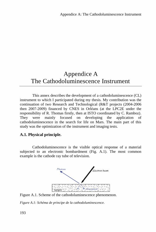
Appendice A: The Cathodoluminescence Instrument
193
Appendice A
The Cathodoluminescence Instrument
This annex describes the development of a cathodoluminescence (CL)
instrument to which I participated during my thesis. My contribution was the
continuation of two Research and Technological (R&T projects (2004-2006
then 2007-2009) financed by CNES in Orléans (at the LPC2E under the
responsibility of R. Thomas firstly, then at ISTO coordinated by C. Ramboz).
They were mainly focused on developing the application of
cathodoluminescence in the search for life on Mars. The main part of this
study was the optimization of the instrument and imaging tests.
A.1. Physical principle.
Cathodoluminescence is the visible optical response of a material
subjected to an electronic bombardment (Fig. A.1). The most common
example is the cathode ray tube of television.
Figure A.1. Scheme of the cathodoluminescence phenomenon.
Figure A.1: Schéma de principe de la cathodoluminescence.
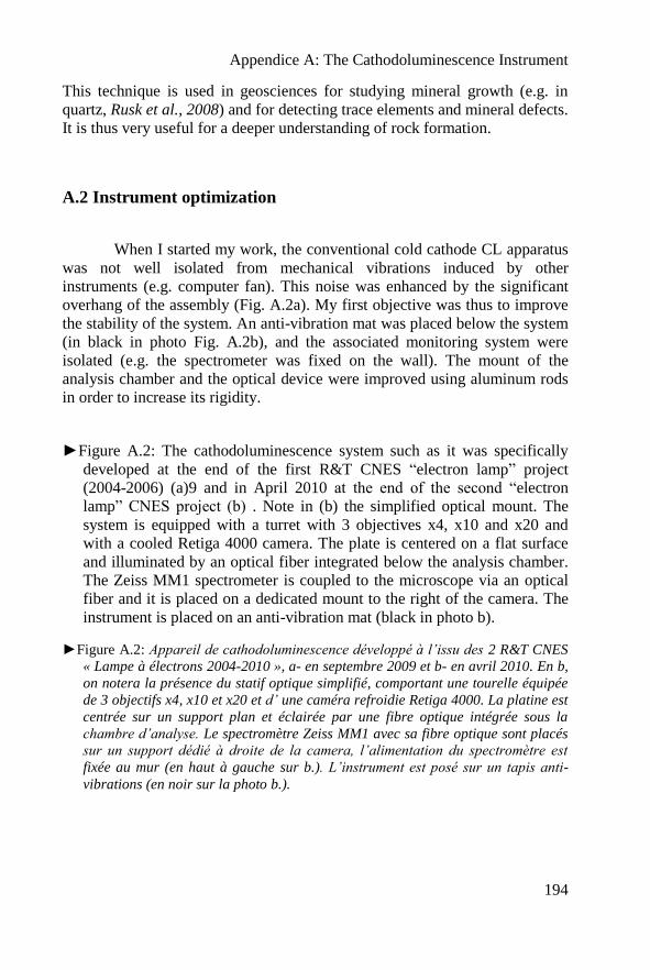
Appendice A: The Cathodoluminescence Instrument
194
This technique is used in geosciences for studying mineral growth (e.g. in
quartz, Rusk et al., 2008) and for detecting trace elements and mineral defects.
It is thus very useful for a deeper understanding of rock formation.
A.2 Instrument optimization
When I started my work, the conventional cold cathode CL apparatus
was not well isolated from mechanical vibrations induced by other
instruments (e.g. computer fan). This noise was enhanced by the significant
overhang of the assembly (Fig. A.2a). My first objective was thus to improve
the stability of the system. An anti-vibration mat was placed below the system
(in black in photo Fig. A.2b), and the associated monitoring system were
isolated (e.g. the spectrometer was fixed on the wall). The mount of the
analysis chamber and the optical device were improved using aluminum rods
in order to increase its rigidity.
►Figure A.2: The cathodoluminescence system such as it was specifically
developed at the end of the first R&T CNES “electron lamp” project
(2004-2006) (a)9 and in April 2010 at the end of the second “electron
lamp” CNES project (b) . Note in (b) the simplified optical mount. The
system is equipped with a turret with 3 objectives x4, x10 and x20 and
with a cooled Retiga 4000 camera. The plate is centered on a flat surface
and illuminated by an optical fiber integrated below the analysis chamber.
The Zeiss MM1 spectrometer is coupled to the microscope via an optical
fiber and it is placed on a dedicated mount to the right of the camera. The
instrument is placed on an anti-vibration mat (black in photo b).
►Figure A.2: Appareil de cathodoluminescence développé à l’issu des 2 R&T CNES
« Lampe à électrons 2004-2010 », a- en septembre 2009 et b- en avril 2010. En b,
on notera la présence du statif optique simplifié, comportant une tourelle équipée
de 3 objectifs x4, x10 et x20 et d’ une caméra refroidie Retiga 4000. La platine est
centrée sur un support plan et éclairée par une fibre optique intégrée sous la
chambre d’analyse. Le spectromètre Zeiss MM1 avec sa fibre optique sont placés
sur un support dédié à droite de la camera, l’alimentation du spectromètre est
fixée au mur (en haut à gauche sur b.). L’instrument est posé sur un tapis anti-
vibrations (en noir sur la photo b.).
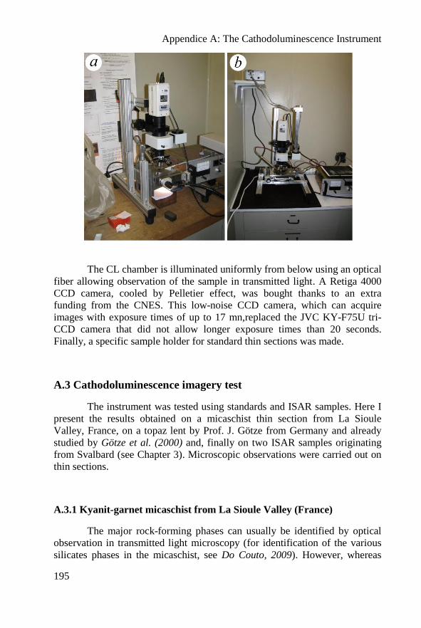
Appendice A: The Cathodoluminescence Instrument
195
The CL chamber is illuminated uniformly from below using an optical
fiber allowing observation of the sample in transmitted light. A Retiga 4000
CCD camera, cooled by Pelletier effect, was bought thanks to an extra
funding from the CNES. This low-noise CCD camera, which can acquire
images with exposure times of up to 17 mn,replaced the JVC KY-F75U tri-
CCD camera that did not allow longer exposure times than 20 seconds.
Finally, a specific sample holder for standard thin sections was made.
A.3 Cathodoluminescence imagery test
The instrument was tested using standards and ISAR samples. Here I
present the results obtained on a micaschist thin section from La Sioule
Valley, France, on a topaz lent by Prof. J. Götze from Germany and already
studied by Götze et al. (2000) and, finally on two ISAR samples originating
from Svalbard (see Chapter 3). Microscopic observations were carried out on
thin sections.
A.3.1 Kyanit-garnet micaschist from La Sioule Valley (France)
The major rock-forming phases can usually be identified by optical
observation in transmitted light microscopy (for identification of the various
silicates phases in the micaschist, see Do Couto, 2009). However, whereas
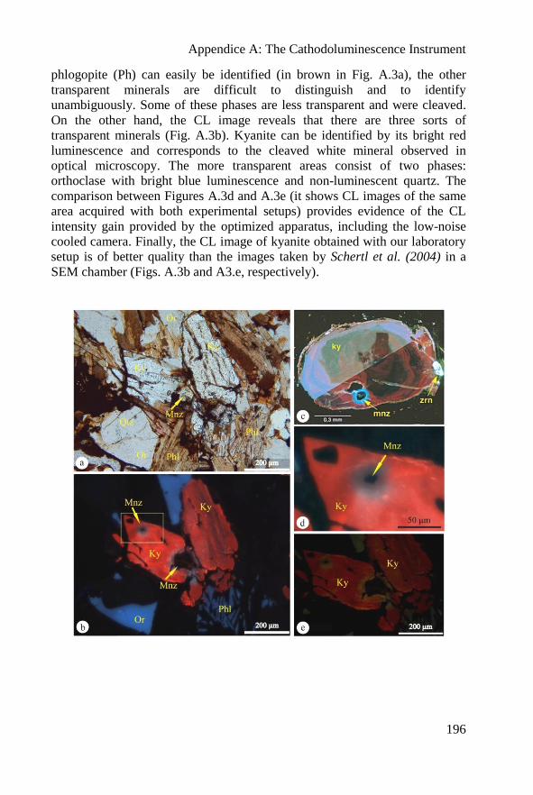
Appendice A: The Cathodoluminescence Instrument
196
phlogopite (Ph) can easily be identified (in brown in Fig. A.3a), the other
transparent minerals are difficult to distinguish and to identify
unambiguously. Some of these phases are less transparent and were cleaved.
On the other hand, the CL image reveals that there are three sorts of
transparent minerals (Fig. A.3b). Kyanite can be identified by its bright red
luminescence and corresponds to the cleaved white mineral observed in
optical microscopy. The more transparent areas consist of two phases:
orthoclase with bright blue luminescence and non-luminescent quartz. The
comparison between Figures A.3d and A.3e (it shows CL images of the same
area acquired with both experimental setups) provides evidence of the CL
intensity gain provided by the optimized apparatus, including the low-noise
cooled camera. Finally, the CL image of kyanite obtained with our laboratory
setup is of better quality than the images taken by Schertl et al. (2004) in a
SEM chamber (Figs. A.3b and A3.e, respectively).
Appendice A: The Cathodoluminescence Instrument
197
◄Figure A.3: Photomicrographs of a thin section of a mica-garnet-kyanite
micaschist from La Sioule Valley, France. a- Optical micrograph in
transmitted light. b- CL image of the same area as in (a). (c-to d) Focused
CL image of a kyanite crystal with a monazite inclusion. (c) CL image
taken in a SEM chamber by Schertl et al. (2004). (d) CL image taken with
our optimized cold cathode CL apparatus(e) Same as (e) with our
equipment, before optimization. Ky = kyanite, Phl = phlogopite, Qtz =
quartz, Or = orthoclase and Mnz = monazite. Analysis conditions (b, d, e):
~15 kV, ~110 µA, PAr = 50 mTorr.
◄Figure A.3 : Phots o d’une lame mince de micaschiste à grenat-disthène (série de
la Sioule). a- lumière naturelle. b- vue en cathodoluminescence de la même zone
que (a). (c- e) images en cathodoluminescence d’un cristal de disthène avec
monazite en inclusion. On peut comparer une image de CL sur MEB d’après
Schertl et al. (2004 (c),) avec l’image de CL acquise avec notre appareil de CL à
cathode froide optimisé (d) (la chambre est la Cathodyne commercialisée par
OPEA).et enfin (e) avec notre équpement non optimisé iLégende : Ky=disthène,
Phl=phlogopite, Qtz= quartz, Or= orthoclase, Mnz=monazite. Conditions
d’analyse (b,d,e) : ~15 kV, ~ 110 µA, PAr=50 mTorr.
Figure A.3 also shows the presence of a small accessory monazite
mineral, a Th-U-bearing phosphate, rich in rare earth elements (here Ce)).
Although the monazite itself is non luminescent, it is indirectly revealed on
the CL image by crystal lattice defects that are created in the disthène crystal
by the radioactive decay of Th and U, yielding the bluish pleochroïc halo
(provoked
A.3.2. Topaz from Euba
This sample from Euba, Germany, was provided by Prof. Jens Götze.
It consists of euhedral crystals of topaz (Al2SiO4(F,OH)2). Figure A.4a shows
an optical view of the three crystals (Fig. A.4-a) and the associated Raman
map of the upper crystal (Fig. A.4-b). The CL image (c) was obtained and
published by Götze et al., 2000, and image (d) was obtained using the
instrument developed in Orléans. The green grains observed by CL could be a
dickite (Al2Si2O5(OH)4) as suggested by Raman spectroscopy. Feldspars are
also associated in the paragenesis.
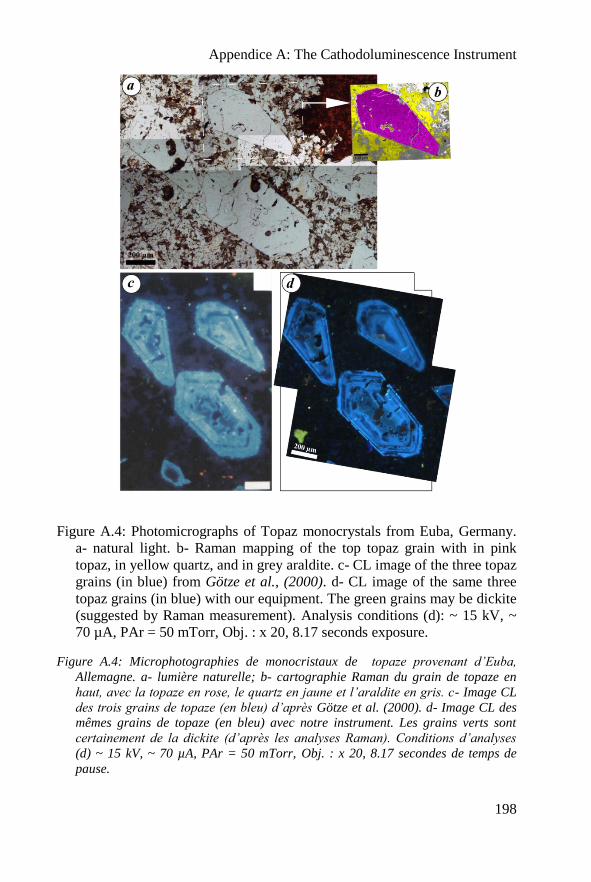
Appendice A: The Cathodoluminescence Instrument
198
Figure A.4: Photomicrographs of Topaz monocrystals from Euba, Germany.
a- natural light. b- Raman mapping of the top topaz grain with in pink
topaz, in yellow quartz, and in grey araldite. c- CL image of the three topaz
grains (in blue) from Götze et al., (2000). d- CL image of the same three
topaz grains (in blue) with our equipment. The green grains may be dickite
(suggested by Raman measurement). Analysis conditions (d): ~ 15 kV, ~
70 µA, PAr = 50 mTorr, Obj. : x 20, 8.17 seconds exposure.
Figure A.4: Microphotographies de monocristaux de topaze provenant d’Euba,
Allemagne. a- lumière naturelle; b- cartographie Raman du grain de topaze en
haut, avec la topaze en rose, le quartz en jaune et l’araldite en gris. c- Image CL
des trois grains de topaze (en bleu) d’après Götze et al. (2000). d- Image CL des
mêmes grains de topaze (en bleu) avec notre instrument. Les grains verts sont
certainement de la dickite (d’après les analyses Raman). Conditions d’analyses
(d) ~ 15 kV, ~ 70 µA, PAr = 50 mTorr, Obj. : x 20, 8.17 secondes de temps de
pause.
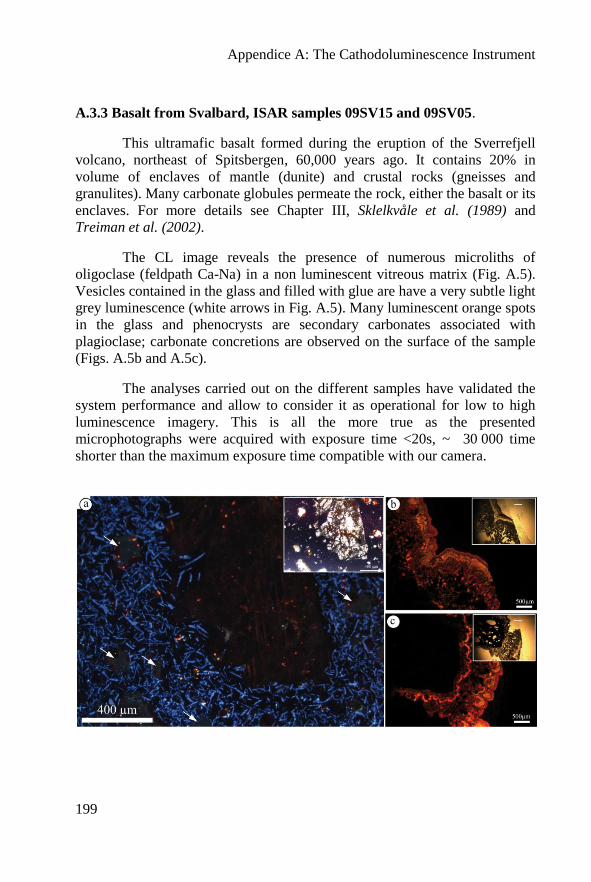
Appendice A: The Cathodoluminescence Instrument
199
A.3.3 Basalt from Svalbard, ISAR samples 09SV15 and 09SV05.
This ultramafic basalt formed during the eruption of the Sverrefjell
volcano, northeast of Spitsbergen, 60,000 years ago. It contains 20% in
volume of enclaves of mantle (dunite) and crustal rocks (gneisses and
granulites). Many carbonate globules permeate the rock, either the basalt or its
enclaves. For more details see Chapter III, Sklelkvåle et al. (1989) and
Treiman et al. (2002).
The CL image reveals the presence of numerous microliths of
oligoclase (feldpath Ca-Na) in a non luminescent vitreous matrix (Fig. A.5).
Vesicles contained in the glass and filled with glue are have a very subtle light
grey luminescence (white arrows in Fig. A.5). Many luminescent orange spots
in the glass and phenocrysts are secondary carbonates associated with
plagioclase; carbonate concretions are observed on the surface of the sample
(Figs. A.5b and A.5c).
The analyses carried out on the different samples have validated the
system performance and allow to consider it as operational for low to high
luminescence imagery. This is all the more true as the presented
microphotographs were acquired with exposure time <20s, ~ 30 000 time
shorter than the maximum exposure time compatible with our camera.
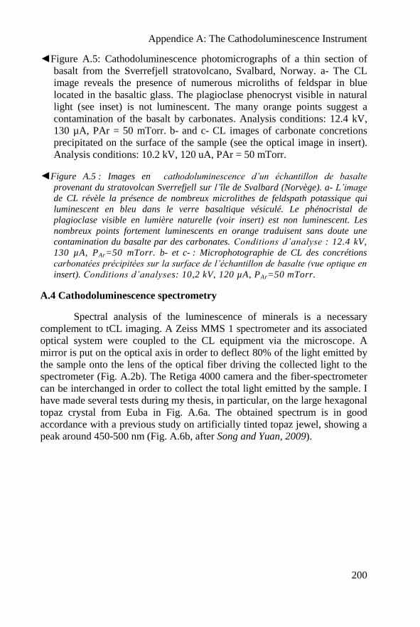
Appendice A: The Cathodoluminescence Instrument
200
◄Figure A.5: Cathodoluminescence photomicrographs of a thin section of
basalt from the Sverrefjell stratovolcano, Svalbard, Norway. a- The CL
image reveals the presence of numerous microliths of feldspar in blue
located in the basaltic glass. The plagioclase phenocryst visible in natural
light (see inset) is not luminescent. The many orange points suggest a
contamination of the basalt by carbonates. Analysis conditions: 12.4 kV,
130 µA, PAr = 50 mTorr. b- and c- CL images of carbonate concretions
precipitated on the surface of the sample (see the optical image in insert).
Analysis conditions: 10.2 kV, 120 uA, PAr = 50 mTorr.
◄Figure A.5 : Images en cathodoluminescence d’un échantillon de basalte
provenant du stratovolcan Sverrefjell sur l’île de Svalbard (Norvège). a- L’image
de CL révèle la présence de nombreux microlithes de feldspath potassique qui
luminescent en bleu dans le verre basaltique vésiculé. Le phénocristal de
plagioclase visible en lumière naturelle (voir insert) est non luminescent. Les
nombreux points fortement luminescents en orange traduisent sans doute une
contamination du basalte par des carbonates. Conditions d’analyse : 12.4 kV,
130 µA, PAr=50 mTorr. b- et c- : Microphotographie de CL des concrétions
carbonatées précipitées sur la surface de l’échantillon de basalte (vue optique en
insert). Conditions d’analyses: 10,2 kV, 120 µA, PAr=50 mTorr.
A.4 Cathodoluminescence spectrometry
Spectral analysis of the luminescence of minerals is a necessary
complement to tCL imaging. A Zeiss MMS 1 spectrometer and its associated
optical system were coupled to the CL equipment via the microscope. A
mirror is put on the optical axis in order to deflect 80% of the light emitted by
the sample onto the lens of the optical fiber driving the collected light to the
spectrometer (Fig. A.2b). The Retiga 4000 camera and the fiber-spectrometer
can be interchanged in order to collect the total light emitted by the sample. I
have made several tests during my thesis, in particular, on the large hexagonal
topaz crystal from Euba in Fig. A.6a. The obtained spectrum is in good
accordance with a previous study on artificially tinted topaz jewel, showing a
peak around 450-500 nm (Fig. A.6b, after Song and Yuan, 2009).
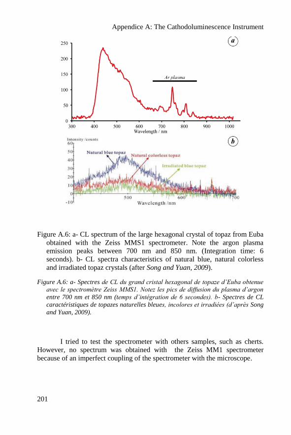
Appendice A: The Cathodoluminescence Instrument
201
Figure A.6: a- CL spectrum of the large hexagonal crystal of topaz from Euba
obtained with the Zeiss MMS1 spectrometer. Note the argon plasma
emission peaks between 700 nm and 850 nm. (Integration time: 6
seconds). b- CL spectra characteristics of natural blue, natural colorless
and irradiated topaz crystals (after Song and Yuan, 2009).
Figure A.6: a- Spectres de CL du grand cristal hexagonal de topaze d’Euba obtenue
avec le spectromètre Zeiss MMS1. Notez les pics de diffusion du plasma d’argon
entre 700 nm et 850 nm (temps d’intégration de 6 secondes). b- Spectres de CL
caractéristiques de topazes naturelles bleues, incolores et irradiées (d’après Song
and Yuan, 2009).
I tried to test the spectrometer with others samples, such as cherts.
However, no spectrum was obtained with the Zeiss MM1 spectrometer
because of an imperfect coupling of the spectrometer with the microscope.

Appendice A: The Cathodoluminescence Instrument
202
A.5. Conclusions
During this thesis, I have participated in the development and
optimization of the cathodoluminescence system on different types of rocks.
CL imagery is a very helpful technique for detecting microstructures in rocks:
fractures, alteration phases, vesicles, accessory minerals, irradiation haloes
etc…. For instance, the microliths in the basalt from Svalbard revealed by CL
are difficult to detect with optical microscopy. The observation of monazite in
situ in space missions (suche as in the micaschist from La Sioule) could be
very interesting for a Sample Return Mission since this mineral rich in
uranium and thorium is adequate for dating. Finally, CL could be relevant for
the search for life (e.g. specific mineralogy or traces elements, Barbin et al.,
1999). According to Thomas et al., 2009, it could be a useful complement for
characterizing rock mineralogy during exploration of the Martian surface. In
particular, it can help to the search for evidence of past biological activity and
the identification of ancient geochemical conditions.
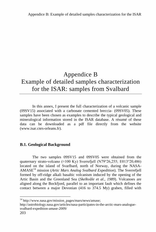
Appendice B: Example of detailed samples characterization for the ISAR
203
Appendice B
Example of detailed samples characterization
for the ISAR: samples from Svalbard
In this annex, I present the full characterization of a volcanic sample
(09SV15) associated with a carbonate cemented breccia- (09SV05). These
samples have been chosen as examples to describe the typical geological and
mineralogical information stored in the ISAR database. A résumé of these
data can be downloaded as a pdf file directly from the website
(www.isar.cnrs-orleans.fr).
B.1. Geological Background
The two samples 09SV15 and 09SV05 were obtained from the
quaternary strato-volcano (≈100 Ky) Sverrefjell (N79°26,255; E013°20,486)
located on the island of Svarlbard, north of Norway, during the NASA-
AMASE14
mission (Artic Mars Analog Svalbard Expedition). The Sverrefjell
formed by off-ridge alkali basaltic volcanism induced by the opening of the
Artic Basin and the Greenland Sea (Skelkvåle et al., 1989). Volcanoes are
aligned along the Bockfjord, parallel to an important fault which defines the
contact between a major Devonian (416 to 374.5 My) graben, filled with
14
http://www.nasa.gov/mission_pages/mars/news/amase;
http://astrobiology.nasa.gov/articles/nasa-participates-in-the-arctic-mars-analogue-
svalbard-expedition-amase-2009/
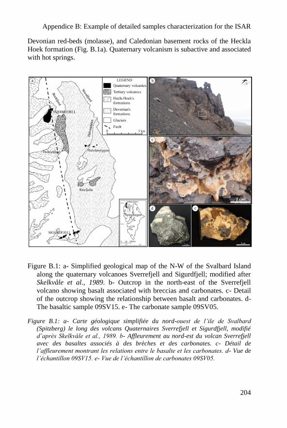
Appendice B: Example of detailed samples characterization for the ISAR
204
Devonian red-beds (molasse), and Caledonian basement rocks of the Heckla
Hoek formation (Fig. B.1a). Quaternary volcanism is subactive and associated
with hot springs.
Figure B.1: a- Simplified geological map of the N-W of the Svalbard Island
along the quaternary volcanoes Sverrefjell and Sigurdfjell; modified after
Skelkvåle et al., 1989. b- Outcrop in the north-east of the Sverrefjell
volcano showing basalt associated with breccias and carbonates. c- Detail
of the outcrop showing the relationship between basalt and carbonates. d-
The basaltic sample 09SV15. e- The carbonate sample 09SV05.
Figure B.1: a- Carte géologique simplifiée du nord-ouest de l’ile de Svalbard
(Spitzberg) le long des volcans Quaternaires Sverrefjell et Sigurdfjell, modifié
d’après Skelkvåle et al., 1989. b- Affleurement au nord-est du volcan Sverrefjell
avec des basaltes associés à des brèches et des carbonates. c- Détail de
l’affleurement montrant les relations entre le basalte et les carbonates. d- Vue de
l’échantillon 09SV15. e- Vue de l’échantillon de carbonates 09SV05.
Appendice B: Example of detailed samples characterization for the ISAR
205
- 09SV15 is a primitive and ultramafic basalt (Fig. B.1b-d) containing
approximately 20% of xenoliths (mantle dunites, sample 09SV02, and some
gneissic crustal elements).
- 09SV05 is a zoned carbonate (Fig. B.1c,e) that was deposited by
hydrothermal exhalation or possibly by aqueous alteration during eruption
under an ice cap (Steele et al., 2007; Amundsen et al., 2011).
B.2 : 09SV15 analysis
B.2.1: Global description
09SV15 is a massive and highly vesicular basalt (fig. B.1d), with
vesicle sizes ranging between 1 and 5 mm. The outer surface of the hand
specimen shows some brownish grey bleaching, indicating very minor
weathering. The fresh basalt surface is dark grey. Dunite xenoliths up to 2 cm
in size and some macroscopic olivine grains (0.5-1 mm size) can be observed
(Fig. B.1b-d). The mineralogy of the basalt is olivine (Fo90) occurring as
phenocrysts with pyroxenes (Mg#57) associated with plagioclase microlites
and minor glass. The basalt is brechified and cemented by different carbonates
layers and globules (09SV05).
B.2.2: Optical microscopy and cathodoluminescence
Optical and cathodoluminescence observations permit identification
of the typical basaltic mineralogy: olivine (Fo90) occurring as phenocrysts,
pyroxenes (Mg#57) associated with plagioclase microlites, and minor glass
(Fig. B.2). Secondary minerals include amphibole, oxides and spinels. This
sample has the particularity of containing quartz grains, possibly a xenolithic
contribution. The CL image reveals the presence of numerous microliths of
oligoclase (feldpath Ca-Na) in a non luminescent vitreous matrix (Fig. B.2).
Vesicles contained in the glass and filled with glue show very subtle light
grey luminescence (white arrows in Fig. B.2). Many orange luminescent spots
in the glass and phenocrysts are secondary carbonates associated with
plagioclase; carbonate concretions are observed at the surface of the sample
(09SV05).
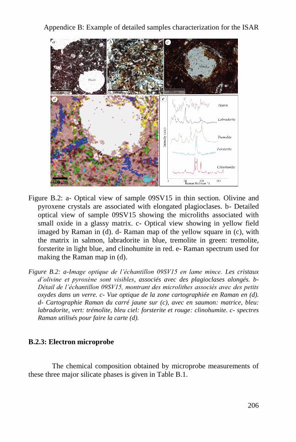
Appendice B: Example of detailed samples characterization for the ISAR
206
Figure B.2: a- Optical view of sample 09SV15 in thin section. Olivine and
pyroxene crystals are associated with elongated plagioclases. b- Detailed
optical view of sample 09SV15 showing the microliths associated with
small oxide in a glassy matrix. c- Optical view showing in yellow field
imaged by Raman in (d). d- Raman map of the yellow square in (c), with
the matrix in salmon, labradorite in blue, tremolite in green: tremolite,
forsterite in light blue, and clinohumite in red. e- Raman spectrum used for
making the Raman map in (d).
Figure B.2: a-Image optique de l’échantillon 09SV15 en lame mince. Les cristaux
d’olivine et pyroxène sont visibles, associés avec des plagioclases alongés. b-
Détail de l’échantillon 09SV15, montrant des microlithes associés avec des petits
oxydes dans un verre. c- Vue optique de la zone cartographiée en Raman en (d).
d- Cartographie Raman du carré jaune sur (c), avec en saumon: matrice, bleu:
labradorite, vert: trémolite, bleu ciel: forsterite et rouge: clinohumite. c- spectres
Raman utilisés pour faire la carte (d).
B.2.3: Electron microprobe
The chemical composition obtained by microprobe measurements of
these three major silicate phases is given in Table B.1.
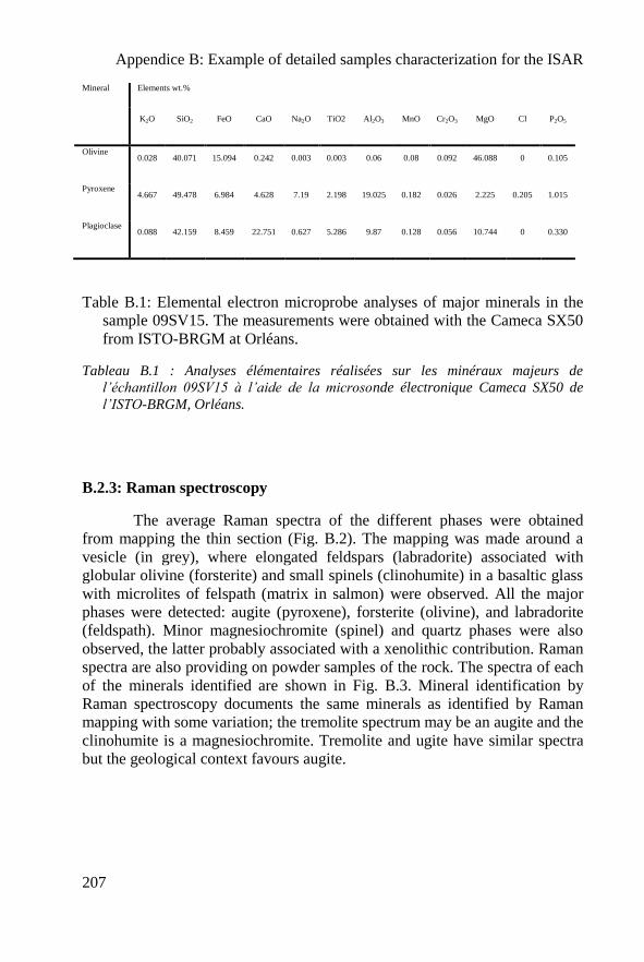
Appendice B: Example of detailed samples characterization for the ISAR
207
Mineral Elements wt.%
K2O SiO2 FeO CaO Na2O TiO2 Al2O3 MnO Cr2O3 MgO Cl P2O5
Olivine 0.028 40.071 15.094 0.242 0.003 0.003 0.06 0.08 0.092 46.088 0 0.105
Pyroxene 4.667 49.478 6.984 4.628 7.19 2.198 19.025 0.182 0.026 2.225 0.205 1.015
Plagioclase 0.088 42.159 8.459 22.751 0.627 5.286 9.87 0.128 0.056 10.744 0 0.330
Table B.1: Elemental electron microprobe analyses of major minerals in the
sample 09SV15. The measurements were obtained with the Cameca SX50
from ISTO-BRGM at Orléans.
Tableau B.1 : Analyses élémentaires réalisées sur les minéraux majeurs de
l’échantillon 09SV15 à l’aide de la microsonde électronique Cameca SX50 de
l’ISTO-BRGM, Orléans.
B.2.3: Raman spectroscopy
The average Raman spectra of the different phases were obtained
from mapping the thin section (Fig. B.2). The mapping was made around a
vesicle (in grey), where elongated feldspars (labradorite) associated with
globular olivine (forsterite) and small spinels (clinohumite) in a basaltic glass
with microlites of felspath (matrix in salmon) were observed. All the major
phases were detected: augite (pyroxene), forsterite (olivine), and labradorite
(feldspath). Minor magnesiochromite (spinel) and quartz phases were also
observed, the latter probably associated with a xenolithic contribution. Raman
spectra are also providing on powder samples of the rock. The spectra of each
of the minerals identified are shown in Fig. B.3. Mineral identification by
Raman spectroscopy documents the same minerals as identified by Raman
mapping with some variation; the tremolite spectrum may be an augite and the
clinohumite is a magnesiochromite. Tremolite and ugite have similar spectra
but the geological context favours augite.
Appendice B: Example of detailed samples characterization for the ISAR
208
B.2.4: IR spectroscopy
The IR analyses were carried out on pellet made of 0,506 mg of
powdered sample mixed with KBr and pressed at 8 tons. The spectral
acquisition was made in transmission. The two major elements in the basalt
(Fig. B.4a) were detected with this method: olivine (in large dotted line, Fig.
B.4a) and pigeonite (in dotted line, Fig. B.4a). The IR pigeonite identification
can be correlated to the augite identification made by Raman spectroscopy
and microprobe elemental analysis. The small peak at 749.26 cm-1
is
labradorite.
B.2.5: Mössbauer analyses
Mössbauer analyses were made on both the weathered (in grey in Fig.
B.5c,d) and unweathered (in black on Fig. B.5c,d) surfaces of the basalt.The
two analyses are very similar and show olivine and pyroxenes, thus
confirming the lack of significant weathering of the sample. However the
intensity of the olivine peak from the inner, cut rock surface is greater than
that of the surface, indicating that the olivine on surface is more altered (Fig.
B.5c). The “npOx” correspond to microcrystalline oxides.
B.2.6: XRD analyses
The XRD analyses show the major mineral phases: forsterite, augite,
nepheline and anorthite (Fig. B.5e). Identification of anorthite is in good
accordance with the detection of labradorite by Raman spectroscopy since
these two minerals are in the plagioclase-feldspar solid solution. The presence
of nepheline, a feldspathoid, is intriguing because it indicates a lack of SiO2 in
the melt, although Raman and microprobe analysis show the presence of
quartz grains. The coexistence of these two phases is unlikely, although
Skjelkåle et al. (1989) also observed this mineral in the basalt (xenocrystals).
Appendice B: Example of detailed samples characterization for the ISAR
209
B.3: 09SV05 analysis
B.3.1: Global description
The 09SV05 carbonate globules are compositionally layered and were
compared by Treiman et al. (2002) to the Martian meteorite ALH 84001. The
first layer closest to the host rock is composed by a fine size carbonate grain
with carbonated cement. The second level is composed of clusters of
magnetite and dolomite with a nebulitic, porous texture suggesting a rapid
crystallization. The penultimate layer is composed of large globules of
dolomite (in dark brown) included in a fine radiate-fibrous matrix of dolomite
and magnesite. The external layer of the crust consists of a massive layer of
radiating magnesite crystals. This layer is sometimes weathered in the surface.
B.3.2: Optical microscopy and cathodoluminescence
Optical and cathodoluminescence (CL) observations document
different carbonate phases: the external rings are formed first by magnesite
and secondly by dolomite (Fig. B.3). The absence of luminescence in the CL
image demonstrates the lack of Mn and Fe. CL confirms the nebulitic, porous
texture of the small carbonates grains in the second layer. These carbonates
contain Mn (demonstrated by orange-red colour in CL associated to the
luminance of the ion Mn2+
in the carbonate crystalline lattice, Haberman et
al., 2000). The interface between the carbonates and the basalt is outlined by
the presence of small pyrite grains. Texture and structure are clearly observed
in CL and the different layers are distinguishable except the external layer that
does not contain luminescent elements.
B.3.4: Electron microprobe
The electron microprobe analyses demonstrate that the highly
luminescent spots observed in CL are associated with a high content of Mg
and Mn.
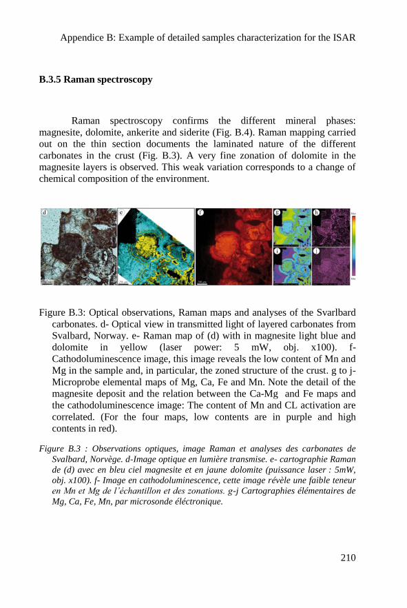
Appendice B: Example of detailed samples characterization for the ISAR
210
B.3.5 Raman spectroscopy
Raman spectroscopy confirms the different mineral phases:
magnesite, dolomite, ankerite and siderite (Fig. B.4). Raman mapping carried
out on the thin section documents the laminated nature of the different
carbonates in the crust (Fig. B.3). A very fine zonation of dolomite in the
magnesite layers is observed. This weak variation corresponds to a change of
chemical composition of the environment.
Figure B.3: Optical observations, Raman maps and analyses of the Svarlbard
carbonates. d- Optical view in transmitted light of layered carbonates from
Svalbard, Norway. e- Raman map of (d) with in magnesite light blue and
dolomite in yellow (laser power: 5 mW, obj. x100). f-
Cathodoluminescence image, this image reveals the low content of Mn and
Mg in the sample and, in particular, the zoned structure of the crust. g to j-
Microprobe elemental maps of Mg, Ca, Fe and Mn. Note the detail of the
magnesite deposit and the relation between the Ca-Mg and Fe maps and
the cathodoluminescence image: The content of Mn and CL activation are
correlated. (For the four maps, low contents are in purple and high
contents in red).
Figure B.3 : Observations optiques, image Raman et analyses des carbonates de
Svalbard, Norvège. d-Image optique en lumière transmise. e- cartographie Raman
de (d) avec en bleu ciel magnesite et en jaune dolomite (puissance laser : 5mW,
obj. x100). f- Image en cathodoluminescence, cette image révèle une faible teneur
en Mn et Mg de l’échantillon et des zonations. g-j Cartographies élémentaires de
Mg, Ca, Fe, Mn, par microsonde éléctronique.
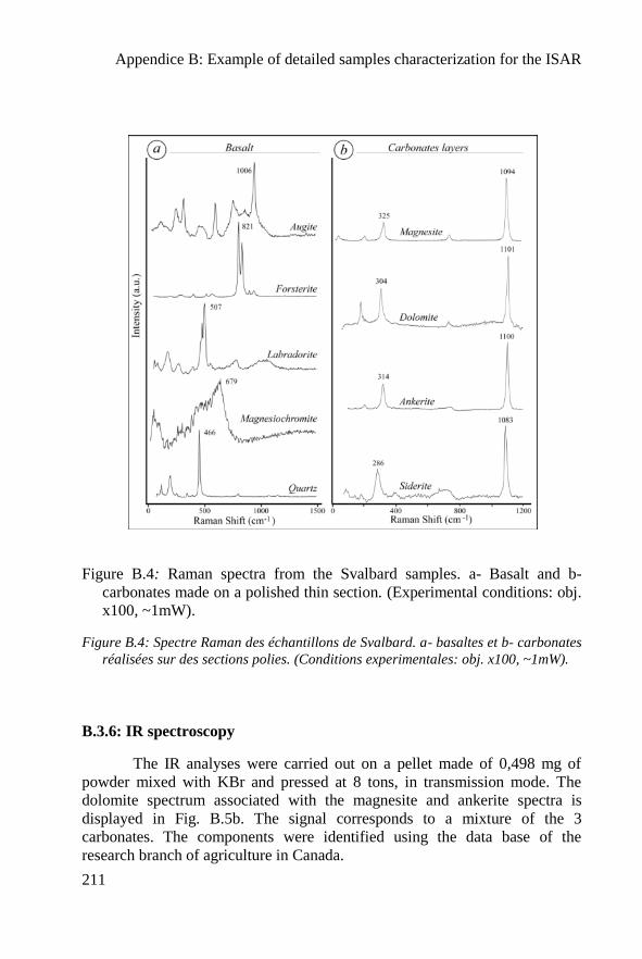
Appendice B: Example of detailed samples characterization for the ISAR
211
Figure B.4: Raman spectra from the Svalbard samples. a- Basalt and b-
carbonates made on a polished thin section. (Experimental conditions: obj.
x100, ~1mW).
Figure B.4: Spectre Raman des échantillons de Svalbard. a- basaltes et b- carbonates
réalisées sur des sections polies. (Conditions experimentales: obj. x100, ~1mW).
B.3.6: IR spectroscopy
The IR analyses were carried out on a pellet made of 0,498 mg of
powder mixed with KBr and pressed at 8 tons, in transmission mode. The
dolomite spectrum associated with the magnesite and ankerite spectra is
displayed in Fig. B.5b. The signal corresponds to a mixture of the 3
carbonates. The components were identified using the data base of the
research branch of agriculture in Canada.
Appendice B: Example of detailed samples characterization for the ISAR
212
B.3.7: Mössbauer analyses
Carbonates in Mössbauer can be easily be confused with pyroxene
(Morris et al., 2010). Since this technique is very sensitive to iron, it is
particularly suited for analyzing F- containing carbonates (fig. B.5d).
However, it is not possible to differentiate the pure Fe carbonate siderite from
other carbonates containing some iron (e.g. ankerite). The other analyses
show that these carbonates phases were partially mixed.
B.3.8: XRD analyses
The XRD analyses on the carbonate powder bring to light the
presence of at least 3 carbonates (Fig. B.5f). The first one is crystalline
dolomite. The two other carbonates always present peaks shifted to the right
with respect to the dolomite meaning that they contain smaller ions in their
crystalline lattice. Based on the previous analyses, it is possible to say that
these atoms are probably magnesium, iron or manganese. These two
carbonates could thus be siderite and magnesite, or a mix of poorly to
moderaly crystalline carbonates.
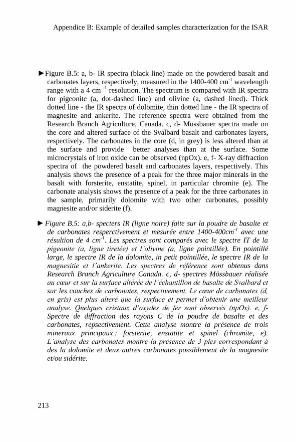
Appendice B: Example of detailed samples characterization for the ISAR
213
►Figure B.5: a, b- IR spectra (black line) made on the powdered basalt and
carbonates layers, respectively, measured in the 1400-400 cm-1
wavelength
range with a 4 cm -1
resolution. The spectrum is compared with IR spectra
for pigeonite (a, dot-dashed line) and olivine (a, dashed lined). Thick
dotted line - the IR spectra of dolomite, thin dotted line - the IR spectra of
magnesite and ankerite. The reference spectra were obtained from the
Research Branch Agriculture, Canada. c, d- Mössbauer spectra made on
the core and altered surface of the Svalbard basalt and carbonates layers,
respectively. The carbonates in the core (d, in grey) is less altered than at
the surface and provide better analyses than at the surface. Some
microcrystals of iron oxide can be observed (npOx). e, f- X-ray diffraction
spectra of the powdered basalt and carbonates layers, respectively. This
analysis shows the presence of a peak for the three major minerals in the
basalt with forsterite, enstatite, spinel, in particular chromite (e). The
carbonate analysis shows the presence of a peak for the three carbonates in
the sample, primarily dolomite with two other carbonates, possibly
magnesite and/or siderite (f).
►Figure B.5: a,b- specters IR (ligne noire) faite sur la poudre de basalte et
de carbonates resperctivement et mesurée entre 1400-400cm-1
avec une
résultion de 4 cm-1
. Les spectres sont comparés avec le spectre IT de la
pigeonite (a, ligne tiretée) et l’olivine (a, ligne pointillée). En pointillé
large, le spectre IR de la dolomite, in petit pointillée, le spectre IR de la
magnesitie et l’ankerite. Les spectres de référence sont obtenus dans
Research Branch Agriculture Canada. c, d- spectres Mössbauer réalisée
au cœur et sur la surface altérée de l’échantillon de basalte de Svalbard et
sur les couches de carbonates, respectivement. Le cœur de carbonates (d,
en gris) est plus alteré que la surface et permet d’obtenir une meilleur
analyse. Quelques cristaux d’oxydes de fer sont observés (npOx). e, f-
Spectre de diffraction des rayons C de la poudre de basalte et des
carbonates, repsectivement. Cette analyse montre la présence de trois
mineraux principaux : forsterite, enstatite et spinel (chromite, e).
L’analyse des carbonates montre la présence de 3 pics correspondant à
des la dolomite et deux autres carbonates possiblement de la magnesite
et/ou sidérite.
Appendice B: Example of detailed samples characterization for the ISAR
214
Appendice C: Geochemical data
215
Appendice C:
Geochemical data
Appendice C: Geochemical data
216
Appendice C: Geochemical data
217
Appendice C: Geochemical data
218
Nicolas BOST
L’analyse géochimique et minéralogique de matériaux analogues
de Mars et la création de l’International Space Analogue Rock Store
L’objectif de la thèse est de créer une collection de roches et minéraux analogues pour calibrer et tester les futurs (et existants) instruments de vol, en accord avec la géologie de Mars afin de préparer les futures missions in situ (MSL-2011 et ExoMars-2018). Les échantillons sont caractérisés avec des instruments de laboratoire (microscope, Raman, IR, DRX, MEB, microsonde électronique, et ICP-MS), mais aussi avec des instruments de vol en développement (Mössbauer MIMOS II, les spectromètres ExoMars Raman et IR (MicroOmega)). L’ensemble des échantillons sont décrits sur une base de données en ligne à l’adresse : www.isar.cnrs-orleans.fr. Une partie de cette thèse est dédiée au développement d’un instrument de cathodoluminescence, qui peut être adapté au spatial.
L’étude des processus d’altération des basaltes sur Terre montrant des similarités avec les processus de surface et de subsurface présent sur Mars, permet d’aider à mieux comprendre et interpréter les objets sur Mars. Pour cela, et pour compléter la collection, des basaltes altérés dans des conditions hydrothermales et acides ont été collecté dans la mine de Skouriotissa à Chypre. L’évolution minéralogique des basaltes à travers les différents facies d’altération a été étudiée.
Parce que les basaltes terrestres sont plus pauvres en Fe et Mg que les basaltes martiens, deux basaltes artificiels ont été synthétisé. Ces deux échantillons sont différents en terme de refroidissement (~110°C/h et trempé). On note que le basalte refroidi lentement montre des textures spinifex similaires aux komatiites. Si ce type de basalte est présent sur Mars, et altéré en présence d’eau, ceci peut avoir d’importantes implications exobiologiques.
Mots clés: Mars, lithothèque, ISAR, analogues, basalte, sédiments volcaniques, phyllosilicate, silice hydrothermale, échantillons artificiels, processus d’altérations, exobiologie, Chypre.
Geochemical and mineralogical analysis of Mars analogue materials and the creation of the International Space Analogue
Rock Store (ISAR) In order to prepare for the next in situ missions to Mars (MSL-2011 and ExoMars-2018), the objective of my thesis is to create a collection of relevant analogue rocks and minerals for calibrating and testing future (and existing) space flight instruments, in accordance with the geology of Mars. They were characterized using standard laboratory instrumentation (optical microscopy, Raman, IR, XRD, SEM, electron microprobe and ICP-MS), as well as by flight instrumentation in development (Mössbauer MIMOSII, ExoMars Raman and IR (MicrOmega) spectrometers). All the samples are described in an online database in the following web site: www.isar.cnrs-orleans.fr. A part of this thesis is dedicated to the development of a cathodoluminescence (CL) instrument that could potentially be adapted for space flight.
Study of alteration processes of basalts on Earth that show some similarities to surface and subsurface processes occurring on Mars may help understand and interpret martian features. Therefore, to complete the collection, samples of hydrothermal and acidic weathered basalts were collected from the Skouriotissa mine in Cyprus. The mineralogical evolution of the basalt through different alteration facies was studied.
Because terrestrial basalts are poorer in Fe and Mg than martian basalts, I synthesized two artificial martian basalts. The two artificial basalts are different in terms of cooling rate (~110°C/h and drop-quenched, >1200°C/h). Interestingly, the more slowly-cooled sample exhibits a spinifex texture, similar to that of komatiites. If similar basalts occur on Mars, such rocks when altered by aqueous processes may have astrobiological implications.
Key-words: Mars, rockstore, ISAR, analogues, basalt, volcanic sediment, phyllosilicate, hydrothermal silica, artificial samples, weathering processes, astrobiology, Cyprus.
Centre de Biophysique Moléculaire Rue Charles Sadron, 45071 ORLEANS Cedex2 Institut des Sciences de la Terre d’Orléans
1A rue de la Férollerie, 45071 ORLEANS Cedex2


