Embed Size (px)
Citation preview

339
Size 7.25 x 10 inches
Hoa, T.T., R A. Hodgson, D.T. Oanh, N. T Phuong, N. J Preston and P.K. Walker. 2005. Genotypic variations in tandem repeatDNA segments between ribonucleotide reductase subunit genes of Whipe Spot Syndrome Virus (WSSV) isolates from Vietnam.In P. Walker, R. Lester and M.G. Bondad-Reantaso (eds). Diseases in Asian Aquaculture V, pp. 339-351. Fish Health Section,Asian Fisheries Society, Manila.
Diseases in Asian Aquaculture V
Genotypic Variations in Tandem Repeat DNA Segments betweenRibonucleotide Reductase Subunit Genes of White Spot
Syndrome Virus (WSSV) Isolates from Vietnam
TRAN TT HOA1,3, RICHARD AJ HODGSON1, DANG TH OANH3,NGUYEN T PHUONG3, NIGEL J PRESTON2 AND PETER J WALKER1
1CSIRO Livestock Industries, St Lucia, Australia2CSIRO Marine Research, Cleveland, Australia
3Cantho University, Cantho, Vietnam
ABSTRACT
White spot syndrome is a viral disease that affects most commercially cultivated marineshrimp species. The disease first emerged in East Asia in 1991 and has since spreadthroughout most shrimp farming regions of Asia and the Americas. Disease outbreaksusually result in high mortalities in affected ponds. However, shrimp may also be infectedchronically with no signs of disease and often obtain the infection in hatcheries from infectedbroodstock. A wide range of other crustaceans can also act as apparently healthy carriersof infection. In this report, variations in the number of a 54 nucleotide tandem repeatsequence (TRS), located between genes encoding the large (RR1) and small (RR2) subunitsof ribonucleotide reductase, were used as a WSSV strain-specific genetic marker. Themarker was applied to examine the extent of variation among WSSV isolates from Penaeusmonodon hatcheries and farms in different regions of Vietnam and to obtain a betterunderstanding of the progression of infection in ponds during grow-out. Analysis ofapproximately 157 WSSV isolates showed common variations in the number of repeats,with some broodstock harbouring more than one genotype. In healthy ponds and in healthybroodstock or postlarval batches collected from hatcheries, WSSV genotypes containing4-, 5-, 6- 7-, 8- and 9- TRS elements were detected with no evidence of any predominantgenotype. However, amongst shrimp sampled from disease outbreak ponds, the 7-TRSgenotype dominated. On the other hand, WSSV genotypes containing greater numbers oftandem repeat elements (i.e. 9-, 14- and 23-TRS) were found in unidentified speciesincluding a large crab, a small crab and wild shrimp, respectively. High repeat numbergenotypes (i.e. 23-TRS and 14-TRS) were not detected in cultured shrimp from the samepond. These results suggested that stocked postlarvae rather than invading wild crustaceanswere the source of WSSV infection and disease. The results also suggest that genotypeanalysis in this TRS region will be a useful tool for tracking virulent strains of WSSV.

Tran TT Hoa et al
340
Size 7.25 x 10 inches
INTRODUCTION
White spot disease (WSD) is a lethal viral infection of farmed marine shrimp that hascaused major economic losses since it first emerged in East Asia in 1992 (Wang et al.,1996; Chou et al., 1995; Zhan et al., 1998). From the original focus of infection in P.japonicus in Fujian Province in China, the disease spread rapidly to other farmed shrimpspecies and, by late 1994, had been observed in most major shrimp farming countries fromJapan to India (Nakano et al., 1994; Park et al., 1998; Mohan et al., 1998; Flegel, 1997).Following the first reports from in Texas in 1995 (Lightner et al., 1997), WSD has alsoestablished in western hemisphere shrimp species and now appears to be endemic over awide area of the Americas from the Gulf of Mexico to Peru. White spot syndrome virus(WSSV), the causative agent of WSD, can also infect a broad range of wild crustaceansincluding marine and freshwater shrimp, crabs and crayfish (Flegel et al., 1997). In marineshrimp, WSSV can either exist as a chronic infection without visible signs of disease orcause a highly lethal acute infection resulting in up to 100% mortality in a pond within 3-10days of the first signs of disease (Zhan and Wang, 1998; Lightner, 1996). As no effectivevaccines or other preventive or prophylactic treatments are available, infection and diseaseare presently managed primarily through pathogen exclusion and stress reduction practices.
WSSV is a large, ellipsoid, enveloped DNA virus with an unusual flagellum-like tail(Wongteerasupaya et al., 1995). In structure and genome organization, WSSV is distinctfrom other known viruses and has recently been classified as the type species of the newgenus Whispovirus within the new family Nimaviridae (van Hulten et al., 2000; Mayo,2002). The WSSV genome is a circular double-stranded DNA of approximately 300 kb(van Hulten et al., 2001; Yang et al., 2001). The genome sequence is remarkably conservedamongst isolates from different hosts and different geographic locations (Lo et al., 1999)but there is evidence of significant variation at some specific loci. Restriction fragmentlength polymorphism (RFLP) analysis has been used to show that WSSV isolates identifiedin a crayfish at the US National Zoological Park was distinct from five isolates identified inshrimp from China, India, Thailand and the USA (Wang et al., 2000). Comparison of thecomplete genome sequence of WSSV isolates from shrimp from Thailand, Taiwan and theChinese mainland has also revealed a 12.1 kb deletion in one virus. However the overallsequence identity (98-100%) between the isolates was very high (van Hulten et al., 2001;Yang et al., 2001; GenBank accession numbers: AF369029, AF332093, AF440570). Changet al. (2001) have reported a single point mutation in the large subunit ribonucleotidereductase gene (rr1) that distinguished a New Jersey crab (Callinectes sapidus) isolatefrom 17 other WSSV isolates from crabs, shrimp and crayfish from the USA and Asia.Most recently, frequent variations have been reported among WSSV isolates from diseaseoutbreak ponds in Thailand by analysing the copy number and sequence of a 54 base pair(bp) tandem repeat sequence element located between the rr1 and rr2 genes(Wongteerasupaya et al., 2003).
In this paper, we report variations in the number of rr1-rr2 54 bp tandem repeats amongWSSV isolates from P. monodon broodstock, postlarvae and juveniles collected fromhatcheries and grow-out ponds in different provinces of Vietnam, and from other crustaceanscollected from ponds. The data indicates a predominance of a 7 tandem repeat sequence (7-TRS) WSSV genotype in diseased shrimp that was not evident in broodstock or postlarval

Genotypic Variations in Tandem Repeat DNA Segments between Ribonucleotide Reductase Subunit Genesof White Spot Syndrome Virus (WSSV) Isolates from Vietnam
341
Size 7.25 x 10 inches
batches from hatcheries, juvenile P. monodon from healthy ponds, or in crabs and wildshrimp collected from diseased ponds
MATERIALS AND METHODS
Origin of crustacean samples
Tissue samples from Penaeus monodon broodstock and postlarvae were collected fromhatcheries, and P. monodon juveniles and other crustaceans were collected grow-out pondsin different provinces of Vietnam between December 2001 and June 2002 (see Tables 2, 3,and 4). Whole shrimp postlarvae, pleopods (broodstock) or whole heads (juvenile shrimp),crabs and wild shrimp were stored in alcoholic preservative (80% ethanol, 20% glycerol)for not more than 2 months before DNA extraction.
DNA extraction and WSSV DNA detection
DNA was extracted from P. monodon postlarvae (eyes-removed), broodstock (pleopods)and juveniles (gill and sub-cuticular epidermis), and from small crabs (half cadavers), largecrabs (legs) and wild shrimp (half heads) by using the DNA extraction reagents supplied inthe IQ-WSSV-2000 (Farming IntelliGene Technology Corp., Taiwan) WSSV detection kit.DNA was extracted conducted according to the manufacturer’s protocol and stored in ETbuffer (0.1mM EDTA, 0.1mM Tris-HCl pH 7.0) at -70°C until required. WSSV DNA wasdetected by using the IQ-WSSV-2000 PCR kit (Farming IntelliGene Technology Corp.,Taiwan) according to the manufacturer’s instructions.
Polymerase chain reaction (PCR) for genotype analysis of WSSV isolates
WSSV genotypes were determined by PCR amplification of the TRS region between therr1 and rr2 genes. One-step and 2-step nested PCR procedures were applied using theprimer combinations shown in Table 1. Samples containing higher quantities of WSSVDNA were analysed by the 1-step method. The 1-step reaction (50 µl) contained 1 x Taqbuffer, 1.5 mM MgCl
2, 200 µM dNTP mix, 2.5 U Taq DNA polymerase (Promega Corp,
Wisconsin, USA), 25 pmol each primer (Wrb6r -F and Wrb6r -R; Wongteerasupaya et al.,2003) and 100 ng template DNA. The reaction mixture was placed in a thermocycler(I-Cycler, Bio-Rad Laboratories Inc., California, USA) pre-heated at 85°C (“hot-start”method) and PCR was conducted using 40 cycles of amplification at 94°C/ 20 s, 60°C/20 s, 72°C/ 75 s, followed by a final incubation at 72°C/ 10 min. The amplified productswere stored at 4°C until analysed by electrophoresis.
Table 1. Sequences of the PCR primers used for WSSV genotype analysis.
Primer Sequence Tm (°C)
Wrb6r - F* 5’ TCTACTCGAGGAGGTGACGAC 3’ 66°C
Wrb6r - R* 5’ AGCAGGTGTGTACACATTTCATG 3’ 66°C
Geno-WS - F 5’ TATTGACCCCGACCACCGCTGC 3’ 72°C
Geno-WS - R 5’ TCCGCCTCTGCCCACGCATTGA 3’ 72°C
* Wongteerasupaya et al. (2003).
Tran TT Hoa et al
342
Size 7.25 x 10 inches
For samples containing low quantities of WSSV DNA, a 2-step PCR protocol was employedby incorporating an initial PCR using primers (geno-WS-F and geno-WS-R) outside theregion amplified in the 1-step PCR protocol. The first amplification was conducted usingthe same reaction conditions as in the 1-step protocol but employed a “hot-start” at 85°C,and 40 cycles at 94°C/ 20 s, 66°C/ 20 s, 72°C/ 90 s, followed by a final incubation at 72°C/10 min. In the second step, 0.5 µl of primary PCR product was used as the DNA templateand the reaction was conducted using PCR primers Wrb6r-F and Wrb6r -R under theconditions described above in the 1-step PCR protocol.
PCR products were resolved by electrophoresis in 2% agarose-TAE gels containing0.5 g/ml ethidium bromide and visualized by UV transillumination. Nucleic acid extractsand all PCR reagents were handled in a laminar flow cabinet using aerosol-resistant tips toavoid contamination. Primary PCR products were handled in a separate work area fromthat in which the nested PCR was performed.
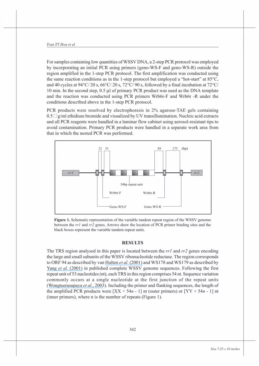
Figure 1. Schematic representation of the variable tandem repeat region of the WSSV genomebetween the rr1 and rr2 genes. Arrows show the location of PCR primer binding sites and theblack boxes represent the variable tandem repeat units.
RESULTS
The TRS region analysed in this paper is located between the rr1 and rr2 genes encodingthe large and small subunits of the WSSV ribonucleotide reductase. The region correspondsto ORF 94 as described by van Hulten et al. (2001) and WS178 and WS179 as described byYang et al. (2001) in published complete WSSV genome sequences. Following the firstrepeat unit of 53 nucleotides (nt), each TRS in this region comprises 54 nt. Sequence variationcommonly occurs at a single nucleotide at the first junction of the repeat units(Wongteerasupaya et al., 2003). Including the primer and flanking sequences, the length ofthe amplified PCR products were [XX + 54n - 1] nt (outer primers) or [YY + 54n - 1] nt(inner primers), where n is the number of repeats (Figure 1).
21 51
54bp repeat unit
Wrb6r-F
rr-1 rr-2
Geno-WS-F Geno-WS-R
Wrb6r-R
89 175 (bp)

Genotypic Variations in Tandem Repeat DNA Segments between Ribonucleotide Reductase Subunit Genesof White Spot Syndrome Virus (WSSV) Isolates from Vietnam
343
Size 7.25 x 10 inches
WSSV isolates from healthy broodstock and postlarvae collected from hatcheries
Tissue samples were obtained from healthy broodstock collected from hatcheries in KienGiang Province on 26 December 2001 and from pooled postlarval samples collected fromhatcheries in Kien Giang, Phan Thiet, Ca Mau, Phan Rang, Cam Ranh and Vung TauProvinces from 4 January to 20 March 2002. The samples were tested for the presence ofWSSV DNA using the IQ 2000 WSSV PCR test. Samples in which WSSV DNA was detectedwere examined using either the 1-step or 2-step PCR to determine the TRS genotype.
Figure 2. PCR assay of WSSV DNA from different pools ofpostlarve and different broodstock (Penaeus monodon).Lane M: 1kb plus DNA ladder; lanes 1-4: postlarval samples(WS160, WS141, WS63C, WS339, respectively); lanes 5-9:broodstock samples (WS45, WS38, WS40, WS44, WS48,respectively).
The WSSV TRS genotype in each sample is shown in Table 2 and examples of the genotypeanalysis are shown in Figure 2. Each pooled postlarval sample produced a single PCRproduct, suggesting infection with a single WSSV genotype. In some samples containingvery high levels of target DNA (eg. WS63C, Figure 2, lane 3), a ladder effect was observedbelow the major PCR product. This appeared to be an amplification artefact as dilution ofthe sample eliminated the ladder but not the major product. However, the possibility thatsmaller, minor products may have been obscured by the ladder cannot be excluded. TheTRS number in postlarval samples ranged from 4 to 9 copies with no predominance of anyone genotype. Genotypes 4-TRS, 6-TRS and 7-TRS were each detected in samples collectedfrom hatcheries in different provinces at different times. For example, a 7-TRS genotypewas detected in WS93 collected in Kien Giang on the Gulf of Thailand on 4 January 2002,and in WS63C collected in Phan Thiet on the South China Sea coast on 29 January 2002.Two different genotypes (4-TRS and 6-TRS) were also detected in different postlarvalsamples (WS152 and WS159) collected from the same province on the same day (PhanThiet, 18 January 2002).

Tran TT Hoa et al
344
Size 7.25 x 10 inches
In contrast to postlarval samples, most DNA extracted from single pleopods of individualbroodstock produced 2 or 3 major PCR products. Due to variations in the intensity of thedifferent sized products, the exact number of TRS genotypes in a single sample was oftendifficult to determine. However, the results clearly indicated that mixed infections withseveral WSSV genotypes were common. In addition, all genotypes detected in postlarvaefrom hatcheries in different provinces were represented in the set of genotypes detected inbroodstock from Kien Giang Province on 26 December 2001. The data indicates a widedistribution of different WSSV genotypes in broodstock and postlarvae collected fromhatcheries in central and southern Vietnam.
WSSV genotypes in from juvenile shrimp from healthy and diseased grow-out ponds
Tissue samples were obtained from 128 juvenile P. monodon from 14 disease outbreakponds in Soc Trang, Ca Mau and Bac Lieu Provinces in the Mekong Delta region of southernVietnam from 2 January to 6 June 2002. Tissue samples were also obtained from 187 juvenileP. monodon from four healthy ponds in Soc Trang Province that were sampled at 30 dayintervals following stocking (i.e. 30, 60 and 90 days). WSSV DNA sequences were analysedusing the 1-step or 2-step genotyping PCR. The TRS genotype in each sample is shown inTable 3 and examples of the genotype analysis are shown in Figure 3.
Table 2. Genotypes of WSSV isolates from healthy broodstock and postlarvae collected from hatcheriesin 6 provinces of central and southern Vietnam.
Host Province Date collected Sample Product size TRS1of hatchery ID (bp) genotype
P. monodon Phan Thiet 18.2.02 WS 152 399 4postlarvae Ca Mau 18.2.02 WS 160 399 4
Phan Rang 05.2.02 WS 141 507 6Phan Thiet 18.2.02 WS 159 507 6Cam Ranh 18.2.02 WS 182 507 6Kien Giang 04.1.02 WS 93 561 7Phan Thiet 29.1.02 WS 61C 561 7Phan Thiet 29.1.02 WS 63C 561 7Vung Tau 20.3.02 WS 339 615 8
P. monodon Kien Giang 26.12.01 WS 45 399, 453 4, 5broodstock Kien Giang 26.12.01 WS 46 399, 453 4, 5
Kien Giang 26.12.01 WS 47 399, 453 4, 5Kien Giang 26.12.01 WS 38 399, 453, 507 4, 5, 6Kien Giang 26.12.01 WS 39 453 5Kien Giang 26.12.01 WS 40 453 5Kien Giang 26.12.01 WS 43 453 5Kien Giang 26.12.01 WS 44 453, 507, 561 5, 6, 7Kien Giang 26.12.01 WS 48 615, 669 8, 9

Genotypic Variations in Tandem Repeat DNA Segments between Ribonucleotide Reductase Subunit Genesof White Spot Syndrome Virus (WSSV) Isolates from Vietnam
345
Size 7.25 x 10 inches
Table 3. Genotypes of WSSV isolates from healthy and diseased juveniles from grow-out ponds in 3provinces of the Mekong Delta of Vietnam.
Province Date collected Pond ID Pond No of Product size TRScondition samples (bp)
(positive/tested)
Soc Trang 06.06.02 TB D 11/11 399 4
Soc Trang 14.03.02 N D 8/8 669 9
Soc Trang 15.01.02 H D 2/2 561 7Soc Trang 17.02.02 N7 D 10/10 561 7Soc Trang 17.02.02 N9 D 10/10 561 7Soc Trang 06.06.02 TH D 2/2 561 7Soc Trang 19.06.02 TH1 D 5/5 561 7Soc Trang 14.03.02 VC D 2/2 561 7Soc Trang 14.03.02 T D 6/6 561 7Ca Mau 02.04.02 CN D 5/5 561 7Ca Mau 02.04.02 DD D 3/3 561 7Bac Lieu 02.01.02 L1 (30) D 26/31 561 7Bac Lieu 02.01.02 L1 (30) D 5/31 561, 669 7, 9
03.02.02 L1 (60) D 29/29 561 702.01.02 L1 (60) D 4/4 561, 669 7, 9
Soc Trang 17.02.02 V5 H 1/21 669 9Soc Trang 17.02.02 V6 H 1/20 561, 669 7, 9Soc Trang 17.02.02 V7 H 1/19 561, 615, 669 7, 8, 9Soc Trang 14.04.02 V8 (90) H 1/19 615 8
1/19 669 91/12 561 7
Figure 3. PCR amplification of different tandem repeats DNA fragmentsfrom juvenile shrimp in different grow-out ponds in Vietnam. Lanes M,1kb plus DNA ladder; lanes 1-3, pond TB; lane 4-6, pond N7; lane 7-9,pond N; lane 10, pond L2; and lane 11, negative control.

Tran TT Hoa et al
346
Size 7.25 x 10 inches
Most shrimp sampled from diseased ponds appeared to contain a single WSSV TRSgenotype. Moreover, except for 2 ponds (L1 and L2) on a single farm in Bac Lieu Province,all shrimp collected from the same diseased pond were infected with the same WSSVgenotype. The 7-TRS genotype was detected in 12 of 14 (86%) diseased ponds and in 109of 128 (85%) diseased juvenile shrimp. Genotype 9-TRS was detected in shrimp from 3outbreak ponds including 2 ponds (L1 and L2) from one farm that were also infected withgenotype 7-TRS. Pond L1 was sampled at day 30 after stocking and again at day 60 duringa disease outbreak. At day 30, 26 of 31 (84%) shrimp sampled were infected only withgenotype 7-TRS and the remaining shrimp were co-infected with both genotypes 7-TRSand 9-TRS. However, at day 60, only the 7-TRS genotype was detected in all 29 shrimpsampled. In pond H in Soc Trang Province, the 7-TRS genotype was also detected inpostlarvae with disease only 4 days after stocking.
In all 4 healthy ponds from the same region (Soc Trang Province), WSSV was detected inonly a small proportion of sampled shrimp. Genotypes 7-TRS, 8-TRS and 9-TRS weredetected in healthy P. monodon from these ponds. Although the number of WSSV-positiveshrimp in healthy ponds was small, there was no evident dominance of any single WSSVTRS genotype. However, there was evidence of multiple WSSV genotypes in shrimp from2 of the 4 ponds and, in pond V7, a single shrimp was co-infected with 3 different TRSgenotypes.
WSSV isolates from crustaceans collected from grow-out ponds
Five samples of individual large crabs, small crabs and wild shrimp (unidentified species)were collected from disease outbreak ponds L2, H and T, and healthy pond V8 from BacLieu and Soc Trang Provinces in the Mekong Delta. The samples were tested for the presenceof WSSV DNA using the IQ-WSSV-2000 PCR test and with the genotyping PCR as describedabove for the shrimp samples. The TRS genotype analyses are shown in Figure 4 and theresults are summarised in Table 4.
Table 4. Genotypes of WSSV isolates from crustacean carriers collected from grow-out ponds in 2 provincesof the Mekong Delta of Vietnam.
Province Date Sample ID Pond Pond Species Product TRS Shrimpcollected ID condition size (bp) genotype TRS
Bac 02.01.02 L60SC L1 D Small crab ND1 - 7Lieu (60)Soc 15.01.02 HU H D Wild 939 14 7
Trang shrimpSoc 14.03.02 TU T D Wild 1423 23 7
Trang TSC shrimp 1423 23 7Small crab
Soc 17.02.02 V8LC V8 H Large crab 669 9 7Trang (30)

Genotypic Variations in Tandem Repeat DNA Segments between Ribonucleotide Reductase Subunit Genesof White Spot Syndrome Virus (WSSV) Isolates from Vietnam
347
Size 7.25 x 10 inches
Figure 4. PCR amplification of different tandem repeats of WSSV DNA fragmentsfrom crustacean carriers. Lane M, 1kb plus DNA ladder; lane N, negative control; lane1,L60SC; lane2, V8LC; lane3, HU; lane4, TSC; lane5, TU. (SC = small crab; LC = largecrab; U = wild shrimp; see Table 4).
A small crab collected from outbreak pond L1 in Bac Lieu Province was weakly WSSV-positive by the IQ-WSSV-2000 test but no product could be amplified using the 2-stepgenotyping PCR test. The crab sample was taken from the pond at day 60 after stockingduring a disease outbreak in which the 7-TRS WSSV genotype was identified in all 29diseased shrimp sampled from the pond (Table 3). Shrimp pond L1 had also been sampledat day 30 after stocking at which time all 31 shrimp sampled were infected with WSSV. Thedata indicates that, despite a long-term infection in the shrimp and a contemporaneousdisease outbreak in the pond, the level of WSSV infection in the crab was low.
In two other disease outbreak ponds (H and T) from Soc Trang Province, the 7-TRS WSSVgenotype was detected in diseased shrimp but different WSSV genotypes were detected incrabs and wild shrimp collected from these ponds during the outbreaks. In pond H, a 14-TRS genotype was detected in wild shrimp. In pond T, a 23-TRS genotype was detected inboth wild shrimp and small crabs. In healthy pond V8 sampled at day 30 after stocking, a 9-TRS WSSV genotype was detected in a large crab. Although pond V8 remained healthythroughout grow-out, 7-TRS WSSV genotype was detected in a single shrimp sampled atday 90 after stocking. Overall, this limited study has identified no correlation between theWSSV genotypes present in P. monodon and other crustaceans in either healthy or diseasedponds.

Tran TT Hoa et al
348
Size 7.25 x 10 inches
DISCUSSION
Genotype analysis to distinguish individual viral isolates has potential to reveal importantaspects of the epidemiology of WSSV infection including the identification of hosts andvectors, transmission routes and the sources of disease outbreaks. Variability in the numberof a tandem repeat sequence has been applied in this paper to genotype analysis of WSSVisolates in cultured shrimp and wild crustaceans from hatcheries and ponds in severalprovinces of central and southern Vietnam. Six WSSV genotypes (4-TRS, 5-TRS, 6-TRS,7-TRS, 8-TRS and 9-TRS) were observed in healthy broodstock and postlarvae. Of these,three genotypes (4-TRS, 7-TRS and 9-TRS) were found in shrimp sampled from diseaseoutbreak ponds. The 9-TRS genotype was also detected in one crab collected from a healthypond. High repeat number genotypes, 23-TRS and 14-TRS, were detected in unidentifiedspecies of small crab and wild shrimp but not in cultured shrimp. Comparison with threeother WSSV isolates for which the complete nucleotide sequence is deposited in GenBankindicates that a 1994 isolate from P. monodon in southern Taiwan (AF440570) and a 1996isolate from P. monodon in Thailand (AF369029) each have a 6-TRS genotype, and a 1996isolate from P. japonicus in Xiamen Province of eastern China (AF332093) has a 12-TRSgenotype. In a study of juvenile P. monodon collected from 55 diseased ponds in centraland southern Thailand in 2000-2002, a wide range of genotypes (6-TRS to 20-TRS) wereidentified at the same variable locus (Wongteerasupaya et al., 2003). Of these 6-TRS (14.5%),7-TRS (10.9%), 8-TRS (32.8%) and 9-TRS (14.5%) genotypes were most commonlydetected. Very low copy number WSSV genotypes (1-TRS, 2-TRS or 3-TRS) have not yetbeen observed in these previous studies. As the 6-TRS genotype 1994 Taiwanese isolate isthe earliest currently available, the detection of 4-TRS and 5-TRS genotypes in VietnameseP. monodon is of some interest. If there has been a progressive evolutionary expansion ofTRS copy numbers since the original emergence of WSD in East Asia in 1992, the commonoccurrence of low TRS copy numbers amongst Vietnamese isolates suggests that WSSVmay have may translocated from East Asia during the initial phase of the panzootic.
In this study, the 7-TRS genotype clearly predominated in juvenile P. monodon from diseaseoutbreak ponds in three provinces of the Mekong Delta. However, the predominance of the7-TRS genotype was not evident either in healthy P. monodon broodstock or postlarvae, orin the limited number of WSSV-positive shrimp identified in healthy grow-out ponds. The7-TRS genotype was also absent from healthy wild crustaceans collected from diseasedponds. The predominance of the 7-TRS genotype in P. monodon collected from diseaseoutbreak ponds suggests that a virulence determinant may be associated with the 7-TRSmarker. However, as discussed above, the 7-TRS genotype was not predominant in a previousstudy of diseased P. monodon from Thailand (Wongteerasupaya et al., 2003) and there isadequate evidence that several other TRS genotypes have caused WSD in Thailand, Taiwan,the Chinese mainland and Vietnam. Further is required to confirm that the Vietnamese 7-TRS genotype is more commonly associated with WSD outbreaks. If so, the virulencedeterminant associated with this marker may be one that confers increased risk of diseasedue to an increased sensitivity to environmental stress. As WSSV appears to replicate moreefficiently at lower temperatures (Vidal et al., 2001), such a determinant could be a highertemperature optimum of the viral polymerase. Comparisons of the virulence of the 7-TRSgenotype and other WSSV TRS genotypes in controlled bioassays would assist in resolving

Genotypic Variations in Tandem Repeat DNA Segments between Ribonucleotide Reductase Subunit Genesof White Spot Syndrome Virus (WSSV) Isolates from Vietnam
349
Size 7.25 x 10 inches
this issue.
Despite the small number of available samples, the analysis of TRS genotypes of WSSVisolates from wild crustaceans was informative. The data indicated that: i) the WSSVgenotypes detected in wild crustaceans from healthy and diseased ponds were differentfrom the WSSV genotype in the co-inhabitant P. monodon; ii) wild shrimp and crabs fromthe same diseased pond shared the same uncommon TRS genotype; and iii) crabs from adiseased pond appeared to be infected at a very low level while a disease outbreak andmortalities occurred in the farmed shrimp. The detection of unusually high TRS copy numbers(14-TRS and 23-TRS) in wild crustaceans was also of interest and may have arisen by rapidevolution and adaptation of the virus to a local infection cycle in these host species.
Overall, the data suggests that wild crustaceans are not a common source of WSSV infectionor disease in farmed shrimp. This is supported by previous studies (Hsu et al., 1999;Withyachumnarnkul, 1999; Peng et al., 2001) in which the elimination of infected seed wasshown to reduce the risk of disease in ponds significantly. However, a more detailedlongitudinal study of ponds from stocking to harvest in various locations will be necessaryto more clearly define the origins of WSD. Clearly, genotype analysis will be a very usefultool in studying the dynamics of WSSV infection in the pond environment and assist indeveloping the most cost-effective strategies for the management of disease.
ACKNOWLEDGEMENTS
The authors wish to thank Professor Robert Lester (The University of Queensland) for hisencouragement, and Dr Jeff Cowley and Dr Lisa Leeton (CSIRO Livestock Industries) fortheir valuable suggestions during this research. Ms Tran TT Hoa was enrolled as a post-graduate research student at the University of Queensland, supported jointly by CSIRO andthe Australian Centre for International Agricultural Research (ACIAR).

Tran TT Hoa et al
350
Size 7.25 x 10 inches
REFERENCES
Chang, Y.-S., Peng S.-E., Wang, H.-C., Hsu, H.-C., Ho, C.-H., Wang, C.-H., Wang, S.-Y., Lo, C.-F.and Kou, G.-H. 2001. Sequencing and amplified restriction fragment length polymorphismanalysis of ribonuleotide reductase large subunit gene of the white spot syndrome virus inblue crab (Callinectes sapidus) from American coastal waters. Marine Biotechnology 3, 163-171.
Chou, H.-Y., Huang, C.-Y., Wang, C.-H., Chiang, H.-C. and Lo, C.-F. (1995). Pathogenicity of abaculovirus infection causing white spot syndrome in cultured penaeid shrimp in Taiwan.Disease of Aquatic Organisms 23, 165-173.
Flegel, T.W. 1997. Major viral diseases of the black tiger prawn (Penaeus monodon) in Thailand.World Journal of Microbiology and Biotechnology 13, 433-442.
Hsu, H.-C., Lo, C.-F., Lin, S.-C., Liu, K.-F., Peng, S.-E., Chang, Y.-S., Chen, L.-L., Liu, W.-J. andKou, G.-H. 1999. Studies on effective PCR screening strategies for white spot syndromevirus (WSSV) detection in Penaeus monodon brooders. Diseases of Aquatic Organisms 39,13-19.
Lightner, D.V. 1996. A Handbook of Shrimp Pathology and Diagnostic Procedures for Diseases ofCultured Penaied Shrimp. World Aquatic Society, Baton Rouge.
Lightner, D.V., Redman, R.M., Poulos, B.T., Nunan, L.M., Mari, J.L. and Hasson, K.W. 1997. Riskof spread of penaeid shrimp viruses in the Americas by the international movement of liveand frozen shrimp. Scientific and Technical Review of the Office International des Epizooties16, 146-160.
Lo, C.-F., Hsu, H.-C, Tsai, M.-F., Ho, C.-H., Peng, S.-E, Kou, G.-H. and Lightner, D.V. 1999. Specificgenomic DNA fragment analysis of different geographical clinical samples of shrimp whitespot syndrome virus. Diseases of Aquatic Organisms 35, 175-185.
Mayo, M.A. 2002. A summary of taxonomic changes recently approved by ICTV. Archives of Virology147, 1655-1656.
Mohan, C.V., Shankar, K.M., Kulkarni S. and Sudha, P.M. (1998). Histopathology of cultured shrimpshowing gross signs of yellow head syndrome and white spot syndrome during 1994 Indianepizootics. Diseases of Aquatic Organisms 34, 9-12.
Nakano H., Koube, H., Umezawa, S., Momoyama, K., Hiraoka, M., Inouye, K., and Oseko, N.1994. Mass mortalities of cultured kuruma shrimp, Penaeus japonicus, in Japan in 1993:epizootiological survey and infection trials. Fish Pathology 29, 135-139.
Park, J.H. Lee, Y.S., Lee, S. and Lee, Y. 1998. An infectious viral disease of penaeid shrimp newlyfound in Korea. Diseases of Aquatic Organisms 34, 71-75.
Peng, S.-E., Lo, C.-F., Lin, S.-C., Chen, L.-L., Chang, Y.-S., Liu, K.-F., Su, M.-S. and Kou, G.-H.2001. Performance of WSSV-infected and WSSV-negative Penaeus monodon postlarvae inculture ponds. Diseases of Aquatic Organisms 46, 165-172.
van Hulten, M.C.W., Tsai, M.-F., Schipper, C.A., Lo, C.-F., Kou, G.-H. and Vlak, J.M. 2000. Analysisof a genomic segment of white spot syndrome virus of shrimp containing ribonucleotidereductase genes and repeat regions. Journal of General Virology 81, 307-316.
van Hulten, M.C.W., Witteveldt, J., Peters, S., Kloosterboer, N., Tarchini, R., Fiers, M., Sandbrink,H., Lankhorst, R.K. and Vlak, J. 2001. The white spot syndrome virus DNA genome sequence.Virology 286, 7-22.

Genotypic Variations in Tandem Repeat DNA Segments between Ribonucleotide Reductase Subunit Genesof White Spot Syndrome Virus (WSSV) Isolates from Vietnam
351
Size 7.25 x 10 inches
Vidal, O.M., Granja, C.B., Aranguren, F., Brock, J.A. and Salazar, M. 2001. A profound effect ofhyperthermia on survival of Litopenaeus vannamei juveniles infected with white spot syndromevirus. Journal of the World aquaculture Society 32, 364-372.
Wang C.S., Tang, K.F.J., Kou, G.H. and Chen, S.N. (1996). Yellow head disease-like virus infectionin the kuruma shrimp Penaeus japonicus cultured in Taiwan. Fish Pathology 31, 177-182.
Wang, Q., Nunan, L.M. and Lightner, D.V. 2000. Identification of genomic variations amonggeographic isolates of white spot syndrome virus using restriction analysis and Southern blothybridization. Diseases of Aquatic Organisms 43, 175-181.
Withyachumnarnkul, B. 1999. Results from black tiger shrimp Penaeus monodon culture pondsstocked with postlarvae PCR-positive or -negative for white-spot syndrome virus (WSSV).Diseases of Aquatic Organisms 39, 21-27.
Wongteerasupaya, C., Pungchai, P., Withyachumnarnkul, B., Boonsaeng, V., Panyim, S., Flegel,T.W. and Walker, P.J. 2003. High variation in repetitive DNA fragment length for white spotsyndrome virus (WSSV) isolates in Thailand. Diseases of Aquatic Organisms 54, 253-257.
Wongteerasupaya, C., Vickers, J.E., Sriurairatana, S., Nash, G.L., Akarajamorn, A., Boonsaeng V.,Panyim, S., Tassanakajon, A., Withyachumnarnkul, B. and Flegel, T.W. 1995. A non-occluded,systemic baculovirus that occurs in cells of ectodermal and mesodermal origin and causeshigh mortality in the black tiger prawn Penaeus monodon. Diseases of Aquatic Organisms21, 69-77.
Yang, F., He, J., Lin, X., Li, Q., Pan, D., Zhang, X. and Xu, X. 2001. Complete genome sequence ofthe shrimp white spot bacilliform virus. Journal of Virology 75, 11811-11820.
Zhan, W.B. and Wang, Y.H. 1998. White spot syndrome virus infection of cultured shrimp in China.Journal of Aquatic Animal Health 10, 405-410.


