Embed Size (px)
Citation preview

Genetically encoded fluorescent sensors of membrane potential
B. J. Baker1, H. Mutoh2, D. Dimitrov2, W. Akemann2, A. Perron2, Y. Iwamoto2, L. Jin1, L. B.Cohen1, E. Y. Isacoff3, V. A. Pieribone1,4, T. Hughes5, and T. Knöpfel2,*1Department of Cellular and Molecular Physiology, Yale University School of Medicine, New Haven,CT 06520, USA2Laboratory for Neuronal Circuit Dynamics, Brain Science Institute, RIKEN, 2-1 Hirosawa, Wako-Shi, Saitama 351-0198, Japan3Department of Molecular and Cell Biology, University of California Berkeley, Berkeley, CA 94720,USA4John B. Pierce Laboratory, New Haven, CT 06520, USA5Department of Cell Biology and Neuroscience, Montana State University, Bozeman, MT 59717,USA
AbstractImaging activity of neurons in intact brain tissue was conceived several decades ago and, after manyyears of development, voltage-sensitive dyes now offer the highest spatial and temporal resolutionfor imaging neuronal functions in the living brain. Further progress in this field is expected from theemergent development of genetically encoded fluorescent sensors of membrane potential. Thesefluorescent protein (FP) voltage sensors overcome the drawbacks of organic voltage sensitive dyessuch as non-specificity of cell staining and the low accessibility of the dye to some cell types. In atransgenic animal, a genetically encoded sensor could in principle be expressed specifically in anycell type and would have the advantage of staining only the cell population determined by thespecificity of the promoter used to drive expression. Here we critically review the current status ofthese developments.
IntroductionOptical imaging is a remarkably flexible method for studying various cellular activities.Organic dyes have been developed that can faithfully report many biological variablesincluding calcium concentration, pH, or membrane potential (Davila et al., 1973; Brown et al.,1975; MacDonald and Jobsis, 1976). These optical probes enable simultaneous measurementsfrom many locations and have been used to study the physiology of single neurons as well aslarge populations of cells (selected work reviewed in Grinvald and Hildesheim, 2004; Bakeret al., 2005; and Knöpfel et al., 2006).
Two major drawbacks of these organic dyes are the non-specificity of cell staining and the lowaccessibility of the dye to some cell types. They either stain all cells in a tissue to which they
© The Author(s) 2008.*author for correspondence; [email protected]. J. Baker, H. Mutoh, D. Dimitrov, and W. Akemann contributed equally to this article.Open AccessThis article is distributed under the terms of the Creative Commons Attribution Noncommercial License which permits anynoncommercial use, distribution, and reproduction in any medium, provided the original author(s) and source are credited.
NIH Public AccessAuthor ManuscriptBrain Cell Biol. Author manuscript; available in PMC 2009 November 11.
Published in final edited form as:Brain Cell Biol. 2008 August ; 36(1-4): 53–67. doi:10.1007/s11068-008-9026-7.
NIH
-PA Author Manuscript
NIH
-PA Author Manuscript
NIH
-PA Author Manuscript

are exposed, or if delivered in a patch pipette they can only label one or a small number ofcells. In the absence of targeting of the organic dyes to specific cell types, optical signals ofinterest are often drowned out either by background fluorescence from inactive cells or bysignals in cells that are not the focus of interest. In addition, with the exception of preparationswith sparse cell staining, diffuse labeling limits the spatial resolution so that single cellresponses cannot be readily resolved. One general solution to this problem is to make opticalreporters from proteins. Since protein-based reporters are encoded in DNA, they can be placedunder the control of cell-specific promoters and introduced in vivo using gene transfertechniques. Moreover, protein-based reporters can, in principle, be rationally “tuned” bymodification of their functional domains with mutations that adjust their dynamic range ofoperation. In a transgenic animal, a genetically encoded sensor could in principle be expressedin any cell type and would have the advantage of staining only the cell population determinedby the specificity of the promoter used to drive expression.
Protein-based reporters are generally constructed from two parts: a “sensor” protein thatundergoes a conformational rearrangement that depends on the parameter measured (e.g., avoltage-gated ion channel that senses membrane potential), and a fluorescent protein (FP)reporter fluorophore whose optical output is modulated by the sensor protein (for a recentreview, see Knöpfel et al., 2006). The first FP voltage sensor, denoted FlaSh, was obtained byinserting GFP downstream of the pore region in the Drosophila voltage-gated potassiumchannel, Shaker (Siegel and Isacoff, 1997). When FlaSh was expressed in Xenopus oocytes,changes in membrane potential were reported by changes of the fluorescence. However, thisinitial success was not followed by published reports of signals from mammalian brains usingFlaSh or the two subsequent first generation voltage sensors, VFSP-1 (Sakai et al., 2001;Knöpfel et al., 2003) and SPARC (Ataka and Pieribone, 2002).
A group of laboratories (Yale, PI Cohen; Berkeley, PI Isacoff; Yale, PI Pieribone; Montana,PI Hughes; and RIKEN BSI, PI Knöpfel) has begun an effort to improve genetically encodedFP voltage sensors for use in mammalian preparations. In order to realize a new, improvedgeneration of sensors, inefficient targeting to mammalian plasma membrane was identified asa major limitation of the first generation FP voltage sensors. Therefore, the group started aconcerted effort to find a more suitable signaling protein and FP reporter-combination, whilstalso fine-tuning emerging sensor proteins.
Here we review the historical development of the first generation FP voltage sensors, describethe state-of-the-art second generation FP voltage sensors, and outline efforts for the nextgeneration FP voltage sensors. We then compare these traditional fully genetically encodablesensors with new partially genetically encoded probes. Finally, we critically discuss thepractical and theoretical limitations of current FP voltage sensor-based imaging techniques.
First generation FP voltage sensorsThe first generation FP voltage sensors were developed by molecular fusion of a GFP-basedfluorescent reporter to voltage-gated ion channels or components thereof. There are threeprototypes obtained from three different laboratories, each of which constitutes a proof ofprinciple and provided valuable insights (Fig. 1).
The very first prototype, FlaSh, was generated in the Isacoff laboratory and obtained by fusingwtGFP to the C-terminus of the Drosophila Shaker potassium channel (Siegel and Isacoff,1997). When expressed in oocytes, an 80-mV depolarization of the plasma membrane resultedin a 5% decrease in fluorescence (Fig. 1 left panel), this was designated FlaSh, for “fluorescentShaker”.
Baker et al. Page 2
Brain Cell Biol. Author manuscript; available in PMC 2009 November 11.
NIH
-PA Author Manuscript
NIH
-PA Author Manuscript
NIH
-PA Author Manuscript
To reduce unwanted effects on the cell's physiology, FlaSh was rendered non-conducting byintroducing a W434F mutation, preventing ions from moving through the pore whilemaintaining voltage-dependent rearrangements (Perozo et al., 1993). In order to resolve actionpotentials, an ideal sensor would generate a robust signal on a millisecond timescale. The signalstrength from FlaSh is comparable to that of the voltage-sensitive organic dye, di4-ANEPPS;however, the on and off rates are rather slow (τ-on ~100 ms; τ-off ~60 ms). In an effort toimprove the kinetics of FlaSh, Guerrero et al. (2002) replaced the wtGFP with several differentFPs. The change in optical characteristics mediated by the different FPs was substantial. Thesignal intensity, the direction of the fluorescent change, and the speed of FlaSh were all alteredby the chromophore in an unsystematic way. Variants involving wtGFP and uvGFP both havereduced fluorescence in response to depolarization steps, while Ecliptic variants of GFP, YFP,and CFP exhibit increased fluorescence in response to depolarization. Remarkably, upondepolarization, an increase in fluorescence is seen when eGFP is excited at 450 nm, but thereis a decrease in fluorescence when excited at 480 nm. The speed of the response is also governedby the chromophore, with the Ecliptic variant of GFP generating the fastest response (τ-on ~5ms). While the mechanism underlying the fluorescence change that results from an alterationin membrane potential remains poorly understood, it is clear that the fluorescent reportercontributes significantly to the kinetics of the optical response and is an important parameterto vary in an attempt to improve genetically encoded voltage sensors.
The second prototypic design realized in the Knöpfel laboratory and termed VSFP1 exploitsthe voltage-dependent conformational changes around the fourth transmembrane segment (S4)of the voltage-gated potassium channel Kv2.1 and uses either fluorescence resonance energytransfer (FRET) (Fig. 1, middle panel; Sakai et al., 2001) or a permuted FP (Knöpfel et al.,2003). The third prototype, SPARC (Pieribone laboratory; Fig. 1, right panel), was generatedby inserting a FP between domains I and II of the rat skeletal muscle Na+ channel (Ataka andPieribone, 2002).
These first generation voltage sensors are capable of optically reporting changes in membranepotential, but their use in mammalian systems is significantly hindered by their poor plasmamembrane expression. When expressed in HEK 293 cells, the expression of these constructsis primarily intracellular and little if any of these first generation FP voltage sensors are co-localized at the cell surface with di8-ANEPPS (Fig. 2). Figure 2B shows the profiles of the FPin green and that of di8-ANEPPS in red along the red lines in the right column of Fig. 2A.Much better co-localization occurs with Kv1.4 with GFP at the N-terminus and with the cation/chloride cotransporter, NKCC1, with YFP fused near the carboxyl terminus. There does seemto be some plasma membrane expression for Flare (a Kv1.4 variant of FlaSh), but the best-case scenario still exhibits fluorescence of a predominantly intracellular origin. Moreimportantly, no functional optical signals could be detected using Flare, VSFP1, or SPARC(Baker et al., 2007).
The absence of a signal from the first generation probes is in part due to their low membraneexpression and in part due to a large, nonresponsive background fluorescence that would maskany voltage-dependent signal. Because Kv1.4 with an N-terminal GFP exhibits excellentmembrane expression, several strategies to release Flare or its Kv2.1-based homologues fromthe ER have been tried. Unfortunately, mutagenesis of potential ER retention signals, additionof ER release motifs, and expression in hippocampal neurons that endogenously expresstrafficking partners all failed to significantly improve the plasma membrane expression (Rayet al., unpublished observations; Baker and Cohen, unpublished observations).
For the second generation of FP voltage sensors, we hoped to overcome this issue of poortargeting to neuronal membranes.
Baker et al. Page 3
Brain Cell Biol. Author manuscript; available in PMC 2009 November 11.
NIH
-PA Author Manuscript
NIH
-PA Author Manuscript
NIH
-PA Author Manuscript

Second generation FP voltage sensorsRecently, several lines of evidence supported the concept of self-contained voltage sensordomains in naturally occurring proteins such as Kv channels. Thus, a voltage sensor domainmay function in the absence of the structures seen in the complete Kv channel complex. Thefirst body of evidence pointing toward this possibility came from crystal structures andstructure-function modeling of KvAP and Kv1.2 that indicated that the secondary structure ofthe S1–S4 portion of Kv channels is relatively independent from neighboring parts of thesechannels (Jiang et al., 2003a, b; Long et al., 2005a, b). The second line of evidence was basedon the identification of voltage sensor domains in non-ion channel proteins. Ci-VSP (Cionaintestinalis Voltage-Sensor-containing Phospatase), for example, is a voltage-controlledenzyme consisting of a transmembrane voltage sensor domain and a cytosolic phosphoinositidephosphatase domain (Murata et al., 2005). The voltage sensor domain from this protein wasshown to be functional (i.e., able to generate gating current in response to voltage steps) whenthe enzyme domain was removed (Murata et al., 2005). More recently, it was also shown bythe Isacoff laboratory that the Ci-VSP can exist as a monomer in the plasma membrane (Kohoutet al., 2007). A third line of evidence for voltage sensor domains that can function in isolationemerged with the discovery of a new protein family termed Hv1 or voltage sensor domain onlyproteins (VSOPs). These membrane proteins mediate voltage-dependent proton transport andhave a domain homologous to the S1–S4 portion of Kv channels but lack the putative poreforming S5–S6 domain (Ramsey et al., 2006; Sasaki et al., 2006). Hv1 is a dimer, with eachof the voltage sensor domains containing its own gated pore; moreover, the pore can functionwhen mutations are made that turn the channel into a monomer consisting of only a singlevoltage sensor domain (Tombola et al., 2008).
A self-contained voltage sensor domain that functions without additional protein componentsor without the need for subunit multimerization is a reasonable candidate module for an FPvoltage sensor, since it avoids potential interference with assembly that may be caused by theFP. Therefore the discovery of Ci-VSP was of immediate interest. In addition, since Ci-VSPis not a channel, a sensor domain based on it would not need to be rendered non-conductingwhile still maintaining high expression, a challenge for channel-based sensors. Moreover, theCi-VSP being of heterologous origin might make it less susceptible to post-translationalmodifications and mistargeting in mammalian expression systems. With the improvement ofplasma membrane targeting in mind, the use of Ci-VSP as the basis of a sensor was exploredby the Knöpfel laboratory (Dimitrov et al., 2007). Because the voltage sensor domain of Ci-VSP is homologous to that of canonical Kv channel subunits, the first step was to generateconstructs similar to the first generation VSFPs (VSFP1s), but with the voltage sensor domainof Kv2.1 replaced by that of Ci-VSP. Since the exact role of the linkers coupling the Ci-VSPvoltage sensor domain to the enzyme domain that it controls is unclear, the CFP and YFP FRETpair was fused to the C-terminus of the voltage sensor domain conserving four different lengthsof the intrinsic Ci-VSP S4-segment-downstream sequence. With this approach the cytosolicphosphatase domain was essentially replaced with the FRET pair. When expressed in PC12cells or hippocampal neurons, all of the initial series of second-generation VSFP (VSFP2)variants displayed bright fluorescence and clear targeting to the plasma membrane (Dimitrovet al., 2007). More importantly, all of these first four constructs showed voltage-dependentmodulation of cyan and yellow fluorescent output (Fig. 3). The FRET fluorescence changecould result from a change in the distance separating the two chromophores or from a changein the angle between them or both. Furthermore, the Cyan signal is substantially larger thanthe Yellow signal for VSFP2A (Fig. 3), suggesting that non-FRET mechanisms are alsooccurring. At present we have no understanding of the mechanism coupling the voltage-dependent structural changes in the voltage-sensitive domain to the changes in fluorescenceof the chromophores.
Baker et al. Page 4
Brain Cell Biol. Author manuscript; available in PMC 2009 November 11.
NIH
-PA Author Manuscript
NIH
-PA Author Manuscript
NIH
-PA Author Manuscript

Consistent with “gating” currents measured in Ci-VSP, the fluorescence voltage curves forVSFP2A-D (Fig. 3, upper traces) exhibited half maximal activation (V1/2) at potentials farabove the physiological range of mammalian neurons. To shift the voltage dependency to amore physiological range, the Knöpfel laboratory investigated a series of mutational alterationsin the S4 segment of Ci-VSP's voltage sensing domain. The most successful mutation wasR217Q which resulted in the protein termed VSFP2.1 (Fig. 4A, B). Like the initial constructs,VSFP2.1 displayed bright fluorescence and clear targeting to the plasma membrane. VSFP2.1exhibited a V1/2 value of approximately −70 mV both at 22 and at 35°C (Fig. 4). At 22°C,activation and deactivation kinetics were similar to those of VSFP2A-D. At 35°C, activationwas considerably faster (Fig. 4B). Figure 5 illustrates expression of VSFP2.1 (using codonoptimized DNA) in a cultured cortical neuron along with neuronal responses to hyperpolarizingcurrent injections. This experiment illustrates that, in contrast to calcium imaging, voltageimaging can directly monitor membrane hyperpolarization. The fluorescence–voltagerelationship with a V1/2 value in the physiological range of neuronal membrane fluctuationstogether with the relatively fast kinetics makes VSFP2.1 a suitable candidate for opticalmeasurements of neuronal activity, such as large synaptic potentials; single action potentials,action potential trains; and bistabilities in resting membrane potential. To demonstrate thisprediction, PC12 cells were voltage-clamped with membrane potential traces obtained frommouse mitral cells. For the trace shown in Fig. 6A, the mitral cell was stimulated by injectionof a current pulse through a patch-clamp electrode to generate a series of action potentials. Thefluorescent output of VSFP2.1 could clearly resolve individual action potentials (arrows in Fig.6A) as well as the slower underlying membrane depolarization. As expected from the responsekinetics of VSFP2.1, the optical readout of the fast action potentials was reduced relative tothe slower components of the membrane potential change. This phenomenon was also clearlyseen when using the response of a mitral cell to a single shock stimulation of the olfactorynerve (Fig. 6B). This response consisted of a burst of fast action potentials on a slow synapticpotential. The response of VSFP2.1 mainly represented the slow synaptic potential. Moreimportantly, it should be noted that the responses shown in Fig. 6B could be resolved in singlesweeps with a very reasonable signal-to-noise ratio (for experimental details, see Dimitrov etal., 2007).
Next generation FP voltage sensorsFurther development of VSFP2.1 is progressing in several directions: (i) linker optimization,(ii) alternative FP variants, and (iii) a search for additional ways to fuse or insert FPs into Ci-VSP or alternative sensor proteins.
Linker optimized variantsIt is well established for other types of FP sensors that sensitivity of the probe (ΔF/F or ΔR/R values per change in membrane voltage) can be increased by optimizing the length and aminoacid composition of the linker between the sensor protein and the FP. To such effect, theperformance of VSFP2.1 was improved by removal of five amino acids that initially originatedfrom engineered restriction sites in VSFP2.1 (Dimitrov et al., unpublished observation;Villalba-Galea et al., 2008). Further improvement of sensor sensitivity by optimizing linker islikely to be realized in near future.
Alternative FP colorsCyan- and yellow-emitting variants of green fluorescent protein (GFP) from Aequoreavictoria (CFP and YFP, respectively) are the most widely used components for FRET-basedbiosensors. However, there are reasons why alternative colors, particularly red shifted versions,are more desirable: (i) for deep tissue imaging, (ii) for reduced interference from tissueautofluorescence and hemoglobin absorption, and (iii) in combination with other fluorescent
Baker et al. Page 5
Brain Cell Biol. Author manuscript; available in PMC 2009 November 11.
NIH
-PA Author Manuscript
NIH
-PA Author Manuscript
NIH
-PA Author Manuscript

probes (spectral separation). Several efficient variants of these were very recently generatedin the Knöpfel laboratory, the most promising comprised combinations of either a green FPwith a red FP or a yellow FP with a far-red FP (Dimitrov et al., unpublished observations).
In addition, for some applications, non-FRET monochromatic probes are more favorable.Single FP sensors have been demonstrated previously including the Flash type of voltagesensors and several types of Ca2+ sensors (Baird et al., 1999; Nakai et al., 2001). Thus, werecently developed analogous single FP voltage sensors based on VSFP2s (Lundby et al.,2008; Mutoh et al., unpublished observation; Baker and Cohen, unpublished observation). Thebest variant characterized to date exhibited an activation time constant (τ-on) of <3 ms atdepolarized membrane potentials (Lundby et al., 2008).
Alternative designsIn a canonical voltage sensor domain, it is not only the S4 segment that responds to voltageshifts, but rather the whole domain undergoes conformational changes in a complex manner(Pathak et al., 2007). It follows, therefore, that finding alternative sites for insertions of FPsalong the voltage sensor domain that would not disturb the function and folding of either thevoltage sensor domain or the FP may lead to new designs of improved functional voltagesensors. To explore this approach, the Hughes laboratory is using its transponson technologyto generate a library of key positions in the Ci-VSP that would allow for insertion of a FPwithout disturbing the folding of the Ci-VSP (critical for voltage sensing), the folding of theFP itself (critical for the fluorescence report), or plasma membrane targeting.
In addition to the above there are many additional, only partially explored approaches forconformational tuning of the parts constituting the sensor. These include addition ofprenylation signals or other signals for post-translational modifications that may help improvethe plasma membrane targeting and/or conformational states, in a way that would beadvantageous for the desired function.
Genetic targeting of neuronsPerhaps the most important feature of genetically encodable sensors is the possibility to achieveindicator loading in specific cell populations. This is the case because cell type-specific proteinexpression can be achieved by the use of appropriate regulatory gene sequences (promoters).This concept had been proposed many years ago in conjunction with the bioluminescentcalcium-sensitive reporter protein aqueorin; however, the light output of bioluminescent probeswas rather limited and consumed a co-factor that needed to be applied to the preparation. Overthe last years cell type-specific expression of FP variants, or fusion proteins containing them,has been demonstrated in numerous transgenic and gene targeted mouse lines. For someexperimental questions it is desirable to label cells sparsely so that individual cells can easilybe identified by fluorescence imaging in densely packed tissue. Sparse labeling can be achievedwith “shotgun” promoters such as Thy1 (Feng et al., 2000), virus-based transfection (Dittgenet al., 2004), or transgenic strategies for combinatorial expression of FP components (Livet etal., 2007) but is associated with considerable uncertainty about the identity of the labeled cellsand/or whether the labeled cells are representative for the given target population.
The specificity of virus-based transfection can be increased by cell-specific promoters, but thistechnique is limited by the size of recombinant sequence which can be packaged in to the virus.Perhaps more promising is the combination of viruses with mouse lines expressingrecombinases, such as Cre and Flp, in specific subsets of neurons. This technology permitstargeted expression of FP sensors in any subset neuron, for which one of the now manyrecombinase-mice lines are available. For that purpose the sensor gene in a lox-stop-lox cassette
Baker et al. Page 6
Brain Cell Biol. Author manuscript; available in PMC 2009 November 11.
NIH
-PA Author Manuscript
NIH
-PA Author Manuscript
NIH
-PA Author Manuscript
is inserted into a virus and then expressed under lox recombination as defined by therecombinase mouse.
A more expensive and time-consuming approach is direct transgenic or genetic targetedexpression of the FP sensor. The principal advantage being reproducible preparation forexperimental convenience and for a greater certainty in establishing what is stained (and—equally important—what is not). With such genetic models the precise targeting allows opticalsignals to be reliably attributed to specific subsets of cells within a network consisting of manydifferent cell types, whilst also allowing low noise imaging to be performed without opticallyresolving the labeled cells (Knöpfel et al., 2006;Díez-García et al., 2005, 2007; Qiu andKnoöpfel, 2007).
Genetically encoded sensors of membrane potential compared to alternativegenetic targeting approaches for fluorescence voltage-sensitive probes
In addition to voltage sensors that are entirely genetically encoded, at least two otherapproaches to overcoming indiscriminant dye staining use genetics in combination withconventional organic chromophores. The first is a hybrid approach that combines theadvantages of conventional voltage-sensitive dyes (relatively large ΔF/F) with genetictargeting (Chanda et al., 2005; DiFranco et al., 2007). The hybrid probe hVOS employs FRETbetween membrane-anchored GFP and a dye, DPA, that partitions into the plasma membranein a membrane voltage-dependent manner. While this hybrid probe produces the largestreported ΔF/F values among all genetically encoded voltage-sensitive probes, the highconcentration of the lipophilic anion DPA has a significant effect on the activity of excitablemembranes (Fernández et al., 1983; Chanda et al., 2005; Zimmermann et al., 2008). Thusreduced DPA concentrations were required to obtain action potential responses (Chanda et al.,2005) resulting in much smaller fractional changes (ΔF/F).
Using an optimized DPA-based voltage reporter, Sjulson and Miesenboöck (2008) found afailure to detect action potentials in the Drosophila antennal lobe, presumably because spikeinitiation and/or propagation are inhibited by the capacitive load added even at reduced DPAmembrane densities.
The second semi-genetic method uses the genetically targetable expression of an enzyme withan organic dye as a substrate. The idea is that only the enzymatically processed dye stains theplasma membrane where it functions as a conventional voltage-sensitive dye. In a proof ofprinciple study (Hinner et al., 2006), a lipophilic dye was made water-soluble by the additionof phosphate groups to the hydrophobic tail. Cell-specific staining was achieved by geneticallyexpressing an outward-facing membrane-bound phosphatase that cleaves the hydrophilicphosphate group from the engineered dye, resulting in a closely localized population oflipophilic product dye that integrates and stains the membrane of the specified target cells.While this approach has potential and essentially could be used with a variety of establishedvoltage-sensitive lipid dyes, in its current state the applicability in neuronal tissues is unproven.Several obstacles become apparent; first, the usual issues of dye accessibility to the area ofstudy and dye toxicity. Second, a question arises whether the phosphatase would not catalyzeintrinsic processes disturbing the physiological state of the tissue, or the dye itself would notbecome substrate to intrinsic proteins resulting in unwanted staining. Third, in a tissue wherethe individual cells are closely packed together, the “released” dye is expected to diffuse andbind to plasma membranes of “not-selected-for” cells that are adjacent to the cell expressingthe enzyme.
Baker et al. Page 7
Brain Cell Biol. Author manuscript; available in PMC 2009 November 11.
NIH
-PA Author Manuscript
NIH
-PA Author Manuscript
NIH
-PA Author Manuscript

Signal-to-noise considerationsThe motivation for the development of FP voltage sensors is to overcome unselective labelingobtained when staining tissue with organic voltage-sensitive dyes. Selective staining of specificsubsets of neurons will allow a more stringent interpretation of optically monitored voltagesignals, in terms of their source. The second, similarly important benefit is an increase in S/Nratio because undesired staining contributes to noise but not to signal. In this context, it wasas a consequence of unspecific labeling (in the form of limited membrane targeting) that thefirst generation of FP voltage sensors failed. The second generation overcame this problem.Yet, the signal-to-noise ratio (RS/N) is probably the most critical parameter, particularly whena high temporal resolution is required. For in vivo experiments a low RS/N often results froma contamination of the sensor signal by movement-induced changes in the measured lightintensity, for instance: changes in blood volume or hemoglobin oxygenation, light scatteringor endogenous tissue fluorescence (for a recent review, see Baker et al., 2005). Synchronizationwith breathing and/or heartbeat along with careful selection of excitation and emissionwavelengths can reduce these noise signals significantly (Baker et al., 2005).
There is, however, a physical limit for RS/N in optical recordings due to the statistics of photondetection (expanded in Knöpfel et al. 2006 and Sjulson and Miesenböck, 2007). Thus, in thebest case, RS/N is increased with the square root of the number of photons detected per temporaland spatial bin. Accordingly, it can be predicted that the first successful (because leastdemanding) application of FP voltage sensors in intact tissue will be voltage imaging frompopulations of subsets of neurons, as previously demonstrated with FP calcium sensors (Díez-García et al., 2005, 2007). Spatial averaging over many neurons that carry a common voltagesignal will provide a sufficient emission intensity to detect small modulations (Vranesic et al.,1994). RS/N decreases with the sampling rate (as the number of sampled photon decreases withshorter sampling intervals, but also because of an increased contribution of noise resulting fromthe sampling process). For these reasons, along with the fact that imaging sampling frequencyis limited for some imaging techniques (like laser scanning), an FP voltage sensor withsomewhat slow response characteristics may be more suitable for certain applications. Indeed,slow response kinetics act as a signal conditioning mechanisms (as required by the samplingtheorem). Yet, timing information may be recovered from slowly responding indicators bydeconvolution techniques (Yaksi and Friedrich, 2006).
Capacitive load and other possible caveatsGenetically encoded optical sensors make it possible to record from multiple cells inundamaged tissue. However, as a recombinant gene product, the sensor itself can interfere withintrinsic biochemical and/or physical cell functions. Hence, as in the case of any artificiallyintroduced probe, FP sensor-derived data require verification against controls to demonstratethe absence of probe-induced undesired effects. Unlike synthetic potentiometric probes, e.g.cyanin and oxonal dyes, which are notorious for their phototoxicity (Grinvald et al., 1982;Kalyanaraman et al., 1987), FP-based sensors are not known, and are not expected, to formtoxic illumination-induced products. Similar to synthetic probes, however, they add to thenonlinear dielectric polarization of the membrane resulting in increased membrane “capacitiveload” (Sjulson and Miesenböck, 2007). The effect is based on a simple physical mechanism:The voltage sensor domain moves positive charges across the membrane in response to changesin membrane potential. This charge movement manifesting as a transmembrane capacitivecurrent (well known as gating currents for ion channels) redistributes charge between the innerand outer membrane surface in a voltage sensor domain state-dependent manner, tantamountto a voltage-and time-dependent nonlinear capacitance. Expression of voltage sensor domain-containing proteins like VSFPs therefore contributes an extra capacitance to the membrane.Depending on its magnitude and activation time course, this extra capacitance may influence
Baker et al. Page 8
Brain Cell Biol. Author manuscript; available in PMC 2009 November 11.
NIH
-PA Author Manuscript
NIH
-PA Author Manuscript
NIH
-PA Author Manuscript

the subthreshold and spiking properties of target membranes. The physiological effect of extramobile membrane charge can be inferred from the behavior of excitable membranes afteruptake of lipophilic anions, like dipicrylamine (DPA), causing a dose-dependent amplificationof membrane capacitance and, at higher doses, decrement of action potential amplitude,increased action potential width, slowing of spike propagation (Fernández et al., 1983), andreduced field potentials (Chanda et al., 2005). As the biophysical mechanism is fairly basic,simulation models can be used to predict and delimit the effects of extra mobile charges on theelectrical response properties of excitable membranes (Fernández et al., 1983; DiFranco et al.,2007).
Future directionsIn this review we have focused on the most advanced approach for FP voltage sensorengineering based on a fusion between a voltage sensor protein and an FP reporter domain.However, an alternative design of an FP voltage sensor might be to use the FP as both thesensor and reporter. Such a protein would have the smallest possible size and minimalinteraction with other organism-intrinsic proteins. To achieve this, the FP would need to belocalized at the water-lipid interface of the plasma membrane or even largely within the lipidphase. Sensitivity to membrane voltage could be based on either voltage-dependent changesin conformation and lipid–protein interactions or be a direct effect of the transmembraneelectric field on the optical properties of the chromophore (Molecular Stark effect). Thechallenge, however, is to change the amino acid composition of the FP molecule so that itwould intrinsically translocate to the hydrophobic phase without interruption to the maturation,constitution, and existence of a functional chromophore.
In conclusion, FP sensors will open paths to yet unexplored territories of functionalneuroimaging. In addition to the FP voltage sensors on which this review focuses, there arenow many different FP sensors for monitoring biochemical processes and transmitter dynamics(Tsien 2005), which can be applied to neuronal circuits. Reading neuronal circuit activity canalso be complemented with emerging optical methods to write activity into neuronal circuits(Miesenbock and Kevrekidis, 2005; Boyden et al., 2005; Knöpfel 2008). In the future thesetechniques may be combined to yield a bidirectional optical interface to brain function.
AcknowledgmentsThe authors thank Dr. Steven Middleton for careful commenting on this article. This article was supported by NIHgrant NS057631 (LBC, EYI, VAP, TH, TK) and an intramural grant from RIKEN BSI (TK).
ReferencesAtaka K, Pieribone VA. A genetically targetable fluorescent probe of channel gating with rapid kinetics.
Biophys. J 2002;82:509–516. [PubMed: 11751337]Baird GS, Zacharias DA, Tsien RY. Circular permutation and receptor insertion within green fluorescent
proteins. Proc. Natl. Acad. Sci. USA 1999;96:11241–11246. [PubMed: 10500161]Baker BJ, Kosmidis EK, Vucinic D, Falk C, Cohen L, Djurisic M, Zecevic D. Imaging brain activity with
voltage- and calcium-sensitive dyes. Cell. Mol. Neurobiol 2005;25:245–282. [PubMed: 16050036]Baker BJ, Lee H, Pieribone VA, Cohen LB, Isacoff EY, Knöpfel T, Kosmidis EK. Three fluorescent
protein voltage sensors exhibit low plasma membrane expression in mammalian cells. J. Neurosci.Methods 2007;161:32–38. [PubMed: 17126911]
Brown JE, Cohen LB, De Weer P, Pinto LH, Ross WN, Salzberg BM. Rapid changes in intracellular freecalcium concentration. Detection by metallochromic indicator dyes in squid giant axon. Biophys. J1975;15:1155–1160. [PubMed: 1201331]
Boyden ES, Feng J, Bamberg E, Nagel G, Deisseroth K. Millisecond-time-scale, genetically targetedoptical control of neural activity. Nat. Neurosci 2005;8:1263–1268. [PubMed: 16116447]
Baker et al. Page 9
Brain Cell Biol. Author manuscript; available in PMC 2009 November 11.
NIH
-PA Author Manuscript
NIH
-PA Author Manuscript
NIH
-PA Author Manuscript

Chanda B, Blunck R, Faria LC, Schweizer FE, Mody I, Bezanilla F. A hybrid approach to measuringelectrical activity in genetically specified neurons. Nat. Neurosci 2005;8:1619–1626. [PubMed:16205716]
Davila HV, Salzberg BM, Cohen LB, Waggoner AS. A large change in axon fluorescence that providesa promising method for measuring membrane potential. Nat. New Biol 1973;241:159–160. [PubMed:4512623]
Díez-García J, Akemann W, Knöpfel T. In vivo calcium imaging from genetically specified target cellsin mouse cerebellum. Neuroimage 2007;34:859–869. [PubMed: 17161628]
Díez-García J, Matsushita S, Mutoh H, Nakai J, Ohkura M, Yokoyama J, Dimitrov D, Knöpfel T.Activation of cerebellar parallel fibers monitored in transgenic mice expressing a fluorescent Ca2+
indicator protein. Eur. J. Neurosci 2005;22:627–635. [PubMed: 16101744]DiFranco M, Capote J, Quiñonez M, Vergara JL. Voltage-dependent dynamic FRET signals from the
transverse tubules in mammalian skeletal muscle fibers. J. Gen. Physiol 2007;130:581–600.[PubMed: 18040060]
Dimitrov D, He Y, Mutoh H, Baker BJ, Cohen L, Akemann W, Knöpfel T. Engineering andcharacterization of an enhanced fluorescent protein voltage sensor. PLoS ONE 2007;2(5):e440.[PubMed: 17487283]doi:10.1371/journal.pone.0000440
Dittgen T, Nimmerjahn A, Komai S, Licznerski P, Waters J, Margrie TW, Helmchen F, Denk W, BrechtM, Osten P. Lentivirus-based genetic manipulations of cortical neurons and their optical andelectrophysiological monitoring in vivo. Proc. Natl. Acad. Sci. USA 2004;101:18206–18211.[PubMed: 15608064]
Feng G, Mellor RH, Bernstein M, Keller-Peck C, Nguyen QT, Wallace M, Nerbonne MN, Lichtman JW,Sanes JR. Imaging neuronal subsets in transgenic mice expressing multiple spectral variants of GFP.Neuron 2000;28:41–51. [PubMed: 11086982]
Fernández JM, Taylor RE, Bezanilla F. Induced capacitance in the squid giant axon. Lipophilic iondisplacement currents. J. Gen. Physiol 1983;82:331–346. [PubMed: 6631402]
Grinvald A, Hildesheim R. VSDI: a new era in functional imaging of cortical dynamics. Nat. Rev.Neurosci 2004;5:874–885. [PubMed: 15496865]
Grinvald A, Hildesheim R, Farber IC, and Anglister L. Improved fluorescent probes for the measurementof rapid changes in membrane potential. Biophys. J 1982;39:301–308. [PubMed: 7139029]
Guerrero G, Siegel MS, Roska B, Loots E, Isacoff EY. Tuning FlaSh: redesign of the dynamics, voltagerange and color of the genetically-encoded optical sensor of membrane potential. Biophys. J2002;83:3607–3618. [PubMed: 12496128]
Hinner MJ, Hübener G, Fromherz P. Genetic targeting of individual cells with a voltage-sensitive dyethrough enzymatic activation of membrane binding. ChemBioChem 2006;7:495–505. [PubMed:16440375]
Jiang Y, Lee A, Chen J, Ruta V, Cadene M, Chait BT, MacKinnon R. X-ray structure of a voltage-dependent K+ channel. Nature 2003;423:33–41. [PubMed: 12721618]
Jiang Y, Ruta V, Chen J, Lee A, MacKinnon R. The principle of gating charge movement in a voltage-dependent K+ channel. Nature 2003;423:42–48. [PubMed: 12721619]
Kalyanaraman B, Feix JB, Sieber F, Thomas JP, Girotti AW. Photodynamic action of merocyanine 540on artificial and natural cell membranes: involvement of singlet molecular oxygen. Proc. Natl. Acad.Sci. USA 1987;84:2999–3003. [PubMed: 3033673]
Knöpfel T. Expanding the toolbox for remote control of neuronal circuits. Nat. Methods 2008;5:293–295. [PubMed: 18376391]
Knöpfel T, Díez-García J, Akemann W. Optical probing of neuronal circuit dynamics: geneticallyencoded versus classical fluorescent sensors. Trends Neurosci 2006;29:160–166. [PubMed:16443289]
Knöpfel T, Tomita K, Shimazaki R, Sakai R. Optical recordings of membrane potential using geneticallytargeted voltage-sensitive fluorescent proteins. Methods 2003;30:42–48. [PubMed: 12695102]
Kohout SC, Ulbrich MH, Bell SC, Isacoff EY. Subunit organization and functional transitions in Ci-VSP.Nat. Struct. Mol. Biol 2007;15:106–108. [PubMed: 18084307]
Baker et al. Page 10
Brain Cell Biol. Author manuscript; available in PMC 2009 November 11.
NIH
-PA Author Manuscript
NIH
-PA Author Manuscript
NIH
-PA Author Manuscript
Livet J, Weissman TA, Kang H, Draft RW, Lu J, Bennis RA, Sanes JR, Lichtman JW. Transgenicstrategies for combinatorial expression of fluorescent proteins in the nervous system. Nature2007;450:56–62. [PubMed: 17972876]
Long SB, Campbell EB, MacKinnon R. Crystal structure of a mammalian voltage-dependent Shakerfamily K+ channel. Science 2005;309:897–903. [PubMed: 16002581]
Long SB, Campbell EB, MacKinnon R. Voltage sensor of Kv1.2: structural basis of electromechanicalcoupling. Science 2005;309:903–908. [PubMed: 16002579]
Lundby A, Mutoh H, Dimitrov D, Akemann W, Knöpfel T. Engineering of a genetically encodablefluorescent voltage sensor exploiting fast Ci-VSP voltage-sensing movements. PLoS ONE2008;3:e2514. [PubMed: 18575613]
MacDonald VW, Jobsis FF. Spectrophotometric studies on the pH of frog skeletal muscle. pH changeduring and after contractile activity. J. Gen. Physiol 1976;68:179–195. [PubMed: 8583]
Miesenbock G, Kevrekidis IG. Optical imaging and control of genetically designated neurons infunctioning circuits. Annu. Rev. Neurosci 2005;28:533–563. [PubMed: 16022604]
Murata Y, Iwasaki H, Sasaki M, Inaba K, Okamura Y. Phosphoinositide phosphatase activity coupled toan intrinsic voltage sensor. Nature 2005;435:1239–1243. [PubMed: 15902207]
Nakai J, Ohkura M, Imoto K. A high signal-to-noise Ca2+ probe composed of a single green fluorescentprotein. Nat. Biotechnol 2001;19:137–141. [PubMed: 11175727]
Pathak MM, Yarov-Yarovoy V, Agarwal G, Roux B, Barth P, Kohout S, Tombola F, Isacoff EY. Closingin on the resting state of the Shaker K+ channel. Neuron 2007;56:124–140. [PubMed: 17920020]
Perozo E, MacKinnon R, Bezanilla F, Stefani E. Gating currents from a nonconducting mutant revealopen-closed conformations in Shaker K+ channels. Neuron 1993;11:353–358. [PubMed: 8352943]
Qiu DL, Knöpfel T. An NMDA receptor/nitric oxide cascade in presynaptic parallel fiber-Purkinje neuronlong-term potentiation. J. Neurosci 2007;27:3408–3415. [PubMed: 17392457]
Ramsey SI, Moran MM, Chong JA, Clapman DE. A voltage-gated proton-selective channel lacking thepore domain. Nature 2006;440:1213–1216. [PubMed: 16554753]
Sakai R, Repunte-Canonigo V, Raj CD, Knöpfel T. Design and characterization of a DNA-encoded,voltage-sensitive fluorescent protein. Eur. J. Neurosci 2001;13:2314–2318. [PubMed: 11454036]
Sasaki M, Takagi M, Okamura Y. A voltage sensor-domain protein is a voltage-gated proton channel.Science 2006;312(5773):589–592. [PubMed: 16556803]
Siegel MS, Isacoff EY. A genetically encoded optical probe of membrane voltage. Neuron 1997;19:735–741. [PubMed: 9354320]
Sjulson L, Miesenböck G. Optical recording of action potentials and other discrete physiological events:a perspective from signal detection theory. Physiology 2007;22:47–55. [PubMed: 17289930]
Sjulson L, Miesenböck G. Rational optimization and imaging in vivo of a genetically encoded opticalvoltage reporter. J. Neurosci 2008;28:5582–5593. [PubMed: 18495892]
Tombola F, Ulbrich M, Isacoff EY. The voltage-gated proton channel Hv1 has two pores each controlledby one voltage sensor. Neuron 2008;58(4):546–556. [PubMed: 18498736]
Tsien RY. Building and breeding molecules to spy on cells and tumors. FEBS Lett 2005;579:927–932.[PubMed: 15680976]
Villalba-Galea CA, Dimitrov D, Mutoh H, Lundby A, Sadntner W, Bezanilla F, Knöpfel T. Chargemovement of the voltage sensitive fluorescent protein. Biophys. J 2008;94:1362.
Vranesic I, Iijima T, Ichikawa M, Matsumoto G, Knöpfel T. Signal transmission in the parallel fiber-Purkinje cell system visualized by high-resolution imaging. Proc. Natl. Acad. Sci. USA1994;91:13014–13017. [PubMed: 7809165]
Yaksi E, Friedrich RW. Reconstruction of firing rate changes across neuronal populations by temporallydeconvolved Ca2+ imaging. Nat. Methods 2006;3(5):377–383. [PubMed: 16628208]
Zimmermann D, Kiesel M, Terpitz U, Zhou A, Reuss R, Kraus J, Schenk WA, Bamberg E, SukhorukovVL. A combined patch-clamp and electrorotation study of the voltage- and frequency-dependentmembrane capacitance caused by structurally dissimilar lipophilic anions. J. Membr. Biol2008;221:107–121. [PubMed: 18197354]
Baker et al. Page 11
Brain Cell Biol. Author manuscript; available in PMC 2009 November 11.
NIH
-PA Author Manuscript
NIH
-PA Author Manuscript
NIH
-PA Author Manuscript

Fig. 1.Fist generation FP voltage sensors. FlaSh (left panel) was generated by fusing wtGFP to theC-terminus of Drosophila Shaker potassium channel. Simultaneous two-electrode voltage-clamp recording and photometry in Xenopus oocytes show current and fluorescence changesin response to voltage steps (V) between −60 and 10 mV, in 10 mV increments. Holdingpotential was −80 mV. FlaSh exhibits on and off gating currents (Ig) but no ionic current.Integrating the gating current gives the total gating charge (Q) moved during the pulse. FlaShfluorescence (F) decreases reversibly in response to membrane depolarizations. Traces are theaverage of 20 sweeps. Modified from Siegel and Isacoff (1997). VSFP1 (middle panel) consistof an isolated voltage sensor domain coupled to a pair of cyan and yellow fluorescent proteins.Recordings were done in HEK 293 cells. Modified from Sakai et al. (2001). SPARC (rightpanel) was generated by inserting an FP between domains I and II of the rat skeletal muscleNa+ channel. Recordings were done in Xenopus oocytes. Modified from Ataka and Pieribone(2002).
Baker et al. Page 12
Brain Cell Biol. Author manuscript; available in PMC 2009 November 11.
NIH
-PA Author Manuscript
NIH
-PA Author Manuscript
NIH
-PA Author Manuscript

Fig. 2.Confocal images of HEK 293 cells. (A) SPARC, VSFP-1, Flare, Kv1.4-N-GFP, or NKCC1-YFP were expressed in HEK 293 cells and imaged via confocal microscopy. The images onthe left show HEK 293 cells expressing the fluorescent construct. The images on the right arethe same cells after the addition of di8-ANEPPS to the bathing medium. Di8-ANEPPSfunctions as a fluorescent plasma membrane marker. (B) The profiles show the green (FPvoltage sensor) and red (di8-ANEPPS) fluorescence along the arrow in the images. The arrowsindicated the location of the external membrane. Taken from Baker et al. (2007).
Baker et al. Page 13
Brain Cell Biol. Author manuscript; available in PMC 2009 November 11.
NIH
-PA Author Manuscript
NIH
-PA Author Manuscript
NIH
-PA Author Manuscript
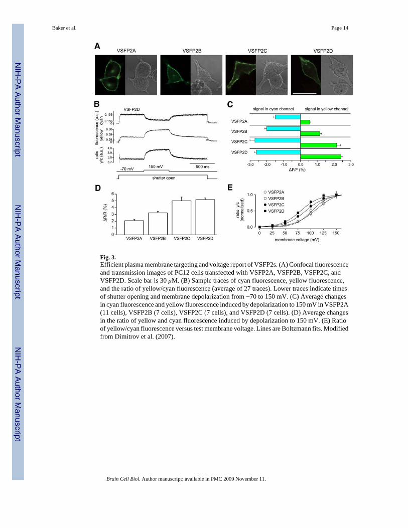
Fig. 3.Efficient plasma membrane targeting and voltage report of VSFP2s. (A) Confocal fluorescenceand transmission images of PC12 cells transfected with VSFP2A, VSFP2B, VSFP2C, andVSFP2D. Scale bar is 30 μM. (B) Sample traces of cyan fluorescence, yellow fluorescence,and the ratio of yellow/cyan fluorescence (average of 27 traces). Lower traces indicate timesof shutter opening and membrane depolarization from −70 to 150 mV. (C) Average changesin cyan fluorescence and yellow fluorescence induced by depolarization to 150 mV in VSFP2A(11 cells), VSFP2B (7 cells), VSFP2C (7 cells), and VSFP2D (7 cells). (D) Average changesin the ratio of yellow and cyan fluorescence induced by depolarization to 150 mV. (E) Ratioof yellow/cyan fluorescence versus test membrane voltage. Lines are Boltzmann fits. Modifiedfrom Dimitrov et al. (2007).
Baker et al. Page 14
Brain Cell Biol. Author manuscript; available in PMC 2009 November 11.
NIH
-PA Author Manuscript
NIH
-PA Author Manuscript
NIH
-PA Author Manuscript

Fig. 4.Properties of VSFP2.1. Response–voltage relationship and kinetics of VSFP2.1 at 22°C (A1–A3) and at 35°C (B1–B3). (A1, B1) Ratio of yellow/cyan fluorescence during a family of 500ms voltage steps from a holding potential of −70 mV to test potentials of −140 to +40 mV (20mV increments). Traces are grand averages over average responses from 4 cells (A1) and 6cells (B1). (A2, B2) Ratio of yellow/cyan fluorescence versus test membrane voltage.Connected symbols are data from individual cells. Red lines are Boltzmann fits with V1/2 valuesas indicated. (A3, B3) Activation and deactivation time constants. Taken from Dimitrov et al.(2007).
Baker et al. Page 15
Brain Cell Biol. Author manuscript; available in PMC 2009 November 11.
NIH
-PA Author Manuscript
NIH
-PA Author Manuscript
NIH
-PA Author Manuscript

Fig. 5.Neuronal expression and functionality of VSFP2.1. Primary mouse cortical cultures weretransfected with a codon optimized version of VSFP2.1 at 10 DIV. Left panel shows apyramidal-shaped neuron expressing this FP voltage sensor. Right panel shows response ofmembrane potential, yellow fluorescence, and cyan fluorescence to a hyperpolarizing currentinjected via a patch pipette. Fluorescence values are expressed as percentage of total measuredlight intensity, without correction for background fluorescence (Mutoh et al., unpublishedobservations).
Baker et al. Page 16
Brain Cell Biol. Author manuscript; available in PMC 2009 November 11.
NIH
-PA Author Manuscript
NIH
-PA Author Manuscript
NIH
-PA Author Manuscript

Fig. 6.VSFP2.1 can monitor physiological neuronal membrane voltage dynamics. PC12 cellsexpressing VSFP2.1 were voltage-clamped with a voltage trace obtained from a current-clamped mouse olfactory bulb mitral cell. The mitral cell was stimulated to generate a seriesof action potentials by intracellular injection of a current pulse (A) or by electrical stimulationof the olfactory nerve (B). Traces in (A) are averages of 50 sweeps, upper traces (in B) are theaverage of 90 sweeps, and the lower four traces in (B) are single sweeps. Traces showmembrane potential (V), yellow fluorescence (Fy), cyan fluorescence (Fc), and the ratio ofyellow and cyan fluorescence (Fy/Fc). Fluorescence signals were digitally low pass filtered(0.2 kHz) and were not corrected for dye bleaching. Recordings were done at 35°C. Takenfrom Dimitrov et al. (2007).
Baker et al. Page 17
Brain Cell Biol. Author manuscript; available in PMC 2009 November 11.
NIH
-PA Author Manuscript
NIH
-PA Author Manuscript
NIH
-PA Author Manuscript


