Embed Size (px)
Citation preview

JOURNAL OF CLINICAL MICROBIOLOGY, Nov. 2004, p. 5109–5120 Vol. 42, No. 110095-1137/04/$08.00�0 DOI: 10.1128/JCM.42.11.5109–5120.2004Copyright © 2004, American Society for Microbiology. All Rights Reserved.
Genetic Diversity of Human Pathogenic Members of the Fusariumoxysporum Complex Inferred from Multilocus DNA Sequence
Data and Amplified Fragment Length Polymorphism Analyses:Evidence for the Recent Dispersion of a Geographically
Widespread Clonal Lineage andNosocomial Origin
Kerry O’Donnell,1* Deanna A. Sutton,2 Michael G. Rinaldi,2 Karen C. Magnon,3 Patricia A. Cox,4Sanjay G. Revankar,2† Stephen Sanche,2‡ David M. Geiser,5 Jean H. Juba,5
Jo-Anne H. van Burik,6 Arvind Padhye,7 Elias J. Anaissie,8Andrea Francesconi,9 Thomas J. Walsh,9
and Jody S. Robinson1
National Center for Agricultural Utilization Research, Agriculture Research Service, U.S. Department of Agriculture, Peoria,Illinois1; Department of Pathology, University of Texas Health Science Center,2 Baptist Hospital,3 and 11503 Woollcott,4
San Antonio, Texas; Department of Plant Pathology, The Pennsylvania State University, University Park, Pennsylvania5;Division of Infectious Diseases, University of Minnesota, Minneapolis, Minnesota6; Centers for Disease Control
and Prevention, Atlanta, Georgia7; University of Arkansas for Medical Sciences, Little Rock, Arkansas8;and Immunocompromised Host Section, Pediatric Oncology Branch, National
Cancer Institute, Bethesda, Maryland9
Received 11 February 2004/Returned for modification 29 March 2004/Accepted 14 July 2004
Fusarium oxysporum is a phylogenetically diverse monophyletic complex of filamentous ascomycetous fungithat are responsible for localized and disseminated life-threatening opportunistic infections in immunocom-petent and severely neutropenic patients, respectively. Although members of this complex were isolated frompatients during a pseudoepidemic in San Antonio, Tex., and from patients and the water system in a Houston,Tex., hospital during the 1990s, little is known about their genetic relatedness and population structure. Thisstudy was conducted to investigate the global genetic diversity and population biology of a comprehensive setof clinically important members of the F. oxysporum complex, focusing on the 33 isolates from patients at theSan Antonio hospital and on strains isolated in the United States from the water systems of geographicallydistant hospitals in Texas, Maryland, and Washington, which were suspected as reservoirs of nosocomialfusariosis. In all, 18 environmental isolates and 88 isolates from patients spanning four continents weregenotyped. The major finding of this study, based on concordant results from phylogenetic analyses ofmultilocus DNA sequence data and amplified fragment length polymorphisms, is that a recently dispersed,geographically widespread clonal lineage is responsible for over 70% of all clinical isolates investigated,including all of those associated with the pseudoepidemic in San Antonio. Moreover, strains of the clonallineage recovered from patients were conclusively shown to genetically match those isolated from the hospitalwater systems of three U.S. hospitals, providing support for the hypothesis that hospitals may serve as areservoir for nosocomial fusarial infections.
Members of the phylogenetically diverse monophyleticFusarium oxysporum complex (FOC) are best known as cos-mopolitan soilborne plant pathogens that are responsible foreconomically devastating vascular wilts of an enormous rangeof agronomically important plant hosts (6). Members of theFOC are also frequently isolated from nonplant sources, par-
ticularly from the soil but also from air and animals. Over thepast 2 decades, however, fusaria have emerged as opportunisticpathogens causing life-threatening disseminated infections inimmunocompromised patients (3). In patients who are persis-tently neutropenic, deeply invasive fusarial infections cause100% mortality (18). Most localized and disseminated cases offusariosis are caused by members of the Fusarium solani spe-cies complex, followed by members of the FOC (1). Fortu-nately, the recent development of one strain of F. oxysporum asa model system will greatly facilitate the molecular geneticdissection of fungal virulence determinants during plant andanimal pathogenesis (24).
Although molecular epidemiological studies have been com-pleted for nosocomial fusariosis (1, 25), most of the analyseswere conducted on members of the F. solani species complex.
* Corresponding author. Mailing address: Microbial Genomics Re-search Unit, National Center for Agricultural Utilization Research,ARS-USDA, 1815 North University St., Peoria, IL 61604-3999. Phone:(309) 681-6383. Fax: (309) 681-6672. E-mail: [email protected].
† Present address: Dallas VA Medical Center, Division of InfectiousDiseases, Dallas, TX 75216.
‡ Present address: Royal University Hospital, Division of InfectiousDiseases, Saskatoon, Saskatchewan S7N 0W8, Canada.
5109

Nevertheless, a relationship between an environmental isolateand a patient isolate was determined in one case of infectiondue to a member of the FOC (1). Little is known about themolecular epidemiology of clinically important members of theFOC, even though they were isolated from a San Antonio,Tex., hospital pseudoepidemic (i.e., a false epidemic due tocontamination of clinical specimens) associated with bronchos-copy specimens in 1997 to 1998 (S. E. Sanche, D. A. Sutton, K.Magnon, R. Cox, S. Revankar, and M. G. Rinaldi, Abstr. 98thGen. Meet. Am. Soc. Microbiol., abstr. F-102, 1998) (hereafterreferred to as Texas hospital A) and as part of a 1996 envi-ronmental survey of a Houston, Tex., hospital water systemsuspected of serving as a reservoir of nosocomial fusariosis (1,14) (hereafter referred to as Texas hospital B). Phylogeneticanalyses of the FOC have been limited to phytopathogens (2,21, 26). Results of these genetic diversity studies using mul-tilocus DNA sequence typing (MLST) and amplified fragmentlength polymorphisms (AFLPs) have shown that some planthost-specific pathogens, called formae speciales, havepolyphyletic evolutionary origins (2, 21, 26) and that this com-plex appears to consist of a large number of predominately orexclusively clonal lineages distributed among at least threeclades. The latter finding was not unexpected, because nomember of the FOC has been shown to undergo sexual repro-duction, even though the few strains tested were shown topossess apparently functional mating-type (MAT) genes thatare expressed and processed correctly (38).
The objectives of this study were to (i) investigate the ge-netic relatedness and population structure of a comprehensiveset of isolates of the FOC from patients and the hospitalenvironment spanning four continents, focusing on those re-covered from the Texas hospital A pseudoepidemic; (ii) eval-uate the hypothesis that hospital water systems might serve asreservoirs for nosocomial fusariosis by comparing FOC strainsrecovered from the environment in hospitals in Texas (14),Maryland, and Washington with isolates derived from patients;and (iii) investigate the phylogenetic diversity and evolutionaryorigins of human- and hospital environment-derived isolates bycomparing them with phytopathogenic strains chosen to rep-resent the known pathogenic and phylogenetic diversity of theFOC. To achieve these objectives, we have developed an initialset of MLST- and AFLP-based molecular markers that can beincorporated into long-range epidemiological studies. Resultsof the MLST and AFLP analyses reported here provide inde-pendent support for the validity of a clinically important, wide-spread clonal lineage within the F. oxysporum complex.
MATERIALS AND METHODS
Fungal strains. Isolates of the F. oxysporum species complex (FOC) frompatients, the environment, and other plant and animal sources were assembledfrom several national and international culture collections, with most of theisolates supplied by The University of Texas Health Science Center, San Anto-nio, Tex. (Table 1). All strains are stored cryogenically in the AgriculturalResearch Service (NRRL) Culture Collection, National Center for AgriculturalUtilization Research, Peoria, Ill., for future reference.
DNA isolation, amplification, and sequencing. Liquid cultures were grown inyeast-malt broth, and total genomic DNA was extracted from freeze-dried my-celia by the hexadecyltrimethyl-ammonium bromide (Sigma, St. Louis, Mo.)protocol described by O’Donnell et al. (20). All PCR and sequencing primers arelisted in Table 2. The total reaction volume of all PCR mixtures was 50 �l andincluded approximately 5 ng of total genomic DNA and MgCl2 at a final con-centration of 25 mM. Amplification of a portion of the translation elongation
factor (1�) gene and the mitochondrial small-subunit (mtSSU) ribosomal DNA(rDNA) was accomplished by using the EF-1–EF-2 (21) and MS1-MS2 (35) PCRprimer pairs, respectively, and AmpliTaq (Applied Biosystems [ABI], FosterCity, Calif.) in a 9700 thermocycler and the following cycling parameters: 1 cycleof 30 s at 94°C; 40 cycles of 30 s at 94°C, 30 s at 52°C, and 90 s at 72°C; and then10 min at 72°C and a 4°C soak. The entire nuclear ribosomal intergenic spacer(IGS) region (�2.5 kb) was amplified with the NL11-CNS1 primer pair (Table2), using Platinum Taq DNA polymerase Hi-Fi (Invitrogen Life Technologies,Carlsbad, Calif.) in an ABI 9700 thermocycler and the following cycling param-eters: 1 cycle of 90 s at 94°C; 40 cycles of 30 s at 94°C, 30 s at 52°C, and 3 minat 68°C; and then 1 cycle of 5 min at 68°C and a 4°C soak.
The sequences with GenBank accession numbers AB011379 and AB011378were used to design PCR primers to amplify the MAT1-1 and MAT1-2 idiom-orphs, respectively (38). The MAT1-1 idiomorph was amplified as two overlap-ping segments by using the FOM132-FOM123 and FOM122-FOM111 primerpairs (Table 2), using the Platinum Taq PCR protocol described above. PCRprimers FOM211 and FOM212 were used to amplify the MAT1-2 idiomorph,using the AmpliTaq PCR protocol described above. A multiplex PCR, employingthe FOM111-FOM112 primer pair for the MAT1-1-2 gene and the FOM211-FOM212 primer pair for the MAT1-2-1 gene, was used to screen all of the strainsincluded in this study for MAT idiomorph (i.e., MAT1-1 or MAT1-2), using theAmpliTaq PCR protocol outlined above.
PCR products were purified by using Montage PCR96 Cleanup filter plates(Millipore Corp., Billerica, Mass.) and then sequenced by using ABI BigDyechemistry version 3.0 in a 9700 thermocycler with the following cycling param-eters: 1 cycle of 15 s at 96°C; 40 cycles of 15 s at 96°C, 10 s at 50°C, and 4 minat 60°C; and then a 4°C soak. Sequencing reaction mixtures were purified viaethanol precipitation and then run on an ABI 3100 or 3730 genetic analyzer.Sequences were edited and aligned by using Sequencher version 4.1.2 (GeneCodes, Ann Arbor, Mich.), after which the alignments were improved manually.
AFLP analysis. All genomic DNA samples included in the AFLP analysis werefirst treated with 2 �l of RNase A (10 �g/�l) (Sigma) per 200-�l total genomicDNA sample for 30 min at 65°C, after which they were subjected to a lithiumchloride (Sigma) cleanup protocol. Briefly this protocol consisted of adding anequal volume of ice-cold 5 M LiCl to each genomic DNA, icing for 15 min, andthen centrifuging at 13,000 � g for 15 min. After the supernatant was removedto a fresh tube, 1/16 volume of 5 M NaCl was added to each sample, followed by2 volumes of ice-cold 95% ethanol. The samples were then placed in a �80°Cfreezer for 20 min to precipitate the DNA. Once the samples were removed fromthe freezer and thawed, DNAs were pelleted in a microcentrifuge at 13,000 � gfor 10 min, followed by a 70% ethanol wash, and they were then resuspended in50 �l double-distilled water (ddH2O). DNA quantification of all genomic DNAsamples was done by running them into a 1.5% agarose gel together with aknown concentration of a HindIII (A2AGCTT) digest of � DNA (New EnglandBiolabs [NEB], Beverly, Mass.). Restriction-ligation was conducted at 37°C over-night in an ABI 9700 thermocycler in a total volume of 10 �l by combining �100ng of total genomic DNA in 4.5 �l of ddH2O with 5.5 �l of the following mastermix: 1 �l of 10� T4 ligase buffer (NEB) (50 mM Tris-HCl [pH 7.5], 10 mMMgCl2, 10 mM dithiothreitol, 1 mM ATP, 25 �g of bovine serum albumin [BSA]per ml), 1 �l of NaCl (0.5 M), 0.5 �l of 10� BSA (1 �g/�l), 1 �l of MseI adaptermix (50 pmol/�l), 1 �l of EcoRI (G2AATTC) adapter mix (5 pmol/�l), and 1 �lof an enzyme master mix (for each set of 10 reactions) consisting of 1 �l of 10�T4 DNA ligase buffer (NEB), 1 �l of NaCl (0.5 M), 0.5 �l of 10� BSA (1 �g/�l),0.5 �l of EcoRI (NEB) (100 U/�l), 0.2 �l of MseI (T2TAA) (NEB) (50 U/�l),0.33 �l of T4 DNA ligase (NEB) (2,000 U/�l), and 6.5 �l of ddH2O. All adaptersand primers used for the AFLP analysis are listed in Table 2. Once completed,the restriction-ligation mix was diluted 1:2 in ddH2O and stored at �20°C whennot in use.
The preselective amplification was performed in a total volume of 10 �l by firstaliquoting 8 �l of a master mix consisting of 1 �l of 10� Invitrogen PCR buffer,1 �l of 2 mM deoxynucleoside triphosphates, 0.5 �l of 50 mM MgCl2, 1 �l ofEcoRI nonselective primer (1 pmol/�l), 1 �l of MseI nonselective primer (1pmol/�l), 0.065 �l of Taq polymerase (Invitrogen), and 3.5 �l of ddH2O intoeach reaction tube, to which 2 �l of a diluted restriction-ligation mix was added.Amplifications were performed in an ABI 9700 thermocycler programmed asfollows: 2 min at 72°C followed by 5 min at 94°C; 20 cycles of 30 s at 94°C, 30 sat 56°C, and 60 s at 72°C; and 1 cycle of 5 min at 72°C followed by a 4°C soak.Several amplification product mixtures were checked for a smear of DNA in therange of 100 to 800 bp, which is indicative of a successful preselective amplifi-cation, by electrophoreses of 5 �l of the reaction products into a 1.5% agarosegel, followed by staining with ethidium bromide and visualization over a UVtransilluminator. Preselective amplicons were diluted 1:11 with ddH2O, afterwhich they were vortexed briefly and then stored at �20°C when not in use.
5110 O’DONNELL ET AL. J. CLIN. MICROBIOL.

TABLE 1. Strains of the F. oxysporum complex and outgroups included in this study
NRRL no. Other designationa Yr Isolate sourceb Geographic origin Hospital orlaboratory data
22533 CBS 244.61 1961 Aechmea fasciata Germany22548 CBS 744.79 1979 Zygocactus truncatus Germany22550 CBS 794.70 1970 Albizzia julibrissin Iran22555 CBS 797.70 1970 Solanum tuberosum Iran22903c IMI 375363 1987 Pseudotsuga menziesii Oregon25184c CBS 573.94 1994 Peat Germany25375 IMI 169612 1973 Human South Pacific25378 IMI 214661 1977 Human Oklahoma25387 ATCC 26225 1971 Toenail New Zealand25420 BBA 66843 Gossypium sp. United States25433 BBA 69050 Gossypium sp. China25509 FRC O-1853 1995 Cyclamen, nonpathogen The Netherlands25512 FRC O-1858 1995 Cyclamen, nonpathogen The Netherlands25594 ATCC 16415 Ipomoea batatas South Carolina25598 ATCC 18774 Soybean South Carolina25603 Ploetz A2 Banana Australia25728 CBS 463.91 1991 Human Germany25749 ATCC 64530 1985 Foot ulcer Belgium26022 Ploetz JLT44 Banana France26024 Ploetz STB2 Banana Honduras26033 Kistler CL58 1996 Tomato Florida26035 Kistler Guil2 1987 Phoenix canariensis Tenerife, Canary Islands26178 Gordon B9-9S Cucumis melo Maryland26180 Gordon CR-III 1989 Soil California26203 Kistler 73 Tomato Italy26360 FRC O-755 1975 Eye Tennessee26361 FRC O-783 1976 Human Tennessee26362 FRC O-784 1976 Human South Carolina26363 FRC O-1168 1982 Peritoneal fluid Rhode Island26365 FRC O-1562 1987 Brain autopsy New York26367 FRC O-1591 1987 Human Maryland26368 FRC O-1723 1989 Amputated toe California26370 FRC O-1732 1989 Foot Louisiana26372 FRC O-1746 1990 Catheter, leukemic New York26373 FRC O-1750 1992 Lung Chile26374 FRC O-1777 1993 Leg ulcer California26376 FRC O-1683 1988 Blood New York26381 Kistler CL-57 1996 Tomato Florida26383 Kistler GD-40 1996 Tomato Florida26386 UTHSC 97–95 1997 Lung, unknown fever San Antonio, Tex. Hospital A26387 UTHSC 96–2463 1996 Sputum, intestinal hemorrhage San Antonio, Tex. Hospital A26388 UTHSC 96–2063 1996 Peritoneal dialysate New York26389 UTHSC 96–1960 1991 Nail Connecticut26390 UTHSC 96–1867 1996 Bronchial wash, lump in chest San Antonio, Tex. Hospital A26391 UTHSC 96–1804 1996 Bronchial wash, respiratory neoplasm San Antonio, Tex. Hospital A26392 UTHSC 96–1710 1996 Bronchial wash, bronchitis San Antonio, Tex. Hospital A26393 UTHSC 96–1087 1996 Nail Connecticut26394 UTHSC 96–840 1996 Leg ulcer California26395 UTHSC 95–2234 1995 Whale blowhole Ohio26397 UTHSC 95–1193 1995 Hand abscess San Antonio, Tex. Hospital A26398 UTHSC 95–1173 1995 Foot wound Florida26399 UTHSC 95–1058 1995 Bronchial wash, respiratory failure San Antonio, Tex. Hospital A26400 UTHSC 95–727 1995 Throat California26401 UTHSC 95–404 1995 Ointment contaminate San Antonio, Tex. Private industrial
laboratory A26402 UTHSC 95–315 1995 Ointment contaminate San Antonio, Tex. Private industrial
laboratory A26403 UTHSC 95–144 1995 Left calf Minnesota26406 Gordon K419 1989 Cucumis melo Jalisco, Mexico26409 ATCC 10913 Tobacco Maryland26442 IMI 141108 Lilium sp. South Carolina26551 UAMH 5692 1987 Leg ulcer Saskatchewan, Canada26677 FRL F7325 1987 Nail Sydney, Australia26679 FRL F7463 1987 Gum abscess Sydney, Australia26680 FRL F8433 1989 Leukemic Sydney, Australia28013 CDC B-5736 1996 Blood, leukemic Delaware28031 CDC B-3882 1983 Toe nail South Carolina28244 BBA 70516 1998 Greenhouse irrigation water Finland28245 BBA 70517 1998 Greenhouse irrigation water Finland28670 UWash 97–25989 1997 Sink drain Seattle, Wash. Hospital28678 UWash 97–25459 1997 Mouthwash Seattle, Wash. Hospital, patient B28680 UWash 97–9417–2 1997 Lung Seattle, Wash. Hospital, patient K28683 UWash 98–1455 1998 Mouthwash Seattle, Wash. Hospital, patient F
Continued on following page
5111

TABLE 1—Continued
NRRL no. Other designationa Yr Isolate sourceb Geographic origin Hospital orlaboratory data
28684 UWash 97–11584 1997 Mouthwash Seattle, Wash. Hospital, patient L28685 UWash 93–11340 1993 Enteric stool screen Seattle, Wash. Hospital, patient P28686 UWash 95–6570 1995 Sinus wash Seattle, Wash. Hospital, patient O28687 UWash 93–8070 1993 Kidney autopsy Seattle, Wash. Hospital, patient E31166 Hospital B#9 2000 Renal renal cancer Houston, Tex. Hospital B32176 Hospital B#F15 2000 Lung cancer, lung autopsy Houston, Tex. Hospital B32377 Hospital B#B 2000 Sputum, leukemic Houston, Tex. Hospital B32507 FRC O-1885 1996 Sink faucet Houston, Tex. Hospital B32509 FRC O-1887 1996 Shower drain Houston, Tex. Hospital B32511 FRC O-1895 1996 Cancer patient Houston, Tex. Hospital B32512 FRC O-1896 1996 Cancer patient Houston, Tex. Hospital B32513 FRC O-1908 1996 Cold-water filter Houston, Tex. Hospital B32514 FRC O-1909 1997 Cold-water filter Houston, Tex. Hospital B32515 FRC O-1910 1997 Cold-water filter Houston, Tex. Hospital B32516 FRC O-1911 1997 Cold-water filter Houston, Tex. Hospital B32517 FRC O-1912 1997 Cold-water filter Houston, Tex. Hospital B32525 PD 22005805–2 2002 Tomato seed The Netherlands32527 PD 22005805–4 2002 Tomato seed The Netherlands32914 UTHSC 01–1015 2001 BAL California32915 UTHSC 01–1482 2001 Leg Colorado32916 UTHSC 01–1998 2001 Blood CVC, esophageal cancer Houston, Tex. Hospital B32917 UTHSC 01–2476 2001 Foot, cellulitis San Antonio, Tex. Hospital A32920 UTHSC 02–1874 2002 Periesophageal Washington, D.C.32921 UTHSC 02–1980 2002 Water supply Baltimore, Md. Hospital32922 UTHSC 00–2402 2000 Environmental San Antonio, Tex. Private industrial
laboratory B32925 UTHSC 00–1311 2000 Food San Antonio, Tex. Private industrial
laboratory B32927 UTHSC 00–221 2000 Meatus San Antonio, Tex. Commercial
laboratory32929 UTHSC 99–1742 1999 BAL San Antonio, Tex. Hospital A32930 UTHSC 99–1135 1999 BAL, AIDS San Antonio, Tex. Hospital A32931 UTHSC 99–853 1999 Blood Massachusetts32932 UTHSC 98–2469 1998 Blood Pennsylvania32933 UTHSC 98–2404 1998 BAL, pneumonia San Antonio, Tex. Hospital A32935 UTHSC 98–2189 1998 Blood Maine32938 UTHSC 98–1748 1998 BAL, dysphagia San Antonio, Tex. Hospital A32939 UTHSC 98–1747 1998 BAL San Antonio, Tex. Hospital A32940 UTHSC 98–1741 1998 BAL, malignant neoplasm San Antonio, Tex. Hospital A32941 UTHSC 98–1670 1998 Spleen, splenomegaly San Antonio, Tex. Hospital A32942 UTHSC 98–1645 1998 BAL San Antonio, Tex. Hospital A32943 UTHSC 98–835 1998 BAL, pneumonia San Antonio, Tex. Hospital A32944 UTHSC 98–834 1998 BAL San Antonio, Tex. Hospital A32945 UTHSC 98–718 1998 Lung, unspecified neoplasm San Antonio, Tex. Hospital A32946 UTHSC 98–709 1998 Nail Connecticut32947 UTHSC 98–361 1998 BAL Colorado32948 UTHSC 97–2184 1997 BAL, nodular goiter San Antonio, Tex. Hospital A32949 UTHSC 97–2090 1997 BAL, mass in chest San Antonio, Tex. Hospital A32950 UTHSC 97–1922 1997 BAL, asthma San Antonio, Tex. Hospital A32951 UTHSC 97–1684 1997 BAL, malignant neoplasm San Antonio, Tex. Hospital A32952 UTHSC 97–1055 1997 Toe bone Wisconsin32953 UTHSC 97–1476 1997 Open leg wound San Antonio, Tex. Hospital A32954 UTHSC 97–1466 1997 BAL San Antonio, Tex. Hospital A32955 UTHSC 97–1465 1997 BAL, bronchitis San Antonio, Tex. Hospital A32956 UTHSC 97–1184 1997 BAL, voice disturbance San Antonio, Tex. Hospital A32957 UTHSC 97–891 1997 BAL, bronchitis San Antonio, Tex. Hospital A32958 UTHSC 97–476 1997 Lung wash, hemoptysis San Antonio, Tex. Hospital A32960 UTHSC 97–357 1997 BAL, pneumonia San Antonio, Tex. Hospital A32961 UTHSC 97–291 1997 BAL San Antonio, Tex. Hospital A32962 UTHSC 97–290 1997 BAL, CVA San Antonio, Tex. Hospital A32999 UTHSC 97–2110 1997 Sputum, respiratory failure San Antonio, Tex. Hospital A34936 Di Pietro 4287 1984 Tomato Spain36064 FRC O-1747 1991 Human cancer Houston, Tex. Hospital B
a ATCC, American Type Culture Collection, Manassas, Va.; BBA, Biologische Bundesanstalt fur Land-und Forstwirtschaft, Institute fur Mikrobiologie, Berlin,Germany; CBS, Centraalbureau voor Schimmelcultures, Utrecht, The Netherlands; CDC, Centers for Disease Control and Prevention, Atlanta, Ga.; Di Pietro, AntonioDi Pietro, Universidad de Cordoba, Cordoba, Spain; FRC, Fusarium Research Center, The Pennsylvania State University, State College; FRL, Fusarium ResearchLaboratory, University of Sydney, Sydney, Australia; Gordon, Thomas Gordon, University of California, Davis; Hospital B, Hospital B, Houston, Tex. (13); IMI, CABIBiosciences, Egham, Surrey, England; PD, Plantenziektenkundige Dienst, Wageningen, The Netherlands; Ploetz, Randy Ploetz, University of Florida, Homestead;UAMH, Microfungus Collection, University of Alberta, Edmonton, Alberta, Canada; UTHSC, University of Texas Health Science Center, San Antonio; UWash,University of Washington, Seattle.
b With the exception of strain NRRL 26395 from a whale blowhole, all other clinical isolates are from humans. BAL, bronchoalveolar lavage; CVA, cough variantasthma; CVC � central venous catheter.
c Sequences of Fusarium commune NRRL 22903 (27) and Fusarium sp. strain NRRL 25184 were used to root the phylogram in Fig. 4.
5112

The selective amplification was performed in a total volume of 10 �l by firstdispensing 8-�l aliquots of a master mix consisting of 1 �l of 10� Invitrogen PCRbuffer, 1 �l of 2 mM deoxynucleoside triphosphates, 0.5 �l of 50 mM MgCl2, 1�l of either EcoRI-CC or EcoRI-TC 6-FAM-labeled selective primer (Qiagen,Alameda, Calif.) (Table 2), 1 �l of MseI-G selective primer (3 pmol/�l), 0.065 �lof Invitrogen Taq polymerase, and 3.5 �l of ddH2O, to which 2 �l of a dilutedpreselective amplicon was then added. Selective amplifications were performedin an ABI 9700 thermocycler programmed as follows: 3 min at 94°C; 9 cycles of30 s at 94°C, 30 s at 65°C, and then ramping down 1°C/cycle from 65 to 57°C,followed by 60 s at 72°C; 40 cycles of 30 s at 94°C, 30 s at 56°C, and 60 s at 72°C;and then 1 cycle of 5 min at 72°C followed by a 4°C soak. Amplifications werechecked by running 5 �l of several amplicons into a 1.5% agarose gel as de-scribed above. Selective amplification mixtures were diluted 1:10 or 1:20 inddH2O, after which 1.5 �l of each mixture was added to a master mix consistingof 9.7 �l of HighDye formamide (ABI) and 0.3 �l of the GeneScan ROX 500(ABI) size standard. All samples were run on an ABI 3100 genetic analyzer,using the GeneScan version 3.7 analysis software default run module for datacollection and GenoTyper version 3.7 for scoring fragments in the range of 50 to500 bp relative to the ROX 500 size standard. Only electropherogram peaksabove 100 fluorescent units were scored for the presence or absence of bands ofthe same size. Reproducibility of the AFLP data was assessed by running twoexemplars of each unique AFLP haplotype through the entire protocol, startingfrom the beginning with the isolation of total genomic DNA (15). Only bandsdetected in the duplicate AFLP analyses (90%) were included in the phyloge-netic analyses.
Phylogenetic analysis. The MLST and AFLP matrices analyzed in the presentstudy are available at http://www.ncaur.usda.gov/MGB/MGB-O’Donnell.htm.All phylogenetic analyses were conducted with PAUP* version 4.0b10 (28).Searches for the most-parsimonious trees (MPTs) used a heuristic search with1,000 random addition replicates and tree bisection with reconnection branchswapping, after excluding ambiguously aligned nucleotide positions. TheTempleton Wilcoxon signed rank (WS-R) test implemented in PAUP* was usedto assess whether the various partitions could be combined, using 70% bootstrapmajority rule trees from each partition as constraints. Results of the WS-R tests(P � 1.0) indicated that all of the partitions could be combined. Clade stability
was assessed via parsimony bootstrapping in PAUP*, using a heuristic searchwith 1,000 pseudoreplications of the data and 10 random addition sequences perreplicate and tree bisection with reconnection branch swapping. Constraintsforcing the monophyly of all of the clinical isolates and all of the clinical isolateswithin clade 3 were compared with the MPTs by using the Kishino-Hasegawa testin PAUP*. Phylograms were either midpoint rooted or outgroup rooted based onmore inclusive phylogenetic analyses (2, 21, 27).
Nucleotide sequence accession numbers. The sequences determined in thisstudy have been deposited in the GenBank database under accession numbersAY527415 to AY527732.
RESULTS
The genetic relatedness and diversity of FOC isolates frompatients associated with a pseudoepidemic at a San Antonio,Tex., hospital that peaked in 1997 to 1998 (hospital A) wereinvestigated together with those of isolates collected from thehospital water systems of three U.S. hospitals by comparingthem with a geographically diverse set of 88 clinical and 18environmental isolates from the FOC (Table 1). Aligned par-tial DNA sequences of translation elongation factor (EF-1�,655 bp) and the entire nuclear ribosomal intergenic spacerregion (IGS rDNA, 2,510 bp) were analyzed phylogenetically,using sequences of two strains of the banana pathogen F.oxysporum f. sp. cubense (NRRL 25603 and 26024) to root thetrees based on a more inclusive phylogenetic analysis (21).Results of the Templeton WS-R combinability test (P � 1.0)implemented in PAUP* (28), using 70% bootstrap consensustrees as constraints, indicated that the EF-1� and IGS rDNApartitions could be analyzed as a combined data set. Maxi-mum-parsimony analysis of the combined data set resolved 21unique EF-1�–IGS rDNA haplotypes among the 88 clinicalisolates (Fig. 1), including one widespread clonal lineage whichaccounted for 63 (72%) of the clinical isolates and 17 out of 18of the nonclinical isolates. Of the 82 strains of the clonallineage sequenced, 80 shared an identical EF-1�–IGS rDNAhaplotype, while the other two strains, which were isolated assaprophytes of cyclamen in The Netherlands (36), differedfrom other members of the clonal lineage by only a single basepair mutation within the IGS rDNA (Fig. 1B). All 33 isolatesfrom the Texas hospital A pseudoepidemic are members of theclonal lineage. By comparison, 16 of the 21 EF-1�–IGS hap-lotypes associated with patients were represented by single-tons, with the second most common haplotype being repre-sented by only three strains (Fig. 1A). Eight of the 17nonclinical isolates of the clonal lineage were recovered fromthe water systems of geographically distant hospitals in Hous-ton, Tex., in 1996 to 1997 (1, 14); Seattle, Wash., in 1997; andBaltimore, Md., in 2002. Strains of the clonal lineage were alsorecovered from clinical cases in Canada, Belgium, and Ger-many; from a greenhouse irrigation system in Finland wheretomatoes were being grown; and from tomato seed and cycla-men in The Netherlands.
To further characterize their population biology, all of thestrains were subjected to a mating type (MAT) idiomorph test,using primer pairs FOM111-FOM112 and FOM211-FOM212for MAT1-1 and MAT1-2, respectively. This assay revealed thatall 82 strains of the clonal lineage and strains of the four mostclosely related haplotypes possess a MAT1-1 idiomorph (Fig.1B). In addition, strains representing 14 of the 21 uniqueEF-1�–IGS haplotypes from patients were MAT1-1, compared
TABLE 2. Primers used for PCR, DNA sequencing, andAFLP genotyping
Primer Locus Sequence (5 to 3)a
EF-1 EF-1� ATGGGTAAGGARGACAAGACEF-11ta EF-1� GTGGGGCATTTACCCCGCCEF-22t EF-1� AGGAACCCTTACCGAGCTCEF-2 EF-1� GGARGTACCAGTSATCATGMS1 mtSSU rDNA CAGCAGTCAAGAATATTAGTCAATGMS2 mtSSU rDNA GCGGATTATCGAATTAAATAACMS21 mtSSU rDNA CTCTCCTCCTCAAGTACTGCGFM136 MAT1-1 ATGGTCTACAGCCAGTCGCAFOM132 MAT1-1 GGTAGTGTTGTTTGTGGTTGFOM122 MAT1-1 TCCATGCCAAGATCCTCAGCFOM123 MAT1-1 AAGGCAGAGTCAGAAATCCAFOM112 MAT1-1 GCTGCTGCATCTTGGATTGCFOM111 MAT1-1 GCTTGATCTGTTCGGTCATGFOM211 MAT1-2 ACATATCGATAGCATCTACCFOM212 MAT1-2 AGGCGGTAATCTGCTGTGTANL11 IGS rDNA CTGAACGCCTCTAAGTCAGOCNL13 IGS rDNA TGTGATGTATGCGGTCCTAGGONL13B IGS rDNA GGTTCGAGGATCGATTCGAGGOCNS3C IGS rDNA GCAAGATCTGATACTGAGAGGCNS1 IGS rDNA GAGACAAGCATATGACTACEcoRI-a Adapter CTCGTAGACTGCGTACCEcoRI-b Adapter AATTGGTACGCAGTCTACMseI-a Adapter GACGATGAGTCCTGAGMseI-b Adapter TACTCAGGACTCATEcoRI-ns Nonselective GACTGCGTACCAATTCMseI-ns Nonselective GATGAGTCCTGAGTAAEcoRI-CC Selective 6-FAM-GACTGCGTACCAATTCCCEcoRI-TC Selective 6-FAM-GACTGCGTACCAATTCTCMseI-G Selective GATGAGTCCTGAGTAAG
a R, AG; S, CG.
VOL. 42, 2004 MLST AND AFLP TYPING OF THE FUSARIUM OXYSPORUM COMPLEX 5113

FIG. 1. (A) Distribution of the 88 human isolates among the 21 EF-1�–IGS rDNA sequence haplotypes. (B) One of 16 most-parsimoniousphylograms inferred from the combined EF-1�–IGS rDNA sequence data rooted with sequences of F. oxysporum f. sp. cubense NRRL 25603 and26024 from clade 1 of the FOC (21). All ingroup strains are from humans, except for strain NRRL 26395 from a whale and the 18 environmental
5114 O’DONNELL ET AL. J. CLIN. MICROBIOL.

with only 7 MAT1-2 strains, which were all associated withsingleton haplotypes (Fig. 1B).
Further MLST characterization of 74 strains of the clonallineage from clinical and environmental sources was con-ducted, together with that of strains representing six differentEF-1�–IGS rDNA haplotypes, using partial EF-1� (651 bp)and mtSSU rDNA (677 bp) sequences and the entire IGSrDNA (2,506 bp). Maximum-parsimony analysis of the com-bined data set, totaling 3.8 kb (Table 1), revealed that 74strains of the clonal lineage included in this analysis had anidentical MLST (Fig. 2A). In an effort to obtain a finer level ofgenetic discrimination, these strains were subjected to AFLPgenotyping, using two combinations of EcoRI plus 2-bp6-FAM-labeled selective primers together with an MseI plus1-bp selective primer (Table 2). Parsimony analysis of theAFLP matrix, consisting of 173 binary characters (i.e., restric-tion fragments ranging from 50 to 500 bp were coded as 1 forpresent and 0 for absent), yielded 64,000 MPTs of 128 stepsin length (Fig. 2B) (consistency index [CI] � 0.84; retentionindex [RI] � 0.88) in which the clonal lineage formed a highlysimilar exclusive group consisting of only seven unique AFLPgenotypes (AG1 to -7) (Fig. 2C). AFLP genotyping of the twosaprobic strains isolated from cyclamen in The Netherlands(NRRL 25509 and NRRL 25512) revealed that they possessthe AG2 AFLP genotype (data not shown). When the AFLPdata for 74 strains of the clonal lineage were analyzed sepa-rately, the MPTs were only seven steps in length (CI � 0.86; RI� 0.98) as 167 of the 173 characters were identical across allstrains (five polymorphic sites were synapomorphic and onewas autapomorphic). Of the 28 clinical strains from hospital Ain San Antonio, Tex. (where the pseudoepidemic was re-ported) that were subjected to AFLP fingerprinting, represen-tatives of the three most common AFLP genotypes were re-covered between 1996 and 2001 (AG1, n � 19; AG2, n � 7;and AG3, n � 2). Similarly, five AFLP genotypes of the clonallineage (i.e., AG1 to -5) were recovered during the same timeframe from hospital B in Houston, Tex., including a hospitalenvironmental isolate that shared the identical AG2 genotypewith an isolate from a patient (NRRL 32507 and NRRL 32511,both isolated in 1996). Matched patient-environment isolatesof the AG2 genotype (NRRL 28670 and NRRL 28678) werealso recovered in 1997 from a Seattle, Wash., hospital.
Genetic relationships among the AFLP genotypes were in-vestigated further by PCR amplification and sequencing of theentire MAT1-1 idiomorph from one or more strains represent-ing the six most common AFLP genotypes (i.e., AG1 to -6) ofthe clonal lineage and strains of five near relatives (Fig. 3),using PCR primers described by Yun et al. (38) (Table 2).Parsimony analysis of the 4,017 aligned MAT1-1 nucleotidecharacters yielded a single MPT of 41 steps (Fig. 3B) (CI �1.0) in which exemplars of the six AFLP genotypes were iden-tical (e.g., NRRL 26370) or nearly identical (e.g., NRRL32931) to the two closest known relatives of the clonal lineage(Fig. 1B), reflecting the high conservation of the MAT genes,
which have been shown to be under strong purifying selection(23). Hypothetical translations of the three MAT1-1 genes sug-gest that they all encode functional proteins (GenBank acces-sion numbers AY527415 to AY527427).
Finally, to address whether isolates from patients havemonophyletic or multiple independent evolutionary originswithin the FOC, parsimony analyses were conducted onaligned partial EF-1� (655 bp) and mtSSU rDNA (694 bp)sequences representing 17 strains from patients and 21 phyto-pathogenic strains chosen to represent the known pathogenicdiversity of the FOC (2, 21). Parsimony analysis of the com-bined data set (1,349 bp) yielded 18 MPTs of 134 steps inlength (CI � 0.90; RI � 0.94) with human isolates nestedwithin three of the four clades (Fig. 4). However, most of thehuman isolates, including those of the widespread clonal lin-eage, were nested in clade 3, the most phylogenetically diverseclade. Constraints forcing the monophyly of the human isolateswithin clade 3 and all of the human isolates within the FOC inseparate analyses were 11 and 33 steps longer, respectively,and significantly less parsimonious than the MPTs (Kishino-Hasegawa test, P � 0.009 and P � 0.0001, respectively). Re-sults of the MAT idiomorph test revealed that MAT1-1 andMAT1-2 strains are represented in all four clades.
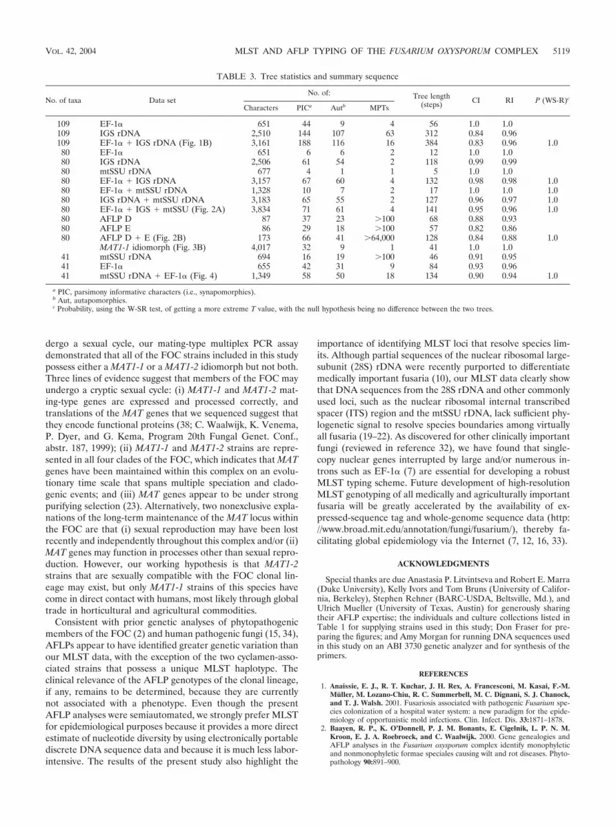
A summary of the tree statistics is given in Table 3.
DISCUSSION
This study describes the first MLST- and AFLP-based mo-lecular markers for genotyping clinically important members ofthe FOC. These tools were used in a molecular epidemiolog-ical investigation of a pseudoepidemic in hospital A in SanAntonio, Tex., that peaked in 1997 to 1998 (S. E. Sanche, D. A.Sutton, K. Magnon, R. Cox, S. Revankar, and M. G. Rinaldi,Abstr. 98th Gen. Meet. Am. Soc. Microbiol., abstr. F-102,1998) and in a survey of the water systems of three U.S.hospitals suspected as being reservoirs of nosocomial fusario-sis. The major finding of this study, based on concordant re-sults from phylogenetic analyses of multilocus DNA sequencedata and AFLPs, is that a geographically widespread clonallineage comprises 70% of all FOC clinical isolates, includingall of the strains recovered from the hospital A pseudoepi-demic and from the water systems of a hospital in Houston,Tex. (hospital B) (1, 14) and of hospitals in Baltimore, Md.,and Seattle, Wash. This clonal lineage consisted of only sevenhighly similar AFLP genotypes, and all of its members sharedidentical or nearly identical EF-1� and IGS rDNA sequencesand possessed only MAT1-1 idiomorphs, indicating that theywere of clonal origin. To date, molecular epidemiological stud-ies that have identified fungal pathogens with a highly clonalpopulation structure are restricted to a relatively small numberof clinically (8, 9), zoologically (17), and agriculturally (4, 5, 11)important species, including members of the FOC (13, 29).However, most clinically important fungi investigated to date
isolates indicated by shading. Note that 82 of the ingroup strains are members of a widespread clonal lineage. The number 1 or 2 following thefive-digit NRRL culture collection number indicates that the strain was typed by the mating-type (MAT) idiomorph PCR assay as MAT1-1 orMAT1-2, respectively. All strains of the clonal lineage and the five most closely related strains are MAT1-1 (identified by boldface internodes).Internodes supported by bootstrap values of �70% are indicated.
VOL. 42, 2004 MLST AND AFLP TYPING OF THE FUSARIUM OXYSPORUM COMPLEX 5115

FIG. 2. (A) One of four most-parsimonious midpoint rooted phylograms inferred from the combined EF-1�–IGS rDNA–mtSSU rDNAsequence data for the 80-taxon matrix. (B) One of 64,000 most-parsimonious phylograms inferred from the AFLP data, indicating the sevenAFLP genotypes (AG1 to -7) for 74 strains of the widespread clonal lineage. Geographic origin and year isolated are indicated. A, San Antonio,Tex., hospital A, reporting the pseudoepidemic; B, Houston, Tex., hospital B, reporting the water system as a potential reservoir of nosocomialfusariosis (1, 14). Internodes supported by bootstrap values of �70% are indicated. (C) Distribution of 74 clinical and environmental strains ofthe widespread clonal lineage among the seven AFLP genotypes.
5116 O’DONNELL ET AL. J. CLIN. MICROBIOL.

exhibit both clonality and recombination (reviewed in refer-ences 31 and 37).
Lacking the ability to satisfy Koch’s postulates regardingFOC isolates from patients, we cannot distinguish isolates ca-pable of infecting humans from other isolates, including po-tential secondary invaders or superficial environmental isolatesnot involved in infection. The frequent reoccurrence of thesame MLST and AFLP genotypes from patients in differentgeographic regions, however, strongly suggests that these iso-lates are the etiological agents of these infections. Furtherstudy comparing the pathogenic potentials of different mem-bers of the FOC by utilizing animal and other models of patho-genicity (6, 24) is needed to shed light on this issue. However,if differences in pathogenic potential exist among the 74 mem-bers of the major clonal lineage associated with patients, theyare not reflected in the extremely low level of genetic diversityobserved.
The results of the present study support the findings ofAnaissie et al. (1), who reported a possible molecular matchbetween strains of F. oxysporum from a patient and the envi-
ronment from Houston, Tex., hospital B, where the initialenvironmental survey for nosocomial fusaria was conducted(14). The two isolates had similar banding patterns based onrandom amplified polymorphic DNA (RAPD), interrepeatPCR, and restriction fragment-length polymorphism analysesconducted at the National Cancer Institute, Bethesda, Md.However, the authors of that study were conservative in notclassifying this patient-environment isolate pair as a matchbecause a RAPD analysis conducted at a second laboratoryyielded discordant results. Because this pair of isolates wasconclusively shown to be a member of the FOC widespreadclonal lineage in the present study (Fig. 1B) (NRRL 32507 andNRRL 36064), it is clear that the RAPD results from thesecond laboratory in Houston represent a false negative. In all,13 of the 14 FOC strains (i.e., 92.8%) that were isolated atHouston hospital B from cancer patients and the environmentfrom 1991 through 2001 were conclusively shown to be mem-bers of the FOC clonal lineage via MLST and AFLP analyses(Table 1). The 13 isolates of the clonal lineage from hospital Bincluded 6 of the 7 isolates from cancer patients and all 7isolates isolated from the hospital water system by Kuchar (14)in 1996 and 1997.
Paradoxically, a separate molecular epidemiological study,also conducted at Houston, Tex., hospital B in 1996 and 1997,reported a complete mismatch between 15 environmentalfusaria isolated by Kuchar (14) and 10 clinical isolates com-pared with them by means of RAPD data (25). Two nonexclu-sive scenarios are offered to explain the discordant results ofAnaissie et al. (1) and Raad et al. (25). First, because most ofthe fusaria isolated at hospital B were members of the F. solanispecies complex (1, 14), it is possible that strains of the FOCclonal lineage may not have been included in the latter study,because isolates were not identified by species names. Second,reproducibility of the RAPD data may have been an issue inthe study by Raad et al. (25). Due to problems of reproduc-ibility and portability from laboratory to laboratory, RAPDand other forms of nondiscrete DNA data are rapidly beingreplaced with electronically portable MLST schemes (30).
Because most members of the FOC clonal lineage from SanAntonio, Tex., hospital A were recovered from bronchoalveo-lar lavage specimens, as in numerous other nosocomial out-breaks and pseudoepidemics (http://www.umdnj.edu/rspthweb/bibs/fob_infc.htm), contaminated bronchoscopes or inade-quate bronchoscope sterilization was suspected, but notproven, as the source of the contamination. Our molecularmarkers provided conclusive evidence that the AG2 genotypeof the clonal lineage recovered from the water systems ofhospital B in Houston, Tex., in 1996 and a Seattle, Wash.,hospital in 1997 was a precise molecular match with strainsrecovered from cancer patients at these hospitals during thesame years, suggesting potential nosocomiality. Similarly,AFLP markers have shown that some waterborne environmen-tal isolates of Aspergillus fumigatus are genetically identical tothose from hospital patients with invasive aspergillosis (34).The FOC clonal lineage may be widespread in hospital watersystems within the United States, because four virtually iden-tical AFLP genotypes of it were recovered from the threehospitals surveyed, including environmental isolates of theAG1, AG2, and AG4 genotypes from hospital B in 1996 (14).Not surprisingly, water also appears to serve as a reservoir for
FIG. 3. (A) MAT1-1 idiomorph showing coding and noncoding re-gions and directions of transcription of the three MAT genes. PCR andsequencing primers are indicated by half-arrows (Table 2). The twointergenic regions are arbitrarily designated A and B. (B) Single most-parsimonious midpoint rooted phylogram inferred from the MAT1-1nucleotide sequence data. Note that strains representing six AFLPgenotypes (AG1 to -6) of the clonal lineage are identical to oneanother and to outgroup strain NRRL 26370 (100% bootstrap sup-port). The AB011379 MAT1-1 sequence was obtained from GenBank.
VOL. 42, 2004 MLST AND AFLP TYPING OF THE FUSARIUM OXYSPORUM COMPLEX 5117

nonhospital environmental isolates of the FOC clonal lineage,because the AG2 genotype was isolated from a greenhouseirrigation system in Finland where tomatoes were being grownand from the blowhole of a whale at a marine park in Ohio.Other potential environmental sources of the clonal lineageinclude agricultural soils (AG3, San Joaquin, Calif.) and in-dustrial laboratories (AG1 and AG3, San Antonio, Tex.).Based on these observations, we hypothesize that any watersource within and outside a hospital may be a potential reser-voir for the FOC clonal lineage, and we suggest careful screen-ing of all water sources that come in contact with immunocom-promised patients.
Intercontinental distributions of the AG1, AG2, and AG3genotypes in Europe and North America suggest recent dis-persion that may have resulted from the relatively recent globaltrade of horticultural and agricultural plants and plant prod-ucts. This scenario is consistent with the fact that members ofthe FOC are ubiquitous inhabitants of plants (e.g., NRRL25509 and NRRL 25512 were isolated as nonpathogens ofcyclamen in The Netherlands) (36). Vigilant surveillance of theclonal lineage within U.S. hospitals seems warranted, becauseit has been recovered in 16 different states, including fromhospital water systems in Texas, Maryland, and Washington(Table 1). One surprise of this study is that the clonal lineage
is phylogenetically distinct from all of the plant pathogens inour MLST database, which includes partial EF-1�–mtSSUrDNA sequences from over 700 FOC strains. Although thisfinding does not preclude the possibility that the clonal lineageis a plant pathogen, it suggests that it may not be economicallysignificant, because most described phytopathogens within theFOC are represented in our database. The present study ex-tends our current knowledge of FOC phylogeny (2, 21)through the discovery of a fourth clade containing the onlystrain isolated from a human eye infection (Fig. 4). Anothersurprise to emerge from the present study is the extreme rarityof eye infections caused by members of the FOC (i.e., only 1 of88 among the human isolates), especially when compared withthe F. solani species complex, where 43.5% of the clinicalisolates subjected to MLST genotyping (i.e., 121 of 278) wererecovered from ocular mycoses (N. Zhang et al., unpublisheddata). Because fusaria are opportunistic pathogens of humans,it was not surprising to discover the clinical isolates exhibitindependent evolutionary origins within three of the FOCclades as well as support for a polyphyletic origin of humanisolates within clade 3. This finding parallels studies of severalplant pathogens within the FOC that appear to have evolvedhost specificity polyphyletically (2, 21, 26).
Although no member of the FOC has been shown to un-
FIG. 4. Phylogenetic diversity of human isolates within the F. oxysporum complex inferred from parsimony analysis of the combined EF-1�–mtSSU rDNA sequence data. The human isolates exhibit a polyphyletic distribution among three of the four clades. The widespread clonal lineageis nested within clade 3. Internodes supported by bootstrap values of �70% are indicated. Sequences of Fusarium commune NRRL 22903 andFusarium sp. strain NRRL 25184 were used as outgroups to root the phylogram.
5118 O’DONNELL ET AL. J. CLIN. MICROBIOL.
dergo a sexual cycle, our mating-type multiplex PCR assaydemonstrated that all of the FOC strains included in this studypossess either a MAT1-1 or a MAT1-2 idiomorph but not both.Three lines of evidence suggest that members of the FOC mayundergo a cryptic sexual cycle: (i) MAT1-1 and MAT1-2 mat-ing-type genes are expressed and processed correctly, andtranslations of the MAT genes that we sequenced suggest thatthey encode functional proteins (38; C. Waalwijk, K. Venema,P. Dyer, and G. Kema, Program 20th Fungal Genet. Conf.,abstr. 187, 1999); (ii) MAT1-1 and MAT1-2 strains are repre-sented in all four clades of the FOC, which indicates that MATgenes have been maintained within this complex on an evolu-tionary time scale that spans multiple speciation and clado-genic events; and (iii) MAT genes appear to be under strongpurifying selection (23). Alternatively, two nonexclusive expla-nations of the long-term maintenance of the MAT locus withinthe FOC are that (i) sexual reproduction may have been lostrecently and independently throughout this complex and/or (ii)MAT genes may function in processes other than sexual repro-duction. However, our working hypothesis is that MAT1-2strains that are sexually compatible with the FOC clonal lin-eage may exist, but only MAT1-1 strains of this species havecome in direct contact with humans, most likely through globaltrade in horticultural and agricultural commodities.
Consistent with prior genetic analyses of phytopathogenicmembers of the FOC (2) and human pathogenic fungi (15, 34),AFLPs appear to have identified greater genetic variation thanour MLST data, with the exception of the two cyclamen-asso-ciated strains that possess a unique MLST haplotype. Theclinical relevance of the AFLP genotypes of the clonal lineage,if any, remains to be determined, because they are currentlynot associated with a phenotype. Even though the presentAFLP analyses were semiautomated, we strongly prefer MLSTfor epidemiological purposes because it provides a more directestimate of nucleotide diversity by using electronically portablediscrete DNA sequence data and because it is much less labor-intensive. The results of the present study also highlight the
importance of identifying MLST loci that resolve species lim-its. Although partial sequences of the nuclear ribosomal large-subunit (28S) rDNA were recently purported to differentiatemedically important fusaria (10), our MLST data clearly showthat DNA sequences from the 28S rDNA and other commonlyused loci, such as the nuclear ribosomal internal transcribedspacer (ITS) region and the mtSSU rDNA, lack sufficient phy-logenetic signal to resolve species boundaries among virtuallyall fusaria (19–22). As discovered for other clinically importantfungi (reviewed in reference 32), we have found that single-copy nuclear genes interrupted by large and/or numerous in-trons such as EF-1� (7) are essential for developing a robustMLST typing scheme. Future development of high-resolutionMLST genotyping of all medically and agriculturally importantfusaria will be greatly accelerated by the availability of ex-pressed-sequence tag and whole-genome sequence data (http://www.broad.mit.edu/annotation/fungi/fusarium/), thereby fa-cilitating global epidemiology via the Internet (7, 12, 16, 33).
ACKNOWLEDGMENTS
Special thanks are due Anastasia P. Litvintseva and Robert E. Marra(Duke University), Kelly Ivors and Tom Bruns (University of Califor-nia, Berkeley), Stephen Rehner (BARC-USDA, Beltsville, Md.), andUlrich Mueller (University of Texas, Austin) for generously sharingtheir AFLP expertise; the individuals and culture collections listed inTable 1 for supplying strains used in this study; Don Fraser for pre-paring the figures; and Amy Morgan for running DNA sequences usedin this study on an ABI 3730 genetic analyzer and for synthesis of theprimers.
REFERENCES
1. Anaissie, E. J., R. T. Kuchar, J. H. Rex, A. Francesconi, M. Kasai, F.-M.Muller, M. Lozano-Chiu, R. C. Summerbell, M. C. Dignani, S. J. Chanock,and T. J. Walsh. 2001. Fusariosis associated with pathogenic Fusarium spe-cies colonization of a hospital water system: a new paradigm for the epide-miology of opportunistic mold infections. Clin. Infect. Dis. 33:1871–1878.
2. Baayen, R. P., K. O’Donnell, P. J. M. Bonants, E. Cigelnik, L. P. N. M.Kroon, E. J. A. Roebroeck, and C. Waalwijk. 2000. Gene genealogies andAFLP analyses in the Fusarium oxysporum complex identify monophyleticand nonmonophyletic formae speciales causing wilt and rot diseases. Phyto-pathology 90:891–900.
TABLE 3. Tree statistics and summary sequence
No. of taxa Data setNo. of: Tree length
(steps) CI RI P (WS-R)c
Characters PICa Autb MPTs
109 EF-1� 651 44 9 4 56 1.0 1.0109 IGS rDNA 2,510 144 107 63 312 0.84 0.96109 EF-1� � IGS rDNA (Fig. 1B) 3,161 188 116 16 384 0.83 0.96 1.080 EF-1� 651 6 6 2 12 1.0 1.080 IGS rDNA 2,506 61 54 2 118 0.99 0.9980 mtSSU rDNA 677 4 1 1 5 1.0 1.080 EF-1� � IGS rDNA 3,157 67 60 4 132 0.98 0.98 1.080 EF-1� � mtSSU rDNA 1,328 10 7 2 17 1.0 1.0 1.080 IGS rDNA � mtSSU rDNA 3,183 65 55 2 127 0.96 0.97 1.080 EF-1� � IGS � mtSSU (Fig. 2A) 3,834 71 61 4 141 0.95 0.96 1.080 AFLP D 87 37 23 100 68 0.88 0.9380 AFLP E 86 29 18 100 57 0.82 0.8680 AFLP D � E (Fig. 2B) 173 66 41 64,000 128 0.84 0.88 1.0
MAT1-1 idiomorph (Fig. 3B) 4,017 32 9 1 41 1.0 1.041 mtSSU rDNA 694 16 19 100 46 0.91 0.9541 EF-1� 655 42 31 9 84 0.93 0.9641 mtSSU rDNA � EF-1� (Fig. 4) 1,349 58 50 18 134 0.90 0.94 1.0
a PIC, parsimony informative characters (i.e., synapomorphies).b Aut, autapomorphies.c Probability, using the W-SR test, of getting a more extreme T value, with the null hypothesis being no difference between the two trees.
VOL. 42, 2004 MLST AND AFLP TYPING OF THE FUSARIUM OXYSPORUM COMPLEX 5119
3. Boutati, E. I., and E. J. Anaissie. 1997. Fusarium, a significant emergingpathogen in patients with hematologic malignancy: ten years’ experience ata cancer center and implications for management. Blood 90:999–1008.
4. Carbone, I., J. B. Anderson, and L. M. Kohn. 1999. Patterns of descent inclonal lineages and their multilocus fingerprints are resolved with combinedgene genealogies. Evolution 53:11–21.
5. Couch, B. C., and L. M. Kohn. 2000. Clonal spread of Sclerotium cepivorumin onion production with evidence of past recombination events. Phytopa-thology 90:514–521.
6. Di Pietro, A., M. P. Madrid, Z. Caracuel, J. Delgado-Jarana, and M. I. G.Roncero. 2003. Fusarium oxysporum: exploring the molecular arsenal of avascular wilt pathogen. Mol. Plant Pathol. 4:315–325.
7. Geiser, D. M., M. del M. Jimenez-Gasco, S. Kang, I. Makalowska, N. Veer-araghavan, T. J. Ward, N. Zang, G. A. Kuldau, and K. O’Donnell. 2004.FUSARIUM-ID v. 1.0: a DNA sequence database for identifying Fusarium.Eur. J. Plant Pathol. 110:1–7.
8. Graser, Y., J. Kuhnisch, and W. Presber. 1999. Molecular markers revealexclusively clonal reproduction in Trichophyton rubrum. J. Clin. Microbiol.37:3713–3717.
9. Halliday, C. L., and D. A. Carter. 2003. Clonal reproduction and limiteddispersal in an environmental population of Cryptococcus neoformans var.gattii isolates from Australia. J. Clin. Microbiol. 41:703–711.
10. Hennequin, C., E. Abachin, F. Symoens, V. Lavarde, G. Reboux, N. Nolard,and P. Berche. 1999. Identification of Fusarium species involved in humaninfections by 28S rRNA gene sequencing. J. Clin. Microbiol. 37:3586–3589.
11. Hovmøoller, M. S., A. F. Justesen, and J. K. M. Brown. 2002. Clonality andlong-distance migration of Puccinia striformis f. sp. tritici in North-WestEurope. Plant Pathol. 51:24–32.
12. Kang, S., J. E. Ayers, E. D. DeWolf, D. M. Geiser, G. Kuldau, G. W.Moorman, E. Mullins, W. Uddin, J. C. Correll, G. Deckert, Y.-H. Lee, Y.-W.Lee, F. N. Martin, and K. Subbarao. 2002. The internet-based fungal patho-gen database: a proposed model. Phytopathology 92:232–236.
13. Koenig, R. L., R. C. Ploetz, and H. C. Kistler. 1997. Fusarium oxysporum f.sp. cubense consists of a number of divergent and globally distributed clonallineages. Phytopathology 87:915–923.
14. Kuchar, R. T. 1996. Isolation of Fusarium from hospital plumbing fixtures:implications for environmental health and patient care. M.S. thesis. TheUniversity of Texas Health Science Center, Houston.
15. Litvintseva, A. P., R. E. Marra, K. Nielsen, J. Heitman, R. Vilgalys, and T. G.Mitchell. 2003. Evidence of sexual reproduction among Cryptococcus neo-formans serotype A isolates in sub-Saharan Africa. Eukaryot. Cell 2:1162–1168.
16. Maiden, M. C. J., J. A. Bygraves, E. Feil, G. Morelli, J. E. Russell, R. Urwin,Q. Zhang, J. Zhou, R. Zurth, D. A. Caugant, I. M. Feavers, M. Achtman, andB. G. Spratt. 1998. Multilocus sequence typing: a portable approach to theidentification of clones within populations of pathogenic microorganisms.Proc. Natl. Acad. Sci. USA 95:3140–3145.
17. Morehouse, E. A., T. Y. James, A. R. D. Ganley, R. Vilgalys, L. Berger, P. J.Murphy, and J. E. Longcore. 2003. Multilocus sequence typing suggests thechytrid pathogen of amphibians is a recently emerged clone. Mol. Ecol.12:395–403.
18. Nucci, M., and E. Anaissie. 2002. Cutaneous infection by Fusarium species inhealthy and immunocompromised hosts: implications for diagnosis and man-agement. Clin. Infect. Dis. 35:909–920.
19. O’Donnell, K. 2000. Molecular phylogeny of the Nectria haematococca-Fusarium solani species complex. Mycologia 92:919–938.
20. O’Donnell, K., E. Cigelnik, and H. I. Nirenberg. 1998. Molecular systematicsand phylogeography of the Gibberella fujikuroi species complex. Mycologia90:465–493.
21. O’Donnell, K., H. C. Kistler, E. Cigelnik, and R. C. Ploetz. 1998. Multipleevolutionary origins of the fungus causing Panama disease of banana: Con-
cordant evidence from nuclear and mitochondrial gene genealogies. Proc.Natl. Acad. Sci. USA 95:2044–2049.
22. O’Donnell, K., H. C. Kistler, B. K. Tacke, and H. H. Casper. 2000. Genegenealogies reveal global phylogeographic structure and reproductive isola-tion among lineages of Fusarium graminearum, the fungus causing wheatscab. Proc. Natl. Acad. Sci. USA 97:7905–7910.
23. O’Donnell, K., T. J. Ward, D. M. Geiser, H. C. Kistler, and T. Aoki. 2004.Genealogical concordance between the mating type locus and seven othernuclear genes supports formal recognition on nine phylogenetically distinctspecies within the Fusarium graminearum clade. Fungal Genet. Biol. 41:600–623.
24. Ortoneda, M., J. Guarro, M. P. Madrid, Z. Caracuel, M. I. G. Roncero, E.Mayayo, and A. Di Pietro. 2004. Fusarium oxysporum as multihost model forthe genetic dissection of fungal virulence in plants and animals. Infect.Immun. 72:1760–1766.
25. Raad, I., J. Tarrand, H. Hanna, M. Albitar, E. Janssen, M. Boktour, G.Bodey, M. Mardani, R. Hachem, D. Kontoyiannis, E. Whimbey, and R.Rolston. 2002. Epidemiology, molecular mycology, and environmentalsources of Fusarium infection in patients with cancer. Infect. Control Hosp.Epidemiol. 23:532–537.
26. Skovgaard, K., H. I. Nirenberg, K. O’Donnell, and S. Rosendahl. 2001.Evolution of Fusarium oxysporum f. sp. vasinfectum races inferred frommultigene genealogies. Phytopathology 91:1231–1237.
27. Skovgaard, K., S. Rosendahl, K. O’Donnell, and H. I. Nirenberg. 2003.Fusarium commune is a new species identified by morphological and molec-ular phylogenetic data. Mycologia 95:630–636.
28. Swofford, D. L. 2002. PAUP*. Phylogenetic analysis using parsimony (*andother methods), version 4. Sinauer Associates, Sunderland, Mass.
29. Tantaoui, A., M. Quinten, J.-P. Geiger, and D. Fernandez. 1996. Character-ization of a single clonal lineage of Fusarium oxysporum f. sp. albediniscausing Bayoud disease of date palm in Morocco. Phytopathology 86:787–792.
30. Taylor, J. W., and M. C. Fisher. 2003. Fungal multilocus sequence typing—it’s not just for bacteria. Curr. Opin. Microbiol. 6:351–356.
31. Taylor, J. W., D. M. Geiser, A. Burt, and V. Koufopanou. 1999. The evolu-tionary biology and population genetics underlying fungal stain typing. Clin.Microbiol. Rev. 12:126–146.
32. Taylor, J. W., D. J. Jacobson, S. Kroken, T. Kasuga, D. M. Geiser, D. S.Hibbett, and M. C. Fisher. 2000. Phylogenetic species recognition and spe-cies concepts in fungi. Fungal Genet. Biol. 31:21–31.
33. Urwin, R., and M. C. J. Maiden. 2003. Multi-locus sequence typing: a tool forglobal epidemiology. Trends Microbiol. 11:479–487.
34. Warris, A., C. H. W. Klaassen, J. F. G. M. Meis, M. T. de Ruiter, H. A. deValk, T. G. Abrahamsen, P. Gaustad, and P. E. Verweij. 2003. Molecularepidemiology of Aspergillus fumigatus recovered from water, air, and patientsshows two clusters of genetically distinct strains. J. Clin. Microbiol. 41:4101–4106.
35. White, T. J., T. Bruns, S. Lee, and J. Taylor. 1990. Amplification and directsequencing of fungal ribosomal RNA genes for phylogenetics, p. 313–322, InM. A. Innis, D. H. Gelfand, J. J. Sninsky, and T. J. White (ed.), PCRprotocols, a guide to methods and applications. Academic Press, Inc., SanDiego, Calif.
36. Woudt, L. P., A. Neuvel, A. Sikkema, M. Q. J. M. van Grinsven, W. A. J. deMilliano, C. L. Campbell, and J. F. Leslie. 1995. Genetic variation in Fusar-ium oxysporum from cyclamen. Phytopathology 85:1348–1355.
37. Xu, J., and T. G. Mitchell. 2002. Strain variation and clonality in Candidaspp. and Cryptococcus neoformans, p. 739–749. In R. A. Calderone and R. L.Cihlar (ed.), Fungal pathogenesis: principles and clinical applications. Mar-cel Dekker, New York, N.Y.
38. Yun, S.-H., T. Arie, I. Kaneko, O. C. Yoder, and B. G. Turgeon. 2000.Molecular organization of mating type loci in heterothallic, homothallic, andasexual Gibberella/Fusarium species. Fungal Genet. Biol. 31:7–20.
5120 O’DONNELL ET AL. J. CLIN. MICROBIOL.


