Embed Size (px)
Citation preview
DOI: 10.1126/scitranslmed.3004108, 165ra162 (2012);4 Sci Transl Med
et al.Stefania CortiPatients with Spinal Muscular AtrophyGenetic Correction of Human Induced Pluripotent Stem Cells from
Editor's Summary
sequences and suggests the potential of this approach for clinical translation.patient-specific iPSCs and their motor neuron progeny that are genetically corrected and free of exogenousin the spinal cord and improved the disease phenotype. This study demonstrates the feasibility of generating
derived motor neurons engrafted−Upon direct transplantation into a severe SMA mouse model, corrected SMA-iPSCwhereas motor neurons derived from genetically corrected SMA-iPSCs showed rescue of the disease phenotype.
derived motor neurons reproduced disease-specific features,−a functional SMN1-like protein. Uncorrected SMA-iPSC to produceSMN2nonintegrating episomal vectors and then performed genetic editing with oligonucleotides to modify
motor neurons that do not show the disease phenotype. The authors generated human SMA-iPSCs using nonviral, generatethe feasibility of genetically engineering induced pluripotent stem cells (iPSCs) derived from SMA patients to
. investigateet alwithin exon 7 that results in the production of an incomplete and nonfunctional protein. Now, Corti by a single nucleotide variantSMN1, that differs from SMN2is no effective therapy. Humans have a paralogous gene,
the survival motor neuron 1 (SMN1) protein. The mutant protein causes loss of spinal cord motor neurons, and there Spinal muscular atrophy (SMA) is an autosomal recessive disorder caused by mutations in the gene encoding
Engineering iPSC-Derived Motor Neurons for Cell Therapy
http://stm.sciencemag.org/content/4/165/165ra162.full.htmlcan be found at:
and other services, including high-resolution figures,A complete electronic version of this article
http://stm.sciencemag.org/content/suppl/2012/12/17/4.165.165ra162.DC1.html can be found in the online version of this article at: Supplementary Material
http://www.sciencemag.org/about/permissions.dtl in whole or in part can be found at: article
permission to reproduce this of this article or about obtaining reprintsInformation about obtaining
is a registered trademark of AAAS. Science Translational Medicinerights reserved. The title NW, Washington, DC 20005. Copyright 2012 by the American Association for the Advancement of Science; alllast week in December, by the American Association for the Advancement of Science, 1200 New York Avenue
(print ISSN 1946-6234; online ISSN 1946-6242) is published weekly, except theScience Translational Medicine
on
Mar
ch 2
7, 2
013
stm
.sci
ence
mag
.org
Dow
nloa
ded
from
R E S EARCH ART I C L E
STEM CELLS
Genetic Correction of Human Induced Pluripotent StemCells from Patients with Spinal Muscular AtrophyStefania Corti,1 Monica Nizzardo,1 Chiara Simone,1 Marianna Falcone,1 Martina Nardini,1
Dario Ronchi,1 Chiara Donadoni,1 Sabrina Salani,1 Giulietta Riboldi,1 Francesca Magri,1
Giorgia Menozzi,2 Clara Bonaglia,2 Federica Rizzo,1 Nereo Bresolin,1,2 Giacomo P. Comi1*
rch
27, 2
013
Spinal muscular atrophy (SMA) is among the most common genetic neurological diseases that cause infant mor-tality. Induced pluripotent stem cells (iPSCs) generated from skin fibroblasts from SMA patients and geneticallycorrected have been proposed to be useful for autologous cell therapy. We generated iPSCs from SMA patients(SMA-iPSCs) using nonviral, nonintegrating episomal vectors and used a targeted gene correction approach basedon single-stranded oligonucleotides to convert the survival motor neuron 2 (SMN2) gene into an SMN1-like gene.Corrected iPSC lines contained no exogenous sequences. Motor neurons formed by differentiation of uncorrectedSMA-iPSCs reproduced disease-specific features. These features were ameliorated in motor neurons derived fromgenetically corrected SMA-iPSCs. The different gene splicing profile in SMA-iPSC motor neurons was rescued aftergenetic correction. The transplantation of corrected motor neurons derived from SMA-iPSCs into an SMA mousemodel extended the life span of the animals and improved the disease phenotype. These results suggest thatgenerating genetically corrected SMA-iPSCs and differentiating them into motor neurons may provide a source ofmotor neurons for therapeutic transplantation for SMA.
Ma
on
stm
.sci
ence
mag
.org
Dow
nloa
ded
from
INTRODUCTION
Spinal muscular atrophy (SMA) is an autosomal recessive genetic dis-order caused by a genetic defect in the survival motor neuron 1 (SMN1)gene resulting from deletions or other mutations (1, 2). SMA ariseswhen there are no functional copies of SMN1, and the affected indi-vidual therefore must fully rely on the protein produced from SMN2, agene paralogous to SMN1 (1, 2). A decrease in full-length SMN pro-tein results in the selective degeneration of spinal cord motor neurons(3). Patients with SMA typically show generalized muscle weaknessand paralysis that can progress very rapidly to early childhood death(4). There is no cure.
SMN2 shows a high sequence homology to SMN1, and the only crit-ical difference is the C-to-T base change 6 base pairs (bp) inside exon 7.This alteration, but not other variations in the SMN genes, affects thesplicing of SMN2 (5). This splicing change yields 10% of the full-lengthprotein and high concentrations of an unstable, truncated protein lack-ing exon 7 (SMND7) (5). The disease severity inversely correlates withSMN2 copy number (6). Worms, flies, and mice lack SMN2, which canbe introduced only by transgenic modifications (7–10). Several thera-peutic strategies have targeted increased exon 7 inclusion in SMN2 tran-scripts, and a human cell–based assay is a critical tool (10).
The differentiation of viral vector–generated induced pluripotentstem cells (iPSCs) from SMA patients into motor neurons has recentlybeen demonstrated (11). Ex vivo correction of the SMN1 mutation inSMA-iPSCs might allow the generation of disease-free motor neuronsfor cell therapy. The vectors used, however, can produce insertionalmutations that interfere with normal cell function, and transgene ex-pression can modify differentiation into specific lineages (12) or evenlead to tumorigenesis (13), limiting their usefulness.
1Dino Ferrari Centre, Neuroscience Section, Department of Pathophysiology andTransplantation, University of Milan, Neurology Unit, IRCCS Foundation Ca’ GrandaOspedale Maggiore Policlinico, Milan 20135, Italy. 2IRCCS Eugenio Medea, BosisioParini, Lecco 23842, Italy.*To whom correspondence should be addressed. E-mail: [email protected]
www.ScienceTr
Here, we have generated iPSCs from SMA patients who are freefrom vectors and transgenic sequences and have differentiated them intomotor neurons. Motor neurons generated from SMA-iPSCs presentedspecific disease-related features indicative of selective motor neuron de-generation. We designed oligodeoxynucleotides to SMN2 that induceda permanent genetic modification of a single nucleotide in exon 7, thusmodifying the coding region of SMN2 to a more SMN1-like sequence.This resulted in the inclusion of exon 7 and the production of a greateramount of full-length SMN2.We then compared the phenotypes ofmo-tor neurons derived from corrected and untreated SMA-iPSCs in vitrousing gene expression and splicing genome-wide analysis, and in vivoafter transplantation into the spinal cords of transgenic SMA mice.
RESULTS
Nonviral vector generation and characterizationof SMA-iPSCsWe generated iPSC lines from two type I SMA patients (SMA-iPSC-1and SMA-iPSC-2) using a nonviral vector method based on nucleofectionof adult fibroblasts with constructs encoding OCT4, SOX2, NANOG,LIN28, c-Myc, and KLF4 (14) (Fig. 1, A and B, and figs. S1 and S2). Theseplasmids are progressively lost from cells, leading to the generation ofiPSCs free of vector and exogenous sequences. As a control, we gener-ated iPSCs from the father of patient 1 (heterozygous, HET-iPSC-1), andwe used an already generated wild-type cell line (iPSC 19.9). We isolatedat least three clonal iPSC lines for each individual free from reprogram-ming vectors (Fig. 1V). All iPSC lines displayed embryonic stem (ES)cell morphology (Fig. 1, C to N) and expressed the pluripotency markersNANOG, SOX2, OCT4, SSEA-4, and TRA-1-60 (Fig. 1, F to H and Lto N). All iPSC lines also maintained euploid karyotypes (Fig. 1O), coulddifferentiate into all three germ layers in vitro (fig. S3), and could formteratomas in vivo (Fig. 1, P to U). DNA fingerprinting confirmed theirorigin from parental fibroblasts (table S1). These data and genome-wide
anslationalMedicine.org 19 December 2012 Vol 4 Issue 165 165ra162 1

R E S EARCH ART I C L E
gene expression analysis confirmed the reprogramming of fibroblaststo a pluripotent state (fig. S4).
SMN gene analysis and genetic conversion of SMN2 to SMN1using oligodeoxynucleotidesPrevious work has described the possibility of genome editing at thesingle base level by introducing single-stranded DNA oligonucleotides
www.ScienceTr
into the cells (15). Taking advantage of this strategy, we used SMN2sequence-specific oligodeoxynucleotides to direct the exchange of a Tto C at position +6 of exon 7, thus modifying the coding region ofSMN2 to a more SMN1-like sequence and therefore promoting theinclusion of exon 7. We transfected iPSCs with oligodeoxynucleotides75 bases in length, designed to hybridize to the transcribed DNA strandof the SMN2 gene (15) (Fig. 2A). To demonstrate that the correction
anslationalMedicine.org 19 De
on
Mar
ch 2
7, 2
013
stm
.sci
ence
mag
.org
Dow
nloa
ded
from
was permanent and heritable and to ob-tain a stable corrected cell population, weprepared clonal cell cultures of transfectedcells. We screened the clones for geneticcorrection by detection of SMN proteinnuclear foci (also known as Gemini bodiesor gems) and polymerase chain reaction(PCR). We analyzed 47 subclones of thetransfected SMA-iPSC-1 line, and we iso-lated three corrected clones that showed50% SMN correction. We also performedthe same gene editing in the second set ofSMA-iPSC lines: 59, 61, and 65 subclonesof SMA-iPSC-2 lines were analyzed, re-sulting in 2, 3, and 2 positive subclones,respectively. Therefore, the overall conver-sion efficiency (calculated as number of pos-itive clones from the clones analyzed) was10 of 232 (4%). The corrected genotypewas stable during subsequent culturingfor 12 passages, indicating that a perma-nent and heritable change had occurred.
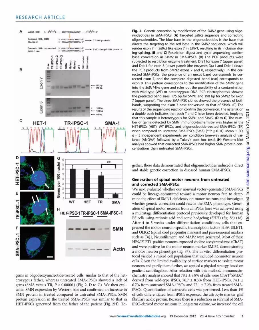
Using direct sequencing and allele-specific restriction fragment length poly-morphism analysis, we evaluated the SMNgenotype of the iPSC-corrected clones (Fig.2, B and C, and fig. S5). The untreatediPSCs of SMA patient 1 and iPSCs treatedwith scrambled oligonucleotides carried ahomozygous deletion of the SMN1 gene.In contrast, a 50% conversion of the SMN2genotype to SMN1 was detected in cor-rected oligonucleotide-treated clones. ThePCR products were subjected to restric-tion enzyme treatment: Dra1 for exon 7and Dde1 for exon 8 (the enzymes Dra Iand Dde I cleave the PCR products fromSMN2 exons 7 and 8, respectively). Thesedata demonstrated in the corrected iPSCsthe presence of an uncut band correspond-ing to corrected exon 7, and the completecut of exon 8 (Fig. 2B). This pattern cor-responds to the conversion of SMN2 intoSMN1 and rules out the possibility of acontamination with wild-type or hetero-zygous DNA. The father’s iPSCs presenteda heterozygous genotype. The pluripotencyfeatures of the SMA-iPSCs were main-tained after genetic correction.
The genetic correction resulted in a sig-nificantly increased number of detectable
Fig. 1. Reprogramming human fibroblasts of an SMA patient and his father without genomic vector inte-gration, and selection of iPSC clones. (A) Schematic representation of the nonviral reprogramming protocol
for adult human fibroblasts. (B) Episomal vector maps. (C to N) Immunocytochemical characterization ofiPSC clones derived from an SMA patient (SMA-iPSCs) and his heterozygous father (HET-iPSCs). TheseiPSCs express pluripotency transcription factors including NANOG (red), SOX2 (red), and OCT4 (green) (Cto E and I to K), aswell as stemcell surfacemarkers (SSEA-4, red) and TRA-1-60 (green). Blue, 4′,6-diamidino-2-phenylindole (DAPI) nuclear stain. (F) to (H) and (L) to (N) are representative images. (O) These cells (HET-iPSCs) were karyotypically normal. (P to U) SMA-iPSC clones and HET-iPSCs form teratomas in vivo thatcontain the three germ layers as shown by representative images from one SMA-iPSC and one HET-iPSCclone: ectoderm (TuJ1, green), mesoderm (desmin, red), and endoderm (a-fetoprotein, red). (V) PCR anal-ysis of episomal DNA in iPSC clones. Genomic (G) and episomal (E) DNAs from nontransfected and vector-transfected adult human fibroblasts were used as negative (−) and positive (+) controls, respectively. Noplasmid integration into iPSCs was observed. Scale bars, 100 mm (C to N); 150 mm (L to N) and (P to U).cember 2012 Vol 4 Issue 165 165ra162 2
R E S EARCH ART I C L E
on
Mar
ch 2
7, 2
013
stm
.sci
ence
mag
.org
Dow
nloa
ded
from
gems in oligodeoxynucleotide-treated cells, similar to that of the het-erozygous father, whereas untreated SMA-iPSCs showed a lack ofgems (SMA versus TR, P < 0.00001) (Fig. 2, D to G). We then eval-uated SMN expression by Western blot and confirmed an increase inSMN protein in treated compared to untreated SMA-iPSCs. SMNprotein expression in the treated SMA-iPSCs was similar to that inHET-iPSCs generated from the father of the patient (Fig. 2H). To-
www.ScienceTr
gether, these data demonstrated that oligonucleotides induced a directand stable genetic correction in diseased human SMA-iPSCs.
Generation of spinal motor neurons from untreatedand corrected SMA-iPSCsWe next evaluated whether our nonviral vector–generated SMA-iPSCscould be lineage-committed toward a motor neuron fate to deter-mine the effect of SMN1 deficiency on motor neurons and investigatewhether genetic correction could rescue the SMA phenotype. Gener-ation of spinal motor neurons from all iPSCs lines was achieved usinga multistage differentiation protocol previously developed for humanES cells using retinoic acid and sonic hedgehog (SHH) (fig. S6) (16).After 4 to 5 weeks under differentiation conditions, cells that ex-pressed the motor neuron–specific transcription factors HB9, ISLET1,and OLIG2 (spinal cord progenitor markers) and pan-neuronal markerssuch as TuJ1, Neurofilament, and MAP2 were generated. Most of theseHB9/ISLET1-positive neurons expressed choline acetyltransferase (ChAT)and were positive for the motor neuron marker SMI32, demonstratinga motor neuron phenotype (fig. S7). The in vitro differentiation pro-tocol yielded a mixed cell population that included nonmotor neuroncells. Given the limited availability of surface markers to isolate motorneurons and purify them further, we applied a physical strategy based ongradient centrifugation. After selection with this method, immunocyto-chemistry analysis showed that 78.2 ± 8.8% of cells were ChAT+SMI32+
derived from wild-type iPSCs, 76.7 ± 8.3% from HET-iPSCs, 74.1 ±6.7% from untreated SMA-iPSCs, and 77.1 ± 7.2% from treated SMA-iPSCs. Quantification of astrocytic cells was performed. Less than 1%of cells differentiated from iPSCs expressed the astrocyte marker glialfibrillary acidic protein. Because there is a reduction in survival of SMA-iPSC–derived motor neurons in long-term culture, we increased the cell
Fig. 2. Genetic correction by modification of the SMN2 gene using oligo-nucleotides in SMA-iPSCs. (A) Targeted SMN2 sequence and correctingoligonucleotides. The blue base in the oligonucleotides is the base thatdirects the targeting to the red base in the SMN2 sequence, which willrender exon 7 in SMN2 like exon 7 in SMN1, resulting in its inclusion dur-ing splicing. (B and C) Restriction digest and cycle sequencing confirmbase conversion in SMN2 in SMA-iPSCs. (B) The PCR products weresubjected to restriction enzyme treatment: Dra1 for exon 7 (upper panel)and Dde1 for exon 8 (lower panel) (the enzymes Dra I and Dde I cleavethe PCR products from SMN2 exons 7 and 8, respectively). In the cor-rected SMA-iPSCs, the presence of an uncut band corresponds to cor-rected exon 7, and the complete digested band (cut) corresponds toexon 8. This pattern corresponds to the modification of the SMN2 geneinto the SMN1-like gene and rules out the possibility of a contaminationwith wild-type (WT) or heterozygous DNA. PCR electrophoresis showedthe predicted band sizes: 175 bp for SMN1 and 190 bp for SMN2 for exon7 (upper panel). The three SMA-iPSC clones showed the presence of bothbands, supporting the exon 7 base conversion to that of SMN1. (C) Theresults of the sequencing reaction confirm the conversion. The asterisk ona double peak indicates that both T and C have been detected, implyingthat this sample is heterozygous for SMN1 and SMN2. (D to G) The num-ber of gems detected by SMN immunocytochemistry was higher in theHET-iPSCs (HET), WT iPSCs, and oligonucleotide-treated SMA-iPSCs (TR)when compared to untreated SMA-iPSCs (SMA) (**P ≤ 0.01). Mean ± SD,n = 5 independent experiments per condition [one-way analysis of var-iance (ANOVA) followed by a Tukey’s post hoc test]. (H) Western blotanalysis showed that corrected SMA-iPSCs had higher SMN protein con-centrations than untreated SMA-iPSCs.
anslationalMedicine.org 19 December 2012 Vol 4 Issue 165 165ra162 3
R E S EARCH ART I C L E
on
Mar
ch 2
7, 2
013
stm
.sci
ence
mag
.org
Dow
nloa
ded
from
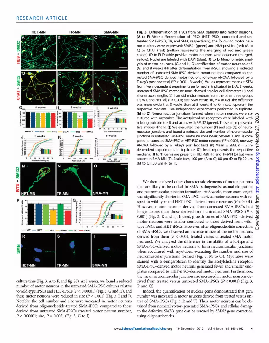
culture time (Fig. 3, A to F, and fig. S8). At 8 weeks, we found a reducednumber of motor neurons in the untreated SMA-iPSC cultures relativeto wild-type iPSCs and HET-iPSCs (P < 0.00001) (Fig. 3, G and H), andthese motor neurons were reduced in size (P < 0.001) (Fig. 3, I and J).Notably, the cell number and size were increased in motor neuronsderived from oligonucleotide-treated SMA-iPSCs compared to thosederived from untreated SMA-iPSCs (treated motor neuron number,P < 0.00001; size, P = 0.002) (Fig. 3, G to J).
www.ScienceTr
We then analyzed other characteristic elements of motor neuronsthat are likely to be critical in SMA pathogenesis: axonal elongationand neuromuscular junction formation. At 8 weeks, mean axon lengthwas significantly shorter in SMA-iPSC–derived motor neurons with re-spect to wild-type and HET-iPSC–derived motor neurons (P < 0.001).However, motor neurons derived from corrected SMA-iPSCs hadlonger axons than those derived from untreated SMA-iPSCs (P <0.001) (Fig. 3, K and L). Indeed, growth cones of SMA-iPSC–derivedmotor neurons were smaller compared to those derived from wild-type iPSCs and HET-iPSCs. However, after oligonucleotide correctionof SMA-iPSCs, we observed an increase in size of the motor neuronsderived from them (P < 0.001, treated versus untreated SMA motorneurons). We analyzed the difference in the ability of wild-type andSMA-iPSC–derived motor neurons to form neuromuscular junctionswhen cocultured with myotubes, evaluating the number and size ofneuromuscular junctions formed (Fig. 3, M to O). Myotubes werestained with a-bungarotoxin to identify the acetylcholine receptor.SMA-iPSC–derived motor neurons generated fewer and smaller end-plates compared to HET-iPSC–derived motor neurons. Furthermore,the mean neuromuscular junction size increased in motor neurons de-rived from treated versus untreated SMA-iPSCs (P < 0.001) (Fig. 3,P and Q).
Indeed, the quantification of nuclear gems demonstrated that gemnumber was increased in motor neurons derived from treated versus un-treated SMA-iPSCs (Fig. 3, R and T). Thus, motor neurons can be ob-tained from nonviral vector–generated SMA-iPSCs, and cellular damageto the defective SMN1 gene can be rescued by SMN2 gene correctionusing oligonucleotides.
Fig. 3. Differentiation of iPSCs from SMA patients into motor neurons.(A to F) After differentiation of iPSCs (HET-iPSCs, corrected and un-treated SMA-iPSCs, TR, and SMA, respectively), the following motor neu-ron markers were expressed: SMI32- (green) and HB9-positive (red) (A toC) or ChAT (red) (yellow represents the merging of red and greencolors). (D to F) Double-positive motor neurons were observed (merged,yellow). Nuclei are labeled with DAPI (blue). (G to L) Morphometric anal-ysis of motor neurons. (G and H) Quantification of motor neurons at 5(G) and 8 weeks (H) after differentiation from iPSCs, showing a reducednumber of untreated SMA-iPSC–derived motor neurons compared to cor-rected SMA-iPSC–derived motor neurons (one-way ANOVA followed by aTukey’s post hoc test) (*P < 0.001, 8 weeks). Values represent means ± SEMfrom five independent experiments performed in triplicate. (I to L) At 8 weeks,untreated SMA-iPSC motor neurons showed smaller cell diameters (J) andshorter axon lengths (L) than did motor neurons from the other three groupsTR, WT, and HET (all, P < 0.001; size: SMA versus TR, P = 0.002). The differencewas more evident at 8 weeks than at 5 weeks (I to K). Insets represent therespective medians. Five independent experiments performed in triplicate.(M to O) Neuromuscular junctions formed when motor neurons were co-cultured with myotubes. The acetylcholine receptors were labeled witha-bungarotoxin (red) and axons with SMI32 (green). These are representa-tive images. (P and Q) We evaluated the number (P) and size (Q) of neuro-muscular junctions and found a reduced size and number of neuromuscularjunctions in untreated SMA-iPSC motor neurons (SMA; patients 1 and 2) com-pared to corrected SMA-iPSC or HET-iPSC motor neurons (*P < 0.001, one-wayANOVA followed by a Tukey’s post hoc test). (P) Mean ± SEM, n = 5 in-dependent experiments in triplicate. (Q) Inset represents the respectivemedians. (R to T) Gems are present in HET-MN (R) and TR-MN (S) but wereabsent in SMA-MN (T). Scale bars, 100 mm (A to C); 80 mm (D to F); 20 mm(M to O); 50 mm (R to T).
anslationalMedicine.org 19 December 2012 Vol 4 Issue 165 165ra162 4
R E S EARCH ART I C L E
on
Mar
ch 2
7, 2
013
stm
.sci
ence
mag
.org
Dow
nloa
ded
from
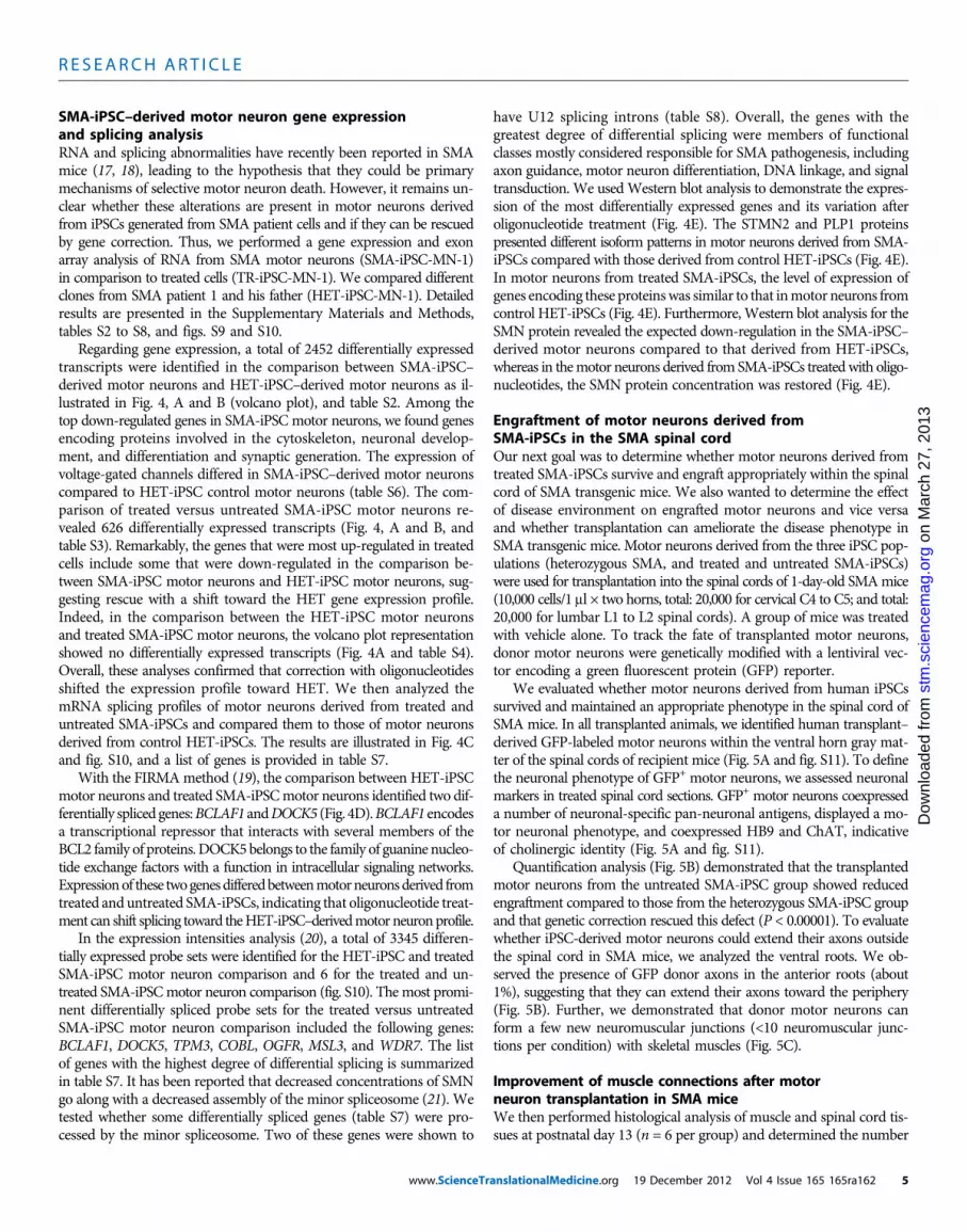
SMA-iPSC–derived motor neuron gene expressionand splicing analysisRNA and splicing abnormalities have recently been reported in SMAmice (17, 18), leading to the hypothesis that they could be primarymechanisms of selective motor neuron death. However, it remains un-clear whether these alterations are present in motor neurons derivedfrom iPSCs generated from SMA patient cells and if they can be rescuedby gene correction. Thus, we performed a gene expression and exonarray analysis of RNA from SMA motor neurons (SMA-iPSC-MN-1)in comparison to treated cells (TR-iPSC-MN-1). We compared differentclones from SMA patient 1 and his father (HET-iPSC-MN-1). Detailedresults are presented in the Supplementary Materials and Methods,tables S2 to S8, and figs. S9 and S10.
Regarding gene expression, a total of 2452 differentially expressedtranscripts were identified in the comparison between SMA-iPSC–derived motor neurons and HET-iPSC–derived motor neurons as il-lustrated in Fig. 4, A and B (volcano plot), and table S2. Among thetop down-regulated genes in SMA-iPSC motor neurons, we found genesencoding proteins involved in the cytoskeleton, neuronal develop-ment, and differentiation and synaptic generation. The expression ofvoltage-gated channels differed in SMA-iPSC–derived motor neuronscompared to HET-iPSC control motor neurons (table S6). The com-parison of treated versus untreated SMA-iPSC motor neurons re-vealed 626 differentially expressed transcripts (Fig. 4, A and B, andtable S3). Remarkably, the genes that were most up-regulated in treatedcells include some that were down-regulated in the comparison be-tween SMA-iPSC motor neurons and HET-iPSC motor neurons, sug-gesting rescue with a shift toward the HET gene expression profile.Indeed, in the comparison between the HET-iPSC motor neuronsand treated SMA-iPSC motor neurons, the volcano plot representationshowed no differentially expressed transcripts (Fig. 4A and table S4).Overall, these analyses confirmed that correction with oligonucleotidesshifted the expression profile toward HET. We then analyzed themRNA splicing profiles of motor neurons derived from treated anduntreated SMA-iPSCs and compared them to those of motor neuronsderived from control HET-iPSCs. The results are illustrated in Fig. 4Cand fig. S10, and a list of genes is provided in table S7.
With the FIRMAmethod (19), the comparison between HET-iPSCmotor neurons and treated SMA-iPSCmotor neurons identified two dif-ferentially spliced genes:BCLAF1 andDOCK5 (Fig. 4D).BCLAF1 encodesa transcriptional repressor that interacts with several members of theBCL2 family of proteins.DOCK5belongs to the family of guanine nucleo-tide exchange factors with a function in intracellular signaling networks.Expressionof these twogenesdifferedbetweenmotorneuronsderived fromtreated and untreated SMA-iPSCs, indicating that oligonucleotide treat-ment can shift splicing toward theHET-iPSC–derivedmotor neuronprofile.
In the expression intensities analysis (20), a total of 3345 differen-tially expressed probe sets were identified for the HET-iPSC and treatedSMA-iPSC motor neuron comparison and 6 for the treated and un-treated SMA-iPSCmotor neuron comparison (fig. S10). The most promi-nent differentially spliced probe sets for the treated versus untreatedSMA-iPSC motor neuron comparison included the following genes:BCLAF1, DOCK5, TPM3, COBL, OGFR, MSL3, and WDR7. The listof genes with the highest degree of differential splicing is summarizedin table S7. It has been reported that decreased concentrations of SMNgo along with a decreased assembly of the minor spliceosome (21). Wetested whether some differentially spliced genes (table S7) were pro-cessed by the minor spliceosome. Two of these genes were shown to
www.ScienceTr
have U12 splicing introns (table S8). Overall, the genes with thegreatest degree of differential splicing were members of functionalclasses mostly considered responsible for SMA pathogenesis, includingaxon guidance, motor neuron differentiation, DNA linkage, and signaltransduction. We usedWestern blot analysis to demonstrate the expres-sion of the most differentially expressed genes and its variation afteroligonucleotide treatment (Fig. 4E). The STMN2 and PLP1 proteinspresented different isoform patterns in motor neurons derived from SMA-iPSCs compared with those derived from control HET-iPSCs (Fig. 4E).In motor neurons from treated SMA-iPSCs, the level of expression ofgenes encoding these proteinswas similar to that inmotor neurons fromcontrol HET-iPSCs (Fig. 4E). Furthermore,Western blot analysis for theSMN protein revealed the expected down-regulation in the SMA-iPSC–derived motor neurons compared to that derived from HET-iPSCs,whereas in themotor neurons derived fromSMA-iPSCs treatedwith oligo-nucleotides, the SMN protein concentration was restored (Fig. 4E).
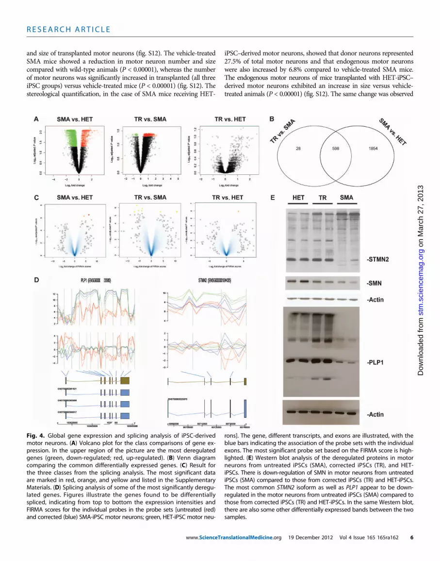
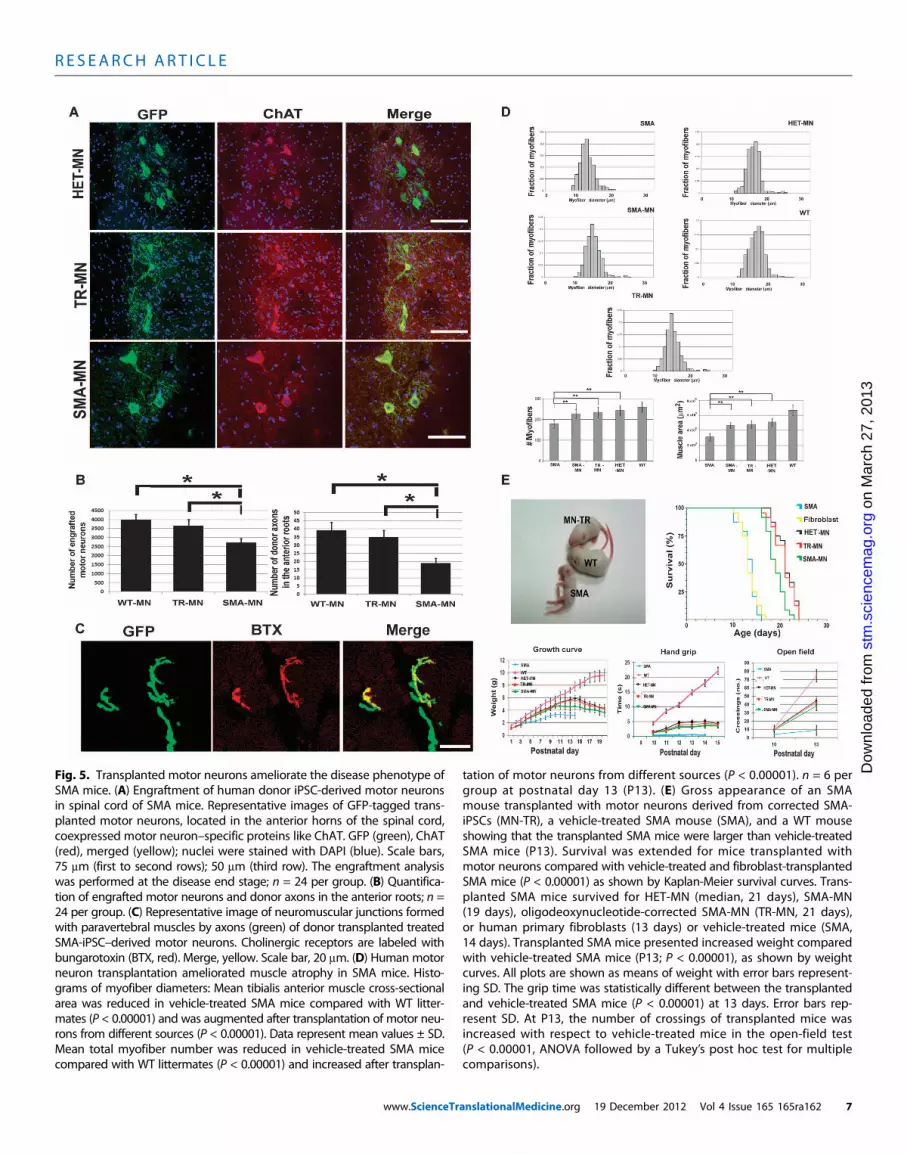
Engraftment of motor neurons derived fromSMA-iPSCs in the SMA spinal cordOur next goal was to determine whether motor neurons derived fromtreated SMA-iPSCs survive and engraft appropriately within the spinalcord of SMA transgenic mice. We also wanted to determine the effectof disease environment on engrafted motor neurons and vice versaand whether transplantation can ameliorate the disease phenotype inSMA transgenic mice. Motor neurons derived from the three iPSC pop-ulations (heterozygous SMA, and treated and untreated SMA-iPSCs)were used for transplantation into the spinal cords of 1-day-old SMAmice(10,000 cells/1 ml × two horns, total: 20,000 for cervical C4 to C5; and total:20,000 for lumbar L1 to L2 spinal cords). A group of mice was treatedwith vehicle alone. To track the fate of transplanted motor neurons,donor motor neurons were genetically modified with a lentiviral vec-tor encoding a green fluorescent protein (GFP) reporter.
We evaluated whether motor neurons derived from human iPSCssurvived and maintained an appropriate phenotype in the spinal cord ofSMA mice. In all transplanted animals, we identified human transplant–derived GFP-labeled motor neurons within the ventral horn gray mat-ter of the spinal cords of recipient mice (Fig. 5A and fig. S11). To definethe neuronal phenotype of GFP+ motor neurons, we assessed neuronalmarkers in treated spinal cord sections. GFP+ motor neurons coexpresseda number of neuronal-specific pan-neuronal antigens, displayed a mo-tor neuronal phenotype, and coexpressed HB9 and ChAT, indicativeof cholinergic identity (Fig. 5A and fig. S11).
Quantification analysis (Fig. 5B) demonstrated that the transplantedmotor neurons from the untreated SMA-iPSC group showed reducedengraftment compared to those from the heterozygous SMA-iPSC groupand that genetic correction rescued this defect (P < 0.00001). To evaluatewhether iPSC-derived motor neurons could extend their axons outsidethe spinal cord in SMA mice, we analyzed the ventral roots. We ob-served the presence of GFP donor axons in the anterior roots (about1%), suggesting that they can extend their axons toward the periphery(Fig. 5B). Further, we demonstrated that donor motor neurons canform a few new neuromuscular junctions (<10 neuromuscular junc-tions per condition) with skeletal muscles (Fig. 5C).
Improvement of muscle connections after motorneuron transplantation in SMA miceWe then performed histological analysis of muscle and spinal cord tis-sues at postnatal day 13 (n = 6 per group) and determined the number
anslationalMedicine.org 19 December 2012 Vol 4 Issue 165 165ra162 5
R E S EARCH ART I C L E
and size of transplanted motor neurons (fig. S12). The vehicle-treatedSMA mice showed a reduction in motor neuron number and sizecompared with wild-type animals (P < 0.00001), whereas the numberof motor neurons was significantly increased in transplanted (all threeiPSC groups) versus vehicle-treated mice (P < 0.00001) (fig. S12). Thestereological quantification, in the case of SMA mice receiving HET-
www.ScienceTr
iPSC–derived motor neurons, showed that donor neurons represented27.5% of total motor neurons and that endogenous motor neuronswere also increased by 6.8% compared to vehicle-treated SMA mice.The endogenous motor neurons of mice transplanted with HET-iPSC–derived motor neurons exhibited an increase in size versus vehicle-treated animals (P < 0.00001) (fig. S12). The same change was observed
on
Mar
ch 2
7, 2
013
stm
.sci
ence
mag
.org
Dow
nloa
ded
from
Fig. 4. Global gene expression and splicing analysis of iPSC-derivedmotor neurons. (A) Volcano plot for the class comparisons of gene ex-
rons]. The gene, different transcripts, and exons are illustrated, with theblue bars indicating the association of the probe sets with the individual
pression. In the upper region of the picture are the most deregulatedgenes (green, down-regulated; red, up-regulated). (B) Venn diagramcomparing the common differentially expressed genes. (C) Result forthe three classes from the splicing analysis. The most significant dataare marked in red, orange, and yellow and listed in the SupplementaryMaterials. (D) Splicing analysis of some of the most significantly deregu-lated genes. Figures illustrate the genes found to be differentiallyspliced, indicating from top to bottom the expression intensities andFIRMA scores for the individual probes in the probe sets [untreated (red)and corrected (blue) SMA-iPSC motor neurons; green, HET-iPSC motor neu-
exons. The most significant probe set based on the FIRMA score is high-lighted. (E) Western blot analysis of the deregulated proteins in motorneurons from untreated iPSCs (SMA), corrected iPSCs (TR), and HET-iPSCs. There is down-regulation of SMN in motor neurons from untreatediPSCs (SMA) compared to those from corrected iPSCs (TR) and HET-iPSCs.The most common STMN2 isoform as well as PLP1 appear to be down-regulated in the motor neurons from untreated iPSCs (SMA) compared tothose from corrected iPSCs (TR) and HET-iPSCs. In the same Western blot,there are also some other differentially expressed bands between the twosamples.
anslationalMedicine.org 19 December 2012 Vol 4 Issue 165 165ra162 6
R E S EARCH ART I C L E
on
Mar
ch 2
7, 2
013
stm
.sci
ence
mag
.org
Dow
nloa
ded
from
Fig. 5. Transplanted motor neurons ameliorate the disease phenotype ofSMA mice. (A) Engraftment of human donor iPSC-derived motor neurons
tation of motor neurons from different sources (P < 0.00001). n = 6 pergroup at postnatal day 13 (P13). (E) Gross appearance of an SMA
in spinal cord of SMA mice. Representative images of GFP-tagged trans-planted motor neurons, located in the anterior horns of the spinal cord,coexpressed motor neuron–specific proteins like ChAT. GFP (green), ChAT(red), merged (yellow); nuclei were stained with DAPI (blue). Scale bars,75 mm (first to second rows); 50 mm (third row). The engraftment analysiswas performed at the disease end stage; n = 24 per group. (B) Quantifica-tion of engrafted motor neurons and donor axons in the anterior roots; n =24 per group. (C) Representative image of neuromuscular junctions formedwith paravertebral muscles by axons (green) of donor transplanted treatedSMA-iPSC–derived motor neurons. Cholinergic receptors are labeled withbungarotoxin (BTX, red). Merge, yellow. Scale bar, 20 mm. (D) Human motorneuron transplantation ameliorated muscle atrophy in SMA mice. Histo-grams of myofiber diameters: Mean tibialis anterior muscle cross-sectionalarea was reduced in vehicle-treated SMA mice compared with WT litter-mates (P < 0.00001) and was augmented after transplantation of motor neu-rons from different sources (P < 0.00001). Data represent mean values ± SD.Mean total myofiber number was reduced in vehicle-treated SMA micecompared with WT littermates (P < 0.00001) and increased after transplan-
www.ScienceTr
mouse transplanted with motor neurons derived from corrected SMA-iPSCs (MN-TR), a vehicle-treated SMA mouse (SMA), and a WT mouseshowing that the transplanted SMA mice were larger than vehicle-treatedSMA mice (P13). Survival was extended for mice transplanted withmotor neurons compared with vehicle-treated and fibroblast-transplantedSMA mice (P < 0.00001) as shown by Kaplan-Meier survival curves. Trans-planted SMA mice survived for HET-MN (median, 21 days), SMA-MN(19 days), oligodeoxynucleotide-corrected SMA-MN (TR-MN, 21 days),or human primary fibroblasts (13 days) or vehicle-treated mice (SMA,14 days). Transplanted SMA mice presented increased weight comparedwith vehicle-treated SMA mice (P13; P < 0.00001), as shown by weightcurves. All plots are shown as means of weight with error bars represent-ing SD. The grip time was statistically different between the transplantedand vehicle-treated SMA mice (P < 0.00001) at 13 days. Error bars rep-resent SD. At P13, the number of crossings of transplanted mice wasincreased with respect to vehicle-treated mice in the open-field test(P < 0.00001, ANOVA followed by a Tukey’s post hoc test for multiplecomparisons).
anslationalMedicine.org 19 December 2012 Vol 4 Issue 165 165ra162 7
R E S EARCH ART I C L E
on
Mar
ch 2
7, 2
013
stm
.sci
ence
mag
.org
Dow
nloa
ded
from
in animals transplanted with motor neurons from treated and un-treated SMA-iPSCs versus vehicle control (P < 0.00001). Thus, thetransplantation of human iPSC-derived motor neurons induced aneuroprotective effect relative to vehicle in SMA mice. However, thedegree of this improvement was in proportion to the rate of engraft-ment and thus was higher for motor neurons derived from HET-iPSCs and treated SMA-iPSCs compared to those derived fromuntreated SMA-iPSCs.
Indeed, motor neuron transplantation resulted in a statisticallysignificant increase in total muscle area (P < 0.00001) and mean myo-fiber diameter (P < 0.00001; Fig. 5D and fig. S13). These muscle areachanges were observed also in the case of mice transplanted with mo-tor neurons from treated and untreated SMA-iPSCs (P < 0.00001 versusvehicle-treated).
Amelioration of the disease phenotype and life extension aftertransplantation of iPSC-derived motor neurons in SMA miceWe determined the functional efficacy of motor neuron transplanta-tion. After transplant, the physical appearance of the SMA mice wasimproved in comparison to that of vehicle-treated SMA mice (Fig. 5E,upper left panel, and movie S1). All mice receiving motor neuron trans-plants showed an increase in body weight that was statistically differ-ent at 13 days of age relative to vehicle-treated SMA mice (HET-iPSCmotor neurons versus vehicle, untreated SMA-iPSC motor neuronsversus vehicle, and treated SMA-iPSC motor neurons versus vehicle:P < 0.00001; Fig. 5E, bottom left panel).
To investigate whether transplanted motor neurons can rescue neuro-muscular functions in SMA animals, we tested transplanted and con-trol mice with the grip and open-field assays (22, 23). At 13 days ofage, all three groups of transplanted SMA mice presented stableperformance on the grip test for a few seconds, whereas nontrans-planted SMA mice could not perform the task (HET-iPSC motorneuron transplant versus vehicle, untreated SMA-iPSC motor neu-ron transplant versus vehicle, and untreated SMA-iPSC motor neu-ron transplant versus vehicle: P < 0.00001) (Fig. 5E, bottom centerpanel). In addition, all motor neuron–transplanted SMA mice showedsome locomotor activity and maintained some exploratory behaviorcompared to vehicle-treated SMA mice at 13 days of age (HET-iPSCmotor neuron transplant versus vehicle, untreated SMA-iPSC motorneuron transplant versus vehicle, and untreated SMA-iPSCmotor neu-ron transplant versus vehicle: P < 0.00001) (Fig. 5E, bottom right panel,and movie S1).
Mice that received motor neurons had improved survival rates whencompared to their vehicle-treated counterparts. Figure 5E (upper rightpanel) illustrates the Kaplan-Meier survival curves. The median lifespans of SMA mice receiving motor neuron transplants were 21 (HET-iPSC-MN), 19 (SMA-iPSC-MN), and 21 (treated SMA-iPSC-MN) days,whereas for vehicle-treated SMAmice, it was 14 days (n = 24 per group)(HET-iPSC, and treated and untreated SMA-iPSC motor neuron trans-plant versus vehicle, P < 0.00001). The maximum survival period observedwas 24 days for mice receiving HET-iPSC motor neuron transplantsand 16 days for vehicle-treated SMA mice. Survival was increased by7 days for mice receiving HET-iPSC and treated SMA-iPSC motor neu-ron transplants, representing a gain of 50% over the lifetime. Comparedto the HET-iPSCmotor neuron–transplanted group, the untreated SMA-iPSC motor neuron–transplanted group showed a slightly reducedsurvival (P = 0.00027), whereas the treated SMA-iPSC motor neuron–transplanted group showed increased survival compared to the untreated
www.ScienceTr
group (P = 0.0018). SMA mice transplanted with human primary fibro-blasts presented a survival trend similar to vehicle-treated SMA mice(median 13 days), demonstrating that the effect of motor neurontransplantation was specific (HET-iPSC motor neurons versus fibro-blasts, P < 0.00001). Thus, motor neuron transplantation provided asurvival benefit in an SMA mouse model.
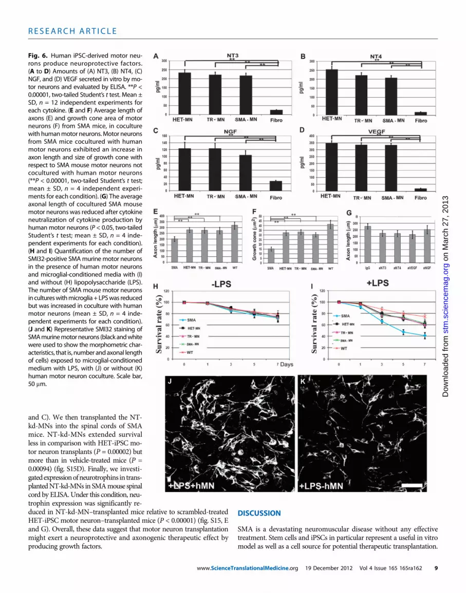
Production of neuroprotective factors by transplantedhuman motor neuronsTo analyze possible mechanisms for the observed improvement inSMA mice after transplantation, we evaluated whether human motorneurons derived from SMA-iPSCs, treated SMA-iPSCs, or HET-iPSCsby themselves can produce factors with a neuroprotective role on mousemotor neurons (Fig. 6, A to D, and fig. S14). We found that all of thehuman motor neurons release growth factors including neurotrophin3 (NT3), NT4, vascular endothelial growth factor (VEGF), and nervegrowth factor (NGF) (Fig. 6). They expressed moderate concentrationsof ciliary neurotrophic factor (CNTF) and low concentrations of brain-derived neurotrophic factor (BDNF) and glial-derived neurotrophicfactor (GDNF) (Fig. 6).
To analyze the effect of donor human motor neurons on SMA patho-genesis, we used a coculture system with a bottom layer of primarymotor neurons from SMA mice and a top layer of human motor neu-rons. As previously described, SMA mouse primary motor neuronsalone presented a significant reduction in axon length relative to wild-type mouse primary motor neurons (P < 0.00001) (Fig. 3I). When SMAmouse primary motor neurons were cocultured in the presence of hu-man motor neurons, axon length increased (Fig. 6E). Indeed, growthcones of SMN-deficient motor neurons from SMA mice were signif-icantly smaller than those of HET-iPSC–derived motor neurons (P <0.00001). Notably, we observed increased growth cone size for SMAmouse motor neurons after coculture with human motor neurons (P <0.00001) (Fig. 6F). We hypothesized that up-regulation of neurotrophinexpression may contribute to axon length enhancement. To test thishypothesis, we neutralized relevant cytokines individually or in combi-nation by adding neutralizing antibodies to the motor neuron culturemedia. This treatment significantly reduced axon length for SMAmouse motor neurons (P < 0.05) (Fig. 6G). To study the effects of hu-man motor neurons on neuronal survival in a neurotoxic environment,primary SMA mouse motor neurons were exposed to microglial-conditioned media or LPS-activated microglial-conditioned mediain the presence of human motor neurons (Fig. 6, H and I). The find-ings demonstrated that human motor neurons secrete active neuro-protective factors that promote SMA mouse motor neurons in aninflammatory/toxic environment and enhance axonal elongationin vitro (Fig. 6, J and K).
The expression of neurotrophins by human motor neurons trans-planted into the spinal cords of SMAmice, as evaluated by enzyme-linkedimmunosorbent assay (ELISA), demonstrated increased concentra-tions of neurotrophins when compared to vehicle-treated or fibroblast-transplanted SMA mouse controls (P < 0.00001) (fig. S14). To providea mechanistic demonstration of the therapeutic role of neurotrophinexpression by human motor neurons, we transplanted human motorneurons in which the relevant neurotrophin genes (NT3, NT4, andVEGF) were knocked down using short hairpin RNAs (shRNAs).These silenced cells are designated here as NT-kd-MNs. The ELISAanalysis revealed a ≈70% reduction in neurotrophins compared withmotor neurons transduced with nonsense control shRNAs (fig. S15, A
anslationalMedicine.org 19 December 2012 Vol 4 Issue 165 165ra162 8
R E S EARCH ART I C L E
on
Mar
ch 2
7, 2
013
stm
.sci
ence
mag
.org
Dow
nloa
ded
from
and C). We then transplanted the NT-kd-MNs into the spinal cords of SMAmice. NT-kd-MNs extended survivalless in comparison with HET-iPSC mo-tor neuron transplants (P = 0.00002) butmore than in vehicle-treated mice (P =0.00094) (fig. S15D). Finally, we investi-gatedexpressionofneurotrophins in trans-plantedNT-kd-MNs in SMAmouse spinalcord by ELISA. Under this condition, neu-trophin expression was significantly re-
duced in NT-kd-MN–transplanted mice relative to scrambled-treatedHET-iPSC motor neuron–transplanted mice (P < 0.00001) (fig. S15, Eand G). Overall, these data suggest that motor neuron transplantationmight exert a neuroprotective and axonogenic therapeutic effect byproducing growth factors.www.ScienceTr
DISCUSSION
SMA is a devastating neuromuscular disease without any effectivetreatment. Stem cells and iPSCs in particular represent a useful in vitromodel as well as a cell source for potential therapeutic transplantation.
Fig. 6. Human iPSC-derived motor neu-rons produce neuroprotective factors.(A to D) Amounts of (A) NT3, (B) NT4, (C)NGF, and (D) VEGF secreted in vitro by mo-tor neurons and evaluated by ELISA. **P <0.00001, two-tailed Student’s t test. Mean ±SD, n = 12 independent experiments foreach cytokine. (E and F) Average length ofaxons (E) and growth cone area of motorneurons (F) from SMA mice, in coculturewith humanmotor neurons.Motor neuronsfrom SMA mice cocultured with humanmotor neurons exhibited an increase inaxon length and size of growth cone withrespect to SMA mouse motor neurons notcocultured with human motor neurons(**P < 0.00001, two-tailed Student’s t test;mean ± SD, n = 4 independent experi-ments for each condition). (G) The averageaxonal length of cocultured SMA mousemotor neurons was reduced after cytokineneutralization of cytokine production byhumanmotor neurons (P < 0.05, two-tailedStudent’s t test; mean ± SD, n = 4 inde-pendent experiments for each condition).(H and I) Quantification of the number ofSMI32-positive SMAmurinemotor neuronsin the presence of human motor neuronsand microglial-conditioned media with (I)and without (H) lipopolysaccharide (LPS).The number of SMAmouse motor neuronsin cultureswithmicroglia+ LPSwas reducedbut was increased in coculture with humanmotor neurons (mean ± SD, n = 4 inde-pendent experiments for each condition).(J and K) Representative SMI32 staining ofSMAmurinemotor neurons (black andwhitewere used to show themorphometric char-acteristics, that is, number andaxonal lengthof cells) exposed to microglial-conditionedmedium with LPS, with (J) or without (K)human motor neuron coculture. Scale bar,50 mm.
anslationalMedicine.org 19 December 2012 Vol 4 Issue 165 165ra162 9
R E S EARCH ART I C L E
on
Mar
ch 2
7, 2
013
stm
.sci
ence
mag
.org
Dow
nloa
ded
from
To translate reprogramming technology from basic science towardclinical application, efficient derivation of human patient iPSCs thatare free of foreign or chemical elements is crucial.
Here, we derived viral vector– and transgene-free human iPSCsfrom SMA patients using oriP/EBNA1-based vectors (12). Ebert et al.(11) previously described generating SMA-iPSCs and motor neuronsusing viral methods. Our work represents an advance because weachieved generation and characterization of SMA patient–specific mo-tor neurons without genetic modification, thus increasing the possi-bility of applying these cells in basic research and transplantationregimens. Exploiting the full therapeutic potential of iPSCs will requireprecise and safe strategies for genetic modification in the case of theuse of autologous cells from patients. In addition, four independentgroups have recently described a complete rescue of the disease pheno-type in SMAmice after intravenous injection of SMN-encoding adeno-associated virus (24–27), suggesting that cell and gene therapy are possiblycomplementary therapeutic strategies.
We used oligonucleotides to induce a permanent nonviral geneticcorrection of SMA-iPSCs such that SMN2 was converted into SMN1by modifying a single base in exon 7. We demonstrate a genetic mod-ification of human iPSCs from SMA patients with this technique. Thisstrategy is specific, overcomes the limits of repeated treatment, doesnot introduce exogenous genes, and can be applied for other humangenetic diseases. Furthermore, the SMN protein defect seems to affectmotor neurons selectively. The use of SMA-iPSC–derived motor neu-rons described here can contribute to knowledge about the pathogenicmechanisms of SMA. We note that although we achieved a high pu-rity of motor neurons from iPSCs, other cell populations were presentat modest levels. In any case, our model can reproduce some aspect ofSMA disease and potentially may be useful for in vitro drug screening.
We observed specific disease effects of the SMNdefect on SMAmotorneurons including a reduction inmotor neuron survival and in their size,in line with that observed previously (11), although to a lesser degree.The centrifugation protocol used heremight have selected for a strongermotor neuron population, resulting in overall improved results relativeto those previously described. We also detected a reduction in axonalgrowth and neuromuscular junction formation in the motor neuronsderived from SMA-iPSCs relative to control HET-iPSCs. Our data sup-port the idea that more than one manifestation of motor neuron dysfunc-tion plays a role in SMA, including a reduction in motor neuron survivaland size, aswell as in axonal growth andneuromuscular junction forma-tion. Notably, we demonstrated that gene correction by converting SMN2into SMN1 using oligonucleotides rescued neuropathological featuresof SMA in motor neurons and correlated with SMN expression.
We performed a detailed evaluation of transcriptional changes inthe SMA-iPSC motor neurons in comparison to HET-iPSC and treatedSMA-iPSC motor neurons. Our analysis identified several pathwaysthat might shed light on SMA pathogenesis and its selectivity for mo-tor neurons, and disease initiation and progression. We detected alter-ations in a subset of genes involved in RNAmetabolism, motor neurondevelopment, and axonal guidance. In particular, the expression ofvoltage-gated channels was altered in SMA-iPSC–derived motor neurons.A relatively restricted number of genes showed different splicing profilesin SMA-iPSC– versus HET-iPSC–derived motor neurons. Zhang et al.(17) reported widespread splicing abnormalities in SMA mouse tis-sues, including the spinal cord. Bäumer et al. (18) indicated that theseevents are a late occurrence in SMA and are therefore not likely tocontribute to early disease pathogenesis. Our results do not support
www.ScienceTra
the hypothesis that widespread splicing abnormalities are presentin motor neurons. Rather, they suggest the possibility that SMN defi-ciency critically affects splicing of one or a few transcripts and that thefew splicing changes observed in our motor neuron models can con-tribute to SMA pathogenesis. Indeed, the genes with the higher degreeof differential splicing belonged to classes of functions that are con-sidered to have primary involvement in SMA pathogenesis, such asaxon guidance, motor neuron differentiation, RNA metabolism, andsignal transduction. The SMN defect can alter the minor spliceosomeand, consequently, the genes that have U12 introns. Only two genes,DOCK5 and LPHN2, belong to this family. DOCK5 is involved in axonguidance and was a top differentially spliced gene identified in this work,warranting further analysis to define its role in SMA pathogenesis. Fi-nally, our experiments demonstrated that most genes differentiallyexpressed or spliced in motor neurons from SMA-iPSCs versus HET-iPSCs are shifted toward the HET pattern after gene correction, fur-ther supporting the efficacy of this gene repair strategy.
STMN2, a gene of particular interest from our analysis, is down-regulated and differentially spliced in SMA-iPSC versus HET-iPSC mo-tor neurons as demonstrated both at the RNA and at the protein levels.Furthermore, the expression pattern of STMN2 was shifted towardthe HET profile after oligonucleotide correction. The involvement ofSTMN2 in cytoskeleton metabolism and neuronal growth cone de-velopment and differentiation may be important in the context ofSMA pathogenesis.
Another interesting gene was PLP1, which was down-regulated inSMA-iPSC motor neurons, and this down-regulation was rescued af-ter oligonucleotide correction. PLP1 is expressed in nonmyelinatingneuronal cell types and in motor neurons, where it plays a role in vesic-ular metabolism. These aspects may be important in the context ofSMA pathogenesis and deserve further investigation.
Our in vivo experiments showed that iPSC-derived motor neuronscan survive and integrate into the spinal cord of SMA mice and ame-liorate the SMA type I phenotype. Donor-derived motor neurons cor-rectly engrafted in the anterior spinal cords of transplanted mice andmaintained a specific motor neuron phenotype. However, the SMA-iPSC motor neurons presented a slightly reduced survival and dimen-sion as well as a decreased propensity for axonal elongation. Theseresults suggest that SMA-iPSC motor neuron transplantation canproduce an “in vivo model” of disease. The pathological features ofSMA-iPSC motor neurons were rescued by genetic modification witholigonucleotides, opening the path to the possibility of autologous celltherapy. Transplanted SMA mice had a longer life span (about 50%)compared to vehicle-treated mice, an effect that was more pronouncedwith HET-iPSC and treated SMA-iPSC motor neurons than with un-treated SMA-iPSC motor neurons. However, even if the difference in thetherapeutic effect of wild-type motor neurons versus HET-iPSC and un-treated SMA-iPSC motor neurons is small in our experimental settings,over the long-term, the reduced survival of these motor neurons wouldbe problematic. We believe that ex vivo genetic correction of the SMA-iPSCs before transplantation would be important to obtain the maxi-mum therapeutic benefit from this approach.
Indeed, the positive effects of transplanted motor neurons ap-peared to be specific because they were not observed after transplan-tation of fibroblasts. The increased survival time of SMAmice we observedafter motor neuron cell transplantation, although limited with respectto other results like gene therapy, is noteworthy for two reasons: (i)The full potential of the motor neuron transplantation effect cannot
nslationalMedicine.org 19 December 2012 Vol 4 Issue 165 165ra162 10
R E S EARCH ART I C L E
on
Mar
ch 2
7, 2
013
stm
.sci
ence
mag
.org
Dow
nloa
ded
from
be fully appreciated considering the limited time this model allowedfor the formation of new neuromuscular junctions, and (ii) genetherapy, based on the experimental data available, seems to be mosteffective when applied in the very first stage of SMA (24–27). Thissuggests that a “window of opportunity” exists during the early stagesof SMA in which gene therapy can have therapeutic benefits, at leastin this experimental setting. The possibility of motor neuron replace-ment after the death of endogenous motor neurons is the ideal goalthat has to be pursued to treat already symptomatic SMA patients,although such a goal is still a long way in the future.
Motor neuron transplantation also ameliorated defects in neuro-muscular function compared to vehicle-treated SMA mice. This effectcorrelated with the number of donor-derived motor neurons, as wellas with the change in endogenous motor neurons that were increased inboth size and number, suggesting a neuroprotective effect of the trans-planted motor neurons. We previously demonstrated that transplanta-tion of murine neural stem cells, an undifferentiated population relativeto the human postmitotic motor neurons used here, has a beneficialeffect on motor neuron disease phenotypes in SMA mice (22, 23). Inour past experiments (22, 23), mouse neural stem cells committed to amotor neuronal fate improved survival of SMA mice more than trans-planting other cell types (undifferentiated cells, astrocytes, and fibro-blasts), supporting the notion that transplantation of cholinergicprecursors or motor neurons may be advantageous in the context ofSMA spinal cord pathology. Regarding the possibility of transplantingundifferentiated iPSCs, this strategy is associated with a high risk oftumor development. Several studies report that undifferentiated iPSCscould also form teratomas in both immunocompromised and immuno-competent recipient animals (28, 29). Thus, the elimination of undiffer-entiated cells must be pursued. In our study, we wanted to investigatepotential mechanisms that could account for the observed ameliorationof the SMA phenotype after motor neuron transplantation. The forma-tion of the few neuromuscular junctions likely contributed little to thephenotypic improvement. We demonstrated that human motor neu-rons derived from iPSC lines by themselves can produce several neuro-trophic factors with a putative neuroprotective role through paracrineaction on other motor neurons, as shown in the coculture experiments.Furthermore, we demonstrated that SMA spinal cords transplantedwith human motor neurons showed increased concentrations of VEGF,NGF, NT3, and NT4.
After preventing production of neurotrophins by shRNAs, the thera-peutic effect of motor neuron transplantation was reduced. These datasuggest that motor neuron transplantation exerts a neuroprotectiveand axonogenic therapeutic effect by producing growth factors thatmight have therapeutic value beyond motor neuron replacement. Ourexperiments underscore the fact that motor neurons ameliorate someof the effects of SMA pathogenesis by providing neurotrophic support.The benefits of this cell population, however, will not be fully exploiteduntil motor neurons can be stimulated to form functional neuromus-cular junctions with the appropriate muscle targets in an efficient way.Future studies are needed to examine experimental strategies, like com-bining cell and pharmacological approaches to promote axonal elon-gation, as we have previously described (30), with the aim of achievingfully functional motor neurons. Our study has demonstrated transplan-tation of motor neurons derived from iPSCs generated from humanSMA patients and their potential for ameliorating the SMA phenotype.Our findings may contribute to the development of cell therapies forSMA and other motor neuron diseases in the future.
www.ScienceTra
MATERIALS AND METHODS
See also the Supplementary Materials and Methods.
Reprogramming of human somatic cellsReprogramming of human skin fibroblasts with oriP/EBNA1-basedepisomal vectors was carried out by nucleofection of the combina-tions of episomal plasmids (NHDF kit VPD-1001 with U-20 program,Amaxa). We used vectors that have been previously described (1). Thecomplementary DNAs (cDNAs) for the open reading frames of thehuman genes OCT4, SOX2, NANOG, LIN28, c-Myc, and KLF4 werederived through direct PCR of human stem cell cDNA. We used twoplasmid combinations (19 and 6) (14). After transfection, fibroblasts(1.0 × 10⁶ cells per nucleofection) were plated onto 3 × 10–cm dishescovered with Matrigel (BD Biosciences) in fibroblast culture medium,which was changed every other day.
At 4 days from the transfection, we replaced the fibroblast culturemedium with human ES cell culture medium (mTeSR, Stemcell Tech-nologies Inc.) for 8 to 10 days. At day 18 after transfection, it waspossible to identify the first colonies with an iPSC-like morphology.Between days 18 and 20 after transfection, we stained one of the 3 ×10–cm dishes of reprogramming culture with alkaline phosphatase(Millipore) to identify the eventual presence of human iPSC colonies.Between days 25 and 30, we passed the other two 10-cm dishes to fresh10-cm Matrigel-covered dishes (1 ml each plate) at a ratio of 1:3. Wethen picked the iPSC colonies that were morphologically more similarto ES cells to further analyze and expand them. Efficiency of fibroblastreprogramming was about three to six colonies per 106 fibroblast cells,in line with that described previously (14).
Oligodeoxynucleotide transfectionA total of 500,000 cells were plated into 60-mm dishes and incubatedovernight. The following day, the cells were transfected with oligo-deoxynucleotide 75-T, targeted to the transcribed (T) strand of DNA.The oligodeoxynucleotides were previously designed so that the centernucleotide directed the base change (15). Different doses of oligodeoxy-nucleotide (3, 6, 10, or 20 mg/5 × 105 cells) were mixed with jetPRIME(Polyplus Transfection Inc.), and the complex was added directly tothe medium. The oligodeoxynucleotides were allowed to incorporateovernight, after which the medium was replaced. The cells were col-lected at different time points for DNA, RNA, and protein analysis (24,48, and 72 hours and 7 days). Transfection efficiencies were optimizedwith an oligodeoxynucleotide bearing a fluorescent tag (Cy3) as a tracerat different concentrations. A scrambled oligodeoxynucleotide sequencewas used as control in all experiments. The oligodeoxynucleotideswere produced by Sigma-Genosys. For generation of a clonal cell cul-ture, at 24 hours from oligodeoxynucleotide transfection (10 mg) as de-scribed above, the cells were harvested and reseeded at clonal density(1 cell per well in a 96-well plate), and clones were grown.We screenedthe clones for the presence of genetic correction by PCR (see below).
iPSC differentiation in motor neuronsWe generated spinal motor neurons using a multistage differentiationprotocol developed for human ES cells (16). For the production of mo-tor neurons from SMA-iPSCs, TR-iPSCs (patients 1 and 2), and HET-iPSCs, cells were plated with neuronal medium composed of Dulbecco’smodified Eagle’s medium/F12 (Gibco, Invitrogen), supplemented withMEM nonessential amino acids solution, N2, and heparin (2 mg/ml;
nslationalMedicine.org 19 December 2012 Vol 4 Issue 165 165ra162 11
R E S EARCH ART I C L E
on
Mar
ch 2
7, 2
013
stm
.sci
ence
mag
.org
Dow
nloa
ded
from
Sigma-Aldrich). After 10 days, we added retinoic acid (0.1 mM; Sigma-Aldrich) for neural caudalization. At day 17, we collected the pos-teriorized neuroectodermal cells. These clusters were then suspendedfor a week in the same medium with retinoic acid (0.1 mM) and SHH(100 to 200 ng/ml; R&D Systems Inc.). On day 24, we added othergrowth factors like BDNF, GDNF, and insulin-like growth factor-1(10 ng/ml; PeproTech).
Microarray analysis of iPSC expressionMicroarray expression analysis of iPSCs (SMA, HET), iPSC 19.9 fromthe J. Thomson Lab (14), and parental fibroblasts was carried out withhuman genome U133 Plus 2.0 GeneChip arrays (Affymetrix). Thisanalysis was performed by DNA Vision Laboratory. Total probe setswere 54,675, corresponding to 51,337 accession numbers as reportedin the hgu133plus2 annotation package. The GeneChip IVT ExpressKit and the GeneChip Hybridization wash and stain kit were usedwith 100 ng of total RNA that was mixed with 2 ml of 1:500,000 dilutedspikes. Synthesis of the first strand and the second strand of cDNAwas performed according to the kit instructions at 42°C, transcribed,and amplified into complementary RNA (cRNA). The cRNA was quan-tified by NanoDrop. Then, fragmentation of 12.5 mg of cRNA was per-formed and hybridized onto the GeneChip according to the Affymetrixprotocol. An Affymetrix RUO platform was used, and data were pro-cessed with the Affymetrix GCOS program. Quality control was per-formed based on Affymetrix quality control metrics. All chips werenormalized by the quantile method and background-corrected withrobust multiarray analysis (RMA) (14). After normalization, the probe-level data were then summarized with median polish to obtain theprobe set level measurement. We then collapsed 54,675 probe setsto 51,337 transcripts by keeping the average log intensity values forprobe sets that represent a common accession number. Hierarchicalcluster analyses were performed with 1 − PCC (Pearson correlation co-efficient) as the distance parameter. The maximum distance betweencluster members was used as the basis for merging lower-level clusters(complete linkage) into higher-level clusters. To assess the certainty ofthe existence of a cluster, we applied multiscale bootstrap resampling(10,000 bootstraps) to the hierarchical clustering of the 12 samples andcalculated P values of the hypotheses as well as bootstrap probabilitiesfor each cluster (14). Microarray data are available in the Gene Expres-sion Omnibus (GEO) database (GSE27206).
Genome-wide and splicing analysis in iPSC-derivedmotor neuronsWe then performed exon microarrays to determine whether therewere any changes in expression and splicing in SMA cells comparedto treated cells and unaffected cells. To do so, total RNA from isolatedmotor neurons, three SMA-iPSC–derived motor neuron samples (dif-ferent subclones), three unaffected cells, and three SMA-treated cells(different subclones), each prepared and processed separately, wereanalyzed with the Affymetrix human exon 1.0 ST microarray. Thisanalysis was performed by DNA Vision Laboratory according to theAffymetrix-recommended protocols. Affymetrix GeneChipWhole Tran-script Sense Target labeling array and GeneChip Hybridization washand stain kit were used: Ribosomal RNA was first reduced with theRiboMinus human/mouse kit (Invitrogen) starting from 1 mg of totalRNA. Subsequently, purified RNA was used as a template in the syn-thesis of double-stranded cDNA according to instructions providedwith the Affymetrix protocol. Double-stranded cDNA was in vitro–
www.ScienceTra
transcribed, producing cRNA. Finally, cRNA was newly retrotran-scribed (second cycle), and single-stranded cDNA obtained through thispassage was purified, quantified by NanoDrop, biotin-labeled, and frag-mented according to the manufacturers’ protocols. The hybridizationand scan protocols (Affymetrix RUO platform), suitable for HumanExon 1.0 ST microarrays, were provided by Affymetrix. RAW data wereacquired with Affymetrix GCOS software. The groups of interest werecompared with moderated t tests (20). Before adjustment for multiplecomparisons (31), we filtered out the less-variable transcripts based onthe interquartile range: A total of 8940 (3145 up-regulated and 5795down-regulated) transcripts were kept. Differentially expressed tran-scripts were defined as having an adjusted P value of <0.05.
The results are illustrated with volcano plots in which the most sig-nificant transcripts are located toward the top of the figure and thelargest fold changes are at the sides. The statistically differentially ex-pressed genes (with an adjusted P value of <0.05, that is, a −log10 P valueof >1.3) are shown in red (up-regulated) or green (down-regulated),respectively. We then analyzed the group of 8940 selected genes with adifferent statistical procedure. We calculated the ratio between all SMAsamples and controls, obtaining nine values from each probe, from whichwe evaluated the mean value among values included in the tails of thedistribution andwith a homogeneous log2 ratio (meaning log2 ratio of thenine ratios to be positive in at least four cases for the up-regulated genesand negative in at least four cases for the down-regulated genes, to have aregular trend of the average and excluding the casual extreme values).
The detection of alternative splicing was performed to identify iso-forms with the robust multichip analysis (FIRMA) method (19). Thismethod is based on the RMA normalization (32, 33). The final goal ofthe FIRMA method is the gene expression valuation of each samplethrough the setting of a linear additive model with the followingformula: log2(PMik) = ci + pk + eik, where ci is the chip effect forthe chip i; pk is the probe effect for probe k; log2(PMik) is log2 ofthe exact match intensity for the probe k on the chip i corrected andnormalized with the background; and eik is the remains for probe k onchip i. A sum value for the eik was evaluated for each probe set andused as scored. The differences in these scores are indicative of splicingphenomena. We have applied a moderated t test (20) to the FIRMAscores using the groups of interest. The results of each comparison areillustrated using two plots. The histograms illustrate the distribution ofthe adjustedP values. The individual adjustedP values are shown on thebottom of the figure as rungs, and the vertical dashed line represents the0.05 threshold. The volcano plot is generated with unadjusted P values.Microarray data are available in the GEO database (GSE27205).
Animal modelsThe triple-mutant SMA mouse harbors two transgenic alleles and atargeted mutant. The Tg(SMN2*D7)4299Ahmb allele is an SMA cDNAlacking exon 7, whereas the Tg(SMN2)89Ahmb allele is the entire hu-man SMN2 gene. Heterozygous Smn knockout mice with SMN2transgenes were bred to obtain homozygous mice for the knockoutSmn alleles (SMA mice, SMN2+/+;SmnD7+/+;mSmn−/−) [line 4299;FVB.Cg-Tg(SMN2*D7)4299Ahmb Tg(SMN2)89Ahmb Smn1tm1Msd].The mice were genotyped with a PCR assay on genomic DNA fromtail biopsies, as previously described (9).
All transgenic animals were obtained from the Jackson Laboratory.The in vivo experiments were approved by the University of Milan andMinistry of Health review boards, in agreement with U.S. NationalInstitutes of Health guidelines (34).
nslationalMedicine.org 19 December 2012 Vol 4 Issue 165 165ra162 12
R E S EARCH ART I C L E
on
Mar
ch 2
7, 2
013
stm
.sci
ence
mag
.org
Dow
nloa
ded
from
Cell transplantation in SMA miceBefore cell transplantation, motor neurons were cultured and collectedby gradient centrifugation as described and washed in phosphate-buffered saline (PBS). Human fibroblasts were used as control and wereharvested with trypsin and then washed in PBS. To obtain GFP-positivecells, we used a lentiviral-based TurboGFP-encoding vector accordingto the manufacturer’s instructions (Mission TurboGFP Control Vector,Sigma-Aldrich). Cell culture medium was changed the day after trans-fection and the following 2 to 3 days after transduction, washing outany possible contaminating plasmid. We also transfected the controlfibroblasts before transplantation. Under these conditions, we did notobserve any motor neuron labeling. This experiment served as controlfor leakage of the plasmid into the host motor neurons.
iPSC-purified motor neurons were used for transplantation into thespinal cords of 1-day-old SMA mice (10,000 cells/1 ml × two horns,total: 20,000 for cervical C4 to C5; and total: 20,000 for lumbar L1to L2 spinal cords) by direct injection as described (11, 19). There werefour experimental groups of mice: Group 1 (HET motor neuron) re-ceived transplanted HET-iPSC–derived motor neurons; group 2 (SMAmotor neuron) received SMA-iPSC–derived motor neurons; group 3(TR motor neuron) received genetically corrected SMAmotor neurons;and group 4 received vehicle (saline solution injection as mock trans-plantation, as the control group). Group 5 received human fibroblastsas an additional control group. Siblings were assigned equally in thegroups. Each group had the same number of males and females. The im-munosuppressor FK506 was administered intraperitoneally at 1.0 mg/kgto all animals in all groups for the entire length of the experiment.
These mice were monitored up to the end stage for neuromuscularfunction, survival, and histology of the human transplanted cell phe-notype. Another series of mice (treated and vehicle-treated SMA miceand wild type) were analyzed for histological quantification (for eachgroup per point: n = 6; 13 days).
Assessment of survival and phenotypeTransplanted and vehicle-treated SMA mice were assessed daily aftertransplantation for signs of disease. Observers were blinded to the treat-ments. The mice were sacrificed at the clinical endpoint, at which theyshowed problems in feeding, a clear downward progression (mice with30% weight reduction and severe paralysis), and breathing difficulties, asdescribed previously (22). Body weight was monitored daily. SMA micewere evaluated for grip strength as previously reported (22, 23). Theinvestigators performing the functional analysis were blind to the treat-ment group. Wild-type mice as well as treated and vehicle-treated SMAmice were evaluated to see how long they could sustain their weightholding onto a metal bar suspended in midair (between 10 and 15 daysof age). The data were analyzed by ANOVA followed by a Tukey’s posthoc test for multiple comparisons. Ambulatory function was investi-gated in an open-field test (23, 34). The apparatus was a wooden box(28 × 28 × 5 cm). The floor was partitioned into 16 equal squares of7 × 7 cm. The mice were located in the center of the open field andobserved individually. Each mouse was allowed to walk freely for 5 min,and the number of grids crossed was recorded.
Coculture of primary motor neurons of the SMA transgenicmice and human motor neuronsWe obtained primary motor neuron cultures from embryonic day12.5 SMA and wild-type mice as previously reported (22). The culturemedium was a motor neuron medium supplemented with a cocktail
www.ScienceTra
of trophic factors as described above. We carried out a coculture assayto separate the primary motor neurons from the motor neurons witha microporous membrane, as previously described (22, 23).
Enzyme-linked immunosorbent assayNT3, NT4, VEGF, NGF, CNTF, BDNF, GDNF, and TGF-a (transform-ing growth factor–a) secretion by MNs was determined with ELISAs, fol-lowing the manufacturer’s instructions (R&D Systems). Medium wasadded (1.5 × 10⁵ cells, 1.5 ml) and, 24 hours later, was removed for theELISA test (12 independent experiments for each cytokine).
The same cytokines were investigated by ELISA in the lumbar spi-nal cord (n = 6 for each condition) after transplantation. The data wereanalyzed with Student’s t tests.
SUPPLEMENTARY MATERIALS
www.sciencetranslationalmedicine.org/cgi/content/full/4/165/165ra162/DC1Materials and MethodsTextFig. S1. Scheme of the entire experimental design of the article.Fig. S2. Nonviral generation of iPSCs from fibroblasts of the SMA patient and his father andanother unrelated SMA patient.Fig. S3. Patient-specific iPSCs are pluripotent stem cells and can differentiate into threegerminal layers.Fig. S4. Genome-wide gene expression analysis of the reprogrammed cells.Fig. S5. Genetic conversion of SMN2 gene to SMN1 by the use of oligonucleotides in SMA-iPSC-2.Fig. S6. Scheme of differentiation of MNs from iPSCs.Fig. S7. Differentiation into MNs of iPSC-SMA-1.Fig. S8. Differentiation into MNs of iPSC-SMA-2.Fig. S9. Gene ontology cluster analysis.Fig. S10. Result of the iPSC-derived motor neuron splicing analysis.Fig. S11. Human MNs engrafted into the spinal cord of SMA mice.Fig. S12. Human MN transplantation increases MN size and number.Fig. S13. MN transplantation improved muscle trophism in SMA mice.Fig. S14. Human iPSC-derived MNs produce neuroprotective factors in vitro and in vivo.Fig. S15. Human MN transplantation increases MN size and number. Knocking down neurotrophinsreduced the neuroprotective action of MNs.Table S1. DNA fingerprinting analysis.Table S2. Gene expression profile of iPSC-SMA–derived motor neurons versus iPSC-WT–derivedmotor neurons.Table S3. Gene expression profile of iPSC-SMA–derived motor neurons versus iPSC-TR–derivedmotor neurons.Table S4. Gene expression profile of iPSC-WT–derived motor neurons versus iPSC-SMA–derivedmotor neurons.Table S5. Common genes among the three compared groups.Table S6. Expression and splicing levels of voltage-gated channels.Table S7. List of genes with the highest degree of differential splicing.Table S8. Expression levels of U12 genes with higher degree in splicing alteration.Movie S1. Motor neuron–transplanted mice (TR-MN) in comparison with wild-type mice (WT)and untreated mice (SMA).
REFERENCES AND NOTES
1. S. Lefebvre, L. Bürglen, S. Reboullet, O. Clermont, P. Burlet, L. Viollet, B. Benichou,C. Cruaud, P. Millasseau, M. Zeviani, D. Le Paslier, J. Frézal, D. Cohen, J. Weissenbach,A. Munnich, J. Melki, Identification and characterization of a spinal muscular atrophy-determining gene. Cell 80, 155–165 (1995).
2. D. D. Coovert, T. T. Le, P. E. McAndrew, J. Strasswimmer, T. O. Crawford, J. R. Mendell,S. E. Coulson, E. J. Androphy, T. W. Prior, A. H. Burghes, The survival motor neuronprotein in spinal muscular atrophy. Hum. Mol. Genet. 6, 1205–1214 (1997).
3. T. O. Crawford, C. A. Pardo, The neurobiology of childhood spinal muscular atrophy.Neurobiol. Dis. 3, 97–110 (1996).
nslationalMedicine.org 19 December 2012 Vol 4 Issue 165 165ra162 13
R E S EARCH ART I C L E
on
Mar
ch 2
7, 2
013
stm
.sci
ence
mag
.org
Dow
nloa
ded
from
4. T. L. Munsat, K. E. Davies, International SMA consortium meeting. (26-28 June 1992,Bonn, Germany). Neuromuscul. Disord. 2, 423–428 (1992).
5. C. L. Lorson, E. Hahnen, E. J. Androphy, B. Wirth, A single nucleotide in the SMN generegulates splicing and is responsible for spinal muscular atrophy. Proc. Natl. Acad. Sci. U.S.A.96, 6307–6311 (1999).
6. S. Lefebvre, P. Burlet, Q. Liu, S. Bertrandy, O. Clermont, A. Munnich, G. Dreyfuss, J. Melki,Correlation between severity and SMN protein level in spinal muscular atrophy. Nat. Genet.16, 265–269 (1997).
7. B. Schrank, R. Götz, J. M. Gunnersen, J. M. Ure, K. V. Toyka, A. G. Smith, M. Sendtner,Inactivation of the survival motor neuron gene, a candidate gene for human spinalmuscular atrophy, leads to massive cell death in early mouse embryos. Proc. Natl. Acad.Sci. U.S.A. 94, 9920–9925 (1997).
8. U. R. Monani, M. Sendtner, D. D. Coovert, D. W. Parsons, C. Andreassi, T. T. Le, S. Jablonka,B. Schrank, W. Rossoll, T. W. Prior, G. E. Morris, A. H. Burghes, The human centromericsurvival motor neuron gene (SMN2) rescues embryonic lethality in Smn−/− mice andresults in a mouse with spinal muscular atrophy. Hum. Mol. Genet. 9, 333–339 (2000).
9. T. T. Le, L. T. Pham, M. E. Butchbach, H. L. Zhang, U. R. Monani, D. D. Coovert, T. O. Gavrilina,L. Xing, G. J. Bassell, A. H. Burghes, SMND7, the major product of the centromeric survivalmotor neuron (SMN2) gene, extends survival in mice with spinal muscular atrophy andassociates with full-length SMN. Hum. Mol. Genet. 14, 845–857 (2005).
10. M. Stavarachi, P. Apostol, M. Toma, D. Cimponeriu, L. Gavrila, Spinal muscular atrophydisease: A literature review for therapeutic strategies. J. Med. Life 3, 3–9 (2010).
11. A. D. Ebert, J. Yu, F. F. Rose Jr., V. B. Mattis, C. L. Lorson, J. A. Thomson, C. N. Svendsen,Induced pluripotent stem cells from a spinal muscular atrophy patient. Nature 457,277–280 (2009).
12. J. Yu, M. A. Vodyanik, K. Smuga-Otto, J. Antosiewicz-Bourget, J. L. Frane, S. Tian, J. Nie,G. A. Jonsdottir, V. Ruotti, R. Stewart, I. I. Slukvin, J. A. Thomson, Induced pluripotentstem cell lines derived from human somatic cells. Science 318, 1917–1920 (2007).
13. K. Okita, T. Ichisaka, S. Yamanaka, Generation of germline-competent induced pluripotentstem cells. Nature 448, 313–317 (2007).
14. J. Yu, K. Hu, K. Smuga-Otto, S. Tian, R. Stewart, I. I. Slukvin, J. A. Thomson, Human inducedpluripotent stem cells free of vector and transgene sequences. Science 324, 797–801(2009).
15. D. DiMatteo, S. Callahan, E. B. Kmiec, Genetic conversion of an SMN2 gene to SMN1: Anovel approach to the treatment of spinal muscular atrophy. Exp. Cell Res. 314, 878–886(2008).
16. B. Y. Hu, S. C. Zhang, Differentiation of spinal motor neurons from pluripotent human stemcells. Nat. Protoc. 4, 1295–1304 (2009).
17. Z. Zhang, F. Lotti, K. Dittmar, I. Younis, L. Wan, M. Kasim, G. Dreyfuss, SMN deficiencycauses tissue-specific perturbations in the repertoire of snRNAs and widespread defectsin splicing. Cell 133, 585–600 (2008).
18. D. Bäumer, S. Lee, G. Nicholson, J. L. Davies, N. J. Parkinson, L. M. Murray, T. H. Gillingwater,O. Ansorge, K. E. Davies, K. Talbot, Alternative splicing events are a late feature of pathology ina mouse model of spinal muscular atrophy. PLoS Genet. 5, e1000773 (2009).
19. E. Purdom, K. M. Simpson, M. D. Robinson, J. G. Conboy, A. V. Lapuk, T. P. Speed, FIRMA:A method for detection of alternative splicing from exon array data. Bioinformatics 24,1707–1714 (2008).
20. G. K. Smyth, Linear models and empirical Bayes methods for assessing differential expressionin microarray experiments. Stat. Appl. Genet. Mol. Biol. 3, Article3 (2004).
21. N. Boulisfane, M. Choleza, F. Rage, H. Neel, J. Soret, R. Bordonné, Impaired minor tri-snRNPassembly generates differential splicing defects of U12-type introns in lymphoblastsderived from a type I SMA patient. Hum. Mol. Genet. 20, 641–648 (2011).
22. S. Corti, M. Nizzardo, M. Nardini, C. Donadoni, S. Salani, D. Ronchi, F. Saladino, A. Bordoni,F. Fortunato, R. Del Bo, D. Papadimitriou, F. Locatelli, G. Menozzi, S. Strazzer, N. Bresolin,G. P. Comi, Neural stem cell transplantation can ameliorate the phenotype of a mousemodel of spinal muscular atrophy. J. Clin. Invest. 118, 3316–3330 (2008).
23. S. Corti, M. Nizzardo, M. Nardini, C. Donadoni, S. Salani, D. Ronchi, C. Simone, M. Falcone,D. Papadimitriou, F. Locatelli, N. Mezzina, F. Gianni, N. Bresolin, G. P. Comi, Embryonicstem cell-derived neural stem cells improve spinal muscular atrophy phenotype in mice.Brain 133, 465–481 (2010).
24. K. D. Foust, X. Wang, V. L. McGovern, L. Braun, A. K. Bevan, A. M. Haidet, T. T. Le, P. R. Morales,M. M. Rich, A. H. Burghes, B. K. Kaspar, Rescue of the spinal muscular atrophy pheno-type in a mouse model by early postnatal delivery of SMN. Nat. Biotechnol. 28, 271–274(2010).
25. M. A. Passini, J. Bu, E. M. Roskelley, A. M. Richards, S. P. Sardi, C. R. O’Riordan, K. W. Klinger,L. S. Shihabuddin, S. H. Cheng, CNS-targeted gene therapy improves survival and motorfunction in a mouse model of spinal muscular atrophy. J. Clin. Invest. 120, 1253–1264(2010).
26. C. F. Valori, K. Ning, M. Wyles, R. J. Mead, A. J. Grierson, P. J. Shaw, M. Azzouz, Systemicdelivery of scAAV9 expressing SMN prolongs survival in a model of spinal muscular atrophy.Sci. Transl. Med. 2, 35ra42 (2010).
www.ScienceTra
27. E. Dominguez, T. Marais, N. Chatauret, S. Benkhelifa-Ziyyat, S. Duque, P. Ravassard, R. Carcenac,S. Astord, A. Pereira de Moura, T. Voit, M. Barkats, Intravenous scAAV9 delivery of a codon-optimized SMN1 sequence rescues SMA mice. Hum. Mol. Genet. 20, 681–693 (2011).
28. Y. Zhang, D. Wang, M. Chen, B. Yang, F. Zhang, K. Cao, Intramyocardial transplantation ofundifferentiated rat induced pluripotent stem cells causes tumorigenesis in the heart.PLoS One 6, e19012 (2011).
29. T. Yamashita, H. Kawai, F. Tian, Y. Ohta, K. Abe, Tumorigenic development of inducedpluripotent stem cells in ischemic mouse brain. Cell Transplant. 20, 883–891 (2011).
30. S. Corti, M. Nizzardo, M. Nardini, C. Donadoni, S. Salani, R. Del Bo, D. Papadimitriou,F. Locatelli, N. Mezzina, F. Gianni, N. Bresolin, G. P. Comi, Motoneuron transplantationrescues the phenotype of SMARD1 (spinal muscular atrophy with respiratory distresstype 1). J. Neurosci. 29, 11761–11771 (2009).
31. Y. Benjamini, Y. Hochberg, Controlling the false discovery rate: A practical and powerfulapproach to multiple testing. J. R. Statist. Soc. B 57, 289–300 (1995).
32. R. A. Irizarry, B. M. Bolstad, F. Collin, L. M. Cope, B. Hobbs, T. P. Speed, Summaries ofAffymetrix GeneChip probe level data. Nucleic Acids Res. 31, e15 (2003).
33. R. A. Irizarry, B. Hobbs, F. Collin, Y. D. Beazer-Barclay, K. J. Antonellis, U. Scherf, T. P. Speed,Exploration, normalization, and summaries of high density oligonucleotide array probelevel data. Biostatistics 4, 249–264 (2003).
34. S. Corti, F. Locatelli, D. Papadimitriou, R. Del Bo, M. Nizzardo, M. Nardini, C. Donadoni,S. Salani, F. Fortunato, S. Strazzer, N. Bresolin, G. P. Comi, Neural stem cells LewisX+CXCR4+modify disease progression in an amyotrophic lateral sclerosis model. Brain 130, 1289–1305(2007).
35. J. T. Dimos, K. T. Rodolfa, K. K. Niakan, L. M. Weisenthal, H. Mitsumoto, W. Chung, G. F. Croft,G. Saphier, R. Leibel, R. Goland, H. Wichterle, C. E. Henderson, K. Eggan, Induced pluripotentstem cells generated from patients with ALS can be differentiated into motor neurons. Science321, 1218–1221 (2008).
36. G. van der Steege, P. M. Grootscholten, P. van der Vlies, T. G. Draaijers, J. Osinga, J. M. Cobben,H. Scheffer, C. H. Buys, PCR-based DNA test to confirm clinical diagnosis of autosomal recessivespinal muscular atrophy. Lancet 345, 985–986 (1995).
37. C. Andreassi, C. Angelozzi, F. D. Tiziano, T. Vitali, E. De Vincenzi, A. Boninsegna, M. Villanova,E. Bertini, A. Pini, G. Neri, C. Brahe, Phenylbutyrate increases SMN expression in vitro:Relevance for treatment of spinal muscular atrophy. Eur. J. Hum. Genet. 12, 59–65 (2004).
38. M. Gingras, V. Gagnon, S. Minotti, H. D. Durham, F. Berthod, Optimized protocols for isolationof primary motor neurons, astrocytes and microglia from embryonic mouse spinalcord. J. Neurosci. Methods 163, 111–118 (2007).
39. T. Wada, M. Honda, I. Minami, N. Tooi, Y. Amagai, N. Nakatsuji, K. Aiba, Highly efficientdifferentiation and enrichment of spinal motor neurons derived from human and monkeyembryonic stem cells. PLoS One 4, e6722 (2009).
40. M. Nagai, D. B. Re, T. Nagata, A. Chalazonitis, T. M. Jessell, H. Wichterle, S. Przedborski,Astrocytes expressing ALS-linked mutated SOD1 release factors selectively toxic to motorneurons. Nat. Neurosci. 10, 615–622 (2007).
41. Q.Xiao,W. Zhao, D. R. Beers, A. A. Yen,W.Xie, J. S. Henkel, S. H. Appel,Mutant SOD1G93Amicrogliaare more neurotoxic relative to wild-type microglia. J. Neurochem. 102, 2008–2019 (2007).
42. P. A. Curmi, O. Gavet, E. Charbaut, S. Ozon, S. Lachkar-Colmerauer, V. Manceau, S. Siavoshian,A. Maucuer, A. Sobel, Stathmin and its phosphoprotein family: General properties, biochemicaland functional interaction with tubulin. Cell Struct. Funct. 24, 345–357 (1999).
43. G. Siegel, G. Obernosterer, R. Fiore, M. Oehmen, S. Bicker, M. Christensen, S. Khudayberdiev,P. F. Leuschner, C. J. Busch, C. Kane, K. Hübel, F. Dekker, C. Hedberg, B. Rengarajan, C. Drepper,H. Waldmann, S. Kauppinen, M. E. Greenberg, A. Draguhn, M. Rehmsmeier, J. Martinez,G. M. Schratt, A functional screen implicates microRNA-138-dependent regulation ofthe depalmitoylation enzyme APT1 in dendritic spine morphogenesis. Nat. Cell Biol. 11,705–716 (2009).
44. E. C. Jacobs, E. R. Bongarzone, C. W. Campagnoni, K. Kampf, A. T. Campagnoni, Soma-restricted products of the myelin proteolipid gene are expressed primarily in neurons inthe developing mouse nervous system. Dev. Neurosci. 25, 96–104 (2003).
45. T. Schmitt-John, C. Drepper, A. Mussmann, P. Hahn, M. Kuhlmann, C. Thiel, M. Hafner,A. Lengeling, P. Heimann, J. M. Jones, M. H. Meisler, H. Jockusch, Mutation of Vps54causes motor neuron disease and defective spermiogenesis in the wobbler mouse.Nat. Genet. 37, 1213–1215 (2005).
46. V. Swarup, J. P. Julien, ALS pathogenesis: Recent insights from genetics and mousemodels. Prog. Neuropsychopharmacol. Biol. Psychiatry 35, 363–369 (2011).
47. L. Bürglen, T. Seroz, P. Miniou, S. Lefebvre, P. Burlet, A. Munnich, E. V. Pequignot, J. M. Egly,J. Melki, The gene encoding p44, a subunit of the transcription factor TFIIH, is involved inlarge-scale deletions associated with Werdnig-Hoffmann disease. Am. J. Hum. Genet. 60,72–79 (1997).
Acknowledgments: We thank M. Sironi for the revision of themanuscript and the AssociazioneAmici del Centro Dino Ferrari for their support. Funding: The financial support of research grantsto S.C. and G.P.C. is gratefully acknowledged: FSMA and SMA Europe Grant, Telethon grantGGP09107, Telethon grant GGP10062; FIRB RBFR08RV86, Ministry of Health: GR-2009-1483560;
nslationalMedicine.org 19 December 2012 Vol 4 Issue 165 165ra162 14
R E S EARCH ART I C L E
Ass. GirotondoOnlus Grant; and SMArathonOnlus Grant.Author contributions: S.C., M. Nizzardo,C.S., M.F., and F.R. performed in vitro and in vivo experiments; M. Nardini and D.R. performedmolecular biology experiments; S.S. performed proteomic analysis; C.D., M.F., and S.C. per-formed neuropathological analysis; G.R. and G.M. analyzed the microarray data and assistedwith quantitative analysis; C.B. performed cytogenetic analysis and assisted with cell analysis;S.C. participated in all the experiments; N.B. contributed ideas and supported the work; S.C.and G.P.C. conceived the experiments and wrote themanuscript; M. Nizzardo, C.S., D.R., F.M., F.R.,and G.R. contributed in the manuscript writing. Competing interests: The authors declare thatthey have no competing interests.
www.ScienceTra
Submitted 4 April 2012Accepted 30 November 2012Published 19 December 201210.1126/scitranslmed.3004108
Citation: S. Corti, M. Nizzardo, C. Simone, M. Falcone, M. Nardini, D. Ronchi, C. Donadoni,S. Salani, G. Riboldi, F. Magri, G. Menozzi, C. Bonaglia, F. Rizzo, N. Bresolin, G. P. Comi,Genetic correction of human induced pluripotent stem cells from patients with spinalmuscular atrophy. Sci. Transl. Med. 4, 165ra162 (2012).
nslationalMedicine.org 19 December 2012 Vol 4 Issue 165 165ra162 15
on
Mar
ch 2
7, 2
013
stm
.sci
ence
mag
.org
Dow
nloa
ded
from


