Embed Size (px)
Citation preview
Romanian Biotechnological Letters Vol. 16, No. 5, 2011 Copyright © 2011 University of Bucharest Printed in Romania. All rights reserved ORIGINAL PAPER
6488 Romanian Biotechnological Letters, Vol. 16, No. 5, 2011
Genetic and epigenetic aspects in cardio-vascular disease and ageing
Received for publication, January 17, 2011 Accepted, September 4, 2011
IRINA HUICA1, ANCA BOTEZATU1, IULIA V. IANCU1, ELENA LUPEANU2, MARIANA ANTON3, CRISTINA D. GOIA RUSANU1, GABRIELA ANTON1 1„Stefan S. Nicolau” Institute of Virology, Bucharest, 285 Mihai Bravu Ave, Romania, [email protected] 2„Ana Aslan” Institute, Otopeni, 307 Bucuresti-Ploiesti Ave, Romania 3UMF „Carol Davila”, Bucharest, 37 Dionisie Lupu, Romania
Abstract
Cardiovascular disease represents a major cause of mortality worldwide but the genetic and epigenetic mechanisms involved are not entirely known. Atherosclerosis plays a major role, often leading to ischemic heart disease and stroke. Many studies linked the evolution of atherosclerosis with genetic factors but only a small number of them are well characterized. Our study aimed to find an association between TIMP-1 (tissue inhibitors of metalloproteinases) and ESRα (estrogen receptor α) genes methylation and the polymorphisms of MTHFR (5,10-methylenetetrahydrofolate reductase) in cardio-vascular disease (CVD) and ageing. Two groups were studied: case group – 37 old patients with cardiovascular conditions and a control group – 25 young/normal subjects. The methods used for genetic and epigenetic investigations were MS-PCR (Methylation Specific Polymerase Chained Reaction) and PCR-RFLP(restriction fragment length polymorphism). In the case group, we found a higher degree of methylation of the two studied genes as well as a larger percentage of MTHFR polymorphisms. A good association between these conditions and CVD/ageing was found.
Key words: MTHFR, TIMP-1, ESRα, cardio-vascular disease
Introduction
One of the major causes of mortality in developed countries is represented by cardiovascular disease [1]. Atherosclerosis has the primary role, often leading to ischemic heart disease and stroke. Many studies linked the evolution of atherosclerosis with genetic factors, a small number of genes being well characterized [2,3]. The complexity of processes that take place in the coronary arterial wall involve inflammatory-healing response, numerous risk factors and genetic polymorphism that might increase the incidence of coronary artery disease (CAD) [4].
In 1976, Wilcken et al. indicated a possible association between plasma homocysteine level and vascular disease [5]. Since then, several studies have shown that high concentrations of plasma homocysteine may be associated with premature vascular diseases and thromboembolic vascular lesions. Therefore, plasma levels of homocysteine were considered a potential risk factor for cardiovascular disease [6-10]. The results of these studies were inconsistent regarding the entire population aspects; therefore, the mechanisms of hyperhomocysteinemia in this disease remained controversial. MTHFR (5,10-methylenetetrahydrofolate reductase) gene polymorphisms were established as the most important cause resulting in elevated levels of plasma homocysteine. On the other hand, in this complicated process the diet plays a major role, especially the dietary folate, vitamin B6 and B12 intake deficiency [11-13].
Homocysteine is an intermediate product in methyl group metabolism. Dietary methionine is converted to S-adenosylmethionine and then to S-adenosylhomocysteine. In reactions catalyzed by MTHFR enzyme, methyltetrahydrofolate is the donor of the methyl

Genetic and epigenetic aspects in cardio-vascular disease and ageing
Romanian Biotechnological Letters, Vol. 16, No. 5, 2011 6489
group for methionine. [14,15]. As co-factor for MTHFR, riboflavin, when deficient, was also correlated with high plasma homocysteine [16,17].
A common mutation of the structural MTHFR gene is 677C3T, resulting in alanine-to-valine substitution in the enzyme, which has a reduced activity and sensitivity to heat inactivation. The second polymorphism, A1298C, results in the substitution of alanine aminoacid with glutamine in C-terminal regulatory region of the protein. The enzyme activity for A1298C mutant homozygote gene is almost similar with the activity of the C677T heterozygote. The MTHFR 677TT genotype was associated with different types of cancer and cardiovascular disease [18,19] while A1298C polymorphism was associated with ovarian cancer and lymphoblastic acute leukemia [20,21].
Different stages of cardiovascular disease might be influenced by alterations in the extracellular matrix remodelling of blood vessel walls. The enzyme family that catalyses the degradation and rearrangement of extracellular matrix proteins are matrix metalloproteinases (MMPs) or matrixins, mainly produced by endothelial cells (EC), smooth muscle cells (SMC), macrophages or, in post-infarction left ventricular remodelling, by cardiomyocytes [22]. The main MMPs inhibitors are tissue inhibitors of metalloproteinases (TIMPs). TIMPs family consists of four members TIMP1, 2, 3, 4 [23-27]. TIMPs-1, -2, and -4 are secreted as soluble proteins, whereas TIMP-3 is associated with the matrix components as an insoluble protein. One of the important targets of TIMP-1 is represented by MMP-9 [28]. Several studies have shown that extracellular matrix degradation by MMPs, specifically MMP-9, is involved in the pathogenesis of a wide spectrum of cardiovascular disorders, including atherosclerosis, restenosis, cardiomyopathy, congestive heart failure, myocardial infarction, and aortic aneurysm [29,30]. Local overexpression of TIMPs (TIMP-1, TIMP-2 or TIMP-3) in a human vein graft model prevented MMP-induced neointima formation [31,32]. This could be an application that offers the opportunity to prevent disease progression by locally high-level transient TIMP overexpression, within the vasculature. Even if those molecules were studied extensively, little is known about the epigenetic regulation of TIMP-1 gene.
We choose to study the methylation level of TIMP-1 gene, knowing that protein TIMP-1 exercises an inhibitory effect on most MMPs [33].
Estrogens and estrogen receptors have important physiological roles in men as well as in women. There are two known estrogen receptors: estrogen receptor α (ESR1) and estrogen receptor β (ESR2). Both receptors are expressed in a wide range of tissues, including macrophages, vascular smooth muscle, and vascular endothelial cells [34]. Estrogen receptors regulate gene expression by both estrogen-dependent and estrogen independent mechanisms that results in activation of transcription. Several studies regarding genetic association of ESR1 gene variants in relation to coronary artery disease were realized, but limited to a few hundred individuals [35-39], to coronary artery wall atherosclerosis [40], and to variation in high-density lipoprotein, cholesterol or E-selectin levels in response to estrogen therapy [41,42].
Recent studies revealed that estrogens have a protecting role against heart disease. However, despite the fact that estrogen replacement therapy in postmenopausal women is associated with 40-50% reduction in the risk of heart disease, no effect was observed in women with known coronary artery disease [43,44]. Methylation of ER-α gene may result in an inability of SMCs to respond to protective estrogen effects leading to atherosclerotic disease. Materials and methods Blood samples were collected from patients with cardio-vascular conditions (n=37, aged 57-86 years old, mean = 71.5, median = 72 years old), and from healthy subjects (n=25, aged 20-53 years old, mean = 35.96, median = 35 years old).

IRINA HUICA, ANCA BOTEZATU, IULIA V. IANCU, ELENA LUPEANU, MARIANA ANTON, CRISTINA D. GOIA RUSANU, GABRIELA ANTON
6490 Romanian Biotechnological Letters, Vol. 16, No. 5, 2011
DNA isolation. DNA was isolated from 300µl blood samples using High Pure PCR Template Preparation Kit (Roche Molecular Biochemicals, Mannheim, Germany), according to the manufacturer recommendations. DNAs concentration and purity were evaluated with NanoDrop spectrophotometer (NanoDropTechnologies, Montchanin, DE). After isolation, DNA samples were stored at -20°.
Unmethylated C residues conversion was performed with bisulphite treatment using EpiTect Bisulfite kit (Qiagen, Valencia, California, USA). Incubation of the target DNA with sodium bisulphite results in conversion of unmethylated cytosine residues into uracil, leaving the methylated cytosines unchanged. 700ng/µl of each blood isolated DNA was bisulphite treated along with positive control and negative control (CpGenome Universal Methylated DNA and CpGenome Universal Unmethylated DNA, Millipore, Billerica, MA, USA). Aliquots of bisulphite treated DNAs were stored at -80°C.
Primers. The design of selected primers discriminates between the methylated and unmethylated status of the CpG islands and do not allow misalignment. The primers were designed with the online bioinformatics tool MethPrimer. This tool convert the DNA sequence into DNA sequence bisulphite treated and shows the density of CpG islands. The DNA sequences used for the primer design were adopted for each tested gene from NCBI database (Table1). The primers were synthesized by Invitrogen Corporation (Carlsbad, CA).
Table.1 Data related to target genes and the specific primer sequences for methylated and unmethylated promoters.
Gene ID TIMP-1 ESR-α NCBI Genomic
Reference Sequence No
NG_012533.1 NG_008493.1
Methylated primers
Forward (5’- 3’): TGTTTGTTTATTTAGTTTGGTTTGC
Reverse: (5’-3’): TAACGATAAATATTTAAAAAACGAT
Forward (5’- 3’): TAGCGTTGTGTAGTAGTTAC
Reverse: (5’-3’): AAACTTATAAAAATCAAAAAC
Amplicon size [bp] 239 150
Unmethylated primers
Forward (5’- 3’): TTTATTTAGTTTGGTTTGTGG
Reverse (5’-3’): TTAACAATAAATATTTAAAAAACAAT
Forward (5’- 3’): GTGTTGTGAGTAGTTTAGTT
Reverse: (5’-3’): TCCATAAACTTATAAAAATCA
Amplicon size [bp] 234 151
Function Matrix metalloproteinases (MMPs) inhibitors Estrogen receptor
MS-PCR (Methylation Specific PCR) consists of two different PCRs that amplified
bisulphite treated DNA samples with primers for methylated and unmethylated sequences. MS-PCR was performed using Platinum Taq DNA Polymerase (1U), 1X Enzyme Buffer, 1.5mM MgCl2, 200µM of each dNTP, 0.3 µM of each specific primer, 5µl target DNA. The final volume of PCR mix was 25µl. 35 cycles of PCR were performed for each target gene - the primers annealing conditions are presented in Table 1. The amplicons were evaluated in 2% agarose electrophoresis gel. The presence or the absence of one of the amplicons correlates with the methylation pattern of the target gene.
PCR – RFLP technique for MTHFR polymorphisms. In order to genotype MTHFR (C677T and A1298C) mutations, the methods of Frosst et al. [45] and Weisberg et al. [46] were used. The primers sequences for C677T mutation were:
5’-TGAAGGAGAAGGTGTCTGCGGGA-3’ (forward) and 5’-AGGACGGTGCGGTGAGAGTG-3’ (reverse), and the primers for A1298C polymorphism were: 5’-GGGAGGAGCTGACCAGTGCAG-3’ (forward) and

Genetic and epigenetic aspects in cardio-vascular disease and ageing
Romanian Biotechnological Letters, Vol. 16, No. 5, 2011 6491
5’GGGGTCAGGCCAGGGGCAG-3’ (reverse). The PCR conditions were: - C677T polymorphism: 30 cycles of 94°C - 45 sec., 65°C - 45 sec., 72°C -45 sec.; - A1298C polymorphism: 35 cycles of 94°C- 45 sec., 65°C - 45 sec., 72°C - 45 sec.
The 198 bp (C677T polymorphism) and 138 bp (A1298C polymorphism) amplicons were subjected to enzymatic restriction with Hinf I and Fnu4 HI respectively according to the manufacturer instructions (New England Biolabs). After digestion, the samples were migrated in 3% agarose gel electrophoresis. Heterozygote (CT) presented three fragments: 198 bp, 175 bp and 23 bp. In case of A1298C polymorphism, heterozygote presented three fragments of 138 bp, 119 bp and 19 bp.
Statistical analysis was performed using GraphPad Instat 3. Results and discussion
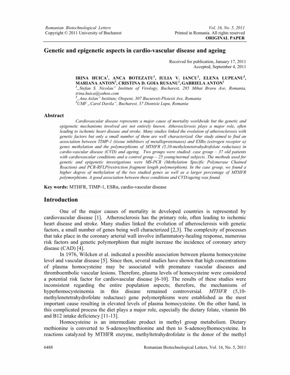
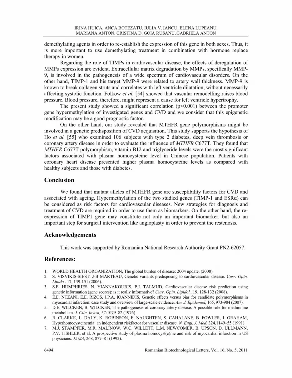
A hypermethylation of TIMP-1 and ESRα genes was observed predominantly for CVD/old patients, while normal/young subjects presented mostly unmethylated genes (Figure 1).
M U M U M U M U M U M U M U
DNA Meth- Unmeth- S1 S2 S3 S4 S5 Ladder DNA DNA
Fig. 1. Agarose gel electrophoresis for ESRα gene S1, S2 and S4 samples are hemimethylated while S3, S5 samples are hypermethylated.
M U M U M U M U M U M U M U
DNA Meth- Unmeth- S1 S2 S3 S4 S5 Ladder DNA DNA
Fig. 2. Agarose gel electrophoresis for TIMP1 gene. S1, S2 samples are unmethylated and S3, S4 and S5 samples are hypermethylated.
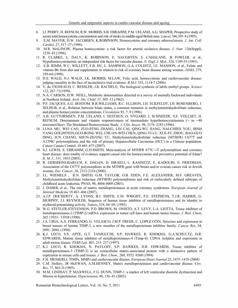
ESRα and TIMP1 genes methylation pattern observed in the two groups (CVD/old and normal/young) is presented in figure 3.
05
101520253035
E S R TIMP -1 E S R TIMP -1
C V D / old normal / young
M U
Fig. 3. Methylation status of ESRα and TIMP1 genes.

IRINA HUICA, ANCA BOTEZATU, IULIA V. IANCU, ELENA LUPEANU, MARIANA ANTON, CRISTINA D. GOIA RUSANU, GABRIELA ANTON
6492 Romanian Biotechnological Letters, Vol. 16, No. 5, 2011
In order to establish a correlation between old patients with CVD and genes hypermethylation, Fisher’s exact test was performed. ESRα and TIMP1 presented a statistically extremely significant frequency of hypermethylation in old patients with CVD versus normal group (P <0.001 for each gene) (Table 2).
Specificity (percentage of healthy patients who are identified as not having the condition) and the sensitivity (predicts the capacity to identify all the patients from the CVD/old group that are presenting the disease) were calculated. The correlation was determined for ESR-α (OR=35) and for TIMP1 (OD=15.278). We found that the relative risk value is higher than 1 for hypermethylated investigated genes. This shows a good association between the epigenetic silencing of the two investigated genes and ageing and CVD.
Table 2. Fisher’s exact test results for target genes.
The most specific (89%) and sensitive (84%) hypermethylated gene is ESR-α. TIMP1
presented a high grade of specificity (89%) but a low sensitivity (64.7%). The MTHFR polymorphisms were studied in the two groups (CVD/old and
normal/young) (Figure 4 and 5).
Fig. 4. MTHFR C677T polymorphism.
The upper part of Fig.4 represents the electrophoresis of the 198 bp amplicons for
C677T polymorphism and the lower part shows the results of restriction reaction with Hinf I endonuclease. Samples S-1, 4, 6 are normal homozygote, S-2, 3, 5 are heterozygote, and S7 is mutant homozygote.
Fig. 5. MTHFR A1298C polymorphism
The samples S1-6 are heterozygote, and S7 is normal homozygote.
Genes
Positive for methylation n/total cases (%) Odd ratio Relative
risk Specificity Sensitivity Positive
predictive value
p-valueYoung/ normal Old/ CVD
ESRα 4/25(16) 33/37(89.19) 43.313 7.77 0.8919 0.84 0.84 <0.0001
Confidence interval 9.758-192.25
3.031-19.919 0.7461-0.9697 0.6392-
0.9546 0.6392-0.9546
TIMP1 3/25 (12) 25/37 (67.57) 15.278 2.713 0.8929 0.6471 0.88 <0.0001
Confidence interval 3.808-61.288
1.677-4.417 0.7178-0.9773 0.4647-
0.8027 0.6878-0.9746

Genetic and epigenetic aspects in cardio-vascular disease and ageing
Romanian Biotechnological Letters, Vol. 16, No. 5, 2011 6493
We found MTHFR C677T polymorphism in 59.45% of CVD/old group patients; 7/37 (18.92%) patients were mutant homozygote (TT) and 15/37 (40.54%) heterozygote (CT), while in normal/young group, only 32% presented this mutation (1/25, 4% TT and 7/25, 28% CT). Regarding A1298C mutation, we found it in 62.16% of CVD/old group. 5/37, 13.51% were TT homozygote and 18/37, 48.65% were CT heterozygote. In the normal/young group only 40% (10/25) presented this mutation, all of them being heterozygote CT. These data are represented in figure 6.
0
2
4
6
8
10
12
14
16
18
MTHF R 1 MTHF R 2 MTHF R 1 MTHF R 2
C V D / old normal / young
normal homoz ygote
heteroz ygote
mutant homoz ygote
Fig. 6. MTHFR polymorphisms
The χ2 test was performed with 1 degree of freedom, and 5% level of significance. The
results support the hypothesis that both mutant alleles are susceptibility factors for CVD (C677T χ2=5.409, ptrend=0.02; A1298C χ2=4.765, ptrend=0.0291). On the other hand, heterozygote or homozygote patients with MTHFR C677T polymorphism are more likely to develop CVD than normal homozygote (OD=3.54).
In multiple systems, promoter CpG island methylation is associated with inactivation of gene transcription [47]. This process is involved in X-chromosome inactivation [48] in which promoter methylation is essential for maintaining the silenced status. CpG islands methylation in the promoter region is also involved in genomic imprinting [49]. This establishes which allele (maternal or paternal) is expressed in a given tissue. In addition, abnormal methylation of the promoter area of many genes appears to be an important feature of human neoplasia.
The methylation of ESRα and TIMP genes promoters is a hallmark for aging being involved in CVD. This study demonstrated that the TIMP1 gene promoter hypermethylation is frequent in old patients and is very rare in young people. We have to underline the fact that methylation of ESRα gene promoter is an event that appears more frequently in the middle or even earlier in life. This study established a possible link between lack of expression of the ESRα gene and atherosclerosis; some of the potential protective effects of estrogens are mediated by changes in the lipid profile, having direct effects on the vessel wall [50,51]. This observation is very important especially for menopausal women who used hormone-replacing therapy. Our data confirmed the discovery of a randomized study realized on a group of women with known coronary artery disease, study that demonstrated no benefit to hormone replacement [52]. Other study showed that in the cardiovascular system ESRα gene promoter methylation is present in a non-uniform, mosaic pattern. ESRα a gene promoter methylation can be found at variable levels in all vascular tissues including the right atrium, saphenous veins and the proximal aorta [53]. The aim of researchers is to determine the prediction potential of ESRα gene promoter hypermethylation in developing CVD and to use
IRINA HUICA, ANCA BOTEZATU, IULIA V. IANCU, ELENA LUPEANU, MARIANA ANTON, CRISTINA D. GOIA RUSANU, GABRIELA ANTON
6494 Romanian Biotechnological Letters, Vol. 16, No. 5, 2011
demethylating agents in order to re-establish the expression of this gene in both sexes. Thus, it is more important to use demethylating treatment in combination with hormone replace therapy in women.
Regarding the role of TIMPs in cardiovascular disease, the effects of deregulation of MMPs expression are evident. Extracellular matrix degradation by MMPs, specifically MMP-9, is involved in the pathogenesis of a wide spectrum of cardiovascular disorders. On the other hand, TIMP-1 and his target MMP-9 were related to artery wall thickness. MMP-9 is known to break collagen struts and correlates with left ventricle dilatation, without necessarily affecting systolic function. Folkow et al. [54] showed that vascular remodelling raises blood pressure. Blood pressure, therefore, might represent a cause for left ventricle hypertrophy. The present study showed a significant correlation (p<0.001) between the promoter gene hypermethylation of investigated genes and CVD and we consider that this epigenetic modification may be a good prognostic factor. On the other hand, our study revealed that MTHFR gene polymorphisms might be involved in a genetic predisposition of CVD acquisition. This study supports the hypothesis of Ho et al. [55] who examined 106 subjects with type 2 diabetes, deep vein thrombosis or coronary artery disease in order to evaluate the influence of MTHFR C677T. They found that MTHFR C677T polymorphism, vitamin B12 and triglyceride levels were the most significant factors associated with plasma homocysteine level in Chinese population. Patients with coronary heart disease presented higher plasma homocysteine levels as compared with healthy subjects and those with diabetes. Conclusion
We found that mutant alleles of MTHFR gene are susceptibility factors for CVD and associated with ageing. Hypermethylation of the two studied genes (TIMP-1 and ESRα) can be considered as risk factors for cardiovascular diseases. New strategies for diagnosis and treatment of CVD are required in order to use them as biomarkers. On the other hand, the re-expression of TIMP1 gene may constitute not only an important biomarker, but also an important step for surgical intervention like angioplasty in order to prevent the restenosis.
Acknowledgements
This work was supported by Romanian National Research Authority Grant PN2-62057.
References: 1. WORLD HEALTH ORGANIZATION, The global burden of disease: 2004 update. (2008). 2. S. VISVIKIS-SIEST, J-B MARTEAU, Genetic variants predisposing to cardiovascular disease. Curr. Opin.
Lipido,. 17, 139-151 (2006). 3. S.E. HUMPHRIES, N. YIANNAKOURIS, P.J. TALMUD, Cardiovascular disease risk prediction using
genetic information (gene scores): is it really informative? Curr. Opin. Lipidol,. 19, 128-132 (2008). 4. E.E. NTZANI, E.E. RIZOS, J.P.A. IOANNIDIS, Genetic effects versus bias for candidate polymorphisms in
myocardial infarction: case study and overview of large-scale evidence. Am. J. Epidemiol, 165, 973-984 (2007). 5. D.E. WILCKEN, B. WILCKEN, The pathogenesis of coronary artery disease. A possible role for methionine
metabolism. J. Clin. Invest, 57:1079–82 (1976) 6. R. CLARKE, L. DALY, K. ROBINSON, E. NAUGHTEN, S. CAHALANE, B. FOWLER, I. GRAHAM,
Hyperhomocysteinemia: an independent riskfactor for vascular disease. N. Engl. J. Med, 324,1149–55 (1991) 7. M.J. STAMPFER, M.R. MALINOW, W.C. WILLETT, L.M. NEWCOMER, B. UPSON, D. ULLMANN,
P.V. TISHLER, et al. A prospective study of plasma homocyst(e)ine and risk of myocardial infarction in US physicians. JAMA, 268, 877–81 (1992).
Genetic and epigenetic aspects in cardio-vascular disease and ageing
Romanian Biotechnological Letters, Vol. 16, No. 5, 2011 6495
8. I.J. PERRY, H. REFSUM, R.W. MORRIS, S.B. EBRAHIM, P.M. UELAND, A.G. SHAPER, Prospective study of serum total homocysteine concentration and risk of stroke in middle-aged British men. Lancet, 346,395–8 (1995).
9. E.M. MAYER, D.W. JACOBSEN, K.ROBINSON, Homocysteine and coronary atherosclerosis. J. Am. Coll. Cardiol, 27, 517–27 (1996).
10. M.R. MALINOW, Plasma homocysteine: a risk factor for arterial occlusive disease. J. Nutr. 126(Suppl), 1238–43 (1996).
11. R. CLARKE, L. DALY, K. ROBINSON, E. NAUGHTEN, S. CAHALANE, .B FOWLER, et al., Hyperhomocysteinemia: an independent risk factor for vascular disease. N. Engl. J. Med., 324, 1149-55 (1991).
12. E.B. RIMM, W.C. WILLETT, F.B. HU, L. SAMPSON, G.A. COLDITZ, J.E. MANSON, et al., Folate and vitamin B6 from diet and supplements in relation to risk of coronary heart disease among women. JAMA, 279, 359-64 (1998).
13. D.S. WALD, N.J. WALD, J.K. MORRIS, M.LAW, Folic acid, homocysteine and cardiovascular disease: judging causality in the face of inconclusive trial evidence. B.M.J. 333, 1114-7 (2006).
14. V. du VIGNEAUD, C. RESSLER, J.R. RACHELE, The biological synthesis of labile methyl groups. Science 112, 267–71(1950).
15. N.A. CARSON, D.W. NEILL, Metabolic abnormalities detected in a survey of mentally backward individuals in Northern Ireland. Arch. Dis. Child. 37, 505–13 (1962).
16. P.F. JACQUES, A.G. BOSTOM, R.R.WILLIAMS, R.C. ELLISON, J.H. ECKFELDT, I.H. ROSENBERG, J. SELHUB, et al., Relation between folate status, a common mutation in methylenetetrahydrofolate reductase, and plasma homocysteine concentrations. Circulation 93, 7–9 (1996).
17. A.B. GUTTORMSEN, P.M. UELAND, I. NESTHUS, O. NYGARD, J. SCHNEEDE, S.E. VOLLSET, H. REFSUM, Determinants and vitamin responsiveness of intermediate hyperhomocysteinemia (> or =40 micromol/liter). The Hordaland Homocysteine Study. J. Clin. Invest. 98, 2174–2183 (1996).
18. LI-NA MU, WEI CAO, ZUO-FENG ZHANG, LIN CAI, QING-WU JIANG, NAI-CHIEH YOU, BINH YANG GOLDSTEIN,GUO-RONG WEI, CHUAN-WEI CHEN, QING-YI LU, XUE-FU ZHOU, BAO-GUO DING, JUN CHANG, SHUN-ZHANG YU, Methylenetetrahydrofolate reductase (MTHFR) C677T and A1298C polymorphisms and the risk of primary Hepatocellular Carcinoma (HCC) in a Chinese population. Cancer Causes Control, 18 665–675 (2007).
19. S.J. LEWIS, S. EBRAHIM, G.D.SMITH, Meta-analysis of MTHFR 677C→T polymorphism and coronary heart disease: does totality of evidence support causal role for homocysteine and preventive potential of folate? B. M. J., 331, 1053 (2005).
20. R. GERSHONI-BARUCH, E. DAGAN, D. ISRAELI, L. KASINETZ, E. KADOURI, E. FRIEDMAN, Association of the C677T polymorphism in the MTHFR gene with breast and/or ovarian cancer risk in Jewish women. Eur. Cancer, 36, 2313-2316 (2000).
21. J.L. WIEMELS , R.N. SMITH, G.M. TAYLOR, O.B. EDEN, F.E. ALEXANDER, M.F. GREAVES, Methylenetetrahydrofolate reductase (MTHFR) polymorphisms and risk of molecularly defined subtypes of childhood acute leukemia. PNAS, 98, 4004-4009 (2001).
22. J. DABEK et al., The role of matrix metalloproteinases in acute coronary syndromes. European Journal of Internal Medicine 18 463–466 (2007).
23. A.J.P. DOCHERTY, A. LYONS, B.J. SMITH, E.M. WRIGHT, P.E. STEPHENS, T.J.R. HARRIS, G. MURPHY, J.J. REYNOLDS, Sequence of human tissue inhibitor of metalloproteinases and its identity to erythroid-potentiating activity. Nature, 318, 66–69, (1985).
24. W.G. STETLER-STEVENSON, P.D. BROWN, M. ONISTO, A.T. LEVY, L.A. LIOTTA, Tissue inhibitor of metalloproteinases-2 (TIMP-2) mRNA expression in tumor cell lines and human tumor tissues. J. Biol. Chem, 265 13933– 13938 (1990).
25. J.A. URIA, A.A. FERRANDO, G. VELASCO, J.M.P. FREIJE, C. LOPEZ-OTIN, Structure and expression in breast tumors of human TIMP-3, a new member of the metalloproteinase inhibitor family. Cancer Res, 54, 2091–2094, (1994).
26. K.J. LECO, S.S. APTE, G.T. TANIGUCHI, S.P. HAWKES, R. KHOKHA, G.A.SCHULTZ, D.R. EDWARDS, Murine tissue inhibitor of metalloproteinases-4 (Timp-4): CDNA isolation and expression in adult mouse tissues. FEBS Lett. 401, 213–217 (1997).
27. K.J. LECO, R. KHOKHA, N. PAVLOFF, S.P. HAWKES, D.R. EDWARDS, Tissue inhibitor of metalloproteinases-3 (TIMP-3) is an extracellular matrix-associated protein with a distinctive pattern of expression in mouse cells and tissues. J. Biol. Chem., 269, 9352–9360 (1994).
28. F.H. MESSERLI, TIMPs, MMPs and cardiovascular disease. European Heart Journal 25, 1475–1476 (2004) 29. C.M. Dollery, JR McEWAN, A.M.HENNEY, Matrix metalloproteinases and cardiovascular disease. Circ.
Res, 77, 863–8 (1995). 30. M.M. LINDSAY, P. MAXWELL, F.G. DUNN, TIMP-1: a marker of left ventricular diastolic dysfunction and
fibrosis in hypertension. Hypertension, 40, 136–41 (2002).
IRINA HUICA, ANCA BOTEZATU, IULIA V. IANCU, ELENA LUPEANU, MARIANA ANTON, CRISTINA D. GOIA RUSANU, GABRIELA ANTON
6496 Romanian Biotechnological Letters, Vol. 16, No. 5, 2011
31. S.J. GEORGE, J.L. JOHNSON, G.D. ANGELINI, A.C. NEWBY, A.H. BAKER, Adenovirus-mediated gene transfer of the human TIMP-1 gene inhibits SMC migration and neointima formation in human saphenous vein. Hum. Gene Ther, 9, 867-877 (1998b).
32. S.J. GEORGE, C.T. LLOYD, G.D. ANGELINI, A.C. NEWBY, A.H. BAKER, Inhibition of late vein graft neointima formation in human and porcine models by adenovirus-mediated overexpression of tissue inhibitor of metalloproteinase-3. Circulation, 101, 296-304 (2000).
33. A.H. BAKER, D.R. EDWARDS, G. MURPHY, Metalloproteinase inhibitors: biological actions and therapeutic opportunities. J. Cell. Sci., 115, 3719–27 (2002).
34. M.E. MENDELSOHN, R.H. KARAS, The protective effects of estrogen on the cardiovascular system. N. Engl. J. Med., 340, 1801-1811 (1999).
35. Y. MATSUBARA, M. MURATA, K. KAWANO, et al., Genotype distribution of estrogen receptor polymorphisms in men and postmenopausal women from healthy and coronary populations and its relation to serum lipid levels. Arterioscler. Thromb. Vasc. Biol., 17, 3006-3012 (1997).
36. T.A KUNNAS, P. LAIPPALA, A. PENTTILA, T. LEHTIMAKI, P.J. KARHUNEN, Association of polymorphism of human alpha oestrogen receptor gene with coronary artery disease in men: a necropsy study. BMJ, 321, 273-274, (2000).
37. H. LU, T. HIGASHIKATA, A. INAZU, et al. Association of estrogen receptor-alpha gene polymorphisms with coronary artery disease in patients with familial hypercholesterolemia. Arterioscler. Thromb. Vasc. Biol., 22, 817-823 (2002).
38. D. EVANGELOPOULOS, M. ALEVIZAKI, J. LEKAKIS, et al. Molecular analysis of the estrogen receptor alpha gene in men with coronary artery disease: association with disease status. Clin. Chim. Acta., 331, 37-44, (2003).
39. D. PETROVIC, B. PETERLIN, Estrogen receptor dinucleotide (ta) polymorphism does not predict premature myocardial infarction in Caucasian women. Cardiology, 99, 163-165 (2003).
40. T. LEHTIMAKI, T.A. KUNNAS, K.M. MATTILA, et al. Coronary artery wall atherosclerosis in relation to the estrogen receptor 1 gene polymorphism: an autopsy study. J. Mol. Med., 80, 176-180 (2002).
41. D.M. HERRINGTON, T.D. HOWARD, G.A. HAWKINS, et al., Estrogen-receptor polymorphisms and effects of estrogen replacement on high-density lipoprotein cholesterol in women with coronary disease. N. Engl. J. Med., 346, 967-974 (2002).
42. D.M. HERRINGTON, T.D. HOWARD, K.B. BROSNIHAN, et al., Common estrogen receptor polymorphism augments effects of hormone replacement therapy on E selectin but not C-reactive protein. Circulation, 105, 1879-1882 (2002).
43. M.J. STAMPFER, G.A. COLDITZ, W.C. WILLETT et al., Postmenopausal estrogen therapy and cardiovascular disease. New Engl. J. Med., 325, 756–762 (1991).
44. E. BARRETT-CONNOR, T.L. BUSH, Estrogen and coronary heart disease in women. J. Am. Med. Assoc, 265, 1861–1867 (1991).
45. P. FROSST, H.G. BLOM, R. MILOS, P. GOYETTE, C.A. SHEPPARD, R.G. MATTHEWS, G.J. BOERS, M. den HEIJER, L.A. KLUIJTMANS, L.P. van den HEUVEL, et al. A candidate genetic risk factor for vascular disease: a common mutation in methylenetetrahydrofolate reductase. Nat. Genet., 10, 11–13 (1995).
46. I.S. WEISBERG, P.F. JACQUES, J. SELHUB, A.G. BOSTOM, Z. CHEN, R.C. ELLISON, J.H. ECKFELDT, R. ROZEN, The 1298A_C polymorphism in methylenetetrahydrofolate reductase (MTHFR): in vitro expression and association with homocysteine. Atherosclerosis. 156(2), 409-15 (2001).
47. A.P. BIRD, CpG-rich islands and the function of DNA methylation. Nature, 321, 209–213 (1986). 48. B.R. MIGEON, Insights into X chromosome inactivation from studies of species variation. DNA methylation
and replication, and vice versa. Genet. Res., 56, 91–98 (1990). 49. A.C. FERGUSON-SMITH, H. SASAKI, B.M. CATTANACH, M.A. SURANI, Parental origin-specific
epigenetic modification of the mouse H19 gene. Nature, 362, 751–755 (1993). 50. M.E. Mendelsohn, R.H. Karas, Estrogen and the blood vessel wall. Curr. Opin. Cardiol, 9, 619–626 (1994). 51. S.E. REIS, S.T. GLOTH, R.S. BLUMENTHAL et al., Ethinyl estradiol acutely attenuates abnormal coronary
vasomotor responses to acetylcholine in postmenopausal women. Circulation, 89, 52–60 (1994). 52. S. HULLEY, D. GRADY, T. BUSH et al. Randomized trial of estrogen plusprogestin for secondary prevention
of coronary heart disease in postmenopausal women. J. Am. Med. Assoc., 280, 605–613, (1998). 53. W.S. POST, P.J. GOLDSCHMIDT-CLERMONT, C.C. WILHIDE, A.W. HELDMAN, M.S. SUSSMAN, P.
OUYANG, E.E. MILLIKEN, J.P.J. ISSA, Methylation of the estrogen receptor gene is associated with aging and atherosclerosis in the cardiovascular system. Cardiovasc. Res., 43, 985–991 (1999).
54. B. FOLKOW, ‘‘Structural factor’’ in primary and secondary hypertension. Hypertension, 16, 89–101 (1990). 55. C.H. HO, B.I.T. KUO, C.W. KONG, W.K. CHAU, H.C. HSU, J.P. GAU, Y.B. YU, Influence of
methylenetetrahydrofolate reductase (MTHFR) C677T polymorphism, B vitamins and other factors on plasma homocysteine and risk of thromboembolic disease in Chinese. J. Chin. Med. Asso., 68, 560–5 (2005).


