Embed Size (px)
Citation preview
Gas Phase Electron Ionization Fragmentation of Dextromethorphan and Related Quaternary Ammonium Salts
Irving W. Wainer, Mamoru Ohashi,? Robert P. BarronS and Walter Benson Division of Drug Chemistry, Food and Drug Administration, Washington, DC 20204, USA
The in-beam electron ionization (EI) spectra of quaternary ammonium salts of dextrumethorphan and piperidine consist of peaks arising both from thermal degradation products and from the fragmentation of the molecular cation [R4N]+. Mass-analysed ion kinetic energy and ion kinetic energy spectra indicate that several major ions believed to be thermally induced before EI are also produced directly from [R4N]+ fragmentation. However, the contribution of the direct fragmentation of [R,N]+ upon EI is small in comparison to that of the thermal degradation products. This behavior was observed in the conformationally flexible and rigid piperidinium rings studied.
INTRODUCTION
Dextromethorphan hydrobromide is a non-narcotic antitussive that belongs to the phenanthrene alkaloid group, which also contains morphine. Its enantiomorph, levomethorphan, is a potent, controlled narcotic.' The difference in biological activity between the two enan- tiomers creates the need for a rapid and accurate assay to monitor and control the level of the Zevo-isomer in pharmaceutical preparations containing dextromethor- phan. As part of the effort in our laboratory to develop such an assay, we prepared a series of N-alkyldex- tromethorphan quaternary ammonium iodide salts2 and obtained their spectra, using the techniques of in-beam electron ionization (EI) mass spectrometry: mass- analysed ion kinetic energy (MIKE) and ion kinetic energy (IKE) ~pectrometry.~ These spectra indicate the existence of the intact quaternary ammonium ions, [%N]+, and their subsequent fragmentation upon EI in the gas phase.
Until recently, quaternary ammonium cations were believed to undergo thermal degradation before being detected in a mass ~pectrometer.~-* However, in 1980 Stoll and Rollgen observed the presence of simple quaternary ammonium cations in the gas phase: and Cotter and Yergey reported their thermal desorption spectra." Using an electron impact flash desorption (EI/ D) technique, Lee and co-workers recently observed fragment ions in a series of simple tetraalkylammonium cations." Our laboratory has also reported the detection of EI-induced fragmentation of quaternary ammonium cations arising from a series of simple tetraalkylam- monium salts." Today, mass spectrometric analysis of quaternary ammonium salts is becoming commonplace as demonstrated by the recent papers by Isa and Yamada13 and Glish and Smith.I4 We report here the in-beam mass spectral characteristics of a series of N- alkyl quaternary ammonium salts of dextromethorphan.
t Visiting scientist on leave from the University of Electro-
$ Author to whom reprint requests should be addressed. Communications, Chofu-shi, Tokyo, Japan.
The MIKE spectra from the cation of the N-alkyldex- tromethorphan quaternary ammonium salts confirm that these ions undergo fragmentation upon EI in the gas phase within the constraints of the technique. However, the mechanism of this fragmentation, especially that involving the quaternized piperidine moiety, was unclear. The quaternary ammonium salts of piperidine have been reported to undergo three types of thermal reactions before ionization in a mass spectrometer: dealkylation (the most prevalent type), Hofmann degra- dation and ~ubsti tution.~ The EI fragmentation of the piperidinium molecular cation, however, has not been previously reported. In order to attempt to elucidate the fragmentation mechanism of the rigid piperidinium cation moieties in the dextromethorphan quaternary ammonium salts, we prepared and studied a series of flexible N-alkyl-N-methylpiperidinium iodide salts.
EXPERIMENTAL
Spectra
Mass spectra were recorded on a Varian-MAT 3 11A double-focusing mass spectrometer equipped with a modified field desorption/field ionization/electron ionization (FD/ FI/EI) source and on a Finnigan 4023T quadrupole mass spectrometer equipped with a direct insertion tip made of stainless steel for in-beam ELJ5
The Varian-MAT3 1 1A FD/FI/EI source was adapted for in-beam use by modification of the heating current contact of the FD probe. A similar modification was reported by Soltmann el al. in the EI/D technique.I6 We used an unactivated tungsten wire (1 0 km diameter) in place of an activated emitter and, except for the usual accelerating voltage of 3 kV, no high voltage (HV) was applied. The sample (1-10 bg) was loaded on the FD wire, placed at a position less than 3 mm from the center of the ionizing electron beam and then quickly heated by a 50 mA ~urrent. '~" ' The spectra were recorded under the following conditions: ionizing energy 80 eV; source
@ Wiley Heyden Ltd, 1984
CCC-0306-042X/ 84/00 1 1-0529 $03 .OO
BIOMEDICAL MASS SPECTROMETRY, VOL. 11, NO. 10, 1984 529
I. W. WAINER, M. OHASHI, R. P. BARRON A N D W. BENSON
temperature 280-300 "C; sample heater 50 mA; emission current 500 PA; accelerating voltage 3 kV.
MIKE spectra4 were obtained under similar condi- tions with electric sector scanning. The scanning speed was 4 or 7 V s-l and the recording chart was set at a speed of 63 mm min-'. HV-scanning IKE spectra4 were obtained by scanning the acceleration voltage from 1 kV to 3 kV, with a scanning speed of 13 or 7 V s-' and a recording speed of 63 mm min-'.
The thermal desorption spectra of the N,N-dialkyl- piperidinium salts were obtained with the Finnigan instrument. The samples were loaded on the in-beam tip, placed in the ionizing chamber at 300 "C and heated to 400 "C by the sample heater. In-beam EI spectra were also recorded under similar conditions with a 70 eV ionizing electron beam.
Synthesis of the quaternary ammonium salts
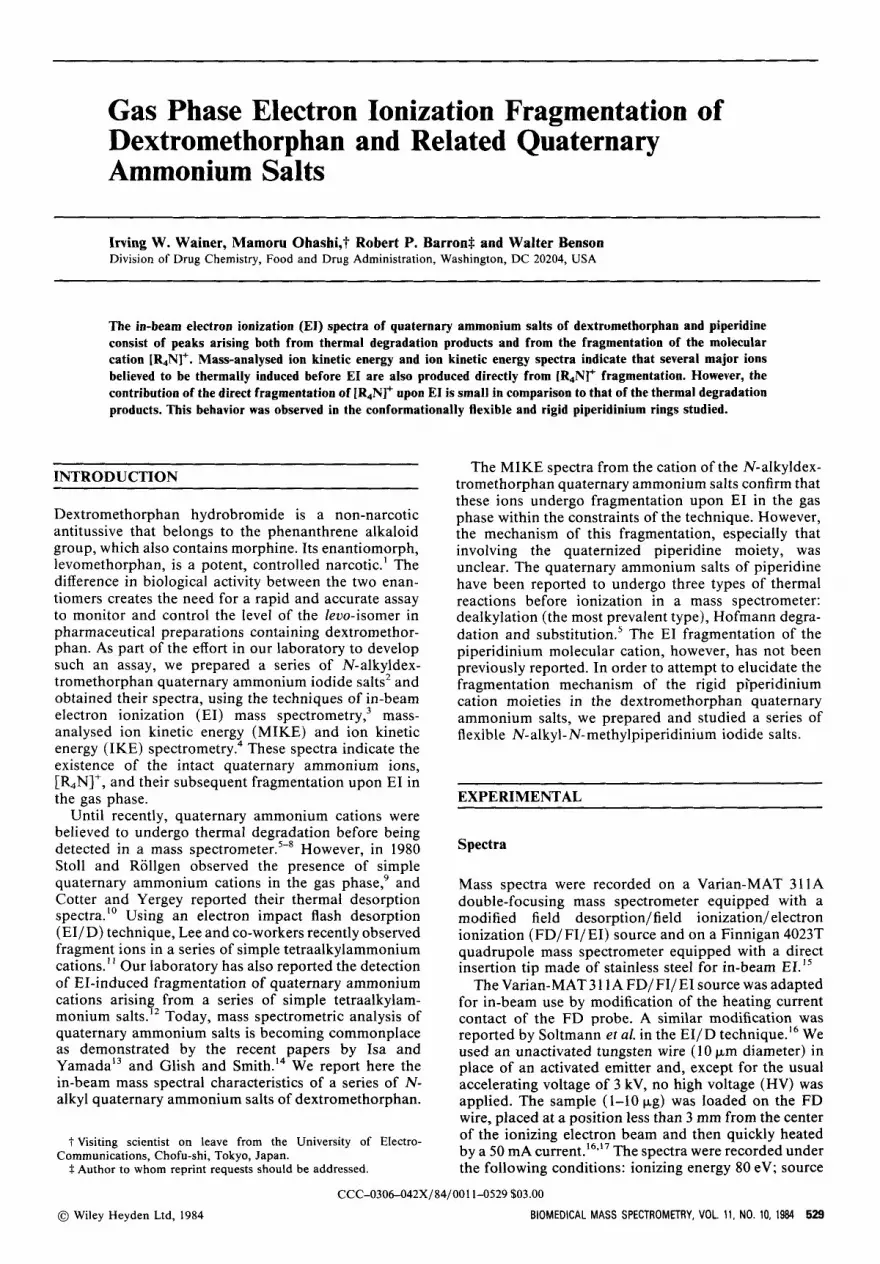
Several N-alkyl-N-methylpiperidinium salts (1,2,4 and 5, Fig. 1) were prepared by the alkylation of N-methyl- piperidine with the appropriate n-alkyl iodide; com- pound 3 (Fig. 1) was prepared from N-ethylpiperidine and trideuteromethyl iodide.' The N-alkyldex- tromethorphan quaternary ammonium salts (7-11, Fig. 1) were prepared from dextromethorphan and the cor- responding n-alkyl iodides, whereas compound 12 (Fig. 1) was prepared from dextromethorphan and 1-iOd0-3- methylhexane.2
The alkylations were carried out in methylene chloride at 80 "C. A 10- to 30-fold excess of the alkyl iodide was used and the progress of the reaction was monitored by nuclear magnetic resonance (NMR) spectroscopy. At the completion of the reaction, the solvent was evapor- ated and the quaternary salts were recrystallized twice from methanol-ether. Yields were nearly quantitative. NMR spectra confirmed the structure assignmenh2
RESULTS AND DISCUSSION
Thermal desorption spectra of N,N-dialkylpiperidinium salts
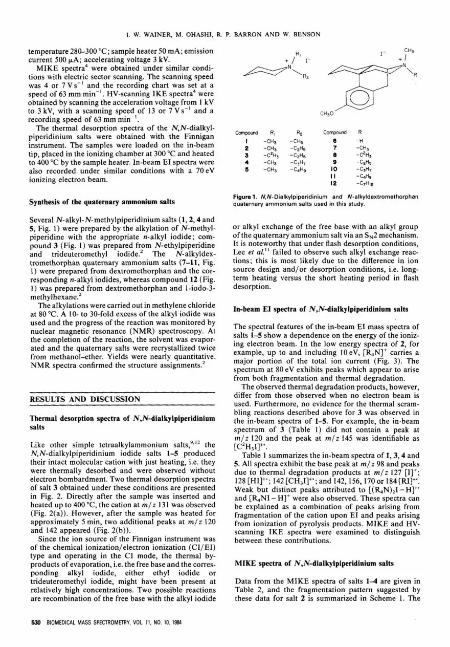
Like other simple tetraalkylammonium salt^,^^'^ the N,N-dialkylpiperidinium iodide salts 1-5 produced their intact molecular cation with just heating, i.e. they were thermally desorbed and were observed without electron bombardment. Two thermal desorption spectra of salt 3 obtained under these conditions are presented in Fig. 2. Directly after the sample was inserted and heated up to 400 "C, the cation at m / z 13 1 was observed (Fig. 2(a)). However, after the sample was heated for approximately 5 min, two additional peaks at m / z 120 and 142 appeared (Fig. 2(b)).
Since the ion source of the Finnigan instrument was of the chemical ionization/ electron ionization (CI/EI) type and operating in the CI mode, the thermal by- products of evaporation, i.e. the free base and the corres- ponding alkyl iodide, either ethyl iodide or trideuteromethyl iodide, might have been present at relatively high concentrations. Two possible reactions are recombination of the free base with the alkyl iodide
CH3 1- + / ' I - C l
Canpound R, RZ Compound R I -CH3 -CH3 6 -H 2 -CH3 -CzH, ? -CH3 3 -CZH3 -C2H5 8 -C2H3
4 -CH3 -C,H, 9 -C2H5 5 -CH3 -C4H9 10 -C3H7
I I -C4H9 12 -WI(I
Figure 1. N,N-Dialkylpiperidinium and N-alkyldextromethorphan quaternary ammonium salts used in this study.
or alkyl exchange of the free base with an alkyl group of the quaternary ammonium salt via an SN2 mechanism. It is noteworthy that under flash desorption conditions, Lee et al." failed to observe such alkyl exchange reac- tions; this is most likely due to the difference in ion source design and/or desorption conditions, i.e. long- term heating versus the short heating period in flash desorption.
In-beam EI spectra of N,N-dialkylpiperidinium salts
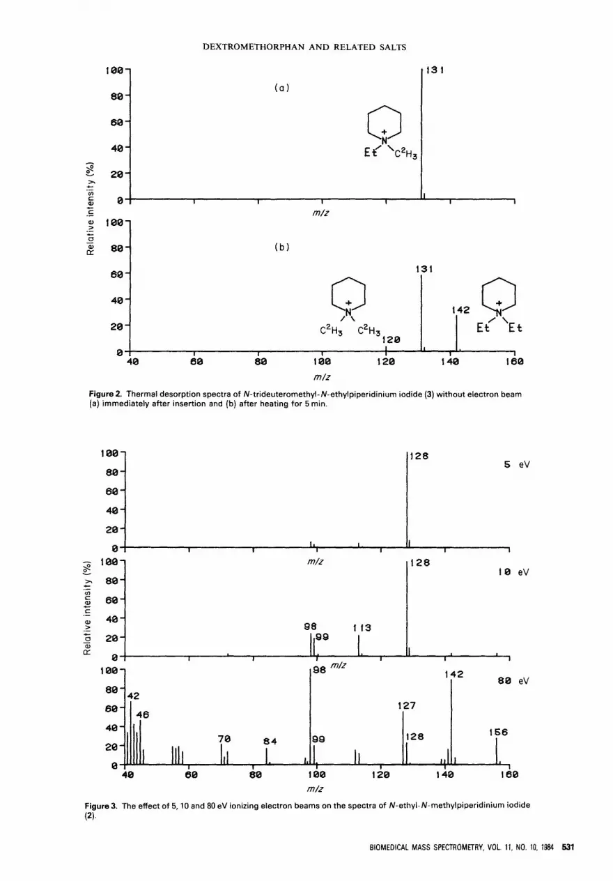
The spectral features of the in-beam EI mass spectra of salts 1-5 show a dependence on the energy of the ioniz- ing electron beam. In the low energy spectra of 2, for example, up to and including IOeV, [&N]+ carries a major portion of the total ion current (Fig. 3). The spectrum at 80 eV exhibits peaks which appear to arise from both fragmentation and thermal degradation.
The observed thermal degradation products, however, differ from those observed when no electron beam is used. Furthermore, no evidence for the thermal scram- bling reactions described above for 3 was observed in the in-beam spectra of 1-5. For example, the in-beam spectrum of 3 (Table 1) did not contain a peak at m / z 120 and the peak at m / z 145 was identifiable as [C2H31]".
Table 1 summarizes the in-beam spectra of 1,3,4 and 5. All spectra exhibit the base peak at m / z 98 and peaks due to thermal degradation products at m / z 127 [I]+; 128 [HI]+'; 142 [CH,I]+'; and 142,156,170 or 184 [RII+'. Weak but distinct peaks attributed to [(&N),I -HI+' and [&NI - HIf were also observed. These spectra can be explained as a combination of peaks arising from fragmentation of the cation upon EI and peaks arising from ionization of pyrolysis products. MIKE and HV- scanning IKE spectra were examined to distinguish between these contributions.
MIKE spectra of N,N-dialkylpiperidinium salts
Data from the MIKE spectra of salts 1-4 are given in Table 2, and the fragmentation pattern suggested by these data for salt 2 is summarized in Scheme 1. The
530 BIOMEDICAL MASS SPECTROMETRY, VOL. 11, NO. 10, 1984
DEXTROMETHORPHAN AND RELATED SALTS
100-
80 - 60 -
40 - Et’ ‘ c ~ H ~
( 0 )
8 20- > v)
c .- I I I I
13 1
I I 1
60 -
40 -
20 -
0
m/z
Figure 2. Thermal desorption spectra of N-trideuterornethyl-N-ethylpiperidinium iodide (3) without electron beam [a) immediately after insertion and (b) after heating for 5 rnin.
131
/ \ 142 Q / \
Et Et 120
C2H, C2H3
I I I 1 1 I I
1
5 eV 100-
80 - 60 - 40 - 20 - n
128
I , I I
100-
80 - 60 - 40 - 20 - 0
m/z 128 10 eV
98 113 99
I I I I 1 I
I
m/z
42
60- 46 40 - 20 - 70 84
Figure 3. The effect of 5,lO and 80 eV ionizing electron beams on the spectra of N-ethyl-N-methylpiperidinium iodide (2).
80 eV 142
127
156 QQ 128
BIOMEDICAL MASS SPECTROMETRY, VOL. 11, NO. 10, 1984 531
0--1 II 1 1. I , . I I 1 I 1 I I 1
I. W. WAINER, M. OHASHI, R. P. BARRON AND W. BENSON
Table 1. In-beam EI spectral data for N,N-dialkylpiperidinium iodide salts"
rn/z (intensity)
Compoundb [K]' [K-CH,]+' [K-CH,]+ [K-R]+' (K-RH]' [I]' (HI]+' [CH,I]+' [RIJf' Others
99
113'
1 27h
141
(37)
(17)
(4)
(8)
a K=quaternary ammonium cation. See Fig. 1. m / z 113 [K-C2H3]+'. m/z 112 [K-CZH3H]'. m / z 102 [K-C,H,]+'. m/z 145 (C2H3l]+'. m / z 98 [K-C, H3H3]+. Doublet peaks identified on the basis of their exact masses.
n
-CH3
Scheme 1
proposed fragmentation processes are supported by a comparison of the MIKE spectrum of the N-methyl-N- ethyl salt (2) with that of the N-trideuteromethy1-N- ethyl salt (3). Both spectra contain fragment ions m / z 113 and 112, which can arise via pathways a and b, respectively. The spectrum of 2 also contains fragment ions m / z 72 [&N - C,H,]+, 98 [&N - RH]', 99 [&N - R]' and 100 [&N - R - HI+. In the spectrum of 3, these
Table 2. MIKE spectral data and fragmentation pathways for N,N-dialkylpiperidinium iodide salts"
Fragment ion ( m / z ) Fragmentation
pathwayb Compound lb Compound 2b Compound 3b Compound 4b
a 99 113 113 127 b 98 112 112 126 C - 100 103 100 d 99 99 102 99 e 98 98 100 98 f 58 72 75 86
a-a' - - 98 - a See Fig. 1, compounds 1 4
See Scheme 1.
fragments are shifted to m / z 75, 100, 102 and 103, sup- porting the existence of pathways f, e, d and c, respec- tively. The spectrum of 3 also contains the fragment ion m / z 98, which is most likely the result of the f3-cleavage of the product resulting from the loss of the methyl radical (pathway a').
These fragmentation processes are in accord with those observed in the simple tetraalkylammonium saltsi2 and are similar to the thermal fragmentations of these salts.5 It is noteworthy that evidence for the transition of a cation to a cation radical (Scheme 1, pathways a and d) was clearly observed, although the loss of radicals from even-electron ions was reported to be a rare occur- rence in direct contradiction to the even-electron rule," and is not observed by thermal desorption (TD)," laser desorption (LD)," plasma desorption (PD)," secondary ion mass spectrometry (SIMS)20 and fast atom bombard- ment (FAB).2' However, such behavior has been repor- ted in FD2* and laser microprobe mass analyser (LAMMA) l 9 spectra of tetraalkyl quaternary cations.
IKE spectra of 2 and 4 were obtained by scanning the acceleration voltage and were used to determine the parent ions of certain daughter ions. Table 3 lists the
Table3. HV-scanning IKE spectral data for N,N- dialkylpiperidinium iodides
Compound' Daughter ion ( r n / z ) Parent ion (rnlr)
2 100 128 99 128
114 98 128
113 72 128
100 4 100 142
99 142 128
98 142 127
86 142 127
a See Fig. 1.
532 BIOMEDICAL MASS SPECTROMETRY, VOL. 11, NO. 10, 1984
DEXTROMETHORPHAN AND RELATED SALTS
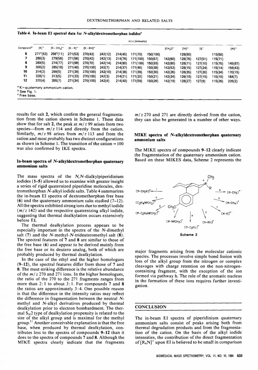
Table 4. In-beam El spectral data for N-alkyldextromethorphan iodides"
m/z (intensity)
Compoundb [K]+ [K-CH,]" [K-R]+' [K-RH]+ [CH,I]+' [HI]" Ill+ [RI]+'
6 271"(53) 256'(11) 271(53) 270(43) 242(12) 214(45) 171(70) 150(100) 128(50) 115(50) 7 286(3) 279(56) 271(56) 270(42) 242(13) 214(76) 171(100) 150(67) 142(60) 128(76) 127(51) 115(71) 8 289(5) 274(77) 271(98) 270(70) 242(14) 214(90) 171(100) 150(93) 142(80) 128(71) 127(10) 115(75) 145(97) 9 300(2) 285(18) 271(46) 270(100) 242(7) 214(37) 171(40) 150(38) 142(53) 128(15) 127(24) 115(14) 156(43)
10 314(2) 299(5) 271(38) 270(100) 242(10) 214(38) 171(39) 150(30) 142(26) 128(35) 127(30) 115(34) 170(15) 11 328(1) 313(5) 271(33) 270(100) 242(3) 214(21) 171(25) 150(21) 142(34) 128(10) 127(15) 115(10) 184(7) 12 370(4) 355(7) 271(34) 270(100) 242(4) 214(40) 171(59) 150(26) 142(19) 128(27) 127(9) 115(26) 226(2)
a K=quaternary ammonium cation. See Fig. 1. Free base.
results for salt 2, which confirm the general fragmenta- tion from the cation shown in Scheme l. These data show that for salt 2, the peak at m / z 99 arises from two species-from m / z 114 and directly from the cation. Similarly, m / z 98 arises from m / z 113 and from the cation and most probably has two distinct configurations as shown in Scheme 1. The transition of the cation-, 100 was also confirmed by IKE spectra.
In-beam spectra of N-alkyldextromethorphan quaternary ammonium salts
The mass spectra of the N,N-dialkylpiperidinium iodides (1-5) allowed us to examine with greater insight a series of rigid quaternized piperidine molecules, dex- tromethorphan N-alkyl iodide salts. Table 4 summarizes the in-beam EI spectra of dextromethorphan free base (6) and the quaternary ammonium salts studied (7-12). All the spectra exhibited strong ions due to methyl iodide ( m / z 142) and the respective quaternizing alkyl iodide, suggesting that thermal dealkylation occurs extensively before EI.
The thermal dealkylation process appears to be especially important in the spectra of the N-dimethyl salt (7) and the N-methyl-N-trideuteromethyl salt (8). The spectral features of 7 and 8 are similar to those of the free base (6 ) and appear to be derived mainly from the free base or its deutero analog, both of which are probably produced by thermal dealkylation.
In the case of the ethyl and the higher homologues (9-12), the spectral features differ from those of 7 and 8. The most striking difference is the relative abundance of the m/ z 270 and 27 1 ions. In the higher homologues, the ratio of the 270 to the 271 fragments ranges from more than 2 : 1 to about 3 : 1. For compounds 7 and 8 the ratios are approximately 3 : 4. One possible reason is that the difference in the intensity ratios may reflect the difference in fragmentation between the neutral N- methyl and N-alkyl derivatives produced by thermal dealkylation prior to electron bombardment. The ther- mal S,2 type of dealkylation propensity is related to the size of the alkyl group and is maximal for the methyl group.' ' Another conceivable explanation is that the free base, when produced by thermal dealkylation, con- tributes less to the spectra of compounds 9-12 than it does to the spectra of compounds 7 and 8. Although the MIKE spectra clearly indicate that the fragments
m / z 270 and 271 are directly derived from the cation, they can also be generated in a number of other ways.
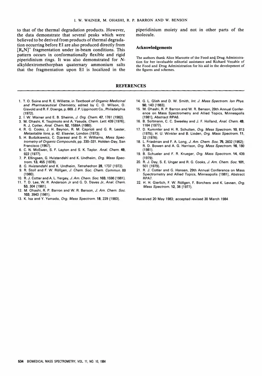
MIKE spectra of N-alkyldextromethorphan quaternary ammonium salts
The MIKE spectra of compounds 9-12 clearly indicate the fragmentation of the quaternary ammonium cation. Based on these MIKES data, Scheme 2 represents the
C"3 + I
CM-C2MNRCH31++' -I/ I e \d 1 CM-Rl'
CM-NRCH31' [M-RH]+
CM-CsH7f'
Scheme 2
major fragments arising from the molecular cationic species. The processes involve simple bond fission with loss of the alkyl group from the nitrogen or complex cleavages with charge retention on the non-nitrogen- containing fragment, with the exception of the ion formed via pathway h. The role of the aromatic nucleus in the formation of these ions requires further investi- gation.
CONCLUSION
The in-beam EI spectra of piperidinium quaternary ammonium salts consist of peaks arising both from thermal degradation products and from the fragmenta- tion of the cation. On the basis of the alkyl iodide intensities, the contribution of the direct fragmentation of [%N]+ upon EI is believed to be small in comparison
BIOMEDICAL MASS SPECTROMETRY, VOL. 11, NO. 10, 1984 533
I. W. WAINER, M. OHASHI, R. P. BARRON A N D W. BENSON
to that of the thermal degradation products. However, the data demonstrate that several peaks which were believed to be derived from products of thermal degrada- tion occurring before EI are also produced directly from [%N]+ fragmentation under in-beam conditions. This pattern occurs in conformationally flexible and rigid
piperidinium moiety and not in other parts of the molecule.
Acknowledgements
The authors thank Alice Marcotte of the Food and Drug Administra- piperidinium rings' It was tion for her invaluable editorial assistance and Richard Venable of alkyldextromethorphan quaternary ammonium salts the Food and Drug Administration for his aid in the development of
demonstrated for N-
that the fragmentation upon El is localized in the the figures and schemes.
REFERENCES
' T. 0. Soine and R. E. Willette. in Textbook of Oroanic Medicinal 14. G. L. Glish and D. W. Smith. lnt. J. Mass Soectrom. /on Phvs. I .
2. 3.
4.
5.
6.
7.
8. 9.
10. 11.
12.
13.
and Pharmaceutical Chemistry, edited by C.-O. Wilson, 0. Gisvold and R. F. Doerge, p. 669. J. P. Lippincott Co.. Philadelphia (1 972). I. W. Wainer and E. B. Sheinin, J. Org. Chem. 47, 1761 (1982). M. Ohashi, K. Tsujirnoto and A. Yasuda, Chem. Lett. 439 (1976); R. J. Cotter, Anal. Chem. 52, 1589A (1980). R. G. Cooks, J. H. Beynon, R. M. Caprioli and G. R. Lester, Metastable lons, p. 42. Elsevier, London (1973). H. Budzikiewicz, C. Djerassi and D. H. Williams, Mass Spec- trometry of Organic Compounds, pp. 330-331. Holden-Day, San Francisco (1967). C. N. McEwen. S. F. Layton and S. K. Taylor. Anal. Chem. 49, 922 (1977). P. Ellingsen, G. Hvistendahl and K. Undheim, Org. Mass Spec- trom. 13, 455 (1978). G. Hvistendahl and K. Undheim, Tetrahedron 28, 1737 (1972). R. Stoll and F. W. Rollgen, J. Chem. SOC. Chem. Commun. 89 (1 980). R. J. Cotter and A. L. Yergey, J. Am. Chem. SOC. 103,1596 (1981). T. D. Lee, W. R. Anderson Jr and G. D. Daves Jr, Anal. Chem. 53, 304 (1981). M. Ohashi, R. P. Barron and W. R. Benson, J. Am. Chem. SOC. 103, 3943 (1981). K. Isa and Y. Yamada, Org. Mass Spectrom. 18, 229 (1983).
50, 143 (1 983). 15. M. Ohashi, R. P. Barron and W. R. Benson, 29th Annual Confer-
ence on Mass Spectrometry and Allied Topics, Minneapolis (1981). Abstract RPA6.
16. B. Soltrnann, C. C. Sweeley and J. F. Holland, Anal. Chem. 49, 1164 (1977).
17. D. Kurnmler and H. R. Schulten, Org. Mass Spectrom. 10, 813 (1975); H. U. Winkler and B. Linden, Org. Mass Spectrom. 11, 32 (1976).
18. L. Friedman and F. A. Long, J. Am. Chem. SOC. 75,2832 (1952); R. D. Bowen and A. G. Harrison, Org. Mass Spectrom. 16, 180 (1981).
19. B. Schueler and F. R. Krueger. Org. Mass Spectrom. 14, 439 (1979).
20. R. J. Day, S. E. Unger and R. G. Cooks, J. Am. Chem. Soc. 101, 501 (1979).
21. R. J. Cotter and G. Hansen, 29th Annual Conference on Mass spectrometry and Allied Topics, Minneapolis (1 981), Abstract RPA7.
22. H. H. Gierlich, F. W. Rollgen, F. Borchers and K. Levsen, Org. Mass Spectrom. 12, 38 (1977).
Received 20 May 1983; accepted revised 30 March 1984
534 BIOMEDICAL MASS SPECTROMETRY, VOL. 11, NO. 10, 1984


