Embed Size (px)
Citation preview
Seediscussions,stats,andauthorprofilesforthispublicationat:https://www.researchgate.net/publication/225778810
FundamentalConceptsinSymbioticInteractions:LightandDark,DayandNight,SquidandLegume
ArticleinJournalofPlantGrowthRegulation·January2000
DOI:10.1007/s003440000025·Source:PubMed
CITATIONS
19
READS
20
2authors:
Someoftheauthorsofthispublicationarealsoworkingontheserelatedprojects:
AntifungalActivityofBacillusspeciesagainstFusariumandAnalysisoftheMechanismsUsedin
Biocontrol.Viewproject
AnnM.Hirsch
UniversityofCalifornia,LosAngeles
213PUBLICATIONS5,447CITATIONS
SEEPROFILE
MargaretMcFall-Ngai
UniversityofWisconsin–Madison
147PUBLICATIONS6,631CITATIONS
SEEPROFILE
AllcontentfollowingthispagewasuploadedbyAnnM.Hirschon03December2016.
Theuserhasrequestedenhancementofthedownloadedfile.Allin-textreferencesunderlinedinblueareaddedtotheoriginaldocument
andarelinkedtopublicationsonResearchGate,lettingyouaccessandreadthemimmediately.
TH E M A T I C AR T I C L E S
Fundamental Concepts in SymbioticInteractions: Light and Dark, Day
and Night, Squid and Legume
Ann M. Hirsch1,* and Margaret J. McFall-Ngai2
1Department of Molecular, Cell and Developmental Biology and Molecular Biology Institute, University of California, Los Angeles,California 90095, USA; and 2Kewalo Marine Laboratory, Pacific Biomedical Research Center, University of Hawaii,
Honolulu, Hawaii 96813, USA
ABSTRACT
The legume-Rhizobium symbiosis and that betweenEuprymna scolopes and Vibrio fischeri show some sur-prising physiological similarities as well as differ-ences. Both interactions rely on exchange of signalmolecules, some of which are derived from bacterialcell surface molecules. Although the legume-Rhizobium symbiosis is nutritionally based as aremany animal-microbe symbioses, it is not obligatebecause the plant initiates nodule formation onlywhen the soil is deficient in nitrogen. In contrast,the squid-Vibrio symbiosis is obligate for the squid
but is not nutritionally based. Rather, the bacteriaproduce light, which enables the animal to evadepredators. These similarities and differences are de-scribed and discussed in term of the overall questionof whether or not these two symbiotic relationshipshave evolved from commensal or pathogenic/parasitic interactions between prokaryotes and eu-karyotes.
Key words: Euprymna scolopes; Legume; Rhizobium;Sepiolid; Symbiosis; Vibrio fischeri
Nor knowest thou what argumentThy life to thy neighbor’s creed has lent.All are needed by each one;Nothing is fair or good alone.
—Ralph Waldo Emerson
INTRODUCTION
This review addresses two broad questions: (1) whatdo a squid and a legume have in common, and (2)have either of the symbioses in which these organ-
isms are involved evolved from a pathogenic inter-action or a commensal one? At the outset, these twomulticellular organisms would appear to share veryfew traits. The squid lives in the sea, comes out atnight, and produces an eerie luminescence from itslight-emitting organ, whereas the plant keeps itsroots in the soil hidden in the dark while its aerialparts use light for photosynthesis. Nevertheless,both produce a specialized organ that is inhabited bybacteria (Figures 1A, 2A–C). Host-bacterial interac-tions enable both the squid and the legume to per-form some amazing feats that they could not other-wise do.*Corresponding author; e-mail: [email protected]
J Plant Growth Regul (2000) 19:113–130DOI: 10.1007/s003440000025
© 2000 Springer-Verlag
113
Early in the development of the field of biology,the biotic world was classified into two main divi-sions, plants and animals, on the basis of the con-spicuous differences in their anatomy, morphology,behavior, and ecology. In the twentieth century,with ever increasingly sophisticated tools of analysis,cellular-level similarities between these two groupsbecame more apparent, until in the late 1970s,plants and animals were grouped together in theDomain Eukarya, which contains all organisms witheukaryotic cells (Woese and others 1990). The othertwo domains, Archaea and Bacteria (that is,prokaryotes), defined by their lack of a nucleus,separated from the line that was to give rise to eu-karyotes probably more than 3 billion years ago.
Biochemical and molecular studies revealed morefundamental diversity among the array of prokary-otes than differences between the plants and ani-mals. More recent molecular analyses of plant andanimal cell biology, as well as new information pro-vided by genome sequencing, reveal a surprisingly
large number of biochemical pathways/genes con-trolling signal transduction that are shared by plantsand animals (Lam and others 1999; Meyerowitz1999; O’Neill and Greene 1998; Wei and Deng1999). Common pathways in these two groups mostlikely reflect ancient functions, that is, challengesfaced by their common ancestor. One such ancientfunction is the mediation of interactions betweeneukaryotic cells and environmental prokaryotes.The presence of similar mechanisms underlyingthese interactions is suggested not only by the abilityof certain prokaryotes, for example, Pseudomonasaeruginosa and Vibrio cholerae, to form relationshipswith both plant and animal hosts (Epstein 1993;Mahajan-Miklos and others 1999; Rahme and oth-ers 1995) but also by the occurrence of similarmechanisms controlling the interactions betweenplant and animal hosts and their prokaryotic part-ners (Kopp and Medzhitov 1999; LeVier and others2000).
De Bary (1879) was the first to use the term
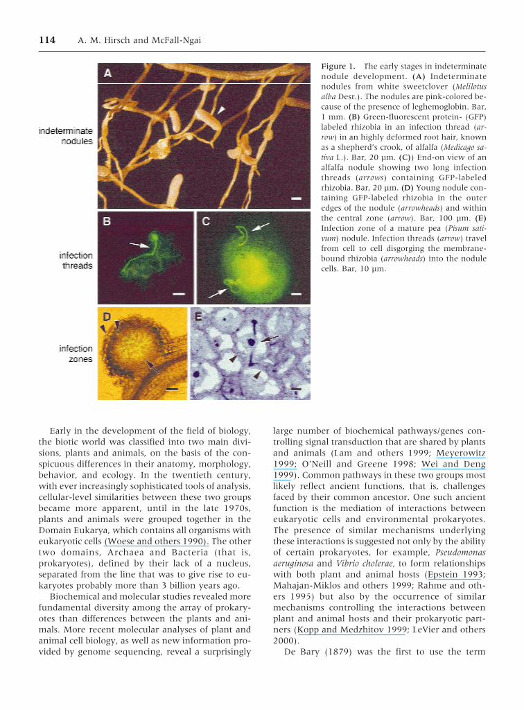
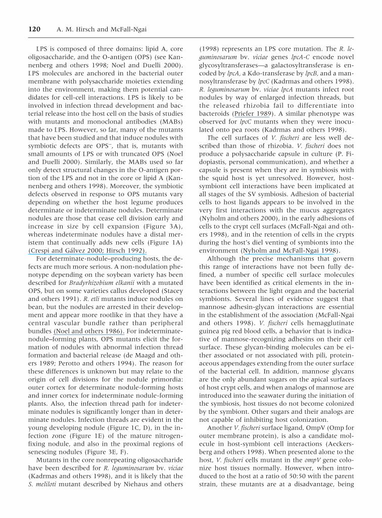
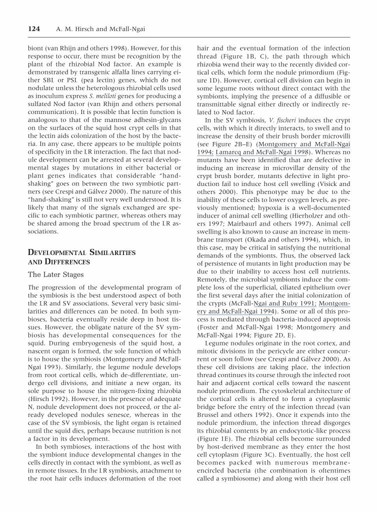
Figure 1. The early stages in indeterminatenodule development. (A) Indeterminatenodules from white sweetclover (Melilotusalba Desr.). The nodules are pink-colored be-cause of the presence of leghemoglobin. Bar,1 mm. (B) Green-fluorescent protein- (GFP)labeled rhizobia in an infection thread (ar-row) in an highly deformed root hair, knownas a shepherd’s crook, of alfalfa (Medicago sa-tiva L.). Bar, 20 µm. (C)) End-on view of analfalfa nodule showing two long infectionthreads (arrows) containing GFP-labeledrhizobia. Bar, 20 µm. (D) Young nodule con-taining GFP-labeled rhizobia in the outeredges of the nodule (arrowheads) and withinthe central zone (arrow). Bar, 100 µm. (E)Infection zone of a mature pea (Pisum sati-vum) nodule. Infection threads (arrow) travelfrom cell to cell disgorging the membrane-bound rhizobia (arrowheads) into the nodulecells. Bar, 10 µm.
114 A. M. Hirsch and McFall-Ngai
“symbiosis,” which he defined as the “living to-gether of differently named organisms” (“desZusammenlebens ungleichnamiger Organismen”).In current usage, symbiosis often implies mutual-ism—a beneficial arrangement in which, in an idealsituation, each partner gives and takes equally, butDe Bary used this term to describe both symbioticand parasitic interactions, in which one partnertakes more than gives. Pathogenesis is microbialparasitism that results in a disease or infection. Mostof these complex interactions are also defined on thebasis of nutrition, but the benefits (or stakes) canalso be physical ones. In any case, how modern-daysymbiotic interactions between prokaryotes andtheir plant and animal partners evolved is unclear.However, one hypothesis is that parasitism or patho-genesis is the default, and the host is manipulated byits parasite (see Corsaro and others 1999; Lederberg2000; Steinert and others 2000). One of the manyquestions that a study of diverse eukaryotic-prokaryotic partnerships might address is whetherthe first bi-domain associations were mutualistic,parasitic, or something else.
This review explores some of the similarities anddifferences between a well-studied plant-bacterialsymbiosis, the relationship between legumes and ni-trogen-fixing rhizobia, and a more recently devel-oped model of animal-bacterial symbiosis, the asso-ciation of the squid Euprymna scolopes (Figure 2A)
and its luminous prokaryotic partner, Vibrio fischeri.A comparison of these associations offers the oppor-tunity to consider which features are shared bybroadly divergent host-symbiont interactions andwhich characteristics may be particular to interac-tions with either plants or animals, but not both. Inaddition, because both of these symbioses are con-sidered beneficial or mutualistic, analyses of theirsimilarities and differences should provide insightinto traits that are unique to beneficial interactions.Similarly, if these shared responses are also presentin parasitic/pathogenic associations, as well as acrossthese broad phylogenetic boundaries, they may rep-resent a class of general responses that mediate plantand animal interactions with microbes no matterwhat the outcome of the interaction.
THE GENERAL NATURE OF THE SYMBIOSES
Both the legume-rhizobia (LR) and the squid-vibrio(SV) symbioses have been reviewed individually re-cently (Crespi and Galvez 2000; McFall-Ngai 1999;Stougaard 2000; Visick and McFall-Ngai 2000).Thus, detailed descriptions in this review will be re-stricted to those aspects that are useful in the com-parison of the associations.
Symbioses are often classified on the basis of theirbroad characteristics, for example, how they are
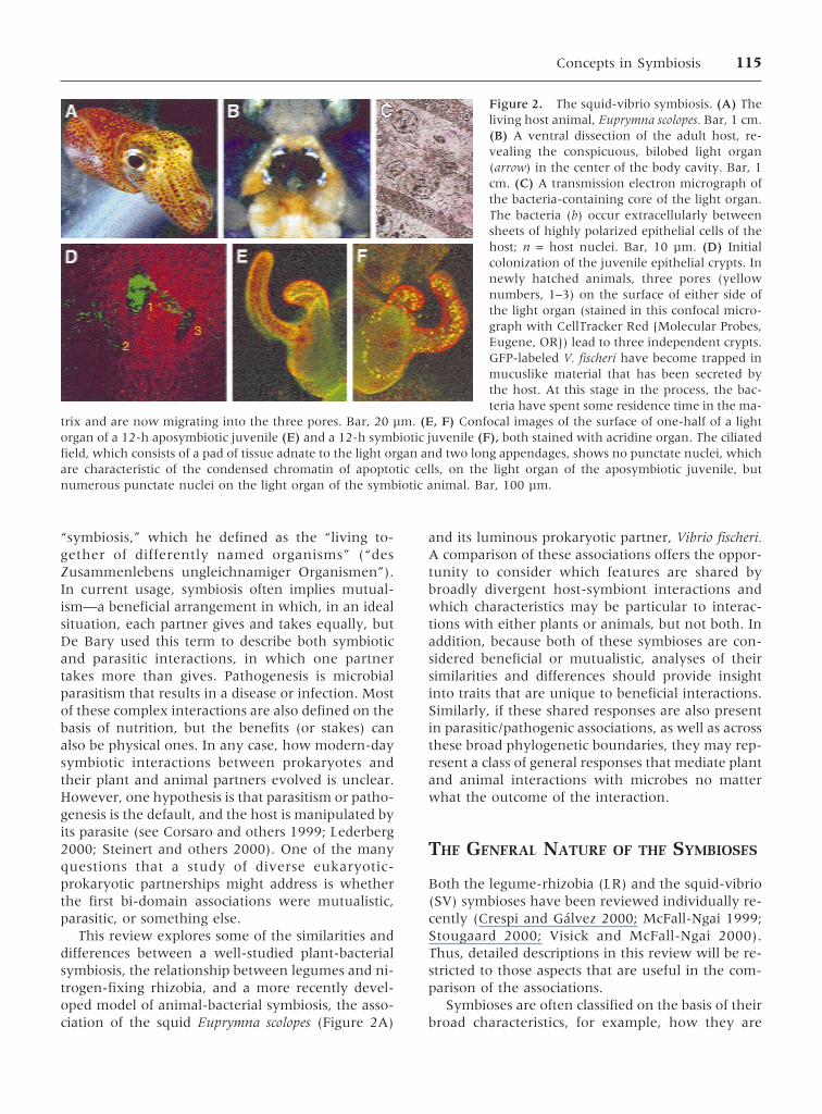
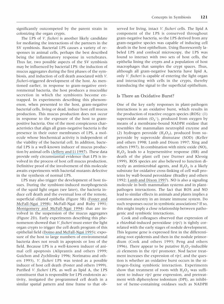
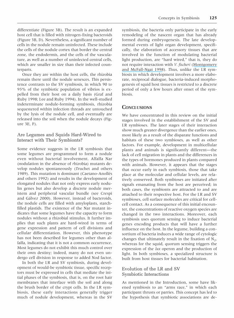
Figure 2. The squid-vibrio symbiosis. (A) Theliving host animal, Euprymna scolopes. Bar, 1 cm.(B) A ventral dissection of the adult host, re-vealing the conspicuous, bilobed light organ(arrow) in the center of the body cavity. Bar, 1cm. (C) A transmission electron micrograph ofthe bacteria-containing core of the light organ.The bacteria (b) occur extracellularly betweensheets of highly polarized epithelial cells of thehost; n = host nuclei. Bar, 10 µm. (D) Initialcolonization of the juvenile epithelial crypts. Innewly hatched animals, three pores (yellownumbers, 1–3) on the surface of either side ofthe light organ (stained in this confocal micro-graph with CellTracker Red [Molecular Probes,Eugene, OR]) lead to three independent crypts.GFP-labeled V. fischeri have become trapped inmucuslike material that has been secreted bythe host. At this stage in the process, the bac-teria have spent some residence time in the ma-
trix and are now migrating into the three pores. Bar, 20 µm. (E, F) Confocal images of the surface of one-half of a lightorgan of a 12-h aposymbiotic juvenile (E) and a 12-h symbiotic juvenile (F), both stained with acridine organ. The ciliatedfield, which consists of a pad of tissue adnate to the light organ and two long appendages, shows no punctate nuclei, whichare characteristic of the condensed chromatin of apoptotic cells, on the light organ of the aposymbiotic juvenile, butnumerous punctate nuclei on the light organ of the symbiotic animal. Bar, 100 µm.
Concepts in Symbiosis 115
maintained between generations, whether they areobligate or facultative under field conditions, andthe types of “products” that are exchanged betweenthe partners (Douglas 1994). Both the LR and SVsymbioses are horizontally transmitted betweengenerations; that is, the symbiont is not passed in oron the host’s germ cells, but rather with each hostgeneration, the symbiont must be acquired anewfrom the environment. In contrast with the faculta-tive LR associations, which occur only under condi-tions of nitrogen limitation in the soil, the SV sym-bioses appear to be obligate for the squid; squid hostshave never been found without their symbionts.This difference in the two associations is reflected inthe nature of the exchange between the partners. Inthe LR symbiosis, the bacterial cells are believed toundergo a terminal differentiation, at least in thecase of those bacteroids living within the cells ofindeterminate nodule-forming hosts (Figure 3C, D).The bacteroids fix nitrogen and transport nitrog-enous compounds to the legume (Hirsch 1992).Thus, the benefit of this symbiosis to the host is nu-tritional, and whether a symbiosis is formed andwhether it persists depends on environmental con-ditions. In contrast, in the SV symbiosis, the extra-cellular bacterial partners produce light, which thehost uses to camouflage itself, presumably as an es-
sential antipredatory strategy (McFall-Ngai 1990).However, under laboratory conditions, the squidhost can be antibiotically cured of its symbionts withno ill physiologic effects (Doino and McFall-Ngai1995). In other words, no evidence exists that thesymbionts provide essential nutrients to the host. Inaddition, although the bacterial cells differentiate inthe SV symbiosis, it is not a terminal differentiation.V. fischeri cells can, and do each day (see below),revert to their free-living state (Graf and Ruby 1998;Nyholm and McFall-Ngai 1998). Another obviousdifference between the two associations is in thetype of environment in which they are found. TheLR symbiosis is terrestrial, whereas the SV relation-ship is aquatic. These important characteristics of thetwo symbioses define the essential nature of the twoassociations and affect the patterns of their establish-ment, development, and stable maintenance overthe life history of the host.
INITIATION OF THE INTERACTION
In horizontally transmitted symbioses, mechanismsmust exist to bring the symbiont into the vicinity ofsusceptible host tissues. This process has been exten-sively studied in the LR symbiosis, and a wide vari-
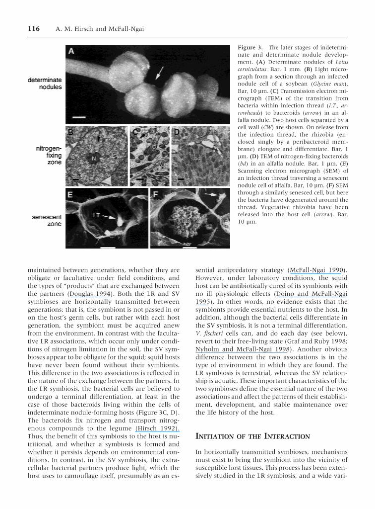
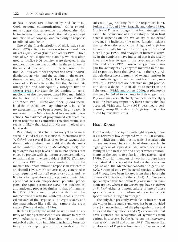
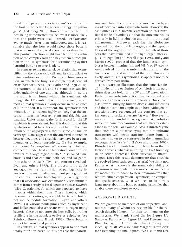
Figure 3. The later stages of indetermi-nate and determinate nodule develop-ment. (A) Determinate nodules of Lotuscorniculatus. Bar, 1 mm. (B) Light micro-graph from a section through an infectednodule cell of a soybean (Glycine max).Bar, 10 µm. (C) Transmission electron mi-crograph (TEM) of the transition frombacteria within infection thread (I.T., ar-rowheads) to bacteroids (arrow) in an al-falfa nodule. Two host cells separated by acell wall (CW) are shown. On release fromthe infection thread, the rhizobia (en-closed singly by a peribacteroid mem-brane) elongate and differentiate. Bar, 1µm. (D) TEM of nitrogen-fixing bacteroids(bd) in an alfalfa nodule. Bar, 1 µm. (E)Scanning electron micrograph (SEM) ofan infection thread traversing a senescentnodule cell of alfalfa. Bar, 10 µm. (F) SEMthrough a similarly senesced cell, but herethe bacteria have degenerated around thethread. Vegetative rhizobia have beenreleased into the host cell (arrow). Bar,10 µm.
116 A. M. Hirsch and McFall-Ngai
ety of genes and chemical signals produced by eitherthe host or symbiont have been identified as crucialplayers in mediating this process. In contrast, onlythe broad outlines of the process by which the lightorgan is induced have been defined in the SV sym-biosis, but the little that is known suggests that someintriguing similarities exist between this associationand the LR symbiosis.
Attraction
The LR symbiosis takes place in the soil, which iscomposed of a suspension of particles made up oforganic and inorganic material dispersed in water.Compared with the relatively chaotic, aquatic envi-ronment, the soil appears relatively stable. However,soil can be inundated with torrential rains, leadingto erosion and disruption of soil layers. Rhizobiamay also be widely dispersed and few in number,especially in soils where legumes do not routinelygrow. Some estimates have suggested that theremay be fewer than 102 rhizobial cells/g of soil(Singleton and others 1992). Therefore, to maximizethe possibility of interaction, legume seeds and rootssecrete flavonoids and related molecules that attractthe rhizobia. These chemoattracting molecules,which exhibit some bacterial strain specificity, alsoserve to induce rhizobial nod genes so that the bac-teria synthesize the primary morphogenetic signal(Nod factor) for inducing the host’s response (seereviews by Crespi and Galvez 2000; Long 1996;Schultze and Kondorosi 1998). Nod factors are vari-able-length N-acetylglucosamine oligomers with ei-ther a C-16 or C-18 acyl tail at the nonreducing endand various other substitutions at the reducing end(Figure 4A). Nod factors appear to be the mainrhizobial inducer molecules for nodulation becausethe purified molecules elicit, in a host-specific way,many of the plant responses observed in the earlystages of nodule formation (Crespi and Galvez2000). Eventually, the rhizobia enter a deformedroot hair by way of an infection thread (Figure 1B),but cell-cell contact is required for the thread toform.
Recent studies of the SV association have re-vealed striking similarities to the LR symbiosis inthese early events. On hatching, squid tissues thatare destined to become symbiotic are exposed to mi-crobes in the ambient seawater, which bathes theinternal squid tissues through the normal ventila-tory movements of the host. The interaction of thehost with environmental gram-negative bacteria in-duces the secretion of mucuslike material in the vi-cinity of susceptible host tissues (Nyholm and others2000). Juvenile squid have unique, complex ciliated
fields of epithelia (McFall-Ngai and Ruby 1991) thatmaintain the secreted mass in place, preventing itfrom being washed away by the ventilatory currentsof the host (Figure 5A). Bacteria aggregate in thesecreted matrix of this mucuslike material and, bysome yet undetermined mechanism, the populationof V. fischeri cells preferentially accumulates in thisaggregate. After a residence time of several hourswithin the matrix, the bacterial cells migrate to andenter pores on the surface of the light organ (Figure2D). These pores lead, by way of long ciliated ducts,to epithelia-lined crypt spaces that become filled bythe growing population of V. fischeri cells. Althoughother types of bacterial cells will aggregate, only V.fischeri cells are capable of successfully completingthis migration to enter the crypts where they prolif-erate. The bacterial symbionts remain extracellular,
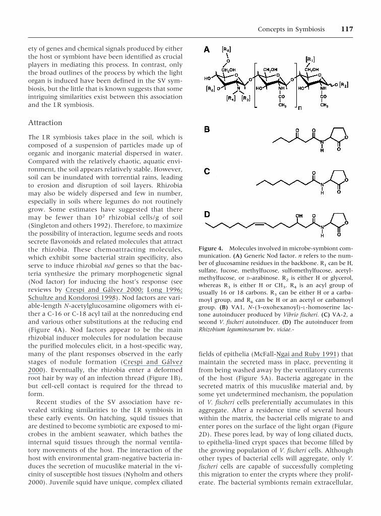
Figure 4. Molecules involved in microbe-symbiont com-munication. (A) Generic Nod factor. n refers to the num-ber of glucosamine residues in the backbone. R1 can be H,sulfate, fucose, methylfucose, sulfomethylfucose, acetyl-methylfucose, or D-arabinose. R2 is either H or glycerol,whereas R3 is either H or CH3. R4 is an acyl group ofusually 16 or 18 carbons. R5 can be either H or a carba-moyl group, and R6 can be H or an acetyl or carbamoylgroup. (B) VA1, N-(3-oxohexanoyl)-L-homoserine lac-tone autoinducer produced by Vibrio fischeri. (C) VA-2, asecond V. fischeri autoinducer. (D) The autoinducer fromRhizobium leguminosarum bv. viciae.-
Concepts in Symbiosis 117
associated with the apical surfaces of the host cryptcells, throughout the life history of the host (Figure2C). Studies with V. fischeri mutants that are defec-tive in motility (Graf and others 1994) indicate that,although they aggregate, they do not leave the ma-trix to colonize host tissues. In addition, the bacteria,when they do eventually move to colonize, show astrong vector toward the site of entry, suggestingthat chemotaxis is also essential (Figure 2D).
The principal differences between the LR and theSV symbioses in these early events are in the orderin which they occur. In the relatively structured en-vironment of the soil, more extensive chemical gra-dients can be established. These gradients appear toprovide the mechanism by which the rhizobial sym-bionts are initially recruited to the root from thegeneral rhizosphere population of bacteria; that is,their enrichment in the vicinity of the root hair is adirect result of chemotaxis to the root. In the fluidenvironment of the seawater, which is dominatedby high shear stress and turbulent flow, stable
chemical gradients are less apt to form. Thus, it islikely that only after the bacteria accumulate in themucuslike material that is adjacent to the susceptibletissues does chemotaxis of the bacteria toward hosttissues play a significant role in colonization. Themolecules that attract the two symbionts are likelyto be different. For rhizobia, plant-secreted second-ary metabolites (flavonoids, anthocyanins and re-lated molecules [Paiva 2000]) are the major attrac-tants, whereas for the vibrios no specific chemicalhas been identified yet.
Autoinduction
Many bacterial species that occur in associationswith eukaryotic tissues, whether the relationship ispathogenic or beneficial, exhibit a group behaviorcalled “quorum-sensing” (Fuqua and others 1996).In this behavior, the bacteria constitutively producelow levels of one or more specific pheromone-likemolecules, or autoinducers, that accumulate in thesurrounding environment only when that species ofbacterium achieves a high population density in aconfined area (Visick and Ruby 1999). In manygram-negative bacteria, these autoinducers are afamily of chemically distinct, but homologous, acyl-homoserine lactones (HSL; Figure 4B–D). Havingreached a critical ambient concentration, the auto-inducer diffuses back into the bacteria, positively up-regulating its own production, as well as the tran-scription of a series of other genes organized as aregulon. Typically, the gene expression up-regulatedby autoinducers directs the synthesis of productsthat are characteristic of the symbiotic state.
The phenomenon of autoinduction was first de-scribed in V. fischeri, which was found to produceluminescence in culture only after achieving a highpopulation density (Nealson 1999; Nealson andMarkovitz 1970; Nealson and others 1970). The luxIgene of the luminescence (or lux) regulon encodesthe gene that directs synthesis of a 3-oxo-hexanoylHSL (Figure 4B) in V. fischeri (Eberhard and others1981). The product of the luxR gene is a transcrip-tional activator that, when bound to autoinducer,directs the induction of the luxCDABE genes, whichencode the structural proteins required to drive theemission of bioluminescence (Engebrecht and oth-ers 1983). Recently, a second autoinducer synthasegene (ainS) and an associated regulatory gene (ainR)have been identified in V. fischeri (Gilson and others1995). The relative roles of these two autoinducers(Figure 4B, C) in the control of bacterial lumines-cence in the various niches occupied by V. fischerihave not yet been fully defined (Visick and others2000). Homologues of luxI and luxR have been
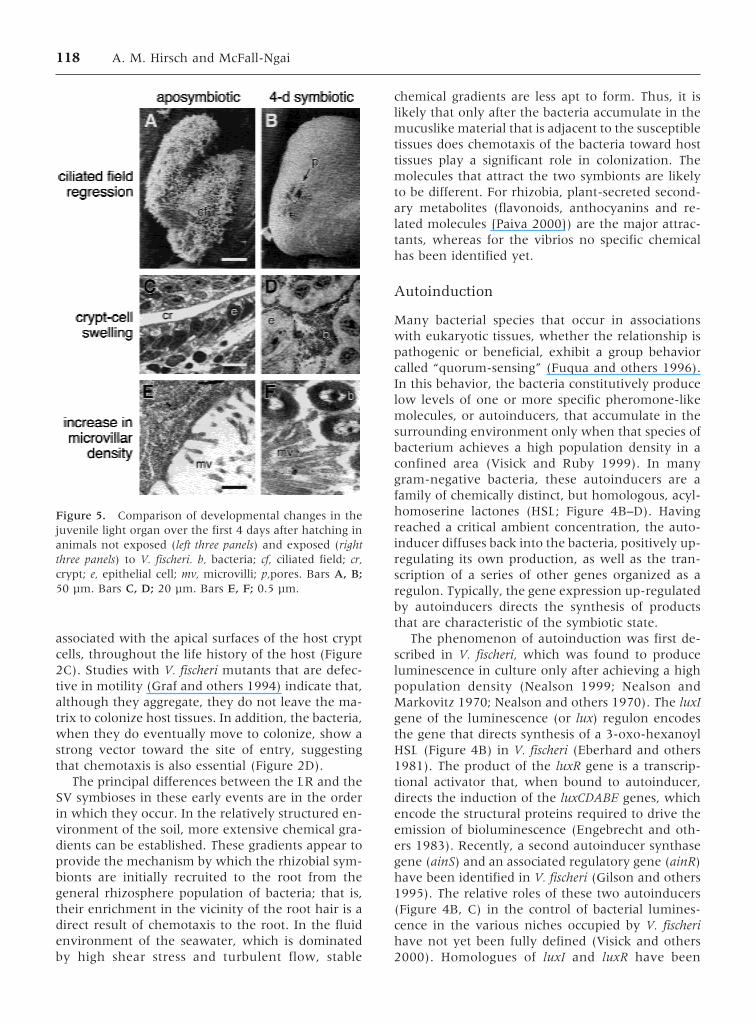
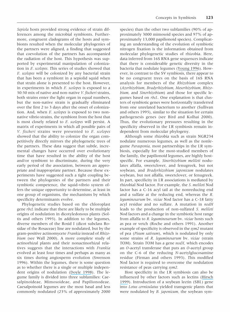
Figure 5. Comparison of developmental changes in thejuvenile light organ over the first 4 days after hatching inanimals not exposed (left three panels) and exposed (rightthree panels) to V. fischeri. b, bacteria; cf, ciliated field; cr,crypt; e, epithelial cell; mv, microvilli; p,pores. Bars A, B;50 µm. Bars C, D; 20 µm. Bars E, F; 0.5 µm.
118 A. M. Hirsch and McFall-Ngai
found in at least 15 other bacterial genera, includingRhizobium, Pseudomonas, and Agrobacterium (Dunnyand Winans 1999).
Studies of V. fischeri in symbioses, such as that inEuprymna scolopes, have revealed that 3-oxo-hexanoyl HSL accumulates to inducing levels in theconfines of the light organ crypts (Boettcher andRuby 1995), producing a brightly luminescentpopulation of symbionts (Boettcher and Ruby1990). When and where during the initiation of theSV association the initial autoinduction takes placeremains unresolved. Perhaps the several-hour resi-dence time of V. fischeri in the aggregates outside ofthe light organ is a period when the bacteria inducespecific genes, such as those associated with autoin-duction, that prepare the bacteria for interactionwith host tissues.
Rhizobia also undergo autoinduction or quorum-sensing. The rhiABC (rhi for rhizosphere-expressed)genes of R. leguminosarum bv. viciae are expressed inthe rhizosphere or in stationary-phase laboratorycultures. RhiA is the most prominent single proteinof the cell and is not expressed in bacteroids, thenitrogen-fixing form of rhizobia (Dibb and others1984). The rhiABC operon is regulated by RhiR andis induced by N-acylated homoserine lactones (AHL;Figure 4D) (Gray and others 1996). The rhiI gene isalso regulated by rhiR in a cell-density–dependentmanner, and like luxI, rhiI appears to be involved inAHL synthesis (Rodelas and others 1999). It is notknown how important autoinduction is for nodula-tion. In R. leguminosarum bv. viciae, the rhi genes arelocated within the nod/nol/noe operons, and repres-sion of rhi expression by nod/nol/noe-induced com-pounds suggests an interaction between these tworegulons (Economou and others 1989). Defects innodule development are detected in certain nod rhidouble mutants (Cubo and others 1992). As in theSV symbiosis, the aggregation of rhizobia in therhizosphere before root hair penetration may be re-quired for the induction of genes for later stages ofthe symbiosis. In addition, in R. leguminosarum bv.phaseoli, more cells survive stationary phase aftercarbon or nitrogen starvation if grown at a highrather than a low density, and survival of low-density cells can be improved by adding AHL(Thorne and Williams 1999).
Cell Surfaces
Rhizobia are typical gram-negative bacteria with acytoplasmic and outer membrane enclosing a peri-plasmic space. They secrete various types of extra-cellular material, much of which is composed ofpolysaccharides—lipopolysaccharides (LPS), capsu-
lar polysaccharides (CPS), cyclic b-glucans, andacidic exopolysaccharides (EPS); the latter are se-creted into the culture medium. Mutants that aredefective in the production of these polysaccharidesare often blocked in various stages of nodule devel-opment, particularly nodule invasion by means ofinfection threads.
The succinoglycan biosynthetic pathway for exo-polysaccharide production in Sinorhizobium meliloti isperhaps one of the best understood for the rhizobialcell surface molecules. This polysaccharide is an oc-tasaccharide polymer composed of one galactoseand seven glucose residues, with acetyl, succinyl,and pyruvyl modifications (Aman and others 1981;Reuber and Walker 1993). S. meliloti mutants that donot produce EPS or synthesize defective EPS formsmall, bacteria-free nodules on alfalfa with infectionthreads that abort within the root hairs (Cheng andWalker 1998; Finan and others 1985; Leigh and oth-ers 1987). One hypothesis explaining the function ofEPS is that it serves as a suppressor of plant defensereactions, thereby enabling rhizobia to enter thehost cell (Becker and others 2000). Another func-tion for EPS, especially low molecular weight EPS II,is that it serves as a signal molecule (Gonzalez andothers 1996). Other experiments in which EPS mu-tants were inoculated onto transgenic legumes car-rying a nonhost lectin gene demonstrate that rhizo-bial exopolysaccharide may interact with lectin inthe attachment stages of nodule formation (vanRhijn and others 1998; van Rhijn and others per-sonal communication. The specific binding of a le-gume lectin to a compatible Rhizobium allows thetwo symbionts to recognize each other (see Hirsch1999). This binding, along with other proteins in-volved in rhizobial attachment, enables a consor-tium of bacteria to become established at the tip of asusceptible root hair. The resulting increase in con-centration of compatible Nod factor brings aboutroot hair deformation and, at higher concentrations,infection thread formation and nodule developmenton a heterologous host (van Rhijn and others 1998;van Rhijn and others submitted). Thus, EPS appearsto play a critical role in the earliest stages of noduledevelopment.
A symbiotically active form of a strain-specific Kantigen, a capsular polysaccharide containing a Kdo(3-deoxy-D-manno-octulosonic acid derivative) en-ables S. meliloti AK631 to nodulate alfalfa eventhough it synthesizes neither succinoglycan or EPSII (Putnoky and others 1990). Mutagenizing genesinvolved in K antigen synthesis eliminated AK631’sability to induce nitrogen-fixing nodules (Campbelland others 1998). Several of these mutants werealso found to be defective in LPS.
Concepts in Symbiosis 119
LPS is composed of three domains: lipid A, coreoligosaccharide, and the O-antigen (OPS) (see Kan-nenberg and others 1998; Noel and Duelli 2000).LPS molecules are anchored in the bacterial outermembrane with polysaccharide moieties extendinginto the environment, making them potential can-didates for cell-cell interactions. LPS is likely to beinvolved in infection thread development and bac-terial release into the host cell on the basis of studieswith mutants and monoclonal antibodies (MABs)made to LPS. However, so far, many of the mutantsthat have been studied and that induce nodules withsymbiotic defects are OPS−, that is, mutants withsmall amounts of LPS or with truncated OPS (Noeland Duelli 2000). Similarly, the MABs used so faronly detect structural changes in the O-antigen por-tion of the LPS and not in the core or lipid A (Kan-nenberg and others 1998). Moreover, the symbioticdefects observed in response to OPS mutants varydepending on whether the host legume producesdeterminate or indeterminate nodules. Determinatenodules are those that cease cell division early andincrease in size by cell expansion (Figure 3A),whereas indeterminate nodules have a distal mer-istem that continually adds new cells (Figure 1A)(Crespi and Galvez 2000; Hirsch 1992).
For determinate-nodule–producing hosts, the de-fects are much more serious. A non-nodulation phe-notype depending on the soybean variety has beendescribed for Bradyrhizobium elkanii with a mutatedOPS, but on some varieties callus developed (Staceyand others 1991). R. etli mutants induce nodules onbean, but the nodules are arrested in their develop-ment and appear more rootlike in that they have acentral vascular bundle rather than peripheralbundles (Noel and others 1986). For indeterminate-nodule–forming plants, OPS mutants elicit the for-mation of nodules with abnormal infection threadformation and bacterial release (de Maagd and oth-ers 1989; Perotto and others 1994). The reason forthese differences is unknown but may relate to theorigin of cell divisions for the nodule primordia:outer cortex for determinate nodule-forming hostsand inner cortex for indeterminate nodule-formingplants. Also, the infection thread path for indeter-minate nodules is significantly longer than in deter-minate nodules. Infection threads are evident in theyoung developing nodule (Figure 1C, D), in the in-fection zone (Figure 1E) of the mature nitrogen-fixing nodule, and also in the proximal regions ofsenescing nodules (Figure 3E, F).
Mutants in the core nonrepeating oligosaccharidehave been described for R. leguminosarum bv. viciae(Kadrmas and others 1998), and it is likely that theS. meliloti mutant described by Niehaus and others
(1998) represents an LPS core mutation. The R. le-guminosarum bv. viciae genes lpcA-C encode novelglycosyltransferases—a galactosyltransferase is en-coded by lpcA, a Kdo-transferase by lpcB, and a man-nosyltransferase by lpcC (Kadrmas and others 1998).R. leguminosarum bv. viciae lpcA mutants infect rootnodules by way of enlarged infection threads, butthe released rhizobia fail to differentiate intobacteroids (Priefer 1989). A similar phenotype wasobserved for lpcC mutants when they were inocu-lated onto pea roots (Kadrmas and others 1998).
The cell surfaces of V. fischeri are less well de-scribed than those of rhizobia. V. fischeri does notproduce a polysaccharide capsule in culture (P. Fi-dopiastis, personal communication), and whether acapsule is present when they are in symbiosis withthe squid host is yet unresolved. However, host-symbiont cell interactions have been implicated atall stages of the SV symbiosis. Adhesion of bacterialcells to host ligands appears to be involved in thevery first interactions with the mucus aggregates(Nyholm and others 2000), in the early adhesions ofcells to the crypt cell surfaces (McFall-Ngai and oth-ers 1998), and in the retention of cells in the cryptsduring the host’s diel venting of symbionts into theenvironment (Nyholm and McFall-Ngai 1998).
Although the precise mechanisms that governthis range of interactions have not been fully de-fined, a number of specific cell surface moleculeshave been identified as critical elements in the in-teractions between the light organ and the bacterialsymbionts. Several lines of evidence suggest thatmannose adhesin-glycan interactions are essentialin the establishment of the association (McFall-Ngaiand others 1998). V. fischeri cells hemagglutinateguinea pig red blood cells, a behavior that is indica-tive of mannose-recognizing adhesins on their cellsurface. These glycan-binding molecules can be ei-ther associated or not associated with pili, protein-aceous appendages extending from the outer surfaceof the bacterial cell. In addition, mannose glycansare the only abundant sugars on the apical surfacesof host crypt cells, and when analogs of mannose areintroduced into the seawater during the initiation ofthe symbiosis, host tissues do not become colonizedby the symbiont. Other sugars and their analogs arenot capable of inhibiting host colonization.
Another V. fischeri surface ligand, OmpV (Omp forouter membrane protein), is also a candidate mol-ecule in host-symbiont cell interactions (Aeckers-berg and others 1998). When presented alone to thehost, V. fischeri cells mutant in the ompV gene colo-nize host tissues normally. However, when intro-duced to the host at a ratio of 50:50 with the parentstrain, these mutants are at a disadvantage, being
120 A. M. Hirsch and McFall-Ngai
significantly outcompeted by the parent strain incolonizing the organ crypts.
The LPS of V. fischeri is another likely candidatefor mediating the interactions of the partners in theSV symbiosis. Bacterial LPS causes a variety of re-sponses in animal cells, perhaps the best describedbeing the inflammatory response in vertebrates.Thus far, two possible aspects of the SV symbiosismay be influenced by bacterial LPS: the induction ofmucus aggregates during the first phases of the sym-biosis, and induction of cell death associated with V.fischeri–triggered development of the host. As men-tioned earlier, in response to gram-negative envi-ronmental bacteria, the host produces a mucuslikesecretion in which the symbionts become en-trapped. In experiments describing this phenom-enon, when presented to the host, gram-negativebacterial cells, living or dead, induce host cell mucusproduction. This mucus production does not occurin response to the exposure of the host to gram-positive bacteria. One of the most conspicuous char-acteristics that align all gram-negative bacteria is thepresence in their outer membranes of LPS, a mol-ecule whose biochemical activity is independent ofthe viability of the bacterial cell. In addition, bacte-rial LPS is a well-known inducer of mucus produc-tion by animal cells (Jeffery and Li 1997). These dataprovide only circumstantial evidence that LPS is in-volved in the process of host cell mucus production.Definitive proof of the involvement of this moleculeawaits experiments with bacterial mutants defectivein the synthesis of normal LPS.
LPS may also trigger the development of host tis-sues. During the symbiont-induced morphogenesisof the squid light organ (see later), the bacteria in-duce cell death and the eventual loss of the remote,superficial ciliated epithelia (Figure 5B) (Foster andMcFall-Ngai 1998); McFall-Ngai and Ruby 1991;Montgomery and McFall-Ngai 1994) that are in-volved in the suspension of the mucus aggregates(Figure 2D). Early experiments describing this phe-nomenon showed that V. fischeri must enter the lightorgan crypts to trigger the cell death program of thisepithelial field (Doino and McFall-Ngai 1995); expo-sure of the host to large numbers of environmentalbacteria does not result in apoptosis or loss of thefield. Because LPS is a well-known inducer of ani-mal cell apoptosis (Aliprantis and others 1999;Guichon and Zychlinsky 1996; Norimatsu and oth-ers 1995), V. fischeri LPS was tested as a possibleinducer of host cell death (Foster and others 1999).Purified V. fischeri LPS, as well as lipid A, the LPSconstituent that is responsible for LPS endotoxin ac-tivity, instigated the programmed cell death in asimilar spatial pattern and time frame to that ob-
served for living, intact V. fischeri cells. The lipid Acomponent of the LPS is conserved throughoutgram-negative bacteria, so the LPS derived from anygram-negative species was capable of inducing celldeath in the host epithelium. Using fluorescently la-beled LPS and confocal microscopy, the LPS wasfound to interact with two sets of host cells, theepithelia lining the crypts and a population of hostmacrophages that samples the crypt spaces. Thus,although all gram-negative bacteria have lipid A,only V. fischeri is capable of entering the light organand interacting with cells in the crypts, therebytransducing the signal to the superficial epithelium.
Is There an Oxidative Burst?
One of the key early responses in plant-pathogeninteractions is an oxidative burst, which results inthe production of reactive oxygen species (ROS): (1)superoxide anion (O2
−), produced from oxygen bymeans of a membrane-bound NADPH oxidase thatresembles the mammalian neutrophil enzyme and(2) hydrogen peroxide (H2O2), produced from su-peroxide by superoxide dismutase (SOD) (Kellerand others 1998; Lamb and Dixon 1997; Xing andothers 1997). In combination with nitric oxide (NO),H2O2 leads to a hypersensitive response (HR) anddeath of the plant cell (see Durner and Klessig1999). ROS species are also believed to function di-rectly as antimicrobial agents, and H2O2 is a likelysubstrate for oxidative cross-linking of cell wall pro-teins by wall-bound peroxidase (Bradley and others1992; Lamb and Dixon 1997). NO is also a signalingmolecule in both mammalian systems and in plant-pathogen interactions. The fact that ROS and NOlead to similar effects in animals and plants implies acommon ancestry in an innate immune system. Dosuch responses occur in symbiotic associations? If so,this might imply a close relationship between patho-genic and symbiotic interactions.
Cook and colleagues observed that expression ofa rhizobial-induced peroxidase (rip1) is tightly cor-related with the early stages of nodule development.This legume gene is expressed first in the differenti-ating root epidermis and then in the nodule primor-dium (Cook and others 1995; Peng and others1996). There appear to be putative H2O2-induciblecis elements in the rip1 promoter. Nod factor treat-ment increases the expression of rip1, and the ques-tion is whether an oxidative burst occurs in the ni-trogen-fixing symbiotic interaction. Experimentsshow that treatment of roots with H2O2 was suffi-cient to induce rip1 gene expression, and pretreat-ment with diphenylene iodonium (DPI), an inhibi-tor of heme-containing oxidases such as NADPH
Concepts in Symbiosis 121
oxidase, blocked rip1 induction by Nod factor (D.Cook, personal communications). Other experi-ments suggest that superoxide is produced after Nodfactor treatment, and its production, along with rip1induction in Medicago sp., occurred only in responseto sulfated Nod factor.
One of the first descriptions of nitric oxide syn-thase (NOS) activity in plants was in roots and nod-ules of Lupinus albus (Cueto and others 1996). Threesites of NADPH-diaphorase staining, which is oftenused to localize NOS activity, were detected in thenodules: in the vascular bundles, in the periphery ofthe infected zone, and in the infected zone of thenodule. However, other enzymes also have NADPH-diaphorase activity, and the staining might overes-timate the amount of NOS. The biological signifi-cance of NOS may lie in the fact that NO inhibitsnitrogenase and consequently nitrogen fixation(Meyer 1981). For example, NO binding to leghe-moglobin or the oxygen-regulated FixL may inhibitthe function of these two heme proteins (Mathieuand others 1998). Cueto and others (1996) specu-lated that rhizobial LPS may induce NOS, but so farno experiments have been reported. In any case it isnot certain how NO is involved in symbiotic inter-actions. No evidence of programmed cell death ex-ists in response to a compatible rhizobial strain, so itseems unlikely that ROS and NO are involved to alarge scale.
Respiratory burst activity has not yet been mea-sured in squid cells in response to interactions withV. fischeri, but several lines of evidence suggest thatthe oxidative environment is critical in the dynamicsof the symbiosis (Ruby and McFall-Ngai 1999). Thelight organ has high levels of an mRNA species thatencode a protein with significant sequence similarityto mammalian myeloperoxidase (MPO) (Tomarevand others 1993), a protein abundant in cells thatmediate the innate immune response in vertebrates(Klebanoff 1991). MPO converts H2O2, generated asa consequence of host cell respiratory burst, and ha-lide ions to hypohalous acid, a potent antimicrobialagent that acts on phagocytosed potential patho-gens. The squid peroxidase (SPO) has biochemicaland antigenic properties similar to that of mamma-lian MPO. SPO occurs in regions of the organ thatinteract directly with the symbionts, that is, the api-cal surfaces of the crypt cells, the crypt spaces, andthe macrophage-like cells that sample the cryptspaces (Weis and others 1996).
Bacteria typically are unable to withstand the ac-tivity of halide peroxidases but are known to rely ontwo mechanisms by which to circumvent this anti-microbial activity: by inhibiting respiratory burst ac-tivity or by competing with the peroxidase for the
substrate H2O2 resulting from the respiratory burst,Dukan and Touati 1996; Tartaglia and others 1989).Studies of V. fischeri suggest that both strategies areused. The occurrence of a respiratory burst in hostdefense depends on the availability of molecularoxygen. The luciferase (the mixed-function oxidasethat catalyzes the production of light) of V. fischerihas an unusually high affinity for oxygen (Ruby andMcFall-Ngai 1999), and analyses of luciferase activ-ity in the symbiosis have indicated that it drasticallylowers the free oxygen in the crypt spaces (Boet-tcher and others 1996). Lowered oxygen would im-pair the activity of any enzymes involved in the ini-tial respiratory burst that gives rise to the H2O2. Al-though direct measurements of oxygen tension inthe symbiotic light organ have not been made, mu-tants of V. fischeri that are defective in light produc-tion show a defect in their ability to persist in thelight organ (Visick and others 2000), a phenotypethat may be linked to a change in their oxygen use.V. fischeri may also effectively compete for the H2O2
resulting from any respiratory burst activity that hasoccurred. Visick and Ruby (1998) described a peri-plasmic, group III catalase in V. fischeri that is in-duced by oxidative stress.
HOST RANGE
The diversity of the squids with light organ symbio-ses is relatively low compared with the LR associa-tions, which are highly host specific. Bacterial lightorgans are found in a couple of dozen species ineight genera of sepiolid squids, which occur as afamily in both nearshore and deeper water environ-ments in the tropics to polar latitudes (McFall-Ngai1999). Thus far, members of two host groups havebeen studied, species of the IndoPacific genus Eu-prymna and the Mediterranean/Atlantic genus Se-piola. Strains of only two bacterial species, V. fischeriand V. logei, have been isolated from these host lightorgans (Fidopiastis and others 1998). All Euprymnaspp. analyzed thus far harbor V. fischeri in their sym-biotic tissues, whereas the Sepiola spp. have V. fischerior V. logei, either as a monoculture of one of thesespecies or as a mixed culture of these two vibriospecies within a single light organ.
The only data presently available for host range ofthe vibrios in the squid symbioses has been providedby (1) characterization of the phylogenies of severalhosts and their symbionts and (2) experiments thathave explored the recognition of symbionts fromvarious host species by the Hawaiian host Euprymnascolopes (Nishiguchi and others 1998). The molecularphylogenies of V. fischeri from various Euprymna and
122 A. M. Hirsch and McFall-Ngai
Sepiola hosts provided strong evidence of strain dif-ferences among the microbial symbionts. Further-more, congruent cladograms of the hosts and sym-bionts resulted when the molecular phylogenies ofthe partners were aligned, a finding that suggestedthat coevolution of the partners has accompaniedthe radiation of the host. This hypothesis was sup-ported by experimental manipulation of coloniza-tion in E. scolopes. This series of studies showed thatE. scolopes will be colonized by any bacterial strainthat has been a symbiont in a sepiolid squid whenthat strain alone is presented to the host. However,in experiments in which E. scolopes is exposed to a50:50 mix of native and non-native V. fischeri strains,both strains enter the light organ in equal numbers,but the non-native strain is gradually eliminatedover the first 2 to 3 days after the onset of coloniza-tion. And, when E. scolopes is exposed to two non-native vibrio strains, the symbiont from the host thatis most closely related to E. scolopes will persist. Amatrix of experiments in which all possible pairs ofV. fischeri strains were presented to E. scolopesshowed that the ability to colonize the organ com-petitively directly mirrors the phylogenetic trees ofthe partners. These data suggest that subtle, incre-mental changes have occurred over evolutionarytime that have resulted in the ability of the hostand/or symbiont to discriminate, during the veryearly period of the association, between an appro-priate and inappropriate partner. Because these ex-periments have suggested such a tight coupling be-tween the phylogenies of the partners and theirsymbiotic competence, the squid-vibrio system of-fers the unique opportunity to determine, at least inone group of organisms, the mechanisms by whichspecificity determinants evolve.
Phylogenetic studies based on the chloroplastgene rbcL indicate that there are likely to be multipleorigins of nodulation in dicotyledonous plants (Sol-tis and others 1995). In addition to the legumes,diverse members of the Rosid I (dicot subclass Ro-sidae of the Rosaceae) line are nodulated, but by thegram-positive actinomycete Frankia instead of Rhizo-bium (see Wall 2000). A more complete study ofactinorhizal plants and their nonactinorhizal rela-tives suggests that the interactions with Frankiaevolved at least four times and perhaps as many assix times during angiosperm evolution (Swenson1996). Within the legumes, there is some questionas to whether there is a single or multiple indepen-dent origins of nodulation (Doyle 1998). The le-gume family is divided into three subfamilies: Cae-salpinoideae, Mimosoideae, and Papilionodeae.Caesalpoinoid legumes are the most basal and lesscommonly nodulated (23% of approximately 2000
species) than the other two subfamilies (90% of ap-proximately 3000 mimosoid species and 97% of ap-proximately 13,000 papilionoid species). Complicat-ing an understanding of the evolution of symbioticnitrogen fixation is the information obtained frommolecular phylogenetic studies of rhizobia. Here,data inferred from 16S RNA gene sequences indicatethat there is considerable genetic diversity in thebacteria that nodulate legumes (Young 1996). How-ever, in contrast to the SV symbiosis, there appear tobe no congruent trees on the basis of 16S RNAanalysis for members of the Rhizobium complex(Azorhizobium, Bradyrhizobium, Mesorhizobium, Rhizo-bium, and Sinorhizobium) and those for specific le-gumes based on rbcL. One explanation is that clus-ters of symbiotic genes were horizontally transferredfrom one unrelated bacterium to another (Sullivanand others 1995), similar to the situation for certainpathogenesis genes (see Bird and Kolhai 2000).Thus, the evolutionary pressures resulting in thespecificity observed in the LR symbiosis remain in-dependent from molecular phylogeny.
Although some rhizobia such as strain NGR234nodulate numerous legumes, as well as the nonle-gume Parasponia, most partnerships in the LR sym-biosis, especially for the most-studied members ofthe family, the papilionoid legumes, are highly host-specific. For example, Sinorhizobium meliloti nodu-lates alfalfa, sweetclover, and fenugreek, but notsoybean, and Bradyrhizobium japonicum nodulatessoybean, but not alfalfa, sweetclover, or fenugreek.In part, specificity in LR associations is mediated byrhizobial Nod factor. For example, the S. meliloti Nodfactor has a C-16 acyl tail at the nonreducing endand a sulfate at the reducing end, whereas the R.leguminosarum bv. viciae Nod factor has a C-18 fattyacyl residue and no sulfate. A mutation in nodHleads to the production of non-sulfated S. melilotiNod factors and a change in the symbiotic host rangefrom alfalfa to R. leguminosarum bv. viciae hosts suchas pea or vetch (Roche and others 1991). Anotherexample of specificity is observed in the sym2 mutantof pea (Pisum sativum), which is nodulated by onlysome strains of R. leguminosarum bv. viciae (strainTOM). Strain TOM has a gene nodX, which encodesan O-acetyl transferase that puts an O-acetyl groupon the C-6 of the reducing N-acetylglucosamineresidue (Firman and others 1993). This modifiedNod factor is required to overcome the nodulationresistance of peas carrying sym2.
Host specificity in the LR symbiosis can also beinfluenced by other factors such as lectins (Hirsch1999). Introduction of a soybean lectin (SBL) geneinto Lotus corniculatus yielded transgenic plants thatwere nodulated by B. japonicum, the soybean sym-
Concepts in Symbiosis 123
biont (van Rhijn and others 1998). However, for thisresponse to occur, there must be recognition by theplant of the rhizobial Nod factor. An example isdemonstrated by transgenic alfalfa lines carrying ei-ther SBL or PSL (pea lectin) genes, which do notnodulate unless the heterologous rhizobial cells usedas inoculum express S. meliloti genes for producing asulfated Nod factor (van Rhijn and others personalcommunication). It is possible that lectin function isanalogous to that of the mannose adhesin-glycanson the surfaces of the squid host crypt cells in thatthe lectin aids colonization of the host by the bacte-ria. In any case, there appears to be multiple pointsof specificity in the LR interaction. The fact that nod-ule development can be arrested at several develop-mental stages by mutations in either bacterial orplant genes indicates that considerable “hand-shaking” goes on between the two symbiotic part-ners (see Crespi and Galvez 2000). The nature of this“hand-shaking” is still not very well understood. It islikely that many of the signals exchanged are spe-cific to each symbiotic partner, whereas others maybe shared among the broad spectrum of the LR as-sociations.
DEVELOPMENTAL SIMILARITIESAND DIFFERENCES
The Later Stages
The progression of the developmental program ofthe symbiosis is the best understood aspect of boththe LR and SV associations. Several very basic simi-larities and differences can be noted. In both sym-bioses, bacteria eventually reside deep in host tis-sues. However, the obligate nature of the SV sym-biosis has developmental consequences for thesquid. During embryogenesis of the squid host, anascent organ is formed, the sole function of whichis to house the symbiosis (Montgomery and McFall-Ngai 1993). Similarly, the legume nodule developsfrom root cortical cells, which de-differentiate, un-dergo cell divisions, and initiate a new organ, itssole purpose to house the nitrogen-fixing rhizobia(Hirsch 1992). However, in the presence of adequateN, nodule development does not proceed, or the al-ready developed nodules senesce, whereas in thecase of the SV symbiosis, the light organ is retaineduntil the squid dies, perhaps because nutrition is nota factor in its development.
In both symbioses, interactions of the host withthe symbiont induce developmental changes in thecells directly in contact with the symbiont, as well asin remote tissues. In the LR symbiosis, attachment tothe root hair cells induces deformation of the root
hair and the eventual formation of the infectionthread (Figure 1B, C), the path through whichrhizobia wend their way to the recently divided cor-tical cells, which form the nodule primordium (Fig-ure 1D). However, cortical cell division can begin insome legume roots without direct contact with thesymbionts, implying the presence of a diffusible ortransmittable signal either directly or indirectly re-lated to Nod factor.
In the SV symbiosis, V. fischeri induces the cryptcells, with which it directly interacts, to swell and toincrease the density of their brush border microvilli(see Figure 2B–E) (Montgomery and McFall-Ngai1994; Lamarcq and McFall-Ngai 1998). Whereas nomutants have been identified that are defective ininducing an increase in microvillar density of thecrypt brush border, mutants defective in light pro-duction fail to induce host cell swelling (Visick andothers 2000). This phenotype may be due to theinability of these cells to lower oxygen levels, as pre-viously mentioned; hypoxia is a well-documentedinducer of animal cell swelling (Hierholzer and oth-ers 1997; Mairbaurl and others 1997). Animal cellswelling is also known to cause an increase in mem-brane transport (Okada and others 1994), which, inthis case, may be critical in satisfying the nutritionaldemands of the symbionts. Thus, the observed lackof persistence of mutants in light production may bedue to their inability to access host cell nutrients.Remotely, the microbial symbionts induce the com-plete loss of the superficial, ciliated epithelium overthe first several days after the initial colonization ofthe crypts (McFall-Ngai and Ruby 1991; Montgom-ery and McFall-Ngai 1994). Some or all of this pro-cess is mediated through bacteria-induced apoptosis(Foster and McFall-Ngai 1998; Montgomery andMcFall-Ngai 1994; Figure 2D, E).
Legume nodules originate in the root cortex, andmitotic divisions in the pericycle are either concur-rent or soon follow (see Crespi and Galvez 2000). Asthese cell divisions are taking place, the infectionthread continues its course through the infected roothair and adjacent cortical cells toward the nascentnodule primordium. The cytoskeletal architecture ofthe cortical cells is altered to form a cytoplasmicbridge before the entry of the infection thread (vanBrussel and others 1992). Once it expends into thenodule primordium, the infection thread disgorgesits rhizobial contents by an endocytotic-like process(Figure 1E). The rhizobial cells become surroundedby host-derived membrane as they enter the hostcell cytoplasm (Figure 3C). Eventually, the host cellbecomes packed with numerous membrane-encircled bacteria (the combination is oftentimescalled a symbiosome) and along with their host cell
124 A. M. Hirsch and McFall-Ngai
differentiate (Figure 3B). The result is an expandedhost cell that is filled with nitrogen-fixing bacteroids(Figure 3B, D). Nevertheless, a significant number ofcells in the nodule remain uninfected. These includethe cells of the nodule cortex that border the centralzone, the endodermis, and the cells of the vascula-ture, as well as a number of uninfected central cells,which are smaller in size than their infected coun-terparts.
Once they are within the host cells, the rhizobiaremain there until the nodule senesces. This persis-tence contrasts to the SV symbiosis, in which 90 to95% of the symbiotic population of vibrios is ex-pelled from their host on a daily basis (Graf andRuby 1998; Lee and Ruby 1994). In the well-studiedindeterminate nodule-forming symbiosis, rhizobiasequestered within infection threads are untouchedby the lysis of the nodule cell, and eventually arereleased into the soil when the nodule decays (Fig-ure 3E, F).
Are Legumes and Squids Hard-Wired toInteract with Their Symbionts?
Some evidence suggests in the LR symbiosis thatsome legumes are programmed to form a noduleeven without bacterial involvement. Alfalfa Nar(nodulation in the absence of rhizobia) mutants de-velop nodules spontaneously (Truchet and others1989). This mutation is dominant (Caetano-Anollesand others 1992) and results in the development ofelongated nodules that not only express early nodu-lin genes but also develop a discrete nodule mer-istem and peripheral vascular bundle (see Crespiand Galvez 2000). However, instead of bacteroids,the nodule cells are filled with amyloplasts, starch-filled plastids. The existence of the Nar mutant in-dicates that some legumes have the capacity to formnodules without a rhizobial stimulus. It further im-plies that such plants are hard-wired in terms ofgene expression and pattern of cell divisions andcellular differentiation. However, this phenotypehas not been described for legumes other than al-falfa, indicating that it is not a common occurrence.Most legumes do not exhibit this much control overtheir own destiny; indeed, many do not even un-dergo cell division in response to added Nod factor.
In both the LR and SV symbiosis, during devel-opment of would-be symbiotic tissue, specific recep-tors must be expressed in cells that mediate the ini-tial phases of the symbiosis, that is, on the root hairmembranes that interface with the soil and alongthe brush border of the crypt cells. In the LR sym-biosis, these early interactions generally triggermuch of nodule development, whereas in the SV
symbiosis, the bacteria only participate in the earlyremodeling of the nascent organ that has alreadyformed during embryogenesis. The late develop-mental events of light organ development, specifi-cally, the elaboration of accessory tissues that areinvolved in the function of modulating bacteriallight production, are “hard wired,” that is, they donot require interaction with V. fischeri (Montgomeryand McFall-Ngai 1998). Thus, unlike the LR sym-biosis in which development involves a more elabo-rate, reciprocal dialogue, bacteria-induced morpho-genesis of squid host tissues is restricted to a discreteperiod of only a few hours after onset of the sym-biosis.
CONCLUSIONS
We have concentrated in this review on the initialstages involved in the establishment of the SV andLR symbioses. The later stages of their interactionshow much greater divergence than the earlier ones,most likely as a result of the disparate functions andhabitats of these two symbioses, as well as otherfactors. For example, development in multicellularplants and animals is significantly different—thelack of cell migration in plants and the differences inthe types of hormones produced in plants comparedwith animals. However, it appears that the stagesthat occur early in each symbiosis, those that takeplace at the molecular and cellular levels, are rela-tively conserved. Both symbioses are initiated aftersignals emanating from the host are perceived; inboth cases, the symbionts are attracted to and areattached to their respective host. For the LR and SVsymbioses, cell surface molecules are critical for cell-cell contact. As a consequence of this initial encoun-ter, the concentrations of reactive oxygen species arechanged in the two interactions. Moreover, eachsymbiosis uses quorum sensing to induce bacterialgenes encoding products that will have a furtherinfluence on the host. In the legume, building a con-sortium of bacteria induces a wide range of cytologicchanges that ultimately result in the fixation of N2,whereas for the squid, quorum sensing triggers theexpression of the lux operon and the production oflight. In both symbioses, a specialized structure isbuilt from host tissues for bacterial habitation.
Evolution of the LR and SVSymbiotic Interactions
As mentioned in the Introduction, some have lik-ened symbiosis to an “arms race,” in which eachparticipant thrusts or parries. This concept has led tothe hypothesis that symbiotic associations are de-
Concepts in Symbiosis 125
rived from parasitic associations—“Domesticatingthe host is the better long-term strategy for patho-gens” (Lederberg 2000). However, rather than thehost being domesticated, we believe it is more likelythat the prokaryote was “tamed.” Prokaryotesevolve much faster than eukaryotes, and it is rea-sonable that the host would select those bacteriathat were more likely to do good rather than harm.This positive selection might have led to the evolu-tion of the complex lock and key system of recogni-tion in the LR symbiosis for discriminating againstharmful bacteria or free-loaders.
In contrast to the master-slave relationship exem-plified by the eukaryotic cell and its chloroplast ormitochondrion or by the VA mycorrhizal associa-tion, in which the fungus is completely dependenton its host for survival (see Barker and Tagu 2000),the partners of the LR and SV symbioses can liveindependently of one another, although in naturethe squid is not found without its symbiont. Al-though the LR symbiosis is nutrition-based, as aremost animal symbioses, it only occurs in the absenceof N in the soil. If N is present, the symbiosis is notinitiated. Thus, it seems highly unlikely that the an-cestral interaction between plant and rhizobia wasparasitic. Unfortunately, the fossil record for the LRsymbiosis is nonexistent, but it is doubtful that thissymbiosis would have been present before the evo-lution of the angiosperms, that is, some 250 millionyears ago. Data suggest that the ancestral interactionbetween legumes and rhizobia may have been com-mensal or at least saprophytic. (1) For example,commensal Mesorhizobium loti become symbioticallycompetent under field and laboratory conditions ontransfer of a large region of DNA, a so-called sym-biosis island that contains both nod and nif genes,from other rhizobia (Sullivan and Ronson 1998; Sul-livan and others 1995). The process is no doubtanalogous to the transmission of pathogenicity is-lands seen in mammalian and plant pathogens, butthe end result is not homologous. (2) A suggestionthat LR association was evolutionarily less intimatecomes from a study of basal legumes such as Gleditsia(tribe Caesalpinieae), which are reported to haverhizobia within their roots. These rhizobia, whichpresumably fix nitrogen, resemble bacteroids, but donot induce nodule formation (Bryan and others1996). (3) Various nonlegumes such as sugar caneand kallar grass also associate with nitrogen-fixingbacteria; these do not enter the host cells, but ratherproliferate in the apoplast or live as epiphytes (seeReinhold-Hurek and Hurek 1998). These bacteriacannot be considered parasitic.
In contrast, animal symbioses appear to be almosttotally nutrition-based, so it is possible that parasit-
ism could have been the ancestral mode whereby aninvader evolved into a symbiotic form. However, theSV symbiosis is a notable exception to this nutri-tional mode of symbiosis in that the outcome resultsprimarily in light production and not in nutritionalenhancement. Moreover, each day, the vibrios areexpelled from the squid light organ, and the repopu-lation of the organ is the result of growth of thosecells that have remained in the light organ after ex-pulsion (Nyholm and McFall-Ngai 1998). Ruby andMorin (1979) proposed that the luminescent sym-bioses between marine fish and Vibrio or Photobacte-rium evolved from a transient relationship of thebacteria with the skin or gut of the host. This seemslikely, and thus this symbiosis also appears not to bederived from parasitism.
This discussion illustrates that the “one-size-fits-all” model of the evolution of symbiosis from para-sitism does not hold for the SV and LR associations.Each host-microbe interaction must be studied care-fully for its differences and similarities. Our currentbias toward studying human disease and infectionsand the concomitant emphasis on host-pathogen in-teractions have perpetuated the concept that eu-karyotes and prokaryotes are “at war.” However, itmay be more useful to recognize that evolutionworks on basic mechanisms that are already estab-lished in the cell. For example, bacA, a rhizobial genethat encodes a putative cytoplasmic membranetransporter with seven transmembrane domains,has been shown to be conserved in the mammalianpathogen Brucella abortus (LeVier and others 2000).Rhizobial bacA mutants lyse on release from the in-fection threads, whereas mutating the bacA homologin brucellae decreased their survival in macro-phages. Does this result demonstrate that rhizobiaare evolved from pathogenic bacteria? We think not.Rather what it shows is the remarkable ability oforganisms to manipulate their molecular and cellu-lar machinery to adapt to new environments thatrequire either cooperation (symbiosis) or competi-tion (pathogenesis). What we need to do now islearn more about the basic operating principles thatenable these symbioses to occur.
ACKNOWLEDGMENTS
We are grateful to members of our respective labo-ratories, many of whom are responsible for the re-search described herein, for their comments on themanuscript. We thank Yimei Lin for Figure 1A,Nancy A. Fujishige for Figure 2A, and Pieternel vanRhijn for Figure 3A. The late John G. Torrey pro-vided Figure 3B. We also thank Margaret Kowalczykfor assembling the final figures. We also thank Drs.
126 A. M. Hirsch and McFall-Ngai
Stefan J. Kirchanski and Edward G. Ruby for theirhelpful comments. The research in AMH’s labora-tory is funded by USDA Cooperative Agreement 96-35305-3583, NIH AT00151, and UCBioSTAR S98-86. The research in MMN’s laboratory is funded byNSF IBN 9904601 and NIH RO-1-RR12294.
REFERENCESAeckersberg FT, Welch T, Ruby EG. 1998. Possible role of an
outer membrane protein of Vibrio fischeri in its symbiotic infec-tion of Euprymna scolopes. Abst Annu Meet Am Soc Microbiol98:374.
Aliprantis AO, Yang RB, Mark MR, Suggett S, Devaux B, RadolfJD, Klimpel GR, Godowski P, Zychlinsky A. 1999. Cell activa-tion and apoptosis by lipoproteins through toll-like receptor-2.Science 285:736–739.
Aman P, McNeil M, Franzen LE, Darvill AG, Albersheim P. 1981.Structural elucidation, using HPLC-MS and GLC-MS, of theacidic exopolysaccharide secreted by Rhizobium meliloti strainRm1021. Carbohydr Res 95:263–281.
Barker SJ, Tagu D. 2000. The roles of auxins and cytokinins inmycorrhizal symbiosis. J Plant Growth Regul 19:144–154.
Becker A, Niehaus K, Puhler A. 2000. The role of rhizobial ex-tracellular polysaccharides (EPS) in the Sinorhizobium meliloti-alfalfa symbiosis. In: Triplett EW, editor. Prokaryotic nitrogenfixation: A model system for the analysis of a biological process.Wymondham: Horizon Press. p 433–447.
Bird DM, Koltai H. 2000. Plant parasitic nematodes: habitats, hor-mones and horizontally acquired genes. J Plant Growth Regul19:183–194.
Boettcher KJ, Ruby EG. 1990. Depressed light emission by sym-biotic Vibrio fischeri of the sepiolid squid, Euprymna scolopes. JBacteriol 172:3701–3706.
Boettcher KJ, Ruby EG. 1995. Detection and quantification ofVibrio fischeri autoinducer from symbiotic squid light organs. JBacteriol 177:1053–1058.
Boettcher KJ, Ruby EG, McFall-Ngai MJ. 1996. Bioluminescencein the symbiotic squid Euprymna scolopes is controlled by a dailybiological rhythm. J Comp Physiol 179:65–73.
Bradley DJ, Kjellbom P, Lamb CJ. 1992. Elicitor- and wound-induced oxidative cross-linking of a proline-rich plant cell wallprotein: a novel, rapid defense response. Cell 70:21–30.
Bryan JA, Berlyn GP, Gordon JC. 1996. Toward a new concept ofthe evolution of symbiotic nitrogen fixation in the Legumino-sae. Plant Soil 186:151–159.
Caetono-Anolles G, Joshi PA, Gresshoff PM. 1992. Nodulation inthe absence of Rhizobium. In: Gresshoff PM, editor. Currenttopics in plant molecular biology, vol. 1, Plant biotechnologyand development. Boca Raton, FL: CRC Press. p 61–70.
Campbell GRO, Reuhs BL, Walker GC. 1998. Different pheno-typic classes of Sinorhizobium meliloti mutants defective in syn-thesis of K antigen. J Bacteriol 180:5432–5436.
Cheng HP, Walker GC. 1998. Succinoglycan is required for ini-tiation and elongation of infection threads during nodulationof alfalfa by Rhizobium meliloti. J Bacteriol 180:5183–5191.
Cook D, Dreyer D, Bonnet D, Howell M, Nony E, VandenBosch K.1995. Transient induction of a peroxidase gene in Medicagotruncatula precedes infection by Rhizobium meliloti. Plant Cell7:43–55.
Corsaro D, Venditti D, Padula M, Valassina M. 1999. Intracellularlife. Crit Rev Microbiol 25:39–79.
Crespi M, Galvez S. 2000. Molecular mechanisms in root noduledevelopment. J Plant Growth Regul 19:155–166.
Cubo MT, Economou A, Murphy G, Johnston AW, Downie JA.1992. Molecular characterization and regulation of the rhizo-sphere-expressed genes rhiABCR that can influence nodulationby Rhizobium leguminosarum biovar viciae. J Bacteriol 174:4026–4035.
Cueto M, Hernandez-Perera O, Martin R, Bentura ML, Rodrigo J,Lamas S, Golvano MP. 1996. Presence of nitric acid synthaseactivity in roots and nodules. FEBS Lett 398:159–164.
De Bary A. 1879. Die Erscheinung der Symbiose. Cassel, LI,Tagebl.: Naturforsch. Versamm. p 1–121.
de Maagd RA, Rao AS, Mulders IHM, Goosen-de Roo L, VanLoos-drecht MCM, Wijffelman CA, Jugtenberg BJJ. 1989. Isolationand characterization of mutants of Rhizobium leguminosarum bv.viciae 248 with altered lipopolysaccharides: possible role of sur-face charge or hydrophobicity in bacterial release from the in-fection thread. J Bacteriol 171:1143–1150.
Dibb NJ, Downie JA, Brewin NJ. 1984. Identification of a rhizo-sphere protein encoded by the symbiotic plasmid of Rhizobiumleguminosarum. J Bacteriol 158:621–627.
Doino JA, McFall-Ngai MJ. 1995. A transient exposure to sym-biosis-competent bacteria induces light organ morphogenesisin the host squid. Biol Bull 189:347–355.
Douglas AE. 1994. Symbiotic Interactions. Oxford: Oxford Scien-tific Pubs.
Doyle JJ. 1998. Phylogenetic perspectives on nodulation: evolv-ing views of plants and symbiotic bacteria. Trends Plant Sci3:473–478.
Dukan S, Touati D. 1996. Hypochlorous acid stress in Escherichiacoli: resistance, DNA damage, and comparison with hydrogenperoxide stress. J Bacteriol 178:6145–6150.
Dunny GM, Winans SC. 1999. Cell-cell signaling in bacteria.Washington, DC: ASM Press.
Durner J, Klessig DF. 1999. Nitric oxide as a signal in plants. CurrOpin Plant Biol 2:369–374.
Eberhard A, Burlingame AL, Eberhard C, Kenyon GL, NealsonKH, Oppenheimer NJ. 1981. Structural identification of auto-inducer of Photobacterium fischeri luciferase. Biochemistry20:2444–2449.
Economou A, Hawkins FKL, Downie JA, Johnston AWB. 1989.Transcription of rhiA, a gene on a Rhizobium leguminosarum bv.viciae Sym plasmid, requires rhiR and is repressed by flavanoidsthat induce nod genes. Mol Microbiol 3:87–93.
Engebrecht J, Nealson K, Silverman M. 1983. Bacterial biolumi-nescence: isolation and genetic analysis of functions fromVibrio fischeri. Cell 32:773–781.
Epstein PR. 1993. Algal blooms in the spread and persistence ofcholera. Biosystems 31:209–221.
Fidopiastis PM, von Boletzky S, Ruby EG. 1998. A new niche forVibrio logei, the predominant light organ symbiont of squids inthe genus Sepiola. J Bacteriol 180:59–64.
Finan TM, Hirsch AM, Leigh JA, Johansen E, Kuldau GA, DeeganS, Walker GC, Signer ER. 1985. Symbiotic mutants of Rhizo-bium meliloti that uncouple plant from bacterial differentiation.Cell 40:869–877.
Firmin JL, Wilson KE, Carlson RW, Davies AE, Downie JA. 1993.Resistance to nodulation of cv. Afghanistan peas is overcomeby nodX, which mediates an O-acetylation of the Rhizobiumleguminosarum lipo-oligosaccharide nodulation factor. Mol Mi-crobiol 10:351–360.
Foster JS, Apicella MA, McFall-Ngai MJ. 1999. Vibrio fischeri li-
Concepts in Symbiosis 127
popolysaccharide induces developmental apoptosis, but notcomplete morphogenesis, of the Euprymna scolopes symbioticlight organ. Dev Biol 210:337.
Foster JS, McFall-Ngai MJ. 1998. Induction of apoptosis by co-operative bacteria in the morphogenesis of host epithelial tis-sues. Dev Genes Evol 208:295–303.
Fuqua C, Winans SC, Greenberg EP. 1996. Census and consensusin bacterial ecosystems: the LuxR-LuxI family of quorum-sensing transcriptional regulators. Annu Rev Microbiol50:727–751.
Gilson L, Kuo A, Dunlap PV. 1995. AinS and a new family ofautoinducer synthesis proteins. J Bacteriol 177:6946–6951.
Gonzalez JE, Reuhs BL, Walker GC. 1996. Low molecular weightEPS II of Rhizobium meliloti allows nodule invasion in Medicagosativa. Proc Natl Acad Sci USA 93:8636–8641.
Graf J, Dunlap PV, Ruby EG. 1994. Effect of transposon-inducedmotility mutations on colonization of the host light organ byVibrio fischeri. J Bacteriol 176:6986–6991.
Graf J, Ruby EG. 1998. Characterization of the nutritional envi-ronment of a symbiotic light organ using bacterial mutants andchemical analyses. Proc Natl Acad Sci USA 95:1818–1822.
Gray KM, Pearson JP, Downie JA, Boboye BEA, Greenberg EP.1996. Cell-to-cell signaling in the symbiotic nitrogen-fixationbacterium Rhizobium leguminosarum: autoinduction of a station-ary phase and rhizosphere-expressed genes. J Bacteriol178:372–376.
Guichon A, Zychlinsky A. 1996. Apoptosis as a trigger of inflam-mation in Shigella-induced cell death. Biochem Soc Trans24:1051–1054.
Hierholzer C, Kelly E, Tsukada K, Loeffert E, Watkins S, Billiar T,Tweardy D. 1997. Hemorrhagic shock induces G-CSF expres-sion in bronchial epithelium. Am J Physiol 273:L1058–L1064.
Hirsch AM. 1992. Developmental biology of legume nodulation.New Phytol 122:211–237.
Hirsch AM. 1999. Role of lectins (and rhizobial exopolysaccha-rides) in legume nodulation. Curr Opin Plant Biol 2:320–326.
Jeffery PK, Li D. 1997. Airway mucosa: secretory cells, mucus,and mucin genes. Eur Respir J 10:1655–1662.
Kadrmas JL, Allaway D, Studholme RE, Sullivan JT, Ronson CW,Poole PS, Raetz CRH. 1998. Cloning and overexpression ofglycosyltransferases that generate the lipopolysaccharide coreof Rhizobium leguminosarum. J Biol Chem 273:26432–26440.
Kannenberg EL, Reuhs BL, Forsberg LS, Carlson RW. 1998. Li-popolysaccharides and K-antigens: their structures, biosynthe-sis and functions. In: Spaink HP, Kondorosi A, Hooykaas PJJ,editors. The Rhizobiaceae. Molecular biology of model plant-associated bacteria. Dordrecht: Kluwer Academic Publishers. p119–154.
Keller T, Damude HG, Werner D, Doerner P, Dixon RA, Lamb C.1998. A plant homolog of the neutrophil NADPH oxidasegp91phox subunit gene encodes a plasma membrane proteinwith Ca2+ binding motifs. Plant Cell 10:255–266.
Klebanoff SJ. 1991. Myeloperoxidase: occurrence and biologicalfunction. In: Everse J, Everse KE, Grisham MB, editors. Per-oxidases in chemistry and biology. Boca Raton, FL: CRC Press.p 2–35.
Kopp EB, Medzhitov R. 1999. The Toll-receptor family and con-trol of innate immunity. Curr Opin Immunol 11:13–18.
Lam E, Pontier D, del Pozo O. 1999. Die and let live—programmed cell death in plants. Curr Opin Plant Biol 2:502–507.
Lamarcq L, McFall-Ngai MJ. 1998. Induction of a gradual, revers-
ible morphogenesis of its host’s epithelial brush border byVibrio fischeri. Infect Immun 66:777–785.
Lamb C, Dixon RA. 1997. The oxidative burst in plant diseaseresistance. Annu Rev Plant Physiol Plant Mol Biol 48:251–275.
Lederberg J. 2000. Infectious history. Science 288:287–293.
Lee KH, Ruby EG. 1994. Competition between Vibrio fischeristrains during initiation and maintenance of a light organ sym-biosis. J Bacteriol 176:1985–1991.
Leigh JA, Reed JW, Hanks JF, Hirsch AM, Walker GC. 1987.Rhizobium meliloti mutants that fail to succinylate their calco-fluor-binding exopolysaccharide are defective in nodule inva-sion. Cell 51:579–587.
LeVier K, Philips RW, Grippe VK, Roop II RM, Walker GC. 2000.Similar requirements of a plant symbiont and a mammalianpathogen for prolonged intracellular survival. Science 287:2492–2493.
Long SR. 1996. Rhizobium symbiosis: nod factors in perspective.Plant Cell 8:1885–1898.
Mahajan-Miklos S, Tan MW, Rhame LG, Ausubel FM. 1999.Pseudomonas aeruginosa killing of Caenorhabditis elegans used toidentify P. aeruginosa virulence factors. Proc Natl Acad Sci USA96:2408–2413.
Mairbaurl H, Wodopia R, Eckes S, Schulz S, Bartsch P. 1997.Impairment of cation transport in A549 cells and rat alveolarepithelial cells by hypoxia. Am J Physiol 273:797–806.
Mathieu C, Moreau S, Frendo P, Puppo A, Davies MJ. 1998.Direct detection of radicals in intact soybean nodules: presenceof nitric oxide-leghemoglobin complexes. Free Rad Biol Med24:1242–1249.
McFall-Ngai MJ. 1990. Crypsis in the pelagic environment. AmerZool 30:175–188.
McFall-Ngai MJ. 1999. Consequences of evolving with bacterialsymbionts: Lessons from the squid-vibrio association. AnnuRev Ecol Syst 30:235–256.
McFall-Ngai M, Brennan C, Weis V, Lamarcq L. 1998. Mannoseadhesin-glycan interactions in the Euprymna scolopes-Vibrio fis-cheri symbiosis. In: LeGal Y, Halvorson HO, editors. New de-velopments in marine biology. New York: Plenum Publ. Co. p273–277.
McFall-Ngai MJ, Ruby EG. 1991. Symbiont recognition and sub-sequent morphogenesis as early events in an animal-bacterialmutualism. Science 254:1491–1494.
Meyer J. 1981. Comparison of carbon monoxide, nitric oxide, andnitrite as inhibitors of the nitrogenase from Clostridium pasteuri-anum. Arch Biochem Biophys 210:246–256.
Meyerowitz EM. 1999. Plants, animals and the logic of develop-ment. Trends Cell Biol 9:M65–68.
Montgomery MK, McFall-Ngai MJ. 1993. Embryonic develop-ment of the light organ of the sepiolid squid Euprymna scolopesBerry. Biol Bull 184:296–308.
Montgomery MK, McFall-Ngai MJ. 1994. Bacterial symbionts in-duce host organ morphogenesis during early postembryonicdevelopment of the squid Euprymna scolopes. Development120:1719–1729.
Montgomery MK, McFall-Ngai MJ. 1998. Late postembryonic de-velopment of the symbiotic light organ of Euprymna scolopes(Cephalopoda: Sepiolidae). Biol Bull 195:326–336.
Nealson KH. 1999. Early observations defining quorum-dependent gene expression. In: Dunny GM, Winans SC, edi-tors. Cell-cell signaling in bacteria. Washington, DC: ASMPress. p 277–289.
Nealson KH, Markovitz A. 1970. Mutant analysis and enzyme
128 A. M. Hirsch and McFall-Ngai
subunit complementation in bacterial bioluminescence in Pho-tobacterium fischeri. J Bacteriol 104:300–312.
Nealson KH, Platt T, Hastings JW. 1970. Cellular control of thesynthesis and activity of the bacterial luminescent system. JBacteriol 104:313–322.
Niehaus K, Lagares A, Puhler A. 1998. A Sinorhizobium melilotilipopolysaccharide mutant induces effective nodules on thehost plant Medicago sativa (alfalfa) but fails to establish a sym-biosis with Medicago truncatula. Mol Plant-Microbe Interact11:906–914.
Nishiguchi MK, Ruby EG, McFall-Ngai MJ. 1998. Competitivedominance among strains of luminous bacteria provides anunusual form of evidence for parallel evolution in sepiolidsquid-vibrio symbioses. Appl Environ Microbiol 64:3209–3213.
Noel KD, Duelli DM. 2000. Rhizobium lipopolysaccharide and itsrole in symbiosis. In: Triplett EW, editor. Prokaryotic nitrogenfixation: A model system for the analysis of a biological process.Wymondham: Horizon Press. p 415–431.
Noel KD, Vandenbosch KA, Kulpaca B. 1986. Mutations in Rhizo-bium phaseoli that lead to arrested development of infectionthreads. J Bacteriol 168:1392–1401.
Norimatsu M, Ono T, Aoki A, Ohishi K, Takahashi T, WatanabeG, Taya K, Sasamoto S, Tamura Y. 1995. Lipopolysaccharide-induced apoptosis in swine lymphocytes in vivo. Infect Immun63:1122–1126.
Nyholm SV, McFall-Ngai MJ. 1998. Sampling the light organ mi-croenvironment of Euprymna scolopes: Description of a popula-tion of host cells in association with the bacterial symbiontVibrio fischeri. Biol Bull 195:89–97.
Nyholm SV, Stabb EV, Ruby EG, McFall-Ngai MJ. 2000. Harvest-ing symbiotic vibrios: Imposing a magnet on the environmen-tal haystack. Proc Natl Acad Sci USA (in press).
Okada Y, Setoyama H, Matsumoto S, Imaoka A, Nanno M,Kawaguchi M, Umesaki Y. 1994. Effects of fecal microorgan-isms and their chloroform-resistant variants derived from mice,rats, and humans on immunological and physiological charac-teristics of the intestines of germ-free mice. Infect Immun62:5442–5446.
O’Neill LA, Greene C. 1998. Signal transduction pathways acti-vated by the IL-1 receptor family: ancient signaling machineryin animals, insects and plants. J Leukoc Biol 63:650–657.
Paiva NL. 2000. An introduction to the biosynthesis of chemicalsused in plant-microbe communication. J Plant Growth Regul19:131–144.
Peng HM, Dreyer DA, VandenBosch KA, Cook D. 1996. Genestructure and differential regulation of the Rhizobium-inducedperoxidase gene rip1. Plant Physiol 112:1437–1446.
Perotta S, Brewin JJ, Kannenberg EL. 1994. Cytological evidencefor a host defense response that reduces cell and tissue invasionin pea nodules by lipopolysaccharide-defective mutants ofRhizobium leguminosarum strain 3841. Mol Plant-Microbe Inter-act 7:99–112.
Priefer UB. 1989. Genes involved in lipopolysaccharide produc-tion and symbiosis are clustered on the chromosome of Rhizo-bium leguminosarum biovar viciae VF39. J Bacteriol 171:6161–6168.
Putnoky P, Petrovics G, Kereszt A, Grosskopf E, Ha DTC, BanfalviZ, Kondorosi A. 1990. Rhizobium meliloti lipopolysaccharide andexopolysaccharide can have the same function in the plant-bacterium interaction. J Bacteriol 172:5450–5458.
Rahme LG, Stevens EJ, Wolfort SF, Shao J, Tompkins RG,Ausubel FM. 1995. Common virulence factors for bacterialpathogenicity in plants and animals. Science 268:1899–1902.
Reinhold-Hurek B, Hurek T. 1998. Life in grasses: diazotrophicendophytes. Trends Microbiol 6:133–139.
Reuber TL, Walker GC. 1993. Biosynthesis of succinoglycan, asymbiotically important exopolysaccharide of Rhizobium me-liloti. Cell 74:269–280.
Roche P, Debelle F, Maillet F, Lerouge P, Faucher C, Truchet G,Denarie J, Prome JC. 1991. Molecular basis of symbiotic hostspecificity in Rhizobium meliloti: nodH and nodPQ genes encodethe sulfation of lipo-oligosaccharide signals. Cell 67:1131–1143.
Rodelas B, Lithgow JK, Wisniewski-Dye F, Hardman A, WilkinsinA, Economou A, Williams P, Downie JA. 1999. Analysis ofquorum-sensing-dependent control of rhizosphere-expressed(rhi) genes in Rhizobium leguminosarum bv. viciae. J Bacteriol181:3816–3823.
Ruby EG, McFall-Ngai MJ. 1999. The many roles of oxygen in thesymbiotic bacterial colonization of an animal epithelium.Trends Microbiol 7:414–419.
Ruby EG, Morin JG. 1979. Luminous enteric bacteria of marinefishes in a study of their distribution, density and dispersion.Appl Environ Microbiol 38:406–411.
Schultze M, Kondorosi A. 1998. Regulation of symbiotic rootnodule development. Annu Rev Genet 32:33–57.
Singleton PW, Bohlool BB, Nakao PL 1992. Legume response torhizobial inoculation in the tropics: myths and realities. Mythsand Science of Soils of the Tropics. Special Publication No. 29of the Soil Science Society of America and the American Soci-ety of Agronomy, Madison, WI. p 135–155.
Soltis DE, Soltis PS, Morgan DR, Swensen SM, Mullin BC, DowdJM, Martin PG. 1995. Chloroplast gene sequence data suggesta single origin of the predisposition for symbiotic nitrogen fixa-tion in angiosperms. Proc Natl Acad Sci USA 92:2647–2651.
Stacey G, So JS, Roth LE, Lakshmi SK B, Carlson RW. 1991. Alipopolysaccharide mutant of Bradyrhizobium japonicum thatuncouples plant from bacterial differentiation. Mol Plant-Microbe Interact 4:332–340.
Steinert M, Hentschel U, Hacker J. 2000. Symbiosis and patho-genesis: Evolution of the microbe-host interaction. Naturwis-senschaften 87:1–11.
Stougaard J. 2000. Regulators and regulation of legume root nod-ule development. Plant Physiol (in press).
Sullivan JT, Patrick HN, Lowther WL, Scott DB, Ronson CW.1995. Nodulating strains of Rhizobium loti arise through chro-mosomal symbiotic gene transfer in the environment. ProcNatl Acad Sci USA 92:8985–8989.
Sullivan JT, Ronson CW. 1998. Evolution of rhizobia by acquisi-tion of a 500-kb symbiosis island that integrates in to a phe-tRNA gene. Proc Natl Acad Sci USA 95:5145–5149.
Swenson SM. 1996. The evolution of actinorhizal symbioses: evi-dence for multiple origins of the symbiotic association. Am JBot 83:1503–1512.
Tartaglia LA, Storz G, Ames GN. 1989. Identification and molecu-lar analysis of oxyR-regulated promoters important for the bac-terial adaptation to oxidative stress. J Mol Biol 210:709–719.
Thorne SH, Williams HD. 1999. Cell density-dependent starva-tion survival of Rhizobium leguminosarum bv. phaseoli: identifi-cation of the role of an N-acyl homoserine lactone in adapta-tion to stationary-phase survival. J Bacteriol 181:981–990.
Concepts in Symbiosis 129
Tomarev SI, Zinovieva RD, Weis VM, Chepelinsky AB, Piatigor-sky J, McFall-Ngai MJ. 1993. Abundant mRNAs in the squidlight organ encode proteins with a high similarity to mamma-lian peroxidases. Gene 132:219–226.
Truchet G, Barker DG, Camut S, de Billy F, Vasse J, Huguet T.1989. Alfalfa nodulation in the absence of Rhizobium. Mol GenGenet 219:65–68.
van Brussel AAN, Bakuizen R, van Spronsen PC, Spaink HP, TakT, Lugtenberg BJJ, Kijne JW. 1992. Induction of preinfectionthread structures in the leguminous host plant by mitogeniclipoligosaccharaides of Rhizobium. Science 257:70–72.
van Rhijn P, Fujishige NA, Lim P-O, Hirsch AM, Sugar-bindingactivity of pea (Pisum sativum) lectin is essential for heterolo-gous infection of transgenic alfalfa (Medicago sativa L.) plants byRhizobium leguminosarum biovar viciae.Sugar-binding activity ofpea (Pisum sativum) lectin is essential for heterologous infectionof transgenic alfalfa (Medicago sativa L.) plants by Rhizobiumleguminosarum biovar viciae. personal communication
van Rhijn P, Goldberg RB, Hirsch AM. 1998. Lotus corniculatusnodulation specificity is changed by the presence of a soybeanlectin gene. Plant Cell 10:1233–1250.
Visick KL, Foster JS, Doino J, McFall-Ngai MJ, Ruby EG. 2000.Vibrio fischeri lux genes play an important role in colonizationand development of the host light organ. J Bacteriol 182:4578–4586.
Visick KL, McFall-Ngai MJ. 2000. Minireview. An exclusive con-tract: specificity in the Vibrio fischeri-Euprymna scolopes partner-ship. J Bacteriol 182:1779–1787.
Visick KL, Ruby EG. 1998. The periplasmic, group III catalase ofVibrio fischeri is required for normal symbiotic competence andis induced both by oxidative stress and by approach to station-ary phase. J Bacteriol 189:2087–2092.
Visick KL, Ruby EG. 1999. The emergent properties of quorumsensing: consequences to bacteria of autoinducer signaling intheir natural environment. In: Dunny GM, Winans SC, editors.Cell-cell signaling in bacteria. Washington, DC: ASM Press. p333–352.
Wall LG. 2000. The actinorhizal symbiosis. J Plant Growth Regul19:167–182.
Wei N, Deng XW. 1999. Making sense of the COP9 signalo-some—a regulatory protein complex conserved from Arabidop-sis to human. Trends Genet 15:98–103.
Weis VM, Small A, McFall-Ngai MJ. 1996. A peroxidase related tothe mammalian antimicrobial protein myeloperoxidase in the
Euprymna-Vibrio mutualism. Proc Natl Acad Sci USA 93:13683–13688.
Woese CR, Kandler O, Wheelis ML. 1990. Towards a naturalsystem of organisms: proposal for the domains archaea, bacte-ria and eucarya. Proc Natl Acad Sci USA 87:4576–4579.
Xing T, Higgins VJ, Blumwald E. 1997. Race-specific elicitors ofCladosporium fulvum promote translocation of cytosolic compo-nents of NADPH oxidase to the plasma membrane of tomatocells. Plant Cell 87:249–259.
Young JPW. 1996. Phylogeny and taxonomy of rhizobia. PlantSoil 186:45–52.
Ann M. HirschGuest Editor
130 A. M. Hirsch and McFall-Ngai


![Foster 2008 Legume Sm Plot[1]](https://img.dokumen.tips/doc/110x75/6321245b80403fa2920c9a0b/foster-2008-legume-sm-plot1.jpg)


