Embed Size (px)
Citation preview

TISSUE ENGINEERING
Volume 9 Number 1 2003
copy Mary Ann Liebert Inc
Functional Assessment of Tissue-Engineered Meniscal
Cartilage by Magnetic Resonance Imaging and Spectroscopy
ANDREacute A NEVES ME1 NICK MEDCALF ME2 and KEVIN BRINDLE PhD1
ABSTRACT
A perfusion bioreactor system was used to grow bioartificial meniscal cartilage tissue in vitro Mag-netic resonance imaging and magnetic resonance spectroscopy methods were used to characterizethe flow and perfusion profiles and the growth distribution and bioenergetics of the fibrochon-drocytes in the resulting constructs These measurements were correlated with each other and withsubsequent histologic analysis The study has demonstrated that these noninvasive magnetic reso-nance methods will be useful for designing bioreactor operation strategies and cell scaffolds thatlead to the production of tissue-engineered meniscal cartilage constructs with properties resemblingthose of the native tissue
51
INTRODUCTION
CARTILAGE DEGENERATION due to primary osteoarthri-
tis or trauma is a major cause of disability in mid-
dle-aged and older people1 The relative inability of the
tissue to self-repair means that these injuries are main-
tained for years and can eventually lead to further de-
generation (secondary osteoarthritis) Treatment depends
on the nature and extent of the injury For example some
tears in meniscal cartilage can be repaired simply by su-
turing and relatively small areas of damaged articular car-
tilage have been repaired by cell transplantation2 How-
ever where the loss of tissue is more extensive treatment
frequently requires tissue transplantation Tissue grafts
can be autografts in which cartilage is replaced with
small tissue plugs from the same or another joint in the
same individual or full allografts in which tissue is trans-
planted from another individual The problem with the
former approach is that transplantable material is limited
and concerns over disease transmission and immuno-
genicity have limited the latter3 These limitations have
stimulated the development of tissue-engineered struc-
tures that mimic the function of native tissue and that
could be used in the repair of larger cartilage defects
These have included three-dimensional scaffolds based
on synthetic4 or natural polymers5 which have been im-
planted either alone or after seeding with chondrocytes
There has been significant progress in growing carti-
lage tissues in vitro with properties similar to those found
in vivo However the scale-up of this technology to a
clinical setting remains a significant problem6 Several
bioreactor systems have been used for cartilage produc-
tion including spinner flasks7 and perfusion cultures8 In
the case of the latter the constant availability of fresh
medium the mechanical action of shear stress and the
ability to transport nutrients through an increasingly
dense mass of cells and extracellular matrix material has
favored their use9 However the optimal flow parame-
ters and scaffold geometries for the generation of menis-
cal tissue in these systems have yet to be identified To
do this there is a need for methods that can be used to
assess in an intact and functioning reactor the effects of
perfusion and nutrient diffusion on reactor performance
in terms of cell growth and distribution and matrix pro-
duction
A suite of magnetic resonance imaging (MRI) and
1Department of Biochemistry University of Cambridge Cambridge United Kingdom2Smith amp Nephew Group Research Centre Heslington York United Kingdom
magnetic resonance spectroscopy (MRS) methods has
been developed for assessing the performance of in-
tensive mammalian cell bioreactor systems These in-
clude methods for measuring cell growth and distribu-
tion10 cell volume1112 the distribution of oxygen13
and cellular metabolism14 and methods for measuring
nutrient flow and diffusion15 We show here that these
methods can be used to monitor and optimize the per-
formance of a perfusion bioreactor system for growing
meniscal cartilage in vitro In comparison with articu-
lar cartilage relatively little work has been done on
the in vitro synthesis of tissue-engineered meniscal
constructs6
MATERIALS AND METHODS
Cell subculture routine and medium composition
Sheep meniscal fibrochondrocytes were supplied by
Smith amp Nephew Group Research Centre (SampN GRC
Heslington York UK) as primary cells (P0) These
were subsequently propagated in static culture flasks to
the fourth passage (P4) to ensure consistency of the
bioreactor inoculum Cells were grown in Dulbeccorsquos
modified Eaglersquos medium containing glucose (45 g
L21) L-glutamine (584 mg L21) 10 fetal bovine
serum 10 mM HEPES 01 mM nonessential amino
acids and gentamicin (20 mg L21) (G medium) a mod-
ification of a medium composition proposed else-
where16 The cells were seeded at a density of 3 3 104
cells cm22 in fresh medium for propagation and split
when they reached 70 confluency Production
medium (P) which stimulates the production of extra-
cellular matrix material consisted of the same compo-
nents as in G medium but also included 04 mM L-pro-
line and ascorbic acid (50 mg mL21) in the form of
ascorbate phosphate16
Scaffold properties and seeding method
Scaffolds consisting of 15-mm-diameter polyethylene
terephthalate (PET) fibers with a void volume of 97 and
a density of 45 mg cm23 were supplied in the form of
disks 12 mm in diameter and 4 mm thick by SampN GRC
The scaffolds were seeded with P4 sheep fibrochondro-
cytes in well-mixed 250-mL spinner flasks (Fisher Scien-
tific Pittsburgh PA) using a method described else-
where17 Each flask was inoculated with 96 3 107
fibrochondrocytes corresponding to 12 3 107 cells per
scaffold Over a period of 3 days cells attached to the sur-
face of the scaffolds with no significant cell loss and an
adhesion yield greater than 95 The scaffolds were then
transferred aseptically to the bioreactor and perfused at a
number of different flow rates for periods of up to 2 weeks
NEVES ET AL
Bioreactor setup and operation
The system used here is a modification of that used
previously11 Each of the bioreactors which were de-
signed and custom-made in-house consisted of a poly-
sulfone (RS Components Northants UK) tube (20-mm
id) with a capped cylindrical chamber at the top (40-
mm id) The fixed bed consisted of three seeded scaf-
folds positioned perpendicularly to the ascending flow of
medium The lower section of the bioreactor was fitted
with plastic spacers (ultra-high-density polyethylene RS
Components) which allowed for separation of the scaf-
folds and for flow of medium both through and around
the scaffolds (Fig 1) The presence of the flow-diverting
slots at the edges of the spacers prevented excessive
build-up of pressure as the scaffolds became filled with
cells and matrix material The complete system was as-
sembled under aseptic conditions in a laminar flow hood
The bioreactors were then perfused with fresh production
medium (P) at the specified flow rates Medium was re-
placed continuously in a conditioning vessel at a dilution
rate of 025 day21 Ascorbate phosphate was added every
2ndash3 days16 at a concentration of 50 mg mL21
Magnetic resonance imaging and magneticresonance spectroscopy methods
Magnetic resonance imaging and spectroscopy were
performed with a vertical wide-bore Oxford Instruments
(Oxford UK) magnet (94 T 89-cm bore diameter)
equipped with an unshielded gradient set interfaced to a
Varian (Palo Alto CA) UnityPlus spectrometer con-
trolled by a SUN SPARCstation IPX running VNMR
53B software 1H spectra and images were acquired at
400 MHz with a Varian 25-mm 1H imaging probe and31P spectra were acquired at 1613 MHz with a Bruker
25-mm 1H31P probe
Diffusion-weighted MRI
Diffusion-weighted MR images were acquired with a
stimulated echo (STEAM) sequence as described previ-
ously11 An echo time (TE) of 40 ms and pulsed magnetic
field gradients of 02 T m21 and 25-ms duration were
used The mixing time (TM) was 03 s providing a dif-
fusion weighting (b) of 564 3 109 rad2 s m22 The field-
of-view was 25 3 25 mm acquired into 64 3 128 data
points giving an in-plane resolution of 01 3 04 mm
Measurement of construct perfusion using acontrast agent
A 10 mM solution of the contrast agent gadolin-
ium(III)-diethyltriaminepentaacetic acid (Gd-DTPA)
(Magnevist Schering West Sussex UK) was added to
the perfusion medium in the conditioning vessel and
52
contrast agent inflow into the constructs was observed
with a series of T1-weighted spin-echo images The ac-
quisition parameters for these images were TR 5 130 ms
TE 5 123 ms and the slice thickness was 20 mm The
field-of-view was 20 3 20 mm acquired into 512 3 128
(phase encode) data points giving an in-plane resolution
of 008 3 016 mm Maps of the paramagnetic contribu-
tion to the relaxation rate (R1p) were derived for each of
the T1-weighted images in the time course1115 R1p is di-
rectly proportional to the concentration of the contrast
agent
MRI measurements of flow
Axial flow through and around the scaffolds was mea-
sured by a time-of-flight MR imaging method This was
based on a selective inversion recovery pulse sequence in
which slice-selective spin-tagging and detection pulses
were followed by a bipolar readout gradient18 Flow rates
were determined on a pixel-by-pixel basis from the flow-
dependent changes in the apparent T1 The acquisition
parameters for these images were TR 5 01 ms TE 5
273 ms and the slice thickness was 30 mm Images were
acquired at 12 different delays ranging from 000625 to
64 s between the 180deg slice-selective inversion pulse and
the low flip-angle slice-selective detection pulse This
range of delays was chosen to ensure full recovery of the
water proton magnetization to its equilibrium state both
in the presence and absence of flow The field-of-view
was 25 3 25 mm acquired into 64 3 64 (phase encode)
data points giving an in-plane resolution of 02 3
MRI ANALYSIS OF MENISCAL CARTILAGE
04 mm Four transients were acquired per phase encode
increment giving a total image acquisition time of 40 s
31P magnetic resonance spectroscopymeasurements of cellular energy metabolism
31P nuclear magnetic resonance (NMR) spectra of the
bioreactors were acquired as described previously11 A
40-ms 90deg pulse and a repetition time of 14 s were used
Histologic analysis
Constructs were harvested from the bioreactors at the
end of the cultivation period of 2 weeks and immersed
in a 4 solution of formaldehyde before histologic ex-
amination Samples were later dehydrated with graded
concentrations of ethanol and embedded in glycol
methacrylate (GMA) resin using an embedding kit
(Technovit 7100 TAA Laboratories Equipment Alder-
maston UK) Blocks were sectioned and histologic sec-
tions (8 mm thick) were produced by an automated mi-
crotome with disposable tungsten carbide knives
Sections were stained with Mayerrsquos hematoxylin and
phloxine B (Sigma Dorset UK)
Collagen and glycosaminoglycan analysis
The glycosaminoglycan (GAG) content of the samples
was determined spectrophotometrically using the di-
methylmethylene blue dye (DMB) method19 Total col-
lagen content was determined from the measured hy-
droxyproline content of the constructs after acid
hydrolysis and reaction with p-dimethylaminobenzalde-
hyde and chloramine-T20 using a ratio of hydroxypro-
line to collagen of 014321
RESULTS
Cell distribution and content
The time-dependent changes in the signal intensities
in diffusion-weighted images of scaffolds perfused at two
different flow rates are shown in Fig 2 These changes
parallel to some extent changes in the nucleoside
triphosphate (NTP) content of the scaffolds determined
by 31P MRS (Fig 2B) as has been observed previously
for CHO cells growing in a fixed-bed bioreactor11 and is
consistent with signal intensity in the diffusion-weighted
MR image being primarily a measure of cell content
Both sets of data indicate that at 30 mL min21 there was
a progressive increase in cell content over the 2 weeks
of culture However at 60 mL min21 there was an ini-
tial increase in cell content followed by a subsequent de-
cline in the second half of the culture which was ob-
served in the diffusion-weighted images as a loss of signal
intensity at the center of the scaffold (Fig 2A)
53
The T1 or spin-lattice relaxation time is the time constant
for recovery of the bulk magnetization in an NMR experiment
to its equilibrium value following a perturbation The relaxation
rate (R1) is the inverse of this (R1 5 1T1) In the T1-weighted
imaging experiment used here signal intensity is directly pro-
portional to the relaxation rate This in turn is directly pro-
portional to the concentration of the contrast agent Gd-DTPA
which enhances spin-lattice relaxation
FIG 1 Bioreactor design (A) Flow cell with cell scaffold
(indicated by cross-hatching) located between two supporting
disks (B) Top view of a supporting disk The slots around the
edge allow for diversion of flow away from the construct as
this becomes progressively blocked with cells and extracellu-
lar matrix material
Scaffold perfusion
Perfusion of the scaffolds was measured by adding
a contrast agent (Gd-DTPA molecular weight 590
gmol21) to the medium This paramagnetic agent which
is used in clinical imaging to enhance tissue contrast and
to measure perfusion produces an increase in signal in-
tensity in T1-weighted images Analysis of these changes
in intensity can be used to produce concentration maps
of the contrast agent15 Thus by acquiring a series of im-
ages following injection of the contrast agent the per-
fusion of the scaffolds can be monitored This is illus-
trated in Fig 3 which shows a series of T1-weighted
images acquired from a scaffold after injection of the
contrast agent on days 7 and 14 after cell seeding The
increase in signal intensity approximately 6 min after
contrast agent injection indicated arrival of the agent in
the spaces around the scaffold There was then an in-
crease in signal within the scaffold as it became infil-
trated by the contrast agent The rate of contrast agent
NEVES ET AL
inflow into the scaffold was clearly faster on day 7 than
on day 14 The arrow in Fig 3A indicates a region that
enhanced relatively rapidly This contained a needle hole
that was made in the scaffold during cell seeding The
needle hole had become blocked by day 14 Figure 4
shows the time-dependent changes in contrast agent con-
centration in five different regions of interest (ROIs) in
the two different scaffolds perfused at different flow rates
(30 and 60 mL min21) Four of these ROIs were within
the volume of the construct (ROI1 to ROI4 labeled in
the direction of medium flow) and the fifth was imme-
diately above it These perfusion profiles were obtained
immediately after cell seeding and on days 7 and 14 of
cultivation The perfusion profiles obtained immediately
after cell seeding of the four internal ROIs and the ex-
ternal ROI were similar to each other and appeared to be
independent of the flow rate The similarity of the pro-
files for the external and internal ROIs coupled with the
direct measurements of linear flow velocities within the
construct (see below) showed that penetration of the con-
54
FIG 2 (A) Diffusion-weighted MR images acquired from a cell-seeded scaffold during a bioreactor run The image plane was
within the construct and was parallel to the direction of flow Signal intensity is inversely proportional to the ADC of water that
is high signal intensity corresponds to a higher cell content (see text) (i) Flow rate 5 30 mL min21 (ii) flow rate 5 60 mL
min21 (B) (i) Relative time-dependent changes in the average signal intensity in the diffusion-weighted images shown in (A)
and (ii) the NTP content of the constructs determined by 31P MRS (s) Flow rate 5 30 mL min21 (j) flow rate 5 60 mL min21
trast agent into the scaffold at this stage of the culture
was driven primarily by flow rather than by diffusion
However by day 7 in the scaffold perfused at a flow rate
of 30 mL min21 the rate of inflow of contrast agent was
much reduced and this was further reduced by day 14
Closer examination of the perfusion profiles obtained on
days 7 and 14 shows that the regions exposed directly
(ROI1) or indirectly (ROI4) to the flow were now more
efficiently perfused than the internal ROIs This indicates
that diffusion rather than flow is now primarily respon-
sible for inflow of the contrast agent into the construct
The diffusion coefficient for the contrast agent in the
outer ROIs was determined by using a simple one-di-
mensional non-steady-state diffusion model (infinite slab
model) proposed by Crank22 (see Fig 5A) We have used
this model previously to determine the diffusion coeffi-
cient of Gd-DTPA in cell-filled carriers in a fixed-bed
bioreactor15 A diffusion coefficient of (7 6 05) 3
10211 m2 s21 was determined for ROI1 which is simi-
lar to a value of 92 3 10211 m2 s21 found by Foy and
Blake23 for the diffusion of Gd-DTPA in native human
articular cartilage
The value of R1p which is directly proportional to the
concentration of the contrast agent can be used to esti-
mate the volume fraction of the construct from which the
agent is excluded15 By day 14 at a flow rate of 30 mL
MRI ANALYSIS OF MENISCAL CARTILAGE
min21 the excluded volume fraction was estimated at
55 (Fig 5B) Removal of the construct at this stage and
subsequent cell counting gave a cell volume fraction of
53 assuming that a fibrochondrocyte has a volume of
1028 cm3 (estimated by using a calibration of cell num-
ber vs wet weight results not shown) The collagen and
GAG contents were measured at 57 and 11 of the fi-
nal dry weight respectively (or 06 and 011 of the
final wet weight) and thus could make only a negligible
contribution to the excluded volume fraction Measure-
ments of NTP content and cell counting indicated that at
this flow rate the cell number in the scaffold increased
by nearly a factor of 2 during the 14 days of the culture
Yet the excluded volume data suggested that there was a
nearly 7-fold increase in cell content from 8 on day 1
to 55 on day 14 This underestimate of the excluded or
cell volume fraction on day 1 is due to the effects of flow
which by decreasing the apparent T1 increased the esti-
mated R1p or Gd-DTPA concentration in the scaffold
However by day 14 the increase in cell number and de-
position of matrix material has reduced flow through the
construct to undetectable levels (see below) and thus the
measured water T1 in the construct is now a true reflec-
tion of the Gd-DTPA concentration This also explains
why at a flow rate of 60 mL min21 at which there is al-
ways macroscopic flow of material through the construct
55
FIG 3 Time course of Gd-DTPA inflow in a construct on day 7 (A) and day 14 (B) of the bioreactor run The imaging plane
was parallel to the direction of flow and perpendicular to the plane of the construct The medium flow rate was 30 mL min21
The arrow indicates the location of the hole made in the scaffold during the seeding procedure The numbers indicate the time
(minutes seconds) after injection of the contrast agent into the medium conditioning vessel (see Materials and Methods)
(see below) the estimated volume fraction was always
lower than the real fraction based on cell counting and
analysis of collagen and GAG content Thus on day 14
at this higher flow rate the total excluded volume frac-
tion based on cell counting and analysis of extracellular
matrix materials was 34 whereas the estimate based
on measurement of contrast agent concentration was
only 15
Flow
Axial velocity profiles for different cross sections of the
bioreactor were determined using a method based on the
principle of time-of-flight18 The T1 map produced by this
imaging sequence when appropriately calibrated can be
used to produce a map of the axial flow velocities in the
construct and surrounding bioreactor Such a calibration is
shown in Fig 6 for a phantom consisting of two coaxial
tubes This geometry was chosen because it is similar to
that of the bioreactor and produces a laminar flow profile
that can easily be calculated using fluid dynamics theory
Flow velocities in the central tube showed a parabolic pro-
file that is typical of laminar flow in a cylinder (Fig 6A)
There was good agreement between the observed veloci-
ties and those expected from theory (Fig 6B) In addition
NEVES ET AL
laminar flow in the annular region of the phantom produced
the expected semitorispherical flow profile The calibration
depends on the true T1 of the medium that is the T1 in the
absence of flow and therefore was repeated at different Gd-
DTPA concentrations so that these flow measurements
could be interleaved with the measurements of construct
perfusion using the contrast agent There was a linear rela-
tionship between the flow-dependent changes in the ap-
parent T1 relaxation time and the Gd-DTPA concentration
(data not shown)
The flow velocities in an unseeded scaffold showed
the expected linear relationship with medium flow rate
(Fig 7AndashD) The flow rate (F) in a cylinder is defined
by fluid flow theory as the product of the average ve-
locity of the liquid (v) and the area of the cylinderrsquos cross-
section (A) A linear relationship between F and v indi-
cates therefore a constant A which in this case is the void
area inside the scaffold through which flow occurs These
velocities were reduced after cell seeding (Fig 7E and
F) The marked decrease in flow velocities between day
1 and day 14 for the scaffold perfused at 30 mL min21
and the smaller decreases observed in the scaffold per-
fused at 60 mL min21 are consistent with the cell den-
sities measured directly by cell counting and those in-
ferred from 31P MRS measurements of NTP content (Fig
56
FIG 4 Time-dependent changes in contrast agent concentration (expressed as the paramagnetic contribution to relaxation rate
R1p) in specific regions of interest (ROIs) within (ROI1 to ROI4) and immediately above the construct (ROI5) The ROIs are la-
beled in the direction of the flow (ascending) The location of the ROIs which bisected the center of the construct and measured
3 3 95 3 085 mm is indicated in (C) Perfusion profiles for two constructs perfused at two different flow rates 30 mL min21
(A) and 60 mL min21 (B) are shown Key (m) ROI1 (r) ROI2 (j) ROI3 (u) ROI4 (d) ROI5
2B) from diffusion-weighted MRI measurements (Fig
2A and B) and from contrast agent-enhanced MRI mea-
surements of the excluded volume fraction (Fig 5B) The
initial velocity profiles obtained on day 1 from the cell-
seeded scaffolds (Fig 7E and F) show greater flow ve-
locities at the center of the scaffolds because of the hole
made during the cell-seeding process This hole (ap-
proximately 07 mm in diameter) was completely blocked
after 7 days of cell culture at a flow rate of 30 mL min21
but did not become blocked even after 14 days of cul-
ture at 60 mL min21 The higher flow rate clearly pro-
duced better perfusion of the scaffold although ulti-
mately this was destructive reducing the cell and matrix
content at the center of the scaffolds This was observed
in the diffusion-weighted imaging (Fig 2) and was con-
firmed by subsequent histologic analysis (Fig 8)
The constructs showed a distinctive morphology which
was most clearly observed at a flow rate of 30 mL min21
Cells located at the periphery were aligned perpendicularly
to the flow of medium and exhibited a fibroblast-like mor-
phology The formation of this external layer has been re-
ported previously for bioartificial cartilage cultured in spin-
ner flasks17 and in bioreactors8 The cells in the core of
the constructs displayed a morphology that more closely
resembled that of fibrochondrocytes in native meniscal tis-
sue that is rounded cells that had larger spaces between
them Nevertheless some of these cells had pyknotic nu-
clei indicating possible nutrient deprivation
DISCUSSION
Perfusion bioreactor systems have the advantage that
in principle they allow close control of cell culture con-
MRI ANALYSIS OF MENISCAL CARTILAGE
ditions However their use as a system to generate fi-
brocartilaginous tissues with properties suitable for
transplantation requires the specification of a range of
operating parameters These include cell-seeding density
medium selection (which might include specific growth
factor supplementation24) selection of appropriate ma-
trices on which to grow the cells4 and the application of
physical stimuli such as pressure and flow-induced shear
stress that have been shown to influence cell growth and
extracellular matrix composition25 In the case of menis-
cal cartilage tissue limited experience with the in vitro
culture of meniscal fibrochondrocytes has provided some
information26 However the optimal conditions under
which meniscal cartilage can be grown in vitro with com-
position and properties most closely resembling those
found in vivo have yet to be determined6
MRI has been used predominantly as a technique for
imaging tissue anatomy in the clinic However numer-
ous MRI methods have been developed that report on
other aspects of tissue physiology27 including brain func-
tion arterial blood flow and tissue perfusion Some of
these methods could potentially be useful for character-
izing the behavior of engineered tissues and indeed there
are now many examples of MR-based methods that have
been used to assess the performance of mammalian cell
bioreactor systems of the type that could be used for tis-
sue-engineering applications14
The goal of the present study was to validate MR-based
methods for optimizing the operating conditions of a tis-
sue-engineering bioreactor in order to generate meniscal
cartilage constructs with properties approaching those of
the native tissue Diffusion-weighted MRI experiments
and 31P MRS measurements of NTP content were used
to monitor cell growth and distribution and MRI mea-
57
FIG 5 Inflow of Gd-DTPA into tissue-engineered meniscal cartilage constructs (A) The perfusion profile for ROI1 (see Fig
4) on day 14 of cultivation The contrast agent concentration is expressed as the paramagnetic contribution to the relaxation rate
(R1p) The data were fitted (solid line) to an infinite slab model in order to determine the diffusion coefficient of the contrast
agent in the construct (B) Excluded volume fraction in the construct as a function of the cultivation time and flow rate
NEVES ET AL58
FIG 6 Measurements of flow in a phantom The phantom consisted of two concentric tubes of diameter 64 and 207 mm
Water was pumped through the central tube at the specified flow rate and returned in the annular space between the two tubes
(A) Linear flow velocities (in mm s21) were estimated by the time-of-flight MRI method (see text) (B) The radial flow ve-
locity profile for a cross-section of the inner tube The flow rate was 40 mL min21 The data were fitted (solid line) to the
HaumlgenndashPoiseille equation V 5 Vmax [1 2 (rr0)2] where V(r) is the linear velocity at radius r and r0 is the radius of the in-
ner tube
MRI ANALYSIS OF MENISCAL CARTILAGE 59
FIG 7 Linear flow velocities in unseeded and cell-seeded scaffolds Maps of flow velocities were acquired from a 3-mm-thick
slice within the plane of the scaffolds that is perpendicular to the direction of medium flow (AndashD) Flow velocities at the spec-
ified flow rates in unseeded scaffolds (E and F) Flow velocities in cell-seeded scaffolds on the specified days of a bioreactor
run at the medium flow rates of 30 mL min21 (E) and 60 mL min21 (F)
surements were used to measure the bulk flow of medium
around and within the constructs and to measure their
perfusion by a small-molecule contrast agent The non-
invasive nature of these measurements allowed a con-
tinuous assessment of bioreactor behavior and the influ-
ence of different flow rates on its performance This
information would have been difficult to obtain by con-
ventional analytical methods For example histologic
analysis could have revealed cell distribution in the scaf-
folds but only at the end of a reactor run Laser Doppler
techniques could have been used to measure flow28 how-
ever the introduction of sensors into the reactor may
have affected reactor performance as could the intro-
duction of tracer particles28 Furthermore this technique
would not have been capable of monitoring flow within
the constructs themselves
A relatively high magnetic field was used in the ex-
periments described here 94 T when compared with the
fields commonly used in clinical MRI systems typically
between 15 and 3 T The greater sensitivity that results
from the use of a higher field strength allowed the ac-
quisition of images with much better spatial resolution
For example Stone et al29 used a clinical MRI system
at 15 T to determine the volumes of knee meniscal car-
tilage With a 16- to 18-cm field-of-view and a data ac-
quisition matrix of 256 3 256 data points they obtained
NEVES ET AL
a spatial resolution of approximately 07 3 07 mm The
spatial resolutions in the images acquired in this study
were considerably better This was important in view of
the small size of the constructs used (12 mm in diame-
ter and 4 mm thick)
In the tissue-engineered meniscal cartilage constructs
described here essentially two main processes occur si-
multaneously proliferation of the fibrochondrocytes and
biosynthesis of their extracellular matrix the major com-
ponents being collagen (type I) and GAG The scaffolds
on which the cells were grown in this study were non-
biodegradable and thus there could be no remodeling of
the tissue as a result of scaffold degradation The MR
measurements of cell content and distribution and the
measurements of medium flow and construct perfusion
showed that as the cell mass increased there was a pro-
gressive decrease in the porosity of the construct and in
the global mass transfer rate A similar decrease in mass
transfer was observed by Freed and co-workers who
measured the permeability of an articular cartilage con-
struct to glucose30 In a perfusion system such as that
used here the medium is continuously renewed in the
vicinity of the construct thus eliminating the presence
of external concentration gradients via flow-associated
mixing However the resistance to internal mass trans-
fer prevailed throughout the cultures and was not re-
60
FIG 8 Histologic sections of 2-week-old constructs grown at medium flow rates of 30 mL min21 (A) and 60 mL min21 (B)
Magnifications of the regions indicated in (A) are shown in (1) and (2)
solved by increasing the medium flow rate which be-
came destructive above 60 mL min21 From the contrast
agent-based measurements of construct perfusion the
diffusion coefficient of the agent (Gd-DTPA) was esti-
mated at the periphery and found to be similar to that
measured for the same agent in native articular carti-
lage23 This provides a clear indication that there will be
nutritional limitation of cells at the center of the con-
struct a fact that was confirmed by subsequent histo-
logic analysis which showed cells with pyknotic nuclei
at the center of the constructs
These studies have demonstrated as has been observed
previously for articular cartilage16 that generation of
meniscal cartilage tissue is highly dependent on bioreac-
tor operating conditions The rapid decrease in mass
transport which accompanies construct maturation is a
particularly important problem to address especially if
thicker implants are required We have shown here how
noninvasive MR methods can be used online to deter-
mine how bioreactor operating strategies and matrix
geometries can influence construct perfusion and cell
growth and thus can be used to optimize these parame-
ters MRI methods have also been shown to be capable
of assessing quantitatively the composition of cartilage
extracellular matrix material both in vitro in a bioreac-
tor31 and in vivo32 Because MRI is commonly used in
the clinic to assess the recovery of the knee meniscus
from traumatic injury33 there is the possibility that in the
future methods similar to those described here could be
used to characterize the behavior of a bioartificial carti-
lage construct postimplantation albeit with lower spatial
resolution
CONCLUSIONS
The local changes in cell density perfusion and
medium flow velocities in meniscal cartilage constructs
have been evaluated by MRI- and MRS-based meth-
ods Histologic analysis of the resulting constructs has
been evaluated and related to fluid movement There
was a distinct outer layer which contained cells with
a fibroblastic morphology The inner core of the con-
structs showed a lower cell density and a cell mor-
phology that was more characteristic of native menis-
cal tissue Consistent with the MR measurements of
flow and perfusion cells in this region showed some
evidence of nutrient deprivation as indicated by the
presence of pyknotic nuclei Thus these scaffolds are
not able to support homogeneous cell growth and ex-
tracellular matrix production throughout the whole
thickness of the constructs The MR-based methods de-
scribed and implemented here could be used to screen
alternative scaffold geometries and bioreactor opera-
tion strategies
MRI ANALYSIS OF MENISCAL CARTILAGE
ACKNOWLEDGMENTS
The authors thank M Anderson R Turner S Russell
and E Robinson (SampN Group Research Centre) for sup-
port and D Reed for manufacturing the bioreactors used
in these studies AAN thanks FCT (Portugal) for his
scholarship (PRAXIS XXIBD 1951999) The authors
acknowledge Smith amp Nephew plc for this collaboration
REFERENCES
1 OrsquoDriscoll SW The healing and regeneration of articular
cartilage J Bone Joint Surg Am 80 1795 1998
2 Temenoff JS and Mikos AG Review Tissue engi-
neering for regeneration of articular cartilage Biomateri-
als 21 431 2000
3 Fairbank TJ Knee joint changes after meniscectomy J
Bone Joint Surg 30B 664 1948
4 Freed LE Vunjak-Novakovic G Biron RJ Eagles
DB Lesnoy DC Barlow SK and Langer R Biode-
gradable polymer scaffolds for tissue engineering Biotech-
nology (NY) 12 689 1994
5 Uchio Y Ochi M Matsusaki M Kurioka H and Kat-
sube K Human chondrocyte proliferation and matrix syn-
thesis cultured in Atelocollagen gel J Biomed Mater Res
50 138 2000
6 Sweigart MA and Athanasiou KA Toward tissue en-
gineering of the knee meniscus Tissue Eng 7 111 2001
7 Vunjak-Novakovic G Obradovic B Martin I Bursac
PM Langer R and Freed LE Dynamic cell seeding of
polymer scaffolds for cartilage tissue engineering Biotech-
nol Prog 14 193 1998
8 Sittinger M Schultz O Keyszer G Minuth WW and
Burmester GR Artificial tissues in perfusion culture Int
J Artif Organs 20 57 1997
9 Carver SE and Heath CA Influence of intermittent
pressure fluid flow and mixing on the regenerative prop-
erties of articular chondrocytes Biotechnol Bioeng 65274 1999
10 Callies R Jackson ME and Brindle KM Measure-
ments of the growth and distribution of mammalian cells
in a hollow-fiber bioreactor using nuclear magnetic reso-
nance imaging Biotechnology (NY) 12 75 1994
11 Thelwall PE and Brindle KM Analysis of CHO-K1 cell
growth in a fixed bed bioreactor using magnetic resonance
spectroscopy and imaging Cytotechnology 15 1999
12 Pfeuffer J Flogel U and Leibfritz D Monitoring of cell
volume and water exchange time in perfused cells by dif-
fusion-weighted 1H NMR spectroscopy NMR Biomed 1111 1998
13 Williams SNO Callies RM and Brindle KM Map-
ping of oxygen tension and cell distribution in a hollow-
fiber bioreactor using magnetic resonance imaging Bio-
technol Bioeng 58 56 1997
14 Brindle KM Investigating the performance of intensive
mammalian cell bioreactor systems using magnetic reso-
nance imaging and spectroscopy Biotechnol Genet Eng
Rev 15 499 1998
61
15 Thelwall P Neves A and Brindle KM Measurement
of bioreactor perfusion using dynamic contrast agent-en-
hanced magnetic resonance imaging Biotechnol Bioeng
75 682 2001
16 Vunjak-Novakovic G Martin I Obradovic B Treppo
S Grodzinsky AJ Langer R and Freed LE Bioreac-
tor cultivation conditions modulate the composition and
mechanical properties of tissue-engineered cartilage J Or-
thop Res 17 130 1999
17 Vunjack-Novakovic G Freed LE Biron RJ and
Langer R Effects of mixing on the composition and mor-
phology of tissue-engineered cartilage AIChE 42 850
1996
18 Wehrli FW Shimakawa A Gullberg GT and Mac-
Fall JR Time-of-flight MR flow imaging Selective sat-
uration recovery with gradient refocusing Radiology 1601986
19 Farndale RW Buttle DJ and Barrett AJ Improved
quantitation and discrimination of sulphated glycosamino-
glycans by use of dimethylmethylene blue Biochim Bio-
phys Acta 883 173 1986
20 Woessner JF The determination of hydroxyproline in tis-
sue and protein samples containing small proportions of
this imino acid Arch Biochem Biophys 93 440 1961
21 Hollander AP Heathfield TF Webber C Iwata Y
Bourne R Rorabeck C and Poole AR Increased damage
to type II collagen in osteoarthritic articular cartilage detected
by a new immunoassay J Clin Invest 93 1722 1994
22 Crank J The Mathematics of Diffusion London Oxford
University Press 1964
23 Foy BD and Blake J Diffusion of paramagnetically la-
beled proteins in cartilage Enhancement of the 1-D NMR
imaging technique J Magn Reson 148 126 2001
24 Collier S and Ghosh P Effects of transforming growth
factor b on proteoglycan synthesis by cell and explant cul-
tures derived from the knee joint meniscus Osteoarthritis
Cartilage 3 127 1995
25 Carver SE and Heath CA Increasing extracellular ma-
trix production in regenerating cartilage with intermittent
physiological pressure Biotechnol Bioeng 62 166 1999
NEVES ET AL
26 Webber RJ Zitaglio T and Hough AJ Jr In vitro cell
proliferation and proteoglycan synthesis of rabbit meniscal
fibrochondrocytes as a function of age and sex Arthritis
Rheum 29 1010 1986
27 Koretsky AP Functional assessment of tissues with mag-
netic resonance imaging Ann NY Acad Sci 961 203
2002
28 Begley CM and Kleis SJ The fluid dynamic and shear
environment in the NASAJSC rotating-wall perfused-ves-
sel bioreactor Biotechnol Bioeng 70 32 2000
29 Stone KR Stoller DW Irving SG Elmquist C and
Gildengorin G 3D MRI volume sizing of knee meniscus
cartilage Arthroscopy 10 641 1994
30 Freed LE Vunjak-Novakovic G Marquis JC and
Langer R Kinetics of chondrocyte growth in cellndashpolymer
implants Biotechnol Bioeng 43 597 1994
31 Potter K Butler JJ Horton WE and Spencer RG
Response of engineered cartilage tissue to biochemical
agents as studied by proton magnetic resonance micros-
copy Arthritis Rheum 43 1580 2000
32 Bashir A Gray ML Hartke J and Burstein D Non-
destructive imaging of human cartilage glycosaminogly-
can concentration by MRI Magn Reson Med 41 857
1999
33 Mandelbaum BR Finerman GA Reicher MA Hartz-
man S Bassett LW Gold RH Rauschning W and
Dorey F Magnetic resonance imaging as a tool for eval-
uation of traumatic knee injuries Anatomical and patho-
anatomical correlations Am J Sports Med 14 361 1986
Address reprint requests to
Andre A Neves
Department of Biochemistry
University of Cambridge
80 Tennis Court Road
Cambridge CB2 1GA UK
E-mail atrmdn2molebiocamacuk
62
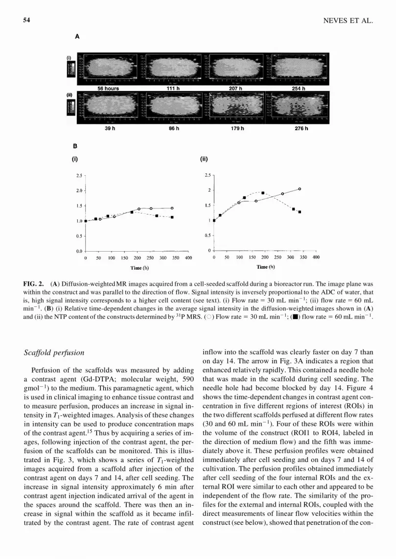
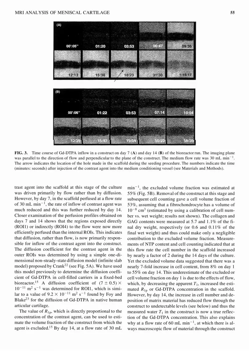
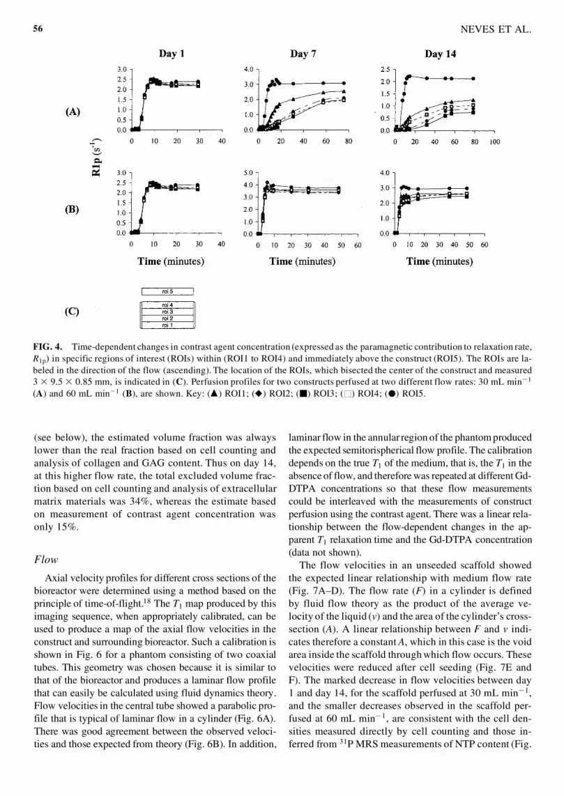
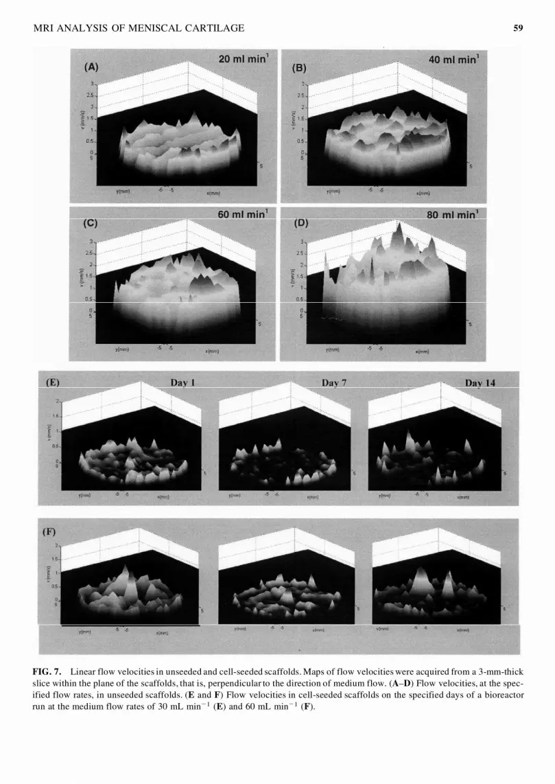