Embed Size (px)
Citation preview
Fluoxetine and the dentate gyrus: memory, recovery of function,and electrophysiology
Julian R. Keitha, Ying Wub, Jonathon R. Eppb, and Robert J. Sutherlandb
aDepartment of Psychology, University of North Carolina at Wilmington, North Carolina, USA
bCanadian Centre for Behavioural Neuroscience, University of Lethbridge, Alberta, Canada
AbstractChronic fluoxetine increases neurogenesis in the dentate gyrus (DG). In view of the widespreadclinical use of fluoxetine and the well-established role of the DG in memory, surprisingly few studieshave examined the effects of fluoxetine on memory and hippocampal electrophysiology.Additionally, few studies have evaluated the potential for fluoxetine to promote recovery of functionafter DG damage. Therefore, we studied the effects of long-term administration of fluoxetine on bothspatial-reference memory and working memory, recovery of function after intrahippocampalcolchicine infusions, which can destroy 50-70% of DG granule cells, and electrophysiologicalresponses in the DG to perforant path stimulation in freely moving rats. Chronic fluoxetine did notaffect matching-to-place or reference-memory performance in intact rats in the Morris watermazetask. Surprisingly, in rats with DG damage, recovery of function on both tasks was adversely affectedby chronic fluoxetine. Finally, unlike an earlier study that reported fluoxetine-induced increases inhippocampal population spike amplitudes and excitatory postsynaptic potential slopes in urethane-anesthetized rats, electrophysiological measures in DG of freely moving rats were not affected bychronic fluoxetine treatment.
Keywordsantidepressant; dentate gyrus; electrophysiology; fluoxetine; hippocampus; memory; rat; selectiveserotonin reuptake inhibitor
IntroductionAlthough antidepressants are prescribed for millions of individuals each year, major gaps stillexist in our understanding of how they affect the brain. Researchers have recently learned thatlong-term exposure to anti-depressant drugs can impact plasticity mechanisms in thehippocampus. Most notably, researchers have recently reported that antidepressants canincrease neuron proliferation (neurogenesis) in the dentate gyrus (DG) of the hippocampus(Malberg et al., 2000; Manev et al., 2001, 2003; Nakagawa et al., 2002; Malberg and Duman,2003; Santarelli et al., 2003). Evidence that antidepressant-induced increases in DGneurogenesis are functionally relevant has emerged from experiments that used genetic andradiological methods for reducing neurogenesis and a rodent model of anxiety and depression(Santarelli et al., 2003). Fluoxetine, desipramine, and imipramine each block the effects ofchronic mild stress on novelty-suppressed feeding, cued fear conditioning, and grooming inwild-type mice. The neurogenic and stress-related behavioral effects of antidepressants,
Correspondence to Dr Julian R. Keith, Department of Psychology, University of North Carolina at Wilmington, Wilmington, NorthCarolina 28403-5612, USA Tel: + 1 403 394 3900; fax: + 1 910 962 7010; e-mail: [email protected].
NIH Public AccessAuthor ManuscriptBehav Pharmacol. Author manuscript; available in PMC 2009 April 14.
Published in final edited form as:Behav Pharmacol. 2007 September ; 18(5-6): 521–531. doi:10.1097/FBP.0b013e3282d28f83.
NIH
-PA Author Manuscript
NIH
-PA Author Manuscript
NIH
-PA Author Manuscript
however, are not observed in mutant mice that lack 5-HT1A receptors. Additionally,hippocampal X-irradiation, which block DG neurogenesis, also abolish the effects ofantidepressants on behavior in stressed mice (Santarelli et al., 2003). Thus, the effects ofantidepressants on feeding and grooming seem to require increases in neuron proliferationwithin the DG.
Whether antidepressants impact the memory-related functions of the hippocampus is not yetknown. Numerous reports of suspected fluoxetine-induced memory impairment have,however, been reported in the clinical literature (Mirow, 1991; Bradley and Kulik, 1993;Friedman, 1994; Bangs et al., 1994; Joss et al., 2003; Huang et al., 2004). Unfortunately, theclinical studies cited above were complicated by the fact that the psychiatric disorders for whichantidepressants are prescribed can disturb cognitive performance; some researchers haveconcluded that antidepressants, per se, might not be responsible for the cognitive impairmentssometimes reported in clinically depressed populations (Gorenstein et al., 2006). Experimentalanalyses of the effects of antidepressant exposure on hippocampus-dependent memory tasksare needed to resolve this issue.
In rodents, the DG is essential for normal spatial memory. A well-established procedure forstudying rodent spatial memory is the Morris watermaze task (MWT) (Morris, 1984). TheMWT involves training rodents to swim to an underwater escape platform in a circularswimming pool. Rats' latencies to find the escape platform decrease systematically over thecourse of training as they come to use progressively more direct paths to swim to the platformfrom any starting position in the pool. DG damage impairs the ability of the rats to learn hiddenplatform locations (Morris et al., 1982; Sutherland et al., 1983, 2001). In this study, weinvestigated the effects of fluoxetine both on the performance of a well-learned place response(reference memory) and on the rapid acquisition of a new place response (matching-to-place)in the MWT.
Prolonged exposure to elevated glucocorticoids can destroy DG granule cells. Researchershave hypothesized that antidepressants, including fluoxetine, promote recovery of functionafter stress-induced hippocampal damage by restoring granule-cell density and plasticity in thehippocampus (Garcia, 2002). If fluoxetine does indeed promote the regeneration of thedamaged DG, as others (Garcia, 2002) have hypothesized, it is important to evaluate thepossibility that fluoxetine might promote recovery of function after severe DG injury.Therefore, we assessed the effects of fluoxetine treatments on reference memory and matching-to-place abilities in rats that had received bilateral DG microinjections of colchicine, amicrotubule-binding agent that is particularly toxic to granule cells (Goldschmidt and Steward,1980, 1982, 1989; Sutherland et al., 1983; Nakayama and Sawada, 2002). For this study, acolchicine dose was selected, based on pilot studies, which showed that it consistentlydestroyed approximately 50-70% of the DG granule cells while generally sparing the cornuammonis (CA) fields of the hippocampus; the goal was to preserve enough hippocampalcircuitry so that behavioral recovery might be observed postoperatively.
Finally, little is known about how fluoxetine affects DG electrophysiology. In urethane-anesthetized rats, fluoxetine injections increase the slope of the field excitatory postsynapticpotential (fEPSP) recorded in the rat DG in response to a fixed perforant-path stimulus (Stewartand Reid, 2000). The increased fEPSP slope indicates that fluoxetine treatments enhanced thesynaptic connections between granule-cell dendrites and perforant-path axons (Erickson etal., 1993). Urethane, the anesthetic drug used in the study discussed above, is, however, knownto affect significantly neurotransmitter-receptor systems that are involved in hippocampalneurotransmission, namely N-methyl-D-aspartate and α-amino-3-hydroxy-5-methyl-4-isoxazolepropionic acid glutamate receptors, and γ aminobutyric acid subtype A receptors(Hara and Harris, 2002). Therefore, this study examined the effects of chronic fluoxetine
Keith et al. Page 2
Behav Pharmacol. Author manuscript; available in PMC 2009 April 14.
NIH
-PA Author Manuscript
NIH
-PA Author Manuscript
NIH
-PA Author Manuscript
administration on perforant-path-evoked extracellular field potentials in rats that had electrodespermanently placed in the DG and the perforant path (PP), the input pathway from entorhinalcortex to the DG.
MethodsHousing and injections
Thirty-nine adult male Long-Evans hooded rats (Canadian Centre for BehaviouralNeuroscience vivarium, Lethbridge, Alberta, Canada) initially weighing 200-250 g were used.Animal treatments and maintenance of the colony were in accordance with the United StatesNational Institutes of Health and Canadian Research Council on Animal Care standards.Animals were individually housed (12-h light/dark cycle) with free access to food and water.Fluoxetine hydrochloride (Eli Lilly, Indianapolis, Indiana, USA), diluted in distilled water andobtained as a solution at a concentration of 4 mg/ml, was administered by intraperitonealinjection daily at a dose of 5 mg/kg (injection volume was 1.25 ml/kg). Isotonic (0.9%) sodiumchloride solution was administered daily to the control animals at a volume of 1.25 ml/kg.
Spatial learning and memoryThe purpose of this experiment was to determine (i) whether chronic exposure to fluoxetineaffects spatial navigation in the MWT, a task for which the DG is essential; and (ii) whetherfluoxetine promotes the recovery of spatial-navigation abilities in rats with DG damage. Theexperiment was performed in three phases: preoperative training, intrahippocampal colchicinemicro-injections (versus sham surgery), and a 6-week-long regimen of daily injections(fluoxetine versus saline, intraperitoneal), during which daily (Monday to Friday) spatial-navigation testing in the watermaze task took place.
Twenty-one rats received training on the MWT. The MWT testing sessions were conducted intwo circular white fiberglass pools (1.5 m diameter, 30.5 cm deep). The pools were filled with23 cm of water and contained a small white platform (10 cm diameter, 20 cm high) that wassubmerged 2 cm below the surface of the water (20-22°C), which was made opaque with instantmilk. The pools were located in two adjacent rooms that had large, distinctive visual stimuli,which were mounted on the walls and hung from the ceiling. A digital video tracking system(HVS Image, Twickenham, Middlesex, UK) interfaced with microcomputers recorded andstored swimming paths, speed, and trial duration.
Each rat was first trained on a reference memory problem that involved swimming to a hiddenescape platform in a fixed position in one of the swimming pools. Individual trials began witha rat being placed in the pool at one of the four cardinal compass positions around the wall ofthe pool according to a pseudorandom sequence, such that each starting location was used onceper session. The maximum duration for an individual swim trial was 90 s. If the rat found theplatform within the 90-s period, it was allowed to remain on the platform for 7 s. If the ratfailed to find the platform within the 90-s period, it was placed by the experimenter on theplatform for 7 s and then placed back into a holding cage for approximately 5 min beforestarting the next trial. Daily sessions consisted of four trials and were conducted for 2 weeks(Monday to Friday).
When 10 daily sessions of preliminary training in performance setting had been completed,sessions were modified to incorporate training on the matching-to-place task, which wascarried out in a second setting. During training on the matching-to-place, the submerged escapeplatform was moved to a new location in the pool before each session, where it remained forthe entire session. The platform location was determined in a quasi-random manner, with theconstraint that the new location was not permitted to be within 20 cm of the location used
Keith et al. Page 3
Behav Pharmacol. Author manuscript; available in PMC 2009 April 14.
NIH
-PA Author Manuscript
NIH
-PA Author Manuscript
NIH
-PA Author Manuscript
during the earlier session (center-to-center). Thus, each rat first received four swims each dayon the reference-memory problem, and then received four swims in matching-to-place (therooms used for the reference memory and matching-to-place were counterbalanced across theexperimental conditions). Rats were trained on both problems 5 days a week (Monday toFriday) for 4 weeks, and highly stable performances on both problems were established inevery rat before surgery.
To permit the rats to recover from surgery, no behavioral testing took place during the firstpostoperative week. During the second postoperative week, daily behavioral testing resumed.Each rat received a daily injection of fluoxetine or saline. On weekdays, all injections weregiven 1-2 h after the daily behavioral testing was completed; on Saturday and Sunday, theywere given at approximately the same time of day as on weekdays. The injection regimen andspatial-navigation testing described above continued for 6 postoperative weeks. At the end ofthe behavioral study, all rats received one final session that involved swimming to an escapeplatform that extended 7 cm above the water surface and was covered in black terry cloth. Thisvisible platform training session took place in the environment that was used for matching-to-place training.
Colchicine microinjections into dentate gyrusAll 21 rats that were trained on the MWT (described above) were anesthetized with isofluorane(4% with 2l/ min of oxygen and 2% after a surgical plane was established). A midline incisionwas made in the scalp and periosteum. Six 0.5-mm diameter holes (three overlying eachhemisphere) were drilled in the skull overlying the target tissue. The stereotaxic coordinatesof the holes, relative to bregma, were A-P, 3.3, 4.8, and 5.8 mm, and lateral, 1.5, 3.2, and 5.0mm. A 30-gauge injection needle was stereotaxically inserted through each hole to the targetdepth (3.7, 4.2, and 7.5 mm). Microinjection of a solution containing colchicine (2 mg/ ml)and 0.1 M phosphate-buffered saline (PBS) was administered at each of three sites in thehippocampus. An infusion of 0.5 μl of solution was injected at each site at a rate of 0.125 μl/min. The injection needles remained in place for 4 min after each injection, to allow diffusionof the colchicine solution away from the needle tip. The needles were removed and the scalpclosed with 9-mm autoclips. Rats were monitored until they fully recovered from anesthesia.
Six weeks postoperatively, when behavioral testing was complete, rats received an overdoseof pentobarbitol (120 mg/kg) and were perfused transcardially with 200 ml of PBS, followedby an equal volume of 4% paraformalde-hyde. Brains were extracted and stored in 4%paraformaldehyde solution for 24 h. The brains were then transferred to 30% sucrose in PBSfor at least 4 days before sectioning. A frozen sliding microtome was used to cut 50-μm coronalsections through the entire extent of the hippocampus. Every fifth section was mounted on aslide, stained with cresyl violet, and DG lesions were evaluated.
ElectrophysiologyEighteen rats were anesthetized with isoflurane (O2 flow rate 1.5 l/min at 1.5-2% isoflurane)for chronic electrode implantation. Electrode implantations were unilateral and placed in theright cerebral hemisphere. The stimulating and recording electrodes were made of a singlestrand of Teflon-coated stainless-steel wire (114 mm outer diameter). Wires having a largerdiameter (178 mm), soldered to stainless-steel screws, served as the ground and indifferentelectrodes, which were screwed into the frontal and occipital bones, respectively. Burr holeswere made at 3.5 mm posterior to bregma and 1.8 mm lateral to the midline for the recordingelectrode, and 8.1 mm posterior to the bregma and 4.3 mm lateral to the midline for thestimulating electrode. The dura was punctured with a sharp needle, and the recording electrodewas positioned in the hilus of the DG by monitoring multiple-unit discharges as it was loweredthrough the neocortical and hippocampal layers. The stimulating electrode was lowered into
Keith et al. Page 4
Behav Pharmacol. Author manuscript; available in PMC 2009 April 14.
NIH
-PA Author Manuscript
NIH
-PA Author Manuscript
NIH
-PA Author Manuscript
the PP while stimulating at a rate of 0.05 Hz. The recording electrode was further adjusted tomaximize the response. Proper placements of the recording and stimulating electrodes producean unmistakable signature waveform in the evoked response (McNaughton et al., 1986;Sutherland et al., 1997). Gold Amphenol (Wallingford, Connecticut, USA) connector pinsattached to the electrodes were arranged and fixed in a stable position using dental acrylic, andanchored by jeweler's screws that were tapped into the skull. The skin incision was closed withsurgical glue and the rats received injections of penicillin G (60 000 IU, intramuscular) andbuprenorphine (0.10 mg/kg).
Input/output recordingsOne week postoperatively, a 6-week injection regimen was initiated and electrophysiologyrecording sessions were carried out twice weekly. During the recording sessions, the rats werefree to move about in a clear polycarbonate cage that was 35.5 cm long, 23.2 cm wide, and19.8 cm high. For each recording session, a rat was transported from the vivarium in its homecage to the recording room, immediately plugged into the stimulating and recording apparatus,and habituated to the recording situation for 20 min. Stimuli were delivered at the rate of 0.05Hz and monitored during the habituation period.
Constant voltage stimulus pulses (AMPI Master 8 pulse former and an Isoflex optically isolatedconstant current stimulator, AMPI, Jerusalem, Israel) were delivered to the PP through astimulator that was connected to the implanted perforant-path electrode through the goldAmphenol connector pin (pulse duration = 100 μs, rate = 0.05 Hz). The output of the stimulatorwas controlled by a Pentium III microcomputer running DataWave (version 3.3) software(DataWave Technologies, Longmont, Colorado, USA). Granule-cell population responseswere amplified by a Grass (model P15D) (Grass Technologies, West Warwick, Rhode Island,USA) differential preamplifier, filtered (1/2 amplitude low frequency - 1 Hz; 1/2 amplitudehigh frequency = 10 kHz), and amplified by a Neurolog filter (Digi-timer Limited, WelwynGarden City, UK) and amplifier (total amplification was × 200). Waveforms were displayedon a Nicolet digital storage oscilloscope (Tektronix, Inc, Beaverton, Oregon, USA). The outputwas sampled online by the computer and stored on the hard drive for offline analysis. Allrecordings occurred between 13 : 00 and 18 : 00 h.
Following habituation, input/output (I/O) recordings were sampled at five different perforant-path stimulus intensities (100, 200, 300, 400, and 500 μA), always in an ascending series. Foreach of the stimulus-intensity series, 20 single perforant-path stimuli were delivered at a rateof 0.05 Hz. The field fEPSP component of the response was measured as the amplitude inmillivolts between two points on the rising phase of the response. The amplitude, in millivolts,of the field population spike (PS) was measured from the negative peak of the waveform to atangent fitted between the onset and offset of the spike (McNaughton et al., 1986).
A mixed-factor experimental design was used to evaluate the effects of chronic fluoxetine onevoked field potentials. The between-group factor was drug treatment (fluoxetine versus saline)and the repeated-measures factor was week (baseline through week 6 of the injection regimen).Stimulus intensity was a repeated-measures factor and was nested within week. I/O functionswere obtained from each rat twice each week (i.e. Monday/ Wednesday or Tuesday/Thursdayschedule), and the data from the two sessions for each individual rat were averaged beforebeing entered into the analysis. Group assignments were based on the data from the two baselinerecording sessions, such that the groups were closely matched in terms of the means andstandard deviations of the groups' fEPSPs and PSs. The experimenter responsible for allelectrophysiological recordings was blind with respect to the rats' experimental groupassignments.
Keith et al. Page 5
Behav Pharmacol. Author manuscript; available in PMC 2009 April 14.
NIH
-PA Author Manuscript
NIH
-PA Author Manuscript
NIH
-PA Author Manuscript
All statistical analyses reported below were conducted with the Statistical Package for theSocial Sciences (SPSS 11 for Mac; SPSS, Chicago, Illinois, USA).
ResultsSpatial learning and recovery from dentate gyrus damage
During the 4 weeks of preoperative training, all rats mastered both the reference-memory andmatching-to-place versions of the MWT. Figure 1 shows the mean swim-path lengths basedon the performances of all the rats, averaged over the final three preoperative training sessions.During the terminal sessions of preoperative training, the swimming paths on the reference-memory problem were consistently short in every trial. Therefore, the rats learned to userelatively direct routes to swim to the platform during the reference-memory problem. On thematching-to-place problem, path lengths on the first trial were relatively long; this is to beexpected, given that the escape platform was moved to a new location for each session.Systematic within-session decreases in swim-path lengths were apparent and, by the fourthtrial, path lengths were as short as those obtained on the fixed-platform problem. Thus, ratsrapidly learned the new escape-platform location during the training on the matching-to-placeproblem. A repeated-measures analysis of variance (ANOVA) revealed significant maineffects of task [F(1,64) = 135.55, P < 0.001] and trial [F(3,192) = 40.78, P < 0.001], and asignificant task × trial interaction [F(3,192) = 6.81, P < 0.001]. Posthoc Tukey's `HonestlySignificantly Different' (HSD) tests revealed that the swim-path lengths on the first trial of therepeated-acquisitions problem were significantly longer than they were on the other three trials.
Figure 2 shows representative cresyl-violet-stained coronal sections from a sham-surgerycontrol rat and from a colchicine-lesion rat. The colchicine-induced damage encompassedvirtually all the granule cells in the dorsal hippocampus [anteroposterior (AP) - 3.3 mm]. AtAP - 4.3 mm, in the colchicine-lesion rats, only about 10% of the granule cells remained. Inthe ventral hippocampus (AP - 5.8 mm), although the granule-cell layer was visibly thinnerthan in sham rats, for all the rats in the colchicine group it was clear that many granule cellshad been spared. The CA fields had generally been spared and appeared intact.
Figure 3 shows the swim-path lengths during testing on the reference-memory task as a functionof surgery (sham controls versus hippocampal), injection condition (saline versus fluoxetine),and week. The datapoints that represent preoperative baseline performances (time 0 in Figs 3and 4) are each based on data from the final 3 test days before surgery. The datapoints shownfor weeks 1-6 represent data averaged across five daily test sessions (Monday to Friday). Shamsurgery had no effect on performance in either task. Moreover, fluoxetine did not affect theperformance on either task by the sham-surgery rats (Figs 3 and 4).
Rats that received colchicine microinjections into DG demonstrated severe behavioralimpairments postoperatively on the reference-memory task (Fig. 3). During postoperativeweeks 2-6, performance by the rats with DG damage showed significant improvement relativeto postoperative week 1. Interestingly, during weeks 2-4, DG damaged rats that received dailysaline injections performed better on the reference-memory task than those that receivedfluoxetine. A mixed-factor ANOVA yielded a significant main effect of surgery condition (DGdamage versus sham), [F(1,18) = 52.15, P < 0.001], and a marginally significant surgery × drug(saline versus fluoxetine) interaction, [F(1,18) = 3.44, P < 0.08]. Posthoc Tukey's HSD testsrevealed that the sham-surgery group had significantly shorter paths than the DG-damage group(P < 0.05), at each postoperative point shown in Fig. 3. Additionally, planned comparisonsrevealed that on the reference-memory problem, the DG-lesion saline group located theplatform using significantly shorter swimming paths than the DG-lesion fluoxetine group did,during weeks 2-4 [P < 0.05]. Virtually identical results were observed for escape latency asfor path length (bottom panel of Fig. 3).
Keith et al. Page 6
Behav Pharmacol. Author manuscript; available in PMC 2009 April 14.
NIH
-PA Author Manuscript
NIH
-PA Author Manuscript
NIH
-PA Author Manuscript
Figure 4 shows the results from the matching-to-place task. As mentioned earlier, in thematching-to-place task, long escape paths were typical during the first trial, because the ratshad not yet had an opportunity to learn the new escape-platform location. Thus, Fig. 4 doesnot include data from the first trial of the matching-to-place task. As was the case in thereference-memory task, the rats in the sham-surgery group traveled consistently shorterdistances to find the escape platform. DG damage severely disrupted the abilities of the rats toremember new platform locations. Additionally, it is apparent from the figure that the saline-treated, DG-damage group steadily improved on the repeated-acquisitions task over the courseof 6 postoperative weeks; although they remained impaired, relative to controls with intactDG. Fluoxetine-treated, DG-damaged rats, however, showed no evidence of improvement onthe matching-to-place task during the postoperative phase of the experiment. A mixed-factorANOVA revealed significant main effects of surgery [F(6,18) = 41.87, P < .001] and week[F(6,108) = 5.37, P < .001], and week × surgery interaction [F(6,108)=4.40, P < .001]. PosthocTukey's HSD tests confirmed that rats in the sham-surgery group had significantly shorter pathlengths than rats in the DG-damage group at every postoperative time point. Again, escapelatencies (bottom panel) closely paralleled the pattern observed with path lengths.
A detailed examination of the data from the matching-to-place task during week 6 isinformative. Figure 5 shows average path lengths as a function of trial. First, rats in the sham-surgery group learned the new location more rapidly than DG-damaged rats, swimmingminimal distances to reach the hidden escape platform by the second trial. Second, DG-damaged rats that received daily saline injections performed equally well as the sham-surgerycontrol rats by the third trial. Finally, rats with DG damage that received fluoxetine daily failedto learn new platform locations. A mixed-factor ANOVA revealed a significant main effect oftrial [F(3,300) = 21.20, P < .001], and a significant trial × drug interaction [F(3,300) = 2.69, P< .05]. Posthoc Tukey's HSD analysis confirmed that the DG-damaged rats that received salinediffered from the sham-surgery groups only on trial 2, whereas the DG-damaged rats thatreceived fluoxetine had significantly longer swimming paths than all the other groups in trials2-4 [P < 0.05].
Figure 6 shows the average swimming speed for each group. It is clear that there were no groupdifferences in the swimming speeds [all P values not significant (NS)]. Figure 7 shows averageescape latencies for the rats that were tested on the visible-platform task. Rats in all the groupsquickly reached the escape platform when the platform was visible, and no group differenceswere apparent (all P values NS).
Fluoxetine effects on neurotransmission in dentate gyrusAt the end of the 7-week chronic electrophysiological recording protocol, 12 of the 18 ratsoriginally implanted for the electrophysiology study (six per group) still produced high-qualityevoked field potentials. Only the data from the rats that had high-quality evoked field potentialsthroughout the entire protocol were included in the analysis of the electrophysiology results;however, we wish to note that the two experimental groups did not differ from one another inthis respect. Individual I/O curves for each week are presented in Fig. 8 (PS) and Fig. 9 (fEPSP).The individual panels in each figure present data averaged across two of the recording sessionsthat were conducted each week. It is clear from the figures that PS amplitude and slope of thefEPSP increased systematically as a function of PP stimulus intensity. It is also apparent thatthe evoked potentials of the two groups were closely matched when the experiment began.Finally, the PS and fEPSP were not significantly (P > 0.05) affected by fluoxetine after 6 weeksof drug administration, although a nonsignificant trend toward reduced PS amplitudes andfEPSP slopes is apparent in Figs 8 and 9.
Keith et al. Page 7
Behav Pharmacol. Author manuscript; available in PMC 2009 April 14.
NIH
-PA Author Manuscript
NIH
-PA Author Manuscript
NIH
-PA Author Manuscript
DiscussionThe aims of the experiments presented here were to determine whether fluoxetine affected thememory processes for which the DG was essential, promoted recovery of function in animalswith DG damage, and affected DG neurotransmission in nonanesthetized animals. Fluoxetinehad no effect on the reference-memory or matching-to-place performances in the MWT, in ratswith an intact DG. The spatial-learning procedure used for this study was one that firmlyestablished a high degree of skill at the MWT in all the animals, before initiating the chronicfluoxetine administration regimen. Therefore, there was little scope for the observation offurther improvement that could have been produced by fluoxetine. In contrast, the procedureused here was very well suited for detecting drug-induced disturbances, either in referencememory or in matching-to-place memory; earlier work by our laboratory has demonstratedthat very subtle changes from steady-state baseline are more readily detected using the presentprocedure than by between-group procedures (Galizio et al., 2003; Keith et al., 2003;Padlubnaya et al., 2005). Numerous studies based on clinical populations have reported thatfluoxetine can cause memory disturbances in depressed patients (Mirow, 1991; Bradley andKulik, 1993; Friedman, 1994; Bangs et al., 1994; Joss et al., 2003; Huang et al., 2004). Chronicfluoxetine administration, however, disrupted neither the reference memory nor the matching-to-place memory. Our data support the conclusion that, in intact rats, chronic fluoxetineadministration does not impair spatial learning or memory.
The current study also addresses the issue of whether the DG is equivalently involved inreference and matching-to-place learning in the MWT. Although the adaptation of the MWTthat was used in this study has also been used in earlier studies that examined the pharmacologyof spatial learning, the effects of DG damage under this particular procedure had not beenreported previously (Galizio et al., 2003; Keith et al., 2003; Padlubnaya et al., 2005).Colchicine microinjections into the DG profoundly impaired the reference-memory andmatching-to-place performances during the early postoperative period, confirming that the DGis essential for the adaptation of the MWT procedure used here. Although improvement wasevident over the 6 weeks of postoperative training, the rats with DG lesions remained impaired,relative to rats with an intact DG throughout that period. The impairments that were observedin the animals with DG damage were not due to poor swimming ability (speed of swimmingwas not affected by surgery), or general disruptions of sensorimotor factors, or motivation toescape from the water, as evidenced by the normal performances on the visible escape-platformtask by rats with DG damage. The finding that relatively small hippocampal lesions, whichwere restricted largely to the DG, could cause enduring retrograde and anterograde amnesiasin a spatial-learning task (characteristic of more complete hippocampal lesions) is noteworthy(Sutherland et al., 2001). Clearly, an intact DG is necessary for both retention and acquisitionof new spatial information.
The hypothesis that fluoxetine promotes DG repair, however, was not supported by our results.Environmental enrichment and exercise, both factors that impact the structure and physiologyof the hippocampus, have been reported to improve spatial navigation in rats after ischemic orneurotoxic brain lesions (Risedal et al., 2002; Dahlqvist et al., 2003; Will et al., 2004; Gobboand O'Mara, 2005). Conversely, in this study, rats that received fluoxetine demonstrated poorerpostoperative spatial-navigation performance both on the reference-memory and matching-to-place tasks, relative to DG-damaged rats that received saline. A limitation of this study is thatwe did not evaluate the effects of fluoxetine treatment on neurogenesis. Thus, our results didnot permit a direct evaluation of the relationship between DG neurogenesis and recovery offunction after DG damage. These findings might, nevertheless, have some relevance to theclinical use of fluoxetine in the treatment of poststroke depression. The findings suggest thatfurther research assessing possible adverse cognitive effects of fluoxetine in patients with grosshippocampal damage is warranted.
Keith et al. Page 8
Behav Pharmacol. Author manuscript; available in PMC 2009 April 14.
NIH
-PA Author Manuscript
NIH
-PA Author Manuscript
NIH
-PA Author Manuscript
In view of the numerous pilot studies that assessed many different colchicine injection doses,we have found that the colchicine dose used in this study creates the smallest DG lesions thatreliably impair spatial learning and memory. The extent of DG damage induced by colchicineinjections in this study was, nevertheless, far greater than what one would expect to observefrom exposure to high glucocorticoid levels. Thus, the lesion method used in this study wasnot an ideal test of the hypothesis that more subtle DG damage, such as that caused bychronically elevated glucocorticoids, can be reversed by fluoxetine. As an alternative tocolchicine injections, in a recent study, we used adrenalectomy (ADX) to produce selectiveDG granule-cell loss (Spanswick et al., 2007). Bilateral ADX reduced the average thicknessof the DG granule-cell layer by approximately 62%; it significantly impaired spatial learningand memory on the same behavioral tests as were used in this study. As in this study, chronicfluoxetine treatment did not reverse the spatial-learning impairment caused by ADX-inducedgranule-cell loss, even though a substantial population of granule cells had been spared by theADX.
Daily fluoxetine (5 mg/kg) injections did not affect perforant-path→DG-evoked fEPSPs orPSs, when measured in nonanesthetized, freely moving rats. Other researchers have reportedthat chronic exposure to fluoxetine can increase both the slope of the fEPSP and the amplitudeof the PS (Stewart and Reid, 2000). The methodological details of this study differed frompreviously published work in three key aspects: fluoxetine dose, duration of the dosingprotocol, and state of animals when the electrophysiological measurements were collected.First, in the earlier study, fluoxetine (1 mg/kg) was administered daily for 15 days; whereas,in this study, the fluoxetine (5 mg/kg) administration protocol lasted for 6 weeks. Second,earlier researches on the effects of fluoxetine on in-vivo hippocampal electrophysiology werecarried out in a single recording session when the rats were still under deep urethane anesthesiaand undergoing surgery at the time of data collection (Stewart and Reid, 2000). In this study,electrophysiological measurements were made twice a week for 6 weeks in nonanesthetizedfreely moving rats.
Given the fact that chronic fluoxetine exposure has been reported to increase DG neurogenesis,the failure to observe significant effects of long-term exposure to fluoxetine on DGelectrophysiology was surprising. Evidence from in-vitro studies of newborn neurons in thesubgranular zone of the DG in hippocampal slices suggests that the addition of large numbersof immature neurons can alter the overall electrophysiology of the DG. For example, whole-cell patch-clamping studies in hippocampal slices have revealed that immature DG granulecells display membrane properties that differ from those observed in mature granule cells.Compared with mature granule cells, immature granule cells have slower membrane timeconstants, higher input resistance, more depolarized resting membrane potentials, generateaction potentials with smaller peak amplitudes, and can generate isolated Ca2
+ spikes that boostfast Na+ action potentials (Overstreet et al., 2004; Schmidt-Hieber et al., 2004). Theseproperties of immature neurons might be expected to increase the fEPSP and PS in a DG thatcontained significantly larger than normal numbers of new neurons; such electrophysiologicaleffects were not observed in this study.
The neuropharmacological properties of fluoxetine are, however, complex; one can identifyevidence from the literature to support a prediction that chronic exposure to fluoxetine woulddecrease the fEPSP and PS. For example, fluoxetine increases the 5-HT1A receptor sensitivityin the DG (Elena Castro et al., 2003). That 5-HT1A receptor activation causes neuronalhyperpolarization through G-protein coupled opening of K+ channels has been established well(Nicoll et al., 1990). Ultrastructural localization studies have reported evidence that 5-HT1Areceptors are present both somatically and on the dendrites of glutamatergic hippocampalneurons (Kia et al., 1996). Collectively, fluoxetine-induced increases in extracellular 5-HT andthe sensitization of 5-HT1A receptors would be expected to increase the 5-HT1A receptor-
Keith et al. Page 9
Behav Pharmacol. Author manuscript; available in PMC 2009 April 14.
NIH
-PA Author Manuscript
NIH
-PA Author Manuscript
NIH
-PA Author Manuscript
mediated tonic hyperpolarization in granule cells. Again, however, no evidence was found thatsuch fluoxetine-induced changes detectably influence either the fEPSP or PS responses toperforant-path inputs. The multiple opposing neuropharmacological properties of chronicfluoxetine treatments, such as increases in the population of immature neurons in the DG andchanges in receptor sensitivities, might, possibly, counteract one another.
ConclusionIn summary, spatial reference and matching-to-place memory were not affected by fluoxetine.Fluoxetine treatments did adversely affect recovery of function in rats with DG lesions,indicating that rather than generally promoting DG repair, at least under some circumstances,fluoxetine can exacerbate the cognitive effects of hippocampus injury. Finally, unlike theresults observed using anesthetized rats, recordings of chronic DG electrophysiology over a 6-week fluoxetine regimen did not reveal an effect of fluoxetine in awake, freely moving rats.
AcknowledgementsThis study was supported by NIMH MH06715601A1 (J.R.K) and NIMH MH061460 and AHFMR (R.J.S).
ReferencesBangs ME, Petti TA, Janus MD. Fluoxetine-induced memory impairment in an adolescent. J Am Acad
Child Adolesc Psychiatry 1994;33:1303–1306. [PubMed: 7995797]Bradley SJ, Kulik L. Fluoxetine and memory impairment. J Am Acad Child Adolesc Psychiatry
1993;32:1078–1079. [PubMed: 8407756]Dahlqvist P, Ronnback A, Risedal A, Nergardh R, Johansson IM, Seckl JR, et al. Effects of postischemic
environment on transcription factor and serotonin receptor expression after permanent focal corticalischemia in rats. Neuroscience 2003;119:643–652. [PubMed: 12809685]
Elena Castro M, Diaz A, del Olmo E, Pazos A. Chronic fluoxetine induces opposite changes in G proteincoupling at pre and postsynaptic 5-HT1A receptors in rat brain. Neuropharmacology 2003;44:93–101.[PubMed: 12559126]
Erickson CA, McNaughton BL, Barnes CA. Comparison of long-term enhancement and short-termexploratory modulation of perforant path synaptic transmission. Brain Res 1993;615:275–280.[PubMed: 8395959]
Friedman EH. Fluoxetine and memory impairment. J Am Acad Child Adolesc Psychiatry 1994;33:763.[PubMed: 8056743]
Galizio M, Keith JR, Mansfield WJ, Pitts RC. Repeated spatial acquisition: effects of NMDA antagonistsand morphine. Exp Clin Psychopharmacol 2003;11:79–90. [PubMed: 12622346]
Garcia R. Stress, metaplasticity, and antidepressants. Curr Mol Med 2002;2:629–638. [PubMed:12420802]
Gobbo OL, O'Mara SM. Exercise, but not environmental enrichment, improves learning after kainic acid-induced hippocampal neurodegeneration in association with an increase in brain-derived neurotrophicfactor. Behav Brain Res 2005;159:21–26. [PubMed: 15794993]
Goldschmidt RB, Steward O. Preferential neurotoxicity of colchicine for granule cells of the dentategyrus of the adult rat. Proc Natl Acad Sci U S A 1980;77:3047–3051. [PubMed: 6930683]
Goldschmidt RB, Steward O. Neurotoxic effects of colchicine: differential susceptibility of CNS neuronalpopulations. Neuroscience 1982;7:695–714. [PubMed: 7070670]
Goldschmidt RB, Steward O. Comparison of the neurotoxic effects of colchicine, the vinca alkaloids,and other microtubule poisons. Brain Res 1989;486:133–140. [PubMed: 2720425]
Gorenstein C, de Carvalho SC, Artes R, Moreno RA, Marcourakis T. Cognitive performance in depressedpatients after chronic use of anti-depressants. Psychopharmacology (Berl) 2006;185:84–92.[PubMed: 16485140]
Hara K, Harris RA. The anesthetic mechanism of urethane: the effects on neurotransmitter-gated ionchannels. Anesth Analg 2002;94:313–318. [PubMed: 11812690]table of contents
Keith et al. Page 10
Behav Pharmacol. Author manuscript; available in PMC 2009 April 14.
NIH
-PA Author Manuscript
NIH
-PA Author Manuscript
NIH
-PA Author Manuscript
Huang SC, Tsai SJ, Chang JC. Fluoxetine-induced memory impairment in four family members. Int JPsychiatry Med 2004;34:197–200. [PubMed: 15387402]
Joss JD, Burton RM, Keller CA. Memory loss in a patient treated with fluoxetine. Ann Pharmacother2003;37:1800–1803. [PubMed: 14632599]
Keith JR, Pitts RC, Pezzuti T, Galizio M. Effects of positive GABA(A) modulators on a multiple-component, repeated-test of spatial learning. Behav Pharmacol 2003;14:67–75. [PubMed: 12576883]
Kia HK, Brisorgueil MJ, Hamon M, Calas A, Verge D. Ultrastructural localization of 5-hydroxytryptamine1A receptors in the rat brain. J Neurosci Res 1996;46:697–708. [PubMed:8978504]
Malberg JE, Duman RS. Cell proliferation in adult hippocampus is decreased by inescapable stress:reversal by fluoxetine treatment. Neuropsychopharmacology 2003;28:1562–1571. [PubMed:12838272]
Malberg JE, Eisch AJ, Nestler EJ, Duman RS. Chronic antidepressant treatment increases neurogenesisin adult rat hippocampus. J Neurosci 2000;20:9104–9110. [PubMed: 11124987]
Manev H, Uz T, Manev R. Glia as a putative target for antidepressant treatments. J Affect Disord2003;75:59–64. [PubMed: 12781351]
Manev R, Uz T, Manev H. Fluoxetine increases the content of neurotrophic protein S100beta in the rathippocampus. Eur J Pharmacol 2001;420:R1–2. [PubMed: 11408041]
McNaughton BL, Barnes CA, Rao G, Baldwin J, Rasmussen M. Long-term enhancement of hippocampalsynaptic transmission and the acquisition of spatial information. J Neurosci 1986;6:563–571.[PubMed: 3005525]
Mirow S. Cognitive dysfunction associated with fluoxetine. Am J Psychiatry 1991;148:948–949.[PubMed: 2053640]
Morris RG. Developments of a water-maze procedure for studying spatial learning in the rat. J NeurosciMethods 1984;11:47–60. [PubMed: 6471907]
Morris RG, Garrud P, Rawlins JN, O'Keefe J. Place navigation impaired in rats with hippocampal lesions.Nature 1982;297:681–683. [PubMed: 7088155]
Nakayama T, Sawada T. Involvement of microtubule integrity in memory impairment caused bycolchicine. Pharmacol Biochem Behav 2002;71:119–138. [PubMed: 11812515]
Nakagawa S, Kim JE, Lee R, Chen J, Fujioka T, Malberg J, et al. Localization of phosphorylated cAMPresponse element-binding protein in immature neurons of adult hippocampus. J Neurosci2002;22:9868–9876. [PubMed: 12427843]
Nicoll RA, Malenka RC, Kauer JA. Functional comparison of neuro-transmitter receptor subtypes inmammalian central nervous system. Physiol Rev 1990;70:513–565. [PubMed: 1690904]
Overstreet LS, Hentges ST, Bumaschny VF, de Souza FS, Smart JL, Santangelo AM, et al. A transgenicmarker for newly born granule cells in dentate gyrus. J Neurosci 2004;24:3251–3259. [PubMed:15056704]
Padlubnaya D, Galizio M, Pitts RC, Keith JR. Chlordiazepoxide interactions with scopolamine anddizocilpine: novel cooperative and antagonistic effects on spatial learning. Behav Neurosci2005;119:1331–1338. [PubMed: 16300439]
Risedal A, Mattsson B, Dahlqvist P, Nordborg C, Olsson T, Johansson BB. Environmental influences onfunctional outcome after a cortical infarct in the rat. Brain Res Bull 2002;58:315–321. [PubMed:12128159]
Santarelli L, Saxe M, Gross C, Surget A, Battaglia F, Dulawa S, et al. Requirement of hippocampalneurogenesis for the behavioral effects of antidepressants. Science 2003;301:805–809. [PubMed:12907793]
Schmidt-Hieber C, Jonas P, Bischofberger J. Enhanced synaptic plasticity in newly generated granulecells of the adult hippocampus. Nature 2004;429:184–187. [PubMed: 15107864]
Spanswick SC, Epp JR, Keith JR, Sutherland RJ. Adrenalectomy-induced granule cell degeneration inthe hippocampus causes spatial memory deficits that are not reversed by chronic treatment withcorticosterone or fluoxetine. Hippocampus 2007;17:137–146. [PubMed: 17183555]
Stewart CA, Reid IC. Repeated ECS and fluoxetine administration have equivalent effects onhippocampal synaptic plasticity. Psychopharmacology (Berl) 2000;148:217–223. [PubMed:10755734]
Keith et al. Page 11
Behav Pharmacol. Author manuscript; available in PMC 2009 April 14.
NIH
-PA Author Manuscript
NIH
-PA Author Manuscript
NIH
-PA Author Manuscript
Sutherland RJ, Whishaw IQ, Kolb B. A behavioural analysis of spatial localization following electrolytic,kainate- or colchicine-induced damage to the hippocampal formation in the rat. Behav Brain Res1983;7:133–153. [PubMed: 6830648]
Sutherland RJ, McDonald RJ, Savage DD. Prenatal exposure to moderate levels of ethanol can have long-lasting effects on hippocampal synaptic plasticity in adult offspring. Hippocampus 1997;7:232–238.[PubMed: 9136052]
Sutherland RJ, Weisend MP, Mumby D, Astur RS, Hanlon FM, Koerner A, et al. Retrograde amnesiaafter hippocampal damage: recent vs. remote memories in two tasks. Hippocampus 2001;11:27–42.[PubMed: 11261770]
Will B, Galani R, Kelche C, Rosenzweig MR. Recovery from brain injury in animals: relative efficacyof environmental enrichment, physical exercise or formal training (1990-2002). Prog Neurobiol2004;72:167–182. [PubMed: 15130708]
Keith et al. Page 12
Behav Pharmacol. Author manuscript; available in PMC 2009 April 14.
NIH
-PA Author Manuscript
NIH
-PA Author Manuscript
NIH
-PA Author Manuscript
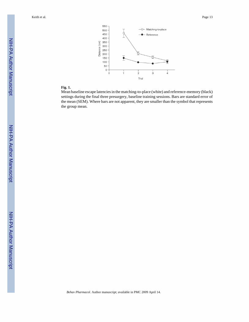
Fig. 1.Mean baseline escape latencies in the matching-to-place (white) and reference-memory (black)settings during the final three presurgery, baseline training sessions. Bars are standard error ofthe mean (SEM). Where bars are not apparent, they are smaller than the symbol that representsthe group mean.
Keith et al. Page 13
Behav Pharmacol. Author manuscript; available in PMC 2009 April 14.
NIH
-PA Author Manuscript
NIH
-PA Author Manuscript
NIH
-PA Author Manuscript
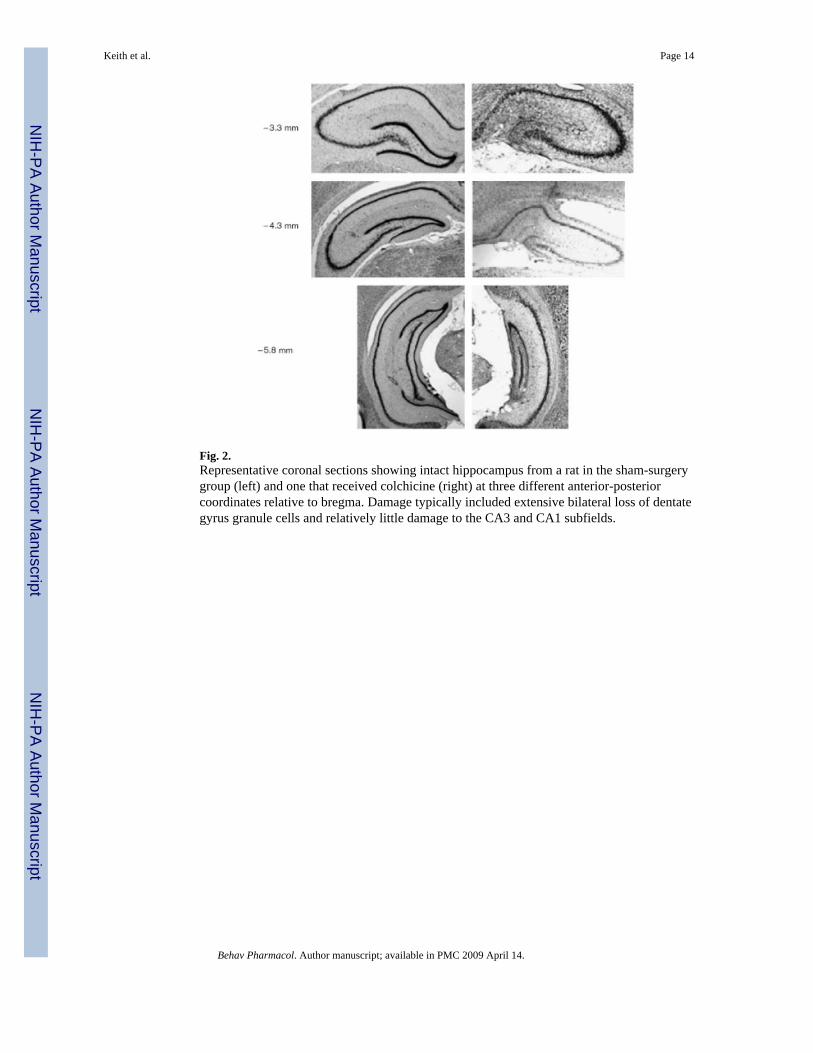
Fig. 2.Representative coronal sections showing intact hippocampus from a rat in the sham-surgerygroup (left) and one that received colchicine (right) at three different anterior-posteriorcoordinates relative to bregma. Damage typically included extensive bilateral loss of dentategyrus granule cells and relatively little damage to the CA3 and CA1 subfields.
Keith et al. Page 14
Behav Pharmacol. Author manuscript; available in PMC 2009 April 14.
NIH
-PA Author Manuscript
NIH
-PA Author Manuscript
NIH
-PA Author Manuscript
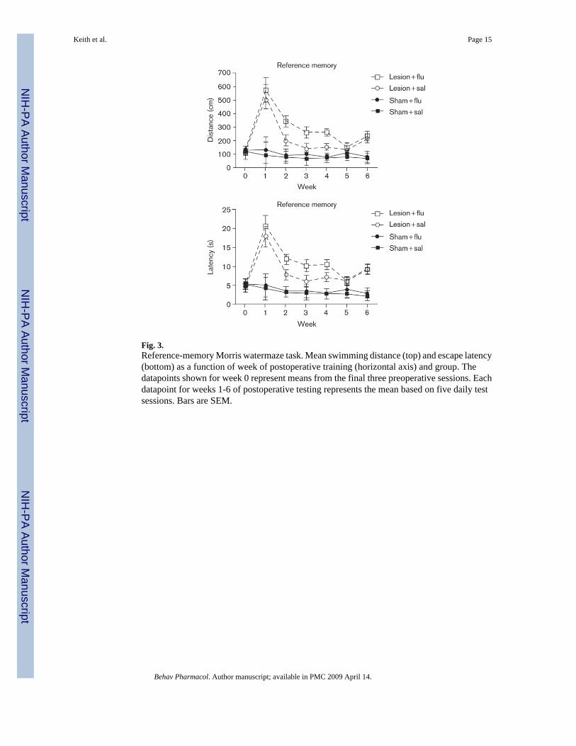
Fig. 3.Reference-memory Morris watermaze task. Mean swimming distance (top) and escape latency(bottom) as a function of week of postoperative training (horizontal axis) and group. Thedatapoints shown for week 0 represent means from the final three preoperative sessions. Eachdatapoint for weeks 1-6 of postoperative testing represents the mean based on five daily testsessions. Bars are SEM.
Keith et al. Page 15
Behav Pharmacol. Author manuscript; available in PMC 2009 April 14.
NIH
-PA Author Manuscript
NIH
-PA Author Manuscript
NIH
-PA Author Manuscript
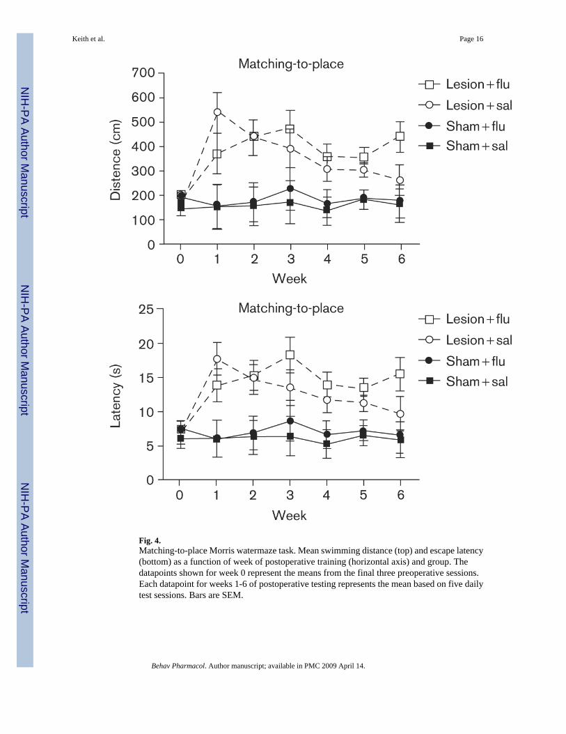
Fig. 4.Matching-to-place Morris watermaze task. Mean swimming distance (top) and escape latency(bottom) as a function of week of postoperative training (horizontal axis) and group. Thedatapoints shown for week 0 represent the means from the final three preoperative sessions.Each datapoint for weeks 1-6 of postoperative testing represents the mean based on five dailytest sessions. Bars are SEM.
Keith et al. Page 16
Behav Pharmacol. Author manuscript; available in PMC 2009 April 14.
NIH
-PA Author Manuscript
NIH
-PA Author Manuscript
NIH
-PA Author Manuscript
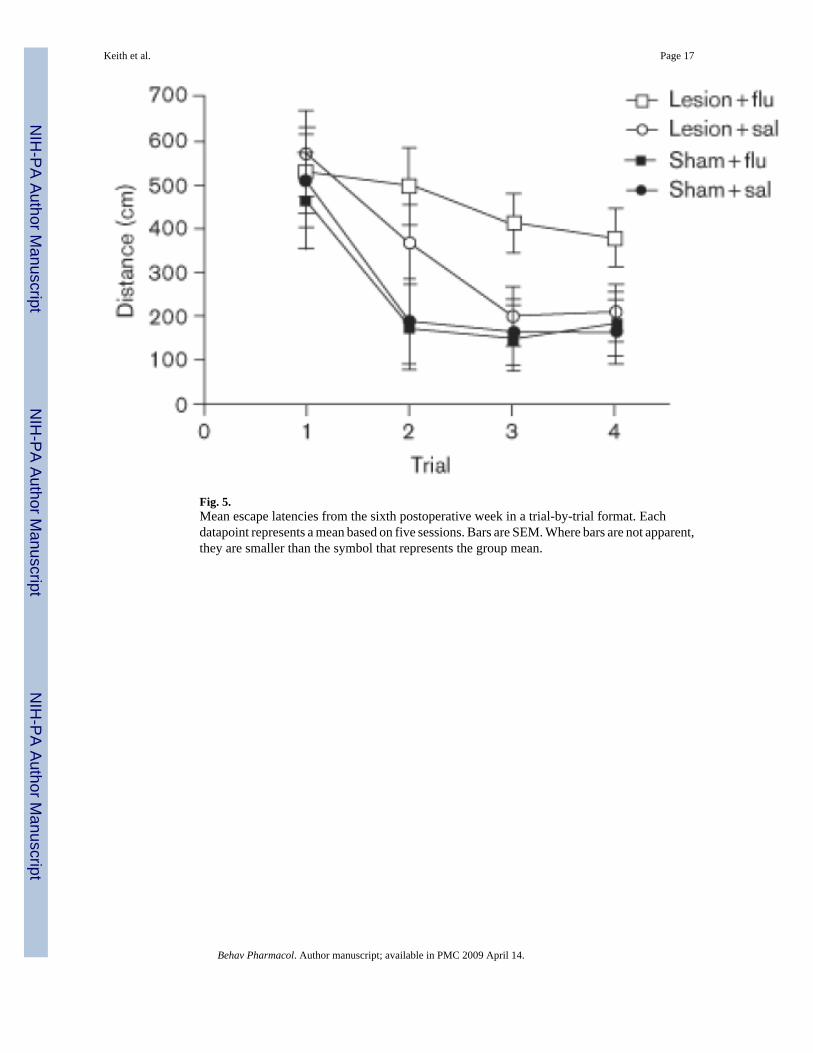
Fig. 5.Mean escape latencies from the sixth postoperative week in a trial-by-trial format. Eachdatapoint represents a mean based on five sessions. Bars are SEM. Where bars are not apparent,they are smaller than the symbol that represents the group mean.
Keith et al. Page 17
Behav Pharmacol. Author manuscript; available in PMC 2009 April 14.
NIH
-PA Author Manuscript
NIH
-PA Author Manuscript
NIH
-PA Author Manuscript
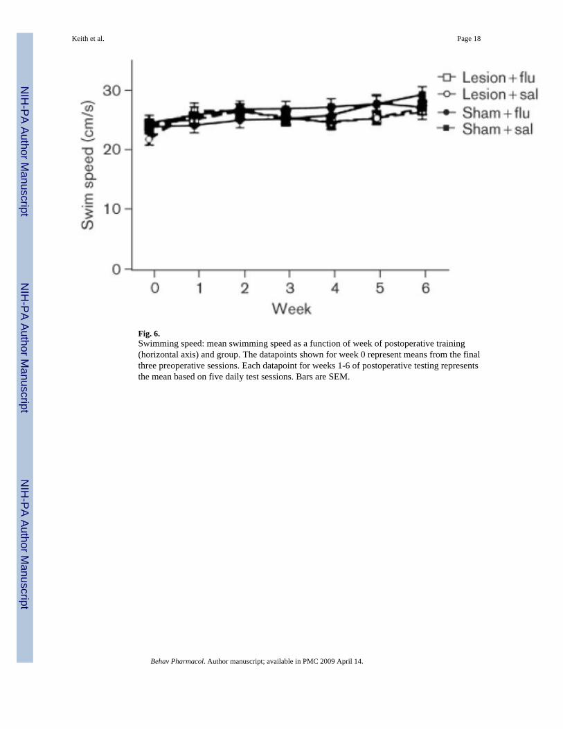
Fig. 6.Swimming speed: mean swimming speed as a function of week of postoperative training(horizontal axis) and group. The datapoints shown for week 0 represent means from the finalthree preoperative sessions. Each datapoint for weeks 1-6 of postoperative testing representsthe mean based on five daily test sessions. Bars are SEM.
Keith et al. Page 18
Behav Pharmacol. Author manuscript; available in PMC 2009 April 14.
NIH
-PA Author Manuscript
NIH
-PA Author Manuscript
NIH
-PA Author Manuscript
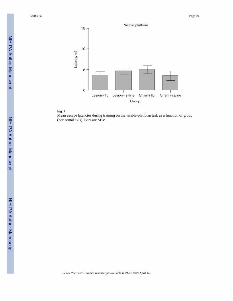
Fig. 7.Mean escape latencies during training on the visible-platform task as a function of group(horizontal axis). Bars are SEM.
Keith et al. Page 19
Behav Pharmacol. Author manuscript; available in PMC 2009 April 14.
NIH
-PA Author Manuscript
NIH
-PA Author Manuscript
NIH
-PA Author Manuscript
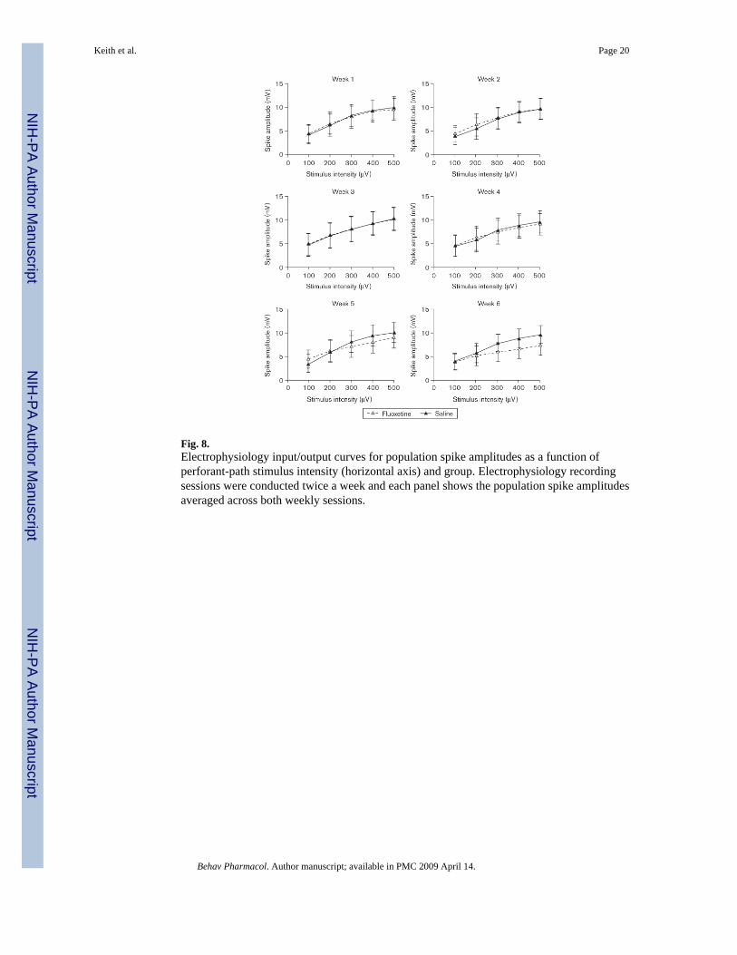
Fig. 8.Electrophysiology input/output curves for population spike amplitudes as a function ofperforant-path stimulus intensity (horizontal axis) and group. Electrophysiology recordingsessions were conducted twice a week and each panel shows the population spike amplitudesaveraged across both weekly sessions.
Keith et al. Page 20
Behav Pharmacol. Author manuscript; available in PMC 2009 April 14.
NIH
-PA Author Manuscript
NIH
-PA Author Manuscript
NIH
-PA Author Manuscript
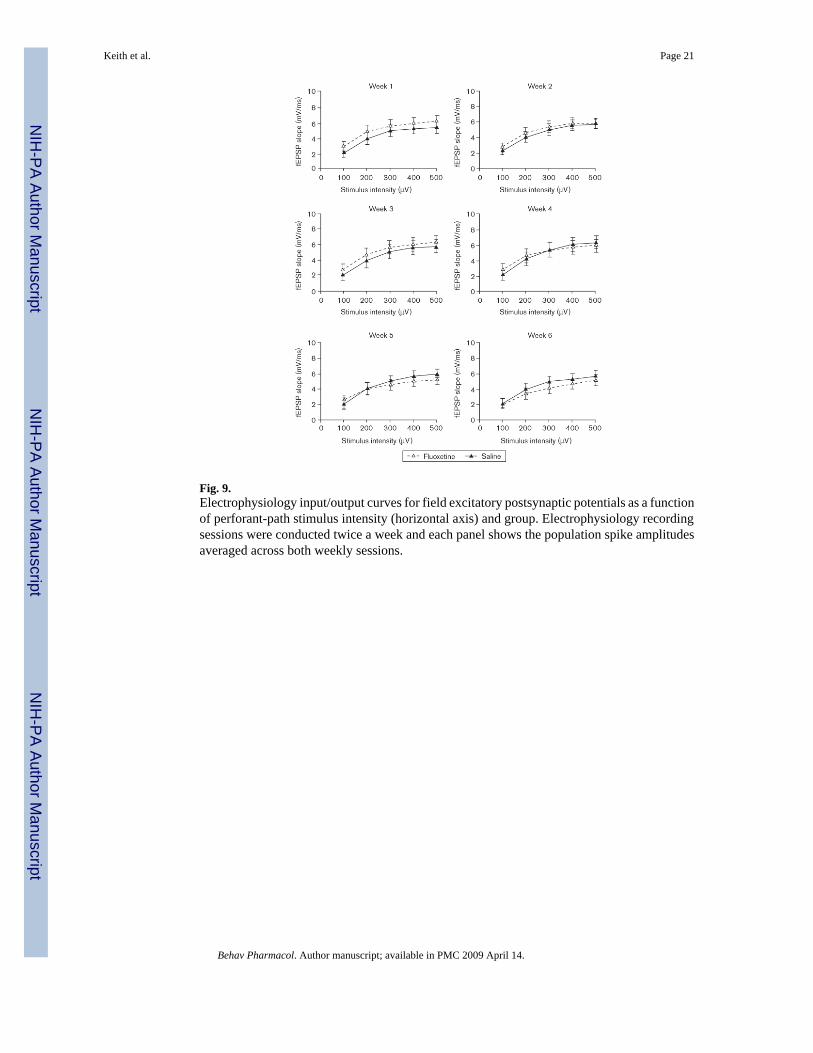
Fig. 9.Electrophysiology input/output curves for field excitatory postsynaptic potentials as a functionof perforant-path stimulus intensity (horizontal axis) and group. Electrophysiology recordingsessions were conducted twice a week and each panel shows the population spike amplitudesaveraged across both weekly sessions.
Keith et al. Page 21
Behav Pharmacol. Author manuscript; available in PMC 2009 April 14.
NIH
-PA Author Manuscript
NIH
-PA Author Manuscript
NIH
-PA Author Manuscript


