Embed Size (px)
Citation preview

Fertility Cryopreservation
9780521517782pre.indd i9780521517782pre.indd i 3/12/2010 10:36:27 PM3/12/2010 10:36:27 PM

9780521517782pre.indd ii9780521517782pre.indd ii 3/12/2010 10:36:27 PM3/12/2010 10:36:27 PM
Fertility Cryopreservation
Ri-Cheng Chian McGill University, Montreal, Canada
Patrick Quinn Sage IVF, Redmond, OR, USA
9780521517782pre.indd iii9780521517782pre.indd iii 3/12/2010 10:36:27 PM3/12/2010 10:36:27 PM
cambrid ge universit y press Cambridge, New York, Melbourne, Madrid, Cape Town, Singapore, São Paulo, Delhi, Dubai, Tokyo
Cambridge University Press Th e Edinburgh Building, Cambridge CB2 8RU, UK
Published in the United States of America by Cambridge University Press, New York
www.cambridge.org Information on this title: www.cambridge.org/9780521517782
© Cambridge University Press 2010
Th is publication is in copyright. Subject to statutory exception and to the provisions of relevant collective licensing agreements, no reproduction of any part may take place without the written permission of Cambridge University Press.
First published 2010
Printed in the United Kingdom at the University Press, Cambridge
A catalogue record for this publication is available from the British Library
Library of Congress Cataloguing in Publication data Fertility cryopreservation / [edited by] Ri-Cheng Chian, Patrick Quinn.
p. ; cm. Includes bibliographical references and index. ISBN 978-0-521-51778-2 (hardback) 1. Infertility–Prevention. 2. Generative organs–Cryopreservation. I. Chian, Ri-Cheng. II. Quinn, Patrick, 1946– III. Title.[DNLM: 1. Infertility–therapy. 2. Antineoplastic Agents–adverse eff ects. 3. Cryopreservation–methods. 4. Fertility–radiation eff ects. 5. Infertility–etiology. 6. Neoplasms–complications. WP 570 F4115 2010] RC889.F43 2010 362.196’692–dc22 2010003022
ISBN 978–0–521–51778–2 Hardback
Cambridge University Press has no responsibility for the persistence or accuracy of URLs for external or third-party Internet websites referred to in this publication, and does not guarantee that any content on such websites is, or will remain, accurate or appropriate.
Every eff ort has been made in preparing this publication to provide accurate and up-to-date information which is in accord with accepted standards and practice at the time of publication. Although case histories are drawn from actual cases, every eff ort has been made to disguise the identities of the individuals involved. Nevertheless, the authors, editors and publishers can make no warranties that the information contained herein is totally free from error, not least because clinical standards are constantly changing through research and regulation. Th e authors, editors and publishers therefore disclaim all liability for direct or consequential damages resulting from the use of material contained in this publication. Readers are strongly advised to pay careful attention to information provided by the manufacturer of any drugs or equipment that they plan to use.
9780521517782pre.indd iv9780521517782pre.indd iv 3/12/2010 10:36:27 PM3/12/2010 10:36:27 PM

v
Section 1: Cryobiology 1 Cryobiology: an overview 1
Ri-Cheng Chian
2 Suppression of ice in aqueous solutions and its application to vitrifi cation in assisted reproductive technology 10 Patrick Quinn
3 Movement of water and cryoprotectants in mouse oocytes and embryos at diff erent stages: relevance to cryopreservation 16 Magosaburo Kasai and Keisuke Edashige
4 Cryoprotectants 24 Jason E. Swain and Gary D. Smith
Section 2: Cryopreservation of sperm and testicular tissue
5 Cryopreservation of sperm: an overview 39 Fady Shehata and Ri-Cheng Chian
6 Sperm cryopreservation for a donor program 46 Zheng Li and Peng Xu
7 Cryopreservation of surgically retrieved sperm 51 Peter T. K. Chan
8 Testicular tissue cryopreservation 57 Ariel Revel and Javier Mejia
Section 3: Cryopreservation of embryos
9 Cryopreservation of embryos: an overview 67 David H. Edgar and Debra A. Gook
10 Cryopreservation of pronuclear stage human embryos 76 Barry Behr and Yimin Shu
11 Cryopreservation of day two and day three embryos 89 Yunxia Cao and Zhiguo Zhang
12 Cryopreservation of blastocysts 95 Yoshiharu Morimoto
13 Aseptic vitrifi cation of human blastocysts: protocol development and clinical application 106 Pierre Vanderzwalmen, Luc Grobet, Yannis Prapas, Patricia Frias, and Nicolas Zech
Section 4: Cryopreservation of oocytes 14 Cryopreservation of human oocytes: an
overview 114 Ri-Cheng Chian
15 Cryopreservation of oocytes by slow cooling 120 Andrea Borini and Giovanni Coticchio
16 Vitrifi cation of human oocytes with diff erent tools 131 Patrick Quinn
17 Vitrifi cation of human oocytes using the McGill Cryoleaf protocol 144 Jack Yu Jen Huang, Seang Lin Tan, and Ri-Cheng Chian
18 Cryopreservation of human oocytes and embryos either by direct plunging into liquid nitrogen or by using an aseptic approach 157 Evgenia Isachenko, Vladimir Isachenko, Jurgen M. Weiss, and Rolf Kreienberg
Contents
List of Contributors page vii Preface xi Acknowledgements xiii
9780521517782pre.indd v9780521517782pre.indd v 3/12/2010 10:36:28 PM3/12/2010 10:36:28 PM
vi
Contents
19 Vitrifi cation of human oocytes for a donor program 169 Elkin Lucena, Carolina Lucena, and Sandra Mojica
20 Obstetric and perinatal outcomes in pregnancies conceived following oocyte cryopreservation 178 William Buckett and Ri-Cheng Chian
Section 5: Cryopreservation of ovarian tissue
21 Cryopreservation of ovarian tissue: an overview 189 Jacques Donnez, Pascale Jadoul, Olivier Donnez, Anne-Sophie van Eyck, Jean Squiffl et, and Marie-Madeleine Dolmans
22 In vitro culture of human primordial follicles 200 Benoit Schubert and Johan Smitz
23 Concepts of human ovarian tissue cryobanking 213 Vladimir Isachenko, Friedrich Gagsteiger, Evgenia Isachenko, Juergen Weiss, and Rolf Kreienberg
24 Transplantation of cryopreserved ovarian tissues 218 Dror Meirow
25 Whole ovary cryopreservation 233 Jason G. Bromer and Pasquale Patrizio
26 Transplantation of whole frozen–thawed ovaries 241 Amir Arav and Yehudit Natan
Section 6: Ethical considerations 27 Ethical considerations in fertility
cryopreservation in young cancer patients 248 Edwin C. Hui
Appendix 1 Ethical considerations in fertility cryopreservation in young cancer patients 257
Index 260
9780521517782pre.indd vi9780521517782pre.indd vi 3/12/2010 10:36:28 PM3/12/2010 10:36:28 PM
vii
Amir Arav Institute of Animal Science Agricultural Research Organization Bet Dagan, Israel
Barry Behr Department of Obstetrics and Gynecology University of Stanford School of Medicine Stanford, CA, USA
Andrea Borini Tecnobios Procreazione Centre for Reproductive Health Bologna, Italy
Jason G. Bromer Department of Obstetrics, Gynecology, and Reproductive Sciences Yale University School of Medicine New Haven, CT, USA
William Buckett Department of Obstetrics and Gynecology McGill University Montréal, Québec, Canada
Yun xia Cao Reproduction Medicine Centre Department of Obstetrics and Gynecology Th e First Affi liated Hospital of AnhuiMedical University Hefei, China
Peter T. K. Chan Department of Urology McGill University Montréal, Québec, Canada
Ri-Cheng Chian Department of Obstetrics and Gynecology McGill University Montréal, Québec, Canada
Giovanni Coticchio Tecnobios Procreazione Centre for Reproductive Health Bologna, Italy
Marie-Madeleine Dolmans Department of Gynecology Université Catholique de Louvain Brussels, Belgium
Jacques Donnez Department of Gynecology Université Catholique de Louvain Brussels, Belgium
Olivier Donnez Department of Gynecology Université Catholique de Louvain Brussels, Belgium
Keisuke Edashige Laboratory of Animal Science College of Agriculture Kochi University Kochi, Japan
David H. Edgar Reproductive Services Royal Women’s Hospital Melbourne, Australia
Patricia Frias Fertility and Sterility National Centre Cochabamba, Bolivia
Friedrich Gagsteiger 2IVF Zentrum Ulm Ulm, Germany
Debra A. Gook Reproductive Services Royal Women’s Hospital Carlton, Victoria, Australia
Contributors
9780521517782pre.indd vii9780521517782pre.indd vii 3/12/2010 10:36:28 PM3/12/2010 10:36:28 PM
viii
List of contributors
Luc Grobet GIGA-Research, University of Liège Liège, Belgium
Jack Yu Jen Huang Department of Obstetrics and Gynecology McGill University Montréal, Québec, Canada
Edwin C. Hui Faculty of Medicine Th e University of Hong KongHong Kong
Evgenia Isachenko Section of Reproductive Medicine Department of Obstetrics and Gynecology University of Ulm Ulm, Germany
Vladimir Isachenko Section of Reproductive Medicine Department of Obstetrics and Gynecology University of Ulm Ulm, Germany
Pascale Jadoul Department of Gynecology Université Catholique de Louvain Brussels, Belgium
Magosaburo Kasai Laboratory of Animal Science College of Agriculture Kochi University Kochi, Japan
Rolf Kreienberg Section of Reproductive Medicine Department of Obstetrics and Gynecology University of Ulm Ulm, Germany
Zheng Li Shanghai Human Sperm Bank Shanghai Institute of AndrologyDepartment of Urology Shanghai Jiaotong University School of Medicine Shanghai, China
Carolina Lucena CECOLFES Bogota, Colombia
Elkin Lucena CECOLFES Bogota, Colombia
Dror Meirow IVF Unit, Department of Obstetrics and Gynecology Sheba Medical Center Tel Hashomer, Israel
Javier Mejia Department of Obstetrics and Gynecology Hadassah Medical Center Hebrew University-Hadassah Medical School Jerusalem, Israel
Sandra Mojica CECOLFES Bogota, Colombia
Yoshiharu Morimoto IVF Namba Clinic Osaka, Japan
Yehudit Natan Institute of Animal Science Agricultural Research Organization Bet Dagan, Israel
Pasquale Patrizio Yale University Fertility Center New Haven, CT, USA
Yannis Prapas Iakentro IVF Centre, Th essaloniki, Greece
Patrick Quinn Sage IVF, A Cooper Surgical Company, Redmond, Oregon, USA
Ariel Revel Department of Obstetrics and Gynecology Hadassah Medical Center Hebrew University-Hadassah Medical School Jerusalem, Israel
Benoit Schubert Hôpitaux de Paris Unit of Biology of Reproduction Groupe Hospitalier Pitié-Salpétrière Paris, France
Fady Shehata Department of Obstetrics and Gynecology McGill University Montréal, Québec, Canada
9780521517782pre.indd viii9780521517782pre.indd viii 3/12/2010 10:36:28 PM3/12/2010 10:36:28 PM
ix
List of contributors
Yimin Shu IVF Program Division of Reproductive Endocrinology and Infertiltiy Stanford University Medical Center Stanford, CA, USA
Gary D. Smith Department of Obstetrics and Gynecology University of Michigan Ann Arbor, MI, USA
Johan Smitz Follicle Biology Laboratory Center for Reproductive Medicine Free University Brussels, Brussels, Belgium
Jean Squiffl et Department of Gynecology Université Catholique de Louvain Cliniques Universitaires St Luc Brussels, Belgium
Jason E. Swain Center for Reproductive Medicine University of Michigan Ann Arbor, MI, USA
Seang Lin Tan Department of Obstetrics and GynecologyMcGill University Montréal, Québec, Canada
Pierre Vanderzwalmen IVF Centers Prof. ZechBregenz, Austria
Anne-Sophie van Eyck Department of Gynecology Université Catholique de Louvain Cliniques Universitaires St Luc Brussels, Belgium
Jurgen M. Weiss Department of Obstetrics and Gynecology, University of Ulm, Ulm, Germany
Peng Xu Department of Andrology Center for Reproductive Medicine Shenyang Jinhua Hospital Shenyang, China
Nicolas Zech IVF Centers Prof. Zech Bregenz, Austria
Zhiguo Zhang Reproduction Medicine Centre Department of Obstetrics and Gynecology Th e First Affi liated Hospital of AnhuiMedical University Hefei, China
9780521517782pre.indd ix9780521517782pre.indd ix 3/12/2010 10:36:28 PM3/12/2010 10:36:28 PM

9780521517782pre.indd x9780521517782pre.indd x 3/12/2010 10:36:28 PM3/12/2010 10:36:28 PM
xi
An introduction to fertility cryopreservation Infertility or impaired fertility may be caused by a wide range of reasons, including reproductive dis orders, gonadal toxic therapy (chemotherapy, radiation ther-apy), surgery, genetic predisposition, or exposure to environmental toxins. Among these, a large group of potential infertility patients will include survivors of childhood and adult cancer. Since the late 1970s, the incidence of cancer in children has increased by up to 20% while mortality rates have declined remarkably as a result of progress in cancer treatment [ 1 ].
Each year, more than half a million young adult men and women living in the USA have been diagnosed with some form of invasive cancer [ 2 , 3 ]. With earlier diagno-sis and aggressive chemotherapy and/or radiotherapy coupled with bone marrow transplantation, more than 90% of teenage boys and girls as well as young adults aff ected by some malignancies will survive [ 4 ]. It has been estimated that approximately 1 in 250 young adults will be long-term survivors of cancer [ 5 ]. However, there is oft en a loss of both endocrine and reproductive function because of the sensitivity of the ovaries to cyto-toxic treatment and ionizing radiation, and one of the major concerns is whether these patients will be able to have healthy children aft er cancer cure treatment [ 6 , 7 ]. Th erefore, it has been suggested that providing options for preservation of fertility for men and women is not only an important issue for reproductive health but also a quality-of-life consideration [ 8 ].
Currently, there are a number of options available to try to preserve fertility. Adult males have the option of cryopreserving their sperm for later use, but this is not the case for prepubertal boys and and for some post-adolescent boys [ 7 , 9 , 10 ]. Adult females have an option for embryo cryopreservation, but this is feasible only if a male partner is available and is not suitable for pre-pubertal girls [ 11 ]. Furthermore, this option requires time for the preparation and stimulation of the ovaries, which delays the treatment of cancer, and the ovarian
Preface
stimulation may be deleterious in the context of certain types of cancer. Th erefore, attempts have been made to preserve fertility with gametes (sperm and oocytes) and gonadal tissues (testicular and ovarian tissues) as well as with whole gonadal organs (ovary). Apart from sperm and embryo cryopreservation, other technolo-gies are still considered to be largely experimental by the American Society for Reproductive Medicine (ASRM) [ 3 ] even though tremendous developments have been achieved recently, especially with cryopreservation of oocytes and ovarian tissues. In fact, it is important to be aware that developing new technologies for preserving or restoring fertility should be considered in relation to the long-term eff ects of such technologies, healthy babies.
It has become apparent as fertility cryopreserva-tion is increasingly practiced throughout the world that there is a real need for a comprehensive book for fertility cryopreservation. We have endeavored to col-lect contributors with international expertise in all aspects of fertility cryopreservation, from gametes (sperm and oocytes) and embryos to gonadal tissues as well as whole gonadal organs and who cover all areas from basic science to clinical application. Th e book is divided into fi ve sections.
Section 1 covers the scientifi c rationale for cryo-biology by outlining aqueous solutions, mechanism of cell cryopreservation, and cryoprotectants as well as the pathway for the movement of water and cryo-protectants during cryopreservation. Here we have to mention that cryobiology is a complicated area and not all the details can be covered in these chapters.
Section 2 covers cryopreservation of human sperm and testicular tissue. It gives brief information about historical aspects of sperm cryopreservation and the protocols developed. It also covers donor program and freezing of surgically retrieved sperm.
Section 3 covers human embryo cryopreservation from pronuclear stage to cleavage stage to blastocyst stage, using slow-freezing or rapid cooling (vitrifi ca-tion) methods. It also covers the recent development
9780521517782pre.indd xi9780521517782pre.indd xi 3/12/2010 10:36:28 PM3/12/2010 10:36:28 PM
xii
Preface
of blastocyst cryopreservation with the vitrifi cation method.
Section 4 covers human oocyte cryopreservation either with slow freezing or vitrifi cation. It briefl y intro-duces the use of diff erent tools to vitrify the oocytes, and the effi ciency of donor programs with frozen–thawed oocytes. It also covers the initial information available about prenatal development and live births using vitrifi ed oocytes.
Finally, Section 5 covers diff erent technologies for the cryopreservation of ovarian tissue. It describes briefl y the technologies for in vitro culture of primor-dial follicles isolated from ovarian tissue. It also cov-ers transplantation of cryopreserved ovarian tissue and whole ovaries. Section 6 considers ethical issues involved in fertility cryopreservation with gametes, embryos, and gonadal tissues.
Although it is still considered a relatively new pro-cedure for oocyte cryopreservation, ovarian tissue freezing can be followed by ovarian tissue transplant-ation. We hope that this book will be a helpful overview in the fi eld of fertility cryopreservation and its develop-ment and will contribute towards its increased avail-ability. We believe that fertility cryopreservation off ers an option for people who need it urgently in order to have a possibility of having their own biological chil-dren in the near future.
References 1. Jemal A , Murray T , Samuels A , et al. Cancer statistics,
2003 . CA Cancer J Clin 2003; 53 : 5 –26.
2. American Cancer Society . Cancer Facts and Figures – 2001 . Atlanta, GA : American Cancer Society , 2001 .
3. Lamar CA , DeCherney AH . Fertility preservation: state of the science and future research direction . Fertil Steril 2009; 91: 316 –319.
4. Ries LAG , Percy CL , Bunin GR . Introduction. In Ries LAG , Smith MA , Gurney JG , et al. eds. Cancer Incidence and Survival among Children and Adolescents: United States SEER Program 19 75– 19 95 . [NIH Pub. No. 99–4649.] Bethesda, MD: National Cancer Institute, 1999, pp. 1–15.
5. Bleyer WA . Th e impact of childhood cancer on the United States and the world . Cancer 1990; 40 : 355 –367.
6. Apperley JF , Reddy N . Mechanism and management of treatment-related gonadal failure in recipients of high dose chemoradiotherapy . Blood Rev 1995; 9: 93 –116.
7. Revel A , Revel-Vilk S . Pediatric fertility preservation: is it time to off er testicular tissue cryopreservation? Mol Cell Endocrinol 2008; 282: 143 –149.
8. Garner E , Goldstein DP , Berkowitz RS , Wenzel L . Psychosocial and reproductive outcomes of gestational trophoblastic diseases . Best Pract Res Clin Obstet Gynecol 2003; 17: 959 –968.
9. Sanger WG , Olson JH , Sherman JK . Semen cryobanking for men with cancer: criteria change . Fertil Steril 1992; 58: 1024 –1027.
10. Oehninger S . Strategies for fertility preservation in female and male cancer survivors . J Soc Gynecol Invest 2005; 12: 222 –231.
11. Su HI , Lin K , Gracia CR . Early menopause in cancer survivors: fertility options . Menopausal Med 2008; 16: 51 –56.
9780521517782pre.indd xii9780521517782pre.indd xii 3/12/2010 10:36:28 PM3/12/2010 10:36:28 PM

xiii
To my mother, Dr. Hui-Shu Jin (D.V.M.), who showed me for the fi rst time liquid nitrogen (LN 2 ) and frozen–thawed bull sperm under a microscope when I was a child at primary (elementary) school.
Ri-Cheng Chian
To my loving wife Kay, and family, who make all of this worthwhile.
Patrick Quinn
Acknowledgements
9780521517782pre.indd xiii9780521517782pre.indd xiii 3/12/2010 10:36:28 PM3/12/2010 10:36:28 PM

9780521517782pre.indd xiv9780521517782pre.indd xiv 3/12/2010 10:36:28 PM3/12/2010 10:36:28 PM
Fertility Cryopreservation ed. Ri-Cheng Chian and Patrick Quinn. Published by Cambridge University Press. Copyright © Cambridge University Press 2010.
Section 1Chapter
1 Water is the fundamental molecule of life. Th e bio-
chemical constituents of a cell are either dissolved or suspended in water. Water is essential for the survival of all known forms of life; without an environment of water, life would not exist. Water has many distinct properties that are critical for the proliferation of life and these set it apart from other substances. It enables the proliferation of life by allowing organic compounds to react in ways that ultimately allow replication. Water is vital both as a solvent for many of the body’s solutes and as an essential part of many metabolic processes within the body. Water is essential and central to these metabolic processes. Metabolic processes are aff ected by temperature. When the temperature falls, cells may slow down or stop all metabolic processes, and extremely low temperature may cause cell death.
During the physical process of freezing, water tends to crystallize in pure form, while the dissolved or sus-pended materials concentrate in the remaining liquid. In the living cell, this process is quite destructive. In a relatively slow-freezing process, ice fi rst begins to form in the fl uid surrounding the cells, and the concentra-tion of dissolved materials in the remaining li quid increases. A concentration gradient is established across the cell wall, and water moves out of the cell in response to the osmotic force. As freezing contin-ues, the cell becomes relatively dehydrated. Salts may concentrate to extremely high levels. In a similar man-ner, the acid–base ratio of the solution may be altered during the concentration process.
Dehydration can aff ect the gross organization of the cell and also molecular relationships, some of which depend on the presence of water at particular sites. Cellular collapse resulting from loss of water may bring into contact intracellular components that are normally separated to prevent any destructive inter-action. Finally, as the ice crystals grow in size, the cell walls may be ruptured by the crystals themselves or
Introduction Cryobiology deals with life at low temperature [ 1 , 2 ]. Th e word cryobiology is relatively new. Literature search indicates that cryobiology was fi rst used in the early 1950s to describe the newly developing fi eld of low temperature biology [ 3 – 9 ]. Living things must be able to adapt to the changing surface environment of the earth in order to preserve the existence of life itself. Th e principal eff ects of cold on living tissue are destruction of life and preservation of life at a reduced level of activ-ity. Both of these eff ects are demonstrated in nature. Death by freezing is a relatively common occurrence in severe winter storms. Among cold-blooded animals, winter weather usually results in a coma-like sleep that may last for a considerable length of time. Th erefore, the defi nition of cryobiology is to study living organ-isms at low temperature. In other words, cryobiology is the branch of biology involving the study of the eff ects of low temperatures on organisms (most oft en for the purpose of achieving cryopreservation).
In cryobiological applications, much lower tem-peratures are used are present in natural environments. Liquid nitrogen (at −196°C or −320°F) can either destroy living tissue in a matter of seconds or it can preserve it for years, and possibly for centuries, with essentially no detectable biochemical activity. Th e end result when heat is withdrawn from living tissue depends on proc-esses occurring in the individual cells. Basic knowledge of the causes of cell death, especially during the process of freezing, and the discovery of methods which pre-vented these causes, have led to practical applications for long-term storage of both living cells and living tissues. In the industrial food area, the microorgan-isms used in cheese production can be frozen, stored, and transported without loss of lactic acid-producing activity. In the medical fi eld, it is commonly known that whole blood or separated blood cells can be cryopre-served and stored for their valuable applications.
Cryobiology Cryobiology: an overview Ri-Cheng Chian
1
9780521517782c01.indd 19780521517782c01.indd 1 3/12/2010 7:34:27 AM3/12/2010 7:34:27 AM
2
Section 1. Cryobiology
by the high concentration gradients that are imposed upon the walls. To prevent dehydration, steps must be taken to stop the separation of water in the form of pure ice so that all of the cell fl uids can solidify together .
Cryobiology is the core of fertility cryopreserva-tion. Th e earliest application of fertility cryopreserva-tion was in the storage of animal sperm cells for use in artifi cial insemination. Th e principal application for human fertility cryopreservation was also begun with sperm freezing, and then with embryo and oocyte as well as gonadal cryopreservation. Knowledge and medical achievement have steadily advanced in the fi eld of fertility cryopreservation, especially with recent oocyte and ovarian tissue cryopreservation. Th ese his-toric accomplishments in the application of the scien-tifi c method can provide overwhelming support for continuing on this path. Th is chapter will try to set out briefl y the scientifi c background and our current basic knowledge of cryobiology.
Basic science of cryobiology Nature of water Water appears in nature in all three common states of matter: vapor, liquid, and solid. Water is a tasteless, odorless liquid at standard temperature and pressure. Th e color of water and ice is, intrinsically, a very light blue, although water appears colorless in small quan-tities. Ice also appears colorless, and water vapor is essentially invisible as a gas. Th e maximum density of water occurs at 3.98°C (39.16°F). Water becomes even less dense upon freezing, expanding 9%. Th is causes an unusual phenomenon: ice fl oats upon water, and so organisms can live inside a partly frozen pond because the water on the bottom has a temperature of around 4°C (39°F). Th e boiling point of water is 100°C (212°F) at sea level and one atmosphere pressure. Th e freezing point of water is very close to 0°C (32°F) in the pres-ence of nucleating substances, but in their absence it can be supercooled to −42°C (−43.6°F) before freezing. For most substances, freezing and melting points are approximately equal. Th erefore, the melting point of ice at one atmosphere pressure is very close to 0°C (32°F). Th e melting point of water is relatively insensitive to change in pressure because the solid–liquid transition represents only a small change in volume.
Th e transition between liquid water and solid ice is one of the most commonly observed events in nature. As mentioned above, when water is cooled, it oft en is taken substantially below the freezing point before ice
begins to form. Th is is because of the need for nuclea-tion to occur before an ice crystal can begin to grow. Nucleation refers to the process by which a minimum crystal is formed, which can then expand. Th e con-tinued expansion of the crystal is a process known as growth. When an ice nucleus begins to grow, any sol-utes that are present in the liquid will be excluded from this growing ice front. If the rate of crystal growth is faster than the rate at which diff usion of the particular solutes can carry them away from the ice front, then a concentration gradient will very quickly form in the liquid that surrounds the ice crystal. Th e concentrated solute will then lower the freezing point of the solution. When a certain amount of ice has formed, the solution at the interface will have a freezing point equal to the temperature of the interface. At this point, ice growth will be limited by diff usion of the solute away from the crystal. If the temperature is reduced to far below the melting point with supercooling speed, the solution may be prevented from reaching this situation of ice crystal nucleation and growth. If water is cooled suf-fi ciently fast enough so that nucleation cannot occur, it is possible to avoid ice crystal formation [ 10 ]; this process is known as vitrifi cation.
Temperature measurement Th ermometers measure temperature by using mate-rials that change in some way when they are heated or cooled. In a mercury or alcohol thermometer, the liquid expands as it is heated and contracts when it is cooled, so the length of the liquid column is longer or shorter depending on the temperature. Modern ther-mometers are calibrated in standard temperature units such as Fahrenheit (F), Celsius (C), or kelvin (K).
Celsius is converted to Fahrenheit by multiplying by 1.8 (or 9/5) and add 32, and to Kelvin by adding 273 (e.g. 37°C is equivalent to 98.6°F and 310K).
Glass transition temperature Th e glass transition temperature ( T g ) is the tempera-ture at which an amorphous solid becomes brittle on cooling or soft on heating. Glass transition is a pseudo-second phase transition in which a super-cooling melt yields on cooling a glassy structure with properties similar to those of crystalline materials. Below T g , amorphous solids in a glassy state, and most of their joining bonds are intact. It is important to note that T g is a kinetic parameter and, therefore, parametrically depends on the melt cooling rate. Consequently, the slower the melt cooling rate, the
9780521517782c01.indd 29780521517782c01.indd 2 3/12/2010 7:34:28 AM3/12/2010 7:34:28 AM
Chapter 1. Cryobiology: an overview
3
lower the value of T g . In addition, T g depends on the measurement conditions, which are not universally defi ned.
At a certain temperature, the average kinetic energy of molecules no longer exceeds the binding energy between neighboring molecules, and growth of an organized solid crystal begins. Formation of an ordered system takes a certain amount of time since the molecules must move from their current location to energetically preferred points at crystal nodes. As the temperature falls, molecular motion slows down further and if the cooling rate is fast enough, molecules never reach their destination: the substance enters into dynamic arrest and a disordered glassy solid form. A full discussion of T g requires an understanding of mechanical loss mechanisms of specifi c functional groups and molecular arrangements. Th e value of T g is somewhat dependent on the time-scale of the imposed change in contrast to the melting point temperatures of crystalline materials. Time and temperature are inter-changeable quantities when dealing with glasses, a fact oft en expressed in the time–temperature superposi-tion principle. An alternative way to discuss the same issue is to say that a T g is only a point on the tempera-ture scale if the change is imposed at one particular fre-quency. Since T g is dependent on the cooling rate as the glass is formed, the glass transition is not considered a true thermodynamic phase transition by many in the fi eld.
Th e viscosity at T g depends on the sample prepar-ation (especially the cooling curve), the heating or cooling curve during measurement, and the chemical composition. Proteins possess a T g value below which both anharmonic motions and long-range correlated motion within a single molecule are quenched. Th e origin of this transition is primarily a consequence of caging by glassy water, but it can also be modeled in the absence of explicit water molecules, suggesting that part of the transition refl ects internal protein dynam-ics. Glass formation of water below the melting point can occur, usually through very rapid cooling or the introduction of agents that suppress the formation of ice crystals.
Vitrifi cation Vitrifi cation is defi ned as the process of glass solidifi ca-tion of a liquid. Th e liquid is in a metastable state until it gets below a characteristic temperature, T g , which is indicated by a sharp exothermic event. Th is heat loss occurs because of the loss of metastable clusters. Th ose
clusters that have more energy than can be held by the bonds which they are able to form will oscillate for a short time and then disintegrate. Upon reaching T g , the excess energy will be lost, thereby stabilizing the clus-ters. Once below T g , the system is not merely a viscous liquid but is also a solid that is in a stable thermody-namic state. Achieving vitrifi cation with pure water requires very small amounts and incredibly fast cool-ing. However, it is important to mention that vitrifi -cation can also occur in aqueous solution during slow freezing.
Aqueous solutions A solution is a mixture containing at least two kinds of pure substance. In most solutions, one material predominates and this is called the solvent, with the other compounds being called solutes. When the com-ponents of a solution are in diff erent states of matter, the solvent is considered to be the one that does not undergo a change of state upon mixing. Aqueous solu-tions are important for cryobiology since the freezingof biological systems always involves solutions con-taining substances such as electrolytes, non-electro-lytes, polymers, and so on. During the phase change that occurs with vitrifi cation, the concentration and distribution of the solutions are altered, sometimes accompanied by irreversible chemical reactions.
Molarity and molality Th e composition of a solution is described by the con-centration of its constitutions. Th ere are two primary ways of expressing concentration: molarity and molal-ity. Molarity is another term for concentration (M) and is the number of moles of solute in 1 liter of solution. Molality ( m ) is the number of moles of solute associated with 1000 g of solvent. Th erefore, molarity is based on the volume of solution whereas molality is based on the weight of solvent. Th e diff erence becomes most noticea-ble when temperature eff ects are considered. Because the volume of liquids can expand or contract with changes in temperature, molarity can change with change of temperature. By comparison, the weight of solvent is constant with temperature, so molality gives a measure of concentration that is independent of temperature.
Solubility A property of any particular combination of solute and solvent is the solubility of the solute in the sol-vent. Th is is the amount of solute that can be associ-ated with a given amount of solvent in the context of
9780521517782c01.indd 39780521517782c01.indd 3 3/12/2010 7:34:28 AM3/12/2010 7:34:28 AM
4
Section 1. Cryobiology
a solution. Most solids show a well-defi ned satura-tion point in li quid when no more solid can be dis-solved. Th e ratio of solute to solvent at this point defi nes the solubility. In general, solubility in liquids increases with temperature. Th erefore, the solubility is a temperature- dependent property, although there are some exceptions. If a saturated solution is made at a certain temperature and then the temperature of the solution decreases, the solubility of the solution will be exceeded. Th e solution becomes supersaturated and exists in a metastable state. Th e solute will precipitate out of solution, usually forming crystals in the liquid. Even in crystalline form, solute molecules continually leave and join the crystal surface, going back and forth from solution to the solid phase. Th is has the eff ect of increasing the average crystal size, since small crystals have a high surface energy while large crystals have a small surface energy. Th e constant movement between crystal and solution tends to minimize the total sur-face energy of all the crystals present in the solution. Th e solubility is important for cryobiology because the solubility of solutes in extracellular and intracellular water will be changed following the change of temper-ature. Th erefore, changes in solubility during freezing may induce ice crystal formation and cause cell death.
Colligative properties Th e properties that solutions exhibit that arise from the behavior of the collection rather than from the behavior of individual components are called colliga-tive properties. Vapor pressure, boiling point eleva-tion, and freezing point depression are three such properties, depending upon the concentration of the solutions rather than the chemical properties of the constituents.
Osmosis Water can be transported across semipermeable mem-branes separating compartments containing diff er-ent concentrations of solutes. Th e membrane must be impermeable to the solute but permeable for water. Th is process is called osmosis and has enormous sig-nifi cance for living organisms. Th e most important and most widely occurring process for water transfer in and out of living cells is osmosis.
Cell permeability Changes in the extracellular osmotic pressure will cre-ate a situation in which a cell will attempt to attain equi-libration by either gaining or losing water until there is
no osmotic gradient across the plasma membrane. If the cell volume is measured as a function of time, then it can be seen that equilibrium is only achieved aft er a certain amount of time has elapsed. Th e kinetics of water movement out of the cell is determined by the physical structure of the membrane. With biologic al membranes, the phenomenological permeability is complicated. Since freezing and thawing introduce opportunities for osmotic swelling and shrinkage of cells, it may be important to know the tolerance of each type of cell for the osmotic pressure during a given cell type’s response to exposure to low temperatures.
Cryoinjury Some of the classic papers in the fi eld of cryobiology describe the theories and the mechanisms of cryoinjury during cell freezing and thawing [ 11 – 14 ]. Th ese theo-ries have made great contributions to developments and understanding in cryobiology [ 15 – 19 ] . Cryoinjury has been successfully simulated by changing the concentra-tion of solutes surrounding cells in suspension so as to simulate the changes in concentration that take place upon freezing and thawing when water is subtracted or added back to yield the original pre-freezing concen-trations [ 20 – 29 ]. Many theories and mechanisms have been proposed for cryoinjury, but none may exactly explain the nature of the phenomenon. For example, it is not surprising that survival rates can vary from cell type to cell type for the same cooling rate and freez-ing solution. Although there are mathematical models describing how to calculate appropriate cooling rates for avoiding intracellular ice formation [ 30 – 41 ], the theoretical predictions may not apply for all types of cell, particularly for aqueous solutions supplemented with cryoprotective additives. Some cryoprotectants reduce the injury of cells during freezing and thawing. Cryoprotectants are usually divided into two broad classes based on their ability to diff use across cell mem-branes. Penetrating cryoprotectants are able to move across cell membranes whereas non-penetrating agents cannot. Since Chapter 4 specifi cally deals with cryo-protectants, this chapter will describe the penetrating cryoprotectants and will touch briefl y on the potential mechanisms involved in cell cryopreservation.
Cryoprotectants Discovery of cryoprotectant properties Although a good survival rate of deep-frozen cells has occasionally been observed without a protective
9780521517782c01.indd 49780521517782c01.indd 4 3/12/2010 7:34:28 AM3/12/2010 7:34:28 AM
5
Chapter 1. Cryobiology: an overview
agent, a suitable cryoprotectant usually increases the survival rate. Usually the literature indicates that Polge et al. [ 3 ] fi rst reported that glycerol has cryo protective function to improve survival rate of fr ozen–thawed cells (chicken spermato-zoa). However, the cryoprotective eff ect of glycerol was discovered much earlier than is usually stated [ 42 – 45 ]. Although survived cells have been occa-sionally without a cryoprotectant, the presence of a suitable cryoprotectant usually increases the cell sur-vival rates considerably. Th e discovery that glycerol, fi rst, and later dimethyl sulfoxide protect eukaryotic cells against freezing damage marked the beginning of modern cryobiology [ 9 ].
Today, the most commonly used cryoprotectants in the fi eld are glycerol, dimethyl sulfoxide, ethylene glycol, and propylene glycol . Th e cryoprotective action of each type of cryoprotective agent must be similar, but although many hypotheses have been proposed to explain their mechanism of action, it is still unclear what role they do actually play in the freezing or vitri-fi cation solutions. For example, Lovelock [ 4 ] proposed that glycerol acted colligatively (altering the phase diagram of the solution) to reduce the high salt con-centration that occurs during freezing. Later, phase diagrams were produced that described the mechan-ism of action of cryoprotectants, especially dimethyl sulfoxide [ 15 ]. Phase diagrams are used to describe equilibrium situations in which two or more phases of matter exist together as pure substances or in solu-tions. In the freezing system, the primary component is water, and the entire system is a collection of com-partments fi lled with an aqueous solution. As aqueous solutions are cooled, the water forms a crystalline solid that has almost no solubility for the solutes that were in the aqueous solution. As ice forms, the solutes will be confi ned to the remaining liquid phase, becoming more concentrated. Because this lowers the freezing point of the aqueous phase, the system can remain in equilibrium with a substantial unfrozen fraction. As cooling continues, the solubility limit of the solution will also be reached, leading to the precipitation of sol-utes. Th e ternary systems of glycerol– NaCl–water and dimethylsulfoxide–NaCl–water have been described [ 15 , 26 , 27 ]. From these diagrams, it is clear that the solubility and eutectic behavior of a single solute can be altered signifi cantly by the amount and type of addi-tional solutes introduced into the system. It is also clear that the equilibrium between solids and liquid becomes increasingly complex as the number of components
is increased. Th erefore, the action of cryoprotectants can be described as lowering the freezing point and reducing/preventing ice crystal formation of aqueous ( freezing) solutions.
Th e following compounds are commonly used cryoprotectants in fi eld of cryobiology. Since Chapter 4 will deal with more details about the cryoprotectants, here we just briefl y introduce these classic permeable cryoprotectants.
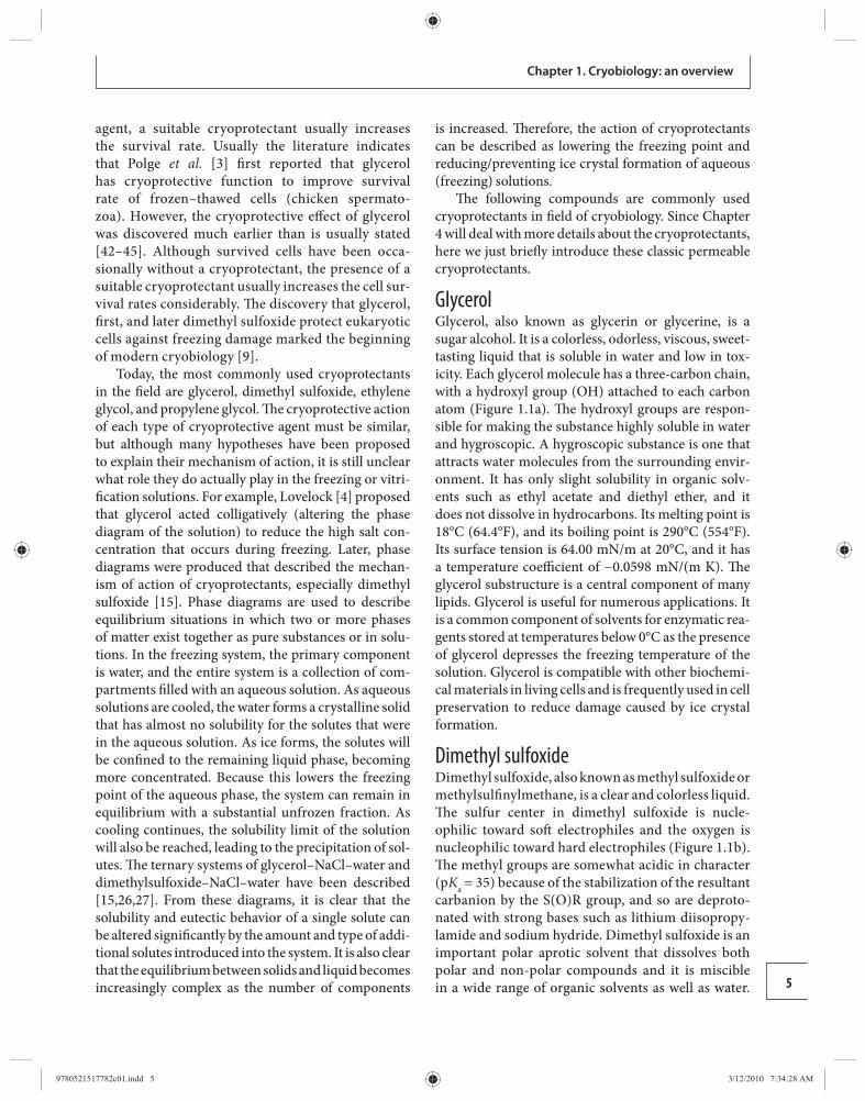
Glycerol Glycerol, also known as glycerin or glycerine, is a sugar alcohol. It is a colorless, odorless, viscous, sweet-tasting liquid that is soluble in water and low in tox-icity. Each glycerol molecule has a three-carbon chain, with a hydroxyl group (OH) attached to each carbon atom ( Figure 1.1 a). Th e hydroxyl groups are respon-sible for making the substance highly soluble in water and hygroscopic. A hygroscopic substance is one that attracts water molecules from the surrounding envir-onment. It has only slight solubility in organic solv-ents such as ethyl acetate and diethyl ether, and it does not dissolve in hydrocarbons. Its melting point is 18°C (64.4°F), and its boiling point is 290°C (554°F). Its surface tension is 64.00 mN/m at 20°C, and it has a temperature coeffi cient of −0.0598 mN/(m K). Th e glycerol substructure is a central component of many lipids. Glycerol is useful for numerous applications. It is a common component of solvents for enzymatic rea-gents stored at temperatures below 0°C as the presence of glycerol depresses the freezing temperature of the solution. Glycerol is compatible with other biochemi-cal materials in living cells and is frequently used in cell preservation to reduce damage caused by ice crystal formation.
Dimethyl sulfoxide Dimethyl sulfoxide, also known as methyl sulfoxide or methylsulfi nylmethane, is a clear and colorless li quid. Th e sulfur center in dimethyl sulfoxide is nucle-ophilic toward soft electrophiles and the oxygen is nucleophilic toward hard electrophiles ( Figure 1.1 b). Th e methyl groups are somewhat acidic in character (p K a = 35) because of the stabilization of the resultant carbanion by the S(O)R group, and so are deproto-nated with strong bases such as lithium diisopropy-lamide and sodium hydride. Dimethyl sulfoxide is an import ant polar aprotic solvent that dissolves both polar and non-polar compounds and it is miscible in a wide range of organic solvents as well as water.
9780521517782c01.indd 59780521517782c01.indd 5 3/12/2010 7:34:28 AM3/12/2010 7:34:28 AM
6
Section 1. Cryobiology
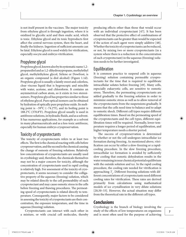
It dissolves a variety of organic substances, includ-ing carbohydrates, polymers, and peptides, as well as many inorganic salts and gases. Its melting point is 18.5°C (65.3°F), and its boiling point is 189°C (372.2°F). It has a distinctive property of penetrat-ing the skin very readily. Its taste has been described as oyster- or garlic-like. Other reported side eff ects include stomach upset, sensitivity to light, visual dis-turbances, and headache. Skin irritation can develop at the site where dimethyl sulfoxide is applied topi-cally. Loading levels of 50–60 wt% are oft en observed compared with 10–20 wt% with typical solvents. For this reason, dimethyl sulfoxide plays a role in sample management and high-throughput screening opera-tions in drug design [ 46 ] . In cryobiology, dimethyl sulfoxide has been used as a cryoprotectant and it is still an important cryoprotectant for vitrifi cation used to preserve organs, tissues, and cell suspensions.
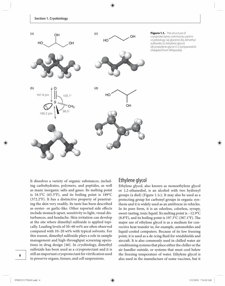
Ethylene glycol Ethylene glycol, also known as monoethylene glycol or 1,2-ethanediol, is an alcohol with two hydroxyl groups (a diol) ( Figure 1.1 c). It may also be used as a protecting group for carbonyl groups in organic syn-thesis and it is widely used as an antifreeze in vehicles. In its pure form, it is an odorless, colorless, syrupy, sweet-tasting, toxic liquid. Its melting point is −12.9°C (8.8°F), and its boiling point is 197.3°C (387.1°F). Th e major use of ethylene glycol is as a medium for con-vective heat transfer in, for example, automobiles and liquid-cooled computers. Because of its low freezing point, it is used as a de-icing fl uid for windshields and aircraft . It is also commonly used in chilled water air conditioning systems that place either the chiller or the air handler outside, or systems that must cool below the freezing temperature of water. Ethylene glycol is also used in the manufacture of some vaccines, but it
105.1°
98.0°186.2 pm
O
SCH3
167.8 pm
(b)
OHHO
(c)
OH
HO
(d)
(a) OH
HO OH
Figure 1.1. The structure of cryoprotectants commonly used in cryobiology: (a) glycerol; (b), dimethyl sulfoxide; (c) ethylene glycol;(d) propylene glycol (1,2-propanediol) (Adapted from Wikipedia).
9780521517782c01.indd 69780521517782c01.indd 6 3/12/2010 7:34:28 AM3/12/2010 7:34:28 AM
7
Chapter 1. Cryobiology: an overview
is not itself present in the vaccines. Th e major toxicity from ethylene glycol is through ingestion, where it is oxidized to glycolic acid and then oxalic acid, which is toxic. Ethylene glycol and its toxic byproducts fi rst aff ect the central nervous system, then the heart, and fi nally the kidneys. Ingestion of suffi cient amounts can be fatal. Ethylene glycol is used widely for vitrifi cation, especially oocyte and embryo vitrifi cation.
Propylene glycol Propylene glycol, known also by its systematic name 1,2-propanediol and as 1,2-dihydroxypropane, methylethyl glycol, methylethylene glycol, Sirlene or Dowfrost, is an organic compound (a diol alcohol) ( Figure 1.1 d). Propylene glycol is usually a faintly sweet and colorless, clear viscous liquid that is hygroscopic and miscible with water, acetone, and chloroform. It contains an asymmetrical carbon atom, so it exists in two stereoi-somers. Propylene glycol has properties similar to those of ethylene glycol. Pure optical isomers can be obtained by hydration of optically pure propylene oxide. Its melt-ing point is −59°C (−74.2°F), and its boiling point is 188.2°C (370.8°F). Propylene glycol usually is used in antifreeze solutions, in hydraulic fl uids, and as a solvent. It has numerous applications, for example as a solvent in many pharmaceuticals and as a less-toxic antifreeze, especially for human embryo cryopreservation.
Toxicity of cryoprotectants Th e toxicity of cryoprotectants refers to at least two eff ects. Th e fi rst is the chemical reacting with cells before cryopreservation, and the second is the chem ical causing the change of osmosis of freezing solutions. Relatively low concentrations of cryoprotectants are usually used in cryobiology and, therefore, the chemicals themselves may not be a major concern for toxicity, although the concentration of cryoprotectants used in rapid cooling is relatively high. For assessment of the toxicity of cryo-protectants, it seems necessary to consider the colliga-tive property of the aqueous (freezing) solution, which may be related directly to the cell permeability of each cryoprotectant and may cause osmotic stress in the cells before freezing and thawing procedures. Th e permeat-ing speed of cryoprotectants is related directly to tem-perature. Consequently, major factors to be considered in assessing the toxicity of cryoprotectants are their con-centration, the exposure temperature, and the time in aqueous (freezing) solution.
Cryoprotectants can interact with each other in a mixture, or with crucial cell molecules, thereby
producing eff ects other than those that would occur with an individual cryoprotectant [ 47 ]. It has been observed that the protective eff ect of combinations of cryoprotectants can be greater than would be expected if the action of each agent were simply additive [ 48 ]. Whether the toxicity of cryoprotectants can be reduced, or not, by mixing two or more cryoprotectants (in a system where there is a reduction in the concentration of each cryoprotectant) in the aqueous (freezing) solu-tion needs to be further investigated.
Equilibration It is common practice to suspend cells in aqueous (freezing) solution containing permeable cryopro-tectants for the time that is required to equilibrate intracellular solutes before freezing [ 49 ]. Many cells, especially eukaryotic cells, are sensitive to osmotic stress. Th erefore, the permeating cryoprotectants are added gradually to the freezing solution in order to minimize osmotic stress as well as to allow removal of the cryoprotectants from the suspensions gradually. It means that the cells need time to balance and to adapt to osmotic shock. Diff erent cell types require diff erent equilibration times. Based on the permeating speed of the cryoprotectants and the cell types, diff erent equi-libration times will be required. Normally, lower tem-perature requires a longer period of equilibration, and higher temperature needs a shorter period.
Th e success of cryopreservation is determined by whether or not the cell undergoes intracellular ice formation during freezing. As mentioned above, vitri-fi cation can occur by either a slow-freezing or a rapid- cooling procedure. In the slow freezing procedure, intracellular ice formation is avoided by suffi ciently slow cooling that osmotic dehydration results in the water remaining in near chemical potential equilibrium with the outside solution and ice. In the rapid cooling procedure, the cooling rate needed for vitrifi cation is approaching T g . Diff erent freezing solutions with dif-ferent concentrations of cryoprotectants need diff erent cooling rates for vitrifi cation. Th ese rates are derived primarily from calculations using mathematic al models of ice crystallization in very dilute solutions [ 20 , 50 – 53 ]. However, the actual situation may diff er from the theoretical rate in the diff erent cell types.
Conclusions Cryobiology is the branch of biology involving the study of the eff ects of low temperatures on organisms and is most oft en used for the purpose of achieving
9780521517782c01.indd 79780521517782c01.indd 7 3/12/2010 7:34:28 AM3/12/2010 7:34:28 AM
8
Section 1. Cryobiology
cryopreservation. Cryobiology is the core of fertility cryopreservation. Th e principal application for human fertility cryopreservation began with sperm freezing, and then developed to include embryo and oocyte as well as gonadal cryopreservation. Although knowledge and medical achievements have advanced in fertility cryopreservation, especially with the recent develop-ment of oocyte and ovarian tissue cryopreservation, cryobiology can still be considered as a relatively new branch of biology.
Many factors aff ect successful cryopreservation of cells. First, it may depend on the cell type, cell size, cell growth phase, cell water content, cell lipid content, and the composition of the cells, as well as cell density. Second, it may depend on the composition of the freez-ing or vitrifi cation medium, the cooling rate, the storage temperature, the duration of storage, and the warming rate and recovery medium. Th ird, it may be important to add cryoprotectants(s) into aqueous (freezing) solu-tions for freezing.
Th e mechanism of action of cryoprotectants can be considered as lowering the freezing point and prevent-ing ice crystal formation in intracellular and extracel-lular solvents. It has been considered that there may be minor or severe cryoprotectant toxicity. Th is toxicity is related directly to the concentration used, and the cell exposure temperature and time. Although some math-ematical theories have been developed in cryobiology [ 54 ], these theories may not be applicable to all types of cell. Further theoretical considerations are needed for developments in the fi eld of cryobiology.
References 1. Luyet B. Working hypotheses on the nature of life .
Biodynamics 1934; 1: 1 –7. 2. Luyet B. Th e vitrifi cation of organic colloids and of
protoplasm . Biodynamica 1937; 1: 1 –14. 3. Polge C , Smith AU , Parkes , AS . Revival os spermatozoa
aft er vitrifi cation and dehydration at low temperatures . Nature 1949; 164: 666.
4. Lovelock JE. Th e haemolysis of human red blood cells by freezing and thawing . Biochim Biophys Acta 1953; 10: 414 –426.
5. Lovelock JE. Th e mechanism of the protective action of glycerol against haemolysis by freezing and thawing . Biochim Biophys Acta 1953; 11: 28 –36.
6. Luyet B , Gonzales F . Growth of nerve tissue aft er freezing in liquid nitrogen . Biodynamica 1953; 7: 171 –174.
7. Lovelock JE , Polge C . Th e immobilization of spermatozoa by freezing and thawing and protective action of glycerol . Biochem J 1954; 58: 618 –622.
8. Luyet B , Rapatz G . Patterns of ice formation in some aqueous solutions . Biodynamica 1958; 8: 1 –68.
9. Lovelock JE , Bishop MWH . Prevention of freezing damage to living cells by dimethylsulphoxide . Nature 1959; 183: 1394 –1395.
10. Zachariassen KE , Kristiansen E . Ice nucleation and antinucleation in nature . Cryobiology 2000; 41: 257 –279.
11. Mazure P. Th e kinetics of water loss from cells at subzero temperature and the likelihood of intracellular freezing . J Gen Physiol 1963; 47: 347 –369.
12. Mazur P. Cryobiology: the freezing of biological systems . Science 1970; 168: 939 –949.
13. Mazur P. Freezing of living cells: mechanisms and implications . Am J Physiol 1984; 247 : C125 –142.
14. Luyet B , Rasmussen D . Study by diff erential thermal analysis of the temperatures of instability of rapidly cooled solutions of glycerol, ethylene glycol, sucrose, and glucose . Biodynamica 1968; 10: 167 –191.
15. Cocks FH , Brower WE . Phase diagram relationships in cryobiology . Cryobiology 1974; 11: 340 –358.
16. McGann LE . Diff ering action of penetrating and nonpenetrating cryoprotective agents . Cryobiology 1978; 15: 382 –390.
17. Meryman HT . Modifi ed model for the mechanism of freezing injury in erythrocytes . Nature 1968; 218: 333 –336.
18. Meryman HT . Osmotic stress as a mechanism of freezing injury . Cryobiology 1971; 8: 489 –500.
19. Pegg DE , Diaper MP . On the mechanism of injury to slowly frozen erythrocytes . Biophys J 1988; 54: 471 –488.
20. Boutron P , Kaufmann A . Stability of the amorphous state in the system water–glycerol–dimethylsulfoxide . Cryobiology 1978; 15: 93 –108.
21. Boutron P , Kaufmann A . Stability of the amorphous state in the system water-glycerol–ethylene glycol . Cryobiology 1979; 16: 83 –89.
22. Boutron P. Stability of the amorphous state in the system water–1,2-propanediol . Cryobiology 1979; 16: 557 –568.
23. McGann LE . Optimal temperature ranges for control of cooling rate . Cryobiology 1979; 16: 211 –216.
24. Meryman HT , Williams RJ , Douglas MSJ . Freezing injury from ‘ solution eff ects ’ and its prevention by natural or artifi cial cryoprotection . Cryobiology 1977; 14: 287 –302.
25. Rall WF , Reid DS, Farrant J . Innocuous biological freezing during warming . Nature 1980; 286 : 511–514 .
26. Pegg DE . Simple equation for obtaining melting points and eutectic temperatures for the ternary system glycerol/sodium chloride/water . Cryo Letters 1983; 4: 259 –268.
27. Pegg DE . Equations for obtaining melting points and eutectic temperatures for the ternary system dimethyl sulphoxide/sodium chloride/water . Cryo Letters 1986; 7: 387 –394.
9780521517782c01.indd 89780521517782c01.indd 8 3/12/2010 7:34:28 AM3/12/2010 7:34:28 AM
9
Chapter 1. Cryobiology: an overview
28. Pegg DE , Arnaud FG . Equations for obtaining melting points in the quaternary system propane-1,2-diol/glycerol/sodium chloride/water . Cryo Letters 1988; 9: 404 –417.
29. Boutron P , Arnaud F . Comparison of the cryoprotection of red blood cells by 1,2-propanediol and glycerol . Cryobiology 1984; 21: 348 –358.
30. Fahy GM . Analysis of ‘ solution eff ects ’ injury: equations for calculating phase diagram information for the ternary systems NaCl–dimethylsulfoxide–water and NaCl–glycerol–water . Biophys J 1980; 32: 837 –850.
31. Fahy GM . Simplifi ed calculation of cell water content during freezing and thawing in nonideal solutions of cryoprotective agents and its possible application to the study of ‘ solution eff ects ’ injury . Crybiology 1981; 18: 473 –482.
32. Fahy GM . Cryoprotectant toxicity neutralizers reduce freezing damage . Cryo Letters 1983; 4: 309 –314.
33. Fahy GM . Cryoprotectant toxicity reduction: specifi c or nonspecifi c? Cryo Letters 1984; 5: 287 –294.
34. Fahy GM . Th e relevance of cryoprotectant ‘ toxicity ’ to cryobiology . Cryobiology 1986; 23: 1 –13.
35. Rall WF , Mazur P , Souzu H . Physical–chemical basis of the protection of slowly frozen human erythrocytes by glycerol . Biophys J 1978; 23: 101 –120.
36. Fahy GM , Karow AM , Jr. Ultrastructure-function correlative studies for cardiac cryopreservation. V. Absence of a correlation between electrolyte toxocity and cryoinjury in the slowly frozen, cryoprotected rat heart . Cryobiology 1977; 14: 418 –427.
37. Fahy GM , MacFarlane DR , Angell CA , et al. Vitrifi cation as an approach to cryopreservation . Cryobiology 1984; 21: 407 –426.
38. Fahy GM , Lilley TH , Linsdell H , et al. Cryoprotectant toxicity and cryoprotectant toxicity reduction: in search of molecular mechanism . Cryobiology 1990; 27: 247 –268.
39. Fahy GM , Wowk B , Wu J , et al. Improved vitrifi cation solutions based on predictability of vitrifi cation solution toxicity . Cryobiology 2004; 48: 22 –35.
40. Fahy GM , Wowk B , Wu J , et al. Cryopreservation of organs by vitrifi cation: perspectives and recent advances . Crybiology 2004; 48: 157 –178.
41. Rall WF , Fahy GM . Ice-free cryopreservation of mouse embryos at −196°C by vitrifi cation . Nature 1985; 313 : 573–575.
42. Keith SC . Factors infl uencing the survival of bacteria at temperatures in the vicinity of the freezing point of water . Science 1913; 37: 877 –879.
43. Spencer RR , Parker RR . Rocky mountain spotted fever . Public Health Rep 1924; 39: 3027 –3040.
44. Francis E . Duration of viability of Pasteurella pestis . Public Health Rep 1932; 47: 1287 –1294.
45. Pabst AM . Use of below freezing temperatures for maintenance of meningococcus cultures (Neisseria intracellularis) . Public Health Rep 1935; 50: 732 –737.
46. Balakin KV , Savchuk NP , Tetko IV . In silico approaches to prediction of aqueous and DMSO solubility of drug-like compounds: trends, problems and solutions . Curr Med Chem 2006; 13: 223 –241.
47. Ruwart MJ , Holland JF , Haug A . Fluorimetric evidence of interactions involving cryoprotectants and biomolecules . Cryobiology 1975; 12: 26 –33.
48. Chian R-C , Kuwayama M , Tan L , et al . High survival rate of bovine oocytes matured in vitro following vitrifi cation . J Reprod Dev 2004; 50: 685 –696.
49. Mazur P , Seki S , Pinn IL , Kleinhans FW , Edashige K . Extra- and intracellular ice formation in mouse oocytes . Cryobiology 2005; 51: 29 –53.
50. Bruggeller P , Mayer E . Complete vitrifi cation in pure liquid water and dilute aqueous solutions . Nature 1980; 288: 569 –571.
51. Boutron P . Comparison with the theory of the kinetics and extent of ice crystallization and of the glass-forming tendency in aqueous cryoprotective solutions . Cryobiology 1986; 23: 88 –102.
52. Toner M , Cravalho EG , Chiang YM . Vitrifi cation of biological cell suspensions: the importance of ultrarapid cooling and warming . Cryobiology 1988; 25: 551.
53. Baudot A , Odagescu V . Th ermal properties of ethylene glycol and aqueous solutions . Cryobiology 2004; 48: 283 –294.
54. Fahy GM . Th eoretical considerations for oocyte cryopreservation by freezing . Reprod Biomed Online 2007; 14: 709 –714.
9780521517782c01.indd 99780521517782c01.indd 9 3/12/2010 7:34:28 AM3/12/2010 7:34:28 AM
Fertility Cryopreservation, ed. Ri-Cheng Chian and Patrick Quinn. Published by Cambridge University Press. Copyright © Cambridge University Press 2010.
Chapter
2 Increasing knowledge of cryobiological mechan-
isms gave major insights to improve freezing–thawing protocols ( Chapter 1 ). As one of the major components in any cell, including oocytes and embryos, is water, great care has to be taken that lethal ice crystal forma-tion does not take place. Other critical factors during slow cooling include solution eff ects, osmotic stress, and intracellular dehydration. A summary of some of these factors and how they are handled with vitrifi ca-tion are given in Tables 2.1 and 2.2 [ 3 ].
Membrane permeability to water and solutes Th e oocyte is the largest human cell (130 μm diam-eter). Large cells have a low surface-to-volume ratio and hence they are less effi cient at taking up cryopro-tectants (CPAs) and at losing water. Th e overall eff ect is that oocytes are more likely to retain water during cryopreservation and thus can be damaged by intracel-lular ice formation and growth. Also, oocytes can be seriously damaged even before freezing by excessive osmotic stress resulting from the addition or dilution of CPAs. Th e fl ow of water across each unit of the cell surface as a function of time is called the hydraulic con-ductivity or hydraulic coeffi cient (LP). Hydraulic con-ductivity is related to the volume of the cell. Th ere is great variability between cell types and even species among mammals. In fact, membrane permeability changes with the developmental stages of oocyte, zygote, and embryo [ 4 – 6 ]. An example with human oocytes that caused some confusion initially is that freshly collected human oocytes are more prone to cryoinjuries than ones that are 1 day old, such as oocytes that fail to fer-tilize. Subsequently, it was found that methods estab-lished with “old” oocytes were oft en not suitable for the more sensitive freshly collected oocytes with full devel-opmental potential [ 7 ], and it was suggested that this
Introduction Th is chapter draws on several sources that have com-prehensively reviewed this topic and here I want to especially acknowledge Dr. Helmy Selman, who organ-ized a conference in Perugia, Italy on cryopreservation in 2006. Other sources are the classic work by Fahy and colleagues [ 1 ], which gives a thorough and in-depth look at the theoretical and biophysical aspects of this topic. Th is review is structured so that the more prac-tical aspects are considered fi rst followed by some of the more theoretical components involved. Th is approach will allow fi rst an analysis of what works in a clinical set-ting and then a return to consider what is happening at the physical and molecular level. I believe this approach will allow a better understanding of the process.
Background Slow cooling cryopreservation of mammalian oocytes and embryos has been extensively discussed and reviewed both in the chapters of this book and in other literature in the area for more than four decades and will not be further discussed in detail in this chapter. A concise description can be found in Wikipedia [ 2 ].
Although cryopreservation of human embryos and zygotes has become a well-established procedure in assisted reproductive technology (ART), oocyte freezing has proved to be technically challenging and until recently remained experimental. For several dec-ades, attempts to cryopreserve human oocytes have been performed in many in vitro fertilization (IVF) centers worldwide, with variable results. When the slow freezing method traditionally used for embryos or zygotes was initially applied to oocytes, there were poor success rates. A major factor contributing to the poor results was that much of the cryobiology carried out by embryologists was empirical, based on “rules of thumb” rather than basic principles and knowledge.
Suppression of ice in aqueous solutions and its application to vitrifi cation in assisted reproductive technology Patrick Quinn
10
9780521517782c02.indd 109780521517782c02.indd 10 3/12/2010 7:48:24 AM3/12/2010 7:48:24 AM
11
Chapter 2. Suppression of ice in aqueous solutions
could be because of changes in membrane composition between the two types of oocyte [ 8 ].
Temperature During handling of oocytes and embryos, it is important to choose a suitable temperature. Elevated temperature
could increase the CPA toxicity. Very slow and very high cooling rates can damage cells through osmotic eff ect and pH changes. Very slow cooling rates tend to encourage extracellular ice formation, while the very high cooling rates tend to promote intracellular ice for mation: both could be lethal to cells. With vitrifi cation, however, the cooling and warming rate are suffi ciently fast to never allow ice crystal formation either upon cool-ing or upon warming. Most other physiological changes that occur with slow cooling cryopreservation, such as osmotic eff ect, solution eff ects, and dehydration, are also eliminated by vitrifi cation as during the process the entire solution remains unchanged except for the transition of water from a liquid to a glassy phase [ 3 ]. With warming, the rate is suffi cient that devitrifi cation, the formation of ice crystals from vitrifi ed water, does not occur.
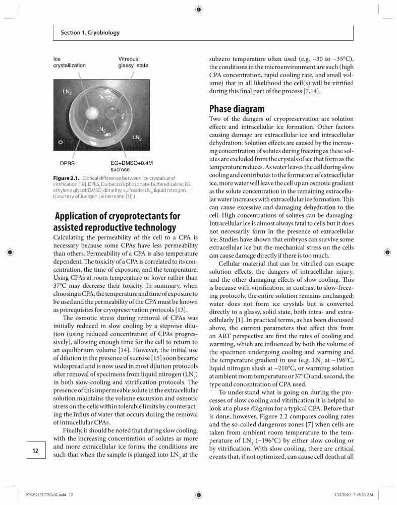
Vitrifi cation “Ice formation is not compatible with the survival of living system and ought to be avoided during freezing” (Luyet, 1937 [ 9 ]), and “good vitrifi cation is not inju-rious, there being no molecular disturbance, while an incomplete vitrifi cation or devitrifi cation and, a forti-ori, crystallization, are injurious to the extent that they disrupt the living structure” (Luyet and Gehenio, 1940 [ 10 ]). Th ese two concise statements epitomize the core of vitrifi cation. Ice-free cryopreservation is an attempt to circumvent the hazards of water crystallization as ice. Th e vitreous state is essentially a solidifi ed, amorphous liquid state obtained by specifi c conditions of cooling and solute concentration that inhibit ice nucleation and growth. During cooling, the molecular motions within a liquid are slowed and eventually arrested with extreme viscosity. Th e arrested liquid state is a glass. So, vitrifi cation is the direct conversion of a liquid state into glass, bypassing or rapidly passing through the critical temperature of ice formation (no phase tran-sition). A striking illustration of vitrifi cation is shown in Figure 2.1 . More details of vitrifi cation will be given below in the discussion of the phase diagram .
Probability of vitrifi cation Th ree key factors infl uence the probability of success-ful vitrifi cation: cooling and warming rates, the com-position of the CPA solution, and the sample volume [ 1 2]. Increasing CPA concentration and decreasing sample volume will each increase the probability of vit-rifi cation. Pressure is another factor that increases the chance of vitrifi cation [ 1 ] but this has had very little, if any, application in the clinical ART arena.
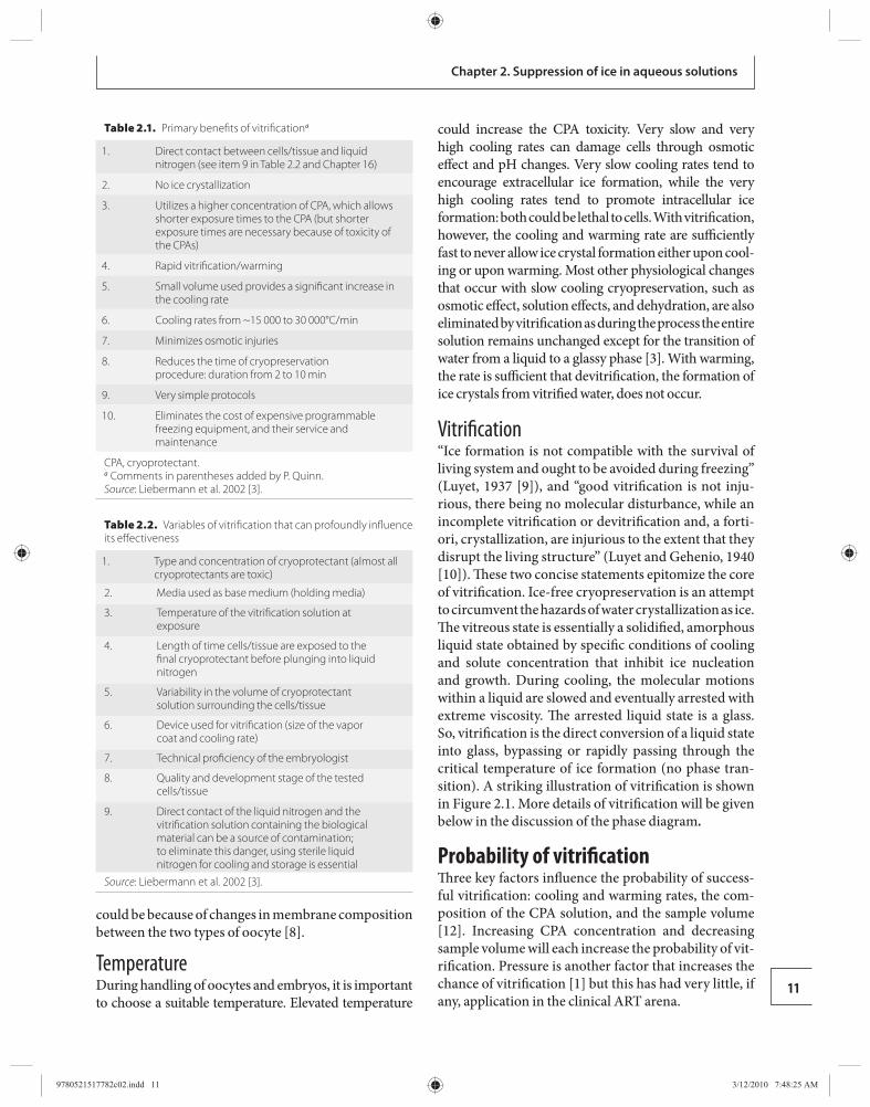
Table 2.2. Variables of vitrifi cation that can profoundly infl uence its eff ectiveness
1. Type and concentration of cryoprotectant (almost all cryoprotectants are toxic)
2. Media used as base medium (holding media)
3. Temperature of the vitrifi cation solution at exposure
4. Length of time cells/tissue are exposed to the fi nal cryoprotectant before plunging into liquid nitrogen
5. Variability in the volume of cryoprotectant solution surrounding the cells/tissue
6. Device used for vitrifi cation (size of the vapor coat and cooling rate)
7. Technical profi ciency of the embryologist
8. Quality and development stage of the tested cells/tissue
9. Direct contact of the liquid nitrogen and the vitrifi cation solution containing the biological material can be a source of contamination; to eliminate this danger, using sterile liquid nitrogen for cooling and storage is essential
Source : Liebermann et al. 2002 [ 3 ].
Table 2.1. Primary benefi ts of vitrifi cation a
1. Direct contact between cells/tissue and liquid nitrogen (see item 9 in Table 2.2 and Chapter 16)
2. No ice crystallization
3. Utilizes a higher concentration of CPA, which allows shorter exposure times to the CPA (but shorter exposure times are necessary because of toxicity of the CPAs)
4. Rapid vitrifi cation/warming
5. Small volume used provides a signifi cant increase in the cooling rate
6. Cooling rates from ~15 000 to 30 000°C/min
7. Minimizes osmotic injuries
8. Reduces the time of cryopreservation procedure: duration from 2 to 10 min
9. Very simple protocols
10. Eliminates the cost of expensive programmable freezing equipment, and their service and maintenance
CPA, cryoprotectant. a Comments in parentheses added by P. Quinn. Source : Liebermann et al. 2002 [ 3 ].
9780521517782c02.indd 119780521517782c02.indd 11 3/12/2010 7:48:25 AM3/12/2010 7:48:25 AM
Section 1. Cryobiology
12
Application of cryoprotectants for assisted reproductive technology Calculating the permeability of the cell to a CPA is necessary because some CPAs have less permeability than others. Permeability of a CPA is also temperature dependent. Th e toxicity of a CPA is correlated to its con-centration, the time of exposure, and the temperature. Using CPAs at room temperature or lower rather than 37°C may decrease their toxicity. In summary, when choosing a CPA, the temperature and time of exposure to be used and the permeability of the CPA must be known as prerequisites for cryopreservation protocols [ 13 ].
Th e osmotic stress during removal of CPAs was initially reduced in slow cooling by a stepwise dilu-tion (using reduced concentration of CPAs progres-sively), allowing enough time for the cell to return to an equilibrium volume [ 1 4]. However, the initial use of dilution in the presence of sucrose [ 1 5] soon became widespread and is now used in most dilution protocols aft er removal of specimens from liquid nitrogen (LN 2 ) in both slow-cooling and vitrifi cation protocols. Th e presence of this impermeable solute in the extracellular solution maintains the volume excursion and osmotic stress on the cells within tolerable limits by counteract-ing the infl ux of water that occurs during the removal of intracellular CPAs.
Finally, it should be noted that during slow cooling, with the increasing concentration of solutes as more and more extracellular ice forms, the conditions are such that when the sample is plunged into LN 2 at the
subzero temperature oft en used (e.g. −30 to −35°C), the conditions in the microenvironment are such (high CPA concentration, rapid cooling rate, and small vol-ume) that in all likelihood the cell(s) will be vitrifi ed during this fi nal part of the process [ 7 , 14 ].
Phase diagram Two of the dangers of cryopreservation are solution eff ects and intracellular ice formation. Other factors causing damage are extracellular ice and intracellular dehydration. Solution eff ects are caused by the increas-ing concentration of solutes during freezing as these sol-utes are excluded from the crystals of ice that form as the temperature reduces. As water leaves the cell during slow cooling and contributes to the formation of extracellular ice, more water will leave the cell up an osmotic gradient as the solute concentration in the remaining extracellu-lar water increases with extracellular ice formation. Th is can cause excessive and damaging dehydration to the cell. High concentrations of solutes can be damagin g. Intracellular ice is almost always fatal to cells but it does not necessarily form in the presence of extracellular ice. Studies have shown that embryos can survive some extracellular ice but the mechanical stress on the cells can cause damage directly if there is too much.
Cellular material that can be vitrifi ed can escape solution eff ects, the dangers of intracellular injury, and the other damaging eff ects of slow cooling. Th is is because with vitrifi cation, in contrast to slow-freez-ing protocols, the entire solution remains unchanged; water does not form ice crystals but is converted directly to a glassy, solid state, both intra- and extra-cellularly [ 1 ]. In practical terms, as has been discussed above, the current parameters that aff ect this from an ART perspective are fi rst the rates of cooling and warming, which are infl uenced by both the volume of the specimen undergoing cooling and warming and the temperature gradient in use (e.g. LN 2 at −196°C, liquid nitrogen slush at −210°C, or warming solution at ambient room temperature or 37°C) and, second, the type and concentration of CPA used.
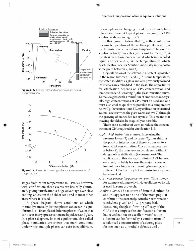
To understand what is going on during the pro-cesses of slow cooling and vitrifi cation it is helpful to look at a phase diagram for a typical CPA. Before that is done, however, Figure 2.2 compares cooling rates and the so-called dangerous zones [ 7 ] when cells are taken from ambient room temperature to the tem-perature of LN 2 (−196°C) by either slow cooling or by vitrifi cation. With slow cooling, there are critical events that, if not optimized, can cause cell death at all
DPBS EG+DMSO+0.4Msucrose
Icecrystallization
Vitreous,glassy state
LN2
LN2
LN2
©
Figure 2.1. Optical diff erence between ice crystals and vitrifi cation [ 18 ]. DPBS, Dulbecco’s phosphate-buff ered saline; EG, ethylene glycol; DMSO, dimethyl sulfoxide; LN 2 , liquid nitrogen. (Courtesy of Juergen Liebermann [11].)
9780521517782c02.indd 129780521517782c02.indd 12 3/12/2010 7:48:25 AM3/12/2010 7:48:25 AM
13
Chapter 2. Suppression of ice in aqueous solutions
stages from room temperature to −196°C; however, with vitrifi cation, these events are basically elimin-ated, giving vitrifi cation a huge advantage over slow cooling, at least in the fi eld of ART and in most other areas where it is used.
A phase diagram shows conditions at which thermo dynamically distinct phases can occur in equi-librium [ 1 6]. Examples of diff erent phases of water that can occur in cryopreservation are liquid, ice, and glass. In a phase diagram, lines of equilibrium, also called phase boundaries, are shown that mark conditions under which multiple phases can exist in equilibrium,
for example water changing to and from a liquid phase into an ice phase. A typical phase diagram for a CPA solution is shown in Figure 2.3 .
In this fi gure, T f (also called T m ) is the equilibrium freezing temperature of the melting point curve, T h is the homogeneous nucleation temperature before the solution actually nucleates (i.e. begins to freeze), T g is the glass transition temperature at which supercooled liquid vitrifi es, and T d is the temperature at which devitrifi cation occurs. Solutions normally supercool to some point between T f and T h .
Crystallization of the solvent (e.g. water) is possible in the region between T f and T h . At some temperature, the water solidifi es as glass and any previously formed ice crystals are embedded in the glass. Th e opportunity for vitrifi cation depends on CPA concentration and temperature and lies along T g , the glass transition curve. To make a glass with a minimum of embedded ice crys-tals, high concentrations of CPA must be used and one must also cool as quickly as possible to a temperature below T g. Devitrifi cation ( T d ), crystallization in vitrifi ed system, occurs when the glass warms above T g through the growing of embedded ice crystals. Th is means that thawing should also be as quickly as possible.
Th ere are a number of ways to reduce the concen-tration of CPA required for vitrifi cation [ 1 ].
Apply a high hydrostatic pressure . Increasing the pressure lowers T h and increases T g , thus shift ing the point of intersection of these two curves to a lower CPA concentration. Once the temperature is below T g , the pressure can be released without danger of crystallization (ice formation). Th e application of this strategy in clinical ART has not occurred, probably because the major factors of low volumes, high rates of cooling/warming, and suffi cient CPA to vitrify but minimize toxicity have been invoked.
Add a non-permeating polymer or agent . Th is strategy, for example adding polyvinylpyrrolidone or Ficoll, is used in some protocols.
Combine CPAs . The mixture of dimethyl sulfoxide and EG appears to be one of the most popular combinations currently. Another combination is ethylene glycol and 1,2-propanediol. Measuring the glass-forming efficacy of the CPAs that compose the vitrification solution has revealed that an excellent vitrification solution can be formed by a combination of a balanced concentration of a strong glass former such as dimethyl sulfoxide and a
Tem
pera
ture
(�C
)
�20
0
�20
�40
�60
Time (min)
30 90 120
Slow cooling
Vitrification�196
Warming
Warming
Chilling and thaw injuries
Extra- and intracellular ice,solution effects, osmoticstress between �5 and �80�C,and fracture between �50and �150�C [7]
Injuries in danger zones
Vitrification occursfor both slow cooledand vitrified samples.Least dangerouszone. Accidentalwarming is the mostfrequent injury
Danger zones
Figure 2.2. Cooling rates and dangerous zones during cryopreservation .
180
160
100
80
60
40
20
0
40
20
0 2 4 6CPA concentration (M)
Tf
Th
Tg
TdTem
pera
ture
(°C
)
Figure 2.3. Phase diagram of hypothetical aqueous solution of cryoprotectants .
9780521517782c02.indd 139780521517782c02.indd 13 3/12/2010 7:48:25 AM3/12/2010 7:48:25 AM
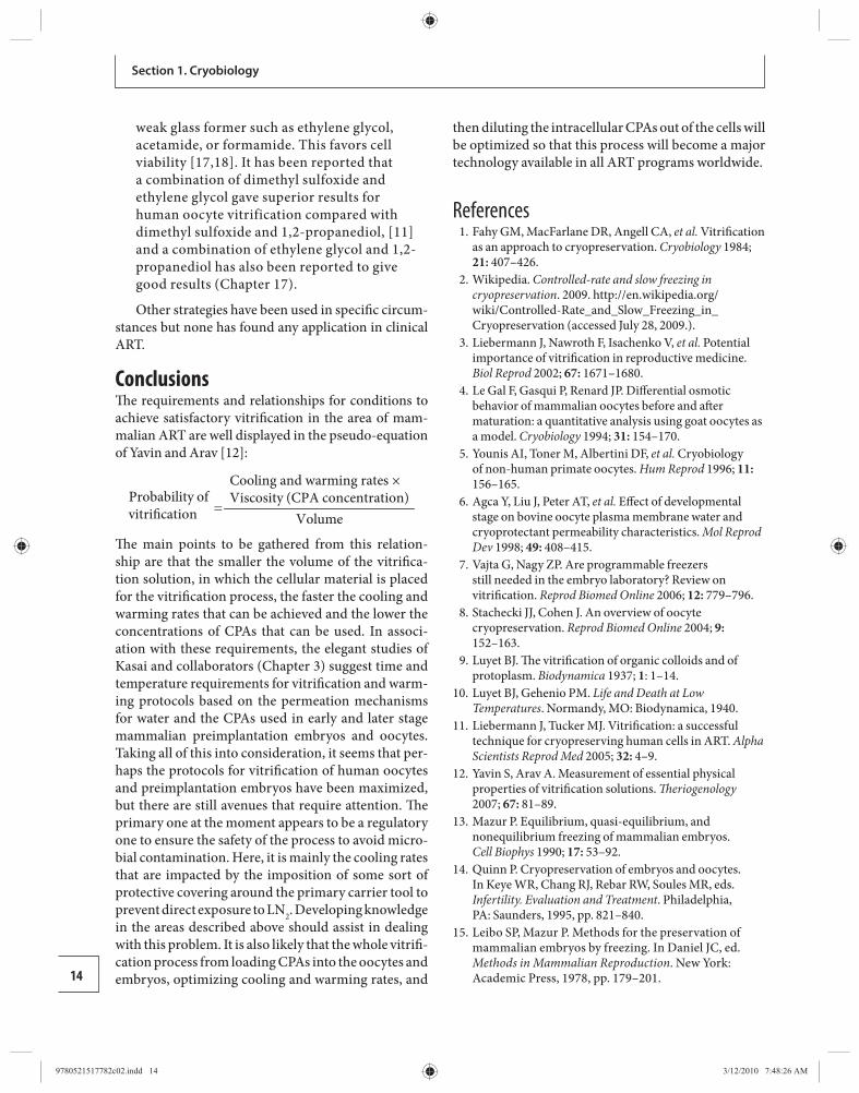
14
Section 1. Cryobiology
weak glass former such as ethylene glycol, acetamide, or formamide. This favors cell viability [ 1 7, 1 8]. It has been reported that a combination of dimethyl sulfoxide and ethylene glycol gave superior results for human oocyte vitrification compared with dimethyl sulfoxide and 1,2-propanediol, [ 1 1] and a combination of ethylene glycol and 1,2-propanediol has also been reported to give good results ( Chapter 17 ).
Other strategies have been used in specifi c circum-stances but none has found any application in clinical ART.
Conclusions Th e requirements and relationships for conditions to achieve satisfactory vitrifi cation in the area of mam-malian ART are well displayed in the pseudo-equation of Yavin and Arav [ 12 ]:
Cooling and warming rates ×Viscosity (CPA concentration)Probability of
vitrification Volume=
Th e main points to be gathered from this relation-ship are that the smaller the volume of the vitrifi ca-tion solution, in which the cellular material is placed for the vitrifi cation process, the faster the cooling and warming rates that can be achieved and the lower the concentrations of CPAs that can be used. In associ-ation with these requirements, the elegant studies of Kasai and collaborators ( Chapter 3 ) suggest time and temperature requirements for vitrifi cation and warm-ing protocols based on the permeation mechanisms for water and the CPAs used in early and later stage mammalian preimplantation embryos and oocytes. Taking all of this into consideration, it seems that per-haps the protocols for vitrifi cation of human oocytes and preimplantation embryos have been maximized, but there are still avenues that require attention. Th e primary one at the moment appears to be a regulatory one to ensure the safety of the process to avoid micro-bial contamination. Here, it is mainly the cooling rates that are impacted by the imposition of some sort of protective covering around the primary carrier tool to prevent direct exposure to LN 2 . Developing knowledge in the areas described above should assist in dealing with this problem. It is also likely that the whole vitrifi -cation process from loading CPAs into the oocytes and embryos, optimizing cooling and warming rates, and
then diluting the intracellular CPAs out of the cells will be optimized so that this process will become a major technology available in all ART programs worldwide.
References 1. Fahy GM , MacFarlane DR , Angell CA , et al. Vitrifi cation
as an approach to cryopreservation . Cryobiology 1984; 21: 407 –426.
2. Wikipedia. Controlled-rate and slow freezing in cryopreservation. 2009 . http://en.wikipedia.org/wiki/Controlled-Rate_and_Slow_Freezing_in_Cryopreservation (accessed July 28, 2009.).
3. Liebermann J , Nawroth F , Isachenko V , et al. Potential importance of vitrifi cation in reproductive medicine . Biol Reprod 2002; 67: 1671 –1680.
4. Le Gal F , Gasqui P , Renard JP . Diff erential osmotic behavior of mammalian oocytes before and aft er maturation: a quantitative analysis using goat oocytes as a model . Cryobiology 1994; 31: 154 –170.
5. Younis AI , Toner M , Albertini DF , et al. Cryobiology of non-human primate oocytes . Hum Reprod 1996; 11: 156 –165.
6. Agca Y , Liu J , Peter AT , et al. Eff ect of developmental stage on bovine oocyte plasma membrane water and cryoprotectant permeability characteristics . Mol Reprod Dev 1998; 49: 408 –415.
7. Vajta G , Nagy ZP . Are programmable freezers still needed in the embryo laboratory? Review on vitrifi cation . Reprod Biomed Online 2006; 12: 779 –796.
8. Stachecki JJ , Cohen J . An overview of oocyte cryopreservation . Reprod Biomed Online 2004; 9: 152 –163.
9. Luyet BJ . Th e vitrifi cation of organic colloids and of protoplasm . Biodynamica 1937; 1 : 1 –14.
10. Luyet BJ , Gehenio PM . Life and Death at Low Temperatures . Normandy, MO : Biodynamica, 1940 .
11. Liebermann J , Tucker MJ . Vitrifi cation: a successful technique for cryopreserving human cells in ART . Alpha Scientists Reprod Med 2005; 32: 4 –9.
12. Yavin S , Arav A . Measurement of essential physical properties of vitrifi cation solutions . Th eriogenology 2007; 67: 81 –89.
13. Mazur P . Equilibrium, quasi-equilibrium, and nonequilibrium freezing of mammalian embryos . Cell Biophys 1990; 17: 53 –92.
14. Quinn P . Cryopreservation of embryos and oocytes . In Keye WR , Chang RJ , Rebar RW , Soules MR , eds. Infertility. Evaluation and Treatment . Philadelphia, PA : Saunders , 1995, pp. 821–840 .
15. Leibo SP , Mazur P . Methods for the preservation of mammalian embryos by freezing. In Daniel JC , ed. Methods in Mammalian Reproduction . New York: Academic Press , 1978, pp. 179–201.
9780521517782c02.indd 149780521517782c02.indd 14 3/12/2010 7:48:26 AM3/12/2010 7:48:26 AM

15
Chapter 2. Suppression of ice in aqueous solutions
16. Wikipedia . Phase Diagram . 2009 . http://en.wikipedia.org/wiki/Phase_diagram (accessed July 30, 2009.).
17. Fahy GM . Biological eff ects of vitrifi cation and devitrifi cation. In Pegg DE , Karow AM , eds.
Th e Biophysics of Organ Cryopreservation . New York: Plenum , 1987, pp. 265–297.
18. Fahy GM , Wowk B , Wu J , Paynter S . Improved vitrifi cation solutions based on the predictability of vitrifi cation solution toxicity . Cryobiology 2004; 48: 22 –35.
9780521517782c02.indd 159780521517782c02.indd 15 3/12/2010 7:48:26 AM3/12/2010 7:48:26 AM

Fertility Cryopreservation, ed. Ri-Cheng Chian and Patrick Quinn. Published by Cambridge University Press. Copyright © Cambridge University Press 2010.
Chapter
3 Th ere are two pathways for the movement of water
and cryoprotectants across the plasma membrane of oocytes/embryos. One is simple diff usion through the lipid bilayer, in which case permeability is low and temperature dependency is high. Th e other is facilitated diff usion via channels, in which case permeability is markedly high and temperature dependency is low. Th erefore, low permeability with high temperature dependency is suggestive of the movement of water and cryoprotectants across the plasma membrane by simple diff usion, whereas high permeability with low tempera-ture dependency is suggestive of movement via channels.
Specifying the principal pathway for the move-ment of water and cryoprotectants in oocytes/embryos at various stages will help in the selection of suitable cryoprotectant(s) and optimal conditions for cryo-preservation, for example the time, temperature, and number of steps (one or multiple) for exposure of oocytes/embryos to the cryoprotectant solution before cooling, and the procedure for removal of the cryopro-tectant aft er warming.
Measurement of the permeability of the plasma membrane to water and cryoprotectants Th e permeability to water and cryoprotectants of mammalian oocytes/embryos can be measured as changes in volume in hypertonic solutions containing sucrose or cryoprotectant [ 5 ]. Figure 3.1 shows the method that we have developed for measuring changes in volume [ 6 , 7 ]. An oocyte or embryo is placed in a drop of PB1 medium [ 8 ] covered with paraffi n oil in a Petri dish (90 mm × 10 mm). It is held by a hold-ing pipette connected to a micromanipulator on an inverted microscope. Th e inner diameter of the mouth of the pipette is small enough not to distort the oocyte/
Introduction During cryopreservation, mammalian oocytes and embryos can be damaged by chilling, the toxicity of the cryoprotectant, the formation of extracellular ice, the formation of intracellular ice, fracturing, osmotic swelling, and osmotic shrinkage [ 1 ]. Such damage needs to be avoided to maintain the survival of oocytes/embryos aft er cryopreservation. Most of these injuries are closely related to the cryobiological properties of oocytes/embryos, such as sensitivity to chilling, the permeability of the plasma membrane to water and cryoprotectants, sensitivity to the chemical toxicity of the cryoprotectant, and tolerance of osmotic swelling and shrinkage.
Th e most suitable procedure for the cryopreserva-tion of oocytes/embryos diff ers among maturational/developmental stages even in the same species. For example, mouse morulae can be vitrifi ed without an appreciable loss of viability by a simple one-step method using ethylene glycol as the permeating cryoprotect-ant [ 2 ], whereas pretreatment with a low concentra-tion of ethylene glycol is preferable when vitrifying 2-cell embryos [ 3 , 4 ]. In the mouse, therefore, morulae appear to survive cryopreservation more easily than 2-cell embryos. In general, diff erences in cryobiological properties among oocytes/embryos at diff erent matur-ational/developmental stages will be closely related to the diff erence in the most suitable procedure for cryopreservation.
Among these cryobiological properties, the perme-ability of the plasma membrane to water and cryopro-tectants would be most important, because it is closely related to major types of cell injury during cryopreser-vation: damage caused by the toxicity of the cryopro-tectant, by the formation of intracellular ice, and by osmotic swelling during removal of the cell-permeating cryoprotectant.
Movement of water and cryoprotectants in mouse oocytes and embryos at diff erent stages: relevance to cryopreservation Magosaburo Kasai and Keisuke Edashige
16
9780521517782c03.indd 169780521517782c03.indd 16 3/12/2010 6:40:28 PM3/12/2010 6:40:28 PM
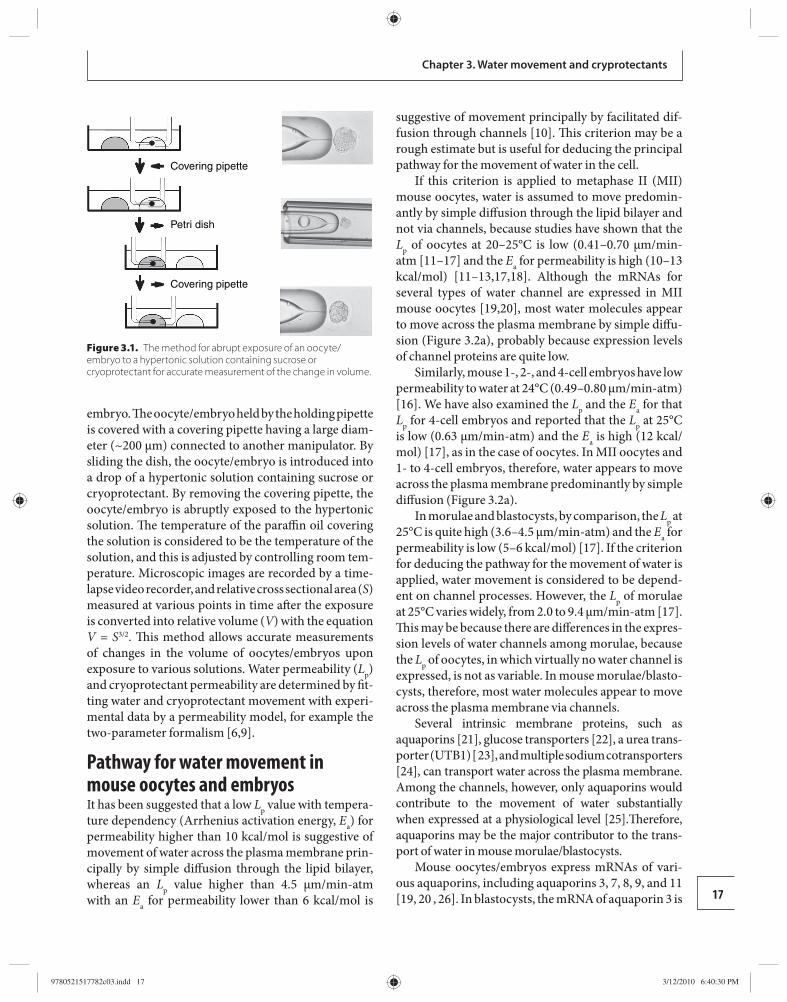
17
Chapter 3. Water movement and cryprotectants
embryo. Th e oocyte/embryo held by the holding pipette is covered with a covering pipette having a large diam-eter (~200 μm) connected to another manipulator. By sliding the dish, the oocyte/embryo is introduced into a drop of a hypertonic solution containing sucrose or cryoprotectant. By removing the covering pipette, the oocyte/embryo is abruptly exposed to the hypertonic solution. Th e temperature of the paraffi n oil covering the solution is considered to be the temperature of the solution, and this is adjusted by controlling room tem-perature. Microscopic images are recorded by a time-lapse video recorder, and relative cross sectional area ( S ) measured at various points in time aft er the exposure is converted into relative volume ( V ) with the equation V = S 3/2 . Th is method allows accurate measurements of changes in the volume of oocytes/embryos upon expos ure to various solutions. Water permeability ( L p ) and cryoprotectant permeability are determined by fi t-ting water and cryoprotectant movement with experi-mental data by a permeability model, for example the two-parameter formalism [ 6 , 9 ].
Pathway for water movement in mouse oocytes and embryos It has been suggested that a low L p value with tempera-ture dependency (Arrhenius activation energy, E a ) for permeability higher than 10 kcal/mol is suggestive of movement of water across the plasma membrane prin-cipally by simple diff usion through the lipid bilayer, whereas an L p value higher than 4.5 μm/min-atm with an E a for permeability lower than 6 kcal/mol is
suggestive of movement principally by facilitated dif-fusion through channels [ 10 ]. Th is criterion may be a rough estimate but is useful for deducing the principal pathway for the movement of water in the cell.
If this criterion is applied to metaphase II (MII) mouse oocytes, water is assumed to move predomin-antly by simple diff usion through the lipid bilayer and not via channels, because studies have shown that the L p of oocytes at 20–25°C is low (0.41–0.70 μm/min-at m [ 11 – 17 ] and the E a for permeability is high (10–13 kcal/mol) [ 11 – 13 , 17 , 18 ]. Although the mRNAs for several types of water channel are expressed in MII mouse oocytes [ 19 , 20 ], most water molecules appear to move across the plasma membrane by simple diff u-sion ( Figure 3.2a ), probably because expression levels of channel proteins are quite low.
Similarly, mouse 1-, 2-, and 4-cell embryos have low permeability to water at 24°C (0.49–0.80 μm/min-atm) [ 16 ]. We have also examined the L p and the E a for that L p for 4-cell embryos and reported that the L p at 25°C is low (0.63 μm/min-atm) and the E a is high (12 kcal/mol) [ 17 ], as in the case of oocytes. In MII oocytes and 1- to 4-cell embryos, therefore, water appears to move across the plasma membrane predominantly by simple diff usion ( Figure 3.2a ).
In morulae and blastocysts, by comparison, the L p at 25°C is quite high (3.6–4.5 μm/min-atm) and the E a for permeability is low (5–6 kcal/mol) [ 17 ]. If the criterion for deducing the pathway for the movement of water is applied, water movement is considered to be depend-ent on channel processes. However, the L p of morulae at 25°C varies widely, from 2.0 to 9.4 μm/min-atm [ 17 ]. Th is may be because there are diff erences in the expres-sion levels of water channels among morulae, because the L p of oocytes, in which virtually no water channel is expressed, is not as variable. In mouse morulae/blasto-cysts, therefore, most water molecules appear to move across the plasma membrane via channels.
Several intrinsic membrane proteins, such as aquaporins [ 21 ], glucose transporters [ 22 ], a urea trans-porter (UTB1) [ 23 ], and multiple sodium cotransporters [ 24 ], can transport water across the plasma membrane. Among the channels, however, only aquaporins would contribute to the movement of water substantially when expressed at a physiological level [ 25 ]. Th erefore, aquaporins may be the major contributor to the trans-port of water in mouse morulae/blastocysts.
Mouse oocytes/embryos express mRNAs of vari-ous aquaporins, including aquaporins 3, 7, 8, 9, and 11 [ 19 , 20 , 26 ]. In blastocysts, the mRNA of aquaporin 3 is
Covering pipette
Covering pipette
Petri dish
Figure 3.1. The method for abrupt exposure of an oocyte/embryo to a hypertonic solution containing sucrose or cryoprotectant for accurate measurement of the change in volume.
9780521517782c03.indd 179780521517782c03.indd 17 3/12/2010 6:40:30 PM3/12/2010 6:40:30 PM
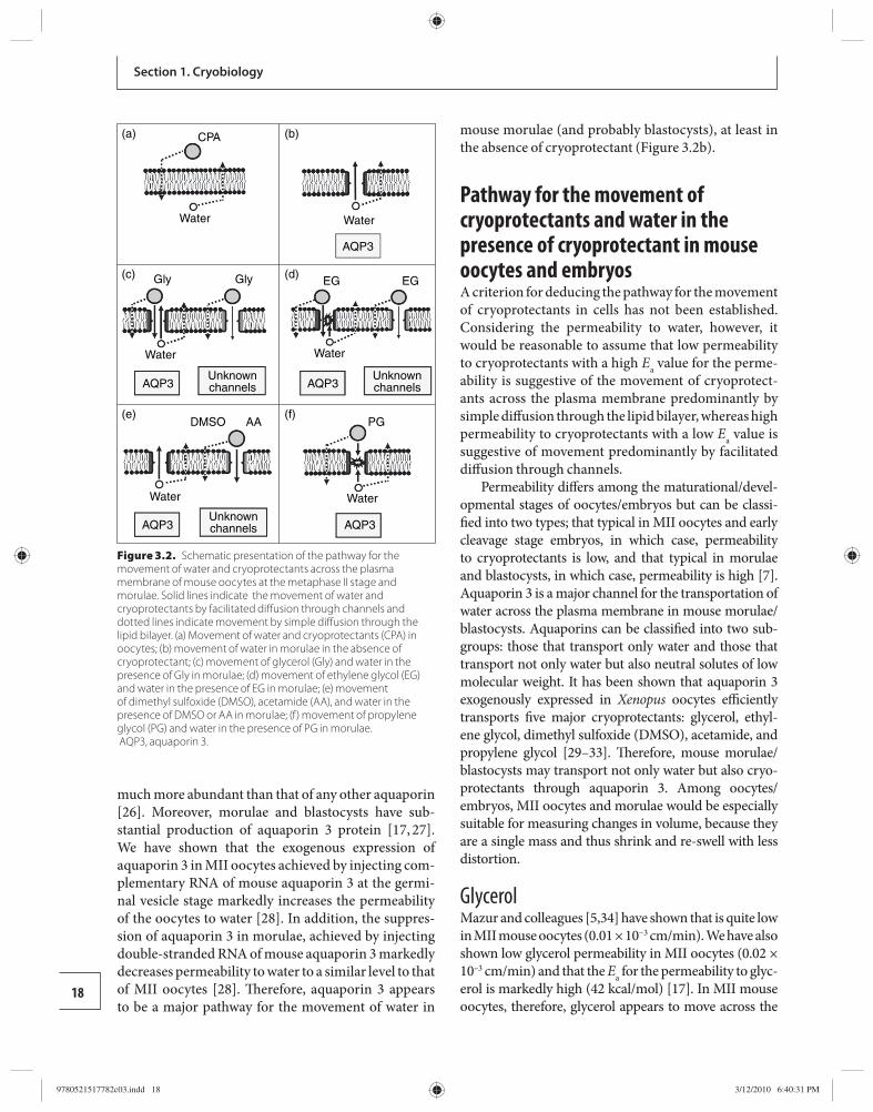
Section 1. Cryobiology
18
much more abundant than that of any other aquaporin [ 26 ]. Moreover, morulae and blastocysts have sub-stantial production of aquaporin 3 protein [ 17 , 27 ]. We have shown that the exogenous expression of aquaporin 3 in MII oocytes achieved by injecting com-plementary RNA of mouse aquaporin 3 at the germi-nal vesicle stage markedly increases the permeability of the oocytes to water [ 28 ]. In addition, the suppres-sion of aquaporin 3 in morulae, achieved by injecting double-stranded RNA of mouse aquaporin 3 markedly decreases permeability to water to a similar level to that of MII oocytes [ 28 ]. Th erefore, aquaporin 3 appears to be a major pathway for the movement of water in
mouse morulae (and probably blastocysts), at least in the absence of cryoprotectant ( Figure 3.2b ).
Pathway for the movement of cryoprotectants and water in the presence of cryoprotectant in mouse oocytes and embryos A criterion for deducing the pathway for the movement of cryoprotectants in cells has not been established. Considering the permeability to water, however, it would be reasonable to assume that low permeability to cryoprotectants with a high E a value for the perme-ability is suggestive of the movement of cryoprotect-ants across the plasma membrane predominantly by simple diff usion through the lipid bilayer, whereas high permeability to cryoprotectants with a low E a value is suggestive of movement predominantly by facilitated diff usion through channels.
Permeability diff ers among the maturational/devel-opmental stages of oocytes/embryos but can be classi-fi ed into two types; that typical in MII oocytes and early cleavage stage embryos, in which case, permeability to cryoprotectants is low, and that typical in morulae and blastocysts, in which case, permeability is high [ 7 ]. Aquaporin 3 is a major channel for the transportation of water across the plasma membrane in mouse morulae/blastocysts. Aquaporins can be classifi ed into two sub-groups: those that transport only water and those that transport not only water but also neutral solutes of low molecular weight. It has been shown that aquaporin 3 exogenously expressed in Xenopus oocytes effi ciently transports fi ve major cryoprotectants: glycerol, ethyl-ene glycol, dimethyl sulfoxide (DMSO), acetamide, and propylene glycol [ 29 – 33 ]. Th erefore, mouse morulae /blastocysts may transport not only water but also cryo-protectants through aquaporin 3. Among oocytes/embryos, MII oocytes and morulae would be especially suitable for measuring changes in volume, because they are a single mass and thus shrink and re-swell with less distortion.
Glycerol Mazur and colleagues [ 5 , 34 ] have shown that is quite low in MII mouse oocytes (0.01 × 10 −3 cm/min). We have also shown low glycerol permeability in MII oocytes (0.02 × 10 –3 cm/min) and that the E a for the permeability to glyc-erol is markedly high (42 kcal/mol) [ 17 ]. In MII mouse oocytes, therefore, glycerol appears to move across the
CPA(a) (b)
(c) (d)
(e) (f)
Water
Water
Water Water
Water
Gly
DMSO AA PG
Gly EG EG
Water
AQP3
AQP3AQP3
AQP3 AQP3
Unknownchannels
Unknownchannels
Unknownchannels
Figure 3.2. Schematic presentation of the pathway for the movement of water and cryoprotectants across the plasma membrane of mouse oocytes at the metaphase II stage and morulae. Solid lines indicate the movement of water and cryoprotectants by facilitated diff usion through channels and dotted lines indicate movement by simple diff usion through the lipid bilayer. (a) Movement of water and cryoprotectants (CPA) in oocytes; (b) movement of water in morulae in the absence of cryoprotectant; (c) movement of glycerol (Gly) and water in the presence of Gly in morulae; (d) movement of ethylene glycol (EG) and water in the presence of EG in morulae; (e) movement of dimethyl sulfoxide (DMSO), acetamide (AA), and water in the presence of DMSO or AA in morulae; (f) movement of propylene glycol (PG) and water in the presence of PG in morulae. AQP3, aquaporin 3.
9780521517782c03.indd 189780521517782c03.indd 18 3/12/2010 6:40:31 PM3/12/2010 6:40:31 PM
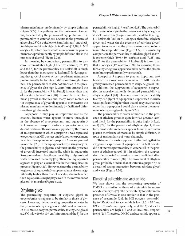
19
Chapter 3. Water movement and cryprotectants
plasma membrane predominantly by simple diff usion ( Figure 3.2a ). Th e pathway for the movement of water may be aff ected by the presence of cryoprotectant. Th e permeability to water of MII oocytes in the presence of glycerol at 25°C is quite low (0.6 μm/min-atm) and the E a for this permeability is high (14 kcal/mol) [ 17 , 28 ]. In MII oocytes, therefore, water would move across the plasma membrane predominantly by simple diff usion also in the presence of glycerol ( Figure 3.2a ).
In morulae, by comparison, permeability to gly-cerol is remarkably high (4.7 × 10 –3 cm/min) [ 7 , 17 ] and the E a for the permeability (10 kcal/mol) is much lower than that in oocytes (42 kcal/mol) [ 17 ], suggest-ing that glycerol moves across the plasma membrane predominantly by facilitated diff usion through chan-nels. Th e permeability to water of morulae in the pres-ence of glycerol is also high (2.2 μm/min-atm) and the E a for the permeability (9 kcal/mol) is lower than that of oocytes (14 kcal/mol) [ 28 ]. In morulae, therefore, most glycerol molecules and many water molecules (in the presence of glycerol) appear to move across the plasma membrane predominantly by facilitated diff u-sion through channels.
Aquaporin 3 may play an important role as the channel, because water appears to move through it in the absence of cryoprotectant, and aquaporin 3 is known to transport various cryoprotectants, as described above. Th is notion is supported by the results of an experiment in which aquaporin 3 was expressed ex ogenously in MII oocytes and of another experiment in which the expression of aquaporin 3 was suppressed in morulae [ 28 ]. In the aquaporin 3-expressing oocytes, the permeability to glycerol and water (in the presence of glycerol) increased markedly, while in aquaporin 3-suppressed morulae, the permeability to glycerol and water decreased markedly [ 28 ]. Th erefore, aquaporin 3 appears to play an essential role in the transportation process ( Figure 3.2c ). However, since the permeability to glycerol of aquaporin 3-suppressed morulae was sig-nifi cantly higher than that of oocytes, channels other than aquaporin 3 might play a role in the movement of glycerol in morulae ( Figure 3.2c ).
Ethylene glycol Th e permeating properties of ethylene glycol in oocytes/embryos appear to be similar to those of gly-cerol. However, the permeating properties of water in the presence of ethylene glycol are diff erent in morulae. In MII mouse oocytes, permeability to ethylene glycol at 25°C is low (0.6 × 10 –3 cm/min-atm) and the E a for the
permeability is high (17 kcal/mol) [ 28 ]. Th e permeabil-ity to water of oocytes in the presence of ethylene glycol at 25°C is also low (0.4 μm/min-atm) and the E a is high (10 kcal/mol) [ 28 ]. In MII oocytes, therefore, ethylene glycol and water (in the presence of ethylene glycol) appear to move across the plasma membrane predom-inantly by simple diff usion ( Figure 3.2a ). In morulae, by comparison, the permeability to ethylene glycol at 25°C is extremely high (10.0 × 10 –3 cm/min-atm) [ 7 , 28 ], and the E a for the permeability (9 kcal/mol) is lower than that in oocytes (17 kcal/mol) [ 28 ]. In morulae, there-fore, ethylene glycol appears to move across the plasma membrane predominantly via channels.
Aquaporin 3 appears to play an important role, because its exogenous expression in MII oocytes greatly increased permeability to ethylene glycol [ 28 ]. In addition, the suppression of aquaporin 3 expres-sion in morulae markedly decreased permeability to ethylene glycol [28]. However, since the permeability to ethylene glycol of aquaporin 3-suppressed morulae was signifi cantly higher than that of oocytes, channels other than aquaporin 3 could play a role in the move-ment of ethylene glycol ( Figure 3.2d ).
Th e permeability to water of morulae in the pres-ence of ethylene glycol is quite low (0.5 μm/min-atm) and the E a for the permeability is quite high (14 kcal/mol) [ 28 ]. In the presence of ethylene glycol, there-fore, most water molecules appear to move across the plasma membrane of morulae by simple diff usion, in spite of an abundance of water channels.
Th is speculation is supported by the fi nding that the exogenous expression of aquaporin 3 in MII oocytes did not increase permeability to water at all in the pres-ence of ethylene glycol [ 28 ]. In addition, the suppres-sion of aquaporin 3 expression in morulae did not aff ect permeability to water [ 28 ]. Th e movement of ethylene glycol probably hinders that of water in aquaporin 3 as a result of strong interaction between ethylene glycol and water ( Figure 3.2d ).
Dimethyl sulfoxide and acetamide We have shown that the permeating properties of DMSO are similar to those of acetamide in mouse oocytes/embryos [ 7 ]. Th e permeability to water in the presence of DMSO is also similar to that in the pres-ence of acetamide [ 28 ]. In MII oocytes, permeabil-ity to DMSO and to acetamide is low (1.0 × 10 –3 and 0.9 × 10 –3 cm/min, respectively) and the E a values for permeability are high (18 and 23 kcal/mol, respect-ively) [ 28 ]. Th erefore, DMSO and acetamide appear to
9780521517782c03.indd 199780521517782c03.indd 19 3/12/2010 6:40:33 PM3/12/2010 6:40:33 PM
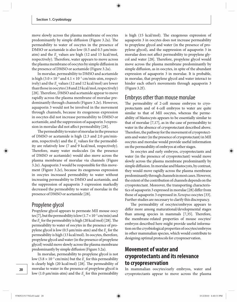
20
Section 1. Cryobiology
move slowly across the plasma membrane of oocytes predominantly by simple diff usion ( Figure 3.2a ). Th e permeability to water of oocytes in the presence of DMSO or acetamide is also low (0.5 and 0.5 μm/min-atm) and the E a values are high (12 and 15 kcal/mol, respectively). Th erefore, water appears to move across the plasma membrane of oocytes by simple diff usion in the presence of DMSO or acetamide ( Figure 3.2a ).
In morulae, permeability to DMSO and acetamide is high (3.0 × 10 –3 and 4.1 × 10 –3 cm/min-atm, respect-ively) and the E a values (12 and 12 kcal/mol) are lower than those in oocytes (18 and 23 kcal/mol, respectively) [ 28 ]. Th erefore, DMSO and acetamide appear to move rapidly across the plasma membrane of morulae pre-dominantly through channels ( Figure 3.2e ). However, aquaporin 3 would not be involved in the movement through channels, because its exogenous expression in oocytes did not increase permeability to DMSO or acetamide, and the suppression of aquaporin 3 expres-sion in morulae did not aff ect permeability [ 28 ].
Th e permeability to water of morulae in the presence of DMSO or acetamide is high (2.3 and 2.0 μm/ min- atm, respectively) and the E a values for the permeabil-ity are relatively low (7 and 9 kcal/mol, respectively). Th erefore, many water molecules (in the presence of DMSO or acetamide) would also move across the plasma membrane of morulae via channels (Figure 3.2e). Aquaporin 3 would be responsible for this move-ment ( Figure 3.2e ), because its exogenous expression in oocytes increased permeability to water without increasing permeability to DMSO and acetamide, and the suppression of aquaporin 3 expression markedly decreased the permeability to water of morulae in the presence of DMSO or acetamide [ 28 ].
Propylene glycol Propylene glycol appears to permeate MII mouse ooc ytes [ 7 ], but the permeability is low (1.7 × 10 –3 cm/min) and the E a for the permeability is high (20 kcal/mol) [ 28 ]. Th e permeability to water of oocytes in the presence of pro-pylene glycol is low (0.5 μm/min-atm) and the E a for the permeability is high (13 kcal/mol). In oocytes, therefore, propylene glycol and water (in the presence of propylene glycol) would move slowly across the plasma membrane predominantly by simple diff usion (Figure 3.2a).
In morulae, permeability to propylene glycol is not low (3.8 × 10 –3 cm/min) but the E a for this permeability is clearly high (20 kcal/mol) [ 28 ]. Th e permeability of morulae to water in the presence of propylene glycol is low (1.0 μm/min-atm) and the E a for this permeability
is high (15 kcal/mol). Th e exogenous expression of aquaporin 3 in oocytes does not increase permeability to propylene glycol and water (in the presence of pro-pylene glycol), and the suppression of aquaporin 3 in morulae does not aff ect permeability to propylene gly-col and water [ 28 ]. Th erefore, propylene glycol would move across the plasma membrane predominantly by simple diff usion, as in oocytes, in spite of the abundant expression of aquaporin 3 in morulae. It is probable, in morul ae, that propylene glycol and water interact to hinder each other’s movements through aquaporin 3 ( Figure 3.2f ).
Embryos other than mouse morulae Th e permeability of 2-cell mouse embryos to cryo-protectants and of 4-cell embryos to water are quite similar to that of MII oocytes, whereas the perme-ability of bl astocysts appears to be essentially similar to that of morulae [ 7 , 17 ], as in the case of permeability to water in the absence of cryoprotectant described above. Th erefore, the pathway for the movement of cryoprotect-ants and water (in the presence of cryoprotectant) in MII oocytes and morulae would provide useful information on the permeability of embryos at other stages.
In oocytes and early embryos, cryoprotectants and water (in the presence of cryoprotectant) would move slowly across the plasma membrane predominantly by simple diff usion. In morulae and blastocysts, by contrast, they would move rapidly across the plasma membrane predominantly through channels in most cases. However, the extent of the contribution of channels depends on the cryoprotectant. Moreover, the transporting characteris-tics of aquaporin 3 expressed in morulae [ 28 ] diff er from those of aquaporin 3 expressed in Xenopus oocytes [ 33 ]. Further studies are necessary to clarify this discrepancy.
Th e permeability of oocytes/embryos appears to diff er more among maturational/developmental stages than among species in mammals [ 7 , 35 ]. Th erefore, the membrane-related properties of mouse oocytes/embryos described here might provide useful informa-tion on the cryobiological properties of oocytes/embryos in other mammalian species, which would contribute to designing optimal protocols for cryopreservation.
Movement of water and cryoprotectants and its relevance to cryopreservation In mammalian oocytes/early embryos, water and cryoprotectants appear to move across the plasma
9780521517782c03.indd 209780521517782c03.indd 20 3/12/2010 6:40:33 PM3/12/2010 6:40:33 PM
21
Chapter 3. Water movement and cryprotectants
membrane slowly by simple diff usion. Th erefore, a longer exposure to the cryoprotectant solution(s) would be necessary to dehydrate the cells and allow the cryoprotectant(s) to permeate suffi ciently. In addition, the suitable period of exposure to the cryoprotect-ant would vary depending on the temperature. When oocytes/embryos are cryopreserved by vitrifi cation, length of time and temperature are critical, because vitrifi cation solutions contain a high concentration of cryoprotectant(s) and toxicity is dependent on the period of exposure and temperature [ 36 ].
For vitrifi cation of oocytes/early embryos, a two-step treatment would be eff ective, in which oocytes/embryos are fi rst pretreated in a solution containing a low concentration of cryoprotectant for permeation in less toxic conditions, and then in the vitrifi cation solu-tion for a short time to cause the embryos to shrink by rapid dehydration. Aft er warming, it would be pref-erable to remove permeated cryoprotectant(s) from oocytes/early embryos at a higher temperature to pre-vent osmotic over-swelling, because the movement of water and cryoprotectants by simple diff usion is largely aff ected by temperature.
In morulae, by comparison, a one-step treatment would be eff ective even for vitrifi cation, because water and cryoprotectants permeate rapidly via channels. However, the period of exposure to the vitrifi cation solution needs to be limited to prevent damage. Th e temperature at which morulae are exposed to the cry-oprotectant before cooling and at which the permeated cryoprotectant is removed from morulae aft er warm-ing may not be very important in terms of permeabil-ity, because facilitated diff usion through channels is less aff ected by temperature. For considerations of the toxicity of cryoprotectants, however, exposure to the cryoprotectant at high temperature should be avoided, because cryoprotectants are more toxic at higher temperature.
In blastocysts, cells would be highly permeable to water and cryoprotectants, as in morulae. However, blastocysts have a large amount of water in the blas-tocoele, within which ice is likely to form during cryo-preservation. Th erefore, it is preferable that most of the water in the blastocoele diff uses out before cryopreser-vation. An eff ective way to reduce the chance of intra-cellular ice forming is ultrarapid vitrifi cation, in which oocytes/embryos suspended in a minimal amount of vitrifi cation solution are cooled and warmed extremely rapidly [ 37 , 38 ]. In addition, the inner cell mass faces not the outside of the blastocyst but the blastocoele or
trophoectoderm, indicating that water and cryopro-tectants do not move across the plasma membrane of the inner cell mass directly from/to the solution in which the blastocyst is suspended but through the trophoectodermal cells or the blastocoele solution. Furthermore, it has been reported that the distribution of aquaporins is not homogeneous among the apical and basolateral sides of the trophoectoderm [ 27 ], sug-gesting that the blastocyst has compartments diff ering in permeability to water and cryoprotectants. When blastocysts are cryopreserved by vitrifi cation, there-fore, they are usually exposed to the cryoprotectant(s) in two steps to promote permeation under less toxic conditions [ 3 , 39 ].
Conclusions Th e permeability of oocytes and embryos varies among maturational/developmental stages because the pathway by which water and cryoprotectants move across the plasma membrane is diff erent. Th erefore, the survival of oocytes/embryos aft er cryopreservation diff ers among maturational/devel-opmental stages when the same cryopreservation protocol is used. In MII oocytes (and probably 2- to 4-cell embryos), water and cryoprotectants would move across the plasma membrane slowly by sim-ple diff usion through the lipid bilayer. In morulae (and probably blastocysts), glycerol, ethylene glycol, DMSO, and acetamide would move rapidly by facili-tated diff usion through channels, but propylene gly-col would move by simple diff usion. Water appears to move in morulae via channels in the presence of gly-cerol, DMSO, or acetamide, but by simple diff usion in the presence of ethylene glycol or propylene glycol. In order to design protocols suitable for the cryopreser-vation of oocytes/embryos, it is necessary to consider the pathway for the movement of cryoprotectants and water at each stage of development.
Conditions suitable for the cryopreservation of mammalian oocytes and embryos diff er among mat-urational/developmental stages even in the same spe-cies. Diff erences in cryobiological properties would be responsible for the variation. Th e permeability of the plasma membrane to water and cryoprotectants is the most important of these properties because it is closely related to major types of cellular injury during cryo-preservation, such as the damage caused by the toxicity of cryoprotectants, by the formation of intracellular ice, and by osmotic swelling during removal of the cell-permeating cryoprotectant.
9780521517782c03.indd 219780521517782c03.indd 21 3/12/2010 6:40:34 PM3/12/2010 6:40:34 PM
22
Section 1. Cryobiology
In mouse oocytes at the MII stage and embryos at early cleavage stages (early embryos), water and cryo-protectants appear to move slowly across the plasma membrane predominantly by simple diff usion, because permeability is low and temperature dependency is high. In morulae and blastocysts, on the other hand, water appears to move rapidly across the plasma mem-brane predominantly by facilitated diff usion through channels, because permeability is high and temperature dependency is low. Accordingly, morulae and blasto-cysts are highly permeable to cryoprotectants, such as glycerol, ethylene glycol, DMSO, and acetamide, and tolerate cryopreservation better than oocytes and early embryos.
As a channel, aquaporin 3, which is permeable to water and many cryoprotectants, is expressed abun-dantly in mouse morulae but not in oocytes. It has been shown in morulae that aquaporin 3 plays an important role in the movement of water, glycerol, and ethylene glycol, although not in the movement of DMSO and acetamide. In addition, water also moves across the plasma membrane of morulae by simple diff usion in the presence of ethylene glycol or propylene glycol. As an exception, propylene glycol appears to perme-ate morulae predominantly by simple diff usion con-comitant with the slow movement of water by simple diff usion. It has been shown that the permeability char-acteristics of mouse oocytes/embryos at various stages are similar to those of bovine oocytes/embryos. Th is suggests that the permeability of oocytes/embryos will be similar among species in mammals. Information on the pathway for the movement of cryoprotectants and water would be useful as a basis for the selection of a suitable cryoprotectant(s) and for the design of suitable protocols for the cryopreservation of oocytes/embryos at various stages.
References 1. Kasai M , Ito K , Edashige K . Morphological appearance of
the cryopreserved mouse blastocyst as a tool to identify the type of cryoinjury . Hum Reprod 2002; 17 : 1863 –1874.
2. Kasai M , Komi JH , Takakamo A , et al. A simple method for mouse embryo cryopreservation in a low toxicity vitrifi cation solution, without appreciable loss of viability . J Reprod Fertil 1990; 89 : 91 –97.
3. Kasai M . Cryopreservation of mammalian embryos . Mol Biotech 1997; 7 : 173 –179.
4. Edashige K , Asano A , An TZ , et al. Restoration of resistance to osmotic swelling of vitrifi ed mouse embryos by short-term culture . Cryobiology 1999; 38 : 273 –280.
5. Jackowski S , Leibo SP , Mazur P . Glycerol permeability of fertilized and unfertilized mouse ova . J Exp Zool 1980; 212 : 329 –341.
6. Edashige K , Yamaji Y , Kleinhans FW , et al. Artifi cial expression of aquaporin-3 improves the survival of mouse oocytes aft er cryopreservation . Biol Reprod 2003; 68 : 87 –94.
7. Pedro PB , Yokoyama E , Zhu SE , et al. Permeability of mouse oocytes and embryos at various developmental stages to fi ve cryoprotectants . J Reprod Dev 2005; 51 : 235 –246.
8. Whittingham DG . Survival of mouse embryos aft er freezing and thawing . Nature 1971; 233 : 125 –126.
9. Kleinhans FW . Membrane permeability modeling: Kedem–Katchalsky vs a two-parameter formalism . Cryobiology 1998; 37 : 271 –289.
10. Verkman AS , van Hoek AN , Ma T , et al. Water transport across mammalian cell membranes . Am J Physiol 1996; 270 : C12 –C30.
11. Leibo SP . Water permeability and its activation energy of fertilized and unfertilized mouse ova . J Membr Biol 1980; 53 : 179 –188.
12. Hunter J , Bernard A , Fuller B , et al. Measurements of the membrane water permeability (L P ) and its temperature dependence (activation energy) in human fresh and failed-to-fertilize oocytes and mouse oocytes . Cryobiology 1992; 29 : 240 –249.
13. Benson CT , Critser JK . Variation of water permeability (LP) and its activation energy (Ea ) among unfertilized golden hamster and ICR murine oocytes . Cryobiology 1994; 31 : 215 –223.
14. Gao DY , Benson CT , Liu C , et al. Development of a novel microperfusion chamber for determination of cell membrane transport properties . Biophys J 1996; 71 : 443 –450.
15. Litkouhi B , Marlow D , McGrath JJ , et al. Th e infl uence of cryopreservation on murine oocyte water permeability and osmotically inactive volume . Cryobiology 1997; 34 : 23 –35.
16. Pfaff RT , Liu J , Gao D , et al. Water and DMSO membrane permeability characteristics of in-vivo and in-vitro derived and cultured murine oocytes and embryos . Mol Hum Reprod 1998; 4 : 51 –59.
17. Edashige K , Tanaka M , Ichimaru N , et al. Channel-dependent permeation of water and glycerol in mouse morulae . Biol Reprod 2006; 74 : 625 –632.
18. Toner M , Cravalho EG , Armant DR . Water transport and estimated transmembrane potential during freezing of mouse oocytes . J Membr Biol 1990; 115 : 261 –272.
19. Edashige K , Sakamoto M , Kasai M . Expression of mRNAs of the aquaporin family in mouse oocytes and embryos . Cryobiology 2000; 40 : 171 –175.
20. Off enberg H , Barcroft LC , Caveney A , et al. mRNAs encoding aquaporins are present during murine
9780521517782c03.indd 229780521517782c03.indd 22 3/12/2010 6:40:34 PM3/12/2010 6:40:34 PM
23
Chapter 3. Water movement and cryprotectants
preimplantation development . Mol Reprod Dev 2000; 57 : 323 –330.
21. King LS , Kozono D , Agre P . From structure to disease: the evolving tale of aquaporin biology . Nat Rev Mol Cell Biol 2004; 5 : 687 –698.
22. Fischbarg J , Kuang KY , Vera JC , et al. Glucose transporters serve as water channels . Proc Natl Acad Sci USA 1990; 87 : 3244 –3247.
23. Yang B , Verkman AS . Urea transporter UT3 functions as an effi cient water channel: direct evidence for a common water/urea pathway . J Biol Chem 1998; 273 : 9369 –9372.
24. Loo DDF , Hirayama BA , Meinild AK , et al. Passive water and ion transport by cotransporters . J Physiol 1999; 518: 195 –202.
25. Verkman AS , Mitra AK . Structure and function of aquaporin water channels . Am J Physiol Renal Physiol 2000; 278 : F13 –F28.
26. Off enberg H , Th omsen PD . Functional challenge aff ects aquaporin mRNA abundance in mouse blastocysts . Mol Reprod Dev 2005; 71 : 422 –430.
27. Barcroft LC , Off enberg H , Th omsen P , et al. Aquaporin proteins in murine trophectoderm mediate transepithelial water movements during cavitation . Dev Biol 2003; 256 : 342 –354.
28. Edashige K , Ohta S , Tanaka M , et al. Th e role of aquaporin 3 in the movement of water and cryoprotectants in mouse morulae . Biol Reprod 2007; 77 : 365 –375.
29. Ishibashi K , Sasaki S , Fushimi K , et al. Molecular cloning and expression of a member of the aquaporin family with permeability to glycerol and urea in addition to water expressed at the basolateral membrane of kidney collecting duct cells . Proc Natl Acad Sci USA 1994; 91 : 6269 –6273.
30. Meinild AK , Klaerke DA , Zeuthen T . Bidirectional water fl uxes and specifi city for small hydrophilic molecules in aquaporins 1–5 . J Biol Chem 1998; 273 : 32446 –32451.
31. Tsukaguchi H , Shayakul C , Berger UV , et al. Molecular characterization of a broad selectivity neutral solute channel . J Biol Chem 1998; 273 : 24737 –24743.
32. Zeuthen T , Klaerke DA . Transport of water and glycerol in aquaporin 3 is gated by H + . J Biol Chem 1999; 274 : 21631 –21636.
33. Yamaji Y , Valdez DM , Seki S , et al. Cryoprotectant permeability of aquaporin-3 expressed in Xenopus oocytes . Cryobiology 2006; 53 : 258 –267.
34. Mazur P , Rigopoulos N , Jackowski SC , et al. Preliminary estimates of the permeability of mouse ova and early embryos to glycerol . Biophys J 1976; 16 : 232a .
35. Pedro PB , Kasai M , Mammaru Y , et al. Change in the permeability to diff erent cryoprotectants of bovine oocytes and embryos during maturation and development . Proc 13th Int Congr Anim Reprod 1996; 3 : P15 –P19.
36. Kasai M , Nishimori M , Zhu SE , et al. Survival of mouse morulae vitrifi ed in an ethylene glycol-based solution aft er exposure to the solution at various temperatures . Biol Reprod 1992; 47 : 1134 –1139.
37. Martino A , Songsasen N , Leibo SP . Development into blastocysts of bovine oocytes cryopreserved by ultra-rapid cooling . Biol Reprod 1996; 54: 1059 –1069.
38. Kasai M . Advances in the cryopreservation of mammalian oocytes and embryos: development of ultrarapid vitrifi cation . Reprod Med Biol 2002; 1 : 1 –9.
39. Zhu SE , Kasai , M , Otoge , H , et al. Cryopreservation of expanded mouse blastocysts by vitrifi cation in ethylene glycol-based solutions . J Reprod Fertil 1993; 98 : 139 –145.
9780521517782c03.indd 239780521517782c03.indd 23 3/12/2010 6:40:34 PM3/12/2010 6:40:34 PM
Fertility Cryopreservation, ed. Ri-Cheng Chian and Patrick Quinn. Published by Cambridge University Press. Copyright © Cambridge University Press 2010.
Chapter
4 and the functioning of cryoprotectants. Factors such as hydrogen bonding, water’s interactions with ions and macromolecules, dielectric constants, thermodynamic equilibrium, and osmosis all provide information on the underlying biophysical and biochemical mechan-isms by which cryopreservation and cryoprotectants operate. Th ough brief mention of these factors may be insightful in certain instances, a detailed discussion is beyond the scope of this chapter, and more in-depth analysis can be found in accompanying chapters or other reviews [ 1 ,2].
Several substances convey cryoprotective activ-ity, with up to 56 individual compounds reported to convey a protective eff ect in various cellular systems [ 3 ,4]. For purposes of this chapter, we will focus on various cryoprotectants and their application in pre-serving gametes and embryos ( Table 4.1 ). In general, although cryoprotectants defend against cellular dam-age by regulating dynamics of water during cooling, the actual manner by which they convey their protection depends largely on whether the particular compounds permeate the cell or whether they remain in the extra-cellular space ( Figure 4.1 ). Th erefore, cryoprotectants are grouped into two major categories: penetrating and non-penetrating agents.
Penetrating cryoprotectants Penetrating cryoprotectants are generally small, non-ionic compounds with a high solubility in water at low temperatures. Given time, these chemical compounds can diff use through cellular membranes, permeatin g the cell and equilibrating within the cytoplasm, replacing the bulk of intracellular water without over-dehydrating the cell. Permeating cryoprotectants solidify at lower temperatures than water and thus subsequently reduce the amount of intracellular ice formation at a given temperature. Th is decreased amount of intra cellular ice-crystal formation mitigates
Introduction Cryopreservation has been a very important factor in the progress and advancement of clinical in vitro fertilization (IVF), allowing for long-term storage of sperm, embryos, and, more recently, oocytes for future use. Th is is not only helpful for preserving fertility in patients who may be undergoing treatment for can-cer or other life-threatening illnesses but also allows for storage of these cells to permit genetic screening, as well as the ability to test samples for communicable diseases prior to use. Furthermore, methods to store, rather than discard, embryos provide alternatives to alleviate various ethical concerns. As a result, cryo-preservation has become the standard of care in infer-tility treatment.
Th ough it may now be considered a routine pro-cedure, cryobiology itself is a complex discipline and multiple variables must be accounted for in developing successful protocols. Many of these considerations are discussed in the accompanying chapters and include cell- and tissue-specifi c sensitivities, which are infl u-enced by cell size, membrane permeability, lipid content, and other factors. Regardless of these variables, a primary consideration, and one of the most basic components of a successful cryopreservation protocol, is selection of an eff ective cryoprotectant additive. Cryoprotectants func-tion as their name implies, guarding cells from damage during cryopreservation by either freezing or vitrifi ca-tion. Protective eff ects of these agents are conveyed pri-marily through interactions with, and manipulation of, intracellular and extracellular water.
Th e manner in which water interacts within and around the cell, as well as its behavior during cooling/freezing, is a key determinant in maintaining cellular viability during cryopreservation. Consequently, an understanding of the physicochemical properties of water is required for a truly complete understanding of the theoretical and functional basis of cryobiology
Cryoprotectants Jason E. Swain and Gary D. Smith
24
9780521517782c04.indd 249780521517782c04.indd 24 3/12/2010 3:25:05 PM3/12/2010 3:25:05 PM
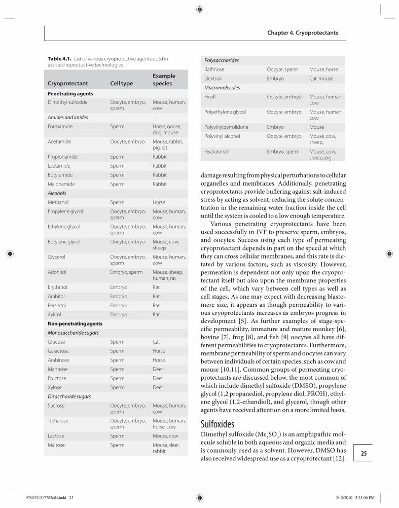
25
Chapter 4. Cryoprotectants
damage resulting from physical perturbations to cellular organelles and membranes. Additionally, penetrating cryoprotectants provide buff ering against salt-induced stress by acting as solvent, reducing the solute concen-tration in the remaining water fraction inside the cell until the system is cooled to a low enough temperature.
Various penetrating cryoprotectants have been used successfully in IVF to preserve sperm, embryos, and oocytes. Success using each type of permeating cryoprotectant depends in part on the speed at which they can cross cellular membranes, and this rate is dic-tated by various factors, such as viscosity. However, permeation is dependent not only upon the cryopro-tectant itself but also upon the membrane properties of the cell, which vary between cell types as well as cell stages. As one may expect with decreasing blasto-mere size, it appears as though permeability to vari-ous cryoprotectants increases as embryos progress in development [ 5 ]. As further examples of stage-spe-cifi c permeability, immature and mature monkey [6], bovine [ 7 ], frog [8], and fi sh [ 9 ] oocytes all have dif-ferent permeabilities to cryoprotectants. Furthermore, membrane permeability of sperm and oocytes can vary between individuals of certain species, such as cow and mouse [10, 11 ] . Common groups of permeating cryo-protectants are discussed below, the most common of which include dimethyl sulfoxide (DMSO), propylene glycol (1,2 propanediol, propylene diol, PROH), ethyl-ene glycol (1,2-ethandiol), and glycerol, though other agents have received attention on a more limited basis.
Sulfoxides Dimethyl sulfoxide (Me 2 SO 4 ) is an amphipathic mol-ecule soluble in both aqueous and organic media and is commonly used as a solvent. However, DMSO has also received widespread use as a cryoprotectant [12].
Table 4.1. List of various cryoprotective agents used in assisted reproductive technologies
Cryoprotectant Cell typeExample species
Penetrating agents
Dimethyl sulfoxide Oocyte, embryo, sperm
Mouse, human, cow
Amides and imides
Formamide Sperm Horse, goose, dog, mouse
Acetamide Oocyte, embryo Mouse, rabbit, pig, rat
Propionamide Sperm Rabbit
Lactamide Sperm Rabbit
Butyramide Sperm Rabbit
Malonamide Sperm Rabbit
Alcohols
Methanol Sperm Horse
Propylene glycol Oocyte, embryo, sperm
Mouse, human, cow
Ethylene glycol Oocyte, embryo, sperm
Mouse, human, cow
Butylene glycol Oocyte, embryo Mouse, cow, sheep
Glycerol Oocyte, embryo, sperm
Mouse, human, cow
Adonitol Embryo, sperm Mouse, sheep, human, rat
Erythritol Embryo Rat
Arabitol Embryo Rat
Perseitol Embryo Rat
Xylitol Embryo Rat
Non-penetrating agents
Monosaccharide sugars
Glucose Sperm Cat
Galactose Sperm Horse
Arabinose Sperm Horse
Mannose Sperm Deer
Fructose Sperm Deer
Xylose Sperm Deer
Disaccharide sugars
Sucrose Oocyte, embryo, sperm
Mouse, human, cow
Trehalose Oocyte, embryo, sperm
Mouse, human, horse, cow
Lactose Sperm Mouse, cow
Maltose Sperm Mouse, deer, rabbit
Polysaccharides
Raffi nose Oocyte, sperm Mouse, horse
Dextran Embryo Cat, mouse
Macromolecules
Ficoll Oocyte, embryo Mouse, human, cow
Polyethylene glycol Oocyte, embryo Mouse, human, cow
Polyvinylpyrrolidone Embryo Mouse
Polyvinyl alcohol Oocyte, embryo Mouse, cow, sheep,
Hyaluronan Embryo, sperm Mouse, cow, sheep, pig
9780521517782c04.indd 259780521517782c04.indd 25 3/12/2010 3:25:06 PM3/12/2010 3:25:06 PM
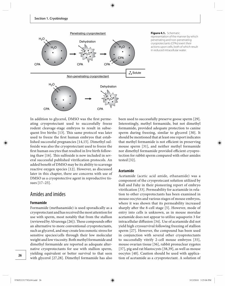
Section 1. Cryobiology
26
In addition to glycerol, DMSO was the fi rst perme-ating cryoprotectant used to successfully freeze rodent cleavage-stage embryos to result in subse-quent live births [ 13 ]. Th is same protocol was later used to freeze the fi rst human embryos that estab-lished s uccessful pregnancies [14, 15 ]. Dimethyl sul-foxide was also the cryoprotectant used to freeze the fi rst human oocytes that resulted in live birth follow-ing thaw [16]. Th is sulfoxide is now included in sev-eral successful published vitrifi cation protocols. An added benefi t of DMSO may be its ability to scavenge reactive oxygen species [ 12 ]. However, as discussed later in this chapter, there are concerns with use of DMSO as a cryoprotective agent in reproductive tis-sues [17– 25 ].
Amides and imides Formamide Formamide (methanamide) is used sporadically as a cryoprotectant and has received the most attention for use with sperm, most notably that from the stallion (reviewed by Alvarenga [26]). Th ese compounds off er an alternative to more conventional cryoprotectants, such as glycerol, and may create less osmotic stress for sensitive species/cells through their low molecular weight and low viscosity. Both methyl formamide and dimethyl formamide are reported as adequate alter-native cryoprotectants for use with stallion sperm, yielding equivalent or better survival to that seen with glycerol [ 27 ,28]. Dimethyl formamide has also
been used to successfully preserve goose sperm [ 29 ]. Interestingly, methyl formamide, but not dimethyl formamide, provided adequate protection to canine sperm during freezing, similar to glycerol [30]. It should be mentioned that at least one report indicates that methyl formamide is not effi cient in preserving mouse sperm [ 31 ], and neither methyl formamide nor dimethyl formamide provided effi cient cryopro-tection for rabbit sperm compared with other amides tested [32].
Acetamide Acetamide (acetic acid amide, ethanamide) was a component of the cryoprotecant solution utilized by Rall and Fahy in their pioneering report of embryo vitrifi cation [ 33 ]. Permeability for acetamide in rela-tion to other cryoprotectants has been examined in mouse oocytes and various stages of mouse embryos, where it was shown that its permeability increased sharply aft er the 8-cell stage [5]. However, mode of entry into cells is unknown, as in mouse morulae acetamide does not appear to utilize aquaporin 3 for intracellular diff usion [ 34 ]. Use of acetamide did not yield high cryosurvival following freezing of stallion sperm [27]. However, the compound has been used in conjunction with several other cryoprotectants to successfully vitrify 2-cell mouse embryos [ 35 ], mouse ovarian tissue [36], rabbit pronuclear zygotes [ 37 ], pig and rat blastocysts [38, 39 ], as well as mouse oocytes [40]. Caution should be used with applica-tion of acetamide as a cryoprotectant. A solution of
Figure 4.1. Schematic representation of the manner by which penetrating and non-penetrating cryoprotectants (CPAs) exert their actions upon cells, both of which result in reduced intracellular water.
Penetrating cryoprotectant
Non-penetrating cryoprotectant
Dehydration
Dehydration
H2O
H2O
H2O
H2O
H2O
H2O
CPA CPA
CPA CPA
CPA
CPA
Solute
9780521517782c04.indd 269780521517782c04.indd 26 3/12/2010 3:25:06 PM3/12/2010 3:25:06 PM
27
Chapter 4. Cryoprotectants
30–40% acetamide provided very poor cryosurvival following vitrifi cation of mouse 8-cell embryos com-pared with other cryoprotectants at similar concen-trations, as these embryos had abnormal morphology and could not develop to blastocysts upon warming [ 41 ]. Furthermore, it was shown that short exposure to acetamide at 1.5 mol/l was best to avoid toxicity to mouse oocytes and embryos [5].
Other amides Additional amides that have been tested as cryopro-tectants for use with sperm are propionamide, lacta-mide, butryamide, and maloamide [ 32 ], but each has met with limited success. Other amides that have been used as cryoprotective agents in microbiology, but have not received widespread use in assisted reproduction therapy (ART), include methyl acetamide, demethyl-acetamide, and succinimide [3].
Alcohols (monhydric, diols, triols, polyols) Methanol Methanol as a cryoprotectant is used more com-monly for preservation of various microorganisms (reviewed by Hubalek [ 3 ]). Th ere are several reports in the literature using methanol to freeze gametes and embryos from various marine species [42– 45 ], but very little mention of methanol for use in mammalian reproductive tissues. At least one report indicates that methanol can be used to successfully cryopreserve equine embryos and has similar effi ciency to glycerol [ 46 ].
Propylene glycol Propylene glycol is a solvent, commonly used in vari-ous lotions and creams, and sometimes as a food addi-tive in ice cream. It is, therefore, generally considered non-toxic at low levels. Bovine blastocysts exposed for up to 30 minutes to 1.6 mol/l PROH showed no apparent adverse eff ects on continued development [47, 48 ]. Propylene glycol was one of the fi rst permeat-ing cryoprotectants used to successfully cryopreserve mammalian sperm [49] as well as human pronuclear embryos [ 50 ]. It continues to be used successfully in a variety of both slow-rate and vitrifi cation proto-cols found throughout the reproductive scientifi c literature.
Ethylene glycol Ethylene glycol is most widely known for its use as an antifreeze in vehicles, which makes it a logical choice
for use as an “antifreeze” cryoprotectant. Ethylene gly-col seems to have minimal eff ects on mouse oocyte morphology or cytoskeletal structure [ 51 ], and expo-sure of bovine blastocysts for up to 30 minutes to 1.6 mol/l ethylene glycol had no apparent adverse eff ects on development [ 47 , 48 ]. Mouse epididymal sperm is extremely permeable to ethylene glycol [ 52 ], which appears to utilize aquaporin 3 channels for cell entry [ 34 ]. Th ere are numerous protocols throughout the literature utilizing ethylene glycol as a cryoprotect-ant additive for sperm, oocytes, and embryos from a variety of species.
Butylene glycol Butylene glycol (1,3-butanediol) is not a widely used cryoprotectant additive in reproductive tissues. One report indicates the compound was not eff ective in freezing bovine blastocysts when used alone at 1.1 mol/l [ 53 ] but was able to provide adequate cryosurvival fol-lowing vitrifi cation of bovine [ 54 ] and mouse [ 55 ] blas-tocysts when used in a complex formulation of multiple cryoprotectants. Compared with nine other cryopro-tectants, butylene glycol was the most toxic to mouse and sheep embryos [ 56 ], and a similar compound, 2,3-butanediol, also caused membrane abnormal blebbing in mouse oocytes [ 57 ].
Glycerol Glycerol is a triol, or sugar alcohol, and is arguably the most used/studied cryoprotectant. Th ough several stud-ies examined the eff ects of cooling/freezing on the male gamete from several species in the 1930s and 1940s, glycerol was the fi rst permeating cryoprotectant used to successfully cryopreserve mammalian sperm [ 49 , 58 , 59 ], including that from humans [ 60 ]. Additionally, gly cerol has been used to successfully freeze the fi rst mouse oocyte [ 61 , 62 ] and was also the penetrating cryoprotect-ant used to obtain the fi rst live birth following freezing of various stage mouse embryos [ 13 ]. It is proposed that glycerol acts primarily by adding to the colligative prop-erties of a solution and reducing the salt concentration during freezing [ 63 ]. Although permeability of glycerol is low compared with that of other cryoprotectants, sev-eral successful cryopreservation protocols still utilize glycerol. It appears that aquaporin 3 plays an instrumen-tal role in the facilitated diff usion of glycerol into oocytes and embryos [ 34 , 64 ,65].
Adonitol Adonitol belongs to a group known as polyols or sugar alcohols, and it is postulated that these compounds
9780521517782c04.indd 279780521517782c04.indd 27 3/12/2010 3:25:07 PM3/12/2010 3:25:07 PM
28
Section 1. Cryobiology
may off er cryoprotection through interactions of their many hydroxyl groups, forming hydrogen bonds with the cellular membrane as well as water [ 66 ]. It appears that the cryoprotective nature of these compounds increases with increases in the number of hydroxyl groups, provided that the compounds are still able to permeate the cell [66]. Th is is important because increasing numbers of hydroxyl groups seemed to lower membrane permeability. Adonitol was used as a cryo-protectant with mouse sperm but did not off er supe-rior protection when compared with more traditional cryoprotectants, such as DMSO [ 31 ]. Additionally, adonitol, along with several other polyols, was used to successfully freeze ram [67] and human [ 68 ] sperm though none of the treatments was superior to glycerol alone. Finally, adonitol was used in the fast-rate freez-ing of rat embryos and gave the highest cryosurvival (88% survival) when compared with DMSO and vari-ous other polyol compounds tested at 1 mol/l [66]. It should be noted, however, that adonitol, along with DMSO, did facilitate fusing of blastomere membranes [ 66 ] and, therefore, requires careful use.
Other alcohols Ethanol, though used to cryopreserve some micro-organisms, has not been widely used for reproductive cells or tissues . Other alcohols and their derivatives that have been used as cryoprotectants in other scien-tifi c disciplines include trimethylene glycol, diethylene glycol, polyethylene oxide, and polypropylene glycol [ 3 ]. Additional polyols studied for embryo cryopreser-vation include erythritol, sorbitol, and xythyritol [ 66 ].
Non-penetrating cryoprotectants Non-penetrating cryoprotectants are generally long-chain polymers too large to diff use into cells. Th ese compounds act to increase osmolarity of the extracel-lular space, which results in cellular dehydration and thus reduces the chance of intracellular ice crystal for-mation. It is also postulated that some non-permeating cryoprotectants may absorb on membrane surfaces, inhibiting ice crystal formation in the immediate vicinity of the cell by keeping ice in the amorphous state. Additionally, non-penetrating cryoprotectants are oft en included in media used for warming/thaw-ing of cells to help to avoid osmotic shock. Osmotic shock describes the phenomenon that occurs when the osmotic pressure of the intracellular compart-ment is greater than the extracellular compartment, which occurs as a result of cellular dehydration. Upon
thawing, traumatic cell expansion and lysis can occur following the accompanying water infl ux.
Sugars Non-permeating cryoprotectants are commonly mono-, oligo-, and polysaccharides. Some of these sugars are used by various plant and animal species in nature as a means of protection from cold climate (reviewed by Fuller et al . [ 69 ]). Consequently, their application in laboratory cryobiology seems logical. Monosaccharides dissolve in solution more readily than disaccharides and vitrify at lower temperatures [ 70 ]. However, because monosaccharides are prone to non-enzymatic glycosylation, which can result in protein interactions, many cryopreservation proto-cols utilize di- and polysaccharides. Th ough studies indicate that several sugars are non-toxic to oocytes and embryos at concentrations up to 7 mmol/l for 10 minutes [ 70 ], other sugars at higher concentrations or for extended time periods can impact cell morphology and functionality [ 70 ,71]. It is important to note that diff erent sugars provide diff erent levels of protection during cryopreservation [ 70 , 72 – 75 ], and this can vary depending on media composition and cooling condi-tions. Th ese diff erences may depend, in part, on the dif-ferential eff ects of specifi c sugars on raising the glass transition temperature of vitrifi cation solutions [70 ].
Monosaccharides
Glucose Glucose is a hexose sugar (six carbon atoms). Th is sugar is used as an energy source by gametes and embryos and is, therefore, included in most culture media in millimo-lar concentrations. As a result, glucose is present in most cryopreservation media as part of the base solution, though not commonly mentioned as a specifi c cryopro-tective agent. Interestingly, glucose is one of the sugars that appears to be used by organisms in nature to protect themselves from cold, harsh climates (reviewed by Fuller et al. [ 69 ]) but has not received widespread attention in reproductive methodology. One study mentions the use of glucose for successful freezing of cat sperm [ 76 ].
Galactose Galactose is another hexose sugar that has received limited attention as a non-permeating cryoprotectant. Galactose has been shown to be detrimental to stallion sperm motility when included in freeze extender media, compared with use of non-glycolysable disaccharide sugars [ 74 ].
9780521517782c04.indd 289780521517782c04.indd 28 3/12/2010 3:25:07 PM3/12/2010 3:25:07 PM
29
Chapter 4. Cryoprotectants
Arabinose Arabinose is a pentose sugar that has also been shown to be detrimental to stallion sperm motility when included in freezing solution [ 74 ].
Other monosaccharides Other monosaccharides, such as fructose, xylose, and mannose, have been tested on a limited basis as cryo-protective agents for deer sperm [ 75 ] , and mannose was actually one of the two least eff ective sugars at preserv-ing mouse embryos for short-term storage at 0°C [ 77 ].
Disaccharides
Sucrose Sucrose is a naturally occurring sugar used by various plants and animals to avoid damage during freezing (reviewed by Fuller et al. [ 69 ]). Consequently, it makes the compound an obvious choice as a non-permeating cryoprotectant. In regard to gametes and embryos, sucrose was fi rst used to vitrify frog sperm [ 78 ], and 1–2 mol/l concentrations were also used, in conjunc-tion with PROH, in the fi rst protocol that resulted in successful cryopreservation of human pronuclear stage embryos [ 50 ]. As an additive in conjunction with permeating cryoprotectants, sucrose is now used in numerous freeze–thaw protocols in a variety of repro-ductive tissues across a wide range of species. It should be noted that exposure to concentrations of 1 mol/l sucrose for 10 minutes resulted in lack of fertilization in mouse oocytes [ 70 ], possibly through premature cortical granule exocytosis [ 71 ].
Trehalose Trehalose is another naturally occurring sugar found in systems able to tolerate freezing in nature and, as a result, the sugar has received great attention in cryo-biology and has been used extensively in other cell systems because of its apparent unique properties. For example, the hydration radius of trehalose is approxi-mately 2.5 times greater than that of other sugars [ 79 ], which may result in its exclusion from the hydration shells of proteins, thereby increasing protein stability [ 80 ]. Trehalose has been used as a component of various embryo vitrifi cation protocols [ 55 , 81 – 84 ] but was no better than sucrose or raffi nose in vitrifying/warming of mouse pronuclear stage embryos [ 85 ]. Additionally, because trehalose is a non-permeating cryoprotectant unable to cross cellular membranes, the sugar has been microinjected into oocytes and this benefi ted both human and mouse cryosurvival [86– 88 ]. Trehalose has
been shown to be benefi cial as an additive for freezing sperm [ 89 ], though its superiority over other sugars in protecting mouse, bull, and stallion sperm is debatable [ 27 , 31 , 90 ].
Lactose Lactose is the disaccharide commonly found in milk and made up of one molecule of glucose and one of galactose. Lactose has been used as a cryoprotecting agent for bovine sperm and proved no diff erent to other disaccharides tested (sucrose or rabinose), though all were better than the monosaccharides tested [74]. Lactose was also used as a cryoprotectant with hybrid mouse sperm; although it did not preserve motility post-thaw as well as sucrose or trehalose, it did produce sperm with high fertilization capability [ 31 ].
Maltose Maltose is malt sugar comprising two units of glucose. In contrast to other reports indicating superiority of di- and polysaccharides as cryoprotectants, mal-tose was not as eff ective as various monosaccharides in preserving red deer sperm following freezing [75]. Additionally, maltose was not as eff ective as sucrose in preserving rabbit semen [ 91 ] . Of seven sugars exam-ined, maltose was one of the two least eff ective in pre-serving mouse embryos during short-term storage at 0°C [ 77 ]. Th erefore, not all disaccharides function equally as cryoprotectants.
Polysaccharides
Raffi nose Raffi nose is a trisaccharide composed of galactose, fructose, and glucose units and is commonly found in various vegetables. Th is sugar has been used as a cryoprotectant primarily for mouse sperm: 18% raffi -nose proved eff ective in protecting mouse sperm, both alone and in conjunction with glycerol [ 92 ]. A similar combination of raffi nose and glycerol was later used to freeze epididymal mouse sperm [ 93 ], and its functionality was confi rmed via IVF and birth of live pups [ 94 ]. When compared with disaccharides, raffi n-ose gave comparable results to sucrose and trehalose in preserving mouse sperm [31] and provided similar protection as glucose and fructose with stallion sperm [ 27 ]. Raffi nose has also been used to preserve sperm from a variety of other species [ 74 , 95 ]. Interestingly, raffi nose was shown to change the vitrifi cation prop-erties of solutions containing ethylene glycol, rais-ing the glass transition temperature more than either
9780521517782c04.indd 299780521517782c04.indd 29 3/12/2010 3:25:07 PM3/12/2010 3:25:07 PM
30
Section 1. Cryobiology
sucrose or trehalose [ 70 ], and so it may be an import-ant component in optimizing vitrifi cation solutions. Pre-exposure of mouse oocytes to raffi nose prior to vitrifi cation in an ethylene glycol/raffi nose vitrifi ca-tion solution was non-toxic and improved viability [ 96 ]. Th is sugar has also been used in vitrifi cation solutions for mouse preantral follicles, which resulted in live birth [ 96 ]. One report did indicate that inclu-sion of raffi nose as a cryoprotectant for mouse follicles increased vacuolization of granulosa cells, though this was likely to have resulted from excessive exposure and cellular dehydration [ 97 ].
Dextran Dextran is a complex branched polysaccharide made of repeating glucose molecules. Dextran 10% has been used in conjunction with other permeating and non-permeating cryoprotectants to successfully freeze cat [ 98 ] and mouse [ 99 ] embryos. Inclusion of dextran in media has also been shown to alleviate cracking of mouse zona pellucida following cryopreservation [ 100 ]. Interestingly, dextran did little to alter the glass transition temperature of an ethylene glycol vitrifi ca-tion solution [ 101 ]. Effi cacy of dextran for use with human oocytes or embryos is unknown.
Macromolecules/polymers Macromolecules or polymers are oft en included, though not required, as additional protective additives in cryopreservation media. Th ese compounds pro-mote vitrifi cation of a solution [ 102 ] and thus may help to reduce toxicity concerns by allowing the concomi-tant decrease in concentrations of potentially harmful permeating agents. Macromolecules tend to increase viscosity of the cryoprotectant solution and also form interactions through hydrogen bonding with water, thereby decreasing the propensity for ice crystal for-mation [ 103 ]. Protein is a common macromolecule that is added to media used for cryopreservation in ART and while it may play a role as a cryoprotectant, it certainly serves as a surfactant to prevent sticking of cells to cryostraws or other containers. Whether pro-tein sources used in clinical IVF, such as human serum albumin, serum substitute supplement, dextran serum supplement, or other similar products all function similarly, or provide comparable levels of protection during cryopreservation, is not known. Several stud-ies have looked at other suitable macromolecules to be used in conjunction with, or as a replacement for, undefi ned protein sources. Two large considerations
with including such macromolecules as cryoprotect-ants include solubility and viscosity.
Ficoll Ficoll is a non-ionic synthetic polymer of sucrose and it could also be grouped as a polysaccharide. Inclusion of Ficoll in cryopreservation media reduced zona pellucida cracking in both human and mouse fol-lowing freezing [ 100 ]. Ficoll is included as one of sev-eral ingredients in various vitrifi cation protocols for oocytes from several species including mouse [ 99 , 104 ], cat [ 105 ], pig [ 106 ], cow [ 107 , 108 ], monkey [ 109 ], and others. Ficoll, used at 10 mg/ml, was also one of the protective agents in the vitrifi cation solution used to preserve over 1000 human oocytes [ 110 ]. Numerous protocols utilize Ficoll as a macromolecule additive to cryopreservation media used to freeze various stages of preimplantion embryos from a variety of species. Interestingly, in solutions of ethylene glycol, Ficoll at concentrations ranging from 5 to 20% did little to alter the glass transition temperature [ 101 ].
Polyethylene glycol Although polyethylene glycol is also an alcohol deriva-tive, this compound is a non-penetrating cryoprotect-ant. It has been used to vitrify mouse oocytes [ 40 ] and improved mouse oocyte vitrifi cation over DMSO alone [ 111 ]. It has also been used in conjunction with other cryoprotectants to successfully freeze human oocytes [ 110 ]. Mouse, bovine [ 112 ], and rat [ 38 ] blastocysts have all also been cryopreserved using polyethylene glycol as a protecting agent.
Polyvinylpyrrolidone Polyvinylpyrrolidone was the fi rst non-permeating cr yoprotectant used as the sole cryoprotectant to suc-cessfully cryopreserve 8-cell mouse embryos at −79°C for 30 minutes, which subsequently led to the fi rst birth of a mammal following cryopreservation [ 113 ]. However, it should be mentioned that this protocol did not succeed using 2-cell embryos and these results could not be repeated with any great success. In con-junction with other cryoprotectants, polyvinylpyrroli-done has been used at various concentrations to freeze bovine oocytes [ 108 ] and was an eff ective replacement for human serum albumin for vitrifying mouse 8-cell embryos [ 114 ]. Although polyvinylpyrrolidone pre-vented zona pellucida cracking following freezing, it did not result in high effi ciency of blastocyst develop-ment compared with other macromolecule cryopro-tectants following freezing of 2-cell embryos [ 100 ].
9780521517782c04.indd 309780521517782c04.indd 30 3/12/2010 3:25:07 PM3/12/2010 3:25:07 PM
31
Chapter 4. Cryoprotectants
Polyvinylpyrrolidone has been shown to raise the glass transition temperature of an ethylene glycol vitrifi ca-tion solution [ 101 ].
Polyvinyl alcohol Polyvinyl alcohol is a water-soluble synthetic polymer and can be used as a replacement for fetal calf serum in media used to cryopreserve mouse embryos [ 115 ], yielding morphologically normal fetuses and live birth of pups [ 116 ]. It has also been used successfully as a macromolecule supplement in bovine oocyte vitrifi ca-tion solution at concentrations ranging from 0.1 to 1% [ 108 , 117 ], and it can be used to freeze various stages of sheep embryos [ 118 , 119 ]. However, with at least some embryo cryopreservation protocols, polyvinyl alcohol can be less eff ective than serum [ 120 , 121 ].
Hyaluronan Hyaluronan (hyaluronic acid, hyaluronate) is a major component of the extracellular matrix and is found throughout the expanded cumulus cell mass. Th is sub-stance has been shown to be a suitable replacement for newborn calf serum in freezing solutions used for murine and bovine embryos [ 121 – 124 ] and ovine blastocysts [ 125 ]. Hyaluronan supplementation also improved post-thaw motility and membrane stability in boar sperm [ 126 ].
Cryoprotectant toxicity and damage Th ough the purpose of cryoprotectants is to pre-vent damage during cooling/freezing, some of these compounds can themselves result in damage to cells. However, in order to validate whether damage is caused by the cryoprotectant itself, and to determine possible mechanisms of toxicity, certain criteria should be met [ 127 ]. Th ese critieria include examining eff ects of cryoprotectants under conditions in which they may actually be used (concentrations, exposure time, tem-perature, etc.) and demonstrating that any alterations actually have an eff ect on function, are irreversible, and result from the cryoprotectant agent itself rather than cooling or the technique involved. For exampl e, the damage induced by ice crystal formation or osmotic stress is oft en referred to but may be caused by the freeze–thaw process itself rather than actual toxic eff ects of the cryoprotecting agent. In general, most toxicity concerns exist with penetrating cryoprotect-ants rather than the non-penetrating cryoprotectants.
In regard to reproductive biology and assessing suitability/toxicity of a cryoprotective agent, certain
endpoint measures are commonly assessed. In sperm, these endpoints oft en include eff ects on motility, capacitation, and ability to fertilize. In oocytes, assess-ment of damage from cryoprotectants oft en focuses on the meiotic spindle, actin cytoskeleton, chromo-somal arrangement, and ability to fertilize. In regard to embryos, toxicity studies tend to examine ability to continue embryonic development, or eff ects on vari-ous molecular and biochemical signaling pathways. Unfortunately, the exact mechanisms by which the toxicity of cryoprotective agents arises are unknown. Examples of damage induced by improper exposure to various cryoprotectants range from removal or fusing of cellular membranes to impaired enzyme activity, altered organelle structure/function, and perturbed protein interactions [ 127 – 129 ]. However, potential mechanisms of damage are being identifi ed. In respect to DMSO and membrane fusing, a model system indi-cates that lipid molecules are desorbed from the mem-brane followed by disintegration of the bilayer structure [ 130 ]. More recently, a new approach to quantify cryo-protectant toxicity has been developed, examining the average water hydrogen bonding of the polar groups [ 131 ]. Th is modeling has been used to design less-toxic cryoprotectant mixtures to enhance survival following freezing in mouse oocytes [ 131 ].
The eff ects of exposure time, concentration, and temperature on toxicity Th e conditions under which cryoprotectant exposure occurs can infl uence their toxicity for reproductive cells. Factors such as time of exposure, concentration of cryoprotectant, as well as temperature of exposure all impact the eff ect of the particular protecting agent. While cryoprotectants themselves are generally non-toxic when exposed to cells for short periods of time, extended exposure or exposure at elevated concentra-tions can result in metabolism of the cryoprotecting agents, which can subsequently disrupt cellular func-tion and viability. As an example, increasing concen-trations of DMSO, PROH, and glycerol were shown to result in increased non-enzymatic formation of formaldehyde, and this formation was dependent upon the type of base media utilized [ 17 ]. Exposure of bovine blastocysts to ethylene glycol concentrations greater than 5.4 mol/l for 10 minutes was detrimen-tal to continued development [ 120 ] . Cryoprotectants can also be damaging to oocytes. Compounds such as DMSO and PROH can cause depolymerization of
9780521517782c04.indd 319780521517782c04.indd 31 3/12/2010 3:25:07 PM3/12/2010 3:25:07 PM
32
Section 1. Cryobiology
oocyte tubulin and malformation of the meiotic spin-dle [ 24 , 25 , 132 , 133 ].Th is is of great concern in regard to chromosomal remodeling, especially when one considers that the oocyte is notorious for high rates of aneuploidy. Additionally, PROH resulted in actin destabilization in rabbit oocytes and zygotes, which was reversible, while no such destabilization was observed with DMSO [ 25 , 134 , 135 ]. However, the same authors reported that DMSO did disrupt actin fi laments in mouse oocytes [ 20 ]. In the case of DMSO, deleterious eff ects on the spindle appear with extended exposure and appear to be irreversible [ 21 ]. In the case of PROH, eff ects appear to be dose dependent [ 136 ]. Extended exposure of oocytes to PROH also leads to degenera-tion and parthenogenetic activation [ 133 , 137 , 138 ]. As mentioned above, with advances in technology, possible mechanisms for cryotoxicity are emerging. Exposure of MII mouse oocytes to 1.5 mol/l PROH for 10 minutes resulted in alterations to the proteonome [ 139 ]. Additionally, DMSO also leads to premature cortical granule release and zona pellucida harden-ing in oocytes, which can aff ect fertilization [ 19 , 140 ]. Th is hardening eff ect may be caused by the premature release of intracellular calcium ions, as under certain conditions DMSO, PROH, and ethylene glycol all induced premature calcium ion oscillations in mouse oocytes [ 18 ].
In addition to concentration and time of exposure, the temperature at which cryoprotectant exposure occurs can also impact cellular damage. It was reported that exposure to 1.5 mol/l DMSO at 37°C perturbed microtubule actin microfi lament arrangement in mouse oocytes, which was less apparent when cooled [ 20 ]. Similarly, use of DMSO at 4°C did not produce the same negative eff ects on subsequent fertilization as when used at 37°C [ 22 ]. Interestingly, examining another common permeating cryoprotectant, it was shown that lowering the temperature of oocyte expo-sure to solutions of PROH reduced side eff ects [ 133 ]. In these cases, lowering the temperature of cryoprotectant exposure likely decreased uptake and metabolism of the toxic agents. However, when using a more viscous cryo-protectant, higher temperatures and resulting greater permeation may be advantageous. As an example, expos ure of buff alo sperm to glycerol at 37°C yielded better post-thaw survival and motility than seen with exposure at 4°C [ 141 ]. Generally, while lowering the temperature of cryoprotectant exposure may alleviate some concerns regarding toxicity of the agent, it also induces depolymerization of tubulin and the oocyte
meiotic spindle [ 142 , 143 ]. Interestingly, a more recent report indicates that exposing oocytes to cryoprotect-ants for fast-rate vitrifi cation at 37°C resulted in main-tenance of the mouse oocyte meiotic spindle, compared with cells exposed to cryoprotectant solutions at 24°C [ 144 ]. Because exposing gametes and embryos to per-meating cryoprotectants at higher temperatures results in faster permeation, this practice likely requires an even shorter exposure time compared with that needed at lower temperatures, and this may be compensating.
Avoiding toxicity and damage From the above discussion, it is apparent that selec-tion of a suitable cryoprotectant and optimizing its concentration, as well as monitoring the time and temperature of exposure, all infl uence the potential damage a cryoprotectant may cause to a cell. Th erefore, a common approach to reduce toxicity has emerged that entails combining various cryoprotectants, both permeating and non-permeating, thereby reducing individual concentrations and mitigating damage while maintaining the overall protective eff ects. A very early example of this can be seen in the research of Ian Wilmut, who successfully produced off spring aft er freezing mouse embryos [ 145 ], as well as the fi rst live birth following cryopreservation of bovine embryos [ 146 ]. Both of these landmark events utilized a proto-col that combined DMSO, sucrose, and polyvinylpyr-rolidone. Combining multiple cryoprotectants may also alleviate, or at least lessen, toxicity through one cryoprotectant counteracting the negative eff ects of another. For example, formamide was shown to miti-gate the toxic eff ects of DMSO [ 127 ]. Many successful cryopreservation protocols now utilize a combination of penetrating and non-penetrating cryoprotectants in conjunction with macromolecule supplementation, and this approach is undoubtedly a large reason for continued increases in effi ciency.
Methods of introducing and removing cryoprotect-ants off er another means to reduce toxicity or resulting damage. Stepwise addition of cryoprotective agents, or gradually increasing concentrations, has been useful. Additionally, stepwise removal of these compounds upon warming/thawing helps to minimize osmotic stress. Furthermore, altering the ionic composition of the carrier solution in which the cryoprotectant is dissolved off ers another means of mitigating damage. Specifi cally, replacing sodium with choline attenuates damage from the solute eff ect and may also benefi t from increasing solution viscosity [ 147 ].
9780521517782c04.indd 329780521517782c04.indd 32 3/12/2010 3:25:07 PM3/12/2010 3:25:07 PM
33
Chapter 4. Cryoprotectants
Conclusions Cryoprotectants serve to prevent damage induced by the cooling/freezing process, in part by reducing ice crystal formation and by reducing stress resulting from osmotic shock. Consequently, variables such as cell size and membrane permeability are important factors in selecting appropriate cryoprotectants. Th ese two vari-ables are likely large contributors to the historical diffi -culty of cryopreserving oocytes, but this also reinforces the idea that no one cryoprotectant or cryopreserva-tion protocol can serve as a universal procedure. To achieve success, critical factors include careful selec-tion of cryoprotective agents; close monitoring of their temperature, time, and concentration of exposure; and their stepwise addition and removal from cells. Furthermore, the base media utilized to dissolve cryo-protectants deserves more attention. We oft en take it for granted, yet this media has varying sugar content and sugar concentration, and these sugars can serve as cryoprotectants themselves. Additionally, a var iety of protein supplements are used in ART, and it is unclear if all of these protein sources function similarly or pro-vide comparable protection during cryopreservation. As diff erent media respond diff erently to the freeze process, some leading to large perturbations in pH, this could be problematic for slow-rate protocols. Lest we forget, base media can also impact the toxic eff ects of cryoprotectants [ 17 ]. When these variables are taken into consideration, effi cient cryopreservation of reproductive cells has been achieved. However, there is still room for improvement. Continued evaluation of various and new cryoprotective agents, their con-centrations and combination with other compounds, and integration of techniques with developments in emer ging biomedical engineering fi elds such as microfl uidics and automated gradual media exchange can still provide opportunities to increase effi ciency. Additionally, continued diligence in assessing molecu-lar and biochemical endpoint measures following cry-oprotectant exposure is crucial if approaches in ART are to be further refi ned [ 18 ].
References 1. Mazur P. Equilibrium, quasi-equilibrium, and
nonequilibrium freezing of mammalian embryos . Cell Biophys 1990 ; 17: 53 –92.
2. Mazur P. Principles of cryobiology . In Fuller B , Lane N , Benson E , eds. Life in the Frozen State. Boca Raton, FL : CRC Press, 2004 , pp. 3–65 .
3. Hubalek Z. Protectants used in the cryopreservation of microorganisms . Cryobiology 2003 ; 46: 205 –229.
4. Karow AM , Jr. Cryoprotectants: a new class of drugs . J Pharm Pharmacol 1969 ; 21: 209 –223.
5. Pedro PB , Yokoyama E , Zhu SE , et al. Permeability of mouse oocytes and embryos at various developmental stages to fi ve cryoprotectants . J Reprod Dev 2005 ; 51: 235 –246.
6. Karlsson JO , Younis AI , Chan AW , Gould KG , Eroglu A . Permeability of the rhesus monkey oocyte membrane to water and common cryoprotectants . Mol Reprod Dev 2009 : 76 : 321 –333.
7. Agca Y , Liu J , Peter AT , Critser ES , Critser JK . Eff ect of developmental stage on bovine oocyte plasma membrane water and cryoprotectant permeability characteristics . Mol Reprod Dev 1998 ; 49: 408 –415.
8. Seki A , Miyauchi S , Hayashi S , et al. Heterologous expression of Pharaonis halorhodopsin in Xenopus laevis oocytes and electrophysiological characterization of its light-driven Cl− pump activity . Biophys J 2007; 92: 2559 –2569.
9. Valdez DM, Jr. , Miyamoto A , Hara T , et al. Water- and cryoprotectant-permeability of mature and immature oocytes in the medaka (Oryzias latipes) . Cryobiology 2005 ; 50: 93 –102.
10. Benson CT , Critser JK. Variation of water permeability (Lp) and its activation energy (Ea) among unfertilized golden hamster and ICR murine oocytes . Cryobiology 1994 ; 31: 215 –223.
11. Chaveiro A , Liu J , Engel B , Critser JK , Woelders H. Signifi cant variability among bulls in the sperm membrane permeability for water and glycerol: possible implications for semen freezing protocols for individual males . Cryobiology 2006 ; 53: 349 –359.
12. Santos NC , Figueira-Coelho J , Martins-Silva J , Saldanha C . Multidisciplinary utilization of dimethyl sulfoxide: pharmacological, cellular, and molecular aspects . Biochem Pharmacol 2003 ; 65: 1035 –1041.
13. Whittingham DG , Leibo SP , Mazur P. Survival of mouse embryos frozen to −196 degrees and −269 degrees C . Science 1972 ; 178: 411 –414.
14. Trounson A , Mohr L. Human pregnancy following cryopreservation, thawing and transfer of an eight-cell embryo . Nature 1983 ; 305 : 707 –709.
15. Zeilmaker GH , Alberda AT , van Gent I , Rijkmans CM , Drogendijk AC . Two pregnancies following transfer of intact frozen–thawed embryos . Fertil Steril 1984 ; 42: 293 –296.
16. Chen C. Pregnancy aft er human oocyte cryopreservation . Lancet 1986 ; i : 884 –886.
17. Karran G , Legge M. Non-enzymatic formation of formaldehyde in mouse oocyte freezing mixtures . Hum Reprod 1996 ; 11: 2681 –2686.
18. Gardner DK , Rienzi L , Katz-Jaff e M , Larman MG . Analysis of oocyte physiology to improve
9780521517782c04.indd 339780521517782c04.indd 33 3/12/2010 3:25:07 PM3/12/2010 3:25:07 PM
34
Section 1. Cryobiology
cryopreservation procedures . Th eriogenology 2007 ; 67: 64 –72.
19. Vincent C , Pickering SJ , Johnson MH . Th e hardening eff ect of dimethyl sulphoxide on the mouse zona pellucida requires the presence of an oocyte and is associated with a reduction in the number of cortical granules present . J Reprod Fertil 1990 ; 89: 253 –259.
20. Vincent C , Pickering SJ , Johnson MH , Quick SJ . Dimethyl sulphoxide aff ects the organisation of microfi laments in the mouse oocyte . Mol Reprod Dev 1990 ; 26: 227 –235.
21. Johnson MH , Pickering SJ. Th e eff ect of dimethyl sulphoxide on the microtubular system of the mouse oocyte . Development 1987 ; 100: 313 –324.
22. Pickering SJ , Braude PR , Johnson MH. Cryoprotection of human oocytes: inappropriate exposure to DMSO reduces fertilization rates . Hum Reprod 1991 ; 6: 142 –143.
23. Van der Elst J , Nerinckx S , Van Steirteghem AC . In vitro maturation of mouse germinal vesicle-stage oocytes following cooling, exposure to cryoprotectants and ultrarapid freezing: limited eff ect on the morphology of the second meiotic spindle . Hum Reprod 1992 ; 7: 1440 –1446.
24. George MA , Pickering SJ , Braude PR , Johnson MH . Th e distribution of alpha- and gamma-tubulin in fresh and aged human and mouse oocytes exposed to cryoprotectant . Mol Hum Reprod 1996 ; 2: 445 –456.
25. Vincent C , Garnier V , Heyman Y , Renard JP . Solvent eff ects on cytoskeletal organization and in-vivo survival aft er freezing of rabbit oocytes . J Reprod Fertil 1989 ; 87: 809 –820.
26. Alvarenga MA , Papa FO , Landim-Alvarenga FC , Medeiros AS . Amides as cryoprotectants for freezing stallion semen: a review . Anim Reprod Sci 2005 ; 89 : 105 –113.
27. Squires EL , Keith SL , Graham JK . Evaluation of alternative cryoprotectants for preserving stallion spermatozoa . Th eriogenology 2004 ; 62: 1056 –1065.
28. Medeiros A , Gomes G , Carmo M , Papa F , Alvarenga M . Cryopreservation of stallion sperm using diff erent amides . Th eriogenology 2002; 58: 273 –276.
. 29. Lukaszewicz E. An eff ective method for freezing White Italian gander semen . Th eriogenology 2002 ; 58: 19 –27.
30. Futino DO , Mendes MCB , Matos WNL , Mondadori RG , Lucci CM . Glycerol, methyl-formamide and dimethyl-formamide in canine semen cryopreservation . Reprod Domest Anim 20 08; epub.
31. Sztein JM , Noble K , Farley JS , Mobraaten LE . Comparison of permeating and nonpermeating cryoprotectants for mouse sperm cryopreservation . Cryobiology 2001 ; 42: 28 –39.
32. Hanada A , Nagase H. Cryoprotective eff ects of some amides on rabbit spermatozoa . J Reprod Fertil 1980 ; 60: 247 –252.
33. Rall W , Fahy G. Ice-free cryopreservation of mouse embryos at −196°C by vitrifi cation . Nature 1985 ; 313 : 573 –575.
34. Edashige K , Ohta S , Tanaka M , et al. Th e role of aquaporin 3 in the movement of water and cryoprotectants in mouse morulae . Biol Reprod 2007 ; 77: 365 –375.
35. Otsuka J , Takahashi A , Nagaoka M , Funabashi H . Optimal equilibration conditions for practical vitrifi cation of two-cell mouse embryos . Comp Med 2002 ; 52: 342 –346.
36. Migishima F , Suzuki-Migishima R , Song SY , et al. Successful cryopreservation of mouse ovaries by vitrifi cation . Biol Reprod 2003 ; 68: 881 –887.
37. Hochi S , Hirabayashi M , Hirao M , et al. Eff ects of cryopreservation of pronuclear-stage rabbit zygotes on the morphological survival, blastocyst formation, and full-term development aft er DNA microinjection . Mol Reprod Dev 2001 ; 60: 227 –232.
38. Kono T , Suzuki O , Tsunoda Y. Cryopreservation of rat blastocysts by vitrifi cation . Cryobiology 1988 ; 25: 170 –173.
39. Yoshino J , Kojima T , Shimizu M , Tomizuka T . Cryopreservation of porcine blastocysts by vitrifi cation . Cryobiology 1993 ; 30: 413 –422.
40. Nakagata N. High survival rate of unfertilized mouse oocytes aft er vitrifi cation . J Reprod Fertil 1989 ; 87: 479 –483.
41. Mukaida T , Wada S , Takahashi K , et al. Vitrifi cation of human embryos based on the assessment of suitable conditions for 8-cell mouse embryos . Hum Reprod 1998 ; 13 : 2874 –2879.
42. Guan M , Rawson DM , Zhang T. Cryopreservation of zebrafi sh (Danio rerio) oocytes using improved controlled slow cooling protocols . Cryobiology 2008 ; 56: 204 –208.
43. Tervit HR , Adams SL , Roberts RD , et al. Successful cryopreservation of Pacifi c oyster (Crassostrea gigas) oocytes . Cryobiology 2005 ; 51: 142 –151.
44. Nascimento IA , Leite MB , Sampaio de Araujo MM , et al. Selection of cryoprotectants based on their toxic eff ects on oyster gametes and embryos . Cryobiology 2005 ; 51: 113 –117.
45. Zhang YZ , Zhang SC , Liu XZ , et al. Toxicity and protective effi ciency of cryoprotectants to fl ounder (Paralichthys olivaceus) embryos . Th eriogenology 2005 ; 63: 763 –773.
46. Bass LD , Denniston DJ , MacLellan LJ , et al. Methanol as a cryoprotectant for equine embryos . Th eriogenology 2004 ; 62: 1153 –1159.
47. Takagi M , Otoi T , Suzuki T. Survival rate of frozen–thawed bovine IVM/IVF embryos in relation to post-thaw exposure time in two cryoprotectants . Cryobiology 1993 ; 30: 466 –469.
48. Takagi M , Boediono A , Saha S , Suzuki T . Survival rate of frozen–thawed bovine IVF embryos in relation
9780521517782c04.indd 349780521517782c04.indd 34 3/12/2010 3:25:07 PM3/12/2010 3:25:07 PM

35
Chapter 4. Cryoprotectants
to exposure time using various cryoprotectants . Cryobiology 1993 ; 30: 306 –312.
49. Polge C , Smith AU , Parkes AS . Revival of spermatozoa aft er vitrifi cation and dehydration at low temperatures . Nature 1949 ; 164 : 666 .
50. Testart J , Lassalle B , Belaisch-Allart J , et al. High pregnancy rate aft er early human embryo freezing . Fertil Steril 1986 ; 46: 268 –272.
51. Hotamisligil S , Toner M , Powers RD. Changes in membrane integrity, cytoskeletal structure, and developmental potential of murine oocytes aft er vitrifi cation in ethylene glycol . Biol Reprod 1996 ; 55: 161 –168.
52. Cooper TG , Barfi eld JP , Yeung CH . Th e tonicity of murine epididymal spermatozoa and their permeability towards common cryoprotectants and epididymal osmolytes . Reproduction 2008 ; 135: 625 –633.
53. Suzuki T , Takagi M , Yamamoto M , et al. Pregnancy rate and survival in culture of in vitro fertilized bovine embryos frozen in various cryoprotectants and thawed using a one-step system . Th eriogenology 1993 ; 40: 651 –659.
54. Pugh PA , Tervit HR , Niemann H. Eff ects of vitrifi cation medium composition on the survival of bovine in vitro produced embryos, following in straw-dilution, in vitro and in vivo following transfer . Anim Reprod Sci 2000 ; 58 : 9 –22.
55. Valdez CA , Abas Mazni O , Takahashi Y , Fujikawa S , Kanagawa H . Successful cryopreservation of mouse blastocysts using a new vitrifi cation solution . J Reprod Fertil 1992 ; 96: 793 –802.
56. Ali J , Shelton JN. Design of vitrifi cation solutions for the cryopreservation of embryos . J Reprod Fertil 1993 ; 99: 471 –477.
57. Todorov I , Berstein A , McGrath J , Fuller B , Shaw R . Studies on 2,3-butanediol as a cryoprotectant for mouse oocytes: use of sucrose to avoid damage during exposure or removal . Cryo Letters 19 93: 14 : 37 –42.
58. Hoagland H , Pincus G. Revival of mammalian sperm aft er immersion in liquid nitrogen . J Gen Physiol 19 42: 337 –344.
59. Parkes A. Preservation of human spermatozoa at low temperatures . Br J Med 19 45: 2 : 212 –213.
60. Sherman JK , Bunge RG. Eff ect of glycerol and freezing on some staining reactions of human spermatozoa . Proc Soc Exp Biol Med 1953 ; 84: 179 –180.
61. Lin TP , Sherman JK , Willett EL . Survival of unfertilized mouse eggs in media containing glycerol and glycine . J Exp Zool 1957 ; 134: 275 –291.
62. Sherman JK , Lin TP. Eff ect of glycerol and low temperature on survival of unfertilized mouse eggs . Nature 1958 ; 181 : 785 –786.
63. Lovestock J. Th e mechanism of the protective eff ect of glycerol against haemolysis by freezing and thawing . Biochem Biophys Acta 19 59; 31 : 28 –36.
64. Seki S , Kouya T , Hara T , et al. Exogenous expression of rat aquaporin-3 enhances permeability to water and cryoprotectants of immature oocytes in the zebrafi sh (Danio rerio) . J Reprod Dev 2007 ; 53: 597 –604.
65. Valdez DM , Jr. , Hara T , Miyamoto A , et al. Expression of aquaporin-3 improves the permeability to water and cryoprotectants of immature oocytes in the medaka (Oryzias latipes) . Cryobiology 2006 ; 53: 160 –168.
66. Utsumi K , Hochi S , Iritani A. Cryoprotective eff ect of polyols on rat embryos during two-step freezing . Cryobiology 1992 ; 29: 332 –341.
67. Molinia FC , Evans G , Maxwell WM. Eff ect of polyols on the post-thawing motility of pellet-frozen ram spermatozoa . Th eriogenology 1994 ; 42: 15 –23.
68. Alvarez JG , Storey BT. Evidence that membrane stress contributes more than lipid peroxidation to sublethal cryodamage in cryopreserved human sperm: glycerol and other polyols as sole cryoprotectant . J Androl 1993 ; 14: 199 –209.
69. Fuller B , Lane N , Benson E. Life in the Frozen State . Boca Raton : CRC Press, 2004 , p. 672.
70. Kuleshova L , MacFarlane D , Trounson A , Shaw J . Sugars exert a major infl uence on the vitrifi cation properties of ethylene glycol-based solutions and have low toxicity to embryos and oocytes . Cryobiology 1999 ; 38: 119 –130.
71. Vincent C , Turner K , Pickering SJ , Johnson MH . Zona pellucida modifi cations in the mouse in the absence of oocyte activation . Mol Reprod Dev 1991 ; 28: 394 –404.
72. Yildiz C , Kaya A , Aksoy M , Tekeli T . Infl uence of sugar supplementation of the extender on motility, viability and acrosomal integrity of dog spermatozoa during freezing . Th eriogenology 2000 ; 54: 579 –585.
73. McWilliams RB , Gibbons WE , Leibo SP . Osmotic and physiological responses of mouse zygotes and human oocytes to mono- and disaccharides . Hum Reprod 1995 ; 10: 1163 –1171.
74. Arns MJ , Webb GW , Kreider JL , Potter GD , Evans JW . Use of diff erent nonglycolysable sugars to maintain stallion sperm viability when frozen or stored at 37 degrees C and 5 degrees C in a bovine serum albumin medium . J Reprod Fertil Suppl 1987 ; 35 : 135 –141.
75. Fernandez-Santos MR , Martinez-Pastor F , Garcia-Macias V , et al. Extender osmolality and sugar supplementation exert a complex eff ect on the cryopreservation of Iberian red deer (Cervus elaphus hispanicus) epididymal spermatozoa . Th eriogenology 2007 ; 67: 738 –53.
76. Hay MA , Goodrowe KL. Comparative cryopreservation and capacitation of spermatozoa from epididymides and vasa deferentia of the domestic cat . J Reprod Fertil Suppl 1993 ; 47 : 297 –305.
77. Kasai M. Nonfreezing technique for short-term storage of mouse embryos . J In Vitro Fert Embryo Transf 1986 ; 3: 10 –14.
9780521517782c04.indd 359780521517782c04.indd 35 3/12/2010 3:25:07 PM3/12/2010 3:25:07 PM
36
Section 1. Cryobiology
78. Luyet B , Hodapp A. Revival of frog’s spermatozoa vitrifi ed in liquid air. Proc Meet Soc Exp Biol 1938; 36 : 433–434.
79. Sola-Penna M , Meyer-Fernandes JR . Stabilization against thermal inactivation promoted by sugars on enzyme structure and function: why is trehalose more eff ective than other sugars? Arch Biochem Biophys 1998 ; 360: 10 –14.
80. Lin TY , Timasheff SN. On the role of surface tension in the stabilization of globular proteins . Protein Sci 1996 ; 5: 372 –381.
81. Honadel TE , Killian GJ. Cryopreservation of murine embryos with trehalose and glycerol . Cryobiology 1988 ; 25: 331 –337.
82. Begin I , Bhatia B , Baldassarre H , Dinnyes A , Keefer CL . Cryopreservation of goat oocytes and in vivo derived 2- to 4-cell embryos using the cryoloop (CLV) and solid-surface vitrifi cation (SSV) methods . Th eriogenology 2003 ; 59: 1839 –1850.
83. Isachenko V , Alabart JL , Dattena M , et al. New technology for vitrifi cation and fi eld (microscope-free) warming and transfer of small ruminant embryos . Th eriogenology 2003 ; 59 : 1209 –1218.
84. Saha S , Otoi T , Takagi M , et al. Normal calves obtained aft er direct transfer of vitrifi ed bovine embryos using ethylene glycol, trehalose, and polyvinylpyrrolidone . Cryobiology 1996 ; 33: 291 –299.
85. Bagis H , Sagirkaya H , Mercan HO , Dinnyes A . Vitrifi cation of pronuclear-stage mouse embryos on solid surface (SSV) versus in cryotube: comparison of the eff ect of equilibration time and diff erent sugars in the vitrifi cation solution . Mol Reprod Dev 2004 ; 67: 186 –192.
86. Eroglu A , Bailey SE , Toner M , Toth TL . Successful cryopreservation of mouse oocytes by using low concentrations of trehalose and dimethylsulfoxide . Biol Reprod 2009 ; 80: 70 –78.
87. Eroglu A , Bailey SE , Toner M , Toth TL . Quantitative microinjection of trehalose into mouse oocytes and zygotes, and its eff ect on development . Cryobiology 2003 ; 46: 121 –134.
88. Eroglu A , Toner M , Toth TL. Benefi cial eff ect of microinjected trehalose on the cryosurvival of human oocytes . Fertil Steril 2002 ; 77: 152 –158.
89. Storey BT , Noiles EE , Th ompson KA. Comparison of glycerol, other polyols, trehalose, and raffi nose to provide a defi ned cryoprotectant medium for mouse sperm cryopreservation . Cryobiology 1998 ; 37: 46 –58.
90. Chen Y , Foote RH , Brockett CC. Eff ect of sucrose, trehalose, hypotaurine, taurine, and blood serum on survival of frozen bull sperm . Cryobiology 1993 ; 30: 423 –431.
91. Vicente JS , Viudes-de-Castro MP . A sucrose–DMSO extender for freezing rabbit semen . Reprod Nutr Dev 1996 ; 36: 485 –492.
92. Tada N , Sato M , Yamanoi J , et al. Cryopreservation of mouse spermatozoa in the presence of raffi nose and glycerol . J Reprod Fertil 1990 ; 89: 511 –516.
93. Ostermeier GC , Wiles MV , Farley JS , Taft RA . Conserving, distributing and managing genetically modifi ed mouse lines by sperm cryopreservation . PLoS ONE , 2008; 3: e2792 .
94. Songsasen N , Betteridge KJ , Leibo SP . Birth of live mice resulting from oocytes fertilized in vitro with cryopreserved spermatozoa . Biol Reprod 1997 ; 56: 143 –152.
95. Garde JJ , del Olmo A , Soler AJ , et al. Eff ect of egg yolk, cryoprotectant, and various sugars on semen cryopreservation in endangered Cuvier ’ s gazelle (Gazella cuvieri) . Anim Reprod Sci 2008 ; 108 : 384 –401.
96. de la Pena EC , Takahashi Y , Atabay EC , Katagiri S , Nagano M . Vitrifi cation of mouse oocytes in ethylene glycol–raffi nose solution: eff ects of preexposure to ethylene glycol or raffi nose on oocyte viability . Cryobiology 2001 ; 42: 103 –111.
97. Nagano M , Atabay EP , Atabay EC , et al. Eff ects of isolation method and pre-treatment with ethylene glycol or raffi nose before vitrifi cation on in vitro viability of mouse preantral follicles . Biomed Res 2007 ; 28: 153 –160.
98. Gomez MC , Pope E , Harris R , Mikota S , Dresser BL . Development of in vitro matured, in vitro fertilized domestic cat embryos following cryopreservation, culture and transfer . Th eriogenology 2003 ; 60: 239 –251.
99. Kuleshova LL , Shaw JM , Trounson AO. Studies on replacing most of the penetrating cryoprotectant by polymers for embryo cryopreservation . Cryobiology 2001 ; 43: 21 –31.
100. Dumoulin JC , Bergers-Janssen JM , Pieters MH , et al. Th e protective eff ects of polymers in the cryopreservation of human and mouse zonae pellucidae and embryos . Fertil Steril 1994 ; 62: 793 –798.
101. Shaw JM , Kuleshova LL , MacFarlane DR , Trounson AO . Vitrifi cation properties of solutions of ethylene glycol in saline containing PVP, Ficoll, or dextran . Cryobiology 1997 ; 35: 219 –229.
102. MacKenzie AP . Non-equilibrium freezing behaviour of aqueous systems . Philos Trans R Soc Lond B Biol Sci 1977 ; 278 : 167 –189.
103. Fuller BJ. Cryoprotectants: the essential antifreezes to protect life in the frozen state . Cryo Letters 2004 ; 25: 375 –388.
104. Endoh K , Mochida K , Ogonuki N , et al. Th e developmental ability of vitrifi ed oocytes from diff erent mouse strains assessed by parthenogenetic activation and intracytoplasmic sperm injection . J Reprod Dev 2007 ; 53: 1199 –1206.
105. Merlo B , Iacono E , Regazzini M , Zambelli D. Cat blastocysts produced in vitro from oocytes vitrifi ed using the cryoloop technique and cryopreserved
9780521517782c04.indd 369780521517782c04.indd 36 3/12/2010 3:25:07 PM3/12/2010 3:25:07 PM
37
Chapter 4. Cryoprotectants
electroejaculated semen . Th eriogenology 2008 ; 70: 126 –130.
106. Huang J , Li Q , Zhao R , et al. Eff ect of sugars on maturation rate of vitrifi ed-thawed immature porcine oocytes . Anim Reprod Sci 2008 ; 106 : 25 –35.
107. Hurtt AE , Landim-Alvarenga F , Seidel GE , Jr. , Squires EL . Vitrifi cation of immature and mature equine and bovine oocytes in an ethylene glycol, Ficoll and sucrose solution using open-pulled straws . Th eriogenology 2000 ; 54: 119 –128.
108. Checura CM , Seidel GE, Jr. Eff ect of macromolecules in solutions for vitrifi cation of mature bovine oocytes . Th eriogenology 2007 ; 67: 919 –930.
109. Yeoman RR , Gerami-Naini B , Mitalipov S , et al. Cryoloop vitrifi cation yields superior survival of Rhesus monkey blastocysts . Hum Reprod 2001 ; 16: 1965 –1969.
110. Liebermann J , Tucker MJ , Sills ES. Cryoloop vitrifi cation in assisted reproduction: analysis of survival rates in > 1000 human oocytes aft er ultra-rapid cooling with polymer augmented cryoprotectants . Clin Exp Obstet Gynecol 2003 ; 30 : 125 –129.
111. O ’ Neil L , Paynter SJ , Fuller BJ . Vitrifi cation of mature mouse oocytes: improved results following addition of polyethylene glycol to a dimethyl sulfoxide solution . Cryobiology 1997 ; 34: 295 –301.
112. Ohboshi S , Etoh T , Sakamoto K , et al. Eff ects of bovine serum proteins in culture medium on post-warming survival of bovine blastocysts developed in vitro . Th eriogenology 1997 ; 47: 1237 –1243.
113. Whittingham D. Survival of mouse embryos aft er freezing and thawing . Nature 1971; 233 : 125 –126.
114. Titterington JL , Robinson J , Killick SR , Hay DM . Synthetic and biological macromolecules: protection of mouse embryos during cryopreservation by vitrifi cation. Hum Reprod 1995 ; 10: 649 –653.
115. Gutierrez A , Garde J , Artiga CG , Munoz I , Pintado B . In vitro survival of murine morulae aft er quick freezing in the presence of chemically defi ned macromolecules and diff erent cryoprotectants . Th eriogenology 1993 ; 39: 1111 –1120.
116. Nowshari MA , Brem G. Th e protective action of polyvinyl alcohol during rapid-freezing of mouse embryos . Th eriogenology 2000 ; 53: 1157 –1166.
117. Asada M , Ishibashi S , Ikumi S , Fukui Y . Eff ect of polyvinyl alcohol (PVA) concentration during vitrifi cation of in vitro matured bovine oocytes . Th eriogenology 2002 ; 58: 1199 –1208.
118. Naitana S , Ledda S , Loi P , et al. Polyvinyl alcohol as a defi ned substitute for serum in vitrifi cation and warming solutions to cryopreserve ovine embryos at diff erent stages of development . Anim Reprod Sci 1997 ; 48 : 247 –256.
119. Leoni G , Bogliolo L , Berlinguer F , et al. Defi ned media for vitrifi cation, warming, and rehydration: eff ects on
post-thaw protein synthesis and viability of in vitro derived ovine embryos . Cryobiology 2002 ; 45: 204 –212.
120. Sommerfeld V , Niemann H. Cryopreservation of bovine in vitro produced embryos using ethylene glycol in controlled freezing or vitrifi cation . Cryobiology 1999 ; 38: 95 –105.
121. Palasz A , Alkemade S , Mapletoft RJ. Th e use of sodium hyaluronate in freezing media for bovine and murine embryos . Cryobiology 1993 ; 30: 172 –178.
122. Palasz AT , Alkemade S , Mapletoft RJ . Development, molecular composition and freeze tolerance of bovine embryos cultured in TCM-199 supplemented with hyaluronan . Zygote 2008 ; 16: 39 –47.
123. Palasz AT , Th undathil J , De La Fuente J , Mapletoft RJ . Eff ect of reduced concentrations of glycerol and various macromolecules on the cryopreservation of mouse and cattle embryos . Cryobiology 2000 ; 41: 35 –42.
124. Franco M , Hansen PJ. Eff ects of hyaluronic acid in culture and cytochalasin B treatment before freezing on survival of cryopreserved bovine embryos produced in vitro . In Vitro Cell Dev Biol Anim 2006 ; 42 : 40 –44.
125. Jolly T , Nibart M , Th ibier C. Hyaluranic acid as a substitute for protein in the deep freezing of embryos for mice and sheep: an in vitro investigation . Th eriogenology 1992; 37 : 473–480 .
126. Pena FJ , Johannisson A , Wallgren M , Rodriguez-Martinez H . Eff ect of hyaluronan supplementation on boar sperm motility and membrane lipid architecture status aft er cryopreservation . Th eriogenology 2004 ; 61: 63 –70.
127. Fahy GM , Lilley TH , Linsdell H , Douglas MS , Meryman HT . Cryoprotectant toxicity and cryoprotectant toxicity reduction: in search of molecular mechanisms . Cryobiology 1990 ; 27: 247 –268.
128. Fahy G. Vitrifi cation: a new approach to organ cryopreservation . Prog Clin Biol Res 1986 ; 224 : 305 –335.
129. Anchordoguy TJ , Rudolph AS , Carpenter JF , Crowe JH . Modes of interaction of cryoprotectants with membrane phospholipids during freezing . Cryobiology 1987 ; 24: 324 –331.
130. Gurtovenko AA , Anwar J. Modulating the structure and properties of cell membranes: the molecular mechanism of action of dimethyl sulfoxide . J Phys Chem B 2007; 111: 10453 –10460.
131. Fahy GM , Wowk B , Wu J , Paynter S . Improved vitrifi cation solutions based on the predictability of vitrifi cation solution toxicity . Cryobiology 2004 ; 48: 22 –35.
132. Van der Elst J , Nerinckx S , Van Steirteghem A . In vitro maturation of mouse germinal vesicle-stage oocytes following cooling, exposure to cryoprotectants and ultrarapid freezing: limited eff ect on the morphology of the second meiotic spindle . Hum Reprod 1992 ; 7: 1440 –1446.
9780521517782c04.indd 379780521517782c04.indd 37 3/12/2010 3:25:07 PM3/12/2010 3:25:07 PM
38
Section 1. Cryobiology
133. Van der Elst J , Van den Abbeel E , Nerinckx S , Van Steirteghem A . Parthenogenetic activation pattern and microtubular organization of the mouse oocyte aft er exposure to 1,2-propanediol . Cryobiology 1992 ; 29: 549 –562.
134. Vincent C , Johnson M. Cooling cryoprotectants and the cytoskeleton of the mammalian oocyte . Oxford Rev Reprod Biol 1992 ; 14 : 73 –100.
135. Vincent C , Pruliere G , Pajot-Augy E , et al. Eff ects of cryoprotectants on actin fi laments during the cryopreservation of one-cell rabbit embryos . Cryobiology 1990 ; 27: 9 –23.
136. Joly C , Bchini O , Boulekbache H , Testart J , Maro B . Eff ects of 1,2-propanediol on the cytoskeletal organization of the mouse oocyte . Hum Reprod 1992 ; 7: 374 –378.
137. Shaw JM , Trounson AO. Parthenogenetic activation of unfertilized mouse oocytes by exposure to 1,2-propanediol is influenced by temperature, oocyte age, and cumulus removal . Gamete Res 1989 ; 24: 269 –279.
138. Gook DA , Osborn SM , Johnston WI . Parthenogenetic activation of human oocytes following cryopreservation using 1,2-propanediol . Hum Reprod 1995 ; 10: 654 –658.
139. Katz-Jaff e MG , Larman MG , Sheehan CB , Gardner DK . Exposure of mouse oocytes to 1,2-propanediol during slow freezing alters the proteome . Fertil Steril 2008 ; 89 (Suppl 5): 1441 –1447.
140. Pickering S , Braude P , Johnson M. Cryoprotection of human oocytes: inappropriate exposure to DMSO reduces fertilization rates . Hum Reprod 1991 ; 6 : 142 –143.
141. Rasul Z , Ahmed N , Anzar M. Antagonist eff ect of DMSO on the cryoprotection ability of glycerol during cryopreservation of buff alo sperm . Th eriogenology 2007 ; 68: 813 –819.
142. Pickering S , Braude P , Johnson M , Cant A , Currie J . Transient cooling to room temperature can cause irreversible disruption of the meiotic spindle in the human oocyte . Fertil Steril 1990 ; 54 : 102 –108.
143. Pickering S , Johnson M. Th e infl uence of cooling on the organization of the meiotic spindle of the mouse oocyte . Hum Reprod 1987 ; 2 : 207 –216.
144. Larman MG , Katz-Jaff e MG , Sheehan CB , Gardner DK . 1,2-Propanediol and the type of cryopreservation procedure adversely aff ect mouse oocyte physiology . Hum Reprod 2007 ; 22: 250 –259.
145. Wilmut I. Th e eff ect of cooling rate, warming rate, cryoprotective agent and stage of development on survival of mouse embryos during freezing and thawing . Life Sci II 1972; 11: 1071 –1079.
146. Wilmut I , Rowson LE. Th e successful low-temperature preservation of mouse and cow embryos . J Reprod Fertil 1973 ; 33: 352 –353.
147. Stachecki JJ , Cohen J , Willadsen SM . Cryopreservation of unfertilized mouse oocytes: the eff ect of replacing sodium with choline in the freezing medium . Cryobiology 1998 ; 37: 346 –354.
9780521517782c04.indd 389780521517782c04.indd 38 3/12/2010 3:25:07 PM3/12/2010 3:25:07 PM
Fertility Cryopreservation, ed. Ri-Cheng Chian and Patrick Quinn. Published by Cambridge University Press. Copyright © Cambridge University Press 2010.
Section 2Chapter
5 Sperm cryopreservation is a widely used and estab-
lished method in humans, animals, fi sh, and insects. In animals, it forms a part of breeding techniques in cattle, pigs, and sheep, as well as in domestic pets such as cats or dogs. In humans, sperm cryopreservation is a widely used technique in assisted reproductive tech-nologies (ART) and fertility preservation in patients with cancer.
Initially, sperm cryopreservation was used to pre-serve bulls’ semen for future use to inseminate cows located at distant farms [ 9 ]. Th is was made possible aft er the discovery of glycerol as a cryoprotectant, an incident that revolutionized cryopreservation meth-odology [ 5 ]. Later on, cryopreservation techniques were used either to increase the production of livestock or to introduce breeding of cattle and sheep in areas of the world that lacked primary meat sources [ 4 ]. Sperm cryopreservation in animals is also a primary method used in protecting precious lines during epidemics of foot and mouth diseases, as sperm samples can be stored for years and used to produce healthy stock whenever the epidemic disease has disappeared [ 4 ].
Nowadays, cryopreservation plays an important role in conservation biology [ 10 ]. Protection of endan-gered species is an important indication for sperm cryopreservation, and scientists can preserve semen from exotic or endangered species for years to use these samples later in artifi cial insemination to breed animals [ 11 ]. Zoos around the world benefi t from this technique to preserve various species, including chee-tahs, leopard cats [ 12 ], and Eld’s deers [ 13 ].
In insects, sperm cryopreservation has played an important role in preservation of genetic species and maintenance of stocks, for example in silkworm [ 14 ], but the use of frozen sperm in artifi cial insem-ination in insects has some specifi c hurdles because of the unique nature of fertilization in insects [ 15 – 17 ]. However, fertilization using frozen–thawed sperm in
Introduction Although the term cryopreservation may sound as though this is one of the recent advancements in medi-cine, the concept is not modern. In fact, as early as the eighteenth century, Lazaro Spallanzani, a famous Italian biologist who is known to have performed the fi rst artifi cial insemination in dogs, had observed the eff ect of freezing on arresting human sperm motility [ 1 ]. Also, the idea of sperm banks was conceived as early as the nineteenth century by Montegazza, who discovered that human sperm can actually survive at temperatures below −15°C and saw this as an oppor-tunity to preserve semen for soldiers going off to a battle in order to avoid depriving them of a legal heir [ 1 , 2 ].
In these early experiments, researchers were keen to observe the eff ect of varying freezing temperatures on sperm viability, but the studies did not extend beyond mere observations and usually these were simply a part of other research projects for diff erent reasons. At the beginning of the nineteenth century, the ability of a sperm to survive at a temperature reaching −160°C was documented; yet, the majority of these sperm samples could not survive the damaging eff ect of ice formation inside the sperm (i.e. freezing injury) because cryopro-tectants were not known at that time [ 3 , 4 ]. Fortunately, the accidental discovery of cryoprotectants, primarily glycerol, boosted the use and application of cryop-reservation techniques in both human and animal models and catalyzed the advance in cryopreservation research [ 2 , 5 ]. Subsequently, the fi rst case report of human pregnancy from a frozen sperm specimen was reported by Bunge and colleagues in the middle of the 20th century [ 1 , 6 ]. By 1973, over 500 babies had been born from use of cryopreserved sperm [ 7 , 8 ]. To date, there are thousands and thousands of infants who were born aft er fertilization using frozen–thawed human sperm.
Cryopreservation of sperm and testicular tissue Cryopreservation of sperm: an overview Fady Shehata and Ri-Cheng Chian
39
9780521517782c05.indd 399780521517782c05.indd 39 3/12/2010 8:06:20 AM3/12/2010 8:06:20 AM
40
Section 2. Cryopreservation of sperm and testicular tissue
Athalia rosae (Hymenoptera) has been successfully achieved [ 18 ].
Clinical applications of sperm cryopreservation in humans Sperm cryopreservation describes a complex multi-step process for preserving male gametes. Th e proces s involves collecting a sperm sample, then gradually cooling the sample in the presence of a cryoprotective agent, followed by storage of the sample for future use. It ends when the sperm is thawed and used to fertilize oocytes naturally or by intracytoplasmic sperm injec-tion (ICSI) [ 19 ]. Today, there are numerous clinical applications for human sperm cryopreservation and sperm banking in a wide range of healthcare services, including primary, curative, and prophylactic medi-cine. However, the major application of sperm cryo-preservation resides in the fi elds of ART and fertility preservation prior to initiation of cancer therapy.
Assisted reproductive technologies In ART, the most common use of sperm cryopreserva-tion is in artifi cial insemination, in vitro fertilization (IVF), or ICSI for various infertile couples [ 20 ]. One of the ART options usually off ered to infertile couples is intrauterine insemination using fresh or frozen sperm. Although the technique is used preferably with a fresh semen sample, the availability of cryopreserved sperm samples is an option usually off ered by infertility clinics in case the male partner cannot produce viable sperm, thus preventing fertilization and potential pregnancy [ 20 ]. Anonymous frozen sperm banking has been a fundamental concept of ART for several decades.
Fertility preservation with cancer therapy Another application of sperm cryopreservation is for cancer patients before initiation of cancer therapy. Th e option of sperm preservation should be off ered to any male undergoing chemotherapy or radiotherapy as these treatments can have long-term or permanent cytotoxic side eff ects on testicular function. One of the advantages of cryopreservation here is that the frozen sperm can stand multiple freezing–thawing cycles with no eff ect on the sperm quality; this is benefi cial in cases where patients might have testicular cancer and require surgical removal of the whole testis, with no future possibility of obtaining sperm again [ 21 ]. For young males before puberty, cryopreservation of tes ticular tissue may be an option for fertility preservation. In
the future, improved chemotherapy regimens may give hope to patients in terms of maintaining their sexual and fertility potential aft er therapy, but at present all male patients diagnosed with cancer who wish to have children should consider sperm cryopreservation or cryopreservation of testicular tissue for future ART use.
Non-malignancy systemic diseases and other conditions Cryopreservation of sperm or testicular tissue can also be used in patients prior to cytotoxic therapy for non-malignant conditions, such as autoimmune diseases, kidney diseases, infl ammatory bowel diseases, and heart transplant procedures [ 22 ]. Most of these condi-tions are not fatal and the patients should be off ered cryopreservation of their sperm or testicular tissue prior to the cytotoxic therapy in order to preserve their fertility for the future.
Surgical procedures involving male genitalia Sperm cryopreservation can also be used prophylacti-cally before, during, or aft er certain surgical procedures as these may have serious adverse eff ects on male geni-tal structure and function. An example is the varicocele ligation procedure [ 23 ] or reconstructive surgery (e.g. vasovasectomy or vasoepididmostomy) [ 24 ]. Sperm cryopreservation may also be used postoperatively to insure against subsequent loss of patency of the vas deferens.
Social and other reasons Sperm cryopreservation could be a mean of preserv-ing fertility in men with social reasons to preserve their rights to have children bearing their names, for ex ample soldiers being deployed to combat missions in war. Other reasons for sperm cryopreservation might be immediately pre and postmortem where relatives of a person wish to ensure that that the person can have a legal heir [ 25 – 28 ].
Sperm cryopreservation protocols Raw techniques in sperm cryopreservation developed as early as the eighteenth century; however, these stud-ies were mere observations of the eff ect of freezing on arresting sperm motility and speculation about the idea of preserving semen in sperm banks. A cascade of
9780521517782c05.indd 409780521517782c05.indd 40 3/12/2010 8:06:21 AM3/12/2010 8:06:21 AM
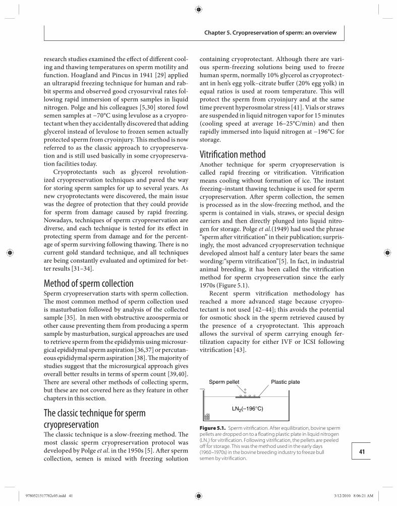
Chapter 5. Cryopreservation of sperm: an overview
41
research studies examined the eff ect of diff erent cool-ing and thawing temperatures on sperm motility and function. Hoagland and Pincus in 1941 [ 29 ] applied an ultrarapid freezing technique for human and rab-bit sperms and observed good cryosurvival rates fol-lowing rapid immersion of sperm samples in liquid nitrogen. Polge and his colleagues [ 5 , 30 ] stored fowl semen samples at −70°C using levulose as a cryopro-tectant when they accidentally discovered that adding glycerol instead of levulose to frozen semen actually protected sperm from cryoinjury. Th is method is now referred to as the classic approach to cryopreserva-tion and is still used basically in some cryopreserva-tion facilities today.
Cryoprotectants such as glycerol revolution-ized cryopreservation techniques and paved the way for storing sperm samples for up to several years. As new cryoprotectants were discovered, the main issue was the degree of protection that they could provide for sperm from damage caused by rapid freezing. Nowadays, techniques of sperm cryopreservation are diverse, and each technique is tested for its eff ect in protecting sperm from damage and for the percent-age of sperm surviving following thawing. Th ere is no current gold standard technique, and all techniques are being constantly evaluated and optimized for bet-ter results [ 31 – 34 ].
Method of sperm collection Sperm cryopreservation starts with sperm collection. Th e most common method of sperm collection used is masturbation followed by analysis of the collected sample [ 35 ]. In men with obstructive azoospermia or other cause preventing them from producing a sperm sample by masturbation, surgical approaches are used to retrieve sperm from the epididymis using microsur-gical epididymal sperm aspiration [ 36 , 37 ] or percutan-eous epididymal sperm aspiration [ 38 ]. Th e majority of studies suggest that the microsurgical approach gives overall better results in terms of sperm count [ 39 , 40 ] . Th ere are several other methods of collecting sperm, but these are not covered here as they feature in other chapters in this section.
The classic technique for sperm cryopreservation Th e classic technique is a slow-freezing method. Th e most classic sperm cryopreservation protocol was developed by Polge et al . in the 1950s [ 5 ]. Aft er sperm collection, semen is mixed with freezing solution
containing cryoprotectant. Although there are vari-ous sperm-freezing solutions being used to freeze human sperm, normally 10% glycerol as cryoprotect-ant in hen’s egg yolk–citrate buff er (20% egg yolk) in equal ratios is used at room temperature. Th is will protect the sperm from cryoinjury and at the same time prevent hyperosmolar stress [ 41 ]. Vials or straws are suspended in liquid nitrogen vapor for 15 minutes (cooling speed at average 16–25°C/min) and then rapidly immersed into liquid nitrogen at −196°C for storage.
Vitrifi cation method Another technique for sperm cryopreservation is called rapid freezing or vitrifi cation. Vitrifi cation means cooling without formation of ice. Th e instant freezing–instant thawing technique is used for sperm cryopreservation. Aft er sperm collection, the semen is processed as in the slow-freezing method, and the sperm is contained in vials, straws, or special design carriers and then directly plunged into liquid nitro-gen for storage. Polge et al. (1949) had used the phrase “sperm aft er vitrifi cation” in their publication; surpris-ingly, the most advanced cryopreservation technique developed almost half a century later bears the same wording:“sperm vitrifi cation”[ 5 ]. In fact, in industrial animal breeding, it has been called the vitrifi cation method for sperm cryopreservation since the early 1970s ( Figure 5.1 ).
Recent sperm vitrifi cation methodology has reached a more advanced stage because cryopro-tectant is not used [ 42 – 44 ]; this avoids the potential for osmotic shock in the sperm retrieved caused by the presence of a cryoprotectant. Th is approach allows the survival of sperm carrying enough fer-tilization capacity for either IVF or ICSI following vitrifi cation [ 43 ].
Sperm pellet Plastic plate
LN2(–196°C)
Figure 5.1. Sperm vitrifi cation. After equilibration, bovine sperm pellets are dropped on to a fl oating plastic plate in liquid nitrogen (LN 2 ) for vitrifi cation. Following vitrifi cation, the pellets are peeled off for storage. This was the method used in the early days (1960–1970s) in the bovine breeding industry to freeze bull semen by vitrifi cation.
9780521517782c05.indd 419780521517782c05.indd 41 3/12/2010 8:06:21 AM3/12/2010 8:06:21 AM
42
Section 2. Cryopreservation of sperm and testicular tissue
Contraindictions, complications, and adverse eff ects Relative contraindications Th ere are no absolute contraindications for cryopreser-vation based on current knowledge, but there are a few relative contraindications that could aff ect the decision to go on with sperm cryopreservation.
Sperm quality and quantity Sperm quality and quantity are the two main factors that can have an eff ect on the success of sperm cryo-preservation. Sperm analysis results indicating poor quality or low quantity may indicate the potential for signifi cant problems with sperm cryopreserva-tion. Consequently, it is advisable not to proceed with sperm cryopreservation for a sperm donor program whenever there is very poor quality or low quantity sperm. Th is can be a problem as patients with cancer oft en show poor sperm analysis even before initiation of chemotherapy [ 27 , 29 ]. Fortunately, with current advanced ART, specifi cally in ICSI, it is possible now to use only one viable sperm to achieve a healthy preg-nancy and live birth.
Infectious diseases Th e ability of some viruses to survive liquid nitrogen freezing has been documented. Hence, all men who are undergoing sperm cryopreservation should undergo regular screening for common infectious diseases as hepatitis A, B, C, and the human immunodefi ciency virus (HIV) [ 45 , 46 ].
Complications and adverse eff ects Th ere are no complications or adverse eff ects for the patient himself, but there may be eff ects of freezing, storage, and thawing on sperm motility, viability, and morphology, and these will be discussed in this section.
Motility When assessing the eff ect of cryopreservation on sperm motility, the cryopreservation process can be consid-ered as three independent processes: freezing, storage, and thawing. Th e eff ects of each of these pro cesses on sperm motility can be considered per se. A consider-able decrease in sperm motility is usually noticed, to approximately 45%, aft er the slow-freezing process [ 47 ]. In patients with abnormal sperm, as in oligoasthe-noteratozooaspermia, sperm motility may be reduced
by up to 75%, suggesting that the sperm sample from fertile males can actually withstand cryoinjury better than samples from infertile patients [ 48 – 50 ]. Other studies have suggested that some agents could be added to the sperm during the thawing process to increase motility, but these agents improve sperm motility not its quality [ 51 ]. A further study examining the thaw-ing recovery of cryopreserved sperm suggested that the freezing–thawing process triggers oxidative stress processes, which aff ect subsequent sperm motility [ 52 ]. Th is study also suggested that adding enzyme enhanc-ers to the medium could improve the post-thawing recovery process [ 52 ].
Viability Viability of sperm aft er cryopreservation is considered a key point for measuring cryopreservation success, and the ultimate indicator of its usefulness. Obviously, there is no reason to preserve semen samples if freez-ing, storage, and thawing will severely aff ect later via-bility. One study has analyzed how sperm viability is aff ected by the freezing–thawing process, and whether the freezing or the thawing phase is responsible for the damage. Th e authors concluded that the freezing pro-cess triggers an apoptotic cascade of events that might explain the failure of some sperms to survive aft er thawing [ 53 ].
Morphology Some studies have looked into the structure of sperm before and aft er the freezing–thawing process and have shown numerous alterations in the cell membrane, creating severe morphological alterations [ 34 , 54 – 57 ]. In addition, there are changes in acrosomal structure, nucleus size, chromatin structure, cell organelles, and plasma membrane integrity [ 58 – 62 ], which are not detectable by light microscopy [ 50 ] but aff ect the structural and functional integrity of the sperm [ 34 , 57 ] during the freezing–thawing process. All of these mor-phological changes will aff ect the ability of the sperm to fertilize an oocyte successfully, even with the use of ICSI and other ART [ 49 , 57 , 63 ].
Eff ect on fertilization and pregnancy rates It has been reported that there is no diff erence in terms of fertilization and pregnancy rates using frozen–thawed sperm versus fresh sperm in IVF/ICSI cycles [ 20 ]. However, in intrauterine insemination, fresh sperm is three times better than frozen sperm [ 54 , 64 ]. With sperm extracted by microsurgical epididymal
9780521517782c05.indd 429780521517782c05.indd 42 3/12/2010 8:06:21 AM3/12/2010 8:06:21 AM
43
Chapter 5. Cryopreservation of sperm an overview
sperm aspiration, the fertilization and pregnancy rates are as good as with fresh sperm [ 23 , 65 ], suggest-ing that sperm extracted from certain sites in the male genital tract might maintain the same quality aft er cryopreservation.
Legal and ethical aspects of sperm cryopreservation Although the scientifi c concept behind sperm cryo-preservation might look simple, the procedure itself is very complex, and it requires highly advanced sperm banking and cryopreservation facilities with proper equipment to handle semen collection, freezing, and storage. Surprisingly, when it comes to complexity, the legal aspect of cryopreservation is no diff erent than its scientifi c aspect in terms of superfi cial simplicity and underlying complexity. However, in contrast to the fi rm foundation that currently underlies sperm cryo-preservation, the legal considerations are ambigu-ous, sometimes vague, and still in a premature stage. Few publications in the literature have addressed this issue, and all the current data regarding informed consent and disposition contracts off ered by the sperm banking and fertility center facilities around the world are very dependent on emerging litigation and court orders [ 66 ]. More details of this issue are given in Chapter 27.
Conclusions Sperm cryopreservation is an ancient method of pre-serving male fertility in a way that will maintain sperm viability for a long period of time. Applications of sperm cryopreservation nowadays are diverse and include humans, animals, and insects. Despite the undeniable morphological and functional adverse eff ects that cryo-preservation exerts on sperm, it still remains one of the core methods of fertility preservation for patients and healthy volunteer donors. Future studies are expected to concentrate on advancing technology to achieve the goal of “damage-free” sperm aft er cryopreservation. Encouragingly, recent advances in ART suggest that it will be possible eventually to achieve a healthy preg-nancy and live birth from cryopreservation of a single sperm/sample.
References 1. Mahony MC , Morshedi M , Scott RT , et al. Role of
spermatozoa cryopreservation in assisted reproduction . Hum Spermatozoa Assist Reprod 1990: 310 –318.
2. Triana V. Artifi cial insemination and semen banks in Italy . Hum Artif Inseminination Semen Preservation 1980: 51 .
3. Matheson GW , Carlborg L , Gemzell C . Frozen human semen for artifi cial insemination . Am J Obstet Gynecol 1969; 104: 495 –501.
4. Pegg DE. Th e history and principles of cryopreservation . Semin Reprod Med 2002; 20: 5 –13.
5. Polge C , Smith AU , Parkes AS . Revival of spermatozoa aft er vitrifi cation and dehydration at low temperatures . Nature 1949; 164: 666 –668.
6. Bunge RG , Sherman JK . Fertilizing capacity of frozen human spermatozoa . Nature 1953; 172: 767 –768.
7. Nelson C , Bunge R . Semen analysis: evidence for changing parameters of male fertility potential . Fertil Steril 1974; 25: 503 –507.
8. Sherman JK . Synopsis of the use of frozen human semen since 1964: state of the art of human semen banking . Fertil Steril 1973; 24: 397 –412.
9. Smith Audrey U . Biological Eff ects of Freezing and Supercooling London : Edward Arnold , 1961 .
10. Wildt DE . Genome resource banking for wildlife research, management, and conservation . ILAR J 2000; 41: 228 –234.
11. Dinnyes A , Liu J , Nedambale TL . Novel gamete storage . Reprod Fertil Dev 2007; 19: 719 –731.
12. Howard JG . Assisted reproductive techniques in nondomestic carnivores . Zoo Wild Anim Med IV 1999; 4: 449 –457.
13. Monfort SL , Asher GW , Wildt DE , et al. Successful intrauterine insemination of Eld ’ s deer (Cervus eldi thamin) with frozen–thawed spermatozoa . J Reprod Fertil 1993; 99: 459 –465.
14. Takemura Y , Kanda T , Horie Y . Artifi cial insemination using cryopreserved sperm in the silkworm, Bombyx mori . J Insect Physiol 2000; 46: 491 –497.
15. Clarke CA . In vitro fertilization: some comparative aspects . J R Soc Med 1990; 83: 214 .
16. Sander K . Fertilization and egg cell activation in insects . Biol Fertilization 1985; 2: 409 –430.
17. Hatakeyama M , Lee JM , Sawa M , et al. Artifi cial reproduction in a hymenopteran insect, Athalia rosae, using eggs matured with heterospecifi c yolk proteins and fertilized with cryopreserved sperm . Arch Insect Biochem Physiol 2000; 43: 137 –144.
18. Hatakeyama M , Sawa M , Oishi K . Fertilization by micro-injection of cryopreserved sperm in the sawfl y, Athalia rosae (Hymenoptera) . J Insect Physiol 1994; 40: 909–912 .
19. Anger JT , Gilbert BR , Goldstein M . Cryopreservation of sperm: indications, methods and results . J Urol 2003; 170: 1079 –1084.
20. Kuczyski W , Dhont M , Grygoruk C , et al. Th e outcome of intracytoplasmic injection of fresh and cryopreserved
9780521517782c05.indd 439780521517782c05.indd 43 3/12/2010 8:06:21 AM3/12/2010 8:06:21 AM
44
Section 2. Cryopreservation of sperm and testicular tissue
ejaculated spermatozoa: a prospective randomized study . Hum Reprod 2001; 16: 2109 –2113.
21. Rofeim O , Brown TA , Gilbert BR . Eff ects of serial thaw–refreeze cycles on human sperm motility and viability . Fertil Steril 2001; 75: 1242 –1243.
22. Ranganathan P , Mahran AM , Hallak J , et al. Sperm cryopreservation for men with nonmalignant, systemic disease: a descriptive study . J Androl 2001; 23: 71 –75.
23. Janzen N , Goldstein M , Schlegel PN , et al. Use of electively cryopreserved microsurgically aspirated epididymal sperm with IVF and intracytoplasmic sperm injection for obstructive azoospermia . Fertil Steril 2000; 74: 696 –701.
24. Tash JA , Goldstein M . Th e use of intra-operative sperm cryopreservation in men with obstructive azoospermia . Fertil Steril 2002; 76 Suppl: 208 .
25. Tash JA , Applegarth L , Kerr S , et al. Post-mortem sperm procurement: the eff ect of instituting guidelines. J Urol 2003 ; 170 : 1922–1925 .
26. Sherman JK . Cryopreservation of human semen. In Keel BA , Webster BW, eds. CRC Handbook of the Laboratory Diagnosis and Treatment of Infertility. Boca Raton, FL: CRC Press , 1990, pp. 229 –259.
27. Ohl DA , Park J , Cohen C , et al. Procreation aft er death or mental incompetence: medical advance or technology gone awry? Fertil Steril 1996; 66: 889 –895.
28. Ahuja KK , Mamiso J , Emmerson G , et al. Pregnancy following intracytoplasmic sperm injection treatment with dead husband’s spermatozoa: ethical and policy considerations . Hum Reprod 1997; 12: 1360 –1363.
29. Hoagland H , Pincus G . Revival of mammalian sperm aft er immersion in liquid nitrogen . J Genet Physiol 1942; 25: 337 –344.
30. Polge C , Lovelock JE . Preservation of bull sperm at −70°C . Vet Rec 1952; 64: 296 –297.
31. Leffl er KS , Walters CA . A comparison of time, temperature, and refreezing variables on frozen sperm motility recovery . Fertil Steril 1996; 65: 272 –274.
32. Ragni G , Caccamo AM , Dalla Serra A , et al. Computerized slow-staged freezing of semen from men with testicular tumors or Hodgkin’s disease preserves sperm better than standard vapor freezing . Fertil Steril 1990; 53: 1072 –1075.
33. Taylor PJ , Wilson J , Laycock R , et al. A comparison of freezing and thawing methods for the cryopreservation of human semen . Fertil Steril 1982; 37: 100 –103.
34. Verheyen G , Pletincx I , Van Steirteghem A . Eff ect of freezing method, thawing temperature and post-thaw dilution/washing on motility (CASA) and morphology characteristics of high-quality human sperm . Hum Reprod 1993; 8: 1678 –1684.
35. Hallak J , Hendin BN , Th omas Jr AJ , et al. Investigation of fertilizing capacity of cryopreserved spermatozoa from patients with cancer . J Urol 1998; 159: 1217 –1220.
36. Patrizio P , Silber S , Ord T , et al. Two births aft er microsurgical sperm aspiration in congenital absence of vas deferens . Lancet 1988; ii: 1364 .
37. Silber SJ , Ord T , Balmaceda J , et al. Congenital absence of the vas deferens: the fertilizing capacity of human epididymal sperm . N Engl J Med 1990; 323 : 1788–1792 .
38. Craft I , Tsirigotis M , Bennett V , et al. Percutaneous epididymal sperm aspiration and intracytoplasmic sperm injection in the management of infertility due to obstructive azoospermia . Fertil Steril 1995; 63: 1038 –1042.
39. Patrizio P . Cryopreservation of epididymal sperm . Mol Cell Endocrinol 2000; 169: 11 –14.
40. Tournaye H , Merdad T , Silber S , et al. No diff erences in outcome aft er intracytoplasmic sperm injection with fresh or with frozen–thawed epididymal spermatozoa . Hum Reprod 1999; 14: 90 –95.
41. Michelmann HW . Sperm and testicular tissue . In Healy , RO , Kovacs DL , McLachlan R , Rodriguez-Armas O , eds. Reproductive Medicine in the Twenty-fi rst Century: Proceedings of the 17th World Congress on Fertility and Sterility, Melbourne, Australia . Boca Raton, FL : Parthenon , 2002 , pp. 287–294.
42. Nawroth F , Isachenko V , Dessole S , et al. Vitrifi cation of human spermatozoa without cryoprotectants . Cryo Letters 2002; 23: 93 –102.
43. Isachenko V , Isachenko E , Katkov II , et al. Cryoprotectant-free cryopreservation of human spermatozoa by vitrifi cation and freezing in vapor: eff ect on motility, DNA integrity, and fertilization ability . Biol Reprod 2004; 71 : 1167 –1173.
44. Isachenko E , Isachenko V , Katkov II , et al. Vitrifi cation of mammalian spermatozoa in the absence of cryoprotectants: from past practical diffi culties to present success . Reprod BioMed Online 2003; 6: 191 –200.
45. Tedder RS , Zuckerman MA , Goldstone AH , et al. Hepatitis B transmission fromcontaminated cryopreservation tank . Lancet 1995; 346: 137 –140.
46. Benifl a JL , Letur-Konirsch H , Collin G , et al. Safety of cryopreservation straws for human gametes or embryos: a preliminary study with human immunodefi ciency virus-1 . Hum Reprod 2000; 15: 2186 –2189.
47. Donnelly ET , Kristine Steele E , McClure N , et al. Assessment of DNA integrity and morphology of ejaculated spermatozoa from fertile and infertile men before and aft er cryopreservation . Hum Reprod 2001; 16: 1191 –1199.
48. Esteves SC , Sharma RK , Th omas Jr AJ , et al. Improvement in motion characteristics and acrosome status in cryopreserved human spermatozoa by swim-up processing before freezing . Hum Reprod 2000; 15: 2173 –2179.
9780521517782c05.indd 449780521517782c05.indd 44 3/12/2010 8:06:21 AM3/12/2010 8:06:21 AM
45
Chapter 5. Cryopreservation of sperm an overview
49. Hammadeh ME , Askari AS , Georg T , et al. Eff ect of freeze–thawing procedure on chromatin stability, morphological alteration and membrane integrity of human spermatozoa in fertile and subfertile men . Int J Androl 1999; 22: 155 –162.
50. Nijs M , Cox A , Bosmans E , et al. Comparison of 3 diff erent cryopreservation methods: are they cryoprotective? In A Journey from Gamete to Newborn: Proceedings of an International Meeting , Leuven, Belgium, 2000, pp. 14–25 .
51. Sharma RK , Kohn S , Padron OF , et al. Eff ect of artifi cial stimulants on cryopreserved spermatozoa from cancer patients . J Urol 1997; 157: 521 –524.
52. Meseguer M , Garrido N , Simon C , et al. Concentration of glutathione and expression of glutathione peroxidases 1 and 4 in fresh sperm provide a forecast of the outcome of cryopreservation of human spermatozoa . J Androl 2004; 25: 773 –780.
53. Paasch U , Sharma RK , Gupta AK , et al. Cryopreservation and thawing is associated with varying extent of activation of apoptotic machinery in subsets of ejaculated human spermatozoa 1 . Biol Reprod 2004; 71 : 1828 –1837.
54. Richter MA , Haning Jr RV , Shapiro SS . Artificial donor insemination: fresh versus frozen semen – the patient as her own control . Fertil Steril 1984; 41: 277 –280.
55. Centola GM , Raubertas RF , Mattox JH . Cryopreservation of human semen: comparison of cryopreservatives, sources of variability, and prediction of post-thaw survival . J Androl 1992; 13: 283 –288.
56. Wong AWY , Ho PC , Kwan M , et al. Factors aff ecting the success of artifi cial insemination by frozen donor semen . Int J Fertil 1989; 34: 25 –29.
57. Critser JK , Arneson BW , Aaker DV . Cryopreservation of human spermatozoa. II. Postthaw chronology of motility and of zona-free hamster ova penetration . Fertil Steril 1987; 47: 980 –984.
58. Barthelemy C , Royere D , Hammahah S , et al. Ultrastructural changes in membranes and acrosome of human sperm during cryopreservation . Syst Biol Reprod Med 1990; 25: 29 –40.
59. Henry MA , Noiles EE , Gao D , et al. Cryopreservation of human spermatozoa, IV. Th e eff ects of cooling rate and warming rate on the maintenance of motility, plasma membrane integrity, and mitochondrial function . Fertil Steril 1993; 60: 911 –918.
60. Nogueira D , Bourgain C , Verheyen G , et al. Light and electron microscopic analysis of human testicular spermatozoa and spermatids from frozen and thawed testicular biopsies . Hum Reprod 1999; 14: 2041 –2049.
61. Holt WV , Head MF , North RD . Freeze-induced membrane damage in ram spermatozoa is manifested aft er thawing: observations with experimental cryomicroscopy . Biol Reprod 1992; 46: 1086 –1094.
62. Mack SR , Zaneveld LJD . Acrosomal enzymes and ultrastructure of unfrozen and cryotreated human spermatozoa . Gamete Res 1987; 18: 375 –383.
63. Van Steirteghem A . Freezing of testicular tissue as a minced suspension preserves sperm quality better than whole-biopsy freezing when glycerol is used as cryoprotectant . Int J Androl 1999; 22: 43 –48.
64. Sherman JK . Synopsis of the use of frozen human semen since 1964: state of the art of human semen banking . Fertil Steril 1973; 24: 397 –412.
65. Oates RD , Lobel SM , Harris DH , et al. Effi cacy of intracytoplasmic sperm injection using intentionally cryopreserved epididymal spermatozoa . Hum Reprod 1996; 11: 133 –138.
66. Schuster TG , Hickner-Cruz K , Ohl DA , et al. Legal considerations for cryopreservation of sperm and embryos . Fertil Steril 2003; 80: 61 –66.
9780521517782c05.indd 459780521517782c05.indd 45 3/12/2010 8:06:21 AM3/12/2010 8:06:21 AM
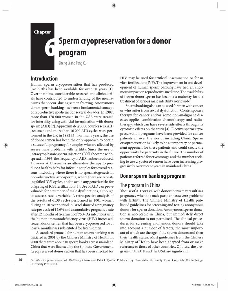
Fertility Cryopreservation, ed. Ri-Cheng Chian and Patrick Quinn. Published by Cambridge University Press. Copyright © Cambridge University Press 2010.
Chapter
6 HIV may be used for artifi cial insemination or for in vitro fertilization (IVF). Th e improvement in and devel-opment of human sperm banking have had an enor-mous impact on reproductive medicine. Th e availability of frozen donor sperm has become a mainstay for the treatment of serious male infertility worldwide.
Sperm banking also can be used for men with cancer or who suff er from sexual dysfunction. Contemporary therapy for cancer and/or some non-malignant dis-eases applies combination chemotherapy and radio-therapy, which can have severe side eff ects through its cytotoxic eff ects on the testis [ 4 ]. Elective sperm cryo-preservation programs have been provided for cancer patients all over the world, including China. Sperm cryopreservation is likely to be a temporary or perma-nent approach for these patients and could create the opportunity for paternity in the future. Th e number of patients referred for cryostorage and the number seek-ing to use cryostored semen have been increasing pro-gressively over recent years in mainland China.
Donor sperm banking program The program in China Th e use of AID or IVF with donor sperm may result in a pregnancy when the male partner has severe problems with fertility. Th e Chinese Ministry of Health pub-lished guidelines for screening and testing anonym ous donors for sperm donation. Anonymous sperm dona-tion is acceptable in China, but immediately direct sperm donation is not permitted. Th e clinical proce-dures for screening anonymous donors should take into account a number of factors, the most import-ant of which are the age of the sperm donors and then their health status. Most guidelines from the Chinese Ministry of Health have been adapted from or make reference to those of other countries. Of these, the pro-grams in the UK and the USA are signifi cant.
Introduction Human sperm cryopreservation that has produced live births has been available for over 50 years [ 1 ]. Over that time, considerable research and clinical tri-als have contributed to understanding of the mecha-nisms that occur during semen freezing. Anonymous donor sperm banking has been a fundamental concept of reproductive medicine for several decades. In 1987, more than 170 000 women in the USA were treated for infertility using artifi cial insemination with donor sperm (AID) [ 2 ]. Approximately 3000 couples seek AID treatment and more than 16 000 AID cycles were per-formed in the UK in 1992 [ 3 ]. For many years, the use of donor semen has been the only approach to obtain a successful pregnancy for couples who are aff ected by severe male problems with fertility. Since the use of intracytoplasmic sperm injection (ICSI) became wide-spread in 1993, the frequency of AID has been reduced. However AID remains an alternative therapy to pro-duce a healthy baby for infertile couples for several rea-sons, including where there is no spermatogenesis in non-obstructive azoospermia, where there are repeat-ing failed ICSI cycles, and to avoid any genetic risks for off spring of ICSI fertilization [ 3 ]. Use of AID can prove valuable for a number of male dysfunctions, although its success rate is variable. A retrospective analysis of the results of 6139 cycles performed in 1001 women during an 18-year period in Israel showed a pregnancy rate per cycle of 12.6% and a cumulative pregnancy rate aft er 12 months of treatment of 75%. As infections with the human immunodefi ciency virus (HIV) increased, frozen donor semen that has been cryopreserved for at least 6 months was substituted for fresh semen.
A standard protocol for human sperm banking was initiated in 2001 by the Chinese Ministry of Health. In 2008 there were about 10 sperm banks across mainland China that were licensed by the Chinese Government. Cryopreserved donor semen that has been checked for
Sperm cryopreservation for a donor program Zheng Li and Peng Xu
46
9780521517782c06.indd 469780521517782c06.indd 46 3/12/2010 8:07:27 AM3/12/2010 8:07:27 AM
47
Chapter 6. Sperm cryopreservation for a donor program
In 2005, the UK Government instituted the require-ment that any sperm donor had to agree to the disclos-ure of their identity to any off spring reaching the age of 18 years. Th ere was a survey of donor opinions about the cancellation of anonymity. Some donors thought it was problematic, indicating that some donors will be reluctant to donate under conditions of non-anonym-ity and others would have concerns about the removal of anonymity [ 5 ].
Th e 2006 Guidelines for Gamete and Embryo Donation provide the latest recommendations for evalu-ation of potential sperm donors in the USA, incorp-orating recent information about optimal screening and testing for sexually transmitted infections, genetic diseases, and psychological assessments. Th is revised document from the American Society for Reproductive Medicine Practice Committee incorporates recent information from the US Centers for Disease Control and Prevention, the US Food and Drug Administration, and the American Association of Tissue Banks, with which all programs off ering gamete and embryo dona-tion services must be thoroughly familiar [ 6 ].
Criteria for selection of anonymous donors in China Th e guidelines for selection of anonymous sperm donors in China are as follows. Sperm donors should have a good health status and no known genetic dis-eases in their family. Sperm donors should be between 22 and 44 years of age, because male aging is associated with a progressive increase in the number of aneuploid sperm. Sperm donor recruitment with confi rmed fer-tility is desirable but not a mandatory requirement. Psychological and genetic evaluation and counseling by professional doctors is recommended strongly for all sperm donors. It is essential for the donors to sign a written consent. It is important that the donor should be well informed about the extent to which information about him might be disclosed and about any procedures that may occur related to a future con-tact by recipients and/or off spring. Physicians, labora-tory staff , or other employees related to the treatment center are prohibited from becoming sperm donors in the donor programs.
Screening and testing program for sperm donors in China Screening and testing of sperm donors are in accord with the regulations published by the Chinese Ministry
of Health. Semen analysis is suggested for several sam-ples (each sample aft er 2 to 7 days of abstinence) before proceeding with more extensive testing for the donors. Th e criteria used for judging the semen sample may vary among laboratories. Th e criteria for semen quality have been applied in China since 2003: (1) liquefaction time less than 60 minutes; (2) sperm concentration more than 60 × 10 6 sperm/ml; (3) percentage of rapid and slow progressive spermatozoa more than 60%; (4) normal morphology in more than 30% of sperm (see the World Health Organization Laboratory Manual for the Examination of Human Semen and Sperm–Cervical Mucus Interaction [ 7 ]) .
Genetic screening for heritable diseases should be evaluated in potential sperm donors. Chromosomal analyses on all donors are required but cystic fi brosis carrier status is not assessed for all donors because this disorder is rare in the Chinese population. Genetic con-sulting should be performed aft er screening family his-tory. Sperm donors should be healthy and have no history of genetic disease. Of course, transmission diseases such as HIV, sexually transmitted infections, and other infec-tions that might be transmitted via sperm donation have to be screened before selecting the donors.
Medical examinations for the sperm donor Sperm donors should be given a complete examin-ation, including assessment for urethral discharge and penile warts and ulcers, plus routine laboratory screening, including blood type and Rhesus factor, before being enrolled as donors. Sperm donors should have follow-up examinations every 6 months for ure-thral discharge and penile warts and ulcers if they remain sperm donors. Th ere is no 100% guarantee that infectious agents will not be transmitted by AID, but exclusion of potential donors at high risk for HIV and other sexually transmitted infections is important. Th e Chinese Ministry of Health requires the following tests: HIV-1 and HIV-2, hepatitis C antibody, hepa-titis B surface antigen, hepatitis B core antibody, and cytomegalovirus. Semen or urethral tests are evaluated for Neisseria gonorrhoeae and Chlamydia trachomatis . Semen screening documentation confi rming that there is no alteration in the donor’s medical history should be recorded at 6 month intervals. If screening is nega-tive, semen samples may be collected and prepared for cryopreservation. Aft er donation, sperm samples must be cryopreserved for a minimum of 180 days before use. Th e donor must be re-screened for HIV antibody and samples may be used only if the results of repeat
9780521517782c06.indd 479780521517782c06.indd 47 3/12/2010 8:07:28 AM3/12/2010 8:07:28 AM
Section 2. Cryopreservation of sperm and testicular tissue
48
testing are negative. Fresh donor sperm is not allowed for AID or IVF in China.
Follow up for the results from sperm donation Sperm banks are required to follow up AID or IVF results and to keep their records in order to allow a limited number of pregnancies with the same donor. A computer management system is required in the sperm bank to save the data. In China, only fi ve women can be allowed to be pregnant with AID or IVF from a sperm donor. It has been recommended in the USA that, in a population of 800 000, limiting a single donor to no more than 25 births would avoid any signifi cant increased risk of inadvertent consanguineous concep-tion [ 6 ]. Since 1992, a limit of 25 off spring for every donor has been imposed in the Netherlands in order to avoid children from donors having a greater risk of consanguineous relationships [ 8 ]. Th ese regulations may require modifi cation if the population using AID or IVF represents an isolated subgroup or if the speci-mens are released over a wide geographic area.
Methods for sperm cryopreservation Clarke et al. [ 9 ] evaluated six donor semen samples aft er 28 years of cryopreservation in liquid nitrogen. Th eir results showed that the samples retained good post-thaw motility and normal levels of binding to the human zona pellucida and that four of the fi ve samples tested also gave normal levels of zona-induced acro-some reaction.
Conventional semen cryopreservation As the temperature of a sperm sample is lowered in liquid nitrogen, extracellular ice crystals form, which leads to an increased concentration of solutes in the extracellular fl uid. Th e resulting osmotic gradient results in dehydration as water moves out of the sperm-atozoa. Damage can occur to the cell if the osmotic gradient is too steep, resulting in water fl ow that is too rapid. Additionally, excessive dehydration causes increased intracellular solute concentration, which can be toxic to sperm cells, especially aff ecting sperm motility aft er thawing. Some experiments have shown that sperm DNA integrity also could be damaged and that sperm DNA fragmentation increases during the freezing– thawing cycle [ 10 ]. Sperm freezing has del-eterious eff ects on spermatozoa, especially on plasma-lemma, acrosome, and tail. Electron microscopy is the
ultimate modality to investigate spermatogenic cells following freeze–thawing [ 11 ].
Th e conventional approach to sperm cryopreser-vation is to simply dilute semen with cryoprotectant and cryopreserve in liquid nitrogen until the sperm samples are thawed for use. A slow-freezing method consists of gradually cooling the sperm over a period of 1 to 2 hours in two or three steps, either manually or using a programmed freezer. Initial cooling rates of the sperm specimen from room temperature to 5°C have been shown to be optimal at 0.5–1°C/min. Th e sample is then frozen at a rate of 1 to 10°C/min from 5°C to −30°C, aft er which the specimen is plunged into liquid nitrogen. Additional information has suggested that holding the specimen at −5°C for 10 minutes and seed-ing the specimen may improve cryosurvival sperm rate aft er the frozen–thawing cycle [ 12 ].
Vitrifi cation of sperm Although the conventional slow-freezing approach has given acceptable results, others have shown that vitrifi -cation may be a better method for sperm cryopreserva-tion [ 13 ]. With the advance of ICSI, theoretically only one sperm is required per oocyte. Th erefore, the vitri-fi cation method may provide infertile men with severe oligozoospermia a good chance to cryopreserve the lim-ited number of sperm needed [ 14 ]. Th ere is a report of vitrifying sperm with the Cryoloop, which could off er an alternative approach for sperm freezing [ 15 ]. Th e abil-ity to cryopreserve oligozoospermic samples has many potential areas for application, including for patients with severe oligozoospermia or asthenozoospermia.
Electroejaculation and sperm cryopreservation Mammalian spermatozoa were among the very fi rst cells to be successfully cryopreserved. Th e use of fro-zen–thawed semen for artifi cial insemination has come to play an important role in reproductive medicine. Electroejaculation is a valuable approach used to obtain semen samples from sexually mature male mammals and has been used for breeding programs and research purposes in various species, as well as in the treatment of anejaculation and ejaculatory dysfunction in infer-tile men who have failed other treatments [ 16 ].
Anejaculation is caused mainly by psychogenic an ejaculation, spinal cord injury, diabetes mellitus, ret-roperitoneal lymph node dissection, transverse myelitis, and multiple sclerosis. Men who suff er from psychogenic
9780521517782c06.indd 489780521517782c06.indd 48 3/12/2010 8:07:28 AM3/12/2010 8:07:28 AM
49
Chapter 6. Sperm cryopreservation for a donor program
anejaculation are healthy males who cannot intention-ally ejaculate even by masturbation. However, they may have normal penile erections and nocturnal emissions. Previous data have shown that electroejaculated sperm have severe asthenozoospermia, sperm motility appar-ently decreasing in those with spinal cord injury or psy-chogenic anejaculation [ 17 ]. Th e asthenozoospermia in those with spinal cord injury may be connected with increased scrotal temperature, urinary infection, stasis of seminal fl uid, neural eff ects, and so on. Th e semen qual-ity of patients with psychogenic anejaculation is similar to that of patients with spinal cord injury, despite the fact that the latter do not have a neurological disorder and its complications [ 18 ].
A complete urological check is necessary prior to electroejaculation in order to exclude and treat any urin-ary tract infl ammation. Patients with spinal cord injur-ies oft en have a problem with poor sperm production as well as ejaculation aft er the damage. A diagnostic trial of electroejaculation aims to obtain semen samples and to examine their quality. Good-quality semen samples are cryopresereved for future use. A fresh specimen can be used at a suitable time in a female partner’s ovulation induction cycle for IVF or ICSI. Patients are prescribed sodium bicarbonate tablets prior to electroejaculation to alkalinize the urine and make it more suitable for sperm survival, because a retrograde sample may need to be retrieved from the bladder through a catheter.
Electroejaculation is usually performed under a general anesthesia while the patient is placed in lateral decubitus. An electric probe is put into the rectum close to the prostate. Th e probe transfers a slight electric cur-rent that stimulates adjacent nerves, resulting in con-traction of the pelvic muscles and ejaculation. Prior to electroejaculation, the bladder is fi lled with Ham’s F-10 medium as a buff er and 20 ml of medium is instilled for gathering of the retrograde-emitted semen sample. Multiple electrical stimulations ranging from 5 to 20 V and lasting for 2 seconds are administered. Th e urethra is manually milked into a sterile container both during and upon completion of electroejaculation to obtain the maximum volume of antegrade semen. Aft er elec-troejaculation, a bladder catheterization is routinely performed for collection of retrograde specimens. Th e sperm obtained in the antegrade and retrograde ejacu-lates are processed by the discontinuous Percoll gra-dient, as described elsewhere. Th e concentration and motility of the antegrade and retrograde portions of electroejaculation are assessed using a Makler count-ing chamber according to published guidelines [ 7 ].
During emission and ejaculation in normal men, the internal urinary sphincter closes under the infl uence of sympathetic innervation from the T10–L2 spinal cord segments, causing semen to be expelled from the penis. In men with a spinal cord injury, whose innervation may be disrupted, the retrograde ejaculation aft er elec-troejaculation could be related to their neuropathol-ogy. Retrograde ejaculation does not occur in patients who are neurologically intact. However, most of these patients will have retrograde ejaculation with electro-ejaculation. Th ese fi ndings suggest that the appearance of a retrograde fraction with electroejaculation in men with a spinal cord injury may refl ect methodological issues rather than neuropathology.
Aft er electroejaculation, semen can be used for IVF or cryopreserved for future use and is an eff ective treat-ment for anejaculatory infertility. Intrauterine insem-ination with sperm (either fresh or frozen) obtained by electroejaculation is the fi rst choice for men infertile with anejaculation [ 19 ]. Th e high percentage of preg-nancies achieved with systemic assisted reproductive technology (ART) using sperm cryopreservation aft er electroejaculation means that this is a valuable and safe method for treating infertility with anejaculation [ 20 ].
Conclusions Sperm cryopreservation plays an important role in the fi eld of male infertility and reproductive medicine. A donor sperm cryopreservation program has been devel-oped and improved in mainland China. Standards, guidelines, and a protocol for sperm donor screening and cryopreservation have been practiced since 2001. So far 10 human sperm banks have been set up and licensed by the Chinese Ministry of Health. With the advance of new approaches for sperm vitrifi cation, treatment of male infertility will become more eff ec-tive without using sperm donors. Using vitrifi cation for cryo preservation of sperm obtained from testicular biopsy, epididymal fl uid, or a semen sample aft er elec-troejaculation could create new hope for infertile men.
References 1. Sherman JK , Bunge RG . Eff ect of glycerol and freezing on
some staining reactions of human spermatozoa . Proc Soc Exp Biol Med 1953; 84 : 179 –800.
2. Critser JK . Current status for semen banking in the USA . Hum Reprod 1998; 13 : 55 –67.
3. Botchan A , Hauser R , Gamzu R , et al. Results of 6139 artifi cial insemination cycles with donor spermatozoa . Hum Reprod 2001; 16 : 2298 –2304.
9780521517782c06.indd 499780521517782c06.indd 49 3/12/2010 8:07:28 AM3/12/2010 8:07:28 AM
50
Section 2. Cryopreservation of sperm and testicular tissue
4. Crha I , Ventruba P , Zakova J , et al. Survival and infertility treatment in male cancer patients aft er sperm banking . Fertil Steril 2009 ; 91 : 2344–2348 .
5. Frith L , Blyth E , Farrand A . UK gamete donors ’ refl ections on the removal of anonymity: implications for recruitment . Hum Reprod 2007; 22 : 1675 –1680.
6. Practice Committee of the American Society for Reproductive Medicine. 2006 Guidelines for Gamete and Embryo Donation. Fertil Steril 2006; 86 (Suppl 5): S38–S50.
7. World Health Organization. Laboratory Manual for the Examination of Human Semen and Sperm–Cervical Mucus Interaction, 4th edn. New York : Cambridge University Press , 1999 .
8. Janssens PM . No reason for a reduction in the number of off spring per sperm donor because of possible transmission of autosomal dominant diseases . Hum Reprod 2003; 18 : 669 –671.
9. Clarke GN , Liu de Y , Baker HW . Recovery of human sperm motility and ability to interact with the human zona pellucida aft er more than 28 years of storage in liquid nitrogen . Fertil Steril 2006; 86 : 721 –722.
10. Hu JH , Li QW , Jiang ZL , et al. Eff ects of diff erent extenders on DNA integrity of boar spermatozoa following freezing–thawing . Cryobiology 2008 ; 57 : 257–262 .
11. Ozkavukcu S , Erdemli E , Isik A , et al. Eff ects of cryopreservation on sperm parameters and ultrastructural morphology of human spermatozoa . J Assist Reprod Genet 2008; 25 : 403 –411.
12. Jin B , Yamasaki C , Yamada N , et al. Th e mechanism by which mouse spermatozoa are injured during freezing . J Reprod Dev 2008; 54 : 265 –269.
13. Isachenko V , Isachenko E , Katkov II , et al. Cryoprotectant-free cryopreservation of human spermatozoa by vitrifi cation and freezing in vapor: eff ect on motility, DNA integrity, and fertilization ability . Biol Reprod 2004; 71 : 1167 –1173.
14. Desai NN , Blackmon H , Goldfarb J . Single sperm cryopreservation on cryoloops: an alternative to hamster zona for freezing individual spermatozoa . Reprod Biomed Online 2004; 9 : 47 –53.
15. Isachenko E , Isachenko V , Katkov II , et al. DNA integrity and motility of human spermatozoa aft er standard slow freezing versus cryoprotectant-free vitrifi cation . Hum Reprod 2004; 19 : 932 –939.
16. Curry MR . Cryopreservation of mammalian semen . Methods Mol Biol 2007; 368 : 303 –311.
17. Hovav1 Y , Sibirsky O , Pollack RN . Comparison between the fi rst and the second electroejaculate qualities obtained from neurologically intact men suff ering from anejaculation . Hum Reprod 2005; 20 : 2620 –2622.
18. DeForge D , Blackmer J , Garritty C , et al. Fertility following spinal cord injury: a systematic review . Spinal Cord 2005; 43 : 693 –703.
19. Kafetsoulis A , Brackett NL , Ibrahim E , et al. Current trends in the treatment of infertility in men with spinal cord injury . Fertil Steril 2006; 86 : 781 –789.
20. Shieh JY , Chen SU , Wang YH , et al. A protocol of electroejaculation and systematic assisted reproductive technology achieved high effi ciency and effi cacy for pregnancy for anejaculatory men with spinal cord injury . Arch Phys Med Rehabil 2003; 84 : 535 –540.
9780521517782c06.indd 509780521517782c06.indd 50 3/12/2010 8:07:28 AM3/12/2010 8:07:28 AM
Fertility Cryopreservation, ed. Ri-Cheng Chian and Patrick Quinn. Published by Cambridge University Press. Copyright © Cambridge University Press 2010.
Chapter
7 where it is not certain whether sperm can be found, obtaining sperm and cryopreserving them ahead of time can allow the couples to make a better informed decision about whether they should proceed with the treatment with advanced reproductive technologies, which can be costly fi nancially and emotionally, or whether they should opt for alternative management such as the use of donor sperm or adoption. Other situ-ations where cryopreservation of surgically retrieved sperm is indicated for men who cannot produce ejacu-lated sperm include:
men who plan to have multiple cycles of ICSI and • are not willing to undergo multiple sessions of sperm retrieval for each subsequent cycle men who anticipate a decline in their reproductive • status (e.g. undergoing cytotoxic therapy for malignancies) men who are not ready for immediate assisted • reproduction (e.g. of young age) or who do not have a female partner for reproduction yet men who are undergoing complex reconstructive • surgeries for the excurrent ductal system (e.g. microsurgical vasoepididymostomy for epididymal obstruction or trans-urethral resection of the ejaculatory duct to relieve obstruction).
Posthumous sperm retrieval represents a special scen-ario where cryopreservation of surgically retrieved sperm is required. Th e techniques for posthumous sperm retrieval are identical to those used in living subjects. Th e psychological, ethical, and legal impli-cations associated with the procedure, which are beyond the scope of this chapter, must be taken into consideration.
Techniques of sperm retrieval Various techniques have been described and modi-fi ed for sperm retrieval since the introduction of ICSI
Introduction With increased understanding of reproductive biology in advanced assisted reproductive technologies (ART) such as intracytoplasmic sperm injection (ICSI), com-plemented with the improved safety and availability of these procedures worldwide, the management of severe male-factor infertility has undergone tremen-dous changes over the past few years. Indeed, even for men with the most challenging presentation of non-obstructive azoospermia, a small amount of sperm can be surgically retrieved for ICSI to allow many of these men, who were once considered sterile, to father their genetic children. Various issues in the cryopreserva-tion of surgically retrieved sperm will be the focus of this chapter.
Indications for cryopreservation of surgically retrieved sperm When the male partner is unable to produce an a de-quate amount of ejaculated sperm with good qualit y for ICSI, surgical sperm retrieval is indicated. Th is includes the following scenarios:
azoospermia caused by obstruction of the male • excurrent ductal system or testicular dysfunction poor quality and quantity of sperm in the • ejaculate, particularly in association with a previous history of recurrent failure to achieve pregnancy with ICSI diffi culty in ejaculating because of neurological or • psychological problems.
For most couples going through in vitro fertiliza-tion (IVF)/ICSI cycles, one major advantage of cryo-preserving sperm is that the timing of sperm and egg collection does not have be planned for specifi c dates, making the scheduling of treatment easier and redu-cing unnecessary stress for the couples. In addition,
Cryopreservation of surgically retrieved sperm Peter T. K. Chan
51
9780521517782c07.indd 519780521517782c07.indd 51 3/12/2010 8:09:23 AM3/12/2010 8:09:23 AM
52
Section 2. Cryopreservation of sperm and testicular tissue
in 1992. Descriptions of the various commonly used procedures for sperm retrieval will be provided here to illustrate the principles of each technique and the potential drawbacks for subsequent cryopreserva-tion. Th e three principal sources of sperm are epididy-mides, testes, and ejaculation. Although techniques for sperm retrieval from the seminal vesicles and vasa deferens have been described, they are less commonly used because of their more invasive nature and variable sperm yields compared with the techniques described below.
Percutaneous epididymal sperm aspiration Sperm acquire motility during their epididymal tran-sit. Hence the level of motility can be used as a criterion in conjunction with morphology to identify a “good” and viable spermatozoon for ICSI. In men who have obstructive azoospermia, such as post-vasectomy and congenital bilateral absence of vas deferens, epididymal aspiration to retrieve sperm is a preferred technique by many reproductive surgeons and embryologists. Th e process is generally performed under local anesthesia. An aspiration needle (gauge 21 to 27) is inserted per-cutaneously to the caput epididymis to aspirate a small amount of epididymal fl uid containing sperm [ 1 ]. Although no incision is required for this procedure, multiple blind passages of the needle may be required to obtain an adequate amount of sperm, particularly if cryopreservation of sperm is planned. Th e potential drawbacks of percutaneous epididymal sperm aspira-tion (PESA), in addition to low yield of sperm, are (1) potentially scarring of the epididymis, making future sperm retrieval more diffi cult at the same site; (2) risks of causing hematoma and related complications post-operatively; and (3) presence of blood in the retrieved sperm samples, which may complicate the processing of the sample and subsequent selection of sperm for ICSI.
Microsurgical epididymal sperm aspiration In microsurgical epididymal sperm aspiration (MESA), the epididymis is exposed and explored microsurgi-cally at 25× or higher magnifi cation to identify a single epididymal tubule for sperm aspiration. To obtain the epididymal fl uid, the dissected tubule can be punctured with a fi ne glass pipette to aspirate the fl uid within. Alternatively, the tubule can be microsurgically incised with a micro-ophthalmic knife to allow the fl uid to be aspirated with micropipettes [ 1 ] or angiocatheters. Compared with PESA, MESA provides a higher sperm
yield, as a larger quantity of bloodless epididymal fl uid can generally be obtained and this allows sperm to be cryopreserved to avoid future sperm retrieval pro-cedures. Hence, for men with obstructive azoosper-mia, MESA is the preferred choice of sperm-retrieval techniques by many experts in reproductive urology. Potential drawbacks of MESA include (1) the require-ment of a scrotal incision, which can be perceived as more invasive, and (2) the requirement of microsurgi-cal skills and instruments on the surgeon’s part.
Testicular sperm aspiration For men with non-obstructive azoospermia, MESA and PESA are inappropriate as there should be no sperm retrievable in the epididymis. Instead, retrieval of testicular sperm for ICSI should be performed. In testicular sperm aspiration (TESA), an aspiration nee-dle is inserted percutaneously to the testis parenchyma to aspirate fl uid or small pieces of seminiferous tubules to obtain sperm. Th is is generally performed under local anesthesia and is the procedure that is technically the easiest to acquire. In fact, in some parts of the world, this procedure is sometimes performed by healthcare professionals with limited urological training to obtain sperm for ICSI. Th e drawbacks of this technique are (1) low yield of sperm in those with non-obstructive azoospermia, hence requiring multiple passages of the needle to the testes, and (2) risks of hematoma and related complications postoperatively. Hence, TESA is best reserved for men who have obstructive azoospermia but failed to have sperm retrieved from the epididymis.
Testicular sperm extraction For men with non-obstructive azoospermia, where the rare foci where there are spermatozoa may be dif-fi cult to identify with TESA, an open testicular sperm extraction (TESE), in which the testicle is exposed through a scrotal incision to obtain a large piece of seminiferous tubules, may yield a higher success rate for obtaining sperm. In TESE, the scrotum is incised to expose the testicular tunica albuginea, which is, in turn, incised to expose the seminiferous tubules for sperm retrieval. Th e obtained tubules should be proc-essed intraoperatively by a qualifi ed embryologist to look for sperm, as it is oft en necessary to obtain mul-tiple samples from diff erent sites of the testis in order to obtain an adequate amount of sperm for most men with non-obstructive azoospermia. Although an inci-sion is required for TESE, making it perceived as an
9780521517782c07.indd 529780521517782c07.indd 52 3/12/2010 8:09:23 AM3/12/2010 8:09:23 AM
Chapter 7. Cryopreservation of surgically retrieved sperm
53
invasive procedure, it can generally be performed as an outpatient procedure under local anesthesia. Since the incision and the exposure of the seminifer-ous tubules are performed under direct vision, the risks of complications such as wound infection and hematoma are actually well below 1% when TESE is performed properly. Th e major drawback of TESE is that a large volume of testicular parenchyma is removed non-selectively to obtain enough sampling of the seminiferous tubules for sperm retrieval, and thorough dissection of these tubules is labor intensive and time consuming for the embryologists/androlo-gists. In addition, the irrecoverable loss of the Leydig cells mass in the interstitium of the testicular paren-chyma may increase the risk of the subsequent devel-opment of hypogonadism in men who already have testicular dysfunction.
Micro-dissection for testicular sperm extraction Most expert reproductive urologists prefer to use micro-TESE to obtain sperm from men with non-obstructive azoospermia. Under 25–40× magnifi -cation, the entire volume of seminiferous tubules is examined thoroughly through a large incision on the tunica albuginea [ 1 ]. Tubules that are more likely to have active spermatogenesis generally appear larger than the fi brotic tubules. Th e selected tubules are removed microsurgically with minimal disturbance of the interstitium and adjacent tubules. Th e advan-tage of micro-TESE is that a much smaller quantity of seminiferous tubules is removed than in conventional TESE. As a result, more time can be focused on the processing of the retrieved seminiferous tubules to obtain sperm. Th e need to make a large incision in the testicular tunica albuginea producs the perception that micro-TESE is more invasive than conventional TESE. In fact, when properly performed with a micro-surgical technique that allows sparing of major tunical vessels, micro-TESE carries a low complication rate [ 2 ]. Th e major drawback of micro-TESE is that it is time consuming. Proper exploration of one testicle of normal size (> 20 ml) generally require approximately 2 hours. In addition, special microsurgical training is required not only for the opening of the incision and the use of microsutures for the closure of incision but also, and most importantly, for the exploration of the seminiferous tubule to allow proper identifi cation of tubules that are most likely to contain sperm.
Vibratory stimulation Men with normal spermatogenesis but failure to ejacu-late from neurological or psychological reasons may benefi t from vibratory stimulation. A vibrator, prefer-ably of adjustable frequency and amplitude of vibra-tion, can be applied to the glans penis or the frenulum of the ventral side of the head of the penis to induce orgasm for ejaculation [ 3 ]. Vibratory stimulation is relatively non-invasive. In men with spinal cord injury, particularly those with injury above the level of T10, vibratory stimulation can be eff ective to obtain sperm. Blood pressure monitoring of these patients during the procedure is required to avoid hypertension from autonomic dysrefl exia.
Electroejaculation In electroejaculation, an electric current is applied trans-rectally through an electric probe to the nerves and ejaculatory organs to trigger an ejaculation [ 3 ]. Th is procedure is painful and generally requires anesthesia, except for men with complete spinal cord injury who have no sensation in the lower body. Ejaculation triggered in this way is not always ante-grade. Consequently, catheterization of the bladder aft er orgasm may be required to obtain sperm. Th e major drawback is that the quality of sperm obtained is variable, since these patients generally are not able to ejaculate oft en to allow the aged sperm to be emitted. Cryopreservation of sperm obtained by electroejacu-lation may further comprise the overall quality of the sperm for future use.
Cryopreservation of surgically retrieved sperm Th e quantity of sperm retrievable surgically from the epididymides and testes is generally small compared with the amount of ejaculated sperm. Hence, eff orts have been made by various investigators to evaluate a wide range of protocols to determine the best proce-dure to preserve surgically retrieved sperm [ 4 , 5 ]. Th e optimal carrier should allow cryopreservation of mul-tiple portions of these sperm in small numbers (e.g. ~10 sperm per carrier, depending on the source and qual-ity) for each cycle of ICSI. Repeated freezing–thawing in the specimen (such as when storing a much larger quantity of sperm in one sample) should be avoided.
Various biological and non-biological carriers have been described in the current literature for this purpose. Examples of biological carriers include algae
9780521517782c07.indd 539780521517782c07.indd 53 3/12/2010 8:09:24 AM3/12/2010 8:09:24 AM
54
Section 2. Cryopreservation of sperm and testicular tissue
( Volvox globator ) [ 6 ] and empty zonae from diff erent species including hamster [ 7 ], mouse, or human [ 5 , 8 ]. Non-biological carriers include the conventional and modifi ed straws and micropipettes, calcium alginate capsules [ 9 ], microdroplets of various media [ 10 , 11 ], agarose gel microspheres, and Cryoloops [ 12 ]. Each pro-tocol has its limitations and drawbacks. Interpretation of data from studies attempting to evaluate cryodam-age of surgically retrieved sperm is diffi cult, as there are no universally accepted markers of sperm function. In addition, many studies reported results from a mixed population of men with obstructive (with normal sper-matogenesis) or non-obstructive azoospermia (with abnormal spermatogenesis). Various measurements, from sperm motility, morphology, ultramorphology as evaluated by electron microscopy, nuclear chroma-tin, and DNA integrity, have been used. An increased rate of damage to the junction of the sperm head and midpiece, as observed by light microscopy, has been reported aft er thawing of testicular sperm and can lead to dysfunction of the parternal centrosome [ 13 ]. Using transmission electron microscopy, Nogueira et al. [ 14 ] observed swelling and rupture of the inner and outer acrosomal and plasma membranes aft er freezing and thawing of testicular samples from men with obstruct-ive azoospermia. Using the Comet assay, Steele et al. [ 15 ] were not able to detect any diff erence between fresh and frozen–thawed testicular spermatozoa from men with obstructive azoospermia with regard to the percentage of damaged DNA.
The use of cryopreserved sperm retrieved by surgery With epididymal sperm, which are generally retrieved from men with obstructive azoospermia, there are reductions in the concentration of viable spermatozoa as well as a signifi cant decrease (by 50%) in the per-centage of motile spermatozoa [ 16 ], but there is little evidence to suggest that there is a diff erence in fer-tilization, cleavage, and implantation or pregnancy outcomes between cryopreserved or fresh samples [ 16 – 25 ].
Th e fertilization rates of frozen–thawed testicular sperm with ICSI ranged from 47 to 79%, which were similar to the use of fresh testicular sperm as reported by most investigators [ 26 , 27 ]. Other investigators, however, noted a signifi cantly decreased fertilization rate or embryo cleavage rate with cryopreserved tes-ticular sperm [ 28 – 31 ]. Th e reported pregnancy rate
with frozen–thawed testicular sperm ranged from 26 to 50%, compared with 26–40% with fresh testicular sperm. As in the case of fertilization rates, most inves-tigators reported similar results whether fresh or fro-zen–thawed testicular sperm were used. It should be noted that most studies did not perform a separate analysis with frozen–thawed testicular sperm from men with obstructive (with normal spermatogenesis) versus non-obstructive azoospermia (with abnormal spermatogenesis). However, studies that analyzed ICSI outcomes with fresh or frozen–thawed testicu-lar sperm from men with non-obstructive azoosper-mia demonstrated no diff erence in most parameters, including fertilization, embryo cleavage, implantation, and clin ical pregnancy rates [ 27 , 32 – 34 ].
Conclusions For men with azoospermia, cryopreservation of sperm retrieved surgically in advance of the fertilization stage can allow better planning for the ICSI cycle and mini-mize unnecessary ovarian stimulation. Despite some notable controversies, current data suggest that cryo-preservation of surgically retrieved sperm from the epididymides or testes, from men with non-obstruc-tive or obstructive azoospermia, provide acceptable results in many reproductive centers. Clinicians must discuss and choose the most appropriate techniques of sperm retrieval with the patients, taking into account the indications and the advantages and disadvantages of each approach. At the same time, clinicians and embryologists must choose the most eff ective protocol to cryopreserve the small quantity of sperm retrieved for, hopefully, multiple cycles of ICSI, taking into account the limitations of each technique. Th e vari-ous current cryopreservation techniques for surgically retrieved sperm will no doubt continue to be modifi ed and refi ned. Further clinical trials and research are required to evaluate the effi ciency and cost-eff ective-ness of these techniques to allow clinicians to provide better counseling for couples who need to undergo these procedures.
References 1. Chan PTK , Schlegel PN . Surgical sperm retrieval. In
Chan PTK , Goldstein M, Rosenwaks Z, eds . Reproductive Medicine Secrets . Philadelphia, PA: Hanley & Belfus, 2004 , pp . 249–258 .
2. Schlegel PN . Nonobstructive azoospermia: a revolutionary surgical approach and results . Semin Reprod Med . 2009; 27 : 165 –170.
9780521517782c07.indd 549780521517782c07.indd 54 3/12/2010 8:09:24 AM3/12/2010 8:09:24 AM
55
Chapter 7. Cryopreservation of surgically retrieved sperm
3. Ohl DA , Quallich SA , Sonksen J . Management of ejaculatory disorders . In Chan PTK , Goldstein M , Rosenwaks Z , eds. Reproductive Medicine Secrets . Philadelphia, PA: Hanley & Belfus, 2004 , pp . 140–148.
4. Küpker W , Al-Hasani S , Johannisson R , et al. Th e use of cryopreserved mature and immature testicular spermatozoa for intracytoplasmic sperm injection: risks and limitations . Semin Reprod Med 2002; 20 : 25 –35.
5. Abdel Hafez F , Bedaiwy M , El-Nashar SA , Sabanegh E , Desai N . Techniques for cryopreservation of individual or small numbers of human spermatozoa: a systematic review . Hum Reprod Update 2009; 15 : 153 –164.
6. Just A , Gruber I , Wöber M , et al. Novel method for the cryopreservation of testicular sperm and ejaculated spermatozoa from patients with severe oligospermia: a pilot study . Fertil Steril 2004; 82 : 445 –447.
7. Walmsley R , Cohen J , Ferrara-Congedo T , Reing A , Garrisi J . Th e fi rst births and ongoing pregnancies associated with sperm cryopreservation within evacuated egg zonae . Hum Reprod 1998; 13 (Suppl 4): 61 –70.
8. Borini A , Sereni E , Bonu MA, Flamigni C . Freezing a few testicular spermatozoa retrieved by TESE . Mol Cell Endocrinol 2000; 169 : 27 –32.
9. Herrler A , Eisner S , Bach V , Weissenborn U , Beier HM . Cryopreservation of spermatozoa in alginic acid capsules . Fertil Steril 2006; 85 : 208 –213.
10. Gil-Salom M , Romero J , Rubio C , et al. Intracytoplasmic sperm injection with cryopreserved testicular spermatozoa . Mol Cell Endocrinol 2000; 169 : 15 –19.
11. Sereni E , Bonu MA , Fava L , et al. Freezing spermatozoa obtained by testicular fi ne needle aspiration: a new technique . Reprod Biomed Online 2008; 16 : 89 –95.
12. Desai NN , Blackmon H , Goldfarb J . Single sperm cryopreservation on cryoloops: an alternative to hamster zona for freezing individual spermatozoa . Reprod Biomed Online 2004; 9 : 47 –53.
13. Verheyen G , Nagy Z , Joris H , et al. Quality of frozen– thawed testicular sperm and its preclinical use for intracytoplasmic sperm injection into in vitro-matured germinal-vesicle stage oocytes . Fertil Steril 1997; 67 : 74 –80.
14. Nogueira D , Bourgain C , Verheyen G , Van Steirteghem AC . Light and electron microscopic analysis of human testicular spermatozoa and spermatids from frozen and thawed testicular biopsies . Hum Reprod 1999; 14 : 2041 –2049.
15. Steele EK , McClure N , Lewis SE . Comparison of the eff ects of two methods of cryopreservation on testicular sperm DNA . Fertil Steril . 2000 ; 74: 450 –453.
16. Cayan S , Lee D , Conaghan J , et al. A comparison of ICSI outcomes with fresh and cryopreserved epididymal spermatozoa from the same couples . Hum Reprod 2001; 16 : 495 –499.
17. Oates RD , Lobel SM , Harris DH , et al. Effi cacy of intracytoplasmic sperm injection using intentionally cryopreserved epididymal spermatozoa . Hum Reprod 1996; 11 : 133 –138.
18. Devroey P , Silber S , Nagy Z , et al. Ongoing pregnancies and birth aft er intracytoplasmic sperm injection with frozen–thawed epididymal spermatozoa . Hum Reprod 1995; 10 : 903 –906.
19. Nagy Z , Liu J , Cecile J , et al. Using ejaculated, fresh, and frozen–thawed epididymal and testicular spermatozoa gives rise to comparable results aft er intracytoplasmic sperm injection . Fertil Steril 1995; 63 : 808 –815.
20. Cha KY , Oum KB , Kim HJ . Approaches for obtaining sperm in patients with male factor infertility . Fertil Steril 1997; 67 : 985 –995.
21. Friedler S , Raziel A , Soff er Y , et al. Th e outcome of intracytoplasmic injection of fresh and cryopreserved epididymal spermatozoa from patients with obstructive azoospermia: a comparative study . Hum Reprod 1998; 13 : 1872 –1877.
22. Hutchon S , Th ornton S , Hall J , Bishop M . Frozen–thawed epididymal sperm is eff ective for intracytoplasmic sperm injection: implications for the urologist . Br J Urol 1998; 81 : 607 –611.
23. Tournaye H , Merdad T , Silber S , et al. No diff erences in outcome aft er intracytoplasmic sperm injection with fresh or with frozen–thawed epididymal spermatozoa . Hum Reprod 1999; 14 : 90 –95.
24. Janzen N , Goldstein M , Schlegel PN , et al. Use of electively cryopreserved microsurgically aspirated epididymal sperm with IVF and intracytoplasmic sperm injection for obstructive azoospermia . Fertil Steril 2000; 74 : 696 –701.
25. Patrizio P . Cryopreservation of epididymal sperm . Mol Cell Endocrinol 2000; 169 : 11 –14.
26. Wald M , Ross LS , Prins GS , et al. Analysis of outcomes of cryopreserved surgically retrieved sperm for IVF/ICSI . J Androl 2006; 27 : 60 –65.
27. Friedler S , Raziel A , Soff er Y , et al. Intracytoplasmic injection of fresh and cryopreserved testicular spermatozoa in patients with nonobstructive azoospermia: a comparative study . Fertil Steril 1997; 68 : 892 –897.
28. Gil-Salom M , Romero J , Minguez Y , et al. Pregnancies aft er intracytoplasmic sperm injection with cryopreserved testicular spermatozoa . Hum Reprod 1996; 11 : 1309 –1313.
29. de Croo I , Van Der Elst J , Everaert K , d e Sutter P , Dhont M . Fertilization, pregnancy and embryo implantation rates aft er ICSI with fresh or frozen–thawed testicular spermatozoa . Hum Reprod 1998; 13 : 1893 –1897.
30. Wood S , Th omas K , Schnauff er K , et al. Reproductive potential of fresh and cryopreserved epididymal and testicular spermatozoa in consecutive intracytoplasmic
9780521517782c07.indd 559780521517782c07.indd 55 3/12/2010 8:09:24 AM3/12/2010 8:09:24 AM

56
Section 2. Cryopreservation of sperm and testicular tissue
sperm injection cycles in the same patients . Fertil Steril 2002; 77 : 1162 –1166.
31. Dirnfeld L , Paz M , Yshai D , et al. Th e impact of early testicular sperm extraction or cryopreservation on the outcome of intracytoplasmic sperm injection: a randomized controlled study . J Assist Reprod Genet 2003; 20 : 205 –209.
32. Küpker W , Schlegel PN , Al-Hasani S , et al. Use of frozen–thawed testicular sperm for intracytoplasmic sperm injection . Fertil Steril 2000; 73 : 453 –458.
33. Prins GS , Dolgina R , Studney P , et al. Quality of cryopreserved testicular sperm in patients with obstructive and nonobstructive azoospermia . J Urol 1999; 161 : 1504 –1508.
34. Verheyen G , Vernaeve V , v an Landuyt L , et al. Should diagnostic testicular sperm retrieval followed by cryopreservation for later ICSI be the procedure of choice for all patients with non- obstructive azoospermia? Hum Reprod 2004; 19 : 2822 –2830.
9780521517782c07.indd 569780521517782c07.indd 56 3/12/2010 8:09:24 AM3/12/2010 8:09:24 AM
Fertility Cryopreservation, ed. Ri-Cheng Chian and Patrick Quinn. Published by Cambridge University Press. Copyright © Cambridge University Press 2010.
Chapter
8 malignancies mean that the majority of children can realistically hope for long-term survival. With survival rates in excess of 70%, it is estimated that 1 in 250 of the young adult population by the year 2010 will be a long-term survivor of childhood cancer [ 3 , 4 ]. Th e principal reason for the increasing survival in total childhood cancer is the improvement in survival from leukemia, especially acute lymphocytic leukemia, which makes up approximately a third of the pediatric cases. Th is improvement is primarily a consequence of more effi cacious cancer therapy, which includes surgery, chemotherapy, and irradiation; these modalities may be given alone or in combination, depending on the diagnosis, the location of the neoplasm, and the age of the patient [ 5 ].
Th is improvement in survival rates is, however, not without its costs. Th e most signifi cant long-term adverse eff ect of anti-cancer therapies appears to be related to gonadal function. It is, therefore, critical to minimize the gonadotoxic eff ects of cancer treatment and to off er modalities that help to preserve human fertility [ 6 ]. It is well established that cytotoxic chemo-therapy agents may produce long-lasting or perman-ent damage to the germinal epithelium, resulting in oligoospermia or azoospermia. Th e extent of gonadal damage is dependent upon several factors including the agent and its dosage, the type of cancer, and pre-treatment sperm concentrations [ 7 – 18 ]. Since multi-agent regimens with synergistic toxicity are usually utilized, it can be diffi cult to determine the specifi c contribution of each individual agent. Nevertheless, it has been established that alkylating agents are the most common agents implicated in gonadotoxicity [ 19 , 20 ]. Indeed, cyclophosphamide, resulting in 90% azoospermia in men treated for sarcoma, appears to be gonadotoxic above a threshold of 7.5 g/m 2 [ 21 ]. Non-alkylating agents implicated in gonadal damage include procarbazine, cisplatin, and vinblastine [ 8 , 9 , 11 – 17 ].
Introduction Cancer among children creates substantial public concern. In France, 2000 new cases of cancer in males below age 15 years are reported every year [ 1 ]. In the USA, approximately 150 out of every million males under 20 years of age are diagnosed with cancer each year [ 2 ]. Consequently, approximately 12 400 children and adolescents in these countries alone are stricken with cancer. Approximately 2300 children and adoles-cents die of cancer each year in the USA, which makes cancer the most common cause of disease-related mor-tality in those aged 1–19 years. A male newborn in the USA has 0.32% probability of developing cancer by age 20 (i.e. a 1 in 300 chance).
Children and adolescents show typical cancer types, usually the more aggressive varieties. Among those younger than 20 years of age, 57% of cancers are leukemia, malignant tumors of the central nervous sys-tem, or lymphoma. Th e relative percentage, however, varied by age group. Leukemia is the most common diagnosis in the USA for those under 14 years of age, but the relative proportion decreased with age through the age groups under 5, 5–9 years, and 10–14 years of age: being 36% for those under 5 years and only 12% for adolescents 15–19 years of age. For those aged 15–19 years, lymphomas have been the most common diag-nosis, making up 25% of the cases. Th e second most common type of cancer in the USA was malignant tumors of the central nervous system for those under 9 years of age, lymphoma for those aged 10–14 years, and leukemia for those aged 15–19 years [ 2 ]. Interestingly, cancer incidence is generally higher for males than females [ 2 ].
While the incidence rates for some forms of child-hood cancer have increased since the mid-1970s, death rates have declined dramatically for most childhood cancers, and survival rates have improved markedly. Th erapeutic advances in the management of childhood
Testicular tissue cryopreservation Ariel Revel and Javier Mejia
57
9780521517782c08.indd 579780521517782c08.indd 57 3/12/2010 8:10:21 AM3/12/2010 8:10:21 AM
58
Section 2. Cryopreservation of sperm and testicular tissue
Whenever possible, less- gonadotoxic agents should be selected. For example, when the ABVD combination (adriamycin, bleomycin, vinblastine, and decarbazine) is used, which contains neither an alkylating agent nor procarbazine, temporary azoospermia is observed in only 33% of patients and oligozoospermia in only 21%. Moreover, full recovery can be expected aft er 18 months in most, if not all, patients [ 16 , 22 ].
Th e major challenge faced by pediatric oncologists today is to maintain the excellent survival rates while striving to achieve optimal quality of life. Since infer-tility is the most frequently encountered and psycho-logically distressing late complication of radiotherapy and chemotherapy for childhood cancer, developing techniques to preserve fertility have become a focus of attention for this patient group. It is commonly thought that the prepubertal stage is a protective factor for the gonad from the eff ects of chemotherapeutic treatment. It has been proved, however, that being prepubertal at exposure is not protective, and the risk of infertility appears to increase with higher doses of therapy [ 23 – 26 ] even though survivors of childhood cancer can expect to achieve normal pubertal development and physiological testosterone production. Th ough Leydig cell insuffi ciency has been suggested as an explanation [ 20 ], the clinical signifi cance of this abnormality begin-ning at a young age is unknown. Since spermatogenesis is aff ected by chemotherapy at any age, fertility pres-ervation should be addressed by physicians when dis-cussing the long-term eff ects of cancer treatment with young male patients and their parents.
Preservation of fertility potential in boys: cryopreservation of sperm when possible Advances in assisted reproductive technology (ART) have created opportunities for preservation of the reproductive potential of young males with cancer. Semen cryopreservation is possible in most ado-lescents with cancer regardless of age or diagnosis [ 27 ]. Even semen obtained from prepubertal boys has been shown to be useful for assisted reproduc-tion techniques. Th ere was no cancer group analyzed from which sperm could not be stored [ 28 ]. In order to obtain sperm from younger patients, provision of high-quality information and more open discussion of the technique are needed [ 29 ].
Nevertheless, the presence of an accompanying person had a three-fold negative eff ect on the ability
of adolescents to produce a sperm sample [ 30 ]. Penile vibration or electroejaculation under general anes-thesia [ 31 ] should be off ered only when the adolescent cannot produce a sperm sample by masturbation [ 32 ]. Anecdotal evidence suggests that for boys who are unable to ejaculate, taking a urine sample aft er mas-turbation may help to isolate some sperm [ 28 ]. With modern in vitro fertilization (IVF) techniques, in par-ticular intracytoplasmic sperm injection (ICSI), which involves the injection of a single sperm directly into an oocyte, the problems of low numbers of sperm and poor sperm motility may be circumvented [ 33 – 35 ]. Pregnancies have also been achieved with ICSI using immature spermatids and secondary spermatocytes extracted from testicular tissue in men with sperm-atogenic arrest [ 36 – 41 ].
Increased awareness of advances in reproduc-tive techniques among hemato-oncologists and other physicians treating males with cancer is of high pri-ority. Th e UK and the American Society of Clinical Oncology guidelines recommend that the implications of oncological treatment for fertility are discussed with all patients [ 42 ]. Although all hemato-oncologists rec-ognize that semen cryopreservation helps the patient psychologically, the vast majority will not refer young men and adolescents and many believe that semen freezing delays cancer treatment. In an Irish study, 54% of consultant oncologists, haematologists, and urolo-gists had heard of ICSI, but 88% were unaware of its benefi ts for patients with low sperm counts [ 43 ]. Older studies described only 20–30% of young patients with cancer considering sperm banking, and only 10–20% reportedly banked their sperm [ 44 – 47 ]. Sperm bank-ing prior to chemotherapy has become signifi cantly more available in recent years. Even so, awareness should be encouraged especially in adolescents and the young adult population. It appears that there is a dis-crepancy between medical possibilities and how they are perceived and conveyed to young male patients and their parents. In consequence, the possibility of child-hood cancer survivors achieving genetic fatherhood is sometimes overlooked. Sperm cryopreservation before cancer chemotherapy should always be made available for young patients with cancer.
Fertility preservation also off ers hope to young patients as they observe that planning for their future is a part of treatment. For many patients, the knowledge that their fertility potential has been secured helps in their emotional battle against cancer. In one study on the psychological impact of sperm conservation, 80%
9780521517782c08.indd 589780521517782c08.indd 58 3/12/2010 8:10:22 AM3/12/2010 8:10:22 AM
Chapter 8. Testicular tissue cryopreservation
59
declared that sperm cryopreservation helped them in their battle against cancer, but 60% were still worried about infertility in spite of having their sperm cryo-preserved [ 48 ]. Although this study was performed in adults, we believe that the results can be extrapolated to the pediatric population. Adolescents can be part of a discussion about future fertility together with their parents. Prepubertal boys, however, are probably too young to discuss such issues. Nevertheless, an eff ort should be made to off er information and potential cryopreservation modalities to the parents and to advise that when cancer survivors reach adulthood a meeting with a fertility specialist is very important, especially when azoospermia or severe oligospermia is detected.
Preservation of fertility potential in boys when freezing sperm is impossible Awareness by physicians is even more essential when dealing with younger populations. Physicians will probably never discuss the negative impact of chemo-therapy on future spermatogenesis with young boys or with their parents. Th is is quite understandable as before adolescence boys cannot cryopreserve sperm because there are no haploid spermatozoa and sperm-atids in their testes.
Th eoretically, testicular tissue from prepubertal boys facing gonadotoxic treatment could be banked for many years for spermatogonial stem cell transplantation. Stem cells, which are defi ned by their biological function, have the ability to both self-renew and to produce daughter cells that undergo diff erentiation. Human testicular cells might be harvested, cryopreserved before the start of chemotherapy, and re-introduced into the testis upon its completion.
In contrast to the situation in the ovary, it is well established that spermatogonial stem cells (SSC) exist in the testis and are responsible for maintaining sperm-atogenesis from puberty for the lifetime of the male. Th ese testicular stem cells originate from the primor-dial germ cells that travel to the gonadal ridges during embryonic development [ 49 ]. Male germline stem cells are the only cells in postnatal mammals that undergo self-renewal and transmit genes to subsequent genera-tions, since all female germline stem cells cease their proliferation before birth. Th e SSCs produce large numbers of diff erentiating germ cells, which develop into spermatozoa throughout postnatal life. Although many adult tissues are maintained by tissue-specifi c stem cells, only spermatogonial and a few other types
of stem cell (hematopoietic, epidermal) have been unequivocally identifi ed by a functional assay in mice [ 50 , 51 ]. Monoclonal antibodies can be used to detect spermatogonia in the non-human primate (macaque) testis but could not be achieved in humans [ 52 ].
Th e feasibility of germ cell isolation from testicu-lar biopsies for low temperature banking was shown in biopsies obtained from men undergoing routine uro-logical surgery [ 53 ]. Slow programmed freezing with dimethyl sulfoxide (DMSO) as a cryoprotectant was recently shown to be effi cient in maintaining sperma-togonia, Sertoli cells, and stromal compartment during freezing, thawing, and tissue culture [ 54 ]. A previous study by the same group has shown that DMSO as a cryoprotectant (at a 0.7 mol/l concentration) main-tained the structure of testicular tissue, especially sper-matogonia, aft er cryopreservation. In comparison, the structure of the basal compartment of the tubules was severely damaged when glycerol was used [ 55 ]. Long-term cryopreservation appears to have no adverse eff ect on meiotic re-combination when compared with fresh samples [ 56 , 57 ]. Testicular tissue and male germ-line stem cells seem to tolerate cryopreservation quite well. Th is step in the procedure appears to be less criti-cal than is the case when preparing ovarian tissue for graft ing.
In the hope that future technologies can utilize their immature gametes, there may be benefi ts in consider-ing gonadal tissue preservation for children prior to chemotherapy or radiotherapy [ 58 ]. When this cancer survivor boy grows and is cured, his testicular precur-sor cells, which were retrieved prior to treatment and stored in liquid nitrogen, might then be considered for restoration of fertility. Possibilities include transplant-ation back into the inactive testes (ipsigeneic germ cell transplantation), maturation in vivo in another host (xenogeneic germ cell transplantation), or in vitro spermatogenesis. Mature sperm could then be used for fertilization by ICSI.
Transplantation of spermatogonial stem cells Th ough still experimental, transplantation of SSCs is currently considered the most promising direc-tion for fertility restoration in young cancer patients. Transplantation of SSCs into tubules of infertile men has the potential to restore spermatogenesis. Using the transplantation technique as a functional assay, SSCs can be detected unequivocally in any donor-cell popu-lation [ 49 ]. Th e method of testis cell transplantation,
59
9780521517782c08.indd 599780521517782c08.indd 59 3/12/2010 8:10:22 AM3/12/2010 8:10:22 AM
60
Section 2. Cryopreservation of sperm and testicular tissue
therefore, has paved the way to identify and study SSCs on the basis of their biological activity.
It should be possible to successfully cryopreserve SSCs from prepubertal testicular tissue and to trans-plant them back into the seminiferous tubules at a later date where they can diff erentiate into mature sperma-tozoa. Establishing a successful method for testicular stem cell transplantation of frozen–thawed testicular cells is essential for fertility restoration. Feasibility was demonstrated in the mouse testis. Stem cell suspen-sion, isolated from the testes of donor male mice, re- populated sterile testes when injected into seminiferous tubules [ 51 ]. Heterologous transplantation from rat to mouse was also shown to be successful [ 59 ]. However, a study on four macaque monkeys injected with germ cells into their testes following irradiation did not gen-erate a recovery in the sperm count, demonstrating the complexity of this procedure [ 60 ].
Sperm obtained aft er stem cell transplantation were shown to be able to fertilize mouse oocytes. Fertile off spring were obtained through ART following the establishment of complete spermatogenesis by graft -ing testis tissue from newborn mice, pigs, or goats into mouse hosts [ 61 , 62 ]. It appears that this was possible only when the graft was obtained from neonatal tis-sue. Th e use of adult testicular tissue was unsuccessful [ 60 , 63 ]. Th e species used for graft ing in the xenotrans-plantation model seem to be important [ 64 ]. Whereas spermatogenesis could be obtained in the immuno-defi cient mouse when graft ing tissue from macaque monkeys [ 65 ], poor results were obtained graft ing marmoset testicular tissue [ 60 , 66 ].
Th e SSCs are Asingle (As) spermatogonia that either renew themselves or produce Apaired sperm-atogonia (Apr), which are predestined to diff erentiate [ 67 ]. Th us, SSCs can self-renew to maintain the stem cell population and they can produce large numbers of diff erentiating cells of the spermatogenic line, even-tually leading to mature spermatozoa to transmit the genome to the next generation.
Th e homeostatic maintenance of fertile testes involves a well-orchestrated balance between the spermatogenic and endocrine compartments. Th e majority of mitotically active germ cells are depleted from seminiferous epithelium following exposure to high-dose ablative therapy. Th erefore, spermatogen-etic recovery in cancer survivors will depend on the ability of mitotically quiescent stem spermatogonia to transform into actively dividing stem and progenitor spermatogonia.
In the testis, SSCs reside on the basement mem-brane of the seminiferous tubule and are almost com-pletely surrounded by somatic Sertoli cells, which form a microenvironment known as a niche. Within the niche, growth factors and extracellular signals regulate the decisions of SSCs either to self-renew or to form daughter cells that will begin the complex dif-ferentiation process of spermatogenesis, resulting in mature spermatozoa aft er approximately 35 days in the mouse and 64 days in the human [ 68 ]. Th e timing of sequential steps in spermatogenesis is tightly regulated by genes of the germ cell, and Sertoli cells support the diff erentiation process. Spermatogenesis thus depends on proper SSC function. Th e genes and the signaling pathways that regulate SSC function are, nonetheless, not yet well defi ned. Genes known to be important in mice include glial cell-derived neurotropic factor (GDNF) [ 69 ] and the lipid kinase phosphoinositide 3-kinase [ 70 ], which seem to be required for spermato-gonial self-renewal in mice. No such factors have been reported in humans.
Th ree diff erent methods of introducing donor cells into the recipient tubules have been developed using mice and rats [ 71 ]. As detailed in Figure 8.1 a, cells can be injected directly into the seminiferous tubules using a micropipette, and the cells fl ow to other tubules through the rete testis, to which all tubules are con-nected. A second method is to insert a micropipette directly into the rete testis ( Figure 8.1 b). Th e third method is to inject donor cells into a large eff erent duct, which leads to the rete testis ( Figure 8.1 c). Because all tubules access the rete testis, a cell suspension enter-ing the rete testis can potentially fi ll a whole system of seminiferous tubules. Whereas all three methods have been used for mice and rats [ 51 , 71 ], only injection of donor cells directly into the rete testis has been used for cynomolgus monkeys [ 60 ] and humans [ 72 ].
Following microinjection by any of the three meth-ods, only a small percentage of the donor SSCs colonize the basement membrane of the seminiferous tubules and begin proliferating. In humans, the average total number of SSCs in boys varies from 13 × 10 6 to 83 × 10 6 during childhood (0–10 years) [ 73 ]. Th is percent-age of donor SSCs colonizing the basement membrane probably varies among species and for the mouse is estimated to be 5–12% [ 74 , 75 ]. Restoration of sper-matogenesis and fertility was recently shown in mouse recipients who had undergone high-dose ablative treatment before freeze–thawed SSC allotransplanta-tion [ 76 ].
9780521517782c08.indd 609780521517782c08.indd 60 3/12/2010 8:10:22 AM3/12/2010 8:10:22 AM
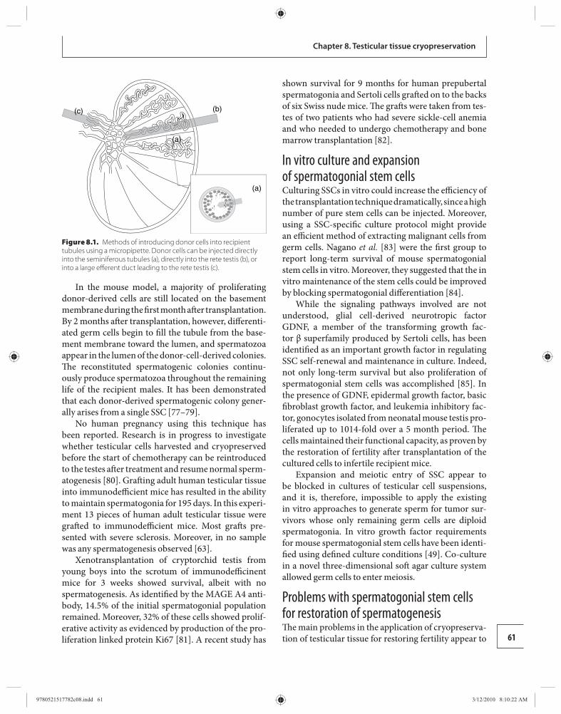
61
Chapter 8. Testicular tissue cryopreservation
In the mouse model, a majority of proliferating donor-derived cells are still located on the basement membrane during the fi rst month aft er transplantation. By 2 months aft er transplantation, however, diff erenti-ated germ cells begin to fi ll the tubule from the base-ment membrane toward the lumen, and spermatozoa appear in the lumen of the donor-cell-derived colonies. Th e reconstituted spermatogenic colonies continu-ously produce spermatozoa throughout the remaining life of the recipient males. It has been demonstrated that each donor-derived spermatogenic colony gener-ally arises from a single SSC [ 77 – 79 ].
No human pregnancy using this technique has been reported. Research is in progress to investigate whether testicular cells harvested and cryopreserved before the start of chemotherapy can be reintroduced to the testes aft er treatment and resume normal sperm-atogenesis [ 80 ]. Graft ing adult human testicular tissue into immunodeffi cient mice has resulted in the ability to maintain spermatogonia for 195 days. In this experi-ment 13 pieces of human adult testicular tissue were graft ed to immunodeffi cient mice. Most graft s pre-sented with severe sclerosis. Moreover, in no sample was any spermatogenesis observed [ 63 ].
Xenotransplantation of cryptorchid testis from young boys into the scrotum of immunodeffi cinent mice for 3 weeks showed survival, albeit with no sperm atogenesis. As identifi ed by the MAGE A4 anti-body, 14.5% of the initial spermatogonial population remained. Moreover, 32% of these cells showed prolif-erative activity as evidenced by production of the pro-liferation linked protein Ki67 [ 81 ]. A recent study has
shown survival for 9 months for human prepubertal spermatogonia and Sertoli cells graft ed on to the backs of six Swiss nude mice. Th e graft s were taken from tes-tes of two patients who had severe sickle-cell anemia and who needed to undergo chemotherapy and bone marrow transplantation [ 82 ] .
In vitro culture and expansion of spermatogonial stem cells Culturing SSCs in vitro could increase the effi ciency of the transplantation technique dramatically, since a high number of pure stem cells can be injected. Moreover, using a SSC-specifi c culture protocol might provide an effi cient method of extracting malignant cells from germ cells. Nagano et al. [ 83 ] were the fi rst group to report long-term survival of mouse spermatogonial stem cells in vitro. Moreover, they suggested that the in vitro maintenance of the stem cells could be improved by blocking spermatogonial diff erentiation [ 84 ].
While the signaling pathways involved are not understood, glial cell-derived neurotropic factor GDNF, a member of the transforming growth fac-tor β superfamily produced by Sertoli cells, has been identifi ed as an important growth factor in regulating SSC self-renewal and maintenance in culture. Indeed, not only long-term survival but also proliferation of spermatogonial stem cells was accomplished [ 85 ]. In the presence of GDNF, epidermal growth factor, basic fi broblast growth factor, and leukemia inhibitory fac-tor, gonocytes isolated from neonatal mouse testis pro-liferated up to 1014-fold over a 5 month period. Th e cells maintained their functional capacity, as proven by the restoration of fertility aft er transplantation of the cultured cells to infertile recipient mice.
Expansion and meiotic entry of SSC appear to be blocked in cultures of testicular cell suspensions, and it is, therefore, impossible to apply the existing in vitro approaches to generate sperm for tumor sur-vivors whose only remaining germ cells are diploid sperm atogonia. In vitro growth factor requirements for mouse spermatogonial stem cells have been identi-fi ed using defi ned culture conditions [ 49 ]. Co-culture in a novel three-dimensional soft agar culture system allowed germ cells to enter meiosis.
Problems with spermatogonial stem cells for restoration of spermatogenesis Th e main problems in the application of cryopreserva-tion of testicular tissue for restoring fertility appear to
(c) (b)
(a)
(a)
Figure 8.1. Methods of introducing donor cells into recipient tubules using a micropipette. Donor cells can be injected directly into the seminiferous tubules (a), directly into the rete testis (b), or into a large eff erent duct leading to the rete testis (c).
9780521517782c08.indd 619780521517782c08.indd 61 3/12/2010 8:10:22 AM3/12/2010 8:10:22 AM
62
Section 2. Cryopreservation of sperm and testicular tissue
be the small number of SSCs present in the testicular biopsy and the potential for contamination by malig-nant cells. In mice, it is estimated that only two stem cells are present in 10 000 germ cells [ 86 ]. Although this ratio may be higher in humans, the low number of SSCs makes this procedure very ineffi cient for fertility preservation in prepubertal boys [ 87 ]. Th e low number of SSCs in the testis necessitates an eff ective purifi ca-tion and isolation technique. Eff ective purifi cation will most likely require specifi c antibody probes to distin-guish SSCs from other cells [ 88 ].
A second serious problem is the risk of contamina-tion by malignant cells. In animal models even as few as 20 leukemic cells can cause a malignant recurrence in the recipient [ 89 , 90 ]. In humans, the threshold for malignant contamination is not known. As the major-ity of pediatric malignancies are capable of metastasiz-ing through blood, a signifi cant risk of contamination of the collected testicular tissue must be considered [ 91 ]. No technique currently exists for the separation of testicular cells from malignant cells. Neither mag-netic activated cell sorting nor fl uorescence activated cell sorting appears to be adequate for complete deple-tion of malignant cells from testicular tissue.
Molecular techniques can currently detect one sin-gle cancer cell among 10 million normal human cells [ 92 , 93 ]. Although more research on alternative decon-tamination techniques is obviously necessary, devel-oping a reliable method to screen a priori testicular tissue for malignant cells could be equally import ant [ 90 ]. Th erefore, transplantation of cells into the testis in malignancies that metastasize through blood should not yet be performed. Patients treated by chemo-therapy for non-malignant hematological disease are obviously the fi rst candidates for transplantation of frozen–thawed testicular tissue. In a mouse lymphoma model, cancer cells were eliminated from the testis-cell suspension by fl uorescence activated cell sorting before transplantation [ 94 ].
Th e ethical aspect of this technique is quite intri-cate. Off ering clinical treatment to a child requires consent from his parents and when the benefi t to the child is clear, it is justifi able. When, however, a surgi-cal procedure is off ered for an experimental approach, this becomes much more controversial. Th e advan-tages and disadvantages of ovarian cryopreservation for young girls with cancer prior to chemotherapy have been investigated and documented [ 95 ]. Th e issue of informed consent is quite diffi cult in young children. Guidelines are required to ensure that this procedure
is applied correctly. Th e rights of parents to consent to, or even request, a procedure that is still at the experi-mental stage need to be addressed. A multidisciplinary team familiar with these issues should be involved and decisions need to be made on an individual basis.
Future possibilities for restoring fertility Future possible methods of restoring fertility might include the derivation of mature sperm cells from human embryonic stem cells [ 96 ]. Embryoid bodies were shown to support maturation of the primordial germ cells into haploid male gametes, which when injected into oocytes round off the somatic diploid chromosome complement and develop into blastocysts [ 97 ]. Moreover, the pluripotent properties of SSCs have recently enabled the establishment of human adult germline stem cells derived from spermatogonial cells of adult human testis. Th e cellular and molecular char-acterization of these cells revealed many similarities to human embryonic stem cells [ 98 ].
Conclusions Spermatogonial stem cells are the viable stem cells that can transmit genetic information to the off spring, and their enormous proliferation contributes to fer-tilization throughout the male lifespan. Although the process of spermatogenesis is relatively effi cient and resistant to damage, male infertility can result from exposure to toxic agents such as chemotherapeutic regi mens, radiation, or occupational chemicals. Cancer and its treatment commonly cause male infertility and thus impair the chances of post-treatment paternity. Th e eff ect of the treatment on future fertility should be discussed with the patient prior to choosing and ini-tiating treatment for cancer. Th e various options are detailed in Figure 8.2 . Moreover, preventive measures should be considered in a multidisciplinary coopera-tion with oncologists, urologists, andrologists, and fer-tility specialists.
Th e fi rst option for fertility preservation is pretreat-ment sperm cell cryopreservation. With developing technology in mind, today’s experimental techniques of harvesting gonadal tissue can be considered in young males; however, it is important to avoid giving these patients and their parents unrealistic expecta-tions of future fertility. Testicular tissue or spermato-gonial cryopreservation and transplantation or testis xenograft ing are in the early phase of experimentation
9780521517782c08.indd 629780521517782c08.indd 62 3/12/2010 8:10:23 AM3/12/2010 8:10:23 AM
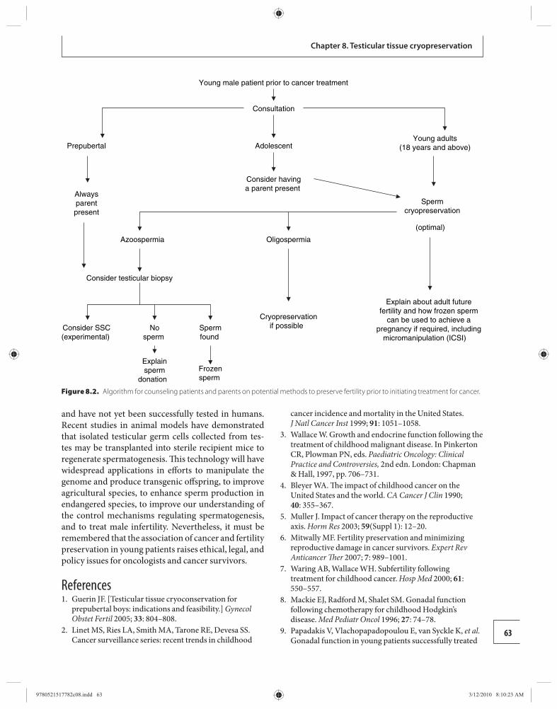
63
Chapter 8. Testicular tissue cryopreservation
and have not yet been successfully tested in humans. Recent studies in animal models have demonstrated that isolated testicular germ cells collected from tes-tes may be transplanted into sterile recipient mice to regenerate spermatogenesis. Th is technology will have widespread applications in eff orts to manipulate the genome and produce transgenic off spring, to improve agricultural species, to enhance sperm production in endangered species, to improve our understanding of the control mechanisms regulating spermatogenesis, and to treat male infertility. Nevertheless, it must be remembered that the association of cancer and fertility preservation in young patients raises ethical, legal, and policy issues for oncologists and cancer survivors.
References 1. Guerin JF . [ Testicular tissue cryoconservation for
prepubertal boys: indications and feasibility. ] Gynecol Obstet Fertil 2005; 33 : 804–808.
2. Linet MS , Ries LA , Smith MA , Tarone RE , Devesa SS . Cancer surveillance series: recent trends in childhood
cancer incidence and mortality in the United States . J Natl Cancer Inst 1999; 91 : 1051 –1058.
3. Wallace W . Growth and endocrine function following the treatment of childhood malignant disease. In Pinkerton CR , Plowman PN , eds. Paediatric Oncology: Clinical Practice and Controversies, 2nd edn. London: Chapman & Hall, 1997, pp. 706–731.
4. Bleyer WA . Th e impact of childhood cancer on the United States and the world . CA Cancer J Clin 1990; 40 : 355 –367.
5. Muller J . Impact of cancer therapy on the reproductive axis . Horm Res 2003; 59 (Suppl 1): 12 –20.
6. Mitwally MF . Fertility preservation and minimizing reproductive damage in cancer survivors . Expert Rev Anticancer Th er 2007; 7 : 989 –1001.
7. Waring AB , Wallace WH . Subfertility following treatment for childhood cancer . Hosp Med 2000; 61 : 550 –557.
8. Mackie EJ , Radford M , Shalet SM . Gonadal function following chemotherapy for childhood Hodgkin ’ s disease . Med Pediatr Oncol 1996; 27 : 74 –78.
9. Papadakis V , Vlachopapadopoulou E , van Syckle K , et al. Gonadal function in young patients successfully treated
Young male patient prior to cancer treatment
Consultation
AdolescentPrepubertalYoung adults
(18 years and above)
Alwaysparentpresent
Consider havinga parent present
Consider testicular biopsy
Consider SSC(experimental)
Nosperm
Explainsperm
donation
Spermfound
Frozensperm
Spermcryopreservation
(optimal)
Azoospermia Oligospermia
Cryopreservationif possible
Explain about adult futurefertility and how frozen sperm
can be used to achieve apregnancy if required, including
micromanipulation (ICSI)
Figure 8.2. Algorithm for counseling patients and parents on potential methods to preserve fertility prior to initiating treatment for cancer.
9780521517782c08.indd 639780521517782c08.indd 63 3/12/2010 8:10:23 AM3/12/2010 8:10:23 AM
64
Section 2. Cryopreservation of sperm and testicular tissue
for Hodgkin disease . Med Pediatr Oncol 1999; 32 : 366 –372.
10. Whitehead E , Shalet SM , Jones PH , Beardwell CG , Deakin DP . Gonadal function aft er combination chemotherapy for Hodgkin’s disease in childhood . Arch Dis Child 1982; 57 : 287 –291.
11. Shalet SM , Hann IM , Lendon M , et al. Testicular function aft er combination chemotherapy in childhood for acute lymphoblastic leukaemia . Arch Dis Child 1981; 56 : 275 –278.
12. Wallace WH , Shalet SM , Lendon M , Morris-Jones PH . Male fertility in long-term survivors of childhood acute lymphoblastic leukaemia . Int J Androl 1991; 14 : 312 –319.
13. Wallace WH , Shalet SM , Crowne EC , et al. Gonadal dysfunction due to cis-platinum . Med Pediatr Oncol 1989; 17 : 409 –413.
14. Watson AR , Rance CP , Bain J . Long term eff ects of cyclophosphamide on testicular function . Br Med J 1985; 291 : 1457 –1460.
15. Heikens J , Behrendt H , Adriaanse R , Berghout A . Irreversible gonadal damage in male survivors of pediatric Hodgkin’s disease . Cancer 1996; 78 : 2020 –2024.
16. Viviani S , Santoro A , Ragni G , et al. Gonadal toxicity aft er combination chemotherapy for Hodgkin’s disease. Comparative results of MOPP vs ABVD . Eur J Cancer Clin Oncol 1985; 21 : 601 –605.
17. da Cunha MF , Meistrich ML , Fuller LM , et al. Recovery of spermatogenesis aft er treatment for Hodgkin’s disease: limiting dose of MOPP chemotherapy . J Clin Oncol 1984; 2 : 571 –577.
18. Trottmann M , Becker AJ , Stadler T , et al. Semen quality in men with malignant diseases before and aft er therapy and the role of cryopreservation . Eur Urol 2007; 52 : 355 –367.
19. Howell SJ , Shalet SM . Spermatogenesis aft er cancer treatment: damage and recovery . J Natl Cancer Inst 2005; 34 : 12 –17.
20. Kenney LB , Laufer MR , Grant FD , Grier H , Diller L . High risk of infertility and long term gonadal damage in males treated with high dose cyclophosphamide for sarcoma during childhood . Cancer 2001; 91 : 613 –621.
21. Meistrich ML , Wilson G , Brown BW , da Cunha MF , Lipshultz LI . Impact of cyclophosphamide on long-term reduction in sperm count in men treated with combination chemotherapy for Ewing and soft tissue sarcomas . Cancer 1992; 70 : 2703 –2712.
22. Viviani S , Ragni G , Santoro A , et al. Testicular dysfunction in Hodgkin’s disease before and aft er treatment . Eur J Cancer 1991; 27 : 1389 –1392.
23. Ben Arush MW , Solt I , Lightman A , Linn S , Kuten A . Male gonadal function in survivors of childhood Hodgkin and non-Hodgkin lymphoma . Pediatr Hematol Oncol 2000 ; 17 : 239 –245.
24. Rafsanjani KA , Faranoush M , Hedayatiasl AA , Vossough P . Gonadal function and fertility in male survivors treated for Hodgkin’s disease in Iran . Saudi Med J 2007; 28 : 1690 –1693.
25. Brougham MF , Kelnar CJ , Sharpe RM , Wallace WH . Male fertility following childhood cancer: current concepts and future therapies . Asian J Androl 2003; 5 : 325 –337.
26. Brougham MF , Wallace WH . Subfertility in children and young people treated for solid and haematological malignancies . Br J Haematol 2005; 131 : 143 –155.
27. Kliesch S , Behre HM , Jurgens H , Nieschlag E . Cryopreservation of semen from adolescent patients with malignancies . Med Pediatr Oncol 1996; 26 : 20 –27.
28. Bahadur G , Ling KL , Hart R , et al. Semen quality and cryopreservation in adolescent cancer patients . Hum Reprod 2002; 17 : 3157 –3161.
29. Edge B , Holmes D , Makin G . Sperm banking in adolescent cancer patients . Arch Dis Child 2006; 91 : 149 –152.
30. Bahadur G , Ling KL , Hart R , et al. Semen production in adolescent cancer patients . Hum Reprod 2002; 17 : 2654 –2656.
31. Schmiegelow ML , Sommer P , Carlsen E , et al. Penile vibratory stimulation and electroejaculation before anticancer therapy in two pubertal boys . J Pediatr Hematol Oncol 1998; 20 : 429 –430.
32. Muller J , Sonksen J , Sommer P , et al. Cryopreservation of semen from pubertal boys with cancer . Med Pediatr Oncol 2000; 34 : 191 –194.
33. Rosenlund B , Sjoblom P , Tornblom M , Hultling C , Hillensjo T . In-vitro fertilization and intracytoplasmic sperm injection in the treatment of infertility aft er testicular cancer . Hum Reprod 1998; 13 : 414 –418.
34. Aboulghar MA , Mansour RT , Serour GI , et al. Fertilization and pregnancy rates aft er intracytoplasmic sperm injection using ejaculate semen and surgically retrieved sperm . Fertil Steril 1997; 68 : 108 –111.
35. Chen SU , Ho HN , Chen HF , et al. Pregnancy achieved by intracytoplasmic sperm injection using cryopreserved semen from a man with testicular cancer . Hum Reprod 1996; 11 : 2645 –2647.
36. Tesarik J , Mendoza C . Spermatid injection into human oocytes. I. Laboratory techniques and special features of zygote development . Hum Reprod 1996; 11 : 772 –779.
37. Tesarik J , Mendoza C , Testart J . Viable embryos from injection of round spermatids into oocytes . N Engl J Med 1995; 333 : 525.
38. Fishel S , Green S , Hunter A , et al. Human fertilization with round and elongated spermatids . Hum Reprod 1997; 12 : 336 –340.
39. Sofi kitis N , Mantzavinos T , Loutradis D , et al. Ooplasmic injections of secondary spermatocytes for non-obstructive azoospermia . Lancet 1998; 351 : 1177 –1178.
9780521517782c08.indd 649780521517782c08.indd 64 3/12/2010 8:10:23 AM3/12/2010 8:10:23 AM
65
Chapter 8. Testicular tissue cryopreservation
40. Vanderzwalmen P , Zech H , Birkenfeld A , et al. Intracytoplasmic injection of spermatids retrieved from testicular tissue: infl uence of testicular pathology, type of selected spermatids and oocyte activation . Hum Reprod 1997; 12 : 1203 –1213.
41. Bernabeu R , Cremades N , Takahashi K , Sousa M . Successful pregnancy aft er spermatid injection . Hum Reprod (Oxford) 1998; 13 : 1898 –1900.
42. Lee SJ , Schover LR , Partridge AH , et al. American Society of Clinical Oncology recommendations on fertility preservation in cancer patients . J Clin Oncol 2006; 24 : 2917 –2931.
43. Allen C , Keane D , Harrison RF . A survey of Irish consultants regarding awareness of sperm freezing and assisted reproduction . Irish Med J 2003; 96 : 23 –25.
44. Schover LR , Rybicki LA , Martin BA , Bringelsen KA . Having children aft er cancer. A pilot survey of survivors ’ attitudes and experiences . Cancer 1999; 86 : 697 –709.
45. Hartmann JT , Albrecht C , Schmoll HJ , et al. Long-term eff ects on sexual function and fertility aft er treatment of testicular cancer . Br J Cancer 1999; 80 : 801 –807.
46. Rieker PP , Fitzgerald EM , Kalish LA , et al. Psychosocial factors, curative therapies, and behavioral outcomes. A comparison of testis cancer survivors and a control group of healthy men . Cancer 1989; 64 : 2399 –2407.
47. Rieker PP , Fitzgerald EM , Kalish LA . Adaptive behavioral responses to potential infertility among survivors of testis cancer . J Clin Oncol 1990; 8 : 347 –355.
48. Saito K , Suzuki K , Iwasaki A , Yumura Y , Kubota Y . Sperm cryopreservation before cancer chemotherapy helps in the emotional battle against cancer . Cancer 2005; 104 : 521 –524.
49. Kubota H , Brinster RL . Technology insight: in vitro culture of spermatogonial stem cells and their potential therapeutic uses . Natl Clin Pract Endocrinol Metab 2006; 2 : 99 –108.
50. Brinster RL , Avarbock MR . Germline transmission of donor haplotype following spermatogonial transplantation . Proc Natl Acad Sci USA 1994; 91 : 11303 –11307.
51. Brinster RL , Zimmermann JW . Spermatogenesis following male germ-cell transplantation . Proc Natl Acad Sci USA 1994; 91 : 11298 –11302.
52. Muller T , Eildermann K , Dhir R , Schlatt S , Behr R . Glycan stem-cell markers are specifi cally expressed by spermatogonia in the adult non-human primate testis . Hum Reprod 2008; 23 : 2292 –2298.
53. Brook PF , Radford JA , Shalet SM , Joyce AD , Gosden RG . Isolation of germ cells from human testicular tissue for low temperature storage and autotransplantation . Fertil Steril 2001; 75 : 269 –274.
54. Keros V , Hultenby K , Borgstrom B , et al. Methods of cryopreservation of testicular tissue with viable spermatogonia in pre-pubertal boys undergoing
gonadotoxic cancer treatment . Hum Reprod 2007; 22 : 1384 –1395.
55. Keros V , Rosenlund B , Hultenby K , et al. Optimizing cryopreservation of human testicular tissue: comparison of protocols with glycerol, propanediol and dimethylsulphoxide as cryoprotectants . Hum Reprod 2005; 20 : 1676 –1687.
56. Lyrakou S , Mantas D , Msaouel P , et al. Crossover analysis using immunofl uorescent detection of MLH1 foci in frozen–thawed testicular tissue . Reprod Biomed Online 2007; 15 : 99 –105.
57. Li J , Leng M , Ma T , Yu D , Shi H , Shi Q . Cryopreservation has no eff ect on meiotic recombination and synapsis in testicular tissues . Fertil Steril 2009 ; 91: 2734–2763 .
58. Bahadur G , Ralph D . Gonadal tissue cryopreservation in boys with paediatric cancers . Hum Reprod 1999; 14 : 11 –17.
59. Clouthier DE , Avarbock MR , Maika SD , Hammer RE , Brinster RL . Rat spermatogenesis in mouse testis . Nature 1996; 381 : 418 –421.
60. Schlatt S , Foppiani L , Rolf C , Weinbauer GF , Nieschlag E . Germ cell transplantation into X-irradiated monkey testes . Hum Reprod 2002; 17 : 55 –62.
61. Honaramooz A , Snedaker A , Boiani M , et al. Sperm from neonatal mammalian testes graft ed in mice . Nature 2002; 418 : 778 –781.
62. Schlatt S , Honaramooz A , Boiani M , Scholer HR , Dobrinski I . Progeny from sperm obtained aft er ectopic graft ing of neonatal mouse testes . Biol Reprod 2003; 68 : 2331 –2335.
63. Geens M , de Block G , Goossens E , et al. Spermatogonial survival aft er graft ing human testicular tissue to immunodefi cient mice . Hum Reprod 2006; 21 : 390 –396.
64. Schlatt S , Kim SS , Gosden R . Spermatogenesis and steroidogenesis in mouse, hamster and monkey testicular tissue aft er cryopreservation and heterotopic graft ing to castrated hosts . Reproduction 2002; 124 : 339 –346.
65. Honaramooz A , Li MW , Penedo MC , Meyers S , Dobrinski I . Accelerated maturation of primate testis by xenograft ing into mice . Biol Reprod 2004; 70 : 1500 –1503.
66. Wistuba J , Mundry M , Luetjens CM , Schlatt S . Cograft ing of hamster (Phodopus sungorus) and marmoset (Callithrix jacchus) testicular tissues into nude mice does not overcome blockade of early spermatogenic diff erentiation in primate graft s . Biol Reprod 2004; 71 : 2087 –2091.
67. de Rooij DG . Proliferation and diff erentiation of spermatogonial stem cells . Reproduction 2001; 121 : 347 –354.
68. Brinster RL . Germline stem cell transplantation and transgenesis . Science 2002; 296 : 2174 –2176.
69. Naughton CK , Jain S , Strickland AM , Gupta A , Milbrandt J . Glial cell-line derived neurotrophic
9780521517782c08.indd 659780521517782c08.indd 65 3/12/2010 8:10:23 AM3/12/2010 8:10:23 AM
66
Section 2. Cryopreservation of sperm and testicular tissue
factor-mediated RET signaling regulates spermatogonial stem cell fate . Biol Reprod 2006; 74 : 314 –321.
70. Lee J , Kanatsu-Shinohara M , Inoue K , et al. Akt mediates self-renewal division of mouse spermatogonial stem cells . Development 2007; 134 : 1853 –1859.
71. Ogawa T , Arechaga JM , Avarbock MR , Brinster RL . Transplantation of testis germinal cells into mouse seminiferous tubules . Int J Dev Biol 1997; 41 : 111 –122.
72. Radford J , Shalet S , Lieberman B . Fertility aft er treatment for cancer: questions remain over ways of preserving ovarian and testicular tissue . Br Med J 1999; 319 : 935 –936.
73. Muller J , Skakkebaek NE . Quantifi cation of germ cells and seminiferous tubules by stereological examination of testicles from 50 boys who suff ered from sudden death . Int J Androl 1983; 6 : 143 –156.
74. Nagano MC . Homing effi ciency and proliferation kinetics of male germ line stem cells following transplantation in mice . Biol Reprod 2003; 69 : 701 –707.
75. Ogawa T , Ohmura M , Yumura Y , Sawada H , Kubota Y . Expansion of murine spermatogonial stem cells through serial transplantation . Biol Reprod 2003; 68 : 316 –322.
76. Wang X , Ding Q , Zhang Y , et al. Two allogeneic descendents derived from the high-dose busulfan-treated infertile mouse model aft er freeze–thawed spermatogonial stem cell transplantation . Fertil Steril 2008; 90 : 1538 –1549.
77. Dobrinski I , Ogawa T , Avarbock MR , Brinster RL . Computer assisted image analysis to assess colonization of recipient seminiferous tubules by spermatogonial stem cells from transgenic donor mice . Mol Reprod Dev 1999; 53 : 142 –148.
78. Nagano M , Avarbock MR , Brinster RL . Pattern and kinetics of mouse donor spermatogonial stem cell colonization in recipient testes . Biol Reprod 1999; 60 : 1429 –1436.
79. Zhang X , Ebata KT , Nagano MC . Genetic analysis of the clonal origin of regenerating mouse spermatogenesis following transplantation . Biol Reprod 2003; 69 : 1872 –1878.
80. Radford J . Restoration of fertility aft er treatment for cancer . Hormone Res 2003; 59 (Suppl 1): 21 –23.
81. Wyns C , Curaba M , Martinez-Madrid B , et al. Spermatogonial survival aft er cryopreservation and short-term orthotopic immature human cryptorchid testicular tissue graft ing to immunodefi cient mice . Hum Reprod 2007; 22 : 1603 –1611.
82. Goossens E , Geens M , de Block G , Tournaye H . Spermatogonial survival in long-term human prepubertal xenograft s . Fertil Steril 2008; 24 : 2019–2022 .
83. Nagano M , Avarbock MR , Leonida EB , Brinster CJ , Brinster RL . Culture of mouse spermatogonial stem cells . Tissue Cell 1998; 30 : 389 –397.
84. Nagano M , Ryu BY , Brinster CJ , Avarbock MR , Brinster RL . Maintenance of mouse male germ line stem cells in vitro . Biol Reprod 2003; 68 : 2207 –2214.
85. Kanatsu-Shinohara M , Ogonuki N , Inoue K , et al. Restoration of fertility in infertile mice by transplantation of cryopreserved male germline stem cells . Hum Reprod 2003; 18 : 2660 –2667.
86. Tegelenbosch RA , de Rooij DG . A quantitative study of spermatogonial multiplication and stem cell renewal in the C3H/101 F1 hybrid mouse . Mut Res 1993; 290 : 193 –200.
87. Aslam I , Fishel S , Moore H , Dowell K , Th ornton S . Fertility preservation of boys undergoing anti-cancer therapy: a review of the existing situation and prospects for the future . Hum Reprod 2000; 15 : 2154 –2159.
88. Aslam I , Robins A , Dowell K , Fishel S . Isolation, purifi cation and assessment of viability of spermatogenic cells from testicular biopsies of azoospermic men . Hum Reprod 1999; 13 : 639 –645.
89. Jahnukainen K , Hou M , Petersen C , Setchell B , Soder O . Intratesticular transplantation of testicular cells from leukemic rats causes transmission of leukemia . Cancer Res 2001; 61 : 706 –710.
90. Geens M , van de Velde H , de Block G , et al. Th e effi ciency of magnetic-activated cell sorting and fl uorescence-activated cell sorting in the decontamination of testicular cell suspensions in cancer patients . Hum Reprod 2007; 22 : 733 –742.
91. Jahnukainen K , Ehmcke J , Soder O , Schlatt S . Clinical potential and putative risks of fertility preservation in children utilizing gonadal tissue or germline stem cells . Pediatric Res 2006; 59 : 40R– 47R.
92. Willemse MJ , Seriu T , Hettinger K , et al. Detection of minimal residual disease identifi es diff erences in treatment response between T-ALL and precursor B-ALL . Blood 2002; 99 : 4386 –4393.
93. Jolkowska J , Derwich K , Dawidowska M . Methods of minimal residual disease (MRD) detection in childhood haematological malignancies . J Appl Genet 2007; 48 : 77 –83.
94. Fujita K , Ohta H , Tsujimura A , et al. Transplantation of spermatogonial stem cells isolated from leukemic mice restores fertility without inducing leukemia . J Clin Invest 2005; 115 : 1855 –1861.
95. Weintraub M , Gross E , Kadari A , et al. Should ovarian cryopreservation be off ered to girls with cancer . Pediatr Blood Cancer 2007; 48 : 4 –9.
96. Ko K , Scholer HR . Embryonic stem cells as a potential source of gametes . Sem Reprod Med 2006; 24 : 322 –329.
97. Geijsen N , Horoschak M , Kim K , et al. Derivation of embryonic germ cells and male gametes from embryonic stem cells . Nature 2004; 427 : 148 –154.
98. Conrad S , Renninger M , Hennenlotter J , et al. Generation of pluripotent stem cells from adult human testis . Nature 2008; 456 : 344–349 [errata in Nature 2009; 460 : 1044].
9780521517782c08.indd 669780521517782c08.indd 66 3/12/2010 8:10:23 AM3/12/2010 8:10:23 AM
Fertility Cryopreservation, ed. Ri-Cheng Chian and Patrick Quinn. Published by Cambridge University Press. Copyright © Cambridge University Press 2010.
Section 3Chapter
9 demonstrated that restricting the number of embryos transferred aft er IVF to two could reduce the risk of multiple births without signifi cantly aff ecting the pregnancy rate [ 6 ]. Th e practice of transferring only two fresh embryos was, however, already under scru-tiny as the risks associated with twin pregnancies were emphasized, and the use of single embryo transfer was soon advocated [ 7 ] and subsequently justifi ed on the basis of both health and economic considerations [ 8 ]. In our unit in Melbourne, for the period between 2004 and 2007, transfer of two embryos (on day 2 of development) to a woman under 36 resulted in a clin-ical pregnancy (at least one fetal heartbeat) rate of 37% and a multigestation pregnancy rate of 36%. Over the same period, and in the same age group, the transfer of a single day 2 embryo resulted in a clinical pregnancy rate of 31% but a multigestation pregnancy rate of only 1.3%. Currently in our unit, 76% of patients in this age group undergo single embryo transfer. Maximizing the effi ciency of ART, however, requires that the preg-nancy potential of the embryos that are not transferred is retained.
Avoiding multigestation pregnancy while maintaining the effi ciency of assisted reproduction It was recognized from the early days of IVF/ET that many of the potential problems associated with the simultaneous transfer of multiple embryos could be circumvented if the non-transferred embryos could be stored for future use. Pregnancies and live births from human cryopreserved IVF embryos [ 9 , 10 ] provided proof of principle and led to the adoption of embryo cryopreservation as a routine adjunct to IVF/ET. When surplus embryos are cryopreserved in conjunction with the transfer of fresh embryos, the practicalities of evaluating success rates from oocyte retrieval cycles
The role of embryo cryopreservation in fertility management Evolution of clinical-assisted reproduction Th e fi rst reported observations of the genesis and early development of the human embryo ex vivo [ 1 ] initiated a chain of events that revolutionized the management of human subfertility and laid the foundations of modern reproductive medicine. Although the fi rst clinical preg-nancies from in vitro fertilization and embryo transfer (IVF/ET) were obtained using oocytes retrieved in the natural menstrual cycle [ 2 ], subsequent successes in controlled ovulatory cycles [ 3 ] led to the widespread adoption of ovarian stimulation as a routine compo-nent of assisted reproductive technology (ART). Th e availability of multiple mature oocytes obtained from a single cycle of ovarian stimulation inevitably increased the probability of establishing a clinical pregnancy. It not only allowed selection of oocytes that could undergo normal fertilization and development, it also allowed in many cases the transfer of multiple cleaving embryos in attempts to overcome the frequently disap-pointing implantation rates observed in the early days of IVF/ET.
Th e advantages associated with the availability and transfer of multiple embryos were, however, off -set by the incidence of multigestation pregnancies. Multigestation pregnancy, while considered a poten-tial bonus by many patients who had experienced long periods of infertility, is widely recognized as a major clinical complication of ART [ 4 ], with associ-ated healthcare cost implications [ 5 ]. As success rates from IVF/ET increased, much attention was turned to developing treatment strategies designed to min-imize the frequency of multiple gestation. By the end of the 1990s, many groups had reduced the number of fresh embryos being transferred and it had been
Cryopreservation of embryos Cryopreservation of embryos: an overview David H. Edgar and Debra A. Gook
67
9780521517782c09.indd 679780521517782c09.indd 67 3/12/2010 8:13:54 AM3/12/2010 8:13:54 AM
68
Section 3. Cryopreservation of embryos
must be considered and account taken of pregnancies that result from subsequent transfer of thawed embryos [ 11 ]. Th is is true when assessing the impact of cryop-reservation on pregnancy and multigestation rates in circumstances where more than one fresh embryo is transferred [ 12 ], but the value of cryopreservation is most evident when assessed in the context of single embryo transfer [ 13 ]. Th e importance of assessing the cumulative pregnancy rate from the use of fresh and cryopreserved embryos is emphasized when compar-ing outcomes from single or double embryo transfer in fresh cycles [ 14 , 15 ]. It is now clear that a success-ful embryo cryopreservation program allows the confi dent introduction of single embryo transfer into clinical practice in the knowledge that the incidence of multigestation pregnancy will be reduced while the cumulative chance of a pregnancy will remain essen-tially unaff ected.
Although not all cryopreserved embryos will sur-vive freezing and thawing (see below), the pregnancy potential of any individual embryo that is already cryo-preserved will not be aff ected by extending the period of storage. Rationally, this should then lead to a further reduction in the risk of multigestation by the adoption of single embryo transfer in cycles involving transfer of cryopreserved embryos [ 16 , 17 ]. Th e role of embryo cry-opreservation in maximizing the safety and effi ciency of ART is highlighted in our unit by the fact that 52% of all embryo transfer procedures involve stored embryos and 42% of all the implantations arising from embryo transfer can be attributed to cryopreserved embryos. Th e important role that embryo cryopreserva tion plays in ART is further emphasized when we consider the fact that many patients will return for the transfer of cryopreserved embryos aft er the birth of one or more children from the same oocyte collection. In our unit, 78% of all women who give birth as a result of a fresh embryo transfer have the prospect of returning for the transfer of stored embryos. In many cases, therefore, families may be completed without the need for further surgical procedures.
Other roles for embryo cryopreservation Embryo cryopreservation is also an essential tool when embryo transfer is not possible in the cycle of oocyte collection. While this situation may occur for a variety of reasons, the most common consideration is the risk of ovarian hyperstimulation syndrome. In such cases, where pregnancy would be expected to exacerbate the symptoms, routine management oft en involves the
cryopreservation of all the embryos generated in the cycle and replacement in subsequent cycles when ovar-ian hyperstimulation syndrome is no longer an issue [ 18 , 19 ].
Of increasing interest is the possibility that repro-ductive potential can be stored for young women at risk of loss of ovarian function as a consequence of cyto-toxic therapies used to eliminate malignant disease. In situations where there is no male partner, the emerging technology of oocyte and ovarian tissue cryopreserva-tion off ers some hope of future fertility for the increas-ing numbers of cancer survivors. Where there is a male partner, however, and where there is adequate time available to undergo ovarian stimulation and oocyte collection prior to commencing therapy, the estab-lished and predictable outcomes from embryo cryo-preservation may, at present, off er the most realistic prospect of post-cure pregnancy.
Limitations Th e value of embryo cryopreservation in all the above circumstances is, of course, predicated on the ability of a suffi cient number of embryos to survive the cryo-preservation process without signifi cant impairment of their ability to establish a normal pregnancy. Th is is the challenge that has been faced by the cryobiologists and embryologists.
Methodology for embryo cryopreservation Although it has been possible to cryopreserve animal and human sperm successfully since the 1950s, it was not until the 1970s that similar success was obtained with mammalian embryos. It had been demonstrated that water-miscible substances known as cryoprotect-ants, which removed much of the intracellular water, were required to overcome some of the problems asso-ciated with ice formation when freezing living cells. Glycerol had been used successfully in this context when cryopreserving semen but it was the introduc-tion of the more rapidly permeating cryoprotectant dimethyl sulfoxide (DMSO) that enabled successful cryopreservation of mouse embryos [ 20 ]. Translation of this method to early cleavage stage human embryos resulted in the fi rst reports of successful pregnancy and birth from human cryopreserved embryos [ 9 , 10 ]. Use of DMSO was soon replaced by 1,2 propanediol (PROH) as the permeating cryoprotectant of choice when cryo-preserving at pronucleate or early cleavage stages using
9780521517782c09.indd 689780521517782c09.indd 68 3/12/2010 8:13:55 AM3/12/2010 8:13:55 AM
Chapter 9. Cryopreservation of embryos: an overview
69
protocols that also included sucrose to increase intrac-ellular dehydration prior to freezing [ 21 , 22 ]. Although this was initially achieved by sequential exposure to PROH and sucrose, we have found subsequently that single-step dehydration is at least as eff ective for embryos on day 2 of development. Despite the still pre-dominant use of PROH, encour aging results have also been reported using ethylene glycol as a permeating cryoprotectant during slow cooling of human cleavage stage embryos [ 23 ].
Th e freezing protocols used for cryopreservation of early preimplantation development stages usually involve cooling of dehydrated embryos in sealed plastic straws at approximately −2°C/min, manual nucleation of ice formation (seeding) at around −7°C, and further slow cooling at approximately −0.3°C/min to −30°C before storage in liquid nitrogen. Following rapid thawing, the use of higher extracellular concentrations of sucrose during the early stages of post-thaw rehy-dration can act as a regulator of the rate at which water enters the cells in response to decreasing extracellu-lar PROH concentrations. In our experience, gradual removal of PROH by serial dilution does not seem to be necessary if a suffi ciently high concentration of sucrose (0.5 mol/l) is used during the initial stage of thawing.
Although PROH has continued to be the most widely used permeating cryoprotectant for embryos at early stages of preimplantation development, the methodology used for the slow cooling of blastocyst stages of development has predominantly been based on the use of the less-permeable glycerol. Early reports of pregnancies following the frozen storage of expand-ing human blastocysts involved the stepwise addition of glycerol alone prior to freezing [ 24 ], and this method was subsequently modifi ed to include the addition of sucrose [ 25 ]. Rapid thawing and rehydration is usually performed according to the principles outlined above for early cleavage stage embryos. Again, there is a pau-city of controlled evidence to support the choice of cryoprotectant used, and successful cryopreservation of blastocyst stage embryos that had been biopsied for preimplantation genetic diagnosis at earlier cleavage stages has been reported using PROH [ 26 ]. Th e start temperature and cooling rate may also play a role in the outcome from slow freezing of blastocysts [ 27 ].
More recently, successful cryopreservation of embryos has been reported using the technique of vitrifi cation. Th is approach involves exposure to very high concentrations of cryoprotectants in very small volumes for very short periods of time, together with
extremely rapid cooling. Vitrifi cation results in tran-sition of water to a glass-like state without signifi cant formation of intracellular or extracellular ice crys-tals. Ultrarapid warming in the presence of elevated concentrations of non-permeating cryoprotectant (usually sucrose) results in rapid removal of the very high levels of intracellular permeating cryoprotectant before rehydration is completed by exposure to reduc-ing concentrations of non-permeating cryoprotectant. Although vitrifi cation of mammalian embryos was described more than 20 years ago [ 28 ], translation to human clinical application has only been evident more recently and has predominantly been associated with attempts to improve the outcome from cryopreserva-tion of blastocyst stage embryos [ 29 ].
Factors aff ecting the clinical effi ciency of embryo cryopreservation Selection of embryos for cryopreservation Th e inclusion criteria for selection of embryos for cryopreservation may diff er widely between units. As a result, the proportion of cycles in which cryopreser-vation occurs may vary from as low as 10% [ 30 ] to as high as 59%, and 73% in women under 36 years of age, in our unit. As discussed above, human embryos can be cryopreserved at all stages of preimplantation devel-opment from the unicellular pronucleate stage to the peri-implantation blastocyst stage, but the probability of implantation following transfer of cryopreserved embryos at any stage will also be dependent to a major extent on the quality of the embryo(s) transferred in addition to clinical factors such as female age at the time of oocyte collection and uterine receptivity at the time of embryo transfer.
A number of considerations will determine the stage at which cryopreservation occurs. In young women without partners who wish to preserve their fertility, unfertilized oocytes can be stored but the potential of individual oocytes to fertilize, develop, and implant will be relatively unknown. Th is approach is also the only option in all circumstances when legisla-tion precludes embryo cryopreservation, which was, but is no longer, the situation in Italy. When embryo cryopreservation is permitted, there is an inverse rela-tionship between the number of embryos stored and the extent to which selection criteria have been applied. Cryopreservation at the pronucleate stage guarantees that all stored oocytes have undergone fertilization and
9780521517782c09.indd 699780521517782c09.indd 69 3/12/2010 8:13:55 AM3/12/2010 8:13:55 AM
70
Section 3. Cryopreservation of embryos
this has been used in many cases where embryo trans-fer does not take place in the cycle of oocyte collection to avoid the risk of ovarian hyperstimulation syndrome [ 31 ]. Th is approach does not allow signifi cant prestor-age selection since the relationship between nuclear morphology and implantation potential remains con-troversial [ 32 , 33 ]. When applied in conjunction with fresh embryo transfer, pronucleate stage cryopreser-vation will also restrict the ability to select the highest quality embryo(s) for fresh transfer. It does, however, guarantee that all fertilized oocytes will be stored and is still a commonly used approach in countries where le gislation does not allow cryopreservation of later stages. Although slow cooling with PROH and sucrose was used for many years when freezing pronucleate stages, it has now been suggested that vitrifi cation may be a more appropriate technique [ 34 ].
Th e most common approach adopted for cryo-preservation of human embryos, to date, has been the slow cooling of cleavage stage embryos, although these stages have also been cryopreserved using vitrifi ca-tion [ 35 ]. Cryopreservation on either day 2 or day 3 of development is feasible, although there has been some suggestion that embryos may survive slow cooling better if frozen on day 2 [ 36 ]. In routine IVF/ET, this allows selection of embryos for fresh transfer on the basis of developmental rate and other morphologic al characteristics and also the establishment of min-imum criteria for cryopreservation. Stricter criteria for storage may improve implantation rates from thawed embryos but it is important to balance this considera-tion with the need to preserve the full potential of any cycle of ovarian stimulation [ 37 ]. Th is may be particu-larly important in cases of cryopreservation for fertil-ity issues prior to cytotoxic therapy. For example, many clinics will only cryopreserve an embryo on day 2 if it has reached the 4-cell stage of development. While it is clear that the implantation rate of 4-cell embryos frozen on day 2 is signifi cantly higher than those with fewer than 4 cells, a decision to exclude embryos with fewer than 4 cells will inevitably result in the discarding of some reproductive potential [ 17 , 38 ].
Embryo selection at cleavage stages can also poten-tially be enhanced by screening for genetic abnor-malities aft er embryo biopsy. Interestingly, although these embryos are more highly selected, their ability to survive standard cryopreservation techniques is sig-nifi cantly compromised [ 39 , 40 ]. However, modifi ed techniques involving an elevated sucrose concentra-tion [ 41 ] or a choline-based, sodium-free medium [ 42 ]
have been successful in improving the cryosurvival of biopsied embryos.
Whether slow cooled or vitrifi ed, the morphologic al features of a blastocyst stage embryo will also relate to the probability of implantation [ 43 ], and the choice of minimum criteria for blastocyst cryopreservation will impact on the outcome. Th ere are a number of reports, some of them confl icting, on the importance for cryo-preservation of degree of expansion, rate of develop-ment to the blastocyst stage, and reduction of the blastocelic cavity prior to cryopreservation; these have been reviewed by Youssry et al. [ 29 ]. Again stricter cri-teria for cryopreservation may improve apparent suc-cess rates at the expense of lost potential.
Cryodamage Having selected the embryos considered suitable for cryopreservation, the most important consideration becomes the possible detrimental eff ects of freezing and thawing on the embryo. Th e nature of cryodamage and its implications will vary between developmental stages. Early evidence of improved outcomes associated with cryopreservation at the pronucleate/zygote stage [ 44 ] led to adoption of the approach by many clinics, although the disadvantage of limiting selection of fresh embryos resulted in a move towards cryopreservation of early cleavage stages as results improved. However, there are still some countries in which embryos cannot be cryopreserved aft er nuclear envelope breakdown, leaving zygote cryopreservation as the main avail-able option. When cryopreservation is performed at the pronucleate stage, cryodamage can be assessed in terms of both survival and subsequent cleavage. Rates of survival of approximately 75% aft er slow cooling of human zygotes have been reported [ 18 , 31 ] in asso-ciation with cleavage rates similar to those of fresh embryos. An earlier comparative study of vitrifi cation and slow cooling of zygotes [ 45 ] demonstrated a signifi -cantly higher survival rate aft er vitrifi cation. However, post-recovery cleavage rates were compromised in both groups and were lower in vitrifi ed zygotes. More recently, a high (89%) survival rate has been reported aft er vitrifi cation of human zygotes [ 34 ]. In this case, post-recovery embryo development was reported to be normal, with the majority of embryos having reached the 4-cell stage with good morphology aft er 24 hours of culture. Damage to the integrity of pronuclei has also been reported aft er cryopreservation of pronuclear zygotes [ 46 ] and appears to be associated with reduced developmental potential.
9780521517782c09.indd 709780521517782c09.indd 70 3/12/2010 8:13:55 AM3/12/2010 8:13:55 AM
71
Chapter 9. Cryopreservation of embryos: an overview
Th e parameters associated with assessment of suc-cess in cryopreservation of cleavage stage embryos are also predominantly survival and subsequent develop-ment. Consequences of cryopreservation such as zona pellucida damage [ 47 ] and perturbation of metabolism [ 35 ] have also been reported. Assessment of survival of cleavage stage embryos is diff erent from that of pre-cleavage stages, where cell survival is all or nothing. Th e phenomenon of partial survival, whereby some, but not all, blastomeres survive, is interesting in itself. Th e implication may be that there are diff erences in indi-vidual, and ostensibly equivalent, blastomeres within the same embryo that impact on their ability to with-stand freezing and thawing. It is tempting to specu-late that these diff erences may relate to diff erences in membrane water permeability, such as those shown for mouse and human oocytes [ 48 ].
Th e widely accepted criterion for embryo survival and eligibility for transfer in a clinical situation is that a minimum of 50% of the original blastomeres sur-vive. However, partially intact thawed embryos have a reduced ability to develop to the blastocyst stage in vitro and result in blastocysts with reduced total cell numbers [ 49 ]. Loss of blastomeres has been shown clearly to result in reduced implantation rates by a number of groups [ 30 , 38 , 50 , 51 ] although the loss of a single blastomere at the 4-cell stage has no impact on implantation potential [ 17 ]. It has been claimed that the detrimental eff ect of blastomere loss may result from a negative infl uence of the necrotic blastomeres and that this may be reversed by removal of the necrotic mater-ial [ 52 ]. Th e proportion of cleavage stage embryos that survives fully intact aft er cryopreservation is not rou-tinely reported but should be in excess of 50% of all embryos [ 38 ].
Th is survival rate may be increased by the use of vit-rifi cation [ 35 ], and we have recently shown that a slow-cooling technique based on our previous work with biopsied embryos can result in 80% of non- biopsied cleavage stage embryos surviving fully intact [53]. Post-thaw resumption of mitosis is oft en used as an indica-tor of appropriate cryosurvival and has been shown to correlate with implantation potential [ 30 , 54 , 55 ], as has the number of blastomeres dividing during post-thaw culture [ 17 ]. Whether this parameter relates to cryo-damage or is a refl ection of intrinsic growth potential is not clear, but we have observed an increased frequency of post-thaw resumption of mi tosis in embryos that underwent an early fi rst cleavage during the pre-freeze culture period.
Assessment of cryodamage in blastocysts is more complex. Cell survival in the trophectoderm com-partment may be easier to assess than in the inner cell mass, where the individual cells are much smaller and oft en highly compacted, although the collapse of the the blastocelic cavity may preclude assessment of both compartments unless re-expansion occurs before transfer. In fact, the ability of a cryopreserved blastocyst to undergo re-expansion may, in itself, indicate functional survival. Assessment of resump-tion of development in cryopreserved blastocysts is, again, more limited since any increase in the number of cells is diffi cult to determine because of the small size of the cells and the morphology of the blastocyst, whether collapsed or re-expanded. Th e occurrence of hatching aft er blastocyst cryopreservation may be used as an indicator of functional survival, and it has been suggested that assisting this process may increase the implantation rate [ 56 ].
Post-cryopreservation selection of embryos for transfer and implantation Th e strictness of the inclusion criteria for embryo cryopreservation and the extent of the cryodamage incurred will be important determinants of the prob-ability of implantation following transfer, but equally important will be the strategy for selection of embryos aft er cryopreservation. For example, when using cryo-preserved zygotes, the options would be to thaw/warm until only the surviving zygote(s) required for transfer is/are obtained and transfer all resulting embryos irre-spective of development and quality. Th is approach, which removes the option for selection, is required by law in countries that do not allow embryo freezing aft er the breakdown of the pronuclear membrane, and this can reduce the apparent effi ciency of the treat-ment by reducing the implantation rate. Alternatively, multiple zygotes may be recovered and cultured for various pe riods prior to selection of the highest quality embryos for transfer. In this case, the implantation rate could clearly be improved but surplus embryos would either be discarded or cryopreserved for a second time. Serial cryopreservation is possible, including a reported live birth from transfer of a cryopreserved embryo that resulted from the fertilization of a cryopreserved unfer-tilized oocyte by a cryopreserved sperm [ 57 ].
Criteria for selection of thawed cleavage stage embryos for transfer will also infl uence the clin-ical effi ciency of embryo transfer. Selection of only intact embryos would reduce the number of
9780521517782c09.indd 719780521517782c09.indd 71 3/12/2010 8:13:55 AM3/12/2010 8:13:55 AM
72
Section 3. Cryopreservation of embryos
embryos transferred but increase the implantation rate [ 30 , 50 , 51 , 58 ]. Similarly, selection for transfer on the basis of resumption of mitosis during post-thaw culture would be expected to result in an increased implantation rate [ 17 , 30 , 54 , 55 ]. However, although implantation was not evident in one study aft er trans-fer of partially intact embryos that failed to resume mitosis in vitro aft er thawing [ 30 ], a number of implantations were reported under the same circum-stances in a study of single cryopreserved embryo transfers [ 17 ].
Post-thaw/warming characteristics of blastocyst stage embryos may also predict which embryos are most likely to implant. Although the extent of cell sur-vival and the ability of cryopreserved blastocysts to re-form a blastocelic cavity and undergo hatching may be associated with increased implantation rates, there is, again, no evidence that implantation will not occur in the absence of these indicators. Th e balance between the increased effi ciency and implantation rates associ-ated with post-thaw embryo selection and the possibil-ity of “throwing the baby out with the bathwater” has been discussed [ 37 ] and will be a matter for considera-tion in a clinical embryo cryopreservation program.
Safety and ethicolegal considerations Birth outcomes As with all forms of ART, there has been extensive follow-up of the outcomes from pregnancies result-ing from the transfer of cryopreserved embryos. Th e available evidence suggests that embryo cryopreserva-tion is not associated with any signifi cant increase in the frequency of adverse perinatal outcomes, and that the health of infants born from cryopreserved embryos is not compromised [ 59 – 61 ]. Interestingly, it has been reported that the increased incidence of low birth-weight associated with transfer of IVF embryos is not evident when cryopreserved embryos are transferred [ 62 , 63 ]. Since most of the births to date have resulted from the transfer of embryos cryopreserved using slow-cooling methods, it will be important that similar follow-up is carried out on pregnancies resulting from vitrifi ed embryos.
Storage considerations Th e transmission of infectious agents as a consequence of storage in liquid nitrogen has been demonstrated in association with the storage of bone marrow and periph-eral blood stem cells [ 64 ]. While no documented case
of disease transmission mediated by liquid nitrogen related to cryostorage of embryos has been reported yet, systems designed to minimize the theoretical possibil-ity from slow cooling or vitrifi cation are an important feature of the technology [ 65 ].
Th ere is no theoretical reason to suspect that the length of time in storage will adversely aff ect cryopreserved embryos, and there have been many reports of pregnancies and births from embryos that have been stored for more than a decade. However, long-term storage of human embryos will increase the likelihood of a number of possibilities, including death of one or both of the gamete providers or sep-aration of a couple who still have embryos in storage. Valid consent processes that cover such possibilities must, therefore, be a routine part of pretreatment counseling when embryo cryopreservation is envis-aged. Concerns over the possible implications of long-term storage of human embryos have led to the introduction of specifi c legislation in some circum-stances. For example, in the UK and in our own state of Victoria, Australia, statutory limits apply to the time an embryo can be kept in storage. An important feature of such legislation is the power of a statutory body to grant an extension of the period in appropri-ate circumstances. Th is option is particularly import-ant in cases where the embryos are derived from the gametes of young people who are storing their repro-ductive potential prior to cytotoxic therapy that may destroy their fertility.
Conclusions Human embryos can be successfully cryopreserved using a range of techniques at all stages of development and can result in the birth of normal, healthy children when transferred back to the uterus. Application of this technology in the context of infertility treatment has had a major impact on the development of more conserva-tive approaches to the number of embryos transferred and the overall cumulative effi ciency of treatment. Th e success rate will be dependent on a number of fac-tors, including the patient characteristics, the extent of cryodamage, and the criteria for embryo selection both prior to and aft er storage. In the context of fertility preservation, embryo cryopreservation, when appro-priate, can off er a realistic and predictable prospect of future pregnancy. Th is overview has attempted to set the scene for the following chapters, which will cover in depth the current state of the technology associated with embryo cryopreservation.
9780521517782c09.indd 729780521517782c09.indd 72 3/12/2010 8:13:55 AM3/12/2010 8:13:55 AM
73
Chapter 9. Cryopreservation of embryos: an overview
References 1. Edwards RG , Steptoe PC , Purdy JM . Fertilization and
cleavage in vitro of preovulator human oocytes . Nature 1970; 227 : 1307 –1309.
2. Edwards RG , Steptoe PC , Purdy JM . Establishing full-term human pregnancies using cleaving embryos grown in vitro . Br J Obstet Gynaecol 1980; 87 : 737 –756.
3. Trounson AO , Leeton JF , Wood C , Webb J , Wood J . Pregnancies in humans by fertilization in vitro and embryo transfer in the controlled ovulatory cycle . Science 1981; 212 : 681 –682.
4. Multiple gestation pregnancy . Th e ESHRE Capri Workshop Group . Hum Reprod 2000; 15 : 1856 –1864.
5. Collins J . Cost effi ciency of reducing multiple births . Reprod Biomed Online 2007; 15 (Suppl 3): 35 –39.
6. Templeton A , Morris JK . Reducing the risk of multiple births by transfer of two embryos aft er in vitro fertilization . N Engl J Med 1998; 339 : 573 –577.
7. Gerris J , de Neubourg D , Mangelschots K , et al. Prevention of twin pregnancy aft er in-vitro fertilization or intracytoplasmic sperm injection based on strict embryo criteria: a prospective randomized clinical trial . Hum Reprod 1999; 14 : 2581 –2587.
8. Gerris J , de Sutter P , de Neubourg D , et al. A real-life prospective health economic study of elective single embryo transfer versus two-embryo transfer in fi rst IVF/ICSI cycles . Hum Reprod 2004; 19 : 917 –923.
9. Trounson A , Mohr L . Human pregnancy following cryopreservation, thawing and transfer of an eight-cell embryo . Nature 1983; 305 : 707 –709.
10. Zeilmaker GH , Alberda AT , van Gent I , Rijkmans CM , Drogendijk AC . Two pregnancies following transfer of intact frozen–thawed embryos . Fertil Steril 1984; 42 : 293 –296.
11. Jones HW , Jr. , Out HJ , Hoomans EH , Driessen SG , Coelingh Bennink HJ . Cryopreservation: the practicalities of evaluation . Hum Reprod 1997; 12 : 1522 –1524.
12. Schnorr JA , Doviak MJ , Muasher SJ , Jones HW , Jr. Impact of a cryopreservation program on the multiple pregnancy rate associated with assisted reproductive technologies . Fertil Steril 2001; 75 : 147 –151.
13. Tiitinen A , Halttunen M , Harkki P , Vuoristo P , Hyden-Granskog C . Elective single embryo transfer: the value of cryopreservation . Hum Reprod 2001; 16 : 1140 –1144.
14. Th urin A , Hausken J , Hillensjo T , et al. Elective single-embryo transfer versus double-embryo transfer in in vitro fertilization . N Engl J Med 2004; 351 : 2392 –2402.
15. Lundin K , Bergh C . Cumulative impact of adding frozen–thawed cycles to single versus double fresh embryo transfers . Reprod Biomed Online 2007; 15 : 76 –82.
16. Hyden-Granskog C , Unkila-Kallio L , Halttunen M , Tiitinen A . Single embryo transfer is an option in frozen embryo transfer . Hum Reprod 2005; 20 : 2935 –2938.
17. Edgar DH , Archer J , McBain J , Bourne H . Embryonic factors aff ecting outcome from single cryopreserved embryo transfer . Reprod Biomed Online 2007; 14 : 718 –723.
18. Queenan JT , Jr. , Veeck LL , Toner JP , Oehninger S , Muasher SJ . Cryopreservation of all prezygotes in patients at risk of severe hyperstimulation does not eliminate the syndrome, but the chances of pregnancy are excellent with subsequent frozen–thaw transfers . Hum Reprod 1997; 12 : 1573 –1576.
19. Tiitinen A , Husa LM , Tulppala M , Simberg N , Seppala M . Th e eff ect of cryopreservation in prevention of ovarian hyperstimulation syndrome . Br J Obstet Gynaecol 1995; 102 : 326 –329.
20. Whittingham DG , Leibo SP , Mazur P . Survival of mouse embryos frozen to −196 degrees and −269 degrees C . Science 1972; 178 : 411 –414.
21. Lassalle B , Testart J , Renard JP . Human embryo features that infl uence the success of cryopreservation with the use of 1,2 propanediol . Fertil Steril 1985; 44 : 645 –651.
22. Testart J , Lassalle B , Belaisch-Allart J , et al. High pregnancy rate aft er early human embryo freezing . Fertil Steril 1986; 46 : 268 –272.
23. Chi HJ , Koo JJ , Kim MY , et al. Cryopreservation of human embryos using ethylene glycol in controlled slow freezing . Hum Reprod 2002; 17 : 2146 –2151.
24. Cohen J , Simons RF , Edwards RG , Fehilly CB , Fishel SB . Pregnancies following the frozen storage of expanding human blastocysts . J In Vitro Fert Embryo Transf 1985; 2 : 59 –64.
25. Menezo Y , Nicollet B , Herbaut N , Andre D . Freezing cocultured human blastocysts . Fertil Steril 1992; 58 : 977 –980.
26. Magli MC , Gianaroli L , Grieco N , et al. Cryopreservation of biopsied embryos at the blastocyst stage . Hum Reprod 2006; 21 : 2656 –2660.
27. Gardner DK , Lane M , Stevens J , Schoolcraft WB . Changing the start temperature and cooling rate in a slow-freezing protocol increases human blastocyst viability . Fertil Steril 2003; 79 : 407 –410.
28. Rall WF , Fahy GM . Ice-free cryopreservation of mouse embryos at −196 degrees C by vitrifi cation . Nature 1985; 313 : 573 –575.
29. Youssry M , Ozmen B , Zohni K , Diedrich K , Al-Hasani S . Current aspects of blastocyst cryopreservation . Reprod Biomed Online 2008; 16 : 311 –320.
30. Guerif F , Bidault R , Cadoret V , et al. Parameters guiding selection of best embryos for transfer aft er cryopreservation: a reappraisal . Hum Reprod 2002; 17 : 1321 –1326.
31. Ferraretti AP , Gianaroli L , Magli C , et al. Elective cryopreservation of all pronucleate embryos in women at risk of ovarian hyperstimulation syndrome: effi ciency and safety . Hum Reprod 1999; 14 : 1457 –1460.
9780521517782c09.indd 739780521517782c09.indd 73 3/12/2010 8:13:55 AM3/12/2010 8:13:55 AM
74
Section 3. Cryopreservation of embryos
32. Scott L , Alvero R , Leondires M , Miller B . Th e morphology of human pronuclear embryos is positively related to blastocyst development and implantation . Hum Reprod 2000; 15 : 2394 –2403.
33. James AN , Hennessy S , Reggio B , et al. Th e limited importance of pronuclear scoring of human zygotes . Hum Reprod 2006; 21 : 1599 –1604.
34. Al-Hasani S , Ozmen B , Koutlaki N , et al. Th ree years of routine vitrifi cation of human zygotes: is it still fair to advocate slow-rate freezing? Reprod Biomed Online 2007; 14 : 288 –293.
35. Balaban B , Urman B , Ata B , et al. A randomized controlled study of human day 3 embryo cryopreservation by slow freezing or vitrifi cation: vitrifi cation is associated with higher survival, metabolism and blastocyst formation . Hum Reprod 2008; 23 : 1976 –1982.
36. Salumets A , Tuuri T , Makinen S , et al. Eff ect of developmental stage of embryo at freezing on pregnancy outcome of frozen–thawed embryo transfer . Hum Reprod 2003; 18 : 1890 –1895.
37. Edgar DH , Archer J , Bourne H . Th e application and impact of cryopreservation of early cleavage stage embryos in assisted reproduction . Hum Fertil (Camb) 2005; 8 : 225 –230.
38. Edgar DH , Bourne H , Speirs AL , McBain JC . A quantitative analysis of the impact of cryopreservation on the implantation potential of human early cleavage stage embryos . Hum Reprod 2000; 15 : 175 –179.
39. Joris H , Van Den Abbeel E , Vos AD , Van Steirteghem A . Reduced survival aft er human embryo biopsy and subsequent cryopreservation . Hum Reprod 1999; 14 : 2833 –2837.
40. Magli MC , Gianaroli L , Fortini D , Ferraretti AP , Munne S . Impact of blastomere biopsy and cryopreservation techniques on human embryo viability . Hum Reprod 1999; 14 : 770 –773.
41. Jericho H , Wilton L , Gook DA , Edgar DH . A modifi ed cryopreservation method increases the survival of human biopsied cleavage stage embryos . Hum Reprod 2003; 18 : 568 –571.
42. Stachecki JJ , Cohen J , Munne S . Cryopreservation of biopsied cleavage stage human embryos . Reprod Biomed Online 2005; 11 : 711 –715.
43. Gardner DK , Lane M , Stevens J , Schlenker T , Schoolcraft WB . Blastocyst score aff ects implantation and pregnancy outcome: towards a single blastocyst transfer . Fertil Steril 2000; 73 : 1155 –1158.
44. Demoulin A , Jouan C , Gerday C , Dubois M . Pregnancy rates aft er transfer of embryos obtained from diff erent stimulation protocols and frozen at either pronucleate or multicellular stages . Hum Reprod 1991; 6 : 799 –804.
45. Van Den Abbeel E , Camus M , van Waesberghe L , Devroey P , Van Steirteghem AC . A randomized comparison of the cryopreservation of one-cell human embryos with a slow controlled-rate cooling
procedure or a rapid cooling procedure by direct plunging into liquid nitrogen . Hum Reprod 1997; 12 : 1554 –1560.
46. Isachenko V , Todorov P , Dimitrov Y , Isachenko E . Integrity rate of pronuclei aft er cryopreservation of pronuclear-zygotes as a criterion for subsequent embryo development and pregnancy . Hum Reprod 2008; 23 : 819 –826.
47. Van Den Abbeel E , Van Steirteghem A . Zona pellucida damage to human embryos aft er cryopreservationand the consequences for their blastomere survival and in-vitro viability . Hum Reprod 2000; 15 : 373 –378.
48. Hunter JE , Bernard A , Fuller BJ , McGrath JJ , Shaw RW . Measurements of the membrane water permeability (Lp) and its temperature dependence (activation energy) in human fresh and failed-to-fertilize oocytes and mouse oocyte . Cryobiology 1992; 29 : 240 –249.
49. Archer J , Gook DA , Edgar DH . Blastocyst formation and cell numbers in human frozen–thawed embryos following extended culture . Hum Reprod 2003; 18 : 1669 –1673.
50. Burns WN , Gaudet TW , Martin MB , et al. Survival of cryopreservation and thawing with all blastomeres intact identifi es multicell embryos with superior frozen embryo transfer outcome . Fertil Steril 1999; 72 : 527 –532.
51. Van Den Abbeel E , Camus M , van Waesberghe L , Devroey P , Van Steirteghem AC . Viability of partially damaged human embryos aft er cryopreservation . Hum Reprod 1997; 12 : 2006 –2010.
52. Rienzi L , Nagy ZP , Ubaldi F , et al. Laser-assisted removal of necrotic blastomeres from cryopreserved embryos that were partially damaged . Fertil Steril 2002; 77 : 1196 –1201.
53. Edgar DH , Karani J , Gook DA . Increasing dehydration of human cleavage-stage embryos prior to slow cooling signifi cantly increases cryosurvival . Reprod Biomed Online 2009; 19 : 521 –525.
54. Van Der Elst J , Van Den Abbeel E , Vitrier S , Camus M , Devroey P , Van Steirteghem AC . Selective transfer of cryopreserved human embryos with further cleavage aft er thawing increases delivery and implantation rates . Hum Reprod 1997; 12 : 1513 –21.
55. Ziebe S , Bech B , Petersen K , et al. Resumption of mitosis during post-thaw culture: a key parameter in selecting the right embryos for transfer . Hum Reprod 1998; 13 : 178 –181.
56. Vanderzwalmen P , Bertin G , Debauche C , et al. Vitrifi cation of human blastocysts with the Hemi-Straw carrier: application of assisted hatching aft er thawing . Hum Reprod 2003; 18 : 1504 –1511.
57. Gook DA , Hale L , Edgar DH . Live birth following transfer of a cryopreserved embryo generated from a cryopreserved oocyte and a cryopreserved sperm: case report . J Assist Reprod Genet 2007; 24 : 43 –45.
9780521517782c09.indd 749780521517782c09.indd 74 3/12/2010 8:13:55 AM3/12/2010 8:13:55 AM
75
Chapter 9. Cryopreservation of embryos: an overview
58. Edgar DH , Bourne H , Jericho H , McBain JC . Th e developmental potential of cryopreserved human embryos . Mol Cell Endocrinol 2000; 169 : 69 –72.
59. Wada I , Macnamee MC , Wick K , Bradfi eld JM , Brinsden PR . Birth characteristics and perinatal outcome of babies conceived from cryopreserved embryos . Hum Reprod 1994; 9 : 543 –546.
60. Wennerholm UB , Albertsson-Wikland K , Bergh C , et al. Postnatal growth and health in children born aft er cryopreservation as embryos . Lancet 1998; 351 : 1085 –1090.
61. Sutcliff e AG , D ’ Souza SW , Cadman J , et al. Minor congenital anomalies, major congenital malformations and development in children conceived from cryopreserved embryos . Hum Reprod 1995; 10 : 3332 –3337.
62. Aytoz A , Van Den Abbeel E , Bonduelle M , et al. Obstetric outcome of pregnancies aft er
the transfer of cryopreserved and fresh embryos obtained by conventional in-vitro fertilization and intracytoplasmic sperm injection . Hum Reprod 1999; 14 : 2619 –2624.
63. Shih W , Rushford DD , Bourne H , et al. Factors aff ecting low birthweight aft er assisted reproduction technology: diff erence between transfer of fresh and cryopreserved embryos suggests an adverse eff ect of oocyte collection . Hum Reprod 2008; 23 : 1644 –1653.
64. Tedder RS , Zuckerman MA , Goldstone AH , et al. Hepatitis B transmission from contaminated cryopreservation tank . Lancet 1995; 346 : 137 –140.
65. Vajta G , Nagy ZP . Are programmable freezers still needed in the embryo laboratory? Review on vitrification . Reprod Biomed Online 2006; 12 : 779 –796.
9780521517782c09.indd 759780521517782c09.indd 75 3/12/2010 8:13:55 AM3/12/2010 8:13:55 AM
76 Fertility Cryopreservation, ed. Ri-Cheng Chian and Patrick Quinn. Published by Cambridge University Press. Copyright © Cambridge University Press 2010.
Chapter
10 ways. Freezing point depression through to the colliga-tive properties of permeating cryoprotectants prevents the formation of destructive intracellular ice crystals. Th e mixture of permeating and non-permeating cryo-protectants aids in cellular dehydration during the cryopreservation process. In addition, the use of non-permeating macromolecules produces an osmotic counterforce to restrict water movement across the membranes and prevent excessive swelling of the cells during the removal of cryoprotectant [ 6 , 7 ]. While glycerol and dimethyl sulfoxide contributed to the fi rst pregnancy following transfer of a frozen–thawed 8-cell stage human embryo [ 8 ], slow-freezing/rapid thawing protocols using 1,2-propanediol and sucrose as cryoprotectants have been the standard protocols of pronuclear and early cleavage stage embryos for years [ 9 – 12 ].
Currently there are two main methods used for the cryopreservation of human oocytes and embryos: slow freezing and vitrifi cation. Th e principle behind con-ventional slow-freezing procedures is to induce ice formation extracellularly, thereby raising the solute concentration. Th e cryopreservation procedure can be divided into the following phases: pre-freeze, freezing, storage, thawing, and post-thaw [ 13 ]. Th e pre-freeze phase gives the cells a brief equilibration period to take up the cryoprotectants. When the cells are transferred from isotonic solution to hyperosmotic solutions with permeating cryoprotectants, they shrink immediately as water leaves in response to the diff erence in osmotic pressure between intracellular and extracellular solu-tions. As the cryoprotectant permeates, water enters the cells to maintain intracellular osmotic equilibrium. Shrinkage stops when equilibrium is reached between the effl ux of water and the infl ux of cryoprotectants. Aft er that, the cryoprotectant enters the cells at a rate dependent on its permeability characteristics at the given temperature. Equilibration is complete when no
Introduction In vitro fertilization (IVF), embryo transfer in its cur-rent form, leads to the generation of multiple oocytes and oft en supernumerary embryos. Th e high incidence of multiple births associated with assisted reproduc-tive technology (ART) has become a major concern. A remarkable 29.3% of all births following IVF in 2002 in the USA were twins and another 7.4% births were of three or more infants [ 1 ]. Th e most eff ective strategy to reduce the risk of multiple births in IVF is to limit the number of embryos replaced per transfer cycle. Many countries (e.g. Brazil, Denmark, Germany, Hungary, Saudi Arabia, Singapore, Sweden, Switzerland, and the UK) have enacted strict laws limiting the number of embryos that can be transferred per cycle [ 2 ]. In the USA, there has been a consistent trend toward the transfer of fewer embryos per cycle since the late 1990s [ 3 ]. It is, therefore, necessary to have a reliable method to cryopreserve embryos that are surplus to immedi-ate embryo transfer requirements and to store them for future use. Supernumerary embryos may be cryopre-served as early as day 1 (pronuclear stage) and up to day 6 or day 7 (blastocyst stage). Th is chapter will focus on the crypreservation of pronuclear stage embryos on day 1.
Basic technique Th e earliest human embryo cryopreservation proto-cols were largely borrowed from the existing mamma-lian animal models of embryo cryopreservation. Live births following frozen–thawed embryo transfer were fi rst reported in mice in 1972 [ 4 ]. Later, Wilmut and Rowson [ 5 ] reported the fi rst live calf following a fro-zen embryo transfer with embryos that were produced in vivo. Cryopreservation of oocytes and embryos routinely involves the use of cryoprotectants. Th e cryoprotectants act as “antifreeze” in several diff erent
Cryopreservation of pronuclear stage human embryos Barry Behr and Yimin Shu
9780521517782c10.indd 769780521517782c10.indd 76 3/12/2010 6:42:23 PM3/12/2010 6:42:23 PM
77
Chapter 10. Cryopreservation of pronuclear stage human embryos
further osmotic and chemical gradients with regard to the cryoprotectants as well as water exist. Optimal equilibration prior to slow cooling should be long enough for permeating cryoprotectants to enter the cells while minimizing the exposure to the toxic eff ects of the cryoprotectants.
Th e optimal cooling rate is determined by a number of biophysical factors and is specifi c to the cell type. A cooling rate of 1–3°C/min is satisfactory for most animal cell types. Oocytes and embryos have lower surface-to-volume ratio and larger size than somatic cells. Accordingly, a slower cooling rate is required to allow enough time for cell dehydration. Aft er being loaded into plastic straws or vials, oocytes or embryos are cooled at a rate of 1–2°C/min from room tempera-ture to a temperature slightly below the melting point of the solution, which is approximately −5° to −7°C. At this temperature, manual seeding is performed by touching the straw or vial with forceps or spatulas pre-chilled with liquid nitrogen to initiate extracellular ice formation in a controlled manner and avoid super-cooling. Th e oocytes and embryos are again allowed to equilibrate for 5–10 minutes and the temperature is lowered at a rate of 0.3–0.5°C/min until below −30°C. Osmotic imbalance caused by the increasing extracel-lular solute concentrations following extracellular ice formation drives the remaining water out of the cells. As ice crystals grow, the extracellular solution becomes more concentrated and the cells are exposed to an increasingly hypertonic environment. Th e cooling process continues until the intracellular cryoprotect-ant concentration is high enough to allow vitrifi cation, preventing intracellular ice formation when the cell is plunged into liquid nitrogen. Th e frozen oocytes and embryos are then stored in a liquid nitrogen tank for future use. As shown in a sample human freezing pro-tocol ( Table 10.1 ), embryos are slow cooled to −35°C before rapid cooling by direct immersion in liquid nitrogen (−196°C).
Th e stage of embryo cryopreservation and the freezing–thawing protocols vary signifi cantly between clinics and are usually dependent upon the preferences and workload of the individual laboratories. Because of the lack of suitable culture medium to support embry-onic development for several days in vitro, embryo transfer was usually performed on day 1 or day 2 in the early days of human IVF. Th erefore, most embryos are frozen at pronuclear or 2- to 4-cell stages. Th e pronuclear stage appeared to be the optimal stage for cryopreserva-tion [ 14 , 15 ]. Th e unicellular form and lack of spindle
apparatus may account for its high post-thaw survival and implantation potential. Because it is diffi cult to evaluate embryo quality at the pronuclear stage, it is a common practice to cryopreserve all supernumerary pronuclear stage embryos irrespective of future embry-onic developmental potential. While diff erent pronu-clear scoring systems have been proposed as a predictor of embryo quality, selection based on the morphology of pronuclei and nucleoli is still highly controversial in the literature [ 16 – 20 ]. Th e freezing strategy utilizing embryos at the cleavage or blastocyst stages diff ers from the pronuclear stage in that only good-quality embryos are selected for cryopreservation. Fewer embryos are, therefore, cryopreserved per retrieval cycle if the freez-ing is performed at cleavage or blastocyst stages than if it occurs at the pronuclear stage. Culturing embryos to multicellular or blastocyst stages allows for better selec-tion of high-quality embryos for transfer and freezing of only robust embryos.
Because of the dynamic nature of pronuclear devel-opment, timing is a critical issue in the cryopreser-vation of pronuclear stage embryos. Several authors have investigated the timing of pronuclear formation, pronuclear migration, DNA synthesis, and cleavage in the human 1-cell embryo [ 21 – 23 ]. According to a time-lapse video cinematography study [ 23 ], male and female pronuclei were visible as early as 2 hours 51 minutes aft er sperm injection. By 5 hours aft er sperm injection, 51% of the oocytes had formed pronuclei. At 8 hours, 80% of the normally fertilized oocytes revealed two pronuclei. Th e peak or the end of the peak of the two-pronuclei appearance curve was at 16 hours, when all but one oocyte revealed both pronuclei, and at 20 hours this fell to 68%. Pronuclei migrate to the center of the oocyte and remain there from approximately 6 to 8 hours aft er fertilization, until just before syngamy is reached. Based on the above data, the freezing pro-cess must be initiated before the zygote proceeds to syngamy (20–22 hours aft er insemination/injection for IVF).
Th awing is the reverse of the freezing process and is as equally important. Th awing involves the removal of the frozen straws or vials from liquid nitrogen, removal of cryoprotectant from the frozen cells by stepwise dilu-tion, and warming the thawed cells to 37°C. Th e opti-mal thawing protocol depends on the specifi c freezing protocol and cryoprotectants used. If the cooling is terminated at a high subzero temperature of −30 to −40°C, a moderately rapid warming (200–350°C/min) is required to maximize the survival rate. Embryos
9780521517782c10.indd 779780521517782c10.indd 77 3/12/2010 6:42:26 PM3/12/2010 6:42:26 PM
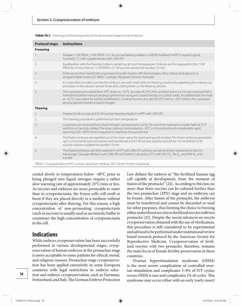
78
Section 3. Cryopreservation of embryos
cooled slowly to temperatures below −60°C prior to being plunged into liquid nitrogen require a rather slow warming rate of approximately 25°C/min or less. As oocytes and embryos are more permeable to water than to cryoprotectants, the frozen cells will swell or burst if they are placed directly in a medium without cryoprotectants aft er thawing. For this reason, a high concentration of non-permeating cryoprotectants (such as sucrose) is usually used as an osmotic buff er to counteract the high concentration of cryoprotectants in the cell.
Indications While embryo cryopreservation has been successfully performed at various developmental stages, cryop-reservation of human embryos at the pronuclear stage is more acceptable to some patients for ethical, moral, and religious reasons. Pronuclear-stage cryopreserva-tion has been applied extensively in some European countries with legal restrictions to embryo selec-tion and embryo cryopreservation, such as Germany, Switzerland, and Italy. Th e German Embryo Protection
Law defi nes the embryo as “the fertilized human egg cell capable of development, from the moment of fusion of the pronuclei” [ 24 ]. According to this law, no more than three oocytes can be cultured further than the two-pronuclear (2PN) stage and no embryos can be frozen. Aft er fusion of the pronuclei, the embryos must be transferred and cannot be discarded or used for other purposes, thus limiting the choice to freezing either unfertilized oocytes or fertilized oocytes with two pronuclei [ 25 ]. Despite the recent advances on oocyte cryopreservation obtained with the use of vitrifi cation, this procedure is still considered to be experimental and advised to be performed under institutional review board research protocol by the American Society for Reproductive Medicine. Cryopreservation of fertil-ized oocytes with two pronuclei, therefore, remains the main focus of female fertility preservation in these countries.
Ovarian hyperstimulation syndrome (OHSS) is the most severe complication of controlled ovar-ian stimulation and complicates 3–8% of IVF cycles; severe OHSS is rare and complicates 1% of cycles. Th is syndrome may occur either with an early (early onset)
Table 10.1. Freezing and thawing protocols for pronuclear stage human embryos
Protocol steps Instructions Freeezing
1. Prepare 1.5 M PROH, 1.5 M PROH + 0.1 M sucrose freezing media in a HEPES-buff ered mHTF (CooperSurgical, Trumbull, CT, USA) supplemented with 20% SPS
2. Equilibration with the freezing media is carried out at room temperature. Embryos are fi rst exposed to the 1.5 M PROH for 10 min then to 1.5 M PROH + 0.1 M sucrose solution for another 10 min
3. Embryos are then loaded into cryostraws (Cryo Bio System, IMV Technologies, Paris, France) and placed in a programmable freezer (CL-8000, Cryologic, Mulgrave, Victoria, Australia)
4. It is important to make sure that the embryos are well mixed with the freezing solutions by pipetting the embryos up and down in the solution several times after adding them to the freezing solution
5. The cryostraws are cooled from 20°C down to −6.5°C at a rate of 2.0°C/min, at which point a 2 min pre seeding hold is maintained before manual seeding is performed using pre-cooled forceps or a cotton swab. An additional 8 min hold at −6.5°C is provided for further equilibration. Cooling resumes at a rate of 0.3°C/min to −35°C before the cryostraws are plunged and stored in liquid nitrogen
Thawing
1. Prepare 0.5 M sucrose and 0.2 M sucrose thawing media in mHTF with 20% SPS
2. The thawing procedure is performed at room temperature
3. Cryostraws are removed from liquid nitrogen and exposed in air for 30 s and then warmed in a water bath at 31°C until the ice has fully melted. The slow cooling is terminated at −35°C in this protocol and a moderately rapid warming (200–350°C/min) is required to maximize the survival rate
4. The frozen embryos are expelled out of the straw using the stylet and quickly located. The frozen embryos are picked up in a minimal amount of solution and transferred to a 0.5 M sucrose solution and left for 10 min before 0.2 M sucrose solution is added for another 10 min
5. The thawed embryos are then washed in mHTF with 20% SPS without sucrose and then transferred to Quinn’s Advantage Cleavage Medium with 20% SPS and further cultured at 37°C with 5% CO 2 , 5% O 2 , and 90% N 2 until transfer
PROH, 1,2-propanediol; mHTF, human tubal fl uid medium; SPS, Serum Protein Substitute.
9780521517782c10.indd 789780521517782c10.indd 78 3/12/2010 6:42:26 PM3/12/2010 6:42:26 PM
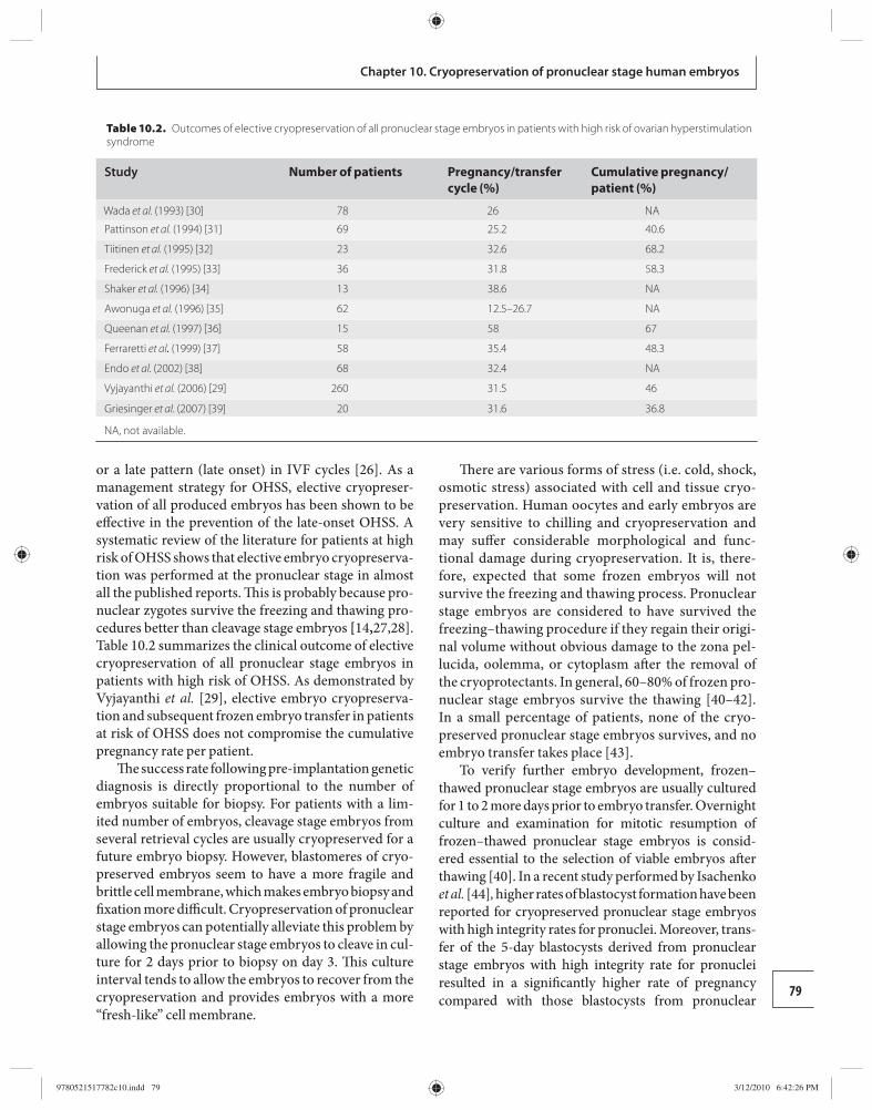
79
Chapter 10. Cryopreservation of pronuclear stage human embryos
or a late pattern (late onset) in IVF cycles [ 26 ]. As a management strategy for OHSS, elective cryopreser-vation of all produced embryos has been shown to be eff ective in the prevention of the late-onset OHSS. A systematic review of the literature for patients at high risk of OHSS shows that elective embryo cryopreserva-tion was performed at the pronuclear stage in almost all the published reports. Th is is probably because pro-nuclear zygotes survive the freezing and thawing pro-cedures better than cleavage stage embryos [ 14 , 27 , 28 ]. Table 10.2 summarizes the clinical outcome of elective cryopreservation of all pronuclear stage embryos in patients with high risk of OHSS. As demonstrated by Vyjayanthi et al. [ 29 ], elective embryo cryopreserva-tion and subsequent frozen embryo transfer in patients at risk of OHSS does not compromise the cumulative pregnancy rate per patient.
Th e success rate following pre-implantation genetic diagnosis is directly proportional to the number of embryos suitable for biopsy. For patients with a lim-ited number of embryos, cleavage stage embryos from several retrieval cycles are usually cryopreserved for a future embryo biopsy. However, blastomeres of cryo-preserved embryos seem to have a more fragile and brittle cell membrane, which makes embryo biopsy and fi xation more diffi cult. Cryopreservation of pronuclear stage embryos can potentially alleviate this problem by allowing the pronuclear stage embryos to cleave in cul-ture for 2 days prior to biopsy on day 3. Th is culture interval tends to allow the embryos to recover from the cryopreservation and provides embryos with a more “fresh-like” cell membrane.
Th ere are various forms of stress (i.e. cold, shock, osmotic stress) associated with cell and tissue cryo-preservation. Human oocytes and early embryos are very sensitive to chilling and cryopreservation and may suff er considerable morphological and func-tional damage during cryopreservation. It is, there-fore, expected that some frozen embryos will not survive the freezing and thawing process. Pronuclear stage embryos are considered to have survived the freezing–thawing procedure if they regain their origi-nal volume without obvious damage to the zona pel-lucida, oolemma, or cytoplasm aft er the removal of the cryoprotectants. In general, 60–80% of frozen pro-nuclear stage embryos survive the thawing [ 40 – 42 ]. In a small percentage of patients, none of the cryo-preserved pronuclear stage embryos survives, and no embryo transfer takes place [ 43 ].
To verify further embryo development, frozen–thawed pronuclear stage embryos are usually cultured for 1 to 2 more days prior to embryo transfer. Overnight culture and examination for mitotic resumption of frozen–thawed pronuclear stage embryos is consid-ered essential to the selection of viable embryos aft er thawing [ 40 ]. In a recent study performed by Isachenko et al. [ 44 ] , higher rates of blastocyst formation have been reported for cryopreserved pronuclear stage embryos with high integrity rates for pronuclei. Moreover, trans-fer of the 5-day blastocysts derived from pronuclear stage embryos with high integrity rate for pronuclei resulted in a signifi cantly higher rate of pregnancy compared with those blastocysts from pronuclear
Table 10.2. Outcomes of elective cryopreservation of all pronuclear stage embryos in patients with high risk of ovarian hyperstimulation syndrome
Study Number of patients Pregnancy/transfer cycle (%)
Cumulative pregnancy/patient (%)
Wada et al. (1993) [30] 78 26 NA
Pattinson et al. (1994) [31] 69 25.2 40.6
Tiitinen et al. (1995) [32] 23 32.6 68.2
Frederick et al. (1995) [33] 36 31.8 58.3
Shaker et al. (1996) [34] 13 38.6 NA
Awonuga et al. (1996) [35] 62 12.5–26.7 NA
Queenan et al. (1997) [36] 15 58 67
Ferraretti et al. (1999) [37] 58 35.4 48.3
Endo et al. (2002) [38] 68 32.4 NA
Vyjayanthi et al. (2006) [29] 260 31.5 46
Griesinger et al. (2007) [39] 20 31.6 36.8
NA, not available.
9780521517782c10.indd 799780521517782c10.indd 79 3/12/2010 6:42:26 PM3/12/2010 6:42:26 PM
80
Section 3. Cryopreservation of embryos
stage embryos with low integrity rates (43% and 10%, respectively). Th e authors concluded that the integrity rate for pronuclei aft er cryopreservation of pronuclear stage embryos was a predictor of future embryo devel-opment and implantation. Because of the negative eff ect of cryopreservation on embryo viability, relatively more embryos are potentially transferred in a frozen cycle compared with a fresh cycle. Sometimes more pro-nuclear stage embryos are required to be thawed if the fi rst set fails to undergo further development.
Factors aff ecting the success of embryo transfer Th e clinical outcome of transfer of frozen pronuclear stage embryos varies substantially between studies. Several factors might contribute to the between-study variants, including patient’s age, embryo quality before freezing, the number of embryos transferred, freez-ing and thawing protocol, and endometrial prepara-tion. Maternal age is one of the most important factors aff ecting the success of fresh embryo transfer. By ana-lysing 103 transfer cycles of cryopreserved embryos at the pronuclear stage, Damario et al. [ 45 ] observed an apparent trend toward a decline in delivery rate per fro-zen embryo transfer associated with increasing female age. In both fresh and frozen embryo transfers of a sin-gle oocyte retrieval, the chances of at least one live birth for women aged < 35, 35–39, and > 39 years were 61.2%, 59.7%, and 18.5%, respectively. A similar trend has also been noted following the transfer of embryos cryopre-served at cleavage stage [ 46 , 47 ].
Th e success of a frozen–thawed embryo transfer has been shown to be associated with the outcome of the fresh cycle from which the embryos were derived. Lin et al. [ 48 ] compared the outcome of two groups of patients who underwent frozen–thawed embryo trans-fer with a clinical pregnancy or without a pregnancy dur-ing the fresh cycle from which the frozen embryos were derived. Th ey reported a signifi cantly higher pregnancy rate (24.1%) in the frozen transfer cycles where a clini-cal pregnancy was achieved during the fresh cycle than in the frozen transfer cycle following a failed fresh cycle (8.5%). Similar results were also reported by Gabrielsen [ 49 ], who found the chance of embryo implantation following a frozen cycle is signifi cantly higher if a preg-nancy was established in the prior fresh cycle. A success-ful fresh cycle, therefore, refl ects better embryo quality and can be used to predict the chances of pregnancy of the following frozen–thawed embryo transfer cycle. Th e number of embryos transferred is another factor
aff ecting the success rate of frozen–thawed embryo transfer. As reported by Testart et al. [ 50 ], transfer of two frozen–thawed pronuclear stage embryos yields a two-fold increase in pregnancy rate compared with a single pronuclear stage embryo transfer. Similarly, signifi -cantly higher rates of pregnancy were obtained aft er the transfer of three or four frozen–thawed pronuclear stage embryos than with the transfer of one or two [ 51 ].
Aft er fertilization, the zona pellucida plays an important role in maintaining the integrity of the embryo. Zona pellucida manipulation, including intracytoplasmic sperm injection (ICSI), assisted hatching, and embryo biopsy, involves an artifi cial breach of the zona pellucida and there has been con-cern that manipulation of the zona pellucida could interfere with the diff usion of the cryoprotectants during the dehydration and rehydration process. Th e impact of the fertilization method on the rates of post-thaw survival and clinical outcome of cryopre-served pronuclear stage embryos has been addressed in several reports. According to Damario et al. [ 15 ], embryos from both IVF and ICSI survived very well aft er thawing (IVF 90.4%; ICSI 91.1%). Implantation rate (IVF 19.1%; ICSI 19.9%) and delivery rate (IVF 36.5%; ICSI 39.8%) were also comparable. Similarly, Al-Hasani et al. [ 27 ] observed comparable rates of post-thaw embryo survival, clinical pregnancy, and spontaneous abortion between cryopreserved pro-nuclear stage embryos resulting from ICSI ( n = 744) and conventional IVF ( n = 333). Opposing results were revealed in the study of Macas et al . [ 52 ], where a signifi cantly lower implantation rate was seen in the ICSI group (10.9%) compared with the IVF group (25.0%). Moreover, a trend to a higher rate of preclin-ical and clinical abortions was noted following the transfer of frozen embryos derived from ICSI (8/14 [57.1%]) compared with IVF (2/11 [11.8%]) in the same study. Taking into account the fact that 11.5% of ICSI and only 2.0% of IVF embryos were found in a stage of syngamy at the time of freezing, the overly long post-insemination time of 20 hours seems to be the principal reason for the lower developmental potential of cryopreserved pronuclear stage embryos from ICSI.
In another study, conducted by Simon et al. [ 53 ], a signifi cantly lower clinical pregnancy rate was achieved from frozen transfer cycles utilizing cleavage stage embryos arising from ICSI than from IVF (20% versus 32.5%). However, there was a higher proportion of good-quality embryos before freezing in conven-tional IVF cycles compared with ICSI cycles in their
9780521517782c10.indd 809780521517782c10.indd 80 3/12/2010 6:42:26 PM3/12/2010 6:42:26 PM
81
Chapter 10. Cryopreservation of pronuclear stage human embryos
study, suggesting that the diff erences in pregnancy rates found between the groups could not be simply attributed to the use of micromanipulation for ICSI. Based on the current data available, the ICSI proce-dure does not compromise the ability of pronuclear stage embryos to withstand cryopreservation and their further developmental potential aft er freezing and thawing. An intact zona pellucida is not necessary for a successful mouse embryo cryopreservation [ 54 ]. As reported by Hershlag and Feng [ 55 ], a 30 μm diameter defect created in the zona pellucida during assisted hatching before freezing does not hinder post-thaw mouse embryonic development. For humans, a prom-ising clinical pregnancy rate (31.4%) has been obtained following the transfer of cryopreserved blastocysts that underwent laser-assisted hatching on the zona on day 3 [ 56 ]. A larger hole is drilled in the zona pellucida for embryo biopsy than used in ICSI and assisted hatch-ing. An adverse eff ect of embryo biopsy on post-thaw embryo survival has been demonstrated, and vitrifi -cation has been shown to be an eff ective method for improving the survival rate of biopsied embryos at cleavage and pronuclear stages [ 57 – 59 ]. A recent study reported a successful pregnancy following vitrifi cation of pronuclear stage oocytes biopsied for polar body aneuploidy screening [ 60 ].
Th e endometrium plays a crucial role in embryo implantation and pregnancy establishment. Cryopre-served embryos have been successfully replaced in both natural ovulation cycles and hormone replacement cycles. Th e main advantage of the natural cycle is the lack of medication, whereas the downregulated hor-mone controlled cycle is more convenient for program-ming the day of frozen embryo transfer and planning the workload for the IVF practice in advance. In a retro-spective study by Al-Shawaf et al. [ 61 ], natural-cycle frozen embryo transfer was performed for 77 patients with proven ovulation and regular menstrual cycles and hormone replacement cycle was used for patients with anovulation, irregular cycles, or older age. No signifi cant diff erence in pregnancy rates (26% and 25%, respect-ively), ongoing/delivery rate (20.8% in both groups), and implantation rate (10.3% and 10.6%, respectively) were found between natural and hormone replacement cycles. In another study, Gelbaya et al. [ 62 ] compared the outcome of frozen embryo transfers in natural and downregulated hormone replacement cycles in patients with regular menstrual cycles. Th e rates of embryo implantation, clinical pregnancy, or live birth rates per cycle or per transfer were similar in both groups.
Impact of developmental stage at freezing on clinical outcome Th ere has been no universal agreement as to which stage of embryonic development at freezing provides a clear advantage for the outcome of a frozen embryo transfer cycle [ 63 ]. As shown in Table 10.3 , controver-sial results have been obtained concerning the rates of survival and the developmental competence between embryos frozen at the pronuclear and early cleavage stages.
In a prospective study performed by Horne et al. [ 41 ], similar rates of embryo survival were found between embryos frozen at the pronuclear (96/129 [74.4%]) and day 2 cleavage (79/102 [77.4%]) stages. Salumets et al. [ 66 ], in a large series involving a total of 4006 embryos thawed in 1657 cycles, directly com-pared the survival rates between three cryopreservation strategies utilizing pronuclear, day 2, or day 3 embryo freezing. Th eir results showed that frozen pronuclear stage embryos survived better (86.5%) than either day 2 (61.7%) or day 3 (43.1%) embryos. On the contrary, Kattera et al. [ 65 ] reported that supernumerary IVF-generated embryos frozen on day 2 had signifi cantly higher survival rates (73.9%) than those frozen at the pronuclear stage (64.4%). Th e pregnancy rate follow-ing the transfer of embryos frozen at the cleavage stage was also higher than at the pronuclear stage. Others have reported either similar or higher rates of preg-nancy and embryo implantation aft er the transfer of embryos frozen at the pronuclear stage than on day 2 [ 41 , 64 ,66 ]. As shown by Horne et al. [ 41 ], while the live birth rate of fresh embryo transfer performed on day 2 was signifi cantly higher than at the pronuclear stage, a trend to higher live birth rate was observed following the transfer of embryos frozen at pronuclear stage (11/44 [25%] versus 4/38 [11%]). Moreover, compar-able rates of cumulative viable pregnancy were reached following one fresh and two frozen–thawed embryo replacements in their study (40.2% and 41.4% for pro-nuclear and cleavage stage, respectively). Demoulin et al. [ 64 ] have reported higher rates of pregnancy (23.2% vs. 8.2%) and embryo implantation (10.7% vs. 4.7%) following the transfer of embryos frozen at the pronuclear stage than at the multicellular stage. However, most of the embryos frozen at the pro-nuclear stage were derived from protocols utilizing
9780521517782c10.indd 819780521517782c10.indd 81 3/12/2010 6:42:26 PM3/12/2010 6:42:26 PM
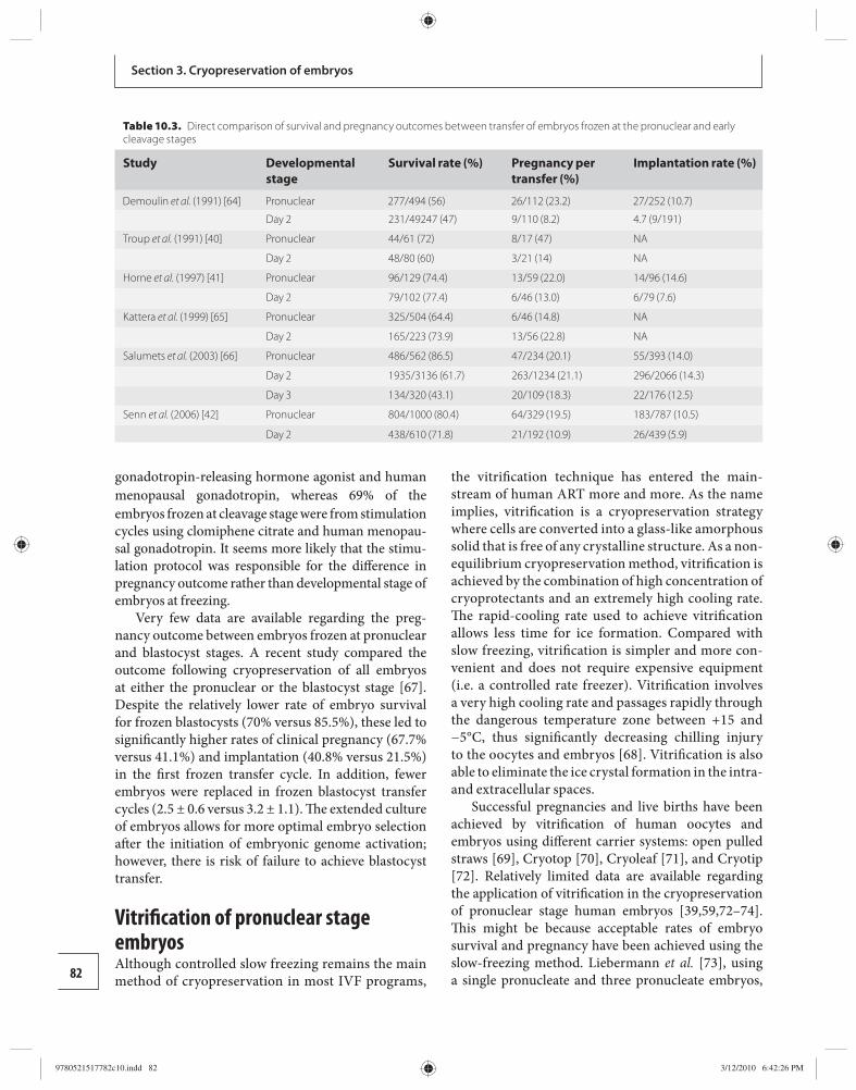
82
Section 3. Cryopreservation of embryos
gonadotropin-releasing hormone agonist and human menopausal gonadotropin, whereas 69% of the embryos frozen at cleavage stage were from stimulation cycles using clomiphene citrate and human menopau-sal gonadotropin. It seems more likely that the stimu-lation protocol was responsible for the diff erence in pregnancy outcome rather than developmental stage of embryos at freezing.
Very few data are available regarding the preg-nancy outcome between embryos frozen at pronuclear and blastocyst stages. A recent study compared the outcome following cryopreservation of all embryos at either the pronuclear or the blastocyst stage [ 67 ]. Despite the relatively lower rate of embryo survival for frozen blastocysts (70% versus 85.5%), these led to signifi cantly higher rates of clinical pregnancy (67.7% versus 41.1%) and implantation (40.8% versus 21.5%) in the fi rst frozen transfer cycle. In addition, fewer embryos were replaced in frozen blastocyst transfer cycles (2.5 ± 0.6 versus 3.2 ± 1.1). Th e extended culture of embryos allows for more optimal embryo selection aft er the initiation of embryonic genome activation; however, there is risk of failure to achieve blastocyst transfer.
Vitrifi cation of pronuclear stage embryos Although controlled slow freezing remains the main method of cryopreservation in most IVF programs,
the vitrifi cation technique has entered the main-stream of human ART more and more. As the name implies, vitrifi cation is a cryopreservation strategy where cells are converted into a glass-like amorphous solid that is free of any crystalline structure. As a non-equilibrium cryopreservation method, vitrifi cation is achieved by the combination of high concentration of cryoprotectants and an extremely high cooling rate. Th e rapid-cooling rate used to achieve vitrifi cation allows less time for ice formation. Compared with slow freezing, vitrifi cation is simpler and more con-venient and does not require expensive equipment (i.e. a controlled rate freezer). Vitrifi cation involves a very high cooling rate and passages rapidly through the dangerous temperature zone between +15 and −5°C, thus signifi cantly decreasing chilling injury to the oocytes and embryos [ 68 ]. Vitrifi cation is also able to eliminate the ice crystal formation in the intra- and extracellular spaces.
Successful pregnancies and live births have been achieved by vitrifi cation of human oocytes and embryos using diff erent carrier systems: open pulled straws [ 69 ], Cryotop [ 70 ], Cryoleaf [ 71 ], and Cryotip [ 72 ]. Relatively limited data are available regarding the application of vitrifi cation in the cryopreservation of pronuclear stage human embryos [ 39 , 59 , 72 – 74 ]. Th is might be because acceptable rates of embryo survival and pregnancy have been achieved using the slow-freezing method. Liebermann et al. [ 73 ], using a single pronucleate and three pronucleate embryos,
Table 10.3. Direct comparison of survival and pregnancy outcomes between transfer of embryos frozen at the pronuclear and early cleavage stages
Study Developmental stage
Survival rate (%) Pregnancy per transfer (%)
Implantation rate (%)
Demoulin et al. (1991) [64] Pronuclear 277/494 (56) 26/112 (23.2) 27/252 (10.7)
Day 2 231/49247 (47) 9/110 (8.2) 4.7 (9/191)
Troup et al. (1991) [40] Pronuclear 44/61 (72) 8/17 (47) NA
Day 2 48/80 (60) 3/21 (14) NA
Horne et al. (1997) [41] Pronuclear 96/129 (74.4) 13/59 (22.0) 14/96 (14.6)
Day 2 79/102 (77.4) 6/46 (13.0) 6/79 (7.6)
Kattera et al. (1999) [65] Pronuclear 325/504 (64.4) 6/46 (14.8) NA
Day 2 165/223 (73.9) 13/56 (22.8) NA
Salumets et al. (2003) [66] Pronuclear 486/562 (86.5) 47/234 (20.1) 55/393 (14.0)
Day 2 1935/3136 (61.7) 263/1234 (21.1) 296/2066 (14.3)
Day 3 134/320 (43.1) 20/109 (18.3) 22/176 (12.5)
Senn et al. (2006) [42] Pronuclear 804/1000 (80.4) 64/329 (19.5) 183/787 (10.5)
Day 2 438/610 (71.8) 21/192 (10.9) 26/439 (5.9)
9780521517782c10.indd 829780521517782c10.indd 82 3/12/2010 6:42:26 PM3/12/2010 6:42:26 PM
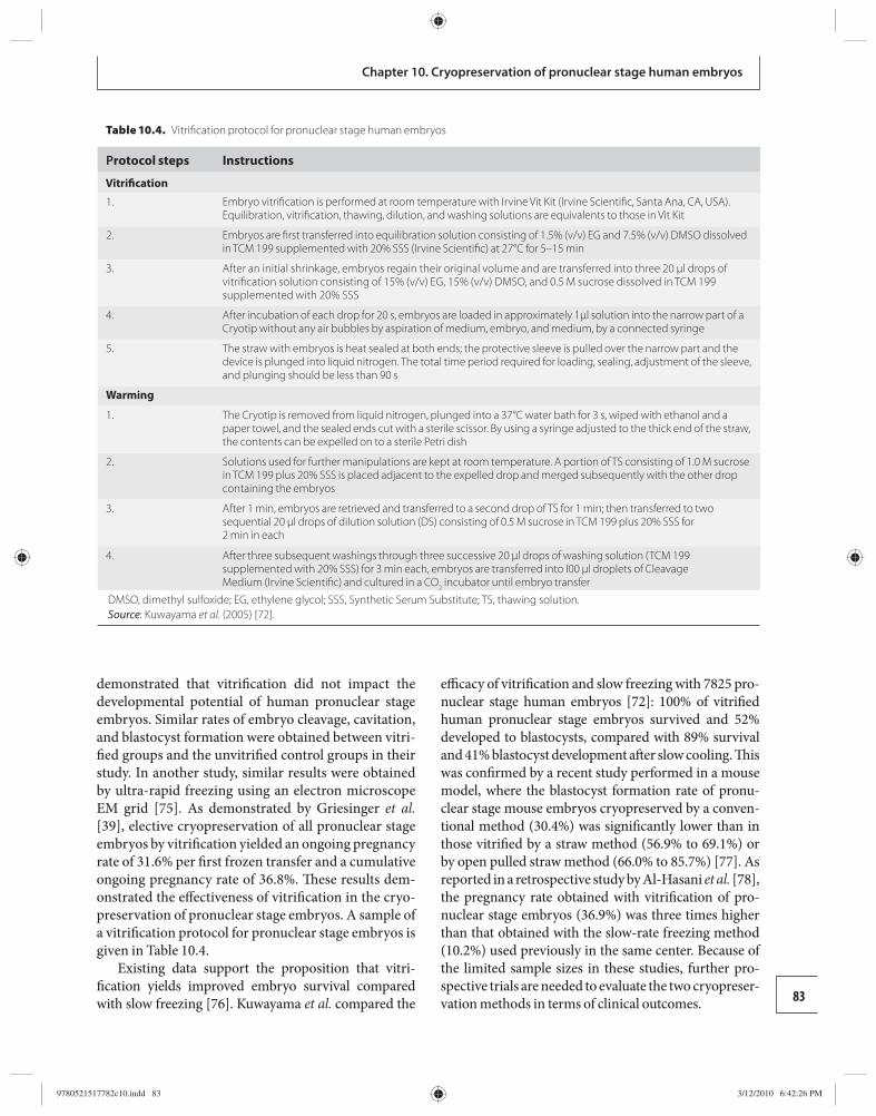
83
Chapter 10. Cryopreservation of pronuclear stage human embryos
demonstrated that vitrifi cation did not impact the developmental potential of human pronuclear stage embryos. Similar rates of embryo cleavage, cavitation, and blastocyst formation were obtained between vitri-fi ed groups and the unvitrifi ed control groups in their study. In another study, similar results were obtained by ultra-rapid freezing using an electron microscope EM grid [ 75 ]. As demonstrated by Griesinger et al. [ 39 ], elective cryopreservation of all pronuclear stage embryos by vitrifi cation yielded an ongoing pregnancy rate of 31.6% per fi rst frozen transfer and a cumulative ongoing pregnancy rate of 36.8%. Th ese results dem-onstrated the eff ectiveness of vitrifi cation in the cryo-preservation of pronuclear stage embryos. A sample of a vitrifi cation protocol for pronuclear stage embryos is given in Table 10.4 .
Existing data support the proposition that vitri-fi cation yields improved embryo survival compared with slow freezing [ 76 ]. Kuwayama et al. compared the
effi cacy of vitrifi cation and slow freezing with 7825 pro-nuclear stage human embryos [ 72 ]: 100% of vitrifi ed human pronuclear stage embryos survived and 52% developed to blastocysts, compared with 89% survival and 41% blastocyst development aft er slow cooling. Th is was confi rmed by a recent study performed in a mouse model, where the blastocyst formation rate of pronu-clear stage mouse embryos cryopreserved by a conven-tional method (30.4%) was signifi cantly lower than in those vitrifi ed by a straw method (56.9% to 69.1%) or by open pulled straw method (66.0% to 85.7%) [ 77 ]. As reported in a retrospective study by Al-Hasani et al. [ 78 ], the pregnancy rate obtained with vitrifi cation of pro-nuclear stage embryos (36.9%) was three times higher than that obtained with the slow-rate freezing method (10.2%) used previously in the same center. Because of the limited sample sizes in these studies, further pro-spective trials are needed to evaluate the two cryopreser-vation methods in terms of clinical outcomes.
Table 10.4. Vitrifi cation protocol for pronuclear stage human embryos
P rotocol steps Instructions
Vitrifi cation
1. Embryo vitrifi cation is performed at room temperature with Irvine Vit Kit (Irvine Scientifi c, Santa Ana, CA, USA). Equilibration, vitrifi cation, thawing, dilution, and washing solutions are equivalents to those in Vit Kit
2. Embryos are fi rst transferred into equilibration solution consisting of 1.5% (v/v) EG and 7.5% (v/v) DMSO dissolved in TCM 199 supplemented with 20% SSS (Irvine Scientifi c) at 27°C for 5–15 min
3. After an initial shrinkage, embryos regain their original volume and are transferred into three 20 μl drops of vitrifi cation solution consisting of 15% (v/v) EG, 15% (v/v) DMSO, and 0.5 M sucrose dissolved in TCM 199 supplemented with 20% SSS
4. After incubation of each drop for 20 s, embryos are loaded in approximately 1μl solution into the narrow part of a Cryotip without any air bubbles by aspiration of medium, embryo, and medium, by a connected syringe
5. The straw with embryos is heat sealed at both ends; the protective sleeve is pulled over the narrow part and the device is plunged into liquid nitrogen. The total time period required for loading, sealing, adjustment of the sleeve, and plunging should be less than 90 s
Warming
1. The Cryotip is removed from liquid nitrogen, plunged into a 37°C water bath for 3 s, wiped with ethanol and a paper towel, and the sealed ends cut with a sterile scissor. By using a syringe adjusted to the thick end of the straw, the contents can be expelled on to a sterile Petri dish
2. Solutions used for further manipulations are kept at room temperature. A portion of TS consisting of 1.0 M sucrose in TCM 199 plus 20% SSS is placed adjacent to the expelled drop and merged subsequently with the other drop containing the embryos
3. After 1 min, embryos are retrieved and transferred to a second drop of TS for 1 min; then transferred to two sequential 20 μl drops of dilution solution (DS) consisting of 0.5 M sucrose in TCM 199 plus 20% SSS for 2 min in each
4. After three subsequent washings through three successive 20 μl drops of washing solution (TCM 199 supplemented with 20% SSS) for 3 min each, embryos are transferred into l00 μl droplets of CleavageMedium (Irvine Scientifi c) and cultured in a CO 2 incubator until embryo transfer
DMSO, dimethyl sulfoxide; EG, ethylene glycol; SSS, Synthetic Serum Substitute; TS, thawing solution. Source : Kuwayama et al. (2005) [ 72 ].
9780521517782c10.indd 839780521517782c10.indd 83 3/12/2010 6:42:26 PM3/12/2010 6:42:26 PM
84
Section 3. Cryopreservation of embryos
High concentration of cryoprotectants is a negative factor in vitrifi cation. Th e total concentration of cryo-protectant ranges from 5 to 8 mol/l in vitrifi cation pro-tocols currently used in the cryopreservation of human oocyte and embryos [ 72 , 79 ]. More severe osmotic stress and toxic eff ects are potentially imposed upon the cells during the vitrifi cation and warming processes than in the slow-freezing process. It is, therefore, of critical importance to restrict the concentration of cryopro-tectants to the minimal level to achieve vitrifi cation. A stepwise addition of cryoprotectants can help to reduce the toxic and osmotic damage induced by cryo-protectants at high concentrations [ 80 ]. Increasing the viscosity or the cooling/warming rates [ 81 ] or decreas-ing the volume [ 82 ] will each independently increase the probability of vitrifi cation. Based on the fact that the higher the cooling rate is, the lower the required cryoprotectant concentration for vitrifi cation, He et al. [ 83 ] recently developed a novel ultra-fast vitrifi cation approach using a small quartz microcapillary with an inner diameter less than 0.4 mm. According to their study, only 2 M 1,2-propanediol is required to achieve apparent vitrifi cation using the quartz microcapillary plunged into liquid nitrogen, with the intracellular concentration (2 M) of cryoprotectants being in the range of that used for slow freezing.
Second, attention should be paid to the safety aspects involved in vitrifi cation, storage, and trans-portation. In order to increase the cooling rate, most vitrifi cation carrier systems require the direct contact between oocyte or embryo and liquid nitrogen, which makes it possible for cross-contamination of samples to occur in liquid nitrogen storage tanks [ 84 ]. A num-ber of viruses, including the human immunodefi -ciency virus and hepatitis B and C viruses, can survive in liquid nitrogen [ 84 ]. While there is no direct evi-dence of cross-contamination of human gametes and embryos stored in the same tank, transmission of viral pathogens to embryos vitrifi ed and stored in open containers by experimentally contaminated liquid nitrogen has been reported in a bovine model [ 85 ]. Th e small sample volume also places the vitri-fi ed cell at a risk of inadvertent thawing as tempera-ture increases when being moved from tank to tank or during inventory.
To guarantee complete isolation of embryos and to avoid the potential contamination by pathogens from liquid nitrogen, diff erent methods of closed vitrifi ca-tion systems have emerged. Mouse pronuclear oocytes vitrifi ed using Cryoloop with or without contact with
liquid nitrogen had similar rates of embryo survival and blastocyst formation. I n addition, contact and non-contact vitrifi cation did not alter the mean total cell number and the mean number of cells compos-ing the inner cell mass and trophectoderm of blasto-cysts from vitrifi ed mouse embryos [ 86 ]. Similarly, no adverse eff ect on the further embryonic development was observed when human pronuclear oocytes were loaded into open pulled straws located in a 0.5 ml closed straw before being plunged into liquid nitro-gen for cooling and storage [ 59 ]. Although keeping a carrier containing the specimen inside an outer pro-tecting container provides a strategy to eliminate the potential contamination associated with direct contact with liquid nitrogen [ 87 ], it does not allow for rapid warming of a small volume and simultaneous removal of the cryoprotectant during the warming process [ 88 ]. It is, therefore, of importance to maintain a delicate balance among all the factors that aff ect the probabil-ity of vitrifi cation in order to prevent crystallization, devitrifi cation, recrystallization, glass fractures, and chilling injury. Another strategy to minimize the potential for transmission is the use of nitrogen vapor instead of liquid nitrogen itself. Vapor storage has been proposed to minimize or eliminate the possibility of cross-contamination [ 89 , 90 ]. A major concern of vapor storage is the risk of loss of cell viability. Human sperm survive satisfactorily with good recovery in the vapor of liquid nitrogen [ 91 ]. Similar rates of embryo sur-vival and blastocyst formation were obtained between 1-cell mouse embryos stored in liquid nitrogen and nitrogen vapor [ 90 ]. Although no published data are available on human embryos, nitrogen vapor storage does not compromise the viability of vitrifi ed human oocytes [ 92 ].
References 1. President’s Council on Bioethics . Reproduction
and Responsibility: Th e Regulation of New Biotechnologies . Washington, DC : US Government Press , 2004. http://www.bioethics.gov/reports/reproductionandresponsibility/chapter2.html.
2. Jones HW , Schnorr JA . Multiple pregnancies: a call for action . Fertil Steril 2001; 75: 11 –13.
3. Jain T , Missmer SA , Hornstein MD . Trends in embryo-transfer practice and in outcomes of the use of assisted reproductive technology in the United States . N Engl J Med 2004; 350: 1639 –1645.
4. Whittingham DG , Leibo SP , Mazur P . Survival of mouse embryos frozen to −196 degrees and −269 degrees C . Science 1972; 178: 411 –414.
9780521517782c10.indd 849780521517782c10.indd 84 3/12/2010 6:42:26 PM3/12/2010 6:42:26 PM
85
Chapter 10. Cryopreservation of pronuclear stage human embryos
5. Wilmut I , Rowson LE . Experiments on the low-temperature preservation of cow embryos . Vet Rec 1973; 92: 686 –690.
6. Leibo SP . A one-step method for direct nonsurgical transfer of frozen–thawed bovine embryos . Th eriogenology 1984; 21 : 767 –790.
7. Kasai M , Komi JH , Takakamo A , et al. A simple method for mouse embryo cryopreservation in a low toxicity vitrifi cation solution, without appreciable loss of viability . J Reprod Fertil 1990; 89 : 91 –97.
8. Trounson A , Mohr L . Human pregnancy following cryopreservation, thawing and transfer of an eight-cell embryo . Nature 1983; 305 : 707 –709.
9. Lassalle B , Testart J , Renard JP . Human embryo features that infl uence the success of cryopreservation with the use of 1,2 propanediol . Fertil Steril 1985; 44 : 645 –651.
10. Testart J , Lassalle B , Belaisch-Allart J , et al. High pregnancy rate aft er early human embryo freezing . Fertil Steril 1986; 46 : 268 –272.
11. Mandelbaum J , Junca AM , Plachot M , et al. Cryopreservation of human embryos and oocytes . Hum Reprod 1988; 3 : 117 –119.
12. Edgar DH , Bourne H , Jericho H , McBain JC . Th e developmental potential of cryopreserved human embryos . Mol Cell Endocrinol 2000; 169 : 69 –72.
13. Schneider U . Cryobiological principles of embryo freezing . J In Vitro Fert Embryo Transf 1986; 3: 3 –9.
14. Veeck LL , Amundson CH , Brothman LJ , et al. Signifi cantly enhanced pregnancy rates per cycle through cryopreservation and thaw of pronuclear stage oocytes . Fertil Steril 1993; 59: 1202 –1207.
15. Damario MA , Hammitt DG , Galanits TM , Session DR , Dumesic DA . Pronuclear stage cryopreservation aft er intracytoplasmic sperm injection and conventional IVF: implications for timing of the freeze . Fertil Steril 1999; 72: 1049 –1054.
16. Scott L , Alvero R , Leondires M , Miller B . Th e morphology of human pronuclear embryos is positively related to blastocyst development and implantation . Hum Reprod 2000; 15: 2394 –2403.
17. Balaban B , Urman B , Isiklar A , et al. Th e eff ect of pronuclear morphology on embryo quality parameters and blastocyst transfer outcome . Hum Reprod 2001; 16: 2357 –2361.
18. James AN , Hennessy S , Reggio B , Wiemer K , Larsen F , Cohen J . The limited importance of pronuclear scoring of human zygotes . Hum Reprod 2006; 21: 1599 –1604.
19. Nicoli A , Valli B , Di Girolamo R , et al. Limited importance of pre-embryo pronuclear morphology (zygote score) in assisted reproduction outcome in the absence of embryo cryopreservation . Fertil Steril 2007; 88 (Suppl 4): 1167 –1173.
20. Guerif F , Le Gouge A , Giraudeau B , et al. Limited value of morphological assessment at days 1 and 2 to predict blastocyst development potential: a prospective study based on 4042 embryos . Hum Reprod 2007; 22: 1973 –1981.
21. Capmany G , Taylor A , Braude PR , Bolton VN . Th e timing of pronuclear formation, DNA synthesis and cleavage in the human 1-cell embryo . Mol Hum Reprod 1996; 2: 299 –306.
22. Nagy ZP , Liu J , Joris H , Devroey P , Van Steirteghem A . Time-course of oocyte activation, pronucleus formation and cleavage in human oocytes fertilized by intracytoplasmic sperm injection . Hum Reprod 1994; 9: 1743 –1748.
23. Payne D , Flaherty SP , Barry MF , Matthews CD . Preliminary observations on polar body extrusion and pronuclear formation in human oocytes using time-lapse video cinematography . Hum Reprod 1997; 12: 532 –541.
24. ESHRE Task Force on Ethics and Law . I. Th e moral status of the pre-implantation embryo . Hum Reprod 2001; 16: 1046 –1048.
25. Orief Y , Nikolettos N , Al Hasani S . Cryopreservation of two pronuclear stage zygotes . Rev Gynaecol Prac 2005; 5: 39 –44.
26. Papanikolaou EG , Tournaye H , Verpoest W , et al. Early and late ovarian hyperstimulation syndrome: early pregnancy outcome and profi le . Hum Reprod 2005; 20: 636 –641.
27. Al-Hasani S , Ludwig M , Gagsteiger F , et al. Comparison of cryopreservation of supernumerary pronuclear human oocytes obtained aft er intracytoplasmic sperm injection (ICSI) and aft er conventional in-vitro fertilization . Hum Reprod 1996; 11: 604 –607.
28. Hoover L , Baker A , Check JH , et al. Clinical outcome of cryopreserved human pronuclear stage embryos resulting from intracytoplasmic sperm injection . Fertil Steril 1997; 67: 621 –624.
29. Vyjayanthi S , Tang T , Fattah A , et al. Elective cryopreservation of embryos at the pronucleate stage in women at risk of ovarian hyperstimulation syndrome may aff ect the overall pregnancy rate . Fertil Steril 2006; 86 : 1773 –1775.
30. Wada I , Matson PL , Troup SA , et al. Does elective cryopreservation of all embryos from women at risk of ovarian hyperstimulation syndrome reduce the incidence of the condition? Br J Obstet Gynaecol 1993 ; 100: 265 –269.
31. Pattinson HA , Hignett M , Dunphy BC , Fleetham JA . Outcome of thaw embryo transfer aft er cryopreservation of all embryos in patients at risk of ovarian hyperstimulation syndrome . Fertil Steril 1994; 62: 1192 –1196.
32. Tiitinen A , Husa LM , Tulppala M , Simberg N , Seppälä M . Th e eff ect of cryopreservation in prevention
9780521517782c10.indd 859780521517782c10.indd 85 3/12/2010 6:42:26 PM3/12/2010 6:42:26 PM
86
Section 3. Cryopreservation of embryos
of ovarian hyperstimulation syndrome . Br J Obstet Gynaecol 1995; 102: 326 –329.
33. Frederick JL , Ord T , Kettel LM , et al. Successful pregnancy outcome aft er cryopreservation of all fresh embryos with subsequent transfer into an unstimulated cycle . Fertil Steril 1995; 64: 987 –990.
34. Shaker AG , Zosmer A , Dean N , et al. Comparison of intravenous albumin and transfer of fresh embryos with cryopreservation of all embryos for subsequent transfer in prevention of ovarian hyperstimulation syndrome . Fertil Steril 1996; 65: 992 –996.
35. Awonuga AO , Dean N , Zaidi J , et al. Outcome of frozen embryo replacement cycles following elective cryopreservation of all embryos in women at risk of developing ovarian hyperstimulation syndrome . J Assist Reprod Genet 1996; 13: 293 –297.
36. Queenan JT , Jr. , Veeck LL , Toner JP , Oehninger S , Muasher SJ . Cryopreservation of all prezygotes in patients at risk of severe hyperstimulation does not eliminate the syndrome, but the chances of pregnancy are excellent with subsequent frozen–thaw transfers . Hum Reprod 1997; 12: 1573 –1576.
37. Ferraretti AP , Gianaroli L , Magli C , et al. Elective cryopreservation of all pronucleate embryos in women at risk of ovarian hyperstimulation syndrome: effi ciency and safety . Hum Reprod 1999; 14: 1457 –1460.
38. Endo T , Honnma H , Hayashi T , et al. Continuation of GnRH agonist administration for 1 week, aft er hCG injection, prevents ovarian hyperstimulation syndrome following elective cryopreservation of all pronucleate embryos . Hum Reprod 2002; 17 : 2548 –2451.
39. Griesinger G , von Otte S , Schroer A , et al. Elective cryopreservation of all pronuclear oocytes aft er GnRH agonist triggering of fi nal oocyte maturation in patients at risk of developing OHSS: a prospective, observational proof-of-concept study . Hum Reprod 2007; 22: 1348 –1352.
40. Troup SA , Matson PL , Critchlow JD , et al. Cryopreservation of human embryos at the pronucleate, early cleavage, or expanded blastocyst stages . Eur J Obstet Gynecol Reprod Biol 1991; 38: 133 –139.
41. Horne G , Critchlow JD , Newman MC , et al. A prospective evaluation of cryopreservation strategies in a two-embryo transfer programme . Hum Reprod 1997; 12 : 542 –547.
42. Senn A , Urner F , Chanson A , et al. Morphological scoring of human pronuclear zygotes for prediction of pregnancy outcome . Hum Reprod 2006; 21 : 234 –239.
43. Veeck LL . Does the developmental stage at freeze impact on clinical results post-thaw? Reprod Biomed Online 2003; 6: 367 –374.
44. Isachenko V , Todorov P , Dimitrov Y , Isachenko E . Integrity rate of pronuclei aft er cryopreservation of pronuclear-zygotes as a criterion for subsequent embryo development and pregnancy . Hum Reprod 2008; 23: 819 –826.
45. Damario MA , Hammitt DG , Session DR , Dumesic DA . Embryo cryopreservation at the pronuclear stage and effi cient embryo use optimizes the chance for a liveborn infant from a single oocyte retrieval . Fertil Steril 2000; 73 : 767 –773.
46. Wang JX , Yap YY , Matthews CD . Frozen–thawed embryo transfer: infl uence of clinical factors on implantation rate and risk of multiple conception . Hum Reprod 2001; 16: 2316 –2319.
47. Salumets A , Hydén-Granskog C , Suikkari AM , Tiitinen A , Tuuri T . The predictive value of pronuclear morphology of zygotes in the assessment of human embryo quality . Hum Reprod 2001; 16: 2177 –2181.
48. Lin YP , Cassidentid L , Chacon RR , et al. Successful implantation of frozen sibling embryos is infl uenced by the outcome of the cycle from which they were derived . Fertil Steril 1995; 63 : 262 –267.
49. Gabrielsen A . Parameters predicting the implantation rate of thawed IVF/ICSI embryos: a retrospective study . Reprod Biomed Online 2006; 12: 70 –76.
50. Testart J , Lassalle B , Belaisch-Allart J , et al. Human embryo freezing . Ann N Y Acad Sci 1988; 541: 532 –540.
51. Irianni FM , Veeck LL , Toner JP , Muasher SJ . Infl uence of number of pre-embryos transferred, progesterone level and oestradiol/progesterone relation at thaw on pregnancy results during replacement of cryopreserved pre-embryos in natural cycles . Hum Reprod 1992; 7: 797 –800.
52. Macas E , Imthurn B , Borsos M , et al. Impairment of the developmental potential of frozen–thawed human zygotes obtained aft er intracytoplasmic sperm injection . Fertil Steril 1998; 69: 630 –635.
53. Simon A , Holzer H , Hurwitz A , et al. Comparisonof cryopreservation outcome following intracytoplasmic sperm injection and conventional in vitro fertilization . J Assist Reprod Genet 1998; 15: 431 –437.
54. Garrisi GJ , Talansky BE , Sapira V , Gordon JW , Navot D . An intact zona pellucida is not necessary for successful mouse embryo cryopreservation . Fertil Steril 1992; 57: 677 –681.
55. Hershlag A , Feng HL . Eff ect of pre-freeze assisted hatching on post-thaw survival of mouse embryos . Fertil Steril 2005; 84: 1752 –1754.
56. Kung FT , Lin YC , Tseng YJ , et al. Transfer of frozen–thawed blastocysts that underwent quarter laser-assisted hatching at the day 3 cleaving stage before freezing . Fertil Steril 2003; 79: 893 –899.
57. Magli MC , Gianaroli L , Fortini D , Ferraretti AP , Munné S . Impact of blastomere biopsy and cryopreservation techniques on human embryo viability . Hum Reprod 1999; 14: 770 –773.
58. Jericho H , Wilton L , Gook DA , Edgar DH . A modifi ed cryopreservation method increases the survival of
9780521517782c10.indd 869780521517782c10.indd 86 3/12/2010 6:42:27 PM3/12/2010 6:42:27 PM
87
Chapter 10. Cryopreservation of pronuclear stage human embryos
human biopsied cleavage stage embryos . Hum Reprod 2003; 18: 568 –571.
59. Isachenko V , Montag M , Isachenko E , van der Ven H . Vitrifi cation of mouse pronuclear embryos aft er polar body biopsy without direct contact with liquid nitrogen . Fertil Steril 2005; 84: 1011 –1016.
60. Naether OG , Rudolf K , Fischer R , et al. Pregnancy aft er vitrifi cation of pronuclear stage oocytes biopsied for polar body aneuploidy screening . Reprod Biomed Online 2008; 16 : 268 –270.
61. Al-Shawaf T , Yang D , al-Magid Y , et al. Ultrasonic monitoring during replacement of frozen/thawed embryos in natural and hormone replacement cycles . Hum Reprod 1993; 8: 2068 –2074.
62. Gelbaya TA , Nardo LG , Hunter HR , et al. Cryopreserved-thawed embryo transfer in natural or down-regulated hormonally controlled cycles: a retrospective study . Fertil Steril 2006; 85: 603 –609.
63. Kolibianakis EM , Zikopoulos K , Devroey P . Implantation potential and clinical impact of cryopreservation: a review . Placenta 2003; 24 (Suppl B): S27 –S33.
64. Demoulin A , Jouan C , Gerday C , Dubois M . Pregnancy rates aft er transfer of embryos obtained from diff erent stimulation protocols and frozen at either pronucleate or multicellular stages . Hum Reprod 1991; 6 : 799 –804.
65. Kattera S , Shrivastav P , Craft I . Comparison of pregnancy outcome of pronuclear- and multicellular-stage frozen–thawed embryo transfers . J Assist Reprod Genet 1999; 16: 358 –362.
66. Salumets A , Tuuri T , Mäkinen S , et al. Eff ect of developmental stage of embryo at freezing on pregnancy outcome of frozen–thawed embryo transfer . Hum Reprod 2003; 18: 1890 –1895.
67. Surrey ES , Keller JL , Stevens J , et al. ‘ Freeze-all’: higher implantation and pregnancy rates with cryopreservation (cryo) at the blastocyst (blast) versus pronuclear (2pn) stage . Fertil Steril 2008; 90 (Suppl 1): S27–S 28.
68. Ambrosini G , Andrisani A , Porcu E , et al. Cryotop vitrifi cation of human oocytes results in high survival rate and healthy deliveries . Reprod Biomed Online 2007; 14: 72 –79.
69. Selman H , Angelini A , Barnocchi N , et al. Ongoing pregnancies aft er vitrifi cation of human oocytes using a combined solution of ethylene glycol and dimethyl sulfoxide . Fertil Steril 2006; 86 : 997 –1000.
70. Antinori M , Licata E , Dani G , et al. Cryotop vitrifi cation of human oocytes results in high survival rate and healthy deliveries . Reprod Biomed Online 2007; 14: 72 –79.
71. Chian RC , Son WY , Huang JY , et al. High survival rates and pregnancies of human oocytes following vitrifi cation: preliminary report . Fertil Steril 2005; 84 (Suppl 1): S36 .
72. Kuwayama M , Vajta G , Ieda S , Kato O . Comparison of open and closed methods for vitrifi cation of human embryos and the elimination of potential contamination . Reprod Biomed Online 2005; 11: 608 –614.
73. Liebermann J , Tucker MJ , Graham JR , et al. Blastocyst development aft er vitrifi cation of multipronuclear zygotes using the Flexipet denuding pipette . Reprod Biomed Online 2002; 4 : 146 –150.
74. Isachenko V , Montag M , Isachenko E , et al. Developmental rate and ultrastructure of vitrifi ed human pronuclear oocytes aft er stepwise versus direct rehydration . Hum Reprod 2004; 19: 660 –665.
75. Park SP , Kim EY , Oh JH , et al. Ultrarapid freezing of human multipronuclear zygotes using electron microscope grids . Hum Reprod 2000 15: 1787 –1790.
76. Loutradi KE , Kolibianakis EM , Venetis CA , et al. Cryopreservation of human embryos by vitrifi cation or slow freezing: a systematic review and meta-analysis . Fertil Steril 2008; 90: 186 –193.
77. Zhao XM , Quan GB , Zhou GB , Hou YP , Zhu SE . Conventional freezing, straw, and open-pulled straw vitrifi cation of mouse two pronuclear (2-PN) stage embryos . Anim Biotechnol 2007; 18: 203 –212.
78. Al-Hasani S , Ozmen B , Koutlaki N , et al. Th ree years of routine vitrifi cation of human zygotes: is it still fair to advocate slow-rate freezing? Reprod Biomed Online 2007; 14: 288 –293.
79. El-Danasouri I , Selman H . Successful pregnancies and deliveries aft er a simple vitrifi cation protocol for day 3 human embryos . Fertil Steril 2001; 76: 400 –402.
80. Rall WF . Factors aff ecting the survival of mouse embryos cryopreserved by vitrifi cation . Cryobiology 1987; 24: 387 –402.
81. Vajta G , Kuwayama M . Improving cryopreservation systems . Th eriogenology 2006; 65: 236 –244.
82. Arav A . Vitrification of oocytes and embryos . In Lauria A , Gandolfi F , eds. New Trends in Embryo Transfer . Cambridge, UK : Portland Press , 1992, pp. 255–264.
83. He X , Park EY , Fowler A , Yarmush ML , Toner M . Vitrifi cation by ultra-fast cooling at a low concentration of cryoprotectants in a quartz micro-capillary: a study using murine embryonic stem cells . Cryobiology 2008; 56: 223 –232.
84. Practice Committee of the American Society for Reproductive Medicine . Guidelines for reducing the risk of viral transmission during fertility treatment . Fertil Steril 2006; 86 (Suppl 5): S11 –S17.
85. Bielanski A , Nadin-Davis S , Sapp T , Lutze-Wallace C . Viral contamination of embryos cryopreserved in liquid nitrogen . Cryobiology 2000; 40: 110 –116.
86. Lane M , Lyons EA , Bavister BD . Cryopreservation reduces the ability of hamster 2-cell embryos to
9780521517782c10.indd 879780521517782c10.indd 87 3/12/2010 6:42:27 PM3/12/2010 6:42:27 PM
88
Section 3. Cryopreservation of embryos
regulate intracellular pH . Hum Reprod 2000; 15: 389 –394.
87. Kuleshova LL , Shaw JM . A strategy for rapid cooling of mouse embryos within a double straw to eliminate the risk of contamination during storage in liquid nitrogen . Hum Reprod 2000; 15: 2604 –2609.
88. Isachenko V , Selman H , Isachenko E , et al. Modifi ed vitrifi cation of human pronuclear oocytes: effi cacy and eff ect on ultrastructure . Reprod Biomed Online 2003; 7: 211 –216.
89. Clarke GN . Sperm cryopreservation: is there a signifi cant risk of cross-contamination? Hum Reprod 1999; 14: 2941 –2943.
90. Tomlinson M , Sakkas D . Is a review of standard procedures for cryopreservation needed? Safe and eff ective cryopreservation: should sperm banks and fertility centres move toward storage in nitrogen vapour? Hum Reprod 2000; 15 : 2460 –2463.
91. Saritha KR , Bongso A . Comparative evaluation of fresh and washed human sperm cryopreserved in vapor and liquid phases of liquid nitrogen . J Androl 2001; 22 : 857 –862.
92. Cobo A , Pérez S , Santos MJ , Pellicer A , Remohí J . Comparison between storage of vitrifi ed oocytes by Cryotop method in liquid nitrogen vs. vapour phase of liquid nitrogen tanks . Fertil Steril 2007; 88 (Suppl 1): S91 .
9780521517782c10.indd 889780521517782c10.indd 88 3/12/2010 6:42:27 PM3/12/2010 6:42:27 PM
Fertility Cryopreservation, ed. Ri-Cheng Chian and Patrick Quinn. Published by Cambridge University Press. Copyright © Cambridge University Press 2010.
Chapter
11 stimulation protocols in which exogenous gonado-tropins were widely used for ovulation induction, the production of multiple embryos frequently occurred in clinics. Transfer of more than one embryo increased the pregnancy rate but could result in multigesta-tion pregnancy with a high risk of pre- and postnatal complications. Th erefore, it becomes apparent that a method of cryopreservation was needed for excess embryos not transferred initially. Cryopreservation of embryos makes it possible to transfer fewer embryos per cycle and minimizes the incidence of multigesta-tion pregnancy, but it does not alter the eff ectiveness of IVF treatment. In addition, in some medical emer-gencies, such as the occurrence of ovary hyperstimu-lation syndrome during treatment, cryopreservation allows all embryos to be safeguarded for later use. Furthermore, if IVF treatment failed from the fresh embryo transfer, the patients could have a further chance for pregnancy and live birth from the frozen–thawed embryos without repeating the controlled ovarian hyperstimulation. If the fresh IVF treatment is successful, the frozen embryos can be stored for later use when the couple wishes to have more children. Th e following summarizes the main reasons why human embryos are cryopreserved [ 8 ]:
embryos not used in the initial transfer can be • cryopreserved for later use, which gives a couple more chance for pregnancy and live birth using cryopreserved embryos from an ovarian • hyperstimulation cycle can obviate the need of repeated ovarian stimulation and egg collection embryo cryopreservation provides an • opportunity to detect infectious diseases and genetic anomalies with ample time for proper screening and analysis embryo freezing off ers the opportunity for fertility • preservation and provides a possible source of embryos for donation to other couples.
Introduction Cryobiology studies commenced in earnest in the late 1940s. Initially, cells with a simple structure, such as erythrocytes, were used as models to explore cryo-preservation. As theory and technical aspects evolved, cryopreservation was gradually applied to a variety of cells with complex structures, and even tissues. In 1972, the fi rst off spring was achieved aft er freezing mouse embryos [ 1 ]. Th is study examined the eff ects of sus-pending medium, cooling rate, fi nal temperature, and warming rate on embryos up to blastocyst stage. It was found that the maximal recovery rate was obtained with a cooling rate of 0.3°C/min, using 1.0 M dimethyl sulf-oxide (DMSO) as a cryoprotectant, and with a warm-ing rate of 4°C/min. However, survival rates v aried for embryos at diff erent developmental stages, for ex ample 70% for 2-cell embryos and 20% for blastocysts. Aft er transferring thawed embryos to foster mothers, approximately 65% of the recipients became pregnant and 40% produced normal full-term off spring. In the same year, there were other reports of successful cryo-preservation of embryos from mouse and other species using 2.0 M DMSO as a cryoprotectant with a cooling rate of 0.2°C/min [ 2 , 3 ]. Subsequently, cryopreserva-tion methodology for mammalian embryos was estab-lished gradually as a slow-freezing method.
Trounson and Mohr (1983) [ 4 ] reported the fi rst preg-nancy from frozen–thawed human embryos using DMSO as a cryoprotectant, and several successful live births were subsequently reported [ 5 – 7 ]. Th e fi rst pregnancy and live births from frozen–thawed human embryos are listed in Table 11.1 . Now embryo freezing is a routine clinical pro-cedure in every in vitro fertilization (IVF) clinic.
Why cryopreserve human embryos? With advancing technology in human IVF dur-ing the late 1980s, and the development of ovarian
Cryopreservation of day two and day three embryos Yunxia Cao and Zhiguo Zhang
89
9780521517782c11.indd 899780521517782c11.indd 89 3/12/2010 6:43:13 PM3/12/2010 6:43:13 PM
90
Section 3. Cryopreservation of embryos
Factors aff ecting successful cryopreservation of human embryos Th ere are many factors aff ecting the viability and devel-opmental potential of the frozen–thawed embryos. Th ere is no doubt that the quality of the thawed embryos directly impacts the subsequent implantation. However, it must be acknowledged that the quality of embryos before cryopreservation is also an important factor aff ecting the viability of embryos post-thawing. It is a common belief that high-quality embryos may have less damage during the process of cryopreserva-tion, and poor-quality embryos may not survive aft er freeze–thawing. Th erefore, it is diffi cult to compare diff erent settings for the outcome of frozen–thawed embryos.
Aft er thawing, the embryos must be viable. How are viable embryos identifi ed following thawing? For day 2 or day 3 embryos, the blastomeres of embryos must be intact aft er freeze–thawing. As mentioned in the previous chapters, the potential for cryodamage is related not only to the quality of the embryos selected for freezing but also to the cryopreservation method chosen. Of course, having viable embryos does not necessarily mean they will have good developmen-tal potential. Viable embryos can have some ultra-structural damage already that is not easily judged through their morphology under light micros copy. Th erefore, various suboptimal freezing–thawing protocols might have diff erent degrees of damage to embryos.
A recent study comparing the damage to embryos from freeze–thawing with plastic ministraws and plastic cryovials showed that there was signifi cantly increased damage to the zona pellucida in the embryos frozen–thawed using cryovials [ 9 ]. Also it has been indicated that embryo biopsy has a signifi cant impact on embryo survival aft er freeze–thawing: 61% of non-biopsied embryos survived compared with 38% of embryos that were biopsied prior to freezing in one study [ 10 ]. Similarly, another study indicated that the
biopsied embryos or embryos with zona drilling had signifi cantly lower amounts of intact blastomeres com-pared with non-biopsied embryos [ 11 ] .
Which embryo stage for cryopreservation? Th ere are some controversies concerning which embryo stage is the most suitable for cryopreserva-tion. Some laboratories like to freeze embryos at cleav-age stage; others prefer to cryopreserve at blasto cyst stage. In our practice until recent, we cryo preserved the remaining embryos aft er fresh transfer at day 3 using the slow-freezing method. We have obtained approximately a 35% clinical pregnancy rate con-stantly with these frozen–thawed embryos. Again it is diffi cult to compare the pregnancy and live birth rates in diff erent IVF centers, because the selective crite-ria and freezing protocols vary in diff erent practice settings.
Th e survival of embryos is not the endpoint. For day 2 or day 3 embryos, it is better practically to cul-ture the thawed embryos further for a period of time before transfer in order to confi rm that these embryos resume development. Signifi cantly higher pregnancy and delivery rates have been reported aft er transferring frozen–thawed embryos that had undergone cleavage compared with those without further development during culture [ 12 ]. Th erefore, the selection method for post-thawed embryos for transfer directly aff ects the clinical outcome in terms of pregnancy and live birth rates.
Cryopreservation technique for human embryos Nowadays, at least two basic techniques have been employed for the cryopreservation of human embryos: slow freezing and vitrifi cation. Slow freezing was used fi rst and is still the most common method for cryo-preservation of human embryos [ 13 ]. Vitrifi cation is
Table 11.1. The fi rst pregnancy and live births from frozen–thawed human embryos reported in 1980s
Authors and year Method of freezing Embryo stage and cryoprotectant Pregnancy and live birth
Trounson and Mohr (1983) [4] Slow freezing 8-cell/DMSO Pregnancy
Zeilmaker et al. (1984) [5] Slow freezing 8-cell/DMSO Live birth
Downing et al. (1985) [6] Slow freezing 8-cell/DMSO Live birth
Cohen et al. (1985) [7] Slow freezing Blastocyst/DMSO Live birth DMSO, dimethyl sulfoxide.
9780521517782c11.indd 909780521517782c11.indd 90 3/12/2010 6:43:15 PM3/12/2010 6:43:15 PM
Chapter 11. Cryopreservation of day two and day three embryos
91
a relatively new technique and has been introduced to cryopreserve human embryos in recent years. Many studies have reported improved success rates in terms of clinical pregnancy and live birth rates with the vitri-fi cation technique [ 14 – 18 ].
Propylene glycol (PROH) is the most commonly used permeating cryoprotectant for cryopreservation of human embryos using the slow-freezing protocol. In this protocol, it is normal to use 1.0–1.5 M PROH as cryoprotectant. However, in the vitrifi cation protocol, the concentration of cryoprotectants is higher. Other permeating cryoprotectants, such DMSO and ethylene glycerol, are also used for cryopreservation of human embryos by either the slow-freezing or vitrifi cation method. Additionally a non-permeating cryoprotect-ant, such as sucrose, has oft en been used for both slow freezing and vitrifi cation [ 19 ].
Human embryos can be cryopreserved at all stages of preimplantation from pronuclear to blastocyst stage [ 20 , 21 ]. Excluding the clinical factors mentioned above, the quality of the thawed embryos directly aff ects the clinical outcome aft er transfer of the frozen–thawed embryos. It is also important to select suitable embryos initially for cryopreservation because the quality of embryos impacts their capability for survival of cryo-preservation. For day 2 or day 3 embryos, it is important to select or examine the embryos carefully before cryo-preservation. Th e criteria for evaluation of embryos are based on the number of blastomeres, the degree of frag-mentation, and the uniformity of cleaved blastomeres. Th e embryo can be evaluated by diff erent scores based on its morphology: grade A, less than 10% fragmenta-tion and equal blastomeres; grade B, 10% to less than 30% fragmentation and equal blastomeres; grade C, 30–50% fragmentation and/or unequal blastomeres; and grade D, more than 50% fragmentation. Our strat-egy is to freeze the supernumerary embryos only if they exhibited a favorable grading with less than 30% frag-mentation of the blastomeres: that is grade A or B.
Interestingly it has been reported that cryopreser-vation of supernumerary embryos on day 2 or day 3 provided similar thawing survival parameters, but bet-ter implantation pregnancy and live birth rates were obtained with day 3 embryos than with cryopreservation of day 2 embryos [ 22 ]. It is possible that day 3 embryos were selected naturally during culture and had better quality than day 2 embryos because a developmental block occurred in the day 2 embryo. So some embryos on day 2 might not be able to develop to day 3 and so would not be selected for cryopreservation on day 3.
Slow-freezing technique Th ere appears to be no a universal slow-freezing proto-col. Computer-controlled automatic freezing devices are oft en used for slow freezing. In general, the standard slow-freezing protocol consists of two stages: freezing and thawing. Th e freezing protocol has the following steps. 1. Embryos are equilibrated fi rst for 5–15 minutes
at room temperature in a solution without cryoprotectant and then transferred to a solution containing a low concentration of cryoprotectant for 10 minutes. Finally, the embryos are transferred to a solution containing a fi nal concentration of cryoprotectant (1.0–1.5 M) together with 0.1 M (0.2 or 0.3 M) sucrose for 5 minutes at room temperature.
2. Embryos are loaded into straws or ampules. 3. Th e samples are then cooled at 2°C/min from
room temperature to −6°C and allowed to equilibrate for 10 minutes before “seeding,” which may be initiated automatically or manually.
4. Th e cooling rate is reduced to 0.3°C/min aft er seeding and the sample cooled to −30°C.
5. In the fi nal cooling, from −30ºC to −180°C, the cooling rate is 40°C/min.
6. Th e samples are then plunged into liquid nitrogen for storage.
Th e thawing protocol has the following steps. 1. Straws or vials are held in air for 30 to 40 seconds
and then transferred to a water bath (31°C) for 60 seconds.
2. Th e embryos are fl ushed out from the straws or vials and transferred to a solution containing 0.5 M sucrose at 37°C for 10 minutes.
3. Th e embryos are transferred to a solution containing 0.2 M of sucrose at 37°C for 10 minutes.
4. Th e embryos are placed in washing medium containing no sucrose for a washing procedure for 10 minutes at 37°C. Th ey are then cultured in an incubator before embryo transfer.
Figure 11.1 shows embryos at diff erent stages within the freeze–thaw procedure.
It has been reported that the pregnancy rate with frozen–thawed embryos was the same as with non-frozen embryos [ 21 ]. A cryosurvival rate of 70–80% can be reached and a 12% implantation rate can be obtained following embryo transfer. However, preg-nancy and implantation rates depend upon the quality
9780521517782c11.indd 919780521517782c11.indd 91 3/12/2010 6:43:15 PM3/12/2010 6:43:15 PM
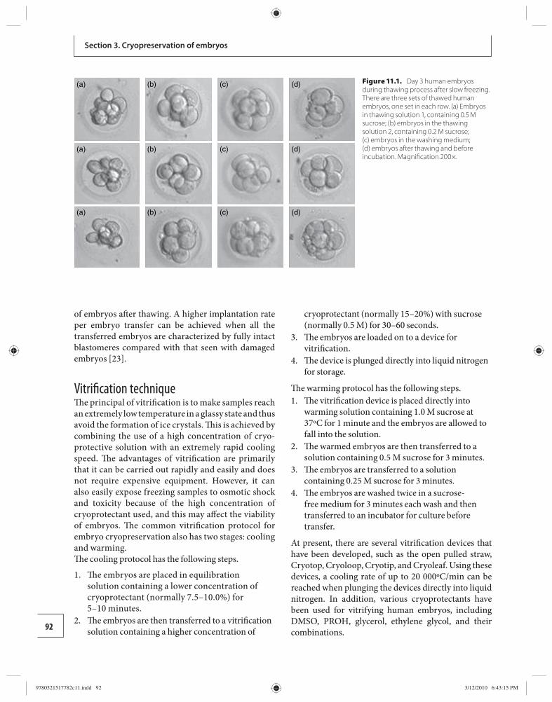
92
Section 3. Cryopreservation of embryos
of embryos aft er thawing. A higher implantation rate per embryo transfer can be achieved when all the transferred embryos are characterized by fully intact blastomeres compared with that seen with damaged embryos [ 23 ].
Vitrifi cation technique Th e principal of vitrifi cation is to make samples reach an extremely low temperature in a glassy state and thus avoid the formation of ice crystals. Th is is achieved by combining the use of a high concentration of cryo-protective solution with an extremely rapid cooling speed. Th e advantages of vitrifi cation are primarily that it can be carried out rapidly and easily and does not require expensive equipment. However, it can also easily expose freezing samples to osmotic shock and toxicity because of the high concentration of cryoprotectant used, and this may aff ect the viability of embryos. Th e common vitrifi cation protocol for embryo cryopreservation also has two stages: cooling and warming. Th e cooling protocol has the following steps.
1. Th e embryos are placed in equilibration solution containing a lower concentration of cryoprotectant (normally 7.5–10.0%) for 5–10 minutes.
2. Th e embryos are then transferred to a vitrifi cation solution containing a higher concentration of
cryoprotectant (normally 15–20%) with sucrose (normally 0.5 M) for 30–60 seconds.
3. Th e embryos are loaded on to a device for vitrifi cation.
4. Th e device is plunged directly into liquid nitrogen for storage.
Th e warming protocol has the following steps. 1. Th e vitrifi cation device is placed directly into
warming solution containing 1.0 M sucrose at 37ºC for 1 minute and the embryos are allowed to fall into the solution.
2. Th e warmed embryos are then transferred to a solution containing 0.5 M sucrose for 3 minutes.
3. Th e embryos are transferred to a solution containing 0.25 M sucrose for 3 minutes.
4. Th e embryos are washed twice in a sucrose-free medium for 3 minutes each wash and then transferred to an incubator for culture before transfer.
At present, there are several vitrifi cation devices that have been developed, such as the open pulled straw, Cryotop, Cryoloop, Cryotip, and Cryoleaf. Using these devices, a cooling rate of up to 20 000ºC/min can be reached when plunging the devices directly into liquid nitrogen. In addition, various cryoprotectants have been used for vitrifying human embryos, including DMSO, PROH, glycerol, ethylene glycol, and their combinations.
(a) (b) (c) (d)
(a) (b) (c) (d)
(a) (b) (c) (d)
Figure 11.1. Day 3 human embryos during thawing process after slow freezing. There are three sets of thawed human embryos, one set in each row. (a) Embryos in thawing solution 1, containing 0.5 M sucrose; (b) embryos in the thawing solution 2, containing 0.2 M sucrose; (c) embryos in the washing medium; (d) embryos after thawing and before incubation. Magnifi cation 200×.
9780521517782c11.indd 929780521517782c11.indd 92 3/12/2010 6:43:15 PM3/12/2010 6:43:15 PM
93
Chapter 11. Cryopreservation of day two and day three embryos
Selection of frozen–thawed embryos for transfer Th e importance of selecting high-quality embryos before cryopreservation has been discussed above. But how are frozen–thawed embryos for transfer selected? Practically, it is based on embryo morphology aft er freeze–thawing. Normally the following criteria are used for selecting embryos: (1) the embryo should have intact blastomeres; (2) the embryo should have a low fragmen-tation of blastomeres; (3) the embryos should show fur-ther development following a period of culture.
It seems the most important factor appears to be to select embryos with continuing development following thawing for transfer, because it has been reported that signifi cantly higher pregnancy and delivery rates have been achieved aft er transferring these embryos com-pared with transfer of embryos without further devel-opment [ 23 , 24 ]. In our experience, we prefer a culture period of 24 hours aft er thawing, and then we select the developing embryos for transfer. Th is procedure con-fi rms that the embryos are viable aft er freeze–thawing, and this can optimize the success rate following trans-fer of the embryos.
Conclusions Cryopreservation of human embryos is an important tool for IVF practice. Human embryos can be cryo-preserved effi ciently from the pronuclear to blastocyst stage. Human embryo cryopreservation is a proven method for fertility preservation. Successful cryopres-ervation of human embryos depends predominantly upon the selection of high-quality embryos before freezing, because embryos of diff ering quality have dif-fering survival rates for freeze–thawing. Th e method for cryopreservation of embryos can be divided into slow freezing and vitrifi cation, and it seems that vitrifi -cation improves embryo survival rate following cryop-reservation. However, the clinical outcome needs to be verifi ed by randomized clinical trials. Th ere is contro-versy concerning cryopreservation of embryos that are on day 2, day 3, or at the blastocyst stage, which directly relates to their condition for the IVF program. Clinical pregnancy and live birth rates are aff ected by the post-thawing selection of embryos for transfer.
References 1. Whittingham DG , Leibo SP , Mazur P . Survival of mouse
embryos frozen to −196 degrees and −269 degrees C . Science 1972; 178 : 411 –414.
2. Wilmut I . Th e low temperature preservation of mammalian embryos . J Reprod Fertil 1972; 31 : 513 –514.
3. Wilmut I . Th e eff ect of cooling rate, warming rate, cryoprotective agent and stage of development on survival of mouse embryos during freezing and thawing . Life Sci 1972; 11: 1071 –1079.
4. Trounson A , Mohr L . Human pregnancy following cryopreservation, thawing and transfer of an eight-cell embryo . Nature 1983; 305: 707 –709.
5. Zeilmaker GH , Alberda AT , van Gent I , Rijkmans CM , Drogendijk AC . Two pregnancies following transfer of intact frozen–thawed embryos . Fertil Steril 1984; 42: 293 –296.
6. Downing BG , Mohr LR , Trounson AO , Freemann LE , Wood C . Birth aft er transfer of cryopreserved embryos . Med J Aust 1985; 142: 409 –411.
7. Cohen J , Simons RF , Fehilly CB , et al. Birth aft er replacement of hatching blastocyst cryopreserved at expanded blastocyst stage . Lancet 1985; 325: 647 .
8. Wood EC . Th e future of in vitro fertilization . Ann N Y Acad Sci 1988; 541 : 715 –721.
9. Van Den Abbeel E , Van Steirteghem A . Zona pellucida damage to human embryos aft er cryopreservation and the consequences for their blastomere survival and in-vitro viability . Hum Reprod 2000; 15: 373 –378.
10. Magli MC , Gianaroli L , Fortini D , Ferraretti AP , Munné S . Impact of blastomere biopsy and cryopreservation techniques on human embryo viability . Hum Reprod 1999; 14: 770 –773.
11. Joris H , Van den Abbeel E , Vos AD , Van Steirteghem A. Reduced survival aft er human embryo biopsy and subsequent cryopreservation . Hum Reprod 1999; 14: 2833 –2837.
12. Van der Elst J , Van den Abbeel E , Vitrier S , et al. Selective transfer of cryopreserved human embryos with further cleavage aft er thawing increases delivery and implantation rates . Hum Reprod 1997; 12: 1513 –1521.
13. Quinn P , Kerin JF . Experience with the cryopreservation of human embryos using the mouse as a model to establish successful techniques . J In Vitro Fert Embryo Transf 1986; 3 : 40 –45.
14. Mukaida T , Nakamura S , Tomiyama T , et al. Vitrifi cation of human blastocysts using cryoloops: clinical outcome of 223 cycles . Hum Reprod 2003; 18 : 384 –391.
15. Mukaida T , Takahashi K , Kasai M . Blastocyst cryopreservation: ultrarapid vitrifi cation using cryoloop technique . Reprod Biomed Online 2003; 6 : 221 –225.
16. Kuwayama M , Vajta G , Ieda S , Kato O . Comparison of open and closed methods for vitrifi cation of human embryos and the elimination of potential contamination . Reprod Biomed Online 2005; 11: 608 –614.
17. Rama Raju GA , Haranath GB , Krishna KM , Prakash GJ , Madan K . Vitrifi cation of human 8-cell embryos,
9780521517782c11.indd 939780521517782c11.indd 93 3/12/2010 6:43:18 PM3/12/2010 6:43:18 PM
94
Section 3. Cryopreservation of embryos
a modifi ed protocol for better pregnancy rates . Reprod Biomed Online 2005; 11 : 434 –437.
18. Desai N , Blackmon H , Szeptycki J , Goldfarb J . Cryoloop vitrifi cation of human day 3 cleavage-stage embryos: post-vitrifi cation development, pregnancy outcomes and live births . Reprod Biomed Online 2007; 14: 208 –213.
19. Jain JK , Paulson RJ . Oocyte cryopreservation . Fertil Steril 2006; 86: 1037 –1046.
20. Ménézo Y , Nicollet B , Herbaut N , André D . Freezing cocultured human blastocysts . Fertil Steril 1992; 58: 977 –980.
21. Behr B , Gebhardt J , Lyon J , Milki AA . Factors relating to a successful cryopreserved blastocyst transfer program . Fertil Steril 2002; 77: 697 –699.
22. Guerif F , Bidault R , Cadoret V , Couet ML , Lansac J , Royere D . Parameters guiding selection of best embryos for transfer aft er cryopreservation: a reappraisal . Hum Reprod 2002; 17: 1321 –1326.
23. American Society for Reproductive Medicine . Day 3 compared with day 2 cryopreservation does not aff ect embryo survival but improves the outcome of frozen–thawed embryo transfers . Fertil Steril 2006; 86: 1537 –1540.
24. Hartshorne GM , Elder K , Crow J , Dyson H , Edwards RG . Th e infl uence of in-vitro development upon post-thaw survival and implantation of cryo preserved human blastocysts . Hum Reprod 1991; 6: 136 –141.
9780521517782c11.indd 949780521517782c11.indd 94 3/12/2010 6:43:18 PM3/12/2010 6:43:18 PM
95Fertility Cryopreservation, ed. Ri-Cheng Chian and Patrick Quinn. Published by Cambridge University Press. Copyright © Cambridge University Press 2010.
Chapter
12 in terms of prevention of ovarian hyperstimulation syndrome and thorough improvement of endometrial receptivity. Moreover, cryobiological techniques can be used in association with preimplantation genetic diagnosis (PGD) to prevent hereditary diseases.
Th irty years have passed since the fi rst baby conceived by in vitro fertilization (IVF) was born. Nowadays, minimum stimulations of the ovary have has become one of the topics of interest and single blas-tocyst transfer is required to prevent multiple pregnan-cies, which is a risk for both the mothers and the babies. Th erefore, cryopreservation techniques for blastocysts are required to preserve surplus blastocysts.
Structural features of the blastocyst Soon aft er the morula enters into the uterine cavity, 4 days aft er fertilization, a blastocele, which is a fl uid-fi lled cavity, appears inside the embryonic mass. Fluid passes through the zona pellucida into the cell from the uterine cavity. Th ereaft er, cells separate into two parts, the trophectoderm and the inner cell mass. At this stage, the embryo is called a blastocyst. Th e blasto-cyst possesses the unique structure of a blastocele and is very diff erent in shape from the other developmen-tal stages of preimplantation embryo. Initially the blastocele is small, but it grows as it accumulates fl uid; the embryo enlarges and is known as an expanded blastocyst. Blastocysts expand and contract periodic-ally. Cryopreservation of blastocysts requires careful attention because of their delicate structure. Ice crystal injury may be the main reason for low survival rates aft er thawing, as blastocysts contain large amounts of water inside the blastocele. In order to address this issue, two methods can be adopted. One is to freeze embryos at the contraction stage and the other is to artifi cially shrink the blastocele (described in detail below). Consequently, in blastocyst cryopreserva-tion, the structure at each stage should be considered
Introduction Cryopreservation is currently limited to sperm or embryos but for extending its use to other fi elds, such as stem cell research, the technology needs improvement. Recently, cryopreservation of gametes in the form of gonadal tissue, such as testicular and ovarian tissues at various developmental stages, has been achieved [ 1 ].
Th e fi rst successful cryopreservation in the fi eld of reproduction was the freezing of animal spermatozoa, performed in Italy in 1776 [ 2 ] . Two centuries later, successful human pregnancies and births from frozen embryos were reported [ 3 ]. Nowadays, it is a routine procedure in infertility centers to freeze pronuclear, multicellular embryos, and blastocysts [ 4 ]. Successful pregnancies from cryopreserved and thawed human blastocysts were fi rst reported by Cohen et al. [ 5 ] in 1985. In the early work on the culture of blastocysts, the survival and pregnancy rates aft er freezing and thaw-ing the blastocysts was low. Ménézo et al. [ 6 ] applied the technique of co-culture for culturing blastocysts. Th e blastocysts were cultured on Vero cells before freez-ing. Th e embryos showed an increase of pre-embryo cell number and Vero cells enhanced the competence for further development [ 7 ]. Th e benefi ts of co-culture were thought to derive from the eff ect of embryotropic factors, including glycoproteins, released from the feeder cell layer and the reduction of the action of toxic compounds such as heavy metals. Another approach was to remove lysed blastomeres from frozen–thawed embryos [ 8 ]. Th e pregnancy rate was much higher in this group than in the unaltered group. Th is study indi-cated that necrotic blastomeres may produce some del-eterious substances.
Recently, a vitrifi cation method has been devel-oped as another option. Oocytes and ovarian tis-sues can be cryopreserved using this new technique. Cryopreservation of gametes, embryos, and ovarian tissues is benefi cial and safe for infertility treatment
Cryopreservation of blastocysts Yoshiharu Morimoto
9780521517782c12.indd 959780521517782c12.indd 95 3/12/2010 6:44:22 PM3/12/2010 6:44:22 PM
96
Section 3. Cryopreservation of embryos
in order to achieve a preferential survival rate aft er thawing.
Which is the best stage for transfer? Early cleavage stage embryos such as those at day 2 or day 3 are transferred in routine work. Recently, improve-ment in culture methods, especially the development of improved media, has made prolonged culture possible, providing another option. Culturing of the blastocyst may be advantageous for the assessment of embryo quality; however, controversy still surrounds the choice of which is the best stage to transfer. Prolonged time in in vitro culture may increase the chances of impairment of embryos through changes in temperature, pH, and osmolality. Meta-analysis of the Cochrane database provides results that have changed over time. In 2002 [ 9 ], the study showed little diff erence in the major out-come for parameters such as birth rates, clinical preg-nancies, and implantations per woman between early stage embryo and blastocyst transfer.
In 2005 [ 10 ], the database also showed no evi-dence of diff erence between the two groups. However, in 2007 [ 11 ], blastocyst transfer was proved to be the preferred method, in terms of a better live birth rate than seen with other methods. Th e study showed that the number of surplus embryos frozen was higher in the early cleaved embryo transfer group and that the transfer cancellation rate was higher in the blastocyst transfer group. Th is change might refl ect improve-ments in media, culture conditions, and the laboratory techniques used in each laboratory.
Which is the best stage for cryopreservation? Th e utilization of blastocysts for transfer has recently been adopted in humans. Th e development of sequen-tial medium enabled this new technology. Blastocysts that had been cultured in sequential medium were shown to be well preserved by cryopreservation [ 12 ].
Th e eff ectiveness of blastocyst cryopreservation has been described in many studies. In a 3 year trial of blastocyst cryopreservation, 76% of blastocysts sur-vived and a 59% pregnancy rate was achieved [ 13 ].
Many authors have discussed which stage of cell growth is appropriate for cryopreservation. Pantos et al. [ 14 ] compared the eff ectiveness of cryopreserva-tion at each stage of embryo development in a retro-spective study. In 170 cycles, using day 6 blastocysts, the survival rate was much better in cleaved embryos
(89%) than in blastocysts (56%). Th e review also iden-tifi ed the same tendency in implantation rate. Troup et al. [ 15 ] compared the survival rates aft er thawing at the pronuclear, cleaved embryo, and blastocyst stages. A greater survival rate was shown at the pronuclear (72%) and cleavage embryo (60%) stages than at the expanded blastocyst stage (38%). Th e proportion of patients who required embryo replacement was also much higher in the pronuclear and cleaved embryo stages. Moreover, Liebermann and Tucker [ 16 ], in their comparison of day 5 and day 6 blastocysts in 254 vitrifi ed transfer cycles, showed no diff erence between the two groups in terms of survival rate, implantation rate, and pregnancy rate. Th e result was the same with slow-freezing cycles. In contrast, cryopreservation at the blastocysts stage has been reported as being more eff ective in some studies.
Which stage, pronuclear or blastocyst, yields more transferable embryos has not yet been identifi ed. We have compared two stages, where both the pronuclear and blastocyst stage embryos had been cryopreserved for the same patient [ 17 ]. Th ere was no diff erence in the number of transferable blastocysts between two groups. However vitrifi cation at the blastocyst stage is more benefi cial for patients and laboratories in terms of cost and space, because the number of vitrifi ed pronu-clear and cleaved embryos is approximately fi ve times that of embryos vitrifi ed at the blastocyst stage.
Selection of culture methods and eff ectiveness of blastocyst cryopreservation Culture methods Th e development of a culture method for blastocysts was a breakthrough in assisted reproductive technology (ART) [ 18 ]. Sequential media for human blas-tocyst culture was fi rst described by Gardner and Lane [ 19 ] and is now used all over the world as a routine pro-cedure for IVF. Even with more options for ART pro-cedures, there is still controversy over which is the best stage for transfer. Some patients conceive with day 2 or day 3 stage embryo transfer aft er failure with transfer of an embryo at the blastocyst stage. More studies are needed to clarify whether blastocysts have the highest competence for further embryonic development into a fetus.
Currently there are two kinds of strategy for blasto-cyst stage culture. One is a sequential culture method
9780521517782c12.indd 969780521517782c12.indd 96 3/12/2010 6:44:24 PM3/12/2010 6:44:24 PM
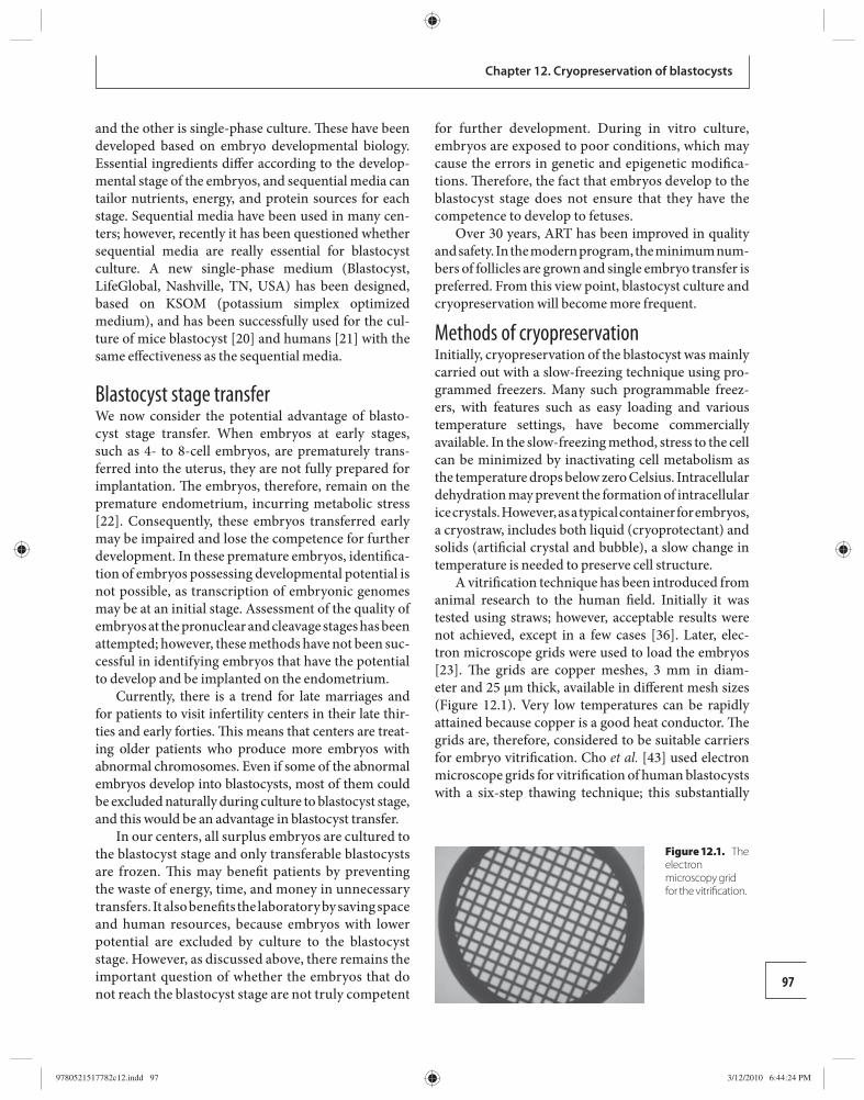
97
Chapter 12. Cryopreservation of blastocysts
and the other is single-phase culture. Th ese have been developed based on embryo developmental biology. Essential ingredients diff er according to the develop-mental stage of the embryos, and sequential media can tailor nutrients, energy, and protein sources for each stage. Sequential media have been used in many cen-ters; however, recently it has been questioned whether sequential media are really essential for blastocyst culture. A new single-phase medium (Blastocyst, LifeGlobal, Nashville, TN, USA) has been designed, based on KSOM (potassium simplex optimized medium), and has been successfully used for the cul-ture of mice blastocyst [ 20 ] and humans [ 21 ] with the same eff ectiveness as the sequential media.
Blastocyst stage transfer We now consider the potential advantage of blasto-cyst stage transfer. When embryos at early stages, such as 4- to 8-cell embryos, are prematurely trans-ferred into the uterus, they are not fully prepared for implantation. Th e embryos, therefore, remain on the premature endometrium, incurring metabolic stress [ 22 ]. Consequently, these embryos transferred early may be impaired and lose the competence for further development. In these premature embryos, identifi ca-tion of embryos possessing developmental potential is not possible, as transcription of embryonic genomes may be at an initial stage. Assessment of the quality of embryos at the pronuclear and cleavage stages has been attempted; however, these methods have not been suc-cessful in identifying embryos that have the potential to develop and be implanted on the endometrium.
Currently, there is a trend for late marriages and for patients to visit infertility centers in their late thir-ties and early forties. Th is means that centers are treat-ing older patients who produce more embryos with abnormal chromosomes. Even if some of the abnormal embryos develop into blastocysts, most of them could be excluded naturally during culture to blastocyst stage, and this would be an advantage in blastocyst transfer.
In our centers, all surplus embryos are cultured to the blastocyst stage and only transferable blastocysts are frozen. Th is may benefi t patients by preventing the waste of energy, time, and money in unnecessary transfers. It also benefi ts the laboratory by saving space and human resources, because embryos with lower potential are excluded by culture to the blastocyst stage. However, as discussed above, there remains the important question of whether the embryos that do not reach the blastocyst stage are not truly competent
for further development. During in vitro culture, embryos are exposed to poor conditions, which may cause the errors in genetic and epigenetic modifi ca-tions. Th erefore, the fact that embryos develop to the blastocyst stage does not ensure that they have the competence to develop to fetuses.
Over 30 years, ART has been improved in quality and safety. In the modern program, the minimum num-bers of follicles are grown and single embryo transfer is preferred. From this view point, blastocyst culture and cryopreservation will become more frequent.
Methods of cryopreservation Initially, cryopreservation of the blastocyst was mainly carried out with a slow-freezing technique using pro-grammed freezers. Many such programmable freez-ers, with features such as easy loading and various temperature settings, have become commercially available. In the slow-freezing method, stress to the cell can be minimized by inactivating cell metabolism as the temperature drops below zero Celsius. Intracellular dehydration may prevent the formation of intracellular ice crystals. However, as a typical container for embryos, a cryostraw, includes both liquid (cryoprotectant) and solids (artifi cial crystal and bubble), a slow change in temperature is needed to preserve cell structure.
A vitrifi cation technique has been introduced from animal research to the human fi eld. Initially it was tested using straws; however, acceptable results were not achieved, except in a few cases [ 36 ]. Later, elec-tron microscope grids were used to load the embryos [ 23 ]. Th e grids are copper meshes, 3 mm in diam-eter and 25 μm thick, available in diff erent mesh sizes ( Figure 12.1 ). Very low temperatures can be rapidly attained because copper is a good heat conductor. Th e grids are, therefore, considered to be suitable carriers for embryo vitrifi cation. Cho et al. [ 43 ] used electron microscope grids for vitrifi cation of human blastocysts with a six-step thawing technique; this substantially
Figure 12.1. The electron micro scopy grid for the vitrifi cation.
9780521517782c12.indd 979780521517782c12.indd 97 3/12/2010 6:44:24 PM3/12/2010 6:44:24 PM
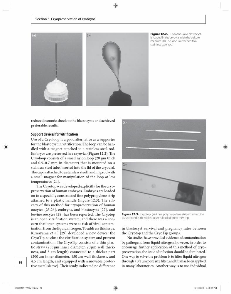
98
Section 3. Cryopreservation of embryos
reduced osmotic shock to the blastocysts and achieved preferable results.
Support devices for vitrifi cation Use of a Cryoloop is a good alternative as a supporter for the blastocyst in vitrifi cation. Th e loop can be han-dled with a magnet attached to a stainless steel rod. Embryos are preserved in a cryovial ( Figure 12.2 ). Th e Cryoloop consists of a small nylon loop (20 μm thick and 0.5–0.7 mm in diameter) that is mounted on a stainless steel tube inserted into the lid of the cryovial. Th e cap is attached to a stainless steel handling rod with a small magnet for manipulation of the loop at low temperatures [ 24 ].
Th e Cryotop was developed explicitly for the cryo-preservation of human embryos. Embryos are loaded on to a specially constructed fi ne polypropylene strip attached to a plastic handle ( Figure 12.3 ). Th e effi -cacy of this method for cryopreservation of human oocytes [ 25 , 26 ], embryos, and blastocysts [ 27 ], and bovine oocytes [ 28 ] has been reported. Th e Cryotop is an open vitrifi cation system, and there was a con-cern that open systems were at risk of viral contam-ination from the liquid nitrogen. To address this issue, Kuwayama et al. [ 29 ] developed a new device, the CryoTip, to close the vitrifi cation system and prevent contamination. Th e CryoTip consists of a thin plas-tic straw (250 μm inner diameter, 20 μm wall thick-ness, and 3 cm length) connected to a thicker part (200 μm inner diameter, 150 μm wall thickness, and 4.5 cm length, and equipped with a movable protec-tive metal sleeve). Th eir study indicated no diff erence
in blastocyst survival and pregnancy rates between the Cryotop and the CryoTip groups.
No studies have provided evidence of contamination by pathogens from liquid nitrogen; however, in order to encourage further application of this method of cryo-preservation, the issue of infection should be eliminated. One way to solve the problem is to fi lter liquid nitrogen through a 0.2 μm pore size fi lter, and this has been applied in many laboratories. Another way is to use individual
(a) (b)
(a)
(b)
Figure 12.3. Cryotop. (a) A fi ne polypropylene strip attached to a plastic handle. (b) A blastocyst is loaded on to the strip.
Figure 12.2. Cryoloop. (a) A blastocyst is loaded in the cryovial with the culture medium. (b) The loop is attached to a stainless steel rod.
9780521517782c12.indd 989780521517782c12.indd 98 3/12/2010 6:44:25 PM3/12/2010 6:44:25 PM
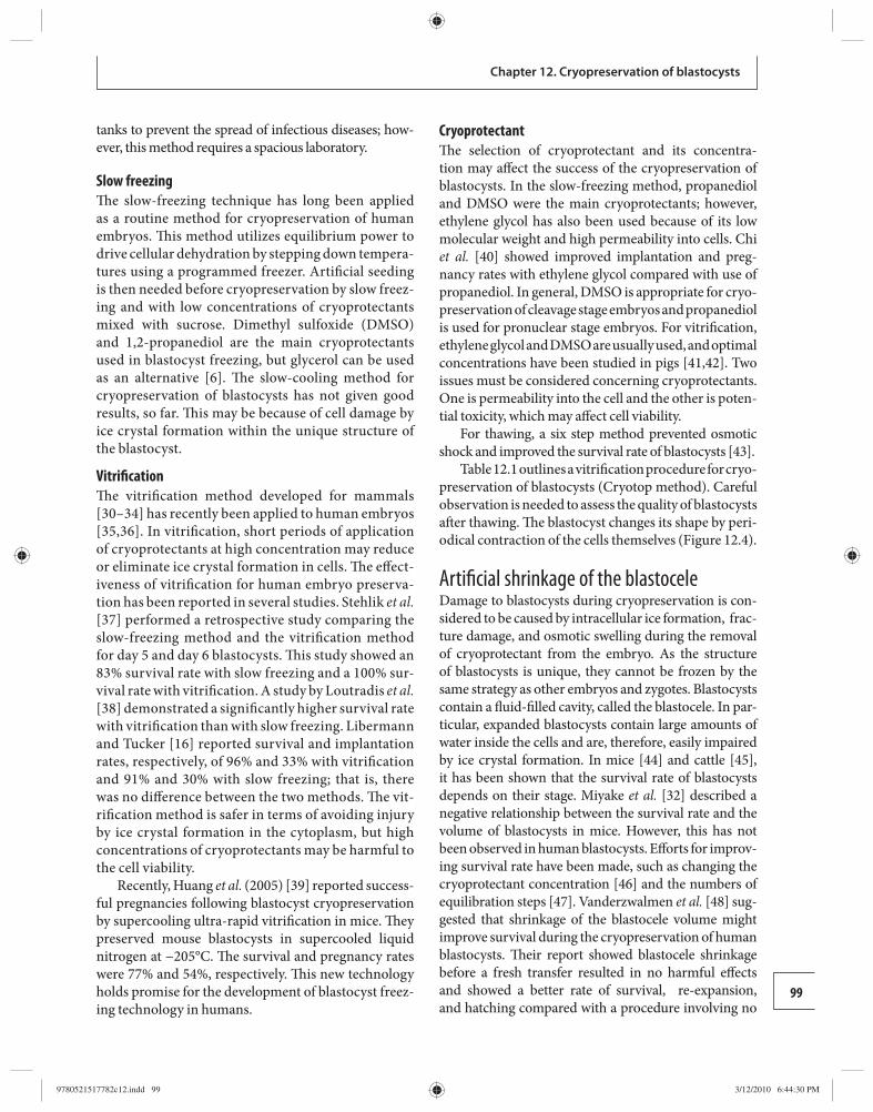
99
Chapter 12. Cryopreservation of blastocysts
tanks to prevent the spread of infectious diseases; how-ever, this method requires a spacious laboratory.
Slow freezing Th e slow-freezing technique has long been applied as a routine method for cryopreservation of human embryos. Th is method utilizes equilibrium power to drive cellular dehydration by stepping down tempera-tures using a programmed freezer. Artifi cial seeding is then needed before cryopreservation by slow freez-ing and with low concentrations of cryoprotectants mixed with sucrose. Dimethyl sulfoxide (DMSO) and 1,2-propanediol are the main cryoprotectants used in blastocyst freezing, but glycerol can be used as an alternative [ 6 ]. Th e slow-cooling method for cryopreservation of blastocysts has not given good results, so far. Th is may be because of cell damage by ice crystal formation within the unique structure of the blastocyst.
Vitrifi cation Th e vitrifi cation method developed for mammals [ 30 – 34 ] has recently been applied to human embryos [ 35 , 36 ]. In vitrifi cation, short periods of application of cryoprotectants at high concentration may reduce or eliminate ice crystal formation in cells. Th e eff ect-iveness of vitrifi cation for human embryo preserva-tion has been reported in several studies. Stehlik et al. [ 37 ] performed a retrospective study comparing the slow-freezing method and the vitrifi cation method for day 5 and day 6 blastocysts. Th is study showed an 83% survival rate with slow freezing and a 100% sur-vival rate with vitrifi cation. A study by Loutradis et al. [ 38 ] demonstrated a signifi cantly higher survival rate with vitrifi cation than with slow freezing. Libermann and Tucker [ 16 ] reported survival and implantation rates, respectively, of 96% and 33% with vitrifi cation and 91% and 30% with slow freezing; that is, there was no diff erence between the two methods. Th e vit-rifi cation method is safer in terms of avoiding injury by ice crystal formation in the cytoplasm, but high concentrations of cryoprotectants may be harmful to the cell viability.
Recently, Huang et al. (2005) [ 39 ] reported success-ful pregnancies following blastocyst cryopreservation by supercooling ultra-rapid vitrifi cation in mice. Th ey preserved mouse blastocysts in supercooled liquid nitrogen at −205°C. Th e survival and pregnancy rates were 77% and 54%, respectively. Th is new technology holds promise for the development of blastocyst freez-ing technology in humans.
Cryoprotectant Th e selection of cryoprotectant and its concentra-tion may aff ect the success of the cryopreservation of blastocysts. In the slow-freezing method, propanediol and DMSO were the main cryoprotectants; however, ethylene glycol has also been used because of its low molecular weight and high permeability into cells. Chi et al. [ 40 ] showed improved implantation and preg-nancy rates with ethylene glycol compared with use of propanediol. In general, DMSO is appropriate for cryo-preservation of cleavage stage embryos and propanediol is used for pronuclear stage embryos. For vitrifi cation, ethylene glycol and DMSO are usually used, and optimal concentrations have been studied in pigs [ 41 , 42 ]. Two issues must be considered concerning cryoprotectants. One is permeability into the cell and the other is poten-tial toxicity, which may aff ect cell viability.
For thawing, a six step method prevented osmotic shock and improved the survival rate of blastocysts [ 43 ].
Table 12.1 outlines a vitrifi cation procedure for cryo-preservation of blastocysts (Cryotop method). Careful observation is needed to assess the quality of blastocysts aft er thawing. Th e blastocyst changes its shape by peri-odical contraction of the cells themselves ( Figure 12.4 ).
Artifi cial shrinkage of the blastocele Damage to blastocysts during cryopreservation is con-sidered to be caused by intracellular ice formation, frac-ture damage, and osmotic swelling during the removal of cryoprotectant from the embryo. As the structure of blastocysts is unique, they cannot be frozen by the same strategy as other embryos and zygotes. Blastocysts contain a fl uid-fi lled cavity, called the blastocele. In par-ticular, expanded blastocysts contain large amounts of water inside the cells and are, therefore, easily impaired by ice crystal formation. In mice [ 44 ] and cattle [ 45 ], it has been shown that the survival rate of blastocysts depends on their stage. Miyake et al. [ 32 ] described a negative relationship between the survival rate and the volume of blastocysts in mice. However, this has not been observed in human blastocysts. Eff orts for improv-ing survival rate have been made, such as changing the cryoprotectant concentration [ 46 ] and the numbers of equilibration steps [ 47 ]. Vanderzwalmen et al. [ 48 ] sug-gested that shrinkage of the blastocele volume might improve survival during the cryopreservation of human blastocysts. Th eir report showed blastocele shrinkage before a fresh transfer resulted in no harmful eff ects and showed a better rate of survival, re-expansion, and hatching compared with a procedure involving no
9780521517782c12.indd 999780521517782c12.indd 99 3/12/2010 6:44:30 PM3/12/2010 6:44:30 PM
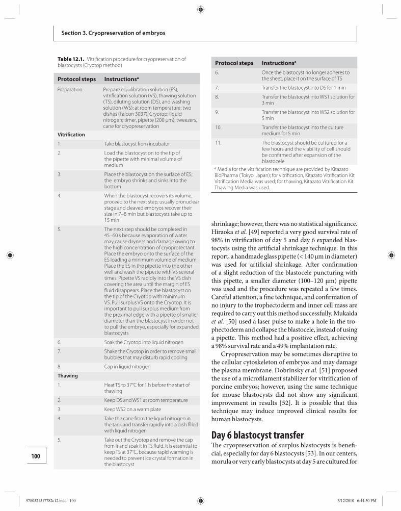
100
Section 3. Cryopreservation of embryos
shrinkage; however, there was no statistical signifi cance. Hiraoka et al. [ 49 ] reported a very good survival rate of 98% in vitrifi cation of day 5 and day 6 expanded blas-tocysts using the artifi cial shrinkage technique. In this report, a handmade glass pipette (< 140 μm in diameter) was used for artifi cial shrinkage. Aft er confi rmation of a slight reduction of the blastocele puncturing with this pipette, a smaller diameter (100–120 μm) pipette was used and the procedure was repeated a few times. Careful attention, a fi ne technique, and confi rmation of no injury to the trophectoderm and inner cell mass are required to carry out this method successfully. Mukaida et al. [ 50 ] used a laser pulse to make a hole in the tro-phectoderm and collapse the blastocele, instead of using a pipette. Th is method had a positive eff ect, achieving a 98% survival rate and a 49% implantation rate.
Cryopreservation may be sometimes disruptive to the cellular cytoskeleton of embryos and may damage the plasma membrane. Dobrinsky et al. [ 51 ] proposed the use of a microfi lament stabilizer for vitrifi cation of porcine embryos; however, using the same technique for mouse blastocysts did not show any signifi cant improvement in results [ 52 ]. It is possible that this technique may induce improved clinical results for human blastocysts.
Day 6 blastocyst transfer Th e cryopreservation of surplus blastocysts is benefi -cial, especially for day 6 blastocysts [ 53 ]. In our centers, morula or very early blastocysts at day 5 are cultured for
Table 12.1. Vitrifi cation procedure for cryopreservation of blastocysts (Cryotop method)
Protocol steps Instructions a
Preparation Prepare equilibration solution (ES), vitrifi cation solution (VS), thawing solution (TS), diluting solution (DS), and washing solution (WS); at room temperature; two dishes (Falcon 3037); Cryotop; liquid nitrogen; timer, pipette (200 μm); tweezers, cane for cryopreservation
Vitrifi cation
1. Take blastocyst from incubator
2. Load the blastocyst on to the tip of the pipette with minimal volume of medium
3. Place the blastocyst on the surface of ES; the embryo shrinks and sinks into the bottom
4. When the blastocyst recovers its volume, proceed to the next step; usually pronuclear stage and cleaved embryos recover their size in 7–8 min but blastocysts take up to 15 min
5. The next step should be completed in 45–60 s because evaporation of water may cause dryness and damage owing to the high concentration of cryoprotectant. Place the embryo onto the surface of the ES loading a minimum volume of medium. Place the ES in the pipette into the other well and wash the pipette with VS several times. Pipette VS rapidly into the VS dish covering the area until the margin of ES fl uid disappears. Place the blastocyst on the tip of the Cryotop with minimum VS. Pull surplus VS onto the Cryotop. It is important to pull surplus medium from the proximal edge with a pipette of smaller diameter than the blastocyst in order not to pull the embryo, especially for expanded blastocysts
6. Soak the Cryotop into liquid nitrogen
7. Shake the Cryotop in order to remove small bubbles that may disturb rapid cooling
8. Cap in liquid nitrogen
Thawing
1. Heat TS to 37°C for 1 h before the start of thawing
2. Keep DS and WS1 at room temperature
3. Keep WS2 on a warm plate
4. Take the cane from the liquid nitrogen in the tank and transfer rapidly into a dish fi lled with liquid nitrogen
5. Take out the Cryotop and remove the cap from it and soak it in TS fl uid. It is essential to keep TS at 37°C, because rapid warming is needed to prevent ice crystal formation in the blastocyst
Protocol steps Instructions a 6. Once the blastocyst no longer adheres to
the sheet, place it on the surface of TS
7. Transfer the blastocyst into DS for 1 min
8. Transfer the blastocyst into WS1 solution for 3 min
9. Transfer the blastocyst into WS2 solution for 5 min
10. Transfer the blastocyst into the culture medium for 5 min
11. The blastocyst should be cultured for a few hours and the viability of cell should be confi rmed after expansion of the blastocele
a Media for the vitrifi cation technique are provided by Kitazato BioPharma (Tokyo, Japan); for vitrifi cation, Kitazato Vitrifi cation Kit Vitrifi cation Media was used; for thawing, Kitazato Vitrifi cation Kit Thawing Media was used.
9780521517782c12.indd 1009780521517782c12.indd 100 3/12/2010 6:44:30 PM3/12/2010 6:44:30 PM
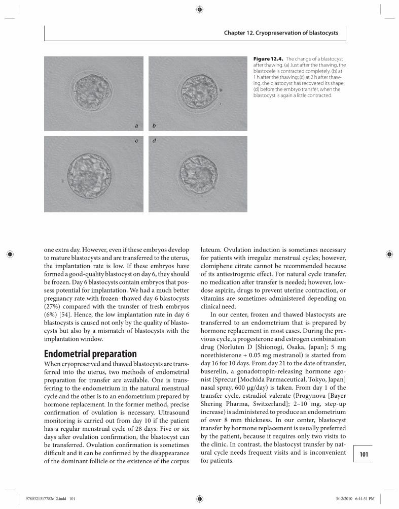
101
Chapter 12. Cryopreservation of blastocysts
one extra day. However, even if these embryos develop to mature blastocysts and are transferred to the uterus, the implantation rate is low. If these embryos have formed a good-quality blastocyst on day 6, they should be frozen. Day 6 blastocysts contain embryos that pos-sess potential for implantation. We had a much better pregnancy rate with frozen–thawed day 6 blastocysts (27%) compared with the transfer of fresh embryos (6%) [ 54 ]. Hence, the low implantation rate in day 6 blastocysts is caused not only by the quality of blasto-cysts but also by a mismatch of blastocysts with the implantation window.
Endometrial preparation When cryopreserved and thawed blastocysts are trans-ferred into the uterus, two methods of endometrial preparation for transfer are available. One is trans-ferring to the endometrium in the natural menstrual cycle and the other is to an endometrium prepared by hormone replacement. In the former method, precise confi rmation of ovulation is necessary. Ultrasound monitoring is carried out from day 10 if the patient has a regular menstrual cycle of 28 days. Five or six days aft er ovulation confi rmation, the blastocyst can be transferred. Ovulation confi rmation is sometimes diffi cult and it can be confi rmed by the disappearance of the dominant follicle or the existence of the corpus
luteum. Ovulation induction is sometimes necessary for patients with irregular menstrual cycles; however, clomiphene citrate cannot be recommended because of its antiestrogenic eff ect. For natural cycle transfer, no medication aft er transfer is needed; however, low-dose aspirin, drugs to prevent uterine contraction, or vitamins are sometimes administered depending on clinical need.
In our center, frozen and thawed blastocysts are transferred to an endometrium that is prepared by hormone replacement in most cases. During the pre-vious cycle, a progesterone and estrogen combination drug (Norluten D [Shionogi, Osaka, Japan]; 5 mg norethisterone + 0.05 mg mestranol) is started from day 16 for 10 days. From day 21 to the date of transfer, buserelin, a gonadotropin-releasing hormone ago-nist (Sprecur [Mochida Parmaceutical, Tokyo, Japan] nasal spray, 600 μg/day) is taken. From day 1 of the transfer cycle, estradiol valerate (Progynova [Bayer Shering Pharma, Switzerland]; 2–10 mg, step-up increase) is administered to produce an endometrium of over 8 mm thickness. In our center, blastocyst transfer by hormone replacement is usually preferred by the patient, because it requires only two visits to the clinic. In contrast, the blastocyst transfer by nat-ural cycle needs frequent visits and is inconvenient for patients.
a b
c d
Figure 12.4. The change of a blastocyst after thawing. (a) Just after the thawing, the blastocele is contracted completely. (b) at 1 h after the thawing; (c) at 2 h after thaw-ing, the blastocyst has recovered its shape; (d) before the embryo transfer, when the blastocyst is again a little contracted.
9780521517782c12.indd 1019780521517782c12.indd 101 3/12/2010 6:44:31 PM3/12/2010 6:44:31 PM
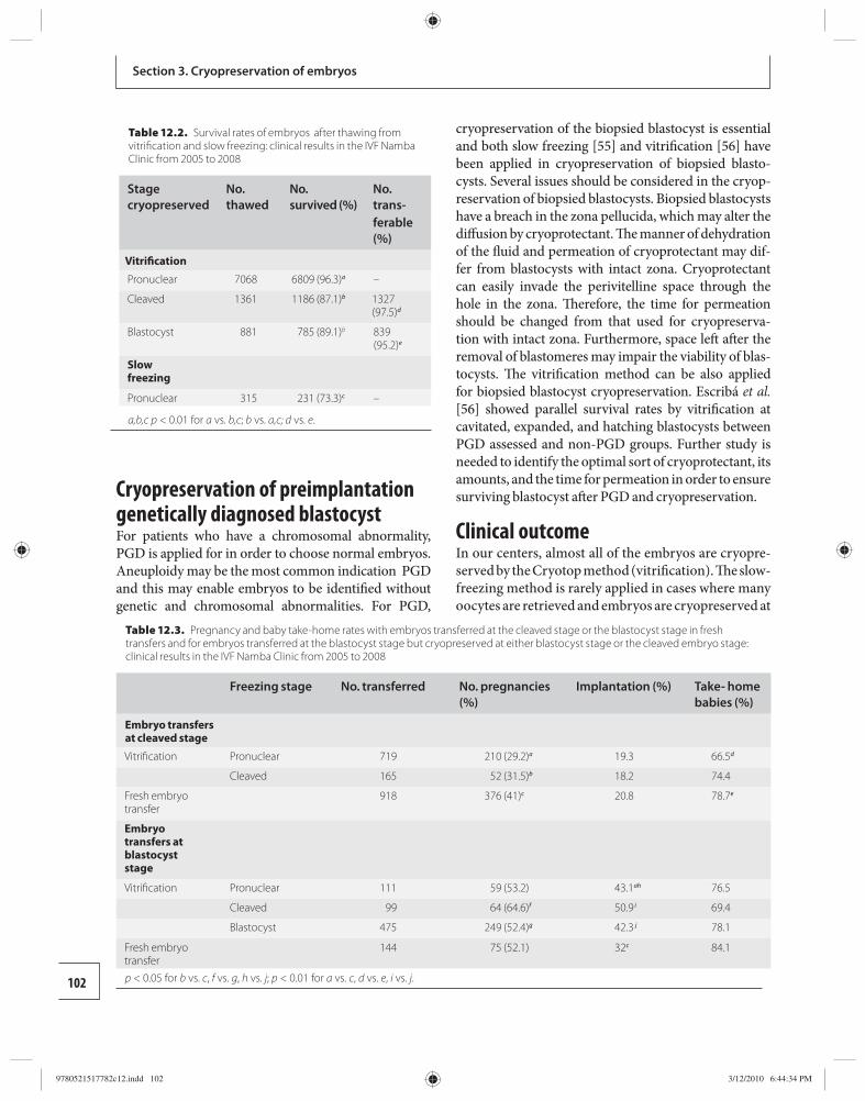
102
Section 3. Cryopreservation of embryos
Cryopreservation of preimplantation genetically diagnosed blastocyst For patients who have a chromosomal abnormality, PGD is applied for in order to choose normal embryos. Aneuploidy may be the most common indication PGD and this may enable embryos to be identifi ed without genetic and chromosomal abnormalities. For PGD,
cryopreservation of the biopsied blastocyst is essential and both slow freezing [ 55 ] and vitrifi cation [ 56 ] have been applied in cryopreservation of biopsied blasto-cysts. Several issues should be considered in the cryop-reservation of biopsied blastocysts. Biopsied blastocysts have a breach in the zona pellucida, which may alter the diff usion by cryoprotectant. Th e manner of dehydration of the fl uid and permeation of cryoprotectant may dif-fer from blastocysts with intact zona. Cryoprotectant can easily invade the perivitelline space through the hole in the zona. Th erefore, the time for permeation should be changed from that used for cryopreserva-tion with intact zona. Furthermore, space left aft er the removal of blastomeres may impair the viability of blas-tocysts. Th e vitrifi cation method can be also applied for biopsied blastocyst cryopreservation. Escribá et al. [ 56 ] showed parallel survival rates by vitrifi cation at cavitated, expanded, and hatching blastocysts between PGD assessed and non-PGD groups. Further study is needed to identify the optimal sort of cryoprotectant, its amounts, and the time for permeation in order to ensure surviving blastocyst aft er PGD and cryopreservation.
Clinical outcome In our centers, almost all of the embryos are cryopre-served by the Cryotop method (vitrifi cation). Th e slow-freezing method is rarely applied in cases where many oocytes are retrieved and embryos are cryopreserved at
Table 12.2. Survival rates of embryos after thawing from vitrifi cation and slow freezing: clinical results in the IVF Namba Clinic from 2005 to 2008
Stage cryopreserved
No.thawed
No. survived (%)
No. trans-ferable (%)
Vitrifi cation
Pronuclear 7068 6809 (96.3) a –
Cleaved 1361 1186 (87.1) b 1327 (97.5) d
Blastocyst 881 785 (89.1) b 839 (95.2) e
Slow freezing
Pronuclear 315 231 (73.3) c –
a,b,c p < 0.01 for a vs. b,c; b vs. a,c; d vs. e.
Table 12.3. Pregnancy and baby take-home rates with embryos transferred at the cleaved stage or the blastocyst stage in fresh transfers and for embryos transferred at the blastocyst stage but cryopreserved at either blastocyst stage or the cleaved embryo stage: clinical results in the IVF Namba Clinic from 2005 to 2008
Freezing stage No. transferred No. pregnancies (%)
Implantation (%) Take- home babies (%)
Embryo transfers at cleaved stage
Vitrifi cation Pronuclear 719 210 (29.2) a 19.3 66.5 d
Cleaved 165 52 (31.5) b 18.2 74.4
Fresh embryo transfer
918 376 (41) c 20.8 78.7 e
Embryo transfers at blastocyst stage
Vitrifi cation Pronuclear 111 59 (53.2) 43.1 ah 76.5
Cleaved 99 64 (64.6) f 50.9 i 69.4
Blastocyst 475 249 (52.4) g 42.3 j 78.1
Fresh embryo transfer
144 75 (52.1) 32 c 84.1
p < 0.05 for b vs. c , f vs. g , h vs. j ; p < 0.01 for a vs. c , d vs. e , i vs. j .
9780521517782c12.indd 1029780521517782c12.indd 102 3/12/2010 6:44:34 PM3/12/2010 6:44:34 PM
103
Chapter 12. Cryopreservation of blastocysts
pronuclear stage, because it wastes a lot of time to cryo-preserve many embryos with vitrifi cation. Th e survival rate aft er thawing was acceptable in both vitrifi cation and slow freezing; however, it was signifi cantly better in vitrifi cation ( Table 12.2 ). With vitrifi cation, the sur-vival rate was the best in the embryos cryopreserved at pronuclear stage. Th e embryos that were frozen at the blastocyst stage showed higher survival, but fewer were transferrable compared with the embryos frozen at the cleavage stage. Th is might be because that the surviving blastocysts included embryos with partial degeneration.
Table 12.3 shows pregnancy and baby take-home rates with embryos transferred at the cleaved stage or the blastocyst stage. Cleaved embryos were transferred aft er thawing in 884 cases and when fresh in 918 cases from 2005 to 2007 in our centers. Th e fresh transfer group showed better pregnancy and baby take-home rates where transfer occurred at the cleaved stage than when it occurred at the blastocyst stage. In transfers at the blastocyst stage, the pregnancy and implantation rate were lower for embryos cryopreserved at blas-tocyst stage than at the cleaved embryo stage. Th ese data are easier to understand if it is remembered that premature blastocysts are included in the number of blastocysts.
References 1. Woods EJ , Benson JD , Agca Y , et al. Fundamental
cryobiology of reproductive cells and tissues . Cryobiology 2004; 48: 146 –156.
2. Bredkjaer HE , Grudzinskas JG . Cryobiology in human assisted reproductive technology . Would Hippocrates approve? Early Pregnancy 2001; 5: 211 –213.
3. Trounson A , Mohr L . Human pregnancy following cryopreservation, thawing and transfer of an eight-cell embryo . Nature 1983; 305: 707 –709.
4. Byrd W . Cryopreservation, thawing, and transfer of human embryos . Semin Reprod Med 2002; 20: 37 –43.
5. Cohen J , Simons RF , Edwards RG , et al. Pregnancies following the frozen storage of expanding human blastocysts . J In Vitro Fert Embryo Transf 1985; 2: 59 –64.
6. Ménézo Y , Nicollet B , Herbaut N , et al. Freezing cocultured human blastocysts . Fertil Steril 1992; 58: 977 –980.
7. Kaufman RA , Ménézo Y , Hazout A , et al. Cocultured blastocyst cryopreservation: experience of more than 500 transfer cycles . Fertil Steril 1995; 64: 1125 –1129.
8. Nagy ZP , Taylor T , Elliott T , et al. Removal of lysed blastomeres from frozen–thawed embryos improves implantation and pregnancy rates in frozen embryo transfer cycles . Fertil Steril 2005; 84: 1606 –1612.
9. Blake D , Proctor M , Johnson N , Olive D . Cleavage stage versus blastocyst stage embryo transfer in assisted conception . Cochrane Database Syst Rev 2002; 2 : CD002118.
10. Blake D , Proctor M , Johnson N , et al. Cleavage stage versus blastocyst stage embryo transfer in assisted conception . Cochrane Database Syst Rev 2005; 4 : CD002118.
11. Blake DA , Farquhar CM , Johnson N , et al. Cleavage stage versus blastocyst stage embryo transfer in assisted conception . Cochrane Database Syst Rev 2007; 4 : CD002118.
12. Virant-Klun I , Tomaźević T , Baćer-Kermavner L , et al. Successful freezing and thawing of blastocysts cultured in sequential media using a modifi ed method . Fertil Steril 2003; 79: 1428 –1433.
13. Veeck LL , Bodine R , Clarke RN , et al. High pregnancy rates can be achieved aft er freezing and thawing human blastocysts . Fertil Steril 2004; 82: 1418 –1427.
14. Pantos K , Stefanidis K , Pappas K , et al. Cryopreservation of embryos, blastocysts, and pregnancy rates of blastocysts derived from frozen–thawed embryos and frozen–thawed blastocysts . J Assist Reprod Genet 2001; 18: 579 –582.
15. Troup SA , Matson PL , Critchlow JD , et al. Cryopreservation of human embryos at the pronucleate, early cleavage, or expanded blastocyst stages . Eur J Obstet Gynecol Reprod Biol 1991; 38: 133 –139.
16. Liebermann J , Tucker MJ . Comparison of vitrifi cation and conventional cryopreservation of day 5 and day 6 blastocysts during clinical application . Fertil Steril 2006; 86: 20 –26.
17. Sawada N , Akamatsu Y , Tsuji Y , et al. Eff ects of developmental stage of human embryos on the development to transferable blastocyst following vitirfi cation and warming . Reprod Biomed Online 2008; 16 (Suppl 2): S28– S29.
18. Youssry M , Ozmen B , Zohni K , et al. Current aspects of blastocyst cryopreservation . Reprod Biomed Online 2008; 16: 311 –320.
19. Gardner DK , Lane M . Culture and selection of viable blastocysts: a feasible proposition for human IVF? Hum Reprod Update 1997; 3: 367 –382.
20. Lawitts JA , Biggers JD . Culture of preimplantation embryos . Method Enzymol 1993; 225: 153 –164.
21. Biggers JD , Racowsky C . Th e development of fertilized human ova to the blastocyst stage in KSOM(AA) medium: is a two-step protocol necessary? Reprod Biomed Online 2002; 5: 133 –140.
22. Gardner DK , Michelle L . Culture systems for the human embryo. In Gardner DK , Weissman A , Howles CM , et al., eds. Textbook of Assisted Reproductive Techniques: Laboratory and Clinical Perspectives, 2nd edn. London : Taylor & Francis, 2004, pp. 211–234.
9780521517782c12.indd 1039780521517782c12.indd 103 3/12/2010 6:44:34 PM3/12/2010 6:44:34 PM
104
Section 3. Cryopreservation of embryos
23. Choi DH , Chung HM , Lim JM , et al. Pregnancy and delivery of healthy infants developed from vitrifi ed blastocysts in an IVF-ET program . Fertil Steril 2000; 74: 838 –839.
24. Mukaida T , Takahashi K , Kasai M . Blastocyst cryopre-servation: ultrarapid vitrifi cation using cryoloop technique . Reprod Biomed Online 2003; 6: 221 –225.
25. Cobo A , Bellver J , Domingo J , et al. New options in assisted reproduction technology: the Cryotop method of oocyte vitrifi cation . Reprod Biomed Online 2008; 17 : 68 –72.
26. Kuwayama M , Vajta G , Kato O , et al. Highly effi cient vitrifi cation method for cryopreservation of human oocytes . Reprod Biomed Online 2005; 11: 300 –308.
27. Kuwayama M . Highly effi cient vitrifi cation for cryopreservation of human oocytes and embryos: the Cryotop method . Th eriogenology 2007; 67: 73 –80.
28. Morató R , Izquierdo D , Paramio MT , et al. Cryotops versus open-pulled straws (OPS) as carriers for the cryopreservation of bovine oocytes: eff ects on spindle and chromosome confi guration and embryo development . Cryobiology 2008; 57: 137 –141.
29. Kuwayama M , Vajta G , Ieda S , et al. Comparison of open and closed methods for vitrifi cation of human embryos and the elimination of potential contamination . Reprod Biomed Online 2005; 11: 608 –614.
30. Kuwayama M , Hamano S , Nagai T . Vitrifi cation of bovine blastocysts obtained by in vitro culture of oocytes matured and fertilized in vitro . J. Reprod Fertil 1992; 96: 187 –193.
31. Massip A , Vanderzwalmen P , Scheff en B , et al. Pregnancies following transfer of cattle embryos preserved by vitrifi cation . Cryo Letters 1986; 7 : 270 –273.
32. Miyake T , Kasai M , Zhu SE , et al. Vitrifi cation of mouse oocytes and embryos at various stages of development in an ethylene glycol-based solution by a simple method . Th eriogenology 1993; 40: 121 –134.
33. Vanderzwalmen P , Gaurois B , Ectors FJ , et al. Some factors aff ecting successful vitrifi cation of mouse blastocysts . Th eriogenology 1988; 30: 1177 –1183.
34. Dattena M , Ptak G , Loi P , et al. Survival and viability of vitrifi ed in vitro and in vivo produced ovine blastocysts . Th eriogenology 2000; 53: 1511 –1519.
35. Choi DH , Chung HM , Lim JM , et al. Pregnancy and delivery of healthy infants developed from vitrifi ed blastocysts in an IVF-ET program . Fertil Steril 2000; 74: 838 –839.
36. Yokota Y , Sato S , Yokota M , et al. Birth of a healthy baby following vitrifi cation of human blastocysts . Fertil Steril 2001; 75: 1027 –1029.
37. Stehlik E , Stehlik J , Katayama KP , et al. Vitrifi cation demonstrates signifi cant improvement versus slow freezing of human blastocysts . Reprod Biomed Online 2005; 11: 53 –57.
38. Loutradis KE , Kolibianakis EM , Venetis CA , et al. Cryopreservation of human embryos by vitrifi cation or slow freezing: a systematic review and meta-analysis . Fertil Steril 2008; 90: 186 –193.
39. Huang CC , Lee TH , Chen SU , et al. Successful pregnancy following blastocyst cryopreservation using super-cooling ultra-rapid vitrifi cation . Hum Reprod 2005; 20: 122 –128.
40. Chi HJ , Koo JJ , Kim MY , et al. Cryopreservation of human embryos using ethylene glycol in controlled slow freezing . Hum Reprod 2002; 17 : 2146 –2151.
41. Berthelot F , Venturi E , Cognié J , et al. Development of OPS vitrifi ed pig blastocysts: eff ects of size of the collected blastocysts, cryoprotectant concentration used for vitrifi cation and number of blastocysts transferred . Th eriogenology . 2007; 68: 178 –185.
42. Cuello C , Sanchez-Osorio J , Almiñana C , et al. Eff ect of the cryoprotectant concentration on the in vitro embryo development and cell proliferation of OPS-vitirifi ed porcine blastocysts. Cryobiology 2008; 56 : 189–194 .
43. Cho HJ , Son WY , Yoon SH , et al. An improved protocol for dilution of cryoprotectants from vitrifi ed human blastocysts . Hum Reprod 2002; 17: 2419 –2422.
44. Scheff en B , Vanderzwalmen P , Massip A . A simple and effi cient procedure for preservation of mouse embryos by vitrifi cation . Cryo Letters 1986; 7 : 260 –269.
45. Vanderzwalmen P , Touati K , Ectors FJ , et al. Vitrifi cation of bovine blastocysts . Th eriogenology 1989; 31: 270 .
46. Ali J , Shelton JN . Successful vitrifi cation of day-6 sheep embryos . J Reprod Fertil 1993; 99: 65 –70.
47. Kuwayama M , Hamano S , Nagai T . Vitrifi cation of bovine blastocysts obtained by in vitro culture of oocytes matured and fertilized in vitro . J Reprod Fertil 1992; 96: 187 –193.
48. Vanderzwalmen P , Bertin G , Debauche C , et al. Births aft er vitrifi cation at morula and blastocyst stages: eff ect of artifi cial reduction of the blastocoelic cavity before vitrifi cation . Hum Reprod 2002; 17: 744 –751.
49. Hiraoka K , Hiraoka K , Kiunutani M , et al. Blastocoele collapse by micropipetting prior to vitrifi cation gives excellent survival and pregnancy outcomes for human day 5 and 6 expanded blastocysts . Hum Reprod 2004; 19: 2884 –2888.
50. Mukaida T , Oka C , Goto T , et al. Artifi cial shrinkage of blastocoeles using either a micro-needle or a laser pulse prior to the cooling steps of vitrifi cation improves survival rate and pregnancy outcome of vitrifi ed human blastocysts . Hum Reprod 2006; 21: 3246 –3252.
51. Dobrinsky JR , Pursel VG , Long CR , et al. Birth of piglets aft er transfer of embryos cryopreserved by cytoskeletal stabilization and vitrifi cation . Biol Reprod 2000; 62: 564 –570.
52. Chen SU , Lee TH , Lien YR , et al. Microsuction of blastocoelic fl uid before vitrifi cation increased survival
9780521517782c12.indd 1049780521517782c12.indd 104 3/12/2010 6:44:34 PM3/12/2010 6:44:34 PM
105
Chapter 12. Cryopreservation of blastocysts
and pregnancy of mouse expanded blastocysts, but pretreatment with the cytoskeletal stabilizer did not increase blastocyst survival . Fertil Steril 2005; 84 (Suppl 2): 1156 –1162.
53. Urman B , Balaban B , Yakin K , et al. Outcome of blastocyst transfer according to availability of excess blastocysts suitable for cryopreservation . Reprod Biomed Online 2003; 7: 587 –592.
54. Murata Y , Oku H , Morimoto Y , et al. Freeze–thaw programmes rescue the implantation of day 6
blastocysts . Reprod Biomed Online 2005; 11: 428 –433.
55. Magli MC , Gianaroli L , Grieco N , et al. Cryopreservation of biopsied embryos at the blastocyst stage . Hum Reprod 2006; 21: 2656 –2660.
56. Escribá MJ , Zulategui JF , Galán A , et al. Vitrifi cation of preimplantation genetically diagnosed human blastocysts and its contribution to the cumulative ongoing pregnancy rate per cycle by using a closed device . Fertil Steril 2008; 89: 840 –846.
9780521517782c12.indd 1059780521517782c12.indd 105 3/12/2010 6:44:35 PM3/12/2010 6:44:35 PM
106 Fertility Cryopreservation, ed. Ri-Cheng Chian and Patrick Quinn. Published by Cambridge University Press. Copyright © Cambridge University Press 2010.
Chapter
13 Hermetically closed containers were already
developed to vitrify mice [ 11 , 12 ] and human [ 13 – 15 ] oocytes and embryos. Although such aseptic systems have been developed, their use remained sporadic. Even Kuwayama, who developed the aseptic CryoTip device, recognized the superiority of the open Cryotop system [ 16 ]. If the embryos are not suffi ciently exposed to the CPA solutions, aseptic cooling that isolates the embryo from the LN 2 may have an adverse eff ect on survival aft er warming. Knowing that the probability to bring the intracellular part into a “glass-like state” depends on the speed of cooling/re-warming and the concentrations of CPA [ 17 ], it is obvious that the likeli-hood of ice crystal formation will increase if the cells are not well protected through too short an exposure time to CPA solutions. Th e conversion of a solution into a glassy state is only achieved if its temperature is very rapidly lowered beneath the glass transition temperature. Before reaching this glass transition tem-perature, the solution has to cross a temperature zone of high risk for ice crystal formation very quickly. Th e bandwidth of this dangerous zone can be narrowed by increasing the concentration and viscosity of the solute. Th erefore, when developing protocols using a closed system, procedures need to be modifi ed to com-pensate for the reduced cooling rate resulting from the heat-insulating barrier.
Th e key to success is to fi nd an optimal balance between the speed of cooling and re-warming with aseptic devices and the necessary quantity of CPA to obtain the vitrifi ed state without being toxic to the cells. In 2003, we published an ultra-rapid vitrifi cation tech-nique for embryos at diff erent stages of development using the hemi-straw as a carrier device [ 18 ]. Aft er two short exposures of 2 to 4 minutes in 10% dimethyl sulfoxide (DMSO) and 10% ethylene glycol (EG) and
Introduction Aft er the initial application of vitrifi cation to cleav-ing embryos [ 1 ] and blastocysts, in closed 0.25 ml insemination straws [ 2 , 3 ], the trend was to increase considerably the cooling and warming rates from < 2000°C/min to > 20 000°C/min [ 4 ]. With higher cool-ing and/or warming rates, the formation of ice crystals decreases considerably. In order to cool biological samples instantaneously below the glass transition temperature ( T g ), thus capturing cells in an amorph-ous state, embryo carrier devices were designed to allow direct contact of the biological sample with liquid nitrogen (LN 2 ). Under such fast cooling condi-tions, it is possible to avoid ice crystal formation, even if cells are exposed for only a short period of time to higher concentrations of cryoprotectant agent (CPA). At present, non-aseptic devices such as the Cryotop, the open pulled straw, the hemi-straw, the Cryoloop or electron microscopy grids are mostly used for ultra-rapid cooling of embryos [ 5 ].
One major drawback of the ultra-rapid cooling process is the risk of bacterial as well as viral contamina-tion of the biological sample during the cooling process as well as during long-term storage [ 6 , 7 ]. Even though the concerns of contamination during LN 2 storage are rather debatable [ 8 ], an European Parliament directive (EU Tissues and Cells Directive 2004/23/EC) [ 9 ], which was actualized in 2006 (COMMISSION Directive 2006/86/EC) [ 10 ], defi nes medical safety requirements for the cryopreservation of human cells. Th e directive mentions that cooling and storage of embryos has to be performed in a way that guarantees aseptic conditions. It was, therefore, necessary to switch from non-aseptic vitrifi cation protocols to a protocol that entails com-plete isolation of the samples from LN 2 during every step, while providing good rates of survival .
Aseptic vitrifi cation of human blastocysts: protocol development and clinical application Pierre Vanderzwalmen , Luc Grobet, Yannis Prapas, Patricia Frias, and Nicolas Zech
9780521517782c13.indd 1069780521517782c13.indd 106 3/12/2010 6:57:29 PM3/12/2010 6:57:29 PM
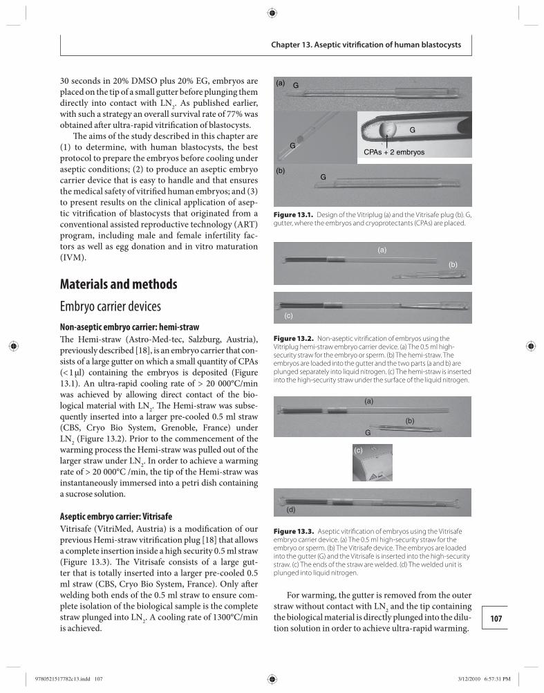
107
Chapter 13. Aseptic vitrifi cation of human blastocysts
30 seconds in 20% DMSO plus 20% EG, embryos are placed on the tip of a small gutter before plunging them directly into contact with LN 2 . As published earlier, with such a strategy an overall survival rate of 77% was obtained aft er ultra-rapid vitrifi cation of blastocysts.
Th e aims of the study described in this chapter are (1) to determine, with human blastocysts, the best protocol to prepare the embryos before cooling under aseptic conditions; (2) to produce an aseptic embryo carrier device that is easy to handle and that ensures the medical safety of vitrifi ed human embryos; and (3) to present results on the clinical application of asep-tic vitrifi cation of blastocysts that originated from a conventional assisted reproductive technology (ART) program, including male and female infertility fac-tors as well as egg donation and in vitro maturation (IVM).
Materials and methods Embryo carrier devices Non-aseptic embryo carrier: hemi-straw Th e Hemi-straw (Astro-Med-tec, Salzburg, Austria), previously described [ 18 ], is an embryo carrier that con-sists of a large gutter on which a small quantity of CPAs (< 1 μl) containing the embryos is deposited ( Figure 13.1 ). An ultra-rapid cooling rate of > 20 000°C/min was achieved by allowing direct contact of the bio-logical material with LN 2 . Th e Hemi-straw was subse-quently inserted into a larger pre-cooled 0.5 ml straw (CBS, Cryo Bio System, Grenoble, France) under LN 2 ( Figure 13.2 ). Prior to the commencement of the warming process the Hemi-straw was pulled out of the larger straw under LN 2 . In order to achieve a warming rate of > 20 000°C /min, the tip of the Hemi-straw was instant aneously immersed into a petri dish containing a sucrose solution.
Aseptic embryo carrier: Vitrisafe Vitrisafe (VitriMed, Austria) is a modifi cation of our previous Hemi-straw vitrifi cation plug [ 18 ] that allows a complete insertion inside a high security 0.5 ml straw ( Figure 13. 3 ). Th e Vitrisafe consists of a large gut-ter that is totally inserted into a larger pre-cooled 0.5 ml straw (CBS, Cryo Bio System, France). Only aft er welding both ends of the 0.5 ml straw to ensure com-plete isolation of the biological sample is the complete straw plunged into LN 2 . A cooling rate of 1300°C/min is achieved.
For warming, the gutter is removed from the outer straw without contact with LN 2 and the tip containing the biological material is directly plunged into the dilu-tion solution in order to achieve ultra-rapid warming.
G(a)
(b)
G
G
G
CPAs + 2 embryos
Figure 13.1. Design of the Vitriplug (a) and the Vitrisafe plug (b). G, gutter, where the embryos and cryoprotectants (CPAs) are placed.
Figure 13.2. Non-aseptic vitrifi cation of embryos using the Vitriplug hemi-straw embryo carrier device. (a) The 0.5 ml high-security straw for the embryo or sperm. (b) The hemi-straw. The embryos are loaded into the gutter and the two parts (a and b) are plunged separately into liquid nitrogen. (c) The hemi-straw is inserted into the high-security straw under the surface of the liquid nitrogen.
Figure 13.3. Aseptic vitrifi cation of embryos using the Vitrisafe embryo carrier device. (a) The 0.5 ml high-security straw for the embryo or sperm. (b) The Vitrisafe device. The embryos are loaded into the gutter (G) and the Vitrisafe is inserted into the high-security straw. (c) The ends of the straw are welded. (d) The welded unit is plunged into liquid nitrogen.
(a)
(b)
(c)
(c)
(d)
(a)
(b)
G
9780521517782c13.indd 1079780521517782c13.indd 107 3/12/2010 6:57:31 PM3/12/2010 6:57:31 PM
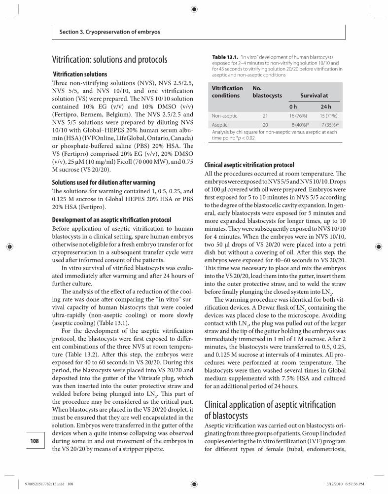
108
Section 3. Cryopreservation of embryos
Vitrifi cation: solutions and protocols Vitrifi cation solutions Th ree non-vitrifying solutions (NVS), NVS 2.5/2.5, NVS 5/5, and NVS 10/10, and one vitrifi cation solution (VS) were prepared. Th e NVS 10/10 solution contained 10% EG (v/v) and 10% DMSO (v/v) (Fertipro, Bernem, Belgium). Th e NVS 2.5/2.5 and NVS 5/5 solutions were prepared by diluting NVS 10/10 with Global–HEPES 20% human serum albu-min (HSA) (IVFOnline, LifeGlobal, Ontario, Canada) or phosphate-buff ered saline (PBS) 20% HSA. Th e VS (Fertipro) comprised 20% EG (v/v), 20% DMSO (v/v), 25 μM (10 mg/ml) Ficoll (70 000 MW), and 0.75 M sucrose (VS 20/20).
Solutions used for dilution after warming Th e solutions for warming contained 1, 0.5, 0.25, and 0.125 M sucrose in Global HEPES 20% HSA or PBS 20% HSA (Fertipro).
Development of an aseptic vitrifi cation protocol Before application of aseptic vitrifi cation to human blastocysts in a clinical setting, spare human embryos otherwise not eligible for a fresh embryo transfer or for cryopreservation in a subsequent transfer cycle were used aft er informed consent of the patients.
In vitro survival of vitrifi ed blastocysts was evalu-ated immediately aft er warming and aft er 24 hours of further culture.
Th e analysis of the eff ect of a reduction of the cool-ing rate was done aft er comparing the “in vitro” sur-vival capacity of human blastocyts that were cooled ultra-rapidly (non-aseptic cooling) or more slowly (aseptic cooling) ( Table 13.1 ).
For the development of the aseptic vitrifi cation protocol, the blastocysts were fi rst exposed to diff er-ent combinations of the three NVS at room tempera-ture ( Table 13.2 ). Aft er this step, the embryos were exposed for 40 to 60 seconds in VS 20/20. During this period, the blastocysts were placed into VS 20/20 and deposited into the gutter of the Vitrisafe plug, which was then inserted into the outer protective straw and welded before being plunged into LN 2 . Th is part of the proced ure may be considered as the critical part. When blasto cysts are placed in the VS 20/20 droplet, it must be ensured that they are well encapsulated in the solution. Embryos were transferred in the gutter of the devices when a quite intense collapsing was observed during some in and out movement of the embryos in the VS 20/20 by means of a stripper pipette.
Clinical aseptic vitrifi cation protocol All the procedures occurred at room temperature. Th e embryos were exposed to NVS 5/5 and NVS 10/10. Drops of 100 μl covered with oil were prepared. Embryos were fi rst exposed for 5 to 10 minutes in NVS 5/5 according to the degree of the blastocelic cavity expansion. In gen-eral, early blastocysts were exposed for 5 minutes and more expanded blastocysts for longer times, up to 10 minutes. Th ey were subsequently exposed to NVS 10/10 for 4 minutes. When the embryos were in NVS 10/10, two 50 μl drops of VS 20/20 were placed into a petri dish but without a covering of oil. Aft er this step, the embryos were exposed for 40–60 seconds to VS 20/20. Th is time was necessary to place and mix the embryos into the VS 20/20, load them into the gutter, insert them into the outer protective straw, and to weld the straw before fi nally plunging the closed system into LN 2 .
Th e warming procedure was identical for both vit-rifi cation devices. A Dewar fl ask of LN 2 containing the devices was placed close to the microscope. Avoiding contact with LN 2 , the plug was pulled out of the larger straw and the tip of the gutter holding the embryos was immediately immersed in 1 ml of 1 M sucrose. Aft er 2 minutes, the blastocysts were transferred to 0.5, 0.25, and 0.125 M sucrose at intervals of 4 minutes. All pro-cedures were performed at room temperature. Th e blastocysts were then washed several times in Global medium supplemented with 7.5% HSA and cultured for an additional period of 24 hours.
Clinical application of aseptic vitrifi cation of blastocysts Aseptic vitrifi cation was carried out on blastocysts ori-ginating from three groups of patients. Group I included couples entering the in vitro fertilization (IVF) program for diff erent types of female (tubal, endometriosis,
Table 13.1. “In vitro” development of human blastocysts exposed for 2–4 minutes to non-vitrifying solution 10/10 and for 45 seconds to vitrifying solution 20/20 before vitrifi cation in aseptic and non-aseptic conditions
Vitrifi cation conditions
No. blastocysts Survival at
0 h 24 hNon-aseptic 21 16 (76%) 15 (71%)
Aseptic 20 8 (40%)* 7 (35%)* Analysis by chi square for non-aseptic versus aseptic at each
time point: * p < 0.02
9780521517782c13.indd 1089780521517782c13.indd 108 3/12/2010 6:57:36 PM3/12/2010 6:57:36 PM
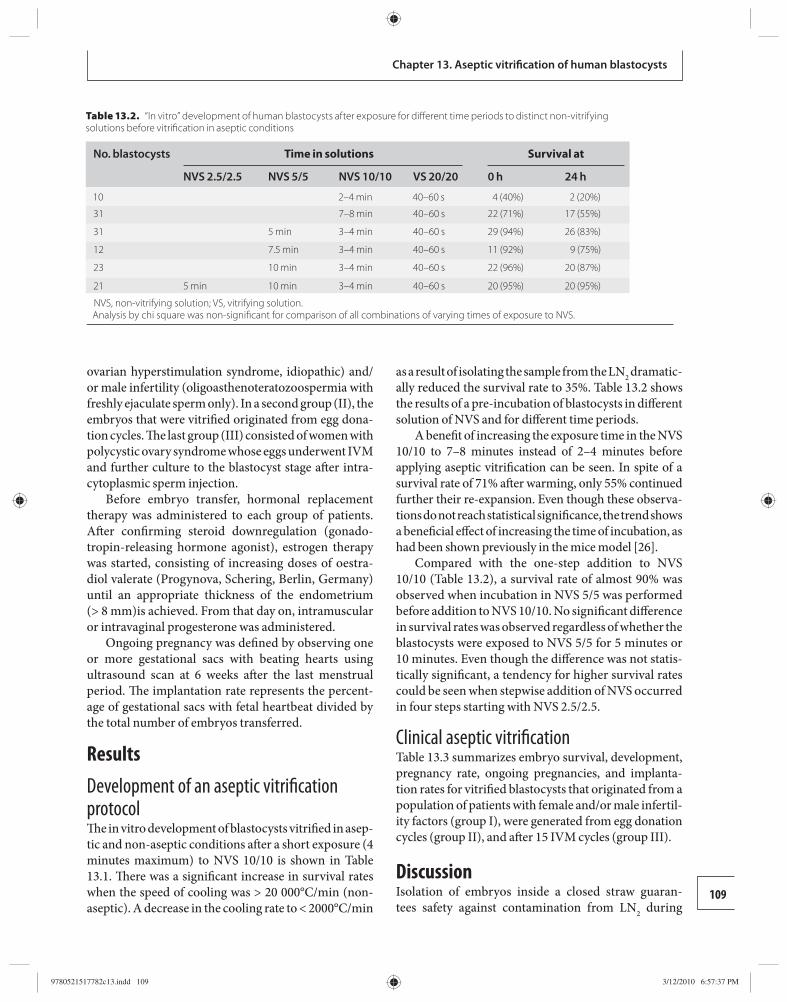
109
Chapter 13. Aseptic vitrifi cation of human blastocysts
ovarian hyperstimulation syndrome, idiopathic) and/or male infertility (oligoasthenoteratozoospermia with freshly ejaculate sperm only). In a second group (II), the embryos that were vitrifi ed originated from egg dona-tion cycles. Th e last group (III) consisted of women with polycystic ovary syndrome whose eggs underwent IVM and further culture to the blastocyst stage aft er intra-cytoplasmic sperm injection.
Before embryo transfer, hormonal replacement therapy was administered to each group of patients. Aft er confi rming steroid downregulation (gonado-tropin-releasing hormone agonist), estrogen therapy was started, consisting of increasing doses of oestra-diol valerate (Progynova, Schering, Berlin, Germany) until an appropriate thickness of the endometrium (> 8 mm)is achieved. From that day on, intramuscular or intravaginal progesterone was administered.
Ongoing pregnancy was defi ned by observing one or more gestational sacs with beating hearts using ultrasound scan at 6 weeks aft er the last menstrual period. Th e implantation rate represents the percent-age of gestational sacs with fetal heartbeat divided by the total number of embryos transferred.
Results Development of an aseptic vitrifi cation protocol Th e in vitro development of blastocysts vitrifi ed in asep-tic and non-aseptic conditions aft er a short exposure (4 minutes maximum) to NVS 10/10 is shown in Table 13.1 . Th ere was a signifi cant increase in survival rates when the speed of cooling was > 20 000°C/min (non-aseptic). A decrease in the cooling rate to < 2000°C/min
as a result of isolating the sample from the LN 2 dramatic-ally reduced the survival rate to 35%. Table 13.2 shows the results of a pre-incubation of blastocysts in diff erent solution of NVS and for diff erent time periods.
A benefi t of increasing the exposure time in the NVS 10/10 to 7–8 minutes instead of 2–4 minutes before applying aseptic vitrifi cation can be seen. In spite of a survival rate of 71% aft er warming, only 55% continued further their re-expansion. Even though these observa-tions do not reach statistical signifi cance, the trend shows a benefi cial eff ect of increasing the time of incubation, as had been shown previously in the mice model [ 26 ].
Compared with the one-step addition to NVS 10/10 ( Table 13.2 ), a survival rate of almost 90% was observed when incubation in NVS 5/5 was performed before addition to NVS 10/10. No signifi cant diff erence in survival rates was observed regardless of whether the blastocysts were exposed to NVS 5/5 for 5 minutes or 10 minutes. Even though the diff erence was not statis-tically signifi cant, a tendency for higher survival rates could be seen when stepwise addition of NVS occurred in four steps starting with NVS 2.5/2.5.
Clinical aseptic vitrifi cation Table 13.3 summarizes embryo survival, development, pregnancy rate, ongoing pregnancies, and implanta-tion rates for vitrifi ed blastocysts that originated from a population of patients with female and/or male infertil-ity factors (group I), were generated from egg donation cycles (group II), and aft er 15 IVM cycles (group III).
Discussion Isolation of embryos inside a closed straw guaran-tees safety against contamination from LN 2 during
Table 13.2. “In vitro” development of human blastocysts after exposure for diff erent time periods to distinct non-vitrifyingsolutions before vitrifi cation in aseptic conditions
No. blastocysts Time in solutions Survival at
NVS 2.5/2.5 NVS 5/5 NVS 10/10 VS 20/20 0 h 24 h
10 2–4 min 40–60 s 4 (40%) 2 (20%)
31 7–8 min 40–60 s 22 (71%) 17 (55%)
31 5 min 3–4 min 40–60 s 29 (94%) 26 (83%)
12 7.5 min 3–4 min 40–60 s 11 (92%) 9 (75%)
23 10 min 3–4 min 40–60 s 22 (96%) 20 (87%)
21 5 min 10 min 3–4 min 40–60 s 20 (95%) 20 (95%)
NVS, non-vitrifying solution; VS, vitrifying solution. Analysis by chi square was non-signifi cant for comparison of all combinations of varying times of exposure to NVS.
9780521517782c13.indd 1099780521517782c13.indd 109 3/12/2010 6:57:37 PM3/12/2010 6:57:37 PM
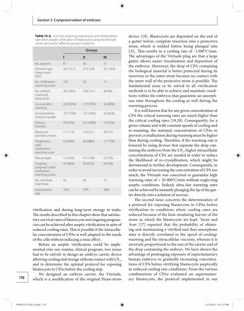
110
Section 3. Cryopreservation of embryos
vitrifi cation and during long-term storage in tanks. Th e results described in this chapter show that satisfac-tory survival rates of blastocysts and ongoing pregnan-cies can be achieved aft er aseptic vitrifi cation in spite of reduced cooling rates. Th is is possible if the intracellu-lar concentration of CPAs is well adapted to the needs of the cells without inducing a toxic eff ect.
Before an aseptic vitrifi cation could be imple-mented into our routine clinical program, two issues had to be solved: to design an embryo carrier device allowing cooling and storage without contact with LN 2 , and to determine the optimal protocol for exposing blastocysts to CPAs before the cooling step.
We designed an embryo carrier, the Vitrisafe, which is a modifi cation of the original Hemi-straw
device [ 18 ]. Blastocysts are deposited on the end of a gutter before complete insertion into a protective straw, which is welded before being plunged into LN 2 . Th is results in a cooling rate of –1300°C/min. Th e advantages of the Vitrisafe plug are that a large gutter allows easier visualization and deposition of the embryos. Moreover, the drop of CPA containing the biological material is better protected during its insertion in the outer straw because no contact with the inner wall of the protective straw is possible. Th e fundamental issue to be solved in all vitrifi cation methods is to be able to achieve and maintain condi-tions within the embryos that guarantee an amorph-ous state throughout the cooling as well during the warming process.
It is well known that for any given concentration of CPA the critical warming rates are much higher than the critical cooling rates [ 19 , 20 ]. Consequently, for a given volume and with constant speeds of cooling and re-warming, the minimal concentration of CPAs to prevent crystallization during warming must be higher than during cooling. Th erefore, if the warming rate is lowered by using devices that separate the drop con-taining the embryos from the LN 2 , higher intracellular concentrations of CPA are needed in order to reduce the likelihood of re-crystallization, which might be detrimental to further development. Consequently, in order to avoid increasing the concentration of CPA too much, the Vitrisafe was conceived to guarantee high warming rates of > 20 000°C/min without neglecting aseptic conditions. Indeed, ultra-fast warming rates can be achieved by instantly plunging the tip of the gut-ter directly into a solution of sucrose.
Th e second issue concerns the determination of a protocol for exposing blastocysts to CPAs before vitrifi cation in conditions where cooling rates are reduced because of the heat-insulating barrier of the straw in which the blastocysts are kept. Yavin and Arav [ 17 ] reported that the probability of obtain-ing and maintaining a vitrifi ed and thus amorphous state is directly correlated to the speed of cooling/warming and the intracellular viscosity, whereas it is inversely proportional to the size of the carrier and of the drop containing the embryo. We have shown the advantage of prolonging exposure of supernumerary human embryos to gradually increasing concentra-tions of CPA before vitrifying blastocysts aseptically in reduced cooling rate conditions. From the various combinations of CPAs evaluated on supernumer-ary blastocysts, the protocol implemented in our
Table 13.3. Survival, ongoing pregnancies, and implantation rates after aseptic vitrifi cation of blastocysts using the Vitrisafe carrier devices for diff erent groups of patients
Groups
I II III
No. patients 87 56 15
Women’s age (mean years [SD])
34.3 ( 3.7) 27.6 (2.8) 32.1 (4.8)
No. vitrifi cation–warming cycles
102 56 15
No. vitrifi ed (warmed) blastocysts
263 (263) 238 (141) 40 (40)
Survival after warming
224 (85%) 137 (97%) 32 (80%)
Survival before embryo transfer
191 (73%) 131 (93%) 26 (65%)
Embryo transfers
94 (92%) 56 (100%) 14 (93%)
Blastocyst transfers (mean)
177 (1.9) 120 (2.1) 26 (1.9)
Preganancy rate/vitrifi cation–warming cycle
62 (60%) 38 (68%) 11 (73%)
Miscarriages 15 (24%) 4 (11.0%) 3 (27%)
Ongoing pregnancy rate/vitrifi cation–warming cycles
47 (46%) 34 (61%) 8 (53%)
No. with fetal heart beat
56 44 10
Implantation rate
32% 37% 38%
9780521517782c13.indd 1109780521517782c13.indd 110 3/12/2010 6:57:37 PM3/12/2010 6:57:37 PM
111
Chapter 13. Aseptic vitrifi cation of human blastocysts
routine clinical program was derived. Th is consists of a stepwise addition of the CPAs in increasing con-centrations. Blastocysts were fi rst exposed for 5 to 10 minutes to NVS 5/5, followed by a shorter exposure of 4 minutes in NVS 10/10, and 40–60 seconds in VS 20/20.
Th e results shown here indicate that stepwise add-ition of the CPAs and vitrifi cation in an aseptic embryo carrier device enables vitrifi cation of blastocysts derived from couples presenting male and/or female infertility factors, and also from egg donor cycles and from IVM cycles. In addition to the aseptic safety achieved, this system of vitrifi cation is simple, very fl exible, and allows the handling of the embryos with-out the pressure of time. It is generally acknowledged that the quality of the blastocysts is an important factor aff ecting the outcome in terms of survival and preg-nancies. Th erefore, application of aseptic vitrifi cation for blastocysts that originated from the ovum donor program can be considered as the reference standard group to assess the effi ciency of this vitrifi cation tech-nique. Th e corresponding oocytes are retrieved from young women and only good-quality blastocysts were selected for vitrifi cation.
Th e application of aseptic vitrifi cation methods seems also very promising for blastocysts that devel-oped from IVM oocytes. In fact, the implantation rates for embryos developing from IVM oocytes are said to be lower than aft er classical IVF techniques [ 21 ]. Obviously, good implantation rates for IVM oocytes were not yet achieved aft er fresh embryo transfer, which appears to be a drawback of this technique. Th e results shown here indicate an improvement if embryos generated from IVM oocytes were vitrifi ed in aseptic conditions and then replaced in a further estra-diol/progesterone-supplemented cycle. Th is highlights that endometrial quality or an inappropriate hormo-nal preparation of the selected population, who had polycystic ovary syndrome, may negatively infl uence implantation.
In view of the effi ciency of our aseptic vitrifi ca-tion method with good-quality blastocysts, we may consider adapting our policy to embryo selection before fresh embryo transfer. Th e receptivity of the endometrium largely contributes to the clinical out-come of IVF/ICSI treatment, especially in stimula-tion cycles. Th e high implantation rates observed in an egg donation program are related to the high potential of good blastocysts to survive aseptic vitri-fi cation methods and this also reinforces the concept
of good endometrial receptivity being achieved in hormone replacement cycles. From these observa-tions, assuming that the receptivity of stimulation cycles is not always optimal and patients may already have a history of repeated implantatory failure, we may consider vitrifying the good-quality blastocysts and transferring those with moderate quality in the fresh cycles. Moreover, according to the current ten-dency of transferring only one embryo per cycle [ 22 ], it is more reasonable to present the results in terms of pregnancy per egg retrieval. As a consequence, in order to present the results in term of cumulative pregnancy rates, a good cryopreservation technique is mandatory when single embryo transfer is applied [ 23 ].
Another advantage of our aseptic vitrifi cation proto col is that it does not necessitate the collapse of the blastocelic cavity. As confi rmed by other teams working in this area, we have demonstrated previously the benefi cial eff ect of reducing the blastocele volume prior to ultra-rapid vitrifi cation [ 3 ]. In ultra-rapid vit-rifi cation, the blastocysts are left for a short period in the NVS 10/10. We assume, therefore, that the cavity was not well protected and that a vitrifi ed state was not well established. Conversely, in the approach described here, where there is an initial exposure of the embryos to a lower concentration of CPA (NVS 5/5 for 5 to 10 minutes), suffi cient CPA is present inside the cavity to prevent intracellular ice crystal formation at the virtri-fi cation point.
More than 20 years ago, in the 1980s, we reported acceptable survival rates followed by births of mice [ 20 ] and bovine [ 24 ] blastocysts that were vitrifi ed in 0.25 ml French mini-straws. Recently Stachecki et al. [ 25 ] have reported excellent results aft er vitrifi cation of human blastocysts loaded in a 0.25 ml straw, guar-anteeing also the safety of the vitrifi cation technique. Th e ultra-rapid way to vitrify blastocysts has to be fi nally revised, and cooling at lower rate is suffi cient if the embryo is well prepared. Aft er vitrifi cation in straws, in open systems, and in aseptic devices, it will not be long before vitrifi cation will occur defi nitely in straws [ 25 , 26 ].
In spite of reduced cooling rates inherent in the aseptic vitrifi cation conditions, acceptable results are obtainable if the intracellular concentrations of CPAs are well adapted to the needs of the cells. If such results could be confi rmed on a large scale, aseptic vitrifi ca-tion may become more routine and advantageous for cryopreservation of human blastocysts and also
9780521517782c13.indd 1119780521517782c13.indd 111 3/12/2010 6:57:37 PM3/12/2010 6:57:37 PM
112
Section 3. Cryopreservation of embryos
other embryo developmental stages. In a recent study [ 27 ], the authors provided evidence that vitrifi cation of day 3 embryos imparts less trauma to cells than the slow-freezing technique and is associated with higher survival and better metabolism and blastocyst formation.
References 1. Mukaida T , Wada S , Takahashi K , et al. Vitrifi cation of
human embryos based on the assessment of suitable conditions for 8-cell mouse embryos . Hum Reprod 1998; 13: 2874 –2879.
2. Yokota Y , Sato S , Yokota M , et al. Birth of a healthy baby following vitrifi cation of human blastocysts . Fertil Steril 2001; 75: 1027 –1029.
3. Vanderzwalmen P , Bertin G , Debauche CH , et al. Births aft er vitrifi cation at morula and blastocyst stages: eff ect of artifi cial reduction of the blastocoelic cavity before vitrifi cation . Hum Reprod 2002; 17: 744 –751.
4. Lane M , Schoolcraft WB , Gardner DK . Vitrifi cation of mouse and human blastocysts using a novel cryoloop container-less technique . Fertil Steril 1999; 72: 1073 –1078.
5. Vajta G , Nagy Z . Are programmable freezers still needed in the embryo laboratory? Review on vitrifi cation . Reprod BioMed Online 2006; 7: 623 –633.
6. Bielanski A . Non-transmission of bacterial and viral microbes to embryos and semen stored in the vapour phase of liquid nitrogen in dry shippers . Cryobiology 2005; 50: 206 –210.
7. Morris G . Th e origin, ultrastructure, and microbiology of the sediment accumulating in liquid nitrogen storage vessels . Cryobiology 2005; 50: 231 –238.
8. Kyuwa S , Nishikawa T , Kaneko T , et al. Experimental evaluation of cross-contamination between cryotubes containing mouse 2-cell embryos and murine pathogens in liquid nitrogen tanks . Exp Anim 2003; 52: 67 –70.
9. European Parliament . EU Tissues and Cells Directive 2004/23/EC . Brussels : EU Publications Offi ce, 2004. http://eur-lex.europa.eu/LexUriServ/LexUriServ.do?uri=OJ:L:2004:102:0048:0058:EN:PDF (accessed October 2, 2009).
10. European Commission . Directive 2006/17/CE of 8 February 2006 . Brussels : EU Publications Offi ce, 2006. http://eur-lex.europa.eu/LexUriServ/site/en/oj/2006/l_038/l_03820060209en00400052.pdf (accessed October 2, 2009).
11. Walker D . (2004) Vitrifi cation versus programmable rate freezing of late stage murine embryos: a randomised comparison prior to application in clinical IVF . Reprod BioMed Online 2004; 8: 558 –568.
12. Larman M , Sheehan C , Gardner D . Vitrifi cation of mouse pronuclear oocytes with no direct liquid nitrogen contact . Reprod BioMed Online 2006; 12: 66 –69.
13. Kuleshova L , Shaw J . A strategy for rapid cooling of mouse embryos within a double straw to eliminate the risk of contamination during storage in liquid nitrogen . Hum Reprod 2000; 15: 2604 –2609.
14. Kuwayama M , Vajta G , Leda S , et al. Comparison of open and closed methods for vitrifi cation of human embryos and the elimination of potential contamination . Reprod BioMed Online 2005; 11: 608 –614.
15. Isachenko V , Katkov I , Yakovenko S , et al. Vitrifi cation of human laser treated blastocysts within cut standard straws (CSS): novel aseptic packaging and reduced concentrations of cryoprotectants . Cryobiology 2007; 54: 305 –309.
16. Kuwayama M . Highly effi cient vitrifi cation for cryopreservation of human oocytes and embryos: the Cryotop method . Th eriogenology 2007; 67: 73 –80.
17. Yavin S , Arav A . Measurement of essential physical properties of vitrifi cation solutions . Th eriogenology 2007; 67: 81 –89.
18. Vanderzwalmen P , Bertin G , Debauche CH , et al. Vitrifi cation of human blastocysts with the Hemi-Straw carrier: application of assisted hatching aft er thawing . Hum Reprod 2003; 18: 1504 –1511.
19. Fahy G . Biological eff ects of vitrifi cation and devitrifi cation. In Pegg DE , Karow AM , Jr., eds. Th e Biophysics of Organ Cryopreservation . New York : Plenum Press , 1987, pp. 265–293.
20. Scheff en B , Vanderzwalmen P , Massip A . A simple and effi cient procedure for preservation of mouse embryos by vitrifi cation . Cryo Letters 1986; 7: 260 –269.
21. Torre A , Frydman A , Feyereisen E , et al. How do we improve implantation rate following in-vitro maturation of oocytes? In Tan SL , Chian R-C, Buckett W , eds. In-vitro Maturation of Human Oocytes: Basic Science to Clinical Application . [ Reproductive Medicine and Assisted Reproductive Techniques Series ] London: Informa Healthcare , 2007, pp. 319–331.
22. Zech N , Lejeune B , Puissant F et al. Prospective evaluation of the optimal time for selecting a single embryo for transfer: day 3 versus day 5 . Fertil Steril 2007; 88: 244 –246.
23. Henman M , Catt J , Wood T , et al. Elective transfer of single fresh blastocysts and later transfer of cryostored blastocysts reduces the twin pregnancy rate and can improve the in vitro fertilization live birth rate in younger women . Fertil Steril 2005; 84: 1620 –1627.
24. Massip A , Vanderzwalmen P , Scheff en B , et al. Pregnancies following transfer of cattle embryos preserved by vitrifi cation . Cryo Letters 1986; 7: 270 –273.
25. Stachecki J , Garrisi J , Sabino S , et al. A new safe, simple and successful vitrifi cation method for bovine and human blastocysts . Reprod BioMed Online 2008; 17: 360 –367.
26. Vanderzwalmen P , Ebner T , Zech N . One decade of experience with vitrifi cation of human embryos in
9780521517782c13.indd 1129780521517782c13.indd 112 3/12/2010 6:57:37 PM3/12/2010 6:57:37 PM

113
Chapter 13. Aseptic vitrifi cation of human blastocysts
straws, hemi-straws and high security vitrifi cation straws . In Tucker MJ , Liebermann J, eds. Vitrifi cation in Assisted Reproduction: A User’s Manual and Trouble-shooting Guide. London: Informa Healthcare , 20 07, pp. 195–217.
27. Balaban B , Urman B , Ata B , et al. A randomized controlled study of human day 3 embryo cryopreservation by slow freezing or vitrifi cation: vitrifi cation is associated with higher survival, metabolism and blastocyst formation . Hum Reprod 2008; 23: 1976 –1982.
9780521517782c13.indd 1139780521517782c13.indd 113 3/12/2010 6:57:37 PM3/12/2010 6:57:37 PM
114 Fertility Cryopreservation, ed. Ri-Cheng Chian and Patrick Quinn. Published by Cambridge University Press. Copyright © Cambridge University Press 2010.
Section 4Chapter
14 negatively aff ect the desired outcome of a pregnancy, namely a healthy mother and a healthy baby: declin-ing fertility, miscarriage, chromosomal abnormalities, hypertensive complications, and stillbirth [ 1 ]. Women’s fertility declines with age and the rates of spontaneous abortions increase signifi cantly aft er their mid 30s. Th e risks of having a baby with Down’s syndrome and any chromosomal abnormalities are also higher in the oocytes produced by older women [ 2 ]. Although the decade between 25 and 35 years of age would seem to be ideal for women to have a baby, today in North America many women still want to have a baby aft er these ideal ages, even substantially older, for various reasons, including their professional arena. It has been emphasized that, for women for whom earlier child-bearing is not an option, the decade between 35 and 45 years of age remains safe enough that maternal age alone should not be a contraindication to childbearing [ 1 ]. Nevertheless, oocyte freezing as a lifestyle option for women to give a birth in the future should be re-considered socially.
Th e fi rst report of successful cryopreservation of mouse oocytes was reported in 1958 by Sherman and Lin [ 3 ]. Nearly two decades later, the fi rst live off spring from frozen–thawed mouse oocytes was reported by Parkening et al. [ 4 ] and Whittingham [ 5 ] using a slow-freezing method. Aft er these reports, there has been very limited success in oocyte cryopreservation until more recent years.
Th e fi rst reported human pregnancy and live birth from cryopreserved oocytes were reported by Chen [ 6 ] and van Uem et al. [ 7 ], respectively. However, most early reports of human oocyte cryopreservation showed dis-mal survival and pregnancy rates [ 8 – 11 ]. In fact, only three cases of clinical pregnancies were reported in the 1980s. Nearly a decade later, when Tucker et al. [ 10 ] and Porcu et al. [ 11 ] demonstrated the use of intracyto-plasmic sperm injection (ICSI) to overcome hardening
Introduction Th e development of an eff ective oocyte cryopreser-vation system will have a signifi cant impact on the clin ical practice of assisted reproduction. In addition to fertility preservation for young women requiring steril izing medical and surgical treatments, cryobank-ing of oocytes will benefi t a large population of single women who wish to delay motherhood for personal, professional, and fi nancial reasons.
An eff ective oocyte cryopreservation program will benefi t infertile couples with moral or religious objections about cryopreservation of embryos. In fact, oocyte freezing has become the only option of cryo-preservation in many countries where the laws forbid cryopreservation of embryos. In a country like Italy, only unfertilized gametes could be cryopreserved until a recent change in the law that again allowed for the cryopreservation of supernumerary embryos.
Women with premature ovarian failure who wish to conceive must rely on donor oocytes. Oocyte donation can be complicated and time consuming, requiring hormonal synchronization of the donor and recipient menstrual cycle. A successful oocyte cryopreservation protocol would eliminate the need for donor–recipie nt menstrual cycle synchronization and enable the estab-lishment of egg banks, facilitating the logistics of coord inating egg donors with recipients. Furthermore, egg cryopreservation allows for temporary quaran-tine of donor eggs to test the donors for transmissible diseases. Th erefore, the establishment of an eff ective oocyte cryopreservation program will likely encour-age more women to donate their eggs and improve the current shortage of donor eggs.
Oocyte freezing may open a new option for women who worry that childbirth may interfere with their careers. Recently, it has been indicated that the eff ect of maternal age on the outcome of pregnancy may be best assessed by examining fi ve specifi c factors that can
Cryopreservation of oocytes Cryopreservation of human oocytes: an overview Ri-Cheng Chian
9780521517782c14.indd 1149780521517782c14.indd 114 3/12/2010 9:22:42 AM3/12/2010 9:22:42 AM
115
Chapter 14. Cryopreservation of human oocytes: an overview
of the zona pellucida and to achieve pregnancies and a live birth, there was a renewed interest in oocyte cryopreservation.
Since the late 1990s, the number of reported live births resulting from oocyte cryopreservation has increased rapidly. Recently Noyes et al. [ 12 ] reviewed 58 reports from 1986 to 2008, indicating a total of 936 live births resulted from cryopreserved oocytes. Compared with congenital anomalies occurring in naturally conceived infants, no diff erence was noted [ 12 , 13 ]. It seems that so far, there are similar numbers of live births from the slow-freezing and the rapid-cooling (vitrifi cation) methods. Th erefore, it is impor-tant to review the technical development of these methods for oocyte cryopreservation.
Classic slow-freezing method To date, pregnancies and live births from cryopre-served oocytes using the classic slow-freezing method have been published as case reports and series involv-ing small numbers of patients. Only two large series of human oocyte cryopreservation using the classic slow-freezing protocols have been published [ 14 , 15 ]. Based on 112 in vitro fertilization (IVF) cycles with cryopre-served mature human eggs, a total of 1769 mature eggs were frozen and 1502 eggs were thawed. Th e survival rate was only 54.1%. Th e rates of fertilization following ICSI and cleavage were 57.7% and 91.2%, respectively. Sixteen pregnancies and 11 live births were reported. In 2004, Borini et al. [ 15 ] reported the clinical outcomes of 59 transfer cycles of embryos derived from cryopre-served oocytes. Out of 737 thawed oocytes, the survival rate was only 37%. In this series, 15 pregnancies and 13 live births were achieved.
When considering all pregnancies and live births obtained from cryopreserved oocytes using the clas-sic slow-freezing method, the survival rates averaged approximately 50% and varied tremendously between the reported cases and series (23% to 89%). In fact, taken together for all the reported live births so far, 56 live births have been reported from cryopreserved human oocytes using the conventional slow-freezing procedure. Th e percentage of live births per thawed egg ranges from 1 to 10% using the classic slow-freezing protocols [ 16 – 19 ].
Modifi cation of slow-freezing techniques Recently improved survival and pregnancy rates have been reported using modifi ed slow-freezing
procedures, particularly increased sucrose concentra-tion in the suspending solution and the use of sodium-free freezing solutions.
Increased sucrose concentration in suspending solution Based on the hypothesis that the use of a higher sucrose concentration during slow freezing increases oocyte dehydration, thus reducing the formation of intracellular ice, Yang et al. [ 20 ] and Fabbri et al. [ 21 ] demonstrated that a higher sucrose concentration (0.2 M and 0.3 M, respectively) in the suspending solution led to a signifi cant improvement in oocyte survival and pregnancy rates. Using a freezing solu-tion containing 0.2 M sucrose and a slow-freezing/rapid thawing protocol, Yang et al. [ 20 ] fi rst reported a case of successful ongoing twin pregnancy and, sub-sequently, in a series of 24 transfer cycles reported a survival rate of 71% and a pregnancy rate of 46% that resulted in 14 live births [ 22 ]. Using the same slow-freezing protocol, Winslow et al. [ 23 ] reported a sim-ilar frozen–thawed survival rate of 69% and achieved 16 live births in 42 transfer cycles.
Similarly, by increasing the sucrose concentration to 0.3 M and prolonging the exposure time to the freezing media, Fabbri et al. [ 21 ] reported a signifi -cantly higher post-thawing survival rate. In fact, using the 0.3 M sucrose slow-freezing protocol, Chen et al. [ 24 ] reported a post-thawing survival rate of 100% and an ongoing twin pregnancy. In a small series of seven patients, Fosas et al. [ 25 ] reported a survival rate of 90%, a pregnancy rate of 57%, and fi ve live births. More recently, based on 21 freezing–thawing cycles, Chen et al. [ 26 ] reported a survival rate of 75%, a pregnancy rate of 33%, and fi ve live births. Similarly, Li et al. [ 27 ] reported a survival rate of 90%, a pregnancy rate of 47%, and six live births in a series of 12 patients. Most recently, clinical out-come of two large series of oocyte cryopreserva-tion using 0.3 M sucrose protocols were published [ 28 , 29 ]. Based on cryopreservation of 927 oocytes in 146 patients, Borini et al. [ 28 ] reported a survival rate of 74.1%, a pregnancy rate of 9.7%, and four live births. Levi Setti et al. [ 29 ] reported a similar survival rate (69.9%) based on 120 patients and 1087 oocytes but achieved a higher pregnancy rate (12.4%) and 13 live births. Overall, the use of 0.3 M sucrose in slow-freezing protocols appeared to improve the survival rate to approximately 70% and has resulted in 37 live births to date [ 26 – 30 ].
9780521517782c14.indd 1159780521517782c14.indd 115 3/12/2010 9:22:42 AM3/12/2010 9:22:42 AM
116
Section 4. Cryopreservation of oocytes
In the last few years, with these modifi ed slow-freezing methods, the effi ciency of oocyte cryo-preservation has improved tremendously, resulting in cumulative pregnancy rates approaching 50% [ 31 ]. Borini et al. [ 32 ] reported that in 660 thawing cycles up to December 2006, 2205 oocytes survived out of 3238 thawing oocytes, a 68.1% survival rate. Five hundred and ninety cycles were performed for embryo transfer, giving rise to a clinical pregnancy rate per transfer of 15.0% (88/590).
Choline-based freezing media Another strategy to improve the effi cacy of oocyte cryopreservation is to change the composition of the freezing media. As highlighted by work of Stachecki et al. [ 33 ], the replacement of sodium with choline in the base solution may prevent salt-induced cryopreser-vation injury, resulting in improved survival rate and embryonic development in vitro using animal models. Another protective mechanism of choline-based media is believed to relate to an increase in viscosity at low temperature. Stachecki et al. [ 33 ] demonstrated high survival of choline-exposed oocytes despite plunging the oocytes into liquid nitrogen at a higher temperature (−20°C) than used in other slow-freezing protocols.
A total of 11 live births have been reported in sev-eral case reports and small series using the choline-based freezing method, although the survival rates were not signifi cantly higher than the conventional slow-freezing method [ 34 – 37 ]. Th erefore, larger clin-ical trials are needed to determine whether the clinical effi cacy of oocyte cryopreservation using low-sodium media is superior to the other freezing protocols.
Rapid-cooling (vitrifi cation) method Th e rapid-cooling (vitrifi cation) method is a promis-ing novel technique and may be more eff ective than a slow-freezing procedure for egg cryopreservation [ 18 ]. Vitrifi cation of human oocytes and embryos has resulted in relatively high survival rates [ 38 – 48 ]. In fact, many pregnancies and live births have been reported aft er cryopreservation of mature human oocytes or embryos using vitrifi cation procedures [ 41 , 49 – 57 ].
In 1999, Kuleshova et al. [ 41 ] reported the fi rst human live birth following vitrifi cation of 17 mature oocytes using open pulled straws and high concentra-tion of ethylene glycol and sucrose. Eleven eggs survived (65%) aft er vitrifi cation–warming and fi ve pronuclear zygotes (46%) were obtained aft er ICSI. In the series of three patients, one patient became pregnant. Yoon
et al. [ 49 ] cryopreserved 90 mature eggs from seven patients by vitrifi cation using electron microscope grids. Fift y-seven eggs survived (63%) aft er thawing. Th e fertilization rate was 75% (43/57) following ICSI. Two healthy live births and one ongoing pregnancy were obtained following transfer of 32 embryos, result-ing in an implantation rate of 9% (3/32). Th e use of a vitrifi cation solution mixture of 15% ethylene glycol, 15% dimethyl sulfoxide, and 0.5 M sucrose appeared to enable a dramatic improvement in the survival rate of vitrifi ed human oocytes. Several groups reported high survival rates (89% to 100%) and successful live births using a vitrifi cation method [ 50 , 51 , 55 – 57 ]. In fact, tremendous progress has been achieved recently for human oocyte cryopreservation using vitrifi cation procedures [ 58 , 59 ].
Recently a clinical trial of oocyte vitrifi cation indi-cated that 383 oocytes survived (82.7%) aft er thawing from a total of 463 oocytes that were vitrifi ed and 17 out of 38 patients became pregnant, with 45% clinical pregnancy rate per embryo transfer [ 60 ]. Th is provides evidence that vitrifi cation of oocytes results in a satis-factory clinical pregnancy rate, with better results than those achieved by the slow-freezing method. It was also demonstrated that pregnancies achieved with vitrifi -cation of oocytes do not appear to be associated with adverse pregnancy outcomes, indicating that vitrifi ca-tion of oocytes represents a novel option and effi cient method for female fertility preservation [ 60 ].
Cryopreservation of oocytes at immature or mature stages In earlier work when there was a poor survival rate for cryopreserved oocytes, an alternative strategy was con-sidered using a slow-freezing method: to cryopreserve oocytes at the immature germinal vesicle (GV) stage instead of cryopreserving at mature metaphase II (MII) stage. Th e immature oocytes are at diplotene state of prophase I. Th eoretically, given their microstructure, immature GV stage oocytes should be more resistant to the damage caused by cooling and would circumvent the risk of polyploidy and aneuploidies, since the chro-matin is diff used and surrounded by a nuclear mem-brane [ 61 , 62 ]. However, diffi culties are associated with in vitro maturation (IVM) of immature oocytes aft er freeze–thawing. Several attempts have been made with immature human oocytes. Although the survival rates seemed to be improved by the slow-freezing method, poor IVM and fertilization are major problems associ-ated with immature egg freezing [ 17 , 63 – 66 ]. It has been
9780521517782c14.indd 1169780521517782c14.indd 116 3/12/2010 9:22:42 AM3/12/2010 9:22:42 AM
117
Chapter 14. Cryopreservation of human oocytes: an overview
known that oocyte cytoplasmic maturation requires cumulus cell and cytoplasmic communication using gap junctions [ 67 ]. So during freezing and thawing procedures, these gap junctions may be disconnected in the cryopreservative solution because this solution usually is hypertonic.
To date, only one human live birth has been reported following cryopreservation of GV stage oocytes and IVM [ 68 ]. Th erefore, in all species, cryopreservation of mature oocytes is still more effi cient than cryopres-ervation of immature oocytes [ 69 ]. Recently a study determined the effi cacy of the vitrifi cation method by vitrifying human oocytes before and aft er IVM of human oocytes [ 70 ]. Th e immature human oocytes recovered (total of 472 oocytes) were divided into two groups: 219 immature oocytes that were vitrifi ed at the GV stage and 253 immature GV stage oocytes that were fi rst matured in vitro (178 to the MII stage); 79 of the 178 were vitrifi ed and 99 were not vitrifi ed but were proc-essed as controls. Aft er thawing, the oocyte survival, maturation, and fertilization rates as well as embry-onic development were compared. Th e results showed no signifi cant diff erence between the survival rates of the oocytes vitrifi ed at GV stage and those vitrifi ed at MII stage (85% versus 86%). However, oocyte matu-ration rates were signifi cantly reduced when oocytes were vitrifi ed at immature GV stage followed by IVM (51%) in comparison with the oocytes without vitrifi ca-tion (70%). Th is study suggested that better results can be achieved by vitrifying mature oocytes rather than immature oocytes [ 69 ]. In fact, recently, several preg-nancies and live births were obtained with the imma-ture oocyte retrieval followed by IVM and vitrifi cation [ 60 , 71 ], providing proof-of-principle evidence that the novel fertility preservation strategy of immature oocyte retrieval, IVM, and vitrifi cation of mature oocytes can lead to successful pregnancies and healthy live births.
Conclusions Th e development of an eff ective oocyte cryopreserva-tion system has had a signifi cant impact on the clinical practice of assisted reproduction. With modifi ed slow-freezing methods, particularly increased sucrose con-centration in suspending solution, improved survival and pregnancy rates have been obtained. However, rapid cooling (vitrifi cation) of human oocytes has resulted in relatively higher survival rates. Pregnancies achieved with cryopreservation of oocytes, regardless of whether slow freezing or vitrifi cation was used, do not appear to be associated with adverse pregnancy
outcomes, indicating that cryopreservation of oocytes represents a novel option and an effi cient method for female fertility preservation. It seems that cryo-preservation of mature stage oocytes has better results than freezing immature stage oocytes, because oocyte maturation rate will be signifi cantly reduced when the oocytes are cryopreserved at an immature stage followed by IVM aft er thawing. Although approxi-mately more than 1000 live births obtained from the cryopreserved oocytes showed no diff erence in con-genital anomalies compared with naturally conceived infants, more live born data and long-term monitoring are required to assure the safest and most expeditious development of oocyte cryopreservation technology.
References 1. Heff ner LJ . Advanced maternal age: how old is too old?
N Engl J Med 2004; 351 : 1927 –1929. 2. Hook EB , Cross PK , Schreinemachers DM .
Chromosomal abnormality rates at amniocentesis and in live-born infants . JAMA 1983; 249 : 2034 –2038.
3. Sherman JK , Lin TP . Survival of unfertilized mouse eggs during freezing and thawing . Proc Soc Exp Biol Med 1958; 98: 902 –905.
4. Parkening TA , Tsunoda Y , Chang MC . Eff ects of various low temperatures, cryoprotective agents and cooling rates on the survival, fertilizability and development of frozen–thawed mouse eggs . J Exp Zool 1976; 197: 369 –374.
5. Whittingham DG . Fertilization in vitro and development to term of unfertilized mouse oocytes previously stored at −196° C . J Reprod Fertil 1977; 49 : 89 –94.
6. Chen C . Pregnancy aft er human oocyte cryopreservation . Lancet 1986 ; 327: 884 –886.
7. van Uem JF , Siebzehnrübl ER , Schuh B , et al. Birth aft er cryopreservation of unfertilized oocytes . Lancet 1987 ; 329: 752 –753.
8. Chen C . Pregnancies aft er human oocyte cryopreservation . Ann N Y Acad Sci 1988; 541: 541 –519.
9. Al-Hasani S , Diedrich K , van der Ven H , et al. Cryopreservation of human oocytes . Hum Reprod 1987 ; 2: 695 –700.
10. Tucker M , Wright G , Morton P , et al. Preliminary experience with human oocyte cryopreservation using 1,2-propanediol and sucrose . Hum Reprod 1996; 11: 1513 –1515.
11. Porcu E , Fabbri R , Seracchioli R , et al. Birth of a healthy female aft er intracytoplasmic sperm injection of cryopreserved human oocytes . Fertil Steril 1997; 68: 724 –726.
12. Noyes N , Porcu E , Borini A . Over 900 oocyte cryopreservation babies born with no apparent increase
9780521517782c14.indd 1179780521517782c14.indd 117 3/12/2010 9:22:42 AM3/12/2010 9:22:42 AM
118
Section 4. Cryopreservation of oocytes
in congenital anomalies. Reprod Biomed Online 2009; 18 : 769–777.
13. Chian R-C , Huang JYJ , Tan SL , et al. Obstetric and perinatal outcome in 200 infants conceived from vitrifi ed oocytes . Reprod Biomed Online 2008; 16: 608 –610.
14. Porcu E , Fabbri R , Ciotti PM , et al. Cycles of human oocyte cryopreservation and intracytoplasmic sperm injection: results of 112 cycles . Fertil Steril 1999; 72 (Suppl 1): S2 .
15. Borini A , Bonu MA , Coticchio G , et al. Pregnancies and births aft er oocyte cryopreservation . Fertil Steril 2004 ; 82 : 601 –605.
16. Tucker MJ , Morton PC , Wright G , et al. Clinical application of human egg cryopreservation . Hum Reprod 1998; 13: 3156 –3159.
17. Mandelbaum J , Belaїsch-Allart J , Junca AM , et al. Cryopreservation in human assisted reproduction is now routine for embryos but remains a research procedure for oocytes . Hum Reprod 1998; 13 (Suppl 3): 161 –177.
18. Kuleshova LL , Lopata A . Vitrifi cation can be more favorable than slow cooling . Fertil Steril 2002; 78 : 449 –454.
19. Wininger JD , Kort HI . Cryopreservation of immature and mature human oocytes . Sem Reprod Med 2002 ; 20: 45 –49.
20. Yang DS , Blohm PL , Winslow KL , et al. A twin pregnancy aft er microinjection of human cryopreserved oocyte with a specially developed oocyte cryopreservation regime . Fertil Steril 1998; 70 (Suppl 3): S86 .
21. Fabbri R , Porcu E , Marsella T , et al. Human oocyte cryopreservation: new perspectives regarding oocyte survival . Hum Reprod 2001; 16 : 411 –416.
22. Yang DS , Blohm PL , Winslow KL , et al. Oocyte donation using cryopreserved donor oocytes . Fertil Steril 2002; 78 (Suppl 3): S14 .
23. Winslow KL , Yang DS , Blohm PL , et al. Oocyte cryopreservation: a three year follow up of sixteen births . Fertil Steril 2001; 76 (Suppl 3): S120 .
24. Chen SU , Lien YR , Tsai YY , et al. Successful pregnancy occurred from slowly freezing human oocytes using the regime of 1.5 mol/l 1,2-propanediol with 0.3 mol/l sucrose . Hum Reprod 2002; 17: 1412 .
25. Fosas N , Marina F , Torres PJ , et al. Th e births of fi ve Spanish babies from cryopreserved donated oocytes . Hum Reprod 2003; 18: 1417 –1421.
26. Chen SU , Lien YR , Chen HF , et al. Observational clinical follow-up of oocyte cryopreservation using a slow-freezing method with 1,2-propanediol plus sucrose followed by ICSI . Hum Reprod 2005; 20: 1975 –1980.
27. Li XH , Chen SU , Zhang X , et al. Cryopreserved oocytes of infertile couples undergoing assisted reproductive
technology could be an important source of oocyte donation: a clinical report of successful pregnancies . Hum Reprod 2005; 20 : 3390 –3394.
28. Borini A , Sciajno R , Bianchi V , et al. Clinical outcome of oocyte cryopreservation aft er slow cooling with a protocol utilizing a high sucrose concentration . Hum Reprod 2006 ; 21: 512 –517.
29. Levi Setti PE , Albani E , Novara PV , et al. Cryopreservation of supernumerary oocytes in IVF/ICSI cycles . Hum Reprod 2006; 21: 370 –375.
30. Tjer GC , Chiu TT , Cheung LP , et al. Birth of a healthy baby aft er transfer of blastocysts derived from cryopreserved human oocytes fertilized with frozen spermatozoa . Fertil Steril . 2005; 83: 1547 –1549.
31. Borini A , Cattoli M , Bulletti C , Coticchio G . Clinical effi ciency of oocyte and embryo cryopreservation . Ann NY Acad Sci 2008; 1127: 49 –58.
32. Borini A , Bianchi V , Bonu MA , et al. Evidence-based clinical outcome of oocyte slow cooling . Reprod Biomed Online 2007; 15: 175 –181.
33. Stachecki JJ , Cohen J , Willadsen SM . Cryopreservation of unfertilized oocytes: the eff ect of replacing sodium with choline in the freezing medium . Cryobiology 1998; 37: 346 –355.
34. Donaldson MJ , Quintans CJ , Rocha M , et al. Pregnancies achieved aft er transfer of embryos obtained by IVF of human oocytes cryopreserved in low sodium: report of two cases . Hum Reprod 2000; 15 : 152.
35. Quintans CJ , Donaldson MJ , Bertolino MV , et al. Birth of two babies using oocytes that were cryopreserved in a choline-based freezing medium . Hum Reprod 2002; 17: 3149 –3152.
36. Boldt J , Cline D , McLaughlin D . Human oocyte cryopreservation as an adjunct to IVF-embryo transfer cycles . Hum Reprod 2003; 18: 1250 –1255.
37. Azambuja R , Badalotti M , Teloken C , et al. Successful birth aft er injection of frozen human oocytes with frozen epididymal spermatozoa . Reprod Biomed Online 2005; 11: 449 –451.
38. Mukaida T , Wada S , Takahashi K , et al. Vitrifi cation of human embryos based on the assessment of suitable conditions for 8-cell mouse embryos . Hum Reprod 1998; 13; 2874 –2879.
39. Mukaida T , Nakamura S , Tomiyama T , et al. Successful birth aft er transfer of vitrifi ed human blastocysts with use of a cryoloop containerless technique . Fertil Steril 2001; 76 : 618 –620.
40. Hong SW , Chung HM , Lim JM , et al. Improved human oocyte development aft er vitrifi cation: a comparison of thawing methods . Fertil Steril 1999; 72 : 142 –146.
41. Kuleshova L , Gianaroli L , Magli C , et al. Birth following vitrifi cation of a small number of human oocytes . Hum Reprod 1999; 14: 3077 –3079.
42. Kuleshova LL , Shaw JM . A strategy for rapid cooling of embryos within a double straw to eliminate the risk of
9780521517782c14.indd 1189780521517782c14.indd 118 3/12/2010 9:22:43 AM3/12/2010 9:22:43 AM
119
Chapter 14. Cryopreservation of human oocytes: an overview
contamination during cryopreservation and storage . Hum Reprod 2000; 15: 2604 –2609.
43. Yokota Y , Yokota H , Yokota M et al. Birth of healthy twins from in vitro development of human refrozen embryos . Fertil Steril 2001; 76: 1063 –1065.
44. Chung HM , Hong SW , Lim JM , et al. In vitro blastocyst formation of human oocytes obtained from unstimulated and stimulated cycles aft er vitrifi cation at various maturational stages . Fertil Steril 2000; 73: 545 –551.
45. Chi HJ , Koo JJ , Kim MY , et al. Cryopreservation of human embryos using ethylene glycol in controlled slow freezing . Hum Reprod 2002; 17: 2146 –2151.
46. Cho HJ , Son WY , Yoon SH , et al. An improved protocol for dilution of cryoprotectants from vitrifi ed human blastocysts . Hum Reprod 2002; 17 : 2419 –2422.
47. Vanderzwalmen P , Bertin G , Debauche CH , et al. Vitrifi cation of human blastocysts with the Hemi-Straw carrier: application of assisted hatching aft er thawing . Hum Reprod 2003; 18: 1504 –1511.
48. Son WY , Yoon SH , Yoon HJ , et al. Pregnancy outcome following transfer of human blastocysts vitrifi ed on electron microscopy grids aft er induced collapse of the blastocoele . Hum Reprod 2003; 18: 137 –139.
49. Yoon TK , Kim TJ , Park SE , et al. Live births aft er vitrifi cation of oocytes in a stimulated in vitro fertilization-embryo transfer program. Fertil Steril 2003; 79: 1323 –1326.
50. Katayama KP , Stehlik J , Kuwayama M et al. High survival rate of vitrifi ed human oocytes results in clinical pregnancy . Fertil Steril 2003; 80 : 223 –224.
51. Kyono K , Fuchinoue K , Yagi A , et al. Successful pregnancy and delivery aft er transfer of a single blastocyst derived from a vitrifi ed mature human oocyte . Fertil Steril 2004; 84: 1017 .
52. Chian R-C , Son WY , Huang JW, et al. High survival rates and pregnancies of human oocytes following vitrifi cation: preliminary report . Fertil Steril 2005; 84 (Suppl 1): S36 .
53. Kim TJ , Hong SW , Chung HM et al. Pregnancy and delivery aft er vitrifi cation of human oocytes . Fertil Steril 2004; 83 (Suppl 1): S13.
54. Kim T , Hong S , Cha K . Pregnancies from cryopreserved oocytes using vitrifi cation protocol . Fertil Steril 2005; 84 (Suppl 1): S179 .
55. Kuwayama M , Vajta G , Ieda S , et al. Comparison of open and closed methods for vitrifi cation of human embryos and the elimination of potential contamination . Reprod Biomed Online 2005; 11: 608 –614.
56. Okimura T , Kato K , Zhan Q , et al. Update on clinical effi ciency of the vitrifi cation method for human oocytes in an in vitro fertilization program . Fertil Steril 2005; 84 (Suppl 1): S174 .
57. Lucena E , Bernal DP , Lucena C , et al. Successful ongoing pregnancies aft er vitrifi cation of oocytes . Fertil Steril 2006; 85: 108 –111.
58. Cobo A , Kuwayama M , Pérez S , Ruiz A , Pellicer A , Remohí J . Comparison of concomitant outcome achieved with fresh and cryopreserved donor oocytes vitrifi ed by the Cryotop method . Fertil Steril 2008; 89: 1657 –1664.
59. Sher G , Keskintepe L , Mukaida T , et al. Selective vitrifi cation of euploid oocytes markedly improves survival, fertilization and pregnancy-generating potential . Reprod Biomed Online 2008; 17: 524 –529.
60. Chian R-C , Huang JYJ , Gilbert L , et al. Obstetric outcomes following vitrifi cation of in vitro and in vivo matured oocytes . Fertil Steril 2009; 91: 2391 –2398.
61. Cooper A , Paynter SJ , Fuller BJ , et al. Diff erential eff ects of cryopreservation on nuclear or cytoplasmic maturation in vitro in immature mouse oocytes from stimulated ovaries . Hum Reprod 1998; 13: 971 –978.
62. Isachenko EF , Nayudu PL . Vitrifi cation of mouse germinal vesicle oocytes: eff ect of treatment temperature and egg yolk on chromosomal normality and cumulus integrity . Hum Reprod 1999; 14: 400 –408.
63. Mandelbaum J , Junca AM , Plachot M , et al. Cryopreservation of human embryos and oocytes . Hum Reprod 1988; 3: 117 –119.
64. Toth TL , Baka SG , Veeck LL , et al. Fertilization and in vitro development of cryopreserved human prophase I oocytes . Fertil Steril 1994; 61: 891 –894.
65. Toth TL , Lanzendorf SE , Sandow BA , et al. Cryopreservation of human prophase I oocytes collected from unstimulated follicles . Fertil Steril 1994; 61: 1077 –1082.
66. Son WY , Park SE , Lee KA , et al. Eff ects of 1,2-propanediol and freezing on the in vitro developmental capacity of human immature oocytes . Fertil Steril 1996; 66: 996 –999.
67. Cha KY , Chian R-C . Maturation in vitro of immature human oocytes for clinical use . Hum Reprod Update 1998; 4: 103 –120.
68. Tucker MJ , Wright G , Morton PC , et al. Birth aft er cryopreservation of immature oocytes with subsequent in vitro maturation . Fertil Steril 1998; 70: 578 –579.
69. Shaw JM , Oranratnachai A , Trounson AO . Fundamental cryobiology of mammalian oocytes and ovarian tissue . Th eriogenology 2000; 53: 59 –72.
70. Cao YX , Xing Q , Zhang ZG , Wei ZL , Zhou P , Cong L . Cryopreservation of immature and in-vitro matured human oocytes by vitrifi cation . Reprod Biomed Online 2009; 19: 369 –373.
71. Chian R-C , Gilbert L , Huang JY , et al. Live birth aft er vitrifi cation of in vitro matured human oocytes . Fertil Steril 2009; 91: 372 –376.
9780521517782c14.indd 1199780521517782c14.indd 119 3/12/2010 9:22:43 AM3/12/2010 9:22:43 AM
120 Fertility Cryopreservation, ed. Ri-Cheng Chian and Patrick Quinn. Published by Cambridge University Press. Copyright © Cambridge University Press 2010.
Chapter
15 oocyte viability. Other studies have aimed at devis-ing more effi cient storage methods. Clinical evidence gained from several hundred babies born from frozen oocytes, although still insuffi cient and incomplete, has not suggested so far that the process of freezing and thawing is associated with an increase in birth abnor-malities. Although the number of published studies is still modest, the number of oocyte cryopreservation treatments is dramatically increasing, although it has not emerged yet as a routine clinical procedure. Th is will undoubtedly have a signifi cant impact on the practice of human IVF in the near future. Th is chap-ter off ers an overview of the current status of oocyte cryopreservation by slow-cooling methodology; it dis-cusses recent advances, unresolved matters, and pos-sible future developments.
Basic principles and developments Biological material undergoes time-dependent loss of viability at temperatures above −135°C. By comparison, data from domestic species have shown that mamma-lian gametes and embryos can be safely cryopreserved in liquid nitrogen at −196°C for decades, without eff ects of the storage period on viability. During cooling to ultra-low temperatures and subsequent re-warming, cells are exposed to a number of stresses that oft en result in cell death. Cooling can directly cause cell injury by aff ect-ing the organization of the plasmalemma and intracel-lular membranes as well as the cytoskeletal elements. It can also induce a variety of phenomena, including intracellular and extracellular ice formation, dehydra-tion, gas bubble formation, and an increase in viscosity and solute and ionic concentration; all these factors are believed to contribute to diff erent forms of cell damage, especially of a mechanical nature. In order to succeed with cryostorage, it is, therefore, crucial to implement some form of cryoprotection to prevent in particular intracellular ice formation and other undesired eff ects
Introduction In vitro fertilization (IVF) treatment would benefi t immensely from a safe and effi cient oocyte freezing method. Embryo freezing, which involves import-ant legal and ethical drawbacks, would no longer be needed. Women facing premature ovarian failure through genetic factors or chemo- or radiotherapies could still pursue the hope of conceiving their own children. Moreover, oocytes from donors could be stored and held in quarantine for an adequate period of time, making treatment with donated oocytes safer and more fl exible. In light of this, it is surprising how the IVF community has virtually ignored the necessity of devising effi cient oocyte freezing methods for more than 20 years. However, studies conducted in the last few years suggest that oocyte cryopreservation can be applied in a systematic and reproducible fashion, in some cases with success rates that appear to compete with those routinely achieved with embryo freezing. Vitrifi cation techniques, recently introduced in the fi eld of human IVF, have given further emphasis to the hope of developing oocyte storage as a viable assisted reproduction strategy. Nevertheless, most IVF special-ists remain reluctant to adopt oocyte cryopreservation. One of the major criticisms raised against oocyte freez-ing is the lack of information on the safety of this meth-odology. While this is true in principle, this concern does not seem to consider that the history of IVF is in fact full of such cases, such as pre-implantation genetic diagnosis (PGD), in vitro maturation (IVM), or more typically intracytoplasmic sperm injection (ICSI), in which clinical application was started without pre-liminary safety studies. Th is does not absolve the IVF community from the moral and professional impera-tive of shedding light on possible unwanted eff ects of the process of freezing and thawing. Several investiga-tions have been conducted to detect possible freezing-induced cellular alterations that might compromise
Cryopreservation of oocytes by slow cooling Andrea Borini and Giovanni Coticchio
9780521517782c15.indd 1209780521517782c15.indd 120 3/12/2010 7:01:53 PM3/12/2010 7:01:53 PM
121
Chapter 15. Cryopreservation of oocytes by slow cooling
that derive directly or indirectly from ice formation. Cryoprotection may be achieved through the use of cryoprotective agents (CPAs), chemicals that interfere with the water–ice transition and interact with biomol-ecules, acting as “water replacement.” Some CPAs, clas-sifi ed as permeating or intracellular, are compounds of relative low toxicity that can enter the cell across the plasmalemma. Other CPAs (non-permeating or extra-cellular) are unable to cross the plasmalemma and do not diff use into the intracellular compartment.
Th e cryopreservation methodology of slow (or controlled rate) cooling requires that relatively low concentrations (typically 1.5 M) of penetrating cryo-protectants, such as propanediol (PROH), dimethyl sulfoxide (DMSO), or ethylene glycol (EG), are applied in combination with very slow cooling rates in the attempt to ensure a fi ne control over various factors that may contribute to cell damage. Th e action of the diverse CPAs is rather multifactorial and only partly under-stood at present, especially when cells are subjected to slow freezing. A major eff ect produced by CPAs is cell dehydration, which is pursued to minimize intracellu-lar ice formation and the ensuing mechanical damage. Th is is initially achieved by exposing cells to hypertonic solutions of CPA at supra-zero temperatures. Generally, this initial phase of dehydration involves an exposure to a single permeating CPA. A second phase of dehy-dration occurs when the cell is subsequently exposed to a mixture of the intracellular CPA used in the ini-tial step and an extracellular CPA, the latter generally sucrose or another oligosaccharide. Th ese conditions re-establish an osmotic disequilibrium and drive a fur-ther phase of dehydration. Aft erward, the cell is cooled down to subzero temperatures slowly (normally at a rate of 2°C/min) to avoid thermal shock. Once the sam-ple temperature has been lowered to –6 to –8°C (just below the equilibrium freezing point of the mixture), ice nucleation (seeding) is intentionally induced while temperature is kept constant to permit dissipation of latent heat of ice nucleation. Th ereaft er, extracellular water is slowly converted into ice while the sample is subjected to a slow cooling rate (0.3°/min). During the transition of water into ice, the solutes present in the freezing mixture selectively segregate into the non-frozen fraction. If not controlled properly, the increase in solute concentration (dictated kinetically by the cooling rate) may aff ect cell viability, having a high salt content has a destabilizing eff ect on membranes and biomolecules (the so-called solution eff ect). However, in practice, the novel osmotic gradient draws further
water out of the cell, reducing the risk of intracellular ice formation. Th erefore, the rate of cooling during the extracellular water–ice transition expresses a fi ne com-promise between the need for suffi cient dehydration on the one hand, and the limitation of the detrimen-tal eff ects of an increase in extracellular solute con-centration on the other. At temperatures between –30 and –40°C, almost all extracellular water has been converted into ice, while almost all freezable water has been extracted from the cell, and the mixture achieves a “glassy matrix” of ice and highly viscous components. Under these conditions, the sample temperature can be quickly lowered to the liquid nitrogen temperature, ideally avoiding signifi cant intracellular ice formation.
Aft er storage, re-warming is performed in a fash-ion that depends on cooling conditions. If a sample has been plunged into liquid nitrogen from a rela-tively high temperature (−20°C to –30°C), it is pos-sible that minute intracellular ice crystal nuclei have formed as a consequence of incomplete dehydration during conversion of extracellular water into ice. Under such conditions, re-warming should be fast to prevent growth of these intracellular ice crystals to a size that may cause harm to the cell organelles and organization. If, however, the samples were moved to liquid nitrogen from a lower plunge temperature, re-warming is compatible with slow warming rates, because it is likely that the more extended phase of dehydration during freezing has coincided with a complete extraction of freezable water from the intracellular environment. Once samples are thawed, cell rehydration is usually obtained by exposure to decreasing concentrations of the intracellular CPA present in the freezing mixture. Rehydration mix-tures may also contain fi xed or decreasing amounts of the extracellular CPA, such as sugars which act as osmotic buff ers, in order to limit the net fl ux of water towards the intracellular compartment during the dilution of the intracellular CPA. Once rehydration and CPA dilution is completed, the cell is placed in standard culture conditions.
Developments in human oocyte cryopreservation Th e fi rst experiences aimed at cryopreserving human oocytes date back to as early as 1986 [ 1 , 2 ], approxi-mately 10 years aft er the pioneering experience of Whittingham [ 3 ], who proved the feasibility of freeze–thawing mature mouse oocytes, fertilizing them in
9780521517782c15.indd 1219780521517782c15.indd 121 3/12/2010 7:01:54 PM3/12/2010 7:01:54 PM
122
Section 4. Cryopreservation of oocytes
vitro, and obtaining embryos able to give rise to viable fetuses. It became immediately evident that oocyte cryopreservation was diffi cult to perform reprodu-cibly and effi ciently, in contrast to embryo freezing. In particular, survival rates were very low (generally below 40%) and, as such, incompatible with the adop-tion of this storage strategy in a clinical context. Being the expression of sporadic experiences, it is not clear to what extent these initial results refl ected the actual capabilities of contemporary cryopreservation meth-odology. However, aft er a loss of interest in oocyte storage that endured for more than 10 years [ 4 ], the disappointing outcome of these experiences was con-fi rmed by the fi rst study describing a more systematic approach to oocyte freezing. In fact, aft er the cryo-preservation of more than 700 oocytes, Borini et al. [ 5 ] reported a survival rate of less than 38%. Fertilization rates were also rather inadequate, despite ICSI, which was not available at the time of the fi rst oocyte freez-ing reports, being employed. In fact, it was, and still is, believed that cryopreservation could cause fertil-ization failure with standard IVF as an eff ect of cor-tical granule release and zona hardening. Th erefore, it became clear that, before oocyte storage could be con-sidered a viable IVF option, slow-cooling methodology required major improvements in order to guarantee a higher effi ciency. Meanwhile, experiments conducted in the early 2000s were starting to clarify and solve the problem of low post-thaw survival rates. As explained above, it was long known that the principal source of cell death in slow-cooling cryopreservation resides in damage to various organelles caused by intracellular ice crystals, which form during freezing–thawing as a consequence of inadequate dehydration .
In 2001, a study by Paynter et al. [ 6 ] suggested that, in eff ect, the freezing conditions usually applied to cryo preserved human oocytes were likely to coincide with a condition of insuffi cient dehydration. In partic-ular, they found that a concentration of 0.1 M sucrose, a non-permeable CPA, in the freezing solution was inad-equate to generate, by an osmotic eff ect, a signifi cant dehydration, especially if the treatment was limited to a few minutes before exposing the oocyte to progres-sively lower temperatures. On this basis, Fabbri et al. [ 7 ] in 2001 tested the simple but founded hypothesis that by increasing the concentration of sucrose in the freezing solution, and therefore increasing the degree of cell dehydration, it was possible to achieve higher survival rates as a consequence of a reduced risk of intracellular ice formation. In eff ect, they found that
concentrations of sucrose of 0.1, 0.2, and 0.3 M were able to generate survival rates of 39%, 58%, and 83%, respectively. Nowadays, such protocols consistently guarantee survival rates of over 70% [ 8 – 11 ]. Recently, Paynter et al. [ 12 ] were able to quantify the dehydration eff ect of diff erent sucrose concentrations and diff erent exposure times, providing information that has led to more fi nely designed protocols [ 8 , 13 .] Analysis of pre-cooling dehydration is only one of the approaches that should be further tested to improve post-thaw sur-vival, especially in consideration that vitrifi cation is emer ging as a very competitive alternative, with post-storage recoveries in excess of 90% [ 14 , 15 ]. In a recent study from our group using only moderate pre-freeze dehydration conditions (0.2 M sucrose), which alone are unable to raise survival rates above 50%, it was pos-sible to achieve a post-thaw recovery of more than 75%. Th is was obtained by maintaining a higher (0.3 M) con-centration of sucrose in the thawing solutions, thereby controlling possible osmotic stress during the removal of the intracellular CPA [ 8 ]. Also, preliminary results suggests that a more systematic study of the oocyte osmotic behavior during post-thaw rehydration may lead to the identifi cation of conditions able to increase survival rate and reduce the rate of osmotic stress and the overall time of exposure to the intracellular CPA at room temperature, thus decreasing the risks of osmotic and chemical damage (A. Borini et al. , unpublished data).
Th e use of alternative and less toxic CPAs is another potential approach in the development of more effi cient slow-cooling protocols. A recent study [ 16 ] tested the possibility of achieving better oocyte post-thaw sur-vival and/or viability by replacing PROH, almost uni-versally adopted in slow-cooling protocols, with EG, which has been suggested to be less toxic [ 17 ]. Initially, an analysis of oolemma permeability to EG was carried out to simulate the oocyte osmotic response on expo-sure to stepwise addition of the CPA, and thus design a cryoprotectant addition protocol aimed at minimiz-ing osmotic stress. Unfortunately, this approach did not appear to off er advantages in terms of survival (not exceeding 55%) in comparison with more trad-itional protocols based on PROH. Also, this protocol was found to cause partial disarrangement of the meta-phase II (MII) spindle and extensive ultrastructural damage (see below) [ 18 ].
Other studies aimed at improving slow-cooling protocols have challenged the potential problem posed by the solution eff ect: the accumulation of solutes in
9780521517782c15.indd 1229780521517782c15.indd 122 3/12/2010 7:01:54 PM3/12/2010 7:01:54 PM
123
Chapter 15. Cryopreservation of oocytes by slow cooling
the unfrozen phase occurring during extracellular ice formation, which is believed to aff ect the stability of the oolemma and the intracellular membranes. In initial studies conducted in the mouse, Stachecki et al. [ 19 , 20 ] replaced sodium, the most represented solute in freez-ing mixtures, with equimolar amounts of choline, which does not diff use through the plasmalemma and is believed to be less toxic, and found dramatic increases in the rates of post-thaw survival, fertilization, and pre-implantation development. In the same species, the association between the use of choline-based freezing media and high post-thaw survival and development rates was subsequently validated with other experi-ments [ 21 ]. Recently, low-sodium media were tested on human oocytes, with results that do not convin-cingly confi rm the experience in the mouse. Stachecki et al. [ 22 ] reported that media based on choline and 0.2 mol/l sucrose guaranteed survival rates as high as 90%. However, these rates were obtained with groups of only 20–30 oocytes and in the absence of controls frozen with sodium-rich media. In addition, they have never been confi rmed by experiments involving larger numbers. It raises some concern, also, in that aft er thawing and fertilization oocytes cryopreserved with choline-based freezing mixtures cleaved with a low rate (78%). In a clinical study carried out by Boldt et al. [ 23 ], in which choline-based media were formu-lated in combination with a sucrose concentration of 0.3 M, survival rates were disappointing irrespective of the adoption of phosphate-buff ered saline or HEPES as pH buff ers in separate treatment groups (61% and 59%, respectively). Other authors have tested the eff ect of sodium-depleted media on the cryopreservation of human oocytes [ 24 , 25 ]. In all these cases, survival rates remained rather low, generally below 62%, not con-fi rming the benefi cial eff ect of choline.
Oocyte quality before and after cryopreservation Th e developmental potential of embryos at the cleavage or blastocyst stage may be predicted to a certain degree according to morphological and dynamic parameters, such as proportion of anucleated cytoplasmic frag-ments, relative blastomere size, multinucleation, pace of cleavage, and so on [ 26 ]. Th ese parameters guide the choice at to whether the embryos are to be replaced in a fresh embryo transfer or whether they are suitable for cryopreservation; embryos which are estimated to possess low developmental capacity are disposed of.
Th erefore, cryopreserved embryos represent a special selection of a cohort generated from a single treat-ment cycle. Aft er thawing, again, embryo implantation potential may be reasonably assessed by morphological and dynamic parameters. Rate of blastomere integrity is very informative in this respect. Edgar et al. [ 27 ] have shown that frozen–thawed fully intact 4-cell embryos implant with a rate of 26%, which is comparable to that for fresh controls. Post-thaw loss of one out of four blastomeres does not aff ect the implantation ability (27.5%). Instead, loss of a further blastomere (i.e. 50% of the total embryo mass) causes a major decrease in the implantation rate (9.4%). Also, ability to resume cleavage refl ects the quality of frozen embryos [ 27 ], as shown by the fact that fully intact 4-cell frozen–thawed embryos that resume cleavage during overnight culture implant with higher effi ciency (27.4%) than embryos that failed to cleave (16.0%) .
Assessment of oocyte quality is one of the most cru-cial, intriguing and, yet, unresolved questions in IVF and gamete biology. Th e paradigm that identifi es a “good quality” MII oocyte (i.e. spherical shape, a regular zona, an intact fi rst polar body and a translucent, homogene-ously colored cytoplasm), is by no means a guarantee of high developmental potential. Equally, evidence which suggests that diff erent types of morphological anomaly (irregular zona pellucida, fragmented fi rst polar body, refractile bodies, dark or pitted cytoplasm, small vacu-oles) possess a predictive value with respect to oocyte implantation ability is controversial and certainly not conclusive [ 28 ]. Furthermore, in contrast to the case of embryos, mature oocytes are in static condition, although transiently, from the cell cycle standpoint, making even narrower the range of possibilities for the functional evaluation of these cells. Non-invasive methodologies for the assessment of oocyte quality based on non-morphological criteria are still in their infancy. In a recent study, Nagy et al. [ 29 ] have shown that biospectroscopy-based technology can discrim-inate characteristic profi les of biomarkers of oxidative metabolism released in the culture medium by human oocytes of diff erent maturation stages (germinal ves-icle, metaphase I, or MII). Th is represents the proof of principle that alternative approaches can open novel routes for an objective estimation of oocyte quality, but further progress is still required before developmen-tally competent oocytes may be selected, irrespective of the meiotic stage. Assessment of oocyte quality aft er cryopreservation appears even more problematic. By standard microscopic observation, frozen–thawed
9780521517782c15.indd 1239780521517782c15.indd 123 3/12/2010 7:01:54 PM3/12/2010 7:01:54 PM
124
Section 4. Cryopreservation of oocytes
oocytes may survive and show no signs of widespread cell damage, such as broken zona pellucida, irregular cytoplasmic granularity, vacuolization, shrinkage, or swelling. Yet, under particular conditions, non-routine microscopy techniques have evidenced various forms of cell damage that are suggestive of a compromised developmental ability. Unfortunately, these techniques require methods of preparation (fi xation and stain-ing) that are incompatible with the preservation of cell viability and, therefore, cannot be adopted for oocyte selection in a clinical context. Nevertheless, extensive information has been generated by electron micros-copy studies as a method to evaluate the performance of diff erent freezing protocols from a non-clinical standpoint.
Nottola et al. [ 30 ] analyzed human oocytes frozen with two slow-cooling protocols involving diff erent sucrose concentrations, 0.1 M [ 31 ] or 0.3 M [ 7 ], in the freezing mixture. Th ey found that, overall, oocytes belonging to both study groups had a well preserved and homogeneous cytoplasm. Organelles (mainly mitochondria–smooth endoplasmic reticulum aggre-gates and mitochondria–vesicle complexes) were abundant and uniformly dispersed in the ooplasm. However, the amount and density of cortical granules appeared to be abnormally reduced in some frozen–thawed samples, independently of the sucrose con-centration in the freezing solution. A reduction in the cortical granule population in frozen–thawed oocytes has been confi rmed also by other studies [ 32 , 33 ] . Furthermore, slight to moderate microvacu-olization was revealed in the ooplasm of some frozen–thawed oocytes, particularly in those treated with 0.3 M sucrose. Ooplasmic vacuolization is frequently detected in mammalian oocytes treated with diff er-ent cryopreservation protocols or exposed to CPA alone [ 34 , 35 ]. It is interesting to note how the degree of microvacuolization of the oocytes frozen with the 0.1 and 0.3 M sucrose protocols is inversely associated with the implantation rates of the ensuing embryos [ 5 , 9 ] (5–6% and 16%, respectively). Th is suggests that microvacuolization, a phenomenon that oft en accom-panies degenerative processes in the intracellular environment, may represent a specifi c form of cryo-damage. Massive increases in microvacuoles and sec-ondary lysosomes were found also in human oocytes frozen by an EG-based slow-cooling protocol [ 18 ]. In a preliminary study, Nottola et al. [ 30 ] have recently analyzed oocytes cryopreserved with a slow-cooling protocol involving diff erential sucrose concentrations
at freezing and thawing (0.2 and 0.3 M), which had been optimized to ensure adequate pre-freeze dehy-dration while controlling osmotic stress during post-thaw rehydration. Th ese oocytes give rise to embryos able to implant with a relatively high rate (above 13%) and are only marginally aff ected by the presence of microvacuoles [ 36 ]. It is, therefore, tempting to specu-late that microvacuoles represent a specifi c form of cryodamage and that freezing conditions that prevent or limit the formation of microvacuoles coincide with those that better preserve oocyte implantation poten-tial. Electron microscopy evidence on vitrifi ed mater-ial is scarce, but preliminary data suggest that the high survival and developmental performance of oocytes vitrifi ed with the Cryotop method may be associated with a low incidence of microvacuolization [ 37 ].
As a highly dynamic and sensitive structure that depends on the fi nely regulated process of tubulin polymerization and depolymerization, it is not sur-prising that the oocyte MII spindle may be aff ected by the physical stresses occurring during dehydration–rehydration and the phase transitions of extracellular water. In a landmark study as early as 1990, Pickering et al. [ 38 ] used epifl orescence microscopy to observe human mature oocytes cooled at room temperature for 10–30 minutes; they showed that the meiotic spin-dle suff ered major structural alterations, which oft en persisted upon re-warming to physiological tempera-ture. For many years, this evidence has generated the concern that cryopreservation is incompatible with maintenance of the MII spindle, discouraging the intro-duction of oocyte freezing in clinical IVF routine. In eff ect, partial disruption of the MII spindle was found in association with certain slow-cooling conditions – low sucrose concentration in the freezing solution (0.1 M) [ 39 ] or the use of EG as an intracellular CPA [ 16 ] – that are known to be particularly inadequate for oocyte storage [ 5 , 16 ]. However, in oocytes frozen with a protocol adopting a higher sucrose concentration (0.3 M) in the freezing solution, an intact chromosome segregation apparatus was found with an incidence comparable to that for freshly collected oocytes [ 39 ]. Integrity of the MII spindle has been shown also in oocytes frozen with slow-cooling protocols involving diff erential sucrose concentration in the freezing and thawing solutions (0.2 and 0.3 M, respectively), replace-ment of sodium with choline, or Cryotop vitrifi cation [ 40 ]. Collectively, these data indicate that disruption of the MII spindle is not an unavoidable consequence of oocyte cryopreservation but that, in fact, the spindle
9780521517782c15.indd 1249780521517782c15.indd 124 3/12/2010 7:01:54 PM3/12/2010 7:01:54 PM
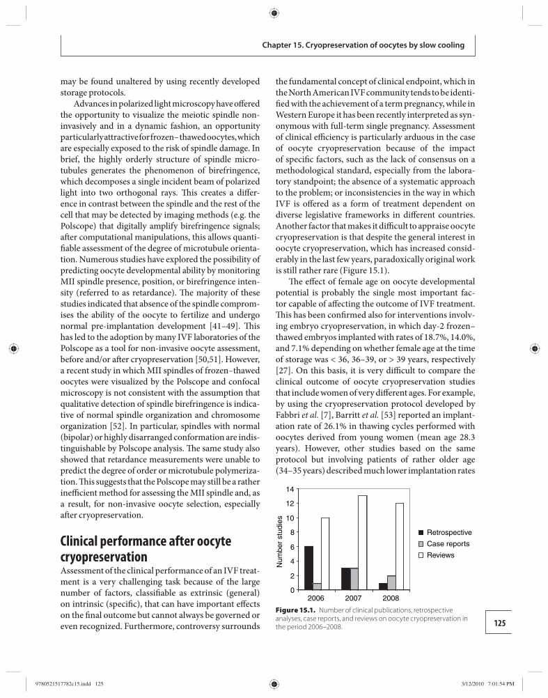
125
Chapter 15. Cryopreservation of oocytes by slow cooling
may be found unaltered by using recently developed storage protocols.
Advances in polarized light microscopy have off ered the opportunity to visualize the meiotic spindle non-invasively and in a dynamic fashion, an opportunity particularly attractive for frozen– thawed oocytes, which are especially exposed to the risk of spindle damage. In brief, the highly orderly structure of spindle micro-tubules generates the phenomenon of birefringence, which decomposes a single incident beam of polarized light into two orthogonal rays. Th is creates a diff er-ence in contrast between the spindle and the rest of the cell that may be detected by imaging methods (e.g. the Polscope) that digitally amplify birefringence signals; aft er computational manipulations, this allows quanti-fi able assessment of the degree of microtubule orienta-tion. Numerous studies have explored the possibility of predicting oocyte developmental ability by monitoring MII spindle presence, position, or birefringence inten-sity (referred to as retardance). Th e majority of these studies indicated that absence of the spindle comprom-ises the ability of the oocyte to fertilize and undergo normal pre-implantation development [ 41 – 49 ]. Th is has led to the adoption by many IVF laboratories of the Polscope as a tool for non-invasive oocyte assessment, before and/or aft er cryopreservation [ 50 , 51 ]. However, a recent study in which MII spindles of frozen–thawed oocytes were visualized by the Polscope and confocal microscopy is not consistent with the assumption that qualitative detection of spindle birefringence is indica-tive of normal spindle organization and chromosome organization [ 52 ]. In particular, spindles with normal (bipolar) or highly disarranged conformation are indis-tinguishable by Polscope analysis. Th e same study also showed that retardance measurements were unable to predict the degree of order or microtubule polymeriza-tion. Th is suggests that the Polscope may still be a rather ineffi cient method for assessing the MII spindle and, as a result, for non-invasive oocyte selection, especially aft er cryopreservation.
Clinical performance after oocyte cryopreservation Assessment of the clinical performance of an IVF treat-ment is a very challenging task because of the large number of factors, classifi able as extrinsic (general) on intrinsic (specifi c), that can have important eff ects on the fi nal outcome but cannot always be governed or even recognized. Furthermore, controversy surrounds
the fundamental concept of clinical endpoint, which in the North American IVF community tends to be identi-fi ed with the achievement of a term pregnancy, while in Western Europe it has been recently interpreted as syn-onymous with full-term single pregnancy. Assessment of clinical effi ciency is particularly arduous in the case of oocyte cryopreservation because of the impact of specifi c factors, such as the lack of consensus on a methodological standard, especially from the labora-tory standpoint; the absence of a systematic approach to the problem; or inconsistencies in the way in which IVF is off ered as a form of treatment dependent on diverse legislative frameworks in diff erent countries. Another factor that makes it diffi cult to appraise oocyte cryo preservation is that despite the general interest in oocyte cryopreservation, which has increased consid-erably in the last few years, paradoxically original work is still rather rare ( Figure 15.1 ).
Th e eff ect of female age on oocyte developmental potential is probably the single most important fac-tor capable of aff ecting the outcome of IVF treatment. Th is has been confi rmed also for interventions involv-ing embryo cryopreservation, in which day-2 frozen–thawed embryos implanted with rates of 18.7%, 14.0%, and 7.1% depending on whether female age at the time of storage was < 36, 36–39, or > 39 years, respectively [ 27 ]. On this basis, it is very diffi cult to compare the clinical outcome of oocyte cryopreservation studies that include women of very diff erent ages. For example, by using the cryopreservation protocol developed by Fabbri et al. [ 7 ], Barritt et al. [ 53 ] reported an implant-ation rate of 26.1% in thawing cycles performed with oocytes derived from young women (mean age 28.3 years). However, other studies based on the same proto col but involving patients of rather older age (34–35 years) described much lower implantation rates
0
2
4
6
Num
ber
stud
ies
8
10
12
14
2006 2007 2008
Retrospective
Case reports
Reviews
Figure 15.1. Number of clinical publications, retrospective analyses, case reports, and reviews on oocyte cryopreservation in the period 2006–2008.
9780521517782c15.indd 1259780521517782c15.indd 125 3/12/2010 7:01:54 PM3/12/2010 7:01:54 PM
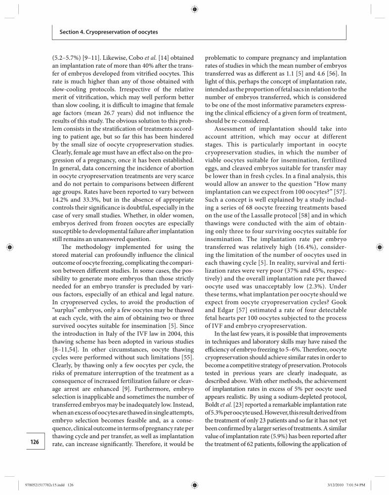
126
Section 4. Cryopreservation of oocytes
(5.2–5.7%) [ 9 – 11 ]. Likewise, Cobo et al. [ 14 ] obtained an implantation rate of more than 40% aft er the trans-fer of embryos developed from vitrifi ed oocytes. Th is rate is much higher than any of those obtained with slow-cooling protocols. Irrespective of the relative merit of vitrifi cation, which may well perform better than slow cooling, it is diffi cult to imagine that female age factors (mean 26.7 years) did not infl uence the results of this study. Th e obvious solution to this prob-lem consists in the stratifi cation of treatments accord-ing to patient age, but so far this has been hindered by the small size of oocyte cryopreservation studies. Clearly, female age must have an eff ect also on the pro-gression of a pregnancy, once it has been established. In general, data concerning the incidence of abortion in oocyte cryopreservation treatments are very scarce and do not pertain to comparisons between diff erent age groups. Rates have been reported to vary between 14.2% and 33.3%, but in the absence of appropriate controls their signifi cance is doubtful, especially in the case of very small studies. Whether, in older women, embryos derived from frozen oocytes are especially susceptible to developmental failure aft er implantation still remains an unanswered question.
Th e methodology implemented for using the stored material can profoundly infl uence the clinical outcome of oocyte freezing, complicating the compari-son between diff erent studies. In some cases, the pos-sibility to generate more embryos than those strictly needed for an embryo transfer is precluded by vari-ous factors, especially of an ethical and legal nature. In cryopreserved cycles, to avoid the production of “surplus” embryos, only a few oocytes may be thawed at each cycle, with the aim of obtaining two or three survived oocytes suitable for insemination [ 5 ]. Since the introduction in Italy of the IVF law in 2004, this thawing scheme has been adopted in various studies [ 8 – 11 , 54 ]. In other circumstances, oocyte thawing cycles were performed without such limitations [ 55 ]. Clearly, by thawing only a few oocytes per cycle, the risks of premature interruption of the treatment as a consequence of increased fertilization failure or cleav-age arrest are enhanced [ 9 ]. Furthermore, embryo selection is inapplicable and sometimes the number of transferred embryos may be inadequately low. Instead, when an excess of oocytes are thawed in single attempts, embryo selection becomes feasible and, as a conse-quence, clinical outcome in terms of pregnancy rate per thawing cycle and per transfer, as well as implantation rate, can increase signifi cantly. Th erefore, it would be
problematic to compare pregnancy and implantation rates of studies in which the mean number of embryos transferred was as diff erent as 1.1 [ 5 ] and 4.6 [ 56 ]. In light of this, perhaps the concept of implantation rate, intended as the proportion of fetal sacs in relation to the number of embryos transferred, which is considered to be one of the most informative parameters express-ing the clinical effi ciency of a given form of treatment, should be re-considered.
Assessment of implantation should take into account attrition, which may occur at different stages. This is particularly important in oocyte cryopreservation studies, in which the number of viable oocytes suitable for insemination, fertilized eggs, and cleaved embryos suitable for transfer may be lower than in fresh cycles. In a final analysis, this would allow an answer to the question “How many implantation can we expect from 100 oocytes?” [ 57 ]. Such a concept is well explained by a study includ-ing a series of 68 oocyte freezing treatments based on the use of the Lassalle protocol [ 58 ] and in which thawings were conducted with the aim of obtain-ing only three to four surviving oocytes suitable for insemination. The implantation rate per embryo transferred was relatively high (16.4%), consider-ing the limitation of the number of oocytes used in each thawing cycle [ 5 ]. In reality, survival and ferti-lization rates were very poor (37% and 45%, respec-tively) and the overall implantation rate per thawed oocyte used was unacceptably low (2.3%). Under these terms, what implantation per oocyte should we expect from oocyte cryopreservation cycles? Gook and Edgar [ 57 ] estimated a rate of four detectable fetal hearts per 100 oocytes subjected to the process of IVF and embryo cryopreservation.
In the last few years, it is possible that improvements in techniques and laboratory skills may have raised the effi ciency of embryo freezing to 5–6%. Th erefore, oocyte cryopreservation should achieve similar rates in order to become a competitive strategy of preservation. Protocols tested in previous years are clearly inadequate, as described above. With other methods, the achievement of implantation rates in excess of 5% per oocyte used appears realistic. By using a sodium-depleted protocol, Boldt et al. [ 23 ] reported a remarkable implantation rate of 5.3% per oocyte used. However, this result derived from the treatment of only 23 patients and so far it has not yet been confi rmed by a larger series of treatments. A similar value of implantation rate (5.9%) has been reported aft er the treatment of 62 patients, following the application of
9780521517782c15.indd 1269780521517782c15.indd 126 3/12/2010 7:01:54 PM3/12/2010 7:01:54 PM
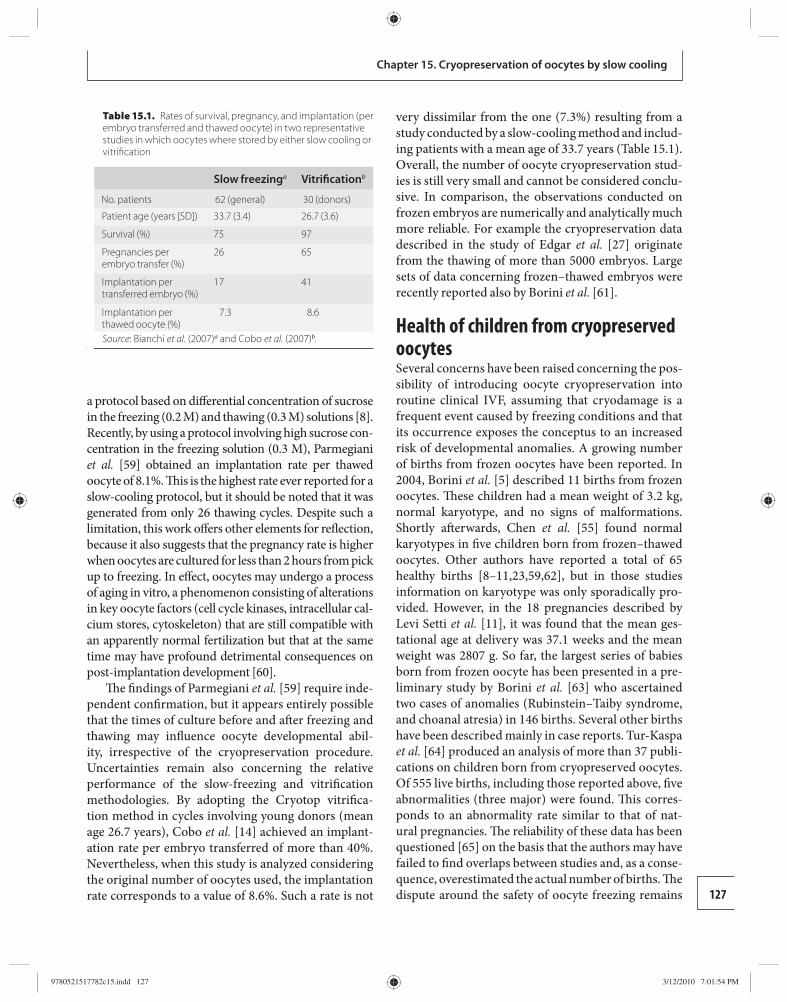
127
Chapter 15. Cryopreservation of oocytes by slow cooling
a protocol based on diff erential concentration of sucrose in the freezing (0.2 M) and thawing (0.3 M) solutions [ 8 ]. Recently, by using a protocol involving high sucrose con-centration in the freezing solution (0.3 M), Parmegiani et al. [ 59 ] obtained an implantation rate per thawed oocyte of 8.1%. Th is is the highest rate ever reported for a slow-cooling protocol, but it should be noted that it was generated from only 26 thawing cycles. Despite such a limitation, this work off ers other elements for refl ection, because it also suggests that the pregnancy rate is higher when oocytes are cultured for less than 2 hours from pick up to freezing. In eff ect, oocytes may undergo a process of aging in vitro, a phenomenon consisting of alterations in key oocyte factors (cell cycle kinases, intracellular cal-cium stores, cytoskeleton) that are still compatible with an apparently normal fertilization but that at the same time may have profound detrimental consequences on post-implantation development [ 60 ].
Th e fi ndings of Parmegiani et al. [ 59 ] require inde-pendent confi rmation, but it appears entirely possible that the times of culture before and aft er freezing and thawing may infl uence oocyte developmental abil-ity, irrespective of the cryopreservation procedure. Uncertainties remain also concerning the relative performance of the slow-freezing and vitrifi cation methodologies. By adopting the Cryotop vitrifi ca-tion method in cycles involving young donors (mean age 26.7 years), Cobo et al. [ 14 ] achieved an implant-ation rate per embryo transferred of more than 40%. Nevertheless, when this study is analyzed considering the original number of oocytes used, the implantation rate corresponds to a value of 8.6%. Such a rate is not
very dissimilar from the one (7.3%) resulting from a study conducted by a slow-cooling method and includ-ing patients with a mean age of 33.7 years ( Table 15.1 ). Overall, the number of oocyte cryopreservation stud-ies is still very small and cannot be considered conclu-sive. In comparison, the observations conducted on frozen embryos are numerically and analytically much more reliable. For example the cryopreservation data described in the study of Edgar et al. [ 27 ] originate from the thawing of more than 5000 embryos. Large sets of data concerning frozen–thawed embryos were recently reported also by Borini et al. [ 61 ].
Health of children from cryopreserved oocytes Several concerns have been raised concerning the pos-sibility of introducing oocyte cryopreservation into routine clinical IVF, assuming that cryodamage is a frequent event caused by freezing conditions and that its occurrence exposes the conceptus to an increased risk of developmental anomalies. A growing number of births from frozen oocytes have been reported. In 2004, Borini et al. [ 5 ] described 11 births from frozen oocytes. Th ese children had a mean weight of 3.2 kg, normal karyotype, and no signs of malformations. Shortly aft erwards, Chen et al. [ 55 ] found normal karyotypes in fi ve children born from frozen–thawed oocytes. Other authors have reported a total of 65 healthy births [ 8 – 11 , 23 , 59 , 62 ], but in those studies information on karyotype was only sporadically pro-vided. However, in the 18 pregnancies described by Levi Setti et al. [ 11 ], it was found that the mean ges-tational age at delivery was 37.1 weeks and the mean weight was 2807 g. So far, the largest series of babies born from frozen oocyte has been presented in a pre-liminary study by Borini et al. [ 63 ] who ascertained two cases of anomalies (Rubinstein–Taiby syndrome, and choanal atresia) in 146 births. Several other births have been described mainly in case reports. Tur-Kaspa et al. [ 64 ] produced an analysis of more than 37 publi-cations on children born from cryopreserved oocytes. Of 555 live births, including those reported above, fi ve abnormalities (three major) were found. Th is corres-ponds to an abnormality rate similar to that of nat-ural pregnancies. Th e reliability of these data has been questioned [ 65 ] on the basis that the authors may have failed to fi nd overlaps between studies and, as a conse-quence, overestimated the actual number of births. Th e dispute around the safety of oocyte freezing remains
Table 15.1. Rates of survival, pregnancy, and implantation (per embryo transferred and thawed oocyte) in two representative studies in which oocytes where stored by either slow cooling or vitrifi cation
Slow freezing a Vitrifi cation b
No. patients 62 (general) 30 (donors)
Patient age (years [SD]) 33.7 (3.4) 26.7 (3.6)
Survival (%) 75 97
Pregnancies per embryo transfer (%)
26 65
Implantation per transferred embryo (%)
17 41
Implantation per thawed oocyte (%)
7.3 8.6
Source : Bianchi et al. (2007) a and Cobo et al. (2007) b .
9780521517782c15.indd 1279780521517782c15.indd 127 3/12/2010 7:01:54 PM3/12/2010 7:01:54 PM

128
Section 4. Cryopreservation of oocytes
open. For instance, no exhaustive data are available on the number of spontaneous abortions. However, the evidence currently available does not point toward a dramatic increase in the incidence of anomalies at birth. Th is appears to legitimize further clinical studies on oocyte cryopreservation. Future studies will need to be more analytical and systematic, including also long-term follow-up.
Conclusions Oocyte cryopreservation has long been envisaged as the ideal solution to the need to capitalize the reproductive potential derived from a cycle of ovar-ian stimu lation, circumventing the ethical and legal problems posed by embryo cryopreservation. Th e fi rst attempts in that direction were conducted with an inappropriate cryopreservation methodology. Th is led to rather inadequate results and, as a consequence, a lack of confi dence and interest. Aft er a period of eclipse lasting more than 10 years, oocyte cryopreser-vation has been re-proposed following some meth-odological modifi cations, which have improved some parameters of effi ciency but not conclusively opti-mized the freezing procedures. Th is has made pos-sible the application of oocyte cryopreservation as a reproducible strategy of treatment. Clinical data are progressively emerging, suggesting that in the near future oocyte cryopreservation could represent a valid alternative to embryo cryopreservation. However, several important aspects have not yet been clarifi ed, especially in relation to the objective evaluation of oocyte implantation potential aft er cryopreservation. Meanwhile, non-clinical studies have established that cell damage from cryo storage may occur under diff er-ent forms and specifi c conditions, but is not inevita-ble. Current knowledge of the health of children from cryopreserved oocytes is still rather limited but does not support the notion of an increased developmental risk for the conceptus. Th is lends credit to the sugges-tion that clinical experi ence in oocyte cryopreserva-tion should be expand ed in order to achieve a high and safe standard of treatment.
References 1. Chen C . Pregnancy aft er human oocyte cryopreservation .
Lancet 1986; i: 884 –886. 2. Al-Hasani S , Diedrich K , van der Ven H , Krebs D .
[Initial results of the cryopreservation of human oocytes]. Geburtshilfe Frauenheilkd 1986; 46 : 643–644 .
3. Whittingham DG . Fertilization in vitro and development to term of unfertilized mouse oocytes previously stored at −196 degrees C. J Reprod Fertil 1977; 49 : 89–94 .
4. Porcu E , Fabbri R , Seracchioli R , et al. Birth of a healthy female aft er intracytoplasmic sperm injection of cryo-preserved human oocytes . Fertil Steril 1997; 68: 724 –726.
5. Borini A , Bonu MA , Coticchio G , et al. Pregnancies and births aft er oocyte cryopreservation . Fertil Steril 2004; 82: 601 –605.
6. Paynter SJ , O ’ Neil L , Fuller BJ , Shaw RW . Membrane permeability of human oocytes in the presence of the cryoprotectant propane-1,2-diol . Fertil Steril 2001; 75: 532 –538.
7. Fabbri R , Porcu E , Marsella T , et al. Human oocyte cryopreservation: new perspectives regarding oocyte survival . Hum Reprod 2001; 16: 411 –416.
8. Bianchi V , Coticchio G , Distratis V , et al. Diff erential sucrose concentration during dehydration (0.2 mol/l) and rehydration (0.3 mol/l) increases the implantation rate of frozen human oocytes . Reprod Biomed Online 2007; 14: 64 –71.
9. Borini A , Sciajno R , Bianchi V , et al. Clinical outcome of oocyte cryopreservation aft er slow cooling with a protocol utilizing a high sucrose concentration . Hum Reprod 2006; 21: 512 –517.
10. De Santis L , Cino I , Rabellotti E , et al. Oocyte cryopreservation: clinical outcome of slow-cooling protocols diff ering in sucrose concentration . Reprod Biomed Online 2007; 14: 57 –63.
11. Levi Setti PE , Albani E , Novara PV , Cesana A , Morreale G . Cryopreservation of supernumerary oocytes in IVF/ICSI cycles . Hum Reprod 2006; 21: 370 –375.
12. Paynter SJ , Borini A , Bianchi V , et al. Volume changes of mature human oocytes on exposure to cryoprotectant solutions used in slow cooling procedures . Hum Reprod 2005; 20: 1194 –1199.
13. Borini A , Bianchi V , Bonu MA , et al. Evidence-based clinical outcome of oocyte slow cooling . Reprod Biomed Online 2007; 15: 175 –181.
14. Cobo A , Kuwayama M , Perez S , et al. Comparison of concomitant outcome achieved with fresh and cryopreserved donor oocytes vitrifi ed by the Cryotop method . Fertil Steril 2008; 89: 1657 –1664.
15. Cobo A , Bellver J , Domingo J , et al. New options in assisted reproduction technology: the Cryotop method of oocyte vitrifi cation . Reprod Biomed Online 2008; 17: 68 –72.
16. De Santis L , Coticchio G , Paynter S , et al. Permeability of human oocytes to ethylene glycol and their survival and spindle confi gurations aft er slow cooling cryopreservation . Hum Reprod 2007; 22: 2776 –2783.
17. Kasai M , Mukaida T . Cryopreservation of animal and human embryos by vitrifi cation . Reprod Biomed Online 2004; 9: 164 –170.
9780521517782c15.indd 1289780521517782c15.indd 128 3/12/2010 7:01:54 PM3/12/2010 7:01:54 PM
129
Chapter 15. Cryopreservation of oocytes by slow cooling
18. Nottola SA , Coticchio G , de Santis L , et al. Ultrastructure of human mature oocytes aft er slow cooling cryopreservation with ethylene glycol . Reprod Biomed Online 2008; 17: 368 –377.
19. Stachecki JJ , Cohen J , Willadsen S . Detrimental eff ects of sodium during mouse oocyte cryopreservation . Biol Reprod 1998; 59: 395 –400.
20. Stachecki JJ , Cohen J , Willadsen SM . Cryopreservation of unfertilized mouse oocytes: the eff ect of replacing sodium with choline in the freezing medium . Cryobiology 1998; 37: 346 –354.
21. Stachecki JJ , Willadsen SM . Cryopreservation of mouse oocytes using a medium with low sodium content: eff ect of plunge temperature . Cryobiology 2000; 40: 4 –12.
22. Stachecki JJ , Cohen J , Garrisi J , et al. Cryopreservation of unfertilized human oocytes . Reprod Biomed Online 2006; 13: 222 –227.
23. Boldt J , Tidswell N , Sayers A , Kilani R , Cline D . Human oocyte cryopreservation: 5-year experience with a sodium-depleted slow freezing method . Reprod Biomed Online 2006; 13: 96 –100.
24. Quintans CJ , Donaldson MJ , Bertolino MV , Pasqualini RS . Birth of two babies using oocytes that were cryopreserved in a choline-based freezing medium . Hum Reprod 2002; 17: 3149 –3152.
25. Petracco A , Azambuja R , Okada L , et al. Comparison of embryo quality between sibling embryos originating from frozen or fresh oocytes . Reprod Biomed Online 2006; 13: 497 –503.
26. Ziebe S , Petersen K , Lindenberg S , et al. Embryo morphology or cleavage stage: how to select the best embryos for transfer aft er in-vitro fertilization . Hum Reprod 1997; 12: 1545 –1549.
27. Edgar DH , Archer J , McBain J , Bourne H . Embryonic factors aff ecting outcome from single cryopreserved embryo transfer . Reprod Biomed Online 2007; 14: 718 –723.
28. Coticchio G , Sereni E , Serrao L , et al. What criteria for the defi nition of oocyte quality? Ann New York Acad Sci 2004; 1034: 132 –144.
29. Nagy ZP , Chang CC , Shapiro DB , et al. Clinical evaluation of the effi ciency of an oocyte donation program using egg cryo-banking . Fertil Steril 2009 ; 92: 520–526 .
30. Nottola SA , Macchiarelli G , Coticchio G , et al. Ultrastructure of human mature oocytes aft er slow cooling cryopreservation using diff erent sucrose concentrations . Hum Reprod 2007; 22: 1123 –1133.
31. Gook DA , Osborn SM , Johnston WI . Cryopreservation of mouse and human oocytes using 1,2-propanediol and the confi guration of the meiotic spindle . Hum Reprod 1993; 8: 1101 –1109.
32. Ghetler Y , Skutelsky E , Ben Nun I , et al. Human oocyte cryopreservation and the fate of cortical granules . Fertil Steril 2006; 86: 210 –216.
33. Gualtieri R , Iaccarino M , Mollo V , et al. Slow cooling of human oocytes: ultrastructural injuries and apoptotic status . Fertil Steril 2009; 91: 1023–1034 .
34. Fuku E , Xia L , Downey BR . Ultrastructural changes in bovine oocytes cryopreserved by vitrifi cation . Cryobiology 1995; 32: 139 –156.
35. Hochi S , Kozawa M , Fujimoto T , et al. In vitro maturation and transmission electron microscopic observation of horse oocytes aft er vitrifi cation . Cryobiology 1996; 33: 300 –310.
36. Coticchio G , Distratis V , Borini A , et al. Reduced ultrastructural damage in human oocytes cryopreserved aft er dehydration involving single-step propanediol exposure . Hum Reprod 2008; 23 (Suppl 1): i150.
37. Nottola SA , Coticchio G , Sciajno R , et al. Ultrastructural changes in human metaphase II oocytes cryopreserved by vitrifi cation . Hum Reprod 2008; 23 (Suppl 1): i34.
38. Pickering SJ , Braude PR , Johnson MH , Cant A , Currie J . Transient cooling to room temperature can cause irreversible disruption of the meiotic spindle in the human oocyte . Fertil Steril 1990; 54: 102 –108.
39. Coticchio G , d e Santis L , Rossi G , et al. Sucrose concentration infl uences the rate of human oocytes with normal spindle and chromosome confi gurations aft er slow-cooling cryopreservation . Hum Reprod 2006; 21: 1771 –1776.
40. Cobo A , Perez S , de los Santos MJ , Zulategui J , Domingo J , Remohi J . Eff ect of diff erent cryopreservation protocols on the metaphase II spindle in human oocytes . Reprod Biomed Online 2008; 17: 350 –359.
41. Wang WH , Meng L , Hackett RJ , Keefe DL . Developmental ability of human oocytes with or without birefringent spindles imaged by Polscope before insemination . Hum Reprod 2001; 16: 1464 –1468.
42. Wang WH , Meng L , Hackett RJ , Odenbourg R , Keefe DL . Th e spindle observation and its relationship with fertilization aft er intracytoplasmic sperm injection in living human oocytes . Fertil Steril 2001; 75: 348 –353.
43. Cooke S , Tyler JP , Driscoll GL . Meiotic spindle location and identifi cation and its eff ect on embryonic cleavage plane and early development . Hum Reprod 2003; 18: 2397 –2405.
44. Moon JH , Hyun CS , Lee SW , et al. Visualization of the metaphase II meiotic spindle in living human oocytes using the Polscope enables the prediction of embryonic developmental competence aft er ICSI . Hum Reprod 2003; 18: 817 –820.
45. Rienzi L , Ubaldi F , Martinez F , et al. Relationship between meiotic spindle location with regard to the polar body position and oocyte developmental potential aft er ICSI . Hum Reprod 2003; 18: 1289 –1293.
46. de Santis L , Cino I , Rabellotti E , et al. Polar body morphology and spindle imaging as predictors of oocyte quality . Reprod Biomed Online 2005; 11: 36 –42.
9780521517782c15.indd 1299780521517782c15.indd 129 3/12/2010 7:01:55 PM3/12/2010 7:01:55 PM
130
Section 4. Cryopreservation of oocytes
47. Rama Raju GA , Prakash GJ , Krishna KM , Madan K . Meiotic spindle and zona pellucida characteristics as predictors of embryonic development: a preliminary study using Polscope imaging . Reprod Biomed Online 2007; 14: 166 –174.
48. Shen Y , Stalf T , Mehnert C , et al. Light retardance by human oocyte spindle is positively related to pronuclear score aft er ICSI . Reprod Biomed Online 2006; 12: 737 –751.
49. Madaschi C , de Souza Bonetti TC , de Almeida Ferreira Braga DP , et al. Spindle imaging: a marker for embryo development and implantation . Fertil Steril 2008; 90: 194 –198.
50. Bianchi V , Coticchio G , Fava L , Flamigni C , Borini A . Meiotic spindle imaging in human oocytes frozen with a slow freezing procedure involving high sucrose concentration . Hum Reprod 2005; 20: 1078 –1083.
51. Rienzi L , Martinez F , Ubaldi F , et al. Polscope analysis of meiotic spindle changes in living metaphase II human oocytes during the freezing and thawing procedures . Hum Reprod 2004; 19: 655 –659.
52. Coticchio G , Sciajno R , Hutt KJ , et al. Comparative analysis of the metaphase II spindle of human oocytes through polarized light and high performance confocal microscopy . Fertil Steril 2009 ; epub ahead pf print.
53. Barritt J , Luna M , Duke M , et al. Report of four donor-recipient oocyte cryopreservation cycles resulting in high pregnancy and implantation rates . Fertil Steril 2007; 87: e13–17.
54. La Sala GB , Nicoli A , Villani MT , et al. Outcome of 518 salvage oocyte-cryopreservation cycles performed as a routine procedure in an in vitro fertilization program . Fertil Steril 2006; 86: 1423 –1427.
55. Chen SU , Lien YR , Chen HF , et al. Observational clinical follow-up of oocyte cryopreservation using a slow-freezing method with 1,2-propanediol plus
sucrose followed by ICSI . Hum Reprod 2005; 20: 1975 –1980.
56. Lucena E , Bernal DP , Lucena C , et al. Successful ongoing pregnancies aft er vitrifi cation of oocytes . Fertil Steril 2006; 85: 108 –111.
57. Gook DA , Edgar DH . Cryopreservation of the human female gamete: current and future issues . Hum Reprod 1999; 14: 2938 –2940.
58. Lassalle B , Testart J , Renard JP . Human embryo features that infl uence the success of cryopreservation with the use of 1,2-propanediol . Fertil Steril 1985; 44: 645 –651.
59. Parmegiani L , Cognigni GE , Bernardi S , et al. Freezing within 2 h from oocyte retrieval increases the effi ciency of human oocyte cryopreservation when using a slow freezing/rapid thawing protocol with high sucrose concentration . Hum Reprod 2008; 23 : 1771–1778 .
60. Ducibella T . Biochemical and cellular insights into the temporal window of normal fertilization . Th eriogenology 1998; 49: 53 –65.
61. Borini A , Cattoli M , Bulletti C , Coticchio G . Clinical effi ciency of oocyte and embryo cryopreservation . Ann New York Acad Sci 2008; 1127: 49 –58.
62. Porcu E , Fabbri R , Damiano G , et al. Clinical experience and applications of oocyte cryopreservation . Mol Cell Endocrinol 2000; 169: 33 –37.
63. Borini A , Cattoli M , Mazzone S , et al. Survey of 105 babies born aft er slow-cooling oocyte cryopreservation . Fertil Steril 2007; 88 : S13– S14.
64. Tur-Kaspa I , Gal M , Horwitz A . Genetics and health of children born from cryopreserved oocytes . Fertil Steril 2007; 87 : S14 .
65. Maher B . Little consensus on egg freezing . Nature 2007; 449: 958 .
9780521517782c15.indd 1309780521517782c15.indd 130 3/12/2010 7:01:55 PM3/12/2010 7:01:55 PM
131Fertility Cryopreservation, ed. Ri-Cheng Chian and Patrick Quinn. Published by Cambridge University Press. Copyright © Cambridge University Press 2010.
Chapter
16 Aspects of this equation will be discussed in more detail later in this chapter and were also considered in Chapter 2 .
Th e role of vitrifi cation in an assisted reproductive technology (ART) setting is gradually replacing trad-itional slow cooling primarily because of its increased effi ciency in terms of its success, its increased speed for preservation of oocytes, and the lack of any require-ment for purchase and maintenance of a controlled rate programmable freezer. Th e issue of success is discussed more fully in Chapter 15 and by Homberg et al. [ 7 ], but it should be noted that some fertility programs have reported exceptionally good clinical results with slow-freezing cryopreservation of human oocytes [ 8 ]. Th ere is an advantage in increased speed in terms of the time required to vitrify a sample of oocytes. However, there are time constraints when large numbers (e.g. ≥ 10) of oocytes from a single patient or oocytes from mul-tiple patients need to be processed around the same time. A programmed slow-cooling device can process multiple samples at the same time but, with experi-ence, it is possible to process considerable numbers of oocytes (and embryos) for vitrifi cation using samples processed in parallel [ 5 ].
Th e reasons for cryopreserving oocytes have been enumerated many times previously [ 9 ] and are sum-marized by Chian in Chapter 14 . Briefl y, the primary reasons are as follows. First, excess oocytes can be cryopreserved directly, rather than as embryos, thus circumventing some legal, ethical, and societal con-cerns. Second, oocytes can be salvaged when there are no sperm available from a male partner. Th ird, oocytes can be collected and stored for later use from patients threatened with loss of fertility from cancer therapy, pelvic disease, surgical needs, and so on. Fourth, women can eff ectively stop their biological clock by cryopreserving oocytes when they are young and using them later in life: collect and store now, use later. Finally,
Introduction Th e primary focus of this chapter will be the eff ects of using diff erent types of a physical device, the carrier tool, to carry or enclose the cellular sample in the process of vitrifi cation of human oocytes. To broaden this topic, consideration will also be given to other devices used for diff erent stages of human pre-implantation embryo development and the oocytes and embryos of other mam-malian species, as these will most likely be applicable to the vitrifi cation of human oocytes. A brief mention will also be made of some of the diff erences in media, or more precisely solutions, that have been used in this process.
Interest in the topic of vitrifi cation has increased exponentially in academic and applied science fi elds since the end of the 1990s [ 1 ]. Th ere are several excel-lent reviews of the history of its development, of its cur-rent status, and of areas of current potential research, as well as texts on vitrifi cation methodology. Several of these publications are presented in other chapters of this publication ( Chapters 14 and 17 ). Other excel-lent publications on these topics include those by Kuwayama [ 2 ] and Tucker and Liebermann [ 3 ].
Th e traditional defi nition of vitrifi cation is the solidifi -cation of a liquid brought about not by crystallization but by the extreme elevation in viscosity during cooling [ 4 ]. A simpler description is that of Vatja et al. [ 5 ]: “Vitrifi cation is a physical phenomenon describing solidifi cation of water or water-based solutions without ice crystal for-mation .” As a working model, the following equation has been proposed by Yavin & Arav [ 6 ], which predicts the probability of successful vitrifi cation based upon three major factors: (1) the viscosity of the sample that can be modulated by cryoprotectant (CPA) concentration; (2) cooling and warming rates; and (3) sample volume:
Cooling and warming rates ×Viscosity (CPA concentration)Probability of
vitrification Volume=
Vitrifi cation of human oocytes with diff erent tools Patrick Quinn
9780521517782c16.indd 1319780521517782c16.indd 131 3/12/2010 7:08:32 PM3/12/2010 7:08:32 PM
132
Section 4. Cryopreservation of oocytesSection 4. Cryopreservation of oocytes
cryopreserved oocytes from donors can be used to cre-ate egg banks in the same way that donor sperm are processed and cryostored in sperm banks [ 10 ].
Th e fi rst clinical pregnancy and subsequent live birth from cryopreserved human oocytes was reported in the 1980s by Chen [ 11 , 12 ] using a slow-cooling regi-men. In fact, the oocytes had been kept submerged in liquid nitrogen (LN 2 ) for only 3 hours before being thawed, inseminated, and returned to the uterus of the mother in the same cycle in which they had been col-lected. In the subsequent decade, there were very few further successes with only approximately 15 babies born [ 13 ]. Nearly all of the initial attempts used slow cooling, but there were some instances of using ultra-rapid cooling and vitrifi cation.
Th e fi rst reported live birth from vitrifi ed human oocytes was by Kuleshova et al. [ 15 ], with other reports relatively soon thereaft er. Reports began to appear slowly at fi rst, with several further live births [ 16 , 17 ], and then was followed since 2003 by increasing numbers. Some of the more impressive publications included the breakout reports of Kuwayama and col-leagues [ 18 , 19 ] together with those of Lucena et al. [ 20 ], Antinori et al. [ 21 ], Cobo et al. [ 22 ], and Chian et al. [ 23 ] (Homburg et al. [ 7 ] and Kuwayama [ 2 ] refer-ence further studies). Th ere does not appear to be any increased risk of adverse obstetric and perinatal out-comes of infants conceived from vitrifi ed oocytes [ 24 ]. Despite the considerable progress in the development and application of the vitrifi cation of human oocytes in a clinical setting [ 2 , 25 ], including the establishment of cryopreserved egg banks and the transport of oocytes internationally [ 10 ], there are still some obstacles that need to be overcome. One of the main obstacles, in the opinion of the author and others [ 2 , 5 ], including the major proponent on the issue of microbial contam-ination in mammalian germplasm cryobiology [ 26 ], is that safety and regulatory issues regarding closed and open vitrifi cation devices, that is those that do not and those that do allow direct contact of LN 2 with the oocyte, are causing a road block, hopefully temporar-ily, to the continued and increased use of this technol-ogy. More discussion on this topic is included in the section below on safety.
Vitrifi cation of human oocytes with diff erent tools In this section, the eff ectiveness and safety of various tools (also referred to as carriers) used in vitrifi ca-tion to hold the oocytes during cooling, storage, and
warming will be discussed, followed by a brief mention of the composition of some of the solutions and how they are used. Here we have a conundrum. Do we talk about the solutions fi rst or the tools? In reality, both are interdependent, so it does not really matter in which order they are discussed. Th e choice here is to describe fi rst some of the tools themselves and then their eff ect-iveness and safety, and then to move on to aspects of some of the solutions used for cooling and warming.
Carrier tools A range of carrier tools used for vitrifi cation in human ART are listed in Table 16.1 and examples are shown in Figure 16.1 . Pictures and diagrams of these tools occur elsewhere in this book and in many other sources (e.g. searching on the Internet for oocyte vitrifi cation pic-tures). Th e carrier tools fall primarily into three types, the primary versions of which are the open pulled straw, Cryotop, and Cryoloop. Th e electron micros-copy (EM) grid was the fi rst carrier tool in which small sample volume and direct contact with LN 2 achieved the very high cooling rates required in vitri-fi cation. Considerations of convenience and safety led to the replacement of the EM grid by the Cryotop and Cryoloop by the middle of the 2000s. Th e open pulled straw was fi rst introduced into mammalian oocyte and embryo vitrifi cation in 1998 [ 29 ] but, while remaining popular for domestic animal oocyte and embryo vitri-fi cation, it has not gained much of a foothold in human ART vitrifi cation applications. One of its derivatives, the Cryotip, was heavily promoted by its commercial producer, especially as it was the fi rst such vitrifi ca-tion tool to obtain FDA 510k clearance in the USA, but apart from a few enthusiasts, its appeal has been limited. Th e major concern is the long learning curve required to load and handle the tool correctly [ 33 ] and to avoid breakage of a loaded carrier during handling and storage [ 34 ]. Th e Cryoloop has some afi cionados in human ART and is marketed by a major ART sup-ply company but its market penetration so far is not widespread. Th e use of both the open pulled straw and Cryoloop has been modifi ed to enable them to be used as closed systems [ 35 , 36 ]. Th e most popular carrier tool so far is the Cryotop and several of its derivatives, espe-cially the hemi-straw [ 31 ], Cryolock [ 20 ], and Cryoleaf ( Chapter 17 ).
Other devices that have been involved in vitrifi -cation are the Vitmaster [ 37 ], which lowers the tem-perature of LN 2 by a further 10–15°C to approximately 210°C, and a device that uses pre-cooled metal surfaces
9780521517782c16.indd 1329780521517782c16.indd 132 3/12/2010 7:08:33 PM3/12/2010 7:08:33 PM
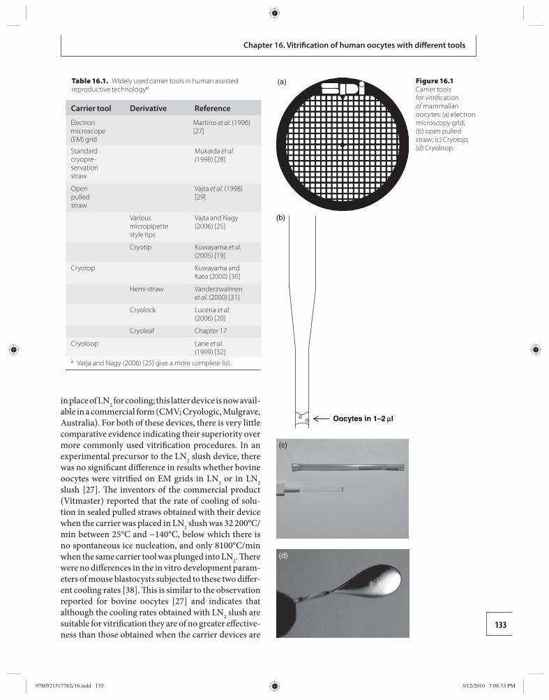
133
Chapter 16. Vitrifi cation of human oocytes with diff erent tools
in place of LN 2 for cooling; this latter device is now avail-able in a commercial form (CMV; Cryologic, Mulgrave, Australia). For both of these devices, there is very little comparative evidence indicating their superiority over more commonly used vitrifi cation procedures. In an experimental precursor to the LN 2 slush device, there was no signifi cant diff erence in results whether bovine oocytes were vitrifi ed on EM grids in LN 2 or in LN 2 slush [ 27 ]. Th e inventors of the commercial product (Vitmaster) reported that the rate of cooling of solu-tion in sealed pulled straws obtained with their device when the carrier was placed in LN 2 slush was 32 200°C/min between 25°C and −140°C, below which there is no spontaneous ice nucleation, and only 8100°C/min when the same carrier tool was plunged into LN 2 . Th ere were no diff erences in the in vitro development param-eters of mouse blastocysts subjected to these two diff er-ent cooling rates [ 38 ]. Th is is similar to the observation reported for bovine oocytes [ 27 ] and indicates that although the cooling rates obtained with LN 2 slush are suitable for vitrifi cation they are of no greater eff ective-ness than those obtained when the carrier devices are
(a)
Oocytes in 1–2 l
(b)
(c)
Figure 16.1 Carrier tools for vitrifi cation of mammalian oocytes: (a) electron microscopy grid; (b) open pulled straw; (c) Cryotop; (d) Cryoloop.
Table 16.1. Widely used carrier tools in human assisted reproductive technology a
Carrier tool Derivative Reference
Electron microscope (EM) grid
Martino et al. (1996) [27]
Standard cryopre-servation straw
Mukaida et al. (1998) [28]
Open pulled straw
Vajta et al. (1998) [29]
Various micropipette style tips
Vajta and Nagy (2006) [25]
Cryotip Kuwayama et al. (2005) [19]
Cryotop Kuwayama and Kato (2000) [30]
Hemi-straw Vanderzwalmen et al. (2000) [31]
Cryolock Lucena et al. (2006) [20]
Cryoleaf Chapter 17
Cryoloop Lane et al. (1999) [32]
a Vatja and Nagy (2006) [ 25 ] give a more complete list.
(d)
9780521517782c16.indd 1339780521517782c16.indd 133 3/12/2010 7:08:33 PM3/12/2010 7:08:33 PM
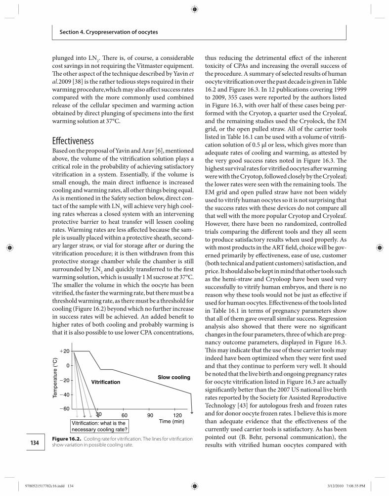
134
Section 4. Cryopreservation of oocytes
plunged into LN 2 . Th ere is, of course, a considerable cost savings in not requiring the Vitmaster equipment. Th e other aspect of the technique described by Yavin et al. 2009 [ 38 ] is the rather tedious steps required in their warming procedure,which may also aff ect success rates compared with the more commonly used combined release of the cellular specimen and warming action obtained by direct plunging of specimens into the fi rst warming solution at 37°C .
Eff ectiveness Based on the proposal of Yavin and Arav [ 6 ], mentioned above, the volume of the vitrifi cation solution plays a critical role in the probability of achieving satisfactory vitrifi cation in a system. Essentially, if the volume is small enough, the main direct infl uence is increased cooling and warming rates, all other things being equal. As is mentioned in the Safety section below, direct con-tact of the sample with LN 2 will achieve very high cool-ing rates whereas a closed system with an intervening protective barrier to heat transfer will lessen cooling rates. Warming rates are less aff ected because the sam-ple is usually placed within a protective sheath, second-ary larger straw, or vial for storage aft er or during the vitrifi cation procedure; it is then withdrawn from this protective storage chamber while the chamber is still surrounded by LN 2 and quickly transferred to the fi rst warming solution, which is usually 1 M sucrose at 37°C. Th e smaller the volume in which the oocyte has been vitrifi ed, the faster the warming rate, but there must be a threshold warming rate, as there must be a threshold for cooling ( Figure 16.2 ) beyond which no further increase in success rates will be achieved. An added benefi t to higher rates of both cooling and probably warming is that it is also possible to use lower CPA concentrations,
thus reducing the detrimental eff ect of the inherent toxicity of CPAs and increasing the overall success of the procedure. A summary of selected results of human oocyte vitrifi cation over the past decade is given in Table 16.2 and Figure 16.3 . In 12 publications covering 1999 to 2009, 355 cases were reported by the authors listed in Figure 16.3 , with over half of these cases being per-formed with the Cryotop, a quarter used the Cryoleaf, and the remaining studies used the Cryolock, the EM grid, or the open pulled straw. All of the carrier tools listed in Table 16.1 can be used with a volume of vitrifi -cation solution of 0.5 μl or less, which gives more than adequate rates of cooling and warming, as attested by the very good success rates noted in Figure 16.3 . Th e highest survival rates for vitrifi ed oocytes aft er warming were with the Cryotop, followed closely by the Cryoleaf; the lower rates were seen with the remaining tools. Th e EM grid and open pulled straw have not been widely used to vitrify human oocytes so it is not surprising that the success rates with these devices do not compare all that well with the more popular Cryotop and Cryoleaf. However, there have been no randomized, controlled trials comparing the diff erent tools and they all seem to produce satisfactory results when used properly. As with most products in the ART fi eld, choice will be gov-erned primarily by eff ectiveness, ease of use, customer (both technical and patient customers) satisfaction, and price. It should also be kept in mind that other tools such as the hemi-straw and Cryoloop have been used very successfully to vitrify human embryos, and there is no reason why these tools would not be just as eff ective if used for human oocytes. Eff ectiveness of the tools listed in Table 16.1 in terms of pregnancy parameters show that all of them gave overall similar success. Regression analysis also showed that there were no signifi cant changes in the four parameters, three of which are preg-nancy outcome parameters, displayed in Figure 16.3 . Th is may indicate that the use of these carrier tools may indeed have been optimized when they were fi rst used and that they continue to perform very well. It should be noted that the live birth and ongoing pregnancy rates for oocyte vitrifi cation listed in Figure 16.3 are actually signifi cantly better than the 2007 US national live birth rates reported by the Society for Assisted Reproductive Technology [ 43 ] for autologous fresh and frozen rates and for donor oocyte frozen rates. I believe this is more than adequate evidence that the eff ectiveness of the currently used carrier tools is satisfactory. As has been pointed out (B. Behr, personal communication), the results with vitrifi ed human oocytes compared with
Tem
pera
ture
(°C
)
�20
0
�20
�40
�60
Time (min)30 60
Vitrification: what is thenecessary cooling rate?
90 120
Slow coolingVitrification
Figure 16.2. Cooling rate for vitrifi cation. The lines for vitrifi cation show variation in possible cooling rate.
9780521517782c16.indd 1349780521517782c16.indd 134 3/12/2010 7:08:35 PM3/12/2010 7:08:35 PM

135 Tabl
e 16
.2.
Surv
ey o
f vitr
ifi ed
hum
an o
ocyt
e su
cces
s rat
es
Stud
y Ca
rrie
r to
ol
No.
ca
ses
Surv
ival
(%
) Em
bryo
s/ET
N
o.
ET
ET/
case
s (%
)
No.
pr
egna
-nc
ies
Preg
na-
ncie
s/ET
(%
)
CPR
(%)
IR
(%)
Live
bi
rths
N
o.
ongo
ing
Live
bi
rth
+ on
goin
g/ET
(%)
Com
men
ts
Kule
shov
a e
t al. (
1999
) [1
5]
OPS
465
1.0
375
133
3333
1N
AP33
Embr
yo
biop
sied
Yoon
et a
l. (2
000)
[16]
EM g
rid7
634.
67
100
343
439
21
43
Kata
yam
a et
al.
(200
3) [1
7]Cr
yoto
p6
94N
A6
100
233
33N
A1
133
Yoon
et a
l. (2
003)
[39]
EM g
rid34
694.
528
826
2121
66
NAP
21Pr
obab
ly
incl
udes
cas
es
from
[16]
Kim
et a
l. (2
005)
[40]
EM g
rid13
713.
812
927
5858
242
342
Okt
ay e
t al.
(200
6) [4
1]
(upd
ate
of
data
from
Ch
ian)
Cryo
leaf
2594
3.6
2510
011
4444
209
936
Kuw
ayam
a et
al. (
2005
) [1
8]
Cryo
top
2991
2.2
2910
012
4134
NA
73
34
Selm
an e
t al. (
2006
) [4
2]
OPS
634
2.3
610
02
3333
21N
A2
NA
Luce
na e
t al. (
2006
) [2
0]
Cryo
top
(now
Cr
yolo
ck)
2375
4.6
2310
013
57N
AN
AN
AN
AN
A
Antin
ori
et a
l. (20
07)
[21]
Cryo
top
120
992.
512
010
039
3333
133
2826
Cobo
et a
l. (2
008)
[22]
Cryo
top
3097
2.1
2377
1565
5241
NA
1148
Chia
n et
al.
(200
9) [2
3]Cr
yole
af58
783.
458
100
2340
3615
19N
AP33
38 IV
F 20
IVM
9780521517782c16.indd 1359780521517782c16.indd 135 3/12/2010 7:08:35 PM3/12/2010 7:08:35 PM

136 Stud
y Ca
rrie
r to
ol
No.
ca
ses
Surv
ival
(%
) Em
bryo
s/ET
N
o.
ET
ET/
case
s (%
)
No.
pr
egna
-nc
ies
Preg
na-
ncie
s/ET
(%
)
CPR
(%)
IR
(%)
Live
bi
rths
N
o.
ongo
ing
Live
bi
rth
+ on
goin
g/ET
(%)
Com
men
ts
Tota
l fro
m
stud
ies
355
340
134
5058
Mea
n fr
om
stud
ies
3087
3.1
2896
1142
3820
67
40
Soci
ety
for
Assis
ted
Repr
oduc
tive
Tech
nolo
gy
(200
7) [4
3]
Auto
logo
us,
fresh
93 7
922.
583
020
4024
33p
< 0
.000
1
Auto
logo
us,
froze
nN
AP2.
219
329
30p
< 0
.000
1
Don
or, f
resh
2.2
9 57
555
p <
0.0
001
Don
or, f
roze
n2.
35
087
32p
< 0
.003
ET, e
mbr
yo tr
ansf
er; C
PR, c
linic
al p
regn
ancy
rate
; IR,
inpl
anta
tion
rate
; OPS
, ope
n pu
lled
stra
w; E
M, e
lect
ron
mic
rosc
opy;
IVF,
in v
itro
Fert
iliza
tion;
IVM
, in
vitr
o m
atur
atio
n; N
AP, n
ot
appl
icab
le; N
A, n
ot a
vaila
ble.
Tab
le 1
6.2
(con
t.)
9780521517782c16.indd 1369780521517782c16.indd 136 3/12/2010 7:08:35 PM3/12/2010 7:08:35 PM
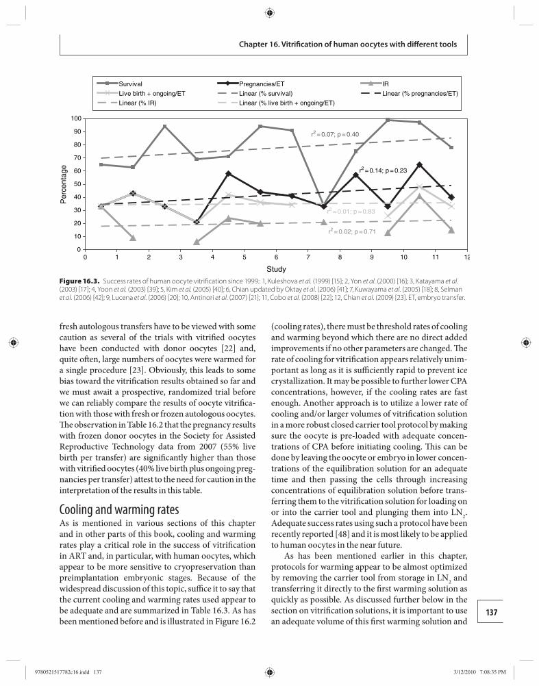
137
Chapter 16. Vitrifi cation of human oocytes with diff erent tools
fresh autologous transfers have to be viewed with some caution as several of the trials with vitrifi ed oocytes have been conducted with donor oocytes [ 22 ] and, quite oft en, large numbers of oocytes were warmed for a single procedure [ 23 ]. Obviously, this leads to some bias toward the vitrifi cation results obtained so far and we must await a prospective, randomized trial before we can reliably compare the results of oocyte vitrifi ca-tion with those with fresh or frozen autologous oocytes. Th e observation in Table 16.2 that the pregnancy results with frozen donor oocytes in the Society for Assisted Reproductive Technology data from 2007 (55% live birth per transfer) are signifi cantly higher than those with vitrifi ed oocytes (40% live birth plus ongoing preg-nancies per transfer) attest to the need for caution in the interpretation of the results in this table.
Cooling and warming rates As is mentioned in various sections of this chapter and in other parts of this book, cooling and warming rates play a critical role in the success of vitrifi cation in ART and, in particular, with human oocytes, which appear to be more sensitive to cryopreservation than preimplantation embryonic stages. Because of the widespread discussion of this topic, suffi ce it to say that the current cooling and warming rates used appear to be adequate and are summarized in Table 16.3 . As has been mentioned before and is illustrated in Figure 16.2
(cooling rates), there must be threshold rates of cooling and warming beyond which there are no direct added improvements if no other parameters are changed. Th e rate of cooling for vitrifi cation appears relatively unim-portant as long as it is suffi ciently rapid to prevent ice crystallization. It may be possible to further lower CPA concentrations, however, if the cooling rates are fast enough. Another approach is to utilize a lower rate of cooling and/or larger volumes of vitrifi cation solution in a more robust closed carrier tool protocol by making sure the oocyte is pre-loaded with adequate concen-trations of CPA before initiating cooling. Th is can be done by leaving the oocyte or embryo in lower concen-trations of the equilibration solution for an adequate time and then passing the cells through increasing concentrations of equilibration solution before trans-ferring them to the vitrifi cation solution for loading on or into the carrier tool and plunging them into LN 2 . Adequate success rates using such a protocol have been recently reported [ 48 ] and it is most likely to be applied to human oocytes in the near future.
As has been mentioned earlier in this chapter, protocols for warming appear to be almost optimized by removing the carrier tool from storage in LN 2 and transferring it directly to the fi rst warming solution as quickly as possible. As discussed further below in the section on vitrifi cation solutions, it is important to use an adequate volume of this fi rst warming solution and
r2 = 0.07; p = 0.40
r2 = 0.14; p = 0.23
r2 = 0.02; p = 0.71
r2 = 0.01; p = 0.83
00 1 2 3 4 5 6 7 8 9 10 11 12
10
20
30
40
50
60
70
80
90
100
Study
Per
cent
age
Survival Pregnancies/ET IR
Live birth + ongoing/ET Linear (% survival) Linear (% pregnancies/ET)
Linear (% IR) Linear (% live birth + ongoing/ET)
Figure 16.3. Success rates of human oocyte vitrifi cation since 1999. : 1, Kuleshova et al. (1999) [ 15 ]; 2, Yon et al. (2000) [ 16 ]; 3, Katayama et al. (2003) [ 17 ]; 4, Yoon et al. (2003) [ 39 ]; 5, Kim et al. (2005) [ 40 ]; 6, Chian updated by Oktay et al. (2006) [ 41 ]; 7, Kuwayama et al. (2005) [ 18 ]; 8, Selman et al. (2006) [ 42 ]; 9, Lucena et al. (2006) [ 20 ]; 10, Antinori et al. (2007) [ 21 ]; 11, Cobo et al. (2008) [ 22 ]; 12, Chian et al. (2009) [ 23 ]. ET, embryo transfer.
9780521517782c16.indd 1379780521517782c16.indd 137 3/12/2010 7:08:35 PM3/12/2010 7:08:35 PM
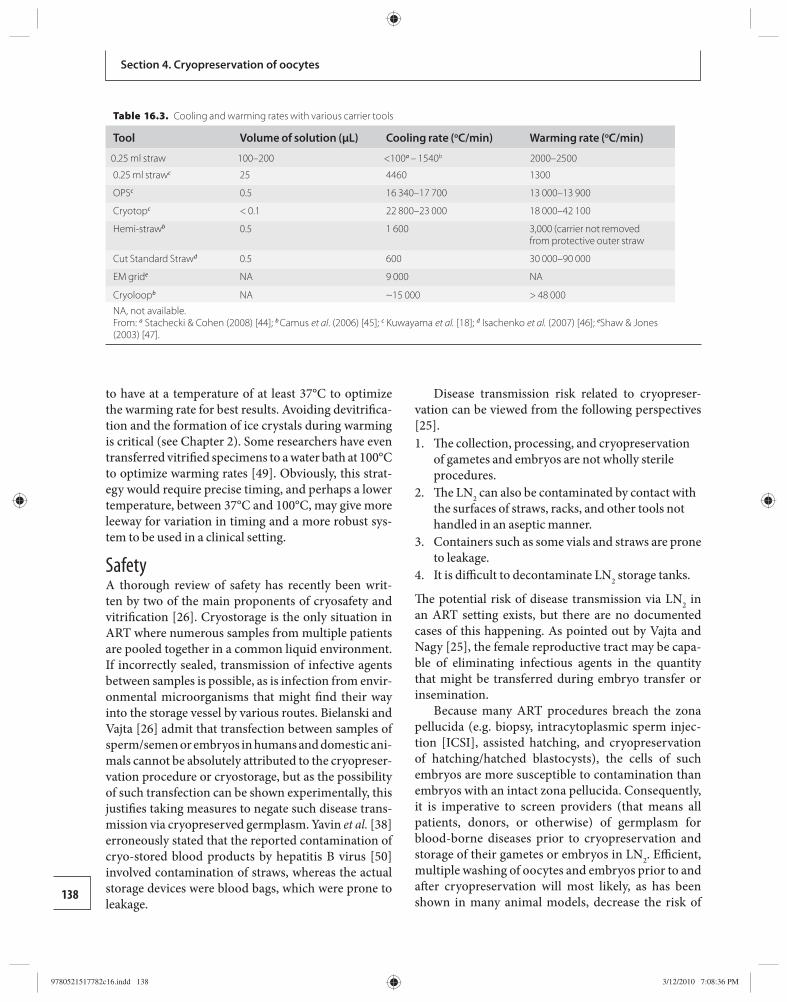
138
Section 4. Cryopreservation of oocytes
to have at a temperature of at least 37°C to optimize the warming rate for best results. Avoiding devitrifi ca-tion and the formation of ice crystals during warming is critical (see Chapter 2 ). Some researchers have even transferred vitrifi ed specimens to a water bath at 100°C to optimize warming rates [ 49 ]. Obviously, this strat-egy would require precise timing, and perhaps a lower temperature, between 37°C and 100°C, may give more leeway for variation in timing and a more robust sys-tem to be used in a clinical setting.
Safety A thorough review of safety has recently been writ-ten by two of the main proponents of cryosafety and vitrifi cation [ 26 ]. Cryostorage is the only situation in ART where numerous samples from multiple patients are pooled together in a common liquid environment. If incorrectly sealed, transmission of infective agents between samples is possible, as is infection from envir-onmental microorganisms that might fi nd their way into the storage vessel by various routes. Bielanski and Vajta [ 26 ] admit that transfection between samples of sperm/semen or embryos in humans and domestic ani-mals cannot be absolutely attributed to the cryopreser-vation procedure or cryostorage, but as the possibility of such transfection can be shown experimentally, this justifi es taking measures to negate such disease trans-mission via cryopreserved germplasm. Yavin et al. [ 38 ] erroneously stated that the reported contamination of cryo-stored blood products by hepatitis B virus [ 50 ] involved contamination of straws, whereas the actual storage devices were blood bags, which were prone to leakage.
Disease transmission risk related to cryopreser-vation can be viewed from the following perspectives [ 25 ]. 1. Th e collection, processing, and cryopreservation
of gametes and embryos are not wholly sterile procedures.
2. Th e LN 2 can also be contaminated by contact with the surfaces of straws, racks, and other tools not handled in an aseptic manner.
3. Containers such as some vials and straws are prone to leakage.
4. It is diffi cult to decontaminate LN 2 storage tanks.
Th e potential risk of disease transmission via LN 2 in an ART setting exists, but there are no documented cases of this happening. As pointed out by Vajta and Nagy [ 25 ], the female reproductive tract may be capa-ble of eliminating infectious agents in the quantity that might be transferred during embryo transfer or insemination.
Because many ART procedures breach the zona pellucida (e.g. biopsy, intracytoplasmic sperm injec-tion [ICSI], assisted hatching, and cryopreservation of hatching/hatched blastocysts), the cells of such embryos are more susceptible to contamination than embryos with an intact zona pellucida. Consequently, it is imperative to screen providers (that means all patients, donors, or otherwise) of germplasm for blood-borne diseases prior to cryopreservation and storage of their gametes or embryos in LN 2 . Effi cient, mul tiple washing of oocytes and embryos prior to and aft er cryopreservation will most likely, as has been shown in many animal models, decrease the risk of
Table 16.3. Cooling and warming rates with various carrier tools
Tool Volume of solution (µL) Cooling rate ( o C/min) Warming rate ( o C/min)
0.25 ml straw 100–200 <100 a – 1540 b 2000–2500
0.25 ml straw c 25 4460 1300
OPS c 0.5 16 340–17 700 13 000–13 900
Cryotop c < 0.1 22 800–23 000 18 000–42 100
Hemi-straw b 0.5 1 600 3,000 (carrier not removed from protective outer straw
Cut Standard Straw d 0.5 600 30 000–90 000
EM grid e NA 9 000 NA
Cryoloop b NA ~15 000 > 48 000 NA, not available. From: a Stachecki & Cohen (2008) [44]; b Camus et al . (2006) [45]; c Kuwayama et al. [18]; d Isachenko et al. (2007) [46]; e Shaw & Jones (2003) [47].
9780521517782c16.indd 1389780521517782c16.indd 138 3/12/2010 7:08:36 PM3/12/2010 7:08:36 PM
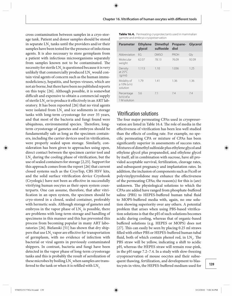
139
Chapter 16. Vitrifi cation of human oocytes with diff erent tools
cross contamination between samples in a cryo-stor-age tank. Patient and donor samples should be stored in separate LN 2 tanks until the providers and/or their samples have been tested for the presence of infectious agents. It is also necessary to store germplasm from a patient with infectious microorganisms separately from samples known not to be contaminated. Th e necessity for sterile LN 2 is questioned because it is very unlikely that commercially produced LN 2 would con-tain viral agents of concern such as the human immu-nodefi ciency, hepatitis, and herpes viruses, which are not air borne, but there have been no published reports on this topic [ 26 ]. Although possible, it is somewhat diffi cult and expensive to obtain a commercial supply of sterile LN 2 or to produce it eff ectively in an ART lab-oratory. It has been reported [ 26 ] that no viral agents were isolated from LN 2 and ice sediments in storage tanks with long-term cryostorage for over 35 years, and that most of the bacteria and fungi found were ubiquitous, environmental species. . Th erefore, long-term cryostorage of gametes and embryos should be fundamentally safe as long as the specimen contain-ers, including the carrier devices used in vitrifi cation, were properly sealed upon storage. Similarly, con-sideration has been given to approaches using open, direct contact between the specimen carrier tool and LN 2 during the cooling phase of vitrifi cation, but the use of sealed containers for storage [ 2 , 25 ]. Support for this approach comes from the report [ 26 ] that current closed systems such as the CryoTop, CBS HSV kits, and the solid surface vitrifi cation device Cryohook (Cryologic) have not been as eff ective in successfully vitrifying human oocytes as their open system coun-terparts. One can assume, therefore, that aft er vitri-fi cation in an open system, the specimen should be cryo-stored in a closed, sealed container, preferably with hermetic seals. Although storage of gametes and embryos in the vapor phase of LN 2 is possible, there are problems with long-term storage and handling of specimens in this manner and this has prevented this process from becoming popular in many ART labo-ratories [ 26 ]. Bielanski [ 51 ] has shown that dry ship-pers that use LN 2 vapor are eff ective for transportation of germplasm, with no evidence of infection with bacterial or viral agents in previously contaminated shippers. In contrast, bacteria and fungi have been detected in the vapor phase of long-term cryostorage tanks and this is probably the result of aerolization of these microbes by boiling LN 2 when samples are trans-ferred to the tank or when it is refi lled with LN 2.
Vitrifi cation solutions Th e four major permeating CPAs used in cryopreser-vation are listed in Table 16.4 . Th e role of media in the eff ectiveness of vitrifi cation has been less well studied than the eff ects of cooling rate. For example, no spe-cifi c permeating CPA or mixture of CPAs has been signifi cantly superior in assessments of success rates. Mixtures of dimethyl sulfoxide plus ethylene glycol and ethylene glycol plus propanediol, and ethylene glycol by itself, all in combination with sucrose, have all pro-vided acceptable survival, fertilization, cleavage rates, and subsequent pregnancy and implantation rates. In addition, the inclusion of components such as Ficoll or polyvinylpyrrolidone may enhance the eff ectiveness of the permeating CPAs; the reason(s) for this is (are) unknown. Th e physiological solutions to which the CPAs are added have ranged from phosphate-buff ered saline (PBS) to HEPES-buff ered human tubal fl uid to MOPS-buff ered media with, again, no one solu-tion showing superiority over any others. A potential problem that arises when using PBS-based vitrifi ca-tion solutions is that the pH of such solutions becomes acidic during cooling, whereas that of organic-based buff ered solutions (e.g. HEPES or MOPS) does not [ 27 ]. Th is can easily be seen by placing 0.25 ml straws fi lled with either PBS or HEPES-buff ered human tubal fl uid, both of which contain phenol red, in LN 2 . Th e PBS straw will be yellow, indicating a shift to acidic pH, whereas the HEPES straw will remain rose-pink, in the pH range 7.2–7.4. In a study with slow-freezing cryopreservation of mouse oocytes and their subse-quent thawing, fertilization, and development to blas-tocysts in vitro, the HEPES-buff ered medium used for
Table 16.4. Permeating cryoprotectants used in mammalian gamete and embryo cryopreservation
Parameter Ethylene glycol
Dimethyl sulfoxide
Propane-diol
Glycerol
Abbreviation EG DMSO PROH Gly
Molecular weight
62.07 78.13 76.09 92.09
Density at 25°C (g/ml)
1.113 1.10 1.036 1.25
Molality of a 10% (v/v) solution
1.79 1.41 1.36 1.36
Percentage (v/v) of a 1 M solution
5.6 7.1 7.4 7.4
9780521517782c16.indd 1399780521517782c16.indd 139 3/12/2010 7:08:36 PM3/12/2010 7:08:36 PM
140
Section 4. Cryopreservation of oocytes
cryopreservation gave signifi cantly better results com-pared with PBS-buff ered medium, but the diff erences were not as dramatic as expected [ 52 ]. Th is reinforces the need for prospective, randomized trials to see if the buff er system used in vitrifi cation solutions has any impact on outcomes. In 2004 [ 53 ], a brief report showed higher success rates with MOPS-buff ered solutions than with HEPES-buff ered medium for the vitrifi cation of day-3 human embryos, and two com-mercial producers use MOPS-buff ered vitrifi cation solutions. It is becoming evident that a physiological salt solution buff ered by either HEPES or MOPS and containing the complete range of amino acids at physi-ological concentrations may be the best approach to maintain oocyte and embryo homeostasis during the stressful cryopreservation process, whether it be vitri-fi cation or slow freezing. In fact, a mixture of MOPS, HEPES, and another zwitterionic buff er, DIPSO (3-[ N -bis(hydroxyethyl)-amino]-2-hydroxypropane sulfonic acid), may prove superior to any one of the buff ering systems alone [ 54 ]. Another component of CPA solu-tions is the type of non-permeating CPA that is used in both the CPA solution and in the dilution solutions upon thawing. Traditionally, sucrose has been used in cryopreservation solutions at concentrations around 0.5 M. In the dilution solutions, again, sucrose has been the saccharide of choice. However, other sugars such as galactose have been just as eff ective and overcome mixing problems associated with the high densities of solutions containing sucrose [ 55 ].
Stepwise addition of CPAs during the equilibration phase of the cooling process is now a standard proce-dure in vitrifi cation [ 25 ]. Th is fi rst solution, usually called the equilibration solution, contains between 25 and 50% (usually 50%) of the fi nal permeating CPA concentration and can be added in increasing concen-trations, for example 25% and then 50% of the fi nal permeating CPA concentration [ 48 ]. A number of dif-ferent methods can be used to pass an oocyte through increasing concentrations of CPA prior to cooling, or through lower concentrations of sucrose aft er warm-ing. To the best of my knowledge, there are no com-parative trials that show that any one procedure is superior, so it is mainly dependent on the choice of embryologists as to which procedure they feel the most comfortable with in terms of ease of use and success rates. Quantal changes in concentrations of the cool-ing and warming solutes can be achieved by physically transferring an oocyte from one defi nitive solution to another with minimal carry over of one solution to the
next. It is now thought that a more gradual mixing of the diff erent solutions is a better technique. Th is can be achieved by adding increasing volumes of the second solution to the fi rst solution over time, as is recom-mended by Kitazato for exposing oocytes to increased concentrations of the CPAs in equilibration solution, or by transferring some of the fi rst solution along with the oocyte into the second solution, for example when diluting oocytes through decreasing concentrations of sucrose upon warming [ 56 ].
A variation of the above procedure is to place the oocytes into smaller volume drops, typically 20 μl, and then to merge the drops, again in increasing or decreasing concentrations depending on whether it is the cooling or warming stages [ 2 , 57 ]. I am among others [ 56 ] who are not great supporters of drop merging during warming, because upon immediate transfer of the oocyte from the carrier in LN 2 to the fi rst warming solution, usually 1 M sucrose, a large volume (1 to 4 ml) of the warming solution is rec-ommended to optimize warming rates and also to give an increased rate of diff usion of CPAs out of the cell down the greater concentration gradient created by the larger volume of the fi rst warming solution. Smaller volumes of subsequent dilutions of sucrose and the washing solutions can be used as the con-centration gradient will then be less because of exo-dus of the CPAs from within the cell; typical volumes for these subsequent solutions range from 0.3 to 1 ml ( Figure 16.4 ) [ 1 , 58 ]. Another cause for concern when using small volume drops of solutions, espe-cially at a higher temperature and with no oil overlay, is excessive evaporation if the drops are left for too long under these conditions.
One aspect of reporting solution and media compos ition in the scientifi c literature is the increas-ing en tanglement of open, scientifi c disclosure versus guarded or no disclosure based on proprietary informa-tion or “for commercial reasons.” I have commented on this previously [ 59 ]. Suffi ce it to say that the response from Dr. James Stachecki [ 60 ] regarding his reticence to reveal the full formulation of his cryopreservation solutions still does not meet the requirement for full scientifi c disclosure, in my opinion. One other aspect of describing ingredient concentrations in solutions is the use of percentages. One of the most common examples of this is when the concentration of protein is described as, for example, 20% [ 60 ]. Traditionally, 20% means 20 g/100 ml or 200 mg/ml [ 61 ]. So, does an author mean this, or rather that they added two
9780521517782c16.indd 1409780521517782c16.indd 140 3/12/2010 7:08:36 PM3/12/2010 7:08:36 PM
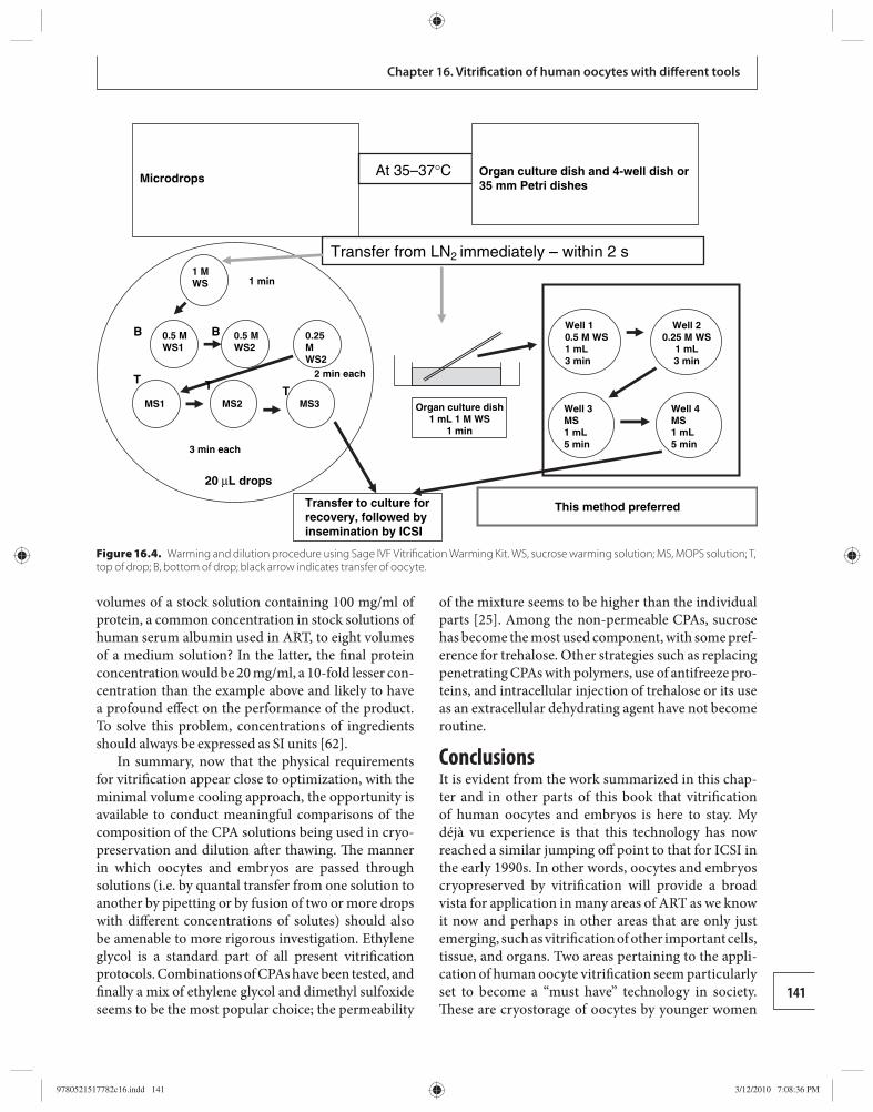
141
Chapter 16. Vitrifi cation of human oocytes with diff erent tools
volumes of a stock solution containing 100 mg/ml of protein, a common concentration in stock solutions of human serum albumin used in ART, to eight volumes of a medium solution? In the latter, the fi nal protein concentration would be 20 mg/ml, a 10-fold lesser con-centration than the example above and likely to have a profound eff ect on the performance of the product. To solve this problem, concentrations of ingredients should always be expressed as SI units [ 62 ].
In summary, now that the physical requirements for vitrifi cation appear close to optimization, with the minimal volume cooling approach, the opportunity is available to conduct meaningful comparisons of the composition of the CPA solutions being used in cryo-preservation and dilution aft er thawing. Th e manner in which oocytes and embryos are passed through solutions (i.e. by quantal transfer from one solution to another by pipetting or by fusion of two or more drops with diff erent concentrations of solutes) should also be amenable to more rigorous investigation. Ethylene glycol is a standard part of all present vitrifi cation p rotocols. Combinations of CPAs have been tested, and fi nally a mix of ethylene glycol and dimethyl sulfoxide seems to be the most popular choice; the permeability
of the mixture seems to be higher than the individual parts [ 25 ]. Among the non-permeable CPAs, sucrose has become the most used component, with some pref-erence for trehalose. Other strategies such as replacing penetrating CPAs with polymers, use of antifreeze pro-teins, and intracellular injection of trehalose or its use as an extracellular dehydrating agent have not become routine.
Conclusions It is evident from the work summarized in this chap-ter and in other parts of this book that vitrifi cation of human oocytes and embryos is here to stay. My déjà vu experience is that this technology has now reached a similar jumping off point to that for ICSI in the early 1990s. In other words, oocytes and embryos cryopreser ved by vitrifi cation will provide a broad vista for application in many areas of ART as we know it now and perhaps in other areas that are only just emerging, such as vitrifi cation of other important cells, tissue, and organs. Two areas pertaining to the appli-cation of human oocyte vitrifi cation seem particularly set to become a “must have” technology in society. Th ese are cryostorage of oocytes by younger women
Microdrops
1 MWS
0.5 MWS2
MS1 MS2 MS3
20 µL drops
Well 10.5 M WS1 mL3 min
Well 20.25 M WS
1 mL3 min
Well 4MS1 mL5 min
Well 3MS1 mL5 min
0.5 MWS1
1 min
2 min each
3 min each
Transfer to culture forrecovery, followed byinsemination by ICSI
T
BB
TT
At 35–37°C Organ culture dish and 4-well dish or35 mm Petri dishes
This method preferred
Transfer from LN2 immediately – within 2 s
0.25MWS2
Organ culture dish1 mL 1 M WS
1 min
Figure 16.4. Warming and dilution procedure using Sage IVF Vitrifi cation Warming Kit. WS, sucrose warming solution; MS, MOPS solution; T, top of drop; B, bottom of drop; black arrow indicates transfer of oocyte.
9780521517782c16.indd 1419780521517782c16.indd 141 3/12/2010 7:08:36 PM3/12/2010 7:08:36 PM
142
Section 4. Cryopreservation of oocytes
who will use them later in life when they feel the time has come to start their family. Th e second application that is partially linked to this is the creation of cryop-reserved oocyte banks. Obviously there has and will be much societal and regulatory discussion of these applications of vitrifi cation technology, but, as oth-ers have discussed [ 7 ], it is this author’s opinion that women and couples seeking eff ective use of modern ART will embrace this philosophy.
References 1. Fahy GM , Rall WF. Vitrifi cation: an overview. In
Tucker MJ , Liebermann J , eds. Vitrifi cation in Assisted Reproduction. A User’s Manual and Trouble-Shooting Guide . London: Informa Healthcare , 2007, pp. 1–20.
2. Kuwayama M. Th e human oocyte: vitrifi cation. In Gardner DK , Weissman A , Howles CM , Shoham Z , eds. Textbook of Asssisted Reproductive Technologies. Laboratory and Clinical Perspectives, 3rd edn. London: Informa Healthcare , 2009, pp. 267–273.
3. Tucker MJ , Liebermann J (eds.). Vitrifi cation in Assisted Reproduction. A User’s Manual and Trouble-Shooting Guide . London: Informa Healthcare , 2007 .
4. Fahy GM , MacFarlane DR , Angell CA , et al. Vitrifi cation as an approach to cryopreservation . Cryobiology 1984; 21: 407–426 .
5. Vajta G , Nagy ZP , Cobo A , et al. Vitrifi cation in ART: myths, mistakes, disbeliefs and confusion. Reprod Biomed Online 2009; in press.
6. Yavin S , Arav A. Measurement of essential physical properties of vitrifi cation solutions . Th eriogenology 2007; 67: 81–89 .
7. Homburg R , van der Veen F , Silber SJ. Oocyte vitrifi cation: women ’ s emancipation set in stone . Fertil Steril 2009; 91: 1319–1320 .
8. Diaz DG , La A-TH , Rodriguez K , et al. Human oocyte cryopreservation: outcome from 70 frozen oocyte cycles using a new modifi ed slow-freeze protocol . Fertil Steril 2008; 90 : S287 .
9. Quinn P. Cryopreservation. In Marrs RP , ed. Assisted Reproductive Technologies . Oxford: Blackwell Scientifi c, 1993, pp. 89–107.
10. Th e World Egg Bank. 2009 . http://www.theworldeggbank.com/tweb2/Home/Default.aspx (accessed July 23, 2009).
11. Chen C. Pregnancy aft er human oocyte cryopreservation . Lancet 1986; 19: 884–886 .
12. Chen C. Pregnancies aft er human oocyte cryopreservation . Ann N Y Acad Sci 1988; 541: 541–549 .
13. Kuleshova LL , Lopata A. Vitrifi cation can be more favorable than slow cooling . Fertil Steril 2002; 78: 449–454 .
14. Quinn P. Cryopreservation of embryos and oocytes. In Keye WR , Chang RJ , Rebar RW , Soules MR , eds. Infertility. Evaluation and Treatment . Philadelphia , PA: Saunders , 1995, pp. 821–840.
15. Kuleshova L , Gianaroli L , Magli C , et al. Birth following vitrifi cation of a small number of human oocytes . Hum Reprod 1999; 14: 3077–3079 .
16. Yon TK , Chung HM , Lim JM , et al. Pregnancy and delivery of healthy infants developed from vitrifi ed oocytes in a stimulated in vitro fertilization–embryo transfer program . Fertil Steril 2000; 74: 180–181 .
17. Katayama KP , Stehlik J , Kuwayama M , et al. High survival rate of vitrifi ed human oocytes results in clinical pregnancy . Fertil Steril 2003; 80: 223–224 .
18. Kuwayama M , Vajta G , Kato O , et al. Highly effi cient vitrifi cation method for cryopreservation of human oocytes . Reprod Biomed Online 2005; 11: 300–308 .
19. Kuwayama M , Vajta G , Ieda S , et al. Comparison of open and closed methods for vitrifi cation of human embryos and the elimination of potential contamination . Reprod Biomed Online 2005; 11: 608–614 .
20. Lucena E , Bernal DP , Lucena C , et al. Successful ongoing pregnancies aft er vitrifi cation of oocytes . Fertil Steril 2006; 85: 108–111 .
21. Antinori M , Licata E , Dani G , et al. Cryotop vitrifi cation of human oocytes results in high survival rate and healthy deliveries . Reprod Biomed Online 2007; 14: 72–79 .
22. Cobo A , Kuwayama M , Perez S , et al. Comparison of concomitant outcome achieved with fresh and cryopreserved donor oocytes vitrifi ed by the Cryotop method . Fertil Steril 2008; 89: 1657–1664 .
23. Chian R-C , Huang JHJ , Gilbert L , et al. Obstetric outcomes following vitrifi cation of in vitro and in vivo matured oocytes . Fertil Steril 2009; 91: 2391–2398 .
24. Chian R-C , Huang JYJ , Tan SL , et al. Obstetric and perinatal outcome in 200 infants conceived from vitrifi ed oocytes . Reprod Biomed Online 2008; 16: 608–610 .
25. Vajta G , Nagy ZP. Are programmable freezers still needed in the embryo laboratory? Review on vitrifi cation . Reprod Biomed Online 2006; 12: 779–796 .
26. Bielanski A , Vajta G. Risk of contamination of germplasm during cryopreservation and cryobanking in IVF units . Hum Reprod 2009; 24 : 2457–2467 .
27. Martino A , Songsasen N , Leibo SP. Development into blastocysts of bovine oocytes cryopreserved by ultra-rapid cooling . Biol Reprod 1996; 54: 1059–1069 .
28. Mukaida T , Wada S , Takahashi K , et al. Vitrifi cation of human embryos based on the assessment of suitable conditions for 8-cell mouse embryos . Hum Reprod 1998; 13: 2874–2879 .
29. Vajta G , Lewis IM , Kuwayama M , et al. Sterile application of the open pulled straw (OPS) vitrifi cation method . Cryo Letters 1998; 19: 389–392 .
30. Kuwayama M , Kato O. All-round vitrifi cation method for human oocytes and embryos . J Assist Reprod Genet 2000; 17: 477 .
31. Vanderzwalmen P , Bertin G , Debauche CH , et al. ‘In vitro’ survival of metaphase II oocytes (MII) and
9780521517782c16.indd 1429780521517782c16.indd 142 3/12/2010 7:08:36 PM3/12/2010 7:08:36 PM
143
Chapter 16. Vitrifi cation of human oocytes with diff erent tools
blastocysts aft er vitrifi cation in an Hemi Straw (HS) system. Fertil Steril 2000; 74 : S215–S216 .
32. Lane M , Schoolcraft WB , Gardner DK. Vitrifi cation of mouse and human blastocysts using a novel cryoloop container-less technique . Fertil Steril 1999; 72: 1073–1078 .
33. Boldt J. CryoTip discussion. EmbryoMail List . 2009 . http://embryomail.net/mhonarchive/em/msg00341.html (accessed 21 May, 2009 ).
34. Athayde K. CryoTip breakage. EmbryoMail List . 2009 . http://embryomail.net/mhonarchive/em/msg00329.html (accessed July 23, 2009).
35. Bielanski A , Hanniman A. Non-cross-contamination of bovine embryos with microbes using the OPS vitrifi cation system . Reprod Fertil Dev 2007; 19: 232 .
36. Larman MG , Sheehan CB , Gardner DK. Vitrifi cation of mouse pronuclear oocytes with no direct liquid nitrogen contact . Reprod BioMed Online 2006; 12: 66–69 .
37. Arav A , Zeron Y , Ocheretny A. A new device and method for vitrifi cation increases the cooling rate and allows successful cryopreservation of bovine oocytes . Th eriogenology 2000; 53: 248 .
38. Yavin S , Aroyo A , Roth Z , et al. Embryo cryopreservation in the presence of low concentration of vitrifi cation solution with sealed pulled straws in liquid nitrogen slush. Hum Reprod 2009; 24 : 797–804.
39. Yoon TK , Kim TJ , Park SE , et al. Live births aft er vitrifi cation of oocytes in a stimulated in vitro fertilization–embryo transfer program . Fertil Steril 2003; 79: 1323–1326 .
40. Kim T , Hong S , Cha K. Pregnancies from cryopreserved oocytes using vitrifi cation protocol . Fertil Steril 2005; 84 : S179 .
41. Oktay K , Cil AP , Bang H. Effi ciency of oocyte cryopreservation: a meta-analysis . Fertil Steril 2006; 86: 70–80 .
42. Selman H , Angelini A , Barnocchi N , et al. Ongoing pregnancies aft er vitrifi cation of human oocytes using a combined solution of ethylene glycol and dimethyl sulfoxide . Fertil Steril 2006; 86: 997–1000 .
43. Society for Assisted Reproductive Technology. SART Clinical Summary Report 2007 . Birmingham, AL : Society for Assisted Reproductive Technology , https://www.sartcorsonline.com/rptCSR_PublicMultYear.aspx?ClinicPKID=0 (accessed August 2, 2009 ).
44. Stachecki JJ , Cohen J. S 3 vitrifi cation system: a novel approach to blastocyst freezing . J Clin Embryol 2008; 11: 5–14 .
45. Camus A , Clairaz P , Ersham A , et al. Comparison of the process of fi ve diff erent vitrifi cation devices . Gynecol Obstet Fertil 2006; 34: 737–745 .
46. Isachenko V , Katkov II , Yakavenko S , et al. Vitrifi cation of human laser treated blastocysts within cut standard straws (CSS): novel aseptic packaging and reduced concentrations of cryoprotectants . Cryobiology 2007; 54: 305–309 .
47. Shaw JM , Jones GM. Terminology associated with vitrifi cation and other cryopreservation procedures for oocytes and embryos . Hum Reprod Update 2003; 9: 583–605 .
48. Vanderzwalmen P , Ectors F , Grobet L , et al. Aseptic vitrification of blastocysts originating from infertile patients, egg donorsand after IVM . Reprod Biomed Online 2009; 19 : 700–707 .
49. Isachenko V , Isachenko E , Reinsberg J , et al. Cryopreservation of human ovarian tissue: eff ect of spontaneous and initiated ice formation . Reprod Biomed Online 2008; 16: 336–345 .
50. Tedder RS , Zuckerman MA , Goldstone AH , et al. Hepatitis B transmission from contaminated cryopreservation tank . Lancet 1995; 346: 137–140 .
51. Bielanski A. Non-transmission of bacterial and viral microbes to embryos and semen stored in the vapor phase of liquid nitrogen in dry shippers . Cryobiology 2005; 50: 206–210 .
52. Quinn P. Cryopreservation of mouse oocytes using sodium-depleted media: comparison of HEPES-HTF and PBS-based formulations . Fertil Steril 2005; 83 : S24–S25 .
53. El-Danasouri I , Selman H , Strehler E , et al. Comparison of MOPS and HEPES buff ers during vitrifi cation . Hum Reprod 2004; 19 : i136 .
54. Swain JE , Pool TB. New pH-buff ering system for media utilized during gamete and embryo manipulations for assisted reproduction . Reprod Biomed Online 2009; 18: 799–810 .
55. McWilliams RB , Gibbons WE , Leibo SP. Osmotic and physiological responses of mouse zygotes and human oocytes to mono- and disaccharides . Hum Reprod 2005; 10: 1163–1171 .
56. Kitazato. Kitazato Cryotop Safety Kit Protocol . 2009 ; http://www.kitazato-biopharma.com/images/cryotop/Cryotop_En.pdf (accessed July 21, 2009).
57. Irvine. Scientifi c Vit Kit Th aw package insert. 2009 . http://www.irvinesci.com/techinfo/docs/40793-Booklet-Rev5.pdf (accessed July 21, 2009).
58. Sage IVF. Directions for Use for Vitrifi cation Warming Kit. 2009 . http://www.coopersurgical.com/Documents/ART-8030%20DFU.pdf (accessed July 22, 2009).
59. Quinn P. Th e impact of culture media on success rate in ART: where have we gone in the last decade? In Gurgan T , Demirol A , eds. Proceedings of the 13th World Congress: In Vitro Fertilization, Assisted Reproduction and Genetics, Istanbul, Turkey, 2005, pp. 221–225.
60. Stachecki J. Response to Letter to the JCE editor . J Clin Embryol 2009; 12: 5 .
61. Wikipedia. Concentration . 2009 . http://en.wikipedia.org/wiki/Concentration (accessed July 22, 2009).
62. Wikipedia. International System of Units. 2009 . http://en.wikipedia.org/wiki/SI_units (accessed July 22, 2009).
9780521517782c16.indd 1439780521517782c16.indd 143 3/12/2010 7:08:36 PM3/12/2010 7:08:36 PM
144 Fertility Cryopreservation, ed. Ri-Cheng Chian and Patrick Quinn. Published by Cambridge University Press. Copyright © Cambridge University Press 2010.
Chapter
17 and derived from ovarian stimulation protocols; these protocols require 4 to 6 weeks to complete the ovarian stimulation cycle and produce relatively high estradiol levels, which may be deleterious in certain cancers, notably estrogen receptor-positive breast cancer, the most common malignancy in women referred for fertil-ity preservation [ 1 ]. Th erefore, there is a pressing need for the development of fertility preservation options applicable to young women facing the risk of premature ovarian failure from gonadotoxic treatment, which do not require male partner participation, gonado tropin ovarian stimulation, or delay in treatment.
A potential strategy to avoid spindle depolymeri-zation is to cryopreserve oocytes at the germinal ves-icle (GV) stage. Th e oocytes are arrested in diplotene state of prophase I. Th eoretically, the use of immature GV stage eggs circumvents the risk of polyploidy and aneuploidies because the chromatin is diff use and sur-rounded by a nuclear membrane [ 19 , 20 ]. However, poor in vitro maturation (IVM) and fertilization have been reported as the major problems associated with cryopreservation of immature oocytes [ 21 – 23 ]. To date, only one human live birth has been reported fol-lowing cryopreservation of a GV stage oocyte and IVM using a slow-freezing method [ 24 ].
A novel strategy of fertility preservation involves retrieval of immature oocytes in an unstimulated menstrual cycle by transvaginal ultrasound guid-ance within days of initial consultation [ 25 ] or from ovarian tissue biopsies [ 26 ], followed by IVM and vitrifi cation of oocytes [ 25 , 27 ]. Th e option of IVM has become an eff ective treatment for many infertile women, including those with polycystic ovaries or polycystic ovary syndrome and those at increased risk of developing ovarian hyperstimulation syndrome [ 28 – 32 ]. Over 1500 healthy infants have been born [ 33 ] and are not associated with any increase in fetal abnormality rates compared with those from IVF or
Introduction In recent years, awareness of the possibilities for fertil-ity preservation has increased. Th e American Society of Clinical Oncology and the American Society of Reproductive Medicine have endorsed gonadotropin stimulation with in vitro fertilization (IVF) and embryo cryopreservation as the only established method of female fertility preservation [ 1 , 2 ]. However, this strat-egy may not be applicable to young women without a male partner and those who object to cryopreservation of embryos for personal, fi nancial, religious, or moral reasons.
Cryopreservation of an entire ovary potentially allows the greatest number of follicles to be preserved [ 3 ], but this has not been successfully performed in humans. Ovarian tissue cryopreservation with ortho-topic transplantation is a promising option [ 4 ] but requires two operations, has produced only three pub-lished live births [ 5 – 7 ], despite numerous attempts, and in cancer patients carries a small, but fi nite, risk of re-introducing malignant cells [ 8 ].
Oocyte cryopreservation represents an attractive and the least invasive option of fertility preservation strategy for patients wishing to retain their choice of a male partner. Conventional slow cooling of oocytes is considered an experimental procedure because of the inconsistent survival rates and limited number of live births achieved [ 1 , 9 ]. Th e percentage of live births per thawed egg ranges from 1 to 10% using slow-freezing protocols [ 9 – 11 ].
Recent advances in vitrifi cation techniques have markedly improved the effi cacy of oocyte cryopreser-vation [ 10 – 13 ], demonstrating that vitrifi cation may be more eff ective than the conventional slow-cooling method [ 10 ]. Several groups including the McGill Reproductive Center (MRC) have reported survival rates over 80% [ 14 – 18 ]. To date, most reports of oocyte cryopreservation have been in oocytes matured in vivo
Vitrifi cation of human oocytes using the McGill Cryoleaf protocol Jack Yu Jen Huang , Seang Lin Tan , and Ri-Cheng Chian
9780521517782c17.indd 1449780521517782c17.indd 144 3/12/2010 10:09:09 AM3/12/2010 10:09:09 AM
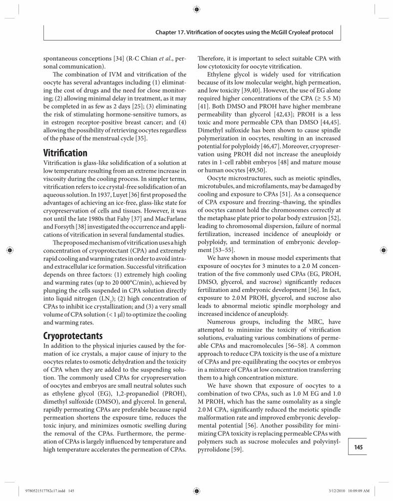
145
Chapter 17. Vitrifi cation of oocytes using the McGill Cryoleaf protocol
spontaneous conceptions [ 34 ] (R-C Chian et al ., per-sonal communication).
Th e combination of IVM and vitrifi cation of the oocyte has several advantages including (1) eliminat-ing the cost of drugs and the need for close monitor-ing; (2) allowing minimal delay in treatment, as it may be completed in as few as 2 days [ 25 ]; (3) eliminating the risk of stimulating hormone-sensitive tumors, as in estrogen receptor-positive breast cancer; and (4) allowing the possibility of retrieving oocytes regardless of the phase of the menstrual cycle [ 35 ].
Vitrifi cation Vitrifi cation is glass-like solidifi cation of a solution at low temperature resulting from an extreme increase in viscosity during the cooling process. In simpler terms, vitrifi cation refers to ice crystal-free solidifi cation of an aqueous solution. In 1937, Luyet [ 36 ] fi rst proposed the advantages of achieving an ice-free, glass-like state for cryopreservation of cells and tissues. However, it was not until the late 1980s that Fahy [ 37 ] and MacFarlane and Forsyth [ 38 ] investigated the occurrence and appli-cations of vitrifi cation in several fundamental studies.
Th e proposed mechanism of vitrifi cation uses a high concentration of cryoprotectant (CPA) and extremely rapid cooling and warming rates in order to avoid intra- and extracellular ice formation. Successful vitrifi cation depends on three factors: (1) extremely high cooling and warming rates (up to 20 000°C/min), achieved by plunging the cells suspended in CPA solution directly into liquid nitrogen (LN 2 ); (2) high concentration of CPAs to inhibit ice crystallization; and (3) a very small volume of CPA solution (< 1 μl) to optimize the cooling and warming rates.
Cryoprotectants In addition to the physical injuries caused by the for-mation of ice crystals, a major cause of injury to the oocytes relates to osmotic dehydration and the toxicity of CPA when they are added to the suspending solu-tion. Th e commonly used CPAs for cryopreservation of oocytes and embryos are small neutral solutes such as ethylene glycol (EG), 1,2-propanediol (PROH), dimethyl sulfoxide (DMSO), and glycerol. In general, rapidly permeating CPAs are preferable because rapid permeation shortens the exposure time, reduces the toxic injury, and minimizes osmotic swelling during the removal of the CPAs. Furthermore, the perme-ation of CPAs is largely infl uenced by temperature and high temperature accelerates the permeation of CPAs.
Th erefore, it is important to select suitable CPA with low cytotoxicity for oocyte vitrifi cation.
Ethylene glycol is widely used for vitrifi cation because of its low molecular weight, high permeation, and low toxicity [ 39 , 40 ]. However, the use of EG alone required higher concentrations of the CPA (≥ 5.5 M) [ 41 ]. Both DMSO and PROH have higher membrane permeability than glycerol [ 42 , 43 ]; PROH is a less toxic and more permeable CPA than DMSO [ 44 , 45 ]. Dimethyl sulfoxide has been shown to cause spindle polymerization in oocytes, resulting in an increased potential for polyploidy [ 46 , 47 ]. Moreover, cryopreser-vation using PROH did not increase the aneuploidy rates in 1-cell rabbit embryos [ 48 ] and mature mouse or human oocytes [ 49 , 50 ].
Oocyte microstructures, such as meiotic spindles, microtubules, and microfi laments, may be damaged by cooling and exposure to CPAs [ 51 ]. As a consequence of CPA exposure and freezing–thawing, the spindles of oocytes cannot hold the chromosomes correctly at the metaphase plate prior to polar body extrusion [ 52 ], leading to chromosomal dispersion, failure of normal fertilization, increased incidence of aneuploidy or polyploidy, and termination of embryonic develop-ment [ 53 – 55 ].
We have shown in mouse model experiments that exposure of oocytes for 3 minutes to a 2.0 M concen-tration of the fi ve commonly used CPAs (EG, PROH, DMSO, glycerol, and sucrose) signifi cantly reduces fertilization and embryonic development [ 56 ]. In fact, exposure to 2.0 M PROH, glycerol, and sucrose also leads to abnormal meiotic spindle morphology and increased incidence of aneuploidy.
Numerous groups, including the MRC, have attempted to minimize the toxicity of vitrifi cation solutions, evaluating various combinations of perme-able CPAs and macromolecules [ 56 – 58 ]. A common approach to reduce CPA toxicity is the use of a mixture of CPAs and pre-equilibrating the oocytes or embryos in a mixture of CPAs at low concentration transferring them to a high concentration mixture.
We have shown that exposure of oocytes to a combination of two CPAs, such as 1.0 M EG and 1.0 M PROH, which has the same osmolality as a single 2.0 M CPA, signifi cantly reduced the meiotic spindle malformation rate and improved embryonic develop-mental potential [ 56 ]. Another possibility for mini-mizing CPA toxicity is replacing permeable CPAs with polymers such as sucrose molecules and polyvinyl-pyrrolidone [ 59 ].
9780521517782c17.indd 1459780521517782c17.indd 145 3/12/2010 10:09:09 AM3/12/2010 10:09:09 AM
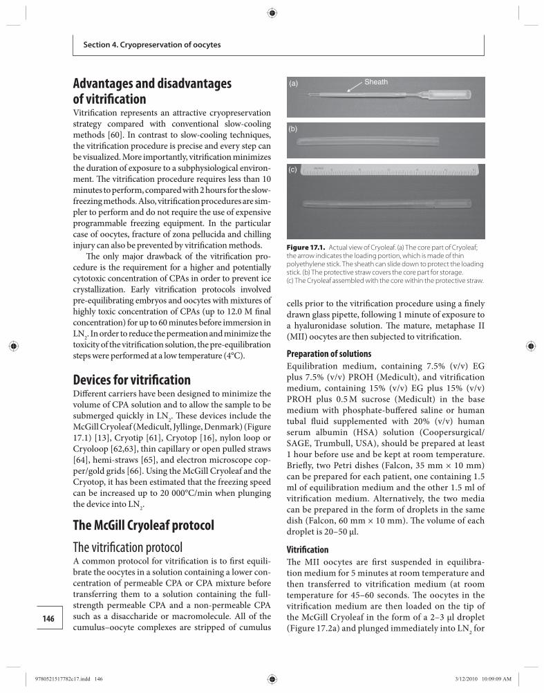
146
Section 4. Cryopreservation of oocytes
Advantages and disadvantages of vitrifi cation Vitrifi cation represents an attractive cryopreservation strategy compared with conventional slow-cooling methods [ 60 ]. In contrast to slow-cooling techniques, the vitrifi cation procedure is precise and every step can be visualized. More importantly, vitrifi cation minimizes the duration of exposure to a subphysiological environ-ment. Th e vitrifi cation procedure requires less than 10 minutes to perform, compared with 2 hours for the slow-freezing methods. Also, vitrifi cation procedures are sim-pler to perform and do not require the use of expensive programmable freezing equipment. In the particular case of oocytes, fracture of zona pellucida and chilling injury can also be prevented by vitrifi cation methods.
Th e only major drawback of the vitrifi cation pro-cedure is the requirement for a higher and potentially cytotoxic concentration of CPAs in order to prevent ice crystallization. Early vitrifi cation protocols involved pre-equilibrating embryos and oocytes with mixtures of highly toxic concentration of CPAs (up to 12.0 M fi nal concentration) for up to 60 minutes before immersion in LN 2 . In order to reduce the permeation and minimize the toxicity of the vitrifi cation solution, the pre-equilibration steps were performed at a low temperature (4°C).
Devices for vitrifi cation Diff erent carriers have been designed to minimize the volume of CPA solution and to allow the sample to be submerged quickly in LN 2 . Th ese devices include the McGill Cryoleaf (Medicult, Jyllinge, Denmark) ( Figure 17.1 ) [ 13 ], Cryotip [ 61 ], Cryotop [ 16 ], nylon loop or Cryoloop [ 62 , 63 ], thin capillary or open pulled straws [ 64 ], hemi-straws [ 65 ], and electron microscope cop-per/gold grids [ 66 ]. Using the McGill Cryoleaf and the Cryotop, it has been estimated that the freezing speed can be increased up to 20 000°C/min when plunging the device into LN 2 .
The McGill Cryoleaf protocol The vitrifi cation protocol A common protocol for vitrifi cation is to fi rst equili-brate the oocytes in a solution containing a lower con-centration of permeable CPA or CPA mixture before transferring them to a solution containing the full-strength permeable CPA and a non-permeable CPA such as a disaccharide or macromolecule. All of the cumulus–oocyte complexes are stripped of cumulus
cells prior to the vitrifi cation procedure using a fi nely drawn glass pipette, following 1 minute of exposure to a hyaluronidase solution. Th e mature, metaphase II (MII) oocytes are then subjected to vitrifi cation.
Preparation of solutions Equilibration medium, containing 7.5% (v/v) EG plus 7.5% (v/v) PROH (Medicult), and vitrifi cation medium, containing 15% (v/v) EG plus 15% (v/v) PROH plus 0.5 M sucrose (Medicult) in the base medium with phosphate-buff ered saline or human tubal fl uid supplemented with 20% (v/v) human serum albumin (HSA) solution (Coopersurgical/SAGE, Trumbull, USA), should be prepared at least 1 hour before use and be kept at room temperature. Briefl y, two Petri dishes (Falcon, 35 mm × 10 mm) can be prepared for each patient, one containing 1.5 ml of equilibration medium and the other 1.5 ml of vitrifi cation medium. Alternatively, the two media can be prepared in the form of droplets in the same dish (Falcon, 60 mm × 10 mm). Th e volume of each droplet is 20–50 μl.
Vitrifi cation Th e MII oocytes are fi rst suspended in equilibra-tion medium for 5 minutes at room temperature and then transferred to vitrifi cation medium (at room tempera ture for 45–60 seconds. Th e oocytes in the vitrifi cation medium are then loaded on the tip of the McGill Cryoleaf in the form of a 2–3 μl droplet ( Figure 17.2a ) and plunged immediately into LN 2 for
(a) Sheath
(b)
(c)
Figure 17.1. Actual view of Cryoleaf. (a) The core part of Cryoleaf; the arrow indicates the loading portion, which is made of thin polyethylene stick. The sheath can slide down to protect the loading stick. (b) The protective straw covers the core part for storage. (c) The Cryoleaf assembled with the core within the protective straw.
9780521517782c17.indd 1469780521517782c17.indd 146 3/12/2010 10:09:09 AM3/12/2010 10:09:09 AM
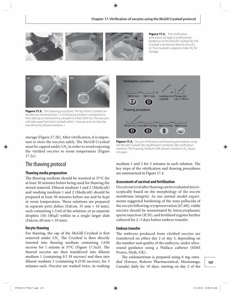
147
Chapter 17. Vitrifi cation of oocytes using the McGill Cryoleaf protocol
storage ( Figure 17.2b ). Aft er vitrifi cation, it is impor-tant to store the oocytes safely. Th e McGill Cryoleaf must be capped under LN 2 in order to avoid ex posing the vitrifi ed oocytes to room temperature ( Figure 17.2c ).
The thawing protocol Thawing media preparation Th e thawing medium should be warmed at 37ºC for at least 30 minutes before being used for thawing the stored material. Diluent medium 1 and 2 (Medicult) and washing medium 1 and 2 (Medicult) should be prepared at least 30 minutes before use and be kept at room temperature. Th ese solutions are prepared in separate petri dishes (Falcon, 35 mm × 10 mm), each containing 1.5 ml of the solution, or as separate droplets (50–100 μl) within in a single larger dish (Falcon, 60 mm × 10 mm).
Oocyte thawing For thawing, the cap of the McGill Cryoleaf is fi rst removed under LN 2. Th e Cryoleaf is then directly inserted into thawing medium containing 1.0 M sucrose for 1 minute at 37ºC ( Figure 17.3a , b ). Th e thawed oocytes are then transferred into diluent medium 1 (containing 0.5 M sucrose) and then into diluent medium 2 (containing 0.25 M sucrose), for 3 minutes each. Oocytes are washed twice, in washing
medium 1 and 2 for 3 minutes in each solution. Th e key steps of the vitrifi cation and thawing procedures are summarized in Figure 17.4 .
Assessment of survival and fertilization Oocyte survival aft er thawing can be evaluated micro-scopically based on the morphology of the oocyte membrane integrity. As our animal model experi-ments suggested hardening of the zona pellucida of the oocyte following cryopreservation [ 67 , 68 ], viable oocytes should be inseminated by intracytoplasmic sperm injection (ICSI), and fertilized zygotes further cultured for 2–3 days before embryo transfer.
Embryo transfer Th e embryos produced from vitrifi ed oocytes are transferred on either day 2 or day 3, depending on the number and quality of the embryos, under ultra-sound guidance using a Wallace catheter (SIMS Protex, Hyth, UK).
Th e endometrium is prepared using 6 mg estra-diol (Estrace, Roberts Pharmaceutical, Mississauga, Canada) daily for 10 days, starting on day 2 of the
(a) (b)
Figure 17.3. The thawing procedure. The tip of the Cryoleaf can be directly immersed into 1.5 ml thawing medium contained in a Petri dish (a) or inserted into a droplet in a Petri Dish (b). The oocytes will slide away from the Cryoleaf within 1 minute and can then be transferred to diluent medium 1.
Vitrification procedure
Thawing procedure
EM (5 min)
TM (37°C) (1 min) DM-1 (3 min) DM-2 (3 min) WM (3 min)
VM (1 min) Loading on toCryoleaf
Plunge intoLN2 (–196°C)
Figure 17.4. Oocyte vitrifi cation and thawing procedures using the McGill Cryoleaf. EM, equilibration medium; VM, vitrifi cation medium; TM, thawing medium; DM, diluent medium; LN 2 , liquid nitrogen.
(a) (b) (c)
Figure 17.2. The vitrifi cation procedure. (a) Eggs or embryos are loaded on to the McGill Cryoleaf. (b) The Cryoleaf is immersed directly into LN 2 . (c) The Cryoleaf is capped under LN 2 for storage.
9780521517782c17.indd 1479780521517782c17.indd 147 3/12/2010 10:09:10 AM3/12/2010 10:09:10 AM
148
Section 4. Cryopreservation of oocytes
menstrual cycle. Once ultrasound scan confi rmed endometrial thickness of 6–8 mm, and 2 days before embryo transfer, vaginal progesterone 200 mg (Prometrium, Schering, Pointe-Claire, Canada) three times daily, or progesterone 50 mg intramuscular once daily, is administered for 14 days. A pregnancy test is done 14 days aft er embryo transfer and an ultrasound scan is performed 2 weeks later to confi rm the pres-ence of a clinical pregnancy; if positive, both estradiol and progesterone are continued until the 12th week of pregnancy for luteal support.
Effi cacy of the McGill Cryoleaf protocol Animal model studies Before introducing oocyte vitrifi cation using the Cryoleaf for routine use in humans at the assisted reproductive technology clinic, the safety and effi cacy of the Cryoleaf vitrifi cation system was evaluated in murine and bovine model studies. In the murine study, vitrifi cation of oocytes using the Cryoleaf was shown to be superior to the choline-supplemented, sodium-depleted slow-freezing method, leading to improved survival of oocytes (92% versus 73%), their fertiliza-tion (64% versus 40%), and their in vitro embryonic development (31% versus 20% blastocyst formation rate) [ 67 ]. Th e fertilization, 2-cell cleavage, and blas-tocyst formation rates of vitrifi ed oocytes remained lower compared with those of fresh oocytes.
Analysis of meiotic spindle integrity and chro-mosome alignment indicates that less damage is detectable in vitrifi ed compared with slow-frozen oocytes, but the incidence of aneuploidy is similar in both oocytes preserved by both processes (both 9%) [ 67 ], raising further questions as to whether aneu-ploidy is the main cause of reduced in vitro embry-onic development. Th e combined eff ect of IVM and vitrifi cation was also evaluated on mouse and bovine oocytes in order to improve our understanding of the effi cacy of this novel strategy of fertility preser-vation [ 68 , 71 ]. In the murine model study, 80% of GV stage oocytes matured following IVM and were cryopreserved by vitrifi cation [ 68 ]. Th ere was no signifi cant diff erence in the post-warming survival of oocytes matured in vitro and in vivo (94% and 92%, respectively). Th e majority of oocytes matured by both methods maintained normal meiotic spin-dle morphology and chromosome alignment (88% and 87%, respectively) following vitrifi cation, and the incidence of aneuploidy was not increased (11%
and 9%, respectively). However, oocytes matured in vitro showed a higher incidence of DNA fragmenta-tion following vitrifi cation–thawing compared with those matured in vivo. Following vitrifi cation–thaw-ing, the cleavage and blastocyst formation rates of the oocytes matured in vitro were signifi cantly lower than those matured in vivo (37% versus 60% and 5% versus 19%, respectively).
Although aneuploidy is a known cause of reduced embryonic development potential and embryonic loss, the above study further suggests that it may not be the primary factor in the reduced embryonic developmental competence of immature mouse oocytes following IVM and vitrifi cation. Th e effi cacy of the Cryoleaf for vitrifi cation of oocytes matured in vitro was also evaluated in bovine model experiments that compared the mixture of EG and PROH with the combination of EG and DMSO, both also contain-ing 0.5 M sucrose [ 71 ]. Th e survival rate of bovine oocytes aft er warming was 98% when the oocytes were vitrifi ed with 15% (v/v) EG plus 15% (v/v) PROH and 92% when vitrifi ed with 15% (v/v) EG plus 15% (v/v) DMSO. Vitrifi cation using the combination of EG and PROH, rather than EG plus DMSO, resulted in signifi cantly higher rates of embryos developing to the blastocyst stage (7% versus 2%) and reach-ing the expanded or hatched blastocyst stage (3% versus 1%).
Th e same study also showed that oocyte survival rates following vitrifi cation can be aff ected by the pres-ence or absence of cumulus cells. Survival rates aft er thawing were signifi cantly higher for bovine oocytes without cumulus cells (93%) than for those with cumulus cells (84%). However, pre-incubation of the oocytes before insemination did not improve subse-quent fertilization rates and embryonic developmental potential. Th e results from these studies indicate that murine and bovine oocytes can be successfully vitri-fi ed using CPA concentrations that are comparable to those used in the slow-cooling protocol. Furthermore, the risk of CPA toxicity is reduced by using a combina-tion of EG and PROH mixture and by shortening the total exposure time to the CPA solutions. In addition to improvement in the survival rate, vitrifi cation leads to higher fertilization and early embryonic develop-ment rates.
Clinical trials A prospective study was conducted at the MRC to evaluate the pregnancy rates and obstetrical
9780521517782c17.indd 1489780521517782c17.indd 148 3/12/2010 10:09:14 AM3/12/2010 10:09:14 AM
149
Chapter 17. Vitrifi cation of oocytes using the McGill Cryoleaf protocol
outcomes following vitrifi cation of oocytes matured in vivo or in vitro between October 2003 and April 2007 [ 13 ]. Th e study comprised two groups. Th e fi rst group included patients undergoing superovula-tion and intrauterine insemination treatment, who over-responded to ovarian stimulation. Instead of canceling their treatment cycle to avoid high-order multiple pregnancies, this group underwent oocyte retrieval and vitrifi cation of oocytes (OS group). Th e second group consisted of women who had failed cycles of intrauterine insemination treatment. In this group, immature oocytes were retrieved in a natural unstimulated menstrual cycle and underwent IVM. Th e in vitro matured oocytes were vitrifi ed (IVM group).
In the OS group, a total of 399 MII and 91 GV oocytes were retrieved. Th e mean maturation rate was 70.3 ± 20.0%. A total of 463 oocytes were vit-rifi ed. Aft er thawing, a mean survival rate of 81.4 ± 22.6% was achieved. Following ICSI, a mean fertili-zation rate of 75.6 ± 22.5% was achieved. Of the 133 embryos transferred (mean embryo transfer of 3.5 ± 1.1), 17 clinical pregnancies were achieved (44.7% per cycle started). Th e implantation rate was 19.1 ± 25.8%.
In the IVM group, a total of 290 GV and 6 MII oocytes were retrieved. Following IVM, the mean maturation rate was 67.3 ± 19.3%. A total of 215 MII oocytes were vitrifi ed. Th e mean survival rate was 67.5 ± 26.1%. Th e fertilization rate following ICSI was 64.2 ± 19.9%. Of 64 embryos transferred (mean embryo transfer of 3.2 ± 1.5), four clinical pregnan-cies were achieved, resulting in a 20% clinical preg-nancy per cycle started. Th e implantation rate was 9.6 ± 24.1%.
Th e mean numbers of MII oocytes vitrifi ed per patient were comparable in the two groups (12.2 ± 5.1 for the OS group and 10.8 ± 5.9 for the IVM group). Th e clinical effi cacy of the OS group was superior to that of the IVM group in terms of oocyte survival aft er thawing, fertilization rate, and embryo quality. Although the implantation per embryo, pregnancy per cycle started, clinical pregnancy per cycle started, and live birth rate per cycle started were higher in OS group, these diff erences were not statis-tically signifi cant.
It is reassuring that the clinical pregnancy and live birth rates achieved by vitrifi cation of mature oocytes are comparable to those achieved using standard IVF treatment, as reported by the American, Canadian, and
European IVF registries [ 72 – 74 ] and are much higher than the results achieved by the conventional slow-cooling methods [ 9 ]. Th is study also demonstrates that pregnancies and live births can be achieved by vitrifi ca-tion of oocytes matured in vitro.
Neonatal outcomes following oocyte vitrifi cation Any new development in IVF treatment must be accompanied by data concerning obstetric and peri-natal outcomes. Few data have been reported on the obstetric and perinatal outcome for the infants con-ceived with vitrifi ed oocytes. In our OS group, there have been nine singleton and six multiple gestation live births (fi ve twins, one triplet), resulting in 22 healthy newborns to date. All newborns were examined for the presence of congenital anomalies by neonatologists from tertiary, university-based hospitals. All singleton pregnancies resulted in term deliveries. Four of the six multiple gestations (67%) delivered at 34 to 37 weeks of gestation. Th e mean gestation age at delivery was 39 weeks and 1 day for singleton and 36 weeks and 4 days for multiple gestations. Th e mean birthweight for singletons and twins were 3193.7 ± 376.8 g and 2277.9 ± 395.7 g, respectively. Among singletons, none of the nine newborns weighed less than 2500 g. In the 13 newborns from multiple gestation pregnancies, nine (69%) had low birthweight (1500–2500 g).
In the IVM group, there have been four healthy singleton live births at term gestation. Th e world’s fi rst baby from vitrifi cation of in vitro matured oocytes was born by Cesarean section for breech presentation at 39 weeks and 1 day of gestation on June 6, 2006, weighing 3480 g [ 27 ].
We recently reported the obstetric and perina-tal outcomes in 165 pregnancies conceived follow-ing oocyte vitrifi cation from the MRC in Canada, the Instituto Mexicano de Infertilidad in Mexico, and the CECOLFES in Colombia [ 75 ]. Th e multiple pregnancy rate was 17% (26 twin and 2 triplet pregnancies). In 137 singleton pregnancies, 51 patients underwent cesarean section delivery (37%). Th e median gestational age at delivery for singletons was 37 weeks. In 28 multiple pregnancies, 27 underwent cesarean section delivery (96%). Th e mean gestation age at delivery for multi-ple gestation was 35 weeks and 5 days. A total of 200 babies were born. Th e mean birthweight was 2920 ± 37 g for singletons and 2231 ± 55 g for multiple births. Th e median Apgar scores at 1 and 5 minutes were simi-lar between singletons and multiple gestations (9 and
9780521517782c17.indd 1499780521517782c17.indd 149 3/12/2010 10:09:14 AM3/12/2010 10:09:14 AM
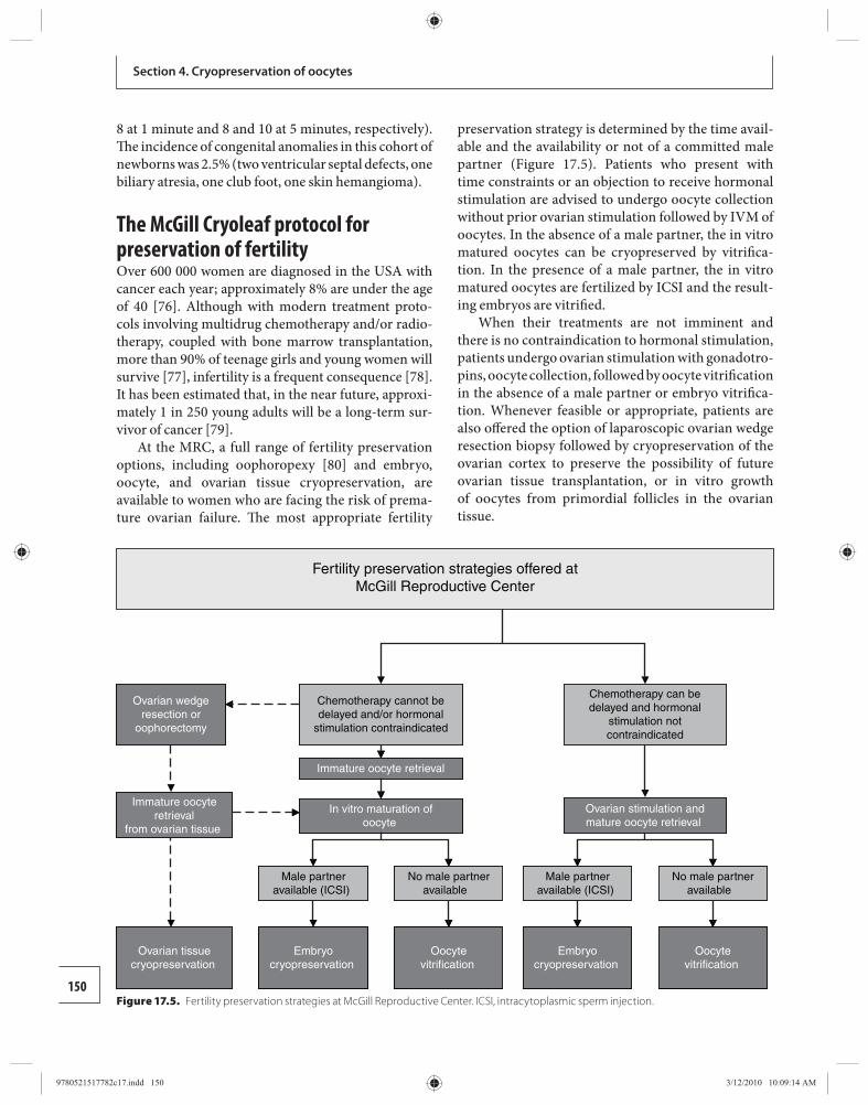
150
Section 4. Cryopreservation of oocytes
8 at 1 minute and 8 and 10 at 5 minutes, respectively). Th e incidence of congenital anomalies in this cohort of newborns was 2.5% (two ventricular septal defects, one biliary atresia, one club foot, one skin hemangioma).
The McGill Cryoleaf protocol for preservation of fertility Over 600 000 women are diagnosed in the USA with cancer each year; approximately 8% are under the age of 40 [ 76 ]. Although with modern treatment proto-cols involving multidrug chemotherapy and/or radio-therapy, coupled with bone marrow transplantation, more than 90% of teenage girls and young women will survive [ 77 ], infertility is a frequent consequence [ 78 ]. It has been estimated that, in the near future, approxi-mately 1 in 250 young adults will be a long-term sur-vivor of cancer [ 79 ].
At the MRC, a full range of fertility preservation options, including oophoropexy [ 80 ] and embryo, oocyte, and ovarian tissue cryopreservation, are available to women who are facing the risk of prema-ture ovarian failure. Th e most appropriate fertility
preservation strategy is determined by the time avail-able and the availability or not of a committed male partner ( Figure 17.5 ). Patients who present with time constraints or an objection to receive hormonal stimulation are advised to undergo oocyte collection without prior ovarian stimulation followed by IVM of oocytes. In the absence of a male partner, the in vitro matured oocytes can be cryopreserved by vitrifi ca-tion. In the presence of a male partner, the in vitro matured oocytes are fertilized by ICSI and the result-ing embryos are vitrifi ed.
When their treatments are not imminent and there is no contraindication to hormonal stimulation, patients undergo ovarian stimulation with gonadotro-pins, oocyte collection, followed by oocyte vitrifi cation in the absence of a male partner or embryo vitrifi ca-tion. Whenever feasible or appropriate, patients are also off ered the option of laparoscopic ovarian wedge resection biopsy followed by cryo preservation of the ovarian cortex to preserve the possibility of future ovarian tissue transplantation, or in vitro growth of oocytes from primordial follicles in the ovarian tissue.
Fertility preservation strategies offered atMcGill Reproductive Center
Ovarian wedgeresection or
oophorectomy
Chemotherapy cannot bedelayed and/or hormonal
stimulation contraindicated
Immature oocyte retrieval
Ovarian stimulation andmature oocyte retrieval
Oocytevitrification
In vitro maturation ofoocyte
Chemotherapy can bedelayed and hormonal
stimulation notcontraindicated
Male partneravailable (ICSI)
Male partneravailable (ICSI)
No male partneravailable
No male partneravailable
Oocytevitrification
Embryocryopreservation
Embryocryopreservation
Immature oocyteretrieval
from ovarian tissue
Ovarian tissuecryopreservation
Figure 17.5. Fertility preservation strategies at McGill Reproductive Center. ICSI, intracytoplasmic sperm injection.
9780521517782c17.indd 1509780521517782c17.indd 150 3/12/2010 10:09:14 AM3/12/2010 10:09:14 AM
151
Chapter 17. Vitrifi cation of oocytes using the McGill Cryoleaf protocol
Oncological indications for fertility preservation To date, the MRC has provided fertility preservation to 123 cancer patients, including 46 with breast cancer [ 25 , 81 ], 35 with hematological cancer, 9 with brain cancer, 10 with soft tissue cancer, and 9 with gynecologic cancer.
Breast cancer We recently reported the use of IVM and oocyte vit-rifi cation for young patients with breast cancer who presented with time constraints or concerns regarding hormonal stimulation. Eighteen women of reproduc-tive age (mean age 33.1 ± 5.0 years; range 24–45) with breast cancer were referred for fertility preservation. Because of time constraints, chemotherapy planned to start within a short period, or concerns about the use of gonadotropin ovarian stimulation, these patients underwent immature oocyte retrieval from unstimu-lated ovaries in a natural menstrual cycle followed by IVM and vitrifi cation of oocytes. Th e median delay in cancer treatment was 7 days. Th e median number of vitrifi ed oocytes per retrieval was seven (range, 1–22). Th e mean estradiol level was physiological, averaging 270.6 ± 90.5 pmol/l on the day of immature oocyte retrieval. Th erefore, this novel strategy is not associ-ated with increased estrogen exposure or delay in can-cer treatment, and thus represents an attractive fertility preservation option for many breast cancer patients.
Another advantage of IVM is the possibility to retrieve oocytes regardless of the phase of the men-strual cycle. Unlike conventional ovarian stimulation protocols, which are dependent upon the phase of the menstrual cycle and can only be initiated at monthly intervals, it is possible to perform immature oocyte retrieval sequentially in both luteal and follicular phase of the menstrual cycle without aff ecting the quantity and maturation rate of the oocytes [ 35 ].
Th is strategy may also be used in selected breast cancer patients undergoing conventional IVF with ovarian stimulation [ 81 ]. Oktay et al. [ 81 ] reported the case of a patient with breast cancer who under-went stimulation with follicle-stimulating hormone and letrozole with the intention of embryo cryo-preservation. Unfortunately, a premature luteiniz-ing hormone surge resulted in ovulation prior to the oocyte retrieval. As her chemotherapy was sched-uled to start in a week and the patient did not wish to delay her cancer treatment, the patient was referred to the MRC. On the day of the consultation, imma-ture oocytes were retrieved, underwent IVM, and
fertilized, successfully generating two embryos for cryopreservation Hematological malignancies We recently reported a series of 30 patients with hema-tological malignancies who were referred for fertility preservation, including 17 patients with Hodgkin’s lymphoma, fi ve with non-Hodgkin’s lymphoma, three with acute myelogenous leukemia, three with myelo-dysplasia syndrome, one with Burkitt’s lymphoma, and one with aplastic anemia. Owing to time constraints, 12 patients underwent immature oocyte retrieval from unstimulated ovaries, followed by IVM and vitrifi cation of oocytes in the absence of a male partner. A median of 10 oocytes was vitrifi ed (range, 2–40). When the can-cer therapy was not imminent, 10 patients underwent ovarian stimulation with gonadotropins and mature oocyte retrieval followed by vitrifi cation of the matured oocytes. A total of 124 mature oocytes were vitrifi ed (median of 12 oocytes per retrieval; range, 3–25).
Ovarian neoplasms We have reported the possibility of fertility preserva-tion for patients with borderline ovarian tumors [ 82 ]. Th is novel strategy involves aspiration of all visible follicles on the surface of the removed ovary using an 18-gauge needle attached to a 10 ml syringe contain-ing oocyte washing medium (Sage/Cooper Surgical, Trumbull, CT, USA). Four immature oocytes were retrieved and underwent IVM. Th ree oocytes matured aft er 48 hours and were cryopreserved. Interestingly, the salpingooophorectomy procedure was performed on day 18 of her cycle, indicating that oocytes can be collected, matured in vitro, and cryopreserved regard-less of the day of menstrual cycle.
Th is novel strategy of direct aspiration of all vis-ible follicles can also be combined with ovarian tissue cryopreservation [ 26 ]. In a series of four patients who underwent oophorectomy or ovarian wedge resection for the purpose of ovarian tissue cryopreservation, the mean number of immature oocytes recovered was three (one, three, four, and three, respectively). Th e mean maturation rate following IVM was 79% (100%, 100%, 50%, and 67%, respectively). In total, eight mature oocytes were vitrifi ed.
Non-oncological indications for fertility preservation Fertility preservation should also be considered in patients undergoing gonadotoxic therapy for other non-oncologic conditions [ 83 ], including systemic
9780521517782c17.indd 1519780521517782c17.indd 151 3/12/2010 10:09:14 AM3/12/2010 10:09:14 AM
152
Section 4. Cryopreservation of oocytes
lupus erythematosus (SLE) or other autoimmune disorders [ 84 ], those who have undergone extensive ovarian surgery for endometriosis [ 85 ] and ovarian neoplasm, and those with mosaic Turner syndrome.
Systemic lupus erythematosus or other autoimmune disorders We recently reported a case series of seven women with SLE and other systemic autoimmune rheumatic diseases who required cyclophosphamide therapy and underwent fertility preservation treatments. Five women had SLE with nephritis; one suff ered from immune thrombocytopenia purpura, and one had microscopic polyangiitis with renal involvement. All women were nulliparous and younger than 35 years old. Patients with SLE underwent IVM of immature oocytes aspirated during a natural menstrual cycle, fol-lowed by vitrifi cation of the matured oocytes if a male partner was not available or vitrifi cation of embryos if one was available. Th e patient with immune thrombo-cytopenia purpura and the patient with microscopic polyangiitis underwent gonadotropin ovarian stimula-tion followed by oocyte or embryo vitrifi cation. Th ere were no complications associated with this treatment and cytotoxic therapy was initiated as scheduled in all cases. Th erefore, treatment consideration should include fertility preservation in young women with systemic autoimmune rheumatic diseases who face imminent gonadotoxic treatment. In patients, where gonadotropin ovarian stimulation is deemed unsafe, IVM followed by vitrifi cation represents a safe and feas ible option.
Endometriosis A new indication for fertility preservation includes young women undergoing extensive ovarian surgery for endometriosis [ 85 ]. We recently reported a case of a young woman at risk of losing her remaining ovary owing to severe and symptomatic endometriosis. As she had no male partner, she was consulted about the possibility of oocyte cryopreservation before further treatment. With the approval of the Hospital Research Ethics Board, she underwent three cycles of ovarian stimulation and had 21 mature oocytes cryopreserved by vitrifi cation.
Turner syndrome Women with Turner syndrome are at risk of premature ovarian failure. We recently conducted a retrospective study to identify patients with Turner syndrome who could be potential candidates for fertility preservation
and to determine their present reproductive and fer-tility status [ 86 ]. Criteria for fertility preservation included: (1) spontaneous menarche, (2) confi rmation by ultrasound examination of the presence of at least one normal ovary, and (3) serum follicle-stimulating hormone levels below 40 IU/l. Using the Montreal Children’s Hospital Cytogenetic Database from 1990 to 2006, 28 patients were identifi ed: 13 (46%) were 45X; nine (32%) had mosaic karyotypes; and six (21%) had karyotypes containing isochromosome or ring X chromosome. Six patients (21%) had spontaneous pubertal development and four (14%) were identifi ed as potential candidates for fertility preservation. One underwent an ovarian stimulation protocol of gonado-tropin-releasing hormone agonist downregulation fol-lowed by recombinant follicle-stimulating hormone and human menopausal gonadotropin stimulation. Two MII stage oocytes were collected and vitrifi ed using the McGill Cryoleaf vitrifi cation system. Another patient conceived spontaneously at the age of 24 years.
We also reported the combination of ovarian tis-sue cryobanking and immature oocyte collection from the tissue followed by IVM and vitrifi cation of in vitro matured oocytes in a young woman with mosaic Turner syndrome [ 87 ]. A 16-year-old female with 20% 45X and 80% 46XX karyotype underwent laparoscopic ovarian wedge resection. Before performing ovar-ian tissue cryopreservation, all visible follicles on the ovarian surface were aspirated. 11 immature GV stage oocytes were recovered and subjected to IVM. Eight oocytes that matured (73% maturation rate) were cryo-preserved by vitrifi cation.
In the case of non-mosaic Turner syndrome, the only option for fertility has been oocyte donation [ 88 ]. However, the demand for oocyte donors exceeds supply for third-party conception in many countries. Moreover, in many countries, including Canada, remuneration for donors either through fi nancial gain or benefi ts is prohibited, which has worsed the short-age of oocyte donors.
We recently reported the fi rst case of mother-to-daughter oocyte donation using oocyte vitrifi cation [ 89 ]. A 33-year-old healthy mother of three children requested to undergo ovarian stimulation in order to cryopreserve oocytes for the purpose of donating to her 6-year-old daughter, who was recently diagnosed with Turner syndrome. Aft er three cycles of ovarian stimu-lation, 30 oocytes were cryopreserved for the daugh-ter’s possible future use. Th is strategy demonstrates the feasibility for the banking of a mother’s oocytes as
9780521517782c17.indd 1529780521517782c17.indd 152 3/12/2010 10:09:14 AM3/12/2010 10:09:14 AM
153
Chapter 17. Vitrifi cation of oocytes using the McGill Cryoleaf protocol
a possible donation to a young daughter with a medical condition that leads to infertility.
While, fertility preservation may not be feasible for most patients with Turner syndrome, aft er careful con-sideration of increased pregnancy-associated risks, fer-tility preservation may be off ered to young females with mosaic Turner syndrome. Th e combination of ovarian tissue cryobanking and immature oocyte collection from the tissue followed by IVM and vitrifi cation of matured oocytes represents a promising approach to fertility preservation for young women with mosaic Turner syndrome. Oocyte cryopreservation allows for mother-to-daughter oocyte donation.
Social indications Fertility declines with increasing age [ 90 ]. Many pro-fessional women wishing to delay motherhood may fi nd themselves facing potential childlessness because of irreversible loss of viable oocytes, which is inevitable with advancing age [ 90 ]. Oocyte vitrifi cation repre-sents an attractive strategy for these women to bypass their biological clock.
Conclusions Primary care physicians and oncologists need to be made aware of the fertility preservation options for young female patients that do not entail male partner participation, gonadotropin ovarian stimulation, or delay in gonadotoxic treatment. Early discussion with the patient should be undertaken and, if desired, the woman should be referred to an IVF center that off ers the full range of fertility preservation options.
As with any new assisted reproductive technol-ogy procedure in humans, it will be necessary to study oocyte vitrifi cation in larger prospective clinical trials for a suffi cient length of time in order to determine whether the development of the resulting children is healthy; but we believe this technique off ers a realis-tic hope of fertility preservation for women without partners. Whether this strategy should be off ered to any woman of advancing reproductive age anxious to preserve her fertility but whose current circumstances preclude pregnancy is debatable. However, the win-dow of opportunity for fertility preservation in young women who must undergo gonadotoxic treatment for life-threatening diseases such as cancer is small, and once treatment is initiated, fertility loss is oft en irreversible.
References 1. Lee SJ , Schover LR , Partridge AH , et al. American
Society of Clinical Oncology recommendations on fertility preservation in cancer patients . J Clin Oncol 2006; 24: 2917–2931 .
2. Ethics Committee of the American Society of Reproductive Medicine. Fertility preservation and reproduction in cancer patients . Fertil Steril 2005; 83: 1622–1628 .
3. Wang X , Chen H , Yin H , et al. Fertility aft er intact ovary transplantation . Nature 2002; 415: 385 .
4. Oktay K , Buyuk E , Veeck LL , et al. Embryo development aft er heterotopic transplantation of cryopreserved ovarian tissue . Lancet 2004; 363: 837–840 .
5. Donnez J , Dolmans MM , Demylle D , et al. Livebirth aft er orthotopic transplantation of cryopreserved ovarian tissue . Lancet 2004; 364: 1405–1410 .
6. Meirow D , Levron J , Eldar-Geva T , et al. Pregnancy aft er transplantation of cryopreserved ovarian tissue in a patient with ovarian failure aft er chemotherapy . N Engl J Med 2005; 353: 318–321 .
7. Demeestere I , Simon P , Emiliani S , Delbaere A , Englert Y. Fertility preservation: successful transplantation of cryopreserved ovarian tissue in a young patient previously treated for Hodgkin ’ s disease . Oncologist 2007; 12: 1437–1442 .
8. Meirow D , Hardan I , Dor J , et al. Searching for evidence of disease and malignant cell contamination in ovarian tissue stored from hematologic cancer patients . Hum Reprod 2008; 23: 1007–1013 .
9. Practice Committee of the American Society of Reproductive Medicine. Ovarian tissue and oocyte cryopreservation . Fertil Steril 2004; 82: 993–998 .
10. Kuleshova LL , Lopata A. Vitrifi cation can be more favorable than slow cooling . Fertil Steril 2002; 78: 449–454 .
11. Oktay K , Cil AP , Bang H. Effi ciency of oocyte cryopreservation: a meta-analysis . Fertil Steril 2006; 86: 70–80 .
12. Huang JY , Tan SL , Chian R-C. Fertility preservation for female . J Reprod Contracept 2006; 17: 109–128 .
13. Chian R-C , Huang JYJ , Gilbert L , et al. Obstetric outcomes following vitrifi cation of in-vitro and in-vivo matured oocytes . Fertil Steril 2009; 91: 2391–2398 .
14. Katayama KP , Stehlik J , Kuwayama M , Kato O , Stehlik E. High survival rate of vitrifi ed human oocytes results in clinical pregnancy . Fertil Steril 2003; 80: 223–224 .
15. Kuwayama M , Vajta G , Kato O , Leibo SP. Highly effi cient vitrifi cation method for cryopreservation of human oocytes . Reprod Biomed Online 2005; 11: 300–308 .
16. Kuwayama M. Highly effi cient vitrifi cation for cryopreservation of human oocytes and embryos: the Cryotop method . Th eriogenology 2007; 67: 73–80 .
9780521517782c17.indd 1539780521517782c17.indd 153 3/12/2010 10:09:14 AM3/12/2010 10:09:14 AM
154
Section 4. Cryopreservation of oocytes
17. Lucena E , Bernal DP , Lucena C , et al. Successful ongoing pregnancies aft er vitrifi cation of oocytes . Fertil Steril 2006; 85: 108–111 .
18. Cobo A , Kuwayama M , Perez S , et al. Comparison of concomitant outcome achieved with fresh and cryopreserved donor oocytes vitrifi ed by the Cryotop method . Fertil Steril 2008; 89: 1657–1664 .
19. Cooper A , Paynter SJ , Fuller BJ. Diff erential eff ects of cryopreservation on nuclear or cytoplasmic maturation in vitro in immature mouse oocytes from stimulated ovaries . Hum Reprod 1998; 13: 971–978 .
20. Isachenko EF , Nayudu PL. Vitrifi cation of mouse germinal vesicle oocytes: eff ect of treatment temperature and egg yolk on chromosomal normality and cumulus integrity . Hum Reprod 1999; 14: 400–408 .
21. Toth TL , Baka SG , Veeck LL , et al. Fertilization and in vitro development of cryopreserved human prophase I oocytes . Fertil Steril 1994; 61: 891–894 .
22. Toth TL , Lanzendorf SE , Sandow BA , et al. Cryopreservation of human prophase I oocytes collected from unstimulated follicles . Fertil Steril 1994; 61: 1077–1082 .
23. Son WY , Park SE , Lee KA , et al. Eff ects of 1,2-propanediol and freezing–thawing on the in vitro developmental capacity of human immature oocytes . Fertil Steril 1996; 66: 995–999 .
24. Tucker MJ , Wright G , Morton PC , Massey JB. Birth aft er cryopreservation of immature oocytes with subsequent in vitro maturation . Fertil Steril 1998; 70: 578–579 .
25. Rao GD , Chian R-C , Son WS , Gilbert L , Tan SL. Fertility preservation in women undergoing cancer treatment . Lancet 2004; 363: 1829–1830 .
26. Huang JY , Tulandi T , Holzer H , Tan SL , Chian R-C. Combining ovarian tissue cryobanking with retrieval of immature oocytes followed by in vitro maturation and vitrifi cation: an additional strategy of fertility preservation . Fertil Steril 2008; 89: 567–572 .
27. Chian R-C , Gilbert L , Huang JYJ , et al. Live birth aft er vitrifi cation of in vitro matured human oocytes . Fertil Steril 2009; 91: 372–376 .
28. Chian R-C , Buckett WM , Tan SL. In-vitro maturation of human oocytes . Reprod Biomed Online 2004; 8: 148–166 .
29. Chian R-C , Lim JH , Tan SL. State of the art in in-vitro oocyte maturation . Curr Opin Obstet Gynecol 2004; 16: 211–219 .
30. Jurema MW , Nogueira D. In vitro maturation of human oocytes for assisted reproduction . Fertil Steril 2006; 86: 1277–1291 .
31. Al-Sunaidi M , Tulandi T , Holzer H , et al. Repeated pregnancies and live births aft er in vitro maturation treatment . Fertil Steril 2007; 87: 1212 .e9–e12.
32. Son W-Y , Chung J-T , Chian R-C , et al. A 38 h interval between hCG priming and oocyte retrieval increases in vivo and in vitro oocyte maturation rate in programmed IVM cycles . Hum Reprod 2008; 23: 2010–2016 .
33. Edwards RG. Foreword. In Tan SL , Chian R-C , Buckett WM , eds. In Vitro Maturation of Human Oocytes Basic Science to Clinical Application. London : Informa Healthcare , 2006, pp. xv–xx .
34. Buckett WM , Chian R-C , Holzer H , et al. Obstetric outcomes and congenital abnormalities after in vitro maturation, in vitro fertilization, and intracytoplasmic sperm injection . Obstet Gynecol 2007; 110: 885–891 .
35. Demirtas E , Elizur S , Holzer H , et al. Immature oocyte retrieval in the luteal phase to preserve fertility in women with cancer facing imminent gonadotoxic therapy: it is worth a try . Reprod Biomed Online 2008; 17: 520–523 .
36. Luyet B. Th e vitrifi cation of organic colloids and protoplasm . Biodynamica 1937; 1: 1–14 .
37. Fahy G. Prospect for vitrifi cation of whole organs . Cryobiology 1981; 18: 617–625 .
38. MacFarlane D , Forsyth M. Recent insights on the role of the cryoprotection against vitrifi cation . Cryobiology 1990; 27: 345–358 .
39. Sommerfeld V , Niemann H. Cryopreservation of bovine in vitro produced embryos using ethylene glycol in controlled freezing or vitrifi cation . Cryobiology 1999; 38: 95–105 .
40. Emiliani S , Bergh M , Vannin A , Biramane J , Englert Y. Comparison of ethylene glycol, 1,2-propanediol and glycerol for cryopreservation of slow-cooled mouse zygotes, 4-cell embryos and blastocysts . Hum Reprod 2000; 15: 905–910 .
41. Shaw J , Oranratnachai A , Trounson A. Fundamental cryobiology of mammalian oocytes and ovarian tissue . Th eriogenology 2000; 53: 59–72 .
42. Mazur P. Cryobiology: the freezing of biological systems . Science 1970; 168: 939–949 .
43. Kasai M. Advances in the cryopreservation of mammalian oocytes and embryos: development of ultrarapid vitrifi cation . Reprod Med Biol 2002; 1: 1–9 .
44. Boutron P , Kaufmann A. Stability of the amorphous state in the system water–1,2-propanediol . Crybiology 1979; 16: 557–568 .
45. Renard J , Babinet C. High survival of mouse embryos aft er rapid freezing and thawing inside plastic straws with 1,2-propanediol as cryoprotectant . J Exp Zool 1984; 230: 443–448 .
46. Glenister P , Wood M , Kirby C , Whittingham D. Incidence of chromosome anomalies in fi rst-cleavage mouse embryos obtained from frozen–thawed oocytes fertilized in vitro . Gamete Res 1987; 16: 205–216 .
47. Kola I , Kirby C , Shaw J , Davey A , Trounson A. Vitrifi cation of mouse oocytes results in aneuploidy zygotes and malformed fetuses . Teratology 1988; 38: 467–474 .
48. Vincent C , Preliere G , Pajot-Augy E , et al. Eff ects of cryoprotectants on actin fi laments during the
9780521517782c17.indd 1549780521517782c17.indd 154 3/12/2010 10:09:14 AM3/12/2010 10:09:14 AM
155
Chapter 17. Vitrifi cation of oocytes using the McGill Cryoleaf protocol
cryopreservation of one-cell rabbit embryos . Cryobiology 1990; 27 : 9–23 .
49. Gook D , Osborn S , Bourne H , Johnston W. Fertilization of human oocytes following cryopreservation; normal karyotypes and absence of stray chromosome . Hum Reprod 1994; 9: 684–691 .
50. van Blerkom J , Davis P. Cytogenic, cellular, and developmental consequences of cryopreservation of immature and mature mouse and human oocytes . Micro Res Tech 1994; 27: 165–193 .
51. Vincent C , Johnson MH. Cooling, cryoprotectants, and the cytoskeleton of the mammalian oocyte . Oxford Rev Reprod Biol 1992; 14: 73–100 .
52. Gook D , Osborn S , Johnson W. Cryopreservation of mouse and human oocytes using 1,2-propanediol and the confi guration of the meiotic spindle Hum Reprod 1993; 8: 1101–1109 .
53. Sathananthan AH , Trounson A , Freeman L. Morphology and fertilization of frozen human oocytes . Gamete Res 1987; 16: 343–354 .
54. Eroglu A , Toner M , Leykin L , Toth TL. Cytoskeleton and polyploidy aft er maturation and fertilization of cryopreserved germinal vesicle-stage mouse oocytes . J Assist Reprod Genet 1998; 15: 447–454 .
55. Eroglu A , Toth TL , Toner M. Alterations of the cytoskeleton and polyploidy induced by cryopreservation of metaphase II mouse oocytes . Fertil Steril 1998; 69: 944–957 .
56. Huang JY , Chen HY , Tan SL , Chian R-C. Eff ects of osmotic stress and cryoprotectant toxicity on mouse oocyte fertilization and subsequent embryonic development in vitro . Cell Preserv Tech 2006; 4: 149–160 .
57. Ali J , Shelton J. Vitrifi cation of preimplantation stages of mouse embryos . J Reprod Fertil 1993; 98: 459–465 .
58. Mukaida T , Wada S , Takahashi K , et al. Vitrifi cation of human embryos based on the assessment of suitable conditions for 8-cell mouse embryos . Hum Reprod 1998; 13: 2874–2879 .
59. Kasai M , Komi J , Takakamo A , et al. A simple method for mouse embryo cryopreservation in a low toxicity vitrifi cation solution, without appreciable loss of viability . J Reprod Fertil 1990; 89: 91–97 .
60. Vajta G , Nagy ZP. Are programmable freezers still needed in the embryo laboratory? Review on vitrifi cation . Reprod Biomed Online 2006; 12: 779–796 .
61. Kuwayama M , Vajta G , Ieda S , Kato O. Comparison of open and closed methods for vitrifi cation of human embryos and the elimination of potential contamination . Reprod Biomed Online 2005; 11: 608–614 .
62. Lane M , Bavister B , Lyons E , Forest K. Container-less vitrifi cation of mammalian oocytes and embryos . Nat Biotechnol 1999; 17: 1234–1236 .
63. Lane M , Schoolcraft W , Gardner D. Vitrifi cation of mouse and human blastocysts using a novel cryoloop
container-less technique . Fertil Steril 1999; 72: 1073–1078 .
64. Vajta G , Holm P , Kuwayama M , et al. Open pulled straw (OPS) vitrifi cation: a new way to reduce cryoinjuries of bovine ova and embryos . Mol Reprod Dev 1998; 51: 53–58 .
65. Vanderzwalmen P , Bertin G , Debauche C , et al. Vitrifi cation of human blastocysts with the Hemi-Straw carrier: application of assisted hatching aft er thawing . Hum Reprod 2003; 18: 1504–1511 .
66. Martino A , Songsasen N , Leibo S. Development into blastocysts of bovine oocytes cryopreserved by ultra-rapid cooling . Biol Reprod 1996; 54: 1059–1069 .
67. Huang JYJ , Chen HY , Tan SL , Chian R-C. Eff ect of choline-supplemented sodium-depleted slow freezing versus vitrifi cation on mouse oocyte meiotic spindles and chromosome abnormalities . Fertil Steril 2007; 88: 1093–1100 .
68. Huang JYJ , Chen HY , Park JYS , Tan SL , Chian R-C. Comparison of spindle and chromosome confi guration in in vitro- and in vivo-matured mouse oocytes aft er vitrifi cation . Fertil Steril 2008; 90: 1424–1432 .
69. Steer CV , Mills CL , Tan SL , Campbell S , Edwards RG. Th e cumulative embryo score: a predictive embryo scoring technique to select the optimal number of embryos to transfer in an in-vitro fertilization and embryo transfer programme . Hum Reprod 1992; 7: 117–119 .
70. Child TJ , Abdul-Jalil AK , Tan SL. Embryo morphology, cumulative embryo score, and outcome in an oocyte in vitro maturation program . Fertil Steril 2002; 77: 424–425 .
71. Chian R-C , Kuwayama M , Tan L , Tan J , Kato O , Nagai T. High survival rate of bovine oocytes matured in vitro following vitrifi cation . J Reprod Dev 2004; 50: 685–696 .
72. Donnez J , Dolmans MM , Demylle D , et al. Restoration of ovarian function aft er orthotopic (intraovarian and periovarian) transplantation of cryopreserved ovarian tissue in a woman treated by bone marrow transplantation for sickle cell anaemia: case report . Hum Reprod 2006; 21: 183–188 .
73. Gunby J , Daya S. Assisted reproductive technologies (ART) in Canada: 2003 results from the Canadian ART Register . Fertil Steril 2007; 88: 550–559 .
74. Andersen AN , Gianaroli L , Felberbaum R , de Mouzon J , Nygren KG. Assisted reproductive technology in Europe, 2002. Results generated from European registers by ESHRE . Hum Reprod 2006; 21: 1680–1697 .
75. Chian R-C , Huang JYJ , Tan SL , et al. Obstetric and perinatal outcome in 200 infants conceived from vitrifi ed oocytes . Reprod Biomed Online 2008; 16: 608–610 .
76. American Cancer Society. Cancer Facts and Figures, 2006 . Atlanta, GA : American Cancer Society , 2006 .
77. Ries L , Percy C , Bunin G. Introduction. In Gurney J , Linet M , Tamra T , Young J , Bunin G , eds. Cancer
9780521517782c17.indd 1559780521517782c17.indd 155 3/12/2010 10:09:14 AM3/12/2010 10:09:14 AM
156
Section 4. Cryopreservation of oocytes
Incidence and Survival among Children and Adolescents: United States SEER Program 1975–1995. [NIH Pub No 99–4649.] Bethesda, MD : National Cancer Institute , 1999, pp. 1–15.
78. Lobo RA . Potential options for preservation of fertility in women . N Engl J Med 2005; 353: 64–73 .
79. Bleyer W. Th e impact of childhood cancer on the United States and the world . Cancer 1990; 40: 355–367 .
80. Tulandi T , Al-Took S. Laparoscopic ovarian suspension before irradiation . Fertil Steril 1998; 70: 381–183 .
81. Oktay K , Demirtas E , Son WY , et al. In vitro maturation of germinal vesicle oocytes recovered aft er premature luteinizing hormone surge: description of a novel approach to fertility preservation . Fertil Steril 2008; 89: 228.e19–e22 .
82. Huang JY , Buckett WM , Gilbert L , Tan SL , Chian R-C. Retrieval of immature oocytes followed by in vitro maturation and vitrifi cation: a case report on a new strategy of fertility preservation in women with borderline ovarian malignancy . Gynecol Oncol 2007; 105: 542–544 .
83. Gidoni Y , Holzer H , Tan SL , Tulandi T. Fertility preservation in non-oncologic patients . Reprod Biomed Online 2008; 16: 792–800 .
84. Elizur SE , Chian R , Pineau CA , et al. Fertility preservation treatment for young women with
autoimmune disease facing treatment with gonadotoxic agents . Rheumatology 2008; 47: 1506–1509 .
85. Elizur SE , Chian R-C , Holzer HEG , et al. Cryopreservation of oocytes in a young woman with severe and symptomatic endometriosis: a new indication for fertility preservation . Fertil Steril 2009; 91: 293. e1–e3 .
86. Lau NM , Huang JYJ , MacDonald S , et al. Feasibility of fertility preservation in young females with Turner syndrome . Reprod Biomed Online 2008; 17: 520–523 .
87. Huang JY , Tulandi T , Holzer H , et al. Cryopreservation of ovarian tissue and in-vitro matured oocytes in a female with mosaic Turner syndrome Hum Reprod 2008; 23: 336–339 .
88. Huang JY , Tulandi T , Holzer H , et al. Increased maternal cardiovascular mortality associated with pregnancy in women with Turner syndrome . Fertil Steril 2006; 86 : S127–S128 .
89. Gidoni YS , Takefman J , Holzer HEG , et al. Cryopreservation of a mother ’ s oocytes for possible future use by her daughter with Turner syndrome: case report . Fertil Steril 2008; 90: e9–e12.
90. Heff ner LJ. Advanced maternal age: how old is too old ? N Engl J Med 2004; 351: 1927–1929 .
9780521517782c17.indd 1569780521517782c17.indd 156 3/12/2010 10:09:14 AM3/12/2010 10:09:14 AM
157Fertility Cryopreservation, ed. Ri-Cheng Chian and Patrick Quinn. Published by Cambridge University Press. Copyright © Cambridge University Press 2010.
Chapter
18 Th ere have been recent reports of successful cryo-
preservation by direct plunging of human metaphase II (MII) ooccytes [ 25 – 28 ], pronuclear zygotes [ 29 – 36 ], and blastocysts [ 37 – 46 ] into liquid nitrogen, a process known as vitrifi cation. Th is method is currently under intensive investigation in numerous laboratories. Vitrifi cation has the following major advantages over slow freezing:
speed: the complete procedure requires only a few • minutes economy: it does not need special equipment. •
Glass formation during the cooling phase of vitrifi ca-tion and stability of the amorphous state of the solu-tion during warming have been reported [ 47 ]. It was reported that the critical cooling rate of a 40% di methyl sulfoxide (DMSO) solution (which is a better glass former than ethylene glycol) to avoid ice formation was 500°C/min, and the calculated critical warming rate to avoid ice formation (devitrifi cation) was over one billion degrees per minute [ 47 ] . Despite the fact that in the solution described by Baudot et al. [ 47 ] the base medium and serum solutes provided additional stability against ice formation, a concentration of 30% penetrating cryoprotectants was not able to prevent ice forming during the cooling and, particularly, during the described warming conditions. Th is explains why these protocols do not involve “real” vitrifi cation, and in this chapter we use the term “rapid freezing” rather than vitrifi cation and “thawing” instead of warming.
Initially, the standard 0.25 ml insemination straws were almost exclusively used as the carrier devices for the cryopreservation of embryos or oocytes by vitrifi -cation [ 48 ], which allowed a cooling rate of approxi-mately 2500°C/min [ 49 ]. New methods are based on the immersion of very small amounts (0.5–1.0 μl) of solutions into liquid nitrogen and most authors have attributed the improved developmental rate
Introduction Th e cryopreservation of human oocytes and blasto cysts is an important topic in assisted reproduction. First, cryop-reservation of germinal vesicle (GV) stage oocytes allows the elimination or reduction of the need for medication to stimulate folliculogenesis and allows oocyte cryobankin g (e.g. for young women who wish to delay motherhood until a later date) [ 1 ]. Second, it is illegal in some coun-tries to cryopreserve oocytes aft er fusion of the pr onuclei. Th ird, the blastocyst is a very favorable stage for embryo transfer, as this can achieve live birth rates > 40% for the fresh transfer and a cumulative healthy baby rate of > 65% per retrieval once additional cryostored blastocysts have been transferred [ 2 – 4 ]. Additionally, during blastulation, the number of cells increases from 10 to more than 100, accompanied by certain developmental processes, such as mitochondrial-dependent nucleic acid synthesis [ 5 ], epigenomic revision plus chromosome X inactivation [ 6 ], and increased genetic transcription [ 7 ]; these allow a process of self-selection that will reduce the prevalence of the more lethal monosomic aneuploidies [ 8 ].
Th e fi rst births from cryopreserved human oocytes were reported almost 20 years ago by Chen [ 9 ], van Uem et al. [ 10 ], and Al-Hasani et al. [ 11 ]. Th ese, as well as others [ 12 – 15 ], have used conventional (pro-grammed or slow) freezing methods. However, the results obtained with this slow-freezing mode of cry-opreservation of oocytes are quite variable [ 15 , 16 ]. However, conventional slow freezing is a widely used method for pronuclear zygotes and embryos [ 17 – 24 ], and numerous babies have been born aft er the trans-fer of embryos derived from frozen pronuclear zygotes with the slow-freezing method. Nevertheless, the low effi cacy of the zygote freezing technique is indicated by the pregnancy rates from this technique, which are lower than those resulting from the use of fresh pronu-clear zygotes.
Cryopreservation of human oocytes and embryos either by direct plunging into liquid nitrogen or by using an aseptic approach Evgenia Isachenko , Vladimir Isachenko , Jurgen M. Weiss , and Rolf Kreienberg
9780521517782c18.indd 1579780521517782c18.indd 157 3/12/2010 10:23:20 AM3/12/2010 10:23:20 AM
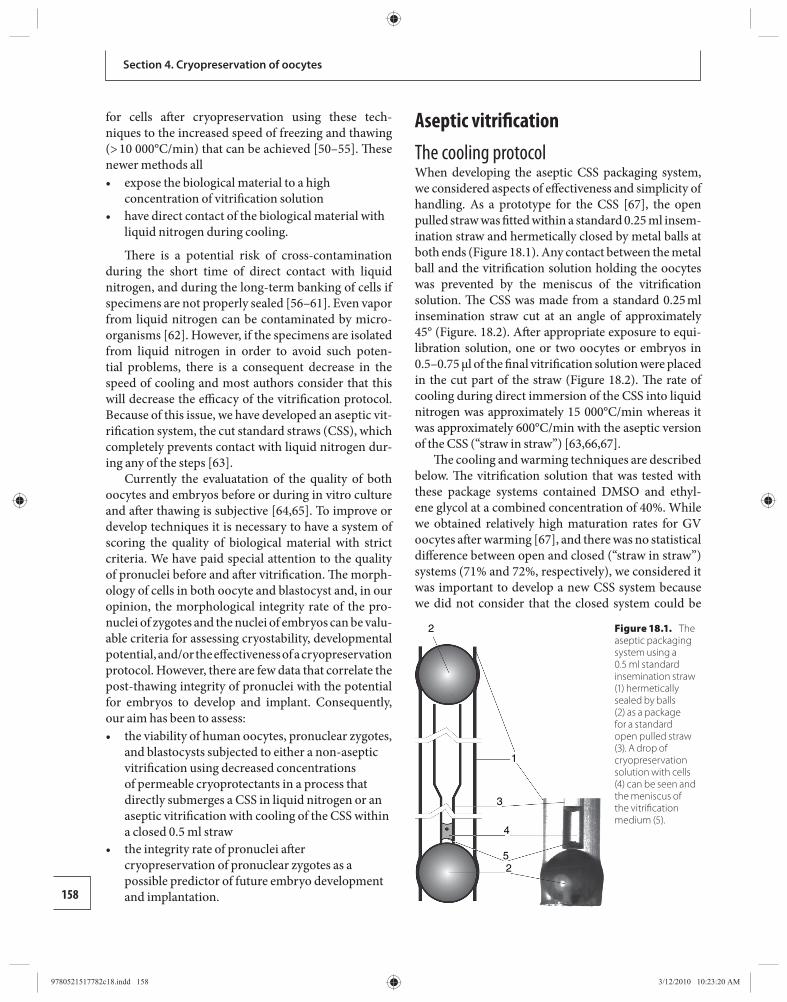
158
Section 4. Cryopreservation of oocytes
for cells aft er cryopreservation using these tech-niques to the increased speed of freezing and thawing (> 10 000°C/min) that can be achieved [ 50 – 55 ]. Th ese newer methods all
expose the biological material to a high • concentration of vitrifi cation solution have direct contact of the biological material with • liquid nitrogen during cooling.
Th ere is a potential risk of cross-contamination during the short time of direct contact with liquid nitrogen, and during the long-term banking of cells if specimens are not properly sealed [ 56 – 61 ]. Even vapor from liquid nitrogen can be contaminated by micro-organisms [ 62 ]. However, if the specimens are isolated from liquid nitrogen in order to avoid such poten-tial problems, there is a consequent decrease in the speed of cooling and most authors consider that this will decrease the effi cacy of the vitrifi cation protocol. Because of this issue, we have developed an aseptic vit-rifi cation system, the cut standard straws (CSS), which completely prevents contact with liquid nitrogen dur-ing any of the steps [ 63 ].
Currently the evaluatation of the quality of both oocytes and embryos before or during in vitro culture and aft er thawing is subjective [ 64 , 65 ]. To improve or develop techniques it is necessary to have a system of scoring the quality of biological material with strict criteria. We have paid special attention to the quality of pronuclei before and aft er vitrifi cation. Th e morph-ology of cells in both oocyte and blastocyst and, in our opinion, the morphological integrity rate of the pro-nuclei of zygotes and the nuclei of embryos can be valu-able criteria for assessing cryostability, developmental potential, and/or the eff ectiveness of a cryopreservation protocol. However, there are few data that correlate the post-thawing integrity of pronuclei with the potential for embryos to develop and implant. Consequently, our aim has been to assess:
the viability of human oocytes, pronuclear zygotes, • and blastocysts subjected to either a non-aseptic vitrifi cation using decreased concentrations of permeable cryoprotectants in a process that directly submerges a CSS in liquid nitrogen or an aseptic vitrifi cation with cooling of the CSS within a closed 0.5 ml straw the integrity rate of pronuclei aft er • cryopreservation of pronuclear zygotes as a possible predictor of future embryo development and implantation.
Aseptic vitrifi cation The cooling protocol When developing the aseptic CSS packaging system, we considered aspects of eff ectiveness and simplicity of handling. As a prototype for the CSS [ 67 ], the open pulled straw was fi tted within a standard 0.25 ml insem-ination straw and hermetically closed by metal balls at both ends ( Figure 18.1 ). Any contact between the metal ball and the vitrifi cation solution holding the oocytes was prevented by the meniscus of the vitrifi cation solution. Th e CSS was made from a standard 0.25 ml insemination straw cut at an angle of approximately 45° (Figure. 18.2 ). Aft er appropriate exposure to equi-libration solution, one or two oocytes or embryos in 0.5–0.75 μl of the fi nal vitrifi cation solution were placed in the cut part of the straw ( Figure 18.2 ). Th e rate of cooling during direct immersion of the CSS into liquid nitrogen was approximately 15 000°C/min whereas it was approximately 600°C/min with the aseptic version of the CSS (“straw in straw”) [ 63 , 66 , 67 ].
Th e cooling and warming techniques are described below. Th e vitrifi cation solution that was tested with these package systems contained DMSO and ethyl-ene glycol at a combined concentration of 40%. While we obtained relatively high maturation rates for GV oocytes aft er warming [ 67 ], and there was no statistical diff erence between open and closed (“straw in straw”) systems (71% and 72%, respectively), we considered it was important to develop a new CSS system because we did not consider that the closed system could be
2
1
3
4
52
Figure 18.1. The aseptic packaging system using a 0.5 ml standard insemination straw (1) hermetically sealed by balls (2) as a package for a standard open pulled straw (3). A drop of cryopreservation solution with cells (4) can be seen and the meniscus of the vitrifi cation medium (5).
9780521517782c18.indd 1589780521517782c18.indd 158 3/12/2010 10:23:20 AM3/12/2010 10:23:20 AM
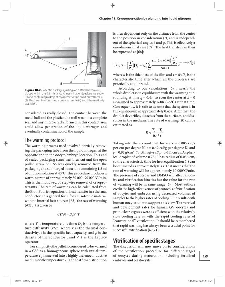
159
Chapter 18. Cryopreservation by plunging into liquid nitrogen
considered as really closed. Th e contact between the metal ball and the plastic tube wall was not a complete seal and any micro-cracks formed in this contact area could allow penetration of the liquid nitrogen and eventually contamination of the sample.
The warming protocol Th e warming process used involved partially remov-ing the packaging tube from the liquid nitrogen at the opposite end to the oocyte/embryo location. Th is end of sealed packaging straw was then cut and the open pulled straw or CSS was quickly removed from the packaging and submerged into a tube containing 1.5 ml of dilution solution at 40°C. Th is procedure produces a warming rate of approximately 30 000–90 000°C/min. Th is is then followed by stepwise removal of cryopro-tectants. Th e rate of warming can be calculated from the Biot–Fourier equation for heat transfer in a thermal conductor. In a general form for an isotropic material with no internal heat sources [ 68 ], the rate of warming (∂ T /∂ t ) is given by
∂ T /∂ t = D T ∇ 2 T
where T is temperature; t is time; D T is the tempera-ture diffusivity ( κ / cρ , where κ is the thermal con-ductivity, c is the specific heat capacity, and ρ is the density of the conductor), and ∇ 2 T is the Laplace operator.
For simplicity, the pellet is considered to be warmed in a CSS as a homogeneous sphere with initial tem-perature T 0 immersed into a highly thermoconductive medium with temperature T f . Th e heat fl ow distribution
is then dependent only on the distance from the center to the position in consideration ( r ), and is independ-ent of the spherical angles θ and φ . Th is is eff ectively a one-dimensional case [ 69 ]. Th e heat transfer can then be expressed as [ 68 ]:
( )2[(2 1) ]
01
sin(2 1)4( , )
2 1
tm
fm
rmRT r t T T e
mπ
τπ
π
∞ − +
=
+ = − + ∑
where d is the thickness of the fi lm and τ = d 2 /D T is the characteristic time aft er which all the processes are practically equilibrated.
According to our calculations [ 69 ], nearly the whole droplet is in equilibrium with the warming sur-rounding at time q = 0.4 τ, so even the center at λ = 0 is warmed to approximately 268K (−5°C) at that time. Consequently, it is safe to assume that the system is in full equilibrium at approximately 0.45 τ . Aft er that, the droplet devitrifi es, detaches from the surfaces, and dis-solves in the medium. Th e rate of warming ( B ) can be estimated as:
0
0.45fT T
Bτ
−≈
Taking into the account that for ice κ = 0.005 cal/s per cm per degree K, c = 0.49 cal/g per degree K, and ρ = 0.92 g/cm 3 [ 70 ], this gives D T = 0.011 cm 2 /s. A spher-ical droplet of volume 0.75 μl has radius of 0.056 cm, so the characteristic time for heat equilibration ( τ ) can be estimated as aproximately 0.3 s. Th at means that the rate of warming will be approximately 90 000°C/min. Th e presence of sucrose and DMSO will aff ect viscos-ity and vitrifi cation kinetics but the value for the rate of warming will be in same range [ 69 ]. Most authors credit the high eff ectiveness of protocols of vitrifi cation of oocytes and embryos using decreased volumes of samples to the higher rates of cooling. Our results with human oocytes do not support this view. Th e survival and development rates for human GV oocytes and pronuclear zygotes were as effi cient with the relatively slow cooling rate as with the rapid cooling rates of “conventional” vitrifi cation. It should be remembered that rapid warming has always been a crucial point for successful vitrifi cation [ 67 , 71 ].
Vitrifi cation of specifi c stages Th e discussion will now move on to considerations of the vitrifi cation procedure for diff erent stages of oocytes during maturation, including fertilized embryos and blastocysts.
1
3
52
4
45°
Figure 18.2. Aseptic packaging using a cut standard straw (1) placed within the 0.5 ml standard insemination (packaging) straw (2) and containing a drop of cryopreservation solution with cells (3). The insemination straw is cut at an angle (4) and is hermetically sealed (5).
9780521517782c18.indd 1599780521517782c18.indd 159 3/12/2010 10:23:21 AM3/12/2010 10:23:21 AM
160
Section 4: Cryopreservation of oocytes
Vitrifi cation of germinal vesicle oocytes Th e potential of vitrifi cation as a method of cryo-preservation was shown for the fi rst time in 1985 [ 72 ] when vitrifi cation was successfully applied to immature oocytes. Usually the vitrifi cation proto-col contained DMSO. However, there are no data regarding the use of DMSO for the vitrifi cation of GV oocytes and, therefore, we performed a series of experiments to investigate the role of DMSO during the vitrifi cation of GV oocytes. Th is cryoprotectant has been reported to aff ect the organization of micro-fi laments in mouse oocytes [ 73 ] as well as to induce chromosomal abnormal ities leading to an increase in the rates of degeneration and digenic polyploid embryos aft er cryopreservation [ 74 ]. Consequently, we also examined the possibility of cryopreserving GV oocytes using a solution completely free of DMSO (40% ethylene glycol plus 0.75 M sucrose). If DMSO is not present in the vitrifi cation medium, there is a sig-nifi cant decrease in maturation rates (51%) compared with GV oocytes vitrifi ed in the presence of DMSO (85%). However, the long contact period (5 minutes) of oocytes with the vitrifi cation solution containing 20% DMSO at 37°C in both “direct” and “straw in straw” vitrifi cation was enough to cause spontaneous oocyte activation and parthenogenetic development. Th e protocol that we now recommend for use in med-ical practice is a compromise between avoiding use of DMSO in the vitrifi cation medium and the partheno-genetic activation that it causes. Based on our results we propose the following protocol. 1. Exposure in three steps to a pre-warming
solution (37ºC) containing 20%, 33%, and 50% DMSO (in 40% ethylene glycol plus 0.75 M sucrose) for 2, 1, and 1 minute, respectively.
2. One-step exposure to a vitrifi cation solution with 20% DMSO in 20% ethylene glycolplus 0.75 M sucrose for 1 minute at room temperature, with subsequent cooling in liquid nitrogen.
Th is protocol has a short (1 minute) period of contact with a high DMSO concentration, in the last of the stepped portion of the protocol, followed by contact with DMSO at room temperature, which in theory can decrease the negative eff ects on oocytes. Th e high maturation rates and the absence of parthenogenesis seen support the use of DMSO with decreasing time and temperature of contact for vitrifi cation of human GV oocytes.
Th e use of DMSO in a vitrifi cation medium for human reproductive cells is supported by studies by Vanderzwalmen et al. [ 38 ], Huang et al. [ 75 ], and Isachenko et al. [ 67 ], who have all used DMSO for vitri-fi cation of blastocysts. Th ese authors all reported high pregnancy rates aft er warming of blastocysts (41% [ 38 ] and 54% [ 75 ]) as well as evidence of the effi ciency of a vitrifi cation media containing 20% DMSO, 20% ethyl ene glycol, sucrose, and other compounds. Th ese pregnancies resulted in the birth of healthy babies: P. Vanderzwalmen (personal communication) reported 98 healthy births including 28 twins from 414 warmed and transferred embryos; C. C. Huang and T. H. Lee (personal communication) reported 10 healthy births from 14 implantations, with three spontan eous abor-tions and one reduction from triplet. Using DMSO for vitrifi cation of oocytes was also successful, with a high survival rate of warmed oocytes, on going clini-cal pregnancy [ 28 ], and later birth of healthy babies aft er embryo transfers (K. P. Katayama, personal communication).
Our studies also examined if vitrifi cation of GV oocytes could be performed using the aseptic tech-nology that we had initially developed for human and mouse pronuclear zygotes, embryos, and human sper-matozoa [ 71 , 76 , 77 ]. Aseptic vitrifi cation using a “straw in straw” system leads to decreased cooling rates. Rall [ 78 ] reported in 1987 that cooling rates of 10°C/min were suffi cient for vitrifi cation of mouse embryos. Th e speed of warming is a very important parameter in a vitrifi cation procedure. All combinations of “slow” or “quick” cooling with “slow” warming have resulted in decreased viability of vitrifi ed mouse embryos [ 79 ]. Elevated warming rates and the absence of visible crystal formation during thawing of oocytes was shown to be most eff ective when the proportion of “speed of warm-ing to speed of cooling” was higher than 1.3 [ 80 , 81 ]. Th ese observations about the lesser importance of the speed of cooling for the vitrifi cation of human pro-nuclear oocytes can probably be applied to all human reproductive cells, including spermatozoa. We recently reported that vitrifi cation of human spermatozoa by fast (20 000°C/min) or relatively slow (600°C/min and 200°C/min) cooling resulted in similar post-thaw char-acteristics [ 82 ] and that slow cooling/rapid warming can also be applied to human GV oocytes. In fact, this approach allowed maturation rates for GV oocytes aft er vitrifi cation of around 85%. Th is high rate is promis-ing for future applications of cryo preservation of GV oocytes for reproductive purposes.
9780521517782c18.indd 1609780521517782c18.indd 160 3/12/2010 10:23:22 AM3/12/2010 10:23:22 AM
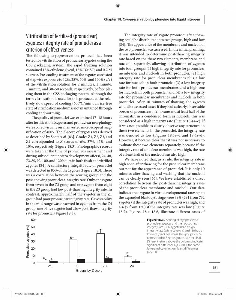
161
Chapter 18. Cryopreservation by plunging into liquid nitrogen
Vitrifi cation of fertilized (pronuclear) zygotes: integrity rate of pronuclei as a criterion of eff ectiveness Th e following cryopreservation protocol has been tested for vitrifi cation of pronuclear zygotes using the CSS packaging system. Th e rapid freezing solution contained 15% ethylene glycol, 15% DMSO, and 0.2 M sucrose. Pre-cooling treatment of the zygotes consisted of stepwise exposure to 12%, 25%, 50%, and 100% (v/v) of the vitrifi cation solution for 2 minutes, 1 minute, 1 minute, and 30–50 seconds, respectively, before pla-cing them in the CSS packaging system. Although the term vitrifi cation is used for this protocol, at the rela-tively slow speed of cooling (600°C/min), an ice-free state of vitrifi cation medium is not maintained through cooling and warming.
Th e quality of pronuclei was examined 17–18 hours aft er fertilization. Zygotes and pronuclear morphology were scored visually on an inverted microscope at mag-nifi cation of 400×. Th e Z -score of zygotes was derived as described by Scott et al. [ 83 ]. Grades Z1, Z2, Z3, and Z4 corresponded to Z -scores of 6%, 37%, 47%, and 10%, respectively ( Figure 18.3 ). Photographic records were taken at the time of pronucleus assessment and during subsequent in vitro development aft er 8, 24, 48, 72, 80, 92, 100, and 120 hours in both fresh and vitrifi ed zygotes [ 84 ]. A satisfactory integrity rate of pronuclei was detected in 85% of the zygotes ( Figure 18.3 ). Th ere was a correlation between the scoring group and the post-thawing pronuclear integrity rate. Only one zygote from seven in the Z2 group and one zygote from eight in the Z3 group had low post-thawing integrity rate. In contrast, approximately half of the zygotes in the Z1 group had poor pronuclear integrity rate. Cryostability in the mid range was observed in zygotes from the Z4 group: one of fi ve zygotes had a low post-thaw integrity rate for pronuclei ( Figure 18.3 ).
Th e integrity rate of zygote pronuclei aft er thaw-ing could be distributed into two groups, high and low [ 84 ]. Th e appearance of the membrane and nucleoli of the two pronuclei was assessed. In the initial planning, it was intended to determine post-thawing integrity rate based on the these two elements, membrane and nucleoli, separately, allowing distribution of zygotes into four groups: (1) high integrity rate for pronuclear membranes and nucleoli in both pronuclei; (2) high integrity rate for pronuclear membranes plus a low rate for nucleoli in both pronuclei; (3) a low integrity rate for both pronuclear membranes and a high one for nucleoli in both pronuclei; and (4) a low integrity rate for pronuclear membranes and nucleoli in both pronuclei. Aft er 10 minutes of thawing, the zygotes would be assessed to see if they had a clearly observable border of pronuclear membrane and at least half of the chromatin in a condensed form as nucleoli; this was considered as a high integrity rate ( Figure 18.4a–e ). If it was not possible to clearly observe any structure in these two elements in the pronuclei, the integrity rate was denoted as low ( Figures 18.5a–d and 18.6a–d ). However, it became clear that it was not necessary to evaluate these two elements separately, because if the integrity rate of a nuclear membrane was high, the rate of at least half of the nucleoli was also high.
We have noted that, as a rule, the integrity rate is high soon aft er thawing for the pronuclear membrane but not for the appearance of pronuclei. It is only 10 minutes aft er thawing and washing that the nucleoli can be clearly seen [ 66 ]. We have established a direct correlation between the post-thawing integrity rates of the pronuclear membrane and nucleoli. Our data indicate that zygote in vitro developmental rates up to the expanded blastocyst stage were 39% (291 from 732 zygotes) if the integrity rate of pronuclei was high, and 4% (5 from 130) if the integrity rate was low ( Figure 18.7 ). Figures 18.4 – 18.6 , illustrate diff erent cases of
60
50
40
30
20
Pro
nucl
ear
zygo
tes
(%)
10
0Z1
a e
b
f
c
fd
e
Z2 Z3Groups by Z-score
Z4
Figure 18.3. Scoring of cryopreserved pronuclear zygotes and their post-thaw integrity rates: 732 zygotes had a high integrity rate (white columns) and 130 had a low rate (black columns). The groups Z1–Z4 correspond to Z-score groups, see text [ 83 ]. Diff erent letters above the columns indicate signifi cant diff erences ( p < 0.05); the same letters indicate no signifi cant diff erences ( p > 0.1).
9780521517782c18.indd 1619780521517782c18.indd 161 3/12/2010 10:23:22 AM3/12/2010 10:23:22 AM
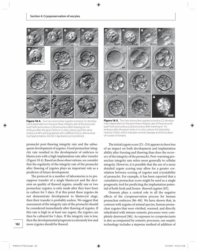
162
Section 4: Cryopreservation of oocytes
pronuclei post-thawing integrity rate and the subse-quent development of zygotes. Good pronuclear integ-rity rate resulted in the development of embryos to blastocysts with a high implantation rate aft er transfer ( Figure 18.4 ). Based on these observations, we consider that the regularity of the integrity rate of the pronuclei aft er thawing of zygotes plays an important role as a predictor of future development
Th e protocol in a number of laboratories is to pre-suppose transfer of a single blastocyst and the deci-sion on quality of thawed zygotes, usually one or two pronuclear zygotes, is only made aft er they have been in culture for 5 days. If at this point these zygotes do not demonstrate development to blastocyst stage, then their transfer is probably useless. We suggest that assessment of the integrity rate of the pronuclei should be considered immediately aft er thawing of zygotes. If this rate is high in at least one zygote, the zygotes can then be cultured for 5 days. If the integrity rate is low, then the developmental prognosis is extremely low and more zygotes should be thawed.
Th e initial zygote score (Z1–Z4) appears to have less of an impact on both development and implantation ability aft er freezing and thawing than does the recov-ery of the integrity of the pronuclei. Post-warming pro-nuclear integrity rate refers more generally to cellular integrity. However, it is possible that the use of a more detailed zygote scoring may allow for a greater cor-relation between scoring of zygotes and cryostability of pronuclei. For example, it has been reported that a cumulative pronuclear score might be used as a single prognostic tool for predicting the implantation poten-tial of both fresh and frozen–thawed zygotes [ 85 ].
Osmosis plays a central role in all the negative eff ects of the cryopreservation process for human pronuclear embryos [ 86 – 88 ]. We have shown that, in contrast with zygotes of animal species, human pronu-clear zygotes that were vitrifi ed, warmed, and directly rehydrated with intense osmotic processes were com-pletely destroyed [ 86 ]. As exposure to cryoprotectants is also accompanied by osmotic processes, our aseptic technology includes a stepwise method of addition of
e
Fresh Fresh 10 min
8 h
24 h 48 h
48 h
92 h 100 h 120 h
72 h 80 h
10 min
f
h i j
k l m
g
a cb d
Figure 18.4. Two two-pronuclear zygotes scored as Z2: develop-ment dependent on the post-thaw integrity rate of the pronuclei. (a,b) Fresh pronucleus; (c,d) pronucleus after thawing; (e–m) embryos after the given times in in vitro culture; (g,h) the same embryo at 48 h photographed with a diff erent focus, demonstrat-ing fragmentation; (m) the 5 day blastocyst transferred.
a
m
cb
Fresh Fresh 10 min
8 h
72 h
100 h 120 h 120 h
80 h 92 h
24 h 48 h
10 min
d
e
h
k
f
i
l
g
j
Figure 18.5. Two two-pronuclear zygotes scored as Z3: develop-ment dependent on the post-thaw integrity rate of the pronuclei. (a,b) Fresh pronucleus; (c,d) pronucleus after thawing; (e–m) embryos after the given times in in vitro culture; (m) stained by Hoechst 33342, which indicates normal cleavage and the location of nuclear chromatin.
9780521517782c18.indd 1629780521517782c18.indd 162 3/12/2010 10:23:23 AM3/12/2010 10:23:23 AM
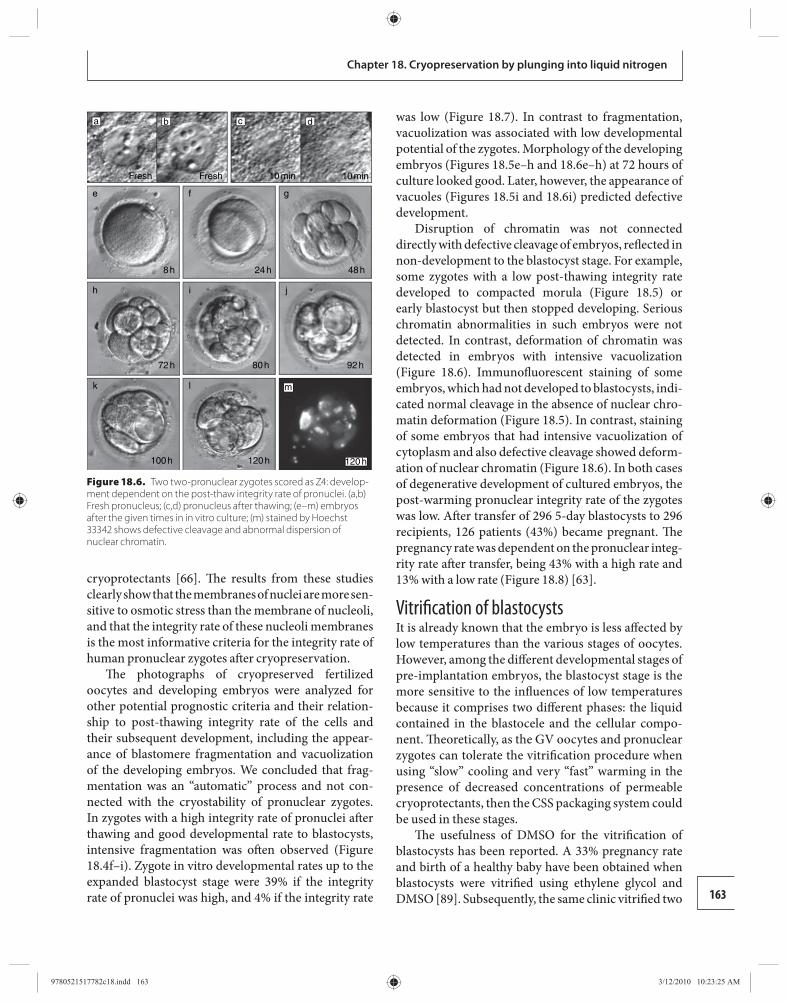
163
Chapter 18. Cryopreservation by plunging into liquid nitrogen
cryoprotectants [ 66 ]. Th e results from these studies clearly show that the membranes of nuclei are more sen-sitive to osmotic stress than the membrane of nucleoli, and that the integrity rate of these nucleoli membranes is the most informative criteria for the integrity rate of human pronuclear zygotes aft er cryopreservation.
Th e photographs of cryopreserved fertilized oocytes and developing embryos were analyzed for other potential prognostic criteria and their relation-ship to post-thawing integrity rate of the cells and their subsequent development, including the appear-ance of blastomere fragmentation and vacuolization of the developing embryos. We concluded that frag-mentation was an “automatic” process and not con-nected with the cryostability of pronuclear zygotes. In zygotes with a high integrity rate of pronuclei aft er thawing and good developmental rate to blastocysts, intensive fragmentation was oft en observed ( Figure 18.4 f–i). Zygote in vitro developmental rates up to the expanded blastocyst stage were 39% if the integrity rate of pronuclei was high, and 4% if the integrity rate
was low ( Figure 18.7 ). In contrast to fragmentation, vacuolization was associated with low developmental potential of the zygotes. Morphology of the developing embryos ( Figures 18.5e–h and 18.6e–h ) at 72 hours of culture looked good. Later, however, the appearance of vacuoles ( Figures 18.5i and 18.6 i) predicted defective development.
Disruption of chromatin was not connected directly with defective cleavage of embryos, refl ected in non-development to the blastocyst stage. For ex ample, some zygotes with a low post-thawing integrity rate developed to compacted morula ( Figure 18.5 ) or early blastocyst but then stopped developing. Serious chromatin abnormalities in such embryos were not detected. In contrast, deformation of chromatin was detected in embryos with intensive vacuolization ( Figure 18.6 ). Immunofl uorescent staining of some embryos, which had not developed to blastocysts, indi-cated normal cleavage in the absence of nuclear chro-matin deformation ( Figure 18.5 ). In contrast, staining of some embryos that had intensive vacuolization of cytoplasm and also defective cleavage showed deform-ation of nuclear chromatin ( Figure 18.6 ). In both cases of degenerative development of cultured embryos, the post-warming pronuclear integrity rate of the zygotes was low. Aft er transfer of 296 5-day blastocysts to 296 recipients, 126 patients (43%) became pregnant. Th e pregnancy rate was dependent on the pronuclear integ-rity rate aft er transfer, being 43% with a high rate and 13% with a low rate ( Figure 18.8 ) [ 63 ].
Vitrifi cation of blastocysts It is already known that the embryo is less aff ected by low temperatures than the various stages of oocytes. However, among the diff erent developmental stages of pre-implantation embryos, the blastocyst stage is the more sensitive to the infl uences of low temperatures because it comprises two diff erent phases: the liquid contained in the blastocele and the cellular compo-nent. Th eoretically, as the GV oocytes and pronuclear zygotes can tolerate the vitrifi cation procedure when using “slow” cooling and very “fast” warming in the presence of decreased concentrations of permeable cryoprotectants, then the CSS packaging system could be used in these stages.
Th e usefulness of DMSO for the vitrifi cation of blastocysts has been reported. A 33% pregnancy rate and birth of a healthy baby have been obtained when blastocysts were vitrifi ed using ethylene glycol and DMSO [ 89 ]. Subsequently, the same clinic vitrifi ed two
a
e f g
h i j
k l m
cb
Fresh Fresh 10 min
8 h
72 h
100 h 120 h 120 h
80 h 92 h
24 h 48 h
10 min
d
Figure 18.6. Two two-pronuclear zygotes scored as Z4: develop-ment dependent on the post-thaw integrity rate of pronuclei. (a,b) Fresh pronucleus; (c,d) pronucleus after thawing; (e–m) embryos after the given times in in vitro culture; (m) stained by Hoechst 33342 shows defective cleavage and abnormal dispersion of nuclear chromatin.
9780521517782c18.indd 1639780521517782c18.indd 163 3/12/2010 10:23:25 AM3/12/2010 10:23:25 AM

164
Section 4: Cryopreservation of oocytes
embryos at the morula stage using the same protocol, resulting in the birth of healthy twins [ 90 ]. Up to March 2005, 86 healthy babies have been born aft er the trans-fer of 332 embryos (birth rate 26%) using the vitrifi -cation solution containing ethylene glycol and DMSO (Y. Yokota, personal communication).
We found that developmental rates for vitrifi ed blastocysts aft er cryopreservation with the non-packaged CSS (direct contact with liquid nitrogen) were very similar with either increased (20% DMSO, 20% EG, sucrose) or decreased (15% DMSO, 15% EG, sucrose) concentrations of permeable cryoprotect-ants with subsequent in vitro culture: 85% and 86%, respectively [ 84 ]. We have also tested two diff erent packaging systems [ 84 ], one that does not prevent contact with liquid nitrogen and one that completely prevents contact. Both systems showed the same rate of re-expansion aft er in vitro culture: 81% and 80%, respectively. Th e re-expansion (the blastocele) of vi able embryos usually begins 2 to 6 hours aft er warm-ing. Th e collapse of a blastocyst (when its appearance is similar to that of a compact morula) is a sign of viability of the embryo aft er warming, and embryos
were not viable aft er vitrifi cation if the collapse of the blastocysts did not occur in the fi rst 10 minutes aft er warming.
We know that embryos with a laser-opened zona pellucida are more diffi cult to cryopreserve than early blastocysts or blastocysts with an intact zona pellucida. An early report found that the survival rate of expanded blastocysts aft er vitrifi cation was relatively low. Th e authors attributed this to the large volume of water in the blastocele and its lethal crystallization during cool-ing [ 91 ]. Laser opening of the zona pellucida can also decrease the cryosurvival of blastocysts; however, the reports on vitrifi cation of laser-treated blastocysts are limited [ 43 ].
We have tried the new CSS method of vitrifi cation on laser-hatched blastocysts that had been chosen for embryo transfer. Clinical pregnancies using CSS vitrifi -cation with direct contact with liquid nitrogen and the aseptic CSS “straw-in-straw” method were established in 6 of 18 patients (33%) in both groups, as determined by the presence of a fetal sac by ultrasound 6 weeks aft er embryo transfer. Th e implantation rates were similar in both groups (22% and 28%, respectively).
1009080706050403020
Z1
1 16 ab
ca1 1114 128 2 33
Z2 Z3 Z4
Groups by Z-score
Bla
stoc
yst t
rans
fer
(%);
preg
nanc
y ra
te (
%)
100
Figure 18.8. Pregnancy rate after transfer of blastocysts is dependent on the quality of the pronuclei (Z-scoring [ 83 ]; see text)and their post-thaw integrity rate: blastocysts from zygotes with low pronuclei integrity rates (5; gray columns); blastocysts from zygotes with high pronuclei integrity rates (291; white columns); pregnancies after transfer of blastocysts from zygotes with low pronuclei integrity rates (1; black columns); pregnancies after transfer of blastocysts from zygotes with high pronuclei integrity rates (125; hatched columns). Numbers on the columns indicate the number in that group. Diff erent letters above the columns indicate signifi cant diff erences ( p < 0.05).
10090807060
Pro
nucl
ear
zygo
tes
and
blas
tocy
sts
(%)
50403020100
Z1
ab a,b a
ccc
17 36 44 52 17 71280 345
Z2
Groups by Z-score
Z3 Z4
Figure 18.7. Blastocyst forma-tion from cryopreserved pronuclear zygotes is dependent on the quality of pronuclei (Z-scoring [ 83 ]; see text) and their post-thaw integrity rate: zygotes with low pronuclei integrity rates (130; gray columns); zygotes with high pronuclei integrity rates (732; white columns); blastocysts from zygotes with low pronuclei integrity rates (5; black columns); blastocysts from zygotes with high pronuclei integrity rates (291; hatched columns). Numbers on the columns indicate the number in that group. Diff erent letters above the columns indicate signifi cant diff erences ( p < 0.05).
9780521517782c18.indd 1649780521517782c18.indd 164 3/12/2010 10:23:26 AM3/12/2010 10:23:26 AM
165
Chapter 18. Cryopreservation by plunging into liquid nitrogen
Conclusions Th e placement of human oocytes, zygotes, and embryos inside an hermetically closed container before plun-ging into liquid nitrogen for vitrifi cation permits a reli-able isolation of the cells from the liquid nitrogen and avoids the potential for contamination by microorgan-isms. Although this technique is associated with a rela-tively slow cooling rate, the developmental potential of these oocytes and embryos is not compromised if the thawing process involves rapid warming and simul-taneous removal of cryoprotectants. Effi cient aseptic vitrifi cation of oocytes and embryos can be performed with decreased concentrations of permeable cryopro-tectants and complete isolation of embryos from liquid nitrogen.
Acknowledgements Th e authors would like to thank Dr. Igor I. Katkov for the mathematical calculations of the speed of oocyte warming in the CSS methodology.
References 1. Leibo SP. Cryopreservation of mammalian oocytes.
In Tulandi T , Gosden R , eds. Preservation of Fertility . London : Taylor & Francis , 2004; pp. 141–154 .
2. Catt J , Wood T , Henman M , et al. Single embryo transfer in IVF to prevent multiple pregnancies . Twin Res 2003; 6: 536–539 .
3. Henman M , Catt JW , Wood T , et al. Elective transfer of single fresh blastocysts and later transfer of cryostored blastocysts reduces the twin pregnancy rate and can improve the in vitro fertilization live birth rate in younger women . Fertil Steril 2005; 84: 1620–1627 .
4. de Boer KA , Catt JW , Jansen RP , et al. Moving to blastocyst biopsy for preimplantation genetic diagnosis and single embryo transfer at Sydney IVF . Fertil Steril 2004; 82: 295–298 .
5. Jansen RP , Burton GJ. Mitochondrial dysfunction in reproduction . Mitochondrion 2004; 4: 577–600 .
6. Taylor DM , Handyside AH , Ray PF , et al. Quantitative measurement of transcript levels throughout human preimplantation development: analysis of hypoxanthine phosphoribosyl transferase . Mol Hum Reprod 2001; 7: 147–154 .
7. Telford NA , Watson AJ , Schultz GA. Transition from maternal to embryonic control in early mammalian development: a comparison of several species . Mol Reprod Dev 1990; 26: 90–100 .
8. Clouston HJ , Herbert M , Fenwick J , et al. Cytogenetic analysis of human blastocysts . Prenat Diagn 2002; 22: 1143–1152 .
9. Chen C. Pregnancy aft er human oocytes cryopreservation . Lancet 1986; 11: 884–886 .
10. van Uem JFHM , Siebzehnrübl ER , Schuh B , et al. Birth aft er cryopreservation of unfertilized oocytes . Lancet 1987; 8535: 752–753 .
11. Al-Hasani S , Diedrich K , van der Ven , et al. Cryopreservation of human oocytes Hum Reprod 1987; 2: 695–700 .
12. Tucker M , Wright G , Morton F , et al. Preliminary experience with human oocytes cryopreservation using 1,2-propanediol and sucrose . Hum Reprod 1996; 11: 1513–1515 .
13. Porcu E , Fabbri R , Seracchioli , et al. Birth of a healthy female aft er intracytoplasmic sperm injection of cryopreserved human oocytes . Fertil Steril 1997; 68: 724–726 .
14. Young E , Kenny A , Puigdomenech E , et al. Triplet pregnancy aft er intracytoplasmic sperm injection of cryopreserved oocytes: case report . Fertil Steril 1998; 70: 360–361 .
15. Boldt J , Cline D , McLaughlin D. Human oocytes cryopreservation as an adjunct to IVF-embryo transfer cycles . Hum Reprod 2003; 18: 1250–1255 .
16. Kuleshova LL , Lopata A. Vitrifi cation can be more favourable than slow cooling . Fertil Steril 2002; 78: 449–454 .
17. Mohr LR , Trounson AO , Freeman L. Deep freezing and transfer of human embryos . J In Vitro Fertil Embryo Transfer 1985 ; 2: 1–10 .
18. Siebzehnrübl ER. Cryopreservation of gametes and cleavage stage embryos . Hum Reprod 1989; 4 (Suppl): 105–110 .
19. Testart J , Lassale B , Forman R , et al. Factors infl uencing the success of human embryo freezing in an in vitro fertilization and embryo transfer program . Fertil Steril 1987; 48: 107 .
20. Veeck LL , Amudson CH , Brothman LJ , et al. Signifi cantly enhanced pregnancy rates per cycle through cryopreservation and thaw of pronuclear stage oocytes . Fertil Steril 1993; 59: 1202–1207 .
21. Van den Abbeel E , Campus M , van Waesberghe L , et al. A randomised comparison of the cryopreservation of one cell human embryos with a slow controlled cooling procedure or a rapid cooling procedure by direct plunging into liquid nitrogen . Hum Reprod 1997; 12: 1554–1560 .
22. Al-Hasani S , Demirel LC , Schöpper B , et al. Pregnancies achieved aft er frozen-thawed pronuclear oocytes obtained by intracytoplasmic sperm injection with spermatozoa extracted from frozen–thawed testicular tissues from non-obstructive azoospermic men . Hum Reprod 1999 ; 14: 2031–2035 .
23. Damario MA , Hammitt DG , Galantis TM , et al. Pronuclear stage cryopreservation aft er intracytoplasmic sperm injection and conventional
9780521517782c18.indd 1659780521517782c18.indd 165 3/12/2010 10:23:27 AM3/12/2010 10:23:27 AM
166
Section 4. Cryopreservation of oocytes
IVF: implication for timing of the freeze . Fertil Steril 1999; 72: 1049–1054 .
24. Borini A , Cattoli M , Bulletti C , Coticchio G. Clinical effi ciency of oocyte and embryo cryopreservation . Ann N Y Acad Sci 2008; 1127: 49–58 .
25. Kuleshova LL, Gianaroli L , Magli C , et al. Birth following vitrifi cation of a small number of human oocytes . Hum Reprod 1999; 14: 3077–3039 .
26. Yoon TK , Chung HM , Lim JM , et al. Pregnancy and delivery of healthy infants developed from vitrifi ed oocytes in a stimulated in vitro fertilization-embryo transfer program . Fertil Steril 2000; 74: 180–181 .
27. Yoon TK , Kim TJ , Park SE , et al. Live birth aft er vitrifi cation of oocytes in a stimulated in vitro fertilization-embryo transfer programm . Fertil Steril 2003; 79: 1323–1326 .
28. Katayama KP , Stehlik J , Kuwayama M , et al. Huge survival rate of vitrifi ed human oocytes results inclinical pregnancy . Fertil Steril 2003; 80: 223–224 .
29. Park SP , Kim EY , Oh JH , et al. Ultra-rapid freezing of human multipronuclear oocytes using electron microscope grids . Hum Reprod 2000; 15: 1787–1790 .
30. Selman HA , El-Danasouri I. Pregnancies derived from vitrifi ed human oocytes . Fertil Steril 2002; 77 , 422–423 .
31. Liebermann J , Tucker MJ , Graham JR , et al. Blastocyst development aft er vitrifi cation of multipronuclear oocytes using the Flexipet denuding pipette . RBM Online 2002 ; 4: 148–152 .
32. Liebermann J , Nawroth F , Isachenko V , et al. Potential importance of vitrifi cation in reproductive medicine . Biol Reprod 2002 ; 67: 1671–1680 .
33. Jelinkova L , Selman HA , Arav A , et al. Twin pregnancy aft er vitrifi cation of 2-pronuclei human embryos . Fertil Steril 2002; 77: 412–414 .
34. Ciotti PM , Porcu E , Notarangelo L , et al. Meiotic spindle recovery is faster in vitrifi cation of human oocytes compared to slow freezing . Fertil Steril 2009; 91 : 2399–2407 .
35. Bagchi A , Woods EJ , Critser JK . Cryopreservation and vitrifi cation: recent advances in fertility preservation technologies . Expert Rev Med Devices 2008; 5: 359–370 .
36. Al-Hasani S , Ozmen B , Koutlaki N , et al. Th ree years of routine vitrifi cation of human zygotes: is it still fair to advocate slow-rate freezing ? RBM Online 2007; 14: 288–293 .
37. Reed ML , Lane M , Gardner DK , et al. Vitrifi cation of human blastocysts using the cryoloop method: successful clinical application and birth of off spring . J Assist Reprod Genet 2002; 19: 304–306 .
38. Vanderzwalmen P , Bertin G , Debauche CH , et al. Vitrifi cation of human blastocysts with the Hemi-Straw carrier: application of assisted hatching aft er thawing . Hum Reprod 2003; 18: 1504–1511 .
39. Mukaida T , Nakamura S , Tomiyama T, et al . Vitrifi cation of human blastocysts using cryoloops: clinical outcome of 223 cycles . Hum Reprod 2003; 18: 384–391 .
40. Son WY , Yoon SH , Park SJ , et al. Ongoing twin pregnancy aft er vitrifi cation of blastocysts produced by in-vitro matured oocytes retrieved from a woman with polycystic ovary syndrome: case report . Hum Reprod 2002; 17: 2963–2966 .
41. Son WY , Lee SY , Chang MJ , et al. Pregnancy resulting from transfer of repeat vitrifi ed blastocysts produced by in-vitro matured oocytes in patient with polycystic ovary syndrome . RBM Online 2005; 10: 398–401 .
42. Takahashi K , Mukaida T , Goto T , et al. Perinatal outcome of blastocyst transfer with vitrifi cation using cryoloop: a 4-year follow-up study . Fertil Steril 2005; 84: 88–92 .
43. Zech NH , Lejeune B , Zech H , et al. Vitrifi cation of hatching and hatched human blastocysts: eff ect of an opening in the zona pellucida before vitrifi cation . RBM Online 2005; 11: 355–361 .
44. Liebermann J , Tucker MJ. Comparison of vitrifi cation and conventional cryopreservation of day 5 and day 6 blastocysts during clinical application . Fertil Steril 2006; 86: 20–26 .
45. Hiraoka K , Hiraoka K , Kinutani M , et al. Vitrifi cation of human hatched blastocysts: a report of 4 cases . J Reprod Med 2007; 52: 413–415 .
46. Lee SY , Son WY , Yoon SH , et al. Clinical-pregnancy outcome aft er vitrifi cation of blastocysts produced from in vitro maturation cycles . Fertil Steril 2007; 88: 1449–1451 .
47. Baudot A , Alger L , Boutron P. Glass-forming tendency in the system water–dimethyl sulfoxide . Cryobiology 2000; 40: 151–158 .
48. Kuleshova LL , Shaw JM. A strategy for rapid cooling of mouse embryos within a double straw to eliminate the risk of contamination during storage in liquid nitrogen . Hum Reprod 2000; 15: 2604–2609 .
49. Palasz AT , Mapletoft RJ. Cryopreservation of mammalian embryos and oocytes: recent advances . Biotechnol Adv 1996; 14: 127–149 .
50. Martino A , Songansen N , Leibo SP. Development into blastocysts of bovine oocytes cryopreserved by ultra-rapid cooling . Biol Reprod 1996; 54: 1059–1069 .
51. Vajta G , Kuwayama M , Holm P , et al. Open pulled straw (OPS) vitrifi cation: a new way to reduce cryoinjuries of bovine ova and embryos . Mol Reprod Dev 1998; 51: 53–58 .
52. Lane M , Bavister BD , Lyons EA , et al. Containerless vitrifi cation of mammalian oocytes . Nat Biotechnol 1999; 17: 1234–1236 .
53. Lane M , Gardner DK. Vitrifi cation of mouse oocytes using a nylon loop . Mol Reprod Dev 2001; 58: 342–347 .
54. Dinnyes A , Dai Y , Jiang S , et al. High developmental rates of vitrifi ed bovine oocytes following
9780521517782c18.indd 1669780521517782c18.indd 166 3/12/2010 10:23:27 AM3/12/2010 10:23:27 AM
167
parthenogenetic activation, in vitro fertilization, and somatic cell nuclear transfer . Biol Reprod 2000; 63: 513–518 .
55. Kuwayama M , Kato O. Successful vitrifi cation of human oocytes . Fertil Steril 2000; 74: 549 .
56. Berry ED , Dorsa WJ , Siragusa GR , et al. Bacterial cross-contamination of meat during liquidnitrogen immersion freezing. J Food Prot 1998; 61: 1103–1108 .
57. Khuu HM , Cowley H , David-Ocampo V , et al. Catastrophic failures of freezing bags for cellular therapy products: description, cause, and consequences . Cytotherapy 2002; 4: 539–549 .
58. Bielanski A , Nadin-Davis S , Sapp T , et al. Viral contamination of embryos cryopreserved in liquid nitrogen . Cryobiology 2000; 40: 110–116 .
59. Bielanski A , Bergeron H , Lau PC , et al. Microbial contamination of embryos and semen during long term banking in liquid nitrogen . Cryobiology 2003; 46: 146–152 .
60. Bielanski A. Experimental microbial contamination and disinfection of dry (vapour) shipper dewars designed for short-term storage and transportation of cryopreserved germplasm and other biological specimens . Th eriogenology 2005; 63: 1946–1957 .
61. Morris GJ. Th e origin, ultrastructure, and microbiology of the sediment accumulating in liquid nitrogen storage vessels . Cryobiology 2005; 50: 231–238 .
62. Fountain D , Ralston M , Higgins N , et al. Liquid nitrogen freezers: a potential source of microbial contamination of hematopoietic stem cell components . Transfusion 1997; 37: 585–591 .
63. Isachenko V , Katkov II , Yakovenko S , et al. Vitrifi cation of human laser treated blastocysts within cut standard straws (CSS): novel aseptic packaging and reduced concentrations of cryoprotectants . Cryobiology 2007; 54: 305–309 .
64. Veeck , LL. An Atlas of Human Gametes and Concepts . London: Parthenon, 1998; p. 215.
65. Veeck , LL, Zaninovic N. An Atlas of Human Blastocysts . London: Parthenon, 2003; p. 286.
66. Isachenko V , Montag M , Isachenko E , et al. Aseptic technology of vitrifi cation of human pronuclear oocytes using open-pulled straws . Hum Reprod 2005; 20: 492–496 .
67. Isachenko V , Montag M , Isachenko E , et al. Aseptic vitrifi cation of human germinal vesicle oocytes using dimethyl sulfoxide as a cryoprotectant . Fertil Steril 2006; 85: 741–747 .
68. Aramanovich IG , Levin VI. Equations of thermal conductivity. In Equations of Mathematical Physics. Moscow : Nauka , 1969, pp. 145–216 .
69. Katkov II , Isachenko V , Isachenko E. Vitrifi cation in small quenched volumes with a minimal amount of or without vitrifi cants: biophysics and thermodynamics
in vitrifi cation in assisted reproduction. In Tucker MJ , Liebermann J, eds. A User’s Manual and Troubleshooting Guide . Oxford : Informa Healthcare , 2007, pp. 21–32.
70. Nave CR. HyperPhysics . 2005 :// hyperphysics.phy-astrastr.gsu.edu/hbase/hph.html (accessedOctober 5, 2009).
71. Isachenko V , Montag M , Isachenko E , et al. Aseptic technology of vitrifi cation of human pronuclear oocytes using open-pulled straws . Hum Reprod 2005 ; 20: 492–496 .
72. Rall WF , Fahy GM. Ice-free cryopreservation of mouse embryos at −196 °C by vitrifi cation . Nature 1985; 313: 573–575 .
73. Vincent C , Pickering SJ , Johnson MH , et al. Dimethyl sulphoxide aff ects the organization of microfi laments in the mouse oocytes . Mol Reprod Dev 1990; 26: 227–235 .
74. Bouquet M , Selva J , Auroux M. Eff ect of cooling and equilibration in DMSO, and cryopreservation of mouse oocytes, on the rates of in vitro fertilization, development and chromosomal abnormalities . Mol Reprod Dev 1995; 40: 110–115 .
75. Huang CC , Lee TH , Chen SU , et al. Successful pregnancy following blastocyst cryopreservation using super-cooling ultra-rapid vitrifi cation . Hum Reprod 2005; 20: 122–128 .
76. Isachenko V , Isachenko E , Katkov II , et al. Cryoprotectant-free cryopreservation of human spermatozoa by vitrifi cation and freezing in vapor: eff ect on motility, DNA integrity, and fertilization ability . Biol Reprod 2004; 71: 1167–1173 .
77. Isachenko V , Montag M , Isachenko E , et al. Vitrifi cation of mouse pronuclear embryos aft er polar body biopsy without direct contact with liquid nitrogen . Fertil Steril 2005 ; 84: 1011–1016 .
78. Rall WF. Factor aff ecting the survival of mouse embryos cryopreserved by vitrifi cation . Cryobiology 1987; 24: 387–402 .
79. Rall WF , Wood MJ. High in vitro and in vivo survival of day 3 mouse embryos vitrifi ed in a non-toxic solution of glycerol and albumin . J Reprod Fertil 1994; 101: 681–688 .
80. Isachenko V , Perez-Sanchez F , Isachenko E , et al. Vitrifi cation of GV-porcine oocytes with intact intracellular lipids: eff ect of the cryoprotectant saturation/dilution stepping, elevated temperature and cytoskeletal inhibitor . Cryobiology 1998; 36: 250–253 .
81. Isachenko V , Gorbunov L , Isachenko E , et al. Some physical and technological aspects of GV porcine oocyte vitrifi cation . Cryobiology 1999; 39: 35 .
82. Isachenko V , Isachenko E , Montag M , et al. Clean technique of cryoprotectant-free vitrifi cation of human spermatozoa . RBM Online 2005 ; 10: 350–354 .
9780521517782c18.indd 1679780521517782c18.indd 167 3/12/2010 10:23:27 AM3/12/2010 10:23:27 AM
168
Section 4: Cryopreservation of oocytes
83. Scott L , Alvero R , Leondires M , et al. Th e morphology of human pronuclear embryos is positively related to blastocyst development and implantation . Hum Reprod 2000; 15: 2394–2403 .
84. Isachenko V , Todorov P , Dimitrov Y , et al. Integrity rate of pronuclei aft er cryopreservation of pronuclear-zygotes as a criteria for subsequent embryo development and pregnancy . Hum Reprod 2008; 23: 819–826 .
85. Senn A , Urner F , Chanson A , et al. Morphological scoring of human pronuclear zygotes for prediction of pregnancy outcome . Hum Reprod 2006; 21: 234–239 .
86. Isachenko V , Montag M , Isachenko E , et al. Developmental rate and ultrastructure of vitrifi ed human pronuclear oocytes aft er stepwise versus direct rehydration . Hum Reprod 2004; 19: 660–665 .
87. Isachenko V , Selman H , Isachenko E , et al. Eff ect of cryoprotectants on the ultrastructure of cooled human pronuclear oocytes . Fertil Steril 2004 ; 81: 720–722 .
88. Isachenko V , Selman H , Isachenko E , et al. Modified vitrification of human pronuclear oocytes: efficacy and effect on ultrastructure . RBM Online 2003; 7: 211–216 .
89. Yokota Y , Sato S , Yokota M , Araki Y. Birth of a healthy baby following vitrifi cation of human blastocysts . Fertil Steril 2001; 75: 1027–1029 .
90. Yokota Y , Yokota H , Yokota M , et al. Birth of healthy twins from in vitro development of human refrozen embryos . Fertil Steril 2001; 76: 1063–1065 .
91. Cho HJ , Son WY , Yoon SH , et al. An improved protocol for dilution of cryoprotectants from vitrified human blastocysts , Hum Reprod 2002; 17: 2419–2422 .
9780521517782c18.indd 1689780521517782c18.indd 168 3/12/2010 10:23:27 AM3/12/2010 10:23:27 AM

169Fertility Cryopreservation, ed. Ri-Cheng Chian and Patrick Quinn. Published by Cambridge University Press. Copyright © Cambridge University Press 2010.
Chapter
19 without the need for additional stimulation. Moreover, it is an important method for preserving fertility in women undergoing radiotherapy, chemotherapy, or surgical removal of the ovaries [ 3 ]. Cryopreservation of human oocytes also impacts certain practical aspects of routine IVF, giving rise to the possibility of freezing oocytes when a semen sample cannot be obtained (e.g. when the partner is unavailable on the day when the sample is required) or when testicular or epididimal samples are required and the two procedures cannot be synchronized.
In the absence of vitrifi cation, there are several options for OD in various programs. “Shared egg donation” is where the donor is an infertile patient willing to donate a given number of eggs in exchange for a subsidy for IVF treatment. Th is program is lim-ited by the number of available eggs for donation and the quality of the oocytes, which depends on the age of the donor patient and the etiology of her infertility [ 4 ]. Donor–recipient synchronization for fresh trans-fer results in higher pregnancy and implantation rates than achieved if embryos are frozen [ 5 ]. Gamete dona-tion by friends or relatives is also an option, but in the case of relatives there may be future problems with consanguinity affi liation.
Vitrifi cation has enabled the creation of a bank of cryopreserved donor eggs as a solution for the grow-ing demand of OD in fertility clinics. However, not many centers are using this option, mainly because of the variability in survival, fertilization, pregnancy, and implantation rates [ 6 , 7 ]. Since the fi rst live birth from a frozen egg in 1986 using slow freezing [ 8 ], advances in cryopreservation techniques for oocytes have not been as eff ective as those achieved with other biological entities such as sperm and embryos. Th is has been explained on the basis of the size and morph-ology of the pre-ovulation egg, a lower permeability of the oolemma membrane, and temperature-sensitive
Introduction Th e demand for oocyte donation (OD) has increased substantially over recent years. Th is has imposed the need for centers for assisted reproductive therapy (ART) to set up their own banks of donor eggs in order to meet the needs of patients participating in in vitro fertilization (IVF) programs. Th e main clinical indi-cations for entering the donor egg program are pre-mature ovarian failure, failed embryo implantations aft er several rounds of ART [ 1 ], and advanced mater-nal age. Premature ovarian failure is characterized by hyperandrogenic amenorrhea before 35 years of age. Women of advanced maternal age may decide to resort to OD because of low ovarian response aft er ovarian stimulation treatment. A further indication for OD is for patients with genetic abnormalities, in particular X-linked abnormalities such as Turner syndrome and gonadal dysgenesis [ 2 ]. It is also important to highlight other factors that have resulted in changes in reproduc-tive expectations, such as the changing role of women in society, their professional development, and their fi nancial position in the household. In other words, women in the 21st century are delaying motherhood and this has detrimental implications for a successful natural pregnancy as their biological clocks do not stop. A high percentage of women complaining of infertility belongs to this older age group, 38–45 years of age, and are considered candidates for OD.
Cryopreservation of human oocytes is of signifi -cant clinical value in reproductive medicine, although it does not yet play a critical role in these treatments. It is also a solution to the ethical, religious, and legal issues associated with embryo storage. Th e application of the technique in standard ART procedures, aside from giving rise to a bank of donor oocytes, allows for the preservation of supernumerary eggs. Th is makes it easier to attempt a new pregnancy in the event that the ovarian stimulation cycle fails or for a further child
Vitrifi cation of human oocytes for a donor program Elkin Lucena , Carolina Lucena , and Sandra Mojica
9780521517782c19.indd 1699780521517782c19.indd 169 3/12/2010 7:20:07 PM3/12/2010 7:20:07 PM
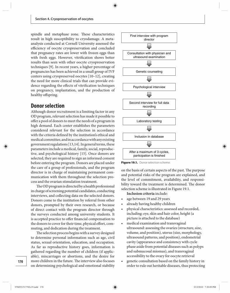
170
Section 4. Cryopreservation of oocytes
spindle and metaphase zone. Th ese characteristics result in high susceptibility to cryodamage. A meta-analysis conducted at Cornell University assessed the effi ciency of oocyte cryopreservation and concluded that pregnancy rates are lower with frozen eggs than with fresh eggs. However, vitrifi cation shows better results than seen with other oocyte cryopreservation techniques [ 9 ]. In recent years, a higher percentage of pregnancies has been achieved in a small group of IVF centers using cryopreserved oocytes [ 10 – 12 ], creating the need for more clinical trials that can provide evi-dence regarding the eff ects of vitrifi cation techniques on pregnancy, implantation, and the production of healthy off spring.
Donor selection Although donor recruitment is a limiting factor in any OD program, relevant selection has made it possible to off er a pool of donors to meet the needs of a program in high demand. Each center establishes the parameters considered relevant for the selection in accordance with the criteria defi ned by the institution’s ethical and medical committee, and in accordance with any existing government regulations [ 13 , 14 ]. In general terms, these parameters include a medical, family, social, reproduc-tive, and psychological history [ 15 ]. Once donors are selected, they are required to sign an informed consent before entering the program. Donors are placed under the care of a group of professionals, and the program director is in charge of maintaining permanent com-munication with them throughout the selection pro-cess and the ovarian stimulation treatment.
Th e OD program is directed by a health professional in charge of screening potential candidates, conducting interviews, and collecting data on the selected donors. Donors come to the institution by referral from other donors, prompted by their own research, or because of direct contact with the program director through the surveys conducted among university students. It is accepted practice to off er fi nancial compensation to the donors to cover for their time, physical eff ort, com-muting, and dedication during the treatment.
Th e selection process begins with a survey designed to determine personal information such as age, civil status, sexual orientation, education, and occupation. As far as reproductive history goes, information is gathered regarding the number of children (if applic-able), miscarriages or abortions, and the desire for more children in the future. Th e interview also focuses on determining psychological and emotional stability
on the basis of certain aspects of the past. Th e purpose and potential risks of the program are explained, and the level of commitment, availability, and responsi-bility toward the treatment is determined. Th e donor selection scheme is illustrated in Figure 19.1 .
Inclusion criteria include: age between 19 and 29 years • already having healthy children • physical characteristics: assessed and recorded, • including: eye, skin and hair color, height (a picture is attached to the database) medical examination and transvaginal • ultrasound: assessing the ovaries (structure, size, volume, and position), uterus (size, morphology, ultrasound patterns, and position), endometrial cavity (appearance and consistency with cycle phase aside from potential diseases such as polyps and submucosal miomas), and transvaginal accessibility to the ovary for oocyte retrieval genetic consultation based on the family history in • order to rule out heritable diseases, thus protecting
First interview with programdirector
Consultation with physician andultrasound examination
Genetic counseling
Psychological interview
Second interview for full datarecording
Laboratory testing
Inclusion in database
After a maximum of 3 cycles,participation is finished
Figure 19.1. Donor selection scheme.
9780521517782c19.indd 1709780521517782c19.indd 170 3/12/2010 7:20:09 PM3/12/2010 7:20:09 PM
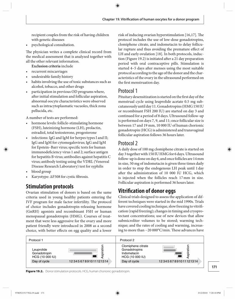
171
Chapter 19. Vitrifi cation of human oocytes for a donor program
recipient couples from the risk of having children with genetic diseases psychological consultation. •
Th e physician writes a complete clinical record from the medical assessment that is analyzed together with all the other relevant information.
Exclusion criteria include recurrent miscarriages • undesirable family history • habits involving the use of toxic substances such as • alcohol, tobacco, and other drugs participation in previous OD programs where, • aft er initial stimulation and follicular aspiration, abnormal oocyte characteristics were observed such as intracytoplasmatic vacuoles, thick zona pellucida, etc.
A number of tests are performed: hormone levels: follicle-stimulating hormone • (FSH), luteinizing hormone (LH), prolactin, estradiol, total testosterone, progesterone infections: IgG and IgM for herpes types I and II; • IgG and IgM for cytomegalovirus; IgG and IgM for Epstein–Barr virus; specifi c tests for human immunodefi ciency virus 1 and 2; surface antigen for hepatitis B virus; antibodies against hepatitis C virus; antibody testing using the VDRL (Venereal Disease Research Laboratory) test for syphilis blood group • Karyotype: ΔF508 for cystic fi brosis. •
Stimulation protocols Ovarian stimulation of donors is based on the same criteria used in young healthy patients entering the IVF program for male factor infertility. Th e protocol of choice includes gonadotropin-releasing hormone (GnRH) agonists and recombinant FSH or human menopausal gonadotropin (HMG). Courses of treat-ment that were less aggressive for the ovary and more patient friendly were introduced in 2008 as a second choice, with better eff ects on egg quality and a lower
risk of inducing ovarian hyperstimulation [ 16 , 17 ]. Th e protocol includes the use of low-dose gonadotropins, clomiphene citrate, and indometacin to delay follicu-lar rupture and thus avoiding the premature eff ect of LH and early ovulation [ 18 ]. In both protocols, induc-tion ( Figure 19.2 ) is initiated aft er a 21 day prepar ation period with oral contraceptive pills. Stimulation is started 4–5 days aft er menses using the most suitable protocol according to the age of the donor and the char-acteristics of the ovary in the ultrasound performed on the fi rst menstruation day.
Protocol 1 Pituitary desensitization is started on the fi rst day of the menstrual cycle using leuprolide acetate 0.5 mg sub-cutaneously until day 11. Gonadotropins (HMG 150 IU or recombinant FSH 200 IU) are started on day 3 and continued for a period of 8 days. Ultrasound follow-up is performed on days 7, 9, and 11; once follicular size is between 17 and 19 mm, 10 000 IU of human chorionic gonadotropin (HCG) is administered and transvaginal follicular aspiration follows 36 hours later.
Protocol 2 A daily dose of 100 mg clomiphene citrate is started on day 3 together with 150 IU HMG for 6 days. Ultrasound follow-up is done on day 6, and once follicles are 14 mm in size, 50 mg of indometacin is given three times daily in order to stop the endogenous LH peak until 1 day aft er the administration of 10 000 IU HCG, which is injected when the follicles reach 17 mm in size. Follicular aspiration is performed 36 hours later.
Vitrifi cation of donor eggs Clinical trials designed to assess the application of dif-ferent techniques were started in the mid 1990s. Trials have covered cooling technique, slow freezing to vitrifi -cation (rapid freezing); changes in timing and cryopro-tectant concentrations; use of new devices that allow submicroliter volumes to be stored; warming tech-nique; and the rates of cooling and warming, increas-ing to more than −20 000°C/min. Th ese advances have
Protocol 1 Protocol 2
LeuprolideGonadotropinHCG (10 000 IU)
GonadotropinsClomiphene citrate
IndometacinHCG (10 000 IU)
Day of cycle 12 34 5 67 8 910
Asp
iratio
n
Asp
iratio
n
11121314 Day of cycle 12 34 5 67 8 91011121314
Figure 19.2. Donor stimulation protocols. HCG, human chorionic gonadotropin.
9780521517782c19.indd 1719780521517782c19.indd 171 3/12/2010 7:20:10 PM3/12/2010 7:20:10 PM
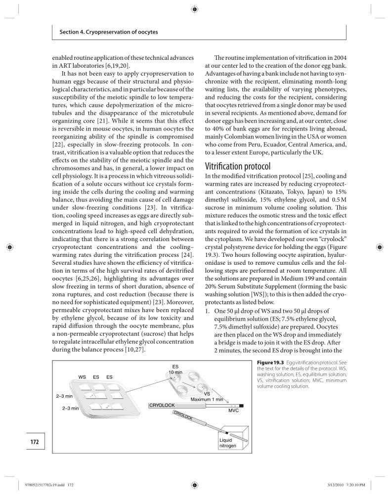
172
Section 4. Cryopreservation of oocytes
enabled routine application of these technical advances in ART laboratories [ 6 , 19 , 20 ].
It has not been easy to apply cryopreservation to human eggs because of their structural and physio-logical characteristics, and in particular because of the susceptibility of the meiotic spindle to low tempera-tures, which cause depolymerization of the micro-tubules and the disappearance of the microtubule organizing core [ 21 ]. While it seems that this eff ect is reversible in mouse oocytes, in human oocytes the reorganizing ability of the spindle is compromised [ 22 ], especially in slow-freezing protocols. In con-trast, vitrifi cation is a valuable option that reduces the eff ects on the stability of the meiotic spindle and the chromosomes and has, in general, a lower impact on cell physiology. It is a process in which vitreous solidi-fi cation of a solute occurs without ice crystals form-ing inside the cells during the cooling and warming balance, thus avoiding the main cause of cell damage under slow-freezing conditions [ 23 ]. In vitrifi ca-tion, cooling speed increases as eggs are directly sub-merged in liquid nitrogen, and high cryoprotectant concentrations lead to high-speed cell dehydration, indicating that there is a strong correlation between cryoprotectant concentrations and the cooling–warming rates during the vitrifi cation process [ 24 ]. Several studies have shown the effi ciency of vitrifi ca-tion in terms of the high survival rates of devitrifi ed oocytes [ 6 , 25 , 26 ], highlighting its advantages over slow freezing in terms of short duration, absence of zona ruptures, and cost reduction (because there is no need for sophisticated equipment) [ 23 ]. Moreover, permeable cryoprotectant mixes have been replaced by ethylene glycol, because of its low toxicity and rapid diff usion through the oocyte membrane, plus a non-permeable cryoprotectant (sucrose) that helps to regulate intracellular ethylene glycol concentration during the balance process [ 10 , 27 ].
Th e routine implementation of vitrifi cation in 2004 at our center led to the creation of the donor egg bank. Advantages of having a bank include not having to syn-chronize with the recipient, eliminating month-long waiting lists, the availability of varying phenotypes, and reducing the costs for the recipient, considering that oocytes retrieved from a single donor may be used in several recipients. As mentioned above, demand for donor eggs has been increasing and, at our center, close to 40% of bank eggs are for recipients living abroad, mainly Colombian women living in the USA or women who come from Peru, Ecuador, Central America, and, to a lesser extent Europe, particularly the UK.
Vitrifi cation protocol In the modifi ed vitrifi cation protocol [ 25 ], cooling and warming rates are increased by reducing cryoprotect-ant concentrations (Kitazato, Tokyo, Japan) to 15% dimethyl sulfoxide, 15% ethylene glycol, and 0.5 M sucrose in minimum volume cooling solution. Th is mixture reduces the osmotic stress and the toxic eff ect that is linked to the high concentrations of cryoprotect-ants required to avoid the formation of ice crystals in the cytoplasm. We have developed our own “cryolock” crystal polystyrene device for holding the eggs ( Figure 19.3 ). Two hours following oocyte aspiration, hyalur-onidase is used to remove cumulus cells and the fol-lowing steps are performed at room temperature. All the solutions are prepared in Medium 199 and contain 20% Serum Substitute Supplement (forming the basic washing solution [WS]); to this is then added the cryo-protectants as listed below. 1. One 50 μl drop of WS and two 50 μl drops of
equilibrium solution (ES; 7.5% ethylene glycol, 7.5% dimethyl sulfoxide) are prepared. Oocytes are then placed on the WS drop and immediately a bridge is made to join it with the ES drop. Aft er 2 minutes, the second ES drop is brought into the
Figure 19.3 Egg vitrifi cation protocol. See the text for the details of the protocol. WS, washing solution; ES, equilibrium solution; VS, vitrifi cation solution; MVC, minimum volume cooling solution.
WS ES ES
2–3 min
ES10 min
VSMaximum 1 min
MVCCRYOLOCK
CRYOLOCK
Liquidnitrogen
2–3 min
9780521517782c19.indd 1729780521517782c19.indd 172 3/12/2010 7:20:10 PM3/12/2010 7:20:10 PM
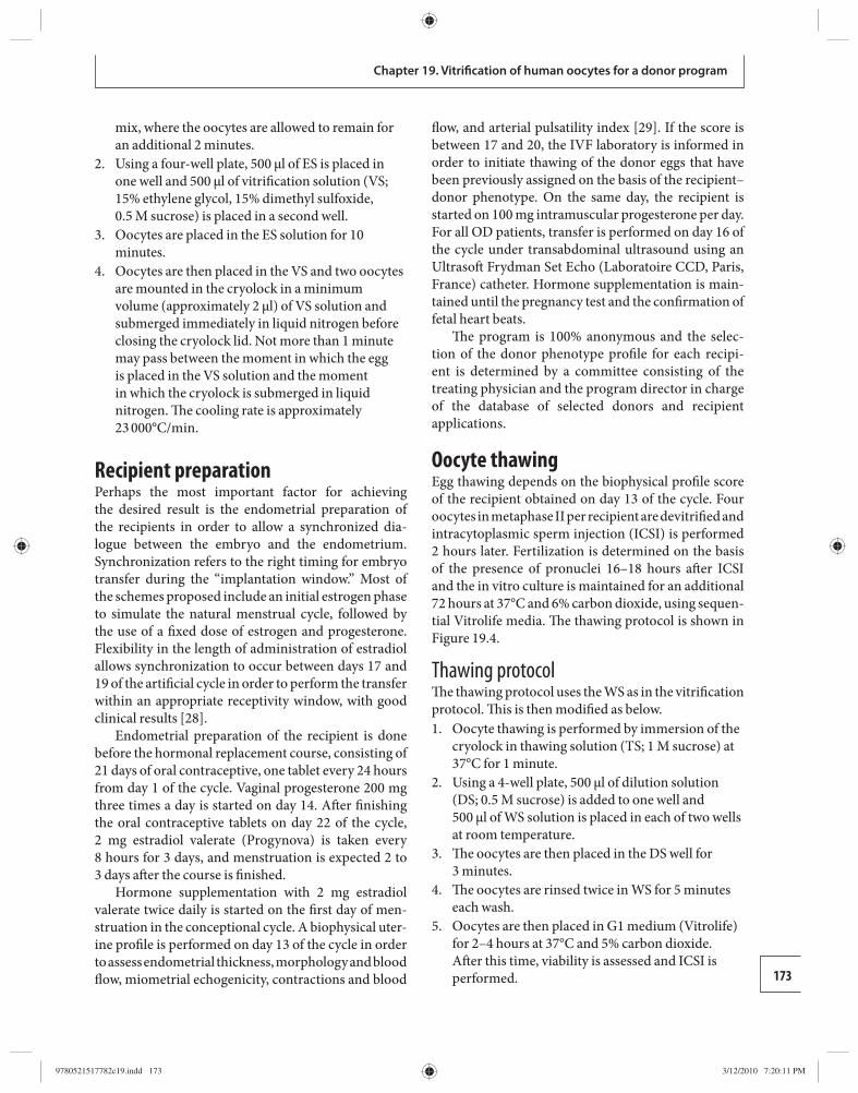
173
Chapter 19. Vitrifi cation of human oocytes for a donor program
mix, where the oocytes are allowed to remain for an additional 2 minutes.
2. Using a four-well plate, 500 μl of ES is placed in one well and 500 μl of vitrifi cation solution (VS; 15% ethylene glycol, 15% dimethyl sulfoxide, 0.5 M sucrose) is placed in a second well.
3. Oocytes are placed in the ES solution for 10 minutes.
4. Oocytes are then placed in the VS and two oocytes are mounted in the cryolock in a minimum volume (approximately 2 μl) of VS solution and submerged immediately in liquid nitrogen before closing the cryolock lid. Not more than 1 minute may pass between the moment in which the egg is placed in the VS solution and the moment in which the cryolock is submerged in liquid nitrogen. Th e cooling rate is approximately 23 000°C/min.
Recipient preparation Perhaps the most important factor for achieving the desired result is the endometrial preparation of the recipients in order to allow a synchronized dia-logue between the embryo and the endometrium. Synchronization refers to the right timing for embryo transfer during the “implantation window.” Most of the schemes proposed include an initial estrogen phase to simulate the natural menstrual cycle, followed by the use of a fi xed dose of estrogen and progesterone. Flexibility in the length of administration of estradiol allows synchronization to occur between days 17 and 19 of the artifi cial cycle in order to perform the transfer within an appropriate receptivity window, with good clinical results [ 28 ].
Endometrial preparation of the recipient is done before the hormonal replacement course, consisting of 21 days of oral contraceptive, one tablet every 24 hours from day 1 of the cycle. Vaginal progesterone 200 mg three times a day is started on day 14. Aft er fi nishing the oral contraceptive tablets on day 22 of the cycle, 2 mg estradiol valerate (Progynova) is taken every 8 hours for 3 days, and menstruation is expected 2 to 3 days aft er the course is fi nished.
Hormone supplementation with 2 mg estradiol valerate twice daily is started on the fi rst day of men-struation in the conceptional cycle. A biophysical uter-ine profi le is performed on day 13 of the cycle in order to assess endometrial thickness, morphology and blood fl ow, miometrial echogenicity, contractions and blood
fl ow, and arterial pulsatility index [ 29 ]. If the score is between 17 and 20, the IVF laboratory is informed in order to initiate thawing of the donor eggs that have been previously assigned on the basis of the recipient–donor phenotype. On the same day, the recipient is started on 100 mg intramuscular progesterone per day. For all OD patients, transfer is performed on day 16 of the cycle under transabdominal ultrasound using an Ultrasoft Frydman Set Echo (Laboratoire CCD, Paris, France) catheter. Hormone supplementation is main-tained until the pregnancy test and the confi rmation of fetal heart beats.
Th e program is 100% anonymous and the selec-tion of the donor phenotype profi le for each recipi-ent is determined by a committee consisting of the treating physician and the program director in charge of the database of selected donors and recipient applications.
Oocyte thawing Egg thawing depends on the biophysical profi le score of the recipient obtained on day 13 of the cycle. Four oocytes in metaphase II per recipient are devitrifi ed and intracytoplasmic sperm injection (ICSI) is performed 2 hours later. Fertilization is determined on the basis of the presence of pronuclei 16–18 hours aft er ICSI and the in vitro culture is maintained for an additional 72 hours at 37°C and 6% carbon dioxide, using sequen-tial Vitrolife media. Th e thawing protocol is shown in Figure 19.4 .
Thawing protocol Th e thawing protocol uses the WS as in the vitrifi cation protocol. Th is is then modifi ed as below. 1. Oocyte thawing is performed by immersion of the
cryolock in thawing solution (TS; 1 M sucrose) at 37°C for 1 minute.
2. Using a 4-well plate, 500 μl of dilution solution (DS; 0.5 M sucrose) is added to one well and 500 μl of WS solution is placed in each of two wells at room temperature.
3. Th e oocytes are then placed in the DS well for 3 minutes.
4. Th e oocytes are rinsed twice in WS for 5 minutes each wash.
5. Oocytes are then placed in G1 medium (Vitrolife) for 2–4 hours at 37°C and 5% carbon dioxide. Aft er this time, viability is assessed and ICSI is performed.
9780521517782c19.indd 1739780521517782c19.indd 173 3/12/2010 7:20:11 PM3/12/2010 7:20:11 PM
174
Section 4. Cryopreservation of oocytes
Results using cryopreserved oocytes in a donation program Th e assessment of the fi rst OD vitrifi cation program in Latin America has shown positive results and sig-nifi cant benefi ts for the recipients. Th ese results are measured on the basis of the survival rate aft er thaw-ing, fertilization, cleavage, pregnancy, losses, and birth of healthy babies. Some observations have been made regarding the behavior of the egg during the process of vitrifi cation and warming: less than 1% of the eggs are aff ected during vitrifi cation, perhaps by technica l problems during the process. In contrast, a larger number do not survive thawing, and there are many ongoing research studies on the eff ects of the technique on the organelles, cytoplasm, and zona pellucida [ 30 ]. It is yet to be determined whether this loss on thaw-ing can be reduced through additional adjustments to cryoprotectant concentrations and protocol timing, or by means of new devices that can ensure the integrity of the meiotic spindle and hence the normal chromo-some distribution in the cryopreserved oocyte.
One of the potential changes on thawing is the development of vacuoles, which are thought to be associated with other ultrastructural abnormalities leading to oocyte degeneration [ 31 ]. Formation of vacuoles is considered an important ooplasm charac-teristic associated with fertilization failure, or embryo arrest in those rare cases in which cleavage occurs [ 30 ]. It has also been demonstrated that zona hardening is an eff ect of the technique resulting from an increase in intracellular calcium, which causes exocytosis of the cortical granules located just under the plasma mem-brane; the enzymes contained in these granules pre-vent polyspermia as a calcium-dependent event [ 31 ]. Th is fertilization control system could be activated
prematurely during freezing, preventing normal fer-tilization [ 32 ] . Th e percentage of losses is also associ-ated in certain instances with high hormone levels in response to the stimulation regimen. For this reason, stimulation proto cols are being reassessed with the idea of reducing dosages or the days of stimulation. Th is might result in a lower number of better quality “competent” eggs capable of tolerating the freezing–thawing process and of resulting in embryo develop-ment and successful pregnancies.
Table 19.1 shows results from 4 years of experience with vitrifi ed oocytes used in the OD program as well as in supernumerary eggs aft er stimulation treatments. To date, 3232 eggs have been vitrifi ed. Of these, 810 are donor eggs. Th e number of OD transfers is higher com-pared with the supernumerary oocytes vitrifi ed from patients, considering that patients’ eggs are thawed only in the event that there is no pregnancy or thecouple wishes to come back for a second pregnancy aft er initial success.
So far, 185 donors have entered the program. Of them, 30% are active and the rest have completed the three donor cycles, have become pregnant, or have left the program voluntarily for personal reasons. Four to fi ve donors are scheduled every month in order to maintain approximately 40 eggs in stock in the bank.
Social and cultural eff ects Oocyte freezing can be used either by women who wish to postpone reproduction and enter the Mother of the Future program or for egg donation. In the former situation, women may want to postpone reproduction for medical reasons, such as radio- or chemotherapy or bilateral oophorectomy, or personal reasons, when they do not want to run the risk of genetic abnorma-lities of their off spring because of aging oocytes.
CRYOLOCK
DS3 min
WS5 min
WS5 min
ICSI
Culture media2–3 h, 37°C–5% CO2
Figure 19.4. Thawing protocol . See the text for the details of the protocol. WS, washing solution; DS, dilution solution; TS, thawing solution; ICSI, intracytoplasmic sperm injection.
9780521517782c19.indd 1749780521517782c19.indd 174 3/12/2010 7:20:11 PM3/12/2010 7:20:11 PM
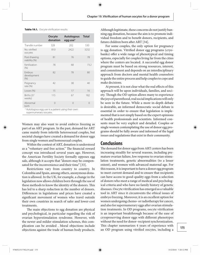
175
Chapter 19. Vitrifi cation of human oocytes for a donor program
Women may also want to avoid embryo freezing as part of an ART program. In the past, demand for ART came mainly from infertile heterosexual couples, but societal changes have created a demand for donor eggs from single women and homosexual couples.
Within the context of ART, donation is understood as a “voluntary and free action.” Th e fi nancial reward concept was introduced several years ago. However, the American Fertility Society formally opposes egg sale, although it accepts that “donors may be compen-sated for the inconvenience and their time” [ 33 ].
Restrictions vary from country to country. In Colombia and Spain, among others, anonymous dona-tion is allowed. In the UK, for example, a change in the legislation now allows children born through the use of these methods to know the identity of the donors. Th is has led to a sharp reduction in the number of donors. Diff erences in legislations and prices have created a signifi cant movement of women who travel outside their own countries in search of safer and lower-cost treatments.
Th e main objections to egg donation are physical and psychological, in particular regarding the risk of ovarian hyperstimulation syndrome. However, with the newer and milder stimulation schemes, this com-plication can be avoided . Moral objections include objections against the trade of human body products.
Although legitimate, these concerns do not justify ban-ning egg donation, because the aim is to promote indi-vidual freedom and to benefi t donors, recipients, and future children born aft er ART [ 34 ].
For some couples, the only option for pregnancy is egg donation. Vitrifi ed donor egg programs (cryo-banks) off er a wide range of phenotypical and timing options, especially for couples living far from the cities where the centers are located. A successful egg donor program must be based on strong resources, interest, and commitment and depends on an interdisciplinary approach from doctors and mental health counselors to guide the entire process and help couples to cope and make decisions.
At present, it is not clear what the real eff ects of this approach will be upon individuals, families, and soci-ety. Th ough the OD option allows many to experience the joys of parenthood, real social implications will only be seen in the future. While a more in-depth debate is desirable, an informed democratic social debate is essential in order to ensure that legislation is imple-mented that is not simply based on the expert opinions of health professionals and scientists. Informed con-sents must be very explicit and detailed. Couples or single women contemplating the use of donor egg pro-grams should be fully aware and informed of the legal issues and regulations that exist in their community.
Conclusions Th e demand for donor eggs from ART centers has been increasing steadily for several reasons, including pre-mature ovarian failure, low response to ovarian stimu-lation treatments, genetic abnormalities (to a lesser extent), and women with advanced maternal age. For this reason, it is important to have a donor egg program to meet current demand and to ensure that recipients can have access to good-quality eggs from a selection of donors who meet a range of medical and psycholog-ical criteria and who have no family history of genetic diseases. Oocyte vitrifi cation has emerged as a valuable tool in ART since it circumvents the ethical issues of embryo freezing. Moreover, it is an excellent option for women undergoing chemo- or radiotherapy for cancer, and also for supernumerary eggs aft er ovarian stimula-tion treatments. In OD programs, oocyte vitrifi cation is an important breakthrough because of the ease of cryopreserving donor eggs with diff erent phenotypes without the need for donor–recipient synchronization. Th is chapter summarizes 4 years of experience with an OD program using vitrifi ed oocytes, including a
Table 19.1. Oocyte vitrifi cation results
Oocyte donation
Autologous egg usea
Total
Transfer number 328 202 530
No. vitrifi ed oocytes
810 2422 3232
Post-thawing viability (%)
81 76 78
Fertilization (%)
78 68 73.2
Embryo development (%)
82 78 80
Pregnancy rate (%)
42 38 40
Losses (%) 15 17 16
Births (37 weeks)
115 67 182
Abnormal birth
0 0 0
a Autologous egg use is a patient using their own supernumerary oocytes .
9780521517782c19.indd 1759780521517782c19.indd 175 3/12/2010 7:20:12 PM3/12/2010 7:20:12 PM
176
Section 4. Cryopreservation of oocytes
description of the routine management of the program and the results obtained.
Acknowledgements We are grateful to Abby Moran, director of the Bio-Psychology Department, Angela Saa, embryologist and IVF laboratory in cheif, and Zulma Suarez, micro-biologist and director of the donor egg program at our institution for their valuable contributions to this chapter, based on their experience with the manage-ment and organization of the program.
References 1. Vegetti W , Marozzi A , Manfredini E , et al. Premature
ovarían failure . Mol Cell Endocrinol 2000; 161: 53 –57. 2. Portnoi MF , Aboura A , Tachdjian G , et al. Molecular
cytogenetic studies of Xq critical regions in premature ovarian failure patients . Hum Reprod 2006; 21: 2329 –2334.
3. Li XH , Chen SU , Zhang X , et al. Cryopreserved oocytes of infertile couples undergoing assisted reproductive technology could be an important sourse of oocyte donation: a clinical report of successful pregnancies . Hum Reprod 2005; 20: 3390 –3394.
4. Shulman A , Frenkel Y , Dor Jehoshua , et al. Th e best donor . Hum Reprod 1999; 14: 2493 –2499.
5. Check JH , Choe JK , Nazari , A , et al. Fresh embryo transfer is more eff ective than frozen for donor oocyte recipients but not for donor . Hum Reprod 2001; 16: 1403 –1408.
6. Fabbri R , Porcu E , Marsella T , et al. Human oocyte cryopreservation: new perspectives regarding oocyte survival . Hum Reprod 2001; 16: 411 –416.
7. Ghetler Y , Skutelsky E , Ben Nun I , et al. Human oocyte cryopreservation and the fate of cortical granules . Fertil Steril 2006; 86 : 210 –215.
8. Chen C . Pregnancy aft er human oocyte cryopreservation . Lancet 1986; i: 884 –886.
9. Okay K , Pelin C , Bang H . Effi ciency of oocyte cryopreservation: a meta-analysis . Fertil Steril 2006; 86 : 70 –79.
10. Chen SU , Lien YR , Chen HF , et al. Observational clinical follow-up of oocyte cryopreservation using a slow-freezing method with 1,2-propanediol plus sucrose followed by ICSI . Hum Reprod 2005; 20: 1975 –1980.
11. Katayama KP , Stehlik J , Kuwayama M , et al. High survival rate of vitrifi ed human oocytes results in clinical pregnancy . Fertil Steril 2003; 80: 223 –224.
12. Kuleshova L , Gianaroli L , Magli C , et al. Birth following vitrifi cation of a small number of human oocytes . Hum Reprod 1999; 14: 3077 –3079.
13. Lindheim SR , Frumovitz MV, Sauer M . Recruitment and screening policies and procedures used to establish a
paid donor oocyte registry . Hum Reprod 1998; 13: 2020 –2024.
14. Birminghan A . Guidelines for gamete and embryo donation . Fertil Steril 2006; 86 : S39 -S50.
15. Marina S , Expósito R , Marina F , et al. Oocyte donor selection from 554 candidates . Hum Reprod 1999; 14 : 2770 –2776.
16. Højgaard A , Ingerslev HJ , Dinesen J . Friendly IVF: patient opinions . Hum Reprod 2001; 16: 1391 –1396.
17. Inge GB , Brinsden PR , Elder KT . Oocyte number per live birth in IVF: were Steptoe and Edwards less wasteful? Hum Reprod 2005; 20: 588 –592.
18. Kadoch IJ . Spontaneous ovulation rate before oocyte retrieval in modifi ed natural cycle IVF with and without indomethacin . Reprod Bio Med Online 2008; 16: 245 –249.
19. Tucker MJ , Morton PC , Wright G , et al. Clinical application of human egg cryopreservation . Hum Reprod 1998; 13: 3156 –3159.
20. Porcu E , Fabbri R , Seracchioli R , et al. Birth of a healthy female aft er intracytoplasmatic sperm injection of cryopreserved human oocytes . Fertil Steril 1997; 68: 724 –726.
21. Mullen SF , Agca Y , Broermann DC , et al. Th e eff ects of osmotic stress on the metaphase II spindle of human oocytes, and the relevance to cryopreservation . Hum Reprod 2004; 5: 1148 –1154.
22. Pickering SJ , Braude PR , Johnson MH , et al. Transient cooling to room temperature can use irreversible disruption of the meiotic spindle in thehuman oocyte . Fertil Steril 1990; 54 : 102 –108.
23. Kuleshova LL , Lopata A . Vitrifi cation can be more favourable than slow cooling . Fertil Steril 2002; 78: 449 –454.
24. Chian R -C, Kuwayama M , Tan L, et al. Highsurvival rate of bovine oocyte matured in vitro following vitrifi cation . J Reprod Develop 2004; 50: 685 –696.
25. Kuwayama M . High effi cient vitrifi cation method for cryopreservation of human oocytes . Reprod Biomed Online 2005; 11: 300 –308.
26. Lucena E , Bernal D , Lucena C , et al. Successful ongoing pregnancies aft er vitrifi cation of oocytes . Fertil Steril 2006; 85: 108 –111.
27. Hong SW , Chung HM , Lim JM , et al. Improved human oocyte development aft er vitrifi cation: a comparison of thawing methods . Fertil Steril 1999; 72: 142 –146.
28. Michalas S , Loutradis D , Drakakis P , et al. A fl exible protocol for the induction of recipient endometrial cycle in an oocyte donation programme . Hum Reprod 1996; 11: 1063 –1066.
29. Applebaum M . Th e uterine biophysical profi le . Ultrasound Obstet Gynecol 1995; 5: 67 –68.
9780521517782c19.indd 1769780521517782c19.indd 176 3/12/2010 7:20:12 PM3/12/2010 7:20:12 PM

177
Chapter 19. Vitrifi cation of human oocytes for a donor program
30. Nottola SA , Macchiarelli , Coticchio G , et al. Ultrastructure of human mature oocyte aft er slow cooling cryopreservation using diff erent sucrose concentrations . Hum Reprod 2007; 22 : 1123 –1132.
31. Larman MG , Katz-Jaff e MG , Sheehan CB , Gardner DK . 1,2-Propanediol and the type of cryopreservation procedure adversely aff ect mouse oocyte physiology . Hum Reprod 2007; 22: 250 –259.
32. Ludwing M , Al-Hasani S , Felberbaum R , et al. New aspects of cryopreservation of oocyte and embryos in assisted reproduction and future perspectives . Hum Reprod 1999; 14: 162 –168.
33. American Fertility Society . Guidelines for oocyte donation . Fertil Steril 1993; 58: 55 –75.
34. Resnik DB . Th e commodifi cation of human reproductive materials . J Med Ethics 1998; 24: 338 –339.
9780521517782c19.indd 1779780521517782c19.indd 177 3/12/2010 7:20:12 PM3/12/2010 7:20:12 PM

178 Fertility Cryopreservation, ed. Ri-Cheng Chian and Patrick Quinn. Published by Cambridge University Press. Copyright © Cambridge University Press 2010.
Chapter
20 also, by extension, have an eff ect on the child. Examples include increased risk of preterm delivery (and its asso-ciated complications), pregnancy-induced hyperten-sion, gestational diabetes, and cesarean delivery [ 12 ] .
Th erefore, it is paramount that data on pregnancy, pregnancy outcome (including miscarriages or termin-ations of pregnancy for congenital or chromosomal abnormalities), pregnancy complications, and neo-natal outcomes are reported and collected. In October 2007, the American Society for Reproductive Medicine stated: “Oocyte cryopreservation, or egg freezing, remains an experimental procedure that should not be off ered or marketed as a means to defer reproductive aging. Currently available data are too limited to allow egg freezing to be considered an established medic al treatment. Because of the experimental nature of egg freezing, the ASRM recommends comprehensive counseling” [ 13 ]. Th is further highlights the impor-tance of disseminating all available information.
Th is chapter will discuss the current state of know-ledge regarding pregnancy outcomes in ART in general and then, more specifi cally, detail the current evidence regarding pregnancy and perinatal outcomes in preg-nancies conceived following oocyte cryopreservation. How these data, which come usually from short-term oocyte cryopreservation programs, may be applied to women undergoing these treatments for more long-term elective social reasons or for women who have undergone sterilizing chemotherapy and/or radio-therapy for cancer treatment will be discussed.
Pregnancy outcomes following assisted reproduction in general Miscarriage Relatively early in the development of ART, workers became aware of the early pregnancy loss – termed
Introduction Assisted reproductive technologies (ART) are prac-ticed all over the world, with clinics off ering these services in all developed countries and many parts of the developing world too. Results generated from the European registries by the European Society of Human Reproduction and Embryology (ESHRE) show that over 350 000 ART cycles every year are performed in a total of 28 diff erent countries [ 1 ]. With a live birth rate of approximately 20% across Europe, this accounts for 70 000 deliveries per year. In the USA alone, over 120 000 cycles ART are performed each year, resulting in over 35 000 deliveries per year [ 2 , 3 ]. With an inci-dence of 20% for twin pregnancies in ART, this means that approximately 200 000 babies are born per year as a result of ART. Since the advent of in vitro fertilization (IVF), well over 1 million babies have been born using this technology [ 4 ].
All this is a far cry from the scepticism and concerns that accompanied the early days of IVF, both in the lay press [ 5 , 6 ] and the medical literature [ 7 ]. However, this is exactly the position again now, with much written and many concerns expressed about fertility preservation in general and oocyte cryopreservation in particular [ 8 , 9 ].
Concerns are primarily two-fold. First, there are the unknown risks to any child or pregnancy conceived fol-lowing oocyte cryopreservation. Th e process of oocyte freezing (whether by conventional slow cryopreserva-tion or by vitrifi cation) is known to result in the loss of some oocytes and in meiotic spindle and chromosome confi guration abnormalities in the oocytes that survive [ 10 ]. Furthermore, the process of vitrifi cation, where the oocytes may be directly exposed to liquid nitrogen, also raises theoretical concerns of transmission of infective disease [ 11 ] . Second, there are increased pregnancy risks to the mother by virtue of her own status, whether age or having survived cancer and cancer treatment, that will
Obstetric and perinatal outcomes in pregnancies conceived following oocyte cryopreservation William Buckett and Ri-Cheng Chian
9780521517782c20.indd 1789780521517782c20.indd 178 3/12/2010 7:22:23 PM3/12/2010 7:22:23 PM
179
Chapter 20. Outcomes in pregnancies conceived following oocyte cryopreservation
chemical beta-human chorionic gonadotropin (β-hCG) abortion or biochemical pregnancy – as opposed to “later” clinical fi rst trimester miscarriage [ 14 ]. Ideally, ART programs should report clinical preg-nancy rates where a clinical pregnancy is defi ned using the World Health Organization/ESHRE defi nition of ultrasound evidence of an intrauterine pregnancy. Biochemical pregnancies are, therefore, pregnancies lost before this stage.
Because very early pregnancy losses may occur following spontaneous conception before awareness of pregnancy, it is impossible to determine the rate of biochemical pregnancy in the population as a whole. However, biochemical pregnancy loss rates following ART are typically around 15–20% [ 15 , 16 ], although rates from as low as 11% to as high as 35% have been reported [ 17 – 19 ]. Th e reasons for biochemical preg-nancy loss are unclear. However, maternal age (par-ticularly over 40 years), smoking, and poor embryo quality at transfer have been shown to be associated with an increased risk of biochemical pregnancy loss following ART [ 15 ].
Clinical miscarriage rates amongst spontaneously conceived pregnancies are reasonably consistent at 10–15% pregnancies [ 20 – 22 ]. However, there are many confounding variables that may aff ect couples under-going ART, predominantly maternal age [ 22 , 23 ], which is higher in women undergoing ART than in non-ART pregnancies. Also, other risks for miscarriage, such as polycystic ovary syndrome, are over-represented in couples undergoing ART. Polycystic ovary syndrome is associated with an increased risk of miscarriage, typ-ically reported at approximately 25%, either following spontaneous conception [ 24 ] or following ovulation induction [ 25 , 26 ]. Following ART, the clinical miscar-riage rate is typically around 15% [ 1 , 2 , 16 , 27 ], although it ranges from 10% to 45% depending on maternal age [ 27 ]. In women with polycystic ovary syndrome or in those who are overweight or obese, the clinical miscar-riage rate is higher [ 28 , 29 ].
In conclusion, clinical miscarriage rates seem to be similar following ART as in spontaneously conceived pregnancies, around 15%, although the higher mater-nal age and higher incidence of polycystic ovary syn-drome seen in women undergoing ART are associated with an increased risk of miscarriage. Biochemical loss is impossible to ascertain following spontaneously con-ceived pregnancies; however, rates of 15–20% are typ-ical following ART. Overall, therefore, approximately 30% of women with positive pregnancy tests following
ART will lose their pregnancies before the end of the fi rst trimester. Th is high loss rate highlights the importance of reporting live birth rates following ART rather than pregnancy rates or clinical pregnancy rates [ 30,31 ] .
Ectopic pregnancy Ectopic pregnancy rates have risen since the mid 1990s, although the reasons are unclear; improved and earlier diagnosis certainly has an eff ect. Currently, estimates are that ectopic pregnancy aff ects approximately 2% of all pregnancies. Although some earlier case series showed a higher incidence of ectopic pregnancy (at approximately 4%) following ART [ 32 , 33 ], most large prospective studies show a rate of 2.0–2.2%, which is similar to that of the general population [ 34 , 35 ]. Th is change is probably because tubal disease was the major indication for ART early in the development of ART.
Late pregnancy loss Th e incidence of late pregnancy loss (aft er 12 weeks of gestation) aft er ART, through mid-trimester miscar-riage, fetal death, or termination of pregnancy for chro-mosomal or other abnormality, is typically between 2 and 4% [ 27 , 36 ]. Th is is higher than that of spontaneously conceived pregnancies (typically around 1%) [ 37 , 38 ].
One reason for this is advancing maternal age, which is associated with a higher incidence of chromo-somal and other abnormalities and may result in mid-trimester termination of pregnancy. However, with the exception of case reports and case series, there are no comparative data for mid-trimester termination of pregnancy following ART. Maternal age is also asso-ciated with a higher spontaneous late pregnancy loss rate, both following spontaneous conceptions and fol-lowing ART [ 27 ].
Congenital abnormality Initial studies comparing babies born following IVF or intracytoplasmic sperm injection (ICSI) with spontane-ously conceived controls suggested no increase in the incidence of congenital abnormalities [ 39 – 41 ], although the magnitude of these were in the hundreds rather than the thousands or tens of thousands that are needed in order to determine any genuine diff erence. However, since the beginning of this century, some two decades aft er the beginnings of IVF, an increasing number of papers have compared ART babies with spontaneously conceived controls [ 42 , 43 ] and there have been several systematic reviews [ 44 ]; these show an increase in the risk of congenital abnormality associated with ART. Current
9780521517782c20.indd 1799780521517782c20.indd 179 3/12/2010 7:22:25 PM3/12/2010 7:22:25 PM
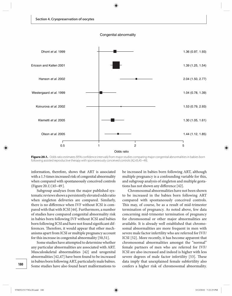
180
Section 4. Cryopreservation of oocytes
information, therefore, shows that ART is associated with a 1.5 times increased risk of congenital abnormality when compared with spontaneously conceived controls ( Figure 20.1 ) [ 45 – 49 ].
Subgroup analyses from the major published sys-tematic reviews shows a persistently elevated odds ratio when singleton deliveries are compared. Similarly, there is no diff erence when IVF without ICSI is com-pared with that with ICSI [ 44 ]. Furthermore, a number of studies have compared congenital abnormality risk in babies born following IVF without ICSI and babies born following ICSI and have not found signifi cant dif-ferences. Th erefore, it would appear that other mech-anisms apart from ICSI or multiple pregnancy account for this increase in congenital abnormality [ 50, 51 ].
Some studies have attempted to determine whether any particular abnormalities are associated with ART. Musculoskeletal abnormalities [ 42 ] and urogenital abnormalities [ 42 , 47 ] have been found to be increased in babies born following ART, particularly male babies. Some studies have also found heart malformations to
be increased in babies born following ART, although multiple pregnancy is a confounding variable for this, and subgroup analysis of singleton and multiple gesta-tions has not shown any diff erence [ 42 ].
Chromosomal abnormalities have not been shown to be increased in the babies born following ART compared with spontaneously conceived controls. Th is may, of course, be as a result of mid-trimester termin ation of pregnancy. As noted above, few data concerning mid-trimester termination of pregnancy for chromosomal or other major abnormalities are available. It is already well established that chromo-somal abnormalities are more frequent in men with severe male factor infertility who are referred for IVF/ICSI [ 52 ]. More recently, it has become apparent that chromo somal abnormalities amongst the “normal” female partners of men who are referred for IVF/ICSI are also increased and indeed is higher with less severe degrees of male factor infertility [ 53 ]. Th ese data imply that unexplained female subfertility also confers a higher risk of chromosomal abnormality.
Congenital abnormality
0.5 1 2
Odds ratio
5
Olson et al. 2005 1.44 (1.12, 1.85)
Klemetti et al. 2005 1.30 (1.05, 1.61)
Koivurova et al. 2002 1.53 (0.79, 2.93)
Westergaard et al. 1999 1.04 (0.78, 1.39)
Hanson et al. 2002 2.04 (1.50, 2.77)
Ericson and Kallen 2001 1.39 (1.25, 1.54)
Dhont et al. 1999 1.36 (0.97, 1.93)
Figure 20.1. Odds ratio estimates (95% confi dence intervals) from major studies comparing major congenital abnormalities in babies born following assisted reproductive therapy with spontaneously conceived controls [42,43,45–49].
9780521517782c20.indd 1809780521517782c20.indd 180 3/12/2010 7:22:25 PM3/12/2010 7:22:25 PM
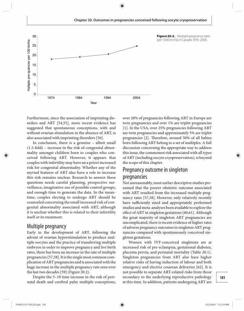
181
Chapter 20. Outcomes in pregnancies conceived following oocyte cryopreservation
Furthermore, since the association of imprinting dis-orders and ART [ 54 , 55 ], more recent evidence has suggested that spontaneous conceptions, with and without ovarian stimulation in the absence of ART, is also associated with imprinting disorders [ 5 6].
In conclusion, there is a genuine – albeit small (1.5-fold) – increase in the risk of congenital abnor-mality amongst children born to couples who con-ceived following ART. However, it appears that couples with infertility may have an a priori increased risk for congenital abnormality. Whether any of the myriad features of ART also have a role to increase this risk remains unclear. Research to answer these questions needs careful planning, prospective sur-veillance, imaginative use of possible control groups, and enough time to generate the data. In the mean-time, couples electing to undergo ART should be counseled concerning the small increased risk of con-genital abnormality associated with ART, although it is unclear whether this is related to their infertility itself or its treatment.
Multiple pregnancy Early in the development of ART, following the advent of ovarian hyperstimulation to produce mul-tiple oocytes and the practice of transferring multiple embryos in order to improve pregnancy and live birth rates, there has been an increase in the rate of multiple pregnancies [ 5 7, 5 8]. It is the single most common com-plication of ART pregnancies and is associated with the huge increase in the multiple pregnancy rate seen over the last two decades [ 5 9] ( Figure 20.2 ).
Despite the 5–10 time increase in the risk of peri-natal death and cerebral palsy multiple conceptions,
over 20% of pregnancies following ART in Europe are twin pregnancies and over 1% are triplet pregnancies [ 1 ]. In the USA, over 25% pregnancies following ART are twin pregnancies and approximately 5% are triplet pregnancies [ 2 ]. Th erefore, around 50% of all babies born following ART belong to a set of multiples. A full discussion concerning the appropriate way to address this issue, the commonest risk associated with all types of ART (including oocyte cryopreservation), is beyond the scope of this chapter.
Pregnancy outcome in singleton pregnancies Not unreasonably, most earlier descriptive studies pre-sumed that the poorer obstetric outcome associated with ART resulted from the increased multiple preg-nancy rates [ 5 7, 5 8]. However, only relatively recently have suffi ciently sized and appropriately performed studies and meta-analyses been available to explore the eff ect of ART in singleton gestations [ 60, 61 ]. Although the great majority of singleton ART pregnancies are uncomplicated, there is recent evidence of higher rates of adverse pregnancy outcomes in singleton ART preg-nancies compared with spontaneously conceived sin-gleton gestations.
Women with IVF-conceived singletons are at increased risk of pre-eclampsia, gestational diabetes, placenta previa, and perinatal mortality ( Table 20.1 ). Singleton pregnancies from ART also have higher relative risks of having induction of labour and both emergency and elective cesarean deliveries [ 62 ]. It is not possible to separate ART-related risks from those secondary to the underlying reproductive pathology at this time. In addition, patients undergoing ART are
0
5
Hal
tiple
pre
-qua
ncie
r pe
r 10
0 bi
rths
10
15
20
25
30
Year
1974 1984 1994 2004
Figure 20.2. Multiple pregnancy rates (per 1000 births) in Canada 1974–2004.
9780521517782c20.indd 1819780521517782c20.indd 181 3/12/2010 7:22:25 PM3/12/2010 7:22:25 PM
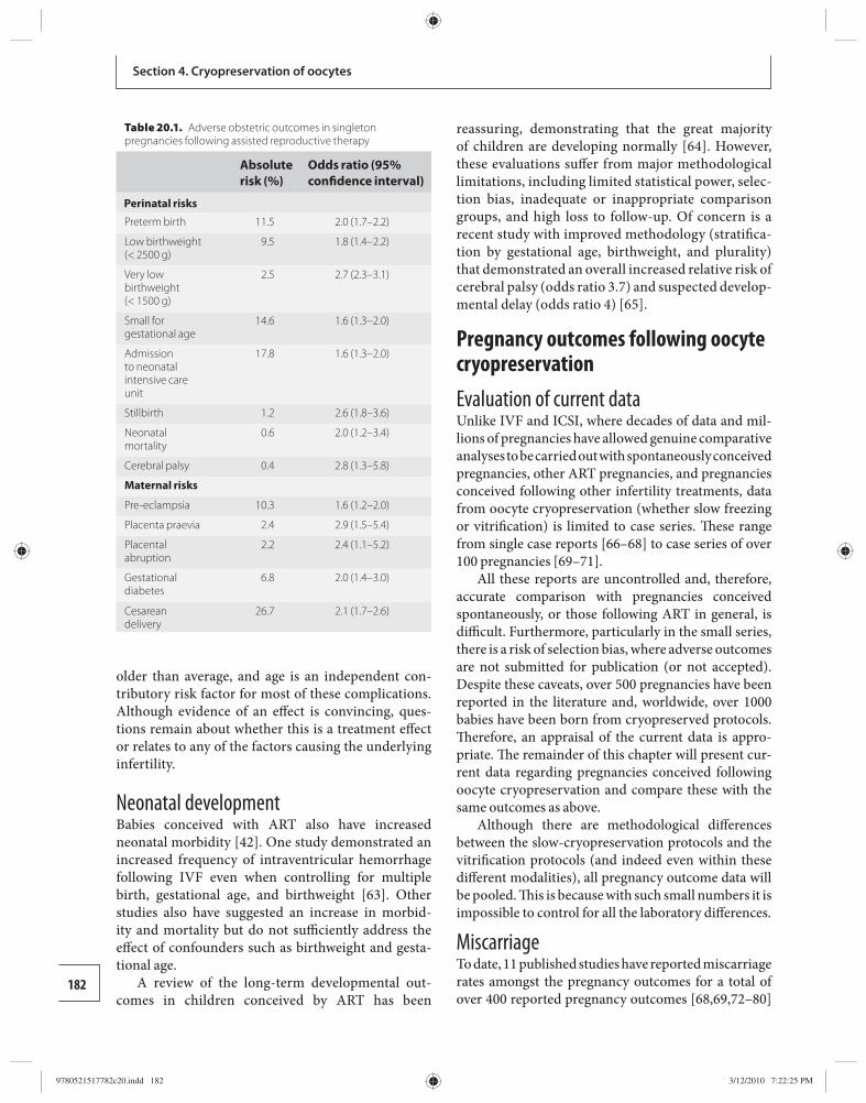
182
Section 4. Cryopreservation of oocytes
older than average, and age is an independent con-tributory risk factor for most of these complications. Although evidence of an eff ect is convincing, ques-tions remain about whether this is a treatment eff ect or relates to any of the factors causing the underlying infertility.
Neonatal development Babies conceived with ART also have increased neonatal morbidity [ 42 ]. One study demonstrated an increased frequency of intraventricular hemorrhage following IVF even when controlling for multiple birth, gestational age, and birthweight [ 63 ]. Other studies also have suggested an increase in morbid-ity and mortality but do not suffi ciently address the eff ect of confounders such as birthweight and gesta-tional age.
A review of the long-term developmental out-comes in children conceived by ART has been
reassuring, demonstrating that the great majority of children are developing normally [ 6 4]. However, these evaluations suff er from major methodological limitations, including limited statistical power, selec-tion bias, inadequate or inappropriate comparison groups, and high loss to follow-up. Of concern is a recent study with improved methodology (stratifi ca-tion by gestational age, birthweight, and plurality) that demonstrated an overall increased relative risk of cerebral palsy (odds ratio 3.7) and suspected develop-mental delay (odds ratio 4) [ 6 5].
Pregnancy outcomes following oocyte cryopreservation Evaluation of current data Unlike IVF and ICSI, where decades of data and mil-lions of pregnancies have allowed genuine comparative analyses to be carried out with spontaneously conceived pregnancies, other ART pregnancies, and pregnancies conceived following other infertility treatments, data from oocyte cryopreservation (whether slow freezing or vitrifi cation) is limited to case series. Th ese range from single case reports [ 6 6– 6 8] to case series of over 100 pregnancies [ 6 9– 71 ].
All these reports are uncontrolled and, therefore, accurate comparison with pregnancies conceived spontaneously, or those following ART in general, is diffi cult. Furthermore, particularly in the small series, there is a risk of selection bias, where adverse outcomes are not submitted for publication (or not accepted). Despite these caveats, over 500 pregnancies have been reported in the literature and, worldwide, over 1000 babies have been born from cryopreserved protocols. Th erefore, an appraisal of the current data is appro-priate. Th e remainder of this chapter will present cur-rent data regarding pregnancies conceived following oocyte cryopreservation and compare these with the same outcomes as above.
Although there are methodological diff erences between the slow-cryopreservation protocols and the vitrifi cation protocols (and indeed even within these diff erent modalities), all pregnancy outcome data will be pooled. Th is is because with such small numbers it is impossible to control for all the laboratory diff erences.
Miscarriage To date, 11 published studies have reported miscarriage rates amongst the pregnancy outcomes for a total of over 400 reported pregnancy outcomes [ 6 8, 6 9, 72 – 80 ]
Table 20.1. Adverse obstetric outcomes in singleton pregnancies following assisted reproductive therapy
Absolute risk (%)
Odds ratio (95% confi dence interval)
Perinatal risks
Preterm birth 11.5 2.0 (1.7–2.2)
Low birthweight (< 2500 g)
9.5 1.8 (1.4–2.2)
Very low birthweight(< 1500 g)
2.5 2.7 (2.3–3.1)
Small for gestational age
14.6 1.6 (1.3–2.0)
Admission to neonatal intensive care unit
17.8 1.6 (1.3–2.0)
Stillbirth 1.2 2.6 (1.8–3.6)
Neonatal mortality
0.6 2.0 (1.2–3.4)
Cerebral palsy 0.4 2.8 (1.3–5.8)
Maternal risks
Pre-eclampsia 10.3 1.6 (1.2–2.0)
Placenta praevia 2.4 2.9 (1.5–5.4)
Placental abruption
2.2 2.4 (1.1–5.2)
Gestational diabetes
6.8 2.0 (1.4–3.0)
Cesarean delivery
26.7 2.1 (1.7–2.6)
9780521517782c20.indd 1829780521517782c20.indd 182 3/12/2010 7:22:25 PM3/12/2010 7:22:25 PM

183
Chapter 20. Outcomes in pregnancies conceived following oocyte cryopreservation
( Table 20.2 ). Th e overall miscarriage rate is 15%, which, as noted above, is comparable with both the rate of clinical miscarriage following spontaneous pregnancy [ 20 – 22 ] and that following ART [ 1 , 2 , 16 , 27 ].
Although these data are uncontrolled and there may be diff erences in the diagnosis of clinical preg-nancy and, therefore, miscarriage rates, it is reas-suring that the rate is very similar to that reported following spontaneous conception and following ART. Th erefore, based on the reported data, it is unlikely that oocyte cryopreservation is associated with an increase in this risk, although further data need to be collected.
Ectopic pregnancy So far there have been two cases of ectopic pregnancy reported in pregnancies conceived following oocyte cryopreservation [ 6 8, 72 ]. Although a bias in report-ing is likely, if all the studies noted above were counted, this would give an ectopic pregnancy rate of 0.5%. Th is is lower than that normally quoted for rates of ectopic
pregnancy in spontaneously conceived pregnancies and ART pregnancies in general [ 33 , 34 ]. However, rates of 0.5% have been reported in some groups undergoing ART, such as recipients of oocyte donation [ 27 ], so it could represent a true incidence.
Late pregnancy loss and oocyte cryopreservation Th ere has only been one report of a late pregnancy loss (at 16 weeks of gestation) in a pregnancy con-ceived by oocyte cryopreservation [ 81 ]. Th ere was another single case of miscarriage where the karyo-type of the fetus was abnormal (45XO), although the gestational age of the miscarried fetus is not given [ 72 ]. Whether these cases represent a genuine inci-dence of this, already rare, outcome of pregnancy is unclear at present.
Congenital abnormality Although many of the case reports and case series have stated that the live birth was a healthy infant, only one study has specifi cally presented data concerning con-genital abnormalities in pregnancies following oocyte cryopreservation [ 7 1]. In this study, 200 children from 165 pregnancies (28 sets of twins and 137 singleton pregnancies) were examined ( Table 20.3 ). A total of fi ve congenital abnormalities in fi ve diff erent infants were reported (two ventriculoseptal defect, one club foot, one skin haemangioma, and one congenital biliary atresia). Th is gives an incidence of congenital abnormality aff ect-ing 2.5% pregnancies, which is in the same order of magnitude as spontaneously conceived pregnancies and pregnancies conceived following ART [ 42 – 44 ].
As noted above under ART, there are multiple con-founding variables when rates of congenital abnor-mality are compared, and continued surveillance and registration of all infants born following oocyte
Table 20.2. Pregnancy loss following oocyte cryopreservation
No. pregnancies
No. pregnancy losses
Miscarriage rate (%)
Quintans et al. [72]
6 3 50
Konc et al. [73]
7 1 14
De Geyter et al. [68]
3 1 33
Borini et al. [69]
267 37 14
Borini et al. [74]
18 3 17
Sher et al. [75]
14 2 14
Kim et al. [76]
16 3 19
Chang et al. [77]
16 2 13
Chian et al. [78]
17 2 12
Chian et al. [78 ]
5 1 20
Yoon et al. [79]
13 2 15
Antinori et al. [80]
39 8 21
Total 421 65 15
Table 20.3. Congenital abnormalities in pregnancies following oocyte cryopreservation
Absolute risk (%)
Total babies 200
Abnormalities
Biliary atresia 1
Club foot 1
Skin hemangioma 1
Ventriculoseptal defect 2
Abnormality rate 2.5% Source: adapted from Chian et al. [ 71 ].
9780521517782c20.indd 1839780521517782c20.indd 183 3/12/2010 7:22:26 PM3/12/2010 7:22:26 PM
184
Section 4. Cryopreservation of oocytes
cryopreservation, such as that proposed by the HOPE registry [ 8 2], are needed.
Multiple pregnancy As noted above, twin and higher-order multiple preg-nancy is the most frequent complication associated with ART in general [ 1 , 2 ], and pregnancies conceived fol-lowing oocyte cryopreservation are no diff erent [ 6 6– 80 , 83 – 8 5]. In reported series, multiple pregnancy rates range from 0% to over 50% [ 6 8, 83 ]. Most series of preg-nancies following oocyte cryopreservation that report multiple pregnancy [ 31,68 , 6 9, 72 – 7 5, 7 9– 80 , 83, 84 ] have twin pregnancy rates at between 15 and 30%. Th is is comparable to that reported in the large ART registries.
Th e risks and consequences of multiple pregnancy are beyond the scope of this chapter. However, as cryopreservation techniques improve and pregnancy and implantation rates increase, just as in ART using fresh oocytes, the number of embryos transferred will decrease and the incidence of multiple pregnancy fol-lowing oocyte cryopreservation will fall.
Obstetric outcomes As twin pregnancies are associated with an increase in preterm delivery and low-birthweight babies, these outcomes should be analyzed only in single-ton pregnancies. Only one study so far has presented these outcomes [ 71 ]. Th is study presents data from 137 singleton pregnancies delivered in three diff erent countries (Canada, Columbia, and Mexico). When the data were pooled, the incidence of delivery before 34 weeks was 4% (6/137) and the incidences of low birth-weight (< 2500 g) and very-low birthweight (< 1500 g) babies were 17% (24/137) and 1% (1/137), respectively. Th is is similar to the rates of these outcomes reported in singleton pregnancies following ART [ 60 , 61 , 62 ].
Although these limited data are encouraging, it is diffi cult to draw defi nitive conclusions as the “back-ground” rates of preterm delivery and low-birthweight babies vary greatly amongst diff erent populations. As noted above, registration, either separately or within existing ART registries, will allow continued monitor-ing of the incidence of these and other risks.
Neonatal and child development As yet, no data have been reported concerning neonatal and child development following oocyte cryopreserva-tion. Th e primary reason for this is that the number of babies born, until recently has been very small. Th erefore, these data will become available over the coming years.
Interpretation of outcomes after oocyte cryopreservation As this review of obstetric and perinatal outcomes in pregnancies conceived following oocyte cryopreser-vation has shown, broadly speaking, the risks are very similar to ART in general. Th is is not surprising for several reasons. First, the process of oocyte cryop-reservation involves similar hormonal treatments (leading to increased estrogen levels in the follicular phase and supraphysiological progesterone level s in the luteal phase and early pregnancy), similar laboratory conditions for fertilization of the thawed oocytes and culture of the resultant embryos, and similar physical interventions (such as ICSI, assisted hatching, and the number of embryos transferred). Consequently, if any of these interventions have an eff ect on pregnancy in ART they will have a similar impact in oocyte cryopreservation. Second, most series reported so far in the literature have included oocyte cryopreservation for various reasons for coup-les undergoing infertility treatment. Th erefore any a priori increased risk will be the same or similar to that in couples undergoing ART with fresh oocytes. Finally, the history of oocyte cryopreservation is still young and much, much more data are needed if small diff erences are to be identifi ed.
As noted elsewhere in this volume, oocyte cryo-preservation is indicated for preserving fertility in women who will undergo potentially sterilizing treat-ment, oft en to cure fatal diseases such as breast cancer or lymphoma. Th erefore, pregnancy outcome in these women may diff er from that in women who present with infertility. Although there are many reports of pregnancies following oocyte cryopreservation, there have been few in cancer survivors and some have used gestational surrogates [ 8 5].
Furthermore, there has been an increasing trend for women to cryopreserve oocytes as “an insurance” against failing ovarian function. Th ese situations are likely to involve cryopreservation for several years. Again, it is diffi cult to predict pregnancy outcome in otherwise fertile women who may have had oocytes cryopreserved for over 5 years. As yet, there are only a few published cases of live birth aft er such long-term oocyte cryopreservation [ 8 6]. As noted above, there may also be diff erences in laboratory/freezing proto-cols that could aff ect obstetric and perinatal outcome. However, well-designed long-term trials are needed to answer this and, in fact, many other questions regard-ing ART in general.
9780521517782c20.indd 1849780521517782c20.indd 184 3/12/2010 7:22:26 PM3/12/2010 7:22:26 PM
185
Chapter 20. Outcomes in pregnancies conceived following oocyte cryopreservation
Conclusions Certainly the data from over 500 pregnancies follow-ing oocyte cryopreservation presented in the world literature are reassuring. Pregnancy outcomes and congenital abnormalities are similar to those reported following spontaneous conceptions and following ART in general.
Obviously, the dataset is still too small to deter-mine small diff erences, and couples should be coun-seled that, although the data so far are reassuring, there may be small increases in the risk of poor pregnancy outcome that cannot be detected at present. Similarly, some abnormalities or complications may become apparent at a much later date. However, the likelihood of either of these outcomes is small.
National or international registries (either within established ART registries or as stand-alone registries) for oocyte cryopreservation should be established and continue to collect treatment, pregnancy outcome, and childhood data over the next several decades.
References 1. Andersen AN , Goosens V , Gianaroli L , et al. Assisted
reproductive technology in Europe, 2003. Results generated from European registries by ESHRE . Hum Reprod 2007; 22 : 1513–1525 .
2. Society for Assisted Reproductive Technology ; American Society for Reproductive Medicine. Assisted reproductive technology in the United States: 2001 results generated from the American Society for Reproductive Medicine/Society for Assisted Reproductive Technology Registry . Fertil Steril 2007; 87 : 1253–1256 .
3. Society for Assisted Reproductive Technology. SART Clinical Summary Report 2005 . Birmingham, AL : Society for Assisted Reproductive Technology , www.sartcorsonline.com/rpt.
4. Adamson GD , de Mouzon J , Lancaster P , for the International Committee for Monitoring Assisted Reproductive Technology , . World collaborative report on in vitro fertilization, 2000 . Fertil Steril 2006; 85 : 1586–1622 .
5. Auerbach S. Fears aired in test tube baby debate . Washington Post 1974; Jul 21 : A1 .
6. Wright P. Less risk of test-tube baby being born with brain damage, doctor says . Th e Times 1974; Sept 7 : 4 .
7. Fitzgerald J. Veto in vitro, or turn off the tube . Can Fam Physician 1978; 24 : 816 .
8. Harwood K. Egg freezing: a breakthrough for reproductive autonomy ? Bioethics 2009; 23 : 39–46 .
9. Goold I , Savalescu J. In favour of freezing eggs for non-medical reasons . Bioethics 2009; 23: 47–58 .
10. Cao YX , Xing Q , Li L , et al. Comparison of survival and embryonic development in human oocytes cryopreserved by slow-freezing and vitrifi cation . Fertil Steril 2009 ; 92 : 1306–1311
11. Tedder RS , Zukerman MA , Goldstone AH , et al. Hepatitis transmission from contaminated cryopreservation tank . Lancet 1995; 346 : 137–140 .
12. Chan BC , Lao TT. Eff ect of parity and advanced maternal age on obstetric outcome . Int J Gynaecol Obstet 2008; 102 : 237–241 .
13. American Society for Reproductive Medicine. ASRM Urges Caution, Strong Counseling for Women Seeking Egg Freezing . Washington, DC: American Society for Reproductivve Medicine, October 16, 2007, http://www.asrm.org/Media/Press/AM07urgecautioneggfreezing.html.
14. Jones HW , Jr. , Acosta AA , Andrews MC , et al. What is a pregnancy? A question for programs of in vitro fertilization . Fertil Steril 1983; 40 : 728–733 .
15. Winter E , Wang J , Davies MJ , Norman R. Early pregnancy loss following assisted reproductive technology treatment . Hum Reprod 2002; 17 : 3220–3223 .
16. Buckett WM , Chian R-C , Dean NL , et al. Pregnancy loss in pregnancies conceived following in vitro oocyte maturation (IVM), conventional in vitro fertilization (IVF), and intra-cytoplasmic sperm injection (ICSI) . Fertil Steril 2008; 90 : 546–550 .
17. Bellver J , Albert C , Labarta E , Pellicer A. Early pregnancy loss in women stimulated with gonadotropin-releasing hormone antagonist protocols according to oral contraceptive pill pretreatment . Fertil Steril 2007; 87 : 1089–1101 .
18. European Recombinant LH Study Group . Human recombinant luteinizing hormone is as eff ective as, but safer than, urinary human chorionic gonadotropin in inducing fi nal follicular maturation and ovulation in in vitro fertilization procedures: results of a multicenter double-blind study . J Clin Endocrinol Metab 2001; 86 : 2607–2618 .
19. Barlow P , Lejeune B , Puissant F , et al. Early pregnancy loss and obstetrical risk aft er in-vitro fertilization and embryo replacement . Hum Reprod 1988; 3 : 671–675 .
20. Miller JF , Williamson E , Glue J , et al. Fetal loss aft er implantation: a prospective study . Lancet 1980; ii : 554–556 .
21. Regan L , Braude P , Trembath PL. Infl uence of past reproductive performance on risk of spontaneous abortion . Br Med J 1989; 299 : 541–545 .
22. Nybo Andersen AM , Wohlfahrt J , Christens P , et al. Maternal age and fetal loss: population based register linkage . Br Med J 2000; 320 : 1708–1712 .
23. Buckett W , Regan L. Sporadic and recurrent miscarriage. In Shaw RA , Soutter WP , Stanton SL , eds. Gynaecology , 3rd edn. London : Churchill Livingstone , 2003, pp. 343–360.
9780521517782c20.indd 1859780521517782c20.indd 185 3/12/2010 7:22:26 PM3/12/2010 7:22:26 PM
186
Section 4. Cryopreservation of oocytes
24. Rotterdam ESHRE/ASRM-sponsored PCOS Consensus Workshop Group . Revised 2003 consensus on diagnostic criteria and long-term health risks related to polycystic ovary syndrome . Fertil Steril 2004; 81 : 19–25 .
25. Homburg R. Pregnancy complications in PCOS . Best Pract Res Clin Endocrinol Metab 2006; 20 : 281–292 .
26. MacGregor AH , Johnson JE , Bunde CA. Further clinical experience with clomiphene citrate . Fertil Steril 1968; 19 : 616–622 .
27. Farr SL , Schieve LA , Jamieson DJ. Pregnancy loss among pregnancies conceived through assisted reproductive technology, United States, 1999–2002 . Am J Epidemiol 2007; 165 : 1380–1386 .
28. Wang JX , Davies MJ , Norman RJ. Polycystic ovarian syndrome and the risk of spontaneous abortion following assisted reproductive technology treatment . Hum Reprod 2001; 16 : 2606–2609 .
29. Maheshwari A , Stofb erg L , Bhattacharya S. Eff ect of overweight and obesity on assisted reproductive technology: a systematic review . Hum Reprod Update 2007; 13 : 433–444 .
30. Centers for Disease Control and Prevention . Current trends in ectopic pregnancy-United States, 1990–1992 . MMWR Surveill Summ 1995; 44 : 46–48 . 31. Fosas N , Marina F , Torres PJ , et al. Th e births of fi ve Spanish babies from cryopreserved donated oocytes . Hum Reprod 2003; 18 : 1417–1421
31. Fosas N , Marina F , Torres PJ , et al. Th e births of fi ve Spanish babies from cryopreserved donated oocytes . Hum Reprod 2003; 18 : 1417–1421 .
32. Ribic-Pucelj M , Tomazevic T , Vogler A , Meden-Vrtovec H. Risk factors for ectopic pregnancy aft er in vitro fertilization and embryo transfer . J Assist Reprod Genet 1995; 12 : 594–598 .
33. Dubuisson JB , Aubriot FX , Mathieu L , et al. Risk factors for ectopic pregnancy in 556 pregnancies aft er in vitro fertilization: implications for preventive management . Fertil Steril 1991; 56 : 686–690 .
34. Verhulst G , Camus M , Bollen N , Van Steirteghem A , Devroey P. Analysis of the risk factors with regard to the occurrence of ectopic pregnancy aft er medically assisted procreation . Hum Reprod 1993; 8 : 1284–1287 .
35. Clayton HB , Schieve LA , Peterson HB , et al. Ectopic pregnancy risk with assisted reproductive technology procedures . Obstet Gynecol 2006; 107 : 595–604 .
36. Buckett WM , Chian R-C , Dean NL , et al. Pregnancy loss in pregnancies conceived aft er in vitro oocyte maturation, conventional in vitro fertilization, and intracytoplasmic sperm injection . Fertil Steril 2008; 90 : 546–550 .
37. Brigham SA , Conlon C , Farquharson RG. A longitudinal study of pregnancy outcome following idiopathic recurrent miscarriage . Hum Reprod 1999; 14 : 2868–2871 .
38. Confi dential Enquiry into Maternal and Child Health . Stillbirth, Neonatal and Post-neonatal Mortality 2000–2003, England, Wales, and Northern Ireland . London : RCOG Press , 2005 .
39. Morin NC , Wirth FH , Johnson DH , et al. Congenital malformations and psychosocial development in children conceived by in vitro fertilization . J Pediatr 1989; 115 : 222–227 .
40. Sutcliff e AG , D’Souza SW , Cadman J , et al. Minor congenital anomalies, major congenital malformations and development in children conceived from cryopreserved embryos . Hum Reprod 1995; 10 : 3332–3337 .
41. Isaksson R , Gissler M , Tiitinen A. Obstetric outcome among women with unexplained infertility aft er IVF: a matched case-control study . Hum Reprod 2002; 17 : 1755–1761 .
42. Klemetti R , Gissier M , Sevon T , et al. Children born aft er assisted fertilization have an increased rate of major congenital anomalies . Fertil Steril 2005; 84 : 1300–1307 .
43. Olson CK , Keppler-Noreuil KM , Romitti PA , et al. In vitro fertilization is associated with an increase in major birth defects . Fertil Steril 2005; 84 : 1308–1315 .
44. Hansen M , Bower C , Milne E , de Klerk N , Kurinczuk JJ. Assisted reproductive technologies and the risk of birth defects: a systematic review . Hum Reprod 2005; 20 : 328–338 .
45. Dhont M , de Sutter P, Ruyssinck G, Martens G, Bekaert A. Perinatal outcome of pregnancies aft er assisted reproduction: a case-control study . Am J Obstet Gynecol 1999 ; 181 : 688–695.
46. Hansen M , Kurinczuk JJ, Bower C, Webb S . Th e risk of major birth defects aft er intracytoplasmatic sperm injection and in vitro fertilization . N Engl J Med 2002 ; 346 : 725–730 .
47. Ericson A , Kallen B. Congenital malformations in infants born aft er IVF: a population-based study . Hum Reprod 2001; 16 : 504–509 .
48. Westergaard HB , Johansen AM, Erb K, Andersen AN. Danish national in-vitro fertilization registry 1994 and 1995: a controlled study of births, malformations and cytogenetic fi ndings. Hum Reprod 1999 ; 14 : 1896–1902 .
49. Koivurova S , Kartikainen AL, Gissler M , et al. Neonatal outcome and congenital malformations in children born aft er in-vitro fertilization . Hum Reprod 2002 ; 17 : 1391–1398.
50. Bonduelle M , Liebaers I , Deketelaere V , et al. Neonatal data on a cohort of 2889 infants born aft er ICSI (1991–1999) and of 2995 infants born aft er IVF (1983–1999) . Hum Reprod 2002; 17 : 671–694 .
51. Place I , Englert Y. A prospective longitudinal study of the physical, psychomotor, and intellectual development of singleton children up to 5 years who were conceived by intracytoplasmic sperm injection compared with
9780521517782c20.indd 1869780521517782c20.indd 186 3/12/2010 7:22:26 PM3/12/2010 7:22:26 PM
187
Chapter 20. Outcomes in pregnancies conceived following oocyte cryopreservation
children conceived spontaneously and by in vitro fertilization . Fertil Steril 2003; 80 : 1388–1397 .
52. Buckett W , Aird L , Luckas M , et al. Intracytoplasmic sperm injection. Karyotyping should be done before treatment . Br Med J 1996; 313 : 1334 .
53. Gekas J , Th epot F , Turleau C . For the Association des Cytogeneticiens de Langue Francaise. Chromosomal factors of infertility in candidate couples for ICSI: an equal risk of constitutional aberrations in women and men . Hum Reprod 2001; 16 : 82–90 .
54. Gosden R , Trasler J , Lucifero D , Faddy M. Rare congenital disorders, imprinted genes, and assisted reproductive technology . Lancet 2003; 361 : 1974–1977 .
55. Chang AS , Moley KH , Wangler M , Feinberg AP , Debaun MR. Association between Beckwith–Wiedemann syndrome and assisted reproductive technology: a case series of 19 patients . Fertil Steril 2005; 83 : 349–354 .
56. Ludwig M , Katalinic A , Gross S , et al. Increased prevalence of imprinting defects in patients with Angelman syndrome born to subfertile couples . J Med Genet 2005; 42 : 2890–2891 .
57. Wood C , Trounson A , Leeton JF , et al. Clinical features of eight pregnancies resulting from in vitrofertilization and embryo transfer . Fertil Steril 1982; 38 : 22–29 .
58. Mushin D , Spensley J , Barreda-Hanson M. Children of IVF . Clin Obstet Gynacol 1985; 12 : 865–876 .
59. Bissonnette F , Cohen J , Collins J , et al. Incidence and complications of multiple gestation in Canada: proceedings of an expert meeting . Reprod Biomed Online 2007; 14 : 773–790 .
60. Helmerhorst FM , Perquin DAM , Donker D , Keirse MJNC. Perinatal outcome of singletons and twins aft er assisted conception: a systematic review of controlled studies . Br Med J 2004; 328 : 261 .
61. Jackson RA , Gibson KA , Wu YW , Croughan MS. Perinatal outcomes in singletons following in vitro fertilization: a meta-analysis . Obstet Gynecol 2004; 103 : 551–563 .
62. Rimm AA , Katayama AC , Diaz M , Katayama KP. A meta-analysis of controlled studies comparing major malformation rates in IVF and ICSI infants with naturally conceived children . J Assist Reprod Genet 2004; 21 : 437–443 .
63. Linder N , Haskin O , Levit O , et al. Risk factors for intraventricular hemorrhage in very low birth weight premature infants: a retrospective case–control study . Pediatrics 2003; 111 : 590–595 .
64. Olivennes F , Fanchin R , Ledee N , et al. Perinatal outcome and developmental studies on children born aft er IVF . Hum Reprod Update 2002; 8 : 117–128 .
65. Stromberg B , Dahlquist G , Ericson A , et al. Neurological sequelae in children born aft er in-vitro fertilisation: a population-based study . Lancet 2002; 359: 461–465 .
66. De Geyter M , Steimann S , Holzgreve W , De Geyter C. First delivery of healthy off spring aft er freezing and thawing of oocytes in Switzerland. Swiss Med Wkly 2007; 137 : 443–447 .
67. Chian R-C , Gilbert L , Huang JY , et al. Live birth aft er vitrifi cation of in vitro matured human oocytes . Fertil Steril 2009; 91 : 372–376 .
68. Kyono K , Nakajo Y , Nishinaka C , et al. A birth from the transfer of a single vitrifi ed-warmed blastocyst using intracytoplasmic sperm injection with calcium ionophore oocyte activation in a globozoospermic patient . Fertil Steril 2009; 91 : 931.e7–e11 .
69. Borini A , Bianchi V , Bonu M A , et al. Evidence based clinical outcome of oocyte slow cooling . Reprod BioMed Online 2007; 15 : 175–181 .
70. Albani E , Barbieri J , Novara PV , et al. Oocyte cryopreservation . Placenta 2008; 29 (Suppl B): 143–146 .
71. Chian R-C , Huang JY , Tan SL , et al. Obstetric and perinatal outcome in 200 infants conceived from vitrifi ed oocytes . Reprod Biomed Online 2008; 16 : 608–610 .
72. Quintans CJ , Donaldson MJ , Bertolino MV , Pasqualini RS. Birth of two babies using oocytes that were cryopreserved in a choline-based freezing medium . Hum Reprod 2002; 17 : 3149–3152 .
73. Konc J , Kanyo K , Varga E , Kriston R , Cseh S. Births resulting from oocyte cryopreservation using a slow freezing protocol with propanediol and sucrose . Syst Biol Reprod Med 2008; 54 : 205–210 .
74. Borini A , Sciajno R , Bianchi V , et al. Clinical outcome of oocyte cryopreservation aft er slow cooling with a protocol utilizing a high sucrose concentration . Hum Reprod 2006; 21 : 512–517 .
75. Sher G , Keskintepe L , Mukaida T , et al. Selective vitrifi cation of euploid oocytes markedly improves survival, fertilization and pregnancy-generating potential . Reprod Biomed Online 2008; 17 : 524–529 .
76. Kim TJ , Laufer LR , Hong SW. Vitrifi cation of oocytes produces high pregnancy rates when carried out in fertile women . Fertil Steril 2010; 93 : 467–474 .
77. Chang CC , Shapiro DB , Bernal DP , et al. Human oocyte vitrifi cation: in-vivo and in-vitro maturation outcomes . Reprod Biomed Online 2008; 17 : 684–688 .
78. Chian R-C , Huang JY , Gilbert L , et al. Obstetric outcomes following vitrifi cation of in vitro and in vivo matured oocytes . Fertil Steril 2009; 91 : 2391–2398 .
79. Yoon TK , Lee DR , Cha SK , et al. R ate of human oocytes and pregnancy outcome aft er vitrifi cation using slush nitrogen in assisted reproductive technologies . Fertil Steril 2007; 88 : 952–956 .
80. Antinori M , Licata E , Dani G , et al. Cryotop vitrifi cation of human oocytes results in high survival rate and healthy deliveries . Reprod Biomed Online 2007; 14 : 72–79 .
9780521517782c20.indd 1879780521517782c20.indd 187 3/12/2010 7:22:26 PM3/12/2010 7:22:26 PM
188
Section 4. Cryopreservation of oocytes
81. Li Y , Chen ZJ , Yang HJ , et al. Comparison of vitrifi cation and slow-freezing of human day 3 cleavage stage embryos: post-vitrifi cation development and pregnancy outcomes . Zhonghua Fu Chan Ke Za Zhi 2007; 42 : 753–755 .
82. Ezcurra D , Rangnow J , Craig M , Schertz J. Th e HOPE Registry: fi rst US registry for oocyte cryopreservation . Reprod Biomed Online 2008; 17 : 743–744 .
83. Chen SU , Lien YR , Tsai YY , et al. S uccessful pregnancy occurred from slowly freezing human oocytes using the regime of 1.5 mol/l 1,2-propanediol with 0.3 mol/l sucrose . Hum Reprod 2002; 17 : 1412 .
84. Ding J , Rana N , Dmowski WP. Successful pregnancies aft er oocyte cryopreservation with slow-freezing method: a report of 3 cases . J Reprod Med 2008; 53 : 813–820 .
85. Yang D , Brown SE , Nguyen K , et al. Live birth aft er the transfer of human embryos developed from cryopreserved oocytes harvested before cancer treatment . Fertil Steril 2007; 87 : 1469 .
86. Parmegiani L , Fabbri R , Cognigni GE , et al. Blastocyst formation, pregnancy, and birth derived from human oocytes cryopreserved for 5 years . Fertil Steril 2008; 90 : 2014 .
9780521517782c20.indd 1889780521517782c20.indd 188 3/12/2010 7:22:26 PM3/12/2010 7:22:26 PM
189Fertility Cryopreservation, ed. Ri-Cheng Chian and Patrick Quinn. Published by Cambridge University Press. Copyright © Cambridge University Press 2010.
Section 5Chapter
21 respectively [ 8 ]. A combination of various che-motherapeutic agents further increases gonadal toxicity. Aft er chemotherapy with MOPP/ABV (mechlo-rethamine, vincristine [Oncovin], procarbazine, pred-nisone/doxorubicin (adriamycin), bleomycin, and vinblastine [or vincristine]) hybrid chemotherapy, Schilsky et al. [ 9 ] found that amenorrhea developed in 89% and 20% of patients over and under 25 years of age at the time of treatment, respectively. Th e median age of patients who became amenorrheic aft er ther-apy was signifi cantly higher than that of patients who maintained normal menses (26 years versus 20 years; p = 0.008).
Radiotherapy Abdominal ionizing radiation that is oft en used in combination with alkylating agents oft en induces POF, rendering patients infertile in almost 100% of cases. Indeed, for radiotherapy, it has been stated that a dose of 5–20 Gy administered to the ovary is suffi cient to completely impair gonadal function [ 10 ] whatever the age of the patient. Th e dose of radiation required to destroy 50% of the oocyte reserve has been found to be < 2 Gy [ 11 ]. Moreover, uterine irradiation at a young age reduces adult uterine volume [ 12 ]. Radiation doses between 14 and 30 Gy have been reported to result in uterine dysfunction [ 13 , 14 ]. Th e practitioner should be aware of this eff ect of radiotherapy on the uterus, which could interfere with the implantation capacity of embryos.
Chemo/radiotherapy Intensive chemotherapy and total body irradiation required before bone marrow transplantation consti-tute the treatment combination presenting the greatest risk of POF. Indeed, the high doses of chemotherapy (commonly using the highly cytotoxic cyclophospha-mide/busulfan regimen) and/or radiotherapy lead to subsequent ovarian failure in almost all cases, children
Introduction According to previous reports, around 700 000 new cancers are expected every year in the female popu-lation in the USA [ 1 ] and 8% of these women will be under 40 years of age. By 2010, it is estimated that 1 in every 250 people in the adult population will be a sur-vivor of childhood cancer [ 2 ].
Advances in the diagnosis and treatment of child-hood, adolescent, and adult cancer have greatly increased the life expectancy of premenopausal women with cancer. Indeed, aggressive chemotherapy and radiotherapy, and bone marrow transplantation, can cure more than 90% of girls aff ected by childhood malignancies [ 3 ], and this has resulted in a growing population of adolescent and adult long-term sur-vivors of childhood malignancies [ 2 ] who may expe-rience infertility problems from induced premature ovarian failure (POF).
Gonadotoxicity Chemotherapy Th e ovaries are very sensitive to cytotoxic treatment, especially to alkylating agents [ 4 ]. Table 21.1 lists the main agents used in chemotherapy in terms of the level of their risk for causing gonadal dysfunction.
Cyclophosphamide is the agent most commonly implicated in causing damage to oocytes and gran-ulosa cells in a dose-dependent manner [ 5 , 6 ]. Th is follicular destruction generally results in loss of both endocrine and reproductive function, depending on the dose and the age of the patient. Indeed, Larsen et al. reported a four-fold increased risk of POF in teen-agers treated for cancer, and a 27-fold increased risk in women aged 21–25 years [ 7 ]. Complete amenor-rhea was reported aft er a dose of 5 g cyclophosphamide in women over 40 years of age, and aft er doses of 9 gand 20 g in women aged 30–40 and 20–30 years,
Cryopreservation of ovarian tissue Cryopreservation of ovarian tissue: an overview Jacques Donnez , Pascale Jadoul , Olivier Donnez , Anne-Sophie van Eyck , Jean Squiffl et , and Marie-Madeleine Dolmans
9780521517782c21.indd 1899780521517782c21.indd 189 3/12/2010 12:24:08 PM3/12/2010 12:24:08 PM
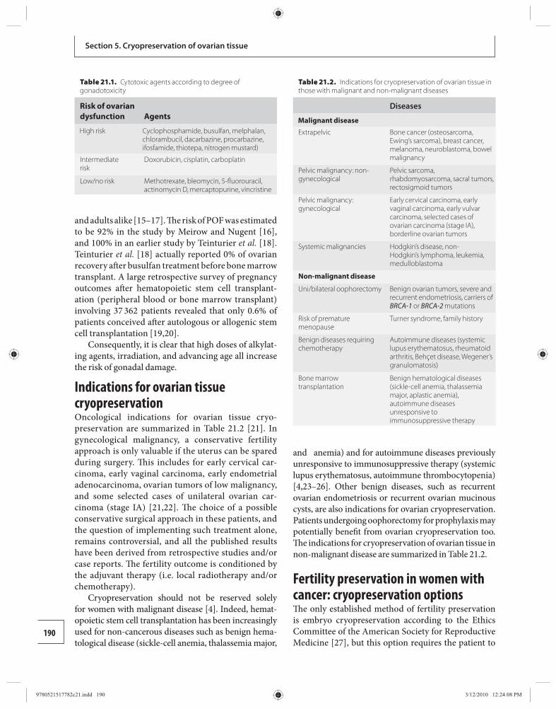
190
Section 5. Cryopreservation of ovarian tissue
and adults alike [ 15 – 17 ]. Th e risk of POF was estimated to be 92% in the study by Meirow and Nugent [ 16 ], and 100% in an earlier study by Teinturier et al. [ 18 ]. Teinturier et al. [ 18 ] actually reported 0% of ovarian recovery aft er busulfan treatment before bone marrow transplant. A large retrospective survey of pregnancy outcomes aft er hematopoietic stem cell transplant-ation (peripheral blood or bone marrow transplant) involving 37 362 patients revealed that only 0.6% of patients conceived aft er autologous or allogenic stem cell transplantation [ 19 , 20 ].
Consequently, it is clear that high doses of alkylat-ing agents, irradiation, and advancing age all increase the risk of gonadal damage.
Indications for ovarian tissue cryopreservation Oncological indications for ovarian tissue cryo-preservation are summarized in Table 21.2 [ 21 ]. In gynecological malignancy, a conservative fertility approach is only valuable if the uterus can be spared during surgery. Th is includes for early cervical car-cinoma, early vaginal carcinoma, early endometrial adenocarcinoma, ovarian tumors of low malignancy, and some selected cases of unilateral ovarian car-cinoma (stage IA) [ 21 , 22 ]. Th e choice of a possible conservative surgical approach in these patients, and the question of implementing such treatment alone, remains controversial, and all the published results have been derived from retrospective studies and/or case reports. Th e fertility outcome is conditioned by the adjuvant therapy (i.e. local radiotherapy and/or chemotherapy).
Cryopreservation should not be reserved solely for women with malignant disease [ 4 ]. Indeed, hemat-opoietic stem cell transplantation has been increasingly used for non-cancerous diseases such as benign hema-tological disease (sickle-cell anemia, thalassemia major,
and anemia) and for autoimmune diseases previously unresponsive to immunosuppressive therapy (systemic lupus erythematosus, autoimmune thrombocytopenia) [ 4 , 23 – 26 ]. Other benign diseases, such as recurrent ovarian endometriosis or recurrent ovarian mucinous cysts, are also indications for ovarian cryopreservation. Patients undergoing oophorectomy for prophylaxis may potentially benefi t from ovarian cryopreservation too. Th e indications for cryopreservation of ovarian tissue in non-malignant disease are summarized in Table 21.2 .
Fertility preservation in women with cancer: cryopreservation options Th e only established method of fertility preservation is embryo cryopreservation according to the Ethics Committee of the American Society for Reproductive Medicine [ 27 ], but this option requires the patient to
Table 21.1. Cytotoxic agents according to degree of gonadotoxicity
Risk of ovarian dysfunction Agents
High risk Cyclophosphamide, busulfan, melphalan, chlorambucil, dacarbazine, procarbazine, ifosfamide, thiotepa, nitrogen mustard)
Intermediate risk
Doxorubicin, cisplatin, carboplatin
Low/no risk Methotrexate, bleomycin, 5-fl uorouracil, actinomycin D, mercaptopurine, vincristine
Table 21.2. Indications for cryopreservation of ovarian tissue in those with malignant and non-malignant diseases
Diseases
Malignant disease
Extrapelvic Bone cancer (osteosarcoma, Ewing’s sarcoma), breast cancer, melanoma, neuroblastoma, bowel malignancy
Pelvic malignancy: non-gynecological
Pelvic sarcoma, rhabdomyosarcoma, sacral tumors, rectosigmoid tumors
Pelvic malignancy: gynecological
Early cervical carcinoma, early vaginal carcinoma, early vulvar carcinoma, selected cases of ovarian carcinoma (stage IA), borderline ovarian tumors
Systemic malignancies Hodgkin’s disease, non-Hodgkin’s lymphoma, leukemia, medulloblastoma
Non-malignant disease
Uni/bilateral oophorectomy Benign ovarian tumors, severe and recurrent endometriosis, carriers of BRCA-1 or BRCA-2 mutations
Risk of premature menopause
Turner syndrome, family history
Benign diseases requiring chemotherapy
Autoimmune diseases (systemic lupus erythematosus, rheumatoid arthritis, Behçet disease, Wegener’s granulomatosis)
Bone marrow transplantation
Benign hematological diseases (sickle-cell anemia, thalassemia major, aplastic anemia), autoimmune diseases unresponsive to immunosuppressive therapy
9780521517782c21.indd 1909780521517782c21.indd 190 3/12/2010 12:24:08 PM3/12/2010 12:24:08 PM
191
Chapter 21. Cryopreservation of ovarian tissue: an overview
be of pubertal age, have a partner or use donor sperm, and be able to undergo a cycle of ovarian stimulation, which is not possible when chemotherapy has to be ini-tiated immediately or when stimulation is contraindi-cated according to the type of cancer.
Crypreservation of oocytes can be performed in postpubertal patients who are able to undergo a stimu-lation cycle, but the eff ectiveness of this technique is still low, with delivery rates from 1 to 5% for frozen–thawed oocytes using the slow-cooling techniques [ 28 ]. Since the recent introduction of oocyte vitrifi cation, delivery rates have almost doubled per thawed oocyte [ 29 ].
Ovarian tissue cryopreservation For patients who need immediate chemotherapy, cryopreservation of ovarian tissue is the only possible option [ 21 , 30 – 34 ]. Th e main aim of this strategy is to re-implant cortical ovarian tissue into the pelvic cavity (orthotopic site) or a heterotopic site like the forearm or abdominal wall once treatment is completed and the patient is disease free [ 4 , 26 , 34 – 46 ].
Lessons from xenografting cryopreserved human ovarian tissue Human ovarian tissue can be successfully cryopre-served, showing good survival and function aft er thaw-ing, as indicated by a review of all relevant studies from 1996 to 2005 [ 47 ]. Th e fi rst case of cryopreservation of human ovarian tissue was described in 1996. Th e choice of a cryoprotectant with maximum permeation capacity but minimum toxicity and ice crystal forma-tion potential is specifi c to each cell and tissue type [ 48 ]. Th erefore, in the ovary, it is a compromise between the stroma, follicular cells, and oocytes [ 47 ]. On the basis of current knowledge, the standard method for human ovarian cryopreservation is slow freezing in a medium containing human serum albumin, and using propan-ediol, dimethyl sulfoxide (DMSO), or ethylene glycol as a cryoprotectant, with or without sucrose [ 47 ].
All reports on human ovarian tissue graft ing to mice [ 4 , 49 ] that have studied the implantation site have shown peritoneal transplantation, either under the peritoneum [ 50 ] or under the kidney capsule [ 51 , 52 ], to be better than subcutaneous transplanta-tion in terms of follicular survival and development. Aft er xenograft ing cryopreserved human ovarian tis-sue into the kidney capsule of immunodefi cient (with severe combined immunodefi ciency) mice, Oktay et al. [ 53 ] achieved follicular development up to the antral stage, and Gook et al. [ 54 , 55 ] obtained metaphase II
stage oocytes. Th e ovulatory capacity of frozen–thawed human follicles in xenograft s was evidenced by the for-mation of morphologically normal corpora lutea and elevated progesterone levels in the immunodefi cient mice [ 54 , 56 ]. Very few data are available on the fi nal maturation of follicles in xenograft s and the quality of oocytes obtained. It has, therefore, not been established whether human oocytes matured in xenograft s are ultrastructurally normal and functionally competent.
Experimental studies have indicated a fall in the number of primordial follicles in graft ed tissue, which could be caused by hypoxia and the delay before re-implanted cortical tissue becomes revascularized. Th e loss of primordial follicles in cryopreserved ovarian tissue aft er transplantation is estimated to be at least 50–65% in some studies [ 50 ].
The crucial issue of revascularization Van Eyck et al. [ 57 ] recently characterized the oxygen environment in human ovarian xenograft s in the early post-graft ing period (until day 21) using electron para-magnetic resonance oximetry. Th is technique allows sensitive, non-invasive, and repeated measurement of the partial pressure of oxygen in vivo. Before day 5, graft s were exposed to hypoxia. From day 5 to day 10, progressive reoxygenation was observed, suggesting an active process of graft revascularization.
Using a combined method of perfusion study and double immunohistochemical staining of human and murine vessels, the same team evaluated the revas-cularization process of human ovarian tissue in this model (Van Eyck et al. [ 58 ]). On day 5, reperfusion of ovarian graft s was initiated by host angiogenesis, as evidenced by the appearance of murine neovessels pen-etrating from the periphery and colocalized with per-fused areas. By day 10, the center of the fragments was perfused and ovarian graft angiogenesis contribut ed to the vascular pattern of the ovarian transplants.
Host and graft angiogenesis, therefore, both appear to contribute to post-transplantation vascular behav-ior and could be potential targets to improve the mech-anisms leading to perfusion of graft s, with the aim of reducing the avascular period.
Autotransplantation of cryopreserved human ovarian tissue Th ere have been numerous reported cases of autotransplantation of cryopreserved ovarian tis-sue, either to an orthotopic or heterotopic site [ 4 , 25 – 26 , 35 – 46 , 59 , 60 ].
9780521517782c21.indd 1919780521517782c21.indd 191 3/12/2010 12:24:08 PM3/12/2010 12:24:08 PM
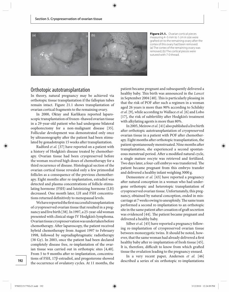
192
Section 5. Cryopreservation of ovarian tissue
Orthotopic autotransplantation In theory, natural pregnancy may be achieved via orthotopic tissue transplantation if the fallopian tubes remain intact. Figure 21.1 shows transplantation of ovarian cortical fragments to the remaining ovary.
In 2000, Oktay and Karlikaya reported laparo-scopic transplantation of frozen–thawed ovarian tissue in a 29-year-old patient who had undergone bilateral oophorectomy for a non-malignant disease [ 35 ]. Follicular development was demonstrated only once by ultrasonography aft er the patient had been stimu-lated by gonadotropin 15 weeks aft er transplantation.
Radford et al. [ 37 ] have reported on a patient with a history of Hodgkin’s disease treated by chemother-apy. Ovarian tissue had been cryopreserved before the woman received high doses of chemotherapy for a third recurrence of disease. Histological section of the ovarian cortical tissue revealed only a few primordial follicles as a consequence of the previous chemother-apy. Eight months aft er re-implantation, estradiol was detected and plasma concentrations of follicle-stimu-lating hormone (FSH) and luteinizing hormone (LH) decreased. One month later, LH and FSH concentra-tions returned defi nitively to menopausal levels.
We have reported the fi rst successful transplantation of cryopreserved ovarian tissue that resulted in a preg-nancy and live birth [ 38 ]. In 1997, a 25-year-old woman presented with clinical stage IV Hodgkin’s lymphoma. Ovarian tissue cryopreservation was undertaken before chemotherapy. Aft er laparoscopy, the patient received hybrid chemotherapy from August 1997 to February 1998, followed by supradiaphragmatic radiotherapy (38 Gy). In 2003, once the patient had been declared completely disease free, re-implantation of the ovar-ian tissue was carried out in orthotopic sites [ 4 , 40 ]. From 5 to 9 months aft er re-implantation, concentra-tions of FSH, 17β-estradiol, and progesterone showed the occurrence of ovulatory cycles. At 11 months, the
patient became pregnant and subsequently delivered a healthy baby. Th is birth was announced in the Lancet in September 2004 [ 40 ]. Th is is particularly pleasing in that the risk of POF aft er such a regimen in a woman aged 26 years is more than 90% according to Schilsky et al. [ 9 ], while according to Wallace et al. [ 6 ] and Lobo [ 17 ], the risk of subfertility aft er Hodgkin’s treatment with alkylating agents is more than 80%.
In 2005, Meirow et al. [ 41 ] also published a live birth aft er orthotopic autotransplantation of cryopreserved ovarian tissue in a patient with POF aft er chemother-apy. Eight months aft er orthotopic transplantation, the patient spontaneously menstruated. Nine months aft er transplantation, she experienced a second spontan-eous menstrual period. Aft er a modifi ed natural cycle, a single mature oocyte was retrieved and fertilized. Two days later, a four-cell embryo was transferred. Th e patient became pregnant from this embryo transfer and delivered a healthy infant weighing 3000 g.
Demeestere et al. [ 43 ] have reported a pregnancy aft er natural conception in a woman who had under-gone orthotopic and heterotopic transplantation of cryopreserved ovarian tissue. Unfortunately, this preg-nancy, obtained by natural conception, ended in mis-carriage at 7 weeks owing to aneuploidy. Th e same team performed a second re-implantation to an orthotopic site in the same patient aft er cessation of graft secretion was evidenced [ 44 ]. Th e patient became pregnant and delivered a healthy baby.
Silber et al. [ 45 ] have reported a pregnancy follow-ing re-implantation of cryopreserved ovarian tissue between monozygotic twins. It should be noted, how-ever, that the same woman had already delivered a fi rst healthy baby aft er re-implantation of fresh tissue [ 45 ]. It is, therefore, diffi cult to know from which graft ed tissue the ovulation leading to the pregnancy ensued.
In a very recent paper, Andersen et al. [ 46 ] described a series of six orthotopic re-implantations
(a) (b)
Figure 21.1. Ovarian cortical pieces measuring 4–5 mm to 1 cm in size were grafted on to the remaining ovary after the cortex of this ovary had been removed. (a) The cortex of the remaining ovary was removed; (b) The cortical pieces were sutured with 7-0 thread.
9780521517782c21.indd 1929780521517782c21.indd 192 3/12/2010 12:24:09 PM3/12/2010 12:24:09 PM
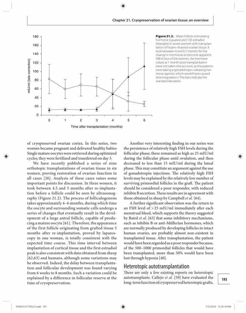
193
Chapter 21. Cryopreservation of ovarian tissue: an overview
of cryopreserved ovarian cortex. In this series, two women became pregnant and delivered healthy babies Single mature oocytes were retrieved during optimized cycles; they were fertilized and transferred on day 3.
We have recently published a series of nine orthotopic transplantations of ovarian tissue in six women, proving restoration of ovarian function in all cases [ 26 ]. Analysis of these cases raises some important points for discussion. In three women, it took between 4.5 and 5 months after re-implanta-tion before a follicle could be seen by ultrasonog-raphy ( Figure 21.2 ). The process of folliculogenesis takes approximately 4–6 months, during which time the oocyte and surrounding somatic cells undergo a series of changes that eventually result in the devel-opment of a large antral follicle, capable of produ-cing a mature oocyte [ 61 ]. Therefore, the appearance of the first follicle originating from grafted tissue 5 months after re-implantation, proved by laparos-copy in one woman, is totally consistent with the expected time course. This time interval between implantation of cortical tissue and the first estradiol peak is also consistent with data obtained from sheep [ 62 , 63 ] and humans, although some variations may be observed. Indeed, the delay between transplanta-tion and follicular development was found varying from 6 weeks to 8 months. Such a variation could be explained by a difference in follicular reserve at the time of cryopreservation.
Another very interesting fi nding in our series was the persistence of relatively high FSH levels during the follicular phase; these remained as high as 25 mIU/ml during the follicular phase until ovulation, and then decreased to less than 15 mIU/ml during the luteal phase. Th is may constitute an argument against the use of gonadotropin injections. Th e relatively high FSH levels may be explained by the relatively low number of surviving primordial follicles in the graft . Th e patient should be considered a poor responder, with reduced inhibin B secretion. Th ese results are in agreement with those obtained in sheep by Campbell et al. [ 64 ].
A further signifi cant observation was the return to an FSH level of > 25 mIU/ml immediately aft er each menstrual bleed, which supports the theory suggested by Baird et al. [ 63 ] that some inhibitory mechanisms, such as inhibin B or anti-Müllerian hormones, which are normally produced by developing follicles in intact human ovaries, are probably almost non-existent in transplanted tissue. Aft er transplantation, the patient would have been regarded as a poor responder because, of the 500–1000 primordial follicles that would have been transplanted, more than 50% would have been lost through hypoxia [ 40 ].
Heterotopic autotransplantation Th ere are only a few existing reports on heterotopic autotransplants. Callejo et al. [ 59 ] have evaluated the long-term function of cryopreserved heterotopic graft s,
0
20
40
60
80
100
120
140
160
180
0 1 2 3 4 5 6
Time after transplantation (months)
mIU
/ml (
late
cor
rect
ion)
Figure 21.2. Mean follicle-stimulating hormone (squares) and 17β-estradiol (triangles) in seven women with transplan-tation of frozen–thawed ovarian tissue. It took between 4 and 6.5 months for the change in hormones to become apparent. (NB in four of the women, the hormone values at 1 month post-transplantation were not taken into account, as the patients were taking a gonadotropin-releasing hor-mone agonist, which would have caused downregulation.) The bars indicate the standard deviation.
9780521517782c21.indd 1939780521517782c21.indd 193 3/12/2010 12:24:10 PM3/12/2010 12:24:10 PM
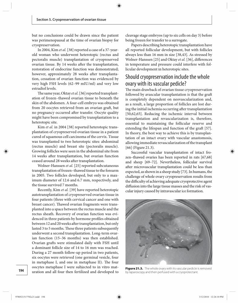
194
Section 5. Cryopreservation of ovarian tissue
but no conclusions could be drawn since the patient was perimenopausal at the time of ovarian biopsy for cryopreservation.
In 2004, Kim et al. [ 38 ] reported a case of a 37-year-old woman who underwent heterotopic (rectus and pectoralis muscle) transplantation of cryopreserved ovarian tissue. By 14 weeks aft er the transplantation, restoration of endocrine function was demonstrated; however, approximately 28 weeks aft er transplanta-tion, cessation of ovarian function was evidenced by very high FSH levels (62–99 mIU/ml) and very low estradiol levels.
Th e same year, Oktay et al. [ 36 ] reported transplant-ation of frozen–thawed ovarian tissue to beneath the skin of the abdomen. A four-cell embryo was obtained from 20 oocytes retrieved from an ovarian graft , but no pregnancy occurred aft er transfer. Oocyte quality might have been compromised by transplantation to a heterotopic site.
Kim et al. in 2004 [ 38 ] reported heterotopic trans-plantation of cryopreserved ovarian tissue in a patient cured of squamous cell carcinoma of the cervix. Tissue was transplanted to two heterotopic sites: abdominal (rectus muscle) and breast site (pectoralis muscle). Growing follicles were seen in the abdominal site from 14 weeks aft er transplantation, but ovarian function ceased around 28 weeks aft er transplantation.
Wolner-Hanssen et al. [ 25 ] reported subcutaneous transplantation of frozen–thawed tissue to the forearm in 2005. Two follicles developed, but only to a max-imum diameter of 12.6 and 6.7 mm, respectively, and the tissue survived 7 months.
Recently, Kim et al. [ 39 ] have reported heterotopic autotransplantation of cryopreserved ovarian tissue in four patients (three with cervical cancer and one with breast cancer). Th awed ovarian fragments were trans-planted into a space between the rectus muscle and the rectus sheath. Recovery of ovarian function was evi-denced in three patients by hormone profi les obtained between 12 and 20 weeks aft er transplantation, but only lasted 3 to 5 months. Th ese three patients subsequently underwent a second transplantation. Long-term ovar-ian function (15–36 months) was then established. Ovarian graft s were stimulated daily with FSH until a dominant follicle size of 14 to 16 mm was reached. During a 27 month follow-up period in two patients, six oocytes were retrieved (one germinal vesicle, four in metaphase I, and one in metaphase II). Th e four oocytes metaphase I were subjected to in vitro mat-uration and all four then fertilized and developed to
cleavage stage embryos (up to six cells on day 3) before being frozen for transfer to a surrogate.
Papers describing heterotopic transplantation have all reported follicular development, but with follicles always less than 16 mm in size [ 38 , 43 ]. As stressed by Wolner-Hanssen [ 25 ] and Oktay et al. [ 36 ], diff erences in temperature and pressure could interfere with fol-licular development in heterotopic sites.
Should cryopreservation include the whole ovary with its vascular pedicle? Th e main drawback of ovarian tissue cryopreservation followed by avascular transplantation is that the graft is completely dependent on neovascularization and, as a result, a large proportion of follicles are lost dur-ing the initial ischemia occurring aft er transplantation [ 50 , 62 , 65 ]. Reducing the ischemic interval between transplantation and revascularization is, therefore, essential to maintaining the follicular reserve and extending the lifespan and function of the graft [ 57 ]. In theory, the best way to achieve this is by transplan-tation of an intact ovary with vascular anastomosis, allowing immediate revascularization of the transplant [ 66 ] ( Figure 21.3 ).
Successful vascular transplantation of intact fro-zen–thawed ovaries has been reported in rats [ 67 , 68 ] and sheep [ 69 – 72 ]. Nevertheless, follicular survival aft er microvascular transplantation could be less than expected, as shown in a sheep study [ 73 ]. In humans, the challenge of whole ovary cryopreservation results from the diffi culty of achieving adequate cryoprotective agent diff usion into the large tissue masses and the risk of vas-cular injury caused by intravascular ice formation.
Figure 21.3. The whole ovary with its vascular pedicle is removed by laparoscopy and then perfused with a cryoprotectant .
9780521517782c21.indd 1949780521517782c21.indd 194 3/12/2010 12:24:10 PM3/12/2010 12:24:10 PM
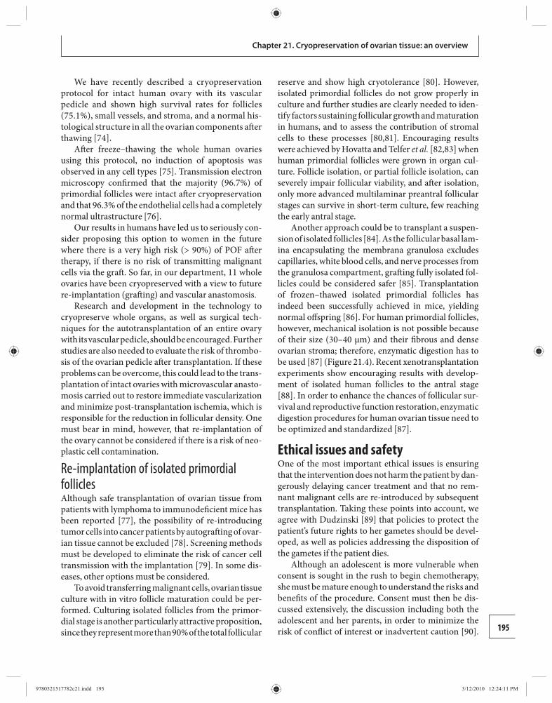
195
Chapter 21. Cryopreservation of ovarian tissue: an overview
We have recently described a cryopreservation protocol for intact human ovary with its vascular pedicle and shown high survival rates for follicles (75.1%), small vessels, and stroma, and a normal his-tological structure in all the ovarian components aft er thawing [ 74 ].
Aft er freeze–thawing the whole human ovaries using this protocol, no induction of apoptosis was observed in any cell types [ 75 ]. Transmission electron microscopy confi rmed that the majority (96.7%) of primordial follicles were intact aft er cryopreservation and that 96.3% of the endothelial cells had a completely normal ultrastructure [ 76 ].
Our results in humans have led us to seriously con-sider proposing this option to women in the future where there is a very high risk (> 90%) of POF aft er therapy, if there is no risk of transmitting malignant cells via the graft . So far, in our department, 11 whole ovaries have been cryopreserved with a view to future re-implantation (graft ing) and vascular anastomosis.
Research and development in the technology to cryo preserve whole organs, as well as surgical tech-niques for the autotransplantation of an entire ovary with its vascular pedicle, should be encouraged. Further studies are also needed to evaluate the risk of thrombo-sis of the ovarian pedicle aft er transplantation. If these problems can be overcome, this could lead to the trans-plantation of intact ovaries with microvascular anasto-mosis carried out to restore immediate vascularization and minimize post-transplantation ischemia, which is responsible for the reduction in follicular density. One must bear in mind, however, that re-implantation of the ovary cannot be considered if there is a risk of neo-plastic cell contamination.
Re-implantation of isolated primordial follicles Although safe transplantation of ovarian tissue from patients with lymphoma to immunodefi cient mice has been reported [ 77 ], the possibility of re-introducing tumor cells into cancer patients by autograft ing of ovar-ian tissue cannot be excluded [ 78 ]. Screening methods must be developed to eliminate the risk of cancer cell transmission with the implantation [ 79 ]. In some dis-eases, other options must be considered.
To avoid transferring malignant cells, ovarian tissue culture with in vitro follicle maturation could be per-formed. Culturing isolated follicles from the primor-dial stage is another particularly attractive proposition, since they represent more than 90% of the total follicular
reserve and show high cryotolerance [ 80 ]. However, isolated primordial follicles do not grow properly in culture and further studies are clearly needed to iden-tify factors sustaining follicular growth and maturation in humans, and to assess the contribution of stromal cells to these processes [ 80 , 81 ]. Encouraging results were achieved by Hovatta and Telfer et al. [ 82 , 83 ] when human primordial follicles were grown in organ cul-ture. Follicle isolation, or partial follicle isolation, can severely impair follicular viability, and aft er isolation, only more advanced multilaminar preantral follicular stages can survive in short-term culture, few reaching the early antral stage.
Another approach could be to transplant a suspen-sion of isolated follicles [ 84 ]. As the follicular basal lam-ina encapsulating the membrana granulosa excludes capillaries, white blood cells, and nerve processes from the granulosa compartment, graft ing fully isolated fol-licles could be considered safer [ 85 ]. Transplantation of frozen–thawed isolated primordial follicles has indeed been successfully achieved in mice, yielding normal off spring [ 86 ]. For human primordial follicles, however, mechanical isolation is not possible because of their size (30–40 μm) and their fi brous and dense ovarian stroma; therefore, enzymatic digestion has to be used [ 87 ] ( Figure 21.4 ). Recent xenotransplantation experiments show encouraging results with develop-ment of isolated human follicles to the antral stage [ 88 ]. In order to enhance the chances of follicular sur-vival and reproductive function restoration, enzymatic digestion procedures for human ovarian tissue need to be optimized and standardized [ 87 ].
Ethical issues and safety One of the most important ethical issues is ensuring that the intervention does not harm the patient by dan-gerously delaying cancer treatment and that no rem-nant malignant cells are re-introduced by subsequent transplantation. Taking these points into account, we agree with Dudzinski [ 89 ] that policies to protect the patient’s future rights to her gametes should be devel-oped, as well as policies addressing the disposition of the gametes if the patient dies.
Although an adolescent is more vulnerable when consent is sought in the rush to begin chemotherapy, she must be mature enough to understand the risks and benefi ts of the procedure. Consent must then be dis-cussed extensively, the discussion including both the adolescent and her parents, in order to minimize the risk of confl ict of interest or inadvertent caution [ 90 ].
9780521517782c21.indd 1959780521517782c21.indd 195 3/12/2010 12:24:11 PM3/12/2010 12:24:11 PM
196
Section 5. Cryopreservation of ovarian tissue
Respecting the code of good practice, all patients who may become infertile have the right to receive proper consideration of their interests for future possibilities in the fi eld of ovarian function preservation. Th e selec-tion of cases should be carried out on the basis of multi-disciplinary staff discussions, including oncologists, gynecologists, biologists, psychologists, and pediatri-cians. Counseling should be given and informed con-sent obtained from the patient. Cancer treatment takes priority over potential restoration of fertility, but off er-ing the chance to preserve fertility may greatly enhance quality of life for cancer survivors and provide support at the time in that it has considerations of hope for the future.
Conclusions Approximately one third of young women exposed to chemotherapy develop ovarian failure. In 2008, we believe it is our ethical responsibility to propose cryo-preservation of ovarian tissue to all adolescents and young women, under local institutional review board guidance, having to undergo chemotherapy with alkylating agents.
Th e age of the patient should be taken into consid-eration, since the follicular reserve of the ovary is age dependent. Because a decline in fertility is now well documented aft er the age of 38 years, the procedure should probably be restricted to patients below this age. In any case, irradiation and chemotherapy appear to be less harmful to the gonads of prepubertal than postpubertal women [ 15 , 16 , 91 ].
Th is is why, since 1996, we have systematically pro-posed cryopreservation to all women under 35 years of age prior to chemotherapy when there is a risk of POF, so far numbering more than 300. We accept that ovar-ian tissue cryopreservation is a more innovative and invasive procedure than sperm cryopreservation, and
that all possible applications in adolescents are ethic-ally complex. However, we believe that ovarian cortex banking should be off ered before chemotherapy in all cases where emergency IVF is not possible.
In conclusion, the six live births obtained aft er transplantation of frozen–thawed ovarian tissue in humans give hope to young cancer patients, but there is still much work to be done. Research programs need to determine whether active angiogenesis can be induced to accelerate the process of neovascularization in graft ed tissue, if isolated human follicles can be graft ed, or indeed if microvascular reanostomosis of an entire cryopreserved ovary is a valuable option.
References 1. Jemal A , Siegel R , Ward E , et al. Cancer statistics, 2006 .
CA Cancer J Clin 2006; 56: 106–130 . 2. Blatt J. Pregnancy outcome in long-term survivors of
childhood cancer . Med Pediatr Oncon 1999; 33: 29–33 . 3. Ries LAG , Percy CL , Bunin GR. Introduction. In Ries
LAG , Smith MA , Gurney JG , et al. , eds. Cancer Incidence and Survival among Children and Adolescents: United States SEER Program 1975–1995 . [NIH Pub. No. 99–4649]. Bethesda, MD : National Cancer Institute , 1999, pp. 1–15.
4. Donnez J , Dolmans MM , Demylle D , et al. Restoration of ovarian function aft er orthotopic (intraovarian and periovarian) transplantation of cryopreserved ovarian tissue in a woman treated by bone marrrow transplantation for sickle cell anaemia: case report . Hum Reprod 2006; 21: 183–188 .
5. Meirow D , Lewis H , Nugent D , Epstein N. Subclinical depletion of primordial follicular reserve in mice treated with cyclophosphamide: clinical importance and proposed accurate investigative tool . Hum Reprod 1999; 14: 1903–1907 .
6. Wallace WH , Th omson AB , Saran F , Kelsey TW. Predicting age of ovarian failure aft er radiation to a fi eld that includes the ovaries . Int J Radiat Oncol Biol Phys 2005; 62: 738–744 .
(a) (b)
Figure 21.4. Enzymatically isolated follicles (between 30 and 110 μm) are visible under an inverted fl uorescence microscope after fl uorescent viability staining (calcein-AM and ethidium homodimer-1). Follicles are visible on light microscopy (a) and fl uorescence microscopy (b), which show that all are viable.
9780521517782c21.indd 1969780521517782c21.indd 196 3/12/2010 12:24:11 PM3/12/2010 12:24:11 PM
197
Chapter 21. Cryopreservation of ovarian tissue: an overview
7. Larsen EC , Muller J , Schmiegelow K , Rechnitzer C , Andersen AN. Reduced ovarian function in long-term survivors of radiation- and chemotherapy-treated childhood cancer . J Clin Endocrinol Metab 2003; 88: 5307–5314 .
8. Shalet SM. Eff ects of cancer chemotherapy on gonadal function of patients . Cancer Treat Rev 1980; 7: 141–152 .
9. Schilsky RL , Sherins RJ , Hubbard SM , et al. Long-term follow up of ovarian function in women treated with MOPP chemotherapy for Hodgkin ’ s disease . Am J Med 1981; 71: 552–556 .
10. Wallace WH , Anderson RA , Irvine DS. Fertility preservation for young patients with cancer: who is at risk and what can be off ered ? Lancet Oncol 2005; 6 : 209–218 .
11. Wallace WH , Th omson AB , Kelsey TW. Th e radiosensitivity of the human oocyte . Hum Reprod 2003; 18: 117–121 .
12. Larsen EC , Schmiegelow K , Rechnitzer C , et al. Radiotherapy at a young age reduces uterine volume of childhood cancer survivors . Acta Obstet Gynecol Scand 2004; 83: 96–102 .
13. Critchley HO , Wallace GH. Impact of cancer treatment on uterine function . J Natl Cancer Inst Monogr 2005; 34 : 64–68 .
14. Bath LE , Critchley HO , Chambers SE , et al. Ovarian and uterine characteristics aft er total body irradiation in childhood and adolescence: response to sex steroid replacement . Br J Obstet Gynaecol 1999; 106: 1265–1272 .
15. Sanders J , Hawley J , Levy W , et al. Pregnancies following high-dose cyclophosphamide with or without high-dose busulfan or total body irradiation and bone marrow transplantation . Blood 1996; 87: 3045–3052 .
16. Meirow D , Nugent D. Th e eff ects of radiotherapy and chemotherapy on female reproduction . Hum Reprod Update 2001; 7: 534–543 .
17. Lobo RA. Potential options for preservation of fertility in women . N Engl J Med 2005; 353: 64–73 .
18. Teinturier C , Hartmann O , Valteau-Couanet D , Benhamou E , Bougneres PF. Ovarian function aft er autologous bone marrow transplantation in childhood: high-dose busulfan is a major cause of ovarian failure . Bone Marrow Transplant 1998; 22: 989–994 .
19. Salooja N , Szydlo RM , Socie G , et al. for the Late Eff ects Working Party of the European Group for Blood and Marrow Transplantation . Pregnancy outcomes aft er peripheral blood or bone marrow transplantation: a retrospective survey . Lancet 2001; 358: 271–276 .
20. Lutchman Singh K , Davies M , Chatterjee R. Fertility in female cancer survivors: pathophysiology, preservation and the role of ovarian reserve testing . Hum Reprod Update 2005; 11: 69–89 .
21. Donnez J , Bassil S. Indications for cryopreservation of ovarian tissue . Hum Reprod Update 1998; 4: 248–259 .
22. Kleine W. Results of fertility preserving operations in malignant ovarian tumors . Zentralbl Gynakol 1996; 118: 317–321 .
23. Slavin S , Nagler A , Aker M , et al. Non-myeloablative stem cell transplantation and donor lymphocyte infusion for the treatment of cancer and life-threatening non-malignant disorders . Rev Clin Exp Hematol 2001; 5: 135–146 .
24. Mattle V , Behringer K , Engert A , Wildt L. Female fertility aft er cytotoxic therapy-protection of ovarian function during chemotherapy of malignant and non-malignant diseases . Eur J Haematol 2005; 75: 77–82 .
25. Wolner-Hanssen P , Hägglund L , Ploman F , et al. Autotransplantation of cryopreserved ovarian tissue to the right forearm 4 ½ years aft er autologous stem cell transplantation . Acta Obstet Gynecol Scand 2005; 84: 695–698 .
26. Donnez J , Squiffl et J , Van Eyck AS , et al. Restoration of ovarian function in orthotopically transplanted cryopreserved ovarian tissue: a pilot experience . Reprod Biomed Online 2008; 16: 694–704 .
27. Ethics Committee of the American Society for Reproductive Medicine. Fertility preservation and reproduction in cancer patients . Fertil Steril 2005; 83: 1622–1628 .
28. Stachecki JJ , Cohen J. An overview of oocyte cryopreservation . Reprod Biomed Online 20 04; 9 : 152–163 .
29. Cobo A , Domingo J , Pérez S , et al. Vitrifi cation: an eff ective new approach to oocyte banking and preserving fertility in cancer patients . Clin Transl Oncol 2008; 10 : 268–273 .
30. Gosden RG , Baird DT , Wade JC , Webb R. Restoration of fertility to oophorectomized sheep by ovarian autograft s stored at –196°C . Hum Reprod 1994; 9 : 597–603 .
31. Oktay K , Newton H , Aubard Y , Salha O , Gosden RG. Cryopreservation of immature human oocytes and ovarian tissue: an emerging technology ? Fertil Steril 1998; 69: 1–7 .
32. Meirow D , Ben Yehuda D , Prus D , et al. Ovarian tissue banking in patients with Hodgkin ’ s disease: is it safe ? Fertil Steril 1998; 69: 996–998 .
33. Donnez J , Godin PA , Qu J , Nisolle M. Gonadal cryopreservation in the young patient with gynaecological malignancy . Current Opin Obstet Gynecol 2000; 12: 1–9 .
34. Donnez J , Dolmans MM , Martinez-Madrid B , Demylle D , Van Langendonkt A. Th e role of cryopreservation for women prior to treatment of malignancy . Curr Opin Obstet Gynecol 2005; 17: 333–338 .
35. Oktay K , Karlikaya G. Ovarian function aft er transplantation of frozen, banked autologous ovarian tissue . N Engl J Med 2000; 342: 1919 .
9780521517782c21.indd 1979780521517782c21.indd 197 3/12/2010 12:24:11 PM3/12/2010 12:24:11 PM
198
Section 5. Cryopreservation of ovarian tissue
36. Oktay K , Buyuk E , Veeck L , et al. Embryo development aft er heterotopic transplantation of cryopreserved ovarian tissue . Lancet 2004; 363: 837–840 .
37. Radford JA , Lieberman BA , Brison D , et al. Orthotopic reimplantation of cryopreserved ovarian cortical strips aft er high-dose chemotherapy for Hodgkin ’ s lymphoma . Lancet 2001; 357: 1172–1175 .
38. Kim SS , Hwang I-T , Lee H-C. Heterotopic autotransplantation of cryobanked human ovarian tissue as a strategy to restore ovarian function . Fertil Steril 2004; 82: 930–932 .
39. Kim SS , Lee WS , Chung MK , et al. Long-term ovarian function and fertility aft er heterotopic autotransplantation of cryobanked human ovarian tissue: 8-year experience in cancer patients . Fertil Steril 200 9; 91 : 2349–2954 .
40. Donnez J , Dolmans MM , Demylle D , et al. Livebirth aft er orthotopic transplantation of cryopreserved ovarian tissue . Lancet 2004; 364 : 1405–1410 .
41. Meirow D , Levron J , Eldar-Geva T , et al. Pregnancy aft er transplantation of cryopreserved ovarian tissue in a patient with ovarian failure aft er chemotherapy . N Engl J Med 2005; 353: 318–321 .
42. Schmidt KL , Yding Andersen C , Loft A , et al. Follow-up of ovarian function post-chemotherapy following ovarian cryopreservation and transplantation . Hum Reprod 2005; 20: 3539–3546 .
43. Demeestere I , Simon P , Buxant F , et al. Ovarian function and spontaneous pregnancy aft er combined heterotopic and orthotopic cryopreserved ovarian tissue transplantation in a patient previously treated with bone marrow transplantation: case report . Hum Reprod 2006; 21: 2010–2014 .
44. Demeestere I , Simon P , Emiliani S , Delbaere A , Englert Y. Fertility preservation: successful transplantation of cryopreserved ovarian tissue in a young patient previously treated for Hodgkin ’ s disease . Oncologist 2007; 12: 1437–1442 .
45. Silber SJ , DeRosa M , Pineda J , et al. A series of monozygotic twins discordant for ovarian failure: ovary transplantation (cortical versus microvascular) and cryopreservation . Hum Reprod 2008; 23 : 1531–1537 .
46. Andersen CY , Rosendahl M , Byskov AG , et al. Two successful pregnancies following autotransplantation of frozen/thawed ovarian tissue . Hum Reprod 2008; 23 : 2266–2272 .
47. Hovatta O. Methods for cryopreservation of human ovarian tissue . Reprod Biomed Online 2005; 10 : 729–734 .
48. Fuller B , Paynter S. Fundamentals of cryobiology in reproductive medicine . Reprod Biomed Online 2004; 9 : 680–691 .
49. Aubard Y. Ovarian tissue xenograft ing . Eur J Obst Gynecol Reprod Biol 2003; 108 : 14–18 .
50. Nisolle M , Godin PA , Casanas-Roux F , et al. Histological and ultrastructural evaluation of fresh and frozen–thawed
human ovarian xenograft s in nude mice . Fertil Steril 2000; 74 : 122–129 .
51. Hernandez-Fonseca H , Bosch P , Sirisathien S , et al. Eff ect of site of transplantation on follicular development of human ovarian tissue transplanted into intact or castrated immunodefi cient mice . Fertil Steril 2004; 81 : 888–892 .
52. Abir R , Orvieto R , Raanani H , et al. Parameters aff ecting successful transplantation of frozen–thawed human fetal ovaries into immunodefi cient mice . Fertil Steril 2003; 80 : 421–428 .
53. Oktay K , Newton H , Mullan J , Gosden RG. Development of human primordial follicles to antral stages in SCID/hpg mice stimulated with follicle stimulating hormone . Hum Reprod 1998; 13 : 1133–1138 .
54. Gook DA , Edgar DH , Borg J , et al. Oocyte maturation, follicle rupture and luteinization in human cryopreserved ovarian tissue following xenograft ing . Hum Reprod 2003; 18 : 1772–1781 .
55. Gook DA , Edgar DH , Borg J , Archer J , McBain JC. Diagnostic assessment of the developmental potential of human cryopreserved ovarian tissue from multiple patients using xenograft ing . Hum Reprod 2005; 20: 72–78 .
56. Kim SS , Soules MR , Battaglia DE. Follicular development, ovulation, and corpus luteum formation in cryopreserved human ovarian tissue aft er xenotransplantation . Fertil Steril 2002; 78: 77–82 .
57. Van Eyck AS , Jordan BF , Gallez B , et al. Electron paramagnetic resonance as a tool to evaluate human ovarian tissue reoxygenation aft er xenograft ing . Fertil Steril 2009 ; 92 : 374–381 .
58. Van Eyck AS , Bouzin C , Feron O , et al. Both host and graft vessels contribute to revascularization of xenograft ed human ovarian tissue in a murine model. Fert Steril 2010 ; in press.
59. Callejo J , Salvador C , Miralles A , et al. Long-term ovarian function evaluation aft er autograft ing by implantation with fresh and frozen–thawed human ovarian tissue . J Clin Endocrinol Metab 2001; 86: 4489–4494 .
60. Oktay K. Spontaneous conceptions and live birth aft er heterotopic ovarian transplantation: is there a germline stem cell connection ? Hum Reprod 2006; 21: 1345–1348 .
61. Gougeon A. Regulation of ovarian follicular development in primates: facts and hypotheses . Endocrin Rev 1996; 17: 121–155 .
62. Baird , DT , Webb R , Campbell BK , Harkness LM , Gosden RG. Long-term ovarian function in sheep aft er ovariectomy and transplantation of autograft s stored at −196°C . Endocrinol 1999; 140 : 462–471 .
63. Baird DT , Campbell BK , Souza C , Telfer EE. Long-term ovarian function in sheep aft er ovariectomy and autotransplantation of cryopreserved cortical strips . Eur J Obst Gynecol Reprod Biol 2004; 113: 55–59 .
9780521517782c21.indd 1989780521517782c21.indd 198 3/12/2010 12:24:11 PM3/12/2010 12:24:11 PM
199
Chapter 21. Cryopreservation of ovarian tissue: an overview
64. Campbell BK , Telfer EE , Webb R , Baird DT. Ovarian autograft s in sheep as a model for studying folliculogenesis . Mol Cell Endocrinol 2000; 163: 131–139 .
65. Liu J , Van der Elst J , van den Broecke R , Dhont M. Early massive follicle loss and apoptosis in heterotopically graft ed newborn mouse ovaries . Hum Reprod 2002; 17: 605–611 .
66. Jadoul P , Donnez J , Dolmans MM , et al. Laparoscopic ovariectomy for whole human ovary cryopreservation: technical aspects . Fertil Steril 2007; 87 : 971–975 .
67. Wang X , Chen H , Yin H , et al. Successful pregnancy aft er intact ovary transplantation . Nature 2002; 415: 385 .
68. Yin H , Wang X , Kim SS , et al. Transplantation of intact rat gonads using vascular anastomosis: eff ects of cryopreservation, ischemia and genotype . Hum Reprod 2003; 18: 1165–1172 .
69. Bedaiwy MA , Jeremias E , Gurunluoglu R , et al. Restoration of ovarian function aft er autotransplantation of intact frozen–thawed sheep ovaries with microvascular anastomosis . Fertil Steril 2003; 79: 594–602 .
70. Revel A , Elami A , Bor A , et al. Whole sheep ovary cryopreservation and transplantation . Fertil Steril 2004; 82: 1714–1715 .
71. Arav A , Revel A , Nathan Y , et al. Oocyte recovery, embryo development and ovarian function aft er cryopreservation and transplantation of whole sheep ovary . Hum Reprod 2005; 20: 3554–3559 .
72. Imhof M , Bergmeister H , Lipovac M , et al. Orthotopic microvascular re-anastomosis of whole cryopreserved ovine ovaries resulting in pregnancy and life birth . Fertil Steril 2006; 85: 1208–1215 .
73. Courbiere B , Caquant L , Mazoyer C , et al. Diffi culties improving ovarian functional recovery by microvascular transplantation and whole ovary vitrifi cation . Fertil Steril 2009; 91 : 2697–2706 .
74. Martinez-Madrid B , Dolmans MM , Van Langendonckt A , Defrère S , Donnez J. Freeze–thawing intact human ovary with its vascular pedicle with a passive cooling device . Fertil Steril 2004; 82: 1390–1394 .
75. Martinez-Madrid B , Camboni A , Dolmans MM , et al. Apoptosis and ultrastructural assessment aft er cryopreservation of whole human ovaries with their vascular pedicle . Fertil Steril 2007; 87: 1153–1165 .
76. Camboni A , Martinez-Madrid B , Dolmans MM , et al. Preservation of fertility in young cancer patients: contribution of transmission electron microscopy . Reprod Biomed Online 2008; 17: 136–150 .
77. Kim SS , Radford J , Harris M , et al. Ovarian tissue harvested from lymphoma patients to preserve fertility may be safe for autotransplantation . Hum Reprod 2001; 16: 2056–2060 .
78. Shaw JM , Bowles S , Koopman P , Wood EC , Trounson AO. Fresh and cryopreserved ovarian tissue samples from donors with lymphoma transmit the cancer to graft recipients . Hum Reprod 1996; 11: 1668–1673 .
79. Meirow D , Hardan I , Dor J , et al. Searching for evidence of disease and malignant cell contamination in ovarian tissue stored from hematologic cancer patients . Hum Reprod 2008; 23: 1007–1013 .
80. Smitz JE , Cortvrindt RG. Th e earliest stages of folliculogenesis in vitro . Reproduction 2002; 123: 185–202 .
81. Hovatta O , Wright C , Krausz T , Hardy K , Winston RML. Human primordial, primary and secondary ovarian follicles in long-term culture: eff ect of partial isolation . Hum Reprod 1999; 14: 2519–2524 .
82. Hovatta O. Cryopreservation and culture of human ovarian cortical tissue containing early follicles . Eur J Obstet Gynecol Reprod Biol 2004; 5 : S50–S54 .
83. Telfer EE , McLaughlin M , Ding C , Th ong KJ. A two-step serum-free culture system supports development of human oocytes from primordial follicles in the presence of activin . Hum Reprod 2008; 23: 1151–1158 .
84. Dolmans MM , Martinez-Madrid B , Gadisseux E , et al. Short-term transplantation of isolated human ovarian follicles and cortical tissue into nude mice . Reproduction 2007; 134: 253–262 .
85. Motta PM , Nottola SA , Familiari G , et al. Morphodynamics of the follicular-luteal complex during early ovarian development and reproductive life . Int Rev Cytol 2003; 223: 177–288 .
86. Carroll J , Gosden RG. Transplantation of frozen–thawed mouse primordial follicles . Hum Reprod 1993; 8: 1163–1167 .
87. Dolmans MM , Michaux N , Camboni A , et al. Evaluation of Liberase, a purifi ed enzyme blend, for the isolation of human primordial and primary ovarian follicles . Hum Reprod 2006; 21 : 413–420 .
88. Dolmans MM , Yuan WY , Camboni A , et al. Development of antral follicles aft er xenograft ing of isolated small human preantral follicles . Reprod Biomed Online 2008; 16 : 705–711 .
89. Dudzinski DM. Ethical issues in fertility preservation for adolescent cancer survivors: oocyte and ovarian tissue cryopreservation . J Pediatr Adolesc Gynecol 2004; 17: 97–102 .
90. Bukovsky A. Can ovarian infertility be treated with bone marrow- or ovary-derived germ cells ? Reprod Biol Endocrinol 2005; 3: 36 .
91. Haie-Meder C , Mlika-Cabanne N , Michel G , et al. Radiotherapy aft er ovarian transposition ovarian function and fertility preservation . Int J Radiat Oncol Biol Phys 1993; 25: 419–424 .
9780521517782c21.indd 1999780521517782c21.indd 199 3/12/2010 12:24:11 PM3/12/2010 12:24:11 PM
200 Fertility Cryopreservation, ed. Ri-Cheng Chian and Patrick Quinn. Published by Cambridge University Press. Copyright © Cambridge University Press 2010.
Chapter
22well) need more attention and study. Other aims such as managing follicular quality aft er cryopreservation or even making an oocyte cell bank could also profi t from observations of in vitro culture.
Among species whose ovaries can be compared with the human in size and structure, culture systems for secondary follicles have been described for sheep [ 15 , 16 ], cattle [ 17 , 18 ], and pigs [ 19 , 20 ]. However, lit-tle progress has been made in developing culture sys-tems to grow primordial or primary follicles in these species. Th is chapter discusses the current achieve-ments obtained with in vitro culture of female germ cells and primordial ovarian follicles and the attempts to improve their development by adding various fac-tors to the culture medium. Th e established methods for the evaluation of survival and growth in culture are also discussed: follicular counts, immunocytochem ical methods, transmission electron microscopy, viability markers, and endocrine assays.
Primordial follicle development in vivo In fetal life, primordial follicles are formed in the ovary by the organization of oocytes and pre-granulosa cells. Autocrine and/or paracrine signaling involv-ing growth factors from granulosa cells, oocytes, and stromal interstitial cells regulate follicular growth up to the antral stage. As soon as the follicle is formed, growth and atresia of some follicles can already be observed, while other follicles stay quiescent until menopause.
Numerous factors are involved in the growth beyond the primordial stage, and while some are ne cessary, it does not appear that exit from the resting pool is depend-ent on any one single factor. Factors from vascular and neuronal origin participate in follicular formation and in the earliest growth processes. Th ese factors can be either stimulatory or inhibitory. Ovarian function is also under the control of the hypothalamic gonadotropin-releasing
Introduction In vitro culture of ovarian tissue is challenging. Th e fi rst live births were obtained in the mouse model start-ing cultures early from preantral follicles [ 1 ]. Nearly 10 years later, the same team has achieved live births from in vitro grown oocytes initiated from the earliest stage of development, the primordial follicle [ 2 , 3 ]. Similar results have not yet been reported in any other species.
Interest is focusing on the in vitro growth of human follicles from the earliest stages. Cancer treatment has improved; more young women of reproductive age are surviving, but quite oft en they will be confronted with infertility as a consequence of radio- and/or chemother-apy. Other diseases such as systemic lupus erythemato-sus, sickle cell anemia, and rheumatoid arthritis might also need suffi ciently intensive chemotherapy to lead to similar consequences for their fertility. It is proposed that ovarian cortical tissue should be removed before admin-istering aggressive therapies. Ovarian cortex containing immature primordial follicles can be successfully cryo-preserved. Th e ultimate aim of ovary cryopreservation is to restore ovarian function, with hormone production and follicular growth. Th ese results have been observed aft er autotransplantation of frozen–thawed ovarian tis-sue, and eight pregnancies have been reported to date [ 4 – 10 ]. Some studies have demonstrated a risk of trans-mitting malignant cells with the cryopreserved tissue [ 11 – 14 ]. In some types of cancer, the risk for transmis-sion of cancer cells can be very high, and in such cases, autotransplantation cannot be proposed. To avoid this risk, follicles could be grown in vitro in order to obtain fully matured oocytes surrounded by the zona pellucida and excluding any other cell type.
In addition, study of follicles during the culture period could also help to improve our understanding of early folliculogenesis in humans and other large mammalian species. Particularly the earliest stages (i.e. those that survive the cryopreservation procedure
In vitro culture of human primordial follicles Benoit Schubert and Johan Smitz
9780521517782c22.indd 2009780521517782c22.indd 200 3/12/2010 7:30:21 PM3/12/2010 7:30:21 PM
201
Chapter 22. In vitro culture of human primordial follicles
hormone pulse generator, which drives production of follicle-stimulating hormone (FSH) and luteinizing hor-mone (LH). Gene knockout technology has revealed that oocyte-derived factors (bone morphogenetic protein [BMP] 6 and growth diff erentiation factors [GDF] 9 and 9B [BMP-15]) of the transforming growth factor [TGF] β superfamily play key roles at the earliest stages of fol-liculogenesis. Targeted deletion of the gene for GDF-9 in mice [ 21 ] or naturally occurring point mutations of the gene for BMP-15 in sheep [ 22 ] result in arrest of fol-licle development at the primary stage. Several lines of evidence point to a role for anti-Müllerian hormone (AMH) as a regulator of primordial follicle activation, inhibiting their recruitment into the growing phase. A mouse knockout model homozygous for AMH showed rapid depletion of the primordial follicle pool [ 23 ].
Oocyte growth during early folliculogenesis was shown to be regulated by the Kit ligand (KL) and its receptor c-Kit. Mice with mutations at the white spot-ting (W locus encoding c-kit) and/or steel locus (Sl locus encoding KL) had a severe or mild defect in early follicular development [ 24 , 25 ]. Th e multitude of processes and factors involved in follicular formation and early follicle growth stages have been reviewed by Juengel and McNatty [ 26 ] and by Matzuk and Lamb [ 27 ]. Identifi cation of autocrine/paracrine factors and their receptors involved in the regulation of the earliest stages of folliculogenesis have led to the investigation of their requirements for follicle development in vitro.
Sources of ovarian tissue Ovarian cortical tissue is mainly populated with pri-mordial and primary follicles and is the major source of immature oocytes for in vitro folliculogenesis. Harvesting ovarian cortical tissue is commonly done through an invasive procedure, and most of the research teams working on human ovarian tissue have retrieved specimens during operations performed for adult con-senting patients who have faced pathologies requiring treatment that would aff ect their fertility (e.g. breast cancer and acute leukemia) [ 28 ], during cesarean sec-tion [ 29 – 33 ], during gynecological or infertility evalua-tion [ 24 , 34 – 36 ], and during sterilizing hysterectomy or oophorectomy [ 37 – 39 ]. Given the limited availability of donated human ovarian tissue, other sources have been investigated. Ovaries removed from female-to-male transsexual patients have also been postulated as a poten-tial source of human ovarian cortical tissue for research [ 40 ], as has been ovarian cortex surrounding benign ovarian cysts [ 41 , 42 ].
Taking into consideration both the heterogeneou s distribution of primordial follicles within the cortex of adult patients and the decline in follicle density with age, fetal (from second or third trimester aborted fetuses) and prepubertal ovaries are very densely popu lated and would be considered as the ideal sources of human follicles for comparing diff erent culture conditions in prospective study protocols. Moreover, ovarian cor-tex from very young patients is already cryopreserved [ 43 , 44 ] for fertility preservation, and study of these tis-sues is needed to ensure they are available when they are potentially wanted for use in the next 10–20 years. Th e very high density of follicles in ovarian cortex from very young patients facilitates the interpretation of the outcomes of cultures. However, tissue from fetal or prepubertal patients has a gonadal physiology that diff ers from that in the adult, and behavior in culture might also be diff erent. Preference has been given to fetal ovarian tissue in some studies [ 45 – 47 ].
Methods of in vitro culture Basically, three major approaches in follicle culture techniques can be distinguished: culture of ovarian cortical tissue (humans and larger animals), culture of entire ovaries (rodents), and culture of isolated follicles (humans, larger animals, and rodents).
Ovarian cortex samples Th e large size of adult human ovaries does not allow their culture as entire organs since nutrients cannot diff use through the diff erent regions of the ovary and diffi culties in gas exchange and accumulation of waste metabolites usually lead to necrosis. For these reasons, follicles are oft en cultured within small thin pieces of ovarian cortex measuring approximately 1 mm 2 and 0.1–1 mm in thickness [ 36 , 38 , 47 – 53 ]. Such a small size is preferred because it off ers an increased ratio of surface area to volume and allows better contact with media and gas exchange [ 33 ]. Culture of ovarian cor-tical tissue has the advantage that it allows not only pre-serving the structural integrity of the tissue but also the physical interaction between the surrounding intersti-tial tissue and the follicles.
Techniques for isolation of follicles In vitro follicle culture also involved the use of isolated early-stage follicles. Th ere are two approaches for the isolation of follicles: mechanical (microdissection) and enzymatic. Th e mechanical approach involves the use of fi ne needles to detach the follicles from the tough
9780521517782c22.indd 2019780521517782c22.indd 201 3/12/2010 7:30:22 PM3/12/2010 7:30:22 PM
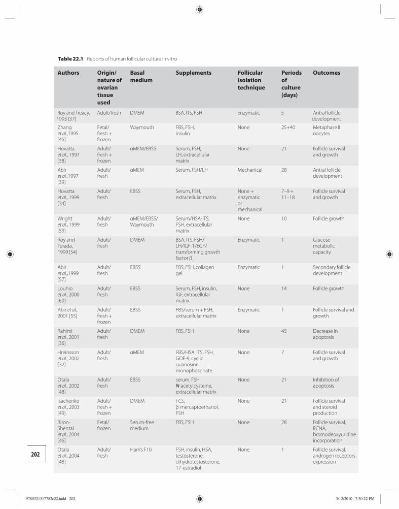
202
Table 22.1. Reports of human follicular culture in vitro
Authors Origin/nature of ovarian tissue used
Basal medium
Supplements Follicular isolation technique
Periods of culture (days)
Outcomes
Roy and Treacy, 1993 [37]
Adult/fresh DMEM BSA, ITS, FSH Enzymatic 5 Antral follicle development
Zhang et al. ,1995 [45]
Fetal/fresh + frozen
Waymouth FBS, FSH,insulin
None 25+40 Metaphase II oocytes
Hovatta et al., 1997 [38]
Adult/fresh + frozen
αMEM/EBSS Serum, FSH, LH, extracellular matrix
None 21 Follicle survival and growth
Abir et al., 1997 [39]
Adult/fresh
αMEM Serum, FSH/LH Mechanical 28 Antral follicle development
Hovatta et al., 1999 [34]
Adult/fresh
EBSS Serum, FSH, extracellular matrix
None + enzymatic or mechanical
7–9 + 11–18
Follicle survival and growth
Wright et al., 1999 [59]
Adult/fresh
αMEM/EBSS/Waymouth
Serum/HSA-ITS, FSH, extracellular matrix
None 10 Follicle growth
Roy and Terada, 1999 [54]
Adult/fresh
DMEM BSA, ITS, FSH/LH/IGF-1/EGF/transforming growth factor β 1
Enzymatic 1 Glucose metabolic capacity
Abir et al., 1999 [57]
Adult/fresh
EBSS FBS, FSH, collagen gel
Enzymatic 1 Secondary follicle development
Louhio et al., 2000 [60]
Adult/fresh
EBSS Serum, FSH, insulin, IGF, extracellular matrix
None 14 Follicle growth
Abir et al., 2001 [55]
Adult/fresh + frozen
EBSS FBS/serum + FSH, extracellular matrix
Enzymatic 1 Follicle survival and growth
Rahimi et al., 2001 [36]
Adult/fresh
DMEM FBS, FSH None 45 Decrease in apoptosis
Hreinsson et al., 2002 [32]
Adult/fresh
αMEM FBS/HSA, ITS, FSH, GDF-9, cyclic guanosine monophosphate
None 7 Follicle survival and growth
Otala et al., 2002 [48]
Adult/fresh
EBSS serum, FSH, N -acetylcysteine, extracellular matrix
None 21 Inhibition of apoptosis
Isachenko et al., 2003 [49]
Adult/fresh + frozen
DMEM FCS, β-mercaptoethanol, FSH
None 21 Follicle survival and steroid production
Biron-Shental et al., 2004 [46]
Fetal/frozen
Serum-free medium
FBS, FSH None 28 Follicle survival, PCNA, bromodeoxyuridine incorporation
Otala et al., 2004 [48]
Adult/fresh
Ham’s F10 FSH, insulin, HSA, testosterone, dihydrotestosterone, 17-estradiol
None 1 Follicle survival, androgen receptors expression
9780521517782c22.indd 2029780521517782c22.indd 202 3/12/2010 7:30:22 PM3/12/2010 7:30:22 PM
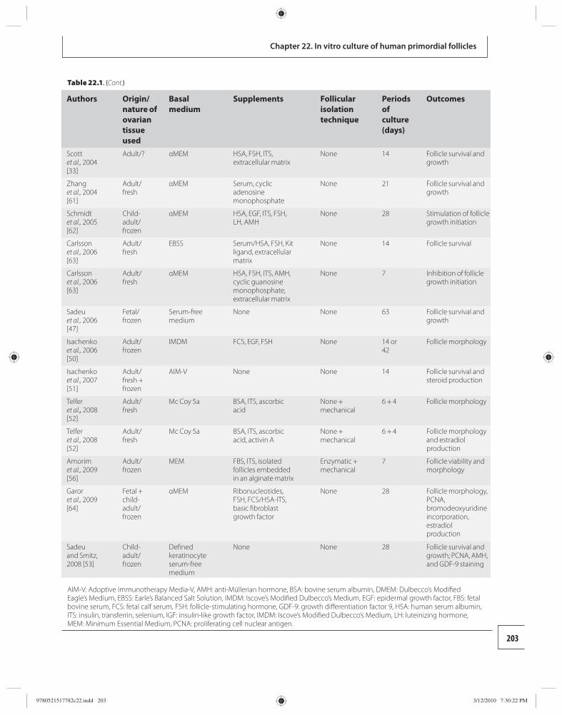
203
Chapter 22. In vitro culture of human primordial follicles
Authors Origin/nature of ovarian tissue used
Basal medium
Supplements Follicular isolation technique
Periods of culture (days)
Outcomes
Scott et al., 2004 [33]
Adult/? αMEM HSA, FSH, ITS, extracellular matrix
None 14 Follicle survival and growth
Zhang et al., 2004 [61]
Adult/fresh
αMEM Serum, cyclic adenosine monophosphate
None 21 Follicle survival and growth
Schmidt et al., 2005 [62]
Child-adult/frozen
αMEM HSA, EGF, ITS, FSH, LH, AMH
None 28 Stimulation of follicle growth initiation
Carlsson et al., 2006 [63]
Adult/fresh
EBSS Serum/HSA, FSH, Kit ligand, extracellular matrix
None 14 Follicle survival
Carlsson et al., 2006 [63]
Adult/fresh
αMEM HSA, FSH, ITS, AMH, cyclic guanosine monophosphate, extracellular matrix
None 7 Inhibition of follicle growth initiation
Sadeu et al., 2006 [47]
Fetal/frozen
Serum-free medium
None None 63 Follicle survival and growth
Isachenko et al., 2006 [50]
Adult/frozen
IMDM FCS, EGF, FSH None 14 or 42
Follicle morphology
Isachenko et al., 2007 [51]
Adult/fresh + frozen
AIM-V None None 14 Follicle survival and steroid production
Telfer et al., 2008 [52]
Adult/fresh
Mc Coy 5a BSA, ITS, ascorbic acid
None + mechanical
6 + 4 Follicle morphology
Telfer et al., 2008 [52]
Adult/fresh
Mc Coy 5a BSA, ITS, ascorbic acid, activin A
None + mechanical
6 + 4 Follicle morphology and estradiol production
Amorim et al., 2009 [56]
Adult/frozen
MEM FBS, ITS, isolated follicles embedded in an alginate matrix
Enzymatic + mechanical
7 Follicle viability and morphology
Garor et al., 2009 [64]
Fetal + child-adult/frozen
αMEM Ribonucleotides, FSH, FCS/HSA-ITS, basic fi broblast growth factor
None 28 Follicle morphology, PCNA, bromodeoxyuridine incorporation, estradiol production
Sadeu and Smitz, 2008 [53]
Child-adult/frozen
Defi ned keratinocyte serum-free medium
None None 28 Follicle survival and growth; PCNA, AMH, and GDF-9 staining
AIM-V: Adoptive Immunotherapy Media-V, AMH: anti-Müllerian hormone, BSA: bovine serum albumin, DMEM: Dulbecco’s Modifi ed Eagle’s Medium, EBSS: Earle’s Balanced Salt Solution, IMDM: Iscove’s Modifi ed Dulbecco’s Medium, EGF: epidermal growth factor, FBS: fetal bovine serum, FCS: fetal calf serum, FSH: follicle-stimulating hormone, GDF-9: growth diff erentiation factor 9, HSA: human serum albumin, ITS: insulin, transferrin, selenium, IGF: insulin-like growth factor, IMDM: Iscove’s Modifi ed Dulbecco’s Medium, LH: luteinizing hormone, MEM: Minimum Essential Medium, PCNA: proliferating cell nuclear antigen.
Table 22.1. (Cont.)
9780521517782c22.indd 2039780521517782c22.indd 203 3/12/2010 7:30:22 PM3/12/2010 7:30:22 PM
204
Section 5. Cryopreservation of ovarian tissue
fi brous interstitial tissue [ 39 , 52 ]. Using the enzymatic method, human follicles have been isolated aft er partial or total disaggregation of ovarian tissue with proteo-lytic enzymes such as collagenase I, II, or IV [ 34 , 54 – 56 ], mixtures of collagenase and deoxyribonuclease [ 37 , 57 ], or liberase [ 58 ]. Mechanical isolation is oft en facili-tated aft er enzymatic application for partial diges-tion. Both enzymatic and mechanical methods have their limitations. Mechanical isolation is a tedious and time- consuming process because of the fi brous nature of ovarian interstitial tissue in larger mammals and in particular in the human. Furthermore, the recovery in terms of number of follicles isolated mechanically is usually very low. Enzymatic isolation generally has a negative eff ect on follicle integrity. Th e enzymes oft en disaggregate the basal membrane and theca cells, lead-ing to disruption of oocyte–granulosa cell communica-tions in culture. Th ese disruptions oft en result in a poor follicular development in vitro. Th erefore, most of the studies using isolated follicles are begun from prean-tral follicles ( Table 22.1 ), as they are bigger with a basal membrane that is more apparent and easier to isolate.
Regardless of these disadvantages, culture of iso-lated follicles is of signifi cant importance. Follicle iso-lation helps to determine the follicular content within the tissue fragment, and culturing “empty” (follicle-free) tissue pieces can be avoided. To prove that the isolation techniques are safe for application in larger animals and humans is not an easy task. Th e validation of the methods was carried out in the rodent models, where secondary follicles were isolated from mouse ovaries using either enzymatic [ 2 ] or mechanical [ 65 ] methods. Th ese follicles were proven to be capable of growing in culture and producing developmentally competent oocytes and live off spring.
Basal culture conditions Several basal culture media have been used to grow follicles from diff erent species either within ovar-ian cortical tissue or as isolated follicle units. Th ese include balanced salt solution (BSS), Iscove’s Modifi ed Dulbecco’s Medium (IMDM), AIM-V, minimum essential medium alpha (α-MEM), D-minimum essen-tial medium (D-MEM), and Waymouth’s medium for humans ( Table 22.1 ). Insulin, transferrin, and selenium alone or in combination with human serum albumin (HSA) and/or serum have been the major components added as supplements to the media with the aim of supporting metabolic and hormonal activities during follicle growth in vitro. In most of the studies, serum
from human or animal origin is used. In others, HSA or bovine serum albumin (BSA) has been used instead of serum with signifi cant results, and a few studies have used albumin-free media [ 53 ] ( Table 22.1 ).
Th e culture containers have also varied, from sim-ple test tubes to plastic culture well plates [ 45 , 47 ]. Well plates have been combined with either uncoated [ 38 , 39 ] or coated tissue culture inserts [ 32 , 33 , 38 , 45 , 46 ]. Th e extracellular matrix compounds utilized to coat the inserts have included collagen I [ 55 ], a mixture of lam-inin, collagen IV, and proteoglycans [ 38 ], and collagen I and collagen III [ 45 ]. Low concentration of growth fac-tors (basal fi broblast growth factor [bFGF], epidermal growth factor [EGF], insulin-like growth factor [IGF] 1, platelet-derived growth factor, nerve growth factor, TGF-β) can be included in the composition of some manufactured extracellular matrix systems. Th ese factors may also help the follicular growth. Using iso-lated follicles, the extracellular matrix is clearly miss-ing; therefore, agar [ 37 ], collagen gel overlay [ 55 , 57 ], or calcium alginate droplets [ 56 ] have been used as sup-porting components. Th e rational is to help to preserve the three-dimensional architecture of the follicles and their environment; the cohesion of follicle cells in vitro in such systems is more likely to approximate the in vivo condition, which is desirable to maintain physiological control mechanisms during culture [ 66 ].
Whole ovary organ culture has been the main approach to activate the growth of resting primordial follicles because the growth of these follicles aft er isola-tion from the stromal environment has been unsuccess-ful. Whole ovary culture has been carried out in species with small ovaries, such as the rodents [ 3 , 67 – 70 ]. Culture of isolated follicles has been confi ned mostly to the study of primary and secondary follicles. Th ese follicles have been isolated from human [ 34 , 37 , 39 , 56 ], goat [ 71 ], cow [ 17 , 72 – 75 ], pig [ 19 , 20 , 76 ], and rodent [ 1 , 3 , 65 , 77 – 81 ] ovaries, as well as from sheep ovaries at the primordial stage [ 82 ] and they have been grown in culture to various endpoint follicle stages.
Supplementation of in vitro culture media Activation of primordial follicles cultured within tissue slices Knowledge of the function of the majority of growth factors acting on the activation of resting primor-dial follicles into the growth phase, with subsequent
9780521517782c22.indd 2049780521517782c22.indd 204 3/12/2010 7:30:22 PM3/12/2010 7:30:22 PM
205
Chapter 22. In vitro culture of human primordial follicles
follicular development to further stages of folliculo-genesis, has been mainly obtained from animal mod-els [ 83 ]. Th e development of follicle culture systems for larger mammals has been attempted in several species including goat [ 84 ], sheep [ 85 ], cow [ 86 , 87 ], and baboon [ 88 ]. Several culture media used to grow human follicles in vitro have been supplemented with these growth factors to assess their potential physio-logical signifi cance in regulating the earliest stages of follicular development in humans.
Th e benefi cial eff ects of several components added to culture media on the activation and/or regulation of the processes of early folliculogenesis in culture has been experimentally reported in several studies, while the biological pathway remains to be determined. For example, it has been shown that supplementing the medium with FSH reduced follicle atresia and increased follicle diameter [ 59 ] ( Table 22.1 ). However, FSH receptors in primordial and primary follicles remain to be shown at these stages. Other factors, such as insulin, IGF-1 and IGF-2 [ 60 ], secondary messen-gers such as 8-bromoguanosine 3′,5′-cyclic monophos-phate [ 33 ], and cyclic adenosine 3′,5′-monophosphate [ 61 ] have also positive eff ects on the development of the early stages of human follicles. Data from studies on human [ 32 ] and goat [ 89 ] follicle culture showed that addition of 200 ng/ml GDF-9 enhances activa-tion of primordial follicles and stimulates the transi-tion from primary to secondary follicles. Th is in vitro observation suggested that GDF-9 might have sup-portive eff ects on the earliest stages of ovarian follicles. It was also demonstrated that AMH inhibits the early development of human [ 90 ] and mouse [ 91 ] follicles in culture. However, the presence of AMH in culture has been shown to increase the development of human follicles in a single study [ 62 ].
Th e addition of KL to the medium supported the survival of human follicles [ 63 ] and stimulated the development of rodent primordial follicles into pri-mary stage [ 69 ]. Addition of bFGF has been shown to increase primordial follicle transition in neonatal rat ovary organ cultures [ 69 , 92 ] and in adult human ovar-ian cortex, but at a 10 times higher concentration and this was not observed in fetal tissue [ 64 ]. Activin A has shown an enhancement of oocyte development in cul-tured ovine preantral follicles [ 93 ] and the growth of rat preantral follicles [ 94 ]. Th is glycoprotein also promotes antrum formation in rat follicles as well as in human [ 52 , 94 ]. Keratinocyte growth factors [ 95 ], leukemia inhibitory factor [ 96 ], BMP-4 [ 97 ], and BMP-7 [ 98 ]
have also been shown to enhance the primordial to primary follicle transition in neonatal rat ovary organ cultures, while stromal-derived factor-1 and its recep-tor CXCR4 [ 70 ] inhibited that transition. Th e develop-ment of goat primordial follicles was stimulated by FSH and EGF in cultures of goat ovarian tissue [ 84 ]. Factors that had been studied previously in animal models and subsequently used in human confi rmed that factors likely had eff ects that were both species dependent and follicular stage dependent, as was already reported for activin A [ 52 ] and bFGF [ 64 ]. For example, activin A appeared folliculogenetic in the prepubertal mouse but not in adults [ 99 ].
Culture of isolated early-stage follicles Attempts to grow isolated human follicles in vitro have resulted in follicle development along diff erent stages of folliculogenesis. In vitro culture of isolated follicles has been carried out for larger mammals, and the eff ect of diff erent growth factors has been investigated. Both EGF and bFGF were shown to sustain oocyte survival during culture of isolated goat preantral follicles, while IGF-1 not only supported follicle survival but also stimulated follicle growth [ 71 ]. Insulin, FSH, IGF-1, EGF, IGF-2, and vascular endothelial growth factor were shown to stimulate follicle growth in bovine pre-antral follicle culture [ 17 , 75 , 100 ]. In humans, isolated follicles are mostly cultured from the secondary or pre-antral stage ( Table 22.1 ). Primary follicles have been isolated in a single study [ 57 ].
Two-step systems Ovarian cortex culture followed by isolated follicle culture has been used in rodent species with success, as pups were born. Th is two-step strategy used intact ovar ies from newborn mice, which were organ cul-tured for 8 days. Oocyte–granulosa cell complexes iso-lated from these were cultured for a further 14 days [ 2 ]. Th e composition of the media was subsequently also modifi ed during the culture periods with the addition of FSH and insulin, transferrin, selenium [ 2 , 3 ]. Human ovary culture is impossible because of its larger size. However, an adaptation of the previous methods has been used with human follicles [ 34 , 52 ]. Primordial and primary follicles were grown within cortical pieces for 6 to 9 days, and then preantral follicles were dis-sected from the strips using 25 gauge needles or with collagenase II. Care was taken to include the adher-ent theca/stromal cells attached to the follicles. Th e medium was also modifi ed. In the most recent study
9780521517782c22.indd 2059780521517782c22.indd 205 3/12/2010 7:30:22 PM3/12/2010 7:30:22 PM
206
Section 5. Cryopreservation of ovarian tissue
using this approach, activin A was added during the second step of the culture [ 52 ].
Methods for evaluation of follicular culture Histological analysis still remains the gold standard method in 2008. Several histomorphometric meth-ods can be used to investigate follicular development in vitro. Regardless of the species, the following mor-phological and morphometric parameters can be ana-lyzed: follicle viability, antral cavity formation, oocyte viability, follicle diameter, and enclosed oocyte diam-eter. Unlike culture of isolated follicles, which can be quickly monitored and evaluated under an inverted microscope, culture of ovarian cortical tissue can only be analyzed aft er processing of tissues for histological examination. Th is approach entails the tissues being fi xed either in formalin or in Bouin’s solution, paraf-fi n embedded, and serially sectioned at the end of each culture period. Th e sections are placed in order on glass slides and stained with hematoxylin, eosin, and saff ran before being analyzed by light microscope for the presence of follicles and the stages of development. Diff erent approaches have been used for estimating follicle numbers, based on histological analysis of tis-sue sections [ 101 ].
Th e oocyte nucleolus is commonly used as a cen-tral mark to avoid double counting of the same folli-cle. Follicle viability is determined based upon defi ned morphological criteria of atresia, such as pyknosis of nuclei, basal membrane integrity, and granulosa cell organization. Follicular development in vitro is assessed by counting the number of follicles of various stages in control (uncultured or cultured in the absence of growth factors under study) versus the number in studied samples throughout the culture period. To assess follicle growth, follicle and oocyte diameters are measured and the number of layers of granulosa cells is counted. Although conventional histological analysis remains an obligatory method in the study of ovarian tissue culture, the major disadvantage of this technique is that it is tedious. As follicles are usually grouped in nests rather than regularly distributed in the cortex, extrapolation from limited tissue pieces to total ovary remains inaccurate.
Transmission electron microscopy provides detailed analysis of follicular ultrastructure and has been used to evaluate intracellular integrity of bovine [ 74 ] and human [ 47 ] follicles aft er prolonged culture
periods. Signs of intracellular damage include the pres-ence of cytoplasmic vacuoles, swollen mitochondria, lipid droplets, chromatin condensation, and wrinkled or ruptured nuclear membrane. Morphological dif-ferences between apoptotic and atretic cells have also been demonstrated by transmission electron micros-copy, and its contribution to the study of ovarian tissue has been recently reviewed [ 102 ].
Follicular viability and cell death in human ovar-ian cortex have been evaluated with fl uorescent mark-ers using fl uorescent microscopy. Th e polyanionic dye calcein is retained in living cells and the cleavage product induced by cellular esterase activity provides an intense, uniform green fl uorescence in the cyto-plasm. Th e nuclei of all cells with damaged membrane are counterstained with ethidium homodimer-1 (red fl uor escence) [ 56 , 103 ]. In binding DNA of nuclei, ethidium homodimer-1 increases in fl uorescence. Considering the huge volume of the oocyte’s cyto-plasm compared with that in other cells of the ovarian cortex, the follicles are easily observed within the tis-sue. Th e oocyte and the granulosa cells can be detailed within the tissue through confocal microscopy [ 103 ] or with isolated follicles [ 56 ]. Th is technique has also been used as a quick method for scoring the density of follicles within human ovarian cortex using sim-ple fl uorescent micros copy [ 41 ]. Similar results can be obtained with rhodamine staining, which refl ects mitochondrial activity. Rhodamine accumulates into active mitochondria with a membrane potential [ 104 ].
Evaluation of the adequacy of culture conditions has been investigated by accounting for follicle death through apoptosis. Morphological characteristics of apoptosis at the early stage include chromatin con-densation and cell shrinkage. Terminal deoxyribo-nucleotidyl transferase-mediated deoxyuridine triphosphate nick end labeling (TUNEL) has been used to detect fragmented DNA in nuclei of granulosa cells and oocyte from goat [ 84 ]. However, false-posi-tive results with this method have been reported [ 36 ]. Apoptosis is an active process that requires expres-sion of specifi c proteases such as caspases. Cultured ovarian tissue pieces have been stained with anti-cas-pase-3 or anti-active caspase-3 [ 36 , 48 ] . Furthermore, pyknotic nuclei can be revealed on tissue sections by their incubation with the fl uorescent dye SYTOX, which binds with high affi nity to the triplex structure of the nucleic acids of dead cells [ 36 ]. Th is DNA stain-ing allows easier nuclei visualization in large areas of tissue, and pictures can be digitalized for computer
9780521517782c22.indd 2069780521517782c22.indd 206 3/12/2010 7:30:23 PM3/12/2010 7:30:23 PM
207
Chapter 22. In vitro culture of human primordial follicles
analysis. Obviously, this staining concerns the follicles and the stromal tissue.
Cell proliferation is a key marker during in vitro culture as evidence that viable follicles are able to grow during the culture period. Granulosa cell expression of proliferating cell nuclear antigen (PCNA) in culture is a valuable marker for cell replication. Th is is a nuclear protein that plays an essential role in cell cycle regula-tion, and it is an important mediator of cell prolifera-tion; it can be detected by immunohistochemistry. Th e correlation between PCNA production and initiation of growth has been shown in fetal bovine and baboon fol-licles [ 88 ]. Th e granulosa cells were strongly immuno-stained for PCNA when their shape changed from fl attened to cuboidal [ 86 , 88 ]. Positive PCNA immuno-staining was also demonstrated in granulosa cells of growing follicles aft er a few days of culture in human tissue [ 47 ]. Th is fi nding could indicate some DNA syn-thesis activity at the early stage of growth initiation. Granulosa cell proliferation has also been demon-strated in cultured follicles using a bromodeoxyuridine incorporation technique. Bromodeoxyuridine can be incorporated into newly synthesized DNA of replicat-ing cells during the S-phase of the cell cycle, and it is, therefore, used as a marker of DNA synthesis in mouse [ 105 ], cow [ 72 ], and human tissue [ 46 , 64 ]. Classical radioactive thymidine ([ 3 H]-thymidine) uptake has also been used to demonstrate DNA synthesis in gran-ulosa cells of isolated follicles in culture. In other stud-ies, [ 3 H]-thymidine was added to the culture medium and the follicles or oocytes–cumulus complexes were examined aft er a few hours for [ 3 H]-thymidine incor-poration [ 37 , 106 ].
Functional approaches have also been used for the evaluation of follicular growth with expression of stage-specifi c markers and the measure of secreted proteins such as estradiol. Follicle stage-specifi c factors such as GDF-9 and AMH are known to be involved in the regulation and initiation of the processes of early fol-liculogenesis in vivo and have been used as functional markers during follicle culture. Th eir expression in oocytes and granulosa cells in human cultured follicles has been observed aft er a 21-day in vitro culture period [ 53 ]. A functional approach to evaluation of in vitro cultured follicles can also be achieved using molecular biology techniques. Gene regulation is the basis for cel-lular diff erentiation, cell growth, and morphogenesis, and the amount of gene expression has been used to determine the function of the cells. Quantitative real-time polymerase chain reaction can be integrated in
the evaluation of cultured follicles. Th is approach is more appropriate for isolated follicles and was investi-gated for GDF-9 and AMH in mouse cultured follicles [ 107 ].
Th e conditioned media collected at each time point of culture can provide information as to the degree of somatic cell diff erentiation in vitro, which should be related to a follicular growth. Several secretion prod-ucts can be measured to evaluate follicle growth dur-ing culture. Absence or low estrogen production could be related either to absence or incomplete aromatiza-tion in granulosa cells or to the non-functionality of the theca compartment. Th e diff erential analysis can be made by adding androgen (androstenedione) to the culture medium. Estradiol production was observed and measured with radioimmunoanalysis [ 39 ] or enzyme-linked immunosorbent assay [ 52 ] during in vitro culture of human tissue. A heterogeneous com-petitive magnetic separation immunoassay analysis was also used to measure estradiol and progesterone in an unchanged medium during the whole 14 day cul-ture period [ 51 ].
Outcomes of follicular culture In vivo, the growth initiation of primordial follicles has an evolution over several months [ 108 ]. However, when media supplemented with growth factors were used, massive activation of primordial follicles to initi-ate growth in vitro within human adult cortical ovar-ian tissues occurred within the fi rst week of culture [ 32 , 34 , 59 , 107 ]. When human fetal ovarian tissues were cultured in growth factor-supplemented media, the growth initiation of primordial follicles took more than 3 weeks [ 45 ]. In a simplifi ed long-term culture system using a defi ned serum-free medium, initiation of follicle growth and progression to subsequent stages of development took almost 2 months [ 47 ] . Culture of human ovarian cortical tissue for 1–3 weeks resulted in 30–53% of the surviving follicles developing into the secondary stage [ 32 , 34 , 38 ]. Th ere was no correla-tion between the development of secondary follicles and a corresponding increase in oocyte diameter, as described in vivo: oocyte growth was limited or not detectable. Th e development of secondary follicles in vitro was characterized by an increase in the number of granulosa cells through cell proliferation but a limited increase in oocyte diameter.
Human primary follicles have been isolated and cultured for 1 day. Follicular growth was observed, with the increase of their diameter from approximately
9780521517782c22.indd 2079780521517782c22.indd 207 3/12/2010 7:30:23 PM3/12/2010 7:30:23 PM
208
Section 5. Cryopreservation of ovarian tissue
34 μm to approximately 69 μm [ 55 , 57 ]. Human pre-antral follicles have been cultured as isolated follicles from 5 days [ 37 ] to 4 weeks [ 39 ]. Th e starting diameters of cultured follicles ranged between 220 μm [ 54 ] and 350 μm [ 39 ]. Already aft er 5 days of culture, the follicles had developed to reach the antral stage, and granulosa cells proliferation in vitro was observed [ 37 ]. Aft er 4 weeks of culture, 70% of the isolated preantral fol-licles had developed an antral cavity (follicular size up to 125 μm). However, atretic cells were observed within the granulosa cells, and follicular development beyond the early antral stage could not be achieved, likely because of suboptimal culture conditions. At the end of culture, only 10–20% of the isolated preantral fol licles contained an enclosed oocyte. Th e missing oocytes in the majority of follicles might have degenerated by atresia before (as a physiological process) or aft er being cultured [ 39 ]. Th ese culture experiments indicate that preantral follicle development can be achieved in vitro, leading to oocyte growth and granulosa cell diff eren-tiation, but at a very low yield.
A two-step strategy, with the addition of activin A in the second period, led to antral cavity formation with signifi cantly less atresia (than without activin A). Th ese follicles grew from primordial/primary stage to the antral stage, reaching a size of 143 μm. In these fol-licles, oocyte growth was also observed: oocyte diam-eter was 31.9 μm for follicles grown in control medium and 51.3 μm for oocytes grown with activin A supple-mentation in the second step [ 52 ].
Conclusions Follicle culture systems off er interesting experimen-tal models to address several biological issues that are important for our understanding of human folliculo-genesis. Th e investigation of mechanisms regulating primordial follicle activation and early development are the major points in the studies described here, and several have observed follicular growth up to the early antral stage. Th is stage appears to be a boundary that is diffi cult to cross.
Regardless of the fi nal purpose of the study, the cul-ture technique is determined by the size of the ovary (the species), the developmental stage of the follicle of interest, and the duration of folliculogenesis. Success of a culture system will fi nally be judged by the pro-duction of developmentally competent oocytes, able to be fertilized and to produce healthy births. Th is goal has only been reached in the mouse model so far. Th e human ovary is very diff erent from that of the rodent
in its size, structure, and folliculogenesis duration. Th erefore, studies on human tissue are mandatory. We are confronted by several problems when research on human tissue is being planned. Th e fi rst diffi culty is to obtain human material, as ethic and legal con-siderations diff er from one country to another. Th e second major problem with human tissue is its fol-licular density, which can be extremely low, and its heterogeneity. Th is leads to a need for a high number of samples. Th e stromal tissue in the ovaries of larger mammals is fi brous and dense, preventing the easy isolation of the follicles and the development of a suc-cessful culture system. Fetal ovaries could be consid-ered as a fi rst choice of material because of their high follicle density and the soft ness of their tissue. Th e few reports with this approach showed that healthy follicles grown in culture are morphologically com-parable to those grown in vivo. However, because its function and physiology might diff er from the adult ovary, the results obtained with fetal tissue should be ultimately confi rmed with adult tissue. Although these diffi culties cannot be denied, the number of studies on human ovarian tissue has increased in the last few years and this provides hope that these prob-lems can be solved.
Although the requirements for normal follicle growth at the earliest stages are still unknown and remain to be determined, follicular growth has been shown in several studies and this growth is usually applied to the majority of the follicle types cultured. Follicular atresia is a major concern during in vitro culture, but is also supposed to be at a high level in nor-mal physiology. Considering the long time required for complete follicular development in larger mam-mals, and the diffi culties with long-term maintenance of tissue culture, it is likely that a multistep procedure should be applied to the culture of primordial follicles of larger mammals in order to achieve a successful cul-ture system.
Follicular growth up to the antral stage is obtained aft er diff erent culture time periods but usually shorter than what is believed to happen in vivo. It is likely that the in vitro conditions cannot mimic the nor-mal physiology, and so this diff erent timing should not be surprising. However, potential side eff ects of this modifi cation of timing have to be determined. For example, cellular development under culture conditions, and especially for long duration, is more susceptible to genetic modifi cations. Th erefore, the genetic status of in vitro grown oocytes will have to
9780521517782c22.indd 2089780521517782c22.indd 208 3/12/2010 7:30:23 PM3/12/2010 7:30:23 PM
209
Chapter 22. In vitro culture of human primordial follicles
be studied before using them for human fertilization and embryo transfer. Long-term culture might result in unappropriated cellular diff erentiation and genetic modifi cation. Hence, molecular techniques should be considered to analyze the normal methylation pattern of primary imprints in oocytes.
Th e fi nal measure of success will be the ability of the in vitro matured oocytes to fertilize and produce healthy off springs. Combination of cryopreservation of ovarian cortical tissue with successful in vitro cul-ture of follicles will bring a major breakthrough in fer-tility preservation. Th e availability of such techniques might increase the demand from a range of women, not just those with cancer.
References 1. Eppig JJ , Schroeder AC. Capacity of mouse oocytes
from preantral follicles to undergo embryogenesis and development to live young aft er growth, maturation, and fertilization in vitro . Biol Reprod 1989; 41: 268 –276.
2. Eppig JJ , O’Brien MJ. Development in vitro of mouse oocytes from primordial follicles . Biol Reprod 1996; 54: 197 –207.
3. O’Brien MJ , Pendola JK , Eppig JJ. A revised protocol for in vitro development of mouse oocytes from primordial follicles dramatically improves their developmental competence . Biol Reprod 2003; 68: 1682 –1686.
4. Donnez J , Dolmans MM , Demylle D , et al. Livebirth aft er orthotopic transplantation of cryopreserved ovarian tissue . Lancet 2004; 364: 1405 –1410.
5. Meirow D , Levron J , Eldar-Geva T , et al. Pregnancy aft er transplantation of cryopreserved ovarian tissue in a patient with ovarian failure aft er chemotherapy . N Engl J Med 2005; 353: 318 –321.
6. Meirow D , Levron J , Eldar-Geva T , et al. Monitoring the ovaries aft er autotransplantation of cryopreserved ovarian tissue: endocrine studies, in vitrofertilization cycles, and live birth . Fertil Steril 2007; 87: 418. e7 –e15.
7. Demeestere I , Simon P , Buxant F , et al. Ovarian function and spontaneous pregnancy aft er combined heterotopic and orthotopic cryopreserved ovarian tissue transplantation in a patient previously treated with bone marrow transplantation: case report . Hum Reprod 2006; 21: 2010 –2014.
8. Demeestere I , Simon P , Emiliani S , et al. Fertility preservation: successful transplantation of cryopreserved ovarian tissue in a young patient previously treated for Hodgkin ’ s disease . Oncologist 2007; 12: 1437 –1442.
9. Silber SJ , DeRosa M , Pineda J , et al. A series of monozygotic twins discordant for ovarian failure: ovary transplantation (cortical versus microvascular) and cryopreservation . Hum Reprod 2008; 23: 1531 –1537.
10. Andersen CY , Rosendahl M , Byskov AG , et al. Two successful pregnancies following autotransplantation of frozen/thawed ovarian tissue . Hum Reprod 2008; 23: 2266 –2272.
11. Shaw JM , Bowles J , Koopman P , et al. Fresh and cryopreserved ovarian tissue samples from donors with lymphoma transmit the cancer to graft recipients . Hum Reprod 1996; 11: 1668 –1673.
12. Meirow D , Ben Yehuda D , Prus D , et al. Ovarian tissue banking in patients with Hodgkin ’ s disease: is it safe ? Fertil Steril 1998; 69: 996 –998.
13. Meirow D , Hardan I , Dor J , et al. Searching for evidence of disease and malignant cell contamination in ovarian tissue stored from hematologic cancer patients . Hum Reprod 2008; 23: 1007 –1013.
14. Kyono K , Uto H , Toya M , et al. Potential indications for ovarian autotransplantation based on the analysis of 5571 autopsy fi ndings of females with malignant diseases under the age of 40 in Japan . Fertil Steril 2008; 90 (Suppl 1): S267 .
15. Cecconi S , Barboni B , Coccia M , et al. In vitro development of sheep preantral follicles . Biol Reprod 1999; 60: 594 –601.
16. Newton H , Picton H , Gosden RG. In vitro growth of oocyte–granulosa cell complexes isolated from cryopreserved ovine tissue . J Reprod Fertil 1999; 115: 141 –150.
17. Gutierrez CG , Ralph JH , Telfer EE , et al. Growth and antrum formation of bovine preantral follicles in long-term culture in vitro . Biol Reprod 2000; 62: 1322 –1328.
18. McCaff ery FH , Leask R , Riley SC , et al. Culture of bovine preantral follicles in a serum-free system: markers for assessment of growth and development . Biol Reprod 2000; 63: 267 –273.
19. Hirao Y , Nagai T , Kubo M , et al. In vitro growth and maturation of pig oocytes . J Reprod Fertil 1994; 100: 333 –339.
20. Wu J , Emery BR , Carrell DT. In vitro growth, maturation, fertilization, and embryonic development of oocytes from porcine preantral follicles . Biol Reprod 2001; 64: 375 –381.
21. Dong J , Albertini DF , Nishimori K , et al. Growth diff erentiation factor-9 is required during early ovarian folliculogenesis . Nature 1996; 383: 531 –535.
22. Galloway SM , McNatty KP , Cambridge LM , et al. Mutations in an oocyte-derived growth factor gene (BMP15) cause increased ovulation rate and infertility in a dosage-sensitive manner . Nat Genet 2000; 25: 279 –283.
23. Durlinger AL , Kramer P , Karels B , et al. Control of primordial follicle recruitment by anti-Mullerian hormone in the mouse ovary . Endocrinol 1999; 140: 5789 –5796.
9780521517782c22.indd 2099780521517782c22.indd 209 3/12/2010 7:30:23 PM3/12/2010 7:30:23 PM
210
Section 5. Cryopreservation of ovarian tissue
24. Huang EJ , Manova K , Packer AI , et al. Th e murine steel panda mutation aff ects kit ligand expression and growth of early ovarian follicles . Dev Biol 1993; 157: 100 –109.
25. Kuroda H , Terada N , Nakayama H , et al. Infertility due to growth arrest of ovarian follicles in Sl/Slt mice . Dev Biol 1988; 126: 71 –79.
26. Juengel JL , McNatty KP. Th e role of proteins of the transforming growth factor-beta superfamily in the intraovarian regulation of follicular development . Hum Reprod Update 2005; 11: 1143 –1160.
27. Matzuk MM , Lamb DJ. Genetic dissection of mammalian fertility pathways . Nat Cell Biol 2002; 4 (Suppl): s41–s9 .
28. Gook DA , McCully BA , Edgar DH , et al. Development of antral follicles in human cryopreserved ovarian tissue following xenograft ing . Hum Reprod 2001; 16: 417 –422.
29. Newton H , Aubard Y , Rutherford A , et al. Low temperature storage and graft ing of human ovarian tissue . Hum Reprod 1996; 11: 1487 –1491.
30. Oktay K , Nugent D , Newton H , et al. Isolation and characterization of primordial follicles from fresh and cryopreserved human ovarian tissue . Fertil Steril 1997; 67: 481 –486.
31. Oktay K , Newton H , Mullan J , et al. Development of human primordial follicles to antral stages in SCID/hpg mice stimulated with follicle stimulating hormone . Hum Reprod 1998; 13: 1133 –1138.
32. Hreinsson JG , Scott JE , Rasmussen C , et al. Growth diff erentiation factor-9 promotes the growth, development, and survival of human ovarian follicles in organ culture . J Clin Endocrinol Metab 2002; 87: 316 –321.
33. Scott JE , Carlsson IB , Bavister BD , et al. Human ovarian tissue cultures: extracellular matrix composition, coating density and tissue dimensions . Reprod Biomed Online 2004; 9: 287 –293.
34. Hovatta O , Wright C , Krausz T , et al. Human primordial, primary and secondary ovarian follicles in long-term culture: eff ect of partial isolation . Hum Reprod 1999; 14: 2519 –2524.
35. Nisolle M , Casanas-Roux F , Qu J , et al. Histologic and ultrastructural evaluation of fresh and frozen–thawed human ovarian xenograft s in nude mice . Fertil Steril 2000; 74: 122 –129.
36. Rahimi G , Isachenko E , Sauer H , et al. Measurement of apoptosis in long-term cultures of human ovarian tissue . Reproduction 2001; 122: 657 –663.
37. Roy SK , Treacy BJ. Isolation and long-term culture of human preantral follicles . Fertil Steril 1993; 59: 783 –790.
38. Hovatta O , Silye R , Abir R , et al. Extracellular matrix improves survival of both stored and fresh human primordial and primary ovarian follicles in long-term culture . Hum Reprod 1997; 12: 1032 –1036.
39. Abir R , Franks S , Mobberley MA , et al. Mechanical isolation and in vitro growth of preantral and small antral human follicles . Fertil Steril 1997; 68: 682 –688.
40. van den Broecke R , Van Der Elst J , Liu J , et al. Th e female-to-male transsexual patient: a source of human ovarian cortical tissue for experimental use . Hum Reprod 2001; 16: 145 –147.
41. Schubert B , Canis M , Darcha C , et al. Human ovarian tissue from cortex surrounding benign cysts: a model to study ovarian tissue cryopreservation . Hum Reprod 2005; 20: 1786 –1792.
42. Schubert B , Canis M , Darcha C , et al. Follicular growth and estradiol follow-up aft er subcutaneous xenograft ing of fresh and cryopreserved human ovarian tissue . Fertil Steril 2008; 89: 1787 –1794.
43. Poirot C , Vacher-Lavenu MC , Helardot P , et al. Human ovarian tissue cryopreservation: indications and feasibility . Hum Reprod 2002; 17: 1447 –1452.
44. Sánchez M , Novella-Maestre E , Teruel J , et al. Th e Valencia Programme for Fertility Preservation . Clin Transl Oncol 2008; 10: 433 –438.
45. Zhang J , Liu J , Xu KP , et al. Extracorporeal development and ultrarapid freezing of human fetal ova . J Assist Reprod Genet 1995; 12: 361 –368.
46. Biron-Shental T , Fisch B , van den Hurk R , et al. Survival of frozen–thawed human ovarian fetal follicles in long-term organ culture . Fertil Steril 2004; 81: 716 –719.
47. Sadeu JC , Cortvrindt R , Ron-El R , et al. Morphological and ultrastructural evaluation of cultured frozen–thawed human fetal ovarian tissue . Fertil Steril 2006; 85 (Suppl 1): 1130 –1141.
48. Otala M , Erkkila K , Tuuri T , et al. Cell death and its suppression in human ovarian tissue culture . Mol Hum Reprod 2002; 8: 228 –236.
49. Isachenko E , Isachenko V , Rahimi G , et al. Cryopreservation of human ovarian tissue by direct plunging into liquid nitrogen . Eur J Obstet Gynecol Reprod Biol 2003; 108: 186 –193.
50. Isachenko V , Montag M , Isachenko E , et al. Eff ective method for in-vitro culture of cryopreserved human ovarian tissue . Reprod Biomed Online 2006; 13: 228 –234.
51. Isachenko V , Isachenko E , Reinsberg J , et al. Cryopreservation of human ovarian tissue: comparison of rapid and conventional freezing . Cryobiology 2007; 55: 261 –268.
52. Telfer E , McLaughlin M , Ding C , et al. A two step serum free culture system supports development of human oocytes from primordial follicles in the presence of activin . Hum Reprod 2008; 23: 1151 –1158.
53. Sadeu JC , Smitz J. Growth diff erentiation factor-9 and anti-Müllerian hormone expression in cultured human follicles from frozen–thawed ovarian tissue . Reprod Biomed Online 2008; 17: 537 –548.
54. Roy SK , Terada DM. Activities of glucose metabolic enzymes in human preantral follicles: in vitro
9780521517782c22.indd 2109780521517782c22.indd 210 3/12/2010 7:30:23 PM3/12/2010 7:30:23 PM
211
Chapter 22. In vitro culture of human primordial follicles
modulation by follicle-stimulating hormone, luteinizing hormone, epidermal growth factor, insulin-like growth factor I, and transforming growth factor beta1 . Biol Reprod 1999; 60: 763 –768.
55. Abir R , Fisch B , Nitke S , et al. Morphological study of fully and partially isolated early human follicles . Fertil Steril 2001; 75: 141 –146.
56. Amorim CA , Van Langendockt A , David A , Dolmans M-M , Donnez J. Survival of human pre-antral follicles aft er cryopreservation of ovarian tissue, follicular isolation and in vitro culture in a calcium alginate matrix . Hum Reprod 2009; 24 : 92 –99.
57. Abir R , Roizman P , Fisch B , et al. Pilot study of isolated early human follicles cultured in collagen gels for 24 hours . Hum Reprod 1999; 14: 1299 –1301.
58. Dolmans MM , Michaux N , Camboni A , et al. Evaluation of Liberase, a purifi ed enzyme blend, for the isolation of human primordial and primary ovarian follicles . Hum Reprod 2006; 21: 413 –420.
59. Wright CS , Hovatta O , Margara R , et al. Eff ects of follicle-stimulating hormone and serum substitution on the in-vitro growth of human ovarian follicles . Hum Reprod 1999; 14: 1555 –1562.
60. Louhio H , Hovatta O , Sjoberg J , et al. Th e eff ects of insulin, and insulin-like growth factors I and II on human ovarian follicles in long-term culture . Mol Hum Reprod 2000; 6: 694 –698.
61. Zhang P , Louhio H , Tuuri T , et al. In vitro eff ect of cyclic adenosine 3′, 5 ′ -monophosphate (cAMP) on early human ovarian follicles . J Assist Reprod Genet 2004; 21: 301 –306.
62. Schmidt KL , Kryger-Baggesen N , Byskov AG , et al. Anti-Mullerian hormone initiates growth of human primordial follicles in vitro . Mol Cell Endocrinol 2005; 234: 87 –93.
63. Carlsson IB , Laitinen MP , Scott JE , et al. Kit ligand and c-Kit are expressed during early human ovarian follicular development and their interaction is required for the survival of follicles in long-term culture . Reproduction 2006 ; 131: 641 –649.
64. Garor R , Abir R , Erman A , et al. Eff ects of basic fi broblast growth factor on in vitro development of human ovarian primordial follicles . Fertil Steril 2009 ; 91 (Suppl 5): 1967 –1975.
65. Cortvrindt R , Smitz J , Van Steirteghem AC. In-vitro maturation, fertilization and embryo development of immature oocytes from early preantral follicles from prepuberal mice in a simplifi ed culture system . Hum Reprod 1996; 11: 2656 –2666.
66. Woodruff TK , Shea LD. Th e role of the extracellular matrix in ovarian follicle development . Reprod Sci 2007; 14 (Suppl): 6 –10.
67. Parrott JA , Skinner MK. Kit-ligand/stem cell factor induces primordial follicle development and initiates folliculogenesis . Endocrinology 1999; 140: 4262 –4271.
68. Kezele P , Skinner MK. Regulation of ovarian primordial follicle assembly and development by estrogen and progesterone: endocrine model of follicle assembly . Endocrinology 2003; 144: 3329 –3337.
69. Nilsson EE , Skinner MK. Kit ligand and basic fi broblast growth factor interactions in the induction of ovarian primordial to primary follicle transition . Mol Cell Endocrinol 2004; 214: 19 –25.
70. Holt JE , Jackson A , Roman SD , et al. CXCR4/SDF1 interaction inhibits the primordial to primary follicle transition in the neonatal mouse ovary . Dev Biol 2006; 293: 449 –460.
71. Zhou H , Zhang Y. Regulation of in vitro growth of preantral follicles by growth factors in goats . Domest Anim Endocrinol 2005; 28: 235 –242.
72. Hulshof SC , Figueiredo JR , Beckers JF , et al. Eff ects of fetal bovine serum, FSH and 17beta-estradiol on the culture of bovine preantral follicles . Th eriogenology 1995; 44: 217 –226.
73. Katska L , Rynska B. Th e isolation and in vitro culture of bovine preantral and early antral follicles of diff erent size classes . Th eriogenology 1998; 50: 213 –222.
74. van den Hurk R , Spek ER , Hage WJ , et al. Ultrastructure and viability of isolated bovine preantral follicles . Hum Reprod Update 1998; 4: 833 –841.
75. Itoh T , Kacchi M , Abe H , et al. Growth, antrum formation, and estradiol production of bovine preantral follicles cultured in a serum-free medium . Biol Reprod 2002; 67: 1099 –1105.
76. Mao J , Wu G , Smith MF , et al. Eff ects of culture medium, serum type, and various concentrations of follicle-stimulating hormone on porcine preantral follicular development and antrum formation in vitro . Biol Reprod 2002; 67: 1197 –1203.
77. Roy SK , Greenwald GS. Hormonal requirements for the growth and diff erentiation of hamster preantral follicles in long-term culture . J Reprod Fertil 1989; 87: 103 –114.
78. Nayudu PL , Osborn SM. Factors infl uencing the rate of preantral and antral growth of mouse ovarian follicles in vitro . J Reprod Fertil 1992; 95: 349 –362.
79. Spears N , Boland NI , Murray AA , et al. Mouse oocytes derived from in vitro grown primary ovarian follicles are fertile . Hum Reprod 1994; 9: 527 –532.
80. Lenie S , Cortvrindt R , Adriaenssens T , et al. A reproducible two-step culture system for isolated primary mouse ovarian follicles as single functional units . Biol Reprod 2004; 71: 1730 –1738.
81. Kreeger PK , Deck JW , Woodruff TK , et al. Th e in vitro regulation of ovarian follicle development using alginate-extracellular matrix gels . Biomaterials 2006; 27: 714 –723.
82. Muruvi W , Picton HM , Rodway RG , et al. In vitro growth of oocytes from primordial follicles isolated from frozen–thawed lamb ovaries . Th eriogenology 2005; 64: 1357 –1370.
9780521517782c22.indd 2119780521517782c22.indd 211 3/12/2010 7:30:23 PM3/12/2010 7:30:23 PM
212
Section 5. Cryopreservation of ovarian tissue
83. Smitz JE , Cortvrindt RG. Th e earliest stages of folliculogenesis in vitro . Reproduction 2002; 123: 185 –202.
84. Silva JR , van den Hurk R , Costa SH , et al. Survival and growth of goat primordial follicles aft er in vitro culture of ovarian cortical slices in media containing coconut water . Anim Reprod Sci 2004; 81: 273 –286.
85. Mery L , Lefevre A , Benchaib M , et al. Follicular growth in vitro: detection of growth diff erentiation factor 9 (GDF9) and bone morphogenetic protein 15 (BMP15) during in vitro culture of ovine cortical slices . Mol Reprod Dev 2006; 74: 767 –774.
86. Wandji SA , Srsen V , Voss AK , et al. Initiation in vitro of growth of bovine primordial follicles . Biol Reprod 1996; 55: 942 –948.
87. Braw-Tal R , Yossefi S. Studies in vivo and in vitro on the initiation of follicle growth in the bovine ovary . J Reprod Fertil 1997; 109: 165 –171.
88. Wandji SA , Srsen V , Nathanielsz PW , et al. Initiation of growth of baboon primordial follicles in vitro . Hum Reprod 1997; 12: 1993 –2001.
89. Martins FS , Celestino JJH , Saraiva MVA , et al. Growth and diff erentiation factor-9 stimulates activation of goat primordial follicles in vitro and their progression to secondary follicles . Reprod Fertil Dev 2008; 20: 916 –924.
90. Carlsson IB , Scott JE , Visser JA , et al. Anti-Mullerian hormone inhibits initiation of growth of human primordial ovarian follicles in vitro. Hum Reprod 2006; 21: 2223 –2227.
91. Durlinger AL , Gruijters MJ , Kramer P , et al. Anti-Mullerian hormone inhibits initiation of primordial follicle growth in the mouse ovary . Endocrinol 2002; 143: 1076 –1084.
92. Nilsson E , Parrott JA , Skinner MK. Basic fi broblast growth factor induces primordial follicle development and initiates folliculogenesis . Mol Cell Endocrinol 2001; 175: 123 –130.
93. Th omas FH , Armstrong DG , Telfer EE. Activin promotes oocyte development in ovine preantral follicles in vitro . Reprod Biol Endocrinol 2003; 1: 76 .
94. Zhao J , Taverne MA , van der Weijden GC , et al. Eff ect of activin A on in vitro development of rat preantral follicles and localization of activin A and activin receptor II . Biol Reprod 2001; 65: 967 –977.
95. Kezele P , Nilsson EE , Skinner MK. Keratinocyte growth factor acts as a mesenchymal factor that promotes ovarian primordial to primary follicle transition . Biol Reprod 2005; 73: 967 –973.
96. Nilsson EE , Kezele P , Skinner MK. Leukemia inhibitory factor (LIF) promotes the primordial to primary follicle transition in rat ovaries . Mol Cell Endocrinol 2002; 188: 65 –73.
97. Nilsson EE , Skinner MK. Bone morphogenetic protein-4 acts as an ovarian follicle survival factor and promotes primordial follicle development . Biol Reprod 2003; 69: 1265 –1272.
98. Lee WS , Yoon SJ , Yoon TK , et al. Eff ects of bone morphogenetic protein-7 (BMP-7) on primordial follicular growth in the mouse ovary . Mol Reprod Dev 2004; 69: 159 –163.
99. Yokota H , Yamada K , Liu X , et al. Paradoxical action of activin A on folliculogenesis in immature and adult mice . Endocrinol 1997; 138: 4572 –4576.
100. Yang MY , Fortune JE. Vascular endothelial growth factor stimulates the primary to secondary follicle transition in bovine follicles in vitro . Mol Reprod Dev 2007; 74: 1095 –1104.
101. Tilly JL. Ovarian follicle counts: not as simple as 1, 2, 3 . Reprod Biol Endocrinol 2003; 1: 11 .
102. Camboni A , Martinez-Madrid B , Dolmans MM , et al. Preservation of fertility in young cancer patients: contribution of transmission electron microscopy . Reprod Biomed Online 2008; 17: 136 –150.
103. Cortvrindt RG , Smitz JE. Fluorescent probes allow rapid and precise recording of follicle density and staging in human ovarian cortical biopsy samples . Fertil Steril 2001; 75: 588 –593.
104. Schotanus K , Hage WJ , Vanderstichele H , et al. Eff ects of conditioned media from murine granulosa cell lines on the growth of isolated bovine preantral follicles . Th eriogenology 1997; 48: 471 –483.
105. Cortvrindt RG , Hu Y , Liu J , et al. Timed analysis of the nuclear maturation of oocytes in early preantral mouse follicle culture supplemented with recombinant gonadotropin . Fertil Steril 1998; 70: 1114 –1125.
106. Cecconi S , Capacchietti G , Russo V , et al. In vitro growth of preantral follicles isolated from cryopreserved ovine ovarian tissue . Biol Reprod 2004; 70: 12 –17.
107. Sadeu JC , Adriaenssens T , Smitz J. Expression of growth diff erentiation factor 9, bone morphogenetic protein 15, and anti-Müllerian hormone in cultured mouse primary follicles . Reproduction 2008; 136: 195 –203.
108. Gougeon A. Regulation of ovarian follicular development in primates: facts and hypotheses . Endocr Rev 1996; 17: 121 –155.
9780521517782c22.indd 2129780521517782c22.indd 212 3/12/2010 7:30:23 PM3/12/2010 7:30:23 PM
213Fertility Cryopreservation, ed. Ri-Cheng Chian and Patrick Quinn. Published by Cambridge University Press. Copyright © Cambridge University Press 2010.
Chapter
23 2. Two important cell types can be cryopreserved
in addition to ovarian tissue: immature oocytes (germinal vesicle [GV] stage) and ovarian stem cells. Th e concept of “double-fertility preservation” was put forward by Isachenko et al. in 2004 [ 20 ]. Th is concept means that, prior to the freezing of ovarian tissue, immature follicles can be punctured to obtain germinal vesicle [GV] oocytes, which can then be frozen aft er in vitro maturation to metaphase II (MII) stage.
Technical requirements for the storage of tissue and gametes For the transportation of specimens to the cryobank, it is normally recommended to transport ovarian tissue or GV oocytes either on ice (0°C) or at 5–6°C. However, aft er a maximum of 1 hour, GV oocytes die at these temperatures. In contrast, if ovaries are transported at 32–37°C, GV oocytes retain their developmental potential for up to 2 hours. Th erefore, we believe that it is necessary to transport ovarian tissue at the higher temperature. Th is will allow the puncture of small antral follicles to obtain GV oocytes aft er the tissue has arrived in the laboratory. Aft er in vitro maturation for 28–36 hours, they can be cryopreserved in the MII stage using the aseptic technique of vitrifi cation [ 21 , 22 ]. It is possible to obtain up to 20 good-quality GV oocytes from a 3 cm 3 piece of ovarian cortex (E. Isachenko et al. , unpublished data). Figure 23.1 shows a GV oocyte before and aft er aseptic vitrifi cation and aft er in vitro maturation up to the MII stage. Th is immature oocyte was punctured from an antral follicle out of a piece of ovarian cortex that was transported into the laboratory at a temperature of 32°C 2 hours aft er surgical removal of the tissue.
Aft er thawing, oocytes or the ovarian tissue can also be used as a source of ovarian stem cells [ 23 ]. Both
Ovarian tissue as a perspective object for cryobanking It is estimated that a total of around 692 000 women were diagnosed with cancer in the USA alone in 2008 [ 1 ]. Because of the increasing eff ectiveness of onco-logical treatments [ 2 ], young women need a chance to regain their reproductive potential aft er treatment. Th is can be achieved by the cryopreservation of ovar-ian tissue before medical intervention. Th erefore, cryopreservation of human ovarian tissue has become one of the main research projects in reproductive medicine.
Aft er thawing, ovarian tissue can be graft ed, fol-lowed by normal follicular development and ovula-tion of mature oocytes with or without gonadotropin stimulation [ 3 ]. In animals, as well as in humans, live births have been reported [ 4 – 7 ].
Cryobanking ovarian tissue Th e concept of cryobanking ovarian tissue is based on two potential approaches. 1. Ovarian tissue itself can be cryopreserved. Aft er
thawing, the tissue can be used either for purely therapeutic purposes or for therapeutic plus reproductive purposes. Th ere are reports of patients with premature ovarian failure (POF) aft er cancer treatment who had restoration of ovarian function aft er re-implantation of ovarian cortex [ 3 , 8 – 19 ]. Th e cryopreserved ovarian tissue can also be used for the restoration of reproductive functions. A review of the literature provides convincing evidence that young women with cancer should have parts of the ovarian cortex removed for cryopreservation before treatment. Aft er thawing and graft ing of this cryopreserved tissue, it may serve as a source of oocytes for future pregnancies.
Concept of human ovarian tissue cryobanking Vladimir Isachenko , Friedrich Gagsteiger , Evgenia Isachenko , Juergen Weiss , and Rolf Kreienberg
9780521517782c23.indd 2139780521517782c23.indd 213 3/12/2010 5:32:04 PM3/12/2010 5:32:04 PM
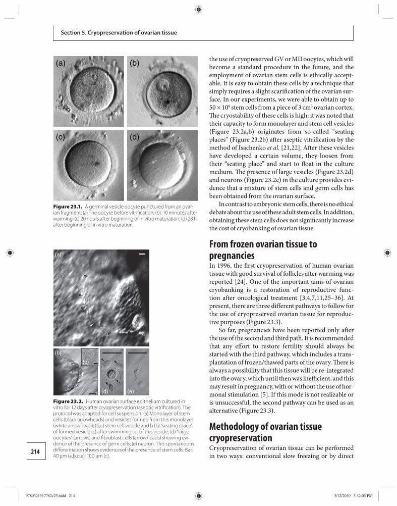
214
Section 5. Cryopreservation of ovarian tissue
the use of cryopreserved GV or MII oocytes, which will become a standard procedure in the future, and the employment of ovarian stem cells is ethically accept-able. It is easy to obtain these cells by a technique that simply requires a slight scarifi cation of the ovarian sur-face. In our experiments, we were able to obtain up to 50 × 10 6 stem cells from a piece of 3 cm 3 ovarian cortex. Th e cryostability of these cells is high: it was noted that their capacity to form monolayer and stem cell ves icles ( Figure 23.2a , b ) originates from so-called “seating places” ( Figure 23.2b ) aft er aseptic vitrifi cation by the method of Isachenko et al. [ 21 , 22 ]. Aft er these vesicles have developed a certain volume, they loosen from their “seating place” and start to fl oat in the culture medium. Th e presence of large vesicles ( Figure 23.2d ) and neurons ( Figure 23.2e ) in the culture provides evi-dence that a mixture of stem cells and germ cells has been obtained from the ovarian surface.
In contrast to embryonic stem cells, there is no eth ical debate about the use of these adult stem cells. In addition, obtaining these stem cells does not signifi cantly increase the cost of cryobanking of ovarian tissue.
From frozen ovarian tissue to pregnancies In 1996, the fi rst cryopreservation of human ovarian tissue with good survival of follicles aft er warming was reported [ 24 ]. One of the important aims of ovarian cryobanking is a restoration of reproductive func-tion aft er oncological treatment [ 3 , 4 , 7 , 11 , 25 – 36 ]. At present, there are three diff erent pathways to follow for the use of cryopreserved ovarian tissue for reproduc-tive purposes ( Figure 23.3 ) .
So far, pregnancies have been reported only aft er the use of the second and third path. It is recommended that any eff ort to restore fertility should always be started with the third pathway, which includes a trans-plantation of frozen/thawed parts of the ovary. Th ere is always a possibility that this tissue will be re-integrated into the ovary, which until then was ineffi cient, and this may result in pregnancy, with or without the use of hor-monal stimulation [ 5 ]. If this mode is not realizable or is unsuccessful, the second pathway can be used as an alternative ( Figure 23.3 ) .
Methodology of ovarian tissue cryopreservation Cryopreservation of ovarian tissue can be performed in two ways: conventional slow freezing or by direct
(a)
(b)
(c)
(d) (e)
Figure 23.2. Human ovarian surface epithelium cultured in vitro for 12 days after cryopreservation (aseptic vitrifi cation). The proto col was adapted for cell suspension. (a) Monolayer of stem cells (black arrowheads) and vesicles formed from this monolayer (white arrowhead); (b,c) stem cell vesicle and h (b) “seating place” of formed vesicle (c) after swimming up of this vesicle; (d) “large oocytes” (arrows) and fi broblast cells (arrowheads) showing evi-dence of the presence of germ cells; (e) neuron. This spontaneous diff erentiation shows evidencesof the presence of stem cells. Bar, 40 μm (a,b,d,e); 100 μm (c).
(a) (b)
(c) (d)
Figure 23.1. A germinal vesicle oocyte punctured from an ovar-ian fragment. (a) The oocyte before vitrifi cation; (b), 10 minutes after warming; (c) 20 hours after beginning of in vitro maturation; (d) 28 h after beginning of in vitro maturation.
9780521517782c23.indd 2149780521517782c23.indd 214 3/12/2010 5:32:05 PM3/12/2010 5:32:05 PM
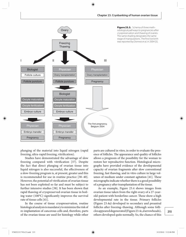
215
Chapter 23. Cryobanking of human ovarian tissue
plunging of the material into liquid nitrogen (rapid freezing, ultra-rapid freezing, vitrifi cation).
Studies have demonstrated the advantage of slow freezing compared with vitrifi cation [ 37 ]. Despite the fact that direct plunging of ovarian tissue into li quid nitrogen is also successful, the eff ectiveness of a slow-freezing program is, at present, greater and this is recommended for use in routine practice [ 38 – 40 ]. However, the potential of vitrifi cation of ovarian tissue has not been exploited so far and must be subject to further intensive studies [ 38 ]. It has been shown that rapid thawing of cryopreserved ovarian tissue in boil-ing water (100°C) signifi cantly improves the survival rate of tissue cells [ 41 ].
In the course of tissue cryopreservation, routine histological analysis is mandatory to minimize the risk of re-implantation of cancerous cells and, therefore, parts of the ovarian tissue are used for histology while other
parts are cultured in vitro, in order to evaluate the pres-ence of follicles. Th e appearance and quality of follicles allows a prognosis of the possibility for the woman to restore her reproductive function. Histological micro-graphs have provided evidence of the developmental capacity of ovarian fragments aft er slow conventional freezing, fast thawing, and in vitro culture in large vol-umes of medium under constant agitation [ 41 ]. Th ese micrographs indicate whether there is a good possibility of a pregnancy aft er transplantation of the tissue.
As an example, Figure 23.4 shows images from ovarian tissue taken from the right ovary of a 27-year- old-patient with borderline cancer. Th ese show a high developmental rate in the tissue. Primary follicles ( Figure 23.4a ) developed to secondary and preantral follicles aft er freezing–thawing. Although some folli-cles appeared degenerated ( Figure 23.4 c,d arrowheads), others developed quite normally. So, the chance of this
Biologist
I II III
Biologist
Follicle culture
Follicle puncture Follicle puncture Pregnancy
Ovary transplantation Ovary transplantation
Oocyte maturation Oocyte maturation
Oocyte fertilization Oocyte fertilization
Embryo culture
Embryo transfer Embryo transfer
Pregnancy Pregnancy
Embryo culture
Physician
PhysicianThe first pregnancy,
Belgium 2004Physician
Physician
Ovary
FreezingThawing
Figure 23.3. Schema of three meth-odological pathways to pregnancies after cryopreservation and thawing of ovaries. The same shading designates the same stage of manipulations. The fi rst live birth was reported by Donnez et al. in 2004 [ 5 ].
9780521517782c23.indd 2159780521517782c23.indd 215 3/12/2010 5:32:08 PM3/12/2010 5:32:08 PM
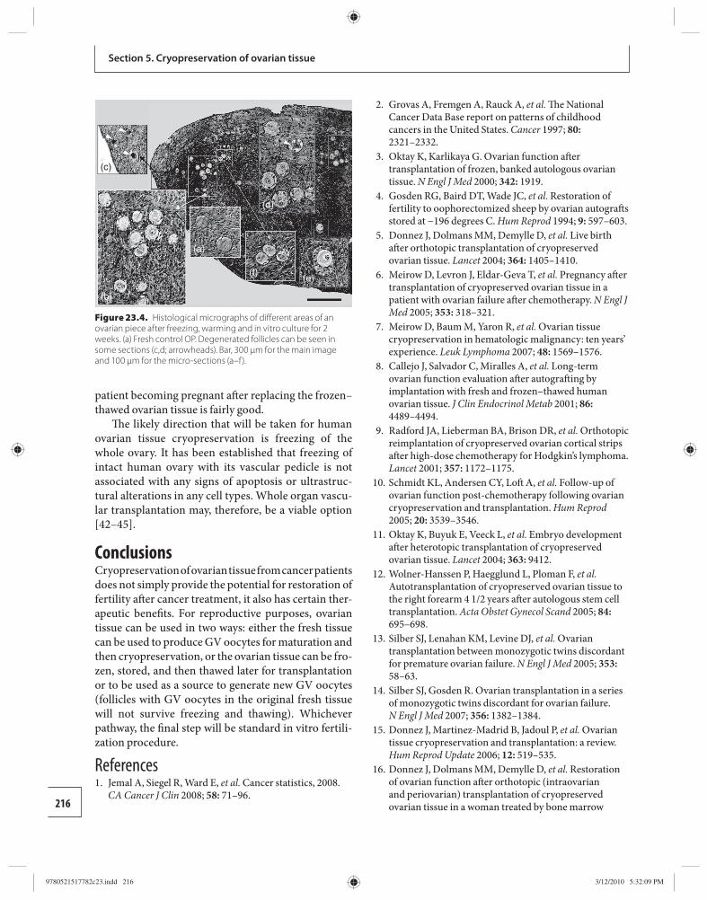
216
Section 5. Cryopreservation of ovarian tissue
patient becoming pregnant aft er replacin g the frozen–thawed ovarian tissue is fairly good.
Th e likely direction that will be taken for human ovarian tissue cryopreservation is freezing of the whole ovary. It has been established that freezing of intact human ovary with its vascular pedicle is not associated with any signs of apoptosis or ultrastruc-tural alterations in any cell types. Whole organ vascu-lar transplantation may, therefore, be a viable option [ 42 – 45 ].
Conclusions Cryopreservation of ovarian tissue from cancer patients does not simply provide the potential for restoration of fertility aft er cancer treatment, it also has certain ther-apeutic benefi ts. For reproductive purposes, ovarian tissue can be used in two ways: either the fresh tissue can be used to produce GV oocytes for maturation and then cryopreservation, or the ovarian tissue can be fro-zen, stored, and then thawed later for transplantation or to be used as a source to generate new GV oocytes (follicles with GV oocytes in the origin al fresh tissue will not survive freezing and thawing). Whichever pathway, the fi nal step will be standard in vitro fertili-zation procedure.
References 1. Jemal A , Siegel R , Ward E , et al. Cancer statistics, 2008 .
CA Cancer J Clin 2008; 58: 71–96 .
2. Grovas A , Fremgen A , Rauck A , et al. Th e National Cancer Data Base report on patterns of childhood cancers in the United States . Cancer 1997; 80: 2321–2332 .
3. Oktay K , Karlikaya G. Ovarian function aft er transplantation of frozen, banked autologous ovarian tissue . N Engl J Med 2000; 342: 1919 .
4. Gosden RG , Baird DT , Wade JC , et al. Restoration of fertility to oophorectomized sheep by ovarian autograft s stored at −196 degrees C . Hum Reprod 1994; 9: 597–603 .
5. Donnez J , Dolmans MM , Demylle D , et al. Live birth aft er orthotopic transplantation of cryopreserved ovarian tissue . Lancet 2004; 364: 1405–1410 .
6. Meirow D , Levron J , Eldar-Geva T , et al. Pregnancy aft er transplantation of cryopreserved ovarian tissue in a patient with ovarian failure aft er chemotherapy . N Engl J Med 2005; 353: 318–321 .
7. Meirow D , Baum M , Yaron R , et al. Ovarian tissue cryopreservation in hematologic malignancy: ten years ’ experience . Leuk Lymphoma 2007; 48: 1569–1576 .
8. Callejo J , Salvador C , Miralles A , et al. Long-term ovarian function evaluation aft er autograft ing by implantation with fresh and frozen–thawed human ovarian tissue . J Clin Endocrinol Metab 2001; 86: 4489–4494 .
9. Radford JA , Lieberman BA , Brison DR , et al. Orthotopic reimplantation of cryopreserved ovarian cortical strips aft er high-dose chemotherapy for Hodgkin ’ s lymphoma . Lancet 2001; 357: 1172–1175 .
10. Schmidt KL , Andersen CY , Loft A , et al. Follow-up of ovarian function post-chemotherapy following ovarian cryopreservation and transplantation . Hum Reprod 2005; 20: 3539–3546 .
11. Oktay K , Buyuk E , Veeck L , et al. Embryo development aft er heterotopic transplantation of cryopreserved ovarian tissue . Lancet 2004; 363: 9412 .
12. Wolner-Hanssen P , Haegglund L , Ploman F , et al. Autotransplantation of cryopreserved ovarian tissue to the right forearm 4 1/2 years aft er autologous stem cell transplantation . Acta Obstet Gynecol Scand 2005; 84: 695–698 .
13. Silber SJ , Lenahan KM , Levine DJ , et al. Ovarian transplantation between monozygotic twins discordant for premature ovarian failure . N Engl J Med 2005; 353: 58–63 .
14. Silber SJ , Gosden R. Ovarian transplantation in a series of monozygotic twins discordant for ovarian failure . N Engl J Med 2007; 356: 1382–1384 .
15. Donnez J , Martinez-Madrid B , Jadoul P , et al. Ovarian tissue cryopreservation and transplantation: a review . Hum Reprod Update 2006; 12: 519–535 .
16. Donnez J , Dolmans MM , Demylle D , et al. Restoration of ovarian function aft er orthotopic (intraovarian and periovarian) transplantation of cryopreserved ovarian tissue in a woman treated by bone marrow
(c)
(b)
(f)(e)
(d)
(a)
Figure 23.4. Histological micrographs of diff erent areas of an ovarian piece after freezing, warming and in vitro culture for 2 weeks. (a) Fresh control OP. Degenerated follicles can be seen in some sections (c,d; arrowheads). Bar, 300 μm for the main image and 100 μm for the micro-sections (a–f).
9780521517782c23.indd 2169780521517782c23.indd 216 3/12/2010 5:32:09 PM3/12/2010 5:32:09 PM
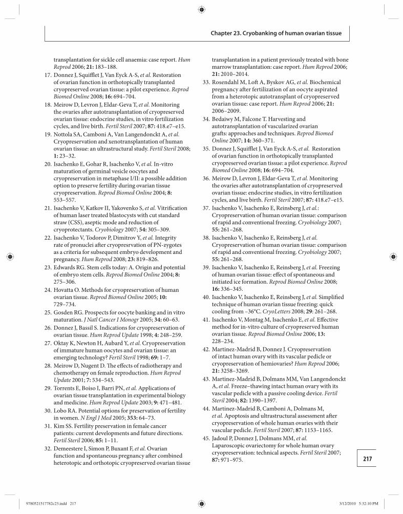
217
Chapter 23. Cryobanking of human ovarian tissue
transplantation for sickle cell anaemia: case report . Hum Reprod 2006; 21: 183–188 .
17. Donnez J , Squiffl et J , Van Eyck A-S , et al. Restoration of ovarian function in orthotopically transplanted cryopreserved ovarian tissue: a pilot experience . Reprod Biomed Online 2008; 16: 694–704 .
18. Meirow D , Levron J , Eldar-Geva T , et al. Monitoring the ovaries aft er autotransplantation of cryopreserved ovarian tissue: endocrine studies, in vitro fertilization cycles, and live birth . Fertil Steril 2007; 87: 418.e7–e15 .
19. Nottola SA , Camboni A , Van Langendonckt A , et al. Cryopreservation and xenotransplantation of human ovarian tissue: an ultrastructural study . Fertil Steril 2008; 1: 23–32 .
20. Isachenko E , Gohar R , Isachenko V , et al. In-vitro maturation of germinal vesicle oocytes and cryopreservation in metaphase I/II: a possible addition option to preserve fertility during ovarian tissue cryopreservation . Reprod Biomed Online 2004; 8: 553–557 .
21. Isachenko V , Katkov II , Yakovenko S , et al. Vitrifi cation of human laser treated blastocysts with cut standard straw (CSS), aseptic mode and reduction of cryoprotectants . Cryobiology 2007; 54: 305–309 .
22. Isachenko V , Todorov P , Dimitrov Y , et al. Integrity rate of pronuclei aft er cryopresrvation of PN-zygotes as a criteria for subsequent embryo development and pregnancy . Hum Reprod 2008; 23: 819–826 .
23. Edwards RG. Stem cells today: A. Origin and potential of embryo stem cells . Reprod Biomed Online 2004; 8: 275–306 .
24. Hovatta O . Methods for cryopreservation of human ovarian tissue . Reprod Biomed Online 2005; 10: 729–734 .
25. Gosden RG. Prospects for oocyte banking and in vitro maturation . J Natl Cancer I Monogr 2005; 34: 60–63 .
26. Donnez J , Bassil S. Indications for cryopreservation of ovarian tissue . Hum Reprod Update 1998; 4: 248–259 .
27. Oktay K , Newton H , Aubard Y , et al. Cryopreservation of immature human oocytes and ovarian tissue: an emerging technology? Fertil Steril 1998; 69: 1–7 .
28. Meirow D , Nugent D. Th e eff ects of radiotherapy and chemotherapy on female reproduction . Hum Reprod Update 2001; 7: 534–543 .
29. Torrents E , Boiso I , Barri PN , et al. Applications of ovarian tissue transplantation in experimental biology and medicine . Hum Reprod Update 2003; 9: 471–481 .
30. Lobo RA. Potential options for preservation of fertility in women . N Engl J Med 2005; 353: 64–73 .
31. Kim SS. Fertility preservation in female cancer patients: current developments and future directions . Fertil Steril 2006; 85: 1–11 .
32. Demeestere I , Simon P , Buxant F , et al. Ovarian function and spontaneous pregnancy aft er combined heterotopic and orthotopic cryopreserved ovarian tissue
transplantation in a patient previously treated with bone marrow transplantation: case report . Hum Reprod 2006; 21: 2010–2014 .
33. Rosendahl M , Loft A , Byskov AG , et al. Biochemical pregnancy aft er fertilization of an oocyte aspirated from a heterotopic autotransplant of cryopreserved ovarian tissue: case report . Hum Reprod 2006; 21: 2006–2009 .
34. Bedaiwy M , Falcone T. Harvesting and autotransplantation of vascularized ovarian graft s: approaches and techniques . Reprod Biomed Online 2007; 14: 360–371 .
35. Donnez J , Squiffl et J , Van Eyck A-S , et al. Restoration of ovarian function in orthotopically transplanted cryopreserved ovarian tissue: a pilot experience . Reprod Biomed Online 2008; 16: 694–704 .
36. Meirow D , Levron J , Eldar-Geva T , et al. Monitoring the ovaries aft er autotransplantation of cryopreserved ovarian tissue: endocrine studies, in vitro fertilization cycles, and live birth . Fertil Steril 2007; 87: 418.e7–e15 .
37. Isachenko V , Isachenko E , Reinsberg J , et al. : Cryopreservation of human ovarian tissue: comparison of rapid and conventional freezing . Cryobiology 2007; 55: 261–268 .
38. Isachenko V , Isachenko E , Reinsberg J , et al. Cryopreservation of human ovarian tissue: comparison of rapid and conventional freezing . Cryobiology 2007; 55: 261–268 .
39. Isachenko V , Isachenko E , Reinsberg J , et al. Freezing of human ovarian tissue: eff ect of spontaneous and initiated ice formation . Reprod Biomed Online 2008; 16: 336–345 .
40. Isachenko V , Isachenko E , Reinsberg J , et al. Simplifi ed technique of human ovarian tissue freezing: quick cooling from –36°C. CryoLetters 2008; 29 : 261–268 .
41. Isachenko V , Montag M , Isachenko E , et al. Eff ective method for in-vitro culture of cryopreserved human ovarian tissue . Reprod Biomed Online 2006; 13: 228–234 .
42. Martinez-Madrid B , Donnez J. Cryopreservation of intact human ovary with its vascular pedicle or cryopreservation of hemiovaries ? Hum Reprod 2006; 21: 3258–3269 .
43. Martinez-Madrid B , Dolmans MM , Van Langendonckt A , et al. Freeze–thawing intact human ovary with its vascular pedicle with a passive cooling device . Fertil Steril 2004; 82: 1390–1397 .
44. Martinez-Madrid B , Camboni A , Dolmans M , et al. Apoptosis and ultrastructural assessment aft er cryopreservation of whole human ovaries with their vascular pedicle . Fertil Steril 2007; 87: 1153–1165 .
45. Jadoul P , Donnez J , Dolmans MM , et al. Laparoscopic ovariectomy for whole human ovary cryopreservation: technical aspects . Fertil Steril 2007; 87: 971–975 .
9780521517782c23.indd 2179780521517782c23.indd 217 3/12/2010 5:32:10 PM3/12/2010 5:32:10 PM
218 Fertility Cryopreservation, ed. Ri-Cheng Chian and Patrick Quinn. Published by Cambridge University Press. Copyright © Cambridge University Press 2010.
Chapter
24 models showed that graft implants of ovarian tissue slices could survive, relying upon rapid growth of blood vessels into the tissue. In humans, ovarian tis-sue transplantation studies have been conducted since 1895 [ 2 ] as a means to restore fertility and/or as a treat-ment for menopausal symptoms before the era of hor-mone therapies. Menstrual cyclicity, pregnancies, and deliveries following ovarian tissue transplantation have been described since [ 3 ]. Th ese early works claimed successful pregnancies as a result of autograft s; while successes were also reported from ovarian allograft s, these claims seem to be exaggerated. It has subsequently become clear that immunosuppressive therapies are essential if ovarian allograft s are to survive [ 4 ]. It has also been shown that the ovary does not have an immu-nological privilege, contrary to early reports. Pioneer experiments with ovarian graft s in animal models and selected clinical case reports of ovarian implantation from the early literature are presented in Tables 24.1 and 24.2 [ 3 ].
Ovarian tissue can be transplanted back to the ori-ginal site (orthotopic) or to a large number of alternative sites (heterotopic) [ 5 ]. Orthotopic graft transplanta-tion to the broad ligament was the favored procedure for establishing a pregnancy because of the simplicity of the procedure. However, from a fertility standpoint, these human ovarian implants were generally disap-pointing and better results have been obtained by transposing the ovary with its vascular pedicle either to the uterine cavity or to the uterine horn [ 6 ]. As a poten-tial treatment for menopause, and not to restore fertil-ity, the graft site is not considered critical and a large number of potential heterotopic sites have been tested, including the rectus muscle, abdominal skin, omen-tum, and cervix [ 3 ]. Graft s in these heterotopic loca-tions can restore menstrual cycles with the exception of sites connected with the portal vein circulation. Th is is largely because hepatic clearance of steroid hormones
Introduction Th e concept of transplantation in reproductive medi-cine has been considered by physicians and scientists for many years and it is now over a century since the fi rst human ovarian graft was performed. Th e ovary is well suited for transplantation since the most abundant primordial follicles are present in the periphery and are the fi rst to benefi t from revascularization. In add-ition, ovarian follicles may be resistant to ischemia as they normally develop within an avascular epithelium and a relatively hypoxic environment. Ovarian tissue transplantation has become clinically feasible with advances in cryobiology and the eff ective cryopreser-vation of ovarian tissue. Today, ovarian tissue harvest-ing is performed by a simple laparoscopic procedure. In addition, accurate patient monitoring is possible before and aft er transplantation using endocrine stud-ies and sonography imaging. For patients, transplanta-tion of ovarian tissue has become an attractive option to overcome the risk of pending premature menopause caused by various clinical conditions.
Among these clinical treatments, radiotherapy and curative chemotherapy, used for cancer treatment, oft en pose a threat to fertility, as ovarian damage is a common long-term side eff ect. For these patients, harvesting and cryopreservation of ovarian tissue before commencing potentially sterilizing chemotherapy has been practiced since the mid 1990s. In addition, the transplantation of thawed tissue has been shown to successfully restore their fertility. Th is chapter describes diff erent aspects of ovarian tissue transplantation including history, basic experiments, and the results of clinical studies.
History of ovarian tissue grafting and transplantation Ovarian graft ing has a long history, beginning in 1863 [ 1 ]. Early experiments using dog, rabbit, and sheep
Transplantation of cryopreserved ovarian tissues Dror Meirow
9780521517782c24.indd 2189780521517782c24.indd 218 3/12/2010 4:32:56 PM3/12/2010 4:32:56 PM
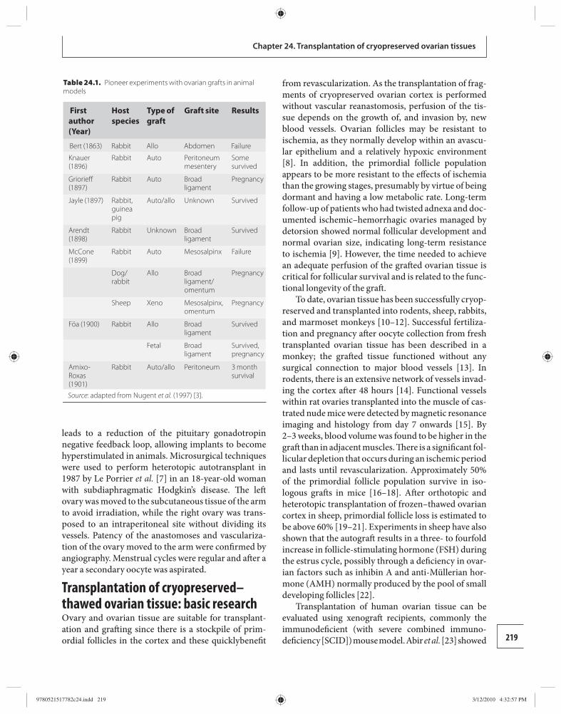
219
Chapter 24. Transplantation of cryopreserved ovarian tissues
leads to a reduction of the pituitary gonadotropin negative feedback loop, allowing implants to become hyperstimulated in animals. Microsurgical techniques were used to perform heterotopic autotransplant in 1987 by Le Porrier et al. [ 7 ] in an 18-year-old woman with subdiaphragmatic Hodgkin’s disease. Th e left ovary was moved to the subcutaneous tissue of the arm to avoid irradiation, while the right ovary was trans-posed to an intraperitoneal site without dividing its vessels. Patency of the anastomoses and vasculariza-tion of the ovary moved to the arm were confi rmed by angiography. Menstrual cycles were regular and aft er a year a secondary oocyte was aspirated.
Transplantation of cryopreserved–thawed ovarian tissue: basic research Ovary and ovarian tissue are suitable for transplant-ation and graft ing since there is a stockpile of prim-ordial follicles in the cortex and these quicklybenefi t
from revascularization. As the transplantation of frag-ments of cryopreserved ovarian cortex is performed without vascular reanastomosis, perfusion of the tis-sue depends on the growth of, and invasion by, new blood vessels. Ovarian follicles may be resistant to ischemia, as they normally develop within an avascu-lar epithelium and a relatively hypoxic environment [ 8 ]. In addition, the primordial follicle population appears to be more resistant to the eff ects of ischemia than the growing stages, presumably by virtue of being dormant and having a low metabolic rate. Long-term follow-up of patients who had twisted adnexa and doc-umented ischemic–hemorrhagic ovaries managed by detorsion showed normal follicular development and normal ovarian size, indicating long-term resistance to ischemia [ 9 ]. However, the time needed to achieve an adequate perfusion of the graft ed ovarian tissue is critical for follicular survival and is related to the func-tional longevity of the graft .
To date, ovarian tissue has been successfully cryop-reserved and transplanted into rodents, sheep, rabbits, and marmoset monkeys [ 10 – 12 ]. Successful fertiliza-tion and pregnancy aft er oocyte collection from fresh transplanted ovarian tissue has been described in a monkey; the graft ed tissue functioned without any surgical connection to major blood vessels [ 13 ]. In rodents, there is an extensive network of vessels invad-ing the cortex aft er 48 hours [ 14 ]. Functional vessels within rat ovaries transplanted into the muscle of cas-trated nude mice were detected by magnetic resonance imaging and histology from day 7 onwards [ 15 ]. By 2–3 weeks, blood volume was found to be higher in the graft than in adjacent muscles. Th ere is a signifi cant fol-licular depletion that occurs during an ischemic period and lasts until revascularization. Approximately 50% of the primordial follicle population survive in iso-logous graft s in mice [ 16 – 18 ]. Aft er orthotopic and heterotopic transplantation of frozen–thawed ovarian cortex in sheep, primordial follicle loss is estimated to be above 60% [ 19 – 21 ]. Experiments in sheep have also shown that the autograft results in a three- to fourfold increase in follicle-stimulating hormone (FSH) during the estrus cycle, possibly through a defi ciency in ovar-ian factors such as inhibin A and anti-Müllerian hor-mone (AMH) normally produced by the pool of small developing follicles [ 22 ].
Transplantation of human ovarian tissue can be e valuated using xenograft recipients, commonly the immunodefi cient (with severe combined immuno-defi ciency [SCID]) mouse model. Abir et al. [ 23 ] showed
Table 24.1. Pioneer experiments with ovarian grafts in animal models
First author (Year)
Host species
Type of graft
Graft site Results
Bert (1863) Rabbit Allo Abdomen Failure
Knauer (1896)
Rabbit Auto Peritoneum mesentery
Some survived
Griorieff (1897)
Rabbit Auto Broad ligament
Pregnancy
Jayle (1897) Rabbit, guinea pig
Auto/allo Unknown Survived
Arendt (1898)
Rabbit Unknown Broad ligament
Survived
McCone (1899)
Rabbit Auto Mesosalpinx Failure
Dog/rabbit
Allo Broad ligament/omentum
Pregnancy
Sheep Xeno Mesosalpinx, omentum
Pregnancy
Föa (1900) Rabbit Allo Broad ligament
Survived
Fetal Broad ligament
Survived, pregnancy
Amixo-Roxas (1901)
Rabbit Auto/allo Peritoneum 3 month survival
Source: adapted from Nugent et al. (1997) [ 3 ].
9780521517782c24.indd 2199780521517782c24.indd 219 3/12/2010 4:32:57 PM3/12/2010 4:32:57 PM
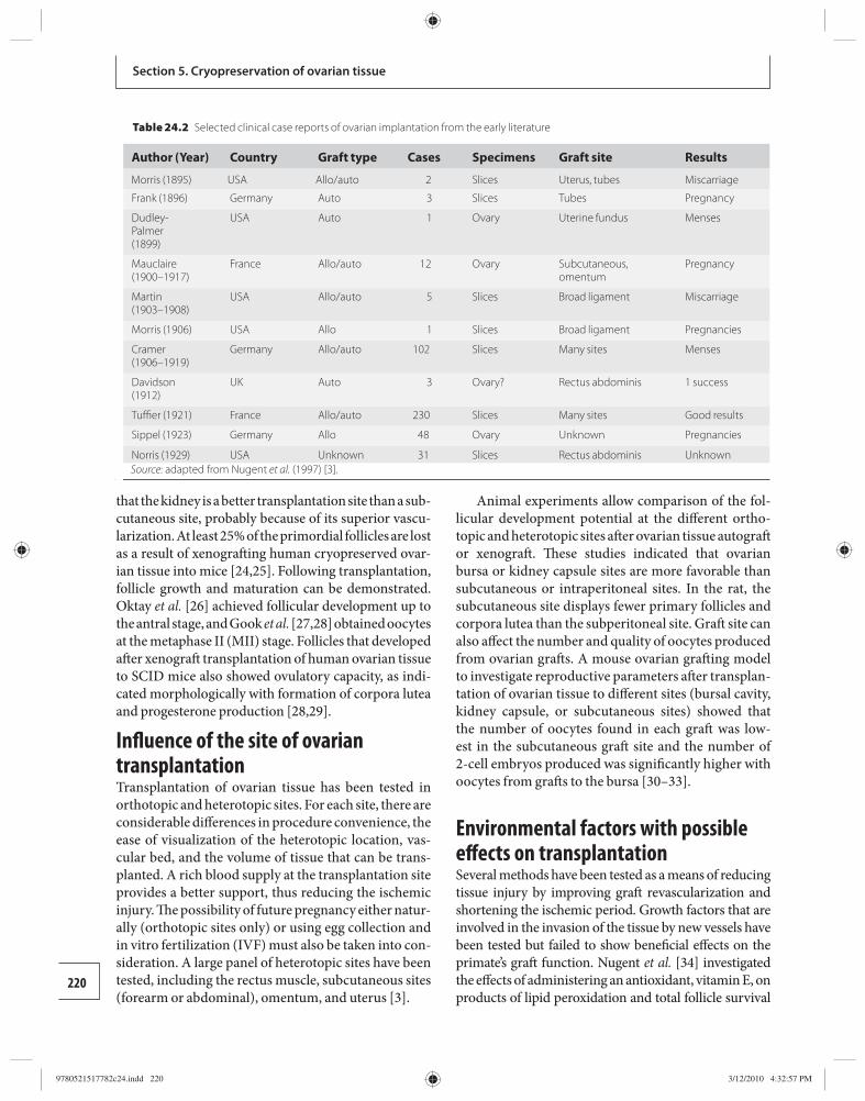
220
Section 5. Cryopreservation of ovarian tissue
that the kidney is a better transplantation site than a sub-cutaneous site, probably because of its superior vascu-larization . At least 25% of the primordial follicles are lost as a result of xenograft ing human cryopreserved ovar-ian tissue into mice [ 24 , 25 ]. Following transplantation, follicle growth and maturation can be demonstrated. Oktay et al. [ 26 ] achieved follicular development up to the antral stage, and Gook et al. [ 2 7, 28 ] obtained oocytes at the metaphase II (MII) stage. Follicles that developed aft er xenograft transplantation of human ovarian tissue to SCID mice also showed ovulatory capacity, as indi-cated morphologically with formation of corpora lutea and progesterone production [ 28 , 29 ].
Infl uence of the site of ovarian transplantation Transplantation of ovarian tissue has been tested in orthotopic and heterotopic sites. For each site, there are considerable diff erences in procedure convenience, the ease of visualization of the heterotopic location, vas-cular bed, and the volume of tissue that can be trans-planted. A rich blood supply at the transplantation site provides a better support, thus reducing the ischemic injury. Th e possibility of future pregnancy either natur-ally (orthotopic sites only) or using egg collection and in vitro fertilization (IVF) must also be taken into con-sideration. A large panel of heterotopic sites have been tested, including the rectus muscle, subcutaneous sites (forearm or abdominal), omentum, and uterus [ 3 ].
Animal experiments allow comparison of the fol-licular development potential at the diff erent ortho-topic and heterotopic sites aft er ovarian tissue autograft or xenograft . Th ese studies indicated that ovarian bursa or kidney capsule sites are more favorable than subcutaneous or intraperitoneal sites. In the rat, the subcutaneous site displays fewer primary follicles and corpora lutea than the subperitoneal site. Graft site can also aff ect the number and quality of oocytes produced from ovarian graft s. A mouse ovarian graft ing model to investigate reproductive parameters aft er transplan-tation of ovarian tissue to diff erent sites (bursal cavity, kidney capsule, or subcutaneous sites) showed that the number of oocytes found in each graft was low-est in the subcutaneous graft site and the number of 2-cell embryos produced was signifi cantly higher with oocytes from graft s to the bursa [ 30 – 33 ].
Environmental factors with possible eff ects on transplantation Several methods have been tested as a means of reducing tissue injury by improving graft revascularization and shortening the ischemic period. Growth factors that are involved in the invasion of the tissue by new vessels have been tested but failed to show benefi cial eff ects on the primate’s graft function. Nugent et al. [ 34 ] investigated the eff ects of administering an antioxidant, vitamin E, on products of lipid peroxidation and total follicle survival
Table 24.2 Selected clinical case reports of ovarian implantation from the early literature
Author (Year) Country Graft type Cases Specimens Graft site Results
Morris (1895) USA Allo/auto 2 Slices Uterus, tubes Miscarriage
Frank (1896) Germany Auto 3 Slices Tubes Pregnancy
Dudley-Palmer (1899)
USA Auto 1 Ovary Uterine fundus Menses
Mauclaire (1900–1917)
France Allo/auto 12 Ovary Subcutaneous, omentum
Pregnancy
Martin (1903–1908)
USA Allo/auto 5 Slices Broad ligament Miscarriage
Morris (1906) USA Allo 1 Slices Broad ligament Pregnancies
Cramer (1906–1919)
Germany Allo/auto 102 Slices Many sites Menses
Davidson (1912)
UK Auto 3 Ovary? Rectus abdominis 1 success
Tuffi er (1921) France Allo/auto 230 Slices Many sites Good results
Sippel (1923) Germany Allo 48 Ovary Unknown Pregnancies
Norris (1929) USA Unknown 31 Slices Rectus abdominis Unknown Source: adapted from Nugent et al. (1997) [ 3 ].
9780521517782c24.indd 2209780521517782c24.indd 220 3/12/2010 4:32:57 PM3/12/2010 4:32:57 PM
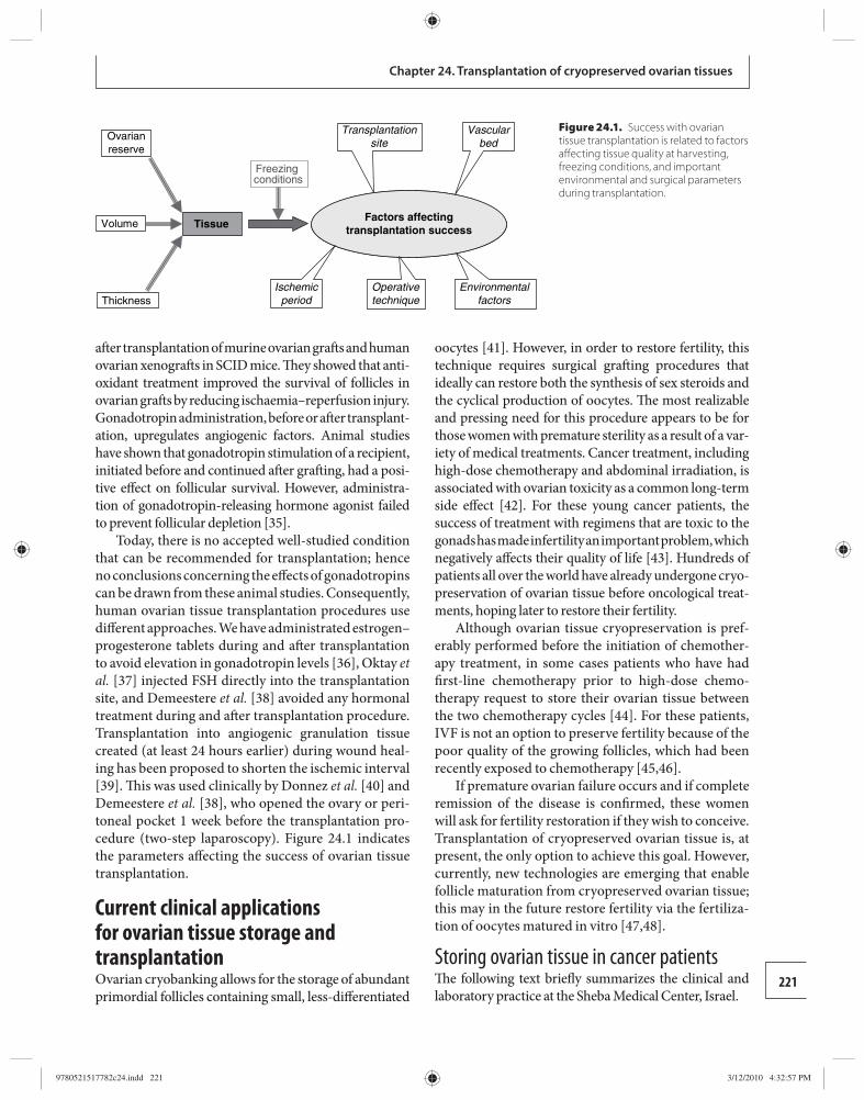
221
Chapter 24. Transplantation of cryopreserved ovarian tissues
aft er transplantation of murine ovarian graft s and human ovarian xenograft s in SCID mice. Th ey showed that anti-oxidant treatment improved the survival of follicles in ovarian graft s by reducing ischaemia–reperfusion injury. Gonadotropin administration, before or aft er transplant-ation, upregulates angiogenic factors. Animal studies have shown that gonadotropin stimulation of a recipient, initiated before and continued aft er graft ing, had a posi-tive eff ect on follicular survival. However, administra-tion of gonadotropin-releasing hormone agonist failed to prevent follicular depletion [ 35 ].
Today, there is no accepted well-studied condition that can be recommended for transplantation; hence no conclusions concerning the eff ects of go nadotropins can be drawn from these animal studies. Consequently, human ovarian tissue transplantation procedures use diff erent approaches. We have administrated estrogen–progesterone tablets during and aft er transplantation to avoid elevation in gonadotropin levels [ 36 ], Oktay et al. [ 37 ] injected FSH directly into the transplantation site, and Demeestere et al. [ 38 ] avoided any hormonal treatment during and aft er transplantation procedure. Transplantation into angiogenic granulation tissue created (at least 24 hours earlier) during wound heal-ing has been proposed to shorten the ischemic interval [ 39 ]. Th is was used clinically by Donnez et al. [ 40 ] and Demeestere et al. [ 38 ], who opened the ovary or peri-toneal pocket 1 week before the transplantation pro-cedure (two-step laparoscopy). Figure 24.1 indicates the parameters aff ecting the success of ovarian tissue transplantation.
Current clinical applications for ovarian tissue storage and transplantation Ovarian cryobanking allows for the storage of abundant primordial follicles containing small, less-diff erentiated
oocytes [ 41 ]. However, in order to restore fertility, this technique requires surgical graft ing procedures that i deally can restore both the synthesis of sex steroids and the cyclical production of oocytes. Th e most realizable and pressing need for this procedure appears to be for those women with premature sterility as a result of a var-iety of medical treatments. Cancer treatment, including high-dose chemotherapy and abdominal irradiation, is associated with ovarian toxicity as a common long-term side eff ect [ 42 ]. For these young cancer patients, the success of treatment with regimens that are toxic to the gonads has made infertility an important problem, which negatively aff ects their quality of life [ 43 ]. Hundreds of patients all over the world have already undergone cryo-preservation of ovarian tissue before oncological treat-ments, hoping later to restore their fertility.
Although ovarian tissue cryopreservation is pref-erably performed before the initiation of chemother-apy treatment, in some cases patients who have had fi rst-line chemotherapy prior to high-dose chemo-therapy request to store their ovarian tissue between the two chemotherapy cycles [ 44 ]. For these patients, IVF is not an option to preserve fertility because of the poor quality of the growing follicles, which had been recently exposed to chemotherapy [ 45 , 46 ].
If premature ovarian failure occurs and if complete remission of the disease is confi rmed, these women will ask for fertility restoration if they wish to conceive. Transplantation of cryopreserved ovarian tissue is, at present, the only option to achieve this goal. However, currently, new technologies are emerging that enable follicle maturation from cryopreserved ovarian tissue; this may in the future restore fertility via the fertiliza-tion of oocytes matured in vitro [ 47 , 48 ].
Storing ovarian tissue in cancer patients Th e following text briefl y summarizes the clinical and laboratory practice at the Sheba Medical Center, Israel.
TissueFactors affecting
transplantation success
Ovarianreserve
Volume
Freezing conditions
ThicknessIschemic
periodOperativetechnique
Environmentalfactors
Transplantationsite
Vascularbed
Figure 24.1. Success with ovarian tissue transplantation is related to factors aff ecting tissue quality at harvesting, freezing conditions, and important environmental and surgical parameters during transplantation.
9780521517782c24.indd 2219780521517782c24.indd 221 3/12/2010 4:32:57 PM3/12/2010 4:32:57 PM
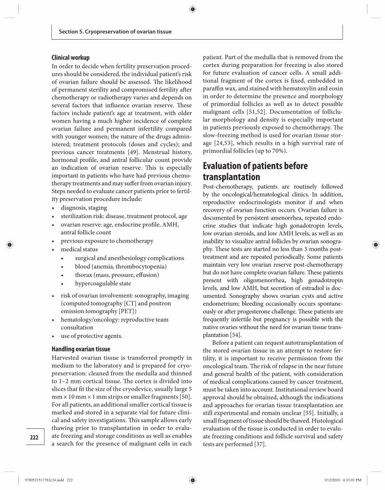
222
Section 5. Cryopreservation of ovarian tissue
Clinical workup In order to decide when fertility preservation proced-ures should be considered, the individual patient’s risk of ovarian failure should be assessed. Th e likelihood of permanent sterility and compromised fertility aft er chemotherapy or radiotherapy varies and depends on several factors that infl uence ovarian reserve. Th ese factors include patient’s age at treatment, with older women having a much higher incidence of complete ovarian failure and permanent infertility compared with younger women; the nature of the drugs admin-istered; treatment protocols (doses and cycles); and previous cancer treatments [ 49 ]. Menstrual history, hormonal profi le, and antral follicular count provide an indication of ovarian reserve. Th is is especially important in patients who have had previous chemo-therapy treatments and may suff er from ovarian injury. Steps needed to evaluate cancer patients prior to fertil-ity preservation procedure include:
diagnosis, staging • sterilization risk: disease, treatment protocol, age • ovarian reserve: age, endocrine profi le, AMH, • antral follicle count previous exposure to chemotherapy • medical status •
surgical and anesthesiology complications • • blood (anemia, thrombocytopenia) • thorax (mass, pressure, eff usion) • hypercoagulable state
risk of ovarian involvement: sonography, imaging • (computed tomography [CT] and positron emission tomography [PET]) hematology/oncology: reproductive team • consultation use of protective agents. •
Handling ovarian tissue Harvested ovarian tissue is transferred promptly in medium to the laboratory and is prepared for cryo-preservation: cleaned from the medulla and thinned to 1–2 mm cortical tissue. Th e cortex is divided into slices that fi t the size of the cryodevice, usually large 5 mm × 10 mm × 1 mm strips or smaller fragments [ 50 ]. For all patients, an additional smaller cortical tissue is marked and stored in a separate vial for future clini-cal and safety investigations. Th is sample allows early thawing prior to transplantation in order to evalu-ate freezing and storage conditions as well as enables a search for the presence of malignant cells in each
patient. Part of the medulla that is removed from the cortex during preparation for freezing is also stored for future evaluation of cancer cells. A small addi-tional fragment of the cortex is fi xed, embedded in paraffi n wax, and stained with hematoxylin and eosin in order to determine the presence and morphology of primordial follicles as well as to detect possible malignant cells [ 51 , 52 ]. Documentation of folliclu-lar morphology and density is especially important in patients previously exposed to chemotherapy. Th e slow-freezing method is used for ovarian tissue stor-age [ 24 , 53 ], which results in a high survival rate of primordial follicles (up to 70%).
Evaluation of patients before transplantation Post-chemotherapy, patients are routinely followed by the oncological/hematological clinics. In addition, reproductive endocrinologists monitor if and when recovery of ovarian function occurs. Ovarian failure is documented by persistent amenorrhea, repeated endo-crine studies that indicate high gonadotropin levels, low ovarian steroids, and low AMH levels, as well as an inability to visualize antral follicles by ovarian sonogra-phy. Th ese tests are started no less than 3 months post-treatment and are repeated periodically. Some patients maintain very low ovarian reserve post-chemotherapy but do not have complete ovarian failure. Th ese patients present with o ligomenorrhea, high gonadotropin l evels, and low AMH, but secretion of estradiol is doc-umented. Sonography shows ovarian cysts and active endometrium; bleeding occasionally occurs spontane-ously or aft er progesterone challenge. Th ese patients are frequently infertile but pregnancy is possible with the native ovaries without the need for ovarian tissue trans-plantation [ 54 ].
Before a patient can request autotransplantation of the stored ovarian tissue in an attempt to restore fer-tility, it is important to receive permission from the oncological team. Th e risk of relapse in the near future and general health of the patient, with consideration of medical complications caused by cancer treatment, must be taken into account. Institutional review board approval should be obtained, although the indications and approaches for ovarian tissue transplantation are still experimental and remain unclear [ 55 ]. Initially, a small fragment of tissue should be thawed. Histological evaluation of the tissue is conducted in order to evalu-ate freezing conditions and follicle survival and safety tests are performed [ 37 ].
9780521517782c24.indd 2229780521517782c24.indd 222 3/12/2010 4:33:01 PM3/12/2010 4:33:01 PM
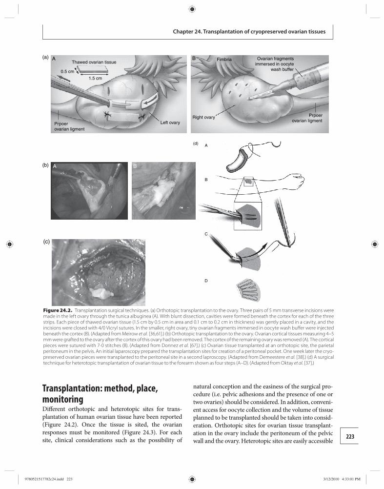
223
Chapter 24. Transplantation of cryopreserved ovarian tissues
Transplantation: method, place, monitoring Diff erent orthotopic and heterotopic sites for trans-plantation of human ovarian tissue have been reported (Figure 24.2). Once the tissue is sited, the ovarian responses must be monitored (Figure 24.3). For each site, clinical considerations such as the possibility of
natural conception and the easiness of the surgical pro-cedure (i.e. pelvic adhesions and the presence of one or two ovaries) should be considered. In addition, conveni-ent access for oocyte collection and the volume of tissue planned to be transplanted should be taken into consid-eration. Orthotopic sites for ovarian tissue transplant-ation in the ovary include the peritoneum of the pelvic wall and the ovary. Heterotopic sites are easily accessible
Fimbria Ovarian fragmentsimmersed in oocyte
wash buffer
Right ovary Prpoerovarian ligment
B
Prpoerovarian ligment
Thawed ovarian tissue
0.5 cm
1.5 cm
Left ovary
A(a)
A(b) B
A(d)
B
C
D
(c)
Figure 24.2. Transplantation surgical techniques. (a) Orthotopic transplantation to the ovary. Three pairs of 5 mm transverse incisions were made in the left ovary through the tunica albuginea (A). With blunt dissection, cavities were formed beneath the cortex for each of the three strips. Each piece of thawed ovarian tissue (1.5 cm by 0.5 cm in area and 0.1 cm to 0.2 cm in thickness) was gently placed in a cavity, and the incisions were closed with 4/0 Vicryl sutures. In the smaller, right ovary, tiny ovarian fragments immersed in oocyte wash buff er were injected beneath the cortex (B). (Adapted from Meirow et al. [ 36 , 61 ].) (b) Orthotopic transplantation to the ovary. Ovarian cortical tissues measuring 4–5 mm were grafted to the ovary after the cortex of this ovary had been removed. The cortex of the remaining ovary was removed (A). The cortical pieces were sutured with 7-0 stitches (B). (Adapted from Donnez et al. [ 67 ].) (c) Ovarian tissue transplanted at an orthotopic site, the parietal peritoneum in the pelvis. An initial laparoscopy prepared the transplantation sites for creation of a peritoneal pocket. One week later the cryo-preserved ovarian pieces were transplanted to the peritoneal site in a second laproscopy. (Adapted from Demeestere et al. [ 38 ].) (d) A surgical technique for heterotopic transplantation of ovarian tissue to the forearm shown as four steps (A–D). (Adapted from Oktay et al. [ 37 ].)
9780521517782c24.indd 2239780521517782c24.indd 223 3/12/2010 4:33:01 PM3/12/2010 4:33:01 PM
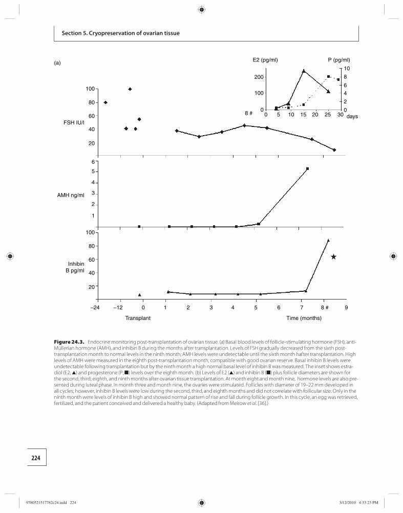
224
Section 5. Cryopreservation of ovarian tissue
100
200
100
8 #0
0 5 10 15 20 25 300
2
4
6
8
10
FSH IU/ldays
(a) P (pg/ml)E2 (pg/ml)
AMH ng/ml
InhibinB pg/ml
Transplant Time (months)
80
60
40
20
100
80
60
40
20
–24 –12 0 1 2 3 4 5 6 7 8 # 9
6
5
4
3
2
1
Figure 24.3. Endocrine monitoring post-transplantation of ovarian tissue. (a) Basal blood levels of follicle-stimulating hormone (FSH), anti-Müllerian hormone (AMH), and inhibin B during the months after transplantation. Levels of FSH gradually decreased from the sixth post-transplantation month to normal levels in the ninth month; AMH levels were undetectable until the sixth month hafter transplantation. High levels of AMH were measured in the eighth post-transplantation month, compatible with good ovarian reserve. Basal inhibin B levels were undetectable following transplantation but by the ninth month a high normal basal level of inhibin B was measured. The inset shows estra-diol (E2; ▲) and progesterone (P; ■) levels over the eighth month. (b) Levels of E2 (▲) and inhibin B (■) plus follicle diameters are shown for the second, third, eighth, and ninth months after ovarian tissue transplantation. At month eight and month nine, hormone levels are also pre-sented during luteal phase. In month three and month nine, the ovaries were stimulated. Follicles with diameter of 19–22 mm developed in all cycles; however, inhibin B levels were low during the second, third, and eighth months and did not correlate with follicular size. Only in the ninth month were levels of inhibin B high and showed normal pattern of rise and fall during follicle growth. In this cycle, an egg was retrieved, fertilized, and the patient conceived and delivered a healthy baby. (Adapted from Meirow et al. [ 36 ].)
9780521517782c24.indd 2249780521517782c24.indd 224 3/12/2010 4:33:23 PM3/12/2010 4:33:23 PM
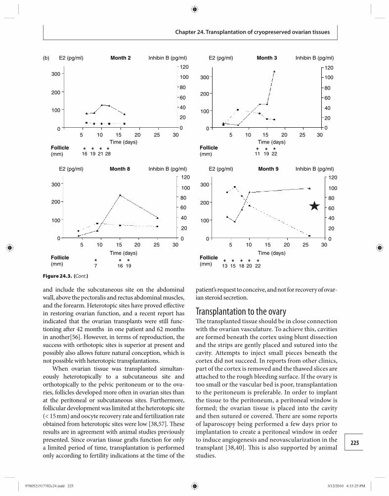
225
Chapter 24. Transplantation of cryopreserved ovarian tissues
and include the subcutaneous site on the abdominal wall, above the pectoralis and rectus abdominal muscles, and the forearm. Heterotopic sites have proved eff ective in restoring ovarian function, and a recent report has indicated that the ovarian transplants were still func-tioning aft er 42 months in one patient and 62 months in another[ 56 ]. However, in terms of reproduction, the success with orthotopic sites is superior at present and possibly also allows future natural conception, which is not possible with heterotopic transplantations.
When ovarian tissue was transplanted simultan-eously heterotopically to a subcutaneous site and orthoto pically to the pelvic peritoneum or to the ova-ries, follicles de veloped more oft en in ovarian sites than at the peritoneal or subcutaneous sites. Furthermore, follicul ar development was limited at the heterotopic site (< 15 mm) and oocyte recovery rate and fertilization rate obtained from heterotopic sites were low [ 38 , 57 ]. Th ese results are in agreement with animal studies previously presented. Since ovarian tissue graft s function for only a limited period of time, transplantation is performed only according to fertility indications at the time of the
patient’s request to conceive, and not for recovery of ovar-ian steroid secretion .
Transplantation to the ovary Th e transplanted tissue should be in close connection with the ovarian vasculature. To achieve this, cavities are formed beneath the cortex using blunt dissection and the strips are gently placed and sutured into the cavity. Attempts to inject small pieces beneath the cortex did not succeed. In reports from other clinics, part of the cortex is removed and the thawed slices are attached to the rough bleeding surface. If the ovary is too small or the vascular bed is poor, transplantation to the peritoneum is preferable. In order to implant the tissue to the peritoneum, a peritoneal window is formed; the ovarian tissue is placed into the cavity and then sutured or covered. Th ere are some reports of laparoscopy being performed a few days prior to implantation to create a peritoneal window in order to induce angiogenesis and neovascularization in the transplant [ 38 , 40 ]. Th is is also supported by animal studies.
(b)
300
E2 (pg/ml) Inhibin B (pg/ml)Inhibin B (pg/ml)Month 2 E2 (pg/ml) Month 3
E2 (pg/ml) Inhibin B (pg/ml)Inhibin B (pg/ml)Month 8 E2 (pg/ml) Month 9
200
100
05
Follicle(mm)
Follicle(mm)
Follicle(mm)
Follicle(mm)
10
16
167 19 13 15 18 20 22
19 21 28 11 19 22
15Time (days) Time (days)
Time (days) Time (days)
20 25 30
5 10 15 20 25 30 5 10 15 20 25 30
5 10 15 20 25 300
20
40
60
80
100
120
0
20
40
60
80
100
120
0
20
40
60
80
100
120
0
20
40
60
80
100
120
300
200
100
0
300
200
100
0
300
200
100
0
*
* * * * * * * *
* * * * **
Figure 24.3. (Cont.)
9780521517782c24.indd 2259780521517782c24.indd 225 3/12/2010 4:33:25 PM3/12/2010 4:33:25 PM
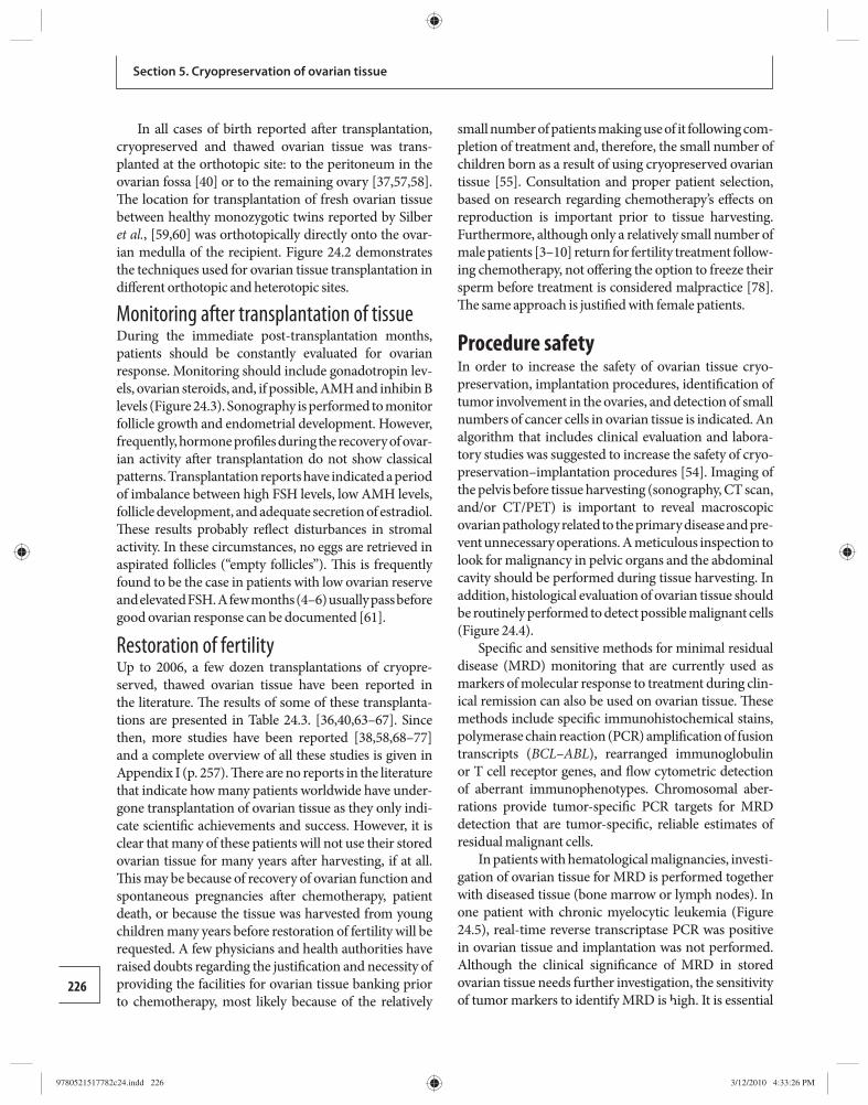
226
Section 5. Cryopreservation of ovarian tissue
In all cases of birth reported aft er transplantation, cryopreserved and thawed ovarian tissue was trans-planted at the orthotopic site: to the peritoneum in the ovarian fossa [ 40 ] or to the remaining ovary [ 37 , 57 , 58 ]. Th e location for transplantation of fresh ovarian tissue between healthy monozygotic twins reported by Silber et al. , [ 59 , 60 ] was orthotopically directly onto the ovar-ian medulla of the recipient. Figure 24.2 demonstrates the techniques used for ovarian tissue transplantation in diff erent orthotopic and heterotopic sites.
Monitoring after transplantation of tissue During the immediate post-transplantation months, patients should be constantly evaluated for ovarian response. Monitoring should include gonadotropin lev-els, ovarian steroids, and, if possible, AMH and inhibin B levels ( Figure 24.3 ). Sonography is performed to monitor follicle growth and endometrial development. However, frequently, hormone profi les during the recovery of ovar-ian activity aft er transplant ation do not show classical patterns. Transplantation reports have indicated a period of imbalance between high FSH levels, low AMH levels, follicle development, and adequate secretion of estradiol. Th ese results probably refl ect disturbances in stromal activity. In these circumstances, no eggs are retrieved in aspirated follicles (“empty follicles”). Th is is frequently found to be the case in patients with low ovarian reserve and elevated FSH. A few months (4–6) usually pass before good ovarian response can be documented [ 61 ].
Restoration of fertility Up to 2006, a few dozen transplantations of cryopre-served, thawed ovarian tissue have been reported in the literature. Th e results of some of these transplanta-tions are presented in Table 24.3 . [36,40,63–67]. Since then, more studies have been reported [38,58,68–77] and a complete overview of all these studies is given in Appendix I (p. 257). Th ere are no reports in the literature that indicate how many patients worldwide have under-gone transplantation of ovarian tissue as they only indi-cate scientifi c achievements and success. However, it is clear that many of these patients will not use their stored ovarian tissue for many years aft er harvesting, if at all. Th is may be because of recovery of ovarian function and spontaneous pregnancies aft er chemotherapy, patient death, or because the tissue was harvested from young children many years before restoration of fertility will be requested. A few physicians and health authorities have raised doubts regarding the justifi cation and necessity of providing the facilities for ovarian tissue banking prior to chemotherapy, most likely because of the relatively
small number of patients making use of it following com-pletion of treatment and, therefore, the small number of children born as a result of using cryopreserved ovarian tissue [ 55 ]. Consultation and proper patient selection, based on research regarding chemotherapy’s eff ects on reproduction is important prior to tissue harvesting. Furthermore, although only a relatively small number of male patients [ 3 – 10 ] return for fertility treatment follow-ing chemotherapy, not off ering the option to freeze their sperm before treatment is considered malpractice [78 ]. Th e same approach is justifi ed with female patients.
Procedure safety In order to increase the safety of ovarian tissue cryo-preservation, implantation procedures, identifi cation of tumor involvement in the ovaries, and detection of small numbers of cancer cells in ovarian tissue is indicated. An algorithm that includes clinical evaluation and labora-tory studies was suggested to increase the safety of cryo-preservation–implantation procedures [ 54 ]. Imaging of the pelvis before tissue harvesting (sonography, CT scan, and/or CT/PET) is important to reveal macroscopic ovarian pathology related to the primary disease and pre-vent unnecessary operations. A meticulous inspection to look for malignancy in pelvic organs and the abdominal cavity should be performed during tissue harvesting . In addition, histological evaluation of ovarian tissue should be routinely performed to detect possible malignant cells ( Figure 24.4 ).
Specifi c and sensitive methods for minimal residual disease (MRD) monitoring that are currently used as markers of molecular response to treatment during clin-ical remission can also be used on ovarian tissue. Th ese methods include specifi c immunohistochemical stains, polymerase chain reaction (PCR) amplifi cation of fusion transcripts ( BCL – ABL ), rearranged immunoglobulin or T cell receptor genes, and fl ow cytometric detection of aberrant immunophenotypes. Chromosomal aber-rations provide tumor-specifi c PCR targets for MRD detection that are tumor-specifi c, reliable estimates of residual malignant cells.
In patients with hematological malignancies, investi-gation of ovarian tissue for MRD is performed together with diseased tissue (bone marrow or lymph nodes). In one patient with chronic myelocytic leukemia ( Figure 24.5 ), real-time reverse transcriptase PCR was positive in ovarian tissue and implantation was not performed. Although the clinical signifi cance of MRD in stored ovarian tissue needs further investigation, the sensitivity of tumor markers to identify MRD is high. It is essential
9780521517782c24.indd 2269780521517782c24.indd 226 3/12/2010 4:33:26 PM3/12/2010 4:33:26 PM

227 Tab
le 2
4.3.
Aut
otra
nspl
anta
tion
of c
ryop
rese
rved
–tha
wed
hum
an o
varia
n tis
sue
Refe
renc
e A
ge a
t fr
eezi
ng
(yea
rs)
Chem
othe
rapy
be
fore
free
zing
In
dica
tion
G
raft
site
* G
raft
size
(mm
) Re
cove
ry
of o
vari
an
func
tion
Out
com
e
Okt
ay &
Kar
likay
a (2
000)
[63]
29
No
Not
can
cer
P 8
pc
(5 ×
10
× 2
) 1
6 w
eeks
↑ E2
; FD
afte
r stim
ulat
ion;
ovu
latio
n;
men
ses
Radf
ord
et a
l. (20
01)
[64]
36Ye
sH
odgk
in’s
lym
phom
aO
2 pc
(10
× 5
× 1
)8 m
onth
s↓
FSH
and
LH
; ↑ E
2; F
D;
ovul
atio
n; m
ense
s (1
cycl
e)
Calle
jo et
al.
(200
1) [6
5]47
No
Not
can
cer
Rect
us
abdo
min
ius
mus
cle
40–4
5 pc
(2–3
m
m 3 )
3–4
mon
ths
↑ E2
; FD
afte
r stim
ulat
ion
(folli
cle
of 2
0 m
m)
Kim
et a
l. (2
004)
[66]
37N
oCe
rvic
al
carc
inom
a (Ib
)
Abov
e pe
ctor
alis
mus
cle
(a) +
ab
ove
rect
us
abdo
min
us
mus
cle
(b)
40 p
c (5
× 5
×
1): 2
0 (a
) + 2
0 (b
)14
wee
ks↑
E2; ↓
FSH
; FD
(fol
licle
of 1
1–16
m
m) o
nly
in (b
); ov
ulat
ion
Okt
ay e
t al.
(200
4) [6
7]30
No
Brea
st c
ance
rSu
bcut
aneo
us
abdo
min
al
wal
l
15 p
c (5
× 5
× 1
to
15 ×
5 ×
2)
3 mon
ths
FD a
fter g
onad
otro
phin
st
imul
atio
n; O
PU; 2
0 oo
cyte
s, 8
IVF-
ICSI
; 4-c
ell e
mbr
yo tr
ansf
er
+ 1
ane
uplo
idic
em
bryo
Don
nez
et a
l. (20
04)
[40]
25N
oH
odgk
in
lym
phom
aP
1 pc
(12
× 4
× 1
) +
67 (3
5 +
32)
pc
(2
× 2
× 1
)
4.5
mon
ths
↑ E2
; ↓ F
SH a
nd L
H; F
D (f
ollic
le
of 2
2 m
m);
regu
lar c
ycle
s (±
5 w
eeks
); pr
egna
ncy;
live
birt
h
Mei
row
et
al. (
2005
) [3
6]
28Ye
sN
on-H
odgk
in
lym
phom
aO
v tr
ansp
lant
(a
); O
inje
ctio
n (b
)
3pc
(15
× 5
× 1
) (a
) + ti
ny
fragm
ents
(b)
8 mon
ths
↓ FS
H, ↑
AM
H a
nd in
hibi
n B;
FD
; ovu
latio
n; m
ense
s; m
odifi
ed n
atur
al c
ycle
; OPU
; 1
met
apha
se II
ooc
yte
IVF-
ICSI
; 4-c
ell e
mbr
yo tr
ansf
er;
preg
nanc
y an
d liv
e bi
rth;
sp
onta
neou
s pre
gnan
cy
misc
arria
ge (3
0 m
onth
s afte
r tr
ansp
lant
)
pc,
pie
ces;
P, pe
lvic
per
itone
um; O
, rem
aini
ng o
vary
; E2,
est
radi
ol; F
D, f
ollic
le d
evel
opm
ent;
FSH
, fol
licle
-stim
ulat
ing
horm
one;
LH
, lut
eini
zing
hor
mon
e; A
MH
, ant
i-Mül
leria
n ho
rmon
e;
OPU
, ovu
m p
ick-
up; I
VF, i
n vi
tro
fert
iliza
tion;
ICSI
, int
racy
topl
asm
ic sp
erm
inje
ctio
n.
9780521517782c24.indd 2279780521517782c24.indd 227 3/12/2010 4:33:27 PM3/12/2010 4:33:27 PM
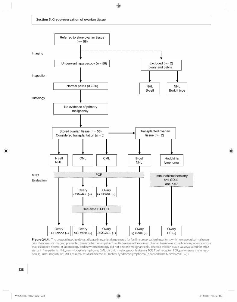
228
Section 5. Cryopreservation of ovarian tissue
Referred to store ovarian tissue(n = 58)
Excluded (n = 2)ovary and pelvis
Underwent laparoscopy (n = 56)
Hodgkin’slymphoma
OvaryBCR/ABL (–)
B-cellNHL
Stored ovarian tissue (n = 56)Considered transplantation (n = 5)
Imaging
Inspection
Normal pelvis (n = 56)
Histology
No evidence of primarymalignancy
T- cellNHL
NHLB-cell
NHLBurkitt type
Transplanted ovariantissue (n = 2)
MRD
Evaluation
CML CML
PCR
OvaryBCR/ABL (–)
OvaryBCR/ABL (–)
OvaryBCR/ABL (+)
OvaryTCR clone (–)
Immunohistochemistryanti-CD30 anti-Ki67
OvaryIg clone (–)
OvaryRS (–)
Real-time RT-PCR
Figure 24.4. The protocol used to detect disease in ovarian tissue stored for fertility preservation in patients with hematological malignan-cies. Preoperative imaging prevented tissue collection in patients with disease in the ovaries. Ovarian tissue was stored only in patients whose ovaries looked normal at laparoscopy and in whom histology did not disclose malignant cells. Thawed ovarian tissue was evaluated for MRD status in fi ve patients. NHL, non-Hodgkin lymphoma; CML, chronic myelogenous leukemia; TCR, T cell receptor; PCR, polymerase chain reac-tion; Ig, immunoglobulin; MRD, minimal residual disease; RS, Richter syndrome lymphoma. (Adapted from Meirow et al. [52].)
9780521517782c24.indd 2289780521517782c24.indd 228 3/12/2010 4:33:27 PM3/12/2010 4:33:27 PM
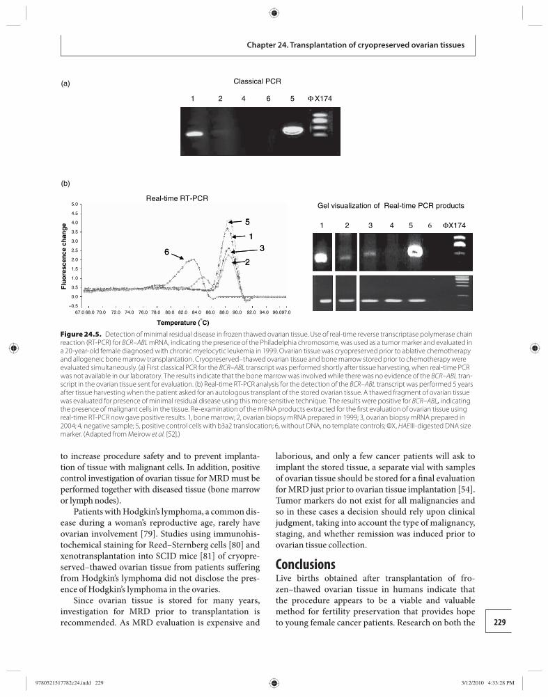
229
Chapter 24. Transplantation of cryopreserved ovarian tissues
to increase procedure safety and to prevent implanta-tion of tissue with malignant cells. In addition, positive control investigation of ovarian tissue for MRD must be performed together with diseased tissue (bone marrow or lymph nodes).
Patients with Hodgkin’s lymphoma, a common dis-ease during a woman’s reproductive age, rarely have ovarian involvement [ 79 ]. Studies using immunohis-tochemical staining for Reed–Sternberg cells [ 80 ] and xenotransplantation into SCID mice [ 81 ] of cryopre-served–thawed ovarian tissue from patients suff ering from Hodgkin’s lymphoma did not disclose the pres-ence of Hodgkin’s lymphoma in the ovaries.
Since ovarian tissue is stored for many years, investigation for MRD prior to transplantation is recommended. As MRD evaluation is expensive and
laborious, and only a few cancer patients will ask to implant the stored tissue, a separate vial with samples of ovarian tissue should be stored for a fi nal evaluation for MRD just prior to ovarian tissue implantation [ 54 ]. Tumor markers do not exist for all malignancies and so in these cases a decision should rely upon clinical judgment, taking into account the type of malignancy, staging, and whether remission was induced prior to ovarian tissue collection.
Conclusions Live births obtained aft er transplantation of fro-zen–thawed ovarian tissue in humans indicate that the procedure appears to be a viable and valuable method for fertility preservation that provides hope to young female cancer patients. Research on both the
Φ
Classical PCR
1 2 4 6 5 X174
ΦX1741 2 3 4 5 65
1
2
36
5
1
2
36
Real-time RT-PCR Gel visualization of Real-time PCR products
(b)
(a)
5.0
4.5
4.0
3.5
3.0
2.5
2.0
1.5
1.0
0.5
0.0
–0.567.0 68.0 70.0 72.0 74.0 76.0 78.0
Flu
ore
scen
ce c
han
ge
Temperature (�C)
80.0 82.0 84.0 86.0 88.0 90.0 92.0 94.0 96.097.0
Figure 24.5. Detection of minimal residual disease in frozen thawed ovarian tissue. Use of real-time reverse transcriptase polymerase chain reaction (RT-PCR) for BCR – ABL mRNA, indicating the presence of the Philadelphia chromosome, was used as a tumor marker and evaluated in a 20-year-old female diagnosed with chronic myelocytic leukemia in 1999. Ovarian tissue was cryopreserved prior to ablative chemotherapy and allogeneic bone marrow transplantation. Cryopreserved–thawed ovarian tissue and bone marrow stored prior to chemotherapy were evaluated simultaneously. (a) First classical PCR for the BCR–ABL transcript was performed shortly after tissue harvesting, when real-time PCR was not available in our laboratory. The results indicate that the bone marrow was involved while there was no evidence of the BCR–ABL tran-script in the ovarian tissue sent for evaluation. (b) Real-time RT-PCR analysis for the detection of the BCR–ABL transcript was performed 5 years after tissue harvesting when the patient asked for an autologous transplant of the stored ovarian tissue. A thawed fragment of ovarian tissue was evaluated for presence of minimal residual disease using this more sensitive technique. The results were positive for BCR–ABL, indicating the presence of malignant cells in the tissue. Re-examination of the mRNA products extracted for the fi rst evaluation of ovarian tissue using real-time RT-PCR now gave positive results. 1, bone marrow; 2, ovarian biopsy mRNA prepared in 1999; 3, ovarian biopsy mRNA prepared in 2004; 4, negative sample; 5, positive control cells with b3a2 translocation; 6, without DNA, no template controls; ΦX, HAE III-digested DNA size marker. (Adapted from Meirow et al. [52].)
9780521517782c24.indd 2299780521517782c24.indd 229 3/12/2010 4:33:28 PM3/12/2010 4:33:28 PM
230
Section 5. Cryopreservation of ovarian tissue
indications for ovarian tissue transplantation and on technique improvement should continue [82].
Acknowledgements Th e authors would like to thank Hannah Biederman for her assistance with the manuscript. References 1. Gosden RG. Ovary and uterus transplantation .
Reproduction 2008; 136: 671–680 . 2. Morris RT. Th e ovarian graft . N Y Med J 1895; 62: 436 . 3. Nugent D , Meirow D , Brook PF , Aubard Y , Gosden R.
Transplantation in reproductive medicine . Hum Reprod Update 1997; 3: 267–280 .
4. Goldman MB. Immunogenetic factors in the survival of ovarian transplants . Transplantation 1974; 17: 518–523 .
5. Aubard Y , Piver P , Cognié Y , et al. Orthotopic and heterotopic autograft s of frozen–thawed ovarian cortex in sheep . Hum Reprod 1999; 14 : 2149–2154 .
6. Estes WL. A method of implanting ovarian tissue in order to maintain ovarian function . Pennsylvania Med J 1909; 13 : 610–613 .
7. Le Porrier M , von Th eobold , P , Roff e JL , et al. A new technique to protect ovarian function before pelvic irrradiation . Cancer 1987; 60: 2201–2204 .
8. Gosden RG , Byatt-Smith JG. Oxygen concentration gradient across the ovarian follicle epithelium: model, predictions and implications . Hum Reprod 1986; 1: 65–68 .
9. Oelsner G , Bider D , Goldenberg M , Admon D , Mashiach S. Long-term follow-up of the twisted ischemic adnexa managed by detorsion . Fertil Steril 1993; 60: 976–979 .
10. Salle B , Demirci B , Franck M , et al. Normal pregnancies and live births aft er autograft of frozen–thawed hemiovaries into ewes . Fertil Steril 2002; 77: 403–408 .
11. Almodin CG , Minguetti-Camara VC , Meister H , et al. Recovery of fertility aft er graft ing of cryopreserved germinative tissue in female rabbits following radiotherapy . Hum Reprod 2004; 19: 1287–1293 .
12. Candy CJ , Wood MJ , Whittingham DG. Follicular development in cryopreserved marmoset ovarian tissue aft er transplantation . Hum Reprod 1995; 10 : 2334–2338 .
13. Lee DM , Yeoman RR , Battaglia DE , et al. Live birth aft er ovarian tissue transplant. Nature 2004; 428: 137–138 .
14. Dissen GA , Lara HE , Fahrenbach WH , et al. Immature rat ovaries become revascularised rapidly aft er autotransplantation and show a gonadotrophin-dependent increase in angiogenic factor gene expression . Endocrinology 1994; 134: 1146–1154 .
15. Israely T , Dafni H , Nevo N , Tsafriri A , Neeman M. Angiogenesis in ectopic xenotransplantation: multiparameter characterization of the neovasculature by dynamic contrast-enhanced MRI . Magn Reson Med 2004; 52: 741–750 .
16. Jones EC , Krohn PL. Orthotopic ovarian transplantation in mice . J Endocrinol 1960; 20: 135–146 .
17. Felicio LS , Nelson JF , Gosden RG , et al. Restoration of ovulatory cycles by young ovarian graft s in aging mice: potentiation by long term ovariectomy decreases with age . Proc Natl Acad Sci USA 1983; 80: 6076–6080 .
18. Liu J , Van der Elst J , van den Broecke R , Dhont M. Early massive follicle loss and apoptosis in heterotopically graft ed newborn mouse ovaries . Hum Reprod 2002; 17: 605–611 .
19. Aubard Y , Piver P , Cognié Y , et al. Orthotopic and heterotopic autograft s of frozen–thawed ovarian cortex in sheep . Hum Reprod 1999; 14: 2149–2154 .
20. Aubard Y. Ovarian tissue xenograft ing . Eur J Obstet Gynecol Reprod Biol 2003; 108: 14–18 .
21. Baird DT , Webb R , Campbell BK , Harkness LM , Gosden RG. Long-term ovarian function in sheep aft er ovariectomy and transplantation of autograft s stored at –196°C . Endocrinology 1999; 140: 462–464 .
22. Campbell BK , Telfer EE , Webb R , Baird DT. Ovarian autograft s in sheep as a model for studying folliculogenesis . Mol Cell Endocrinol 2000; 163: 131–139 .
23. Abir R , Orvieto R , Raanani H , Feldberg D , Nitke S , Fisch B. Parameters aff ecting successful transplantation of frozen–thawed human fetal ovaries into immunodefi cient mice. Fertil Steril 2003; 80 : 421–428 .
24. Newton H , Aubard Y , Rutherford A , et al. Low temperature storage and graft ing of human ovarian tissue . Hum Reprod 1996; 11: 1487–1491 .
25. Nisolle M , Godin PA , Casanas-Roux F , et al. Histological and ultrastructural evaluation of fresh and frozen–thawed human ovarian xenograft s in nude mice . Fertil Steril 2000; 74: 122–129 .
26. Oktay K , Newton H , Mullan J , Gosden RG. Development of human primordial follicles to antral stages in SCID/hpg mice stimulated with follicle stimulating hormone . Hum Reprod 1998; 13: 1133–1138 .
27. Gook DA , Edgar DH , Borg J , et al. Oocyte maturation, follicle rupture and luteinization in human cryopreserved ovarian tissue following xenograft ing . Hum Reprod 2003; 18: 1772–1781 .
28. Gook DA , Edgar DH , Borg J , Archer J , McBain JC. Diagnostic assessment of the developmental potential of human cryopreserved ovarian tissue from multiple patients using xenograft ing . Hum Reprod 2005; 20: 72–78 .
29. Kim SS , Soules MR , Battaglia DE. Follicular development, ovulation, and corpus luteum formation in cryopreserved human ovarian tissue aft er xenotransplantation . Fertil Steril 2002; 78: 77–82 .
30. Imthurn B , Cox SL , Jenkin G , Trounson AO , Shaw JM. Gonadotrophin administration can benefi t ovarian tissue graft ed to the body wall: implications for human ovarian graft ing. Mol Cell Endocrinol 2000; 163: 141–146 .
9780521517782c24.indd 2309780521517782c24.indd 230 3/12/2010 4:33:30 PM3/12/2010 4:33:30 PM
231
Chapter 24. Transplantation of cryopreserved ovarian tissues
31. Callejo J , Vilaseca S , Ordi J , et al. Heterotopic ovarian transplantation without vascular pedicle in syngeneic Lewis rats: long-term evaluation of eff ects on ovarian structure and function. Fertil Steril 2002; 77 : 396–402 .
32. Risvanli A , Timurkan H , Akpolat N , Gulacti I , Ulakoglu E. A study of ovarian autotransplantation without a vascular pedicle in rats . J Assist Reprod Genet 2006; 23: 401–406 .
33. Yang HY , Cox SL , Jenkin G , et al. Graft site and gonadotrophin stimulation infl uences the number and quality of oocytes from murine ovarian tissue graft s Reproduction 2006; 131 : 851–859 .
34. Nugent D , Newton H , Gallivan L , Gosden RG. Protective eff ect of vitamin E on ischaemia–reperfusion injury in ovarian graft s . J Reprod Fertil 1998; 114: 341–346 .
35. Maltaris T , Beckmann MW , Binder H , et al. Th e eff ect of a GnRH agonist on cryopreserved human ovarian graft s in severe combined immunodefi cient mice. Reproduction 2007; 133 : 503–509 .
36. Meirow D , Levron J , Eldar-Geva T , et al. Pregnancy aft er transplantation of cryopreserved ovarian tissue in a patient with ovarian failure aft er chemotherapy . N Engl J Med 2005; 353: 318–321 .
37. Oktay K , Buyuk E , Rosenwaks Z , Rucinski JA. Technique for transplantation of ovarian cortical strips to the forearm . Fertil Steril 2003; 80: 193–198 .
38. Demeestere I , Simon P , Buxant F , et al. Ovarian function and spontaneous pregnancy aft er combined heterotopic and orthotopic cryopreserved ovarian tissue transplantation in a patient previously treated with bone marrow transplantation . Hum Reprod 2006; 21: 2010–2014 .
39. Israely T , Nevo N , Harmelin A , Neeman M , Tsafriri A. Reducing ischaemic damage in rodent ovarian xenograft s transplanted into granulation tissue . Hum Reprod 2006; 21: 1368–1379 .
40. Donnez J , Dolmans MM , Demylle D , et al. Livebirth aft er orthotopic transplantation of cryopreserved ovarian tissue . Lancet 2004; 364: 1405–1410 .
41. Torrents E , Boiso I , Barri PN , Veiga A. Applications of ovarian tissue transplantation in experimental biology and medicine . Hum Reprod Update 2003; 9: 471–481 .
42. Falcone T , Bedaiwy MA. Fertility preservation and pregnancy outcome aft er malignancy . Curr Opin Obstet Gynecol 2005; 17: 21–26 .
43. Meirow D. Ovarian injury and modern options to perserve fertility in female cancer patients treated with high dose radio-chemotherapy for hemato-oncological neoplasias and other cancers . Leuk Lymphoma 1999; 33: 65–76 .
44. Meirow D. Fertility preservation in cancer patients using stored ovarian tissue: clinical aspects . Curr Opin Endocrinol Diabetes Obes 2008; 15: 536–547 .
45. Meirow D , Epstein M , Lewis H . Administration of cyclophosphamide at diff erent stages of follicular maturation in mice: eff ects on reproductive performance and fetal malformations . Hum Reprod 2001; 16: 632–637 .
46. Meirow D , Schiff E. Impact of maternal exposure to chemotherapy on embryos and off spring . J Nat Can Inst 2005; 34: 21–25 .
47. Telfer EE , McLaughlin M , Ding C , Th ong KJ. A two-step serum-free culture system supports development of human oocytes from primordial follicles in the presence of activin . Hum Reprod 2008; 23: 1151–1158 .
48. Xu M , Kreeger PK , Shea LD , Woodruff TK. Tissue-engineered follicles produce live, fertile off spring . Tissue Eng 2006; 12 : 2739–46 .
49. Meirow D , Dor J. Epidemiology and infertility in cancer patients. In Tulandi T , Gosden R , eds. Preservation of Fertility . London : Taylor & Francis , 2004, pp.21–38.
50. Oktay KH , Yih M. Preliminary experience with orthotopic and heterotopic transplantation of ovarian cortical strips . Semin Reprod Med 2002; 20: 63–74 .
51. Seshadri T , Gook D , Lade S , et al. Lack of evidence of disease contamination in ovarian tissue harvested for cryopreservation from patients with Hodgkin lymphoma and analysis of factors predictive of oocyte yield . Br J Cancer 2006; 94: 1007–1010 .
52. Meirow D , Hardan I , Dor J , et al. Searching for evidence of disease and malignant cell contamination in ovarian tissue stored from hematologic cancer patients . Hum Reprod 2008; 23: 1007–1013 .
53. Gosden RG , Baird DT , Wade JC , Webb R. Restoration of fertility to oophorectomised sheep by ovarian autograft s stored at –196°C . Hum Reprod 1994; 9: 597–603 .
54. Meirow D , Baum M , Yaron R , et al. Ovarian tissue cryopreservation in hematologic malignancy: ten years ’ experience . Leuk Lymphoma 2007; 48: 1569–1576 .
55. Anderson RA , Wallace WHB , Baird DT. Ovarian cryopreservation for fertility preservation: indications and outcomes . Reproduction 2008; 136: 681–689 .
56. Oktay K , Oktem O. Ovarian cryopreservation and transplantation for fertility preservation for medical indications: report of an ongoing experience . Fertil Steril 2008; e pub ahead of print.
57. Demeestere I , Simon P , Emiliani S , Delbaere A , Englert Y. Orthotopic and heterotopic ovarian tissue transplantation. Hum Reprod Update 2009; e pub ahead of print.
58. Andersen CY , Rosendahl M , Byskov AG , et al. Two successful pregnancies following autotransplantation of frozen/thawed ovarian tissue . Hum Reprod 2008; 23: 2266–2272 .
59. Silber SJ , Lenahan KM , Levine DJ , et al. Ovarian transplantation between monozygotic twins discordant for premature ovarian failure . N Engl J Med 2005; 353: 58–63 .
9780521517782c24.indd 2319780521517782c24.indd 231 3/12/2010 4:33:30 PM3/12/2010 4:33:30 PM
232
Section 5. Cryopreservation of ovarian tissue
60. Silber SJ , Gosden RG. Ovarian transplantation in a series of monozygotic twins discordant for ovarian failure . N Engl J Med 2007; 356: 1382–1384 .
61. Meirow D , Levron J , Eldar-Geva T , et al. Monitoring the ovaries aft er autotransplantation of cryopreserved ovarian tissue: endocrine studies, in vitro fertilization cycles, and live birth . Fertil Steril 2007; 87: 418 .e417.
62. Dolmans MM , et al. Results of Autotransplantation of Cryopreserved Human Ovarian Tissue . 2008. http://www. isfp-fertility.org/members-only/scientifi c-articles/fertility-preservation-in-women/cryopreservation-of-ovarian-cortex-reimplantation-of-cryopreserved-ovarian-cortex.
63. Oktay K , Karlikaya G. Ovarian function aft er transplantation of frozen, banked autologous ovarian tissue. N Engl J Med 2000 ; 342 : 1919 .
64. Radford JA , Liebermann BA , Brison D , et al. Orthotopic reimplantation of cryopreserved ovarian cortical strips aft er high-dose chemotherapy for Hodgkin’s lymphoma. Lancet 2001 ; 357 : 1172 –1175.
65. Callejo J , Salvador C , Miralles A , et al. Long-term ovarian function evaluation aft er autograft ing by implantation with fresh and frozen-thawed human ovarian tissue. J Clin Endocrinol Metab 2001 ; 86 : 4489 –4494.
66. Kim SS , Hwang I-T , Lee H-C. Heterotopic autotransplantation of cryobanked human ovarian tissue as a strategy to restore ovarian function. Fertil Steril 2004 ; 82 : 930 –932.
67. Oktay K , Buyuk E , Veeck L , et al. Embryo development aft er heterotopic transplantation of cryopreserved ovarian tissue. Lancet 2004 ; 363 : 837 –840.
68. Schmidt KL , Yding Andersen C , Loft A , et al. Follow-up of ovarian function post-chemotherapy following ovarian cryopreservation and transplantation. Hum Reprod 2005 ; 20 : 3539 –3546.
69. Wolner-Hanssen P , Hagglund L , Ploman F , et al. Autotransplantation of cryopreserved ovarian tissue to the right forearm 2005; 4½ years aft er autologous stem cell transplantation. Acta Obstet Gynecol Scand 84: 695 –698.
70. Donnez J , Dolmans MM , Demylle D , et al. Restoration of ovarian function aft er orthotopic (intraovarian and periovarian) transplantation of cryopreserved ovarian tissue in a woman treated by bone marrrow transplantation for sickle cell anaemia: case report. Hum Reprod 2006 ; 21: 183 –188.
71. Oktay K. Spontaneous conceptions and live birth aft er heterotopic ovarian transplantation: is there a germline stem cell connection? Hum Reprod 2006 ; 21: 1345 –1348.
72. Rosendahl M , Loft A , Byskov AG , et al. Biochemical pregnancy aft er fertilization of an oocyte aspirated from a heterotopic autotransplant of cryopreserved ovarian tissue: case report. Hum Reprod 2006 ; 21: 2006 –2009.
73. Demeestere I , Simon P , Emiliani S , Delbaere A , Englert Y. Fertility preservation: successful transplantation of cryopreserved ovarian tissue in a young patient previously treated for Hodgkin’s disease. Oncologist 2007 ; 12: 1437 –1442.
74. Silber SJ , Derosa M , Pineda J , et al. A series of monozygotic twins discordant for ovarian failure: ovary transplantation (cortical versus microvascular) and cryopreservation. Hum Reprod 2008 ; 23: 1531 –1537.
75. Donnez J , Squiffl et J , Van Eyck A-S , et al. Restoration of ovarian function in orthotopically transplanted cryopreserved ovarian tissue: a pilot experience. RBM online 2008 ; 16: 694 –704.
76. Sanchez-Serrano M , Crespo J , Mirabet V , et al. Twins born aft er transplantation of ovarian cortical tissue and oocyte vitrifi cation. Fertil Steril 2010 ; 93: 268 .
77. Piver P , Amiot C , Agnani G , et al. Two pregnancies obtained aft er a new technique of autotransplantation of cryopreserved ovarian tissue. In Proceedings of the 25th Annual Meeting of ESHRE , Amsterdam, the Netherlands, June–July, 2009.
78. Lass A , Akagbosu F , Brinsden P. Sperm banking and assisted reproduction treatment for couples following cancer treatment of the male partner . Hum Reprod Update 2001; 7: 370–377 .
79. Khan MA , Dahill SW , Stewart KS. Primary Hodgkin ’ s disease of the ovary . Br J Obstet Gynaecol 1986; 93: 1300–1301 .
80. Seshadri T , Gook D , Lade S , et al. Lack of evidence of disease contamination in ovarian tissue harvested for cryopreservation from patients with Hodgkin lymphoma and analysis of factors predictive of oocyte yield . Br J Cancer 2006; 94: 1007–1010 .
81. Kim SS , Radford J , Harris M , et al. Ovarian tissue harvested from lymphoma patients to preserve fertility may be safe for autotransplantation . Hum Reprod 2001; 16: 2056–2060 .
82. Donnez J , Martinez-Madrid B , Jadoul P , et al. Ovarian tissue cryopreservation and transplantation: a review . Hum Reprod Update 2006; 12: 519–535 .
9780521517782c24.indd 2329780521517782c24.indd 232 3/12/2010 4:33:30 PM3/12/2010 4:33:30 PM
233Fertility Cryopreservation, ed. Ri-Cheng Chian and Patrick Quinn. Published by Cambridge University Press. Copyright © Cambridge University Press 2010.
Chapter
25 potentially overcome this problem, as reperfusion will occur immediately upon retransplantation and anas-tomosis [ 4 , 19 , 22 , 23 ]. Th e cryopreservation of intact, large organs was a breakthrough in medical science, with early reports of canine kidneys showing low tox-icity and high longevity [ 24 , 25 ].
Yet progress into whole ovary cryopreservation has been slow as two main issues in whole organ cryo-preservation needed to be adequately addressed: the fi rst was related to the feasibility of re-establishing the vascular anastomosis of the organ to be transplanted and the second to the creation of a valid cryopreser-vation protocol. Wang et al. [ 26 ] demonstrated that cryopreservation and subsequent transplantation of the reproductive system “en bloc” could be performed in rats. Subsequently, to prove that anastomosis of the ovarian pedicle in large animals and humans was tech-nically feasible, transplantation of whole fresh ovaries has been successfully performed in rats [ 26 , 27 ], rab-bits [ 28 ], sheep [ 29 , 30 ], dogs [ 31 ], monkeys [ 32 ], and humans [ 19 , 20 , 33 – 35 ].
Th e second issue, researching a valid cryopreserva-tion protocol for large-sized intact ovaries, has posed greater diffi culties and challenges. Th ese include the scarcity of information on the physical changes occur-ring in ovarian tissues during cooling, the physical con-straints related to heat transfer between the core and periphery of the organ, and diffi culties with adequate perfusion and diff usion of cryoprotectants [ 36 , 37 ] to prevent intravascular ice formation [ 19 ].
Nevertheless, successful cryopreservation and vas-cular transplantation of intact frozen–thawed ovaries has been reported in both rats and sheep.
Th is chapter will review the progress in cryo-preservation and transplantation of whole ovaries that have been made in both animal and human models. As cryopreservation of whole tissues from small and large animals and humans present diff erent challenges and
Introduction While previous chapters have focused on preservation of oocytes and embryos as well as ovarian tissue cor-tex, much progress has been made recently in the area of cryopreservation of the whole ovary. Patients who require immediate (within days) gonadotoxic therapy (whether chemo- or radiotherapy, or a combination of both) have few options for fertility preservation: (1) cryopreservation of ovarian tissues as cortical strips; (2) dual cryopreservation of both ovarian cor-tical tissue with cryopreservation and in vitro matura-tion of a few immature oocytes extracted from small follicles visible within the ovarian cortex; or (3) cryo-preservation of one whole ovary [ 1 – 10 ]. To date, ovar-ian cryopreservation and subsequent transplantation procedures, whether as heterotopic or orthotopic allo-graft s, have been almost exclusively limited to avascu-lar cortical pieces [ 8 , 11 , 12 ].
Despite the careful use of cryoprotectants to pre-vent ice crystal formation at the time of freezing, the main drawback to cryopreservation of ovarian cortical strips is the ischemia that occurs at the time of trans-plantation. As these small cortical pieces are graft ed without any vascular anastomosis, they are completely dependent for their survival on the speed at which neovascularization can be established aft er graft ing. As this process requires a minimum of 7 days [ 13 , 14 ], the cells in the graft undergo signifi cant ischemic dam-age, which results in massive loss of primordial follicles [ 15 – 19 ] and, in turn, limits the lifespan of the graft [ 20 , 21 ]. For a patient who desires long-term repro-ductive and endocrine function aft er transplantation, this method of fertility preservation may be subopti-mal. Th erefore, reducing the ischemic interval between transplantation and revascularization is essential for maintaining the viability and functional lifetime of the graft . To this end, cryopreservation of the whole ovary with an intact pedicle and vascular supply can
Whole ovary cryopreservation Jason G. Bromer and Pasquale Patrizio
9780521517782c25.indd 2339780521517782c25.indd 233 3/12/2010 4:39:48 PM3/12/2010 4:39:48 PM
234
Section 5. Cryopreservation of ovarian tissue
diffi culties, these topics will be addressed separately. As this new and exciting fi eld remains in its infancy, this chapter will focus mainly on a review of the existing literature and what conclusions can be drawn from the initial studies that have been performed.
The rodent model Wang et al. in 2002 [ 26 ] fi rst described the feasibility of cryopreservation of whole reproductive organs (ovar-ies, tubes, and uterus) using a rat model. In this pilot study, ovaries from adult female rats were removed, and the ovarian vessels were dissected to create short cuff s from the aorta and vena cava. Seven such dissec-tions were perfused for 30 minutes at 0.35 ml/min with M2 medium containing 0.1 M fructose and increasing concentrations of dimethyl sulfoxide (DMSO). Th e organs were then cooled slowly in cryovials. Aft er over-night storage in liquid nitrogen, the vials were rapidly thawed and the cryoprotectant was removed by per-fusion with a reversed concentration gradient. In four of seven animals receiving transplanted cryopreserved graft s, the ovaries showed subsequent follicular activ-ity, with corpora lutea indicating recent ovulation, and one animal achieved pregnancy. Importantly, tubal and uterine morphology were indistinguishable from non-operated controls. However, the rats had higher serum follicle-stimulating hormone (FSH) levels, fewer fol-licles, and lower estradiol levels and uterine weights than controls. Th ese results indicated that ovarian function was compromised by freezing and suggested that advances in freezing techniques could potentially overcome this problem [ 26 ].
Th e same group followed up this work using a simi-lar model to investigate the long-term longevity of these graft s, as well as to assess the eff ect of ischemia by keep-ing the organs for 24 hours at 4°C prior to freezing [ 27 ]. Graft survival with endocrine function was seen at 60 days and ovulatory response to FSH was seen at 120 days, suggesting long-term graft longevity aft er cryo-preservation and vascular anastomosis. Furthermore, graft function aft er transplant was also seen in graft s that had been held for 24 hours under ischemic con-ditions before freezing, although, as expected, the number of surviving follicles was less [ 27 ].
The sheep model Th e adult female sheep has recently become a stand-ard model to explore intact ovarian cryopreservation. Despite the fact that adult human ovaries are larger in size and have diff erent pedicle anatomy, ovine ovaries
are similar to human ovaries in having a dense fi brous stroma and relatively high primordial follicle den-sity in the cortex [ 38 ]. Similar to the rat model, the fi rst experi ments were carried out to determine the feasibility of transplantation using fresh ovine ova-ries. Jeremias et al. in 2002 [ 30 ] fi rst described a novel method of orthotopic transplantation in an adult sheep with anastomosis of the vascular pedicle. Fresh ovaries were autotransplanted to the abdominal wall via micro-surgical anastomosis of the ovarian to the inferior epi-gastric vessels, and showed resumption of endocrine function post-transplant.
Once a whole ovine ovary transplant was proven feasible, the next step was to repeat the experiments using whole frozen–thawed ovaries. Whole ovary cryopreservation in the sheep has been attempted through both slow-cooling and vitrifi cation method-ologies (see Table 25.1 , below). Th e fi rst transplant of an intact frozen–thawed ovary via microvascular anas-tomosis occurred in 2003 [ 36 ]. Aft er laparascopic dis-section, the protocol utilized consisted of perfusion via the ovarian artery with heparin, followed by Leibovitz L-15 medium, 10% fetal calf serum (FCS), and 1.5 M DMSO at 1.3 ml/min. Ovaries were cryopreserved with cooling at 2°C/min beginning at 4°C and continuing until ice nucleation occurred at −7°C. Th e temperature was then reduced at 2°C/min until −35°C and fi nally by 25°C/min until −140°C was reached; at which point the cryovials were plunged into liquid nitrogen.
Th e ovaries were thawed by placing the cryovials in a water bath at 37°C and perfusing with Leibovitz L-15 medium and 10% FCS for 20 minutes before they were implanted with vascular connections to the epigastric artery.
Of the 11 ovaries transplanted, immediate and long-term patency were documented in 100% and 27% of graft s, respectively. Th ere were no signifi cant diff er-ences in the mean values of apoptosis and follicular viability compared with ovarian cortical strip cryo-preservation and autotransplantation. Postoperative FSH levels were much lower in the whole ovary graft group ( p = 0.03) than in the animals graft ed with ovar-ian cortical strips. Further, the endocrine activity of the whole frozen–thawed and transplanted ovaries was similar to preoperative values in animals with patent vessels [ 36 ].
Post-transplanted ovaries responded to stimula-tion with FSH and produced viable oocytes. Once the ewes were sacrifi ced, all ovaries showed primordial, primary, secondary, antral, and preovulatory follicles
9780521517782c25.indd 2349780521517782c25.indd 234 3/12/2010 4:39:48 PM3/12/2010 4:39:48 PM
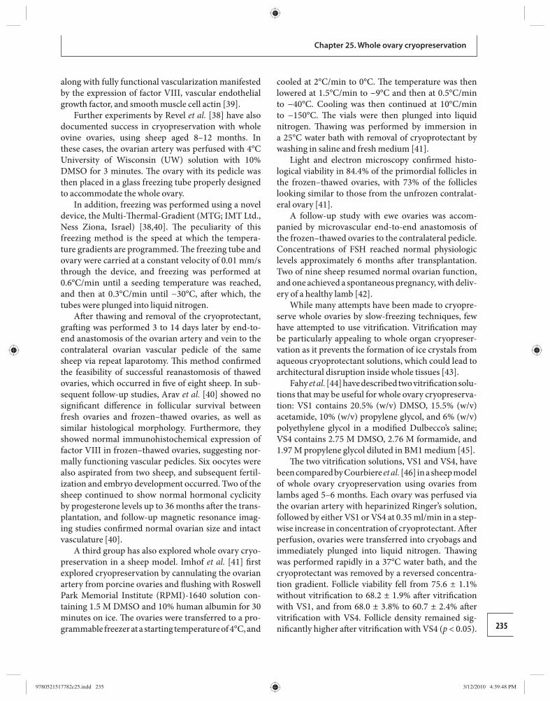
235
Chapter 25. Whole ovary cryopreservation
along with fully functional vascularization manifested by the expression of factor VIII, vascular endothelial growth factor, and smooth muscle cell actin [ 39 ].
Further experiments by Revel et al. [ 38 ] have also documented success in cryopreservation with whole ovine ovaries, using sheep aged 8–12 months. In these cases, the ovarian artery was perfused with 4°C University of Wisconsin (UW) solution with 10% DMSO for 3 minutes. Th e ovary with its pedicle was then placed in a glass freezing tube properly designed to accommodate the whole ovary.
In addition, freezing was performed using a novel device, the Multi-Th ermal-Gradient (MTG; IMT Ltd., Ness Ziona, Israel) [ 38 , 40 ]. Th e peculiarity of this freezing method is the speed at which the tempera-ture gradients are programmed. Th e freezing tube and ovary were carried at a constant velocity of 0.01 mm/s through the device, and freezing was performed at 0.6°C/min until a seeding temperature was reached, and then at 0.3°C/min until −30°C, aft er which, the tubes were plunged into liquid nitrogen.
Aft er thawing and removal of the cryoprotectant, graft ing was performed 3 to 14 days later by end-to-end anastomosis of the ovarian artery and vein to the contralateral ovarian vascular pedicle of the same sheep via repeat laparotomy. Th is method confi rmed the feasibility of successful reanastomosis of thawed ovaries, which occurred in fi ve of eight sheep. In sub-sequent follow-up studies, Arav et al. [ 40 ] showed no signifi cant diff erence in follicular survival between fresh ovaries and frozen–thawed ovaries, as well as similar histological morphology. Furthermore, they showed normal immunohistochemical expression of factor VIII in frozen–thawed ovaries, suggesting nor-mally functioning vascular pedicles. Six oocytes were also aspirated from two sheep, and subsequent fertil-ization and embryo development occurred. Two of the sheep continued to show normal hormonal cyclicity by progesterone levels up to 36 months aft er the trans-plantation, and follow-up magnetic resonance imag-ing studies confi rmed normal ovarian size and intact vasculature [ 40 ].
A third group has also explored whole ovary cryo-preservation in a sheep model. Imhof et al. [ 41 ] fi rst explored cryopreservation by cannulating the ovarian artery from porcine ovaries and fl ushing with Roswell Park Memorial Institute (RPMI)-1640 solution con-taining 1.5 M DMSO and 10% human albumin for 30 minutes on ice. Th e ovaries were transferred to a pro-grammable freezer at a starting temperature of 4°C, and
cooled at 2°C/min to 0°C. Th e temperature was then lowered at 1.5°C/min to −9°C and then at 0.5°C/min to −40°C. Cooling was then continued at 10°C/min to −150°C. Th e vials were then plunged into liquid nitrogen. Th awing was performed by immersion in a 25°C water bath with removal of cryoprotectant by washing in saline and fresh medium [ 41 ].
Light and electron microscopy confi rmed histo-logical viability in 84.4% of the primordial follicles in the frozen–thawed ovaries, with 73% of the follicles looking similar to those from the unfrozen contralat-eral ovary [ 41 ].
A follow-up study with ewe ovaries was accom-panied by microvascular end-to-end anastomosis of the frozen–thawed ovaries to the contralateral pedicle. Concentrations of FSH reached normal physiologic levels approximately 6 months aft er transplantation. Two of nine sheep resumed normal ovarian function, and one achieved a spontaneous pregnancy, with deliv-ery of a healthy lamb [ 42 ].
While many attempts have been made to cryopre-serve whole ovaries by slow-freezing techniques, few have attempted to use vitrifi cation. Vitrifi cation may be particularly appealing to whole organ cryopreser-vation as it prevents the formation of ice crystals from aqueous cryoprotectant solutions, which could lead to architectural disruption inside whole tissues [ 43 ].
Fahy et al. [ 44 ] have described two vitrifi cation solu-tions that may be useful for whole ovary cryopreserva-tion: VS1 contains 20.5% (w/v) DMSO, 15.5% (w/v) acetamide, 10% (w/v) propylene glycol, and 6% (w/v) polyethylene glycol in a modifi ed Dulbecco’s saline; VS4 contains 2.75 M DMSO, 2.76 M formamide, and 1.97 M propylene glycol diluted in BM1 medium [ 45 ].
Th e two vitrifi cation solutions, VS1 and VS4, have been compared by Courbiere et al. [ 46 ] in a sheep model of whole ovary cryopreservation using ovaries from lambs aged 5–6 months. Each ovary was perfused via the ovarian artery with heparinized Ringer’s solution, followed by either VS1 or VS4 at 0.35 ml/min in a step-wise increase in concentration of cryoprotectant. Aft er perfusion, ovaries were transferred into cryobags and immediately plunged into liquid nitrogen. Th awing was performed rapidly in a 37°C water bath, and the cryoprotectant was removed by a reversed concentra-tion gradient. Follicle viability fell from 75.6 ± 1.1% without vitrifi cation to 68.2 ± 1.9% aft er vitrifi cation with VS1, and from 68.0 ± 3.8% to 60.7 ± 2.4% aft er vitrifi cation with VS4. Follicle density remained sig-nifi cantly higher aft er vitrifi cation with VS4 ( p < 0.05).
9780521517782c25.indd 2359780521517782c25.indd 235 3/12/2010 4:39:48 PM3/12/2010 4:39:48 PM
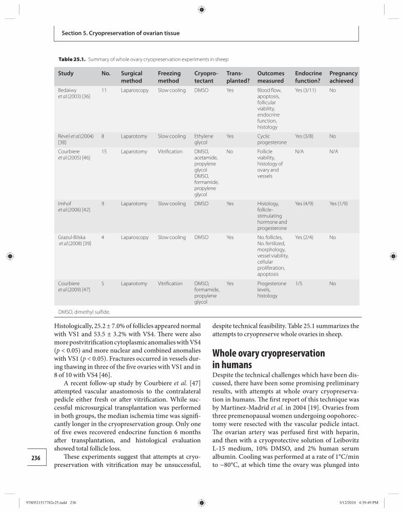
236
Section 5. Cryopreservation of ovarian tissue
Histologically, 25.2 ± 7.0% of follicles appeared normal with VS1 and 53.5 ± 3.2% with VS4. Th ere were also more postvitrifi cation cytoplasmic anomalies with VS4 ( p < 0.05) and more nuclear and combined anomalies with VS1 ( p < 0.05). Fractures occurred in vessels dur-ing thawing in three of the fi ve ovaries with VS1 and in 8 of 10 with VS4 [ 46 ].
A recent follow-up study by Courbiere et al. [ 47 ] attempted vascular anastomosis to the contralateral pedicle either fresh or aft er vitrifi cation. While suc-cessful microsurgical transplantation was performed in both groups, the median ischemia time was signifi -cantly longer in the cryopreservation group. Only one of fi ve ewes recovered endocrine function 6 months aft er transplantation, and histological evaluation showed total follicle loss.
Th ese experiments suggest that attempts at cryo-preservation with vitrifi cation may be unsuccessful,
despite technical feasibility. Table 25.1 summarizes the attempts to cryopreserve whole ovaries in sheep.
Whole ovary cryopreservation in humans Despite the technical challenges which have been dis-cussed, there have been some promising preliminary results, with attempts at whole ovary cryopreserva-tion in humans. Th e fi rst report of this technique was by Martinez-Madrid et al. in 2004 [ 19 ]. Ovaries from three premenopausal women undergoing oopohorec-tomy were resected with the vascular pedicle intact. Th e ovarian artery was perfused fi rst with heparin, and then with a cryoprotective solution of Leibovitz L-15 medium, 10% DMSO, and 2% human serum albumin. Cooling was performed at a rate of 1°C/min to −80°C, at which time the ovary was plunged into
Table 25.1. Summary of whole ovary cryopreservation experiments in sheep
Study No. Surgical method
Freezing method
Cryopro-tectant
Trans-planted?
Outcomes measured
Endocrine function?
Pregnancy achieved
Bedaiwy et al. (2003) [36]
11 Laparoscopy Slow cooling DMSO Yes Blood fl ow, apoptosis, follicular viability, endocrine function, histology
Yes (3/11) No
Revel et al. (2004) [38]
8 Laparotomy Slow cooling Ethylene glycol
Yes Cyclic progesterone
Yes (3/8) No
Courbiere et al. (2005) [46]
15 Laparotomy Vitrifi cation DMSO, acetamide, propylene glycol DMSO, formamide, propylene glycol
No Follicle viability, histology of ovary and vessels
N/A N/A
Imhof et al. (2006) [42]
9 Laparotomy Slow cooling DMSO Yes Histology, follicle-stimulating hormone and progesterone
Yes (4/9) Yes (1/9)
Grazul-Bilska et al. (2008) [39]
4 Laparoscopy Slow cooling DMSO Yes No. follicles, No. fertilized, morphology, vessel viability, cellular proliferation, apoptosis
Yes (2/4) No
Courbiere et al. (2009) [47]
5 Laparotomy Vitrifi cation DMSO, formamide, propylene glycol
Yes Progesterone levels, histology
1/5 No
DMSO, dimethyl sulfi de.
9780521517782c25.indd 2369780521517782c25.indd 236 3/12/2010 4:39:49 PM3/12/2010 4:39:49 PM
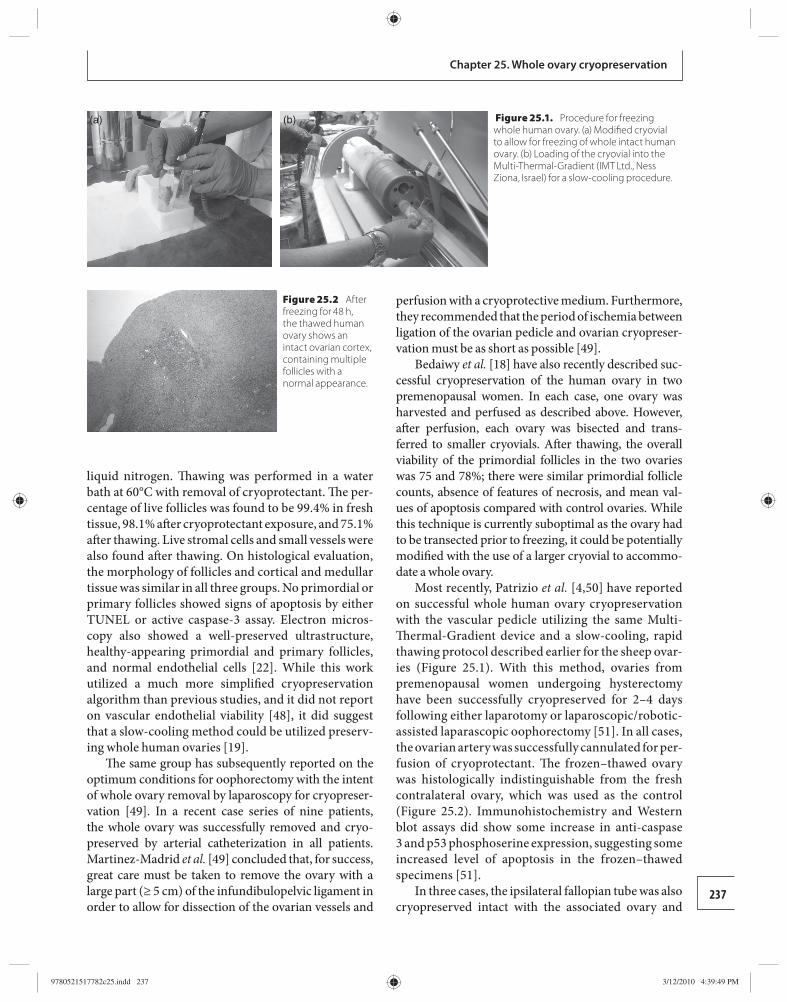
237
Chapter 25. Whole ovary cryopreservation
liquid nitrogen. Th awing was performed in a water bath at 60°C with removal of cryoprotectant. Th e per-centage of live follicles was found to be 99.4% in fresh tissue, 98.1% aft er cryoprotectant exposure, and 75.1% aft er thawing. Live stromal cells and small vessels were also found aft er thawing. On histological evaluation, the morphology of follicles and cortical and medullar tissue was similar in all three groups. No primordial or primary follicles showed signs of apoptosis by either TUNEL or active caspase-3 assay. Electron micros-copy also showed a well-preserved ultrastructure, healthy-appearing primordial and primary follicles, and normal endothelial cells [ 22 ]. While this work util ized a much more simplifi ed cryopreservation algorithm than previous studies, and it did not report on vascular endothelial viability [ 48 ], it did suggest that a slow-cooling method could be utilized preserv-ing whole human ovaries [ 19 ].
Th e same group has subsequently reported on the optimum conditions for oophorectomy with the intent of whole ovary removal by laparoscopy for cryopreser-vation [ 49 ]. In a recent case series of nine patients, the whole ovary was successfully removed and cryo-preserved by arterial catheterization in all patients. Martinez-Madrid et al. [ 49 ] concluded that, for success, great care must be taken to remove the ovary with a large part (≥ 5 cm) of the infundibulopelvic ligament in order to allow for dissection of the ovarian vessels and
perfusion with a cryoprotective medium. Furthermore, they recommended that the period of ischemia between ligation of the ovarian pedicle and ovarian cryopreser-vation must be as short as possible [ 49 ].
Bedaiwy et al. [ 18 ] have also recently described suc-cessful cryopreservation of the human ovary in two premenopausal women. In each case, one ovary was harvested and perfused as described above. However, aft er perfusion, each ovary was bisected and trans-ferred to smaller cryovials. Aft er thawing, the overall viability of the primordial follicles in the two ovaries was 75 and 78%; there were similar primordial follicle counts, absence of features of necrosis, and mean val-ues of apoptosis compared with control ovaries. While this technique is currently suboptimal as the ovary had to be transected prior to freezing, it could be potentially modi fi ed with the use of a larger cryovial to accommo-date a whole ovary.
Most recently, Patrizio et al. [ 4 , 50 ] have reported on successful whole human ovary cryopreservation with the vascular pedicle utilizing the same Multi-Th ermal-Gradient device and a slow-cooling, rapid thawing protocol described earlier for the sheep ovar-ies ( Figure 25.1 ). With this method, ovaries from premenopausal women undergoing hysterectomy have been successfully cryopreserved for 2–4 days following either laparotomy or laparoscopic/robotic-assisted laparascopic oophorectomy [ 51 ]. In all cases, the ovarian artery was successfully cannulated for per-fusion of cryoprotectant. Th e frozen–thawed ovary was histologically indistinguishable from the fresh contralateral ovary, which was used as the control ( Figure 25.2 ). Immunohistochemistry and Western blot assays did show some increase in anti-caspase 3 and p53 phosphoserine expression, suggesting some increased level of apoptosis in the frozen–thawed specimens [ 51 ].
In three cases, the ipsilateral fallopian tube was also cryopreserved intact with the associated ovary and
(a) (b)
Figure 25.1. Procedure for freezing whole human ovary. (a) Modifi ed cryovial to allow for freezing of whole intact human ovary. (b) Loading of the cryovial into the Multi-Thermal-Gradient (IMT Ltd., Ness Ziona, Israel) for a slow-cooling procedure.
Figure 25.2 After freezing for 48 h, the thawed human ovary shows an intact ovarian cortex, containing multiple follicles with a normal appearance.
9780521517782c25.indd 2379780521517782c25.indd 237 3/12/2010 4:39:49 PM3/12/2010 4:39:49 PM
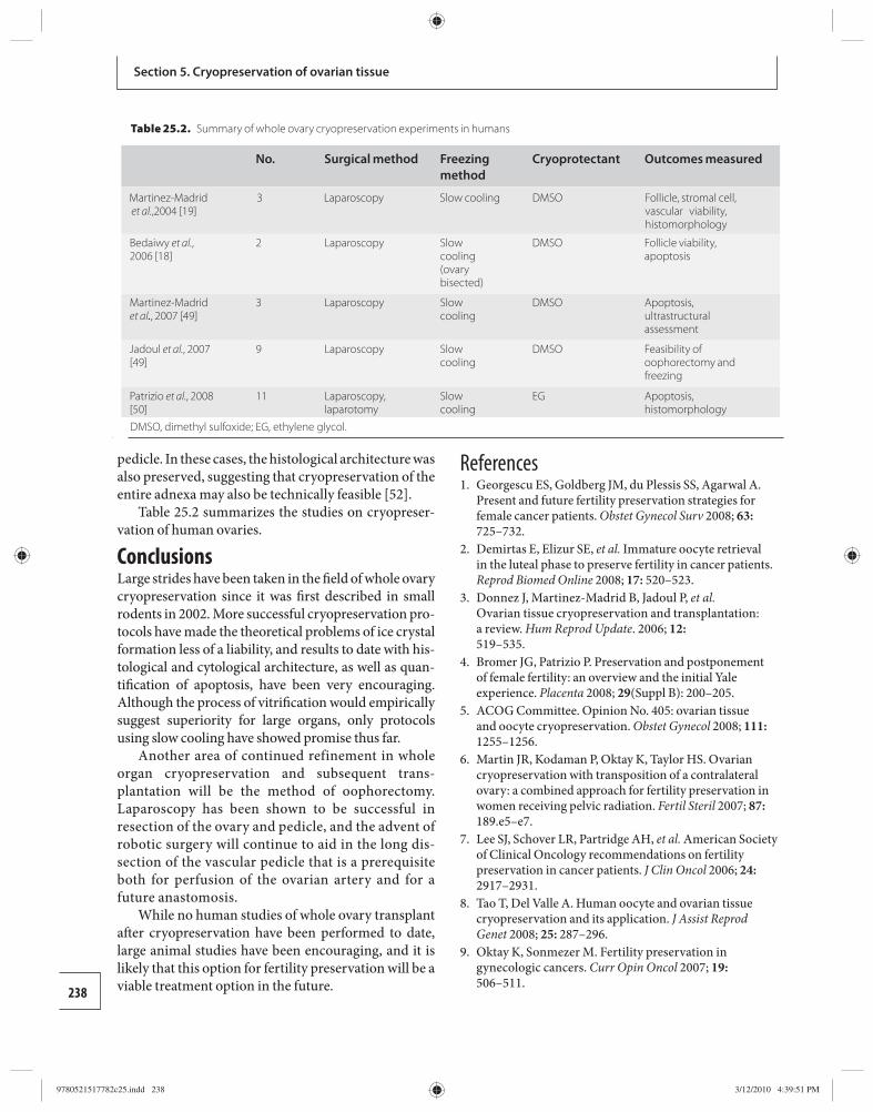
238
Section 5. Cryopreservation of ovarian tissue
pedicle. In these cases, the histological architecture was also preserved, suggesting that cryopreservation of the entire adnexa may also be technically feasible [ 52 ].
Table 25.2 summarizes the studies on cryopreser-vation of human ovaries.
Conclusions Large strides have been taken in the fi eld of whole ovary cryopreservation since it was fi rst described in small rodents in 2002. More successful cryopreservation pro-tocols have made the theoretical problems of ice crystal formation less of a liability, and results to date with his-tological and cytological architecture, as well as quan-tifi cation of apoptosis, have been very encour aging. Although the process of vitrifi cation would empirically suggest superiority for large organs, only protocols using slow cooling have showed promise thus far.
Another area of continued refinement in whole organ cryopreservation and subsequent trans-plantation will be the method of oophorectomy. Laparoscopy has been shown to be successful in resection of the ovary and pedicle, and the advent of robotic surgery will continue to aid in the long dis-section of the vascular pedicle that is a prerequisite both for perfusion of the ovarian artery and for a future anastomosis.
While no human studies of whole ovary transplant aft er cryopreservation have been performed to date, large animal studies have been encouraging, and it is likely that this option for fertility preservation will be a viable treatment option in the future.
References 1. Georgescu ES , Goldberg JM , du Plessis SS , Agarwal A.
Present and future fertility preservation strategies for female cancer patients . Obstet Gynecol Surv 2008; 63: 725–732 .
2. Demirtas E , Elizur SE , et al. Immature oocyte retrieval in the luteal phase to preserve fertility in cancer patients . Reprod Biomed Online 2008; 17: 520–523 .
3. Donnez J , Martinez-Madrid B , Jadoul P , et al. Ovarian tissue cryopreservation and transplantation: a review . Hum Reprod Update . 2006; 12: 519–535 .
4. Bromer JG , Patrizio P. Preservation and postponement of female fertility: an overview and the initial Yale experience . Placenta 2008; 29 (Suppl B): 200–205 .
5. ACOG Committee. Opinion No. 405: ovarian tissue and oocyte cryopreservation . Obstet Gynecol 2008; 111: 1255–1256 .
6. Martin JR , Kodaman P , Oktay K , Taylor HS. Ovarian cryopreservation with transposition of a contralateral ovary: a combined approach for fertility preservation in women receiving pelvic radiation . Fertil Steril 2007; 87: 189.e5–e7 .
7. Lee SJ , Schover LR , Partridge AH , et al. American Society of Clinical Oncology recommendations on fertility preservation in cancer patients . J Clin Oncol 2006; 24: 2917–2931 .
8. Tao T , Del Valle A. Human oocyte and ovarian tissue cryopreservation and its application . J Assist Reprod Genet 2008; 25: 287–296 .
9. Oktay K , Sonmezer M. Fertility preservation in gynecologic cancers . Curr Opin Oncol 2007; 19: 506–511 .
Table 25.2. Summary of whole ovary cryopreservation experiments in humans
No. Surgical method Freezing method
Cryoprotectant Outcomes measured
Martinez-Madrid et al. ,2004 [19]
3 Laparoscopy Slow cooling DMSO Follicle, stromal cell, vascular viability, histomorphology
Bedaiwy et al. , 2006 [18]
2 Laparoscopy Slow cooling (ovary bisected)
DMSO Follicle viability, apoptosis
Martinez-Madrid et al. , 2007 [49]
3 Laparoscopy Slow cooling
DMSO Apoptosis, ultrastructural assessment
Jadoul et al. , 2007 [49]
9 Laparoscopy Slow cooling
DMSO Feasibility of oophorectomy and freezing
Patrizio et al. , 2008 [50]
11 Laparoscopy, laparotomy
Slow cooling
EG Apoptosis, histomorphology
DMSO, dimethyl sulfoxide; EG, ethylene glycol.
9780521517782c25.indd 2389780521517782c25.indd 238 3/12/2010 4:39:51 PM3/12/2010 4:39:51 PM
239
Chapter 25. Whole ovary cryopreservation
10. von Wolff M , Strowitzki T , van der Ven H , Montag M. In vitro maturation is an effi cient technique to generate oocytes and should be considered in combination with cryopreservation of ovarian tissue for preservation of fertility in women . J Clin Oncol 2006; 24: 5336–5337; author reply 7–8.
11. Donnez J , Squiffl et J , Van Eyck AS , et al. Restoration of ovarian function in orthotopically transplanted cryopreserved ovarian tissue: a pilot experience . Reprod Biomed Online 2008; 16: 694–704 .
12. Oktay K. Spontaneous conceptions and live birth aft er heterotopic ovarian transplantation: is there a germline stem cell connection ? Hum Reprod 2006; 21: 1345–1348 .
13. Israely T , Dafni H , Nevo N , Tsafriri A , Neeman M. Angiogenesis in ectopic ovarian xenotransplantation: multiparameter characterization of the neovasculature by dynamic contrast-enhanced MRI . Magn Reson Med 2004; 52: 741–750 .
14. Israely T , Nevo N , Harmelin A , Neeman M , Tsafriri A. Reducing ischaemic damage in rodent ovarian xenograft s transplanted into granulation tissue . Hum Reprod 2006; 21: 1368–1379 .
15. Qi S , Ma A , Xu D , Daloze P , Chen H. Cryopreservation of vascularized ovary: an evaluation of histology and function in rats . Microsurgery 2008; 28: 380–386 .
16. Oktay K. Ovarian tissue cryopreservation and transplantation: preliminary fi ndings and implications for cancer patients . Hum Reprod Update 2001; 7: 526–534 .
17. Oktay K , Karlikaya G. Ovarian function aft er transplantation of frozen, banked autologous ovarian tissue . N Engl J Med 2000; 342: 1919 .
18. Bedaiwy MA , Hussein MR , Biscotti C , Falcone T. Cryopreservation of intact human ovary with its vascular pedicle . Hum Reprod 2006; 21: 3258–3269 .
19. Martinez-Madrid B , Dolmans MM , Van Langendonckt A , Defrere S , Donnez J. Freeze–thawing intact human ovary with its vascular pedicle with a passive cooling device . Fertil Steril 2004; 82: 1390–1394 .
20. Silber SJ , DeRosa M , Pineda J , et al. A series of monozygotic twins discordant for ovarian failure: ovary transplantation (cortical versus microvascular) and cryopreservation . Hum Reprod 2008; 23: 1531–1537 .
21. Gosden RG. Ovary and uterus transplantation . Reproduction 2008; 136: 671–680.
22. Martinez-Madrid B , Camboni A , Dolmans MM , et al. Apoptosis and ultrastructural assessment aft er cryopreservation of whole human ovaries with their vascular pedicle . Fertil Steril 2007; 87: 1153–1165 .
23. Falcone T , Attaran M , Bedaiwy MA , Goldberg JM. Ovarian function preservation in the cancer patient . Fertil Steril 2004; 81: 243–257 .
24. Guttman FM , Lizin J , Robitaille P , Blanchard H , Turgeon-Knaack C. Survival of canine kidneys aft er treatment with dimethyl-sulfoxide, freezing at −80
degrees C, and thawing by microwave illumination . Cryobiology 1977; 14: 559–567 .
25. Guttman FM , Robitaille P , Lizin J , Blanchard H. Physiologic investigation of frozen–thawed canine kidneys . Transpl Proc 1977; 9: 255–256 .
26. Wang X , Chen H , Yin H , et al. F ertility aft er intact ovary transplantation . Nature 2002; 415: 385 .
27. Yin H , Wang X , Kim SS , et al. Transplantation of intact rat gonads using vascular anastomosis: eff ects of cryopreservation, ischaemia and genotype . Hum Reprod 2003; 18: 1165–1172 .
28. Winston RM , Browne JC. Pregnancy following autograft transplantation of Fallopian tube and ovary in the rabbit . Lancet 1974; ii: 494–495 .
29. Goding JR , McCracken JA , Baird DT. Th e study of ovarian function in the ewe by means of a vascular autotransplantation technique . J Endocrinol 1967; 39: 37–52 .
30. Jeremias E , Bedaiwy MA , Gurunluoglu R , et al. Heterotopic autotransplantation of the ovary with microvascular anastomosis: a novel surgical technique . Fertil Steril 2002; 77: 1278–1282 .
31. Paldi E , Gal D , Barzilai A , Hampel N , Malberger E. Genital organs. Auto and homotransplantation in forty dogs . Int J Fertil 1975; 20: 5–12 .
32. Scott JR , Keye WR , Poulson AM , Reynolds WA. Microsurgical ovarian transplantation in the primate . Fertil Steril 1981; 36: 512–515 .
33. Mhatre P , Mhatre J , Magotra R. Ovarian transplant: a new frontier . Trans Proc 2005; 37: 1396–1398 .
34. Leporrier M , von Th eobald P , Roff e JL , Muller G. A new technique to protect ovarian function before pelvic irradiation. Heterotopic ovarian autotransplantation . Cancer 1987; 60: 2201–2204 .
35. Hilders CG , Baranski AG , Peters L , Ramkhelawan A , Trimbos JB. Successful human ovarian autotransplantation to the upper arm . Cancer 2004; 101: 2771–2778 .
36. Bedaiwy MA , Jeremias E , Gurunluoglu R , et al. Restoration of ovarian function aft er autotransplantation of intact frozen–thawed sheep ovaries with microvascular anastomosis . Fertil Steril 2003; 79: 594–602 .
37. Falcone T , Bedaiwy MA. Cryopreservation of intact ovaries: size is a variable? Fertil Steril 2005; 83: 1587; author reply 8.
38. Revel A , Elami A , Bor A , et al. Whole sheep ovary cryopreservation and transplantation . Fertil Steril 2004; 82: 1714–1715 .
39. Grazul-Bilska AT , Banerjee J , Yazici I , et al. Morphology and function of cryopreserved whole ovine ovaries aft er heterotopic autotransplantation . Reprod Biol Endocrinol 2008; 6: 16 .
40. Arav A , Revel A , Nathan Y , et al. Oocyte recovery, embryo development and ovarian function aft er
9780521517782c25.indd 2399780521517782c25.indd 239 3/12/2010 4:39:51 PM3/12/2010 4:39:51 PM
240
Section 5. Cryopreservation of ovarian tissue
cryopreservation and transplantation of whole sheep ovary . Hum Reprod 2005; 20: 3554–3559 .
41. Imhof M , Hofstetter G , Bergmeister H , et al. Cryopreservation of a whole ovary as a strategy for restoring ovarian function . J Assist Reprod Genet 2004; 21: 459–465 .
42. Imhof M , Bergmeister H , Lipovac M . Orthotopic microvascular reanastomosis of whole cryopreserved ovine ovaries resulting in pregnancy and live birth . Fertil Steril 2006; 85 (Suppl 1): 1208–1215 .
43. Fahy GM , Wowk B , Wu J , et al. Cryopreservation of organs by vitrifi cation: perspectives and recent advances . Cryobiology 2004; 48: 157–178 .
44. Rall WF , Fahy GM. Ice-free cryopreservation of mouse embryos at −196 degrees C by vitrifi cation . Nature 1985; 313: 573–575 .
45. Kheirabadi BS , Fahy GM. Permanent life support by kidneys perfused with a vitrifi able (7.5 molar) cryoprotectant solution . Transplantation 2000; 70: 51–57 .
46. Courbiere B , Massardier J , Salle B , et al. Follicular viability and histological assessment aft er cryopreservation of whole sheep ovaries with vascular
pedicle by vitrifi cation . Fertil Steril 2005; 84 (Suppl 2): 1065–1071 .
47. Courbiere B , Caquant L , Mazoyer C , et al. Diffi culties improving ovarian functional recovery by microvascular transplantation and whole ovary vitrifi cation . Fertil Steril 2009; 91 : 2697–2706.
48. Bedaiwy MA , Falcone T. Technical challenges in freeze–thawing of human ovary . Fertil Steril 2005; 83: 1068–1069; author reply 1069–1070.
49. Jadoul P , Donnez J , Dolmans MM , et al. Laparoscopic ovariectomy for whole human ovary cryopreservation: technical aspects . Fertil Steril 2007; 87: 971–975 .
50. Patrizio P , Gavish Z , Martel M , et al. Whole human ovary cryopreservation using a novel multi-gradient freezing device . Fertil Steril 2007; 87 (Suppl 1): s355 .
51. Patrizio P , Bromer JG JJ , Martel M , Silber S , Arav A. Cryopreservation of eleven whole human ovaries: histology, immunohistochemistry, and technical details. Fertil Steril 2008; 88 (Suppl 1): s38 .
52. Patrizio P , Arav A , Bromer JG , Martel M , Azodi M , Silber S. Cryopreservation of whole human ovaries together with fallopian tubes. Fertil Steril 2008; 88 (Suppl 1): s38 .
9780521517782c25.indd 2409780521517782c25.indd 240 3/12/2010 4:39:51 PM3/12/2010 4:39:51 PM
241Fertility Cryopreservation, ed. Ri-Cheng Chian and Patrick Quinn. Published by Cambridge University Press. Copyright © Cambridge University Press 2010.
Chapter
26 surgeons before me have had this idea, but they were prevented from applying it, owing to the lack of a method for re establishing immediately a normal cir-culation through the transplanted structures” [ 4 ].
Rejection has been a major obstacle in transplant-ation attempts. In 1597, the Italian surgeon Gasparo Tagliacozzi, who had performed successful skin autograft s and failing consistently with allograft s, off ered the fi rst suggestion of rejection as the “force and power of individuality” in his work De Curtorum Chirurgia per Insitionem Libri Duo.
In 1933, Dr. YuYu Voronoy in Russia attempted allogeneic transplantation of a human kidney. A kid-ney was taken from a recently deceased individual and connected to a young woman who was suff ering from lead poisoning. No immunosuppression was given. Th e kidney never functioned [ 5 ]. In the late 1940s, Peter Medawar, working for the National Institute for Medical Research, performed experiments that for the fi rst time defi ned the immunology of transplant-ation and improved the understanding of rejection. For his pioneering work in transplant immunology, Dr. Medawar received the Nobel Prize in Medicine in 1960 [ 6 ]. Th e development of immunosuppressant drugs and the fi nding of cyclosporine altered trans-plants from research surgery to life-saving treatment. Belzer [ 7 ] and Collins [ 8 ] provided a grounding in organ preservation that enabled organs to be retrieved at one location and transported to the site of the recipi-ent surgery.
Th e fi rst pioneer of ovarian transplantation was Paul Bert in 1863 in Paris [ 9 ]. He had experimented with rabbits and his initial results were disappointing. However, the work was continued by others and in 1895 the fi rst transplantation in a human was reported by Robert Morris, a New York surgeon [ 10 ]. One of the major obstacles that needs to be overcome is to estab-lish blood fl ow to the transplanted ovary. Th e main
Introduction Transplantation attempts were performed well before people had the knowledge and skill for it to succeed. Th e Chinese physician Pien Chi’ao (550 bc ) reportedly exchanged hearts between a man of strong spirit but weak will with one of a man of weak spirit but strong will in an attempt to achieve balance in each man [ 1 ]. In the second century, the Indian surgeon Sushruta used autograft ed skin transplantation in nose recon-struction rhinoplasty [ 2 ], and there are accounts of the third-century saints Damian and Cosmas replacing the gangrenous leg of the Roman deacon Justinian with the leg of a recently deceased Ethiopian [ 3 ]. Most accounts have the saints performing the transplant in the fourth century, decades aft er their deaths; some accounts have them only instructing living surgeons who performed the procedure.
Th ese transplantation attempts were not well docu-mented and so the outcome is in most cases unknown.Today we know that three main factors determine successful transplantation: (1) surgical technique, (2) overcoming rejection, and (3) maintaining the trans-plant viable.
Alexis Carrel, a French surgeon, was the major pion eer of vascular surgery. He was working in 1902 at the University of Lyons and continued to the University of Chicago and later at the Rockefeller Institute for Medical Research. He performed with Charles Guthrie skillful anastomosis operations and developed new suturing techniques. Th is laid the groundwork for later transplant surgery and won Carrel the 1912 Nobel Prize for Medicine or Physiology. From 1902, Carrel per-formed transplant experiments on dogs. He was surgi-cally successful in moving kidneys, hearts, and spleens and was one of the fi rst to identify the problem of rejec-tion, which remained insurmountable for decades.
Carrel in his Nobel Prize lecture of 1912 said the following about organ transplantation, “… Many
Transplantation of whole frozen–thawed ovaries Amir Arav and Yehudit Natan
9780521517782c26.indd 2419780521517782c26.indd 241 3/12/2010 4:44:27 PM3/12/2010 4:44:27 PM
242
Section 5. Cryopreservation of ovarian tissue
disadvantage of graft ing ovarian tissue over whole ovary is the shorter lifetime of the tissue, which is limited to 3 years. Vascular anastomosis and minimizing ischemia time signifi cantly reduces follicle losses [ 11 ].
Some gynecological surgeons took up the challenge, but evidently with limited success, probably because of the tiny ovarian artery. Nevertheless, 1906 was an annus mirabilis for ovarian transplantation, with a live birth of a baby girl to a woman who had undergone an ovarian homograft transplant by Morris [ 12 ].
Ovarian slices and ovary freezing overview Audrey Smith, known as the mother of cryobiology, dis-covered the cryoprotectant glycerol with Christopher Polge and Alan Parkes [ 13 ]. She was the fi rst to try and freeze a whole organ and performed many of the early freezing experiments, trying to freeze diff erent cells, organs, and even an entire animal. In 1951, Smith and Parkes [ 14 ] tried freezing large slices of rat ovaries (the ovary was cut into three slices) with 15% glycerol. Good results were only achieved when slow freezing was performed but the slices showed reduced viabil-ity aft er 10 days storage at −79ºC. Ovaries minced in serum without protective agents were viable aft er 9 days at −79ºC storage but not aft er 1 hour at −190ºC. Diff erent parameters were evaluated and the best sur-vival was achieved when the ovarian slices were frozen with 15–25% glycerol in rat serum, with a pre-freezing incubation of 5–15 minutes at room temperature, and a slow cooling rate of 1–2ºC/min. Other cryoprotect-ants were evaluated (ethylene glycol, propylene glycol, diethylene glycol) but were not as eff ective as glycerol. Graft s of frozen–thawed ovarian tissue were trans-planted into sterilized irradiated mice, which could be fertilized and give rise to normal off spring [ 15 ]. Th ese studies were summarized by Smith [ 16 ].
Cryopreservation and transplantation of ovar-ian tissue has been successfully performed in animals such as rodents, rabbits, dogs, ewes, and marmoset monkeys [ 17 – 20 ]. In addition, cryopreservation of human ovarian slices has been shown to be success-ful in animal models. Today, women are being off ered the option to cryopreserve ovarian tissue slices prior to chemo- or radiotherapy in many centers in the world. However, to date, only two deliveries of babies with this technology [ 21 , 22 ] have been reported. Th e main problem with ovarian slices is that there is an ischemic period between the transplantation of the graft and revascularization.
Fresh ovarian vascular transplantations have been performed successfully in rabbits [ 23 ], sheep [ 24 ], dogs [ 25 ], monkeys [ 26 ], and humans [ 27 ].
Th e fi rst successful cryopreservation and trans-plantation of an intact ovary was described in 2001 [ 28 , 29 ]. Following this, other groups have reported successful cryopreservation and transplantation of an intact ovary. In 2002, Wang et al. [ 30 ] reported the cryopreservation and subsequent transplantation of rat ovaries and reproductive tract. Four out of seven rats survived for 60 or more days and the transplants were ovulatory and resulted in one pregnancy. Chen et al. [ 31 ] have performed transplants of frozen–thawed rabbit ovaries that showed functionality for up to 7 months aft er microvascular transplantation in 13 out of 15 animals.
However, cryopreservation of organs of large mam-mals and humans is a much more complicated task. Nevertheless, Arav and colleagues have successfully cryopreserved and transplanted whole ovaries with their vascular pedicle in fi ve sheep, showing function-ality in three for 24 to 36 months aft er the transplant-ation [ 32 ]. Currently, we have shown functionality in two out of the three sheep with successful transplants at 6 years aft er the transplant [ 33 ].
Heat and mass transfers in large tissues Heat transfer from an organ (cooling) and to an organ (heating) is conventionally by convection (liquid nitrogen vapor, alcohol bath, cold air, water bath). Heat transfer by convection always begins from the outer part of the organ and progresses towards its center. Additionally, heat transfer through gas or liq-uid material (convection) is slower relatively to solid material (conduction).
Similarly to heat transfer, mass transfers through tissue and organ suff er from the same limitations. In order for organs to be cryopreserved, cryoprotective agents (CPAs) must be introduced into the organ. Th e best way for doing that is by vascular perfusion. Th e CPAs used are usually low-molecular-weight mol-ecules that penetrate the cells membrane by diff usion, such as dimethyl sulfoxide (DMSO), glycerol, and ethylene glycol. Th e perfusion procedure might dam-age the organ and the CPAs are toxic at the concentra-tions needed to promote a protective eff ect during slow freezing. Perfusing a freezing solution for 3 minutes through a sheep ovary is enough to saturate the organ with the CPAs [ 32 ].
9780521517782c26.indd 2429780521517782c26.indd 242 3/12/2010 4:44:28 PM3/12/2010 4:44:28 PM
243
Chapter 26. Transplantation of whole frozen–thawed ovaries
During the freezing process, water freezes and the concentration of solutes increases; as a result, the cells are exposed to increasing concentrations of the CPAs and this has a detrimental eff ect on cell viability [ 34 ]. Th e aim in a slow-freezing process is to have a protocol that is slow enough to dehydrate the cells in order to prevent intracellular crystallization and fast enough to minimize osmotic stress to the cells.
In general the type of damage that occurs in large tissues and organs is similar to the damage that occurs to large volumes of cell suspensions, although, the presence of extracellular ice, which may not be prob-lematic with cell suspensions, can be a major problem when freezing tissues/organs [ 35 ]. For example, iso-lated chondrocytes can survive freezing but the same freezing conditions will not allow successful freezing of cartilage tissue [ 3 6].
Thermal gradient versus cooling rate In order to successfully freeze a biological sample, cool-ing needs to occur at a rate that permits dehydration of the cells down to a level that will form a glass when the solution reaches its glass transition temperature. Th e cells can be preserved below the glass transition temperature in an amorphous state for long periods. Dehydration occurs because of the increasing solute concentration as the freezing process progress, causing water to fl ow out of the cells by exosmosis. Cooling too slowly will extend the exposure time of the cells to an increasingly concentrated and toxic solution, whereas cooling too rapidly can allow intracellular crystalliza-tion to occur. Most cell suspensions will require slow-cooling rates of around 1ºC/min. However, there is evidence that a large tissue or a whole organ will require much slower cooling rates [ 3 7]. When trying to freeze a large tissue or a whole organ, one of the major prob-lems is that cooling rates will vary across the sample depending on the geometry (surface, depth, and vol-ume) and the heat conductivity.
Another issue is supercooling; supercooling is a non-equilibrium thermodynamic state in which a solu-tion is cooled below its freezing point without crystal-lization. It is dependent on time, volume, and viscosity [ 38 ]. In certain tissues/organs where the extracellular matrix has small volume and high viscosity, intracellu-lar crystallization can occur owing to the supercooling conditions.
To start the dehydration of cells, ice crystals need to form in the solution. If ice formation does not start at the freezing point temperature (supercooling), there is
a risk that ice will form intracellular and will kill the cell. If ice formation is not induced by seeding, ice will form spontaneously when the solution is cooled suffi ciently far below its equilibrium melting point. Th is will cause three kinds of problems. First, since ice formation will occur at random, unpredictable temperatures, the sur-vival rates will be highly variable between repeated trials with the same freezing protocol. Second, when ice does form, the large thermal gradient will cause extremely rapid crystallization, which will damage the cells by inducing mechanical stress through uneven expansion or contraction [ 39 ]. Finally, when crystallization starts at low temperatures, water will not leave the cells since dehydration of the cells can only occur within a certain range of subzero temperatures; at lower temperatures, the membrane itself becomes impermeable. Th erefore, supercooling must be avoided and crystallization should start at a temperature as close as possible to the freezing point of the solution.
Latent heat Latent heat is the heat released when crystallization occurs. Th ere are two problems regarding latent heat release. Th e fi rst problem is that the heat is transferred to colder areas in which the ice has just formed and so it could possibly re-warm the ice and cause local melting. Th e second problem is that of the isothermal period. When samples of large volume with relatively low surface-to-volume ratios are frozen, the release of latent heat may cause a long isothermal period (or even heating) in the material being frozen. At the same time, the temperature of the cooling mechanism or of the surrounding medium is lowered, thus increasing the temperature diff erence between the sample and its sur-roundings. Consequently, when latent heat is no longer released, the temperature of the material being frozen will drop very rapidly to a temperature close to the tem-perature of the surrounding environment (because the thermal conductivity of water [0–6] is lower than that of ice [1.6]). Th is might lead to a non-optimal cooling rate and possibly to cellular damage through intracel-lular crystallization.
Recrystallization Slow thawing is known to be a major destructive fac-tor. Th e longer the duration of the thawing period, the greater is the damage that occurs to the cells from the increase in solute eff ects and the maximal growth of ice crystals. Large crystals have an abrasive action, pro-ducing mechanical disruption of cells. Crystal growth
9780521517782c26.indd 2439780521517782c26.indd 243 3/12/2010 4:44:28 PM3/12/2010 4:44:28 PM
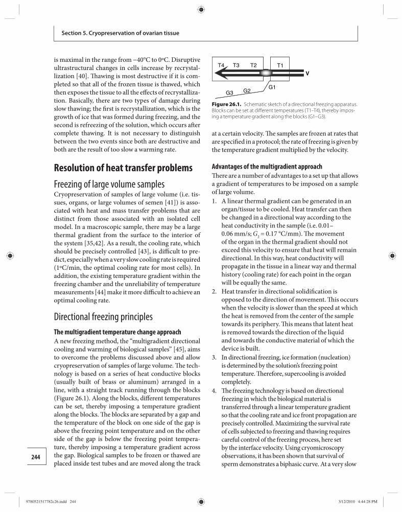
244
Section 5. Cryopreservation of ovarian tissue
is maximal in the range from −40°C to 0ºC. Disruptive ultrastructural changes in cells increase by recrystal-lization [ 40 ]. Th awing is most destructive if it is com-pleted so that all of the frozen tissue is thawed, which then exposes the tissue to all the eff ects of recrystalliza-tion. Basically, there are two types of damage during slow thawing; the fi rst is recrystallization, which is the growth of ice that was formed during freezing, and the second is refreezing of the solution, which occurs aft er complete thawing. It is not necessary to distinguish between the two events since both are destructive and both are the result of too slow a warming rate.
Resolution of heat transfer problems Freezing of large volume samples Cryopreservation of samples of large volume (i.e. tis-sues, organs, or large volumes of semen [ 41 ]) is asso-ciated with heat and mass transfer problems that are distinct from those associated with an isolated cell model. In a macroscopic sample, there may be a large thermal gradient from the surface to the interior of the system [ 3 5, 4 2]. As a result, the cooling rate, which should be precisely controlled [ 4 3], is diffi cult to pre-dict, especially when a very slow cooling rate is required (1ºC/min, the optimal cooling rate for most cells). In addition, the existing temperature gradient within the freezing chamber and the unreliability of temperature measurements [ 4 4] make it more diffi cult to achieve an optimal cooling rate.
Directional freezing principles The multigradient temperature change approach A new freezing method, the “multigradient directional cooling and warming of biological samples” [ 4 5], aims to overcome the problems discussed above and allow cryopreservation of samples of large volume. Th e tech-nology is based on a series of heat conductive blocks (usually built of brass or aluminum) arranged in a line, with a straight track running through the blocks ( Figure 26.1 ). Along the blocks, diff erent temperatures can be set, thereby imposing a temperature gradient along the blocks. Th e blocks are separated by a gap and the temperature of the block on one side of the gap is above the freezing point temperature and on the other side of the gap is below the freezing point tempera-ture, thereby imposing a temperature gradient across the gap. Biological samples to be frozen or thawed are placed inside test tubes and are moved along the track
at a certain velocity. Th e samples are frozen at rates that are specifi ed i n a protocol; the rate of freezing is given by the temperature gradient multiplied by the velocity.
Advantages of the multigradient approach Th ere are a number of advantages to a set up that allows a gradient of temperatures to be imposed on a sample of large volume. 1. A linear thermal gradient can be generated in an
organ/tissue to be cooled. Heat transfer can then be changed in a directional way according to the heat conductivity in the sample (i.e. 0.01–0.06 mm/s; G 2 = 0.17 °C/mm). Th e movement of the organ in the thermal gradient should not exceed this velocity to ensure that heat will remain directional. In this way, heat conductivity will propagate in the tissue in a linear way and thermal history (cooling rate) for each point in the organ will be equally the same.
2. Heat transfer in directional solidifi cation is opposed to the direction of movement. Th is occurs when the velocity is slower than the speed at which the heat is removed from the center of the sample towards its periphery. Th is means that latent heat is removed towards the direction of the liquid and towards the conductive material of which the device is built.
3. In directional freezing, ice formation (nucleation) is determined by the solution’s freezing point temperature. Th erefore, supercooling is avoided completely.
4. Th e freezing technology is based on directional freezing in which the biological material is transferred through a linear temperature gradient so that the cooling rate and ice front propagation are precisely controlled. Maximizing the survival rate of cells subjected to freezing and thawing requires careful control of the freezing process, here set by the interface velocity. Using cryomicroscopy observations, it has been shown that survival of sperm demonstrates a biphasic curve. At a very slow
V
T4 T3 T2 T1
G1G2G3
T4 T3 T2 T1
Figure 26.1. Schematic sketch of a directional freezing apparatus. Blocks can be set at diff erent temperatures (T1–T4), thereby impos-ing a temperature gradient along the blocks (G1–G3).
9780521517782c26.indd 2449780521517782c26.indd 244 3/12/2010 4:44:28 PM3/12/2010 4:44:28 PM
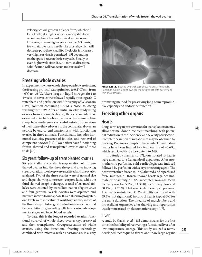
245
Chapter 26. Transplantation of whole frozen–thawed ovaries
velocity, ice will grow in a planer form, which will kill all cells; at a higher velocity, ice crystals form secondary branches and survival will increase. However, at even higher velocities (i.e. 0.3 mm/s), ice will start to form needle-like crystals, which will decrease post-thaw viability. If velocity is increased very high survival is permitted [ 4 5] depending on the space between the ice crystals. Finally, at even higher velocities (i.e. > 4 mm/s), directional solidifi cation will not occur and survival will decrease.
Freezing whole ovaries In experiments where whole sheep ovaries were frozen, the freezing protocol was optimized to 0.1°C/min from −6°C to −35°C. Aft er storage in liquid nitrogen for 1 to 4 weeks, the ovaries were thawed rapidly by using a 66°C water bath and perfusion with University of Wisconsin (UW) solution containing 0.5 M sucrose, following washing with UW. Aft er an initial in vitro study using ovaries from a slaughterhouse, the experiments were extended to include whole ovaries of live animals. Five sheep have undergone successful autotransplantation of the frozen–thawed ovary to the contralateral ovarian pedicle by end-to-end anastomosis, with functioning ovaries in three animals. Functionality includes hor-monal cyclicity, presence of follicles, and retrieval of competent oocytes [ 32 ]. Two heifers have functioning frozen–thawed and transplanted ovaries out of three trials [ 4 6].
Six years follow-up of transplanted ovaries Six years aft er successful transplantation of frozen–thawed ovaries into the three sheep, and aft er inducing superovulation, the sheep were sacrifi ced and the ovaries analyzed. Two of the three ovaries were of normal size and shape, showing some recent corpora lutea, while the third showed atrophic changes. A total of 36 antral fol-licles were counted by transillumination ( Figure 26.2 ) and four germinal vesicle oocytes were aspirated and matured in vitro to metaphase II. Serial serum progester-one levels were indicative of ovulatory activity in two of the three sheep. Histological evaluation revealed normal tissue architecture, including follicles at various develop-mental stages and intact blood vessels.
To date, this is the longest recorded ovarian func-tional survival of whole sheep ovaries cryopreserved and then transplanted. Cryopreservation of whole ovaries, using the directional freezing technology combined with microvascular anastomosis, is a very
promising method for preserving long-term reproduc-tive capacity and endocrine function.
Freezing other organs Hearts Long-term organ preservation for transplantation may allow optimal donor–recipient matching, with poten-tial reduction in the incidence and severity of rejection. Complete cessation of metabolism may be obtained by freezing. Previous attempts to freeze intact mammalian hearts have been limited to a temperature of −3.6ºC, which restricted tissue ice content to 34%.
In a study by Elami et al. [ 4 7], four isolated rat hearts were attached to a Langendorff apparatus. Aft er nor-mothermic perfusion, cold cardioplegia was induced followed by perfusion with a cryoprotecting agent. Th e hearts were then frozen to −8ºC, thawed, and reperfused for 60 minutes. All frozen–thawed hearts regained nor-mal electric activity. At −8ºC, ice content was 64%. Mean recovery was to 65.2% (SD, 30.8) of coronary fl ow and 50.4% (SD, 23.9) of left ventricular developed pressure. Th e hearts maintained 81.3% viability compared with 69.3% (not signifi cant) in control hearts kept at 0ºC for the same duration. Th e integrity of muscle fi bers and intracellular organelles aft er thawing and reperfusion was demonstrated by electron microscopy [ 4 7].
Liver A study by Gavish et al. [ 4 8] demonstrates for the fi rst time the feasibility of recovering a functional liver aft er low temperature storage. Th is study utilized a newly developed technique to freeze and thaw large organs
Figure 26.2. Excised ovary (sheep) showing antral follicles by transillumination (also shown are the sutures left of the artery and vein anastomosis).
9780521517782c26.indd 2459780521517782c26.indd 245 3/12/2010 4:44:28 PM3/12/2010 4:44:28 PM
246
Section 5. Cryopreservation of ovarian tissue
such as the liver. Aft er freezing and keeping the fro-zen livers in storage for up to 21 days, the livers were thawed and their viability was tested by means of cel-lular and tissue integrity and in an auxiliary liver trans-plantation in pigs. Th e livers demonstrated viability of over 80% and maintained their structural integrity and bile production ability.
Conclusions One of the most diffi cult issues for patients need-ing organ transplants is the narrow time window needed between the donation and transplantation of the donated organ. Th e situation is complicated by the logistical challenges of handling these life-giving donations and maintaining organ viability until the time of transplant. With the donation of a human liver, a healthcare provider is restricted to only a few hours of harvest, transport, and transplant time, which precludes optimal pathogen screening and donor–re cipient matching between remote locations. Because of these time constraints, matching is limited to blood type and patient size. Th is creates a risk of immuno-logical mismatches and increases the likelihood of organ rejection. Other types of organs face similar issues, with the clock ticking rapidly when scarce organs become available.
Th e biggest challenge for research is to prolong the shelf-life of donated organs, allowing more people in more places to be matched with the optimal life- saving organs they desperately need. Improved the ability to match donors and recipients would also help to decrease rejection rates and reduce the intensity of immunosuppression treatments aft er transplantation. Th is would be expected to improve patient outcomes while reducing the cost of care.
References 1. Woodford P. Transplantation timeline . Natl Rev Med
2004; 1, No. 2. 2. Dwived G , Dwivedi S. History of medicine. Sushrua: the
clinician–teacher par excellence . Indian J Chest Dis Allied Sci 2007; 49: 243 –244.
3. Conolly BW , Benanzio M. Cosmas and Damian revisited. In Lanzetta M , Dubernard J-M, Petruzzo P , eds. Hand Transplantation . Milan : Springer Milan , 2007, pp. 3 –10.
4. Lindsten J (ed.) Nobel Lectures, Physiology or Medicine 1901–1921 . Amsterdam : Elsevier, 1967 .
5. Petechuk D. Organ Transplantation . Westport, CT : Greenwood Press , 2006, p. 10.
6. Lindsten J (ed.) Nobel Lectures, Physiology or Medicine 1941–1962 . Amsterdam : Elsevier, 1964 .
7. Belzer FO , Ashby BS , Gulyassy PF. Successful seventeen-hour preservation and transplantation of human cadaver kidney . N Engl J Med 1968; 278: 608 –610.
8. Collins GM , Bravo-Shugarman M , Terasaki PI. Kidney preservation for transplantation. Initial perfusion and 30 hours ’ ice storage . Lancet 1969; ii : 1219 –1222.
9. Tuland T , Gosden RG. Preservation of Fertility . London : Informa HealthCare, 20 04, p. 158 .
10. Simmer HH. Robert Tuttle Morris (1857–1945): a pioneer in ovarian transplant . Obstet Gynecol 1970; 35: 314 –328.
11. Yin H , Wang X , Kim SS , et al. Transplantation of intact rat gonads using vascular anastomosis: eff ects of cryopreservation . Hum Reprod 2003; 18: 1165 –1172.
12. Morris RT. A case of heteroplastic ovarian graft ing followed by pregnancy and the delivery of a living child . Med Rec 1906; 69: 697 –698.
13. Polge C , Smith AU , Parkes AS. Revival of spermatozoa aft er vitrifi cation and dehydration at low temperatures . Nature 1949; 164: 666 .
14. Smith AU , Parkes AS. Preservation of ovarian tissue at low temperatures . Lancet 1951; ii : 570 –572.
15. Parrott DMV. Th e fertility of mice with orthotopic ovarian graft s derived from frozen tissue . J Reprod Fertil 1960; 1: 230 –241.
16. Smith AU. Biological Eff ects of Freezing and Supercooling . Baltimore, MD : Williams & Wilkins , 1961 .
17. Candy J , Wood MJ , Whittingham DG. Follicular development in cryopreserved marmoset ovarian tissue aft er transplantation . Hum Reprod 1995; 10: 2234 –2238.
18. Candy CJ , Wood MJ , Whittingham DG. Restoration of a normal reproductive lifespan aft er graft ing cryopreserved mouse ovaries . Hum Reprod 2000; 15: 1300 –1304.
19. Salle B , Demirei B, Franck M , et al. Normal pregnancies and live births aft er autograft of frozen thawed hemi-ovaries into ewes . Fertil Steril 2002; 77: 403 –408.
20. Almodin CG , Minguetti-Camara VC , Meister H , et al. Recovery of fertility aft er graft ing of cryopreserved germinative tissue in female rabbits following radiotherapy . Hum Reprod 2004; 19: 1287 –1293.
21. Donnes J , Dolmans MM , Demylle D , et al. Livebirth aft er orthotopic transplantation of cryopreserved ovarian tissue . Lancet 2004; 364: 1405 –1410.
22. Meirow D , Levron J , Eldar-Geva T , et al. Pregnancy aft er transplantation of cryopreserved ovarian tissue in a patient with ovarian failure aft er chemotherapy . N Engl J Med 2005; 353: 318 –321.
23. Winston RM , Browne JC. Pregnancy following autograft of Fallopian tube and ovary in rabbit . Lancet 1974; ii: 494 –495.
24. Godding JR , McCracken JA , Baird DT. Th e study of ovarian function in the ewe by means of vascular autotransplantation technique . J Endocrinol 1967; 39: 37 –52.
9780521517782c26.indd 2469780521517782c26.indd 246 3/12/2010 4:44:29 PM3/12/2010 4:44:29 PM

247
Chapter 26. Transplantation of whole frozen–thawed ovaries
25. Paldi E , Gal D , Barlizai A , Hampel N , Alberger EM. Genital organs: auto and homotransplantation in forty dogs . Int J Feril 1974; 20: 5 –12.
26. Scott JR , Keye WR , Poulson AM , Reynolds WA. Microsurgical ovarian transplantation in the primate . Fertil Steril 1981; 36: 512 –515.
27. Leporrier M , von Th eobald P , Roff e JL , Muller G. A new technique to protect ovarian function before pelvic irradiation. Heterotopic ovarian autotransplantation . Cancer 1987; 60: 2201 –2204.
28. Arav A , Elami A, Bor A , et al. Successfully whole organ cryopreservation and transplantation . In Annual Meeting of the American Society of Reproductive Medicine, Orlando, USA, October 2001.
29. Revel A , Elami A , Bor A , et al. Whole ovary cryopreservation and transplantation in sheep . Fertil Steril 2001; 76 : S42–S43 .
30. Wang X , Chen H , Yin H , et al. Fertility aft er intact ovary transplantation . Nature 2002; 415: 385 .
31. Chen C , Chen S , Chang F , et al. Autologous heterotopic transplantation of intact rabbit ovary aft er cryopreservation . Hum Reprod 2005; 20 (Suppl 1): i145–i150 .
32. Arav A , Revel A , Nathan Y , et al. Oocyte recovery, embryo development and ovarian function aft er cryopreservation and transplantation of whole sheep ovary . Hum Reprod 2005; 20: 3554 –3559.
33. Arav A Gavish Z , Elami E , et al. Ovarian Function 6 years aft er cryopreservation and transplantation of whole sheep ovaries. Reprod BioMed Online 2010; 20: 48 –52.
34. Mazur P. Cryobiology: the freezing of biological systems . Science 1970; 168: 939 –949.
35. Karlsson JO , Toner M. Long term storage of tissue by cryopreservation: critical issues . Biomaterials 1996; 17: 243 –256.
36. Pegg DE , Wang L , Vaughan D , Hunt CJ. Cryopreservation of articular cartilage. Part 2: mechanisms of cryoinjury . Cryobiology 2006; 52: 347 –359.
37. Hubel A , Toner M , Cravalho EG , Yermush ML , Tompkins RG. Intracellular ice formation during the freezing of hepatocytes cultured in double collagen gel . Biotechnol Prog 1991; 7: 554 –559.
38. Yavin S , Arav A. Measurements of essential physical properties of vitrifi cation solution . Th eriogenology 2007; 67: 81 –89.
39. Fahy GM , Saur J , Williams RJ. Physical problems with the vitrifi cation of large biological systems . Cryobiology 1990; 27: 492 –510.
40. Forsyth M , MacFarlane DR. Recrystallization revisited . Cryo Letters 1986; 7: 367 –378.
41. Gacitua H , Arav A. Successful pregnancies with directional freezing of large volume buck semen . Th eriogenology 2005; 63: 931 –938.
42. Diller KR. Modeling of bioheat transfer processes at high and low temperatures. In Cho YI , ed. Advances in Heat Transfer , Vol. 221: Bioengineering Heat Transfer. San Diego, CA: Academic Press, 1992, pp. 157–357.
43. Mazur P. Th eoretical and experimental eff ects of cooling and warming velocity on the survival of frozen and thawed cells . Cryobiology 1966; 2: 181 –192.
44. Keobe HG , Werner A, Lange V, Schildberg FW. Temperature gradients in freezing chambers of rate-controlled cooling machines . Cryobiology 1993; 30: 349 –352.
45. Arav A , Yavin S , Zeron Y , Natan Y , Dekel I , Gacitua H. New trends in gamete ’ s cryopreservation . Mol Cell Endocrinol 2002; 187: 77 –81.
46. Arav A. Large tissue freezing . J Assist Reprod Genet 2003; 20: 351 .
47. Elami A , Gavish Z , Korach A , et al. Successful restoration of function of frozen and thawed isolated rat hearts . J Th orac Cardiovasc Surg 2005; 135: 666 –672.
48. Gavish Z , Ben-Haim M , Arav A. Cryopreservation of whole murine and porcine livers . Rejuvenation Res 2008; 11: 765–772.
9780521517782c26.indd 2479780521517782c26.indd 247 3/12/2010 4:44:29 PM3/12/2010 4:44:29 PM
248 Fertility Cryopreservation, ed. Ri-Cheng Chian and Patrick Quinn. Published by Cambridge University Press. Copyright © Cambridge University Press 2010.
Section 6Chapter
27 and development of sexual organs. Th e US President’s Cancer Panel has endorsed a similar position [ 4 ], yet, as many as 40% of male cancer patients do not recall being informed of infertility as a side eff ect of cancer treatment [ 5 ].
Postpubertal males Cryopreservation of sperm For postpubertal males, the commonest option is freezing of ejaculated sperm before cancer treatment, followed by artifi cial insemination, in vitro fertiliza-tion (IVF), or intracytoplasmic sperm injection (ICSI) using thawed sperm. Th e availability of ICSI has greatly enhanced the effi cacy of using frozen–thawed sperm, leading to high fertilization and pregnancy rates even when the banked semen samples are not of the best quality because of cancer-related spermatogenic depression. Studies have shown that many oncologists are still unaware of the newer possibilities of assisted reproductive technologies (ART), with the result that young male cancer patients are inadequately informed of their risk of infertility and options available for its reduction [ 6 ]. However, as part of the informed con-sent process, the risks of fertility preservation using frozen–thawed sperm should be clearly explained to the patients prior to banking. Th ese may include pos-sible mutations in the sperm, especially for patients who bank their semen aft er starting their cancer treat-ment. But even if semen samples are banked before treatment, cancer patients may have higher aneuploidy rates and increased DNA damage, which may produce long-term adverse genetic eff ects in their off spring. Th e possibility of controlling for aneuploidy by pre-implantation genetic diagnosis (PGD) may also be sug-gested to patients, but PGD has both benefi ts and risks of its own that need to be disclosed (e.g. it is expensive). Some patients may be reluctant to bank sperm because
Introduction Th is chapter on the ethics of fertility cryopreservation builds on the previous chapters, which have provided the most up-to-date scientifi c understanding and clini-cal applications of cryopreservation of human gametes and tissues. Th e ethical positions represented here are subject to modifi cation and change as new informa-tion on the principles and applications of cryobiology becomes available. Th e discussion excludes approaches to fertility preservation in which freezing–thawing of cells or tissues are not a part (e.g. hormonal gonado-protection); it includes fertility cryopreservation for pre- and postpubertal male and female adolescents and is written in the context of fertility preservation for can-cer patients, although the ethical principles developed here should, in general, apply to more mature men and women seeking fertility preservation for other health issues or for non-therapeutic social reasons (e.g. to postpone having a family).
Cryopreservation of male fertility Male fertility is highly susceptible to toxic eff ects of radiation and chemotherapy at all stages of life, and depending on the therapeutic agents used and the dura-tion of treatment, up to 80% of patients may have long-lasting or permanent gonadal dysfunction resulting in oligozoospermia or azoospermia [ 1 , 2 ]. At the same time, it has been shown that 76% of childless cancer survivors, including male and female patients, desire to have children in the future; 80% believe that having had cancer would make them better parents, and 94% feel that they are healthy enough to be good parents [ 3 ]. Based on these fi ndings, it seems justifi ed to make it a moral duty of doctors treating young male cancer patients to inform their patients of the risks of post-treatment infertility and the options for fertility cryo-preservation available to them, depending on their age
Ethical considerations Ethical considerations in fertility cryopreservation in young cancer patients Edwin C. Hui
9780521517782c27.indd 2489780521517782c27.indd 248 3/11/2010 10:13:09 AM3/11/2010 10:13:09 AM
249
Chapter 27. Ethical considerations in fertility cryopreservation in young cancer patients
of religious or cultural objections to masturbation, which is the most common and least intrusive way to obtain sperm. Such patients should be off ered other methods of obtaining sperm, including the non-in-vasive penile vibratory stimulation. Invasive methods including electroejaculation, which requires a general anesthetic, and surgical retrieval of sperm by either epididymal aspiration or testicular biopsy should not be off ered without full disclosure of the invasive nature and the extra risks entailed in these procedures.
When the relevant risks and benefi ts of fertility preservation have been fully explained to and under-stood by the patients, prevalent medical ethical norms require that patients be allowed to make a decision to agree or disagree to bank sperm. Th is is particularly important for adolescent patients since most of them will not be legally empowered to provide consent and parental consent will be required . For sperm banking by an adolescent male cancer patient, there are three possible scenarios: (1) the adolescent assents and the parents give the legal consent; (2) parents consent and the minor does not assent; (3) parents do not consent and minor requests the treatment. Th e fi rst scenario is fortunately the most common scenario and the attend-ing physician only needs to note two things in particu-lar. First, parents who have just been told of their child’s cancer are very anxious, and they are just as vulner-able as the adolescent patients. Since both the adoles-cent and the parents have to make a major decision in a short time about a procedure with many uncertain ramifi cations, they must fully understand the purpose of sperm banking and have enough time to weigh the risks and benefi ts of the procedure in their own per-sonal and socioeconomical contexts. Second, the attending physician should ensure that the adolescent patient is not under undue infl uence, which can come from two sources, the attending physician and the par-ents. Doctors, on the one hand, should be aware that they oft en unintentionally give their off er of medical service to preserve fertility an imperative character – “you may be sorry later if you don’t decide to do this now” – and must ensure that this so-called “anticipated decision regret phenomenon” plays no part in the deci-sion-making process. On the other hand, parents of young cancer patients are fi lled with guilt and they have the tendency to want to take advantage of every avail-able medical option to promote their child’s wellbeing; consequently they oft en are the over-enthusiastic driv-ing force behind the eff ort to preserve fertility of their child without making an objective assessment of the
risks of preserving fertility [ 7 ]. Th ese undue infl uences are coercive in nature and undermine the integrity of the adolescent’s assent. An assent made under undue infl uence or coercion is not an acceptable assent since the requirements of informed assent and informed consent are essentially the same, with the exception that an assent does not authorize physicians to render treatment.
Th e second and third scenarios are more problem-atic, at least in jurisdictions where parental authority over minor children is still carefully guarded. In the UK, the second scenario is resolved by the Human Fertilisation and Embryology Act (HFEA), which stipu lates that “it is legally impossible for anyone to give consent for storage of gametes on behalf of another. Th us people with parental responsibility cannot give consent on behalf of the child” [ 9 ]. Hence, unless the adolescent is judged to be legally competent to con-sent and gives consent, cryopreservation of sperm will not take place. In the UK, the Family Law Reform Act (1969) also resolves the third scenario, by empowering adolescents 16 years of age and over to consent to med-ical treatment without parental agreement, provided that they are not incompetent. Adolescents under the age of 16 years may also consent to treatment if in the judgment of the attending physician the adolescent has the mental capacity to make the decision [ 10 ]. In the USA, where parents make decisions on the basis of their children’s best interest, the parents will have to decide whether the importance of preserving the pos-sibility of having genetically related off spring in the future outweighs the current risk of sperm cryopreser-vation for the child in the second and third scenarios [ 8 ]. Th is sentiment is shared by many people from cul-tures that emphasize the importance of family blood lineage such as the Chinese. Physicians providing care for adolescent cancer patients should be sensitive of cultural infl uences that may bear on parental decisions to favor preservation of fertility.
Prepubertal males Cryopreservation of testicular tissue Fertility preservation for prepubertal male cancer patients is technically and ethically more complex than for postpubertal males. Th e only option for these patients is removal of testicular tissue surgically from the patient prior to cancer treatment and to store the cryopreserved tissue for use in the future to restore fer-tility through autologous transplantation, xenograft -
9780521517782c27.indd 2499780521517782c27.indd 249 3/11/2010 10:13:11 AM3/11/2010 10:13:11 AM
250
Section 6. Ethical considerations
ing, or in vitro maturation. Since these approaches to use cryopreserved testicular tissues are still considered to be experimental, their employment is regulated by medical research protocols in addition to conventional medical norms. Since most, if not all, prepubertal male patients are of minority age, all the restrictions for postpubertal minors discussed above apply to these patients. In addition, there are additional risks with cryopreservation of testicular tissues. First, testicular biopsy and subsequent re-implantation are consider-ably more invasive than any form of sperm banking; second, and more importantly , there is a real risk of re-implanting malignant cells together with cryopre-served tissues or cells, especially in adolescent patients with hematological cancers. For an experimental pro-cedure to be applied to a minor adolescent, the local institutional review board (IRB) would require that the experimental intervention can be shown to promote the best interest of the minor patient and that there is a reasonable chance for the patient to gain a net benefi t from the procedure. In the USA, cryopreservation of testicular tissue can proceed with parental consent and adolescent assent (assuming that the minor is com-petent to assent). But if the adolescent patient is too immature to comprehend, the parents are allowed to decide on the basis of the minor’s best interest.
Since the cryopreserved testicular tissue may not be used until some time later when the adolescent recovers from his cancer, there is a good chance that the adolescent may either become competent to assent or reach the age of majority for consent. Potentially, at such a time, the adolescent may refuse re-implantation of immature germ cells or in vitro maturation of the germ cells that his parents have previously consented to store. For that matter, an adolescent who has previ-ously assented to cryopreservation of testicular tissue may change his mind and opt for no further treatment. Th is illustrates the dynamic nature of consent taking in medical decision making, which is always a process rather than an event. To resolve this problem, a two-stage consenting process can be adopted, with the fi rst stage primarily concerned with harvesting and cryos-toring of gonadal tissues and the second stage in rela-tion to manipulating and using the tissues to restore fertility. Th e ownership of the preserved tissue may also be decided in the second stage [ 11 ]. Th e advantage of such a two-stage consenting process becomes evi-dent in the case of an adolescent who is too immature to assent at the time his gonadal tissue is removed, and his parents provide the consent for the fi rst stage on
the presumption that such an intervention may be ben-efi cial to their adolescent child; this does not preclude the adolescent from exercising his autonomy in the second stage when he becomes competent enough to take ownership and full control over the use of the tis-sue to restore fertility. Th e adolescent may also exercise his right to decide how to dispose of the cryopreserved tissue in the event he decides not to restore fertility or dies. For the two-stage consent process to work, the adolescent will have to acquire mental competence at some point in the period aft er his tissue has been cryo-preserved and before it is used to restore fertility. Th e two-stage process collapses if, for example, the adoles-cent becomes permanently incompetent owing to his cancer, disease, or treatment, or if he dies while receiv-ing treatment for his cancer. For this reason, some institutions recommend that parental rights should be limited to the fi rst stage of the consent process and excluded from deciding about the use or disposal of the preserved tissue under all circumstances [ 7 ].
In the UK, because of the stipulations of the HFEA about human gametes discussed above, if an adoles-cent is prepubertal and pre-Tanner stage II (in the six Tanner grades of puberty) [ 12 ], then his testicular tis-sue is unlikely to contain gametes as defi ned by HFEA as “a cell with a haploid set of chromosomes which is able to take part in fertilization with another of the opposite sex to form a zygote” [ 9 ]. Th is implies that cryopreservation of the testicular tissue can be con-sented by parents as long as the adolescent assents. However, when the cryopreserved testicular tissue is subsequently manipulated in vitro to create “gametes,” then only a competent adolescent is empowered to give consent. Th is closely resembles the two-stage consent process discussed above except that parents of a pre-pubertal adolescent who has reached Tanner stage II or beyond would not be allowed to consent to tissue preservation since the adolescent’s testicular tissue may contain haploid cells that are defi ned by HFEA as “gametes.” In such a case, only the adolescent can give consent, and only if he is competent to do so.
Cryopreservation of female fertility Postpubertal females Cryopreservation of embryos Like their male counterparts, postpubertal female patients have the advantage of producing mature gametes in their eff ort to preserve fertility before
9780521517782c27.indd 2509780521517782c27.indd 250 3/11/2010 10:13:11 AM3/11/2010 10:13:11 AM
251
Chapter 27. Ethical considerations in fertility cryopreservation in young cancer patients
anti-cancer therapy. However, unlike male gametes, cryopreservation of mature human oocytes results in extensive damage. Hence, at present, the most clinically established approach for fertility cryopreservation in postpubertal females is to fertilize the mature oocytes in vitro and follow this by embryo cryopreservation for future transfer and implantation. In contrast to human oocytes, the human embryo is very resistant to damage caused by cryopreservation, with a post-thaw survival rate anywhere from 35% to 90%. Since the fi rst report of a successful pregnancy using a frozen–thawed embryo in 1983, frozen embryo replacement has become a routine practice in ART [ 13 ]. Th is approach requires the cooperation of the patient’s partner, who must be willing to donate sperm and create embryos for future use; alternatively, the patient must be will-ing to use donor sperm for fertilization. Th is approach also would not benefi t those who for cultural or reli-gious reasons hold the view that human embryos are full human persons with the right to life, since human embryos cannot be frozen for longer than 5 to 10 years, at the end of which they will be destroyed unless they are adopted. At the present time, the number of frozen embryos waiting for adoption far exceeds the number of prospective embryo adopters. Another related con-cern is the approximately 20% risk of embryo loss as a consequence of freezing and thawing. In addition, the long-term risks to off spring derived from frozen–thawed embryos remains unclear, although signifi cant increases in congenital malformation have not yet been demonstrated.
One of the most signifi cant risks in fertility preser-vation through embryo banking is the potential delay in cancer treatment, since hormonal stimulation of the ovary is necessary for IVF and that requires 2–5 weeks for just one cycle [ 13 ]. Another risk is the potential adverse eff ect of hormonal stimulation for the patient’s primary cancer disease. Th is is particularly true for women who have hormonally sensitive tumors such as breast carcinoma. Th ere may also be an increased chance of pregnancy complications, including miscar-riage, low birthweight infants, or premature infants [ 5 ]. For certain cancer patients who need radiother-apy for their primary cancers, in addition to ovarian damage, their uterus may also be adversely aff ected, leading to uterine growth retardation and impaired vascular supply. Th is implies that even if embryos are created and cryopreserved, the patients who recover from cancer may have diffi culty completing the gesta-tion of a full-term fetus or may give birth to an infant
with compromised health [ 14 ]. Th e potential for the recovered cancer patient to relapse as a result of being pregnant should also be considered with seriousness, since pregnancy can theoretically aggravate cancer. It is generally recommended that pregnancy be delayed well aft er cancer treatment is concluded, although the optimal timing is unknown [ 23 ]. For parents acting as the minor adolescent’s proxy decision-maker, weigh-ing these risks against their adolescent’s best interest is a daunting task. Th e two-stage consenting method dis-cussed above is less eff ective for the cryopreservation of embryo since the oocytes have already been used for fertilization and human embryos would have been created aft er the fi rst stage of consent. Nonetheless, it may be ethically prudent to limit the parental consent to retrieval and fertilization of oocytes, particularly for adolescents using donor sperm, and leave the decision to re-implant or dispose of embryos to the adolescents in the second-stage consent later. In the UK, only the competent minor can consent to use her own mature oocytes for creation of embryos. Th e HFEA, which governs the use of male gametes as discussed above, applies equally to the retrieval, storage, and uses of the female gametes. Disposal of embryos created in the event the patient dies of cancer is ethically and legally problematic and will be discussed below in a separate section devoted to disposal of frozen embryos, gam-etes, and reproductive tissues.
Cryopreservation of oocytes For postpubertal female patients who have moral objections against cryopreservation of embryos, are without a partner to serve as a sperm donor for oocyte fertilization, or are reluctant to use donor sperm from strangers, cryopreservation of mature oocytes is an alternative to embryo banking. Banking of mature oocytes avoids the moral controversy of storing embryos, but like embryo banking it also delays can-cer treatment for several weeks because harvesting oocytes requires ovarian stimulation and it exposes the patient to potentially hazardous hormonal stim-ulation. Human oocytes are also highly sensitive to chilling, and low temperature causes cytoskeletal dis-organization and chromosomal and DNA abnormal-ities, all of which impair the oocyte’s developmental competence. Although vitrifi cation and cryoprotective agents may ameliorate the damage caused by freezing and thawing, at the present time cryopreservation of mature oocyte remains the least successful approach and has signifi cant limitations. Th e overall birth rate
9780521517782c27.indd 2519780521517782c27.indd 251 3/11/2010 10:13:11 AM3/11/2010 10:13:11 AM
252
Section 6. Ethical considerations
per cryopreserved oocyte is approximately 2% and the pregnancy rate is reduced by 25–33% compared with using unfrozen oocytes [ 15 ]. Th is suggests that a large number of oocytes would have to be banked to assure a successful pregnancy, and this potentially entails more than one treatment cycle, which is probably an unacceptable amount of delay for cancer treatment. In addition, the long-term eff ects on children born from cryopreserved oocytes are unknown.
In view of the low pregnancy rate and high uncer-tainty about the effi cacy, benefi t, and safety of oocyte cryopreservation under present technologies, this option for fertility preservation is presently seen as an experimental procedure not recommended for routine use. Parents who act as proxy for a postpubertal minor are not justifi ed in opting for this method of fertility preservation unless other methods are unavailable and even then only as part of an IRB-approved protocol with full disclosure of risks and uncertainty of benefi ts to the consenting parents and the minor, who must assent [ 16 ]. Th e Practice Committee of the Society for Assisted Reproductive Technology and the Practice Committee of the American Society for Reproductive Medicine have jointly published a guideline enumer-ating what women considering oocyte cryopreserva-tion must be informed of so that they can make a truly informed decision and give valid consent. Th e guide-line lists 13 “essential elements” of such an informed consent, including the side eff ects and potential risks of oocyte retrieval; methods and costs of oocyte cryo-preservation; thaw-survival rate for oocytes; require-ment for, success rate, and cost of ICSI; clinic-specifi c data and outcome; the high likelihood of natural con-ception for women below the age of 35 years of age; dis-position of unused cryopreserved oocytes and the laws regulating their donation; and the instability of storage facility and the possibility of accidental loss of cryopre-served oocytes in the course of their storage [ 17 ]. Some argue that cancer patients, particularly adolescents and very young adults awaiting anti-cancer therapy, are too vulnerable to be appropriate research subjects and that oocyte cryopreservation should not even be off ered at all [ 18 ].
Prepubertal females Cryopreservation of ovarian tissue For prepubertal females and those postpubertal females who either cannot delay treatment to undergo ovarian stimulation or for whatever reason decide not
to preserve fertility by cryopreservation of embryos or oocytes, cryopreservation and autotransplantation of ovarian tissues has become a viable but experimen-tal option. Compared with oocyte cryopreservation, this approach has greater fertility potential because the ovarian cortex contains primordial follicles with a much larger number of immature oocytes, which are more resistant to cryoinjury, and the procedure does not delay cancer treatment or expose the patient to hormonal stimulation. But it has its own technical chal-lenges and one of these is the intrusive nature of obtain-ing ovarian tissues. For older adolescents, laparoscopic biopsy may be possible, although patients should be warned of the possibilities of postoperative bleeding and infection. But for girls younger than 10 years of age, abdominal surgery may be needed to obtain ovar-ian tissues. Th is makes fertility preservation in young females ethically sensitive and underscores that such a treatment should be recommended to minor patients only when there is a clear favorable risk–benefi t ratio. Another challenge with this procedure is to develop the immature oocytes in ovarian tissue for fertilization aft er the patient recovers from her cancer; currently, autotransplantation of frozen–thawed ovarian tis-sue is the only clinically applicable strategy, and both endocrine and gametogenic functions of the cryopre-served ovarian tissues have been demonstrated aft er autotransplantation. Since transplanted ovarian tissues are susceptible to ischemic injury because of incom-plete or failed revascularization, the longevity of ovar-ian graft s is usually unpredictable, and repeated graft s may be necessary in some cases. Th ese are also risks that minor patients and their parents should be told before they consent to the procedure. So far, no data are available regarding the qualities of oocytes matured in situ in transplanted ovarian tissue, and the potential risks of congenital malformations in the resulting off -spring remains unknown.
Th e most crucial ethical issue in the cryopreser-vation of ovarian tissue is the risk of re-introduction of malignant cells to the recovered patient through the autotransplanted ovarian tissue that was removed before chemotherapy commenced. Although it has been reported that ovarian graft ing in patients with lymphoma is safe [ 19 ], similar data are unavailable for other malignancies. Hence, this approach of fertility preservation is particularly not indicated for female patients with systematic or disseminated malignancies until reliable cancer screening methods become avail-able. Given the various uncertainties of the effi cacy and
9780521517782c27.indd 2529780521517782c27.indd 252 3/11/2010 10:13:11 AM3/11/2010 10:13:11 AM
253
Chapter 27. Ethical considerations in fertility cryopreservation in young cancer patients
risks of this mode of fertility preservation, the treat-ment should only be performed as an experimental procedure under IRB guidelines, with all the risks disclosed. Th e American Society for Reproductive Medicine has narrowly restricted the indication of this experimental procedure as “an alternative for women who immediately face near term medical therapies that clearly threaten their future fertility” [ 20 ]. Similarly, the Edinburgh Criteria limit the procedure for patients with “realistic chance of long-term survival” and “high risk of treatment-induced immediate ovarian fail-ure (estimated > 50%)” [ 21 ]. For patients under 18 years, parental consent with patient assent should be obtained, and the two-stage consent process discussed above in relation to cryopreservation of testicular tis-sue should be used. Th is applies in the UK also since the HFEA does not regulate storage of immature oocytes in ovarian cortex, where no mature gametes are present, but consent by the patient is required in the process of oocyte maturation either in situ or in vitro as gamete formation is under the jurisdiction of the HFEA [ 21 ]. In all circumstances in the USA and the UK, if the minor patient refuses to assent and her parents consent, the fertility doctors should withhold both the removal and transplantation of ovarian tissue [ 22 ].
Ethical issues common to male and female fertility preservation The concern of preservation of ‘fl awed’ genetic material A dilemma in fertility cryopreservation arises from the concern that cancer patients may pass on causative or predisposing factors in their gamete and produce cancerous off spring or off spring with higher risks of cancer. Th is is particularly true for high-penetrance cancer-predisposition syndromes such as hereditary retinoblastoma and breast cancer; the risks involving low-penetrance cancer susceptibility genes are less cer-tain [ 10 ]. Th e question at to whether cancer patients’ off spring may also be exposed to higher health risks because of the cancer treatment that their parents have received or the ART used for their conception has not been defi nitively answered [ 10 ]. Furthermore, the off -spring may also have a higher risk of losing a parent in the event that the cancer recurs and kills a parent who was thought to have been cured. Some have sug-gested that it may be unethical to assist cancer patients with expected shorter lifespan to have off spring [ 23 ].
However, it can be argued that to deny cancer patients the right to preserve their fertility amounts to “forced” sterilization, which is ethically unacceptable. On the one hand, cancer patients must make an informed decision about the cancer and other health-related risks in their child-to-be and weigh that against their own desire to have genetically related off spring. Only they have the right to decide that “no life” is better than a life with health risks for their off spring. On the other hand, if fertility cryopreservation leads to the unfor-tunate result of an off spring developing cancer, it may potentially lead to a “wrongful birth” action from the parents against the doctor and possibly a “wrongful life” from the child with cancer. Th is underscores the importance of providing detailed explanation of all health-related risks, including the risk of begetting a child who develops cancer, to the cancer patient seek-ing fertility cryopreservation service before the service is provided.
Ownership and disposition of cryopreserved material Patients who have cryostored gametes or gonadal tis-sues may die before they have the chance to put them to their “reproductive” uses. Th e critical issue is whether these biomaterials are considered as “properties.” Considering biomaterials as property gives minor children the right to control the bodily materials. For example, minor children should be able to decide that if they do not survive the cancer, the biomaterials cryo-preserved should be destroyed. We have discussed above the two-stage consenting process in which a minor child who survives to majority age should take full control of the disposition of the biomaterials stored and become the sole person entitled to consent either to use of the biomaterials for its original “reproductive” purposes or to dispose of them in one of the several ways. If the minor child does not survive to adulthood, it is recommended that all tissues and gametes should be discarded or donated for research, and, specifi cally, the parents should not have the right to decide on the reproductive uses of their child’s biomaterials post-humously [ 8 ]. A single young adult should preferably also indicate in writing what his/her wishes are as to the posthumous disposition of the biomaterials in the event of his/her premature death.
Although the most common disposition for cryo-preserved gametes and gonadal tissues is to destroy them posthumously, donation of sperm by a sin-gle healthy young adult to others for reproductive
9780521517782c27.indd 2539780521517782c27.indd 253 3/11/2010 10:13:11 AM3/11/2010 10:13:11 AM
254
Section 6. Ethical considerations
purposes is acceptable. For couples, posthumous use of sperm by the surviving partner is acceptable provided there is a prior written instruction by the deceased partner agreeing to this disposition. Th e fact that the child produced will be born without the biological father is not suffi cient reason to deny a couple the use of sperm posthumously. Posthumous oocyte donation to others or spouse is not recommended as the cryop-reservation of oocytes is still an experimental proce-dure and the use of a surrogate mother is involved [ 8 ]. Th ese arrangements refl ect the sound ethical principle of respecting the donor’s autonomy and are generally consistent with the prevalent legal practice that the person’s prior wishes about disposition of biomaterials are controlling aft er death [ 23 ].
Th e disposition of cryopreserved human embryos is more complicated, and the number of cryopreserved embryos is increasing rapidly (400 000 embryos were cryopreserved in the USA alone up to 2002). Th e coup le who have undergone IVF to create embryos should have some prior agreement about the owner-ship of the embryos in the event of a subsequent divorce and the conservatorship of the embryos in the event of the death of one or both partners. For the disposition of cryopreserved embryos, there are essentially fi ve options to choose from: embryo transfer to achieve pregnancy; donate embryos to other infertile couples for embryo transfer; donate embryos for research ; dis-card embryos; and keep embryos in cryostorage and do nothing. Studies have shown that couples’ preferences for embryo disposition are “unstable” and subject to changes as they gain new information and experiences, including the experience of going through IVF to cre-ate the embryos. Th ese fi ndings support the two-stage consent approach: the fi rst query of the couples’ dispos-itional preferences at the time of creation of embryos and cryostorage, and the second query before thawing of the embryos for use. Th is implies that the fi rst stage of the consent (sometimes called “pre-freeze agree-ment”) should not be made binding for the couple. In fact, to require a couple to make a comprehensive decision regarding the disposition of embryos before IVF, embryo transfer, pregnancy or pregnancy loss, and other life events occurring with the passage of time is an uninformed decision since these signifi cant life events can be expected to have profound eff ects on the couples’ view on the moral status of embryos and their proper uses [ 24 ].
Of the fi ve options of embryo disposition, the least problematic option is for the owner-couples to transfer
the cryopreserved embryos to obtain a pregnancy. As long as they are fully informed of the risks of embryo cryopreservation discussed above, the benefi t of having a child fulfi lls the couple’s original intention to create the embryos. Donating embryos to infertile “recipients” is also uncomplicated, but certain guidelines would have to be followed. Th e American Society for Reproductive Medicine has published detailed guidelines regarding embryo donation [ 25 ] and some of the most ethically relevant items include prohibiting monetary compen-sation for the donors except reimbursement for specifi c expenses, for example mandatory blood tests related to donation, screening of embryo donors for viruses and other transmissible diseases, executing a written informed consent for embryo donation that, among other things, specifi cally relinquishes their rights to the embryos and any child or children resulting from their transfer, and receiving appropriate counseling as donors and for the recipients. Embryo recipients must agree to take full responsibility for the embryos and any child or children resulting from their transfer; they must also release the donors from any and all liability from complications of the embryo donation and preg-nancies resulting from their transfer, including the birthing of infants with congenital abnormalities or diseases. Some IVF facilities may have guidelines that decline to transfer donated embryos to “single women” or lesbian couples or recipients who are deemed unsuit-able to be parents by the institution.
Donation of embryos for research is more com-plex, and diff erent studies have found a remarkable ambivalence among couples about embryo donation for research. In some studies, few couples are interested in this option, and in the USA, only 2.8% of embryos are targeted for research purposes [ 24 ]. Research on infertility and embryonic stem cells are the two main areas that attract donation of embryos. An Australian study involving 509 couples with cryostored embryos has reported that only 10% of couples indicate it probable and 34% possible that they would donate their embryos for research. Th e study has shown that coupl es agree that to destroy the surplus embryos is a waste and to donate them for research is to advance sci-entifi c and medical knowledge: both are positive fac-tors that incline them to donate their surplus embryos for research. Factors that disincline donation include seeing the surplus embryos as potential children rather than experimental “specimens” and a perceived lack of control over the type of research that is to be car-ried out on the donated embryos [ 26 ]. Destruction of
9780521517782c27.indd 2549780521517782c27.indd 254 3/11/2010 10:13:11 AM3/11/2010 10:13:11 AM
255
Chapter 27. Ethical considerations in fertility cryopreservation in young cancer patients
embryos for the harvesting of embryonic stem cells also dampens couples’ desires to donate embryos for research. In this regard, guidelines developed by the American Society for Reproductive Medicine require that embryo donors must be informed of the diff er-ences between embryonic stem cell research and other types of research, including the inevitable destruction of the embryo in the process of harvesting the stem cells and the fact that embryonic stem cell lines can be used for many years and have potential commercial value that donors would not share [ 27 ].
Th e last two options for disposing of embryos are discarding embryos and keeping embryos in cryo-storage indefi nitely and doing nothing. Both options are ethically controversial and may refl ect embryo-owners’ belief about the moral values of embryos. Couples who believe that the embryo is nothing more than a mere cluster of cells would readily discard sur-plus embryos for which they have no use. Such a low view of embryonic life is not shared by the majority of people . At the other extreme, there are couples who view the embryo as a person with the moral right to life and they would consider discarding embryos as an act of murder. However, these people are also the most likely to be those who leave their embryos in cryostor-age indefi nitely. In Italy, where the Catholic Church defends the moral rights of the human embryo, a study has reported that as many as 25% of couples have failed to designate what to do with their surplus embryos and “simply ‘let’ their embryos be discarded without assuming the responsibility of signing a defi nitive ‘act of disposal’ ” [ 28 ]. One author has opined that such a relinquishment of responsibility probably represents the embryo-owners’ inability to cope with the ethical dilemma of either discarding or donating the embryo, and not to designate a preference may well repre-sent the wish that the storage facility or the authority would make the decision for them [ 29 ], as the British Government did in 1996 when it ordered British clin-ics to destroy over 3300 embryos that had been cryo-preserved for longer than 5 years. Th ese fi ndings of embryo-donors’ ambivalence underscore the diffi cul-ties associated with the unsettled moral status of the human embryo that practitioners of fertility preser-vation should be aware of lest they fi nd themselves u nwittingly caught in a similar controversy.
References 1. Chung K , Irani J , Knee G , et al. Sperm cryopreservation
for male patients with cancer: an epidemiological
analysis at the University of Pennsylvania . Eur J Obstet Gynecol Reprod Biol 2004; 113 (Suppl 1): S7–S11 .
2. Revel A , Revel-Vilk S. Pediatric fertility preservation: is it time to off er testicular tissue cryopreservation ? Mol Cell Endocrinol 2008; 282 : 143–149 .
3. Schover LR , Brey K , Lichtin A , et al. Knowledge and experience regarding cancer, infertility, and sperm banking in younger male survivors . J Clin Oncol 2002; 20: 1880–1889 .
4. President’s Cancer Panel . Annual Report 2003–2004 . Bethesda, MD : National Cancer Institute, 2004.
5. Schover LR , Rybicki LA , Martin BA , et al. Having children aft er cancer: a pilot survey of survivors ’ attitude and experiences . Cancer 1999; 86: 697–709 .
6. Tournaye H , Goossens E , Verheyen G , et al. Preserving the reproductive potential of men and boys with cancer: current concepts and future prospects . Hum Reprod Update 2004; 10: 525–532 .
7. Bahadur G , Chatterjee R , Ralph D. Case report: testicular tissue cryopreservation in boys. Ethical and legal issues . Hum Reprod 2000; 15: 1416–1420 .
8. ESHIRE Task Force on Ethics and Law . Taskforce 7: ethical considerations for the cryopreservation of gametes and reproductive tissues for self use . Hum Reprod 2004; 19 : 460–462 .
9. Human Fertilisation and Embryology Act . London : HMSO , 1990 .
10. Grundy R , Gosden RG , Hewitt M , et al. Fertility preservation for children treated for cancer (1): scientifi c advances and research dilemmas . Arch Dis Child 2001; 84: 355–359 .
11. Grundy R , Larcher V , Gosden RG , et al. Fertility preservation for children treated for cancer (2): ethics of consent for gamete storage and experimentation . Arch Dis Child 2001; 84: 360–362 .
12. Tanner JM. Foetus into Man: Physical Growth from Conception to Maturity, 2nd edn. Ware, UK : Castlemead, 1989 .
13. Michelmann HW , Nayudu P. Cryopreservation of human embryos . Cell Tissue Banking 2006; 7: 135–141 .
14. Pacey AA. Fertility issues in survivors from adolescent cancers . Canc Treat Rev 2007; 33: 646–655 .
15. Ambrosini G , Andrisani A , Porcu E , et al. Oocytes cryopreservation: state of art . Reprod Toxicol 2006; 22: 250–262 .
16. Jain JK , Paulson RJ. Oocyte cryopreservation. Fertil Steril 2006; 86 (Suppl 4): S1037–S1046 .
17. Practice Committees of the Society for Assisted Reproductive Technology and the American Society of Reproductive Medicine . Essential elements of informal consent for elective oocyte cryopreservation: a Practice Committee opinion . Fertil Steril 2007; 88 : 1495–1496 .
18. Melo-Martin I , Cholst IN. Researching human oocyte cryopreservation: ethical issues . Fertil Steril 2008; 89 : 523–528 .
9780521517782c27.indd 2559780521517782c27.indd 255 3/11/2010 10:13:11 AM3/11/2010 10:13:11 AM
256
Section 6. Ethical considerations
19. Kim SS. Fertility preservation in female cancer patients: current developments and future directions . Fertil Steril 2006; 85 : 1–11 .
20. Practice Committees of the American Society of Reproductive Medicine and the Society for Assisted Reproductive Technology . Ovarian tissue and oocyte cryopreservation . Fertil Steril 2006; 86 (Suppl 1): S142–S147 .
21. Wallace WHB , Anderson RA , Irvine DS. Fertility preservation for young patients with cancer: who is at risk and what can be off ered ? Lancet Oncol 2005; 6: 209–218 .
22. Dudzinski DM. Ethical issues in fertility preservation for adolescent cancer survivors: oocyte and ovarian tissue cryopreservation . J Pediatr Adolesc Gynecol 2004; 17: 97–102 .
23. Ethics Committee of the American Society for Reproductive Medicine . Fertility preservation and reproduction in cancer patients . Fertil Steril 2005; 83 : 1622–1628 .
24. Bankowski BJ , Lyerly AD , Faden RR , et al. Th e social implications of embryo cryopreservation . Fertil Steril 2005; 84 : 823–832 .
25. American Society for Reproductive Medicine . Guidelines for cryopreserved embryo donation . Fertil Steril 2004; 82 (Suppl 1): S16–S17 .
26. McMahon CA , Gibson FL , Leslie GI , et al. Embryo donation for medical research: attitudes and concerns of potential donors . Hum Reprod 2003; 18: 871–877 .
27. Ethics Committee of the American Society for Reproductive Medicine . Donating spare embryos for embryonic stem cell research . Fertil Steril 2009; 91 : 667–670 .
28. Cattoli M , Borini A , Bonu MA. Fate of stored embryos: our 10 years’ experience . Eur J Obstet Gynecol Reprod Biol 2004; 115 (Suppl 1): S16–S18 .
29. Dawson KJ. Th e storage of human embryos . Hum Reprod 1997; 12: 6.
9780521517782c27.indd 2569780521517782c27.indd 256 3/11/2010 10:13:11 AM3/11/2010 10:13:11 AM
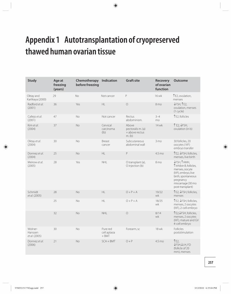
257
Appendix 1 Autotransplantation of cryopreserved thawed human ovarian tissue
Study Age at freezing (years)
Chemotherapy before freezing
Indication Graft site Recovery of ovarian function
Outcome
Oktay and Karlikaya (2000)
29 No Not cancer P 16 wk ↑E2, ovulation, menses
Radford et al. (2001)
36 Yes HL O 8 mo ↓FSH, ↑E2, ovulation, menses (1 cycle)
Callejo et al. (2001)
47 No Not cancer Rectus abdominism.
3–4 mo
↑E2, follicles
Kim et al. (2004)
37 No Cervical carcinoma (Ib)
Above pectoralis m. (a) + above rectus m. (b)
14 wk ↑ E2, ↓FSH, ovulation (in b)
Oktay et al. (2004)
30 No Breast cancer
Subcutaneous abdominal wall
3 mo 30 follicles, 20 oocytes ( IVF) embryo transfer
Donnez et al. (2004)
25 No HL P 4.5 mo ↑E2, ↓FSH, follicles, menses, live birth
Meirow et al. (2005)
28 Yes NHL O transplant (a), O injection (b)
8 mo ↓FSH, ↑AMH, ↑inhibin B, follicles, menses, oocyte (IVF), embryo, live birth, spontaneous pregnancy miscarriage (30 mo post-transplant)
Schmidt et al. (2005)
28 No HL O + P + A 19/22 wk
↑E2, ↓FSH, follicles, menses
25 No HL O + P + A 18/25 wk
↑E2, ↓FSH, follicles, menses, 2 oocytes (IVF), 2-cell embryo
32 No NHL O 8/14 wk
↑E2,↓FSH, follicles, menses, 2 oocytes (IVF), mature and GV 4-cell embryo
Wolner-Hanssen et al. (2005)
30 No Pure red cell aplasia + BMT
Forearm, sc 18 wk Follicles poststimulation
Donnez et al. (2006)
21 No SCA + BMT O + P 4.5 mo ↑E2, ↓FSH,↓LH, FD (follicle of 20 mm), menses
9780521517782app.indd 2579780521517782app.indd 257 3/12/2010 6:35:04 PM3/12/2010 6:35:04 PM

258
Appendix 1
Study Age at freezing (years)
Chemotherapy before freezing
Indication Graft site Recovery of ovarian function
Outcome
Oktay (2006) 29 Yes HL Abdominal wall, sc
2 mo ↑E2, ovulation, pregnancies: 1 miscarriage, 1 live birth from native ovary
Demeestere et al. (2006)
24 Yes HL O + P + abdominal wall, sc
4 mo ↑E2, ↑inhibin B, ↓FSH, follicles in all sites, ovulations, natural conception, pregnancy, miscarriage (7 wk) (aneuploidy)
Rosendahl et al. (2006)
28 No HL O + P + A 19/22 wk
↑E2, ↓FSH, follicles, menses, oocytes (IVF), embryo development, biochemical pregnancy
Demeestere et al. (2007)
24 Yes HL O + abdominal wall, sc
Menses, follicles in orthotopic only, natural conception, live birth
Silber et al. ( 2008)
25 No Not cancer O 4–5 mo First spontaneous cycle: live birth
Donnez et al. (2008)
21 (Donnez et al. (2006)
No SCA +BMT O + P 4.5 mo ↑E2, ↓FSH, menses, follicles, live birth
23 Yes HL + BMT O 5 mo ↑E2, ↓FSH, menses, follicles
28 Yes NHL + BMT O bilateral 6.5 mo ↑E2, ↓FSH, menses, follicles
22 No WG O bilateral 4.5 mo ↑ E2,↓ FSH, menses, Follicles
Andersen et al. (2008)
A: 32 No NHL O 19/22 wk
3 IVF cycles, 2 mature oocytes, transfer of one embryo
B: 28 No HL O + P + A 18/25 wk
Biochemical pregnancy
C: 25 Yes HL O + P + A 16/20 wk?
Clinical pregnancy, miscarriage 7 wk (site O)
Table (Cont.)
9780521517782app.indd 2589780521517782app.indd 258 3/12/2010 6:35:04 PM3/12/2010 6:35:04 PM
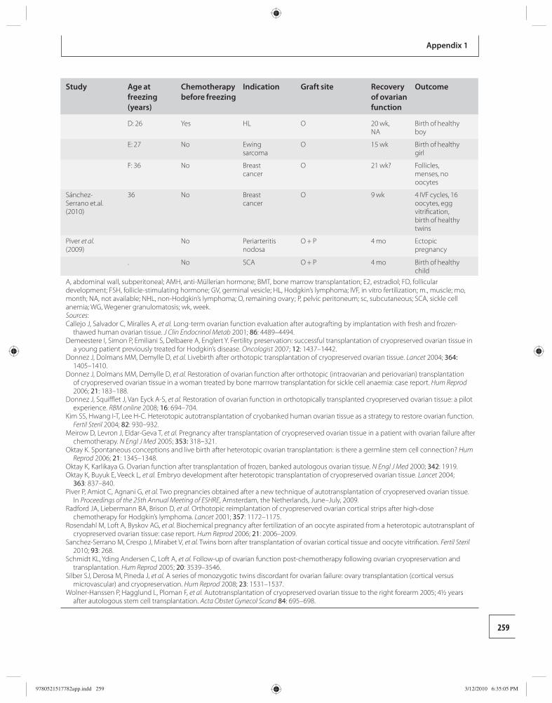
259
Appendix 1
Study Age at freezing (years)
Chemotherapy before freezing
Indication Graft site Recovery of ovarian function
Outcome
D: 26 Yes HL O 20 wk, NA
Birth of healthy boy
E: 27 No Ewing sarcoma
O 15 wk Birth of healthy girl
F: 36 No Breast cancer
O 21 wk? Follicles, menses, no oocytes
Sánchez-Serrano et.al. (2010)
36 No Breast cancer
O 9 wk 4 IVF cycles, 16 oocytes, egg vitrifi cation, birth of healthy twins
Piver et al. (2009)
No Periarteritis nodosa
O + P 4 mo Ectopic pregnancy
. No SCA O + P 4 mo Birth of healthy child
A, abdominal wall, subperitoneal; AMH, anti-Müllerian hormone; BMT, bone marrow transplantation; E2, estradiol; FD, follicular development; FSH, follicle-stimulating hormone; GV, germinal vesicle; HL, Hodgkin’s lymphoma; IVF, in vitro fertilization; m., muscle; mo, month; NA, not available; NHL, non-Hodgkin’s lymphoma; O, remaining ovary; P, pelvic peritoneum; sc, subcutaneous; SCA, sickle cell anemia; WG, Wegener granulomatosis; wk, week. Sources: Callejo J , Salvador C , Miralles A , et al. Long-term ovarian function evaluation after autografting by implantation with fresh and frozen-
thawed human ovarian tissue . J Clin Endocrinol Metab 2001 ; 86 : 4489 –4494. Demeestere I , Simon P , Emiliani S , Delbaere A , Englert Y . Fertility preservation: successful transplantation of cryopreserved ovarian tissue in
a young patient previously treated for Hodgkin’s disease . Oncologist 2007 ; 12 : 1437 –1442. Donnez J , Dolmans MM , Demylle D , et al. Livebirth after orthotopic transplantation of cryopreserved ovarian tissue . Lancet 2004; 364:
1405 –1410. Donnez J , Dolmans MM , Demylle D , et al. Restoration of ovarian function after orthotopic (intraovarian and periovarian) transplantation
of cryopreserved ovarian tissue in a woman treated by bone marrrow transplantation for sickle cell anaemia: case report . Hum Reprod 2006 ; 21 : 183 –188.
Donnez J , Squiffl et J , Van Eyck A-S , et al. Restoration of ovarian function in orthotopically transplanted cryopreserved ovarian tissue: a pilot experience . RBM online 2008 ; 16 : 694 –704.
Kim SS , Hwang I-T , Lee H-C . Heterotopic autotransplantation of cryobanked human ovarian tissue as a strategy to restore ovarian function . Fertil Steril 2004 ; 82 : 930 –932.
Meirow D , Levron J , Eldar-Geva T , et al. Pregnancy after transplantation of cryopreserved ovarian tissue in a patient with ovarian failure after chemotherapy . N Engl J Med 2005; 353: 318 –321.
Oktay K . Spontaneous conceptions and live birth after heterotopic ovarian transplantation: is there a germline stem cell connection? Hum Reprod 2006 ; 21 : 1345 –1348.
Oktay K , Karlikaya G . Ovarian function after transplantation of frozen, banked autologous ovarian tissue . N Engl J Med 2000 ; 342 : 1919 . Oktay K , Buyuk E , Veeck L , et al. Embryo development after heterotopic transplantation of cryopreserved ovarian tissue . Lancet 2004 ;
363 : 837 –840. Piver P , Amiot C , Agnani G , et al. Two pregnancies obtained after a new technique of autotransplantation of cryopreserved ovarian tissue.
In Proceedings of the 25th Annual Meeting of ESHRE , Amsterdam, the Netherlands, June–July, 2009. Radford JA , Liebermann BA , Brison D , et al. Orthotopic reimplantation of cryopreserved ovarian cortical strips after high-dose
chemotherapy for Hodgkin’s lymphoma . Lancet 2001 ; 357 : 1172 –1175. Rosendahl M , Loft A , Byskov AG , et al. Biochemical pregnancy after fertilization of an oocyte aspirated from a heterotopic autotransplant of
cryopreserved ovarian tissue: case report . Hum Reprod 2006 ; 21 : 2006 –2009. Sanchez-Serrano M , Crespo J , Mirabet V , et al. Twins born after transplantation of ovarian cortical tissue and oocyte vitrifi cation . Fertil Steril
2010 ; 93 : 268 . Schmidt KL , Yding Andersen C , Loft A , et al. Follow-up of ovarian function post-chemotherapy following ovarian cryopreservation and
transplantation . Hum Reprod 2005 ; 20 : 3539 –3546. Silber SJ , Derosa M , Pineda J , et al. A series of monozygotic twins discordant for ovarian failure: ovary transplantation (cortical versus
microvascular) and cryopreservation . Hum Reprod 2008 ; 23 : 1531 –1537. Wolner-Hanssen P , Hagglund L , Ploman F , et al. Autotransplantation of cryopreserved ovarian tissue to the right forearm 2005; 4½ years
after autologous stem cell transplantation . Acta Obstet Gynecol Scand 84 : 695 –698.
9780521517782app.indd 2599780521517782app.indd 259 3/12/2010 6:35:05 PM3/12/2010 6:35:05 PM
abdominal ionizing radiation , see radiotherapy
abnormalities, children’s health outcomes , see congenital abnormalities
acetamide , 26–27 , 18 , 19–20 acquired immunodefi ciency syndrome
(AIDS) , 46 activin A , 205 , 208 addonitol , 27–28 adolescents with cancer , 63–69 , 195–196 AID (artifi cial insemination by donor) ,
46 , 48 alcohols , 27 alkylating agents , 57–58 , 189 amide cryoprotectants , see acetamide ;
formamide ; methanamide anejaculation , 48–49 angiogenesis, transplantation and , 191 ,
225 angiogenic granulation tissue , 221 animal breeding
electroejaculation use , 48 sperm cryopreservation , 39–40 , 41
animal studies follicle culture , 200 , 204 McGill Cryoleaf effi cacy , 148 ovarian graft s , 219 ovarian tissue graft s , 218 whole ovary cryopreservation ,
233–234 , 242 , 245–248 anonymity, sperm donors , 46–47 anti-Müllerian hormone
folliculogenesis , 200–201 , 205 , 207 ovarian tissue transplantation , 224 ,
226 antioxidant treatment , 220 antral follicles
in vitro development , 208 puncture , 213 , 214
apoptosis, culture conditions , 206–207
aquaporins , 17 aquaporin 3 , 18
early embryos (mouse) , 20 morulae (mouse) , 19 , 20
oocytes (mouse) , 17–18 aqueous solutions , 3 arabinose, non-penetrating CPAs , 29 Arrhenius activation energy , 17 ART (assisted reproductive
technology) CPA use , 25 , 33 embryo cryopreservation , 67 ice crystal prevention , 12 outcomes , 178–182 , 179
adverse eff ects , 182 aft er oocyte cryopreservation , 178 babies’ neonatal development , 182 multiple pregnancy , 67–68
sperm cryopreservation , 40 vitrifi cation roles , 131
artifi cial insemination by donor (AID) , 46 , 48
artifi cial shrinkage, blastocele , 99–100 , 101
aseptic vitrifi cation , 158–159 , 165 blastocysts , 106–107 , 109–112 ,
163–164 applications , 108–109 outcomes , 110 protocols , 109
cooling speed , 106–107 embryo carrier devices , 107
hermetically sealed , 106–107 illustrated , 158 , 159
GV oocytes , see germinal vesicle oocytes
outcomes , 109 protocols , 108 , 159 solutions , 108 vs. non-aseptic vitrifi cation , 108–109
assisted reproductive technology, see ART
asthenozoospermia , 48 autoimmune diseases , 40 , 151–152 , 190 autotransplantation , see transplantation azoospermia , 57–58
sperm retrieval , 51 , 52–53
bacterial agents, risk , 139 basal culture conditions , 204
basic fi broblast growth factor , 205 biochemical pregnancies , 178 biological carriers, sperm
cryopreservation , 53–54 biomaterials, ownership and
disposition , 253–254 biopsy
blastocysts , 102 embryos , 70 , 81 , 90
biospectroscopy , 123 Biot–Fourier equation , 159 blastoceles , 95–96
shrinkage , 99–100 , 101 , 111 blastocyst aseptic vitrifi cation ,
106–107 , 109–112 , 163–164 applications for , 108–109 in vitro development for , 109–110 outcomes , 110 protocols , 108 , 109
blastocyst cryopreservation , 95 methods , 97–99 optimal stage for , 82 , 96 pregnancy outcomes , 95 selection , 72 slow freezing , 99 water/CPA movement , 21
blastocyst vitrifi cation , 99 carrier devices , 107
Cryoloop , 98 Cryotop , 98 , 100 , 102 electron microscopy grid , 97
culture methods , 96–97 in vitro development , 108–109 , 162 optimal stage for , 100–101 , 102 , 164 pregnancy rates , 160 protocol , 69 rapid freezing , 157–158 selection for , 70 , 77 , 111 structural features , 95–96 two-step treatment , 21
blastocysts aft er thawing , 101 bovine , 31 cooling rate and outcomes , 89 cryodamage , 71 for transfer , 100–101
Index
260
9780521517782ind.indd 2609780521517782ind.indd 260 3/12/2010 10:29:15 PM3/12/2010 10:29:15 PM
261
Index
advantages , 97 , 157 disadvantages , 96 , 97
murine , 17 , 18 blastomeres , 95
cryodamage to , 71 in embryo selection , 91 , 93 in oocyte quality , 123
bone morphogenetic proteins , 200–201 , 205
boys cancer , 58 prepubertal , 59–61
BRC–ABL detection , 229 breast cancer , 151 , 190 1,3 butanediol (butylene glycol) , 27 butylene glycol (1,3 butanediol) , 27
cancer patients , xi adolescents , 63–69 , 195–196 ART outcome , 184 embryo cryobanking , 68 , 251 evaluation for fertility preservation ,
223 fertility preservation , 190–191
female , 68 fertility preservation options , 62–63 ,
190–191 gonadotoxic therapies , 189–190 oocyte cryopreservation , 150–153 ,
251 ovarian stimulation , 144–145 ovarian tissue cryobanking , 189 , 190 ,
213 , 218 , 221 evaluation for transplantation ,
228 histological analysis , 215–216 indications for , 190 safety of , 226–229 storage protocol , 221–222
pregnancy complications aft er treatment , 251
pre-transplantation evaluation , 222 primordial follicle cryopreservation ,
200 recurrence risk
ovarian tissue autotransplantation , 252
SSC contamination , 62 whole ovary transplantation ,
195 sperm cryobanking , 40 , 46 , 51 SSC transplantation , 59–61 treatment delay , 251 treatment planning , 58–59 young patients , 59 , 63–68 , 248 ,
252–253 cancer susceptability genes , 253 carrier devices , 133 , 146
oocyte vitrifi cation , 132–135 , 133 eff ectiveness , 134–135 safety , 138–139
sperm cryopreservation , 53–54 see also embryo carrier devices ;
McGill Cryoleaf cell membrane permeability , 4 , 10–11 ,
16 cryobiological property , 16 cryoinjury and , 10–11 developmental changes , 21 measurement , 16–17 penetrating CPAs , 25 transport channels , 4 , 10–11 , 17
cell membranes, integrity , 161 cells
cryoinjury , 1–2 , 4 permeability , 4 proliferation in vitro , 207
channel diff usion , 16 , 17 early embryos , 20 morulae , 16–22 , 19 , 20 oocytes and morulae , 17
chemotherapy , see gonadotoxic chemotherapy
childhood cancer , 57 , 63–68 , 248 ovarian tissue cryopreservation , 189 ,
252–253 sperm cryopreservation , 58–59 testicular tissue cryopreservation , 59
children, from oocyte cryopreservation , 127–128 , 132
chilling, cryobiological property , 16 China, donor sperm cryopreservation ,
46–48 choline-based freezing media
MII spindle maintenance , 124–125 oocyte cryopreservation , 116 ,
122–123 chromatin abnormalities , 163 chromosomal abnormalities , 180–181 classic technique
slow-freezing oocytes , 115 , 116 sperm cryopreservation , 41
cleavage stage embryo cryopreservation , 68–69 , 70
comparative studies , 81–82 , 96 vs. blastocyst , 102 vs. pronuclear , 82
cryodamage , 71 day 2 , 81–82 , 89 , 90 day 3 , 89 , 90 selection for , 71–72 , 77
clinical assisted reproduction , see ART (assisted reproductive technology)
co-culture, blastocyst cryopreservation , 95
colligative properties , 4 concentration, in CPA toxicity , 31–32 congenital abnormalities
ART and , 179–181 , 180 oocyte cryopreservation and ,
127–128 , 183 –184 consent , see informed consent contamination risk, in vitrifi cation , 84 ,
98–99 , 158 cooling rate
and ice formation , 11 aseptic vitrifi cation , 106–107 , 160 carrier devices and , 135–137 danger zones , 13 developmental stage and outcomes ,
89 donor egg bank protocol , 172–173 minimum volume cooling protocol ,
172–173 phase diagram for CPAs , 12–14 slow-cooling cryopreservation , 77 ,
121 vitrifi cation , 14 , 92 , 134 , 137–138 whole ovary cryopreservation , 243
cortical tissue , see ovarian cortical tissue
CPAs (cryoprotectants) , 24 , 25 , 33 animal model studies , 148 aseptic vitrifi cation , 106–107 ,
110–112 , 158 blastocyst cryopreservation , 99
aseptic vitrifi cation , 110–111 slow-freezing , 99 vitrifi cation , 99
cell permeability to , 12 combination , 13–14 concentrations of , 14 , 137 172 cryodamage reduction , 31 embryo cryopreservation , 68–69 ,
76 , 91 vitrifi cation , 84
history of , 4–5 hypothetical solution , 13 introduction , 24 mechanism of action , 5 , 8 movement pathway , 18 oocyte cryopreservation
slow cooling , 10 , 121 , 122 vitrifi cation , 134 , 139 , 140 , 145
ovarian tissue cryopreservation , 191 ovary (whole) cryopreservation ,
242–243 permeability , 16–17 , 20 , 21 phase diagram for , 12–14 , 13 selection of , 16 slow-freezing cryopreservation , 99 ,
191 sperm cryopreservation , 41
9780521517782ind.indd 2619780521517782ind.indd 261 3/12/2010 10:29:15 PM3/12/2010 10:29:15 PM
Index
262
CPAs (cryoprotectants) (cont.) structures , 6 toxicity , 7
assessment issues , 31 cryobiological property , 16 factors in , 7 , 31–32 risk reduction , 13–14 , 32 , 134 , 145
vitrifi cation , 99 , 148 , 160 concentration in , 14 , 84 without CPAs , 41
zona pellucida manipulation , 102 see also non-penetrating
cryoprotectants ; penetrating cryoprotectants
cryobanking , 213–214 disease transmission risk ,
139 donor eggs , 175 long-term , 72 ovarian stem cells , 213–214 ovarian tissue , 213–214 pronuclear stage embryos , 77 safety considerations , 72 , 84 sperm , 46 storage containers , 90 transportation to , 213
cryobiology , 1–4 , 7–8 cryodamage , 16 in cryopreservation protocols , 24 in developmental stages , 21–22
cryodamage , 4 , 24 and outcomes , 90 blastocysts , 71 , 99–100 cryobiology of , 16 egg banks and , 169–170 embryos , 16 , 70–71 membrane permeability and ,
10–11 oocytes , 16 , 120–121 testing for , 79–80
Cryoleaf , 132 , 134 Cryolock , 132 , 134 , 172–173
in thawing protocol , 173 Cryoloop , 92 , 106 , 132 , 133
blastocyst vitrifi cation , 98 cooling/warming rates , 135–137 oocyte vitrifi cation , 133 sperm vitrifi cation , 48
cryopreservation , 8 cooling rate and danger zones , 13 indications for , 1 , 24 , 78–80 in nature , 1 options for , 190–191 outcomes , 90 slow cooling, history , 10 stress of , 79–80 timing in , 77 toxicity avoidance , 32
water/CPA movement pathways , 20–21
cryopreservation straws , 133 , 135–137 see also cut standard straws (CSS)
cryoprotectants see CPAscryostability evaluation, 158 cryostorage , see cryobanking Cryotip , 98 , 132 , 133 Cryotop , 106 , 132 , 133
blastocyst vitrifi cation , 98 , 100 , 102 cooling/warming rates , 135–137 MII spindle maintenance , 124–125 oocyte vitrifi cation , 134 outcomes , 102
crystal formation , 4 cultural issues in cryopreservation ,
174–175 , 251 culture containers, in vitro follicle
culture , 204 culture duration, clinical effi cacy , 127 culture media , 204–206 ,
205 , 208 culture system
blastocysts , 96–97 follicles , 204–206 , 206–207
cumulus–oocyte complexes , 146 cut standard straws (CSS) , 158 , 165
blastocysts , 163–164 cooling/warming rates , 135–137 development of , 158–159 illustrated , 158 , 159
cyclophosphamide , 189 , 190 cytoplasm, fl uorescent staining , 206 cytotoxic chemotherapy , see gonadotoxic
chemotherapy
danger zones, cooling rate and , 13 dehydration , 1–2 , 122
intracellular , 12 role of CPAs , 121 slow-cooling method , 121 , 122 in whole organ cryopreservation ,
243 developmental ability, zygote initial
score , 162 developmental delay, ART babies , 182 developmental stages
choice for cryopreservation , 90 , 96 cryopreservation procedures , 16 for embryo transfer , 96 permeability changes , 16–22 , 18 ,
20 , 21 dextran , 30 diff usion (facilitated) , see channel
diff usion diff usion (simple) , 16 , 17
early embryos (mouse) , 20 oocytes (mouse) , 18 , 20
dilution procedure , 141 dimethyl sulfoxide (DMSO) , 19–20 ,
25–26 aquaporin 3 transport , 18 aseptic vitrifi cation , 158 blastocyst cryopreservation , 163–164 combined CPAs , 13–14 , 139 cooling/warming rates , 89 , 157 embryo cryopreservation , 68 , 76 , 139 oocyte cryopreservation , 139 , 145 , 160 properties of , 5–6 structure , 6 toxicity , 31 vitrifi cation , 139 , 160 , 163–164
directional freezing , 244 –245 disaccharides , 28 , 29 disease transmission risk, vitrifi cation ,
138–139 DMSO , see dimethyl sulfoxide DNA integrity
follicle viability , 206–207 sperm cryopreservation , 48 , 54
doctors, infl uence of , 249 donor programs
donation defi ned , 175 egg donation , 171 oocyte vitrifi cation , 169–170 , 174 ,
175–176 egg bank , 172 protocol , 172–173
restrictions on , 175 semen cryopreservation , 48 sperm cryopreservation , 46
donor–recipient synchronization , 169 donor selection , 170
criteria , 47 , 170 for recipient , 173 medical tests , 171
double fertility preservation , 213
early embryo cryopreservation , 20–21 see also cleavage stage embryo
cryopreservation early-stage follicle culture , 205–206 ectopic pregnancy , 179 , 183 EG , see ethylene glycol egg banks
cryopreservation , 169–170 donations , 174 , 175
egg donation , see oocyte donation egg yolk-citrate buff er , 41 ejaculation, induced , 53
see also masturbation ejaculatory dysfunction,
electroejaculation therapy , 48–49
electroejaculation , 48–49 , 53 electron microscopy grids , 132 , 133
9780521517782ind.indd 2629780521517782ind.indd 262 3/12/2010 10:29:15 PM3/12/2010 10:29:15 PM
263
Index
blastocyst vitrifi cation , 97 cooling/warming rates , 135–137 embryo vitrifi cation , 106 oocyte vitrifi cation , 133 , 134
embryo biopsy , see biopsy, embryos embryo carrier devices , 106 , 132–135 ,
133 aseptic vitrifi cation , 107 eff ectiveness , 134–135 hermetically sealed , 106–107 non-aseptic , 107 see also Cryoleaf ; Cryolock ;
Cryoloop ; Cryotop ; cut standard straws ; electron microscopy grids ; Vitrisafe
embryo cryopreservation , 70 , 72 , 76 see also cleavage stage embryo
cryopreservation ; pronuclear stage embryo cryopreservation
cooling rate , 77 CPAs in , 16–17 , 20–21 , 26 , 139 cryodamage , 16 day 3 and day 4 , 90–92 ethical issues , 144 , 250–252 evaluation , 158 gonadotoxic chemotherapy , 68 ,
190–191 treatment delay , 190–191 , 251
in ART , 67–68 , 89 , 93 , 102 legal issues , 114 methodology , 68–69 outcomes , 69–72 , 79–80 , 90 , 163
embryo survival , 68 prognostic factors , 163 religious objections , 114 slow-freezing technique , 92 volume change measurement , 17 vs. fresh embryos , 102 vs. oocyte cryopreservation , 114 , 128 water movement in , 16–17 , 20–21
embryo cryopreservation (animals) , 76 non-penetrating CPAs , 29 , 30 penetrating CPAs , 28
embryo cryostorage, indefi nite , 255 embryo donation , 89 , 109 , 254–255 embryo selection
for cryopreservation , 69–70 , 90 , 91 for transfer , 70 , 71–72 , 93
developmental stages , 90 , 93 endometrial receptivity , 111 grading , 147–148 oocyte numbers thawed , 126 oocyte quality , 123
embryo transfer (ET) biopsied embryos , 81 cleavage stage vs. blastocysts , 96 , 102 day 6 stage blastocysts , 100–101
fresh vs. cryopreserved , 80 , 102 in vitro fertilization and , 67 , 102 oocytes GV stage vs. mature , 116 ,
147–148 pronuclear stage , 80–81 pronuclear vs. blastocysts , 81–82 ,
96 pronuclear vs. early cleavage stages ,
82 embryonic stem cells , 62 , 254–255 embryos
cleavage rate , 54 disposal , 254 embryonic stem cell harvesting ,
254–255 implantation , 81 ownership , 254–255 permeability changes , 21
embryos (rodent) 1-cell , 17 , 20 2-cell , 16 , 17 , 20 4-cell , 17 CPA permeability , 18 , 20 non-penetrating CPAs, 30–31 penetrating CPAs , 26–27 , 26 water movement pathway , 17–18
endocrine activity , see hormone levels endometrial preparation , 109
for blastocyst transfer , 101 , 109 , 111 for embryo transfer , 81 , 173 ovarian tissue transplantation , 226
endometriosis , 152 , 190 environmental contamination,
vitrifi cation risk , 138–139 environmental factors, in
transplantation results , 220 enzymatic digestion, follicular
isolation , 195 , 196 , 201–204 epidermal growth factor , 205 epididymal sperm , 52 , 54 equilibration , 7
cooling rate and , 137 donor egg bank protocol , 172–173 in pre-freeze stage , 76–77 slow-freezing technique , 91 vitrifi cation , 92 , 100
oocytes , 137 146 equilibration solution (ES) , 140 , 146 estradiol , 207 , 226 estrogen levels , 207 , 224 estrogen–progesterone administration ,
221 ethanamide , see acetamide 1,2 ethandiol , see ethylene glycol (EG) ethanol , 28 ethical issues
embryo donation , 254
fertility cryopreservation , 248–253 , 253–255
long-term cryostorage , 72 , 255 ovarian transplantation , 195–196 sperm cryopreservation , 43 testicular tissue cryopreservation , 62 young cancer patients , 62–63 , 248 ,
249–250 ethylene glycol (EG) , 27
aseptic vitrifi cation , 158 blastocyst vitrifi cation , 160 , 164 combined CPAs , 13–14 , 139 embryo cryopreservation , 139 mechanism , 18 , 19 oocyte cryopreservation , 139 , 145
vitrifi cation , 139 , 146 , 160 properties of , 6–7 structure , 6 toxicity , 7 , 122
exposure time, CPA toxicity , 31–32 extracellular ice formation , 12 ,
120–121 extracellular matrix , 204
facilitated channel diff usion , see channel diff usion
fallopian tube cryopreservation , 237–238
families, future eff ects of donor programs , 175
Family Law Reform Act (1969) UK , 249
family lineage preservation , 249 female age , see maternal age fertility impairment , xi fertility preservation , xi fertility restoration
future possibilities , 62 ovarian tissue transplantation ,
223–225 , 226 SSC transplantation , 59–61
fertilization cryopreservation complications ,
42–43 frozen–thawed oocytes , 126 , 147 frozen–thawed sperm , 54
fetal hearts, assessment , 126 fetal ovaries, follicle culture , 201 , 207 ,
208 fetal sacs, assessment , 126 fi broblast growth factor (bFGF) , 205 Ficoll , 30 fl uorescent microscopy , 206 follicle-stimulating hormone (FSH)
aft er ovarian tissue graft , 193 , 224 , 226
in culture media , 205
9780521517782ind.indd 2639780521517782ind.indd 263 3/12/2010 10:29:15 PM3/12/2010 10:29:15 PM
264
Index
follicles apoptosis in culture , 206–207 ischemic loss in transplantation , 194 isolation techniques
enzymatic digestion , 195 , 196 , 201–204
mechanical , 201–204 ovarian tissue sources , 201 , 206 ultrastructure , 206
follicular culture , 208–209 diffi culties , 195 , 208–209 evaluation methods , 206–207 hormone production , 207 outcomes , 207–208 publications on , 202–213
follicular development assessment , 206 ischemia and , 219–220 ovarian tissue graft s , 194 , 226
sites , 191 , 220 , 225 ovarian transplantation , 245
follicular viability determination , 206 whole ovary cryopreservation , 195 ,
235–236 , 237 whole ovary transplantation , 234
folliculogenesis aft er ovarian tissue graft , 193 control of , 200–201
forearm, graft sites , 223–225 formamide , 26 fragmentation, in embryos , 163 freeze–thaw damage , see cryodamage freezing devices, programmable , 235 ,
237 freezing point, CPAs in lowering , 5 freezing rate , see cooling rate freezing, cell damage in , 1–2 fresh vs. frozen embryo transfer , 80 , 102 friend/relative, gamete donation , 169 FSH , see follicle-stimulating hormone functional evaluation
follicular growth , 207 of oocytes , 123
functional lifespan ovarian tissue transplantation , 233 ,
241–242 ovary (whole) cryopreservation , 245
fungi, cryostorage risk , 139
galactose , 28 gamete donation, program options , 169
see also embryo donation ; oocyte donation ; sperm donation
gamete donors genetic screening , 47 health screening , 139
gametes cryobank storage , 213–214 informed consent for minors , 250 ownership and disposition , 253–254
gene expression, cultured follicular growth , 207
genetic abnormalities continuation , 253 oocyte donation needed , 169
genetic screening , see pre-implantation genetic diagnosis (PGD)
genital surgery , 40 genitourinary health checks , 47–48 germ cells , 59 germinal vesicle (GV) oocytes ,
116–117 , 160 , 213 aseptic vitrifi cation , 160 , 213 cryopreservation diffi culties , 144 immature oocyte , 116–117 transportation to cryobank , 213 vitrifi cation , 117 , 148 , 160
glass state , 11 , 13–14 glass transition temperature (Tg) , 2–3 glial cell-derived growth factor , 61 glucose , 28 glycerol , 27
embryo cryopreservation , 76 , 139 gamete cryopreservation , 139 in water movement pathway , 18–19 oocyte cryopreservation , 145 properties of , 5 sperm cryopreservation , 41 structure , 6 toxicity , 31–32
gonadal dysgenesis , 169 gonadal tissue cryopreservation ,
253–254 gonadotoxic chemotherapy
autoimmune disorders , 151–152 embryo cryopreservation , 68 in childhood , 57–58 sperm cryobanking , 40 , 46 , 51 toxicity of individual agents , 189 , 190 treatment planning , 40 ,
58–59 , 233 gonadotoxic radiotherapy , see
radiotherapy gonadotropin , 221 , 226 graft ed ovarian tissue , 218 growth diff erentiation factor , 200–201 ,
207 growth factor-supplemented media ,
204 , 207 GV oocytes , see germinal vesicle (GV)
oocytes gynecological malignancy , 190
health outcomes, children of fertility preservation , 127–128 , 253
heart cryopreservation , 245 heart malformations, babies of fertility
preservation , 180 , 183 heat transfer
ultra-rapid warming , 159 whole ovary , 242–243 , 244–245
hematological disease malignant , 151 , 190 , 226–229 non-malignant , 190
hemi-straw embryo carrier , 107 , 132 cooling/warming rates , 135–137
hepatitis A/B/C , 42 , 84 HEPES-buff ered media , 139 heterotopic transplantation
ovarian tissue , 193–194 , 218–219 , 223
sites , 220 , 223–225 histological analysis
follicular development , 206 ovarian tissue , 215 , 226
Hodgkin’s lymphoma oocyte vitrifi cation , 151 ovarian tissue cryobanking , 192 , 229
homosexual couples , 174–175 hormone levels
aft er ovarian tissue graft , 192 , 193 , 194 , 219 , 224
monitoring , 224 , 226 sites , 218–219
aft er whole ovary transplantation , 234
in vitro follicle growth , 207 hormone stimulation, oocyte
harvesting , 171 , 184 hormone therapy, endometrial
preparation , 101 , 109 , 147 , 173 Human Fertilisation and Embryology
Act (HFEA) UK , 249 , 250 , 253 human immunodefi ciency virus (HIV) ,
42 , 47 , 84 hyaluronan , 31 hydraulic conductivity (or coeffi cient) ,
10 hydrostatic pressure, polyvinyl alcohol
and , 13 hypothalamic gonadotropin-releasing
hormone pulse generator , 200–201
ice formation , 1–2 , 10–11 extracellular , 12 , 120–121 intracellular , 7 , 12 , 24–25 , 120–121 optical diff erences , 12 prevention by CPAs , 24–25 , 28 ,
120–121
9780521517782ind.indd 2649780521517782ind.indd 264 3/12/2010 10:29:15 PM3/12/2010 10:29:15 PM
265
Index
solubility and , 4 temperature change rate , 11 ,
120–121 ice, nature of , 2 ICSI , see intracytoplasmic sperm
injection imaging, cancer patients , 228 imide CPAs , see formamide ;
methanamide implantation rates
aft er embryo cryopreservation , 71–72
aft er oocyte cryopreservation , 126 , 127
day 6 stage blastocysts , 100–101 success prediction , 71–72 , 162
in vitro culture follicles , 201–204 , 207–209 , 212–213 ovarian cortical tissue , 201–204 ovarian tissue , 214 , 215–216 SSC expansion , 61 zygote post-thawing integrity and ,
161 in vitro fertilization (IVF)
blastocyst cryopreservation , 95 congenital abnormality , 179–181 cryopreservation in , 24 embryo transfer , 67
embryo selection , 70 outcomes vs. ICSI , 80–81
embryos , 89 donation , 254 multigestation pregnancy
reduction , 67–68 , 76 ownership , 254
oocyte cryopreservation , 120 , 125 outcomes , 102 , 125 ovarian hyperstimulation syndrome
risk , 78–79 sperm donation , 48 sperm harvesting , 49 , 51 surfactants , 30
in vitro maturation (IVM) blastocyst vitrifi cation , 108–110 , 111 follicules , 206 GM stage oocytes , 148 GV stage oocytes , 116–117 , 144–145 ,
213 oocyte vitrifi cation , 148–150 , 151 primordial follicles , 200–201 , 195
infectious disease/infective agents cryostored embryos , 72 embryo vitrifi cation , 84 oocyte vitrifi cation , 138–139 sperm cryopreservation , 42 sperm donor screening , 47–48 see also aseptic vitrifi cation
infertility , embryo donation , 254 IVF patients , 108–109 treatment , 72 young boys , 63–58 , xi see also cancer patients ; gonadotoxic
chemotherapy ; premature ovarian failure
informed consent donor egg programs , 175 donor sperm , 47 embryo cryopreservation , 72 , 251 long-term cryostorage , 72 oocyte cryopreservation , 252 ovarian cryopreservation , 195–196 ovarian tissue cryopreservation ,
252–253 parental consent , 251 sperm cryobanking , 43 , 248–249 two-stage process , 250 validity , 72 young patients , 62 , 250 , 252–253
inhibin B levels , 224 , 226 insect breeding, sperm
cryopreservation , 39–40 insulin , 205 insulin-like growth factor (IGF) , 205 intracellular dehydration , 12 intracellular ice formation , 7 , 12 ,
24–25 , 120–121 intracytoplasmic sperm injection
(ICSI) congenital abnormality , 179–181 donor program , 173 oocyte cryopreservation , 116 semen samples for , 58 sperm cryopreservation , 48 , 53–54 sperm retrieval , 51 testicular sperm , 54 testicular tissue, germ cells from ,
58 , 59 vs. IVF in outcomes , 80–81
intrauterine insemination , 40 , 42 ipsigeneic germ cell
transplantation , 59 ischemia
follicular development following , 219–220
ovarian tissue transplantation , 233 , 241–242
ovary transplantation , 194 ovary vitrifi cation , 236
isolated follicles early-stage, culture , 205–206 enzymatic digestion , 195 , 196 primordial, in vitro maturation , 195
IUI (intrauterine insemination) , 40 , 42
IVF , see in vitro fertilization IVM , see in vitro maturation
Kit ligand , 201 , 205
laboratory fertilization, risks in , 184 lactose, non-penetrating CPA , 29 large volume samples, freezing ,
244–245 late pregnancy loss , 179 , 183 latent heat release , 243 legal aspects
embryo cryopreservation , 72 , 78 posthumous disposal of
biomaterials , 253–254 sperm cryopreservation , 43
liquid nitrogen (LN 2 ) , 41 contact with , 106 , 134 , 158 contamination risk , 98–99 , 106 ,
138–139 osmotic stress reduction , 12 storage in , 146–147 tissue destruction by , 1
liver cryopreservation , 245–246 LN 2 , see liquid nitrogen long-term storage
ART outcome , 184 ethical issues , 251
low birthweight, ART and , 184 luteinizing hormone (LH) , 200–201
macromolecules, CPAs , 30–31 magnetic separation immunoassay
analysis , 207 malignancy , see cancer patients malignancy residual disease , see minimal
residual disease (MRD) maltose , 29 mannose , 29 masturbation , 41 , 58 , 248–249 maternal age
ART outcomes , 80 , 125–126 , 178 late pregnancy loss , 179
oocyte donation needed , 169 maternal ill-health, ART outcomes ,
184 McGill Cryoleaf
effi cacy of , 148 , 148–150 illustrated , 146 , 147 oocyte thawing , 147 –148 oocyte vitrifi cation , 144–145 vitrifi cation , 146
McGill protocols , 147–148 , 150 –153 mechanical isolation, of follicles ,
201–204 medical practitioners, infl uence of , 249 meiotic spindle , 145 , 148 , 172
9780521517782ind.indd 2659780521517782ind.indd 265 3/12/2010 10:29:16 PM3/12/2010 10:29:16 PM
266
Index
membranes , see cell membranes menstrual cycle, natural , 101 mental competence, in consent , 250 ,
251 , 252–253 MESA (microsurgical epididymal
sperm aspiration) , 41 , 42–43 , 52 metaphase II , see MII methanamide , 26 methanol , 27 methyl sulfoxide , see dimethyl
sulfoxide microbial contamination risk, in
vitrifi cation , 158 microdissection for testicular sperm
extraction (Micro-TESE) , 53 microscopy
frozen–thawed oocytes , 123–124 MII spindle maintenance , 125
microsurgical epididymal sperm aspiration (MESA) , 41 , 42–43 , 52
microtubule depolimerization , 172 microvacuolization , 124 MII oocytes
ovarian tissue autotransplantation , 194
rapid freezing , 157–158 spindle damage , 122 , 124–125
MII oocytes (mouse) aquaporin 3 expression , 17–18 CPA permeability , 18 –20 water movement pathway , 17
minimal residual disease (MRD) monitoring , 226 , 228 risk
ovarian tissue graft s , 252 testicular tissue cryopreservation ,
250 use of SSCs , 62 whole ovary graft s , 195
minimum volume cooling protocol , 172–173
miscarriage , 178–179 , 182 molality , 3 molarity , 3 monoethylene glycol (MEG) , see ethylene
glycol monosaccharides , 28–29 MOPS-buff ered media , 139 moral objections
to egg donation , 175 to embryo cryopreservation , 251
morulae (mouse) CPA permeability , 18 , 19 , 20 cryopreservation procedures , 16 water movement pathways , 17 , 18
morulae cryopreservation , 21 motherhood postponed , 153
oocyte cryopreservation , 114 , 131–132 , 174–175 , 178
oocyte vitrifi cation , 142 mother-to-daughter oocyte donation ,
152–153 mouse developmental stages , 16–22 MRD , see minimal residual disease multigestation pregnancies , 67 , 181 ,
184 avoidance , 67–68 , 89 legal restrictions on , 76 rates of , 181
Multi-Th ermal-Gradient (MTG) device , 235 , 237 , 244–245
musculoskeletal abnormalities , 180 , 183
neonatal outcomes, ART babies , 149–150 , 182 , 184
neovasculation, ovarian tissue graft s , 233
nitrogen see also liquid nitrogen slush device , 132–134
non alkylating agents , 57–58 non-aseptic vitrifi cation, 108–109 ,
160 non-biological carriers , 54 non-malignant systemic diseases , 40 non-penetrating cryoprotectants , 26 ,
28–31 , 76 in thawing , 78 slow-cooling cryopreservation , 121 ,
122 vitrifi cation , 140
non-penetrating polymer , 13 non-vitrifying solutions , 108 ,
109–110 nucleation, ice formation , 2 nucleoli, zygote quality , 161
obstetric outcomes oocyte cryopreservation , 132 , 178 ,
184 singleton pregnancies , 181–182
OHSS , see ovarian hyperstimulation syndrome
oligoathenoteratozoopermia , 42 oligozoospermia , 48 , 57–58 oncology , see cancer patients oocyte cryopreservation , 69 , 114–115 ,
117 , 128 advantages of , 144 cell membrane permeability , 16–17 concerns about , 120 cooling rate , 77 CPA permeability , 20–21 , 30 , 139 damage during , 16 , 31–32
developmental stage , 116–117 history , 114–115 , 121–122 , 132 indications for , 120 , 131–132 ,
157–158 legal issues , 251–252 new developments , 122–123 outcomes
eff ectiveness , 125 , 191 implantation rate , 126 in cancer patients , 251–252 perinatal risks , 178 , 184 pregnancy , 182–184 , 185
ownership and disposition , 253–254 publications , 126 quality , 123 , 124–125
MII spindle in , 125 ovarian tissue graft sites , 220 pronuclei in , 158
safety , 127–128 , 184 slow cooling , 10 , 115–116 , 120–121 ,
172 slow cooling vs. vitrifi cation , 145 , 148 social and cultural eff ects , 174–175 treatment delay in cancer patients ,
251 unfertilized , 69–70 volume change measurement , 17 water movement , 20–21 young patients , 251–252 see also oocyte vitrifi cation
oocyte donation , 114 , 169–170 , 174 donor selection , 170 –171 donor stimulation , 171 mother-to-daughter , 152–153 oocyte vitrifi cation , 172 restrictions on , 175
oocyte retrieval in unstimulated menstrual cycle ,
144–145 McGill Cryoleaf protocol , 148–150 ,
151 transvaginal follicular aspiration , 171
oocyte thawing in donor program , 173 McGill Cryoleaf protocol , 147
oocyte vitrifi cation , 115 , 116 , 131–132 , 142 , 153
advantages/disadvantages , 146 , 172 at GV stage , 160 breast cancer patients , 151 cooling rate , 134 CPAs in , 84 , 145 developments in , 144–145 donor programs , 169–170 , 171 , 172 ,
175–176 advantages , 172 outcomes , 174
history , 145
9780521517782ind.indd 2669780521517782ind.indd 266 3/12/2010 10:29:16 PM3/12/2010 10:29:16 PM
267
Index
McGill Cryoleaf protocol , 144–145 , 146
outcomes , 175 neonatal , 149–150 protocols compared , 127 risks , 178 success rates , 137
principles of , 145 solutions for , 139–141
oocyte-derived factors , 200–201 oocytes
evaluation of , 123–124 , 161 , 206 folliculogenesis , 200–201 , 208–209 in vitro aging , 127 membrane permeability , 10–11 , 21 nucleus counting , 206 numbers thawed , 126 reserve , 189 see also germinal vesicle oocytes
oocytes (animal) polymer CPAs , 30 sugars as CPAs , 29
oocytes (mouse) CPA permeability , 18–20 , 26 –27 non-penetrating CPAs , 30 water movement pathway , 17
oophorectomy, for ovary cryopreservation , 237 , 238
open pulled straw (OPS) , 132 , 133 cooling/warming rates , 135–137 in CSS development , 158 oocyte vitrifi cation , 133
organ transplantation , 241 orthotopic transplantation
ovarian tissue , 192–193 , 218–219 surgical techniques , 223
sites for , 220 , 223–225 osmosis , 4 osmotic pressure, extracellular , 4 osmotic shock, prevention , 28 osmotic shrinkage , 16 osmotic stress , 12 , 162–163 osmotic swelling, cryodamage , 16 outcomes ,
aseptic vitrifi cation , 109 classic technique , 115 , 116 embryo cryopreservation
developmental stages at , 81–82 , 91–92
live birth rates , 72 , 81–82 embryos
implantation , 81 transfer , 80–81
GV stage oocyte cryopreservation , 116–117
in vitro culture post-thawing , 161
McGill Cryoleaf vitrifi cation protocol , 148 , 148–150
obstetric, adverse , 182 oocyte cryopreservation , 115–116 ,
125 , 132 protocols compared , 127
oocyte vitrifi cation , 116 , 137 , 175 in donor program , 174
ovarian tissue transplantation , 193 slow-freezing technique , 83 , 91–92 ,
115–116 sperm donation , 48 success rates
embryo transfer , 80–82 oocyte vitrifi cation , 137
sucrose in slow-freezing solutions , 115–116
vitrifi ed vs. slow-freezing technique , 83 see also pregnancy outcomes
ovarian cortex , 200 , 206 , 213 ovarian cortical tissue
culture , 206 primordial follicle activation ,
207 two-step system , 205–206
follicles from , 201 handling , 222 in vitro culture , 201–204
ovarian function , 200–201 ovarian hyperstimulation syndrome
(OHSS) embryo cryopreservation need , 68 ,
78–79 , 89 risk in stimulation protocols , 79 ,
144–145 , 175 ovarian neoplasm , 151 ovarian stem cells , 213–214 ovarian stimulation , 67 , 171 , 184
in breast cancer , 151 outcomes , 81–82
ovarian tissue , 201 , 208 , 252 ovarian tissue cryobanking , 213–214
indications for , 221–222 outcomes , 214–216 procedure safety , 226–229 storage protocol , 221–222 , 226 transportation , 213
ovarian tissue cryopreservation , 189 , 196 , 242
cancer patients , 191 , 252–253 disadvantages , 144 for transplantation , 219–220 histological analysis , 216 in vitro culture , 214 indications for , 190 minimal residual disease risk , 144 ,
229
ownership and disposition , 253–254
protocol , 191 , 214–216 , 222 young patients , 252–253
ovarian tissue transplantation , 191–195 , 218 , 219–220
autotransplantation , 191–195 , 227 clinical cases , 227 cortical pieces , 192 endocrine monitoring , 224 functional lifespan , 225 , 233 ,
241–242 heterotopic , 193–194 history , 218–219 , 220 indications for , 218 , 221–222 ischemia , 241–242 orthotopic , 192–193 patient evaluation , 222 patient monitoring , 226 protocols/procedures , 223 publications , 218 sites for , 218–220 , 220 , 223 success factors surgical techniques , 223 xenograft ing , 191
ovarian vasculature, graft sites , 225 ovary (whole)
as graft site , 223 culture , 204 heat transfer , 242–243
ovary (whole) cryopreservation , 233–234 , 238 , 242
animal models , 234–236 , 245–248 directional freezing , 244–245 follicular viability , 237 heat transfer diffi culties , 242–243 ,
244–245 human studies , 238 pregnancies aft er , 215 protocols , 195 , 236–238 recent developments , 242
ovary (whole) transplantation , 194–195 , 220 , 241 , 245
ovary (whole) with vascular pedicle cryopreservation , 234–236
advantages in , 233 animal models , 233–236 CPA perfusion , 194
transplantation , 194–195 , 234
parental consent embryo cryopreservation , 251 oocyte cryopreservation , 252 ovarian tissue cryopreservation ,
252–253 sperm banking , 249
patient evaluation, pre-transplant , 222
9780521517782ind.indd 2679780521517782ind.indd 267 3/12/2010 10:29:16 PM3/12/2010 10:29:16 PM
268
Index
patient monitoring, post-transplant , 226
pelvic imaging , 226 pelvic peritoneum, graft sites , 223–225 penetrating cryoprotectants , 4–7 ,
24–28 embryo cryopreservation , 76 , 139 oocyte cryopreservation , 121 , 139 ,
145 oocyte vitrifi cation , 140
percutaneous epididymal sperm aspiration (PESA) , 41 , 52
permeability , see cell membrane permeability
PG , see 1,2-propanediol pH diff erence, physiological solutions ,
139 phase diagram for CPAs , 12–14 phosphate-buff ered saline (PBS) , 123 ,
139 physiological solutions, in vitrifi cation ,
139 plasma membranes , see cell
membranes polarized light microscopy , 125 Polscope , 125 polycystic ovary syndrome (PCOS) ,
109 , 144–145 , 179 polyethylene glycol (PEG) , 30 polymerase chain reaction, real-time
207 , 227 polymers , 13 , 30–31 , 145 polyols , 27–28 polysaccharides , 28 , 29–30 polyvinyl alcohol , 31 polyvinylpyrrolidone , 30–31 posthumous disposal of biomaterials ,
253–254 posthumous sperm retrieval , 51 post-insemination time, embryo
transfer outcomes , 80 postpubertal females , 250–252 postpubertal males , 248–249 pre-antral follicles , 204 , 207–208 pre-freeze stage , 76–77 pregnancy outcomes
embryo cryopreservation , 89 , 90 , 95 embryo implantation and , 81 hormone replacement for support ,
147 oocyte cryopreservation , 182–184 ,
185 ovarian cryopreservation , 215 ovarian tissue cryobanking ,
214–216 ovarian tissue transplantation ,
192–193 , 220
pregnancy rates blastocyst stages , 160 , 164 cryopreservation complications ,
42–43 embryo stages , 81–82 , 83 oocyte cryopreservation , 127 oocytes , 251–252 pronuclear integrity and , 163 testicular sperm , 54 vitrifi cation protocols , 83 , 148–150 ,
160 pre-implantation genetic diagnosis
(PGD) , 79 , 89 , 102 blastocyst cryopreservation , 102 embryo biopsy , 82 pronuclear stage cryopreservation ,
79 pre-implantation stage, embryo
cryopreservation protocol , 69 premature ovarian failure (POF)
chemotherapy-induced , 189 , 196 oocyte donation needed , 169 ovarian cryobanking need , 221 ovarian tissue cryobanking need ,
190 , 218 radiotherapy-induced , 189–190
prepubertal boys , 59–61 , 249–250 prepubertal girls , 252–253 prepubertal ovaries, follicle source , 201 preterm delivery, ART and , 184 primordial follicles
activation of , 204–205 assessment , 222 in vitro culture , 195 , 200 , 204–205 ,
207 , 208 in vivo development , 200–201
progesterone , 207 PROH , see 1,2-propanediol pronuclear integrity , 161 , 163 pronuclear membranes , 161 pronuclear stage embryo
cryopreservation , 69–70 , 76–78 , 157–158
comparative studies , 81–82 , 96 cooling rate , 77 cryodamage , 70 developmental stage and outcomes ,
81–82 freezing stage , 77 , 78 indications for , 78–80 morphological evaluation , 161–163 scoring system and outcome , 161 ,
162 , 163 selection for , 77 thawing , 77 timing in , 77 vitrifi cation , 82–84
pronuclear vs. early cleavage stages, embryo transfer , 82
pronuclear zygotes , see pronuclear stage embryo cryopreservation
pronuclei, oocyte and embryo evaluation , 158
1,2-propanediol (PROH, propylene glycol) , 18 , 27
combined CPAs , 13–14 , 139 embryo cryopreservation , 68–69 ,
76 , 139 oocyte cryopreservation , 122 , 145 oocyte vitrifi cation , 139 properties of , 7 structure , 6 toxicity , 31–32 , 122 vitrifi cation solutions , 139 , 146 in water movement pathways , 20
propylene glycol , see 1,2-propanediol protein macromolecules/polymers ,
30 , 33 psychogenic anejaculation , 48–49 publications
on carrier devices , 134–135 on in vitro follicular culture ,
202–213 on oocyte cryopreservation , 126 ,
132 on vitrifi cation , 131
quality of life cancer survivors , xi cancer treatment planning , 58–59 childhood cancer management , 58
radioimmunoassay , 207 radiotherapy, gonadotoxicity , 40 ,
189–190 raffi nose , 29 rapid freezing , 157–158 , 173
see also ultra-rapid cooling ; vitrifi cation
real-time polymerase chain reaction , 207 , 227
recipient preparation , see endometrial preparation
recrystallization, in thawing period , 243–244
rehydration, during warming , 121 rejection, of graft , 241 religious issues, embryo disposal ,
255 research, embryo donation for , 254–255 retrograde ejaculation, semen
collection , 49 revascularization, graft ed ovarian
tissue , 191 , 219
9780521517782ind.indd 2689780521517782ind.indd 268 3/12/2010 10:29:16 PM3/12/2010 10:29:16 PM
269
Index
safety carrier devices , 138–139 embryo cryopreservation , 72 embryo vitrifi cation , 84 oocyte cryopreservation , 132 ovarian tissue cryobanking , 226–229 ovarian transplantation , 195–196
Sage IVF Vitrifi cation Warming Kit , 141
secondary follicle culture , 200 semen cryopreservation
boys , 58–59 donor program , 47 , 48
seminiferous tubules sperm retrieval from , 52–53 spermatogenesis in , 60 SSC transplantation , 59–61
semipermeable membranes, water transportation , 4
sequential culture, for blastocysts , 96–97
Sertoli cells , 60 , 61 serum in culture media , 204 sexually transmitted infections , 47 shared egg donation , 169 shrinkage (artifi cial), of blastocele ,
99–100 , 101 simple diff usion , see diff usion (simple) single embryo transfer (SET) , 67 , 68 single women , 174–175 single-phase culture , 96–97 singleton pregnancies , 181 slow-freezing cryopreservation , 10 ,
157 blastocysts , 69 , 97 , 99 CPA phase diagram , 12–14 donor program , 48 embryos , 69
cleavage stage , 70 days 3/4 , 91–92 pronuclear stage , 76–77 , 83 thawing , 92
Multi-Th ermal-Gradient system , 244–245
oocytes , 115–116 , 120–121 , 127 osmotic stress reduction , 12 ovarian tissue , 191 , 215 solutions for , 115–116 sperm , 42 , 41 testicular tissue , 59 time factors , 131 vitrifi cation compared , 83 , 99 , 102 ,
127 whole organs , 243 whole ovary , 237 , 242–243 whole ovary with vascular pedicle ,
234–236
social eff ects, oocyte crypreservation , 174–175
social reasons for fertility preservation sperm cryopreservation , 39 , 40 see also motherhood postponed
sodium-free freezing media , 126 classic technique , 116 MII spindle maintenance , 124–125 slow-freezing protocols , 122–123
solubility , 3–4 solutes
concentration in cryoinjury , 4 in aqueous solutions , 3 membrane permeability to , 10–11
solution eff ects, in cryopreservation , 12 , 122–123
solutions, in vitrifi cation , 134 , 146 solvents , 3–4 sperm
abnormal , 42 collection , 41
informed consent for , 248–249 techniques , 51–53 , 54 , 58
morphology , 42 motility , 42
sperm (animal) cryopreservation breeding programs , 39–40 CPAs , 26
damage by , 32 non-penetrating
monosaccharides , 29 , 28 polymers , 31 sugars , 28–30
penetrating alcohols , 27 , 28 formamide , 26
sperm cryobanking ethical considerations , 248–249 in China , 46–48
sperm cryobanks sperm cryopreservation , 40 , 43 , 62–63
adverse eff ects , 42–43 boys with cancer , 58–59 clinical applications , 40 complications , 42–43 contraindications , 42 donor programs , 46 , 48 history , 39 , 95 ownership and disposition , 253–254 protocols , 40–41 quality of sperm , 42 , 48 , 54 quantity of sperm , 42 retrieval techniques , 51–53
electroejaculation , 48–49 surgically retrieved , 51 , 53
viability , 42 vitrifi cation , 41 , 84
sperm donation cryopreservation in , 46 , 48 follow-up results , 48
sperm donors, selection/screening , 46–48
spermatids, in ICSI , 58 spermatocytes, in ICSI , 58 spermatogenesis
restoration , 61–62 SSCs in , 60
spermatogonial stem cells , see SSC spermatozoa, vitrifi cation , 160 spinal cord injury , 48–49 , 53 spindle malformation , 144 , 145 SSC (spermatogonial stem cells) , 59 ,
62–63 in spermatogenesis , 60 in vitro culture , 61 isolation and purifi cation , 61–62 testicular tissue cryopreservation ,
61–62 transplantation , 59–61 stage-specifi c markers, follicular
growth , 206–207 stem cells ,
ovarian , 213 see also embryonic stem cells ; SSC
spermatogonial stem cells stimulation protocols, oocyte donation ,
171 storage , see cryobanking straws , see cryopreservation straws ; cut
standard straws (CSS) subcutaneous sites, for graft s , 223–225 subfertility, male , 42 sucrose , 29 , 140
blastocyst vitrifi cation , 164 clinical effi cacy , 124 , 126–127 concentrations , 122 embryo cryopreservation , 68 in CPA mixture , 139 MII spindle maintenance , 124–125 oocyte cryopreservation , 115–116 ,
122 , 123 , 145 oocyte vitrifi cation , 140 osmotic stress reduction , 12 slow-freezing solutions , 12 , 115–116 ,
122 sodium-free freezing media , 123
sugars , 28–30 sulfoxide cryoprotectant , see dimethyl
sulfoxide supercooling , 2–3 , 99 , 243 supplements to culture media , 204–206 surgery
on male genitalia , 40 ovarian tissue graft s , 223
9780521517782ind.indd 2699780521517782ind.indd 269 3/12/2010 10:29:16 PM3/12/2010 10:29:16 PM
270
Index
surgically retrieved sperm , 41 , 51 , 53 survival rates
childhood malignancies , 57 cryopreserved oocytes , 127 , 147 , 174
systemic autoimmune rheumatic diseases , 151–152
systemic lupus erythematosus , 151–152 SYTOX dye , 206–207
temperature and equilibration time , 7 in CPA permeability , 12 in CPA toxicity , 7 , 31–32 measurement , 2 nature of water and , 2 solubility and , 3–4 transportation to cryobank , 213 water movement pathways , 16 , 17
temperature gradient cooling rate , 243 cooling rate and ice formation , 11 directional freezing , 244–245 glass transition (Tg) , 2–3 in cooling and warming , 12 in slow-cooling method , 121 ultra-rapid warming , 159
testicular sperm , 52–53 , 54 testicular sperm aspiration (TESA) , 52 testicular sperm extraction (TESE) ,
52–53 testicular tissue
ownership and disposition , 253–254 sperm retrieval from , 52–53 SSC numbers , 61–62
testicular tissue (animals) , 60–61 testicular tissue cryopreservation ,
63–58 ethical issues , 62 , 249–250 for prepubertal boys , 40 , 59 , 249–250 problems with , 61–62 SSC transplantation , 59–61
Tg (glass transition temperature) , 2–3 thawing
aseptic vitrifi cation and , 110 blastocysts , 100 , 101 , 108 donor program , 173 , 174 embryos , 77–78 , 91–92 non-aseptic vs. aseptic cooling , 108 oocytes , 126 , 147–148 , 174 recrystallization in , 243–244 slow-freezing technique , 91 vitrifi cation and , 100 , 174 whole ovary with vascular pedicle ,
234 , 235 thawing medium , 147 thermal gradient , see temperature
gradient
tissue handling, ovarian cryopreservation , 222
tools , see embryo carrier devices toxicity , see CPAs, toxicity transforming growth factor-β
superfamily , 200–201 transmission electron microscopy , 206 transplantation
environmental factors in results , 220 history , 241 see also heterotopic transplantation ;
orthotopic transplantation ; ovarian tissue transplantation ; ovary (whole) transplantation
transportation , 84 , 213 transvaginal follicular aspiration , 171 treatment delay, cancer patients , 251 trehalose , 29 TUNEL labeling , 206 Turner syndrome , 152–153 , 169 two-stage consenting process , 250 two-step culture system , 205–206 , 208 two-step vitrifi cation , 21
UK consent for minors , 249 , 250 oocyte cryopreservation , 253 sperm donor anonymity , 47
ultra-rapid cooling , 106 , 108 , 111 ultra-rapid warming , 159 urinary tract infl ammations , 49 urine alkalinization , 49 urogenital reconstructive surgery , 51 USA
consent for minors , 249 , 250 oocyte cryopreservation , 252 sperm donor screening/testing , 47
uterine dysfunction, aft er radiotherapy , 189
vacuolization of embryos , 163 of oocytes , 174
vascular pedicle, intact ovary and , 194–195 , 216
vascular surgery, development of , 241 vascular transplantation, whole ovary ,
194–195 Vero cells, co-culture , 95 vibratory stimulation , 53 viral agents, in cryostorage , 139 viscosity , 14 vitamin E, antioxidant treatment , 220 Vitmaster , 132–134 vitreous state , 11 vitrifi cation , 131
advantages , 11 , 146
aneuploidy , 148 aseptic , 106–107 blastocysts , 95 , 97–99
aseptic , 106–107 CPAs , 21 , 99 slow-freezing compared , 99 water movement , 21
closed systems , 84 CPA damage , 12–14 CPA movement , 20–21 developments in , 157–158 disadvantages , 146 embryos , 69
CPAs , 20–21 day 3 and day 4 , 92 pronuclear stage , 77 , 82–84 water movement , 20–21
equilibration in , 7 GV stage oocytes , 117 ice crystal prevention , 11 McGill Cryoleaf procedure , 146 morulae , 21 one-step treatment , 21 oocytes , 115 , 116 , 127 ovarian tissue , 215 probability of , 11 , 131 process of , 2 , 3 pronuclear stage embryos , 69–70 ,
83 publications on , 131 relationship of conditions for , 14 sperm , 41
for donor program , 48 two-step treatment , 21 variables infl uencing eff ectiveness ,
11 vs. ice crystals, optical diff erences ,
12 vs. slow freezing , 102 water movement , 20–21 whole ovary with vascular pedicle ,
234–236 , 235 , 131 see also embryo carrier devices ;
vitrifi cation carrier devices vitrifi cation carrier devices , 92 , 131 ,
146 blastocysts , 97 , 100 oocytes , 132–135
vitrifi cation solutions for oocytes , 139–141 McGill Cryoleaf protocol , 146–147 McGill Cryoleaf vitrifi cation , 146 scientifi c disclosure , 140
vitriplug hemi-straw embryo carrier , 107
Vitrisafe , 107 , 110 volume changes, measurement , 16
9780521517782ind.indd 2709780521517782ind.indd 270 3/12/2010 10:29:16 PM3/12/2010 10:29:16 PM

271
Index
warming aft er slow cooling , 121 aseptic vitrifi cation , 159 , 160 carrier devices and , 135–137 phase diagram for CPAs , 12–14 Sage IVF Vitrifi cation Warming Kit ,
141 vitrifi cation protocols , 92
conditions for , 14 eff ectiveness , 137–138 solutions , 140–141
water glass transition , 2–3 intracellular , 1
reduction by CPAs , 26
nature of , 2 properties in cryopreservation ,
24 water movement pathways , see channel
diff usion ; diff usion (simple) ; osmosis
water permeability , 10–11 , 16 , 21
whole ovary cryopreservation , see ovary (whole) cryopreservation
xenogeneic germ cell transplantation , 59
xenograft ing , 191 , 219–220
young boys, fertility cryopreservation , 58 , 249–250
young cancer patients , see cancer patients, young patients
young men, SSC transplantation , 59–61
zona pellucida breach , 138 , 164 hardening , 174 manipulation , 80–81 , 102
zygotes cryodamage , 70 initial score , 162 selection , 71
9780521517782ind.indd 2719780521517782ind.indd 271 3/12/2010 10:29:16 PM3/12/2010 10:29:16 PM

9780521517782ind.indd 2729780521517782ind.indd 272 3/12/2010 10:29:16 PM3/12/2010 10:29:16 PM


