Embed Size (px)
Citation preview

Immune modulation by dietary
glucans from oat and mushrooms;
results from in vitro, animal and human studies
Julia Jennifer Volman
Thesis_Volman_v06.pdf

The studies in this thesis were performed within the Nutrition and Toxicology Research Institute
Maastricht (NUTRIM), which participates in the Graduate School VLAG (Food Technology, Agrobio-
technology, Nutrition and Health Sciences), accredited by the Royal Netherlands Academy of Arts
and Sciences.
Cover design: Anouk Steenblik
Printed by Datawyse bv / Universitaire Pers Maastricht
© Julia Volman, Maastricht 2009
ISBN 978-90-5278-911-8
Thesis_Volman_v06.pdf

Immune modulation by dietary glucans from oat and mushrooms;
results from in vitro, animal and human studies
PROEFSCHRIFT
ter verkrijging van de graad van doctor
aan de Universiteit Maastricht,
op gezag van de Rector Magnificus,
Prof mr. G.P.M.F. Mols,
volgens het besluit van het College van Decanen,
in het openbaar te verdedigen
op vrijdag 26 februari 2010 om 14.00 uur
door
Julia Jennifer Volman
geboren te Zevenaar op 6 november 1980
Thesis_Volman_v06.pdf
UNIVERSITAIREPERS MAASTRICHT
U P
M

Promotor
Prof. dr. ir. R.P. Mensink
Copromotor
Dr. J. Plat
Beoordelingscommissie
Prof. dr. E.C. Mariman (voorzitter)
Dr. F. Brouns
Prof. dr. J.W. Cohen Tervaert
Prof. dr. J. Garssen
Prof. dr. A. A. Masclee
Printing of this thesis was financially supported by the Netherlands Society of Gastroenterology
(NVGE) and the Section Experimental Gastroenterology (SEG) and Greiner Bio-One.
Thesis_Volman_v06.pdf

CONTENTS
Chapter 1 General introduction 7
Chapter 2 Dietary modulation of immune function by β-glucans 13
Chapter 3 Fecal water from ileostomic patients consuming oat 29
Chapter 4 The absence of functional dectin-1 on enterocytes may
serve to prevent intestinal damage
47
Chapter 5 Effects of mushroom derived β-glucan-rich polysaccharide
extracts on NO production by bone marrow-derived ma-
crophages and NF-κB transactivation in Caco-2 reporter
cells: can effects be explained by structure?
61
Chapter 6 In vivo effects of dietary β-glucans from oat on mucosal
immune responses in man and mice
79
Chapter 7 Dietary oat (1→3), (1→4)-β-D-glucans activate intestinal
NF-κB in mice
93
Chapter 8 Effects of α-glucans from Agaricus bisporus on cytokine
production by LPS and PHA stimulated PBMCs; a placebo-
controlled trial in slightly hypercholesterolemic subjects
109
Chapter 9 General discussion 123
References 139
Summary 153
Samenvatting 157
Appendix Dankwoord
Curriculum Vitae
Publications
Abbreviations
165
167
169
171
Thesis_Volman_v06.pdf

Thesis_Volman_v06.pdf

7
CHAPTER 1
General introduction
Thesis_Volman_v06.pdf

CHAPTER 1
8
Introduction
The human body is constantly threatened by invasive pathogens, like bacteria,
viruses, fungi and parasites. A wide-ranging immune system, which is described
into detail in chapter 2, is necessary to protect the body against these attacks.
This system is compromised of an innate and an adaptive part, which work
closely together. Inflammation is a natural immune response from the host
against harmful stimuli. Although this is a protective mechanism, an excessive
or prolonged inflammatory state (chronic inflammation) may be harmful and is
associated with allergies, rheumatoid arthritis, and inflammatory bowel disease
(IBD) [1-3]. In contrast to chronic inflammation, the inflammatory response
may also be impaired, which may lead to an increased susceptibility for infec-
tions [4]. Evidence accumulates that the composition of the diet influences the
functioning of our immune system. Therefore, changing dietary compositions
as a tool to improve the immune function receives more and more attention.
Immune modulation
In general, a poor nutritional status of the body is related to suppressed im-
munity, which may increase the susceptibility towards infections [4]. Indeed, it
has been reported that well-nourished patients had fewer complications and
recover faster from infection and illness than malnourished patients [5]. En-
hancing the immune response can be beneficial for people with an impaired
functioning of the immune system, like for example the elderly [4, 6]. Besides
optimizing nutritional status by improving the quality of the diet in general,
also specific components of the diet can affect the immune response. For ex-
ample β-glucan, as present in various foods like cereals and mushrooms, has
immune stimulating properties [7-9]. β-Glucans are a highly heterogeneous
group of linked sugar molecules with different structures. Besides β-glucans,
there are also α-glucans. In this respect, mushrooms are interesting since in
addition to β-glucans, they also contain α-glucans [10]. The immune modulat-
ing effects of some of these glucans from different sources are evaluated in this
thesis.
The mechanism often mentioned in relation to immune stimulation in general
and for β-glucans in particular relates to the skewing of the immune response
into T helper (Th)1 direction. Th1 immunity is important for the defence against
intracellular pathogens, like viruses, whereas Th2 immunity gives protection
against extracellular pathogens [11]. In the normal healthy situation there is a
Thesis_Volman_v06.pdf

GENERAL INTRODUCTION
9
balance between the Th1 and Th2 responses and imbalance may lead to dis-
eases. For example, people suffering from allergies have an immune system
that is more oriented toward the Th2 response [1], whereas IBD is related to an
increased Th1 phenotype [12]. Since β-glucans induce Th1 skewing, effects of
β-glucans could theoretically be especially interesting for people suffering from
allergies. Also HIV-infected patients who normally have a decreased Th1 re-
sponse could benefit from the β-glucan-induced Th1 skewing [13]. Overall,
there are several diseases that may benefit from immune modulation through
the diet.
Dietary components handled in the intestine first of all come in contact with
the intestinal mucosa. Therefore, it is likely that dietary components can affect
the intestinal immune system. This highly specialized part of the immune sys-
tem is described in the following paragraph.
Intestinal immune system
The intestinal mucosa is continuously exposed to antigens from harmful patho-
gens, as well as harmless commensal microflora and nutrients at the same
time. To prevent invasion of pathogens from the intestinal lumen into the
body, the epithelial lining is very selective in absorption on one hand, and initi-
ation of an immune response on the other hand. There are several mechanisms
present in the intestine to support the selective function of the intestine. The
first one is the physical barrier, consisting of one single-cell layer of intestinal
epithelial cells (enterocytes) that prevents the invasion of pathogens present in
the intestinal lumen into the body. Enterocytes are closely connected to each
other by junctional complexes, mainly consisting of tight junctions [14]. The
formation of the mucus coat by intestinal goblet cells creates another barrier
for pathogens as it forms a stream that draws bacteria away from the epithelial
lining. In addition, mucus prevents adherence of bacteria to the epithelium
[14]. Another way to limit adherence and invasion of pathogens is the secretion
of antimicrobial peptides by enterocytes and paneth cells [15]. Moreover,
adherence of the commensal microflora to the epithelial lining blocks the
access for pathogens to the epithelial surface [14]. Another aspect of the intes-
tinal immune system is the specialized gut-associated lymphoid tissue (GALT).
This system is present in the lamina propria and consists of multiple lymphoid
follicles [16]. These follicles in the small intestine are called Peyer’s patches.
Moreover, isolated lymphoid follicles and cryptopatches are other intestinal
lymphoid aggregates [17]. Peyer’s patches are covered by follicle-associated
epithelium (FAE), which contains microfold (M)-cells. These specialized epi-
Thesis_Volman_v06.pdf

CHAPTER 1
10
thelial cells together with dendritic cells continuously sample the intestinal
lumen and transport antigens to the subepithelial region of the Peyer’s
patches, where presentation and activation of T and B lymphocytes will take
place [18]. Activated T and B lymphocytes then migrate to the lamina propria,
where they initiate an inflammatory response, like stimulation of IgA secretion
by plasma cells [16]. In addition mesenteric lymph nodes (MLN) provide a
second line of defence by filtering the mesenteric lymph vessels. Figure 1.1
gives an overview of the intestinal immune system. Enterocytes together with
cells of the GALT play an important role in orchestrating the mucosal immune
response.
enterocyte
PPB-cell
T-cell
dendritic cell
intestinal lumen
M-cell
blood vessel
lymph vessel
mucus
paneth cell
Antimicrobial
peptides
follicle associated
epithelium
MLN
Figure 1.1: Simplified schematic view of the intestinal immune system
Two important components of the intestinal immune system are the enterocytes that form a physi-
cal barrier of one single cell layer and the gut-associated lymphoid tissue (GALT) system consisting
of various immune cells (T-cells, B-cells, dendritic cells, and intestinal macrophages) in the small
intestine clustered in follicles known as Peyer’s patches (PP). The Peyer’s patches are covered by
follicle-associated epithelium (FAE), which contains microfold (M)-cells that sample antigens. Pa-
neth cells are located at the base of the intestinal crypt and secrete antimicrobial peptides into the
intestinal lumen together with enterocytes. Mesenteric lymph nodes (MLN) provide a second line of
defence by filtering the mesenteric lymph vessels.
Outline of the thesis
In this thesis the effects of dietary glucans from oat and edible mushrooms on
several parameters of the (intestinal) immune response are described. After
setting out an comprehensive review on the immune modulating effects of
dietary β-glucans in chapter 2, we examined in chapter 3 the ex vivo immune
modulating effects of fecal water prepared from ileostomy contents of patients
who had consumed an oat β-glucan diet, on the response of different entero-
cyte cell lines as compared to fecal water prepared after a placebo diet.
Thesis_Volman_v06.pdf

GENERAL INTRODUCTION
11
Whether the observed effects as described in chapter 3 were mediated via the
β-glucan receptor dectin-1 is the topic of the study in chapter 4. In addition to
oat, glucans (both alpha and beta) are also present in mushrooms and the
effects of different mushroom-derived glucans on nitric oxide production by
bone marrow-derived macrophages and nuclear factor-κB (NF-κB) transactiva-
tion enterocytes are evaluated in the study presented in chapter 5. Moreover,
we evaluated the in vivo effects of dietary β-glucans from oat on mucosal im-
mune responses in man and mice (chapter 6), while the study presented in
chapter 7 is focused on intestinal NF-κB transactivation after oat β-glucans in
mice. Moreover, in the study described in chapter 8 we investigated the effects
of α-glucans from the edible mushroom A. bisporus on peripheral blood mono-
nuclear cell function in human subjects. Finally, the results of all experiments
and possible future implications are discussed in chapter 9.
Thesis_Volman_v06.pdf

Thesis_Volman_v06.pdf

13
CHAPTER 2
Dietary modulation of immune function
by β-glucans
Julia J. Volman, Julian D. Ramakers, Jogchum Plat
Physiology and Behavior 2008; 94(2): 276-284
Thesis_Volman_v06.pdf

CHAPTER 2
14
Abstract
The immune response can be modulated by nutrients like β-glucans, which are
glucose polymers that are major structural components of the cell wall of
yeast, fungi, and bacteria, but also of cereals like oat and barley. There is a lot
of structural variation in the β-glucans from these different sources, which may
influence their physiological functions. In this review the current status con-
cerning possibilities to modulate immune function by β-glucans is discussed. In
vitro as well as in vivo studies in animals and humans show that especially β-
glucans derived from fungi and yeast have immune modulating properties.
Most frequently evaluated are effects on leukocyte activity, which has been
suggested to contribute to the increased resistance against infections observed
after β-glucan interventions. Although most studies supply the β-glucans pa-
renteral (e.g. intravenous or subcutaneous), also enteral administrated (dieta-
ry) β-glucan influence the immune response. Although more human studies are
needed, it is tempting to suggest that dietary β-glucans may be a useful tool to
prime the host immune system and increase resistance against invading patho-
gens.
Thesis_Volman_v06.pdf

DIETARY MODULATION OF IMMUNE FUNCTION BY B-GLUCANS
15
Introduction
There is a constant threat of invasive pathogens attacking the human body. Our
immune system composed of two mutually interactive systems, i.e. the innate
and adaptive immune system, protects the body against these invasions. Evi-
dence accumulates that the composition of the diet influences the functioning
of our immune system. Therefore, changing dietary compositions as a tool to
improve the immune function, currently receives a lot of attention. β-Glucans,
as present in various foods such as cereals and mushrooms, are currently under
investigation for this purpose. The objective of this review is to give an over-
view of the immune modulating properties of (dietary) β-glucans. Therefore we
start with a brief overview of the immune system and β-glucans. Next, the
results of immune modulation by in vitro, parenteral and enteral administrated
β-glucans are described. After that the mechanism by which dietary β-glucans
may work will be discussed followed by giving examples of potential applica-
tions.
Immune system
The immune system protects the human body against invasion of pathogens
(i.e. bacteria, viruses and parasites), which is the result of two mutually interac-
tive systems: the innate and adaptive immune system (figure 2.1). Epithelial
barriers like the skin and the linings of the gastrointestinal tract, lungs and
urinary tract are the first line of defence of the innate immune system. The
innate immune response depends largely on the recognition of conserved
microbial structures (pathogen associated molecular patterns, PAMPs) by so-
called pattern recognition receptors (PRR). The toll-like receptors (TLRs) and
dectin-1 are two examples of PRRs. Examples of PAMPs are cell wall compo-
nents, such as lipopolysaccharide (LPS) from gram-negative bacteria, and β-
glucan from fungi and yeast. Leukocytes that are involved in this early innate
response are granulocytes (a.o. neutrophils), monocytes / macrophages and
natural killer (NK) cells. These cells play an important role in phagocytosis of
pathogens, free radical production (oxidative burst), cytokine production, and
antigen presentation to lymphocytes. The innate immune response is rapid and
functional, but non-specific. In contrast, the adaptive immune response is
somewhat slower, but highly specific. This response depends on 1) the produc-
tion of antibodies (immunoglobulin’s (Igs)) by B-lymphocytes directed against
specific antigens present on pathogens (called humoral immune response) and
Thesis_Volman_v06.pdf

CHAPTER 2
16
on 2) the attack of infected body cells by cytotoxic and helper T-lymphocytes
(called cell-mediated immune response). In addition, due to the presence of
memory cells, the adaptive immune response is characterized by enhanced and
fast responses after repetitive contacts with the same antigen. Ultimately, an
immune response is the result of these two interacting systems. After leuko-
cytes of the innate immune system are activated, they produce cytokines and
present antigens to T- and B-lymphocytes, which in turn activates the adaptive
immune system [19, 20].
Immune system
Innate / Natural Adaptive / Specific
Cells / systems
involved: neutrophils
monocytes / macrophages
natural killer (NK)-cells
complement system
Processes: phagocytosis
antigen presentation
oxidative burst
cytokine production
Th1 Th2
B-cells
humoral
immunity
cell mediatedimmunity
Tc-cells
Th-cells
(Th0)
cytokine production (Th1+Th2)
macrophage activation (Th1)
lysis of infected cells (Tc)
B-cell activation (Th2)
antibody
production
Figure 2.1: Simplified schematic overview of the immune system
Abbreviations: Tc-cells, cytotoxic T-lymphocytes; Th-cells, helper T-lymphocytes.
Cytokines are signalling proteins secreted by a wide variety of cell types and
used for inter-cell communication. The types of cytokines produced determine
whether a naïve helper T-lymphocyte (Th0) develops into a type 1 helper T-
lymphocyte (Th1) or a type 2 helper T-lymphocyte (Th2). For example, interleu-
kin (IL)-12, produced by e.g. activated macrophages, stimulates Th1 cell devel-
opment. This leads to the production of typical Th1 cytokines like IL-1β, interfe-
ron (IFN)γ, IL-2, and tumour necrosis factor (TNF)α, which play an important
role in cell-mediated immunity. In contrast, IL-4, results in the development of
Th2 cells, producing IL-5, IL-6, IL-10, and IL-13, which are involved in the hu-
moral immune response. Recently, also regulatory T-lymphocytes (also defined
as Th3 cells) have been described. Their role is most likely to influence the
Thesis_Volman_v06.pdf

DIETARY MODULATION OF IMMUNE FUNCTION BY B-GLUCANS
17
Th1/Th2 balance by inhibiting Th1 activity by producing anti-inflammatory
cytokines like transforming growth factor (TGF)β and IL-10 [11].
The most important regulator in the immune response is the transcription
factor nuclear factor-κB (NF-κB). As already mentioned immune cells can be
activated directly by binding of PAMPs from pathogens to PRRs or indirectly by
binding of cytokines produced by other cells to their respective receptors. After
ligand receptor binding, a cascade of intracellular signalling events occurs,
ultimately leading to activation of NF-κB. In resting cells, NF-κB is present in
the cytoplasm, bound to inhibitor κB (IκB), a complex that inactivates NF-κB.
After cellular activation, IκB kinase is activated, which in turn phosphorylates
IκB. This phosphorylation is followed by ubiquination and degradation of IκB.
Next, IκB-free NF-κB migrates from the cytoplasm into the nucleus, binds to
response elements and induces gene transcription. NF-κB activation induces
transcription of genes encoding for inflammatory proteins like cytokines and
chemokines and induces free radical production [21].
ββββ-Glucan
β-Glucans are carbohydrates consisting of linked glucose molecules, which are
major structural components of the cell walls of yeast, fungi and some bacteria.
Also cereals, such as barley and oat contain β-glucans as part of their endos-
perm cell walls. Depending on the source, there are clear differences in ma-
cromolecular structure between β-glucans (figure 2.2). The cell wall β-glucans
of yeast and fungi consist of 1,3 β-linked glycopyranosyl residues with small
numbers of 1,6 β-linked branches. In contrast, the oat and barley cell walls
contain unbranched β-glucans with 1,3 and 1,4 β-linked glycopyranosyl resi-
dues, whereas β-glucans from bacterial origin are unbranched 1,3 β-linked
glycopyranosyl residues [7, 8, 22]. Furthermore, besides differences in type of
linkage and branching, β-glucans can vary in solubility, molecular mass, tertiary
structure, degree of branching, polymer charge and solution conformation
(triple or single helix or random coil). All these characteristics may influence
their immune modulating effects. For example, Brown and Gordon [8] have
recently suggested that high molecular weight (MW) and/or particulate β-
glucans from fungi directly activate leukocytes, while low MW β-glucans from
fungi only modulate the response of cells when they are stimulated with e.g.
cytokines.
Thesis_Volman_v06.pdf

CHAPTER 2
18
Polymer of β-(1-3)-D-glycopyranosyl units with
branching at β-(1-6)-D-glycopyranosyl units
Polymer of β-(1-4)-D-glycopyranosyl units separated by
single β-(1-3)-D-glycopyranosyl units.
cereal β-glucan yeast β-glucan
ββββ-Glucan type Structure Description
BacterialLinear β1,3 glucan
(i.e. Curdlan)
FungalShort β1,6 branched, β1,3 glucan
(i.e. Schizophyllan)
YeastLong β1,6 branched, β1,3-glucan
(i.e. WGP β-glucan, Betafectin™)
CerealLinear β1,3/β1,4-glucan
(i.e oat, barley, rye)
Figure 2.2: Structure of cereal and yeast ββββ-glucan and scheme of various ββββ-glucan sources
With respect to the characteristics of the β-glucans, it should be noted that the
isolation method may influence these characteristics. Consequently, differenc-
es can be expected between various β-glucans differentially isolated from the
same source. Table 2.1 lists various, common used, β-glucans from different
sources and their structures.
Thesis_Volman_v06.pdf

DIETARY MODULATION OF IMMUNE FUNCTION BY B-GLUCANS
19
Table 2.1: Various, commonly used, ββββ-glucans from different sources and their structuresab
Name Source Source type Structure
Glomerellan Glomerella cingulata Fungus 1,3 1,6 branched
GRN (grifolan) Grifola frondosa
(Maitake mushroom)
Fungus/mushroom 1,3 1,6 branched
LNT (Lentinan) Lentinus (lentinula)
edode (shiitake mu-
shroom)
Fungus/mushroom 1,3 1,6 branched
Pneumocytis carinii P. carinii Fungus/protozoan 1,3 1,6 branched
P-SG (polysaccharide
from Ganoderma
lucidum)
G. lucidum Fungus/mushroom 1,3 1,6 branched
SPG (Sonifilan/ schizo-
phyllan)
Schizophyllum com-
mune
Fungus 1,3, 1,6 branched
SR (Scleroglucan) Sclerotium rolfsii or S.
glucanicum
Fungus 1,3, 1,6 single
branched
SSG (Sclerotinia sclero-
tiorum glucan)
S. sclerotiorum (asco-
mycotma)
Fungus 1,3 1,6
highly branched
CSBG (Candida spp. β-
glucan)
Candida Albicans Yeast/fungus 1,3 1,6 branched
Glucan phosphate
(GluP)
Saccharomyces cerevi-
siae
Synthetic modified 1,3
PGG (betafectin) S. cerevisiae (Genetically
engineered) yeast
1,3 1,6
highly branched
Saccharomyces cerevi-
siae
S. cerevisiae Yeast 1,3 and small
numbers of 1,6
branches and 1,6
linked
WGP-glucan (whole
glucan particle)
S. cerevisiae (baker’s
yeast)
Yeast 1,3 1,6
Zymocel S. cerevisiae Yeast Crude β-glucan
extract
Zymozan S. cerevisiae Yeast Crude extract with
β-glucan and
mannan; non-
uniform branches
and backbone units
Barley, oat, wheat, rye,
rice
Cereal (Gramineae
(grasses))
1,3 1,4 mixed
linkage, un-
branched
Curdlan Alcaligenes faecalis (gram negative)
bacteria
1,3 unbranched
LAM (Laminarin/
Laminaran)
Laminaria species (e.g.
digitata)
algae e.g. brown
seaweeds
1,3 unbranched
(with some branch-
ing of 1,6)
a Unbranched β-glucans are also regarded as linear
b Yeasts and mushrooms are specific types of fungi
Thesis_Volman_v06.pdf

CHAPTER 2
20
Immune modulating effects of in vitro and parenteral
administrated ββββ-glucans
In vitro and animal studies have shown that 1,3 β-glucans from yeast and fungi
are able to enhance the responsiveness or function of immune cells, whereas
only a limited number of human studies have been carried out. In the next
paragraphs the results of these studies will be discussed in more detail.
In vitro studies
β-Glucans can enhance the functional activity of macrophages and activate
antimicrobial activity of mononuclear cells and neutrophils in vitro [23-25]. This
enhanced immune response is accomplished by an increased pro-inflammatory
cytokine production [26-28], oxidative burst, and chemokine production [23].
Olson et al. [26] demonstrated that fungal Saccharomyces cerevisiae β-glucan
increased in vitro TNFα production by alveolar macrophages isolated from rats.
Also the β-glucan rich yeast particle zymosan – that has often been used to
study β-glucan effects – stimulated macrophages to secrete TNFα [22]. In addi-
tion, human whole blood incubated with soluble yeast β-glucan showed an
enhanced production of TNFα, IL-6, IL-8 and monocyte tissue factor (TF). Fur-
thermore, when LPS was added together with this β-glucan TNFα, IL-8, IL-10
and TF concentrations were strongly increased, whereas no further increase in
IL-6 concentration was observed. In addition, pre-incubation with β-glucan
increased the LPS-induced productions of these parameters [29]. Besides β-
glucans from yeast and fungi, also β-glucans from oat show these immune
modulating effects in vitro. Estrada et al. [7] have demonstrated an increased
IL-1α production by murine macrophages in the presence of oat β-glucan in
vitro, while spleen cells showed an enhanced IL-2, IFNγ and IL-4 secretion.
Poly-[1-6]-D-glucopyranosyl-[1-3]-D-glucopyranose (PGG)-glucan from yeast
increased the activity of leukocytes against various pathogens. Indeed, PGG-
glucan increased migration of neutrophils in vitro toward C5a, whereas chemo-
taxis toward IL-8 was suppressed [30]. Furthermore, pre-incubation of whole
blood with PGG-glucan increased the oxidative burst of in vitro activated iso-
lated leukocytes. However, PGG-glucan induced immune stimulating responses
without effects on inflammatory cytokine production [31]. In addition to ma-
crophages and neutrophils, also the function of dendritic cells can be influ-
enced by β-glucans, since it has been reported that the fungus β-glucan P-SG
induced a Th1 type of cytokine response by human dendritic cells in vitro [32].
Thesis_Volman_v06.pdf

DIETARY MODULATION OF IMMUNE FUNCTION BY B-GLUCANS
21
Moreover, besides leukocytes, also epithelial cells can respond to β-glucans, as
alveolar epithelial cells isolated from rats secrete the chemokine macrophage
inflammatory protein (MIP)-2 after in vitro culturing with Pneumocytis carinii β-
glucan [33]. In contrast to the increased immune response as outlined above,
Nakagawa et al. [34] reported a dampened LPS-induced IL-6, IL-2 and IFNγ
production of cultured human peripheral blood mononuclear cells (PBMCs)
when incubated with Candida spp. β-glucan (CSBG). However, when monocytes
were depleted from the cultured PBMCs the decreased production of IL-2 and
IFNγ disappeared. Therefore the authors suggest that the monocytes were
responsible for the suppressed production of the Th1 type cytokines IL-2 and
IFNγ. When cultured PBMCs were incubated with CSBG without lipopolysaccha-
ride (LPS) stimulation, IL-6 production was only slightly increased [34]. Alto-
gether, it can be concluded from these in vitro studies that β-glucans from
various sources overall enhance the immune response of leukocytes and epi-
thelial cells, mainly by modulating the cytokine production.
Animal studies
Besides in vitro studies, numerous animal studies have been conducted to
evaluate the immune modulating effects of β-glucans. Most studies have been
carried out with isolated leukocytes from animals that were treated with β-
glucan supplied via various routes. Subsequently these isolated cells were
challenged with LPS or pathogens (ex vivo). In general, these ex vivo stimulated
leukocytes from the treated animals showed an increased pro-inflammatory
cytokine production [28, 35], oxidative burst [36] and chemotaxis [37]. There
are indications that the β-glucan induced response is in fact a Th1 specific
response, since splenocytes from mice treated with bacterial 1,4 β-glucan, a
polysaccharide from Ganoderma lucidum (PS-G), and Sclerotinia sclerotiorum
glucan (SSG) show an increased ovalbumin-induced IFNγ production [32, 38,
39], whereas IL-4 [38, 39] and IL-5 [32] were decreased. Suzuki et al. [39] also
reported that isolated splenocytes from SSG-administered mice showed an
increase IgG2a production and a decreased IgG1 production. Since IgG2a res-
ponses are induced by IFNγ and suppressed by IL-4, whereas IgG1 production is
inhibited by IFNγ and stimulated by IL-4, these antibodies responses are re-
spectively regarded as Th1 and Th2. Overall, these studies indeed suggest that
β-glucans induce Th1 specific immune responses. In agreement with in vitro
studies, PGG-glucans showed no stimulating effects on ex vivo cytokine produc-
tion, since LPS-stimulated lymphocytes and monocytes isolated from PGG-
glucan treated mice produced less pro-inflammatory cytokines [40]. Further-
Thesis_Volman_v06.pdf

CHAPTER 2
22
more, the oxidative burst activity of neutrophils was increased in rats, without
effects on TNFα and IL-1β levels [41].
Besides ex vivo effects of β-glucans, also in vivo effects of a variety of β-glucans
have been reported on the response towards pathogen infections in animals. In
general, these studies showed an increased microbial clearance and a reduced
mortality of lethally infected animals [42-45]. For example, subcutaneous injec-
tion of yeast β-glucan (PGG-glucan) and whole glucan particle (WGP) β-glucan
increased survival rate, diminished bacterial load in the lungs and increased the
proportion of bacteria-free animals after infection with anthrax in mice [46]. In
addition, bacterial counts in blood of Staphylococcus aureus challenged rats
treated intramuscular with PGG-glucan were lower than in control rats, and
also the number of monocytes and neutrophils were increased [41]. Bedirli et
al. [45] found an increased survival rate after intramuscular β-glucan (Sigma)
treatment of rats in an experimental model of sepsis, however the elevated
TNFα, IL-1β and IL-6 concentrations in the blood were blocked by β-glucan. The
authors suggest that the protective effects of β-glucan were due to a dam-
pened inflammatory response in vivo [45]. Whereas most studies showed an
increased in vitro and ex vivo cytokine production after β-glucans, these cyto-
kine effects in vivo might be dampened because of feedback mechanism, that
protect the host against excessive inflammatory responses. Besides fungal and
yeast β-glucans, intraperitoneal injection of β-glucans from oat also enhanced
survival after bacterial and parasitic infection in mice [7, 47]. In contrast to the
studies mentioned above, Dritz et al. [48] found no effect on neutrophil or
macrophage function of weanling pigs and instead an increased susceptibility
to bacterial infection after a β-glucan (MacroGard-STM) diet.
In general, ex vivo responses of leukocytes isolated from β-glucans treated
animals are in line with in vitro results. When animals were treated with β-
glucan this ultimately translated into an enhanced survival against pathogen
infections.
Human studies
Effects of various β-glucans have been examined in vitro and in several animal
models, while only a few human studies have been carried out. Three clinical
studies demonstrated that pre-treatment of high-risk surgical patients with
PGG-glucan supplied intravenously (1) decreased the infection incidence and
need for antibiotics, (2) shortened intensive care unit length stay, and (3) ulti-
mately improved survival compared to a saline placebo injection [49-51]. In
another study, a soluble yeast β-glucan isolated from S. cerevisiae - also sup-
Thesis_Volman_v06.pdf

DIETARY MODULATION OF IMMUNE FUNCTION BY B-GLUCANS
23
plied intravenously - decreased overall mortality and septic morbidity com-
pared to a not further specified placebo injection in trauma patient’s [52, 53].
Besides effects on clinical parameters, almost no effects on cytokine concentra-
tions and immunoglobulin concentrations in humans have been reported. Only
3 days after trauma serum IL-1 concentrations were elevated in β-glucan
treated trauma patients as compared to control treated patients, while TNFα
concentrations remained unchanged [53]. Also Lehne et al. [54] found no dif-
ference in cytokine and immunoglobulin concentrations in the blood of humans
after 5 days treatment with three different concentrations of yeast β-glucan
(SBG) compared to baseline, although IgA concentrations were increased in
saliva, only when using a high dose of β-glucan. To summarize, the protective
effects of β-glucans on survival after pathogen infection in humans are in
agreement with effects observed in animals. Furthermore, the finding that
clinical parameters are improved without a severe elevation in concentrations
of circulating cytokines and immunoglobulin’s suggest an immune regulatory
function of β-glucan that goes further than simply immune activation, as they
may prime instead of activate leukocytes in vivo to combat invading pathogens.
Immune modulating effects of enteral administrated
ββββ-glucans
Although the previous section may give the impression that only parenteral
administration of β-glucans protects against pathogen infections, a number of
reports have shown that enteral administration of β-glucans (further men-
tioned as dietary β-glucan) may also have biological effects. This implies that
the addition of β-glucans to the diet may be used to modulate immune func-
tion and by that way might improve the resistance against invading pathogens
in humans. Besides intraperitoneal or subcutaneous injection of oat β-glucans,
also intragastric administration of oat β-glucans in mice enhanced resistance to
bacterial and parasitic infections [47, 55, 56]. Furthermore, Davis et al. [57]
demonstrated that daily ingestion of oat β-glucan counteracted the decrease in
macrophage antiviral resistance induced by exercise stress in mice. Besides oat
β-glucans also other β-glucans have been supplied orally and showed immune
modulating activities. Rice et al. [58] found an increased survival in mice chal-
lenged with S. aureus or Candida albicans after oral administration of glucan
phosphate (GluP). Furthermore, Kournikakis et al. [46] showed that orally ad-
ministrated WGP β-glucan increased survival in mice challenged with the anth-
rax bacteria.
Thesis_Volman_v06.pdf

CHAPTER 2
24
Also after oral administration, effects on immune cell functions have been
reported. Phagocytosis, bactericidal killing and oxidative burst of isolated hete-
rophils (neutrophil like cells in chicken) were all increased in immature chickens
fed a purified β-glucan of an unknown source, ultimately showing an increased
protection against Salmonella enterica organ invasion [59]. The oxidative burst
(NO) of ex vivo LPS-stimulated isolated peritoneal adherent cells from mice was
also increased after oral administration of β-glucans from S. cerevisiae [36]. Li
et al. [60] reported an increased LPS-induced IL-6 and TNFα production in vivo
in the blood from weaned pigs after oral S. cerevisiae β-glucan supplementa-
tion. In contrast, another study by the same group with nearly the same design
showed a decreased IL-6 and TNFα production in response to LPS, whereas IL-
10 concentrations were increased. Moreover, the specific antibody response to
ovalbumin in the first week after injection was increased [61]. This study does
not specify the type of antibody that is produced, which is important regarding
Th1 or Th2 phenotypes. In this respect, Saito et al. reported a decreased in vivo
production of ovalbumin-specific IgG2a antibodies in mice after oral treatment
with soluble branched bacterial 1,4 β-glucan, whereas the concentration IgE
and IgG1 antibodies was increased [38]. This indicates that β-glucan induces a
Th1 response, which is in agreement with the results of ex vivo animal studies.
Intragastric administration of SSG showed an enhanced functionality of perito-
neal macrophages in mice [62]. In addition, oral administration of SSG influ-
enced the function of cells of the Peyer’s patches, which in their turn stimu-
lated the in vitro lysosomal enzyme activity of alveolar macrophages. Alveolar
macrophages isolated from the SSG treated mice also showed increased pha-
gocytic activity and IL-1 production, whereas hydrogen peroxide production
was not affected. Moreover, also the number of alveolar macrophages in-
creased. The authors suggested that after SSG administration via the oral
route, the alveolar macrophages were stimulated by activation of cells of the
Peyer’s patches [63]. Also Tsukada et al. [64] reported an increased number of
intraepithelial lymphocytes in the intestine after oral administration of β-
glucan in mice.
It can be concluded that oral administration of β-glucan is as effective as paren-
teral administration to protect against pathogen infections. Moreover, the
effect seems independent of the source of β-glucan used, although there have
been no careful side-by-side comparisons of β-glucans obtained from different
sources. Therefore, enriching our diet with β-glucans might be relevant in
terms of enhancing our daily protection against pathogens, although further
human studies are needed. In this respect it is important to mention that in
animal feeding β-glucan is already applied as a food additive to strengthen the
immune response [65]. In addition, the idea of β-glucan as a food additive is
Thesis_Volman_v06.pdf

DIETARY MODULATION OF IMMUNE FUNCTION BY B-GLUCANS
25
supported by Ikewaki et al. who recently showed that an already available
health food supplement (Sophy β-glucan) has immune modulating properties in
human PBMCs ex vivo. [66]
Mechanism underlying the effect of dietary ββββ-glucans
After oral administration β-glucans come in contact with the mucosal immune
system (figure 2.3). The intestinal epithelial cells together with the immune
cells of the Peyer’s patches play an important role in regulating immune res-
ponses. As indicated in the previous section, oral administration of β-glucans
can modulate the mucosal immune response by cells of the Peyer’s patches as
well as intestinal intraepithelial lymphocytes [62, 64].
enterocytes
lymphvessel
PP
mucus
M-cell
= B-cell
= T-cell
= dendritic cell
intestinal
lumen
bloodvessel
Figure 2.3: Simplified schematic view of the intestinal immune system
Two important components of the intestinal immune system are the enterocytes that form a physi-
cal barrier of one single cell layer and the gut-associated lymphoid tissue (GALT) system consisting of
various immune cells (T-cells, B-cells, dendritic cells and M-cells) in the small intestine clustered in
follicles known as Peyer’s Patches (PP).
It has been suggested that the protective effects of orally administered 1,3 β-
glucans are mediated through receptor-mediated interactions with Microfold
(M)-cells – specialized epithelial cells for the transport of macromolecules – in
Thesis_Volman_v06.pdf

CHAPTER 2
26
the Peyer’s patches, which lead to increased cytokine production and en-
hanced resistance to infection [62].
Besides these direct effects on immune function in the intestine it has also
been suggested that orally administered barley and yeast WGP β-glucans are
taken up by intestinal macrophages and then transported to lymph nodes,
spleen and bone marrow. Whether the β-glucans are shuttled to these organs
via the lymph or blood compartments has not yet been established. In the
bone marrow, it has been described that β-glucans were degraded by macro-
phages and taken up by bone marrow granulocytes. Since in vitro experiments
have shown that degraded β-glucan particles were released in the medium
[67], this may imply that in vivo they could be released into the circulation to
induce systemic effects. This assumption suggests that orally administered β-
glucans reach the circulation in low amounts. However, the amount of dietary
β-glucan that appears in the circulation by this route is lower than after subcu-
taneous administration [68]. Rice et al. [58] suggested that especially β-glucans
with a high molecular mass (>2 MDa) may be taken up by M-cells from the
intestinal lumen to the underlying lymphoid tissue. In vivo effects of β-glucan
may depend on their molecular weight, which might be caused by differences
in uptake from the intestinal lumen.
Also effects on non-immune cells have been shown [33]. Regarding dietary
immune modulation particularly effects on enterocytes are interesting. In this
respect we have earlier demonstrated in an in vitro study that various entero-
cyte cell lines showed an enhanced immune response when incubated with
fecal water prepared from ileostomic contents obtained from subjects who had
consumed an oat β-glucan diet versus a placebo diet [69].
Irrespective if β-glucans are absorbed or not, there seems to be general con-
sensus that the reported immune modulating effects of fungal and yeast β-
glucans are dectin-1 dependent although also other receptors might play a
role. Alterative receptors involved may be TLR2 and TLR6, NO)3 (CR3), scaven-
ger receptors or lactosylceramide [70]. The dectin-1 receptor (in humans also
called the β-glucan receptor (βGR)) is highly expressed on immune cells, such
as dendritic cells, neutrophils, eosinophils, macrophages, monocytes, and some
T-cells and in humans also on B-cells. Also intestinal cells might express dectin-
1 in lower amounts, although this expression is under debate [42, 71].
Upon β-glucan binding to dectin-1, a cascade of intracellular signals is initiated
which will ultimately lead to NF-κB activation. Indeed several studies reported
that various β-glucans activate NF-κB in vitro as measured by EMSA in leuko-
cytes [27, 31, 72, 73] and in alveolar epithelial cells [74]. NF-κB activation in-
itiates various processes – also described to be modulated by β-glucans – such
as cytokine production, phagocytosis and respiratory burst. Confirmation of the
Thesis_Volman_v06.pdf

DIETARY MODULATION OF IMMUNE FUNCTION BY B-GLUCANS
27
crucial role for dectin-1 in the effects of β-glucans comes from recent studies
with dectin-1 deficient mice. Taylor et al. [75] showed that these mice are more
susceptible to fungal infections with C. albicans than wild-type mice, whereas,
Saijo et al. [76] demonstrated similar findings for protection against P. carinii.
Interestingly, they found no difference in susceptibility between dectin-1 defi-
cient mice and wild-type mice against C. albicans. This discrepancy may be
explained by the use of different strains of C. albicans and the genetic back-
ground of the mice. These studies do however not provide insight which cells
are obligatory for the dectin-1 mediated response towards β-glucans, and/or
whether absorption of β-glucans into the circulation is an essential step in
explaining their immune modulating characteristics. For this a tissue-specific
dectin-1 knock-out approach seems warranted. These questions are important
in light of dietary β-glucan to distinguish between effects on enterocytes and
intestinal immune cells.
Conclusion and applications
β-Glucans are not only present as major structural components of the cell walls
of yeast, fungi and some bacteria, but are also present in our daily diet as part
the endosperm cell wall in cereals, such as barley and oat. Results both from in
vitro studies as well as ex vivo stimulated leukocytes isolated from animals
treated with β-glucans suggest that β-glucans enhance the immune response in
leukocytes and epithelial cells. Since they influence the immune response of
the host, 1,3 β-glucans are often described as biological response modifiers
[77]. In the in vivo situation there is now substantial evidence that these effects
ultimately translate into an enhanced survival after infection with pathogens.
In this respect, effects are observed irrespective of the β-glucan source and/or
route of administration. Therefore, it might be possible to modulate immune
function by increasing the dietary β-glucan intake, for example by developing
functional foods. This may have benefits for specific target populations like for
example elderly or type II diabetic patients, who are both characterized by a
suppressed (Th1) immune response [78-80]. Alternatively, subjects with an
overactive Th2 immune response, such as observed in allergic reactions and
asthma, may benefit in terms of restoring the balance by the described Th1
stimulation. Both concepts should however be confirmed in future well-
controlled human intervention trials.
Thesis_Volman_v06.pdf

Thesis_Volman_v06.pdf

29
CHAPTER 3
Fecal water from ileostomic patients
consuming oat β-glucan enhances immune responses in enterocytes
Julian D. Ramakers, Julia J. Volman, Maria Biörklund,
Gunilla Önning, Ronald P. Mensink and Jogchum Plat
Molecular Nutrition and Food Research 2007; 51(2): 211-220
Thesis_Volman_v06.pdf

CHAPTER 3
30
Abstract
Yeast, fungal and dietary β-glucans have immune-modulating effects in vitro
and in vivo as thought mainly by affecting leukocytes, however effects of oat β-
glucan on enterocytes have never been studied. As recognized, supplying oat β-
glucans as such to cells in culture directly is difficult because of solubility prob-
lems. Therefore, six ileostomic patients consumed in random order a control
diet or an oat β-glucan enriched diet (5 g) and from the collected ileostomic
content, fecal water was prepared and added to two small intestinal cell lines
(INT407, Caco-2) and two colon cell lines (HT29, T84) together with a cytokine
cocktail (interleukin (IL)-1β + interferon γ + tumour necrosis factor α). Several
parameters reflecting immune-modulation were measured. As compared to
placebo fecal water, β-glucan enriched fecal water significantly increased IL-8
production in HT29 (5.0%; p = 0.046) and INT407 cells (22.0%; p = 0.028). Inter-
cellular adhesion molecule (ICAM)-1 expression increased in T84 (11.0%; p =
0.028) and Caco-2 cells (20.4%; p = 0.075). These immune stimulating effects
were confirmed by enhancement of inflammatory expression profiles, as de-
termined with an antibody array. Our findings show immune enhancement by
fecal water from ileostomic patients consuming oat β-glucan both in small
intestinal and colon cell lines after stimulation, which is in agreement with
documented effects in leukocytes. Whether these immune stimulating effects
on enterocytes contribute to the enhanced protection of the host against in-
vading pathogens as observed both in animals and in humans, as well as the
underlying mechanism, needs further evaluation.
Thesis_Volman_v06.pdf

IMMUNE STIMULATING EFFECTS OF OAT B-GLUCAN
31
Introduction
β-Glucans are carbohydrates consisting of linked glucose molecules with a
molecular mass between 50 and 2,300 kDa. They are major structural compo-
nents of the cell walls of yeast and fungi, but also of some cereals such as bar-
ley and oat. In nature there are many different forms of β-glucans varying in
length, molecular mass, tertiary structure, and degree of branching. In this
respect, the cell wall glucans of yeast and fungi consist of β-(1�3)-linked glu-
copyranosyl residues with small numbers of (1�6)-linked branches, whereas
the oat endosperm cell walls contain unbranched β-glucans with β-(1�3) and
β-(1�4) linkages [7, 8, 22]. These characteristics influence their effects on
physiological functions such as lipid and glucose metabolism, and inflammatory
processes.
It is now well established that fungal β-glucans initiate - by a so far not com-
pletely understood mechanism - a very potent immune response in leukocytes
[8]. Less is known about the immune-modulating effects of β-glucans from oats
and barley. Estrada et al., however, have demonstrated that culturing macro-
phages in the presence of oat β-glucans enhanced production of interleukin
(IL)-1β in a dose- and time-dependent manner [7]. Also the production of IL-2,
interferon (IFN)γ and IL-4 in cultured spleen cells was increased by oat β-glucan
[7]. In vivo studies demonstrated that oat β-glucan enhanced resistance to-
wards bacterial challenges and intestinal parasitic infections [7, 47, 55, 56]. All
together, these findings indicate that β-glucans not only from yeast and fungi
but also from oat have immune stimulating effects in vitro and in vivo.
Moreover, Brown and Gordon, have recently suggested that high molecular
weight (MW) and/or particulate β-glucans from fungi directly activate leuko-
cytes, while low MW β-glucans from fungi only modulate the response of cells
when they are stimulated with e.g. cytokines [81]. Whether effects of a low
MW oat β-glucans are also indirect, i.e. comparable to those of low MW fungi
β-glucans is unknown. In our hands, however, the fecal water with and without
low MW oat β-glucans did not induce an immune-stimulation without the
presence of an additional inflammatory trigger. Therefore, in this study we
have chosen to evaluate immune-modulating effects of a low molecular weight
oat (1�3), (1�4) β-glucan (60 kDa) not as only inducing agent, but particularly
as modifier of a cytokine stimulated condition.
Most in vitro studies evaluated the effects of β-glucans in different leukocytes
populations, however effects on enterocytes have never been examined. Since
it is inevitable that enterocytes play an important role in the intestinal defence
against pathogens the present study evaluated the immune modulating effects
Thesis_Volman_v06.pdf

CHAPTER 3
32
of oat β-glucan on human enterocytes. It is however very difficult to study
effects of β-glucans in cell cultures directly, because of problems with solubility
[82]. Therefore, to study effects of oat β-glucan in a physiological matrix, ileos-
tomic patients consumed in random order a control diet or an oat β-glucan
enriched diet. From the collected illeostomic contents fecal water was pre-
pared and added together with a cytokine cocktail (IL-1β, IFNγ and tumour
necrosis factor (TNF)α) to enterocyte cell lines. We have deliberately chosen to
study effects in four different enterocyte cell lines (two small intestinal cell
lines and two colon cell lines) to exclude the possibility that the presence or
absence of the effects found were cell line specific. Next, the cells as well as the
supernatant were analyzed for different inflammatory parameters. By this
approach we were able to evaluate the effects of dietary oat β-glucan con-
sumption under physiological conditions without making a distinction between
direct (i.e. β-glucan-enterocyte interactions) or indirect effects (i.e. factors
present in ileostomic content derived from effects of β-glucan in the intestinal
tract of the patients). As far as we know, this is the first study showing immune
enhancing effects of oat β-glucans in enterocytes by such a physiological ap-
proach.
Materials and methods
Study design
Six patients (3 males and 3 females) with an ileostomy participated in a double
blind, placebo-controlled intervention trial with a cross-over design (figure 3.1).
All subjects were proctocolectomized for ulcerative colitis. The median time
since surgery was 6 years (range: 1-17 yr). The mean age of the subjects was 51
years (range: 38-74 yr). On the first day of the first period, all subjects received
a standardized diet (3 main meals and 3 snacks). On the second day, three
subjects received again a standardized diet, but in addition at breakfast and at
lunch a beverage (250 mL) enriched with β-glucan (2.5 g, i.e. 5 g in total). The
other three subjects received a placebo beverage enriched with rice starch.
One week later, regimes were crossed over. The nutrient compositions of the
two diets are presented for men and women in table 3.1. During the second
day of each period ileostomic contents were collected 15 min before the stan-
dard meals at 7.45 h, 11.45 h, 15.45 h, 19.45 h, 21.45 h and at 7.45 h the fol-
lowing morning.
Thesis_Volman_v06.pdf

IMMUNE STIMULATING EFFECTS OF OAT B-GLUCAN
33
placebo
(n = 3)
β-glucan
(n = 3)standard
diet
(n = 6)
placebo
(n = 3)
β-glucan
(n = 3)
standard
diet
(n = 6)
24 hours
ileostomy
collection
24 hours
ileostomy
collection
day 1 day 2 1 week day 8 day 9
W
ASH
-O
UT
placebo and β-glucan-rich
fecal water preparation
enterocytes(HT29, T84, INT407
and differentiated Caco-2)
+ cytokines + fecal water(IL-1β, IFNγand TNFα)
IL-8 production
cell surface ICAM-1 expression
inflammatory protein
expression profile
16 h
Figure 3.1: Study design
To study immune modulating effects of oat β-glucan on enterocytes in a physiological matrix, (A)
ileostomic patients consumed a diet enriched with β-glucan or placebo and ileostomic contents
were collected. These ileostomic contents were used to prepare fecal water, which (B) was used in
an in vitro study of stimulated enterocytes. For the first in vivo part (A), all ileostomic patients (n =
6) received a standardized diet (diet composition see table 3.1) on the first day of the first period.
On the second day, three subjects received a standardized diet and at breakfast and at lunch a
beverage (250 mL) enriched with β-glucan (2.5 g, i.e. 5 g in total), while the other three subjects
received a placebo beverage enriched with rice starch. One week later, regimes were crossed over.
During this second day until the following morning (24 h in total) ileostomic contents were col-
lected at fixed intervals. Fecal water was prepared by dissolving 24 h pooled freeze-dried ileostom-
ic contents in PBS as described in the methods section and subsequently used for in vitro cell
stimulation experiments. For this second in vitro part (B), placebo and β-glucan fecal water was
added to 4 enterocyte cell lines (HT29, T84, INT407 and differentiated Caco-2) which were subse-
quently stimulated with a pro-inflammatory cytokine cocktail (IL-1β, IFNγ and TNFα) for 16 hours.
After 16 hours culture medium was collected for measuring interleukin (IL)-8 production and
inflammatory protein expression profiles and living cells were used for cell membrane intercellular
adhesion molecule (ICAM)-1 protein expression measurements.
The ileostomic contents was sealed in a plastic jar immediately after each col-
lection and put on dry ice. The next morning the ileostomic contents were
weighted, freeze-dried, weighted again, homogenized and stored at -20 °C.
Ethical approval of the study protocol was obtained from the local Research
Ethics Committee of Lund University. All subjects gave their written informed
consent before participating.
Thesis_Volman_v06.pdf

CHAPTER 3
34
Table 3.1: Nutrient composition of the diets
Men (n = 3) Women (n = 3)
placebo diet ββββ-glucan diet placebo diet βββ-glucan diet
Energy (kcal) 2170.0 2170.0 1813.0 1813.0
Protein (g)
Energy %
97.5
18.0
97.5
18.0
89.2
20.0
89.2
20.0
Fat (g)
Energy %
65.1
27.0
65.1
27.0
53.3
26.0
53.3
26.0
Carbohydrates (g)
Energy %
299.5
55.0
299.5
55.0
240.8
53.0
240.8
53.0
Fibres (g)
β-glucan (g)
11.0
0.0
16.0
5.0
8.0
0.0
13.0
5.0
β-Glucan was isolated from Swedish oats (oats var. Sang from Cerealia (Swe-
den)) at Cereal Base Ceba AB (Lund, Sweden), as described [83]. Briefly, oat
bran was crushed and milled with water and treated with amylases and pro-
teases, while at the end insoluble fibers were removed by ultra-filtration. The
remaining β-glucan fraction was freeze-dried, mixed with water, and used for
the production of the beverages with a β-glucan concentration of 1% (wt/wt).
The β(1�3)-(1�4)-linked unbranched soluble glucan had a mean molecular
weight of 60 kDa determined by VTT Biotechnology, Technical Research Centre
of Finland (Espoo, Finland), by SEC-HPLC using µHydrogel columns, NaOH-
eluent and post-column calcofluor staining with fluorescence detection, as
described [84]. Control beverages were prepared using rice starch, while su-
crose, glucose syrup and rapeseed oil were added to balance the nutrient com-
position of the two experimental products.
Ileostomic contents and fecal water
The ileostomic contents from each subject obtained at each period were
freeze-dried, after which the molecular weights and the concentrations of the
β-glucan in the ileostomic contents were determined, as mentioned above.
The ileostomic contents from each subject used for the cell stimulations were
pooled over the 24-h periods, hereby correcting for differences in volumes
from each interval. Fecal water was prepared by dissolving the pooled freeze-
dried ileostomic contents in phosphate buffered saline (PBS), which was subse-
quently rotated for 5 h. Next, the dissolved illeostomic contents were centri-
fuged for 11.5 min at 13,500 rpm, 17 °C. The supernatant (further called fecal
Thesis_Volman_v06.pdf

IMMUNE STIMULATING EFFECTS OF OAT B-GLUCAN
35
water) was carefully collected and used for the in vitro cell stimulation experi-
ments. The pH of each fecal water sample was determined.
Intestinal cell cultures
Four different human derived intestinal cell lines - two small intestinal and two
colon cell lines - were used. The human small intestinal cell line INT407 was
obtained from the European Tissue Type Collection (Salisbury, UK) and the
human cell line Caco-2 from the American Tissue Type Collection (Rockville,
MD). INT407 cells were cultured in minimum essential medium (MEM) (Invitro-
gen, Breda, the Netherlands) supplemented with 10% heat inactivated fetal calf
serum (FCS) (Invitrogen,) and 1% penicillin streptomycin (PS) (Invitrogen). Caco-
2 cells were cultured in Dulbecco’s Modified Eagle medium (DMEM) (Invitro-
gen) supplemented with 10% FCS, 1% PS, 1% sodium pyruvate (Invitrogen) and
1% non-essential amino acids (Invitrogen). After 18 days culturing Caco-2 cells
in normal DMEM they were differentiated into small intestinal enterocytes
[85]. The human colon cell lines HT29 and T84 were kindly supplied by Prof. Dr.
W.A. Buurman (Department General Surgery, Maastricht University, The Neth-
erlands). HT29 cells were cultured in RPMI 1640 medium (Invitrogen) supple-
mented with 10% FCS and 1% PS. T84 cells were cultured in DMEM nut mix F-
12 (DMEM/F12) (Invitrogen) supplemented with 10% FCS and 1% PS. All cells
were cultured at 37 °C in a 5% CO2 humidified atmosphere. Cells were re-
freshed every second day and were separated by trypsine-0.03% EDTA (Gibco
BRL, Gaithersburg, MD) when they had reached 70-90% confluency.
Cell cytotoxicity
Cell cytotoxicity was measured to determine the maximum concentration of
fecal water that could be added to the cell lines without causing cytotoxic
effects. For this, different concentrations of fecal water were prepared and
tested in two assays, i.e. (1) erythrocytes lytic activity and (2) phenol-red lea-
kage across confluent Caco-2 cell monolayers.
Erythrocytes lytic activity assay
Cytotoxic activity of fecal water was determined by evaluating hemolysis of
erythrocytes. For this, human blood with heparin as anti-coagulant - from the
Thesis_Volman_v06.pdf

CHAPTER 3
36
same volunteer for all experiments - was centrifuged for 10 min at 3,500 rpm
to obtain erythrocytes. Plasma was removed and the remaining erythrocytes
were washed three times with PBS and centrifuged again for 10 min at 3,500
rpm. The cytotoxicity of fecal water was determined by adding different con-
centrations of fecal water - prepared by dissolving different amounts of the
ileostomic content - to the washed human erythrocytes suspension. After
incubation for 45 min at 37 °C, the tubes were centrifuged for 10 min at 3,500
rpm. The supernatant was transferred to a 96-wells plate and diluted 20 times.
The hemoglobin concentration in the supernatant, released after lysis of the
erythrocytes, was measured spectrophotometrically at 540 nm. Percentage
hemolysis was calculated compared to a calibration curve, made from erythro-
cytes suspension incubated with ascending concentrations distilled water in
PBS (0-100% hemolysis). The concentration at which 2% lysis occurred was
defined as the critical lytic concentration (CLC) [86]. Concentrations below the
CLC were determined as not cytotoxic and were used for further experiments.
Phenol-red leakage across confluent Caco-2 monolayers
Cytotoxicity of fecal water was also measured by phenol red leakage across
confluent Caco-2 monolayers, which can be used as a good marker for cell
confluence and tight junction formation and is a easy to use alternative for the
electrical measurement of the transepithelial electrical conductance [87]. For
this, Caco-2 cells were cultured in a polarized transwell system (Costar, Bad-
hoevedorp, the Netherlands). The upper apical chamber contained DMEM
(Invitrogen) with phenol red and the lower basolateral compartment DMEM
without phenol red. Disturbance of the confluent Caco-2 cell monolayer due to
cytotoxicity of the fecal water resulted in a flux of the small phenol red mole-
cule (MW = 354) across the epithelium, which could be measured in the basola-
teral compartment by spectrophotometry at 479 nm [87]. When the cells were
confluent as indicated by the absence of phenol red leakage, cells were re-
freshed with medium containing fecal water at different concentrations. Dime-
thyl sulfoxide (DMSO) (10%) and PBS were used as positive and negative con-
trols, respectively. Cells were incubated for 6 hours with fecal water, 10%
DMSO or PBS added apically. Next, the basolateral medium was collected, and
refreshed after which the cells were incubated for another 18 hours with the
same compounds. The phenol red concentration in the basolateral medium (at
6 and 18 hours) was measured. Values were corrected for phenol red free
medium and compared to the 10% DMSO and PBS controls. Concentrations
Thesis_Volman_v06.pdf

IMMUNE STIMULATING EFFECTS OF OAT B-GLUCAN
37
without phenol red leakage were determined as not cytotoxic and were used
for further experiments.
Cell stimulations
To evaluate the immune modulating effects of fecal water with β-glucan in
vitro cell stimulation experiments were conducted as schematic represented in
figure 3.1b. For this HT29 and T84 cells were plated in 6-well plates at an initial
density of 1.0x10⁶
cells/mL, INT407 at 0.6x10⁶ cells/mL, and Caco-2 at 0.5x10
6
cells/mL in a total volume of 1.5 mL. When the HT29, T84 and INT407 cells had
reached 70-90% confluency and the Caco-2 cells had differentiated for 18 days,
the culture medium was replaced by medium containing fecal water (with or
without β-glucan) and a cocktail consisting of the pro-inflammatory cytokines
IFNγ (100 U/mL), IL-1β (50 U/mL) and TNFα (10 ng/mL). After 16 h of incuba-
tion, culture medium was collected for analysis of IL-8 concentrations and
inflammatory protein expression profiles, while the living cells were used to
determine cell surface intercellular adhesion molecule (ICAM)-1 protein ex-
pression.
IL-8 enzyme-linked immunosorbent assay and inflammatory protein
expression profiles
IL-8 concentrations in culture supernatants were determined using an enzyme-
linked immunosorbent assay (ELISA) as described [88]. Briefly, plates (Greiner
Bio-one, Frickenhausen, Germany) were coated with monoclonal murine anti-
human IL-8 antibodies. Recombinant human IL-8 was used for standard curves.
Immobilized IL-8 was detected using a specific biotinylated rabbit-anti-human
IL-8 polyclonal antibody, followed by the addition of peroxidase-conjugated
streptavidin (Zymed Laboratories, San Francisco, Canada) and tetramethylben-
zidine (TMB) substrate (Kirkegaard & Perry Laboratories, Gaithersburg, MD).
The detection limit of the ELISA was 5 pg/mL.
Cell culture supernatants were also used to evaluate expression profiles of
multiple inflammatory proteins using the human cytokine antibody array III
(Ray Biotech Inc., Norcross, GA). First, cell culture media of all six subjects after
16 hours incubation with fecal water containing β-glucan or placebo were
pooled. Thus, eight arrays (HT29, T84, INT407, Caco-2 with β-glucan or placebo)
were analyzed. One mL of the pooled samples was added to the array mem-
branes. After incubating and washing, the cytokine-bound membrane was
Thesis_Volman_v06.pdf

CHAPTER 3
38
incubated with a cocktail of biotin-labelled antibodies, followed by the addition
of horseradish peroxidase-conjugated streptavidin. Array spot intensity was
detected by using a LAS-3000 Lite Image reader (Raytest GmbH, Straubenhart,
Germany) based on chemiluminescene imaging. Intensity of the spots was
quantified in Arbitrary Units by densitometry using Aida software version 3.50
(Raytest GmbH), thereby correcting for background staining of the gel. Compar-
ison of protein expression profiles was possible after normalization of each
spot on an array using the positive controls, provided by the manufacturer. The
cytokines used for stimulation (IFNγ IL-1β and TNFα) were excluded from anal-
ysis.
Using the same protocol, this array was also used to determine the amounts of
inflammatory proteins possibly present in fecal water. For this, fecal water
containing β-glucan or placebo was prepared for each subject and pooled per
dietary period. For analysis, the fecal water was not diluted.
Flow cytometry analysis of intercellular adhesion molecule-1 (ICAM-1)
ICAM-1 protein expression on the cell surface of living cells was measured by
flow cytometry. After 16 hours incubation, cells were washed three times with
PBS and detached with trypsine-0.03% EDTA. When the cells were detached,
medium was added and cell suspensions were centrifuged for 5 min at 1,200
rpm at room temperature, followed by resuspending the pellets in 500 µL PBS-
1% BSA. Cells were counted and diluted to 1x10⁶ cells/mL in PBS-1% BSA. Re-
combinant-phycoerythrin (R-PE)-conjugated mouse-anti-human CD-54 monoc-
lonal antibody (anti-ICAM-1) or isotype-matched control antibody (Becton
Dickinson Biosciences, San Diego, CA) 20 µL/106 cells was added and incubated
for 30 min on ice in the dark. Next, cell suspensions were centrifuged for 5 min
at 1,500 rpm and pellets were resuspended in 500 µL PBS-1% BSA. Because
almost all cells were ICAM-1 positive, however greatly differed in amount of
ICAM-1 expression, the mean fluorescence of 10,000 living cells was measured
and analyzed with the FACSort (Becton Dickinson) and CellQuest analysis soft-
ware. Percent living cells were not different between the different samples and
the isotype control antibody showed fluorescence below the threshold.
Statistical analysis
Effects on IL-8 production and ICAM-1 expression were examined with the non-
parametric Wilcoxon signed ranks test. For data presentation, effects from the
Thesis_Volman_v06.pdf

IMMUNE STIMULATING EFFECTS OF OAT B-GLUCAN
39
-glucan enriched fecal water were also expressed as percentages relative to
those of the placebo fecal water. All statistical analyses were performed using
SPSS 11.0 (SPSS, Chicago, IL). p-values of less than 0.05 were considered statis-
tically significant.
Results
Characteristics of ileostomic content and fecal water
The mean molecular weight of the β-glucan in the beverages and in the ileos-
tomic content of four subjects was 60 kDa. For two subjects, only a very slight
decrease in molecular weight was observed, indicating that overall the β-
glucan was not degraded while passing the small intestine.
Fecal water concentrations that were not cytotoxic, as determined by erythro-
cyte lysis were in agreement with phenol-red leakage assays (data not shown).
Experiments in all cell lines were therefore carried out with a concentration of
1.44 mg/mL re-dissolved freeze-dried ileostomic content in PBS. The calculated
concentrations of β-glucan added to the cells varied from subject to subject
between 0.12 and 0.18 mg/mL.
The mean pH of fecal water containing β-glucan was not significantly different
as compared to placebo fecal water (7.4 ± 0.2 and 7.3 ± 0.2 (mean ± SD), re-
spectively).
To exclude the possibility that the β-glucan enriched fecal water already con-
tained pro-inflammatory proteins, which may have confounded the results, we
measured inflammatory proteins with the antibody array. However, no detect-
able amounts of inflammatory proteins were present in the fecal water from
the β-glucan or placebo period (data not shown).
IL-8 production
Addition of the β-glucan enriched fecal water without stimulation with cyto-
kines did not induce IL-8 production in any of the four intestinal cell lines (data
not shown). However, when β-glucan enriched fecal water was added in com-
bination with a cytokine mixture we found enhancing effects as compared to
placebo fecal water, illustrating that our low molecular weight oat (1�3),
(1�4) β-glucan (60 kDa) indeed is not an inducing agent but a modifier of a
Thesis_Volman_v06.pdf

CHAPTER 3
40
cytokine stimulated condition. Figure 3.2a shows the median IL-8 concentra-
tions (and ranges) after placebo and β-glucan fecal water for all subjects to-
gether in the four cell lines, while in figure 3.2b percent changes for the β-
glucan fecal water for each subject individually corrected for placebo fecal
water are shown. β-Glucan containing fecal water from 5 subjects increased IL-
8 production by HT29 cells. For one subject, no change was found. Overall, this
resulted in a significant median increase in IL-8 production of 5.4% (range: -
0.3%, 16.6%; p = 0.046). Comparable to HT29 cells, IL-8 production by INT407
cells was increased for all six subjects after addition of fecal water containing β-
glucan, resulting in a significant increase of 22.0% (range: 6.5%, 29.4%; p =
0.028). In contrast, IL-8 production by T84 cells and Caco-2 cells was not signifi-
cantly changed (median: 5.9%; range: -31.1%, 23.1%; p = 0.917 and median:
4.2%; range: -34.8%, 218.3%; p = 0.463, respectively).
HT29B
placebo ββββ-glucan
A
IL-8 concentration (ng/mL)
22
23
24
25
26
HT29
(19.6-27.0)
(20.2-27.9)P=0.046
placebo ββββ-glucan
T84
placebo ββββ-glucan1.0
1.1
1.2
1.3
1.4
1.5
(0.9-1.6)(0.7-1.6)
P=0.917
INT407
placebo ββββ-glucan7
8
9
10
11
12
(5.2-11.0)
(5.6-12.4)P=0.028
placebo ββββ-glucan
Caco-2
1.0
1.5
2.0
2.5
(1.5-4.0)
(1.2-4.9)P=0.463
-5
0
5
10
15
20
% IL-8 change
placebo ββββ-glucan
INT407
0
5
10
15
20
25
30
T84
placebo ββββ-glucan-40
-30
-20
-10
0
10
20
30
40
Caco-2
placebo ββββ-glucan-50
0
50
100
150
200
250
Figure 3.2
β-Glucan containing fecal water increased IL-8 production by enterocytes. IL-8 production by
HT29, T84, INT407 and Caco-2 cells after 16 h incubation with fecal water containing β-glucan or
placebo and stimulation with cytokines (A, B). (A) The median (and ranges) of IL-8 concentrations
(ng/mL) after β-glucan and placebo fecal water and (B) percent changes in IL-8 concentration of
each individual after fecal water containing β-glucan corrected for placebo are shown for the 4
enterocyte cell lines. In panel B, each subject is represented by a dot and the median is
represented by a line.
Thesis_Volman_v06.pdf

IMMUNE STIMULATING EFFECTS OF OAT B-GLUCAN
41
Cell surface ICAM-1 protein expression
In line with effects on IL-8 production, addition of the β-glucan enriched fecal
water without stimulation with cytokines did not induce ICAM-1 expression in
any of the four intestinal cell lines (data not shown), while stimulating effects
on ICAM-1 expression were visible in the presence of cytokine stimulation.
Figure 3.3a shows the ICAM-1 expression after placebo and β-glucan fecal
water for all subject together in each of the four cell lines and in figure 3.3b
percent changes for the β-glucan fecal water for each subject individually cor-
rected for placebo fecal water are shown. These figures show that the β-glucan
containing fecal water from all subjects increased ICAM-1 expression in T84
cells. This resulted in a significant increase of ICAM-1 expression for T84 (me-
dian: 11.0%; range: 3.8%, 31.1%; p = 0.028). ICAM-1 expression in Caco-2 cells
and INT407 cells was increased for five out of the six subjects, with a median
ICAM-1 increase of 20.4% (range: -8.8%, 40.5%; p = 0.075) and 16.7% (range: -
19.0%, 30.0%; p = 0.249), respectively. ICAM-1 expression in HT29 cells, how-
ever, was not significantly changed (median: -0.4%; range: -8.4%, 10.8%; p =
0.917).
% ICAM-1 change
-10
-5
0
5
10
15
HT29
0
5
10
15
20
25
30
35
T84B
placebo ββββ-glucan placebo ββββ-glucan
10
-20
-10
0
20
30
INT407
placebo ββββ-glucan-10
0
10
20
30
40
50
Caco-2
placebo ββββ-glucan
80
85
90
95
100
105
110
115
T84
(47-229)
(49-259)P=0.028
placebo ββββ-glucanICAM-1 expression (a.u.)
1225
1250
1275
1300
HT29
(1078-1461)
(1069-1414)
P=0.917
placebo ββββ-glucan
INT407
placebo ββββ-glucan150
175
200
225
250
(72-231)
(85-284)
P=0.249
placebo ββββ-glucan100
110
120
130
140
150
Caco-2
(90-168)
(105-202)P=0.075
A
Figure 3.3
β-Glucan containing fecal water increased ICAM-1 expression in enterocytes. ICAM-1 expression in
HT29, T84, INT407 and Caco-2 cells after 16 hours incubation with fecal water containing β-glucan
or placebo and stimulation with cytokines (A, B). (A) The median (and ranges) of ICAM-1 expres-
sion (arbitrary units) after β-glucan and placebo fecal water and (B) percent changes in ICAM-1
expression of each individual after fecal water containing β-glucan corrected for placebo. In panel
B, each subject is represented by a dot and the median is represented by a line.
Thesis_Volman_v06.pdf

CHAPTER 3
42
Inflammatory protein expression profiles
In general, β-glucan enriched fecal water increased the expression of almost all
detectable cytokines, chemokines, colony stimulating factors (CSF) and growth
factors compared to placebo fecal water (figure 3.4).
TARC
Rantes
MIP-1δ
MIG
MDC
MCP-3
MCP-2
MCP-1
IL-8
GRO-α
GRO VEGF
PDGFβ
OSM
EGF
Tpo
MCSF
GM-CSF
Ang
IL-10
TNFβ
IL-6
IL-1α
growth factorschemokines CSF cytokines other-300
-200
-100
0
100
200
300 HT29
T84
INT407
Caco-2
Figure 3.4
β-Glucan containing fecal water mostly increased expression of various inflammatory proteins of
enterocytes. Percent change in expression of chemokines, colony stimulating factors (CSF), growth
factors and cytokines after β-glucan as compared to placebo fecal water in HT29, T84, INT407 and
Caco-2 cells as measured by an antibody array. For experimental details see Materials and Me-
thods. GRO, growth regulated protein; IL, interleukin; MCP, monocyte chemotactic protein; MDC,
macrophage derived chemokine; MIG, monokine induced by gamma interferon; MIP, macrophage
inflammatory protein; RANTES, regulated upon activation normal T-cell expressed and secreted;
TARC, thymus and activation regulated chemokine; GM-CSF, granulocyte-macrophage colony
stimulating factor; MCSF, macrophage colony stimulating factor; Tpo, thrombopoietin; EGF,
epidermal growth factor; OSM, oncostatin M; PDGF, platelet-derived growth factor; VEGF, vascu-
lar endothelial growth factor; TNF, tumour necrosis factor; Ang, angiogenin.
No bar means that the protein was not detectable in the culture medium of
that particular cell type or that there was no change in protein expression after
β-glucan compared to placebo fecal water. When examining the expression
profiles into more detail, it can be seen that especially the changes in chemo-
kines expressions were quite consistent. For example, in all cell lines expres-
sions of all detectable MCPs were increased (MCP-2 and 3 expression not mea-
surable in HT-29 cells and no MCPs measurable in Caco-2 cells), while that of
MIG (monokine induced by interferon γ), a chemokine that attracts T-
lymphocytes, was decreased (not measurable in T84 and INT407 cells). The
increases in IL-8 expressions agreed very well with those observed with the
ELISA technique, confirming the validity of the antibody array. It should be
noted however that the relative change measured with the antibody array was
higher in Caco-2. This can be explained by the fact that for the antibody array
pooled samples were used and the results therefore represent a mean value.
Thesis_Volman_v06.pdf

IMMUNE STIMULATING EFFECTS OF OAT B-GLUCAN
43
For the ELISA, median values were presented. Changes in CSF and growth fac-
tor expressions were less consistent. The T84 cells showed an increased ex-
pression for all CSF and growth factors, whereas the HT29 cells showed a de-
creased expression for all these proteins except for GM-CSF. Also for INT407
and Caco-2 cells increases and decreases for these proteins were observed.
Discussion
Not only β-glucans from the cell wall of fungi and yeast, but also β-glucans
from oats have been shown potent immune-stimulators, at least in macro-
phages and spleen cells [7]. Effects on enterocytes have however never been
examined. Our results now show that fecal water prepared from ileostomic
contents of patients consuming diets enriched with oat β-glucans induced an
immune-stimulating response in enterocytes both from small intestinal as well
as from colonic origin. The finding that not all four cells responded significantly
regarding all parameters, but the fact that nearly all responses observed in the
different cell lines were a stimulation strengthens our believe that oat β-
glucans indeed enhanced immune responses in enterocytes. Since our primary
goal was to see whether enterocytes responded to β-glucan enriched fecal
water at all, we did not attempt to define the mechanism underlying this re-
sponse in this phase. Therefore, as far as we know now, these findings could be
ascribed to direct as well as indirect effects of oat β-glucan.
We measured immune modulating effects of β-glucan by examine IL-8 produc-
tion and ICAM-1 expression of enterocytes. IL-8 is a chemokine and ICAM-1 is
an adhesion molecule which expression is both increased in cause of an infec-
tion by pathogens. IL-8 production by enterocytes will induce recruitment of
inflammatory cells such as neutrophils, basophils and lymphocytes to the in-
flamed tissue [89]. ICAM-1 expression on enterocytes may help to keep leuko-
cytes that have transmigrated through the epithelial layer towards the intestin-
al lumen in close contact with the intestinal epithelium [90]. This will prevent
the further invasion of the mucosa by pathogens. Fecal water with β-glucan
increased the production of IL-8 and the cell surface expression of ICAM-1,
which thus indicates an enhanced immune response. This agrees with the
results of an in vivo study with C57BL/6 mice showing that oral administration
of (1�3), (1�6)-β-glucan isolated from baker’s yeast increased the number of
intraepithelial lymphocytes in the intestine [64]. Although effects of yeast
glucans and oat glucans may, because of structural differences, not as such be
extrapolated to each other, the similarity in responses is suggestive.
Thesis_Volman_v06.pdf

CHAPTER 3
44
Inflammatory signatures obtained from the antibody arrays suggest a potential
role of other inflammatory players, because β-glucan elevated the expression
of numerous inflammatory proteins. An important group of proteins that
showed highly consistent elevations in all four enterocyte cell lines were the
monocyte chemotactic proteins (MCPs). The elevation of these chemokines,
which preferably attract monocytes, suggests a role of these cells (besides the
granulocytes and lymphocytes attracted by IL-8) in the β-glucan induced im-
provement in immune function as well. Our results therefore suggest that the
elevated IL-8 production and increased ICAM-1 expression and probably also
the increased production of MCPs by enterocytes may contribute to the in-
creased resistance to pathogens after β-glucan consumption, as shown in mice
[7, 47] and humans [49, 50]. No correlation between the β-glucan concentra-
tion in the fecal water and the overall inflammatory response (in all cell lines)
of each subject was present, probably due to the small β-glucan concentration
interval (0.12-0.18 mg/mL) and the small number of subject. We showed that
the four cell lines did not always respond in the same manner. For example, in
T84 cells the IL-8 production was not significantly different, whereas ICAM-1
expression significantly increased. This suggests that individual inflammation
markers are differently regulated in the various intestinal cell lines. Therefore
we decided to measure a whole range of inflammation related markers in all
four cell lines. Although there are differences between the cell lines and not all
effects were significant, the overall conclusion is that they are nearly all in the
same direction, indicating immune stimulating effects of β-glucan enriched
fecal water in enterocytes.
To what extent our findings can be extrapolated to other types of β-glucans is
not known. β-Glucans are a complex group of molecules with different degrees
of branching, polymer lengths, tertiary structures and solubility’s [22, 77]. We
have used a soluble unbranched β-glucan with β-(1→3) and β-(1→4) linkages,
that was extracted from oat with a mean molecular weight of 60 kDa. Brown
and Gordon have recently suggested that high molecular weight and/or parti-
culate β-glucans of fungi directly activate cells. In contrast, low molecular
weight β-glucans might not directly activate cells, but may modulate the re-
sponse to another challenge. Results of these low molecular weight β-glucans,
however, are conflicting, as both primed and suppressed secondary responses
have been reported [81]. There is no clear explanation for these different re-
sults, but it is known that small differences between β-glucan molecules be-
sides molecular weight also have an impact on their immune-modulating ef-
fects [77, 81]. Unfortunately, characteristics of the β-glucans used in other
studies have not always been reported [81]. In the present study, we have now
shown that a low molecular weight oat (1→3), (1→4) β-glucan (60 kDa) in the
Thesis_Volman_v06.pdf

IMMUNE STIMULATING EFFECTS OF OAT B-GLUCAN
45
physiological matrix of fecal water induced an enhanced immune response in
enterocytes after cytokine stimulation. Moreover, the finding that addition of
the β-glucan enriched fecal water without stimulation with cytokines did not
induce an inflammatory response (data not shown) supports the finding that
low molecular weight β-glucans can modulate, but not evoke, an inflammatory
response. However, despite numerous barriers and mechanisms counteracting
bacterial translocation and too strong inflammatory responses, it is very likely
that there is always some translocation also in a normal situation. [91] There-
fore, it is very likely that a local inflammatory micro milieu is always present in
the intestine [91], suggesting that adding cytokines to stimulate the entero-
cytes might be considered rather physiological. With respect to the physiologi-
cal relevance of the β-glucan induced immune enhancement, the finding that
numerous immune mediators all change in the same direction, suggest that
this can have a strong physiologic effect. However, in contrast to the in vivo
situation in the intestines, the in vitro cultured enterocytes do not produce
mucus, however mucus is not expected to prevent direct contact between
enterocytes and β-glucan since β-glucan is water soluble and is expected to
easily pass the mucus layer.
Dectin-1 plays a central role to explain the immune-modulating effects of β-
glucans [22]. Of the four epithelial intestinal cell lines we have used, dectin-1
mRNA expression has been shown in Caco-2 cells [42]. As far as we know, the
expression of dectin-1 in the other cell lines is not examined yet. The expres-
sion of dectin-1 on intestinal epithelial cells in vivo is contradictory. Presence
and absence on mRNA and protein level and on murine and human intestinal
cells have been reported [58, 71, 92]. Because it is not evident if dectin-1 is
expressed on enterocytes, we cannot exclude that the observed effects of β-
glucan are not mediated by dectin-1. The present study was not initiated to
mechanistically examine the effects of β-glucan. Whether our results are dec-
tin-1 mediated or due to indirect changes in the fecal water composition in-
duced by oat β-glucan will hopefully be addressed in future studies.
As suggested above, the observed effects in our in vitro model may also be
caused by an increased or decreased production of another, yet unknown,
factor. If true, this implies that effects of β-glucan fecal water on enterocytes
are indirect and would in that case never have been found when - if possible -
the oat β-glucans were applied to the cells directly. As an example of indirect
effects, β-glucan could have induced changes in inflammatory proteins in the
fecal water. However we could not identify any inflammatory proteins in the
fecal water with the highly sensitive antibody array. Although the amount of
LPS in the fecal water is not determined, effects of LPS as explanation for the
observed changes in the intestinal cell lines used can be neglected since all cell
Thesis_Volman_v06.pdf

CHAPTER 3
46
lines were found non-responsive to even high concentrations of LPS (data not
shown). However, measuring LPS is not the solution since potential effects of
other microbial products in fecal water can’t be ruled out. However, since each
volunteer was his own control, potential disturbing effects of these factors are
not very likely. Another possible explanation for the observed effects is an
increased production of antimicrobial peptides like defensins after consump-
tion of the β-glucan. These peptides, which will then be present in elevated
amounts in fecal water, may also influence intestinal inflammation by stimulat-
ing chemokine secretion and attracting leukocytes [93]. Another possible me-
chanism explaining the observed effects is degradation of β-glucan into short
chain fatty acids (SCFA). SCFA, for example butyrate, are known to modulate
the intestinal flora and intestinal inflammation [94]. However, in our study we
worked with ileostomic patients and β-glucan did therefore not pass the colon,
the only place where fibers can be degraded into SCFA by bacteria. Also the
molecular weight of β-glucan in the ileostomic content was not substantially
changed, which excludes the formation of degradation products. It is therefore
not likely that the reported effects are caused by differences in SCFA composi-
tion of the fecal water. Finally, it is known that consumption of β-glucan
enriched diets increases fecal bile acid excretion [48]. If anything, however, bile
acids will have immune-suppressive effects [95].
In conclusion, we found that β-glucan enriched fecal water enhanced the im-
mune response in stimulated epithelial intestinal cell lines, which should allow
the host to better defend oneself against invading pathogens. This is to our
opinion the first time that responsiveness of enterocytes towards β-glucans has
been shown. Whether the reported effects of β-glucan enriched fecal water are
direct effects of β-glucan (potentially via dectin-1) or indirect (via so far un-
known mechanism) need further investigation. Finally, extrapolation of our
findings to the in vivo situation remains to be established.
Acknowledgments
We thank Tapani Suortti and Marjatta Salmenkallio-Marttila (VTT Biotechnolo-
gy, Technical Research Centre of Finland, Espoo, Finland) for analysis of the
molecular weight of the β-glucans and Angeliki Öste-Triantafyllou (Cereal Base
Ceba AB, Lund, Sweden) for helping with the formulation of the study products.
We thank Dr. Erik Hertervig, Lund University Hospital, for recruiting the sub-
jects. This work was supported by a grant from the Commission of the Euro-
pean Communities, project QLKI-CY-2000-00535.
Thesis_Volman_v06.pdf

47
CHAPTER 4
The absence of functional dectin-1 on enterocytes may serve to prevent
intestinal damage
Julia J. Volman, Ronald P. Mensink, Wim A. Buurman,
Gunilla Önning, Jogchum Plat
European Journal of Gastroenterology Hepatology; 2010; 22(1): 88-94
Thesis_Volman_v06.pdf

CHAPTER 4
48
Abstract
Enterocytes are exposed to antigens present in the intestinal lumen, like β-
glucans that are carbohydrate structures present not only in the cell wall of
yeast and fungi but also in cereals. β-Glucans are known for their immune
modulating properties and we have earlier reported an increased immune
response by enterocytes after addition of fecal water prepared from ileostomic
contents obtained from participants consuming an oat β-glucan diet versus a
placebo diet. We hypothesized that our observation of immune stimulating
effects by oat β-glucan in enterocytes was mediated via the β-glucan receptor
dectin-1.
Presence of dectin-1 in enterocytes was examined by reverse transcriptase
PCR, western blot, and flow cytometry followed by an evaluation of the func-
tional involvement of dectin-1 by using dectin-1 inhibitors during fecal water
incubations.
Reverse transcriptase PCR and western blot analysis demonstrated dectin-1
presence in the INT407 and Caco-2 nuclear factor-κB (NF-κB) reporter entero-
cyte cell lines. Moreover, also human enterocytes isolated from ileum or colon
biopsies contained dectin-1 protein. However, dectin-1 expression could not be
confirmed by flow cytometry in INT407 cells, suggesting that in these cell lines
dectin-1 is not expressed at the extracellular membrane. Furthermore, dectin-1
inhibitors did not suppress the β-glucan containing fecal water induced inter-
leukin-8 production by INT407 cells and NF-κB transactivation by Caco-2 NF-κB
reporter cells.
INT407 and Caco-2 NF-κB reporter cells seem to express no functional dectin-1.
The absence of this pattern recognition receptor may function to protect the
intestine against inflammatory damage, as the dectin-1 ligand β-glucan is large-
ly present in the intestinal lumen.
Thesis_Volman_v06.pdf

ABSENCE DECTIN-1 ON ENTEROCYTES
49
Introduction
Intestinal epithelial cells are located at the frontline of the mucosal immune
system and play an important role in regulating the immune response [96]. The
presence of various pattern recognition receptors (PRRs) at the luminal surface
of enterocytes is an illustration of this involvement [97]. Binding of pathogen
associated molecular patterns (PAMP) to these PRRs can lead to cytokine pro-
duction, which affects surrounded intestinal immune cells and in that way
modulate the mucosal immune response. Besides the production of cytokines,
also antimicrobial peptides, like defensins, are secreted into the lumen as part
of the innate immune response. These peptides belong to the innate immune
response and deficiencies in antimicrobial peptides are associated with in-
flammatory bowel disease [98, 99]. The task of the gastrointestinal tract, given
the presence of PRRs, is to discriminate between antigens, valuable nutrients,
and potential harmful pathogens. This is challenging, especially as nutrients
and pathogens can share comparable structures like β-glucans. These glucose
polymers are the major structural components of not only the cell wall of yeast
and fungi, but also of cereals like oat and barley. The structures of β-glucans
from different sources are not identical. β-Glucans from yeast and fungi consist
of β-(1→3) linked glucopyranosyl units with β-(1→6) linked side chains, while
β-glucans from oat and barley are composed of unbranched β-(1→3) and β-
(1→4) linkages.
It has been suggested that the various β-glucans give rise to different immune
responses depending on structural properties of the β-glucans [100]. It is
known that fungal β-glucans have clear immune stimulating effects on leuko-
cytes [22]. They stimulate for example the production of proinflammatory
cytokines like interleukin (IL)-1β, IL-6, IL-8 and tumour necrosis factor α (TNFα)
[8]. In addition to fungal β-glucans, also the unbranched β-glucans from oat
have been reported to modulate the immune response. Estrada et al. [7] for
example, have demonstrated an increased IL-1α production by murine macro-
phages in the presence of oat β-glucan in vitro. Moreover, intragastric and
intraperitoneal administration of oat β-glucans in mice enhanced resistance to
bacterial challenge and parasitic infections [7, 47, 55, 56]. Concerning effects
on the immune system it has been suggested that β-glucans mainly induce
helper T-lymphocyte (Th)1 responses [32, 38, 39] by polarization of naïve intes-
tinal T-cells.
It is known that the immune modulating effects of fungal β-glucans are me-
diated by the β-glucan receptor dectin-1 [22]. In man, dectin-1 is mainly ex-
pressed on dendritic cells and macrophages, but also on other cell types like
Thesis_Volman_v06.pdf

CHAPTER 4
50
monocytes, neutrophilic and eosinophilic granulocytes, B-lymphocytes, and a
subset of T cells. The expression of dectin-1 is high at sites with an increased
exposure to pathogens, like the lung and intestine, which matches the role of
dectin-1 as PRR in immune surveillance [70].
We have earlier reported an increased immune response by different human
intestinal epithelial cell lines after addition of fecal water prepared from ileos-
tomic contents obtained from participants who had consumed an oat β-glucan
diet versus a placebo diet [69]. Here we evaluate whether our observation of
immune stimulating effects by oat β-glucan could be explained by dectin-1
function on enterocytes. It has been reported by Rice et al. [58] that only 10%
of intestinal epithelial cells interact with glucans. Therefore, we chose INT407
cells as cell type for the present study since it was highly responsive in our
earlier studies. Caco-2 nuclear factor-κB (NF-κB) reporter cell were used as a
second cell line. The primary aim of our study was to investigate dectin-1 pres-
ence in the enterocyte cell lines as reports concerning the presence of dectin-1
on enterocytes are not conclusive [42, 58]. The second aim was to determine
whether dectin-1 was involved in our earlier observed immune stimulating
effect of oat β-glucan enriched fecal water on human enterocytes. Therefore,
we first examined the presence of dectin-1 in enterocytes followed by an eval-
uation of the functional involvement of dectin-1 by using dectin-1 inhibitors
during β-glucan fecal water incubations.
Materials and methods
Intestinal cell culture
The human cell line INT407 was obtained from the European Collection of Cell
Culture (Salisbury, UK) and the human cell line Caco-2 from the American Tis-
sue Type Collection (Rockville, MD). INT407 cells were cultured in minimum
essential medium (MEM) (Invitrogen, Breda, the Netherlands) supplemented
with 10% heat inactivated fetal bovine serum (FBS) (Invitrogen), 1% penicillin
streptomycin (PS) (Invitrogen), 1% L-glutamine (Invitrogen) and 1% non-
essential amino acids (NEAA) (Invitrogen). Caco-2 cells were grown in Dulbec-
co’s minimum essential medium (D-MEM) (Invitrogen) supplemented with 10
% heat inactivated FBS, 1% PS, 1% sodium pyruvate (Invitrogen) and 1% NEAA.
All cells were cultured at 37 °C in a 5% CO2 humidified atmosphere. The cul-
tures were refreshed every second day, and separated by trypsin-0.03% EDTA
Thesis_Volman_v06.pdf

ABSENCE DECTIN-1 ON ENTEROCYTES
51
(Gibco BRL, Gaithersburg, MD) when a confluence of 70-90% had been
reached.
PBMC isolation
Peripheral blood mononuclear cells (PBMC) were used as positive control for
RT-PCR, Western Blotting and flow cytometry. They were isolated from blood
of healthy volunteers by means of gradient centrifugation using Lymphoprep
according to the manufacturers’ instructions (Axis-Shield PoC AS, Oslo, Nor-
way).
Reverse transcriptase PCR
To verify the presence of dectin-1 in enterocytes, total RNA was extracted from
INT407 cells and from isolated human PBMCs as a positive control with Trizol
according the manufacturer’s instructions (Gibco BRL). Next, cDNA synthesis
was performed [101], and cDNA was used for PCR amplification. Primer se-
quences for dectin-1 targeting exon I and VI were identical to those described
by Grunebach et al. [92]. The forward and reverse primers were 5’-
TTTAGAAAATTTGGATGAAGATGGA-3’ and 5’ ATATGAGGGCACATACACA TTG-3’
respectively [92] The PCR program was as follows: 5 min of pre-treatment at 94
°C, 45 cycles of 94 °C for 30 s, 60 °C for 45 s and 75 °C for 45 s, and annealing
for 10 min at 72 °C. β-Actin was used as a housekeeping gene described by
Kreuzer et al. [102]. The primer sequence and PCR temperature profiles for the
β-actin gene were as follows: 5’-AGCCTCGCCTTTGCCGA-3’ and 5’-
CTGGTGCCTGGGGCG-3’, 5 min of pre-treatment at 94 °C, 40 cycles of 94 °C for
30 s and 60 °C for 60 s and 75 °C for 45 s, and annealing for 2 min at 60 °C. PCR
products were electrophorized and stained with GelStar nucleic acid gel stain-
ing (BioWhittaker Molecular Applications, Rockland, ME) for visualization under
ultraviolet light. Preparing bands from the gels, and subsequent purification
and sequencing using a BigDye Terminator v1.1 cycle sequencing kit (Applied
Biosystems, Foster City, CA) was carried out to confirm dectin-1 PCR products.
Western blot
To determine if the INT407 and Caco-2 NF-κB reporter cell lines and isolated
human enterocytes contained dectin-1 protein, protein extracts were pre-
Thesis_Volman_v06.pdf

CHAPTER 4
52
pared. For this, the enterocytes and human PBMCs (as positive controls) were
washed with ice-cold PBS and lysed on ice in ice-cold buffer containing 10
mmol/L, Tris pH 7.5, 150 mmol/L NaCl, 1% Nonidet P-40, 1% sodium deoxycho-
late, and 0.1% sodium dodecyl sulfate containing protease and phosphatase
inhibitors. Routinely, 15 μg of protein was run on a dodecyl sulfate sulfate
polyacrylamide gel electrophoresis (SDS-PAGE) gel and then electrotransferred
onto a polyvinylidene difluoride membrane (PVDF) membrane (Immun-Blot
PVDF, Bio-Rad Laboratories, Hercules, CA). Resulting membranes were sub-
jected to Ponceau S staining (Bio-Rad laboratories) to verify equal loading and
proper transfer. The Ponceau S was washed and the membranes were probed
with a polyclonal antibody raised against human dectin-1 (R&D systems, Wies-
baden-Nordenstadt, Germany) (1:1000 in 0.1% Tween, 5% nonfat dry milk in
Tris-buffered saline (TBS) overnight). After incubation with horseradish perox-
idase-conjugated secondary antibody (Sigma, St. Louis, MO) (anti-goat 1:5000
in 0.1% Tween, 5% nonfat dry milk in TBS for 2 h), the presence of antibody
binding was visualized and recorded by enhanced chemo-luminescence using
Super Signal West Pico ECL substrate (Pierce Biotechnology, Rockford, IL) on
autoradiographic Super RX film (Fuji Photo Film Co., Tokyo, Japan).
Dectin-1 flow cytometry
Flow cytometry was used to detect extracellular cell surface dectin-1 protein
expression on living INT407 cells. The monocyte fraction of isolated PBMCs was
used as a positive control for dectin-1 expression. INT407 cells were washed
with PBS and detached with trypsin-0.03% EDTA. Next, fresh complete medium
was added and cell suspensions were centrifuged for 5 min at 1200 rpm at
room temperature, followed by resuspending the cell pellets in PBS-1% BSA.
Cells were counted and diluted to 1.25x10⁶ cells/mL in PBS-1% BSA. Monoclon-
al murine anti-human dectin-1 IgG2b antibody or isotype-matched control
antibody (R&D Systems) was added to the cells and incubated for 30 min on ice
in the dark. Next, cell suspensions were centrifuged for 5 min at 1200 rpm and
the pellets were resuspended in PBS-1% BSA. Washing was repeated and cells
were incubated for 30 min on ice in the dark with the secondary antibody goat
anti-murine IgG2b labelled with Alexa Fluor 488 (Molecular probes, Leiden, the
Netherlands). Thereafter, cell suspensions were centrifuged again for 5 min at
1200 rpm, and pellets were resuspended in 200 mL PBS-1% BSA. The amount of
fluorescence of 10 000 living cells was counted and analyzed with FACSort and
CellQuest analysis software (Becton Dickinson, Franklin Lakes, NJ). To distin-
guish monocytes from lymphocytes in the total population of PBMCs, a phy-
Thesis_Volman_v06.pdf

ABSENCE DECTIN-1 ON ENTEROCYTES
53
coerythrin-conjugated monoclonal antibody against CD14 was used (CLB, Ams-
terdam, the Netherlands). To evaluate the possible effects of trypsin treatment
on dectin-1 expression by INT407 cells, PBMCs were incubated with trypsin-
0.03% EDTA for 5 min under similar conditions as INT407 cells, and analyzed for
dectin-1 expression as described above.
Fecal water preparation
Ileostomic contents were used to prepare fecal water as described previously
[69]. Briefly, six patients with an ileostomy consumed a diet with or without oat
β-glucan in random order. During the next 24h ileostomic contents were col-
lected. The ileostomic contents were freeze-dried and pooled over the 24h
periods. Differences in volumes from each time interval were corrected by
adapting the relative contribution to the total volume of the pool sample. The
pooled freeze-dried ileostomic contents were dissolved in PBS and centrifuged
for 11.5 min at 13 500 rpm and at 17°C. The supernatant (further called fecal
water) was carefully aspirated and used for the in vitro cell stimulation experi-
ments. β-Glucan concentrations in the 24h pooled fecal water samples were
calculated from the measured concentrations of β-glucan in the ileostomic
content of each time interval, and amounted to be 120-180 µg/mL [69]. We are
aware that fecal water is a complex biological fluid. However, this is also the
strength as by this approach we also include indirect effects. Moreover, as a
consequence of the cross-over design each ileostomic patient is its own control
making the interference of components other than the β-glucans less likely.
Cell stimulations
To evaluate dectin-1 involvement in the observed immune stimulating effects
of β-glucan enriched fecal water, the effects of an accepted dectin-1 ligand
zymosan (Sigma) [103] and different dectin-1 inhibitors were evaluated. For
this, INT407 cells were plated in six-well plates at an initial density of 0.8x10⁶
cells/mL in a total volume of 1.5 mL. After a confluence of 70-90% had been
reached, culture medium was replaced by fresh medium with or without the
indicated inhibitors, and fecal water or zymosan (100 μg/mL) always in the
presence of an inflammation-inducing cocktail consisting of the cytokines inter-
feronγ (IFNγ) (100 U/mL), IL-1β (50 U/mL) and TNFα (10 ng/mL). After 16h of
stimulation, culture medium was collected to determine IL-8 concentrations by
enzyme-linked immunosorbent assay (ELISA) as described by Bouma et al. [88].
Thesis_Volman_v06.pdf

CHAPTER 4
54
The inhibitors used were a goat anti-human dectin-1 polyclonal antibody raised
against the extracellular domain of dectin-1 (R&D systems) and glucan phos-
phate at 0.2 µg/mL and 100 µg/mL, respectively. Glucan phosphate was kindly
provided by Professor Dr. D.L. Williams (Department of Surgery, James H. Quil-
len College of Medicine, East Tennessee State University, Johnson City, TN).
Stable transfection of NF-κκκκB in Caco-2 cells
To evaluate the effects dectin-1 inhibitors in another intestinal cell line, we
used a stable NF-κB reporter Caco-2 cell line. This cell line was chosen as we
have previously shown immune stimulating effects of oat β-glucan fecal water
in Caco-2 cells [69]. In addition, it is reported by others that Caco-2 cells con-
tain dectin-1 transcripts [42]. The 6κB-TK-luciferase (NF-κB reporter) plasmid
and neomycin resistance plasmid were both kindly provided by Dr R.C. Langen
(Department of Pulmonology, Maastricht University, The Netherlands). Cells
were transfected using Lipofectamine 2000 (Invitrogen) according the manu-
facturers’ instructions. Positive clones were selected by culturing with geneticin
(1 mg/mL). For the experiments, Caco-2 NF-κB reporter cells were plated in six-
well plates at an initial density of 0.5 x 10⁶ cells/mL in a total volume of 1.5 mL
and used for stimulation experiments when they had reached 70-90% conflu-
ence. Culture medium was replaced by fresh medium containing fecal water
with or without dectin-1 antibody (0.2 µg/mL) and glucan phosphate (200
µg/mL) in combination with an inflammation-inducing cytokine cocktail consist-
ing of IFNγ (100 U/mL) and IL-1β (50 U/mL). After 3 hours of incubation, cells
were lysed in luciferase lysis buffer (Promega, Madison, WI) and stored at -80
°C. Luciferase activity was measured according to the manufacturers’ instruc-
tions (Promega) and expressed relative to total protein (Bio-rad assay; Bio-rad,
Hercules, CA).
Results
Dectin-1 expression on enterocytes
To evaluate dectin-1 involvement in the earlier observed immune stimulating
effects in INT407 and Caco-2 cells [69], we first investigated the presence of
dectin-1 in these cells. INT407 cells contained the mRNA products of the recep-
tor. Both the isoforms A (697 bp) and isoform B (559 bp) of dectin-1 were
Thesis_Volman_v06.pdf
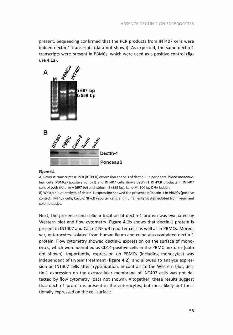
ABSENCE DECTIN-1 ON ENTEROCYTES
55
present. Sequencing confirmed that the PCR products from INT407 cells were
indeed dectin-1 transcripts (data not shown). As expected, the same dectin-1
transcripts were present in PBMCs, which were used as a positive control (fig-
ure 4.1a).
Figure 4.1
A) Reverse transcriptase PCR (RT-PCR) expression analysis of dectin-1 in peripheral blood mononuc-
lear cells (PBMCs) (positive control) and INT407 cells shows dectin-1 RT-PCR products in INT407
cells of both isoform A (697 bp) and isoform B (559 bp). Lane M, 100 bp DNA ladder.
B) Western blot analysis of dectin-1 expression showed the presence of dectin-1 in PBMCs (positive
control), INT407 cells, Caco-2 NF-κB reporter cells, and human enterocytes isolated from ileum and
colon biopsies.
Next, the presence and cellular location of dectin-1 protein was evaluated by
Western blot and flow cytometry. Figure 4.1b shows that dectin-1 protein is
present in INT407 and Caco-2 NF-κB reporter cells as well as in PBMCs. Moreo-
ver, enterocytes isolated from human ileum and colon also contained dectin-1
protein. Flow cytometry showed dectin-1 expression on the surface of mono-
cytes, which were identified as CD14-positive cells in the PBMC mixtures (data
not shown). Importantly, expression on PBMCs (including monocytes) was
independent of trypsin treatment (figure 4.2), and allowed to analyze expres-
sion on INT407 cells after trypsinization. In contrast to the Western blot, dec-
tin-1 expression on the extracellular membrane of INT407 cells was not de-
tected by flow cytometry (data not shown). Altogether, these results suggest
that dectin-1 protein is present in the enterocytes, but most likely not func-
tionally expressed on the cell surface.
Thesis_Volman_v06.pdf

CHAPTER 4
56
Figure 4.2
A) Dectin-1 expression on PBMCs was visualized by flow cytometry with a polyclonal goat anti-
human dectin-1.B) Dectin-1 expression on PBMCs was independent of 5 min trypsin treatment.
Dectin-1 inhibitors do not counteract the immune stimulating effect of
ββββ-glucan fecal water
The dependence of dectin-1 for INT407 cells to respond to β-glucan enriched
fecal water was determined using dectin-1 inhibitors. For this, the cells were
pre-incubated with the polyclonal goat anti-human dectin-1 antibody during
1h, after which fecal water was added for a period of 16h. Figure 4.3a shows
that the antibody was unable to inhibit the enhanced IL-8 production by
INT407 cells after β-glucan fecal water as compared to placebo fecal water. The
observed increase in IL-8 production in the pooled β-glucan fecal water sample
(19.8 % and 23.4 %) was comparable with the median of all six individual sam-
ples (22.0 %) as previously shown [69]. Similar results were found when the
dectin-1 inhibitor glucan phosphate was added to INT407 cells in combination
with fecal water (figure 4.3b), that is, addition of glucan phosphate even en-
Thesis_Volman_v06.pdf

ABSENCE DECTIN-1 ON ENTEROCYTES
57
hanced IL-8 secretion by INT407 cells in β-glucan as well as in placebo fecal
water by 35% and 34% respectively.
Altogether, these data suggest that the enhanced IL-8 production by INT407
cells induced by oat β-glucan-enriched fecal water was independent of dectin-1
at the extracellular membrane. We confirmed these results in another entero-
cytes cell line as shown in figure 4.4. The dectin-1 inhibitors glucan phosphate
and dectin-1 antibody were unable to inhibit the oat β-glucan-enriched fecal
water-induced NF-κB transactivation in Caco-2 NF-κB reporter cells.
Figure 4.3
Dectin-1 inhibitors did not affect the increased IL-8 production after β-glucan by INT407 cells. The
culture supernatant of INT407 cells was analyzed for IL-8 production after 16h incubation with oat
β-glucan-enriched or placebo fecal water in presence of an inflammation inducing cocktail consist-
ing of interferonγ (100 U/mL), interleukin-1β (50 U/mL) and tumour necrosis factorα (10 ng/mL).
Cells were pre-incubated for 1h with 0.2 µg/ml polyclonal anti-human dectin-1 (A) or 100 µg/mL
glucan phosphate (GluP) (B). Values are means and SD of duplicate cultures.
Figure 4.4
Dectin-1 inhibitors did not affect the increased NF-κB transactivation after β-glucan by Caco-2 NF-κB
reporter cells. The cell lysates were analyzed for luciferase activity as measurement for NF-κB
transactivation after 3h incubation with oat β-glucan-enriched or placebo fecal water in presence of
an inflammation inducing cocktail consisting of interferonγ (100 U/mL) and interleukin-1β (50
U/mL). Cells were pre-incubated for 1h with 0.2 µg/ml polyclonal anti-human dectin-1 (A) or 200
µg/mL glucan phosphate (GluP) (B). Values are means and SD of duplicate cultures. RLU, relative
light unit.
Thesis_Volman_v06.pdf

CHAPTER 4
58
The dectin-1 ligand zymosan has no immune stimulating effect in
INT407 cells
In line with the absent effects of the dectin-1 inhibitors on IL-8 production in
INT407 cells after β-glucan enriched fecal water, zymosan (an accepted dectin-
1 ligand) also did not increase IL-8 production in INT407 cells (data not shown).
Discussion
In addition to β-glucans from yeast and fungi, also β-glucans from cereals have
been found to induce immune stimulating effects [7]. The influence of β-
glucans from yeast, fungi and cereals, all present simultaneously within the
intestinal lumen as part of our daily diet, on the intestinal immune response is
of interest.
In this respect, we have earlier reported an increased immune response of
INT407 and Caco-2 cells after addition of fecal water prepared from ileostomic
contents obtained from subjects who had consumed an oat β-glucan-enriched
diet as compared to placebo [69]. It was tempting to speculate that the effects
of oat β-glucan fecal water on these enterocytes cell lines were mediated via
the β-glucan receptor dectin-1. To investigate the possible role of dectin-1 we
first checked dectin-1 presence on enterocytes. Caco-2 cells are already re-
ported to contain dectin-1 transcripts [42] and our results showed dectin-1 at
mRNA level in INT407 cells. Moreover, we demonstrated that dectin-1 protein
is present in the both cell lines, although not expressed on the cell surface.
Besides cell lines, we also showed that isolated human enterocytes from ileum
and colon contained dectin-1 protein. The second aim of our study was to
verify the functional involvement of the β-glucan receptor dectin-1 in the in-
creased immune response of INT407 and Caco-2 cells to β-glucan enriched
fecal water. However, here we present data showing that these effects are
most likely independent of extracellular dectin-1.
Dectin-1 is described as the main β-glucan receptor on leukocytes and general-
ly considered to be responsible for the cellular responses of β-glucan [22].
Northern blots have shown the presence of dectin-1 in the murine intestine
[71]. Other studies, however, reported that besides immune cells, other cell
types also contain β-glucan receptors, including lung epithelial cells (A-549)
[104] and vascular endothelial cells [105]. In line, our results show dectin-1
both at mRNA and protein level in small intestinal INT407 cells. This is in cor-
respondence to the findings of Saegusa et al. [42], who showed dectin-1 tran-
Thesis_Volman_v06.pdf

ABSENCE DECTIN-1 ON ENTEROCYTES
59
scripts in Caco-2 cells, another human derived enterocyte cell line. Moreover,
also human isolated enterocytes from ileum and colon contained dectin-1
protein. However, when using flow cytometry, we were unable to demonstrate
the presence of dectin-1 on the cell surface of INT407 cells. Dectin-1 expression
at the outer membrane, which is considered obligatory for a functional role in
macrophages, seems unlikely [106]. A function for intracellular dectin-1 has up
till now not been described, but cannot be excluded. Similarly, other PRRs like
Toll-like receptors 3, 7, 9 and also nucleotide-binding oligomerization domains
(NODs) exert their function intracellular [107, 108]. Moreover, experiments in
which dectin-1 was blocked supported our conclusion that INT407 and Caco-2
NF-κB reporter cells do not express functional dectin-1 on the cell membrane.
Two different dectin-1 inhibitors did not counteract the immune stimulating
effects of β-glucan-enriched fecal water. Glucan phosphate is a specific dectin-
1 antagonist and often used to block the β-glucan-induced immune effects
[22]. Furthermore, the murine antibody has been described to block the action
of dectin-1 [71]. In addition, also the generally accepted dectin-1 ligand zymo-
san could not stimulate the immune response in INT407 cells as measured by
IL-8 production. This can also be interpreted as an absence of the zymosan
receptor dectin-1.
As the immune stimulating effects of oat β-glucan-enriched fecal water seems
to be dectin-1 independent, an alternative explanation might be that the con-
sumption of a β-glucan-enriched diet has influenced in vivo immune responses
within the GI tract of the ileostomy patients. This in vivo response may have
changed the composition of the in vitro used fecal water, which resulted in an
increased in vitro immune response of enterocyte cell lines exposed to the
fecal water in vitro. It is of course entirely speculative to talk about potential
candidates that could be present in higher or lower amounts in the fecal water
and as such responsible for the observed effects, but changes in antimicrobial
peptides or short chain fatty acid concentrations are a possibility. Next to the
possibility that effects of oat β-glucan-enriched fecal water are indirect, there
might be an alternative β-glucan receptor involved. Possible candidates may be
complement receptor (CR)3 and lactosylceramide, which have been described
as β-glucan receptors present on leukocytes [8]. Evens et al. [74] have shown
that the chemokine responses of alveolar epithelial cells in response to Pneu-
mocytis carinii cell wall β-glucan were indeed dectin-1 independent and were
more likely mediated by lactosylceramide. We did, however, not further ex-
plore the identification of other receptors involved in the effects of oat β-
glucan enriched fecal water.
The question why dectin-1 is not functionally expressed on enterocytes is intri-
guing. The intestinal wall is continuously exposed to antigens from the micro-
Thesis_Volman_v06.pdf

CHAPTER 4
60
biota and of nutritional origin. Therefore, silencing of the intestinal immune
response to these structures is important, as continuous stimulation of the
epithelial lining results in an inflammatory bowel. The absence of functional
dectin-1 on INT407 and Caco-2 NF-κB reporter cells, as models of enterocytes,
may be an example of this tolerance against these antigens as dietary β-glucans
are present in the intestinal lumen.
In summary, we conclude that INT407 and Caco-2 cells - as models for entero-
cytes - do not contain functional dectin-1 at the cell membrane. This may have
a protective function as the luminal surface of the gastrointestinal tract is ab-
undantly supplied with β-glucans both from nutritional and potentially harmful
sources. These dectin-1 ligands would otherwise induce continuous PRR activa-
tion with intestinal damage as a consequence. Finally, the finding that extracel-
lular dectin-1 does not play a functional role in INT407 and Caco-2 cells, indi-
cates that the previously found immune stimulating effects of β-glucan-
enriched fecal water in enterocyte cell lines should be explained by another
mechanism.
Acknowledgements
The authors would like to thank Maurice Konings and Herman Popeijus (De-
partment of Human Biology, Maastricht University, the Netherlands), Gert
Schaart and Esther Kornips (Department of Movement Sciences, Maastricht
University, the Netherlands) for assistance with the dectin-1 sequencing and
Western blots.
Thesis_Volman_v06.pdf

61
CHAPTER 5
Effects of mushroom derived β-glucan-rich polysaccharide extracts on NO
production by bone marrow-derived
macrophages and NF-κB transactivation in Caco-2 reporter cells:
can effects be explained by structure?
Julia J. Volman*, Johannes P.F.G. Helsper*,Song Wei, Johan J.P. Baars, Leo J.L.D.
van Griensven, Anton S.M. Sonnenberg, Ronald P. Mensink, Jogchum Plat
*Contributed equally to the results
Molecular Nutrition and Food Research; in press
Thesis_Volman_v06.pdf

CHAPTER 5
62
Abstract
Mushrooms are known for their immune modulating and anti-tumour proper-
ties. The polysaccharide fraction, mainly β-glucans, is responsible for the im-
mune-modulating effects. Fungal β-glucans have been shown to activate leuko-
cytes, which depend on structural characteristics of β-glucans. As edible mu-
shrooms come in contact with the intestinal immune system, effects on ente-
rocytes are also interesting. Our aim was to evaluate the effect of mushroom
polysaccharide extracts varying in β-glucan structure, on nitric oxide produc-
tion by bone marrow-derived macrophages (BMMs) from mice and on nuclear
factor-κB transactivation in human intestinal Caco-2 cells. We demonstrated
that extracts from Agaricus bisporus stimulated nitric oxide production by
BMM, whereas extracts from Coprinus comatus and spores of Ganoderma
lucidum had only minor effects. Furthermore, extracts of A. blazei Murill and
Phellinus linteus had no effect at all. Almost all mushroom extracts lowered
nuclear factor-κB transactivation in Caco-2 cells. Structural analysis of A. bispo-
rus compared with A. blazei Murill suggests that branching of the β-glucan
chain is essential for immune-stimulating activity. In conclusion, extracts from
A. bisporus activate BMM, without activating enterocytes. These characteristics
make A. bisporus an attractive candidate as a nutritional compound to stimu-
late the immune response in depressed states of immunity.
Thesis_Volman_v06.pdf

EFFECTS OF MUSHROOM EXTRACTS ON NO AND NF-KB
63
Introduction
Mushrooms, which belong to the basidiomycetes and ascomycetes classes of
the kingdom fungi, contain many different biologically active compounds, e.g.
polysaccharides, small proteins, lectins, and polyphenols. The polysaccharides
are mainly β-glucans that are present in the cell walls [109]. It is believed that
these compounds have immune-modulating effects [110-112]. Indeed, it has
been reported that fungal β-glucans increase the phagocytic activity and the
production of reactive oxygen intermediates [113], inflammatory mediators,
and cytokines by leukocytes [22]. For Pleurotus ostreatus (Oyster mushroom), it
was reported that a water extract from fruit bodies and mycelia played a role in
increasing the production of reactive oxygen species (ROS) from neutrophils
and has also additional immune-modulating properties involving all immuno-
competent cells [114].
As part of the human diet, compounds derived from digested edible mu-
shrooms come in contact with enterocytes, which are located at the frontline
of the intestinal immune system. Enterocytes have to discriminate between
harmful and harmless antigens. To perform this task they play an important
role in orchestrating the intestinal immune response and can induce an in-
flammatory response if necessary. Therefore, it is likely that mushrooms might
also affect the immune response of enterocytes. It has been demonstrated that
intestinal macrophages [67] and enterocytes (presumably microfold (M)-cells)
[42, 58] can take up and distribute β-glucans after oral intake. However, one
study suggested that β-glucans from an fungal extract did not enter the blood
as the effect in vivo was strongly diminished as compared with in vitro [115]. To
examine whether the mushroom polysaccharide extracts have an effect on
enterocytes we used a nuclear factor-κB (NF-κB) reporter Caco-2 cell line. NF-
κB is a transcription factor that plays a key role in the immune response. In
inactive form it is present in the cytoplasm bound by its inhibitor IκB. Upon
stimulation IκB is degraded by IκB kinase and NF-κB is free to move to the
nucleus where it can bind to response elements of NF-κB target genes. One of
the consequences of NF-κB activation is activation of the numerous NF-κB
target genes, which results in the production of cytokines like tumour necrosis
factor α (TNFα) and ROS. Responsiveness of macrophages towards β-glucan
treatment has already been reported by many different groups [26, 62, 116,
117]. More into detail, β-glucans have been classified as biological response
modifiers, which can influence the activity of macrophages [26, 62, 117]. For
that reason bone marrow-derived macrophages (BMM) have been used by
others to examine the systemic immune modulating properties of β-glucans.
Thesis_Volman_v06.pdf

CHAPTER 5
64
For this nitric oxide (NO), which is produced by the NF-κB regulated enzyme
NO synthase, was chosen as one of the outcome parameters in the BMM expe-
riments as fungal β-glucans have been reported to modulate NO production
[118-120].
β-Glucans consist of a backbone formed by linear β(1→3)-, β(1→4)- or
β(1→6)-linked glucose molecules, which in addition can contain branches. The
type of linkages of the backbone and branches differs for the different β-
glucans. For example, it has been suggested that β-glucans from yeast and
fungi consist of β(1→3)-linked glucopyranosyl units with β(1→6)-linked side
chains, whereas β-glucans from oat and barley are composed of unbranched
β(1→3) and β(1→4) linkages [7, 8, 22]. The activity of β-glucans may depend
on their structural properties, like polymer length, degree of branching, tertiary
structure, and molecular weight [100]. Besides the structure, the method of
isolation can also affect the activity of β-glucans [109].
The aim of this study was to investigate the effect of several β-glucan-rich
mushroom polysaccharide extracts, which were a priori based on the literature
expected to vary in β-glucan structure, on NO production by BMMs from mice
as well as on NF-κB transactivation in a human small intestinal enterocytes-like
cell line, the Caco-2 cell. As besides the structure, the method of isolation can
also affect the immune-modulating activity of β-glucans, we also evaluated the
effect of the mushroom extract with the highest in vitro activity in the BMM
cells, e.g. the extract from Agaricus bisporus, when isolated with a different
methodology. Additionally, we tried to find a structure-response relationship
by evaluating some structural characteristics of two extracts with opposite
effects, e.g. the extracts from A. bisporus and A. blazei Murill. We hypothesized
that the branched β-glucans are the most potent immune stimulating extracts.
Materials and methods
Preparation of mushroom polysaccharide extracts
Different mushroom polysaccharide extracts were isolated and used for BMM
and Caco-2 NF-κB reporter cell stimulations. Extracts were isolated from spores
(A) and fruit body tissue (B) of Ganoderma lucidum; from different parts of the
A. bisporus, e.g. the caps and stems; and from A. blazei Murill, Coprinus com-
atus, and Phellinus linteus. In addition, α-glucans were extracted from the
stems of A. bisporus to distinguish between α-glucan and β-glucan effects.
Thesis_Volman_v06.pdf

EFFECTS OF MUSHROOM EXTRACTS ON NO AND NF-KB
65
Semi-purified polysaccharides were obtained from fruiting body material by
hot water extraction followed by ethanol precipitation as described before
[113]. A final solution was made in phosphate buffered saline (PBS) at 900
µg/mL, which was then heated for 1h at 100 °C and stored at 4 °C. The extracts
will be further referred to as fruit body polysaccharide extracts. α(1→4)-Glucan
was obtained from A. bisporus stipes by passage of the polysaccharide extract
over a diethylaminoethyl-cellulose column. The colourless non-binding poly-
saccharide has a molecular weight (MW) > 200 kDa, which can be completely
degraded by α-amylase, and is characterized as an α-glucan [113].
A. bisporus mushroom polysaccharide extract was also isolated from culture
mycelium by centrifugation and filtration. To this extract four volumes of etha-
nol were added and precipitation was allowed to proceed overnight at 4 °C.
The resulting precipitate was redissolved in water. To obtain the extract, the
above-described procedure was repeated twice. The resulting fluid was steri-
lized by a 0.45 µm filter and adjusted to a concentration of 900 µg/mL and
further called culture mycelium extracts.
Chemical analysis of glucans from Agaricus species
To try and explain the physiological effects by differences in structure, we
determined the monosaccharide composition and analyzed the glycosidic lin-
kages of A. bisporus and A. blazei Murill mushroom polysaccharide extracts.
Monosaccharide composition
To evaluate the monosaccharide composition, a 5 µL aliquot of the glucan
solutions or suspension was diluted in 0.3 mL 2M trifluoroacetic acid (TFA) and
hydrolyzed overnight at 100 °C. Solutions were evaporated to dryness at 40°C
under a N2 stream. Residual TFA was removed by two evaporation cycles in 0.5
mL methanol, as earlier. The final residue was dissolved in 0.5 mL water and
analyzed for monosaccharides on a Dionex HPLC, equipped with pulsed ampe-
rometric electrochemical gold detector. A PA1-column (250 x 4 mm), fitted
with a PA-1 guard column (10 x 32 mm), was used as the stationary phase and
0.020 M NaOH as the mobile phase at 1.0 mL/min (isocratic elution). Quantifi-
cation of monosaccharides was performed on the basis of external standards.
Thesis_Volman_v06.pdf

CHAPTER 5
66
Linkage analyses via gas chromatography-mass spectrometry of
partially methylated alditol acetates
To determine the distribution of glycosidic linkages in the polysaccharides gas
chromatography-mass spectrometry (GCMS) analysis of partially methylated
alditol acetates (PMAAs) derived from these compounds was performed. Per-
methylation was performed according to one of the protocols described in
Kang et al. [121] where 0.7% water was added during the incubation. Perme-
thylated products were separated by phase partitioning after adding 0.5 mL
cold (4 °C) chloroform and 2.5 mL cold water. The chloroform phase was
washed 3 times with 2.5 mL water to remove all NaOH, evaporated to dryness
and hydrolysed overnight in 0.3 mL 0.5M TFA at 100 °C. Further processing to
PMAAs was performed as described [122]. PMAAs were quantified by GCMS
using a Hewlett Packard HP 5890 gas chromatograph connected to a HP 5972A
mass spectrometer, which was equipped with a ZB-5 column (30 m, i.d. 0.25
mm, layer thickness 0.25 µm), run at the following gradient: 3 min 170°C, 2
°C/min to 210 °C, then 10 °C/min to 230 °C and a final time of 4 min.
BMM stimulation
BMMs were isolated from the tibiae and femurs of C57BL/6 mice. Cells were
cultured in 15 cm bacterial plastic petri dishes in standard RPMI with L-
glutamine and HEPES (Invitrogen, Breda, the Netherlands), 10% heat-
inactivated fetal calf serum (Bodinco BV, Alkmaar, the Netherlands), and 100
U/ml penicillin streptomycin (Invitrogen) with the addition of 15% L929 cell-
conditioned medium kindly provided by Danielle Curfs (Department of Molecu-
lar Genetics, Maastricht University, the Netherlands). After 1 wk differentia-
tion, cells were detached with lidocaine (Sigma Chemicals, St Louis, MO) and
plated at a concentration of 1x10⁶
cells/mL in a final volume of 0.5 mL. Cells
were primed with 100 U/mL IFNγ during 24h to obtain a maximal effect on NO
production, as earlier described [123]. Then the medium was refreshed and
cells were incubated with the mushroom polysaccharide extracts in a concen-
tration range of 50-200 µg/mL for 24 h. These concentrations are physiological
as we reported earlier that the β-glucan concentration in the 24 h pooled fecal
water obtained from ileostomic patients who consumed an oat β-glucan-
enriched diet varied between 120 and 180 µg/mL [69]. Besides mushroom
polysaccharide extracts we also evaluated the effects of cellulose, a linear
molecule which consists of β(1→4) linkages (Sigma-Aldrich, Steinheim, Germa-
ny) on BMM-derived NO production to further substantiate our assumptions
Thesis_Volman_v06.pdf

EFFECTS OF MUSHROOM EXTRACTS ON NO AND NF-KB
67
regarding a possible structure-effect relationship. Supernatants from the BMM
were removed after 24 h incubation and NO concentrations were determined.
To evaluate whether the effects of the mushroom polysaccharide on NO pro-
duction were mediated by the β-glucan receptor dectin-1, we used the dectin-1
inhibitor laminarin. For this, cells were incubated with 250 or 500 µg/mL lami-
narin (Sigma Chemicals) 1h before stimulation with 200 µg/mL A. bisporus
stems, A. bisporus stems α-glucans, A. blazei Murill, and cellulose.
Measurement of NO and TNFαααα concentrations
NO concentrations in the supernatants of BMM were determined as total ni-
trite by the Greiss method [124]. In short, 50 µl supernatant was combined
with 50 µl Greiss reagents (2.5% v/v phosphoric acid, 0.1% w/v naphthylethy-
lene diamine dihydrochloride and 1.0% w/v sulfanilamide in distilled water).
After incubation for 10 min at room temperature, absorbance was determined
at 540 nm. Sodium nitrate (Sigma Chemicals) was used to prepare a standard
curve (0-200 mM). Besides NO production we also determined TNFα produc-
tion by BMM after stimulation with A. bisporus stems, A. bisporus stems α-
glucan, and A. blazei Murill preincubated with 500 µg/mL laminarin. TNFα
concentrations were measured as described [125].
Intestinal cell culture
We also determined the effect of all mushroom polysaccharide extracts on
enterocytes. For this, we used the human cell line Caco-2, which was obtained
from the American Tissue Type Collection (Rockville, MD). Caco-2 cells were
cultured in DMEM (Invitrogen) supplemented with 10% heat-inactivated fetal
bovine serum (Invitrogen), 1% penicillin streptomycin (Invitrogen), 1% sodium
pyruvate and 1% non essential amino acids (Invitrogen). Cells were cultured at
37 °C in a 5% CO2 humidified atmosphere, refreshed every second day and
separated by trypsin-0.03% EDTA (Gibco BRL, Gaithersburg, MD) when they
had reached 70-90% confluence.
Stable transfection of NF-κκκκB in Caco-2 cells
To evaluate the effects of the various mushroom-derived polysaccharide ex-
tracts on transcriptional activity of NF-κB, a stable NF-κB reporter Caco-2 cell
Thesis_Volman_v06.pdf

CHAPTER 5
68
line was created. This cell line was chosen as we have previously shown im-
mune-stimulating effects of oat β-glucan fecal water in Caco-2 cells [69]. The
6κB-TK-luciferase (NF-κB reporter) plasmid and neomycin resistance plasmid
were both kindly provided by Dr. R.C. Langen (Department of Pulmonology,
Maastricht University, The Netherlands). Cells were transfected using Lipofec-
tamine 2000 (Invitrogen) according the manufacturers’ instructions. Positive
clones were selected by culturing with geneticin (1 mg/mL). For the experi-
ments, Caco-2 NF-κB reporter cells were plated in 24-well plates at an initial
density of 0.3x10⁶ cells/mL in a total volume of 0.5 mL and used for stimulation
experiments when they had reached 70-90% confluence. Culture medium was
replaced by fresh medium containing different mushroom polysaccharides at
physiological concentrations in combination with an inflammation-inducing
cytokine cocktail consisting of IFNγ (100 U/mL) and IL-1β (50 U/mL). After 3 h of
incubation, cells were lysed in luciferase lysis buffer (Promega, Madison, WI)
and stored at -80 °C. Luciferase activity was measured according to the manu-
facturers’ instructions (Promega) and expressed relative to total protein (Bio-
rad assay; Bio-rad, Hercules, CA).
Statistical analysis
All parameters are presented as means ± SD and were analyzed for statistical
differences by analysis of variance (ANOVA). When a significant difference in
concentration was found, the four concentrations were compared pairwise
using a Tukey post hoc test for multiple comparison of observed means. Differ-
ences were considered significant at p<0.05 (corrected for four groups by
SPSS). All statistical analyses were performed using SPSS 15.0 (SPSS, Chicago,
IL).
Results
Effects of mushroom polysaccharide extracts on NO and TNFαααα produc-
tion by BMM
As demonstrated in figure 5.1, extracts isolated from A. bisporus fruit bodies
significantly increased NO production by BMM. These effects were almost
identical for extracts isolated from caps and stems. In addition, NO production
was also increased (p<0.05) when BMM were incubated with mycelium extract
Thesis_Volman_v06.pdf

EFFECTS OF MUSHROOM EXTRACTS ON NO AND NF-KB
69
isolated from A. bisporus indicating that these effects were independent of the
method of culturing and isolation. Interestingly, the effects on NO production
were not observed for all fruit body extracts. Spore extracts from G. lucidum
and C. comatus also stimulated NO production but only significant at the high-
est concentration and not as prominent as extracts from A. bisporus. The poly-
saccharide extracts from A. blazei Murill, P. linteus and G. lucidum fruiting
bodies had no NO-inducing activity.
Figure 5.1
Increased NO production by IFNγ (100 U/mL) primed BMM after 24 h stimulation with A. bisporus
mushroom polysaccharide extracts. Extracts isolated from C. comatus and G. lucidum mushroom
polysaccharide extracts had minor effects. Values are means and SD of duplicate cultures. Experi-
ments have been conducted three times and a representative example is shown. Bars with differ-
ent superscripts differ (p<0.05). a Reference for the other mushroom polysaccharide extracts.
As the cell wall of mushroom consists for a large part of the polysaccharide β-
glucan, it is tempting to suggest that these polysaccharides increased NO pro-
duction. Therefore, to test whether effects of A. bisporus were β-glucan-
specific, we also evaluated effects of A. bisporus derived α-glucans from the
stems on BMM-derived NO production. In line with β-glucans isolated from A.
bisporus, A. bisporus stem α-glucans dose-dependently increased NO produc-
tion to a maximum of 12 mM (figure 5.2).
Thesis_Volman_v06.pdf

CHAPTER 5
70
Figure 5.2
Mushroom polysaccharide extracts isolated from A. bisporus alpha dose- dependently increased
NO production by IFNγ (100 U/mL) primed BMM after 24 h stimulation, whereas cellulose had no
effect. Values are means and SD of duplicate cultures. Bars with different superscripts differ
(p<0.005).
However, the maximum NO concentration that was reached was only 50% of
the concentration observed after incubation with the A. bisporus polysaccha-
ride extract containing merely β-glucans. Cellulose, consisting of only β(1→4)
linkages had no effect on NO production by BMM (figure 5.2). For TNFα pro-
duction we observed a comparable doses-response relationship as for NO
(figure 5.3).
Figure 5.3
Mushroom polysaccharide extracts isolated from A. bisporus alpha dose- dependently increased
TNFα production by IFNγ (100 U/mL) primed BMM after 24 h stimulation, whereas cellulose had no
effect. Values are means and SD of duplicate cultures. Bars with different superscripts differ
(p<0.001).
Thesis_Volman_v06.pdf

EFFECTS OF MUSHROOM EXTRACTS ON NO AND NF-KB
71
To evaluate the potential involvement of the β-glucan receptor dectin-1 in the
observed NO production by BMM, the dectin-1 inhibitor laminarin was used. As
shown in figures 5.4 and 5.5, laminarin had no effect on the A. bisporus stems
β-glucan induced NO and TNFα production.
Figure 5.4
Dectin-1 inhibitor laminarin was unable to inhibit the increased NO production by BMM caused by
200 ug/mL A. bisporus mushroom polysaccharide extracts. Values are means and SD of duplicate
cultures. Experiments have been conducted to two times and a representative example is shown.
Comparable with the results described earlier, also in the presence of lamina-
rin, A. bisporus stems α-glucan stimulated the NO and TNFα production to 50%
beyond the level of A. bisporus stems β-glucans, whereas extracts isolated from
A. blazei Murill and cellulose again showed no effect (figures 5.4 and 5.5).
Dectin-1 inhibitor laminarin was unable to inhibit the increased NO production
by BMM caused by 200 ug/mL A. bisporus mushroom polysaccharide extracts.
Values are means and SD of duplicate cultures. Experiments have been con-
ducted to two times and a representative example is shown.
Thesis_Volman_v06.pdf

CHAPTER 5
72
Figure 5.5
Dectin-1 inhibitor laminarin was unable to inhibit the increased TNFα production by BMM caused
by 200 ug/mL A. bisporus mushroom polysaccharide extracts. Values are means and SD of duplicate
cultures. Experiments have been conducted two times and a representative example is shown.
Effects of A. bisporus stems mushroom polysaccharide extracts on
NF-κB transactivation in enterocytes
To evaluate whether the polysaccharide extracts isolated from the edible mu-
shroom A. bisporus and the other mushrooms also modulated the immune
response of enterocytes, we measured NF-κB transactivation in our Caco-2 NF-
κB reporter cell line. Figure 5.6a demonstrates that the effects of extracts
isolated from the fruit bodies were highly variable; however, almost all extracts
lowered the transactivation of NF-κB in Caco-2 cells as compared to buffered
saline. This decreased NF-κB transactivation was most obvious after incubation
with the highest concentration of the extracts, whereas at 100 µg/mL some
extracts even showed an increased NF-κB transactivation. The magnitude of
the effect differed between the mushroom extracts. The extract isolated from
A. blazei Murill and C. comatus fruit bodies showed the most pronounced de-
crease in NF-κB transactivation (35.3 and 41.4% (p=0.074) at the highest con-
centration, respectively), whereas P. linteus had no effect at all. Furthermore,
extracts from both spores and fruiting bodies of G. lucidum reduced NF-κB
transactivation at 50 µg/mL and 200 µg/mL. Finally, extracts isolated from two
different parts of A. bisporus (stems or caps) showed similar effects at the
highest concentration (reductions of 32 and 33%, respectively). Surprisingly, in
contrast to extracts isolated from the A. bisporus fruiting bodies, extracts iso-
Thesis_Volman_v06.pdf

EFFECTS OF MUSHROOM EXTRACTS ON NO AND NF-KB
73
lated from A. bisporus culture medium did not lower NF-κB transactivation
(figure 5.6b).
Figure 5.6
a) Most of the different mushroom polysaccharide extracts decreased NF-κB transactivation by
Caco-2 NF-κB reporter cells after 3 h stimulation in presence of IL-1β (100 U/mL) and IFNγ (50
U/mL). Values are means and SD of duplicate cultures. Experiments have been conducted three
times and a representative example is shown. a p=0.074, 200 µg/mL versus control.
b) NF-κB transactivation by Caco-2 NF-κB reporter cells after 3 h stimulation with A. bisporus iso-
lated from the culture medium in presence of IL-1β (100 U/mL) and IFNγ (50 U/mL) is increased.
Values are means and SD of duplicate cultures. Experiments have been conducted three times and a
representative example is shown.
Chemical analysis of glucans from A. bisporus versus A. Blazei Murill
As the activity of β-glucans depends on the chemical structure, we determined
the monosaccharide composition and linkage type to explain the differences in
immune-modulating properties of A. bisporus and A. blazei Murill. The mono-
saccharide compositions of A. bisporus and A. blazei Murill are shown in table
5.1. The polysaccharides of A. bisporus consisted of mainly glucose (96.3%)
with some minor proportions of galactose (2.9%) and xylose (0.8%). This poly-
saccharide can thus be considered as consisting of mainly glucans. A. blazei
Murill polysaccharide was more complex as it contained in addition to glucose
(57.7%) considerable proportions of galactose (27.7%); mannose and xylose
(not separated, together 7.3%); fucose (4%); and some rhamnose, arabinose,
fructose and GlcNH2 (each about 1%). GlcNH2 was probably present in the
native oligosaccharide as GlcNAc, which was degraded by TFA-hydrolysis.
Thesis_Volman_v06.pdf

CHAPTER 5
74
Table 5.1: Monosaccharide composition of A. bisporus and A. blazei Murril as determined by
HPLC
Monosaccharide A. bisporus A. blazei Murill
Glucose 93.3 57.7
Galactose 2.9 27.7
Mannose/xylose 0.8 7.3
Fucose n.d. 4.0
Rhamnose n.d. 1.2
Arabinose n.d. 1.2
GLcNH2 n.d. 0.9
Fructose n.d. 0.7
n.d., not detected
In addition to the monosaccharide composition, we also measured the glyco-
sidic type of linkage and branching. A. bisporus glucan contained predominantly
1→4 and a few 1→3 linkages, while no 1→6 linkages were measured (table
5.2). In agreement with the presence of 1→3 and 1→4 linkages 1→3,4-
branching glucose residues were also observed at about 1.5%. The relative high
number of terminal glucose residues and low number of branch points indicate
a short-chain molecule. The high proportion of 1→4 glucan suggests that the
main chain contains predominantly this type of linkage.
Table 5.2: Linkages of A. bisporus and A. blazei Murril as determined via GCMS of PMAAs
Linkage type A. bisporus A. blazei Murill
Terminal glucose 9.5 10.8
β(1 → 3) 3.5 5.1
β(1 → 4) 85.5 43.3
β(1 → 6) n.d. 40.8
β(1 → 3, 4) branch 1.6 n.d.
β(1 → 3, 6) branch n.d. n.d.
β(1 → 4, 6) branch n.d. n.d.
n.d., not detected
For A. blazei Murill the linkage was different as more than 80% of the glucose
residues in the glucan fraction were linked by either 1→4 or 1→6-linkages at a
ratio of about 1:1, while a small proportion of 1→3 glucosidic linkages were
also observed and with no branch points. The observations from this PMAA
analysis may be interpreted by either a single main chain with alternating 1→4
and 1→6 linkages or by a mixture of more glucan chains with a single linkage
type.
Thesis_Volman_v06.pdf

EFFECTS OF MUSHROOM EXTRACTS ON NO AND NF-KB
75
Discussion
Fungal β-glucans are known for their immune-modulating and tumour growth-
inhibitory effects [110-112]. It has been suggested that the polysaccharides
present in mushrooms are the most likely candidates for these effects. As edi-
ble mushrooms are present in our daily diet, effects of these mushrooms on
(intestinal) immune responses are of potential interest for optimizing the func-
tioning of our immune system. We here show that extracts isolated from A.
bisporus – either from caps and stems from A. bisporus fruit bodies or from A.
bisporus mycelium – stimulated the production of NO (a NF-κB target gene) of
BMM. Furthermore, purified α-glucan from the stems of A. bisporus dose-
dependently stimulated the NO production also, although the maximum effect
observed was only 50% of that achieved with A. bisporus β-glucan. When using
cellulose, consisting of only β(1→4) linkages, no NO was produced. All results
were identical if TNFα production by BMM was analyzed instead of NO. Im-
mune-modulating effects of β-glucans are generally accepted, whereas the
effects for α-glucan are debated. However, the fact that A. bisporus α-glucans
stimulated BMM to a lesser extent as compared to A. bisporus β-glucans at
least illustrates that effects are glucan-type dependent. Interestingly, effects
were not identical for all mushroom extracts. C. comatus and G. lucidum A
extracts had only minor effects on NO concentrations as compared with the
effects of A. bisporus, whereas A. blazei Murill and P. linteus had no effect at
all. Earlier, immune-stimulating effects of mycelium and fruit body extracts
from A. blazei Murill, although not for all fractions, have been reported [116,
126]. It has been shown that the method of isolation and chemical modification
can influence the activity of β-glucans, which may explain the difference in
results between the studies [25, 109, 111]. The largest difference in effects on
BMM behaviour was found for A. bisporus and A. blazei Murill, in which A.
bisporus was the most potent immune-stimulating extract, whereas A. blazei
Murill extract showed almost no effect. To explain these differences in activat-
ing potential we determined some structural characteristics of both extracts
and tried to define a sort of structure-effect relationship. Chemical analysis
demonstrated that the polysaccharide from A. bisporus consists of mainly
glucose, whereas that from A. blazei Murill is a mixture of different monosac-
charides. This may suggest that a β-glucan almost exclusively composed of
glucose exerts a greater immune-stimulating activity as compared with glucans
containing other monosaccharides also. However, cellulose consisting solely of
glucose molecules has no NO-inducing activity on BMM. This implies that other
factors besides monosaccharide composition play a role. A second pronounced
structural difference between both extracts we observed was the type of lin-
Thesis_Volman_v06.pdf

CHAPTER 5
76
kages. A. bisporus was mostly composed of 1→4 linkages and some 1→3 lin-
kages, while A. blazei Murill contains 1→4, 1→6 and some 1→3 linkages. It has
been reported that 1→3 linkages are essential for immune-modulating activity
and both extracts contain approximately the same amount of 1→3 linkages so
this could not explain the difference in immune-modulating properties. Moreo-
ver, A. bisporus is in our hands free of 1→6-linkages, although earlier reports
describe the presence of such linkages in glucans from this fungus [127]. The
absence of 1→6-linkages in our extract may be due to the origin and develop-
mental stage of the fungal tissue used for the isolation of glucans. Moreover,
our method of extraction mainly isolates the mucilage layer of the cell wall,
which has been reported to consist mostly of 1→4 linked polysaccharides with
minor portions of 1→6 linkages [128]. In contrast, hot water extracts of only
lamellae consist of polysaccharides with relatively high numbers of 1→6 linkag-
es [129], whereas polysaccharide extracts made with KOH instead of hot water,
consist of mainly 1→3 and 1→6 linkages. Despite the fact that our A. bisporus
lacks 1→6-linkages, the immune-stimulating activity as described for other A.
bisporus extracts remains [116]. Furthermore, A. blazei Murill has no branch
points, which indicates a single chain molecule, where A. bisporus is branched
with a few 1→3 side chains. It is generally believed that branches are impor-
tant for immune-modulating activity of glucans [25], so this may explain the
different immune-stimulating activities of A. bisporus and A. blazei Murill. This
is further supported by the observation that oat β-glucan, which also lacks
branches, showed comparable results on both BMM derived NO and TNFα as
A. blazei Murill (data not shown). Oat β-glucan is composed of 1→3 and 1→4
linkages and the difference compared to A. blazei Murill is the absence of 1→6
linkages. However, this is not essential for immune-stimulating activity as these
linkages are also absent in A. bisporus. Moreover, unbranched cellulose has no
NO-inducing activity on BMM, although this could also be explained by the fact
that this β-glucan consist only of 1→4 linkages. Overall, we conclude from this
study that 1→3 branches are essential for immune-stimulating activity of mu-
shroom polysaccharides as this is the main difference between A. bisporus and
all other glucans tested. Nevertheless, we must remark that immune-
modulating effects of other compounds present in the mushroom polysaccha-
ride extracts, like polyphenols, cannot be excluded. Unfortunately, we do not
have detailed information concerning the type or concentration of polyphenols
as present in the mushroom polysaccharide extracts.
There seems to be a general awareness that the β-glucan receptor dectin-1
mediates the β-glucans induced immune stimulating effects of macrophages
[22]. However, in our hands dectin-1 seems not to be involved in the observed
NO production after A. bisporus by BMMs, as preincubation with the dectin-1
Thesis_Volman_v06.pdf

EFFECTS OF MUSHROOM EXTRACTS ON NO AND NF-KB
77
antagonist laminarin did not affect the response. This implies that the mu-
shroom polysaccharide extracts exert their effect via another mechanism or
receptor. This is in agreement with Wei et al. [113], who found no competing
effects of laminarin on ROS production after addition of A. brasiliensis and G.
lucidum by peripheral blood mononuclear cells (PBMCs). Other potential can-
didate receptors for the observed effects that have been suggested in this
perspective are CR3, lacosylceramide, scavenger receptors, Toll-like receptor 2
or 4 [70, 113].
Since the mushroom polysaccharide extracts isolated from the edible A. bispo-
rus showed the most potent immune-stimulating effect on BMM, we were also
interested in its immune-modulating effects on enterocytes as the intestine is
the place where enterocytes have to discriminate between potential harmful
pathogens and vulnerable nutrients. These cells are located at the frontline of
the intestinal immune system and orchestrate the immune response. There-
fore, it has been suggested that oral administration of mushroom extracts is an
easy way of increasing immunological tolerance, anti-tumour effect and the
innate immunity against intracellular pathogens. In this respect, it has been
shown that the consumption of mushroom derivatives can influence the (intes-
tinal) immune response in mice. When extracts isolated from Lentinula edodes
were dispersed with lecithin micelles and administered to mice, the number of
intestinal epithelial lymphocytes in the small intestine increased as well as
TNFα and IL-2 in serum [122]. Furthermore, mice fed with water soluble ex-
tracts isolated from G. lucidum and A. blazei Murill showed prolonged survival
time after X-irradiation and the small intestinal crypt survival was increased
[130], which is another example of immune modulation through consumption
of mushrooms. To evaluate the effects of the mushroom polysaccharide ex-
tracts, and especially those from A. bisporus, we used NF-κB transactivation as
outcome measurement since this transcription factor plays a central role in the
immune response. We demonstrated that the different mushroom polysaccha-
ride extracts isolated from the fruit body tissue lowered NF-κB transactivation
in Caco-2 NF-κB reporter cells, including extracts from the highly consumed A.
bisporus. Although almost all extracts showed a decreased luciferase activity,
the effects were highly variable between the extracts, which may be caused by
biological diversity in our cell model or different threshold concentrations.
However, when the extract from A. bisporus was isolated from the mycelium
instead of the fruit body, no reduced NF-κB transactivation was seen. The
contrast can be explained by the method of isolation, which has been de-
scribed to influence the activity of β-glucans [109].
Thus, the observed effects of the mushroom polysaccharide extracts, especially
those isolated from A. bisporus, on transactivation of NF-κB in Caco-2 NF-κB
Thesis_Volman_v06.pdf

CHAPTER 5
78
reporter cells were opposite to the activating effects seen in BMM. This discre-
pancy may be explained by the fact that intestinal cells are constantly exposed
to pathogens from the intestinal lumen where they mostly dampen the local
immune response to prevent severe intestinal inflammation, which is reflected
by a decreased transactivation of the central inflammation marker NF-κB.
However, systemic immune cells like BMM, are not continuously confronted
with pathogens and if so an effective response is desirable. Moreover, we must
remark that the macrophage data were obtained by using mouse macrophag-
es. However, similar effects of human and mice primary macrophages on pro-
inflammatory cytokine production after exposure to zymosan, a crude β-glucan
extract, have been reported [131].
In summary, we demonstrated that effects of mushroom polysaccharides on
NO production by BMM depend on structural characteristics of the polysaccha-
rides. A. bisporus showed a strong immune-stimulating effect on BMM, whe-
reas A. blazei Murill has almost no effect. In contrast to the exclusive activating
effects of A. bisporus in BMM almost all mushroom extracts lowered NF-κB
transactivation in Caco-2 enterocyte cells. Altogether, the edible mushroom A.
bisporus seems an interesting nutritional compound in situations where the
activity of the immune system needs to be stimulated, as A. bisporus activates
macrophages without activating enterocytes. This might, for example, be bene-
ficial in depressed states of immunity. The functionality of this assumption
needs to be confirmed in studies especially designed for this research question.
Acknowledgements
The authors would like to thank Pieter Goossens and Danielle Curfs (Depart-
ment of Molecular Genetics, Maastricht University, the Netherlands) for the
isolation of BMM.
Thesis_Volman_v06.pdf

79
CHAPTER 6
In vivo effects of dietary β-glucans from oat on mucosal immune responses in
man and mice
Julia J. Volman, Ronald P. Mensink,
Wim A. Buurman, Jogchum Plat
Submitted
Thesis_Volman_v06.pdf

CHAPTER 6
80
Abstract
Antimicrobial peptides and tight junction proteins are crucial for mucosal im-
munity. As oat β-glucan may affect intestinal immunity the aim of the present
study was to evaluate its effects on the presence of antimicrobial peptides and
the tight junction protein zonula occludens (ZO)-1.
For this we analyzed antimicrobial peptide levels in fecal water prepared from
24 h ileostomic bag contents collected from ileostomic patients consuming oat
β-glucan enriched or control diets in a cross-over design. In addition, intestinal
sections of mice, which received oat β-glucan via oral gavages for 3.5 days,
were analyzed for lysozyme and ZO-1 expression.
We observed a trend to lower lysozyme (-17%; p=0.076) and bactericid-
al/permeability-increasing protein (-23%; p=0.098) levels in oat β-glucan
enriched fecal water as compared to placebo. Also mice receiving oat β-glucan
showed a lower lysozyme expression in stained distal small intestinal sections
(p=0.011). Staining of ZO-1 was decreased in β-glucan treated mice indicating
disruption of the tight junction integrity.
In conclusion, the consumption of oat β-glucan decreased the levels of antimi-
crobial peptides in fecal water from human ileostomy patients and its expres-
sion in distal small intestine sections in mice. The decreased intestinal integrity
in mice could be explained by the drop in antimicrobial peptides.
Thesis_Volman_v06.pdf

MUCOSAL IMMUNE EFFECTS OF OAT B-GLUCAN
81
Introduction
β-Glucans are glucose polymers that are major structural components of the
cell wall of yeast, fungi and cereals. Both oat and fungal β-glucans have fre-
quently been reported to modulate immune function [7, 22, 23, 26]. In addi-
tion, we have previously reported that fecal water prepared from ileostomy
contents of patients who had consumed an oat β-glucan diet, increased the in
vitro immune response of enterocytes as compared to fecal water prepared
after a control diet [69]. We have previously suggested that the observed ef-
fects in vitro where not explained by the presence of β-glucans themselves in
the fecal water [132]. It might be that the consumption of a β-glucan enriched
diet has influenced in vivo immune responses already in the gastrointestinal
tract of the ileostomic patients. This suggests that consumption of β-glucan
influences the in vivo immune response in the gastrointestinal tract. This in vivo
response may subsequently have changed the composition of the prepared
fecal water, which could explain the increased in vitro immune response of
enterocytes. To test this hypothesis we examined in the present study whether
β-glucans affect the intestinal expression of antimicrobial proteins and tight
junction proteins. This is not unlikely since nutritional components present in
the lumen may affect the expression of antimicrobial [133-135] and tight junc-
tion proteins [136].
Antimicrobial peptides are mainly produced by paneth cells, which are located
at the base of the intestinal crypts. These secretory cells produce and secrete
numerous antimicrobial peptides, like defensins, lysozyme and secretory phos-
pholipase A2 into the lumen. It is postulated that the luminal presence of anti-
microbial peptides keeps intestinal crypts free of pathogens [137] to prevent
translocation of pathogens over the epithelial lining as they may cause undesir-
able and harmful inflammatory responses or even sepsis. The importance of
antimicrobial peptides was shown by the observation that the expression of
these proteins relates to the resistance of man and mice against bacterial infec-
tions [135, 138, 139]. Of all antimicrobial peptides, defensins are by far most
abundantly secreted. In addition to paneth cells, also enterocytes produce and
secrete antimicrobial peptides, like bactericidal/permeability increasing protein
(BPI), human β-defensins (hBD) and the human cathelicidin peptide LL-37 [140].
To test whether oat β-glucan consumption affects the mucosal immune re-
sponse, we first analyzed the antimicrobial peptide content of fecal water
obtained from ileostomy subjects that had consumed an oat β-glucan enriched
or control diet. In addition, we measured in an animal study the effects of
dietary oat β-glucans on the expression of antimicrobial protein marker lyso-
zyme.
Thesis_Volman_v06.pdf

CHAPTER 6
82
Besides secretion of antimicrobial substances, also an intact epithelial barrier
plays an important role to prevent translocation. The epithelial barrier consist
of enterocytes connected by junctional complexes, like tight junctions, adhe-
rens junctions, gap junctions and desmosomes [141]. Zonula occludens protein
1 (ZO-1) is an intracellular tight junction protein which connects the actin cy-
toskeleton to the paracellular tight junction proteins occludin and claudins
[142]. Changes in expression of ZO-1 have been postulated as valid markers for
changes in intestinal integrity [143]. Therefore, besides analyzing lysozyme
expression we also evaluated the effects on ZO-1 in the β-glucan fed animals.
Material and methods
Experimental design human study and fecal water preparation
Fecal water was prepared from ileostomic contents obtained as described
previously [69]. Briefly, six patients with an ileostomy were randomly assigned
to a diet containing either 5 gram oat β-glucan or rich starch (control fiber).
Next, ileostomic contents were collected during the next 24 h. The ileostomic
contents of each subject were freeze-dried and pooled over the 24 h periods,
thereby correcting for differences in volumes from each interval. Fecal water
was prepared by dissolving the pooled freeze-dried ileostomic contents in
phosphate buffered saline (PBS). Afterwards the ileostomic contents were
centrifuged for 11.5 min at 13,500 rpm at 17 °C. The supernatant (further
called fecal water) was carefully aspirated. By this approach we tried to mimic
the physiological situation as much as possible in which β-glucan has passed
the small intestine.
Antimicrobial peptide enzyme linked immuno-sorbent assays (ELISA)
Antimicrobial peptide concentrations were determined in fecal water. Concen-
trations of lysozyme, LL-37 and HBD-2 were determined using the Lysozyme
ELISA kit (Immun Diagnostik Bensheim, Germany), the Human LL-37 ELISA test
kit (Hycult biotechnology, Uden, The Netherlands) and the human BD-2 ELISA
development kit (PeproTech, London, United Kingdom), respectively, according
the manufacturers’ instructions. BPI was measured using a sandwich ELISA as
described by Dentener [144].
Thesis_Volman_v06.pdf

MUCOSAL IMMUNE EFFECTS OF OAT B-GLUCAN
83
Experimental design of the mice intervention study
Sixteen 6-8 week-old NF-κB luciferase C57BL/6 reporter mice (Cgene, Oslo,
Norway) generated by Carlsen et al. [145] were used in a placebo controlled
dietary intervention study. The rationale behind using these mice was that we
had also determined the effect of oat β-glucan on NF-κB activation as de-
scribed elsewhere (Volman et al, in press). The mice were randomly assigned to
two groups (both 4 males and 4 females), which received either 3 mg highly
purified oat β-glucan of medium viscosity (20-30 cSt) (Megazyme International,
Bray, Ireland; n=8) dissolved in 200 µL sterile endotoxin free PBS or 200 µL PBS
(placebo; n=8). The oat β-glucan or the PBS was administered twice a day (at 8
a.m. and 5 p.m.) during 3.5 day via an oral gavage. Body weights of the mice
were recorded daily. On day 4, all mice were anesthetized with pentobarbitone
i.p. 3 h after the last gavage (8 a.m.) and sacrificed via a cardiac puncture. The
small intestines were removed, cleaned after a midline incision and divided
into two parts: the first (proximal) half for immunofluorescence of ZO-1 and the
second (distal) half for immunofluorescence of lysozyme. From each part 5 cm
was taken from the middle of the section for analysis. We measured the ex-
pression of lysozyme in the distal part of the small intestine, as the expression
of paneth cell defensins increases in the distal direction of the small intestine.
All parts were rolled up, frozen in liquid nitrogen and stored at −80 °C until
analysis. The mice were housed with 4 animals per cage under standard condi-
tions (temperature 20-23 °C, normal day/night rhythm) in wire-topped Macro-
lon type-1 cages with a layer of maize fibers as bedding. The mice were fed
regular mouse diet (Hope Farms, Woerden, the Netherlands) and had free
access to water and food during the entire experiment. The experiment was
approved by the Animal Studies Ethics Committee of Maastricht University, The
Netherlands (DEC 2006-115).
Immunofluorescence of zonula occuludens protein-1 (ZO-1) and lyso-
zyme
To determine the effects of oat β-glucan administration on the distribution of
tight junctions and expression of antimicrobial proteins, we stained sections
from the small intestine for ZO-1 and lysozyme from mice that received oat β-
glucan and control mice. Frozen sections of rolled-up intestines were cut at 6
µm. For ZO-1 staining, frozen sections of 5 cm from the middle of the proximal
part of the small intestine were allowed to air dry, fixed with 4% paraformalde-
hyde, and then washed three times with cold PBS. The sections were blocked
Thesis_Volman_v06.pdf

CHAPTER 6
84
with 1:10 normal goat serum and then incubated for 1h at room temperature
with rabbit anti-ZO-1 polyclonal antibody (Zymed, San Francisco, CA). As a
negative control, sections were incubated with PBS-0.1% BSA. Thereafter,
sections were washed three times with cold PBS, incubated with Texas Red
conjugated anti-rabbit antibody (Jackson, West Grove, PA) for 50 min at room
temperature and washed three times with PBS. Next, sections were incubated
with 4‘,6-diamino-2-phenyl indole (DAPI) (0.5 mg/mL, Sigma) for 2 min, washed
three times with PBS, dehydrated in ascending ethanol series (100-70%), and
mounted in mounting solution (Dako Cytomation, Ely, United Kingdom). Distri-
bution of tight junctions was recorded with Metasystems Image Pro System
(Metasystem, Sandhausen, Germany) mounted on a Leica DM-RE fluorescence
microscope (Leica, Wetzler, Germany). Photomicrographs were recorded at a
200x magnification and evaluated in a blind manner. The loss of epithelial lining
was scored on a scale ranging from 1 to 5 as follows: 1—normal, 2—some
damage, 3—damage, 4—strong damage, and 5—severe damage.
For lysozyme staining, frozen sections of 5 cm from the middle of the distal part
of the small intestine were allowed to air dry and fixed with 4% paraformalde-
hyde. Sections were incubated with rabbit anti-lysozyme antiserum (1:250;
Dako) was incubated for 1h at room temperature and lysozyme was visualized
with detected using a Texas red-conjugated anti-rabbit secondary antibody
(1:50; Jackson ImmunoResearch Laboratories). Counterstaining was performed
with fluorescein-conjugated wheat germ agglutinin at a dilution of 1:50 (Vector
Laboratories) to identify morphology. Slides were mounted in DAPI containing
Vectashield (Vector Laboratories) and visualized using a ApoTome-equipped
microscope (Axioplan 2) connected to a digital camera (AxioCam Mr; all pur-
chased from Carl Zeiss, Inc.). Photomicrographs were recorded at a 630x mag-
nification and evaluated in a blind manner. The presence of lysozyme was
scored on a scale ranging from 1 to 5 as follows: 1—no expression, 2—low
expression, 3—normal expression damage, 4—strong expression, and 5—very
strong expression.
Statistical analysis
Antimicrobial protein concentrations in fecal water are expressed as means ±
SD. Differences between BPI and lysozyme concentrations in fecal water ob-
tained at the end of the β-glucan and control period were tested by the paired
t-test. A one-tailed probability value of less than 0.05 was considered to be
significant. Scores of ZO-1 staining were expressed as median values with
ranges. Differences in scores between β-glucan and placebo groups were
Thesis_Volman_v06.pdf

MUCOSAL IMMUNE EFFECTS OF OAT B-GLUCAN
85
tested by the non-parametric Mann-Whitney test. A two-tailed p-value of less
than 0.05 was considered to be statistically significant. Statistical analyses were
performed using SPSS 15.0 (SPSS, Chicago, IL).
Results
A trend to lower antimicrobial peptide concentrations in ββββ-glucan
fecal water
To evaluate whether consumption of oat β-glucan affected the secretion of
antimicrobial peptides, we evaluated a potential change in fecal water compo-
sition. As shown in figure 6.1, the concentration of lysozyme tended to be
lower in the fecal water of subjects who consumed the β-glucan diet as com-
pared to the placebo diet. Concentrations after β-glucan consumption (1.04 ±
0.36 ng/mL) were 17% lower as compared to the control diet (1.25 ± 0.51
ng/mL). This difference nearly reached statistical significance (p=0.076).
Figure 6.1: Lysozyme concentrations in fecal water
Fecal water was prepared from ileostomic content from subjects who had consumed a β-glucan or
placebo diet and lysozyme concentrations were determined by ELISA. Panel A represents the con-
centrations for individual subjects, whereas panel B shows the average concentration for all subjects
together. Indicated values are means of duplicates.*p=0.076 versus control fecal water.
A similar pattern was observed for BPI levels, which also tended (p=0.098) to
be lower in fecal water after β-glucan consumption (2.36 ± 0.54 ng/mL) as
compared to control (3.10 ± 1.30 ng/mL) (figure 6.2). The levels of HBD-2 and
LL-37 were in all fecal water samples below their detection limits of 8 pg/mL
and 0.14 ng/mL, respectively.
Thesis_Volman_v06.pdf

CHAPTER 6
86
Figure 6.2: BPI concentrations in fecal water
Fecal water was prepared from ileostomic content from subjects who had consumed a β-glucan or
placebo diet and BPI concentrations were determined by ELISA. Panel A represents the concentra-
tions for individual subjects, whereas panel B shows the average concentration for all subjects
together. Indicated values are means of duplicates. *p=0.098 versus control fecal water.
Oral administration of oat β-glucan decreased expression of lysozyme
in the distal part of the small intestine of mice
The expression of the paneth cell marker lysozyme in the crypt of the distal
small intestine was decreased in mice that received an oat β-glucan gavage as
compared to mice in the control group, though not significant (figure 6.3).
A B
Figure 6.3: Oral administration of oat β-glucan decreased expression of lysozyme in the distal part
of the small intestine of mice
Ten mice received twice daily a gavage containing 3 mg/200 µL oat β-glucan (N=5) or a placebo
gavage (N=5) (PBS) during 3.5 days. Immunolocalization of lysozyme (red) showed a decreased
expression in the intestine of mice that received an oat β-glucan gavage (A) as compared to control
(B). Sections were counterstained for the nuclei (blue) and wheat germ agglutinin (green). Photo-
graphs are representative for all tissue sections studied. (magnification 630x).
Thesis_Volman_v06.pdf

MUCOSAL IMMUNE EFFECTS OF OAT B-GLUCAN
87
Oat β-glucan consumption decreased intestinal integrity in the prox-
imal small intestine of mice
Besides the secretion of antimicrobial peptides, we also evaluated the effects
of oat β-glucan consumption on the expression of tight junction protein ZO-1.
Mice who received oat β-glucan gavages showed a lower ZO-1 expression indi-
cating a less intact epithelial lining as compared to control (figure 6.4). Moreo-
ver, the small intestine tissue was more disrupted and disorganized, illustrated
by an irregular distribution of nuclei.
A B
Figure 6.4: Oat β-glucan consumption caused intestinal damage in the proximal small intestine of
mice
Fifteen mice received twice daily a gavage containing 3 mg/200 µL oat β-glucan (N=7) or a placebo
gavage (N=8) (PBS) during 3.5 days. Immunolocalization of ZO-1 (red) showed some loss of ZO-1 in
the upper part of enterocytes from the proximal small intestine of control mice (panel B). However,
oat β-glucan administration leads to significant more loss of ZO-1 (panel A). Moreover, the small
intestine tissue was more disrupted and disorganized, illustrated by an irregular distribution of
nuclei (blue). Photographs are representative for all tissue sections studied. (magnification 200x).
The median score for oat β-glucan was 4 (range 2 to 4), whereas mice that had
been administered a control gavage scored 2 (range 1 to 3) (table 6.1). These
scores were statistically different (p=0.011). Moreover, the expression of ZO-1
showed a tendency towards a negative correlation with the concentration of
lysozyme (r=-0.629; p=0.094).
Thesis_Volman_v06.pdf

CHAPTER 6
88
Table 6.1: Intestinal tight junction protein ZO-1 staining after oat ββββ-glucan and PBS administra-
tion
Proximal small intestine of mice that received an oat β-glucan gavage (N=8) or a control gavage
(N=8) during 3.5 day were stained for ZO-1. The loss of epithelial lining was scored on a scale
ranging from 1 to 5 as follows: 1—normal, 2—some damage, 3—damage, 4—strong damage, and
5—severe damage.
β-glucan placebo
2 1
2 1
4 2
4 2
4 2
- 2
4 3
3 2
4 (2 - 4)* 2 (1 - 3)
*p=0.011 versus placebo
Discussion
The luminal secretion of antimicrobial peptides as well as the maintenance of
epithelial barrier integrity is crucial to prevent translocation of luminal patho-
gens across the epithelial lining. In this study we have now evaluated the effect
of oat β-glucan consumption on the expression of antimicrobial peptides and
tight junction proteins. We observed a tendency towards a decreased expres-
sion of lysozyme and BPI in fecal water obtained from ileostomic patients who
had consumed an oat β-glucan enriched diet as compared to a control diet.
Additionally, the expression of lysozyme was lowered in mice after administra-
tion of oat β-glucan for 3.5 days, whereas the expression of ZO-I was de-
creased. To the best of our knowledge, this is the first study to evaluate the
effects of dietary oat β-glucans on these proteins reflecting mucosal immunity
and gut barrier integrity. Based on these findings we conclude that oat β-
glucans lowered the intestinal expression and luminal secretion of antimicrobi-
al peptides, which as such might have induced a decrease of ZO-1 indicating a
loss of barrier function.
In general, β-glucans are known to enhance immune responses, but its effects
on intestinal immune and barrier function are not understood in detail. It has
already been shown that enriching diets with specific functional ingredients can
affect intestinal barrier function. For example, consumption of probiotics by
humans may modulate barrier function by increasing tight junction expression,
activating leukocytes and modulating cytokine secretion [146, 147]. Moreover,
Thesis_Volman_v06.pdf

MUCOSAL IMMUNE EFFECTS OF OAT B-GLUCAN
89
immune-enhancing formula ingredients (containing albumin, arginine and
isoleucine) increased in vitro hBD-1 expression by human colon cells [133]. We
specifically measured the expression of the paneth cell marker lysozyme in the
distal part of the mice ileum, as the expression of paneth cell defensins in-
creases distally in the small intestine with no expression in the colon [148, 149].
An early decrease in the gene expression of antimicrobial peptides including
lysozyme has been reported after Salmonella infection in the ileal mucosa of
rats followed by an elevation of cytokines in the mucosa and Peyer’s patches
[150]. In contrast to our results, Atlantic salmon who were administered yeast
β-glucan i.p., showed an elevated intracellular lysozyme activity in the intestine
[151]. Although speculative, it is possible that the β-glucan source (oat versus
yeast) explained the difference in immune activity between our and the study
of Paulsen et al. In fact, there are numerous examples of β-glucan structure
specific effects on immune function related parameters [8, 25, 100]. It is also
possible that the routes of administration (oral versus i.p.) effect has contri-
buted to the differences in results.
Besides lysozyme, also BPI concentrations tended to be lower in fecal water
obtained from ileostomic patients that had consumed an oat β-glucan enriched
diet. BPI competes with pathogens for binding to pattern recognition receptors
[152]. Antimicrobial peptides can dampen the endotoxin-stimulated production
of pro-inflammatory cytokines (tumour necrosis factor α (TNFα) and IL-6)
[153]. Therefore, this may explain our earlier observation that oat β-glucan
fecal water, containing less antimicrobial peptides, increased the inflammatory
response of intestinal cell lines. However, we also observed an increased pro-
duction of the chemotaxis-inducing chemokine IL-8 [69], whereas antimicrobial
peptides secreted at sites of infection normally induce chemotaxis [153, 154]. It
should be realized however that enterocytes not only secretes IL-8, but also
other chemokines, which were unfortunately not measured in our earlier in
vitro study.
In general, we observed low levels of all measured antimicrobial peptides in
fecal water. This may be due to dilution of these peptides when preparing the
fecal water, as these peptides exert their antimicrobial activity mainly at the
mucus surface and minimally in the intestinal lumen [155]. In line with this
observation, we were unable to detect hBD-2 and LL-37 in fecal water, which
may also be explained by the fact that these peptides are mainly produced by
colonic epithelial cells [156], while the β-glucan had only passed the small
intestine of the ileostomic patients. A possibility for the diminished antimi-
crobial peptide expression after oat β-glucan consumption may be that β-
glucans blocked the carbohydrate receptors where otherwise pathogens would
adhere. Such a competitive mechanism to prevent bacterial adherence is com-
Thesis_Volman_v06.pdf

CHAPTER 6
90
parable to the defending mechanism of antimicrobial peptides [15]. We hypo-
thesize that as a result the need for antimicrobial peptides might decrease,
reflected by attenuated concentrations in the intestinal lumen. However, the
diminished antimicrobial peptide expression was not accompanied by an in-
creased intestinal protection as mice that had received oat β-glucan gavages
showed more disruption of the epithelial lining as compared to placebo. There-
fore, it might also be that the damaged mucosa has caused the drop in antimi-
crobial peptides, which made it easier for the pathogens present to modulate
tight junctions. Indeed we observed a trend towards a correlation between the
concentration of lysozyme and the degree of damage. On the other hand,
antimicrobial peptides can also influence the expression of tight junction pro-
teins as Mahar and colleagues showed that the antimicrobial peptide melittin
increased the permeability of a Caco-2 monolayer via alterations in tight junc-
tion proteins [157]. Moreover, it has been reported that commensals [158] and
also bacterial infections [159] can induce changes in vitro in the distribution of
ZO-1. Additionally, ZO-1 protein has been shown to redistribute away from the
tight junction during times of increased permeability [160]. Furthermore, the
expression of ZO-1 is decreased in response to pro-inflammatory cytokines like
TNFα and IL-1β [161, 162]. Moreover, these changes were reported to be me-
diated partly by activation of the transcription factor nuclear factor-κB (NF-κB)
[161]. Indeed, we showed increased NF-κB transactivation in the small intes-
tine of mice that received oat β-glucan, which correlated with the ZO-1 expres-
sion in the proximal part of the small intestine in the oat β-glucan group (ρ
0.797; N=7) However, the secretion of pro-inflammatory cytokines was not
affected (Volman et al., in press). Another possibility could be that the loss of
ZO-1 resulted from direct interaction of oat β-glucan with the epithelium. Nev-
ertheless, the resulting loss of epithelial integrity might result in translocation
of pathogens into the circulation. This is by definition undesirable since an
increased permeability has been associated with the development of sepsis
and multiple organ failure [163]. However, we did not observe any signals of
illness in the mice that received oat β-glucan. In addition, there were no signs
of difference in food consumption or bodyweight between the oat β-glucan
and placebo group (data not shown). Moreover, there were no indications of
systemic inflammation like sepsis due to intestinal damage, as we observed no
elevated concentrations of pro-inflammatory cytokines in spleens of mice that
received oat β-glucan as compared to placebo, but we have to realize that the
follow-up was only 3.5 days. Also, when extrapolating to the human situation,
no such side effects in longer term human intervention studies evaluating
effects of β-glucans on for example cholesterol metabolism have been re-
ported to our knowledge.
Thesis_Volman_v06.pdf

MUCOSAL IMMUNE EFFECTS OF OAT B-GLUCAN
91
In summary, fecal water prepared from ileostomic contents of patients who
had consumed an oat β-glucan diet contained lower levels of the antimicrobial
peptides lysozyme and BPI as compared to fecal water from control diets. In
addition, intestinal sections from mice, which were administered oat β-glucan
via oral gavages for 3.5 days showed lower levels of lysozyme. We suggest that
the diminished antimicrobial peptides in oat β-glucan fecal water may have
caused the previously reported increased inflammatory response of intestinal
cell lines. Moreover, the expression of the tight junction marker ZO-1 suggests
disruption of the epithelial lining of mice that had received oat β-glucan gavag-
es. We speculate that the indications of a decreased intestinal integrity in mice
could be explained by the drop in antimicrobial peptides.
Acknowledgements
We thank Maria Biörklund and Gunilla Önning (Biomedical Nutrition, Center for
Chemistry and Chemical Engineering, Lund University, Sweden) for providing
the ileostomic content of patients. The authors would also like to thank Julian
Ramakers (Department of Human Biology, Maastricht University, the Nether-
lands) for technical assistance during de animal study and material collection.
Finally we thank Geertje Thuijls (Department of Surgery, Maastricht university,
the Netherlands) and Mathias Hornef (Institute for Medical Microbiology and
Hospital Epidemiology, Hannover Medical School, Germany) for the ZO-1 and
lysozyme staining, respectively.
Thesis_Volman_v06.pdf

Thesis_Volman_v06.pdf

93
CHAPTER 7
Dietary oat (1→3), (1→4)-β-D-glucans
activate intestinal NF-κB in mice
Julia J. Volman, Ronald P. Mensink, Julian D. Ramakers,
Menno P. de Winther, Harald Carlsen, Rune Blomhoff,
Wim A. Buurman, Jogchum Plat
Based on
Nutrition Research; in press
Thesis_Volman_v06.pdf

CHAPTER 7
94
Abstract
The intestinal immune response can be modulated by dietary components, like
β-glucans. We previously showed that fecal water enriched with oat β-glucan
stimulated the cytokine-induced immune response of enterocytes. It is, how-
ever, not clear whether β-glucans activate the nuclear factor-κB (NF-κB) path-
ways in the intestine in vivo and if so, whether enterocytes, intestinal leuco-
cytes, or both respond to the presence of β-glucans.
Therefore, we evaluated the effects of an oral gavage of 3 mg dietary oat
(1→3), (1→4)-β-D-glucans that were administered twice daily during 3.5 days
on intestinal NF-κB transactivation and subsequent cytokine production of
intestinal leukocytes and enterocytes in sixteen NF-κB reporter mice. We hy-
pothesized that oat β-glucan activates the central immune transcription factor
NF-κB followed by an increased intestinal cytokine secretion.
We found that mice that were administered oat β-glucans (N=8) showed an
increased intestinal NF-κB transactivation in leukocytes (p=0.021) and entero-
cytes (p=0.012), particularly in the proximal part of the small intestine (ileum),
as compared to placebo mice (N=8). Surprisingly, NF-κB was not activated in
the colon. Furthermore, the level of IL-12 was increased in intestinal lysates
from all compartments, whereas the concentration of IFNγ was decreased in
the proximal small intestine (p=0.046). Finally, TNFα showed a trend towards a
reduced production in the colon (p=0.057).
Since we have earlier shown that human enterocyte cell lines do not express
functional dectin-1 in vitro, we now conclude that after consumption dietary
oat β-glucans most likely firstly activate the intestinal leukocytes, which in turn
increases cellular activation of enterocytes.
Thesis_Volman_v06.pdf

DIETARY OAT B-GLUCANS ACTIVATE INTESTINAL NF-KB
95
Introduction
The intestine not only absorbs nutrients, but also forms a crucial barrier to
prevent invasion of pathogens and commensal bacteria into the body. For this,
several mechanisms are present. The mucus coat and the secretion of anti-
microbial proteins create a first line of defence, whereas pathogens that cross
this barrier are confronted with the (gut-associated lymphoid tissue) GALT. The
enterocytes (including Microfold (M)-cells) and intestinal lymphocytes, like
dendritic cells, play an important role in these processes as they orchestrate
the mucosal immune response [164].
There are numerous examples of dietary components affecting the intestinal
immune response, which are therefore interesting candidates to influence
intestinal mucosal processes related to various diseases [20, 165]. A typical
example of a dietary component with such an effect is the viscous soluble fiber
β-glucan. In natural products such as oat, barley and mushrooms, β-glucans are
the major structural components of the cell wall. β-Glucans consist of a back-
bone formed by linear β(1→3), β(1→4) or β(1→6) linked glucose molecules,
which in addition can contain branches. The type of linkages of the backbone
and branches can differs for different β-glucans from various sources. For ex-
ample, it has been suggested that β-glucans from yeast and fungi consist of
β(1→3) linked glucopyranosyl units with β(1→6) linked side chains, whereas β-
glucans from oat and barley are composed of unbranched β(1→3) and β(1→4)
linkages [7, 8, 22]. With respect to their effects on the immune system, β-
glucans can be considered as biological response modifiers [23]. As oat (1→3),
(1→4)-β-D-glucans (oat β-glucan) are part of the human diet, they come in
direct contact with enterocytes, M-cells and dendritic cells (DC), which are all
located at the frontline of the intestinal immune system. It is, however, not
clear whether specifically the enterocytes, the leucocytes or both respond to
the presence of β-glucans in the lumen. In this respect, we have previously
reported that fecal water prepared from ileostomy contents of patients who
had consumed an oat β-glucan enriched diet, increased the immune response
of in vitro cultured enterocyte cell lines [69]. In a second comprehensive series
of experiments, we concluded that these effects were not mediated by the
described β-glucan receptor dectin-1, since dectin-1 did not seem to be func-
tionally expressed in enterocytes [132]. It can however not be excluded that an
alternative β-glucan receptor is involved in the in vitro experiments with the
enterocytes cell lines. Possible candidates may be complement receptor (CR)3
and lactosylceramide, which have been described as β-glucan receptors
present on leukocytes [8]. Another possibility is that in the ileostomy patients
the leucocytes instead of enterocytes had responded to dietary β-glucans,
Thesis_Volman_v06.pdf

CHAPTER 7
96
which changed fecal water composition. Therefore, in this study we aimed to
unravel which cells from the intestinal immune defence system respond to the
presence of dietary β-glucan in the lumen.
In this respect, a number of in vivo studies focused on the role of enterocytes in
response to β-glucan consumption. Saegusa et al. [42] speculated that β-
glucans as part of intact yeast cells are taken up and degraded by enterocytes.
However, Hong et al. [67] suggested that instead of enterocytes, gastrointes-
tinal macrophages played a major role in the uptake and distribution of yeast
and barley β-glucans after oral intake [67]. Overall, from the literature and our
previous results there is no clear answer whether enterocytes or leukocytes are
responsible for the immune modulating effects after β-glucan consumption.
Therefore, the objective of this study was to evaluate the effects of dietary oat
β-glucans that were administered via oral gavages on intestinal translocation of
the transcription factor nuclear factor-κB (NF-κB) and subsequent cytokine
production in intestinal leukocytes and enterocytes of NF-κB reporter mice. NF-
κB is a transcription factor that plays a key role in the immune response. We
hypothesize that the consumption of oat β-glucan activates intestinal NF-κB,
which may be favourable to alert the immune system, resulting in better resis-
tance against attacks by pathogens. Since the consumption of oat β-glucan is
easy to incorporate into the diet of people, this might be a approach to en-
hance the immune response of people with an impaired functioning of the
immune system, like for example the elderly.
Materials and methods
Experimental design
Sixteen 6-8 week-old NF-κB luciferase C57BL/6 reporter mice (Cgene, Oslo,
Norway) stratified by gender (8 male/8 female) generated by Carlsen et al.
[145] were used in a placebo controlled dietary intervention study. The mice
were randomly assigned to two groups (both 4 males and 4 females), which
received either 3 mg highly purified linear oat (1→3), (1→4)-β-D-glucan of
medium viscosity (20-30 cSt) (Megazyme International, Bray, Ireland; n=8)
dissolved in 200 µL sterile endotoxin free phosphate buffered saline (PBS) or
200 µL PBS only (placebo; n=8). The oat β-glucan or the PBS was administered
twice a day (8 a.m. and 5 p.m.) during 3.5 days via a gavage. The rationale
behind the administration during 3.5 days is to evaluate more acute effects.
Thesis_Volman_v06.pdf

DIETARY OAT B-GLUCANS ACTIVATE INTESTINAL NF-KB
97
The amount of 6 mg/day was based on studies from the group of Yun, who
used 3 mg/day oat β-glucan [47, 55, 56]. Body weights of the mice were rec-
orded daily. On the last day, all mice were anesthetized with pentobarbitone
i.p. 3 h after the last gavage (8 a.m.) and sacrificed via a cardiac puncture. The
intestines were removed, cleaned after a midline incision and divided into
three parts: the first (proximal) and second (distal) half of the small intestine,
and the colon. In addition, each section was divided into three parts for 1)
luciferase activity measurement reflecting NF-κB transactivation, 2) immunof-
luorescent NF-κB staining and 3) cytokine protein measurement by means of a
cytometric bead array (CBA) (see figure 7.1).
luci-
feraseIF CBA
luci-
feraseIF CBA luci-
feraseIF CBA
proximal small intestine distal small intestine colon
Figure 7.1: Schematic overview of division of the intestines
The intestines were divided into three parts: the first (proximal) and second (distal) half of the small
intestines, and the colon. Next, each section was divided into three parts: 1) luciferase activity
measurement (luciferase), 2) immunofluorescent NF-B staining (IF) and 3) cytokine detection by
means of a cytometric bead array (CBA).
The intestinal parts for luciferase activity measurements were placed in ice-
cold PBS for further proceeding the same day. The parts for immunofluores-
cence staining of the NF-κB subunit p65 were rolled up, frozen in liquid nitro-
gen and stored at −80 °C until analysis. Finally, the parts to be used for the CBA
were frozen in liquid nitrogen and stored at −80 °C until analysis. Besides the
three intestinal parts, spleens were also removed, weighed, and used for cyto-
kine detection by means of CBA. The mice were housed under standard condi-
tions (temperature 20-23 °C, normal day/night rhythm) in wire-topped Macro-
lon type-1 cages with a layer of maize fibers as bedding with 4 animals per
cage. The mice were fed regular mouse diet ( SNIFF, Soest, Germany) and had
free access to water and food during the entire experiment. The feed (pellets)
contained 9 percent of energy (en%) fat, 58 en% carbohydrates, and 33 en%
protein. Of the crude material, 4.9 % consist of fiber. The experiment was ap-
proved by the Animal Studies Ethics Committee of Maastricht University, The
Netherlands (DEC 2006-115).
Thesis_Volman_v06.pdf

CHAPTER 7
98
Luciferase activity in intestines of NF-κB reporter mice
The enterocyte fractions of the three parts of the intestines (proximal small
intestine, distal small intestine and colon) were used to determine luciferase
activity as measure for NF-κB transactivation. For this, the intestinal parts were
removed from ice-cold PBS and incubated in 10 mL 30 mM EDTA on ice for 20
min. Afterwards, the intestines were transported into tubes containing 0.3
U/mL dispase at 37 ˚C for an additional 20 min. Next, intestines were washed
with PBS in a Petri dish and the mucosal layer was scraped off the intestines
using a blade. The PBS containing the scraped epithelial cells centrifuged at
1000g for 2 min, lysed in luciferase lysis buffer (Promega, Madison, WI) and
stored at -80 °C. Luciferase activity was measured according to the manufac-
turers’ instructions (Promega) and expressed relative to the amount of total
protein (Bio-rad assay; Bio-rad, Hercules, CA).
Immunofluorescence of NF-κκκκB subunit p65
To examine the effects of oat β-glucan administration on the cellular distribu-
tion of NF-κB in enterocytes and intestinal leukocytes, we stained the intestinal
sections (proximal small intestine, distal small intestine and colon) for localiza-
tion of the NF-κB subunit p65. The degree of nuclear translocation was meas-
ured by analysis of the amount of NF-κB p65 present in the nucleus. To diffe-
rentiate between enterocytes and leukocytes, we counterstained the sections
with the leukocyte marker CD45. Frozen sections of rolled-up proximal and
distal small intestines, and colons were cut (6 µm), and allowed to air dry. Next,
sections were fixed with 3% paraformaldehyde, dehydrated in ascending etha-
nol series (100-50%), and washed with PBS. Sections were then incubated at
room temperature with rabbit anti-NF-κB p65 (2 µl/mL, SC-372; Santa Cruz
Biotechnology, Santa Cruz, CA) and rat anti-CD45 (0.5 µg./mL; BD Biosciences,
Heidelberg, Germany) in PBS-1% BSA for 45 min at room temperature. Thereaf-
ter, sections were washed three times with PBS and incubated with goat anti-
rabbit Alexa 555 and chicken anti-rat Alexa 488 labelled secondary antibodies
(Molecular Probes, Leiden, the Netherlands) in PBS for 30 min. Sections were
counterstained with 4‘,6-diamino-2-phenyl indole (DAPI) (0.5 mg/mL, Sigma, St.
Louis, MO) to label nuclei. As a negative control, sections were incubated with
isotype control antibodies in PBS-1% BSA. Slides were then washed three times
with PBS, mounted with Mowiol (Noechst, Frankfurt, Germany) and cover
slipped. Distribution of NF-κB p-65 and CD45 was recorded with a Nikon E800
fluorescence microscope (Uvikon, Bunnink, the Netherlands) coupled to a laser
Thesis_Volman_v06.pdf

DIETARY OAT B-GLUCANS ACTIVATE INTESTINAL NF-KB
99
A101C progressive scan colour CCD camera with identical gain settings. Digitally
captured images were processed and analyzed using LUCIA laboratory image
processing and analysis software (LUCIO G/F v. 4.8, build 099, Laboratory Imag-
ing, Prague, Czech Republic). Nuclear translocation of NF-κB p65 was defined
as Alexa 555 intensity in nuclei stained by DAPI. Intestinal leukocytes were
identified by DAPI and CD45 membrane staining (Alexa 488), whereas entero-
cytes were identified by DAPI staining only. To calculate NF-κB translocation in
leukocytes, Alexa 555 intensity was measured in all nuclei that were also coun-
terstained for CD45. NF-κB translocation in enterocytes was determined in all
other nuclei that were not counterstained for CD45. The average intensity of
four different images of the same slide was used to calculate NF-κB transloca-
tion for each intestinal part of all mice. Negative control stainings were nega-
tive (data not shown).
Homogenisation and CBA
For analysis of cytokine concentrations, homogenates were made from sec-
tions of the proximal and distal parts of the small intestine, the colon, and
spleens with a tissue homogenizer in 9 volumes (w/v) Greenberger lysis buffer
(300 mmol/L NaCl, 15 mmol/L Tris, 2 mmol/L MgCl2, 2 mmol/L Triton X-100
(Sigma), Pepstatin A, Leupeptin, Aprotinine (Roche, Mannheim, Germany), all
20 ng/mL; pH 7.4). The tissues were lysed in the Greenberger lysis buffer for 1
h on ice and centrifuged for 7 min at 3000 rpm and for 10 min at 14,000 rpm.
The supernatant was collected and stored at −80 °C until cytokine analysis
using a CBA. The mouse inflammation and Th1/Th2 CBAs (BD Biosciences) were
used to determine simultaneously the concentrations of various cytokines i.e.
interleukin (IL)-2, IL-4, IL-6, IL-10, IL-12p70, monocyte chemotactic protein
(MCP)-1, interferon (IFN)γ, and tumour necrosis factor (TNF)α according to the
instructions of the manufacturer as described earlier [166]. Briefly, 5 μL of
sample or the cytokine standard mixture was mixed with 5 μL of the mixed
capture beads and 5 μL of the detection antibody-phycoerythrin (PE) reagent
and incubated at room temperature for 2 h in the dark. Two-colour flow cyto-
metric analysis was performed using a FACScan® flow cytometer (Becton Dick-
inson Immunocytometry Systems (BDIS), San Jose, CA). Data were acquired and
analyzed using Becton Dickinson CBA software.
Thesis_Volman_v06.pdf

CHAPTER 7
100
Statistical analysis
Luciferase activity, NF-κB subunit p65 staining and cytokine levels are ex-
pressed as medians with interquartile ranges in the tables and as individual
dots with medians in the figures. Differences between β-glucan and placebo
groups were tested by the non-parametric Mann-Whitney test. Differences
between luciferase activity of enterocytes and leukocytes were tested by the
non-parametric Wilcoxon signed rang test. Correlations between NF-κB trans-
location within leukocytes and enterocytes were calculated using the Spear-
man correlation test. A p-value less than 0.05 was considered to be statistically
significant. Statistical analyses were performed using SPSS 15.0 (SPSS, Chicago,
IL).
Results
NF-κκκκB translocation was increased in the proximal part of the small
intestine of NF-κκκκB reporter mice
NF-κB translocation in leucocytes and enterocytes from the proximal part of
the small intestine was increased after oat β-glucan gavages as compared to
placebo gavages (p=0.021 and p=0.012, respectively; figure 7.2).
Thesis_Volman_v06.pdf

DIETARY OAT B-GLUCANS ACTIVATE INTESTINAL NF-KB
101
Figure 7.2: Oat ββββ-glucan gavage increased NF-κκκκB p-65 translocation in enterocytes and leukocytes
in the proximal part of the small intestine in NF-κκκκB reporter mice as compared to PBS administra-
tion
Sixteen mice received twice daily a gavage containing 3 mg/200 µL oat β-glucan (N=8) or a placebo
gavage (PBS) (N=8) during 3.5 days. Intestinal nuclear NF-KB p-65 staining showed a higher intensity
in nuclei of enterocytes (panel A) and leukocytes (panel B) in the proximal part of the small intestine
in the oat β-glucan group as compared to the control group. NF-KB translocation was defined as NF-
KB p65 intensity (Alexa 555) in nuclei stained by DAPI, whereby intestinal leukocytes were identified
by CD45 staining. The average intensity of four different images of the same slide was used to
calculate NF-KB translocation for each intestinal part of all mice. Values are expressed as individual
dots with the median. *p<0.05 versus control.
Thesis_Volman_v06.pdf

CHAPTER 7
102
The elevated NF-κB translocation in the oat β-glucan group as compared to the
control group is also shown in figure 7.3. NF-κB translocation in leukocytes and
enterocytes of the proximal part of the small intestine after oat β-glucan ga-
vages strongly correlated (Spearman’s rho=0.905, p=0.002). No effects of oat β-
glucan gavage on NF-κB translocation were observed in the distal part of the
small intestine and colon in leukocytes as well as in enterocytes. Interestingly,
the average NF-κB translocation was higher in enterocytes as compared to
leukocytes in all groups (p=0.001, p=0.001 and p=0.000 for the proximal and
distal part of the small intestine, and the colon, respectively).
1) β-glucanA
D
C
B
2) controlA
B
C
D
Figure 7.3: Increased NF-κκκκB translocation in enterocytes and leukocytes of the proximal part of the
small intestine in NF-κκκκB reporter mice after oat ββββ-glucan gavage as compared to PBS administra-
tion
Sixteen mice received twice daily a gavage containing 3 mg/200 µL oat β-glucan (N=8) (1) or a
placebo gavage (PBS) (N=8) (2) during 3.5 days. Immunostaining of cytoplasmatic and nuclear cellu-
lar distribution of NF-κB subunit p65 (panel D; overlay of panel A, B and C). Intestinal nuclear NF-κB
p-65 (red, panel B) was more intense in nuclei (blue, panel A) in the oat β-glucan group as compared
to the placebo in the proximal part of the small intestine. Leukocytes were counterstained with the
marker CD45 (green, panel C) to discriminate them from enterocytes. Photographs are representa-
tive for all slides studied of the proximal small intestine. (magnification 40x)
Next, we evaluated luciferase activity as a measurement for NF-κB transactiva-
tion in lysates of intestinal scrapings. Here we could not differentiate between
enterocytes and leucocytes. As shown in figure 7.4, there was a slight increase
after oat β-glucan gavages in the distal part of the small intestine, although this
did not reach statistical significance (p=0.141). Moreover, NF-κB transactiva-
tion as measured by the luciferase activity did not change in the proximal small
intestine and the colon.
Thesis_Volman_v06.pdf

DIETARY OAT B-GLUCANS ACTIVATE INTESTINAL NF-KB
103
Figure 7.4: Increased intestinal NF-κκκκB translocation in distal part of the small intestine of NF-κκκκB
reporter mice after oat ββββ-glucan administration
Sixteen mice received twice daily a gavage containing 3 mg/200 µL oat β-glucan (N=8) or a placebo
gavage (PBS) (N=8) during 3.5 days. Intestinal NF-κB translocation, determined by luciferase
activity was increased in lysates of intestinal scraping from the distal part of the small intestine of
NF-κB luciferase reporter mice that had received an oat β-glucan gavage as compared to placebo
mice. NF-κB translocation is corrected for protein content and expressed as individual dots with
the median.
Cytokine expression in the intestine and spleen
Besides NF-κB activation, also cytokine concentrations in homogenates of the
three intestinal parts and spleen were evaluated by means of a CBA. As shown
in table 7.1, especially the concentration of Th1 cytokine IL-12 was elevated in
the distal part of the intestine after the oat β-glucan gavages as compared to
placebo (p=0.014). Also in the proximal part of the intestine an increased IL-12
production was observed after oat β-glucan, which nearly showed a trend
(p=0.102). Most importantly, the overall pattern showed an increased IL-12
level in all locations (intestines and spleen) analyzed after oat β-glucan gavage
as compared to placebo. Remarkably, despite the elevated IL-12 concentrations
a decreased level of IFNγ was observed in the proximal part of the small intes-
tine after oat β-glucan gavages as compared to placebo (p=0.046), whereas the
Thesis_Volman_v06.pdf

CHAPTER 7
104
levels were similar in the distal part of the small intestine and for both groups.
Moreover, TNFα levels were elevated in the colon of mice that had received
oat β-glucan gavages as compared to placebo, which nearly reached statistical
significances (p=0.057). Finally, IL-10 and MCP1 levels in the three parts of the
intestine and the spleen did not differ between the two groups. The cytokine
expression levels of IL-2, IL-4, IL-5 and IL-6 were all below the detection limit.
Table 7.1: Cytokine concentrations in homogenates of the proximal and distal part of the small
intestine, colon and spleen of NF-κκκκB reporter mice that have received an oat ββββ-glucan gavage
(N=8) or a placebo gavage (N=8) during 3.5 days
Cytokine Placebo ββββ-Glucan
Proximal small intestine
TNFα (pg/mL) 27 (0-47) 6 (0-42)
IFNγ (pg/mL) 19 (12-99) 12 (0-23)*
MCP1 (pg/mL) 101 (0-122) 88 (64-200)
IL-10 (pg/mL) 130 (0-456) 165 (0-283)
IL-12 (pg/mL) 0 (0-76) 28 (0-45)
Distal small intestine
TNFα (pg/mL) 34 ( 17-56) 37 (0-75)
IFNγ (pg/mL) 28 (13-73) 25 (16-61)
MCP1 (pg/mL) 97 (46-120) 95 (71-200)
IL-10 (pg/mL) 130 (52-200) 166 (102-283)
IL-12 (pg/mL) 0 (0-26) 39 (0-45)*
Colon
TNFα (pg/mL) 0 (0-18) 13 (0-24)**
IFNγ (pg/mL) 0 (0-0) 0 (0-0)
MCP1 (pg/mL) 78 (44-158) 72 (49-139)
IL-10 (pg/mL) 126 (89-321) 141 (76-359)
IL-12 (pg/mL) 0 (0-85) 14 (0-72)
Spleen
TNFα (pg/mL) 51 (41-65) 54 (47-72)
IFNγ (pg/mL) 31 (21-41) 29 (20-42)
MCP1 (pg/mL) 101 (61-127) 102 (88-149)
IL-10 (pg/mL) 175 (79-261) 140 (87-213)
IL-12 (pg/mL) 0 (0-35) 10 (0-24)
Cytokine concentrations were determined with CBA and are expressed as medians with ranges.
* p<0.05 versus placebo,** p<0.10 versus placebo
Thesis_Volman_v06.pdf

DIETARY OAT B-GLUCANS ACTIVATE INTESTINAL NF-KB
105
Discussion
We here show that dietary oat 1→3, 1→4-β-D-glucan that was administered
via oral gavages increased intestinal NF-κB translocation in both leukocytes and
enterocytes. A more pronounced NF-κB activation may be favourable to alert
the immune system against potential pathogens. Indeed, oat β-glucans have
been reported to increase resistance towards bacterial challenges and intestin-
al parasitic infections pathogens [7, 47, 55, 56]. It is well accepted that intestin-
al leukocytes (e.g. DC) and specialized enterocytes (e.g. M-cells) sense the
content of the intestinal lumen and influence the intestinal immune response.
The increased NF-κB translocation in enterocytes and leukocytes indicates that
both cell types were activated by oat β-glucan. We have, however, earlier
shown that in vitro enterocyte cell lines do not functionally express the β-
glucan receptor dectin-1 [132]. Therefore, it is most likely that in this in vivo
situation, intestinal DCs have sensed the oat β-glucan by extending dendrites
through the intestinal epithelium and present β-glucan to naïve T cells [167].
However, in contrast to the human derived enterocyte cell lines, we did not
evaluate whether C57BL/6 mice-derived enterocytes contain functional dectin-
1. Alternatively, M-cells could have taken up oat β-glucan from the intestinal
lumen and deliver it to immune cells in the underlying Peyer’s patches [168].
We here demonstrate that the production of IL-12 was elevated in all com-
partments of the intestines after oat β-glucan as compared to placebo gavages,
suggesting activation of the antigen presenting cells. Since IL-12 is predomi-
nantly secreted by leukocytes and hardly by enterocytes [169], combined with
the possible lack of functional dectin-1 on enterocytes, it is tempting to specu-
late that the oat β-glucans most likely activated the intestinal leukocytes first.
Next, the increased immune response of the intestinal leucocytes may in turn
have activated NF-κB in the enterocytes. Further evidence comes from our
observation that direct in vitro administration of the same purified oat β-glucan
decreased NF-κB transactivation in enterocyte-like Caco-2 NF-κB reporter cell
line (unpublished data), which may suggest that cellular activation of entero-
cytes depends on the presence of (activated) leukocytes. Moreover, it has been
reported that Caco-2 cells produced less cytokines [170] or even none [171] in
absence of leukocytes, indicating the important immune orchestrating role for
interacting intestinal leukocytes and enterocytes supporting our hypothesis.
Hong et al. [67] have demonstrated that intestinal macrophages played a major
role in the uptake and distribution of yeast and barley β-glucans after oral
intake. They showed in vitro that degraded β-glucan particles were released in
the medium by macrophages [67], which may imply that in vivo they can be
Thesis_Volman_v06.pdf

CHAPTER 7
106
released into the circulation to induce systemic effects. However, we observed
no differences in cytokine concentrations in the spleen between the two
groups, suggesting that the oat β-glucan gavage did not affect the systemic
immune response [172]. We must remark that this is no conclusive evidence
for the absence of systemic effects, as we did not analyze for example systemic
cytokine levels. Others have shown that β-glucans were taken up by entero-
cytes (presumably M-cells) instead of intestinal macrophages in vivo [42, 58].
Besides an increase in IL-12, we also demonstrated a trend towards an eleva-
tion in TNFα concentrations in the colon of mice in the oat β-glucan group.
Since NF-κB was not activated in this segment, other transcription factors, like
interferon regulatory factor 3/7 (IRF3/7) or activator protein-1 (AP-1), which
collaborate to induce transcription of cytokines [173] might also be involved.
The same might hold for the reduction of the Th1 cytokine IFNγ in the proximal
part of the small intestine while NF-κB transactivation was increased.
We used two different techniques to determine NF-κB translocation, showing
only activation of NF-κB in the proximal part different segments of the small
intestine with immunofluoresence. This outcome may relate to the location of
the intestinal samples used for the different analysis. The sections used for
immunofluorescence were more distal than those for luciferase activity mea-
surements (figure 7.1). As a result we have more or less used the duodenum
for luciferase activity measurement of the proximal small intestine, which is
known to contain a low number of Peyer’s patches. These nodules play an
important role in generation of a local immune response, which may be re-
flected by activation of NF-κB. We indeed observed no elevated NF-κB transac-
tivation in enterocytes from the proximal part (duodenum) by luciferase activi-
ty measurement, whereas there was a slight increased NF-κB transactivation in
enterocytes from the distal segment of the small intestine. This may result
from the fact that the number of Peyer’s patches increases along the tract with
highest density in the terminal ileum [174, 175]. Moreover, the proximal intes-
tinal sections used for immunofluorescent analysis of NF-κB translocation were
sampled more distal than those used for luciferase activity measurements and
these sections showed an increased NF-κB transactivation. Finally, effects were
specific for the small intestine, since no activation of NF-κB was observed in the
colon. This local NF-κB sensitivity to inflammatory agents has also been re-
ported for NF-κB DNA binding activity and in vivo NF-κB imaging in endotoxin-
injected mice [176, 177]. An explanation for the fact that we did not observe an
increased NF-κB transactivation in the colon after β-glucan feeding may relate
to the suggested formation of short chain fatty acids (SCFA) from β-glucans.
Undigested oat β-glucans that reach the colon, can be fermented by anaerobic
bacteria leading to SCFA. It has been shown that the increased SCFA concentra-
Thesis_Volman_v06.pdf

DIETARY OAT B-GLUCANS ACTIVATE INTESTINAL NF-KB
107
tion may inhibit NF-κB transactivation [178, 179]. It has indeed been shown
that in vitro fermentation of the same oat β-glucan as we used in our this
study, modulates the microbiota composition and resulting SCFA profiles when
compared with a no treatment control [180]. On the other hand, we only ad-
ministered the oat β-glucan for 3.5 days, which might have been too short to
induce immune modulating effects in the colon. Furthermore, as β-glucans are
dietary fibers, they can modulate peristaltic movement, and lead to increased
stool bulk and feces weight [165]. As the placebo gavage contained no dietary
fiber, the presence of fibers as such may have influenced the outcomes. How-
ever, when functioning as a dietary fiber we would rather expect anti-
inflammatory intestinal responses in mice administered oat β-glucan as de-
scribed [165], while we showed that NF-κB translocation was increased in the
small intestine.
In summary, we found that dietary oat β-glucan administered via oral gavages
to NF-κB reporter mice increased NF-κB transactivation of intestinal leukocytes
as well as enterocytes in the proximal part of the small intestine as compared
to placebo. This elevated NF-κB activation may serve to prevent inflammation
by preconditioning the cells against stronger attacks when necessary upon
infection with pathogens. Interestingly, there was an elevated level of IL-12
present in intestinal homogenates. We therefore propose the following model;
upon consumption dietary oat β-glucans activate intestinal leukocytes, which in
turn induce a mild inflammatory phenotype of enterocytes.
Acknowledgements
The authors would like to thank Gert Schaart and Matthijs Hesselink (Depart-
ment of Movement Sciences, Maastricht University, the Netherlands) for assis-
tance with the NF-κB subunit p65 staining and analysis. We also would like to
thank Caroline Verseijden and Anje te Velde (Department of Experimental
Internal Medicine, Academic Medical Centre, University of Amsterdam, the
Netherlands) for assistance with the CBAs.
Thesis_Volman_v06.pdf

Thesis_Volman_v06.pdf

109
CHAPTER 8
Effects of α-glucans from Agaricus bisporus on cytokine production by LPS and PHA stimulated PBMCs; a placebo-
controlled study in slightly hypercholesterolemic subjects
Julia J. Volman, Ronald P. Mensink,
Leo J. L. D. van Griensven, Jogchum Plat
Submitted
Thesis_Volman_v06.pdf

CHAPTER 8
110
Abstract
Mushrooms are known for their immune modulating effect for which the poly-
saccharide fraction, mainly glucans, seems to be responsible. Fungal β-glucans
have been studied extensively, whereas little is known about mushroom α-
glucans. We have earlier shown that the polysaccharide fraction from the mu-
shroom Agaricus bisporus, consisting for 90% of α-glucans, induced in vitro
tumour necrosis factor (TNF)α and nitric oxide production. The purpose of the
present study was to evaluate the effects of consuming A. bisporus α-glucan on
ex vivo cytokine production by human peripheral mononuclear blood cells
(PBMCs).
A double-blind randomized trial was designed in which fifty-six mildly hyper-
cholesterolemic subjects consumed a control fruit juice with no added α-
glucans (200 mL/day) for a 2 weeks run-in period. For the next 5 weeks, the
control group (N=30) continued consumption of the control fruit juice, while
the intervention group (N=26) consumed the same fruit juice enriched with α-
glucans from A. bisporus (5 g glucans/day). Changes in interleukin (IL)-1β, IL-6
and TNFα cytokine production by lipopolysaccharide (LPS)-stimulated PBMCs
were evaluated, as well as changes in T-helper (Th)1/Th2 cytokines by phyto-
hemagglutinin (PHA)-stimulated PBMCs.
Consumption of A. bisporus α-glucans lower LPS-induced TNFα production by
69% (p=0.017) as compared to the control group, whereas no effect on IL-1β
and IL-6 was observed. No obvious Th1-Th2 skewing by PHA-stimulated PBMCs
was observed. However, we observed a trend towards a decreased production
of IL-12 and IL-10.
Our current finding suggests that in vivo, α-glucans have lost their efficacy to
stimulate the immune response as observed in vitro.
Thesis_Volman_v06.pdf

EFFECT A-GLUCAN FROM A. BISPORUS ON HUMAN PBMC
111
Introduction
Mushrooms have been studied extensively for their immune modulating and
anti-tumour properties [22, 110-113]. Indeed, there are many potentially bio-
logically active compounds present in mushrooms, e.g. small proteins, lectins,
polyphenols and polysaccharides. It is assumed that the polysaccharides are a
main bioactive part of the mushrooms, at least in relation to their immune
modulating effects [109]. These polysaccharides are mainly β-glucans that are
present in the cell walls. In addition, also α-glucans are present in the polysac-
charide fraction of mushrooms [10]. Whereas the effects of β-glucans have
been studied extensively, little is known about the immune modulating effect
of mushroom α-glucans. We have reported earlier that a polysaccharide frac-
tion from Agaricus bisporus, which contained 90% α-glucan increased nitric
oxide and tumour necrosis factor (TNF)α production by bone marrow-derived
macrophages from mice in vitro (Volman et al., in press). Furthermore, Bitten-
court et al. [181] have reported that α-glucans from Pseudallescheria boydii
stimulated the in vitro secretion of TNFα and interleukin (IL)-12 by mice ma-
crophages and dendritic cells. This increased secretion of IL-12 indicates a
polarization of naïve T-cells into T-helper (Th) type 1 skewed responses, which
is generally attributed to mushroom β-glucans [32, 38, 39, 182]. In addition,
animal studies have also shown that consumption of A. bisporus stimulated the
immune response [183, 184], but the bioactive fraction from these extracts is
not known. It is possible that these immune-modulating effects were due to A.
bisporus derived α-glucans extracts, which have been studied to some extent in
vitro and in animals. Effects of consuming these extracts on ex vivo cytokine
production by isolated and stimulated human peripheral mononuclear blood
cells (PBMCs) have however, to the best of our knowledge, not been studied in
well-controlled human trials. Therefore, the purpose of the present study was
to evaluate PBMC cytokine secretion patterns after consumption of α-glucan
rich fractions from A. bisporus incorporated into fruit drinks in humans. For
this, PBMCs were isolated and stimulated ex vivo by two approaches. First, we
measured IL-1β, IL-6 and TNFα production after lipopolysacccharide (LPS) sti-
mulation and secondly we evaluated a potential shift in the Th1/Th2 balance of
secreted cytokines (IL-2, IL-4, IL-5, IL-12, IL-13, IL-17 and interferon (IFN)-γ)
after phytohemagglutinin (PHA) induced T-cell proliferation of isolated PBMCs.
Thesis_Volman_v06.pdf

CHAPTER 8
112
Methods
Subjects
Fifty-six apparently healthy men and women with slightly elevated serum total
cholesterol concentrations (between 6.0 – 8.0 mmol/L) were included in this
study. Volunteers were recruited from Maastricht and surrounding villages by
advertisements in local newspapers. In addition, subjects who had participated
in earlier studies at our department were approached. Inclusion criteria were
stable body weight (weight gain or loss <3 kg in the past 3 months); Quetelet
index <32 kg/m2; systolic blood pressure <160 mmHg, and diastolic blood
pressure <95 mmHg; no presence of glucosuria, proteinuria or anemia; no use
of medication or a prescribed diet known to affect lipid or glucose metabolism;
no history of coronary heart disease, cancer, diabetes, kidney, liver, pancreatic
disease, or malignancies <5 y ago; no abuse of drugs and/or alcohol; and no
pregnant or breast-feeding women, serum triacylglycerol concentrations <3.0
mmol/L and serum total cholesterol concentrations between 6.0 and 8.0
mmol/L. Baseline characteristics are shown in table 8.1.
Table 8.1: Baseline characteristics of the subjects before start of the study
Control A. bisporus
Age (years) 57 ± 9 57 ± 7
Sex (males/females) 17/13 15/11
Smokers (yes/no) 2/28 2/24
BMI (kg/m2) 25.4 ± 3.6 26.4 ± 2.8
Total cholesterol (mmol/L) 6.55 ± 0.89 6.65 ± 0.70
Systolic blood pressure (mmHg) 129 ± 12 127 ± 12
Diastolic blood pressure (mmHg) 84 ± 7 84 ± 7
Values are means ± SD
There were no statistical differences between the two groups in these baseline
characteristics. The subjects were requested not to change their habitual diets,
levels of physical exercise, smoking habits, or use of alcohol during the study.
Blood donation or participation in another biomedical trial was not allowed 30
days before the start and during the study. The Medical Ethical Committee of
the University of Maastricht approved the study and all subjects gave written
informed consent before entering the study. Eight subject dropped out during
the study - all during the run-in period - because of various reasons not related
to the experimental products, e.g. holidays, difficulties during venipunctures.
Thesis_Volman_v06.pdf

EFFECT A-GLUCAN FROM A. BISPORUS ON HUMAN PBMC
113
Study design
The study had a placebo-controlled, double-blind, parallel design. Before the
study started, all 56 subjects were randomly divided into two groups. During
the first two weeks of the study (run-in period), all subjects consumed a fruit
juice (200 mL/day) without added α-glucans from A. bisporus. The 200 mL was
consumed at two occasions divided over the day, i.e. ± 100 mL around lunch
and ± 100 mL around diner. During the following 5 weeks intervention period,
the control group (N=30) continued consumption of the control fruit juice,
while the intervention group (N=26; α-glucan group) consumed the same fruit
juice but now enriched with α-glucans from A. bisporus (A. bisporus juice pro-
viding 5 g glucans/day). The glucan extracts were especially for this study pre-
pared by Innerlife (Venlo, the Netherlands) and subsequently incorporated into
the fruit juices. The fruit juice was a blend containing ten different fruits and
contained per 100 mL 47.2 kcal, 11.3 g carbohydrate, 0.01 g fat and 0.47 g
protein. During the study, subjects recorded in diaries about their illnesses,
medication used, alcohol consumption, any deviations to the study protocol,
and any side effects experienced.
At the end of the run-in period (week 2) and the end of the intervention period
(week 7) blood was sampled after an overnight fast of at least 10 h. Subjects
were not allowed to consume alcohol 24 h before blood sampling and were not
allowed to smoke the morning before blood sampling. There were two smokers
in both the control and the α-glucan group. Blood was sampled in endotoxin-
free heparinized tubes and serum tubes (Becton Dickinson Vacutainer Systems,
Franklin Lakes, NJ) for PBMC isolation and clinical chemistry, respectively.
PBMC stimulation
To examine ex vivo cytokine production by PBMCs, cells were isolated from 16
mL heparinized whole blood by means of gradient centrifugation under sterile
conditions using Lymphoprep according to the manufacturers’ instructions
(Nycomed Ltd, Birmingham, United Kingdom). After isolation, PBMCs were
immediately plated in 24-well flat-bottom culture plates.
For evaluation of IL-1β, IL-6 and TNFα production, cells were diluted to 2.5x10⁶
cell/mL and seeded. Cells were mixed with LPS (E Coli 055:B5, Sigma, St Louis,
MO) or polymixin B (Sigma) to a final concentration of 10 ng/mL. Polymixin B
was used as a negative control as it binds to LPS. Cells were cultured in RPMI-
1640 with HEPES and L-glutamine (Invitrogen, Breda, The Netherlands) sup-
plemented with 1% heat-inactivated human serum pool, 1% penicillin strepto-
Thesis_Volman_v06.pdf

CHAPTER 8
114
mycin (Invitrogen) and 1% sodium pyruvate (Invitrogen). The cells were incu-
bated for 4 h at 37 °C in a 5% CO2 humidified atmosphere. After incubation,
the culture media were aspirated and centrifuged at 1,500 RPM for 5 min to
obtain cell-free media, which were stored at -80 °C until further analysis.
For analysis of Th1/Th2 cytokine production, PBMCs were diluted to 1x10⁶
cells/mL and seeded in 24-well flat-bottom culture plates. Next, 50 µg/mL PHA
(Roche, Mannheim, Germany) was added to initiate proliferation of the CD4+ T
cells. Medium was aspirated after 52 h proliferation and centrifuged at 14,000
RPM for 2 min. Culture media were stored at -80 °C until further analysis for
cytokines.
Cytokine analysis
IL-1β, IL-6 and TNFα concentrations in supernatant of 4 h stimulated PBMC
were assessed by commercially available MSD multiplex ELISAs (MSD, Gai-
thersburgh, MD), as described by the manufacturers. Also IL-2, IL-4, IL-5, IL-12,
IL-13, IL-17 and IFN-γ concentrations were analyzed in supernatant of 52 h
stimulated PBMC, by custom panel MSD multiplex ELISAs (MSD).
Clinical chemistry
Serum samples collected at the end of the run-in and intervention period were
analyzed to determine parameters reflecting kidney and liver function (creati-
nine, alanine transaminase, aspartate aminotransferase, γ-glutamyl transpepti-
dase, alkaline phosphatise, and total bilirubin) by routine procedures.
Statistics
Normality was tested by the Shapiro-Wilk test. Baseline characteristics are
presented as means ± SD and were analyzed for statistical differences by an
independent-samples t-test. The baseline cytokine concentrations were tested
by the non-parametric Mann-Whitney test, as these were not normally distri-
buted. For each subject, responses in amounts of cytokines produced were
calculated as the change between the concentration at the end of the run-in
period (week 2) and those at the end of the intervention period (week 7). The
same was done for clinical chemistry parameters. Cytokine concentrations and
clinical chemistry parameters are presented as medians with ranges. Since the
Thesis_Volman_v06.pdf

EFFECT A-GLUCAN FROM A. BISPORUS ON HUMAN PBMC
115
changes in concentrations of the cytokines, liver and kidney function parame-
ters were not normally distributed, the differences in changes were tested by
the non-parametric Mann-Whitney test. Differences were considered statisti-
cally different at p<0.05. All statistical analyses were performed using SPSS 15.0
(SPSS, Chicago, IL).
Results
Effects of αααα-glucans from A. bisporus on cytokine production by LPS
stimulated PBMCs
Concentrations of the cytokines IL-1β, IL-6 and TNFα produced by LPS stimu-
lated PBMCs both at the end of the run-in period as well as at the end of the
experimental period were analyzed (table 8.2).
Table 8.2: Effects of αααα-glucan consumption (5 g/d) from A. bisporus on median IL-1ββββ, IL-6 and
TNFαααα concentration after ex vivo LPS stimulation of PBMCs
Control
(N=30)
A. bisporus
(N=26)
IL-1ββββ (pg/mL)
Run-in 391 (151 to 1189) 488 (217 to 1030)
Test period 455 (126 to1427) 425 (158 to 1364)
Change
15 (-412 to 346) -81 (-387 to 334)
IL-6 (pg/mL)
Run-in 1760 (535 to 4093) 2249 (466 to 4108)
Test period 2310 (776 to 5378) 2334 (894 to 4710)
Change
396 (-695 to 1768) 212 (-834 to 1633)
TNFαααα (pg/mL)
Run-in 3053 (885 to 8203) 3465 (944 to 6688)
Test period 3795 (895 to 8414) 3677 (863 to 7499)
Change 792 (-1071 to 1675) 249a (-656 to 2025)
Values are medians with ranges. PBMCs were stimulated for 4h with 10 ng/mL LPS from E Coli
055:B5. a p=0.017 versus control
At baseline there were no differences between both groups. The median
change in TNFα production by PBMCs was 69% lower in the A. bisporus group
(249 pg/mL, range -656 to 2025 pg/mL) as compared to the change in the con-
trol group (792 pg/mL, range -1071 to 1675 pg/mL; p=0.017). No statistical
Thesis_Volman_v06.pdf

CHAPTER 8
116
differences were found for the changes in IL-1β and IL-6 between both groups.
All polymixin B controls were negative (data not shown).
Effects of αααα-glucan rich fractions from A. bisporus on cytokine produc-
tion by PHA stimulated PBMCs
The effects of the consuming α-glucan enriched drinks on cytokine production
by PHA stimulated PBMCs as compared to the effects of PBMCs isolated from
the control group are shown in table 8.3. At baseline there were no differences
between both groups. The change in IL-12 production after the intervention
period showed a trend towards a decreased production after A. bisporus con-
sumption (-10 pg/mL, range -79 to 37 pg/mL) as compared to control drinks (4
pg/mL, range -51 to 53 pg/mL; p=0.059). Also the production of IL-10 showed a
trend towards a lower production after A. bisporus consumption (-159 pg/mL,
range -1454 to 918 pg/mL) as compared to the control group (56 pg/mL, range
-644 to 1343 pg/mL; p=0.053). There was no effect of the α-glucan fractions
from A. bisporus on the production of IFNγ, IL-2, IL-13, IL-4 and IL-17 of PHA
stimulated PBMCs (p-values for diet effects: IFNγ, p=0.451; IL-2, p=0.274; IL-13,
p=0.961; IL-4, p=0.791 and IL-17, p=0.173) (table 8.3).
Effects of αααα-glucan enriched drinks on kidney and liver function para-
meters
Parameters reflecting kidney and liver function were determined in serum
samples from all subjects at end of the run-in period as well as at the end of
the intervention period. Median creatinine, alanine transaminase, aspartate
aminotransferase, γ-glutamyl transpeptidase, alkaline phosphatise, and total
bilirubin concentrations were all within normal ranges at both occasions (table
8.4). Moreover, the changes were not different between the intervention and
control groups, except for creatinine, which showed a trend towards a lower
concentration in the A. bisporus group (p=0.081).
Thesis_Volman_v06.pdf

EFFECT A-GLUCAN FROM A. BISPORUS ON HUMAN PBMC
117
Table 8.3: Effects of αααα-glucan consumption (5 g/d) from A. bisporus on median IFNγγγγ, IL-12, IL-2, IL-
13, IL-4, IL-10 and IL-17 concentration after ex vivo PHA stimulation of PBMCs
Control
(N=25)
A. bisporus
(N=23)
Th1 cytokines
IFNγ (pg/mL)
Run-in 2549 (918 to 9444) 2432 (525 to 7484)
Test period 2439 (453 to 7935) 1882 (667 to 9036)
Change
-291 (-4068 to 1392) 141 (-1534 to 3106)
IL-12 (pg/mL)
Run-in 59 (37 to 108) 57 ( 47 to 148)
Test period 55 (30 to 125) 55 (24 to 106)
Change
4 (-51 to 53) -10a (-79 to 37)
IL-2 (pg/mL)
Run-in 6362 (1442 to 18194) 8618 (741 to 31607)
Test period 6206 (1513 to 15881) 4776 (600 to 28082)
Change
-937 (-8112 to 5177) -2073 (-10866 to 7649)
Th2 cytokines
IL-13 (pg/mL)
Run-in 3185 (946 to 6631) 3064 (1323 to 7254)
Test period 3019 (942 to 6450) 3157 (1258 to 7980)
Change
-181 (-1835 to 1857) -201 (-3281 to 1147)
IL-4 (pg/mL)
Run-in 179 (94 to 468) 190 (90 to 668)
Test period 154 (76 to 453) 180 (56 to 418)
Change
-12 (-205 to 171) -12 (-292 to 73)
Other cytokines
IL-10 (pg/mL)
Run-in 1310 (399 to 3095) 1106 (618 to 2783)
Test period 1422 (455 to 3956) 1057 (631 to 2127)
Change
56 (-644 to 1343) -159b (-1454 to 918)
IL-17 (pg/mL)
Run-in 993 (402 to 3651) 1414 (451 to 2921)
Test period 741 ( 391 to 2470) 934 (259 to 1902)
Change
-159 (-1362 to 487) -246 (-1958 to 256)
Values are medians with ranges. PBMCs were stimulated for 52 h with 50 µg/mL PHA. a p=0.059 versus control,
b p=0.053 versus control
Thesis_Volman_v06.pdf

CHAPTER 8
118
Table 8.4: Effects of αααα-glucan consumption (5 g/d) from A. bisporus on median serum creatinine,
alanine transaminase (ALAT), aspartate aminotransferase (ASAT), γγγγ-glutamyl transpeptidase (γγγγ-
GT), alkaline phosphatise, and total bilirubin concentrations
Control
(N=30)
A. bisporus
(N=26)
Creatinine (µµµµmol/L)
Run-in 85 (63 to 136) 85 (61 to 119)
Test period 86 (60 to 140) 80 (60 to 125
Change
-1 (-12 to 15) -4a (-32 to 12)
ALAT (U/L)
Run-in 26 (12 to 58) 27 (18 to 54)
Test period 26 (10 to 77) 29 (16 to 81)
Change
2 (-12 to 45) 1 (-12 to 52)
ASAT (U/L)
Run-in 21 (11 to 37) 22 (12 to 32)
Test period 24 (9 to 43) 24 (10 to 41)
Change
2 (-6 to 26) 1 (-11 to 20)
γγγγ-GT (U/L)
Run-in 22 (12 to 46) 27 (15 to 75)
Test period 24 (14 to 66) 25 (14 to 76)
Change
-1 (-9 to 20) 0 (-13 to 28)
Alkaline phosphatise (U/L)
Run-in 61 (20 to 101) 69 (45 to 133)
Test period 60 (24 to 101) 73 (48 to 130)
Change
4 (-9 to 15) 3 (-24 to 30)
Bilirubin (µµµµmol/L)
Run-in 14 (7 to 33) 12 (8 to 25)
Test period 12 (5 to 25) 12 (6 to 28)
Change -2 (-10 to 5) -1 (-10 to 6)
Values are medians with ranges. ap=0.081 versus control
Thesis_Volman_v06.pdf

EFFECT A-GLUCAN FROM A. BISPORUS ON HUMAN PBMC
119
Discussion
In this study we have evaluated the effects of fruit juice enriched with the α-
glucan fraction from A. bisporus on cytokine production patterns by LPS and
PHA stimulated PBMCs. The use of fungal β-glucans, and perhaps also of α-
glucans, has been proposed to skew the Th1-Th2 balance into the Th1-
direction. This would be useful to ameliorate Th2-skewed allergies, and on the
other hand promoting Th1 responses in depressed states of immunity. In con-
trast to the expectation, we here show no skewing into the Th1 direction by A.
bisporus derived α-glucans. On the contrary, we found that the consumption of
α-glucans from A. bisporus rather tended to lower the production of the Th1
cytokine IL-12 as compared to control in PHA-induced proliferating PBMCs. IL-
12 is produced by monocytes/macrophages and, besides stimulating NK cells, it
plays a crucial role in promoting the differentiation of naïve T-cells to a Th1
phenotype. The decreased level of IL-12 after A. bisporus is in line with the fact
that there was no change in the concentration of the Th1 cytokine IFNγ. In
contrast, in mice an elevated IFNγ production by splenocytes after dietary
supplementation with A. bisporus has been reported [184]. Moreover, the
consumption of an α-glucan-protein complex (94.3% sugar) from another fun-
gal source (Tricholoma matsutake) recovered the stress-induced decrease in IL-
12 serum concentration in mice [185]. This suggests that α-glucans could have
in vivo effects on Th1 cytokines, at least in mice.
Besides IL-12, also the production of IL-10 showed a trend towards decreased
production after A. bisporus consumption as compared to control drinks. In
contrast, Yu et al. [186] found that oral administration of A. bisporus had no
effect on ex vivo IL-10 concentrations in mice. Moreover, Wu et al. [184] dem-
onstrated that dietary supplementation of dried A. bisporus powder did not
affect IL-10 production by mice splenocytes. Therefore, the observation that IL-
10 tended to be lower in the A. bisporus group is more or less in line with other
studies. Finally, the α-glucan fractions from A. bisporus did not affect the pro-
duction of IL-2, IL-13, IL-4 and IL-17.
Besides the immune modulating effect of A. bisporus α-glucan rich fractions on
the Th1-Th2 balance of cytokines produced by PBMCs, we also evaluated the
behaviour of PBMC towards LPS. We showed that α-glucans from A. bisporus
reduced TNFα production by LPS stimulated PBMCs as compared to control.
This effect is in contrast to our in vitro findings in which TNFα secretion by
bone marrow-derived macrophages from mice was increased by A. bisporus α-
glucan extract. This in vitro finding was in line with the elevated TNFα and IL-12
secretion by mice macrophages and dendritic cells treated with α-glucans from
Thesis_Volman_v06.pdf

CHAPTER 8
120
Pseudallescheria boydii in vitro [181]. However, our current finding suggests
that in the in vivo situation, α-glucans have lost their efficacy to stimulate the
response to LPS.
There is a vast amount of data from in vitro and animal studies showing im-
mune stimulating effects of mushrooms. Comparing our data with that of oth-
ers, who demonstrated immune stimulating effects, is difficult since earlier
studies have used whole mushroom extracts [183, 184], while we have specifi-
cally evaluated the effect of the α-glucan rich polysaccharide fraction from the
mushrooms, which might have contributed to observed differences in results.
On the other hand, by comparing results of studies evaluating effects of whole
mushroom extracts with our data may provide further insight in effective com-
ponents since we now know that in vivo, α-glucans are not effective in promot-
ing Th1 responses by PHA-stimulated PBMCs, as well as stimulating LPS induced
cytokine production by PBMCs.
The question remains why the A. bisporus α-glucan rich fractions did not stimu-
late human PBMC behaviour in response to ex vivo LPS and PHA treatments,
while there are suggestions from the literature, not only from cell experiments
but also from animal studies, that α-glucans have immune stimulating capacity.
First, the immunological response of mice and humans differs [187], so the use
of different experimental models may have influenced the outcomes. There are
several examples in the literature of treatments that were effective in mice,
whereas they failed to show effects in humans. For example, IFNγ has been
reported to exacerbate disease in an experimental autoimmune model for
multiple sclerosis (MS) in mice [188], whereas human clinical trials showed a
beneficial effect [189]. Additionally, more knowledge regarding intestinal α-
glucan handling is essential to explain the lack of effect. When the α-glucans
are orally administered, the question arises whether they are absorbed in the
gastrointestinal tract and reach the PBMCs as such. For β-glucans it has been
demonstrated that mice intestinal macrophages [67] and enterocytes (presum-
ably Microfold (M)-cells) [42, 58] can take up and distribute them after oral
intake. Whether this also holds for α-glucans needs to be determined. On the
other hand, it might be that absorption of the α-glucans is not crucial to exert
their effects since intestinal dendritic cells sense the intestinal lumen and by
this deliver fragments of the α-glucans to the underlying cells of the intestinal
immune system [167, 168]. Consequently, activated intestinal lymphocytes
may migrate to the mesenteric lymph nodes and come back via lymphatic
vessels and blood to intestinal lamina propria. Hypothetically, as food antigen
specific T cells have been reported in peripheral blood [190, 191], the isolated
PBMCs might contain α-glucan activated lymphocytes that normally would
return to the intestine. Nevertheless, we did not observe a clear effect on cyto-
Thesis_Volman_v06.pdf

EFFECT A-GLUCAN FROM A. BISPORUS ON HUMAN PBMC
121
kine production by isolated PBMCs, suggesting that if lymphocytes are acti-
vated by the mushroom α-glucans, these effects remained local. However, we
only analyzed effects in circulating PBMCs and we did not evaluate the intes-
tinal immune response as such. Besides the idea that the immune modulating
effects only occurred locally, a final explanation for the lack of a clear immune
modulating effect of the A. bisporus derived α-glucans might relate to the
comparable structure with amylose. In vivo these compounds can be digested
by α-amylases present in saliva and pancreatic juice. Furthermore, we investi-
gated the effect of 90% pure α-glucan extract, which does not rule out that
other components of A. bisporus, like β-glucans, can have an immune stimulat-
ing effect. They were, however, only in minor fractions present in our drinks.
Safety of the mushroom α-glucan enriched drinks is important as three cases of
cancer patients who consumed extracts from another edible mushroom (Agari-
cus blazei) for several days to months during conventional chemotherapy re-
ported severe hepatic damage [192]. We found no effects of the A. bisporus α-
glucan drinks on parameters reflecting liver and kidney function. We even
observed a trend towards a lower concentration of creatinine in the A. bisporus
group as compared to the control group. Therefore we conclude that in this
population of healthy subjects with slightly elevated serum total cholesterol
concentrations, consumption of 5 g (dry matter) of a α-glucan rich polysaccha-
ride fraction does not affect liver or kidney function, at least within this 5-
weeks follow up period.
In summary, this is the first large-scale placebo controlled intervention study in
humans to evaluate the immune modulating effects of α-glucan from A. bispo-
rus. In contrast to the expected immune stimulation into a Th1 direction, we
here demonstrated no obvious Th1-Th2 skewing effects of α-glucans from A.
bisporus by PHA-stimulated PBMCs as compared to the control group. Fur-
thermore, we found that the consumption of α-glucans from A. bisporus lowers
TNFα production by LPS stimulated PBMCs as compared to the control group.
The absence of strong immune modulating effect on PBMCs might relate to the
comparable structure of α-glucan with amylose. It is known that these com-
pounds can be digested in vivo by α-amylases that are present in saliva and
pancreatic juice. Therefore, our current finding suggests that in the in vivo
situation, α-glucans have lost their efficacy to stimulate the systemic immune
response observed in vitro. Furthermore, it cannot be excluded that α-glucans
have only local effects in the intestine, which are not reflected in systemic
PBMC behaviour.
Thesis_Volman_v06.pdf

CHAPTER 8
122
Acknowledgements
The authors would like to thank Carla Langejan (Department of Human Biology,
Maastricht University, the Netherlands) for assistance with the MSD multiplex
ELISAs, Florence Brüll and Maurice Konings (Department of Human Biology,
Maastricht University, the Netherlands) for help during the PBMC stimulation.
We also would like to thank Innerlife (Venlo, the Netherlands) for financial
support and delivering the drinks.
Thesis_Volman_v06.pdf

123
CHAPTER 9
General discussion
Thesis_Volman_v06.pdf

CHAPTER 9
124
Introduction
The intestinal mucosa forms a barrier for harmful pathogens to prevent the
luminal microflora from penetrating the intestinal mucosa and from spreading
systemically, whereas at the same time nutrients need to be absorbed [164].
To perform this difficult task, the epithelial lining is very selective in absorption
on one hand, and on initiation of an immune response on the other hand.
There are several mechanisms present in the intestine to maintain the balance
in this process. The formation of the mucus coat, presence of tight junction
proteins and the secretion of anti-microbial proteins are examples of the first
line of defence, whereas pathogens that cross this barrier are directly con-
fronted with the gut-associated lymphoid tissue (GALT) [148]. Enterocytes and
intestinal lymphocytes play an important role as they orchestrate the mucosal
immune response after contact with the intestinal lumen. Dietary components
can affect the intestinal immune response [20, 165] and in this context β-
glucans are certainly interesting candidates in the potential prevention of vari-
ous diseases. β-Glucans are the major structural components of the cell wall
and present in bacteria, fungi and yeast. However, β-glucans are also present in
food sources including various grains and edible mushrooms. Immune stimulat-
ing effects of β-glucans from fungi on leukocytes have been reported in several
studies [8, 23, 111]. In addition to β-glucans, also α-glucans are present in the
polysaccharide fraction of mushrooms [10]. Besides present in our diet, α-
glucans can also be found in the cell wall of yeast [193] and bacteria [194].
Whereas the effects of β-glucans have been studies extensively, little is known
about the immune modulating effects of mushroom α-glucans.
Not only α- and β-glucans from fungi, but, also β-glucans from oats have been
shown to modulate the immune response [7, 47, 55, 56, 195]. As oat β-glucans,
and mushroom α- and β-glucans are part of the human diet, they come in
contact with enterocytes, which are located at the frontline of the intestinal
immune system. Therefore, it is likely that dietary glucans might also affect the
intestinal immune response. In this thesis we evaluated the effects of dietary
glucan from oat and mushrooms on several parameters of the intestinal and
systemic immune responses.
Immune modulation by dietary oat ββββ-glucan
In the first study (chapter 3) we examined the immune modulating effects of
oat β-glucan on enterocytes. For this, six patients with an ileostomy consumed
Thesis_Volman_v06.pdf

GENERAL DISCUSSION
125
a diet enriched with oat β-glucan or a control fiber (rice starch). During the next
24 h ileostomic contents were collected. From these ileostomic contents fecal
water was prepared and added in vitro to four different enteroycte cell lines in
presence of inflammation-inducing cytokines. By this approach we tried to
mimic the physiological situation as much as possible in which the β-glucan was
added to the enterocytes after it has passed the small intestine. We found that
fecal water enriched with oat β-glucan stimulated the cytokine-induced im-
mune response of enterocytes. This finding suggests that the consumption of
oat β-glucan might result in a stronger immune response, which may ultimately
lead to a greater resistance against pathogens. If true, this finding is in line with
the assumed role of β-glucans as biological response modifiers (BMR), which
means that they belong to the group of polysaccharides that have the ability to
enhance the immune system [23].
How does the mucosal immune system “sense” the
glucans
Enterocytes transduce signals from luminal antigens to adjacent immune cells
of the intestinal immune system via specific germ line-encoded pattern recog-
nition receptors (PRRs), like Toll-like receptors (TLRs) and cytoplasmic recep-
tors. PRRs are able to discriminate between the normal commensal microbiota
and nutrients on one hand, and pathogens on the other hand. Pathogen-
associated molecular patterns (PAMPs) present on diverse microbes are initial-
ly recognized by PRRs and their interaction results in the activation of intracel-
lular signalling pathways, nuclear translocation of transcription factor (NF-κB),
and the transcription of pro-inflammatory cytokines [70]. In principle, glucans
can also be considered as a PAMP, since they are conserved structures in the
cell wall of yeast, fungi and bacteria. We hypothesized that our observed im-
mune stimulating effects of oat β-glucan enriched fecal water in enterocytes
were mediated via the β-glucan receptor dectin-1, a PRR that has been identi-
fied as the major β-glucan receptor on macrophages [196]. Human dectin-1 is
expressed on many cells of the immune system including monocytes, macro-
phages, dendritic cells (DCs), and granulocytes, as well as B cells and a subset of
T cells. It has been demonstrated by competitive ELISA that barley β-glucan,
which also consist of an unbranched chain like oat β-glucan, can be recognized
by dectin-1 [197]. However, it has to the best of our knowledge not explicitly
been described that oat β-glucans bind to dectin-1. Therefore, in chapter 4 we
investigated the possible role for dectin-1 on the observed immune stimulating
Thesis_Volman_v06.pdf

CHAPTER 9
126
effects of β-glucan fecal water in INT407 and Caco-2 NF-κB reporter cells. We
have chosen for these two human enterocyte cell lines as they were highly
responsive in the study described in chapter 3. Reverse transcriptase PCR and
western blot analysis demonstrated the presence of dectin-1 in INT407 cells.
Moreover, also human enterocytes isolated from ileum and colon biopsies, as
well as our Caco-2 NF-κB reporter cells contained dectin-1 protein. For Caco-2
cells, these data are in line with observations from Seagusa et al. [42], who
already reported dectin-1 mRNA transcripts in Caco-2 cells. However, in the
INT407 cells, dectin-1 expression could not be confirmed by flow cytometry,
suggesting that dectin-1 is most likely not expressed at the extracellular mem-
brane. Furthermore, two different dectin-1 inhibitors (glucan phosphate and
anti-human dectin-1 antibody) did not suppress the β-glucan containing fecal
water induced interleukin (IL-8) production by INT407 cells and NF-κB transac-
tivation by Caco-2 NF-κB reporter cells. We therefore concluded that immune
stimulating effects of oat β-glucan fecal water described in chapter 3 are most
likely independent of extracellular dectin-1. We therefore speculated that the
absence of functional dectin-1 on enterocytes may have a protective function
in vivo as the luminal surface of the gastrointestinal tract is abundantly sup-
plied with β-glucans, especially from nutritional and also to a certain extend
from potentially harmful sources. These dectin-1 ligands would otherwise
induce continuous PRR activation with potential intestinal damage as a conse-
quence. It also implies that the immune stimulating effects earlier observed
with β-glucan containing fecal water must have been mediated via another
process.
When using fecal water it is difficult to distinguish between direct and indirect
effects of β-glucan on enterocytes. To investigate whether the immune stimu-
lating effects of oat β-glucan fecal water were directly caused by β-glucan in
the fecal water we now added purified oat β-glucan instead of the oat β-glucan
enriched fecal water to Caco-2 NF-κB reporter cells, again in presence of an
inflammation-inducing cytokine cocktail. We now observed a decreased NF-κB
translocation after oat β-glucan as compared to control (PBS) (figure 9.1),
indicating that the earlier observed effects with the β-glucans in fecal water
were indeed more likely indirect. However, we do realize that we now had
used another oat β-glucan than the ileostomy patients had consumed. This is
an important and relevant remark because the immune modulating activity of
β-glucans may depend on their structural properties [100] and method of isola-
tion [109]. Another possibility to explain the immune stimulating effects of β-
glucan enriched fecal water as described in chapter 3 is that the consumption
of a β-glucan enriched diet has influenced in vivo immune responses already in
the gastrointestinal tract of the ileostomic patients. This in vivo response may
Thesis_Volman_v06.pdf

GENERAL DISCUSSION
127
have changed the composition of the prepared fecal water, which could explain
the increased in vitro immune response of enterocytes. If true, this would imply
that the effects we observed in the enterocyte cell lines exposed to fecal water
were indirect to consuming β-glucans, and not directly caused by the presence
of β-glucans in the fecal water. Our hypothesis was now that the consumption
of β-glucans modulated the in vivo secretion of antimicrobial peptides in the
gastrointestinal tract, reflected by different levels of these peptides in fecal
water obtained from ileostomic patients that had consumed an oat β-glucan
enriched diet as compared to a placebo diet.
Figure 9.1
NF-κB transactivation by Caco-2 NF-κB reporter cells after 3 h stimulation with different concentra-
tions oat β-glucan (50-200 µg/mL) in presence of IL-1β (100 U/mL) and interferon (IFN)γ (50 U/mL)
is decreased. Values are means and SD of duplicate cultures.
Antimicrobial peptides are part of the first line of defence against pathogens
and are produced by epithelial cells and paneth cells [198]. To test our hypo-
thesis, we analyzed the concentration of four antimicrobial peptides (BPI, hBD-
2, LL-37 and lysozyme) in oat β-glucan and placebo fecal water (chapter 6). We
found a trend towards lower concentrations of the antimicrobial peptides BPI
and lysozyme in fecal water of ileostomic patients that consumed a diet
enriched with β-glucan as compared to control. We were unable to detect hBD-
2 and LL-37 in fecal water, which could have been expected since these pep-
tides are mainly produced by colonic epithelial cells [156], while the fecal water
had only passed the small intestine of the ileostomic patients. Next, we tried to
confirm the assumption of a changed composition of fecal water from patients
participating in the dietary intervention trial in an mice study in which oat β-
glucans were administered via oral gavages during 3.5 days. Indeed, we ob-
served that the presence of lysozyme (paneth cell marker) was decreased in
mice that were administered oat β-glucan as evaluated by immunohistochemi-
stry on intestinal sections. However, these mice also showed indications of a
Thesis_Volman_v06.pdf

CHAPTER 9
128
decreased intestinal integrity as compared to placebo. We speculate that this
may have been caused by the drop in antimicrobial peptides, which made it
easier for pathogens to modulate tight junctions. This was further supported by
a trend towards a correlation between the concentration of lyzoyme and the
degree of damage. The resulting loss of epithelial integrity may lead to translo-
cation of pathogens into the circulation. However, we did not observe any
signals of illness in the mice that received oat β-glucan. In addition, there were
no signs of difference in food consumption or body weight between the oat β-
glucan and placebo group (figure 9.2). Regarding our hypothesis, it might be
that oat β-glucan fecal water, containing less antimicrobial peptides, increased
the inflammatory response of intestinal cell lines. Indeed, it has been reported
that antimicrobial peptides dampen the endotoxin-stimulated production of
pro-inflammatory cytokines (tumour necrosis factor (TNF)α and IL-6) [153], so a
diminished level of these peptides may indeed stimulate the immune response.
Figure 9.2
The bodyweight of mice that received twice daily a gavage containing 3 mg/200 µL oat β-glucan
(N=8) did not changed during 3.5 days as compared with mice that received a control gavage (PBS)
(N=8). Values are mean and SD
Effect of oat ββββ-glucan on NF-κκκκB activation
Activation of PRRs results in nuclear translocation of transcription factor NF-κB
[199]. It has been reported that β-glucans can activate NF-κB in vitro as meas-
ured by EMSA in leukocytes [27, 31, 72] via dectin-1 [73], and in alveolar epi-
thelial cells [74]. Food substances with the ability to modulate NF-κB activity
can be of both nutritional and pharmacological interest [200]. Therefore, we
examined the effects of oat β-glucan consumption on intestinal NF-κB activa-
tion. From literature it was not clear whether enterocytes, intestinal leuko-
Thesis_Volman_v06.pdf

GENERAL DISCUSSION
129
cytes, or both, were responsible for the immune modulating effects after β-
glucan consumption. For that reason, we evaluated the effects of oat β-glucan
consumption on both cell types (chapter 7). For this we used intestinal samples
of the same NF-κB reporter mice as described in chapter 6. We observed that
dietary oat β-glucans that were administered via oral gavages increased NF-κB
translocation as compared to placebo gavage, both in leukocytes as well as
enterocytes. Effects were specific for the small intestine, since activation of NF-
κB was not observed in the colon. This local NF-κB sensitivity to inflammatory
agents has also been reported for NF-κB DNA binding activity and in vivo NF-κB
imaging in endotoxin-injected mice [176, 177]. An explanation for the absence
of NF-κB activation in the colon after β-glucan feeding may relate to the sug-
gested formation of short chain fatty acids (SCFA) from β-glucans. Undigested
oat β-glucans that reach the colon, can rapidly be fermented by anaerobic
bacteria leading to SCFA. It has been shown that the increased SCFA concentra-
tion may inhibit NF-κB activation [178, 179]. The increased NF-κB translocation
in enterocytes and leukocytes indicates that both cell types were activated by
oat β-glucan, but we were interested if enterocytes or leukocytes were acti-
vated first. Concerning effects on the immune system it has been suggested
that β-glucans mainly induce Th1 responses [32, 38, 39] by polarization of naïve
intestinal T-cells. Interestingly, the level of the Th1 cytokine IL-12 present in
intestinal lysates was clearly increased. We hypothesized that after consump-
tion, dietary oat β-glucans firstly activate the intestinal leukocytes, which in
turn increase cellular activation of enterocytes. Unexpectedly, the concentra-
tion of the Th1 cytokine IFNγ, which is mainly produced by CD4+ T helper cells
in response to increased IL-12 production by the antigen presenting cells (APCs)
was decreased in the proximal small intestine. Finally, TNFα was increased in
the colon. Since NF-κB was not activated in this segment, other transcription
factors, like interferon regulatory factor 3/7 (IRF3/7) or activator protein-1 (AP-
1), which collaborate to induce transcription of cytokines [173] might also be
involved.
As mentioned above, we have also determined the production of antimicrobial
peptides in our mice model, which has been reported to depend on NF-κB
activation [138, 147, 201]. However, we observed a decreased production of
the antimicrobial product lysozyme, whereas NF-κB translocation was ele-
vated. Moreover, antimicrobial peptides have been described to modulate DC
function and to induce polarization into Th1 or Th2 direction [138]. Mouse β-
defensin-2 matured DCs secrete pro-inflammatory (Th1-polarizing) cytokines IL-
12, IL-1α, IL-1β and IL-6 [202]. As we observed a decreased level of antimi-
crobial peptides, whereas IL-12 was increased, this may indicate that DCs were
not involved. On the other hand, intranasally co-administration of defensins
Thesis_Volman_v06.pdf

CHAPTER 9
130
with ovalbumin (OVA) skewed in vitro OVA-induced cytokine secretion by mice
splenocytes to Th2 direction [203].
How should the increased intestinal NF-κκκκB activation in
response to oat ββββ-glucan be interpreted
NF-κB plays a central role in chronic (intestinal) inflammation and activated NF-
κB has been demonstrated in inflammatory bowel disease (IBD) patients [204,
205]. However a protective function for this transcription factor in intestinal
immune cells has also been described [206]. NF-κB activation can be seen as an
early event in the detection of pathogens and the innate host response. A more
pronounced NF-κB activation may be favourable to alert the immune system,
resulting in better resistance against attacks by pathogens. This has been
shown using genetically engineered mice with deletions in the NF-κB pathway.
For example, p50-/-
,p65+/+
mice showed a worsened Helicobactor hepaticus-
induced typhocolitis as compared to wildtype mice [207]. Furthermore, it was
shown that NF-κB directly affects the intestinal clearance of pathogens. For
example, p50-/-
,p65+/+
mice failed to clear Campylobacter jejuni and developed
gastric and duodenal inflammation compared with wildtype mice [208]. As we
observed increased intestinal NK-κB activation after oat β-glucans administra-
tion in mice, it is worth to mention that oat β-glucans have been reported to
increase resistance towards bacterial challenges and intestinal parasitic infec-
tions pathogens [7, 47, 55, 56]. Additionally, NF-κB is important to help main-
tain intestinal barrier integrity by inducing keratinocyte chemokine [209]. How-
ever, we observed that mice that were administered oat β-glucan showed
indications of a decreased intestinal integrity as compared to placebo. Never-
theless, we did not observe a correlation between the intestinal integrity and
NF-κB activation in these mice, so it is unlikely that the decreased intestinal
integrity directly relates to the effects of oat β-glucan on NF-κB. However, as
earlier mentioned, the degree of damage showed a trend towards a correlation
with the concentration of lyzoyme.
Thesis_Volman_v06.pdf

GENERAL DISCUSSION
131
Immune modulation by dietary mushroom αααα- and ββββ-
glucans
Mushrooms have been studied extensively for their immune modulating and
anti-tumour properties [22, 110-113]. There are many potentially biologically
active compounds present in mushrooms, e.g. small proteins, lectins, polyphe-
nols, and polysaccharides. It is assumed that the polysaccharides are a main
bioactive part of the mushrooms, at least in relation to their immune modulat-
ing effects [109]. These polysaccharides are mainly β-glucans that are present
in the cell walls, but also α-glucans are present in the polysaccharide fraction of
mushrooms [10]. Whereas the effects of β-glucans have been studies exten-
sively, little is known about the immune modulating effect of mushroom α-
glucans. Therefore, following the experiments described for oat β-glucans we
also investigated the effects of glucan-rich fractions from edible mushrooms on
intestinal and systemic immune function (chapters 5 and 8). Therefore we used
several glucan-rich mushroom polysaccharide extracts, which were a priori
based on the literature expected to vary in glucan structure (chapter 5). We
found that extracts from Agaricus bisporus strongly stimulated nitric oxide (NO)
production by bone marrow-derived macrophages (BMM), whereas extracts
from Coprinus comatus and spores of Ganoderma lucidum had only minor
effects. In addition, also purified α-glucan from the stems of A. bisporus dose-
dependently stimulated the NO production, although the maximum effect
observed was only 50% of that achieved with A. bisporus β-glucan. Immune
stimulating effects of whole extracts of A. bisporus have been reported earlier
[184]. Furthermore, extracts of A. blazei Murill and Phelinus linteus had no
effect at the NO production by BMM at all. However, immune stimulating
effects of A. blazei Murill extracts, although not for all fractions, have been
reported [116, 126, 210, 211]. It has been shown that the method of isolation
and chemical modification can influence the activity of β-glucans, which may in
theory explain the difference with our A. blazei Murill extract [25, 109, 111]. In
contrast to the exclusive activating effects of A. bisporus in BMMs, almost all
mushroom extracts lowered NF-κB transactivation in Caco-2 cells. However, we
observed in our mice study that oat β-glucans activated intestinal NF-κB. This
may be explained by the difference in structure between oat and mushroom β-
glucans, though we found that oat β-glucans directly added to Caco-2 cells also
decreased NF-κB activation (figure 9.1). Therefore, it is more likely that the
method of administration accounts for these outcomes. β-Glucans directly
added in vitro to Caco-2 cells lowered NF-κB activation, whereas in vivo admin-
istration of the β-glucans stimulated intestinal NF-κB activation. This may be
Thesis_Volman_v06.pdf

CHAPTER 9
132
another indication that β-glucans in vivo firstly activate intestinal leukocytes,
which on their turn stimulate the enterocytes as they are not directly stimu-
lated by β-glucans in vitro.
Structural analysis between the most and least potent immune stimulating
extract on BMMs (A. bisporus and A. blazei Murill, respectively), suggested that
branching of the β-glucan chain is essential for immune stimulating activity, at
least in vitro in BMMs. We therefore concluded that the edible mushroom A.
bisporus seems an interesting nutritional compound to stimulate the immune
response in depressed states of immunity. To evaluate whether the consump-
tion of A. bisporus indeed stimulates systemic immune function we evaluated
in chapter 8 the peripheral blood mononucear cell (PBMC) cytokine secretion
patterns after consumption of α-glucan rich fractions from A. bisporus incorpo-
rated into fruit drinks in humans. However, in contrast with the increased TNFα
secretion of BMM in vitro, we observed that α-glucans from A. bisporus reduce
TNFα production by lipopolysaccharide (LPS) stimulated PBMCs ex vivo as
compared to the control group. The use of fungal β-glucans, and perhaps also
α-glucans, has been proposed to skew the Th1-Th2 balance into the Th1-
direction. This would be useful to ameliorate Th2-skewed allergies, and on the
other hand promoting Th1 responses in depressed states of immunity. Howev-
er, no obvious Th1-Th2 skewing effects of α-glucans from A. bisporus were
observed, although we observed a trend towards a decreased production of IL-
12 and IL-10. Nevertheless, the a priori hypothesized Th1 shift after mushroom
extracts would suggest an increase in IL-12 concentrations. The lower level of
IL-12 after A. bisporus α-glucans is in line with the fact that there was no
change in the concentration of the Th1 cytokine IFNγ. In contrast, in mice an
elevated IFNγ production by splenocytes after dietary supplementation with A.
bisporus has been reported [184]. The lack of a clear immune modulating effect
of the A. bisporus derived α-glucans might relate to the comparable structure
with amylose. In vivo these compounds can be digested by α-amylases present
in saliva and pancreatic juice. Furthermore, it cannot be excluded that glucans
have only local effects in the intestine, which are not reflected in systemic
PBMC behaviour. Also oat β-glucan did not influence systemic inflammatory
parameters of LPS stimulated PBMCs in hypercholesterolemic subjects [212].
Though, we have not evaluated the systemic immune effects of A. bisporus β-
glucan consumption, which might have the potential to induce immune stimu-
lation based on our in vitro results.
Thesis_Volman_v06.pdf

GENERAL DISCUSSION
133
Mechanism of immune modulation by dietary glucans
When glucans are orally administered the question arises whether they stay in
the intestinal lumen and trigger responses via specified receptors from the
outside or otherwise are absorbed in the gastrointestinal tract and enter into
the circulation to influence the systemic immune system. The potential uptake
of β-glucans may depend on the molecular weight and it has been suggested
that especially high molecular mass glucans (>2 MDa) are taken up [58]. The
oat β-glucans that were used in our mice study had a lower molecular weight
(150 kDa) [180], which makes it more likely that they have not been taken up
from the intestinal lumen. This may explain the absence of a systemic effect
reflected by the unchanged cytokine concentrations of the spleen (chapter 7).
Furthermore, it has been reported that low molecular weight yeast β-glucans
(20 kDa) were also not detected in serum of supplemented individuals in a
clinical study [54]. Moreover, the consumption of A. bisporus α-glucan rich
fractions did not induce systemic effects, as cytokine secretion by LPS and
phytohemagglutinin (PHA) stimulated human PBMC ex vivo was not stimulated
(chapter 8). On the other hand, as already discussed, it might be that absorp-
tion of the glucans is not crucial to exert their effects since intestinal DC sense
the intestinal lumen and by this deliver fragments of the glucans to the under-
lying cells of the intestinal immune system [167, 168]. Moreover also M-cells
can take up glucans from the lumen and deliver them to immune cells in the
underlying Peyer’s patches [168]. Consequently, activated intestinal lympho-
cytes migrate to the mesenteric lymph nodes (MLN) and come back via lym-
phatic vessels and blood to intestinal lamina propria. Hypothetically, as food
antigen specific T cells have been reported in peripheral blood [190, 191], the
isolated PBMCs in the human intervention trial might contain α-glucan acti-
vated lymphocytes that normally would return to the intestine. It has been
reported that commensal loaded DCs do not penetrate further than the me-
senteric lymph nodes and were not found in systemic immunological organs
like the spleen. This is most likely to ensure that immune responses to com-
mensal bacteria are induced locally, without potentially damaging systemic
immune responses [148, 164, 213]. Nevertheless, as we did not observe a clear
stimulating effect on cytokine production by isolated PBMCs, this suggests that
if lymphocytes are activated by the A. bisporus α-glucans, these effects re-
mained local. However, in the study described in chapter 8 we only analyzed
effects in circulating PBMCs and we did not evaluate the intestinal immune
response as such. Nevertheless, fecal water prepared from ileostomic contents
of patients who had consumed an oat β-glucan enriched diet, increased the
immune response of in vitro cultured enterocyte cell lines (chapter 3). The
Thesis_Volman_v06.pdf

CHAPTER 9
134
conclusion that dectin-1 did not seem to be functionally expressed in entero-
cytes (chapter 4), whereas the functionality of dectin-1 on leukocytes (mainly
macrophages and dendritic cells) has been reported [196], is another indication
that the enterocytes are not the main responsive cells to orally applied β-
glucan. This supports our earlier proposed hypothesis that the oat β-glucans
most likely activated the intestinal leukocytes first. Next, the stimulated intes-
tinal leucocytes may in turn have activated the enterocytes (figure 9.3).
enterocyte
PPB-cell
T-cell
dendritic cell
intestinal lumen
glucan
1.
2.
M-cell
blood vessel
lymph vessel
mucus
macrophage
Figure 9.3: Simplified overview of hypothesis
Intestinal DC and macrophages sense the intestinal lumen by extending dendrites through the
intestinal epithelium and by this deliver fragments of the glucans to the underlying cells of the
intestinal immune system. Moreover, also M-cells can take up glucans from the lumen and deliver
them to immune cells in the underlying Peyer’s patches. The glucans most likely activate the intes-
tinal leukocytes first. Next, the stimulated intestinal leucocytes may in turn activate the enterocytes.
This hypothesis suggests that intestinal DCs and macrophages have sensed the
oat β-glucan by extending dendrites through the intestinal epithelium and
present β-glucan to naïve T cells [167]. The idea that oral administration of β-
glucan modulates intestinal leukocytes has been proposed earlier [183]. More-
over, it has been reported that Caco-2 cells produced less cytokines [170] or
even none [171] in absence of leukocytes, indicating the important immune
orchestrating role for intestinal leukocytes and supporting our hypothesis.
Moreover, Hong et al. [67] have demonstrated that intestinal macrophages
played a major role in the uptake and distribution of yeast and barley β-glucans
after oral intake. Therefore, we suggest that the consumption of glucans has
only local immune modulating effects in which leukocytes are the primary
responsive cells.
Thesis_Volman_v06.pdf

GENERAL DISCUSSION
135
Can ββββ-glucans be considered as prebiotics?
Much of the literature that features intestinal immune modulation by nutrition
focuses on pro- and prebiotics. Prebiotics can be defined as ‘non-digestible
food ingredients that beneficially affect the host by selectively stimulating the
growth and/or activity of one or limited number of bacteria in the colon’. The
traditional targets for prebiotics are species of Bifidobacterium and Lactobacil-
lus [214]. Most dietary fibers that are fermentable carbohydrates could also be
considered as prebiotics. As glucans are in fact “dietary fibers” they may have
several characteristics in common with prebiotics, which will be discussed in
the following paragraphs.
Prebiotics can change the bacterial composition and bacterial products, which
in turn can modulate (local) cytokine and antibody production. Indeed, oat β-
glucan administration in mice altered the intestinal cytokine expression (chap-
ter 7). In addition, intragastric applied oat β-glucan was reported to increase
intestinal IgA responses as compared with controls in a mice infection model
[56]. However, we as well as the group of Yun did not examine whether these
effects were achieved via changes in (colonic) bacterial composition. The fer-
mentation of prebiotics leads to the formation of SCFA like acetate, proprio-
nate and butyrate. It has been reported that β-glucans reaching the cecum and
colon were rapidly fermented [215, 216]. Furthermore, dietary curdlan, a linear
1→3 β-glucan, selectively increased the total bifidobacteria population and
SCFAs in rats, compared with cellulose fed controls [217]. Moreover, in vitro
fermentation for 48 h in pH-controlled and stirred batch cultures of the same
oat β-glucan as we used in our mice study, modulates the microbiota composi-
tion and resulted in higher amounts of acetate, butyrate and mainly proprio-
nate when compared with a no treatment control. However, counts of the
classical prebiotic bacteria Bifidobacteria and Lactobacilli were not increased
[180]. Barley β-glucan, having structural similarities with oat β-glucan, has been
shown to selectively stimulate the growth of Lactobacilli species in a rat model
[218]. Besides fibers from cereals, fibers from mushrooms (Lentinus edodes and
Flammulina velutipes) also have been reported to increase concentrations of
SCFAs in the cecum of rats that were fed a diet enriched with 50 g/kg mu-
shroom fibers for 4 weeks [219]. Moreover, in vitro fermentation of the mu-
shroom Wolfiporia cocus leads to the production of SCFAs, although two other
mushrooms in that study had no effect. The authors suggested that structural
characteristics of the glucans may explain the variation in fermentability [220].
The only study evaluating in vivo SCFA formation after A. bisporus consumption
reported no effect as compared to cellulose fed rats [221]. It has been shown
that the increased SCFA concentration may inhibit NF-κB transactivation [178,
Thesis_Volman_v06.pdf

CHAPTER 9
136
179]. Indeed, when mice were administered oat β-glucan for 3.5 days we ob-
served an increased NF-κB transactivation in the small intestine of mice,
though not in the colon (chapter 7). The absence of effect in the colon may be
caused by the formation of SCFA. Butyrate is the most studied SCFA and ap-
pears to have the most potent modulating effects on immune cells, like inhibit-
ing lymphocyte proliferation and suppressing the production of the Th1 cyto-
kines IFNγ and IL-12 [222]. However, we observed an increased production of
IL-12 in the intestines of mice that were administered oat β-glucan (also slightly
in colon), although we observed a decreased level of IFNγ in the distal small
intestine. On the other hand it has been suggested that prebiotics can lead to
changes in several regions of the GALT as supplementation with fructooligosac-
charide (FOS) in mice increased immune responses in the small intestine [223,
224]. Moreover it has been reported that butyrate stimulated colonic antimi-
crobial cathelicidin expression [225], while we observed a decreased level of
the antimicrobial marker lysozyme in the distal small intestine of the mice
(chapter 6). This can be explained by the fact that we evaluated the effects of
β-glucan on antimicrobial peptides only in the small intestine. On the other
hand, antimicrobial peptides can also modulate the microbial composition of
the intestine [226]. Whether the effects of oat β-glucans consumption in our
mice study were mediated by changes in the microbiota is an alternative possi-
bility, which needs to be investigated.
Besides modulating the bacterial composition and bacterial products, prebio-
tics may also direct influence the intestinal immune system by binding to re-
ceptors as certain prebiotics contain structures that are similar to those found
on pathogens. Enterocytes express specific receptors which mediate various
cellular reactions when activated, that might be affected by prebiotics [227].
Probiotics have been reported to attenuate the pro-inflammatory responses of
enterocytes on pathogens, rather than stimulating the immune response [228,
229]. In addition, prebiotics have been shown to induce anti-inflammatory
effects in rats with hapten-induced [230, 231] and DSS-induced colitis [232].
Even without activating, prebiotics can modulate the receptor function by
effective blocking of the receptor from its normal ligand [233, 234]. This me-
chanism prevents the attachment of pathogenic bacteria, which may protect
the gut from infection and inflammation. It was shown that prebiotics (galac-
tooligosaccharides) reduced the adherence of pathogens to HEp-2 and Caco-2
cells [234], and the colonic cell line HT29 [235]. Moreover, it has been sug-
gested that the presence of luminal carbohydrates can influence binding and
uptake of soluble and particulate (tiny suspended particles) molecules by M-
cells [236], which may also be the case with α- and β-glucans. However, we
previously showed that the β-glucan receptor dectin-1 did seem not to be
Thesis_Volman_v06.pdf

GENERAL DISCUSSION
137
functionally expressed in enterocytes (chapter 4), which may function to pre-
vent β-glucan-induced inflammatory responses. Although enterocytes have
been reported to contain TLRs [237], which transduce signals from the intestin-
al lumen. In addition to receptors on enterocytes, prebiotics may also interact
with carbohydrate receptors present on leukocytes, like DCs, monocytes and
macrophages. There are multiple receptors described on leukocytes to bind to
β-glucan, like dectin-1, complement receptor 3, lactosylceramide, scavenger
receptors, TLR2 or TLR4 [70, 113] [8]. However, binding of β-glucan to its recep-
tor on leukocytes rather induces pro-inflammatory processes than attenuating
them as reported for prebiotics. So, it seems not likely that the immune mod-
ulating effects of β-glucan is due to receptor blocking. Overall, there are indica-
tions that glucans have prebiotic properties. However, from our studies we
cannot conclude whether glucans are prebiotics as we did not measure if the
immune modulating effects were achieved via changes in bacterial composi-
tion. In addition, the studies described in this thesis were mainly focused on
the effects of glucans in the small intestine instead of the colon, although we
included two colonic cell lines in the study described in chapter 3 showing
immune stimulating effects of oat β-glucan enriched fecal water. However, the
role of microbiota-dependent mechanisms in the oat β-glucan induced immune
stimulation were not investigated in this study.
Conclusions and recommendations
The immune response can be modulated by dietary components and from this
respect glucans as present in oat and mushrooms may be beneficial in prevent-
ing immune related diseases.
We conclude from our studies that the consumption of oat β-glucan stimulates
the ex vivo human and in vivo mice intestinal immune response, but has no
systemic immune effects. Enterocytes play a major role in the response to
dietary oat β-glucan, although our observed effects in enterocytes are not
mediated by the β-glucan receptor dectin-1. Blocking studies are needed to
identify which receptors are involved in the effects of dietary β-glucan. Based
on our studies, we suggest that most likely, the intestinal leukocytes are firstly
activated by β-glucan, which in their turn stimulate enterocytes. This hypothe-
sis should be further investigated in, for example, an in vitro co-model of leu-
kocytes and enterocytes. Furthermore, our findings show that the consumption
of β-glucan from oat diminished the secretion of antimicrobial peptides in
human and mice. Whether this directly relates to the increased immune re-
Thesis_Volman_v06.pdf

CHAPTER 9
138
sponse of enterocytes and observed decreased intestinal integrity in mice
warrants future study.
Whether the consumption of oat β-glucan results in a greater resistance
against pathogens should be evaluated in properly designed human trials. A
possible study design might be by analyzing symptoms of diarrhea caused by
Escherichia coli infection in healthy volunteers after oat β-glucan consumption.
In addition to oat, also mushroom contain glucans. Our results showed that
both α and β-glucans from A. bisporus stimulated in vitro systemic immune
cells in mice. However, the consumption of α-glucans from A. bisporus reduced
TNFα production by LPS stimulated PBMCs in humans. Since we propose that
intestinal leukocytes are firstly activated by β-glucans, future studies should
focus on the effects of α-glucans on intestinal immune function. Also additional
studies on the systemic immune effects of β-glucan from A. bisporus instead of
α-glucans should be performed, since they showed promising in vitro results.
Thesis_Volman_v06.pdf

139
REFERENCES
Thesis_Volman_v06.pdf

REFERENCES
140
1. Vighi G, Marcucci F, Sensi L, Di Cara G, Frati F. Allergy and the gastrointestinal system. Clin
Exp Immunol 2008;153 Suppl 1:3-6.
2. Packey CD, Sartor RB. Interplay of commensal and pathogenic bacteria, genetic mutations,
and immunoregulatory defects in the pathogenesis of inflammatory bowel diseases. J Intern
Med 2008;263(6):597-606.
3. Gorman CL, Cope AP. Immune-mediated pathways in chronic inflammatory arthritis. Best
Pract Res Clin Rheumatol 2008;22(2):221-38.
4. Katona P, Katona-Apte J. The interaction between nutrition and infection. Clin Infect Dis
2008;46(10):1582-8.
5. Johnson CD, Kudsk KA. Nutrition and intestinal mucosal immunity. Clin Nutr 1999;18(6):337-
44.
6. Chandra RK. Impact of nutritional status and nutrient supplements on immune responses and
incidence of infection in older individuals. Ageing Res Rev 2004;3(1):91-104.
7. Estrada A, Yun CH, Van Kessel A, Li B, Hauta S, Laarveld B. Immunomodulatory activities of
oat beta-glucan in vitro and in vivo. Microbiol Immunol 1997;41(12):991-8.
8. Brown GD, Gordon S. Fungal beta-glucans and mammalian immunity. Immunity
2003;19(3):311-5.
9. Volman JJ, Ramakers JD, Plat J. Dietary modulation of immune function by beta-glucans.
Physiol Behav 2007.
10. Zhang P, Cheung PC. Evaluation of sulfated Lentinus edodes alpha-(1-->3)-D-glucan as a
potential antitumor agent. Biosci Biotechnol Biochem 2002;66(5):1052-6.
11. Kidd P. Th1/Th2 balance: the hypothesis, its limitations, and implications for health and
disease. Altern Med Rev 2003;8(3):223-46.
12. Neuman MG. Immune dysfunction in inflammatory bowel disease. Transl Res
2007;149(4):173-86.
13. Becker Y. The changes in the T helper 1 (Th1) and T helper 2 (Th2) cytokine balance during
HIV-1 infection are indicative of an allergic response to viral proteins that may be reversed by
Th2 cytokine inhibitors and immune response modifiers--a review and hypothesis. Virus
Genes 2004;28(1):5-18.
14. Mayer L. Mucosal immunity. Pediatrics 2003;111(6 Pt 3):1595-600.
15. Dann SM, Eckmann L. Innate immune defenses in the intestinal tract. Curr Opin Gastroente-
rol 2007;23(2):115-20.
16. Nagler-Anderson C. Man the barrier! Strategic defences in the intestinal mucosa. Nat Rev
Immunol 2001;1(1):59-67.
17. Spahn TW, Kucharzik T. Modulating the intestinal immune system: the role of lymphotoxin
and GALT organs. Gut 2004;53(3):456-65.
18. Wittig BM, Zeitz M. The gut as an organ of immunology. Int J Colorectal Dis 2003;18(3):181-7.
19. Abbas AK, Lichtman AH. Basic Immunology; Functions and disorders of the immune system.
Philidelphia: Saunders, 2003.
20. Plat J, Mensink RP. Food components and immune function. Curr Opin Lipidol 2005;16(1):31-
7.
21. Karin M, Delhase M. The I kappa B kinase (IKK) and NF-kappa B: key elements of proinflam-
matory signalling. Semin Immunol 2000;12(1):85-98.
22. Brown GD, Herre J, Williams DL, Willment JA, Marshall AS, Gordon S. Dectin-1 mediates the
biological effects of beta-glucans. J Exp Med 2003;197(9):1119-24.
23. Williams DL. Overview of (1-->3)-beta-D-glucan immunobiology. Med Inflamm 1997;6:247-
50.
Thesis_Volman_v06.pdf

REFERENCES
141
24. Tzianabos AO. Polysaccharide immunomodulators as therapeutic agents: structural aspects
and biologic function. Clin Microbiol Rev 2000;13(4):523-33.
25. Zekovic DB, Kwiatkowski S, Vrvic MM, Jakovljevic D, Moran CA. Natural and modified (1-->3)-
beta-D-glucans in health promotion and disease alleviation. Crit Rev Biotechnol
2005;25(4):205-30.
26. Olson EJ, Standing JE, Griego-Harper N, Hoffman OA, Limper AH. Fungal beta-glucan interacts
with vitronectin and stimulates tumor necrosis factor alpha release from macrophages. In-
fect Immun 1996;64(9):3548-54.
27. Young SH, Ye J, Frazer DG, Shi X, Castranova V. Molecular mechanism of tumor necrosis
factor-alpha production in 1-->3-beta-glucan (zymosan)-activated macrophages. J Biol Chem
2001;276(23):20781-7.
28. Adachi Y, Okazaki M, Ohno N, Yadomae T. Enhancement of cytokine production by macro-
phages stimulated with (1-->3)-beta-D-glucan, grifolan (GRN), isolated from Grifola frondosa.
Biol Pharm Bull 1994;17(12):1554-60.
29. Engstad CS, Engstad RE, Olsen JO, Osterud B. The effect of soluble beta-1,3-glucan and
lipopolysaccharide on cytokine production and coagulation activation in whole blood. Int
Immunopharmacol 2002;2(11):1585-97.
30. Tsikitis VL, Albina JE, Reichner JS. Beta-glucan affects leukocyte navigation in a complex
chemotactic gradient. Surgery 2004;136(2):384-9.
31. Wakshull E, Brunke-Reese D, Lindermuth J, et al. PGG-glucan, a soluble beta-(1,3)-glucan,
enhances the oxidative burst response, microbicidal activity, and activates an NF-kappa B-like
factor in human PMN: evidence for a glycosphingolipid beta-(1,3)-glucan receptor. Immuno-
pharmacology 1999;41(2):89-107.
32. Lin YL, Lee SS, Hou SM, Chiang BL. Polysaccharide purified from Ganoderma lucidum induces
gene expression changes in human dendritic cells and promotes T helper 1 immune response
in BALB/c mice. Mol Pharmacol 2006;70(2):637-44.
33. Hahn PY, Evans SE, Kottom TJ, Standing JE, Pagano RE, Limper AH. Pneumocystis carinii cell
wall beta-glucan induces release of macrophage inflammatory protein-2 from alveolar epi-
thelial cells via a lactosylceramide-mediated mechanism. J Biol Chem 2003;278(3):2043-50.
34. Nakagawa Y, Ohno N, Murai T. Suppression by Candida albicans beta-glucan of cytokine
release from activated human monocytes and from T cells in the presence of monocytes. J In-
fect Dis 2003;187(4):710-3.
35. Abel G, Czop JK. Stimulation of human monocyte beta-glucan receptors by glucan particles
induces production of TNF-alpha and IL-1 beta. Int J Immunopharmacol 1992;14(8):1363-73.
36. Mucksova J, Babicek K, Pospisil M. Particulate 1,3-beta-D-glucan, carboxymethylglucan and
sulfoethylglucan--influence of their oral or intraperitoneal administration on immunological
respondence of mice. Folia Microbiol (Praha) 2001;46(6):559-63.
37. Leblanc BW, Albina JE, Reichner JS. The effect of PGG-{beta}-glucan on neutrophil chemotaxis
in vivo. J Leukoc Biol 2006.
38. Saito K, Yajima T, Nishimura H, et al. Soluble branched beta-(1,4)glucans from Acetobacter
species show strong activities to induce interleukin-12 in vitro and inhibit T-helper 2 cellular
response with immunoglobulin E production in vivo. J Biol Chem 2003;278(40):38571-8.
39. Suzuki Y, Adachi Y, Ohno N, Yadomae T. Th1/Th2-Balancing immunomodulating activity of
gel-forming (1-->3)-beta-glucans from fungi. Biol Pharm Bull 2001;24(7):811-9.
40. Soltys J, Quinn MT. Modulation of endotoxin- and enterotoxin-induced cytokine release by in
vivo treatment with beta-(1,6)-branched beta-(1,3)-glucan. Infect Immun 1999;67(1):244-52.
Thesis_Volman_v06.pdf

REFERENCES
142
41. Liang J, Melican D, Cafro L, Palace G, Fisette L, Armstrong R, Patchen ML. Enhanced clearance
of a multiple antibiotic resistant Staphylococcus aureus in rats treated with PGG-glucan is as-
sociated with increased leukocyte counts and increased neutrophil oxidative burst activity.
Int J Immunopharmacol 1998;20(11):595-614.
42. Saegusa S, Totsuka M, Kaminogawa S, Hosoi T. Candida albicans and Saccharomyces cerevi-
siae induce interleukin-8 production from intestinal epithelial-like Caco-2 cells in the pres-
ence of butyric acid. FEMS Immunol Med Microbiol 2004;41(3):227-35.
43. Hetland G, Ohno N, Aaberge IS, Lovik M. Protective effect of beta-glucan against systemic
Streptococcus pneumoniae infection in mice. FEMS Immunol Med Microbiol 2000;27(2):111-
6.
44. Czop JK. The role of beta-glucan receptors on blood and tissue leukocytes in phagocytosis
and metabolic activation. Pathol Immunopathol Res 1986;5(3-5):286-96.
45. Bedirli A, Kerem M, Pasaoglu H, Akyurek N, Tezcaner T, Elbeg S, Memis L, Sakrak O. Beta-
glucan attenuates inflammatory cytokine release and prevents acute lung injury in an expe-
rimental model of sepsis. Shock 2007;27(4):397-401.
46. Kournikakis B, Mandeville R, Brousseau P, Ostroff G. Anthrax-protective effects of yeast beta
1,3 glucans. MedGenMed 2003;5(1):1.
47. Yun CH, Estrada A, Van Kessel A, Park BC, Laarveld B. Beta-glucan, extracted from oat, en-
hances disease resistance against bacterial and parasitic infections. FEMS Immunol Med Mi-
crobiol 2003;35(1):67-75.
48. Dritz SS, Shi J, Kielian TL, et al. Influence of dietary beta-glucan on growth performance,
nonspecific immunity, and resistance to Streptococcus suis infection in weanling pigs. J Anim
Sci 1995;73(11):3341-50.
49. Babineau TJ, Hackford A, Kenler A, et al. A phase II multicenter, double-blind, randomized,
placebo-controlled study of three dosages of an immunomodulator (PGG-glucan) in high-risk
surgical patients. Arch Surg 1994;129(11):1204-10.
50. Babineau TJ, Marcello P, Swails W, Kenler A, Bistrian B, Forse RA. Randomized phase I/II trial
of a macrophage-specific immunomodulator (PGG-glucan) in high-risk surgical patients. Ann
Surg 1994;220(5):601-9.
51. Dellinger EP, Babineau TJ, Bleicher P, et al. Effect of PGG-glucan on the rate of serious post-
operative infection or death observed after high-risk gastrointestinal operations. Betafectin
Gastrointestinal Study Group. Arch Surg 1999;134(9):977-83.
52. de Felippe Junior J, da Rocha e Silva Junior M, Maciel FM, Soares Ade M, Mendes NF. Infec-
tion prevention in patients with severe multiple trauma with the immunomodulator beta 1-3
polyglucose (glucan). Surg Gynecol Obstet 1993;177(4):383-8.
53. Browder W, Williams D, Pretus H, Olivero G, Enrichens F, Mao P, Franchello A. Beneficial
effect of enhanced macrophage function in the trauma patient. Ann Surg 1990;211(5):605-
12; discussion 12-3.
54. Lehne G, Haneberg B, Gaustad P, Johansen PW, Preus H, Abrahamsen TG. Oral administration
of a new soluble branched beta-1,3-D-glucan is well tolerated and can lead to increased sali-
vary concentrations of immunoglobulin A in healthy volunteers. Clin Exp Immunol
2006;143(1):65-9.
55. Yun CH, Estrada A, Van Kessel A, Gajadhar A, Redmond M, Laarveld B. Immunomodulatory
effects of oat beta-glucan administered intragastrically or parenterally on mice infected with
Eimeria vermiformis. Microbiol Immunol 1998;42(6):457-65.
56. Yun CH, Estrada A, Van Kessel A, Gajadhar AA, Redmond MJ, Laarveld B. beta-(1->3, 1->4) oat
glucan enhances resistance to Eimeria vermiformis infection in immunosuppressed mice. Int
J Parasitol 1997;27(3):329-37.
Thesis_Volman_v06.pdf

REFERENCES
143
57. Davis JM, Murphy EA, Brown AS, Carmichael MD, Ghaffar A, Mayer EP. Effects of oat beta-
glucan on innate immunity and infection after exercise stress. Med Sci Sports Exerc
2004;36(8):1321-7.
58. Rice PJ, Adams EL, Ozment-Skelton T, et al. Oral delivery and gastrointestinal absorption of
soluble glucans stimulate increased resistance to infectious challenge. J Pharmacol Exp Ther
2005;314:1079-86.
59. Lowry VK, Farnell MB, Ferro PJ, Swaggerty CL, Bahl A, Kogut MH. Purified beta-glucan as an
abiotic feed additive up-regulates the innate immune response in immature chickens against
Salmonella enterica serovar Enteritidis. Int J Food Microbiol 2005;98(3):309-18.
60. Li J, Li DF, Xing JJ, Cheng ZB, Lai CH. Effects of beta-glucan extracted from Saccharomyces
cerevisiae on growth performance, and immunological and somatotropic responses of pigs
challenged with Escherichia coli lipopolysaccharide. J Anim Sci 2006;84(9):2374-81.
61. Li J, Xing J, Li D, Wang X, Zhao L, Lv S, Huang D. Effects of beta-glucan extracted from Saccha-
romyces cerevisiae on humoral and cellular immunity in weaned piglets. Arch Anim Nutr
2005;59(5):303-12.
62. Suzuki I, Tanaka H, Kinoshita A, Oikawa S, Osawa M, Yadomae T. Effect of orally administered
beta-glucan on macrophage function in mice. Int J Immunopharmacol 1990;12(6):675-84.
63. Sakurai T, Hashimoto K, Suzuki I, Ohno N, Oikawa S, Masuda A, Yadomae T. Enhancement of
murine alveolar macrophage functions by orally administered beta-glucan. Int J Immuno-
pharmacol 1992;14(5):821-30.
64. Tsukada C, Yokoyama H, Miyaji C, Ishimoto Y, Kawamura H, Abo T. Immunopotentiation of
intraepithelial lymphocytes in the intestine by oral administrations of beta-glucan. Cell Im-
munol 2003;221(1):1-5.
65. http://www.myfineequine.com/betaglucan.htm.
66. Ikewaki N, Fujii N, Onaka T, Ikewaki S, Inoko H. Immunological Actions of Sophy beta-Glucan
(beta-1,3-1,6 Glucan), Currently Available Commercially as a Health Food Supplement. Mi-
crobiol Immunol 2007;51(9):861-73.
67. Hong F, Yan J, Baran JT, et al. Mechanism by which orally administered beta-1,3-glucans
enhance the tumoricidal activity of antitumor monoclonal antibodies in murine tumor mod-
els. J Immunol 2004;173(2):797-806.
68. Sandvik A, Wang YY, Morton HC, Aasen AO, Wang JE, Johansen FE. Oral and systemic admin-
istration of beta-glucan protects against lipopolysaccharide-induced shock and organ injury
in rats. Clin Exp Immunol 2007;148(1):168-77.
69. Ramakers JD, Volman JJ, Biorklund M, Onning G, Mensink RP, Plat J. Fecal water from ileos-
tomic patients consuming oat beta-glucan enhances immune responses in enterocytes. Mol
Nutr Food Res 2007;51(2):211-20.
70. Brown GD. Dectin-1: a signalling non-TLR pattern-recognition receptor. Nat Rev Immunol
2006;6:33-43.
71. Taylor PR, Brown GD, Reid DM, Willment JA, Martinez-Pomares L, Gordon S, Wong SY. The
beta-glucan receptor, dectin-1, is predominantly expressed on the surface of cells of the mo-
nocyte/macrophage and neutrophil lineages. J Immunol 2002;169(7):3876-82.
72. Battle J, Ha T, Li C, Della Beffa V, Rice P, Kalbfleisch J, Browder W, Williams D. Ligand binding
to the (1 --> 3)-beta-D-glucan receptor stimulates NFkappaB activation, but not apoptosis in
U937 cells. Biochem Biophys Res Commun 1998;249(2):499-504.
73. McCann F, Carmona E, Puri V, Pagano RE, Limper AH. Macrophage internalization of fungal
beta-glucans is not necessary for initiation of related inflammatory responses. Infect Immun
2005;73(10):6340-9.
Thesis_Volman_v06.pdf

REFERENCES
144
74. Evans SE, Hahn PY, McCann F, Kottom TJ, Pavlovic ZV, Limper AH. Pneumocystis cell wall
beta-glucans stimulate alveolar epithelial cell chemokine generation through nuclear factor-
kappaB-dependent mechanisms. Am J Respir Cell Mol Biol 2005;32(6):490-7.
75. Taylor PR, Tsoni SV, Willment JA, et al. Dectin-1 is required for beta-glucan recognition and
control of fungal infection. Nat Immunol 2007;8(1):31-8.
76. Saijo S, Fujikado N, Furuta T, et al. Dectin-1 is required for host defense against Pneumocystis
carinii but not against Candida albicans. Nat Immunol 2007;8(1):39-46.
77. Bohn JA, BeMiller JN. (1-->3)-beta-D-glucans as biological response modifiers: a review of
structure-functional activity relationships. Carbohyd Pol 1995;28:3-14.
78. Chang FY, Shaio MF. Decreased cell-mediated immunity in patients with non-insulin-
dependent diabetes mellitus. Diabetes Res Clin Pract 1995;28(2):137-46.
79. Muller LM, Gorter KJ, Hak E, Goudzwaard WL, Schellevis FG, Hoepelman AI, Rutten GE.
Increased risk of common infections in patients with type 1 and type 2 diabetes mellitus. Clin
Infect Dis 2005;41(3):281-8.
80. Lesourd BM. Nutrition and immunity in the elderly: modification of immune responses with
nutritional treatments. Am J Clin Nutr 1997;66(2):478S-84S.
81. Brown GD, Gordon S. Immune recognition of fungal beta-glucans. Cell Microbiol 2005;7(4):
471-9.
82. Muller A, Rice PJ, Ensley HE, et al. Receptor binding and internalization of a water-soluble (1--
>3)-beta-D-glucan biologic response modifier in two monocyte/macrophage cell lines. J Im-
munol 1996;156(9):3418-25.
83. Oste-Triantafyllou A. Method for the isolation of a beta-glucan composition from oats and
products made therefrom. Patent 6592914. Sweden, 2000.
84. Suortti T. Size-exclusion chromatographic determination of beta-glucan with postcolumn
reaction detection. J Chromatogr 1993;632(1-2):105-10.
85. Hidalgo IJ, Raub TJ, Borchardt RT. Characterization of the human colon carcinoma cell line
(Caco-2) as a model system for intestinal epithelial permeability. Gastroenterology
1989;96(3):736-49.
86. Lapre JA, Termont DS, Groen AK, Van der Meer R. Lytic effects of mixed micelles of fatty acids
and bile acids. Am J Physiol 1992;263(3 Pt 1):G333-7.
87. Jovov B, Wills NK, Lewis SA. A spectroscopic method for assessing confluence of epithelial cell
cultures. Am J Physiol 1991;261(6 Pt 1):C1196-203.
88. Bouma MG, Stad RK, van den Wildenberg FA, Buurman WA. Differential regulatory effects of
adenosine on cytokine release by activated human monocytes. J Immunol 1994;153(9):4159-
68.
89. Eckmann L, Jung HC, Schurer-Maly C, Panja A, Morzycka-Wroblewska E, Kagnoff MF. Diffe-
rential cytokine expression by human intestinal epithelial cell lines: regulated expression of
interleukin 8. Gastroenterology 1993;105(6):1689-97.
90. Huang GT, Eckmann L, Savidge TC, Kagnoff MF. Infection of human intestinal epithelial cells
with invasive bacteria upregulates apical intercellular adhesion molecule-1 (ICAM)-1) expres-
sion and neutrophil adhesion. J Clin Invest 1996;98(2):572-83.
91. Hart AL, Stagg AJ, Frame M, Graffner H, Glise H, Falk P, Kamm MA. The role of the gut flora in
health and disease, and its modification as therapy. Aliment Pharmacol Ther 2002;16(8):
1383-93.
92. Grunebach F, Weck MM, Reichert J, Brossart P. Molecular and functional characterization of
human Dectin-1. Exp Hematol 2002;30(11):1309-15.
93. Eckmann L. Defence molecules in intestinal innate immunity against bacterial infections. Curr
Opin Gastroenterol 2005;21(2):147-51.
Thesis_Volman_v06.pdf

REFERENCES
145
94. Wachtershauser A, Loitsch SM, Stein J. PPAR-gamma is selectively upregulated in Caco-2 cells
by butyrate. Biochem Biophys Res Commun 2000;272(2):380-5.
95. Calmus Y, Guechot J, Podevin P, Bonnefis MT, Giboudeau J, Poupon R. Differential effects of
chenodeoxycholic and ursodeoxycholic acids on interleukin 1, interleukin 6 and tumor necro-
sis factor-alpha production by monocytes. Hepatology 1992;16(3):719-23.
96. Snoeck V, Goddeeris B, Cox E. The role of enterocytes in the intestinal barrier function and
antigen uptake. Microbes Infect 2005;7(7-8):997-1004.
97. Abreu MT, Fukata M, Arditi M. TLR signaling in the gut in health and disease. J Immunol 2005;
174(8):4453-60.
98. Wehkamp J, Harder J, Weichenthal M, et al. NOD2 (CARD15) mutations in Crohn's disease are
associated with diminished mucosal alpha-defensin expression. Gut 2004;53(11):1658-64.
99. Wehkamp J, Salzman NH, Porter E, et al. Reduced Paneth cell alpha-defensins in ileal Crohn's
disease. Proc Natl Acad Sci U S A 2005;102(50):18129-34.
100. Graham LM, Tsoni SV, Willment JA, Williams DL, Taylor PR, Gordon S, Dennehy K, Brown GD.
Soluble Dectin-1 as a tool to detect beta-glucans. J Immunol Methods 2006;314(1-2):164-9.
101. Plat J, Mensink RP. Effects of plant stanol esters on LDL receptor protein expression and on
LDL receptor and HMG-CoA reductase mRNA expression in mononuclear blood cells of
healthy men and women. Faseb J 2002;16(2):258-60.
102. Kreuzer KA, Lass U, Landt O, et al. Highly sensitive and specific fluorescence reverse tran-
scription-PCR assay for the pseudogene-free detection of beta-actin transcripts as quantita-
tive reference. Clin Chem 1999;45(2):297-300.
103. Brown GD, Gordon S. Immune recognition. A new receptor for beta-glucans. Nature 2001;
413(6851):36-7.
104. Ahren IL, Williams DL, Rice PJ, Forsgren A, Riesbeck K. The importance of a beta-glucan
receptor in the nonopsonic entry of nontypeable Haemophilus influenzae into human mono-
cytic and epithelial cells. J Infect Dis 2001;184(2):150-8.
105. Lowe EP, Wei D, Rice PJ, Li C, Kalbfleisch J, Browder IW, Williams DL. Human vascular endo-
thelial cells express pattern recognition receptors for fungal glucans which stimulates nuclear
factor kappaB activation and interleukin 8 production. Winner of the Best Paper Award from
the Gold Medal Forum. Am Surg 2002;68(6):508-17; discussion 17-18.
106. Herre J, Marshall AS, Caron E, et al. Dectin-1 utilizes novel mechanisms for yeast phagocyto-
sis in macrophages. Blood 2004;104(13):4038-45.
107. Carneiro LA, Magalhaes JG, Tattoli I, Philpott DJ, Travassos LH. Nod-like proteins in inflamma-
tion and disease. J Pathol 2008;214(2):136-48.
108. Ishii KJ, Coban C, Akira S. Manifold mechanisms of toll-like receptor-ligand recognition. J Clin
Immunol 2005;25(6):511-21.
109. Wasser SP. Medicinal mushrooms as a source of antitumor and immunomodulating polysac-
charides. Appl Microbiol Biotechnol 2002;60(3):258-74.
110. Borchers AT, Keen CL, Gershwin ME. Mushrooms, tumors, and immunity: an update. Exp Biol
Med (Maywood) 2004;229(5):393-406.
111. Chen J, Seviour R. Medicinal importance of fungal beta-(1-->3), (1-->6)-glucans. Mycol Res
2007;111(Pt 6):635-52.
112. Lull C, Wichers HJ, Savelkoul HF. Antiinflammatory and immunomodulating properties of
fungal metabolites. Mediators Inflamm 2005;2005(2):63-80.
113. Wei S, Helsper JPFG, Van Griensven LJLD. Phenolic compounds present in medicinal mu-
shroom extracts generate reactive oxygen species in human cells in vitro. Int J Med Mush
2008;10(1):1-13.
Thesis_Volman_v06.pdf

REFERENCES
146
114. Shamtsyan M, Konusova V, Maksimova Y, Goloshchev A, Panchenko A, Simbirtsev A, Petrish-
chev N, Denisova N. Immunomodulating and anti-tumor action of extracts of several mu-
shrooms. J Biotechnol 2004;113(1-3):77-83.
115. Grinde B, Hetland G, Johnson E. Effects on gene expression and viral load of a medicinal
extract from Agaricus blazei in patients with chronic hepatitis C infection. Int Immunophar-
macol 2006;6(8):1311-4.
116. Sorimachi K, Akimoto K, Ikehara Y, Inafuku K, Okubo A, Yamazaki S. Secretion of TNF-alpha,
IL-8 and nitric oxide by macrophages activated with Agaricus blazei Murill fractions in vitro.
Cell Struct Funct 2001;26(2):103-8.
117. Ishibashi K, Miura NN, Adachi Y, Ohno N, Yadomae T. Relationship between solubility of
grifolan, a fungal 1,3-beta-D-glucan, and production of tumor necrosis factor by macrophag-
es in vitro. Biosci Biotechnol Biochem 2001;65(9):1993-2000.
118. Lee JY, Kim JY, Lee YG, Rhee MH, Hong EK, Cho JY. Molecular mechanism of macrophage
activation by Exopolysaccharides from liquid culture of Lentinus edodes. J Microbiol Biotech-
nol 2008;18(2):355-64.
119. Kim JY, Byeon SE, Lee YG, Lee JY, Park J, Hong EK, Cho JY. Immunostimulatory activities of
polysaccharides from liquid culture of pine-mushroom Tricholoma matsutake. J Microbiol Bi-
otechnol 2008;18(1):95-103.
120. Guerra Dore CM, Azevedo TC, de Souza MC, et al. Antiinflammatory, antioxidant and cytotox-
ic actions of beta-glucan-rich extract from Geastrum saccatum mushroom. Int Immunophar-
macol 2007;7(9):1160-9.
121. Kang P, Mechref Y, Klouckova I, Novotny MV. Solid-phase permethylation of glycans for mass
spectrometric analysis. Rapid Commun Mass Spectrom 2005;19(23):3421-8.
122. Shen J, Ren H, Tomiyama-Miyaji C, et al. Potentiation of intestinal immunity by micellary
mushroom extracts. Biomed Res 2007;28(2):71-7.
123. Seguin R, Mann BJ, Keller K, Chadee K. The tumor necrosis factor alpha-stimulating region of
galactose-inhibitable lectin of Entamoeba histolytica activates gamma interferon-primed ma-
crophages for amebicidal activity mediated by nitric oxide. Infect Immun 1997;65(7):2522-7.
124. Ding AH, Nathan CF, Stuehr DJ. Release of reactive nitrogen intermediates and reactive
oxygen intermediates from mouse peritoneal macrophages. Comparison of activating cyto-
kines and evidence for independent production. J Immunol 1988;141(7):2407-12.
125. Dentener MA, Greve JW, Maessen JG, Buurman WA. Role of tumour necrosis factor in the
enhanced sensitivity of mice to endotoxin after exposure to lead. Immunopharmacol Immu-
notoxicol 1989;11(2-3):321-34.
126. Bernardshaw S, Hetland G, Ellertsen LK, Tryggestad AM, Johnson E. An extract of the medi-
cinal mushroom Agaricus blazei Murill differentially stimulates production of pro-
inflammatory cytokines in human monocytes and human vein endothelial cells in vitro. In-
flammation 2005;29(4-6):147-53.
127. Wessels JGH. Developmental regulation of fungal cell wall formation. Ann Rev Phytopathol
1994;32:413-.
128. Calonje M, Mendoza CG, Cabo AM, Novaes-Ledieu M. Some significant differences in wall
chemistry among four commercial Agaricus bisporus strains. Curr Microbiol 1995;30:111-5.
129. Bernardo D, Mendoza CG, Calonje M, Novaes-Ledieu M. Chemical analysis of the lamella
walls of Agaricus bisporus fruit bodies. Curr Microbiol 1999;38(6):364-7.
Thesis_Volman_v06.pdf

REFERENCES
147
130. Kubo N, Myojin Y, Shimamoto F, Kashimoto N, Kyo E, Kamiya K, Watanabe H. Protective
effects of a water-soluble extract from cultured medium of Ganoderma lucidum (Rei-shi) my-
celia and Agaricus blazei murill against X-irradiation in B6C3F1 mice: Increased small intestin-
al crypt survival and prolongation of average time to animal death. Int J Mol Med
2005;15(3):401-6.
131. Fadok VA, Bratton DL, Guthrie L, Henson PM. Differential effects of apoptotic versus lysed
cells on macrophage production of cytokines: role of proteases. J Immunol 2001;166(11):
6847-54.
132. Volman JJ, Mensink RP, Buurman WA, Onning G, Plat J. The absence of functional dectin-1 on
enterocytes may serve to prevent intestinal damage. J Gastroenterol Hepatol 2009;22(1):88-
94.
133. Sherman H, Chapnik N, Froy O. Albumin and amino acids upregulate the expression of human
beta-defensin 1. Mol Immunol 2006;43(10):1617-23.
134. Cash HL, Whitham CV, Behrendt CL, Hooper LV. Symbiotic bacteria direct expression of an
intestinal bactericidal lectin. Science 2006;313(5790):1126-30.
135. Vaishnava S, Behrendt CL, Ismail AS, Eckmann L, Hooper LV. Paneth cells directly sense gut
commensals and maintain homeostasis at the intestinal host-microbial interface. Proc Natl
Acad Sci U S A 2008;105(52):20858-63.
136. Shen TY, Qin HL, Gao ZG, Fan XB, Hang XM, Jiang YQ. Influences of enteral nutrition com-
bined with probiotics on gut microflora and barrier function of rats with abdominal infection.
World J Gastroenterol 2006;12(27):4352-8.
137. Wehkamp J, Schmid M, Stange EF. Defensins and other antimicrobial peptides in inflammato-
ry bowel disease. Curr Opin Gastroenterol 2007;23(4):370-8.
138. Brown KL, Hancock RE. Cationic host defense (antimicrobial) peptides. Curr Opin Immunol
2006;18(1):24-30.
139. Wilson CL, Ouellette AJ, Satchell DP, et al. Regulation of intestinal alpha-defensin activation
by the metalloproteinase matrilysin in innate host defense. Science 1999;286(5437):113-7.
140. Muller CA, Autenrieth IB, Peschel A. Innate defenses of the intestinal epithelial barrier. Cell
Mol Life Sci 2005;62(12):1297-307.
141. Ukena SN, Singh A, Dringenberg U, et al. Probiotic Escherichia coli Nissle 1917 inhibits leaky
gut by enhancing mucosal integrity. PLoS ONE 2007;2(12):e1308.
142. Wittchen ES, Haskins J, Stevenson BR. Protein interactions at the tight junction. Actin has
multiple binding partners, and ZO-1 forms independent complexes with ZO-2 and ZO-3. J Biol
Chem 1999;274(49):35179-85.
143. Montalto M, Cuoco L, Ricci R, Maggiano N, Vecchio FM, Gasbarrini G. Immunohistochemical
analysis of ZO-1 in the duodenal mucosa of patients with untreated and treated celiac dis-
ease. Digestion 2002;65(4):227-33.
144. Dentener MA, Francot GJ, Smit FT, Froon AH, Pennings HJ, Wouters EF, Buurman WA. Pres-
ence of bactericidal/permeability-increasing protein in disease: detection by ELISA. J Infect
Dis 1995;171(3):739-43.
145. Carlsen H, Moskaug JO, Fromm SH, Blomhoff R. In vivo imaging of NF-kappa B activity. J
Immunol 2002;168(3):1441-6.
146. Sherman PM, Ossa JC, Johnson-Henry K. Unraveling mechanisms of action of probiotics. Nutr
Clin Pract 2009;24(1):10-4.
147. Schlee M, Harder J, Koten B, Stange EF, Wehkamp J, Fellermann K. Probiotic lactobacilli and
VSL#3 induce enterocyte beta-defensin 2. Clin Exp Immunol 2008;151(3):528-35.
Thesis_Volman_v06.pdf

REFERENCES
148
148. Lievin-Le Moal V, Servin AL. The front line of enteric host defense against unwelcome intru-
sion of harmful microorganisms: mucins, antimicrobial peptides, and microbiota. Clin Micro-
biol Rev 2006;19(2):315-37.
149. Canny G, Cario E, Lennartsson A, Gullberg U, Brennan C, Levy O, Colgan SP. Functional and
biochemical characterization of epithelial bactericidal/permeability-increasing protein. Am J
Physiol Gastrointest Liver Physiol 2006;290(3):G557-67.
150. Rodenburg W, Bovee-Oudenhoven IM, Kramer E, van der Meer R, Keijer J. Gene expression
response of the rat small intestine following oral Salmonella infection. Physiol Genomics
2007;30(2):123-33.
151. Paulsen SM, Lunde H, Engstad RE, Robertsen B. In vivo effects of beta-glucan and LPS on
regulation of lysozyme activity and mRNA expression in Atlantic salmon (Salmo salar L.). Fish
Shellfish Immunol 2003;14(1):39-54.
152. Schultz H, Weiss JP. The bactericidal/permeability-increasing protein (BPI) in infection and
inflammatory disease. Clin Chim Acta 2007;384(1-2):12-23.
153. Finlay BB, Hancock RE. Can innate immunity be enhanced to treat microbial infections? Nat
Rev Microbiol 2004;2(6):497-504.
154. Bowdish DM, Davidson DJ, Scott MG, Hancock RE. Immunomodulatory activities of small host
defense peptides. Antimicrob Agents Chemother 2005;49(5):1727-32.
155. Meyer-Hoffert U, Hornef MW, Henriques-Normark B, Axelsson LG, Midtvedt T, Putsep K,
Andersson M. Secreted enteric antimicrobial activity localises to the mucus surface layer. Gut
2008;57(6):764-71.
156. Kagnoff MF. Microbial-epithelial cell crosstalk during inflammation: the host response. Ann N
Y Acad Sci 2006;1072:313-20.
157. Maher S, Feighery L, Brayden DJ, McClean S. Melittin as an epithelial permeability enhancer I:
investigation of its mechanism of action in Caco-2 monolayers. Pharm Res 2007;24(7):1336-
45.
158. Zareie M, Riff J, Donato K, et al. Novel effects of the prototype translocating Escherichia coli,
strain C25 on intestinal epithelial structure and barrier function. Cell Microbiol
2005;7(12):1782-97.
159. Chen ML, Pothoulakis C, LaMont JT. Protein kinase C signaling regulates ZO-1 translocation
and increased paracellular flux of T84 colonocytes exposed to Clostridium difficile toxin A. J
Biol Chem 2002;277(6):4247-54.
160. Musch MW, Walsh-Reitz MM, Chang EB. Roles of ZO-1, occludin, and actin in oxidant-
induced barrier disruption. Am J Physiol Gastrointest Liver Physiol 2006;290(2):G222-31.
161. Ma TY, Iwamoto GK, Hoa NT, Akotia V, Pedram A, Boivin MA, Said HM. TNF-alpha-induced
increase in intestinal epithelial tight junction permeability requires NF-kappa B activation.
Am J Physiol Gastrointest Liver Physiol 2004;286(3):G367-76.
162. Poritz LS, Garver KI, Green C, Fitzpatrick L, Ruggiero F, Koltun WA. Loss of the tight junction
protein ZO-1 in dextran sulfate sodium induced colitis. J Surg Res 2007;140(1):12-9.
163. Soeters PB, Luyer MD, Greve JW, Buurman WA. The significance of bowel permeability. Curr
Opin Clin Nutr Metab Care 2007;10(5):632-8.
164. Schenk M, Mueller C. The mucosal immune system at the gastrointestinal barrier. Best Pract
Res Clin Gastroenterol 2008;22(3):391-409.
165. de Vrese M, Schrezenmeir J. Probiotics, prebiotics, and synbiotics. Adv Biochem Eng Biotech-
nol 2008;111:1-66.
166. Ramakers JD, Verstege MI, Thuijls G, Te Velde AA, Mensink RP, Plat J. The PPARgamma
agonist rosiglitazone impairs colonic inflammation in mice with experimental colitis. J Clin
Immunol 2007;27(3):275-83.
Thesis_Volman_v06.pdf

REFERENCES
149
167. Rescigno M, Urbano M, Valzasina B, et al. Dendritic cells express tight junction proteins and
penetrate gut epithelial monolayers to sample bacteria. Nat Immunol 2001;2(4):361-7.
168. Niess JH, Reinecker HC. Lamina propria dendritic cells in the physiology and pathology of the
gastrointestinal tract. Curr Opin Gastroenterol 2005;21(6):687-91.
169. Halpern MD, Holubec H, Dominguez JA, et al. Up-regulation of IL-18 and IL-12 in the ileum of
neonatal rats with necrotizing enterocolitis. Pediatr Res 2002;51(6):733-9.
170. Parlesak A, Haller D, Brinz S, Baeuerlein A, Bode C. Modulation of cytokine release by diffe-
rentiated CACO-2 cells in a compartmentalized coculture model with mononuclear leuco-
cytes and nonpathogenic bacteria. Scand J Immunol 2004;60(5):477-85.
171. Haller D, Bode C, Hammes WP, Pfeifer AM, Schiffrin EJ, Blum S. Non-pathogenic bacteria elicit
a differential cytokine response by intestinal epithelial cell/leucocyte co-cultures. Gut
2000;47(1):79-87.
172. Nicaise P, Gleizes A, Sandre C, Kergot R, Lebrec H, Forestier F, Labarre C. The intestinal
microflora regulates cytokine production positively in spleen-derived macrophages but nega-
tively in bone marrow-derived macrophages. Eur Cytokine Netw 1999;10(3):365-72.
173. Zhong B, Tien P, Shu HB. Innate immune responses: crosstalk of signaling and regulation of
gene transcription. Virology 2006;352(1):14-21.
174. Brayden DJ, Jepson MA, Baird AW. Keynote review: intestinal Peyer's patch M cells and oral
vaccine targeting. Drug Discov Today 2005;10(17):1145-57.
175. Terahara K, Yoshida M, Igarashi O, et al. Comprehensive gene expression profiling of Peyer's
patch M cells, villous M-like cells, and intestinal epithelial cells. J Immunol 2008;180(12):
7840-6.
176. Pritts TA, Moon MR, Wang Q, Hungness ES, Salzman AL, Fischer JE, Hasselgren PO. Activation
of NF-kappaB varies in different regions of the gastrointestinal tract during endotoxemia.
Shock 2000;14(2):118-22.
177. O'Mahony C, Scully P, O'Mahony D, et al. Commensal-induced regulatory T cells mediate
protection against pathogen-stimulated NF-kappaB activation. PLoS Pathog 2008;4(8):
e1000112.
178. Luhrs H, Gerke T, Muller JG, Melcher R, Schauber J, Boxberge F, Scheppach W, Menzel T.
Butyrate inhibits NF-kappaB activation in lamina propria macrophages of patients with ulcer-
ative colitis. Scand J Gastroenterol 2002;37(4):458-66.
179. Tedelind S, Westberg F, Kjerrulf M, Vidal A. Anti-inflammatory properties of the short-chain
fatty acids acetate and propionate: a study with relevance to inflammatory bowel disease.
World J Gastroenterol 2007;13(20):2826-32.
180. Hughes SA, Shewry PR, Gibson GR, McCleary BV, Rastall RA. In vitro fermentation of oat and
barley derived beta-glucans by human faecal microbiota. FEMS Microbiol Ecol 2008;64(3):
482-93.
181. Bittencourt VC, Figueiredo RT, da Silva RB, et al. An alpha-glucan of Pseudallescheria boydii is
involved in fungal phagocytosis and Toll-like receptor activation. J Biol Chem 2006;281(32):
22614-23.
182. Takimoto H, Kato H, Kaneko M, Kumazawa Y. Amelioration of Skewed Th1/Th2 Balance in
Tumor-Bearing and Asthma-Induced Mice by Oral Administration of Agaricus blazei Extracts.
Immunopharmacol Immunotoxicol 2008:1-14.
183. Ren Z, Guo Z, Meydani SN, Wu D. White button mushroom enhances maturation of bone
marrow-derived dendritic cells and their antigen presenting function in mice. J Nutr
2008;138(3):544-50.
Thesis_Volman_v06.pdf

REFERENCES
150
184. Wu D, Pae M, Ren Z, Guo Z, Smith D, Meydani SN. Dietary supplementation with white
button mushroom enhances natural killer cell activity in C57BL/6 mice. J Nutr 2007;137(6):
1472-7.
185. Hoshi H, Yagi Y, Iijima H, Matsunaga K, Ishihara Y, Yasuhara T. Isolation and characterization
of a novel immunomodulatory alpha-glucan-protein complex from the mycelium of Tricho-
loma matsutake in basidiomycetes. J Agric Food Chem 2005;53(23):8948-56.
186. Yu S, Weaver V, Martin K, Cantorna MT. The effects of whole mushrooms during inflamma-
tion. BMC Immunol 2009;10:12.
187. Mestas J, Hughes CC. Of mice and not men: differences between mouse and human immu-
nology. J Immunol 2004;172(5):2731-8.
188. Heremans H, Dillen C, Groenen M, Martens E, Billiau A. Chronic relapsing experimental
autoimmune encephalomyelitis (CREAE) in mice: enhancement by monoclonal antibodies
against interferon-gamma. Eur J Immunol 1996;26(10):2393-8.
189. Panitch HS, Hirsch RL, Haley AS, Johnson KP. Exacerbations of multiple sclerosis in patients
treated with gamma interferon. Lancet 1987;1(8538):893-5.
190. Eigenmann PA. T lymphocytes in food allergy: overview of an intricate network of circulating
and organ-resident cells. Pediatr Allergy Immunol 2002;13(3):162-71.
191. Kantele A, Zivny J, Hakkinen M, Elson CO, Mestecky J. Differential homing commitments of
antigen-specific T cells after oral or parenteral immunization in humans. J Immunol 1999;
162(9):5173-7.
192. Mukai H, Watanabe T, Ando M, Katsumata N. An alternative medicine, Agaricus blazei, may
have induced severe hepatic dysfunction in cancer patients. Jpn J Clin Oncol 2006;36(12):
808-10.
193. Holbrook ED, Rappleye CA. Histoplasma capsulatum pathogenesis: making a lifestyle switch.
Curr Opin Microbiol 2008;11(4):318-24.
194. Sambou T, Dinadayala P, Stadthagen G, et al. Capsular glucan and intracellular glycogen of
Mycobacterium tuberculosis: biosynthesis and impact on the persistence in mice. Mol Mi-
crobiol 2008;70(3):762-74.
195. Estrada A, van Kessel A, Laarveld B. Effect of administration of oat beta-glucan on immune
parameters of healthy and immunosuppressed beef steers. Can J Vet Res 1999;63(4):261-8.
196. Brown GD, Taylor PR, Reid DM, Willment JA, Williams DL, Martinez-Pomares L, Wong SY,
Gordon S. Dectin-1 is a major beta-glucan receptor on macrophages. J Exp Med 2002;196(3):
407-12.
197. Tada R, Adachi Y, Ishibashi K, Tsubaki K, Ohno N. Binding capacity of a barley beta-D-glucan
to the beta-glucan recognition molecule dectin-1. J Agric Food Chem 2008;56(4):1442-50.
198. Wehkamp J, Stange EF. Paneth cells and the innate immune response. Curr Opin Gastroente-
rol 2006;22(6):644-50.
199. Jobin C, Sartor RB. The I kappa B/NF-kappa B system: a key determinant of mucosalinflamma-
tion and protection. Am J Physiol Cell Physiol 2000;278(3):C451-62.
200. Paur I, Austenaa LM, Blomhoff R. Extracts of dietary plants are efficient modulators of nuc-
lear factor kappa B. Food Chem Toxicol 2008;46(4):1288-97.
201. Lehrer RI. Multispecific myeloid defensins. Curr Opin Hematol 2007;14(1):16-21.
202. Biragyn A, Ruffini PA, Leifer CA, et al. Toll-like receptor 4-dependent activation of dendritic
cells by beta-defensin 2. Science 2002;298(5595):1025-9.
203. Brogden KA, Heidari M, Sacco RE, et al. Defensin-induced adaptive immunity in mice and its
potential in preventing periodontal disease. Oral Microbiol Immunol 2003;18(2):95-9.
204. Ellis RD, Goodlad JR, Limb GA, Powell JJ, Thompson RP, Punchard NA. Activation of nuclear
factor kappa B in Crohn's disease. Inflamm Res 1998;47(11):440-5.
Thesis_Volman_v06.pdf

REFERENCES
151
205. Schreiber S, Nikolaus S, Hampe J. Activation of nuclear factor kappa B inflammatory bowel
disease. Gut 1998;42(4):477-84.
206. Karrasch T, Jobin C. NF-kappaB and the intestine: friend or foe? Inflamm Bowel Dis 2008;
14(1):114-24.
207. Erdman S, Fox JG, Dangler CA, Feldman D, Horwitz BH. Typhlocolitis in NF-kappa B-deficient
mice. J Immunol 2001;166(3):1443-7.
208. Fox JG, Rogers AB, Whary MT, Ge Z, Taylor NS, Xu S, Horwitz BH, Erdman SE. Gastroenteritis
in NF-kappaB-deficient mice is produced with wild-type Camplyobacter jejuni but not with C.
jejuni lacking cytolethal distending toxin despite persistent colonization with both strains. In-
fect Immun 2004;72(2):1116-25.
209. Lebeis SL, Bommarius B, Parkos CA, Sherman MA, Kalman D. TLR signaling mediated by
MyD88 is required for a protective innate immune response by neutrophils to Citrobacter
rodentium. J Immunol 2007;179(1):566-77.
210. Ellertsen LK, Hetland G, Johnson E, Grinde B. Effect of a medicinal extract from Agaricus
blazei Murill on gene expression in a human monocyte cell line as examined by microarrays
and immuno assays. Int Immunopharmacol 2006;6(2):133-43.
211. Kasai H, He LM, Kawamura M, et al. IL-12 Production Induced by Agaricus blazei Fraction H
(ABH) Involves Toll-like Receptor (TLR). Evid Based Complement Alternat Med 2004;1(3):259-
67.
212. Theuwissen E, Plat J, Mensink RP. Consumption of oat beta-glucan with or without plant
stanols did not influence inflammatory markers in hypercholesterolemic subjects. Mol Nutr
Food Res 2008.
213. Macpherson AJ, Uhr T. Compartmentalization of the mucosal immune responses to commen-
sal intestinal bacteria. Ann N Y Acad Sci 2004;1029:36-43.
214. Gibson GR, Roberfroid MB. Dietary modulation of the human colonic microbiota: introducing
the concept of prebiotics. J Nutr 1995;125(6):1401-12.
215. Lund EK, Johnson IT. Fermentable carbohydrate reaching the colon after ingestion of oats in
humans. J Nutr 1991;121(3):311-7.
216. Dongowski G, Huth M, Gebhardt E, Flamme W. Dietary fiber-rich barley products beneficially
affect the intestinal tract of rats. J Nutr 2002;132(12):3704-14.
217. Shimizu J, Tsuchihashi N, Kudoh K, Wada M, Takita T, Innami S. Dietary curdlan increases
proliferation of bifidobacteria in the cecum of rats. Biosci Biotechnol Biochem 2001;65(2):
466-9.
218. Snart J, Bibiloni R, Grayson T, et al. Supplementation of the diet with high-viscosity beta-
glucan results in enrichment for lactobacilli in the rat cecum. Appl Environ Microbiol 2006;
72(3):1925-31.
219. Fukushima M, Ohashi T, Fujiwara Y, Sonoyama K, Nakano M. Cholesterol-lowering effects of
maitake (Grifola frondosa) fiber, shiitake (Lentinus edodes) fiber, and enokitake (Flammulina
velutipes) fiber in rats. Exp Biol Med (Maywood) 2001;226(8):758-65.
220. Wong KH, Wong KY, Kwan HS, Cheung PC. Dietary fibers from mushroom sclerotia: 3. In vitro
fermentability using human fecal microflora. J Agric Food Chem 2005;53(24):9407-12.
221. Fukushima M, Nakano M, Morii Y, Ohashi T, Fujiwara Y, Sonoyama K. Hepatic LDL receptor
mRNA in rats is increased by dietary mushroom (Agaricus bisporus) fiber and sugar beet fi-
ber. J Nutr 2000;130(9):2151-6.
222. Cavaglieri CR, Nishiyama A, Fernandes LC, Curi R, Miles EA, Calder PC. Differential effects of
short-chain fatty acids on proliferation and production of pro- and anti-inflammatory cyto-
kines by cultured lymphocytes. Life Sci 2003;73(13):1683-90.
Thesis_Volman_v06.pdf

REFERENCES
152
223. Hosono A, Ozawa A, Kato R, Ohnishi Y, Nakanishi Y, Kimura T, Nakamura R. Dietary fructooli-
gosaccharides induce immunoregulation of intestinal IgA secretion by murine Peyer's patch
cells. Biosci Biotechnol Biochem 2003;67(4):758-64.
224. Nakamura Y, Nosaka S, Suzuki M, et al. Dietary fructooligosaccharides up-regulate immunog-
lobulin A response and polymeric immunoglobulin receptor expression in intestines of infant
mice. Clin Exp Immunol 2004;137(1):52-8.
225. Schauber J, Svanholm C, Termen S, et al. Expression of the cathelicidin LL-37 is modulated by
short chain fatty acids in colonocytes: relevance of signalling pathways. Gut 2003;52(5):735-
41.
226. Salzman NH, Underwood MA, Bevins CL. Paneth cells, defensins, and the commensal micro-
biota: a hypothesis on intimate interplay at the intestinal mucosa. Semin Immunol 2007;
19(2):70-83.
227. Seifert S, Watzl B. Inulin and oligofructose: review of experimental data on immune modula-
tion. J Nutr 2007;137(11 Suppl):2563S-7S.
228. Frick JS, Schenk K, Quitadamo M, Kahl F, Koberle M, Bohn E, Aepfelbacher M, Autenrieth IB.
Lactobacillus fermentum attenuates the proinflammatory effect of Yersinia enterocolitica on
human epithelial cells. Inflamm Bowel Dis 2007;13(1):83-90.
229. Tien MT, Girardin SE, Regnault B, et al. Anti-inflammatory effect of Lactobacillus casei on
Shigella-infected human intestinal epithelial cells. J Immunol 2006;176(2):1228-37.
230. Daddaoua A, Martinez-Plata E, Lopez-Posadas R, et al. Active hexose correlated compound
acts as a prebiotic and is antiinflammatory in rats with hapten-induced colitis. J Nutr
2007;137(5):1222-8.
231. Daddaoua A, Puerta V, Requena P, et al. Goat milk oligosaccharides are anti-inflammatory in
rats with hapten-induced colitis. J Nutr 2006;136(3):672-6.
232. Osman N, Adawi D, Molin G, Ahrne S, Berggren A, Jeppsson B. Bifidobacterium infantis
strains with and without a combination of oligofructose and inulin (OFI) attenuate inflamma-
tion in DSS-induced colitis in rats. BMC Gastroenterol 2006;6:31.
233. Rastall RA, Gibson GR, Gill HS, et al. Modulation of the microbial ecology of the human colon
by probiotics, prebiotics and synbiotics to enhance human health: an overview of enabling
science and potential applications. FEMS Microbiol Ecol 2005;52(2):145-52.
234. Shoaf K, Mulvey GL, Armstrong GD, Hutkins RW. Prebiotic galactooligosaccharides reduce
adherence of enteropathogenic Escherichia coli to tissue culture cells. Infect Immun 2006;
74(12):6920-8.
235. Tzortzis G, Goulas AK, Gee JM, Gibson GR. A novel galactooligosaccharide mixture increases
the bifidobacterial population numbers in a continuous in vitro fermentation system and in
the proximal colonic contents of pigs in vivo. J Nutr 2005;135(7):1726-31.
236. Gebert A. The role of M cells in the protection of mucosal membranes. Histochem Cell Biol
1997;108(6):455-70.
237. Gribar SC, Anand RJ, Sodhi CP, Hackam DJ. The role of epithelial Toll-like receptor signaling in
the pathogenesis of intestinal inflammation. J Leukoc Biol 2008;83(3):493-8.
Thesis_Volman_v06.pdf

153
SUMMARY
Thesis_Volman_v06.pdf

SUMMARY
154
The human body is constantly threatened by various types of pathogens like
bacteria, viruses, fungi and parasites. A wide-ranging immune system protects
the human body against these attacks. Evidence accumulates that the immune
response can be modulated by specific dietary components and in this respect
glucans are interesting candidate compounds to modulate disease risk. In this
thesis we have evaluated the effects of dietary glucans from oat and mu-
shrooms on several parameters of primarily intestinal and to a lesser extend
also of systemic immune responses.
β-Glucans are the major structural components of the cell wall and present in
bacteria, fungi and yeast. Immune modulating effects of β-glucans from yeast
and fungi have been studied extensively. However, β-glucans are also found in
foods including grains and edible mushrooms. Effects of these β-glucans on the
immune response have received much less attention. In addition to β-glucans,
also α-glucans are present in mushrooms. As oat β-glucans and mushroom α-
and β-glucans are part of the human diet, they come in contact with entero-
cytes, which are located at the frontline of the intestinal immune system.
Therefore, it is likely that dietary glucans affect the intestinal immune re-
sponse. In the study described in chapter 3 we showed that fecal water pre-
pared from ileostomy bag contents from ileostomic patients consuming an oat
β-glucan enriched diet stimulated the immune response of enterocytes as
compared fecal water after a control diet. We hypothesized that the observed
immune stimulating effects were mediated via the β-glucan receptor dectin-1.
However, we demonstrated in chapter 4 that enterocytes do not express func-
tional dectin-1. We therefore concluded that immune stimulating effects of
fecal water containing oat β-glucan as described in chapter 4, were most likely
independent of dectin-1. The absence of functional dectin-1 on enterocytes
may have a protective function as the luminal surface of the gastrointestinal
tract is abundantly supplied with β-glucans both from nutritional and potential-
ly harmful sources. These dectin-1 ligands would otherwise induce continuous
immune activation with intestinal damage as a consequence.
Besides direct effects of oat β-glucan on enterocytes, it may also be that the
consumption of an oat β-glucan enriched diet has influenced in vivo immune
responses already in the gastrointestinal tract of the ileostomic patients. This in
vivo response may have changed the ultimate composition of the prepared
fecal water, which could relate to the increased in vitro immune response
enterocytes exposed to fecal water. We demonstrated in chapter 5 a trend
towards lower concentrations of the antimicrobial peptides BPI and lysozyme
in fecal water of ileostomic patients that had consumed a diet enriched with β-
glucan as compared to control. Antimicrobial peptides are part of the first line
of defense against pathogens. Moreover, we confirmed the lower lysozyme
Thesis_Volman_v06.pdf

SUMMARY
155
levels in an mice study in which 6 mg/day dietary oat β-glucans were adminis-
tered via oral gavages for 3.5 days. Regarding our hypothesis, it might be that
oat β-glucan fecal water, containing less antimicrobial peptides, increased the
inflammatory response in the intestinal cell lines as described in chapter 3.
Interestingly, the β-glucan fed mice not only showed a lower lysozyme expres-
sion in enterocytes, but also tended to show a decreased intestinal integrity as
compared to placebo. We speculated that this might have been caused by the
drop in antimicrobial peptides, which made it easier for pathogens to modulate
tight junctions.
Enterocytes and intestinal lymphocytes play an important role as they orches-
trate the mucosal immune response after contact with the intestinal lumen. It
is, however, not entirely understood whether enterocytes, intestinal leuko-
cytes, or both are responsible for the suggested immune modulating effects
after β-glucan consumption. Therefore, we evaluated at the same time the
effects of oat β-glucan consumption on both cell types in one study with mice
(chapter 6). We observed that dietary oat β-glucans that were administered via
oral gavages increased intestinal NF-κB activation as compared to placebo
gavages, both in leukocytes as well as enterocytes. A more pronounced NF-κB
activation may be favourable to alert the immune system more profoundly
against attacks by pathogens. Effects were specific for the small intestine, since
activation of NF-κB was not observed in the colon. An explanation for the ab-
sence NF-κB activation in the colon after β-glucan feeding may relate to the
suggested formation of short chain fatty acids (SCFA) from β-glucans as SCFA
may inhibit NF-κB activation. The increased NF-κB activation in enterocytes
and leukocytes indicated that both cell types were activated by oat β-glucan.
Interestingly, the level of the Th1 cytokine IL-12 present in intestinal lysates
was clearly increased, suggesting activation of the antigen presenting cells
whereas the concentration of the Th1 cytokine IFNγ was decreased in the prox-
imal small intestine. Since IL-12 is predominantly secreted by leukocytes and
hardly by enterocytes combined with the possible lack of functional dectin-1 on
enterocytes (chapter 4), we hypothesized that after consumption, dietary oat
β-glucans firstly activate the intestinal leukocytes, which in turn increases cellu-
lar activation of enterocytes.
Besides oat β-glucan we also investigated the effects of glucan-rich fractions
from edible mushrooms on intestinal and systemic immune function (chapters
7 and 8). For this, we used several glucan-rich mushroom extracts, which were
a priori expected to vary in glucan structure (chapter 7). We incubated these
extracts with differentiated bone marrow-derived macrophages (BMM) from
mice to evaluate changes in NO production, which is produced by the NF-κB
regulated enzyme NO synthase. Furthermore, we examined the effects of the
Thesis_Volman_v06.pdf

SUMMARY
156
extracts on NF-κB transactivation in Caco-2 NF-κB reporter cells. We found that
extracts from Agaricus bisporus strongly stimulated NO production by BMM,
whereas extracts from Coprinus comatus and spores of Ganoderma lucidum
had only minor effects. Furthermore, extracts of A. blazei Murill and Phellinus
linteus had no effect at all. In contrast to the exclusive activating effects of A.
bisporus in BMMs, almost all mushroom extracts lowered NF-κB transactiva-
tion in Caco-2 cells. Structural analysis between the most and least potent
immune stimulating extract on BMMs (A. bisporus and A. blazei Murill, respec-
tively), suggested that branching of the glucan chain is essential for immune
stimulating activity. We therefore concluded that the tested glucan fraction
from the edible mushroom A. bisporus might be an interesting nutritional pos-
sibility to stimulate the immune response in depressed states of immunity. To
evaluate whether the consumption of this A. bisporus derived glucan fraction
indeed stimulates systemic immune function in the in vivo situation, we eva-
luated in chapter 8 the PBMC cytokine secretion patterns after consumption of
α-glucan rich fractions from A. bisporus incorporated into fruit drinks in hu-
mans. However, in contrast with the increased TNFα secretion of BMM in vitro,
we observed that α-glucans from A. bisporus reduced TNFα production by
lipopolysaccharide (LPS)-stimulated PBMCs ex vivo as compared to the control
group. Furthermore, no obvious Th1-Th2 skewing effects of α-glucans from A.
bisporus by phytohaemagglutinin (PHA)-stimulated PBMCs as compared to the
control group were observed. The lack of a clear immune modulating effect of
the A. bisporus derived α-glucans might relate to its comparable structure with
amylose which is digested in the gastrointestinal tract. Furthermore, it cannot
be excluded that glucans have only local effects in the intestine, which are not
reflected in systemic PBMC behavior.
We conclude from our studies that the consumption of oat β-glucan stimulates
the in vitro human and in vivo mice intestinal immune response, but has no
systemic immune stimulating effects. Although enterocytes play a major role in
the response to dietary oat β-glucan, our results do not suggests that these
effects are mediated by the β-glucan receptor dectin-1. Based on our studies,
we suggest that the intestinal leukocytes are firstly activated by β-glucan,
which in turn stimulate enterocytes. Furthermore, our findings showed that the
consumption of β-glucan from oat diminished the secretion of antimicrobial
peptides in human and mice. We finally showed that both α- and β-glucans
from A. bisporus stimulated in vitro systemic immune cells in mice. However,
the consumption of α-glucans from A. bisporus reduced TNFα production by
LPS-stimulated PBMCs in humans, whereas no obvious Th1-Th2 skewing effects
by PHA-stimulated PBMCs were observed.
Thesis_Volman_v06.pdf

157
SAMENVATTING
Thesis_Volman_v06.pdf

SAMENVATTING
158
Het menselijk lichaam wordt constant bedreigd door verschillende soorten
ziekteverwekkers, zoals bacteriën, virussen, schimmels en parasieten. Een
omvangrijk immuunsysteem beschermt het menselijk lichaam tegen deze be-
dreigingen. Er komt steeds meer bewijs dat de immuunreactie, en daarmee het
risico op bepaalde ziekten, door voedingsstoffen wordt beïnvloed. Een voor-
beeld van zo’n voedingsstof zijn de glucanen, die onder andere voorkomen in
haver en paddenstoelen. In dit proefschrift hebben we dan ook de effecten
onderzocht van deze glucanen op parameters die met name een rol spelen bij
immuunreacties in de darm en in mindere mate bij systemische immuunreac-
ties.
β-Glucanen zijn belangrijke structurele bestanddelen van de celwand en aan-
wezig in bacteriën, schimmels en gisten. De immuunmodulerende effecten van
β-glucanen uit schimmels en gisten zijn uitgebreid bestudeerd. Echter, β-
glucanen zijn ook aanwezig in granen en paddenstoelen. De effecten van deze
β-glucanen hebben veel minder aandacht gekregen. Behalve β-glucanen, zijn er
ook α-glucanen aanwezig in paddenstoelen. Aangezien de β-glucanen uit haver
en de α- en β-glucanen uit paddenstoelen in de voeding voorkomen, komen ze
in contact met de darmcellen, die een belangrijk onderdeel zijn van het im-
muunsysteem. In de studie die in hoofdstuk 3 is beschreven, toonden wij aan
dat fecaal water, bereid uit de inhoud van stomazakjes van ileostoma patiënten
die een voeding hadden gegeten verrijkt met β-glucan uit haver, in vitro (“in de
reageerbuis”) de immuunreactie van darmcellen verhoogde in vergelijking met
fecaal water van dezelfde patiënten na een voeding zonder β-glucanen. Wij
stelden hierna de hypothese op dat deze verhoogde immuunreactie werd
gemedieerd via de β-glucan receptor dectin-1. Echter, in hoofdstuk 4 toonden
we juist aan dat darmcellen geen functioneel dectin-1 bevatten. Dit kan moge-
lijk komen, omdat de afwezigheid van functioneel dectin-1 in darmcellen juist
een beschermende werking kan hebben, aangezien het oppervlak van het
maagdarmkanaal continu in aanraking komt met β-glucanen uit de voeding of
uit de celwanden van bijvoorbeeld bacteriën. Deze dectin-1 liganden zouden
anders onafgebroken een immuunrespons opwekken, met darmschade als
gevolg.
Naast de mogelijkheid dat haver β-glucanen rechtstreeks de immuunrespons
van darmcellen beïnvloeden, is het ook mogelijk dat de haver β-glucanen in
vivo (“in het lichaam”) de secretie van stoffen stimuleren die op hun beurt de
immuunrespons beïnvloeden. Deze in vivo reactie zou de samenstelling van het
bereide fecaal water kunnen hebben veranderd, hetgeen weer de verhoogde in
vitro immuunreactie van de darmcellen na blootstelling aan fecaal water zou
kunnen verklaren. In hoofdstuk 5 laten wij zien dat de concentraties van de
antimicrobiële eiwitten BPI and lysozym in fecaal water van ileostoma patiën-
Thesis_Volman_v06.pdf

SAMENVATTING
159
ten, die een voeding verrijkt met β-glucan uit haver hadden gegeten in vergelij-
king in vergelijking met fecaal water van dezelfde patiënten na een voeding
zonder β-glucanen, lager waren. Dit verschil was bijna significant. Antimicrobië-
le eiwitten maken deel uit van de eerstelijns afweer tegen ziekteverwekkers.
De verlaagde lysozym waarden werden ook gevonden in een studie, waarin
muizen gedurende 3,5 dagen dagelijks via orale gavages 6 mg haver β-glucan
kregen. De bevinding dat fecaal water met haver β-glucanen minder antimicro-
biële eiwitten bevatte zou kunnen verklaren dat de ontstekingsreactie van
darmcellijnen in vitro, zoals beschreven in hoofdstuk 3, verhoogd was. Naast
lagere lysozyme waarden, waren er ook aanwijzingen dat de darmintegriteit
van de muizen, die haver β-glucan hadden gekregen, verlaagd was. Dit zou het
gevolg kunnen zijn van de daling in antimicrobiële eiwitten, waardoor het voor
ziekteverwekkers gemakkelijker wordt om de verbindingen tussen de darmcel-
len kapot te maken.
Darmcellen en de witte bloedcellen in de darm spelen beiden een belangrijke
rol bij de immuunreactie, die in het darmlumen optreedt door contact met de
voeding. Echter, het is niet duidelijk of de darmcellen, de witte bloedcellen in
de darm, of beiden verantwoordelijk zijn voor deze immuunreactie na de con-
sumptie van β-glucan. Om deze reden hebben wij de effecten van haver β-
glucan op de immuunrespons van beide celtypen onderzocht in muizen (hoofd-
stuk 6). Wij zagen dat haver β-glucan, die via orale gavages werden toege-
diend, de NF-κB activering in witte bloedcellen en in darmcellen verhoogde, in
vergelijking met controle gavages. Als gevolg van de verhoogde NF-κB active-
ring zou het immuunsysteem alerter kunnen zijn op ziekteverwekkers. De ge-
vonden effecten waren specifiek voor de dunne darm, aangezien er geen NF-κB
activering in de dikke darm werd waargenomen. De afwezigheid van NF-κB
activering in de dikke darm na de consumptie van β-glucanen zou kunnen wor-
den verklaard door de vorming van korte keten vetzuren uit β-glucanen, waar-
van bekend is dat ze de NF-κB activering kunnen remmen. De verhoging van de
NF-κB activiteit in de darmcellen en de witte bloedcellen in de darm wijst erop
dat beide celtypen door haver β-glucanen werden geactiveerd. Het was opval-
lend dat de concentratie van de Th1 cytokine (“signaalstof”) IL-12 duidelijk
verhoogd was in de darmpreparaten, hetgeen de aanwezigheid van antigeen
presenterende cellen suggereert. Echter, de concentratie van Th1 cytokine IFNγ
was toegenomen in het begin van de dunne darm. IL-12 wordt hoofdzakelijk
door witte bloedcellen en nauwelijks door darmcellen uitgescheiden. Tevens
hadden we al aangetoond dat functioneel dectin-1 mogelijk niet aanwezig is op
darmcellen. Op basis van deze bevindingen stelden wij de hypothese op dat na
consumptie van haver β-glucan eerst de witte bloedcellen in de darm worden
geactiveerd, die dan vervolgens de darmcellen stimuleren.
Thesis_Volman_v06.pdf

SAMENVATTING
160
Naast haver β-glucan hebben wij ook gekeken naar de effecten van glucan-rijke
fracties verkregen uit paddenstoelen op de immuunreactie van de darm en op
de systemische immuunreactie (hoofdstukken 7 and 8). Hiervoor hebben wij
gebruik gemaakt van paddenstoelextracten, die rijk waren aan glucanen met
verschillende structuren (hoofdstuk 7). Wij incubeerden deze extracten met
gedifferentieerde macrofagen, die waren verkregen uit het beenmerg van
muizen (BMM). Vervolgens keken wij naar de veranderingen in stikstofoxide
(NO) productie door de BMM, welke wordt gereguleerd door het NF-κB afhan-
kelijke enzym NO synthase. Daarnaast onderzochten wij de effecten van de
extracten op NF-κB activering in Caco-2 NF-κB reporter cellen. We vonden dat
extracten uit Agaricus bisporus de NO productie van BMM sterk stimuleerden,
terwijl de extracten uit Coprinus comatus en de sporen van Ganoderma luci-
dum slechts geringe effecten hadden. De extracten uit A. blazei Murill and
Phellinus linteus hadden helemaal geen effect. Bijna alle paddenstoelextracten
verminderden de NF-κB activering in Caco-2 cellen. Na een structuuranalyse
van de glucanfractie afkomstig van het meest en minst krachtige immuunsti-
mulerende extract (A. bisporus en A. blazei Murill, respectievelijk), suggereer-
den wij dat de vertakkingen van de glucanketen essentieel zijn voor de im-
muunbevorderende activiteit. De glucanfractie van A. bisporus leek dan ook
geschikt om de systemische immuunreactie te bevorderen. Om na te gaan of
consumptie van de glucanfractie van A. bisporus inderdaad de immuunreactie
in vivo bevordert, onderzochten wij in mensen de cytokine productie van peri-
fere bloed mononucleaire cellen (PBMC) na consumptie van α-glucan uit A.
bisporus, welke was toegevoegd aan fruitdrankjes (hoofdstuk 8). Terwijl de α-
glucanen uit A. bisporus de in vitro TNFα secretie van BMM verhoogde, bleek
de ex vivo TNFα productie door PBMCs gestimuleerd met lipopolysaccharide
(LPS) verlaagd te zijn na consumptie van deze glucanen. Daarnaast zagen we
geen duidelijke verschuiving in de Th1-Th2 balans door α-glucanen uit A. bis-
porus bij PBMCs die waren gestimuleerd met phytohaemagglutinin (PHA). Een
verklaring voor de afwezigheid van een duidelijk immuunstimulerende werking
van α-glucanen uit A. bisporus zou de vergelijkbare structuur met amylose
kunnen zijn. Amylose kan in het maagdarmkanaal worden verteerd door enzy-
men en hetzelfde zou met de α-glucanen gebeurd kunnen zijn. Daarnaast kan
niet worden uitgesloten dat de glucanen alleen een effect bewerkstelligen op
de immuunrespons in de darm, maar niet op de systemische immuunreactie
van PBMCs.
Wij concluderen uit onze studies dat de consumptie van β-glucan uit haver de
immuunreactie in de darm in vitro bij mensen en in vivo bij muizen bevordert,
terwijl het de systemische immuunreactie niet stimuleert. Darmcellen spelen
een belangrijke rol in de reactie op haver β-glucanen, maar wij zagen niet dat
Thesis_Volman_v06.pdf

SAMENVATTING
161
deze effecten afhankelijk waren van de β-glucan receptor dectin-1. Daarnaast
suggereren wij dat haver β-glucanen eerst de witte bloedcellen in de darm
activeren, die vervolgens de darmcellen stimuleren. Verder toonden onze
bevindingen aan dat de consumptie van β-glucan uit haver de secretie van
antimicrobiële eiwitten in mensen en muizen verlaagde. Tot slot toonden onze
resultaten aan dat zowel α- als β-glucanen uit de paddenstoel A. bisporus de
systemische immuuncellen van muizen in vitro stimuleerden. Echter, de α-
glucanen uit A. bisporus hadden geen duidelijke effecten op de systemische
immuunrespons bij mensen.
Thesis_Volman_v06.pdf

Thesis_Volman_v06.pdf

163
APPENDIX
Dankwoord
Curriculum Vitae
Publications
Abbreviations
Thesis_Volman_v06.pdf

Thesis_Volman_v06.pdf

APPENDIX
165
DANKWOORD
Opluchting, dat is het gevoel dat overheerst nu ik dit schrijf. Na een lange tijd is
het dan eindelijk zover, mijn boekje is af! Ik denk dat er naast mij nog meer
mensen opluchting voelen bij het bereiken van deze mijlpaal en die wil ik graag
bedanken.
Allereerst wil ik mijn promotor prof. dr. ir. R.P. Mensink en co-promotor dr. J.
Plat bedanken voor de bijdrage die zij hebben geleverd aan het bereiken van
dit punt. Beste Ronald, heel erg bedankt voor je deskundigheid waarmee je
mijn stukken hebt bekeken en hoe je mijn onderzoek hebt begeleid. Beste
Jogchum, je hebt mij altijd gesteund en ik heb veel geleerd van jouw creatieve
kijk op de wereld. Ik had me geen beter promotie team kunnen wensen.
De leden van de beoordelingscommissie, Prof. dr. E.C. Mariman, Dr. F. Brouns,
Prof. dr. J.W. Cohen Tervaert, Prof. dr. J. Garssen en Prof. dr. A. A. Masclee wil
ik bedanken voor het beoordelen van mijn proefschrift.
Daarnaast had ik dit proefschrift niet kunnen schrijven zonder de prettige
werksfeer op de afdeling Humane Biologie. Ik had de vlaaien, kerstdiners, car-
naval in de Perroen en de vakgroepuitstapjes niet willen missen. Collega´s, en
Jos in het bijzonder als initiatiefnemer, bedankt daarvoor! Ook de dames van
het secretariaat en de mannen van IT wil ik bedanken voor alle hulp. Speciale
dank voor de leden en ex-leden van de middenlob waar ik de meeste tijd mee
heb doorgebracht. Elke, bedankt voor de waardevolle gesprekken. Je bent een
toffe! Marjolijn, we zijn samen begonnen en het idee was om ook samen te
eindigen. Dat is helaas niet gelukt, maar je hebt er wel een lief mannetje voor
in de plaats gekregen en dat proefschrift komt ook goed! Bedankt voor de
mooie tijd. Julian, je was mijn begeleidster toen ik stage kwam lopen en ook
tijdens mijn promotietijd heb je me ontelbare keren geholpen, bedankt daar-
voor! Ik mis je nog steeds als sportmaatje. Carla, je gegiebel bracht altijd veel
gezelligheid en de shop avondjes waren een succes! Arienne, Chantal, Chris,
Florence, Herman, Kirsten, Maurice, Martine, Pascal, Ralph (dichtbij de midden-
lob), Ruth, Stan, Stefan en Yvonne, bedankt voor alle gezelligheid tijdens de
lunches, koffiepauzes en etentjes. Naast de collega’s van Humane Biologie wil
ik de mensen van het AMC en van de afdelingen Algemene Heelkunde, Bewe-
gingswetenschappen, Moleculaire Genetica en Pulmonologie bedanken voor de
hulp bij de experimenten en analyses.
Thesis_Volman_v06.pdf

APPENDIX
166
Mijn paranimfen Noud en Leonie verdienen een speciaal plekje. Noud, ondanks
het feit dat we zo verschillend zijn konden we het al snel met elkaar vinden. Je
stond altijd open om mijn verhalen (vooral niet-werk gerelateerd) aan te horen
en ik heb het erg naar mijn zin gehad in ons kleine kamertje! Leonie, ik vind het
super dat ik straks ook naast jou mag staan. Ik heb erg genoten van ons geklets,
shop uitjes en de avonden in de Falstaff. Lieve nimfen, bedankt voor alle steun
en ik ben heel blij dat jullie vandaag achter me staan!
Mijn collega’s van Medtronic wil ik bedanken voor de steun tijdens de laatste
loodjes.
Naast collega’s heb ik gelukkig voldoende vrienden om me heen gehad die me
hebben gesteund. De Diemse duifjes wil ik bedanken voor de interesse naar de
vorderingen van mijn scriptie alias proefschrift. Nu het eindelijk af is heb ik
weer meer tijd om jullie te zien! Anouk, je hebt ervoor gezorgd dat de inhoud is
omsloten met een supermooie kaft. Heel erg bedankt daarvoor! Nicole, Imke
en Joyce, jullie weten als geen ander hoe het is om te promoveren. Bedankt
voor de steun en de gezellige DE-tjes, weekendjes weg en etentjes. David,
bedankt voor je vriendschap en de boeiende film-avondjes. De vriendenclub
van Camiel dank ik voor hun gastvrijheid en vrolijke noot. De dames van de
Melissabeemd wil ik bedanken voor de gezellige tijd. Joëlle, ik zal onze Baileys
avonden op het balkon nooit vergeten. Marie-Jeanne, je hebt voor de nodige
ontspanning naast het promoveren gezorgd al was de promotie ook vaak on-
derwerp van gesprek. Ik heb erg genoten van onze saunabezoeken, stapavon-
den en carnaval samen met Joëlle.
Familie en schoonfamilie wil ik bedanken voor jullie interesse. Mart en Anne-
Mie, bedankt voor jullie steun en enthousiasme als ik weer eens een mijlpaal
had bereikt. Ik voel me erg welkom in jullie gezin!
Lieve papa en mama, heel erg bedankt voor het vertrouwen en alle steun die
jullie mij hebben gegeven. Michelle en Jeanique, mijn allerliefste zussen, be-
dankt!
Camiel, ik beloof je één ding: ik ga nooit meer promoveren. Ik hou van je.
Julia, 26 februari 2010.
Thesis_Volman_v06.pdf

APPENDIX
167
CURRICULUM VITAE
Julia Jennifer Volman was born on November 6th, 1980, in Zevenaar, the Neth-
erlands. She completed secondary school at the Candea College in Zevenaar in
1999. In the same year she started her academic education at the Eindhoven
Technical University, where she studied Biomedical Technology for 6 months.
In 2000 she continued academic education and studied Biological Health
Sciences at the Faculty of Health Sciences of the Maastricht University. During
her internship in 2003 at the department of Human Biology of the Maastricht
University, she examined the immune modulating effects of β-glucan. She
graduated cum laude in 2004, and was appointed as a PhD-student at the de-
partment of Human Biology from September 2004 until February 2009. During
this period she conducted cell-based, animal as well as human studies to ex-
amine the effects of dietary β-glucans on processes related to intestinal and
systemic inflammation, which are described in this thesis. Currently she is
working as a Clinical Research Specialist at Medtronic Bakken Research Center
in Maastricht.
Thesis_Volman_v06.pdf

Thesis_Volman_v06.pdf

APPENDIX
169
PUBLICATIONS
Full papers
Julian D. Ramakers, Julia J. Volman, Gunilla Önning, Maria Biörklund, Ronald P.
Mensink, Jogchum Plat. Immune-stimulating effects of oat β-glucan on entero-
cytes. Mol Nutr Food Res 2007; 51: 211-220.
Julian D. Ramakers, Julia J. Volman, Ronald P. Mensink, Jogchum Plat. Immuun-
systeem via voeding te beïnvloeden: β-glucan en visolie. Voeding Nu 2005;
7(4): 15-17.
Julia J. Volman, Julian D. Ramakers, Jogchum Plat. Dietary modulation of im-
mune function by β-glucans. Physiol Beh 2008; 94(2): 276-284.
Julia J. Volman, Ronald P. Mensink, Wim A. Buurman, Gunilla Önning, Jogchum
Plat. The absence of functional dectin-1 on enterocytes may serve to prevent
intestinal damage. Eur J Gastro Hepatol 2010; 22(1): 88-94.
Julia J. Volman, Johannes P.F.G. Helsper, Song Wei, Johan J.P. Baars, Leo J.L.D.
van Griensven, Ronald P. Mensink, Anton S.M. Sonnenberg, Jogchum Plat.
Effects of mushroom derived β-glucan-rich polysaccharide extracts on NO
production by bone marrow derived macrophages and NF-κB transactivation in
Caco-2 reporter cells: can effects be explained by structure? Mol Nutr Food
Res; In press.
Julia J. Volman, Ronald P. Mensink, Julian D. Ramakers, Menno P. de Winther,
Harald Carlsen, Rune Blomhoff, Wim A. Buurman, Jogchum Plat. Dietary oat
(1→3), (1→4)-β-D-glucans activate intestinal NF-κB in mice. Nutr Res; In press.
Julia J. Volman, Ronald P. Mensink, Leo J. L. D. van Griensven, Jogchum Plat.
Effects of α-glucans from Agaricus bisporus on cytokine production by LPS and
PHA stimulated PBMCs; a placebo-controlled study in slightly hypercholestero-
lemic subjects. Submitted.
Thesis_Volman_v06.pdf

APPENDIX
170
Julia J. Volman, Ronald P. Mensink, Wim A. Buurman, Jogchum Plat. In vivo
effects of dietary β-glucans from oat on mucosal immune responses in man and
mice. Submitted.
Abstracts
Julian D. Ramakers, Julia J. Volman, Gunilla Önning, Maria Biörklund, Ronald P.
Mensink, Jogchum Plat. Immune stimulating effect of oat β-glucan on entero-
cytes in vitro. The FASEB Journal 2005; 19: Abstract#561.10.
Julian D. Ramakers, Julia J. Volman, Gunilla Önning, Maria Biörklund, Ronald P.
Mensink, Jogchum Plat. Immune stimulating effect of oat β-glucan on entero-
cytes in vitro. Eur J Gastroenterol Hepatol 2006; 18(1): A27.
Julia J. Volman, Julian D. Ramakers, Ronald P. Mensink, Jogchum Plat. Immune
stimulating effects of β-glucan on enterocytes in vitro: a possible role for dec-
tin-1. Eur J Gastroenterol Hepatol 2007; 19(10): A46.
Thesis_Volman_v06.pdf

APPENDIX
171
ABBREVIATIONS
ANOVA analysis of variance
AP-1 activator protein-1
βGR β-glucan receptor
BMM bone marrow-derived macrophage
BPI bactericidal/permeability increasing protein
Caco-2 human colon adenocarcinoma cell line; differentiated small intestin-
al cell line
CBA cytometric bead array
CLC critical lytic concentration
CR complement receptor
CSBG Candida spp. β-glucan
CSF colony stimulating factor
DAPI 4‘,6-diamino-2-phenyl indole
DC dendritic cell
DMSO Dimethyl sulfoxide
ELISA enzyme-linked immunosorbent assay
FAE follicle-associated epithelium
FBS fetal bovine serum
FCS fetal calf serum
GALT gut-associated lymphoid tissue
GCMS gas chromatography-mass spectrometry
GluP glucan phosphate
GM-CSF granulocyte/macrophage colony stimulating factors
hBD human beta-defensin
HT29 human colon adenocarcinoma cell line
IBD inflammatory bowel disease
ICAM-1 intercellular adhesion molecule-1
IEL intestinal epithelial lymphocytes
IFN interferon
IκB inhibitor κB
IL interleukin
INT407 human small intestinal cell line
IRF3/7 interferon regulatory factor 3/7
LPS lipopolysaccharide
M-cell microfold cell
MCP monocyte chemotactic protein
MIG monokine induced by interferon gamma
Thesis_Volman_v06.pdf

APPENDIX
172
MIP macrophage inflammatory protein
MLN mesenteric lymph node
MMP matrix metalloproteinase
MW molecular weight
ND not determined
NEAA non-essential amino acids
NF-κB nuclear factor-κB
NK cell natural killer cell
NO nitric oxide
PAMP pathogen associated molecular pattern
PBMC peripheral blood mononuclear cell
PBS phosphate buffered saline
PGG Poly-[1-6]-D-glucopyranosyl-[1-3]-D-glucopyranose
PHA phytohemagglutinin
PMAA partially methylated alditol acetates
PP Peyer’s patches
PRR pattern recognition receptor
PS penicillin streptomycin
PS-G polysaccharide from Ganoderma lucidum
RLU relative light units
ROS reactive oxygen species
SCFA short chain fatty acids
SD standard deviation
SP sodium pyruvate
SSG Sclerotinia sclerotiorum glucan
T84 human colon carcinoma cell line
TBS Tris buffered saline
Tc cell cytotoxic T-lymphocytes
TF tissue factor
TFA trifluoroacetic acid
TGF transforming growth factor
Th cell helper T-lymphocyt
TLR toll-like receptor
TNF tumor necrosis factor
WGP whole glucan particle
ZO zonula occludens
Thesis_Volman_v06.pdf


