Embed Size (px)
Citation preview
Fax +41 61 306 12 34E-Mail [email protected]
Molecular Evolution and Phylogeny
Cytogenet Genome Res 2009;127:128–142 DOI: 10.1159/000295789
Cytogenetics and Molecular Data in Snakes: A Phylogenetic Approach
N. Oguiura a H. Ferrarezzi a R.F. Batistic b
a Instituto Butantan, Lab. de Ecologia e Evolução, b Instituto Butantan, Lab. de Herpetologia, São Paulo , Brazil
Snakes are important for evolutionary and develop-mental research, as well as for their ecological role and, from the medical standpoint, due to perspectives for bio-prospecting of the biomolecules present in their venoms. Characterization of snake genomes is essential to under-standing the overall diversity and evolution of the subor-der Serpentes. In addition, recent molecular data has ad-vanced knowledge about snake relationships since classi-cal morphological classification.
Snake monophyly and its lepidosaurian membership have long been recognized. The same morphological fea-tures that make snakes such a distinctive group (i.e., over-all structural reductions and simplification of the body plan) may also account for many difficulties and uncer-tainties concerning kinships to which a group of lizards is their closest relative, and also relationships among snake lineages.
Systematic biology provides a single information sys-tem that connects all extant and extinct organisms by means of their inherited attributes (morphology and de-velopment, physiology and its extensions) or, ultimately, the genes regulating the expression of all this (without forgetting epigenetic process). Evolutionary theory pro-vides an explanation for such recurrence of patterns. The advancement and testing of these ideas about snake evo-lution depend on an important subject: constraints im-
Key Words Evolution � Mitochondrial genome � Phylogeny � Serpentes � Snake karyotypes � Snake venoms � Toxins
Abstract Snakes are among the most successful groups of reptiles, numbering about 3,000 extant species. In spite of centuries of comparative anatomical and morphological studies, many aspects of snake systematics remain unsolved. To better un-derstand the evolution and diversity of genomic character-istics in Serpentes, we analyzed online sequence data of mi-tochondrial and nuclear genes, as well as cytogenetic data and reviewed other genomic characteristics such as toxin genes. After the analysis of the whole-genome and chromo-somal organization, we find that: (1) cytogenetic compari-sons could provide a useful tool to investigate intergeneric and tribal relationships within the extremely diverse neo-tropical xenodontine snakes; (2) toxin genes could also help to understand snake evolution if special care is taken to choose the sequences because of the difficulty in avoiding paralogs; (3) snake phylogeny based on mitochondrial ge-nome sequences is largely consistent with the relationship obtained using nuclear genes.
Copyright © 2010 S. Karger AG, Basel
Published online: March 10, 2010
Nancy Oguiura Instituto Butantan, Lab. de Ecologia e Evolução Av. Dr. Vital Brasil, 1500, São Paulo, SP, 05503-900 (Brazil) Tel. +55 11 3726 7222, ext. 2073, Fax +55 11 3726 7222, ext. 2014E-Mail nancyoguiura @ butantan.gov.br
© 2010 S. Karger AG, Basel1424–8581/09/1274–0128$26.00/0
Accessible online at:www.karger.com/cgr
Cytogenetics and Molecular Data in Snakes
Cytogenet Genome Res 2009;127:128–142 129
posed by our current knowledge on systematic patterns of snake relationships.
Since snake systematics has undergone a major revi-sion recently, with a proliferating number of classifica-tion proposals, and given that the present review refers to many outdated taxonomic works, it is important to be
explicit about the classification scheme followed by us. Table 1 presents a summary of the present state of snake classification for the family-group categories and higher ones. The higher level taxonomy follows a consensus of current phylogenetic hypothesis [Lawson et al., 2005; Lee et al., 2007; Vidal et al., 2007a, b, 2008, 2009; Wiens et al.,
Table 1. H igher level classification of snakes used herein
Suborder Serpentes Linnaeus, 1758
Infraorder Scolecophidia Cope, 1864 Colubroidea incertae sedis: Superfamily Anomalepidoidea Taylor, 1939 Family Homalopsidae Bonaparte, 1845
Family Anomalepididae Taylor, 1939 Epifamily Pareatoidae Romer, 1956Superfamily Typhlopoidea Merrem, 1920 Family Pareatidae Romer, 1956
Family Leptotyphlopidae Stejnejer, 1891 Epifamily Viperoidae Laurenti, 1768Family Typhlopidae Merrem, 1920 Family Viperidae* Laurenti, 1768
Infraorder Alethinophidia Hoffstetter, 1955 Subfamily Azemiopinae Liem, Marx & Rabb, 1971Parvorder Amerophidia Vidal, Delmas & Hedges, 2007 Subfamily Crotalinae Oppel, 1811
Family Aniliidae Stejnejer, 1907 Subfamily Viperinae Laurenti, 1768Family Tropidophiidae Brongersma, 1951 Epifamily Elapoidae Boie, 1827
Parvorder Henophidia Family Elapidae Boie, 1827Superfamily Uropeltoidea Muller, 1831 Subfamily Elapinae Boie, 1827
Family Cylindrophiidae Fitzinger, 1843 Subfamily Bungarinae Eichwald, 1831Family Uropeltidae Müller, 1831 Subfamily Hydrophiinae Fitzinger, 1843
Subfamily Anomochilinae Cundall,Wallach & Rossman, 1993
Family Lamprophiidae Fitzinger, 1843Subfamily Psammophiinae Dowling, 1967Subfamily Pseudaspidinae Dowling, 1975Subfamily Atractaspidinae Günther, 1858Subfamily Pseudoxyrhophiinae Dowling, 1975Subfamily Lamprophiinae Fitzinger, 1843
Epifamily Colubroidae Oppel, 1811Family Colubridae* Oppel, 1811
Subfamily Calamariinae Bonaparte, 1838Subfamily Grayiinae Meirte, 1992Subfamily Colubrinae Oppel, 1811
Family Natricidae* Bonaparte, 1838Subfamily Natricinae Bonaparte, 1838Subfamily Rhabdophiinae Mahendra, 1984Subfamily Hydraethiopsinae Dowling, 1978
Family Pseudoxenodontidae McDowell, 1987Family Dipsadidae Bonaparte, 1838
Subfamily Carphophiinae Zaher et al., 2009Subfamily Dipsadinae Bonaparte, 1838Subfamily Xenodontinae Bonaparte, 1845
Subfamily Uropeltinae Muller, 1831Superfamily Pythonoidea Fitzinger, 1826
Family Xenopeltidae Bonaparte, 1845Family Loxocemidae Cope, 1861Family Pythonidae Fitzinger, 1826
Superfamily Booidea Gray, 1825Family Calabaridae Family Boidae Gray, 1825
Subfamily Erycinae Bonaparte, 1831Subfamily Boinae Gray, 1825Subfamily Ungaliophiinae McDowell, 1987
Alethinophidia incertae sedis:Superfamily Bolyerioidea Hoffstetter, 1946
Family Xenophidionidae Wallach & Günther, 1998Family Bolyeriidae Hoffstetter, 1946
Parvorder Caenophidia Hoffstetter, 1939Superfamily Acrochordoidea Bonaparte, 1831
Family Acrochordidae Bonaparte, 1831Superfamily Xenodermatoidea Gray, 1849
Family Xenodermatidae Gray, 1849Superfamily Colubroidea Oppel, 1811
The application of the Principle of Coordination (Article 36 of the International Code of Zoological Nomenclature, 1999), allied to the convention of phyletic sequencing (Nelson, 1973), is suffi-cient to inform phylogenetic relationships without the parapher-nalia of creating new names (as done in some other recent propos-als). The epifamily category has been introduced in order to im-
prove subordination and to maintain the stability of some long-standing important clades such as the Colubroidea. Even-tual disagreements found in phylogenetic hypotheses (see text for discussion) were properly expressed by incertae sedis and *sedis mutabilis annotations (Wiley, 1979).

Oguiura /Ferrarezzi /Batistic Cytogenet Genome Res 2009;127:128–142130
2008; Wüster et al., 2008; Kelly et al., 2009; Zaher et al., 2009].
Evolution cannot be separated from systematics and evolution is a population genetic process governed by 4 fundamental forces: natural and sexual selection, muta-tion, recombination, and random genetic drift [Lynch M, 2007]. We reviewed some points of the snake genomes in this manuscript; some are not directly related to the con-struction of relationships among the snakes but are useful to understand it. The genome size of snakes is small, 2.21 pg/nucleus in average [Olmo, 2008], and GC content pro-files are characterized by a great variability among the snakes from 0.4 to 3.6 mg/cm 3 [Hughes et al., 2002]. One important event in differentiation of snakes was the non-synonymous mutations in the homeobox gene HOXA-13 that may be responsible for the absence of legs [Kohlsdorf et al., 2008]. One important genetic marker are microsat-ellites, which are not directly involved in the evolution of snakes, however useful for understanding the genetic processes [Hille et al., 2002; Lukoschek et al., 2008]. We cannot forget to add the Horizontal Gene Transfer to these genetic mechanisms. In this context, many trans-posable sequences were described in snakes as Bov-B LINE [John et al., 1994; Nakashima et al., 1995; Kordis and Gubensek, 1998; Zupunski et al., 2001], Sauria-SINE [Piskurek et al., 2006], CR1-like [Nobuhisa et al., 1998], and Ty1-like [Flavell et al., 1995].
To better understand the evolution and diversity of ge-nomic characteristics in Serpentes, we elected 3 points to consider in this manuscript. So we have analyzed se-quences of available mitochondrial genome (comparing the results with those based solely on nuclear loci), rein-terpreted previous cytogenetic data with an up-to-date phylogenetic hypothesis and reviewed some aspects of toxin genes.
Snake Karyotypes
The cytogenetic age, although it provides substantial information for comparative purposes, has not resulted in improvements of snake systematics. Beçak [1965, 1966], Beçak and Beçak [1969], Singh [1972] and Olmo [1986] conducted massive studies in an attempt to better understand chromosome evolution in this group and its relation to systematics, hence providing a general picture of the chromosomal constitution in the group.
The chromosome complement of snakes is relatively conserved, but this does not mean that translocations, inversions, duplications and deletions did not occur.
Moreover, fusions and fissions clearly happened in many cases. However, some general features seem to be valid for the whole suborder. Typical karyotypes present: (a) Mac-ro- (M) and microchromosomes (m). (b) 2n = 36 in al-most all of the families studied, normally of the 16M + 20m type. (c) Heterochromatin (visualized by C-band-ing) predominantly centromeric or pericentromeric, and telomeric or subtelomeric. Interstitial C-bands were sel-domly reported. (d) AgNORs and rDNA predominantly on microchromosomes. (e) Morphologically differenti-ated sex chromosomes are of the type ZZ/ZW (or multi-ple chromosomes in some cases) or not morphologically differentiated in some groups.
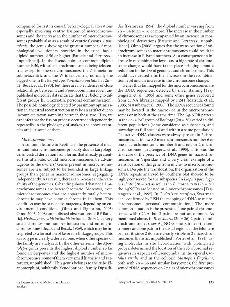
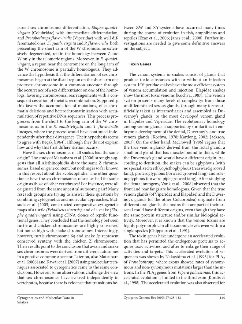
Ancestral reconstruction within Serpentes was done by parsimony optimization using the diploid number plus numbers of macro- and microchromosomes avail-able in the literature [main source: Olmo and Signorino, 2005]. These data were complemented with original ob-servation [Batistic and Ferrarezzi, unpublished] for the Tropidophiidae and Elapomorphini. Note that our Tropi-dophis paucisquamis (2n = 26) report consists of the first and single account for the whole family [Batistic et al., 2002], since that currently ascribed as Tropidophiidae in the literature (from Exiliboa ) actually belongs to the Un-galiophiinae, here included in Charininae, Boidae. Al-though we are aware this is an oversimplification of a complex character set, it was possible to trace a few pre-liminary remarks about the pattern that arises. These re-sults are presented in a reduced form in figure 1 .
The 2n = 36 (16M + 20m), although not exactly identi-cal in the morphology of the chromosomes for all taxa coded as such, is sufficiently similar to be considered a shared homology among most snake groups. This puta-tive ancestral configuration is notably conservative (re-maining apparently unchanged in many extant species) and showing a pervasive distribution throughout the snake tree branches, from which a number of other con-figurations have been derived. It is suggested to be prim-
Fig. 1. Ancestral reconstruction of karyotype configurations (diploid number plus the number of macro- and microchromo-somes) under parsimony optimization on a phylogenetic hypoth-esis of snakes (adapted from various sources). The 2n = 36 with 16 macro- and 20 microchromosomes (green branches) is indicated as a shared ancestral homology throughout most of the family tree, and from which many other karyotypes have been derived at different taxonomic levels. Sexual chromosome (ZW) heteromor-phism is a putative synapomorphy of the superfamily Colubroi-dea.

Cytogenetics and Molecular Data in Snakes
Cytogenet Genome Res 2009;127:128–142 131
Trace character
ZW heteromorphism
Inferred ancestral configuration:2n = 36: 16 macro + 20 microchromosomesisomorphic ZW
*
Character: Diploid number: Macro + microParsimony reconstruction (Unordered)[Steps: 75]
24: 16M 8m26: 20M 6m28: gradually decreasing to 8–12m30: gradually decreasing to 14–16m32 gradual32: 14M 18m32: 16M 16m32: 20M 12m3434: 14M 20m34: 16M 18m34: 18M 16m36 gradual36: 16M 20m36: 18M 18m36: 20M 16m38: 18M 20m38: 20M 18m40: 16M 24m40: 20M 20m42: 16–18M 24–26m3n = 42: 21M 21m42: 22M 20m44: 22–24M 20–22m46: 16M 30m50: 14M 36m
AnomalepididaeLeptotyphlopsRhinotyphlopsRamphotyphlopsTyphlops punctatusTyphlops jamaicensis-richardiAniliidaeTropidophiidaeCylindrophisXenopeltisLoxocemusPythonidaeCandoiaAcrantophisSanziniaEryxCharininiBoaEunectes-EpicratesCorallus cookiCorallus caninusBolyeriidaeAcrochordusAchalinusPareasCrotalinaeEchisDaboiaMacroviperaVipera gr.berusVipera gr.aspisHomalopsidaeLamprophisPseudaspisMalpolonMimophisMicrurusAspidelapsWalterinnesiaHemachatusNaja-BoulengerinaBungarusDendroaspisOphiophagusLaticaudaDemansiaOxyuranusPseudonajaPseudechisCacophisFurinaSimoselapsVermicellaAcanthophis-EchiopsisDenisoniaNotechiniHemiaspisElapognathusRhinoplocephalusAipysurus-EmydocephalusDisteira-KeriliaPelamisEnhydrina-PraescutataHydrophis-MicrocephalophisLimnophisAmphiesmaMacropisthodonRhabdophis-XenochrophisSinonatrixNatrixStoreria-VirginiaThamnophis-NerodiaReginaPseudoxenodonThermophisCarphophiinaeHydromorphusDipsadiniLeptodeiriniElapomorphusApostolepisPhalotrisTachymeniniTropidodryasHelicopsHydrodynastesSordellinaOxyrhopusPseudoboiniPhilodryasLiophis-ErythrolamprusXenodon-WaglerophisAhaetullaColubriniHierophis-PlatycepsOligodonOxybelisSonoriniTrimorphodonChironius -SpilotesPseustesZaocys-ArgyrogenaElaphiniBogertophisLampropeltiniChrysopeleaDendrelaphisBoigaTelescopusDinodonLycodon

Oguiura /Ferrarezzi /Batistic Cytogenet Genome Res 2009;127:128–142132
itive to most family-group taxa, with possible exception of the Typhlopidae, Tropidophiidae, and Xenodermati-dae (for which just a few or a single species have been karyotyped) and possibly also the Elapidae plus Lam-prophiidae clade and the xenodontine dipsadids, both re-sulting in equivocal optimization due to variation among and within terminal taxa.
Diversification occurred from this ancestral karyo-type and from then on possibly all species have suffered at least some modifications in them, altering or not the diploid number. As new tools for chromosomal investiga-tions become available, in addition to the visualization of karyotypes by standard and banding staining, the incor-poration of molecular techniques (FISH and others) made it possible to ‘see’ inside the chromosomes, to local-ize genes such as the crotamine gene mapped in Crotalus durissus terrificus by Rádis-Baptista et al. [2003], the RABSA , DMRT1 and SOX9 genes to the sexual chromo-somes by Matsubara et al. [2006], and others. Chromo-somes may undergo many alterations, with or without any perceptible change in their morphologies. Chromo-somes may also undergo no changes at all in their mor-phology or content, as seen in the cryptodiran turtles known for their notable karyotypic stability [Olmo, 1986; Olmo et al., 2002], further corroborated by Mühlmann-Diaz et al. [2001]. These authors used a whole chromo-some-1-specific probe from a cryptodiran turtle ( Trache-mys ) that resulted in specific hybridization of chromo-some 1 on 4 other turtles, revealing a cytogenetic stability of this chromosome during the past 66–144 mil-lion years. The authors point out that in one third of that time, various hominid species underwent extensive chro-mosomal rearrangements.
In Typhlopidae, a poorly sampled family based on chromosomes, 5 out of 6 species present 2n = 32 and2n = 34 with 16M, as in a ‘typical’ 36 karyotype. The variation in number is due to a diminishing number of microchromosomes, possibly because of translocations and fusion events. However, in Typhlops punctatus , the variation involves macro- and microchromosomes [Olmo and Signorino, 2005; García and Hernando, 2007]. This is also the case for Elapinae, where the karyological al-terations involve macro- and microchromosomes. Mi-crurus from Central America varies its chromosome number from 2n = 26 to 34 [Graham, 1977; Gutiérrez and Bolaños, 1979; Gutiérrez et al., 1988; Luykx et al., 1992]. Contrarily, in South America M. surinamensis surina-mensis has 2n = 38 [Gutiérrez et al., 1988], M. lemniscatus car valhoi presents 2n = 42 [Beçak and Beçak, 1969], M. corallinus has 2n = 40 and M. ibiboboca has 2n = 42 [Se-
rafim et al., 2007]. Serafim et al. [2007] formulated the hypothesis that the chromosome number in the genus tends to increase in South America and decrease in Cen-tral America, changes that especially involve the micro-chromosomes in Central America and the macrochro-mosomes in South America, probably due to fusion and fission processes. The high chromosome variability is also true for Bungarus and Naja and other species in the family. The members of the Hydrophiinae also show con-siderable variation in the number of chromosomes [see Olmo and Signorino, 2005]. In contrast, the subfamily Crotalinae presents very conservative karyotypes, all with 2n = 36 (16M + 20m) and similar morphology, the 4th pair being the sex chromosomes, with female hetero-gamety. Minor differences are observed in the morphol-ogy of the autosomes from one species to another. Also the W is variable in size and morphology from species to species, but always well differentiated from the Z chro-mosome. The small morphological differences in the macrochromosomes of different species in the subfamily suggest that translocations and inversions still happen but possibly without disrupting the main linkage groups. Good evidence of these changes was given in the complex of species Bothrops neuwiedi by Trajtengertz et al. [1995], showing a translocation of rDNA genes to a macrochro-mosome, but apparently still in the process of fixation. Contrary to Elapidae and many Xenodontinae, the Cro-talinae seem to have achieved an ideal condition in the assemblage of many linkage groups of genes, possibly maintained, at least partially, by the absence of changes in the chromosome number. This seems to fit well with the ‘canalization model’ and the achievement of the ‘op-timum karyotype’ proposed by Bickham and Baker [1979]. However, it must be stressed that if this is the case, this optimum karyotype was achieved by modifications without changing the chromosome number and that the sex chromosomes must have been already present in the ancestor that gave rise to the family Viperidae. Those snakes present a wide geographic distribution (absent ba-sically in Africa, Europe, Australia and Antarctica), sug-gesting that the relative stability of the chromosomal set did not interfere with the dispersal of the genus and its adaptation to new environments, accompanied by minor visible rearrangements that might be a consequence of speciation or part of the process.
The opposite situation is found in Elapidae and many Xenodontinae. Their ability to occupy new environments is accompanied by karyotypical alterations. The more conspicuous case is found in the tribe Elapomorphini where the increase in morphological complexity is ac-
Cytogenetics and Molecular Data in Snakes
Cytogenet Genome Res 2009;127:128–142 133
companied (or is it its cause?) by karyological alterations especially involving centric fissions of macrochromo-somes and the increase in the number of microchromo-somes probably also as a result of centric fissions. Apos-tolepis , the genus showing the greatest number of mor-phological evolutionary novelties in the tribe, has a diploid number of 56 or higher [Batistic and Ferrarezzi, unpublished]. In the Pseudoboini, a common diploid number is 50, with all macrochromosomes being telocen-tric, except for the sex chromosomes: the Z is meta- or submetacentric and the W is telocentric, normally the biggest one in the karyotype. Sordellina puctata has 2n = 52 [Beçak et al., 1990], but there are no evidences of close relationships between it and Pseudoboini; moreover, un-published molecular data indicate that they belong to dif-ferent groups [F. Grazziotin, personal communication]. The possible homology detected by parsimony optimiza-tion in ancestral reconstruction may be an artifact due to incomplete taxon sampling between these two. If so, we can infer that the fission process occurred independently, repeatedly in the phylogeny of snakes, the above exam-ples are just some of them.
Microchromosomes A common feature in Reptilia is the presence of mac-
ro- and microchromosomes, probably due to karyologi-cal ancestral derivation from forms that already present-ed this attribute. Could microchromosomes be advan-tageous to the owners? Genes present in microchromo -somes are less subject to be bounded in large linkage groups than genes in macrochromosomes, segregating independently. As a result, there is an increase in the vari-ability of the genomes. C-banding showed that not all mi-crochromosomes are heterochromatic. Moreover, even microchromosomes that are apparently totally hetero-chromatic may have some euchromatin in them. This condition may be or not advantageous, depending on en-vironmental conditions [Olmo and Signorino, 2005; Olmo 2005, 2008; unpublished observations of RF Batis-tic]. Hydrodynastes bicinctus bicinctus has 2n = 24, a very small chromosome number for snakes and no micro-chromosomes [Beçak and Beçak, 1969], which may be in-terpreted as a formation of favorable linkage groups. This karyotype is clearly a derived one, when other species of the family are analyzed. In the other extreme, the Apos-tolepis genus presents the highest diploid number so far found in Serpentes and the highest number of micro-chromosomes, some of them very small [Batistic and Fer-rarezzi, unpublished]. This genus belongs to the tribe El-apomorphini, subfamily Xenodontinae, family Dipsadi-
dae [Ferrarezzi, 1994], the diploid number varying from 2n = 34 to 2n = 56 or more. The increase in the number of chromosomes is accompanied by an increase in mor-phological derivations [Batistic and Ferrarezzi, unpub-lished]. Olmo [2008] argues that the translocation of mi-crochromosomes to macrochromosomes could result in an increase in R-band numbers. As a consequence an in-crease in recombination levels and a high rate of chromo-some change would have taken place bringing about a reduction in the size of genomes and chromosomes. This could have caused a further increase in the recombina-tion level and an increase in the chromosome change.
Genes thus far mapped for the microchromosomes are the rDNA sequences, detected by silver staining [Traj-tengertz et al., 1995] and some other genes recovered from cDNA libraries mapped by FISH [Matsuda et al., 2005; Matsubara et al., 2006]. The rDNA sequences found may be located in the macro- or in the microchromo-somes or in both at the same time. The Ag-NOR pattern in the neuwiedi group of Bothrops (2n = 36) varied in dif-ferent populations (some considered as subspecies, and nowadays as full species) and within a same population. The active rDNA clusters were always present in 2 chro-mosomes, as follows: 2 macrochromosomes number 6 or one macrochromosome number 6 and one or 2 micro-chromosomes [Trajtengertz et al., 1995]. This was the first case of the presence of rDNA genes in macrochro-mosomes in Viperidae and a very clear example of a translocation of this gene from micro- to macrochromo-somes. Despite the translocation, the organization of the rDNA repeats analyzed by Southern blot showed to be highly conserved for the subspecies. In Liophis poecilogy-rus shotti (2n = 32) as well as in B. jararacussu (2n = 36) the AgNORs are located in 2 microchromosomes [Traj-tengertz et al., 1995]. In C. durissus terrificus , Svartman et al. confirmed by FISH the mapping of rDNA to micro-chromosomes [personal communication]. The more common situation is the presence of one pair of chromo-somes with rDNA, but 2 pairs are not uncommon. As mentioned above, in B. insularis (2n = 36) 2 pairs of mi-crochromosomes show Ag-NORs, one pair near the cen-tromere and one pair in the distal region, at the telomere or near it, since 2 dots are clearly visible in 2 microchro-mosomes [Batistic, unpublished]. Porter et al. [1991], us-ing molecular in situ hybridization with biotinylated probes, determined the location of the 28S ribosomal se-quences in 4 species of Caenophidia. In the viperid Cro-talus viridis and in the colubrid Mastophis flagellum , both with 2n = 36 and similar karyotypes, the first pre-sented rDNA sequences on 2 pairs of microchromosomes
Oguiura /Ferrarezzi /Batistic Cytogenet Genome Res 2009;127:128–142134
and the second in only one pair. In the natricids Nerodia fasciata and Thamnophis marcianus , both with 2n = 36 (34M + 2m), the hybridization occurred only in one pair of macrochromosomes (long arm of pair 1 or pair 2). Those observations are in agreement with our own ob-servations in many different species [Batistic, unpub-lished] using silver staining. Microchromosomes were found even in a translocation with sex chromosomes in a population of Bungarus caeruleus from West Bengal re-sulting in a multiple sex chromosome constitution of Z 1 Z 1 Z 2 Z 2 /Z 1 Z 2 W. In the same females, the W suffered a dissociation resulting in multiple W chromosomes: W 1 and W 2 . A predominance of polymorphic females over the females carrying the original chromosome constitu-tion led the authors to suggest that polymorphic bearers had better adaptive flexibility and higher fecundity [Sing et al., 1979].
Sex Chromosomes Morphologically differentiated sex chromosomes are
spread all over the Metazoa. In vertebrates they occur in all classes but not in all groups within a class. In non-avian reptiles they occur in all Orders but not homoge-neously. In Serpentes they are present in some families and not in others. Within families they may be present in some species and absent in others, possibly due to differ-ent stages of morphological differentiation. Therefore, snakes are a good model for the study of sex chromo-somes.
The great majority of South American Boidae presents no morphological differentiation of sex chromosomes in standard staining [Beçak, 1966]. However, genes for mas-culinization or feminization may have accumulated in one or more pairs of chromosomes during evolution without morphological differentiation [Beçak, 1965; Singh et al., 1968]. Ray-Chaudhuri et al. [1970] did not find W chromatin in interphasic nuclei in a number of tissues of many Boidae species. Acrantophis dumerili was found to have a differentiated W chromosome: the size of the Z and the W were similar but the former is metacen-tric and the latter is acrocentric. This was the first report of differentiated sex chromosomes in Boidae [Mengden and Stock, 1980], indeed the only known case in non-Caenophidian snakes. In the Colubroidae, and especially in the Natricidae and Dipsadidae, different stages of dif-ferentiation of the W chromosome occur: they may differ from the Z by the centromere position or by size or by both [Beçak and Beçak, 1969; Beçak et al., 1990]. Aprea et al. [2006] examined 3 species of the Vipera aspis complex and Cerastes vipera of the Viperinae using standard and
banding staining methods. They found C. vipera with a 2n = 36 (16M + 20m) and no sex chromosome differen-tiation. The 3 species of Vipera presented a 2n = 42 karyo-type (22M + 20m) and in the examined females of V. aspis atra and V. aspis aspis C-banding pointed to the existence of a differentiated sex chromosome pair. In this case the W chromosome was almost totally heterochromatic, al-though morphologically indistinguishable from the Z. Mimophis , part of the reptile fauna of Madagascar, is the only psamophiine Lamprophiidae [Vidal et al., 2008] karyotyped to date. Its karyotype (2n = 44: 24M + 20m) revealed the 4th pair as the heteromorphic sex chromo-somes: the Z chromosome is biarmed, and the W is uni-armed. The W element was totally heterochromatic (C-banding) except for an interstitial euchromatic region [Aprea et al., 2003].
The Viperidae and Elapidae show a well differentiated W chromosome [Beçak, 1964, 1966; Ray-Chaudhuri et al., 1971]. A multiple sex chromosome system Z 1 Z 1 Z 2 Z 2 /Z 1 Z 2 W was first described in vertebrates in Bungarus cae-ruleus (Elapidae) [Singh et al., 1970]. The males were2n = 44 and the females 2n = 43 and the W condensed chromatin could be observed in the interphasic nuclei. Later another case of multiple sex chromosomes was found in Hydrophis fasciatus fasciatus (Hydrophiidae) of the type ZZ/ZW 1 W 2 . Also in this case, the interphase nu-clei presented in some tissues 2 kinds of heteropycnotic groups, corresponding to the W 1 and the W 2 [Ray-Chaud-huri and Singh, 1972].
What is the first step in the differentiation of the homomorphic autosome that harbors sex-determining genes? Beçak [1964] suggested that the first step was a pericentromeric inversion in one of the homologs, which prevented crossing-over in the region and the accumula-tion of sex-determining genes in the W. In a second step this region could undergo heterochromatinization and differentiation in morphology [Beçak, 1983]. On the oth-er hand, Ray-Chaudhuri et al. [1971] argued that the first step for this differentiation could be the heterochromati-nization of one chromosome that would later undergo morphological modifications. Minor satellite DNA frac-tions were found in great concentrations in the W chro-mosome, an evidence that crossing-over may have ceased prior to morphological differentiation between the Z and the W chromosomes [Singh et al., 1976; Jones and Singh, 1985]. Another possibility is that both hypotheses are true, depending on the case under consideration. Mat-subara et al. [2006] compared the chromosomes of 3 snakes with different stages of sex chromosome differen-tiation: Python molurus bivittatus (Boidae) with no ap-
Cytogenetics and Molecular Data in Snakes
Cytogenet Genome Res 2009;127:128–142 135
parent sex chromosome differentiation, Elaphe quadri-virgata (Colubridae) with intermediate differentiation, and Protobothrops flavoviridis (Viperidae) with well dif-ferentiated ones. E. quadrivirgata and P. flavoviridis , both presenting the short arm of the W chromosome exten-sively degenerated, retain the homology between Z and W only in the telomeric regions. Moreover, in E. quadri-virgata , a region near the centromere on the long arm of the W chromosome is partially homologous. They ad-vance the hypothesis that the differentiation of sex chro-mosomes began at the distal region on the short arm of a protosex chromosome in a common ancestor through the occurrence of a sex differentiator on one of the homo-logs, favoring chromosomal rearrangements with a con-sequent cessation of meiotic recombination. Supposedly, this favors the accumulation of mutations, of euchro-matin deletions and heterochromatinization with accu-mulation of repetitive DNA sequences. This process pro-gresses from the short to the long arm of the W chro-mosome, as in the E. quadrivirgata and T. flavoviridis lineages, where the process would have continued inde-pendently after their divergence. Their hypothesis seems to agree with Beçak [1964], although they do not explain how and why this first differentiation occurs.
Have the sex chromosomes of all snakes had the same origin? The study of Matsubara et al. [2006] strongly sug-gests that all Alethinophidia share the same Z chromo-somes, based on gene content, but nothing is so far known in this respect about the Scolecophidia. The other ques-tion is: have the sex chromosomes of snakes had the same origin as those of other vertebrates? For instance, were all originated from the same ancestral autosome pair? Many research groups are trying to answer these questions by combining cytogenetics and molecular approaches. Mat-suda et al. [2005] constructed comparative cytogenetic maps of a turtle ( Pelodiscus sinensis ), and of a snake ( Ela-phe quadrivirgata ) using cDNA clones of reptile func-tional genes. They concluded that the homology between turtle and chicken chromosomes are highly conserved but not as high with snake chromosomes. Interestingly, however, turtle chromosome 6q and snake 2p represent conserved synteny with the chicken Z chromosome. Their results point to the conclusion that avian and snake sex chromosomes were derived from different autosomes in a putative common ancestor. Later on, also Matsubara et al. [2006] and Kawai et al. [2007] using molecular tech-niques associated to cytogenetics came to the same con-clusions. However, some observations challenge the view that sex chromosomes have evolved independently in vertebrates, because there is evidence that transitions be-
tween ZW and XY systems have occurred many times during the course of evolution in fish, amphibians and reptiles [Ezaz et al., 2006; Janes et al., 2008]. Further in-vestigations are needed to give some definitive answers on the subject.
Toxin Genes
The venom systems in snakes consist of glands that produce toxic substances with or without an injection system. If Viperidae snakes have the most efficient system of venom accumulation and injection, Elapidae snakes show the most toxic venoms [Kochva, 1987]. The venom system presents many levels of complexity: from those undifferentiated serous glands, through many forms ar-tificially taken as intermediaries and assembled as Du-vernoy’s glands, to the most developed venom glandin Elapidae and Viperidae. The evolutionary homology among venom glands is supported by similarities in em-bryonic development of the dental, Duvernoy’s, and true venom glands [Kochva, 1978; Kardong, 2002; Jackson, 2003]. On the other hand, McDowell [1986] argues that the true venom glands derived from the rictal gland, a small oral gland that has muscles bound to them, while the Duvernoy’s gland would have a different origin. Ac-cording to dentition, the snakes can be aglyphous (with no specialized teeth), opisthoglyphous (rearward grooved fang), proteroglyphous (forward grooved fang) and sole-noglyphous (forward pipe grooved fang). After studying the dental ontogeny, Vonk et al. [2008] observed that the front and rear fangs are homologous. Given that the true venom glands (of Viperidae and Elapidae) and the Duver-noy’s glands (of the other Colubroidea) originate from different oral glands, the toxins that are part of their ar-senal could have different origins, even though they have the same protein structure and/or similar biological ac-tivity. Moreover, it is known that the venom toxins are highly polymorphic in all taxonomic levels even within a single species [Chippaux et al., 1991].
The toxin genes have undergone an accelerated evolu-tion that has permitted the endogenous proteins to ac-quire toxic activities, and after to enlarge their range of activities and targets. This accelerated evolution of se-quences was shown by Nakashima et al. [1995] for PLA 2 of Protobothrops, where exons showed rates of synony-mous and non-synonymous mutations larger than the in-trons. In the PLA 2 genes from Vipera palaestinae, this ac-celerated evolution is limited to the third exon [Kordis et al., 1998]. The accelerated evolution was also observed for

Oguiura /Ferrarezzi /Batistic Cytogenet Genome Res 2009;127:128–142136
other snake toxins such as other PLA 2 s [Moura-da-Silva et al., 1995; Chuman et al., 2000; Fujimi et al., 2002], dis-integrins [Moura-da-Silva et al., 1996, 1997; Juárez et al., 2008], serine proteases [Deshimaru et al., 1996] and 3-fin-ger toxins [Fry et al., 2003; Tamiya and Fujimi, 2006].
Tracing the phylogenetic history among the major groups or between species of snakes using toxins is a com-plex task. There are abounding doubts about snake tax-onomy in the data source [Fry et al., 2003], largely because the systematics of these animals has been neglected in the biomedical literature [Wüster and Harvey, 1996]. The main difficulty is to define whether the genes are orthol-ogous or paralogous, in order to properly examine the phylogenetic relationships. This is particularly important for understanding the evolution of snake venom proteins, for which an exceptionally high amount of copies have been reported, even in one individual [Fujimi et al., 2004; Oguiura et al., 2009]. A third is the lack of information about colubrid toxins due to the difficulty in obtaining the venom. This is currently being solved with studies of transcriptome of Duvernoy’s glands of Philodryas olfersii [Ching et al., 2006] and cDNAs from other colubrids [Fry et al., 2008].
The family of phospholipase type A 2 (PLA 2 ) toxins il-lustrates the complexity of the toxin world and the at-tempt to use their sequences for phylogeny. The PLA 2 tox-ins are classified into 2 groups according to their amino acid sequence and pattern of disulfide bridges: group I, PLA 2 s from the pancreatic juice of mammals and the venom of the Elapidae, and group II, non-pancreatic PLA 2 s of mammals and of the Viperidae. The group I PLA 2 s is divided into 2 subgroups: IA that is produced in the venom gland and IB, non-toxic which is produced in the pancreas [Danse et al., 1997].
The gene structures of group II PLA 2 may be divided into 2 types according to the number of exons and in-trons. PLA 2 genes from Vipera ammodytes are organized as non-pancreatic PLA 2 of humans and mice and contain 5 exons and 4 introns. The PLA 2 genes of Crotalinae snakes ( Crotalus and Protobothrops ) are organized as pancreatic PLA 2 of humans and dogs and contain 4 exons and 3 introns. Another difference between Viperinae and Crotalinae genes is the presence of Bov-B-LINE sequence in introns [Gubensek and Kordis, 1997].
Davidson and Dennis [1990] analyzed evolutionary trees for all PLA 2 s with the amino acid sequences de-scribed, but this approach was successful only in the clas-sification of PLA 2 s in groups I and II, but not in the taxo-nomic classification. Indeed, the recruitment of these 2 toxins might have occurred independently in elapid and
viperid snakes because the PLA 2 toxins have different or-igins and structures [Fry and Wüster, 2004]. Lynch VJ [2007] built evolutionary trees of groups I and II sepa-rately, and he found the same phylogenetic relationships of the group I PLA 2 s in elapid to that found by Slowinski et al. [1997] using different methods of phylogenetic anal-ysis. This success should be consequence of the single or-igin of elapid PLA 2 s and a lack of further events of gene duplication.
Fry and Wüster [2004], with a large number of toxin sequences, amino acids or cDNAs, confirm the conclu-sion of Strydom [1973] that the toxins were recruited from existing proteins in snakes. Phylogenetic analysis of toxin sequences made by Fry and Wüster [2004] showed that they were not recruited at the same time, despite showing similar structures; from 8 families examined, 5 (Kunitz protease inhibitors, CRISP toxins (cysteine-rich secretory proteins), GBL toxins (galactose-binding lec-tins), M12B peptidase toxins and NGF toxins (nerve growth factor)) were recruited to the poison before the separation of Viperidae and Elapidae families, and toxins as lectin-like, PLA 2 and natriuretic peptides are clearly the result of 2 independent recruitment events. The 3FTx (3-finger toxin) family is inferred to have been recruited before the split between the elapid and colubrid lineages, and after divergence of the Viperidae, since they are ab-sent in the latter [Fry et al., 2008]. Another example is the disintegrins; the dimeric disintegrins are widely distrib-uted in Viperinae and Crotalinae, whereas short disinte-grins appear to be restricted to African and Asian Echis and Eristicophis genera. This fact indicates that the emer-gence of dimeric disintegrins represents an early evolu-tionary event predating the Viperinae-Crotalinae split, whereas short disintegrins have evolved much more re-cently after the radiation of Viperinae [Juárez et al., 2008]. The sarafotoxins appear to be unique to the genus Atrac-taspis as are the small basic myotoxins, such as crot-amine, present only in the venom of the rattlesnakes Sis-trurus and Crotalus . Both toxins were recruited from en-dogenous proteins such as endothelins [Kochva et al., 1993] and beta-defensins [Torres and Kuchel, 2004; Rá-dis-Baptista et al., 2004]. However, the extension of the processes from these recruitments and the distinction between the independent and common events remain a vast research field to be explored, from an evolutionary as well as from a systematic point of view. We assumed that the construction of phylogeny using toxin data would not be useful in a discussion of higher level groups of snakes as we did here.

Cytogenetics and Molecular Data in Snakes
Cytogenet Genome Res 2009;127:128–142 137
Mitochondrial Genomes
There are 13 protein-coding genes known in snake mi-tochondrial genomes, 2 ribosomal RNAs genes, and 22 transfer RNAs genes [Kumazawa et al., 1996]. The base compositions are biased in snakes as in other vertebrates with predominance of A-T over G-C base pairs, and a greater A+C content in the gene-rich strand [Asakawa et al., 1991; Yan et al., 2008]. Other features of the complete sequences can be summarized as follows: size ranging from 16,218 bp in Leptotyphlops humilis to 18,905 bp in Boa constrictor , but can fairly exceed in 3 Dipsadinae, mainly due to extensive control and/or repeated regions, reaching an extreme of 23,038 bp in Leptodeira septen-trionalis . Alethinophian snakes in general have longer se-quences due to the duplicated control region, exhibiting 2 identical copies per genome. These have been main-tained stable since their origin and may function as an additional origin of heavy strand replication [Kumazawa et al., 1996; Jiang et al., 2007; Yan et al., 2008].
Since the sequencing of the first snake ( Dinodon semi-carinatus ) mitochondrial complete genome by Kuma-zawa at al. [1996], the comparative mitogenomics ofthe group have been studied by a number of authors, main ly with phylogenetic purposes [Kumazawa et al., 1996; Kumazawa, 2004; Dong and Kumazawa, 2005;Jiang et al., 2007]. Although the subject has recently been reviewed, the fast increasing sampling of new taxa pro-vides a continuously exciting field of research.
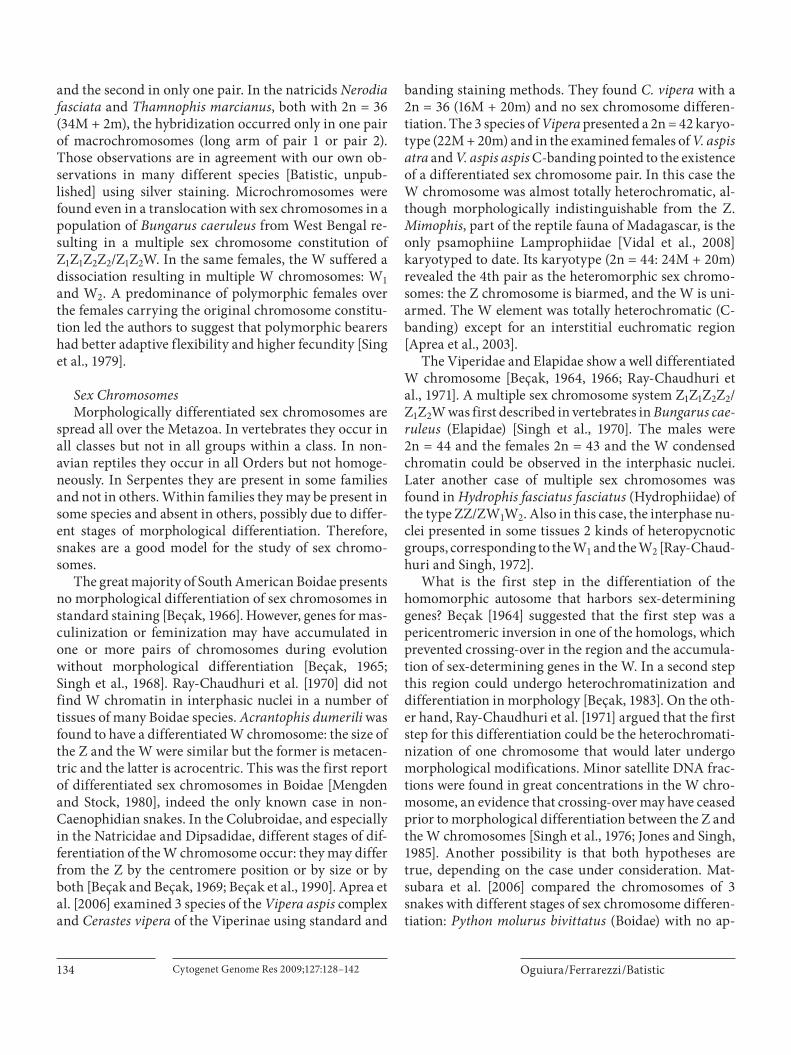
Because the last 2 phylogenetic analyses, published by Yan et al. [2008] and Castoe et al. [2009], included 14 and 15 snake species representing 7 and 10 families, respec-tively, we carried out here a cladistic analysis of all avail-able snake mitochondrial genomes with a complete cod-ing region (a data set comprising 51 sequences from 41 species, representing 15 families).
Methods of genome sequence alignments and cladistic analysis used here are the following. Protein-coding- genes were extracted from GenBank mitochondrial sequences using the software PEGA 0.99a [Patané, 2009], which pars-es a GenBank flat file with multiple accessions, outputting only the specified sequence (DNA or protein) of interest from each accession to a multi-FASTA file. Alignment was done with RevTrans [Rasmus and Pedersen, 2003], then adjusted manually, edited and concatenated using BioEdit [Hall, 2007]. Maximum Parsimony Analysis was conduct-ed using TNT [Goloboff et al., 2000], treating the few in-dels as missing data. The 22 transfer RNAs and the 2 ribo-somal RNAs were not included for 2 reasons: multiple alignment ambiguities due to an abundance of indels, and
incompleteness of some of these regions in the partial ge-nomic sequences used for a few but important taxa. Gene order information was compiled from the same species, after alignment of the whole mitochondrial sequences us-ing Mauve Genome Alignment Software [Darling et al., 2004]. The Mesquite program [Maddison and Maddison, 2009] was used to edit the character matrix.
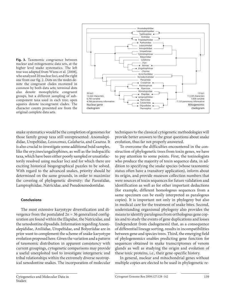
The results of mitogenomic analysis are from the raw empirical dataset using parsimony analysis ( fig. 2 ). Our results were then compared, in terms of taxonomic con-gruence, with the most comprehensive phylogenetic analyses of snake higher taxa, which is based exclusively on nuclear loci [Wiens et al., 2008]. Although the 2 inde-pendent data sets are similar in alignment length and variable positions (nuclear: 13,322 bp, 6,783 variable and mitochondrial: 11,539 bp, 7,958 variable), on the one hand, the number of nuclear genes employed is larger (20 vs. 13), but on the other, the number of parsimony infor-mative positions is higher in the mitochondrial set (7,133 vs. 4,766). However, the most important differences re-gard the sampling of characters and taxa. The nuclear set [Wiens et al., 2008] was previously designed for an analy-sis of higher level snake relationships (18 of the 20 genes used were newly sequenced for this purpose) with a taxon sampling more homogeneously distributed among the snake family-group categories; whereas the mitogenomic set has been accumulated in a public database during the entire last decade, resulting in the efforts of different au-thors with different goals, from evolutionary questions to taxonomy and, more recently, phylogeographic purposes [Mulcahy and Macey, 2009].
Thus, the mitogenomic taxon sampling is highly un-balanced, including many duplicated species, with some families overrepresented, while entirely lacking others. On the other side, the character sampling is complete for all terminal taxa with virtually no missing data in the mitogenomic set, whereas in the nuclear set there are many genes differentially lacking for most taxa (so, a higher proportion of missing data). Notwithstanding the considerable differences and a small number of species in common, the nuclear and mitogenomic data sets share a number of suprageneric taxa (e.g. families and subfami-lies) whose monophyly has been decisively supported in previous studies, so that both sets can be effectively com-pared at this level ( fig. 3 ).
The taxonomic congruence obtained between the nu-clear and mitogenomic datasets is surprising, showing only 2 incongruent clades, concerning the position of Xe-nodermatidae as sister to Acrochordoidea or to Colubroi-dea and the position of Homalopsidae as sister to Elapidae
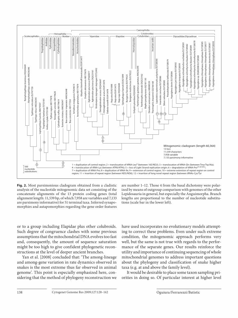
Oguiura /Ferrarezzi /Batistic Cytogenet Genome Res 2009;127:128–142138
or to a group including Elapidae plus other colubroids. Such degree of congruence clashes with some previous assumptions that the mitochondrial DNA evolves too fast and, consequently, the amount of sequence saturation might be too high to give confident phylogenetic recon-structions at the level of deeper ancient branches.
Yan et al. [2008] concluded that: ‘The among-lineage and among-gene variation in rate dynamics observed in snakes is the most extreme thus far observed in animal genome’. This point is especially emphasized here, con-sidering that the method of phylogeny reconstruction we
have used incorporates no evolutionary models attempt-ing to correct these problems. Even under such extreme condition, the mitogenomic approach performs very well, but the same is not true with regards to the perfor-mance of the separate genes. Our results reinforce the utility and importance of continuing sequencing of whole mitochondrial genomes to address important questions about the phylogeny and classification of snake higher taxa (e.g. at and above the family level).
It would be desirable to place some taxon sampling pri-orities in doing so. Of particular interest at higher level
5
3
4
7
8
9
11 10 12
6
21500
nucleotidesubstitutions
Mitogenomic cladogram (length 60,364)13 loci:11,539 characters7,958 variable7,133 parsimony informative
1 = duplication of control region; 2 = translocation of tRNA-Leu2 (between 16S/ND2); 3 = translocation of tRNA-Gln (between Trna-Trp/Ala); 4 = translocation of tRNA-Lys (between ATP8/ATP6); 5 = loss of Light Strand replication origin; 6 = degradation of tRNA-Pro(3’ of CR1); 7 = duplication of tRNA-Pro; 8 = duplication of tRNA-lle; 9 = extension of control region; 10 = extreme extention of repeat region on control region; 11 = insertion of repeat region (between ND5/ND6); 12 = insertion of long novel repeat region (between tRNAs-Cys/Tyr
Ram
phot
yphl
ops
bram
inus
DQ
3436
49
Trop
idop
hiid
ae
ScolecophidiaHenophidia
Boidae Viperidae Elapidae Colubridae
Caenophidia
ColubroideaDipsadidae:Dipsadinae
Ani
liid
ae
Pyth
onid
ae
Xeno
pel
tid
ae
Xeno
der
mat
oid
ea
Acr
ocho
rdoi
dea
Hom
alop
sid
ae
Ince
rtae
sed
is
Cylin
dro
phi
idae
Ram
phot
yphl
ops
aust
ralis
AM
2363
46
Typh
lops
retic
ulat
us E
U74
7730
Typh
lops
miru
s A
M23
6345
Lept
otyp
hlop
s hu
mili
s A
B079
597
Trop
idop
his
haet
ianu
s FJ
7551
81
Ani
lius
scyt
ale
FJ75
5180
Pyth
on re
gius
AB1
7787
8
Xeno
pelti
s un
icol
or A
B179
620
Cylin
drop
his
ruffu
s A
B179
619
Eune
ctes
not
aeus
AM
2363
47
Boa
cons
ticto
r im
pera
tor A
M23
6348
Boa
cons
ticto
r AB1
7735
4
Acha
linus
mei
guen
sis
FJ42
4614
Acro
chor
dus
gran
ulat
us A
B177
879
Glo
ydiu
s bl
omho
ffi b
revi
caud
us E
U91
3477
Ovo
phis
oki
nave
nsis
AB1
7567
0
Agki
stro
don
pisc
ivor
us E
F669
477
Agki
stro
don
pisc
ivor
us D
Q52
3161
Virid
ovip
era
stej
nege
ri st
ejne
geri
FJ75
2492
Dei
nagk
istr
odon
acu
tus
DQ
3436
47
Dei
nagk
istr
odon
acu
tus
EU91
3476
Dab
oia
russ
ellii
EU
9134
78
Naj
a at
ra E
U92
1898
Naj
a at
ra E
U91
3475
Naj
a na
ja D
Q34
3648
Oph
ioph
agus
han
nah
EU92
1899
Bung
arus
fasc
iatu
s EU
5795
23Bu
ngar
us m
ultic
inct
us E
U57
9522
Enhy
dris
plu
mbe
a D
Q34
3650
Pant
hero
phis
gut
tatu
s gu
ttat
us A
M23
6349
Pant
hero
phis
slo
win
skii
DQ
5231
62
Elap
he p
oryp
hyra
cea
GQ
1811
30
Din
odon
sem
icar
inat
us A
B008
539
Ther
mop
his
zhao
erm
ii G
Q16
6168
Lept
odei
ra s
epte
ntrio
nalis
pol
ystic
ta E
U72
8590
Iman
tode
s ce
ncho
a EU
7285
86
Sibo
n ne
bula
tus
EU72
8583
Pseu
dole
ptod
eira
latif
asci
ata
EU72
8579
Hyp
sigl
ena
slev
ini E
U72
8584
Hyp
sigl
ena
jani
texa
na E
U72
8592
Hyp
sigl
ena
torq
uata
EU
7285
91
Hyp
sigl
ena
ochr
orhy
ncha
nuc
hala
ta E
U72
8581
Hyp
sigl
ena
ochr
orhy
ncha
kla
uber
i EU
7285
82
Hyp
sigl
ena
ochr
orhy
ncha
kla
uber
i EU
7285
89
Hyp
sigl
ena
ochr
orhy
ncha
och
rorh
ynch
a EU
7285
78
Hyp
sigl
ena
sp. D
GM
2008
EU
7285
80
Hyp
sigl
ena
chlo
roph
aea
chlo
roph
aea
EU72
8585
Hyp
sigl
ena
chlo
roph
aea
chlo
roph
aea
EU72
8577
Hyp
sigl
ena
chlo
roph
aea
dese
rtic
ola
EU72
8587
Hyp
sigl
ena
chlo
roph
aea
chlo
roph
aea
EU72
8593
Fig. 2. Most parsimonious cladogram obtained from a cladistic analysis of the nucleotide mitogenomic data set consisting of the concatenate alignments of the 13 protein coding genes (total alignment length: 11,539 bp, of which 7,958 are variables and 7,133 are parsimony informative) for 51 terminal taxa. Inferred synapo-morphies and autapomorphies regarding the gene order features
are number 1–12. Those 4 from the basal dichotomy were polar-ized by means of outgroup comparison with genomes of the other Lepidosauria in general, but especially the Anguimorpha. Branch lengths are proportional to the number of nucleotide substitu-tions (scale bar in the lower left).
Cytogenetics and Molecular Data in Snakes
Cytogenet Genome Res 2009;127:128–142 139
snake systematics would be the completion of genomes for those family group taxa still unrepresented: Anomalepi-didae, Uropeltidae, Loxocemus , Calabaria , and Casarea . It is also crucial to investigate some additional boid samples, like the erycines/ungaliophines, as well as the indopacific taxa, which have been either poorly sampled or unsatisfac-torily resolved using nuclear loci and for which there are exciting historical biogeographical puzzles to be solved. With regard to the advanced snakes, priority should be determined on the same grounds, in order to maximize the covering of phylogenetic diversity: the Pareatidae, Lamprophiidae, Natricidae, and Pseudo xenodontidae.
Conclusions
The most extensive karyotype diversification and di-vergence from the postulated 2n = 36 generalized config-uration are found within the Elapidae, the Natricidae, and the xenodontine dipsadids. Information regarding Anom-alepididae, Aniliidae, Uropeltidae, and Bolyeriidae are in prior want to complement the scheme of snake karyotype evolution proposed here. Given the variation and a pattern of taxonomic distribution in apparent consistency with current groupings, cytogenetic comparisons may provide a useful unexploited tool to investigate intergeneric and tribal relationships within the extremely diverse neotrop-ical xenodontine snakes. The incorporation of molecular
techniques to the classical cytogenetic methodologies will provide better answers to the great questions about snake evolution, thus far not properly answered.
To overcome the difficulties encountered in the con-struction of phylogenetic trees from toxin genes, we have to pay attention to some points. First, the toxinologists who produce the majority of toxin sequence data, in ad-dition to specifying the snake species (whose taxonomic status often have a transitory application), inform about its origin, and provide museum collection numbers that were sources of toxin sequences for future validation and identification as well as for other important deductions (for example, different homologous sequences from a same specimen can be easily interpreted as paralogous copies). It is important not only in phylogeny but alsoin medical care for the treatment of snake bites. Second, understanding organismal phylogeny also provides the means to identify paralogous from orthologous gene cop-ies and to study the events of gene duplications and losses (independent from cladogenesis) that, as a consequence of differential lineage sorting, results in incompatibilities between gene and species trees. Third, the emerging field of phylogenomics enables predicting gene function for sequences obtained in snake transcriptomes of venom glands as well as studying the origin and evolution of these toxic proteins, i.e., their gene-specific history.
In general, nuclear and mitochondrial genes without multiple copies are elected to be used in phylogenetic re-
AnomalepididaeLeptotyphlopidae
TyphlopidaeAniliidae
TropidophiidaePythonidae
LoxocemidaeXenopeltidaeUropeltinae
CylindrophinaeBolyeriidae
BoinaeUngaliophiinae
AcrochordidaeXenodermatidae
PareatidaeCrotalinae
AzemiopinaeViperinae
HomalopsidaeElapidae
LamprophiidaeNatricidaeColubridaeDipsadidaeThermophis
CalabariaEryx
Charina
20 loci:13,322 characters6,783 variable4,766 parsimony informativeNuclear genecladogram
13 loci:11,539 characters
7,958 variable7,133 parsimony informative
Mitogenomiccladogram
Fig. 3. Taxonomic congruence between nuclear and mitogenomic data sets, at the higher level snake systematics. The left tree was adapted from Wiens et al. [2008], who analyzed 20 nuclear loci, and the right one from our fig. 2. Dots on the nodes de-note the congruent clades recovered in common by both data sets; terminal dots also denote monophyletic congruent groups, but a different sampling of sub-component taxa used in each tree; open squares denote incongruent clades. The character counts presented are from the original complete data sets.
Oguiura /Ferrarezzi /Batistic Cytogenet Genome Res 2009;127:128–142140
construction studies. For the Serpentes, the c- mos and RAG1 are the most extensively sampled examples, but more than 20 new nuclear loci have been recently sam-pled and analyzed for a limited, but representative, num-ber of snake taxa [Wiens et al., 2008] providing a real improvement on the available character evidence at the family level and above. Mitochondrial genes, although extensively sampled, such as cyt- b and ND4, and rRNAs, are also very useful, but often biased for having hundreds of sequences for a single or a few related species but none or an insufficient representation for many important higher taxa. Of particular interest at higher level snake systematics is the completion of genomes for those fam-ily group taxa that are still unrepresented: Anomalepidi-dae, Uropeltidae, Loxocemus , Calabaria , and Casarea as well as Pareatidae, Lamprophiidae, Natricidae, and Pseu-doxenodontidae, which will permit an equivalent com-parison regarding the taxonomic congruence between nuclear and mitochondrial genomic datasets.
The advantage in using these sequences is the quan-tity of data available in number of sequences and number of taxa and the possibility of easily concatenating infor-mation from different sources in a single original align-
ment. The sequencing of whole mitochondrial genome is also increasing and the phylogenetic trees obtained with these data are convergent to nuclear data. In addition, we found a different performance in the construction of phy-logenetic trees using mitogenomic or separate gene ap-proaches; therefore, we reinforce the usefulness and im-portance of continuing to sequence the whole mitochon-drial genome.
The accelerated evolution detected in toxin genes and mitochondrial genome architecture, combined with the genetic variety that microchromosomes can provide, could explain the extraordinary physiology and metabol-ic flexibility of snakes that enable the snake’s adaptation in such diverse habitats and conditions.
Acknowledgements
This work was supported by funds of the INCTTOX PRO-GRAM of CNPq, Brazil and FAPESP, SP, Brazil. We are grateful to the anonymous reviewers for the valuable suggestions which improved this manuscript and to English Consulting (B.V. Young) for their proofreading services.
References
Aprea G, Odierna G, Andreone F, Glaw F, Ven ces M: Unusual karyotype in the Malagasy colu-brid snake Mimophis mahfalensis . Amphib-Reptil 24: 215–219 (2003).
Aprea G, Gentilli A, Zuffi MAL, Odierna G: The karyology of Vipera aspis , V. atra , V. hugyi , and Cerastes vipera . Amphib-Reptil 27: 113–119 (2006).
Asakawa S, Kumazawa Y, Araki T, Himeno H, Miura K, Watanabe K: Strand-specific nu-cleotide composition bias in echinoderm and vertebrate mitochondrial genomes. J Mol Evol 32: 511–520 (1991).
Batistic RF, Ferrarezzi H, Soma M: O cariótipo de Tropidophis paucisquamis e suas afini-dades com outras famílias. Resumos do III Simpósio do Programa Biota/FAPESP Uni-versidade Federal de São Carlos, Nov 2002 at ht tp://w w w.biota .org.br/publ i /banco/index?show+91144174.
Beçak W: Karyotypes, sex chromosomes, and chromosomal evolution in snakes; in Bu-cherl W, Buckley E, Deulofeu W (eds.): Ven-omous animals and their venoms Vol 1, pp. 53–95 (Academic Press, New York 1964).
Beçak W: Constituição cromossômica e meca-nismo de determinação do sexo em ofídios sul-americanos. I. Aspectos cariotípicos. Mem Inst Butantan 32: 37–78 (1965).
Beçak W: Constituição cromossômica e meca-nismo de determinação do sexo em ofídios sul-americanos. II. Cromossomos sexuais e evolução do cariótipo. Mem Inst Butantan 33: 775–798 (1966).
Beçak W: Evolution and differentiation of sex chromosomes in lower vertebrates. Differen-tiation (Suppl) 23: 3–12 (1983).
Beçak W, Beçak ML: Cytotaxonomy and chro-mosomal evolution in Serpentes. Cytogenet-ics (Basel) 8: 247–262 (1969).
Beçak ML, Rabello-Gay MN, Beçak W, Soma M, Batistic RF, Trajtengertz I: The W chromo-some during the evolution and in sex abnor-malities of snakes. DNA content, C-banding, in Olmo E (ed.): Cytogenetics of Amphibians and Reptiles, pp. 221–240 (Birkhäuser Ver-lag, Basel 1990).
Bickham JW, Baker R: Canalization Model of chromosomal evolution. Bull Carnegie Mus Nat Hist 13: 70–84 (1979).
Castoe TA, de Koning AP, Kim HM, Gu W, Noonan BP, et al: Evidence for an ancient adaptive episode of convergent molecular evolution. Proc Natl Acad Sci USA 106: 8986–8991 (2009).
Ching ATC, Rocha MMT, Leme AFP, Pimenta DC, Furtado MFD, et al: Some aspects of the venom proteome of the Colubridae snake Philodryas olfersii revealed from a Duver-noy’s (venom) gland transcriptome. FEBS Lett 580: 4417–4422 (2006).
Chippaux JP, Williams V, White J: Snake venom variability: methods of study, results and in-terpretation. Toxicon 29: 1279–1303 (1991).
Chuman Y, Nobuhisa I, Ogawa T, Deshimaru M, Chijiwa T, et al: Regional and accelerated molecular evolution in group I snake venom gland phospholipase A2 isozymes. Toxicon 38: 449–462 (2000).
Danse JM, Gasparini S, Ménez A: Molecular bi-ology of snake venom phospholipases A2; in Kini M (ed.): Venom Phospholipase A 2 ; En-zymes, pp. 29–71 (John Wiley & Sons Ltd, England 1997).
Darling ACE, Mau B, Blatter FR, Perna NT: Mauve: multiple alignment of conserved genomic sequence with rearrangements. Genome Res 14: 1394–1403 (2004).
Davidson FF, Dennis EA: Evolutionary relation-ships and implications for the regulation of phospholipase A2 from snake venom to hu-man secreted forms. J Mol Evol 31: 228–238 (1990).
Deshimaru M, Ogawa T, Nakashima KI, No-buhisa I, Chijiwa T, et al: Accelerated evolu-tion of Crotalinae snake venom gland serine proteases. FEBS Lett 397: 83–88 (1996).
Dong S, Kumazawa Y: Complete mitochondrial DNA sequences of six snakes: phylogenetic relationships and molecular evolution of ge-nomic features. J Mol Evol 61: 12–22 (2005).

Cytogenetics and Molecular Data in Snakes
Cytogenet Genome Res 2009;127:128–142 141
Ezaz T, Stiglec R, Veyrunes F, Graves JAM: Rela-tionship between vertebrate ZW and XY sex chromosome systems. Curr Biol 16:R736–R743 (2006).
Ferrarezzi H: Uma sinopse dos gêneros e classi-ficação das Serpentes (Squamata): II. Família Colubridae; in Nascimento EB, Bernardes AT, Cotta GA (eds.): Herpetologia no Brasil, 1, pp. 81–91 (PUC Minas: Fundação Biodi-versitas: Fundação Ezequiel Dias, Belo Hori-zonte 1994).
Flavell AJ, Jackson V, Iqbal MP, Riach I, Waddell S: Ty1-copia group retrotransposon sequenc-es in Amphibia and Reptilia. Mol Gen Genet 246: 65–71 (1995).
Fry BG, Wüster W: Assembling an arsenal: ori-gin and evolution of the snake venom pro-teome inferred from phylogenetic analysis of toxin sequences. Mol Biol Evol 21: 870–883 (2004).
Fry BG, Wüster W, Kini RM, Brusic V, Khan A, Venkataraman D, Rooney AP: Molecular evolution and phylogeny of elapid snake ven-om three-finger toxins. J Mol Evol 57: 110–129 (2003).
Fry BG, Scheib H, Weerd L, Young B, Mc-Naughtan J, et al: Evolution of an arsenal structural and functional diversification of the venom system in the advanced snakes (Caenophidia). Mol Cell Proteomics 7: 215–246 (2008).
Fujimi TJ, Tsuchiya T, Tamiya T: A comparative analysis of invaded sequences from group IA phospholipase A2 genes provides evidence about the divergence period of genes groups and snake families. Toxicon 40: 873–884 (2002).
Fujimi TJ, Yasuoka S, Ogura E, Tsuchiya T, Tami-ya T: Comparative analysis of gene expression mechanisms between group IA and IB phos-pholipase A2 genes from sea snake Laticauda semifasciata . Gene 332: 179–190 (2004).
García JAR, Hernando A: Standard karyotype and nucleolus organizer region of neotropi-cal blindsnake Typhlops brongersmianus (Serpentes: Typhlopidae). Acta Herpetologi-ca 2: 117–120 (2007).
Goloboff P, Farris S, Nixon K: TNT (tree analysis using new technology). Beta version. Pub-lished by the authors. Tucuman, Argentina (2000). At http://www.cladistics.com/
Graham G: The karyotype of the Texas coral snake, Micrurus fulvius tenere. Herpetologi-ca 33: 345–348 (1977).
Gubensek F, Kordis D: Venom phospholipase A2 genes and their molecular evolution; in Kini M (ed.): Venom phospholipase A 2 enzymes, pp. 73–95 (John Wiley & Sons Ltd., England 1997).
Gutiérrez JM, Bolaños R: Cariótipos de las prin-cipales serpientes coral (Elapidae: Micrurus) de Costa Rica. Rev Biol Trop 27: 57–73 (1979).
Gutierrez JM, Solorzano A, Cerdas L: Karyo-types of five species of coral snakes ( Micru-rus ). J Herpetol 22: 109–112 (1988).
Hall T: BioEdit Sequence Alignment Editor for Windows 95/98/NT/XP. Version 7.0.9 (2007). At http://www.mbio.ncsu.edu/BioEdit/bioedit.html.
Hille A, Janssen IAW, Menken SBJ, Schlegel M, Thorpe RS: Heterologous amplification of microsatellite markers from colubroid snakes in European natricines (Serpentes: Natricinae). J Hered 93: 63–66 (2002).
Hughes S, Claya O, Bernardi G: Compositional patterns in reptilian genomes. Gene 295: 323–329 (2002).
Jackson K: The evolution of venom-delivery sys-tems in snakes. Zool J Linn Soc 137: 337–354 (2003).
Janes DE, Organ C, Valenzuela N: New resourc-es inform study of genome size, content, and organization in nonavian reptiles. Integr Comp Biol 48: 447–453 (2008).
Jiang ZJ, Castoe TA, Austin CC, Burbrink FT, Herron MD, et al: Comparative mitochon-drial genomics of snakes: extraordinary sub-stitution rate dynamics and functionality of the duplicate control region. BMC Evol Biol 7: 123 (2007).
John TR, Smith LA, Kaiser II: Genomic sequenc-es encoding the acidic and basic subunits of Mojave toxin: unusually high sequence iden-tity of non-coding regions. Gene 139: 229–234 (1994).
Jones KW, Singh L: Snakes and the evolution of sex chromosomes. Trends Genet 1: 55–61 (1985).
Juárez P, Comas I, González-Candelas F, Calvete JJ: Evolution of snake venom disintegrins by positive Darwinian selection. Mol Biol Evol 25: 2391–2407 (2008).
Kardong KV: Colubrid snakes and Duvernoy’s ‘venom’ glands. J Toxicol Toxin Rev 21: 1–19 (2002).
Kawai A, Nishida-Umehara C, Ishijima J, Tsuda Y, Ota H, Matsuda Y: Different origins of bird and reptile sex chromosomes inferred from comparative mapping of chicken Z-linked genes. Cytogenet Genome Res 117: 92–102 (2007).
Kelly CMR, Barker NP, Villet MH, Broadley DG: Phylogeny, biogeography and classification of the snake superfamily Elapoidea: a rapid radiation in the late Eocene. Cladistics 25: 38–63 (2009).
Kochva E: Oral glands of the reptilia; in Gans C, Gans KA (eds.): Biology of the Reptilia, pp. 43–161 (Academic Press, New York 1978).
Kochva E: The origin of snakes and evolution of the venom apparatus. Toxicon 25: 65–106 (1987).
Kochva E, Bdolah A, Wollberg Z: Sarafotoxins and endothelins: evolution, structure and function. Toxicon 31: 541–568 (1993).
Kohlsdorf T, Cummings MP, Lynch VJ, Stopper GF, Takahashi K, Wagner GP: A molecular footprint of limb loss: sequence variation of the autopodial identity gene Hoxa-13. J Mol Evol 67: 581–593 (2008).
Kordis D, Gubensek F: Unusual horizontal trans-fer of a long interspersed nuclear element be-tween distant vertebrate classes. Proc Natl Acad Sci USA 95: 10704–10709 (1998).
Kordis D, Bdolah A, Gubensek F: Positive Dar-winian selection in Vipera palaestinae phos-pholipase A2 genes is unexpectedly limited to the third exon. Biochem Biophys Res Commun 251: 613–619 (1998).
Kumazawa Y: Mitochondrial DNA sequences of five squamates: phylogenetic affiliation of snakes. DNA Res 11: 137–144 (2004).
Kumazawa Y, Ota H, Nishida M, Ozawa T: Gene rearrangements in snake mitochondrial ge-nomes: highly concerted evolution of con-trol-region-like sequences duplicated and inserted into a tRNA gene cluster. Mol Biol Evol 13: 1242–1254 (1996).
Lawson R, Slowinski JB, Crother BI, Burbrink FT: Phylogeny of the Colubroidea (Serpen-tes): new evidence from mitochondrial and nuclear genes. Mol Phylogenet Evol 37: 581–601 (2005).
Lee MSY, Hugall A, Lawson R, Scanlon J: Phylog-eny of snakes (Serpentes): combining mor-phological and molecular data in likelihood, Bayesian and parsimony analyses. System Biodivers 5: 371–389 (2007).
Lukoschek V, Waycott M, Keogh S: Relative in-formation content of polymorphic microsat-ellites and mitochondrial DNA for inferring dispersal and population genetic structure in the olive sea snake, Aipysurus laevis . Mol Ecol 17: 3062–3077 (2008).
Luykx P, Slowinski JB, McCranie JR: The karyo-type of the coral snake Micrurus ruatanus . Amphib-Reptil 13: 289–292 (1992).
Lynch M: The origins of genome architecture, pp. 363–389 (Sinauer Associates, Inc., Sun-derland 2007).
Lynch VJ: Inventing an arsenal: adaptive evolu-tion and neofunctionalization of snake ven-om phospholipase A 2 genes. BMC Evol Biol 7: 2 (2007).
Maddison WP, Maddison DR: Mesquite: a mod-ular system for evolutionary analysis. Ver-sion 2.7 (2009) at http://mesquiteproject.org.
Matsubara K, Tarui H, Toriba M, Yamada K, Nishida-Umehara C, Agata K, Matsuda Y: Evidence for different origin of sex chromo-somes in snakes, birds, and mammals and step-wise differentiation of snake sex chro-mosomes. Proc Natl Acad Sci USA 103: 18190–18195 (2006).
Matsuda Y, Nishida-Umehara C, Tarui H, Ku-roiwa A, Yamada K, et al: Highly conserved linkage homology between birds and turtles: Bird and turtle chromosomes are precise counterparts of each other. Chromosome Res 13: 601–615 (2005).
McDowell SB: The architecture of the corner of the mouth of colubroid snakes. J Herpetol 20: 353–407 (1986).
Mengden GA, Stock D: Chromosomal evolution in Serpentes; a comparison of G and C chro-mosome patterns of some colubrid and boid genera. Chromosoma 79: 52–61 (1980).

Oguiura /Ferrarezzi /Batistic Cytogenet Genome Res 2009;127:128–142142
Moura-da-Silva AM, Paine MJI, Diniz MRV, Theakston RDG, Crampton JM: The molec-ular cloning of a phospholipase A2 from Bothrops jararacussu snake venom: evolu-tion of venom group II phospholipase A2’s may imply gene duplications. J Mol Evol 41: 174–179 (1995).
Moura-da-Silva AM, Theakston RDG, Cramp-ton JM: Evolution of disintegrin cysteine-rich and mammalian matrix-degrading metallo proteinases: gene duplication and di-vergence of a common ancestor rather than convergent evolution. J Mol Evol 43: 263–269 (1996).
Moura-da-Silva AM, Theakston RDG, Cramp-ton JM: Molecular evolution of phospholi-pase A2s and metalloproteinase/disintegrins from venoms of vipers; in Thorpe RS, Wüster W, Malhotra A (eds.): Venomous Snakes: Ecology, Evolution and Snakebite, pp. 173–187 (Claredon Press, Oxford 1997).
Mühlmann-Diaz MC, Ulsh BA, Whicker FW, Hinton TG, Congdon JD, Robinson JF, Bed-ford JS: Conservation of chromosome 1 in turtles over 66 million years. Cytogenet Cell Genet 92: 139–143 (2001).
Mulcahy DG, Macey JR: Vicariance and disper-sal form a ring distribution in nightsnakes around the gulf of California. Mol Phylogen-et Evol 53: 537–546 (2009).
Nakashima KI, Nobuhisa I, Deshimaru M, Na-kai M, Ogawa T, et al: Accelerated evolution in the protein-coding regions is universal in crotalinae snake venom gland phospholipase A2 isozyme genes. Proc Natl Acad Sci USA 92: 5605–5609 (1995).
Nelson GJ: Classification as an expression of phylogenetic relationships. Syst Zool 22: 344–359 (1973).
Nobuhisa I, Ogawa T, Deshimaru M, Chijiwa T, Nakashima K-I, et al: Retrotransposable CR1-like elements in Crotalinae snake ge-nomes. Toxicon 36: 915–920 (1998).
Oguiura N, Collares MA, Furtado MFD, Ferra-rezzi H, Suzuki H: Intraspecific variationof the crotamine and crotasin genes in Cro-talus durissus rattlesnakes. Gene 446: 35–40 (2009).
Olmo E: A. Reptilia; in John B (ed.): Animal Cytogenetics, 4. Chordata 3, pp. 1–100 (Ge-brueder Borntraeger, Berlin, Stuttgart 1986).
Olmo E: Rate of chromosome changes and repli-cation in reptiles. Genetica 125: 185–203 (2005).
Olmo E: Trends in the evolution of reptilian chromosomes. Integr Comp Bio 48: 486–493 (2008).
Olmo E, Capriglione T, Odierna G: Different ge-nomic evolutionary rates in the various rep-tile lineages. Gene 295: 317–321 (2002).
Olmo E, Signorino G: Chromorep: a reptile chromosomes database (2005) at http://193.206.118.100/professori/chromorep.pdf.
Patané JSL: Program for extraction of GenBank annotated sequences, version 0.99a (2009).
Piskurek O, Austin CC, Okada N: Sauria SINEs: novel short interspersed retroposable ele-ments that are widespread in reptile ge-nomes. J Mol Evol 62: 630–644 (2006).
Porter CA, Hamilton MJ, Sites Jr JW, Baker RJ: Location of ribosomal DNA in chromo-somes of squamate reptiles: systematic and evolutionary implications. Herpetologica 47: 271–280 (1991).
Rádis-Baptista G, Kubo T, Oguiura N, Svartman M, Almeida TMB, et al: Structure and chro-mosomal localization of the gene for crot-amine, a toxin from the South American rattlesnake, Crotalus durissus terrificus . Toxicon 42: 747–752 (2003).
Rádis-Baptista G, Kubo T, Oguiura N, Silva ARBPda, Hayashi MAF, Oliveira EB, Ya-mane T: Identification of crotasin, a crot-amine-related gene of Crotalus durissus ter-rificus . Toxicon 43: 751–759 (2004).
Rasmus W, Pedersen AG: RevTrans – Construct-ing alignments of coding DNA from aligned amino acid sequences. Nucl Acids Res 31: 3537–3539 (2003).
Ray-Chaudhuri SP, Singh L: DNA replication pattern in sex chromosomes of snakes. Nu-cleus 15: 200–210 (1972).
Ray-Chaudhuri SP, Singh L, Sharma T: Evolu-tion of sex chromosomes and formation of W chromatin in snakes. Chromosoma 33: 239–251 (1971).
Ray-Chaudhuri SP, Singh L, Sharma T: Sexual dimorphism in somatic interphase nuclei of snakes. Cytogenetics 9: 410–423 (1970).
Serafim H, Peccinini-Seale DM, Batistic RF: Es-tudo cariotípíco de duas espécies brasileiras do gênero Micrurus (Ophidia: Elapidae). Bi-ota Neotropica 7 (n1): (2007) at http://www.biotaneotropica.org.br/v7n1/pt/abstract?article 8 bn01607012007).
Singh L: Evolution of karyotypes of snakes. Chromosoma 38: 185–236 (1972).
Singh L, Sharma T, Ray-Chaudhuri SP: W chro-mosome in Indian water snake (checkered keel back) Natrix piscator (Colubridae). Ex-perientia 24: 7980 (1968).
Singh L, Sharma T, Ray-Chaudhuri SP: Multiple sex chromosomes in the common Indian krait, Bungarus caeruleus Schneider. Chro-mosoma 31: 386–391 (1970).
Singh L, Purdom IF, Jones KW: Satellite DNA and evolution of sex chromosomes. Chro-mosoma 59: 43–62 (1976).
Singh L, Ray-Chaudhuri SP, Manjundar K, Pur-don IF, Jones KW: Sex specific chromosome polymorphisms in the common Indian krait Bungarus caeruleus Schneider (Ophidia, Elapidae). Chromosoma 73: 93–108 (1979).
Slowinski JB, Knight A, Rooney AP: Inferring species trees from gene trees: a phylogenetic analysis of the Elapidae (Serpentes) based on the amino acid sequences of venom proteins. Mol Phylogenet Evol 8: 349–362 (1997).
Strydom DJ: Snake venom toxins: the evolution of some of the toxins found in snake venoms. Syst Zool 22: 596–608 (1973).
Tamiya T, Fujimi TJ: Molecular evolution of tox-in genes in Elapidae snakes. Mol Divers 10: 529–543 (2006).
Torres AM, Kuchel PW: The � -defensin-fold family of polypeptides. Toxicon 44: 581–588 (2004).
Trajtengertz I, Beçak ML, Ruiz IRG: Ribosomal cistrons in Bothrops neuwiedi (Serpentes) subspecies from Brazil. Genome 38: 601–606 (1995).
Vidal N, Delmas AS, Hedges SB: The higher-lev-el relationships of alethinophidian snakes inferred from seven nuclear and mitochon-drial genes; in Henderson RW, Powell R (eds.): Biology of the Boas and Pythons, pp. 27–33 (Eagle Mountain Publishing LC, Eagle Mountain 2007a).
Vidal N, Delmas AS, David P, Cruaud C, Cou-loux A, Hedges SB: The phylogeny and clas-sification of caenophidian snakes inferred from seven nuclear protein-coding genes. C R Biol 330: 182–187 (2007b).
Vidal N, Branch WR, Pauwels OSG, Hedges SB, Broadley DG, et al: Dissecting the major Af-rican snake radiation: a molecular phylogeny of the Lamprophiidae Fitzinger (Serpentes, Caenophidia). Zootaxa 1945: 51–66 (2008).
Vidal N, Rage JC, Couloux A, Hedges SB: Snakes (Serpentes); in Hedges SB, Kumar S (eds): The Timetree of Life, pp. 390–397 (Oxford University Press, New York 2009).
Vonk FJ, Admiraal JF, Jackson K, Reshef R, de Bakker MA, et al: Evolutionary origin and development of snake fangs. Nature 454: 630–633 (2008).
Wiens JJ, Kuczynski CA, Smith SA, Mulcahy DG, Sites JW Jr, Townsend TM, Reeder, TWB: Ranch lengths, support, and congru-ence: testing the phylogenomic approach with 20 nuclear loci in snakes. Syst Biol 57: 420–431 (2008).
Wiley EO: An annotated Linnean hierarchy, with comments on natural taxa and compet-ing systems. Syst Zool 28: 308–337 (1979).
Wüster W, Harvey AL: Reviews of venomous snake systematics in Toxicon. Toxicon 34: 397–398 (1996); Comment on: Toxicon 34: 399–406 (1996).
Wüster W, Peppin L, Pook CE, Walker DE: A nesting of vipers: Phylogeny and historical biogeography of Viperidae (Squamata: Ser-pentes). Mol Phylogenet Evol 49: 445–459 (2008).
Yan J, Li H, Zhou K: Evolution of the mitochon-drial genome in snakes: gene rearrange-ments and phylogenetic relationships. BMC Genomics 9: 569 (2008).
Zaher H, Grazziotin FG, Cadle JE, Murphy RW, Moura-Leite JC, Bonatto SL: Molecular phy-logeny of advanced snakes (Serpentes, Cae-nophidia) with an emphasis on South Amer-ican Xenodontines: a revised classification and descriptions of new taxa. Papéis Avulsos de Zoologia (São Paulo) 49: 115–153 (2009).
Zupunski V, Gubensek F, Kordis D: Evolutionary dynamics and evolutionary history in the RTE clade of non-LTR retrotransposons. Mol Biol Evol 18: 1849–1863 (2001).


![Overwintering of three species of snakes in northwestern Minnesota [microform]](https://img.dokumen.tips/doc/110x75/6359c9f99e39cba4b605d632/overwintering-of-three-species-of-snakes-in-northwestern-minnesota-microform-.jpg)


