Embed Size (px)
Citation preview
Research Article
Fatty acid composition, lipid oxidation, and fishy odourdevelopment in seabass (Lates calcarifer) skin duringiced storage
Thanasak Sae‐leaw and Soottawat Benjakul
Faculty of Agro‐Industry, Department of Food Technology, Prince of Songkla University, Songkhla, Thailand
Changes in fatty acid profile, lipid hydrolysis and oxidation, development of fishy odour and volatilecompounds in seabass (Lates calcarifer) skin during 18 days of iced storage were investigated. Peroxidevalue (PV) increased up to Day 6 and subsequently decreased up to 18 days (p< 0.05). The continuousincreases in thiobarbituric acid reactive substances (TBARS) values, free fatty acid (FFA) content andlipoxygenase (LOX) activity were noticeable with increasing storage time (p<0.05). Formation of FFAand hydroperoxide was confirmed by the changes in amplitude of peak at 3600–3200/cm and 1711/cm inFourier transform IR spectra, respectively. With increasing storage time, the increase in fishy odourintensity was observed along with the formation of volatiles. Hexanal and nonanal constituted as thedominant volatile aldehydes in skin stored in ice for an extended time. Therefore, the delay of skinprocessing must be avoided to prevent the formation of undesirable fishy odour in skin and its products.
Practical applications: Seabass skin is one of the potential raw materials for production of collagen,gelatin or other derived products. Iced storage of seabass skin affected fatty acid composition, lipidhydrolysis and oxidation, development of fishy odour and volatile compounds. Therefore, skin from freshfish should be used and the delay of further processing should be avoided to prevent undesirable fishyodour in skin and products made therefrom.
Keywords: Fishy odour / Lipids / Lipid oxidation / Seabass / Skin
Received: October 11, 2013 / Revised: January 15, 2014 / Accepted: March 14, 2014
DOI: 10.1002/ejlt.201300381
1 Introduction
Iced storage is an important preservation method to maintainthe quality of fish during handling and storage. Icing has beenused widely to reduce undesirable biochemical and chemicalreactions and to retard the growth of spoilage micro-organisms [1]. However, lipid deterioration still easily takesplace and limits the shelf‐life of fish during storage. Bothlipolysis and lipid oxidation in fish are associated with qualityloss [2]. Hydrolysis, induced by lipases and phospholipases,produces FFAs that undergo further oxidation [3]. Fish
lipids are relatively more susceptible to oxidation due tohigh degree of unsaturation and low content of endogenousantioxidants, compared with other food lipids [4]. Lipidoxidation is associated with the development of undesirableodour, especially fishy odour, in fish stored for an extendedtime [2, 5]. Development of fishy odour in seabass and redtilapiamuscle was primarily associated with lipid oxidation [6].In addition, lipid oxidation induced by lipoxygenase (LOX)was responsible for a strong fishy odour in silver carp mince [7].Moreover, fishy odour in protein hydrolysate caused by lipidoxidation was reported by Yarnpakdee et al. [8].
Seabass (Lates calcarifer) is one of economically importantspecies of Thailand and other countries in Southeast Asiaowing to its white flesh and delicacy. During processing ordressing, skin is generated and considered as a byproduct.To fully exploit those skins, the conversion to value‐addedproducts such as collagen, gelatin as well as hydrolysate withbioactivities has been employed [9–11]. Hydrolysate from fishskin has gained increasing interest as the supplement forskin and health care products, e.g. beauty drink, etc. Owing to
Correspondence: Soottawat Benjakul, Faculty of Agro‐Industry,Department of Food Technology, Prince of Songkla University, Hat Yai,Songkhla 90112, ThailandE‐mail: [email protected]: 66‐7455‐8866
Abbreviations: PV, peroxide value; TBARS, thiobarbituric acid reactivesubstances; FTIR, fourier transform infrared; FFA, free fatty acid; EPA,eicosapentaenoic acid; LOX, lipoxygenase; SFA, saturated fatty acid
Eur. J. Lipid Sci. Technol. 2014, 116, 885–894 885
� 2014 WILEY-VCH Verlag GmbH & Co. KGaA, Weinheim www.ejlst.com
strong fishy odour/flavour of those products, the use of skin islimited. Recently, Sae‐leaw et al. [12] reported that skinobtained fromNile tilapia had the increasing fishy odour whenthe whole fish were kept for a longer time in ice. Such an off‐odour was governed by lipid autoxidation and LOX inducedoxidation. After deskinning, the skins are used as the startingmaterial for manufacturing of collagen or its derived products.Before being further processed, those skins are generallystored in ice and they more likely undergo deteriorationduring the storage. Due to the greater surface area forchemical or microbial reactions, as well as the tissue damageduring dressing, quality loss could be more pronounced thanthose intacted with whole fish. This might be associated withthe rapid development of fishy odour in the stored skin.
However, no information regarding the alteration of lipidsvia chemical and enzymatic reactions and the development offishy odour in seabass skin stored in ice has been reported.This study aimed to evaluate the changes in fatty acidcomposition, lipid hydrolysis and oxidation as well as fishyodour development and volatile compounds in seabass skinduring iced storage of 18 days.
2 Materials and methods
2.1 Chemicals
Trichloroacetic acid, anhydrous sodium sulphate and ferrouschloride were obtained from Merck (Darmstadt, Germany).1,1,3,3‐tetramethoxypropane and linoleic acid were purchasedfromSigma–AldrichChemical Co. (St. Louis,MO).Methanol,acetone, chloroform and ammonium thiocyanate were obtainedfrom Lab‐Scan (Bangkok, Thailand). All chemicals were ofanalytical grade. Disodium hydrogen phosphate, sodiumdihydrogen phosphate, 2‐thiobarbituric acid and cumenehydroperoxidewere procured fromFluka (Buchs, Switzerland).
2.2 Fish collection and preparation
Fresh seabass (Lates calcarifer) having the average weight of0.8–1.0 kg from a farm in KoYor, Songkhla were deskinnedafter capture. The skins were placed in a polystyrene boxcontaining ice using a skin/ice ratio of 1:2 w/w. The sampleswere transported to the Department of Food Technology,Prince of Songkla University, Hat Yai within 1 h.
Upon arrival, seabass skins were washed, placed inpolyethylene bag and kept in ice with a skin/ice ratio of1:2 w/w. The packed samples were placed and distributeduniformly between the layers of ice in the insulated boxes,which were subsequently left at RT (28–30°C). To maintainthe skin/ice ratio, the molten ice was removed and replacedwith new ice every 2 days. The temperature of skin wasmaintained at 0–2°C throughout the storage of 18 days. Fishskins were randomly taken every 3 days, pooled and used asthe composite sample. Fish skins were descaled manually.
The skins were then washed with cold tap water, drained andcut into small pieces (0.5� 0.5 cm2) using the scissor. Theskins were pulverised by blending in the presence of liquidnitrogen. The skin powder was subjected to analyses.
2.3 Changes in fatty acid compositions of lipids fromseabass skin during iced storage
2.3.1 Extraction of lipid
Lipid of skin was extracted as per the method of Blighand Dyer [13]. Skin powder (25g) was homogenised with200mL of a chloroform/methanol/distilled water mixture(50:100:50, v/v/v) at the speed of 9500 rpm for 2min at 4°Cusing an IKALabortechnik homogeniser (Selangor,Malaysia).The homogenate was added with 50mL of chloroform andhomogenised at 9500 rpm for 1min. Subsequently, 25mL ofdistilled water were added and then homogenised again for30 s. The homogenate was centrifuged at 3000g at 4°C for15min using a RC‐5B plus centrifuge (Sorvall, Norwalk, CT).Aqueous phase was transferred into a separating flask. Afterbeing separated, the chloroform layer was drained off into a125mL Erlenmeyer flask containing about 2–5 g of anhydroussodium sulphate, shaken very well, and decanted into a round‐bottom flask through aWhatmanNo. 4 filter paper (WhatmanInternational Ltd., Maidstone, England). The solvent wasevaporated at 25°C using an EYELA rotary evaporator N‐100(Tokyo Rikakikai, Co., Ltd., Tokyo, Japan), and the residualsolvent was removed by flushing with nitrogen.
2.3.2 Determination of fatty acid profile
Fatty acid profile was determined as FAMEs. FAMEs wereprepared according to the method of AOAC [14]. Theprepared methyl ester was injected to the GC (Shimadzu,Kyoto, Japan) equipped with the flame ionisation detector(FID) at a split ratio of 1:20. A fused silica capillary column(30m� 0.25mm), coated with bonded polyglycol liquidphase, was used. The analytical conditions were: injectionport temperature of 250°C and detector temperature of270°C. The oven was programmed from 170 to 225°C at arate of 1°C/min (no initial or final hold). Retention times ofFAME standards were used to identify chromatographicpeaks of the samples. Fatty acid content was calculated, basedon the peak area ratio and expressed as g fatty acid/100 g lipid.
2.4 Oxidation and hydrolysis of lipids from seabassskin during iced storage
2.4.1 Measurement of peroxide value
PV was determined according to the method of Richards andHultin [15] with slight modifications. Skin powder (1 g) washomogenised at a speed of 13,500 rpm for 2min in 11mLof chloroform/methanol (2:1, v/v) using a homogeniser.
886 T. Sae‐leaw, S. Benjakul Eur. J. Lipid Sci. Technol. 2014, 116, 885–894
� 2014 WILEY-VCH Verlag GmbH & Co. KGaA, Weinheim www.ejlst.com
Homogenate was then filtered using Whatman No. 1 filterpaper. To 7mL of the filtrate, 2mL of 0.5% NaCl wereadded. Themixture was vortexed at a moderate speed for 30 sand then centrifuged at 3000g for 3min at 4°C using arefrigerated centrifuge to separate the sample into two phases.Twenty‐five microlitre of 30% w/v ammonium thiocyanateand 25mL of 20mM iron (II) chloride were added to 3mL oflower phase. The reaction mixture was allowed to stand for20min at RT and the absorbance at 500 nm was read. Theblank was prepared in the same manner, except the distilledwater was used instead of ferrous chloride. A standard curvewas prepared using cumene hydroperoxide at a concentrationrange of 0.5–2ppm. PV was expressed as mg cumenehydroperoxide/kg skin after blank subtraction.
2.4.2 Measurement of thiobarbituric acid reactivesubstances
Thiobarbituric acid reactive substances (TBARS) weredetermined as described by Buege and Aust [16]. Skinpowder (0.5 g) was homogenised with 2.5mL of a solutioncontaining 0.375%w/v thiobarbituric acid, 15%w/v trichloro-acetic acid and 0.25mM HCl. The mixture was heated in aboiling water bath (95–100°C) for 10min. Thereafter, themixture was cooled with running tap water and centrifuged at3600g at 25°C for 20min. The absorbance of the supernatantwasmeasured at 532nm.A standard curve was prepared using1,1,3,3‐tetramethoxypropane at the concentrations rangingfrom 0 to 6 ppm. TBARS value was calculated and expressedas mg malonaldehyde equivalents/kg skin.
2.4.3 Fourier transform infrared spectra analysis
Fourier transform infrared (FTIR) analysis of lipid wasperformed in a horizontal ATR Trough plate crystal cell (45°ZnSe; 80mm long, 10mm wide and 4mm thick; PIKETechnology, Inc., Madison, WI) equipped with a BrukerModel Vector 33 FTIR spectrometer (Bruker Co., Ettlingen,Germany). Prior to analysis, the crystal cell was cleaned withacetone, wiped drywith soft tissue and the background scanwasrun. For spectra analysis, lipid sample (200mL) was applieddirectly onto the crystal cell and the cell was clamped into themount of the FTIR spectrometer. The spectra, in the range of4000–400/cm (mid‐IR region) with automatic signal gain, werecollected in 16 scans at a resolution of 4/cm and were ratioedagainst a background spectrum recorded from the clean, emptycell at 25°C. Analysis of spectral data was carried out using theOPUS 3.0 data collection software programme (Bruker Co.).
2.4.4 Measurement of free fatty acid
Free fatty acid (FFA) content was determined according tothe method of Lowry and Tinsley [17]. Lipid sample (0.1 g)was mixed with 5mL of isooctane and swirled vigorously todissolve the sample. The mixture was then added with 1mL
of 5% w/v cupric acetate‐pyridine reagent, prepared bydissolving 5 g of the reagent grade cupric acetate in 100mLof water, filtering and adjusting the pH to 6.0–6.2 usingpyridine. The mixture was shaken vigorously for 90 s using aVortex‐Genie2 mixer (Bohemia, NY) and allowed to standfor 20 s. The absorbance at 715 nm of the upper layer wasread. A standard curve was prepared using palmitic acid inisooctane at concentrations ranging from 0 to 10mM. FFAcontent was expressed as g FFA/100 g lipid.
2.5 Changes in lipoxygenase (LOX) in the skin ofseabass during iced storage
2.5.1 Extraction of LOX
Skin powder was suspended in 50mM phosphate buffer, pH7.0, containing 1mM glutathione and 0.04% Tween‐20, at aratio of 1:9 w/v. The mixture was homogenised at 11,000 rpmfor 3min. The suspension was centrifuged for 15min at 4°Cat 15,000g to remove the tissue debris. The supernatant wascollected and referred to as ‘LOX extract’.
2.5.2 Assay for LOX
LOX activity was assayed spectrophotometrically by moni-toring the formation of conjugated dienes at the absorbance of234 nm as per the method of Hamberg and Samuelsson [18]and Liu and Pan [19] with somemodifications. The substratesolution was prepared by mixing 157.2mL linoleic acid,157.2mLTween 20 and 10mL distilled water. OnemL of 1.0NNaOHwas added and the solution was diluted with 50mMphosphate buffer, pH 7.0 to a final volume of 200mL.Subsequently, 100mL of LOX extract and 900mL of 50mMphosphate buffer, pH 7.0 were added to the substrate solution(2mL) to initiate the reaction. After incubation at 25°C for3min, the absorbance at 234 nm of the mixture was recordedat 0 and 3min of reaction. One unit was defined as LOXcausing an increase in absorbance at 234 nm of 0.001/minunder the specified condition.
2.6 Development of fishy odour and volatilecompounds in seabass skin during iced storage
2.6.1 Measurement of fishy odour
Seabass skins stored in ice at Day 0, 3, 6, 9, 12, 15 and 18 wereused for evaluation of fishy odour. Fishy odour was evaluatedby 8 trained panellists with the ages of 25–32. Prior to theevaluation, the panellists were trained three times a week.Panellists were trained with standards for two sessions using a5‐cm line scale anchored from none (score¼ 0) to extremelystrong fishy odour (score¼ 4) [20]. Before testing, 10 g of fishskinswere placed in a sealable plastic cup.All sampleswere kepton ice until evaluation. The sealable plastic cups were placed atRT for 10min before evaluation. The panellists were asked to
Eur. J. Lipid Sci. Technol. 2014, 116, 885–894 Changes in seabass skin during iced storage 887
� 2014 WILEY-VCH Verlag GmbH & Co. KGaA, Weinheim www.ejlst.com
open the sealable cup and sniff the headspace above the samplesfor determining the intensities of fishy odour.
2.6.2 Measurement of volatile compounds
Seabass skins stored in ice at Day 0, 6, 12 and 18 weredetermined for volatile compounds using solid‐phase micro-extraction gas chromatography mass spectrometry (SPME‐GC‐MS) following the method of Iglesias and Medina [21]with a slight modification.
2.6.2.1 Extraction of volatile compounds
To extract volatile compounds, 3 g of sample was homoge-nised at a speed of 13,500 rpm for 2min with 8mL ofdeionised water. The mixture was centrifuged at 2000g for10min at 4°C. The supernatant (6mL) was heated at 60°Cwith equilibrium time of 10 h in a 20‐mL headspace vial.The SPME fibre (50/30 lm DVB/CarboxenTM/PDMSStableFlexTM; Supelco, Bellefonte, PA) was conditioned at270°C for 15min before use and then exposed to theheadspace. The 20mL‐vial (Agilent Technologies, PaloAlto, CA) containing the sample extract and the volatilecompounds were allowed to adsorb into the SPME fibre at60°C for 1 h. The volatile compounds were then desorbedin the GC injector port for 15min at 270°C.
2.6.2.2 GC–MS analysis
GC–MS analysis was performed in a HP 5890 series II GCcoupled with HP 5972 mass‐selective detector equipped with asplitless injector and coupled with a quadrupole mass detector(Hewlett Packard, Atlanta, GA). Compounds were separatedon a HP‐Innowax capillary column (Hewlett Packard; 30m� 0.25mm ID, with film thickness of 0.25mm). The GC oventemperature program was: 35°C for 3min, followed by anincrease of 3°C/min to 70°C, then an increase of 10°C/min to200°C, and finally an increase of 15°C/min to a finaltemperature of 250°C and holding for 10min. Helium wasemployed as a carrier gas, with a constantflowof 1mL/min.Theinjector was operated in the splitless mode and its temperaturewas set at 270°C. Transfer line temperature was maintained at260°C. The quadrupole mass spectrometer was operated in theelectron ionisation (EI)mode and source temperature was set at250°C. Initially, full‐scan‐mode data were acquired to deter-mine appropriate masses for the later acquisition in scan modeunder the following conditions: mass range: 25–500 amu andscan rate: 0.220 s/scan. All the analyses were performed withionisation energy of 70eV, filament emission current at 150mA,and the electron multiplier voltage at 500V.
2.6.2.3 Analysis of volatile compounds
Identification of volatile compounds in the samples was basedon the retention times of individual aldehydic standards
including pentanal, hexanal, heptanal, octanal and nonanal.Identification of the compounds was also done by consultingChemStation Library Search (Wiley 275.L). Quantitativedetermination was carried out using an internal calibrationcurve that was built using stock solutions of the compoundsin ultra‐pure water saturated in salt and analysing them bythe optimised HS‐SPME method. Quantification limits werecalculated to a signal‐to‐noise (S/N) ratio of 10. Repeatabilitywas evaluated by analysing three replicates of each sample.The identified volatile compounds related to lipid oxidation,including aldehydes, alcohols, ketones, etc., were presentedin the term of abundance.
2.7 Statistical analysis
Experiments were run in triplicate using three different lots ofsamples. The data were subjected to ANOVA. Comparisonof means was carried out by Duncan’s multiple range test.Statistical analysis was performed using the StatisticalPackage for Social Science (SPSS 11.0 for windows, SPSSInc., Chicago, IL).
3 Results and discussion
3.1 Changes in fatty acid compositions of seabassskin during iced storage
Fatty acid conpositions of lipids extracted from seabass skinstored in ice for 18 days are presented in Table 1. Skin lipidsfrom fresh seabass comprised palmitic acid (C16:0; 23.40/100 g lipid) as the most abundant fatty acid, followed by oleicacid (C18:1 (n�9)) (20.90/100 g lipid), DHA (C22:6(n� 3)) (9.82/100 g lipid) and linoleic acid (C18:2 (n�6))(9.29/100 g lipid), respectively. The result was in agreementwith Njinkoué et al. [22] who reported that palmitic acid wasthemajor fatty acid in lipids from skin of three edible fish fromthe Senegalese coast. Palmitic acid was also found as thepredominant saturated fatty acid (SFA) in skin lipids of wildand cultivated gilthead seabream and European seabass,whilst oleic acid was the majorMUFA [23]. Lipids from freshseabass skin contained 35.98% SFA, 29.57% MUFA and27.90% PUFA. Amongst PUFA, DHA C22:6 (n� 3) wasthe dominant fatty acid, followed by eicosapentaenoic acidC20:5 (n� 3) (EPA). In the present study, DHA constitutedapproximately two‐fold higher than EPA. DHA contents ofskin lipids from wild and cultivated gilthead seabream andEuropean seabass were higher than EPA [23]. DHA is usuallymore abundant in fish lipids than EPA [4]. DHA hasbeen found as the main lipid in cell membrane, mainlyphospholipids [24].
During iced storage, SFAs including, palmitic acid, stearicacid and arachidic acid (C 20:0) increased as the storage timeincreased. This was coincidental with the decreases inMUFAand PUFA. Decreases in PUFAs, especially EPA and DHA,
888 T. Sae‐leaw, S. Benjakul Eur. J. Lipid Sci. Technol. 2014, 116, 885–894
� 2014 WILEY-VCH Verlag GmbH & Co. KGaA, Weinheim www.ejlst.com
were observed with increasing storage time. EPA decreased by2.28% and 5.86% atDay 6 and 18, respectively. ForDHA, thedecreases by 2.95% and 2.34% were found at Day 6 and 18,respectively. The decreases in DHA and EPA contents were
due to their susceptibility to oxidation during the extendedstorage [6]. At Day 18 of iced storage, MUFA and PUFAcontents decreased by 1.15% and 7.49%, respectively,whereas SFA content increased by 4.28%, compared withthat found at Day 0. PUFAs were generally more prone tooxidation thanMUFAs. Therefore, changes in fatty acids tookplace in seabass skin stored in ice, in which the content ofunsaturated fatty acids decreased during the storage.
3.2 Lipid oxidation and hydrolysis of seabass skinduring iced storage
3.2.1 Peroxide value (PV)
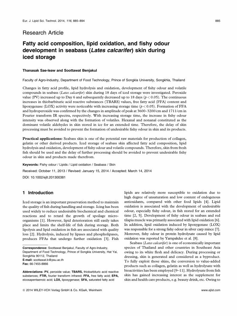
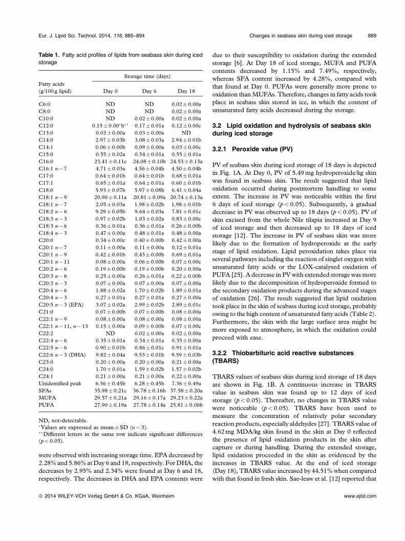
PV of seabass skin during iced storage of 18 days is depictedin Fig. 1A. At Day 0, PV of 5.49mg hydroperoxide/kg skinwas found in seabass skin. The result suggested that lipidoxidation occurred during postmortem handling to someextent. The increase in PV was noticeable within the first6 days of iced storage (p< 0.05). Subsequently, a gradualdecrease in PV was observed up to 18 days (p< 0.05). PV ofskin excised from the whole Nile tilapia increased at Day 9of iced storage and then decreased up to 18 days of icedstorage [12]. The increase in PV of seabass skin was morelikely due to the formation of hydroperoxide at the earlystage of lipid oxidation. Lipid peroxidation takes place viaseveral pathways including the reaction of singlet oxygen withunsaturated fatty acids or the LOX‐catalysed oxidation ofPUFA [25]. A decrease in PVwith extended storage wasmorelikely due to the decomposition of hydroperoxide formed tothe secondary oxidation products during the advanced stagesof oxidation [26]. The result suggested that lipid oxidationtook place in the skin of seabass during iced storage, probablyowing to the high content of unsaturated fatty acids (Table 2).Furthermore, the skin with the large surface area might bemore exposed to atmosphere, in which the oxidation couldproceed with ease.
3.2.2 Thiobarbituric acid reactive substances(TBARS)
TBARS values of seabass skin during iced storage of 18 daysare shown in Fig. 1B. A continuous increase in TBARSvalue in seabass skin was found up to 12 days of icedstorage (p< 0.05). Thereafter, no changes in TBARS valuewere noticeable (p< 0.05). TBARS have been used tomeasure the concentration of relatively polar secondaryreaction products, especially aldehydes [27]. TBARS value of4.62mg MDA/kg skin found in the skin at Day 0 reflectedthe presence of lipid oxidation products in the skin aftercapture or during handling. During the extended storage,lipid oxidation proceeded in the skin as evidenced by theincreases in TBARS value. At the end of iced storage(Day 18), TBARS value increased by 44.51%when comparedwith that found in fresh skin. Sae‐leaw et al. [12] reported that
Table 1. Fatty acid profiles of lipids from seabass skin during icedstorage
Fatty acids(g/100 g lipid)
Storage time (days)
Day 0 Day 6 Day 18
C6:0 ND ND 0.02�0.00aC8:0 ND ND 0.02�0.00aC10:0 ND 0.02�0.00a 0.02�0.00aC12:0 0.15� 0.00�b�� 0.17�0.01a 0.12�0.00cC13:0 0.03� 0.00a 0.03�0.00a NDC14:0 2.97� 0.03b 3.08�0.03a 2.94� 0.01bC14:1 0.06� 0.00b 0.09�0.00a 0.03�0.00cC15:0 0.55� 0.02a 0.54�0.01a 0.55�0.01aC16:0 23.41� 0.11c 24.08� 0.10b 24.53� 0.13aC16:1 n� 7 4.71� 0.03a 4.56� 0.04b 4.50� 0.04bC17:0 0.64� 0.01b 0.64� 0.01b 0.68�0.01aC17:1 0.65� 0.01a 0.64�0.01a 0.60� 0.01bC18:0 5.93� 0.07b 5.97� 0.08b 6.41�0.04aC18:1 n� 9 20.90� 0.11a 20.81� 0.09a 20.74� 0.13aC18:1 n� 7 2.05� 0.03a 1.98� 0.02b 1.98� 0.01bC18:2 n� 6 9.29� 0.05b 9.64�0.03a 7.81�0.01cC18:3 n� 3 0.97� 0.02b 1.03�0.02a 0.83�0.00cC18:3 n� 6 0.36� 0.01a 0.36�0.01a 0.26� 0.00bC18:4 n� 3 0.47� 0.00a 0.48�0.01a 0.48�0.00aC20:0 0.34� 0.00c 0.40� 0.00b 0.42�0.00aC20:1 n� 7 0.11� 0.00a 0.11�0.00a 0.12�0.01aC20:1 n� 9 0.42� 0.01b 0.43� 0.00b 0.69�0.01aC20:1 n� 11 0.08� 0.00a 0.06� 0.00b 0.07�0.00cC20:2 n� 6 0.19� 0.00b 0.19� 0.00b 0.20�0.00aC20:3 n� 6 0.25� 0.00a 0.26�0.01a 0.22� 0.00bC20:3 n� 3 0.07� 0.00a 0.07�0.00a 0.07�0.00aC20:4 n� 6 1.88� 0.02a 1.70� 0.02b 1.89�0.01aC20:4 n� 3 0.27� 0.01a 0.27�0.01a 0.27�0.00aC20:5 n� 3 (EPA) 3.07� 0.02a 2.99� 0.02b 2.89�0.01cC21:0 0.07� 0.00b 0.07� 0.00b 0.08�0.00aC22:1 n� 9 0.08� 0.00a 0.08�0.00a 0.08�0.00aC22:1 n� 11, n� 13 0.15� 0.00a 0.09� 0.00b 0.07�0.00cC22:2 ND 0.02�0.00a 0.02�0.00aC22:4 n� 6 0.35� 0.01a 0.34�0.01a 0.35�0.00aC22:5 n� 6 0.90� 0.01b 0.86�0.01c 0.91�0.01aC22:6 n� 3 (DHA) 9.82� 0.04a 9.53� 0.01b 9.59� 0.03bC23:0 0.20� 0.00a 0.20�0.00a 0.21�0.00aC24:0 1.70� 0.01a 1.59� 0.02b 1.57� 0.02bC24:1 0.21� 0.00a 0.21�0.00a 0.22�0.00aUnidentified peak 6.56� 0.45b 6.28� 0.45b 7.36�0.49aSFAs 35.98� 0.21c 36.78� 0.16b 37.58� 0.20aMUFA 29.57� 0.21a 29.16� 0.17a 29.23� 0.22aPUFA 27.90� 0.19a 27.78� 0.14a 25.81� 0.06b
ND, not‐detectable.�Values are expressed as mean�SD (n¼ 3).��Different letters in the same row indicate significant differences(p< 0.05).
Eur. J. Lipid Sci. Technol. 2014, 116, 885–894 Changes in seabass skin during iced storage 889
� 2014 WILEY-VCH Verlag GmbH & Co. KGaA, Weinheim www.ejlst.com
TBARS value of skin excised from the whole Nile tilapia hadthe increases in TBARS with increasing iced storage time.The increase in TBARS value of seabass skin during icedstorage indicated the destruction of hydroperoxides intothe secondary oxidation products in the later stages of lipidoxidation [28]. Hydroperoxides break down in severalsteps, yielding a wide variety of decomposition products,including aldehydes, etc. Lipid oxidation generates a widerange of secondary aldehyde products, including n‐alkanals,trans‐2‐alkenals, 4‐hydroxy‐trans‐2‐alkenals, and malonalde-hyde [28]. Fish skin has been known to contain high content ofphospholipids [12]. Those phospholipids are susceptible to
oxidation due to high PUFA content [24]. Additionally,seabass skin contained a high content of PUFA (Table 1).Those fatty acids underwent oxidation to a higher extent withincreasing storage time as indicated by the decrease in thosefatty acids (Table 1). Although PV decreased continuouslyduring 12–18 days, TBARS values remained unchanged.This might be due to the balance between decomposition ofhydroperoxide and the loss of some secondary productsduring that period. Thus, those lipid oxidation products werepresent in seabass skin and could lead to the quality loss,particularly the offensive fishy odour.
3.2.3 Free fatty acid (FFA) content
Changes in FFA contents in lipids from seabass skin duringiced storage of 18 days are depicted in Fig. 1C. FFA contentof seabass skin increased as the storage time increased(p< 0.05). The result indicated that lipid hydrolysis occurredin seabass skin during the storage in ice. It was found that FFAcontent increased from 3.63/100 g lipid at Day 0 to 9.85/100 glipid after 18 days of storage. FFA content in skin of ice‐storedNile tilapia continuously increased with increasing storagetime [12]. Lipases, phospholipase A and phospholipase B areimportant enzymes involved in hydrolysis of fish lipids [29].
dc
a bc
e f
0
2
4
6
8
10
0 3 6 9 12 15 18
Pero
xide
val
ue
(mg
cum
ene
hydr
oper
oxid
e/kg
sk
in)
Storage time (days)
(A)
dc c b
a aa
0
2
4
6
8
10
0 3 6 9 12 15 18
TB
AR
S (m
g m
alon
alde
hyde
eq
uiva
lent
s/kg
skin
)
Storage time (days)
(B)
d
c
b
a
0
2
4
6
8
10
12
0 6 12 18
Free
fatt
y ac
id c
onte
nt
(g/1
00 g
lipi
d)
Storage time (days)
(C)
Figure 1. Changes in peroxide values (A), TBARS values (B) andFFA contents (C) of seabass skin during iced storage. Differentletters indicate significant difference (p< 0.05). Bars representSDs (n¼ 3).
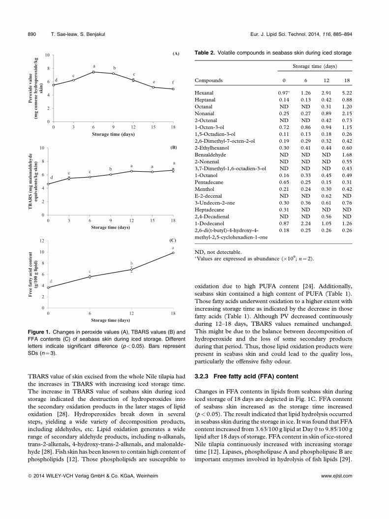
Table 2. Volatile compounds in seabass skin during iced storage
Compounds
Storage time (days)
0 6 12 18
Hexanal 0.97� 1.26 2.91 5.22Heptanal 0.14 0.13 0.42 0.88Octanal ND ND 0.31 1.20Nonanal 0.25 0.27 0.89 2.152‐Octenal ND ND 0.42 0.731‐Octen‐3‐ol 0.72 0.86 0.94 1.151,5‐Octadien‐3‐ol 0.11 0.13 0.18 0.262,6‐Dimethyl‐7‐octen‐2‐ol 0.19 0.29 0.32 0.422‐Ethylhexanol 0.30 0.41 0.44 0.60Benzaldehyde ND ND ND 1.682‐Nonenal ND ND ND 0.553,7‐Dimethyl‐1,6‐octadien‐3‐ol ND ND ND 0.431‐Octanol 0.16 0.33 0.45 0.49Pentadecane 0.65 0.25 0.15 0.31Menthol 0.21 0.24 0.30 0.42E‐2‐decenal ND ND 0.62 ND3‐Undecen‐2‐one 0.30 0.36 0.61 0.76Heptadecane 0.31 ND ND ND2,4‐Decadienal ND ND 0.56 ND1‐Dodecanol 0.87 2.24 1.05 1.262,6‐di(t‐butyl)‐4‐hydroxy‐4‐methyl‐2,5‐cyclohexadien‐1‐one
0.18 0.25 0.26 0.26
ND, not detectable.�Values are expressed as abundance (�109; n¼ 2).
890 T. Sae‐leaw, S. Benjakul Eur. J. Lipid Sci. Technol. 2014, 116, 885–894
� 2014 WILEY-VCH Verlag GmbH & Co. KGaA, Weinheim www.ejlst.com
Nayak et al. [30] reported that extracellular lipase, producedby certain microorganisms, such as Pseudomonas fragi,contributed to the lipolytic breakdown of fish lipids. Lipaseand phospholipase are also found in fish skin [31]. FFAreleased are more prone to oxidation, compared with thoseesterified with a glycerol backbone. This plausibly led to theincreased lipid oxidation as evidenced by the increases inPV and TBARS value in seabass skin stored in ice for theextended time (Fig. 1A and B).
3.2.4 FTIR spectra of lipids from skin during icedstorage
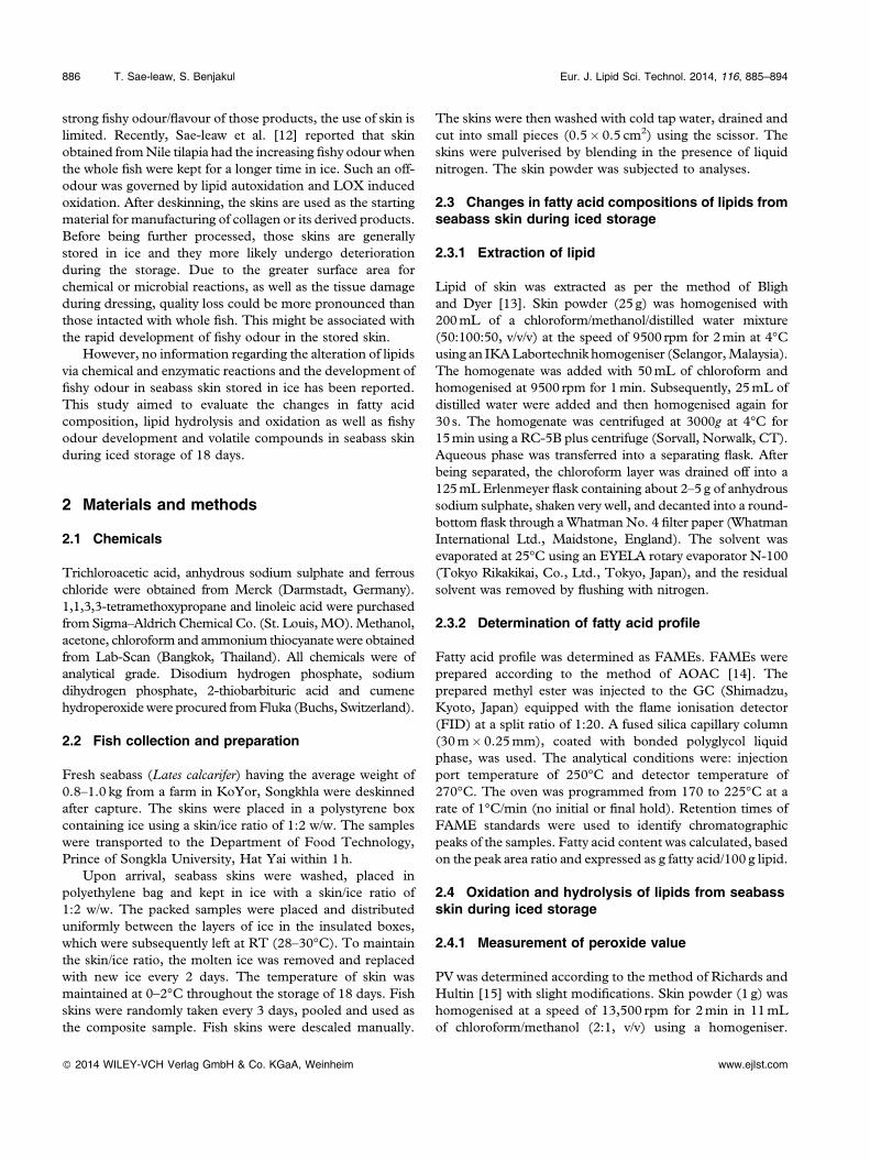
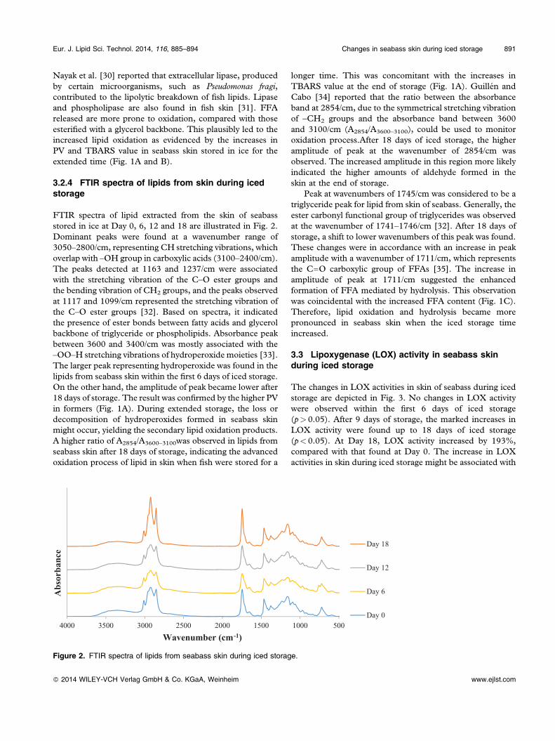
FTIR spectra of lipid extracted from the skin of seabassstored in ice at Day 0, 6, 12 and 18 are illustrated in Fig. 2.Dominant peaks were found at a wavenumber range of3050–2800/cm, representing CH stretching vibrations, whichoverlap with –OH group in carboxylic acids (3100–2400/cm).The peaks detected at 1163 and 1237/cm were associatedwith the stretching vibration of the C–O ester groups andthe bending vibration of CH2 groups, and the peaks observedat 1117 and 1099/cm represented the stretching vibration ofthe C–O ester groups [32]. Based on spectra, it indicatedthe presence of ester bonds between fatty acids and glycerolbackbone of triglyceride or phospholipids. Absorbance peakbetween 3600 and 3400/cm was mostly associated with the–OO–H stretching vibrations of hydroperoxide moieties [33].The larger peak representing hydroperoxide was found in thelipids from seabass skin within the first 6 days of iced storage.On the other hand, the amplitude of peak became lower after18 days of storage. The result was confirmed by the higher PVin formers (Fig. 1A). During extended storage, the loss ordecomposition of hydroperoxides formed in seabass skinmight occur, yielding the secondary lipid oxidation products.A higher ratio of A2854/A3600–3100was observed in lipids fromseabass skin after 18 days of storage, indicating the advancedoxidation process of lipid in skin when fish were stored for a
longer time. This was concomitant with the increases inTBARS value at the end of storage (Fig. 1A). Guillén andCabo [34] reported that the ratio between the absorbanceband at 2854/cm, due to the symmetrical stretching vibrationof –CH2 groups and the absorbance band between 3600and 3100/cm (A2854/A3600–3100), could be used to monitoroxidation process.After 18 days of iced storage, the higheramplitude of peak at the wavenumber of 2854/cm wasobserved. The increased amplitude in this region more likelyindicated the higher amounts of aldehyde formed in theskin at the end of storage.
Peak at wavenumbers of 1745/cm was considered to be atriglyceride peak for lipid from skin of seabass. Generally, theester carbonyl functional group of triglycerides was observedat the wavenumber of 1741–1746/cm [32]. After 18 days ofstorage, a shift to lower wavenumbers of this peak was found.These changes were in accordance with an increase in peakamplitude with a wavenumber of 1711/cm, which representsthe C––O carboxylic group of FFAs [35]. The increase inamplitude of peak at 1711/cm suggested the enhancedformation of FFA mediated by hydrolysis. This observationwas coincidental with the increased FFA content (Fig. 1C).Therefore, lipid oxidation and hydrolysis became morepronounced in seabass skin when the iced storage timeincreased.
3.3 Lipoxygenase (LOX) activity in seabass skinduring iced storage
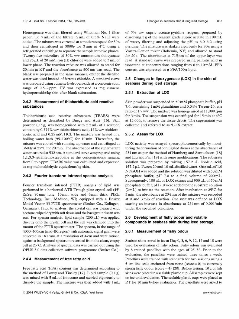
The changes in LOX activities in skin of seabass during icedstorage are depicted in Fig. 3. No changes in LOX activitywere observed within the first 6 days of iced storage(p> 0.05). After 9 days of storage, the marked increases inLOX activity were found up to 18 days of iced storage(p< 0.05). At Day 18, LOX activity increased by 193%,compared with that found at Day 0. The increase in LOXactivities in skin during iced storage might be associated with
5001000150020002500300035004000
Abs
orba
nce
Wavenumber (cm-1)
Day 18
Day 12
Day 6
Day 0
Figure 2. FTIR spectra of lipids from seabass skin during iced storage.
Eur. J. Lipid Sci. Technol. 2014, 116, 885–894 Changes in seabass skin during iced storage 891
� 2014 WILEY-VCH Verlag GmbH & Co. KGaA, Weinheim www.ejlst.com
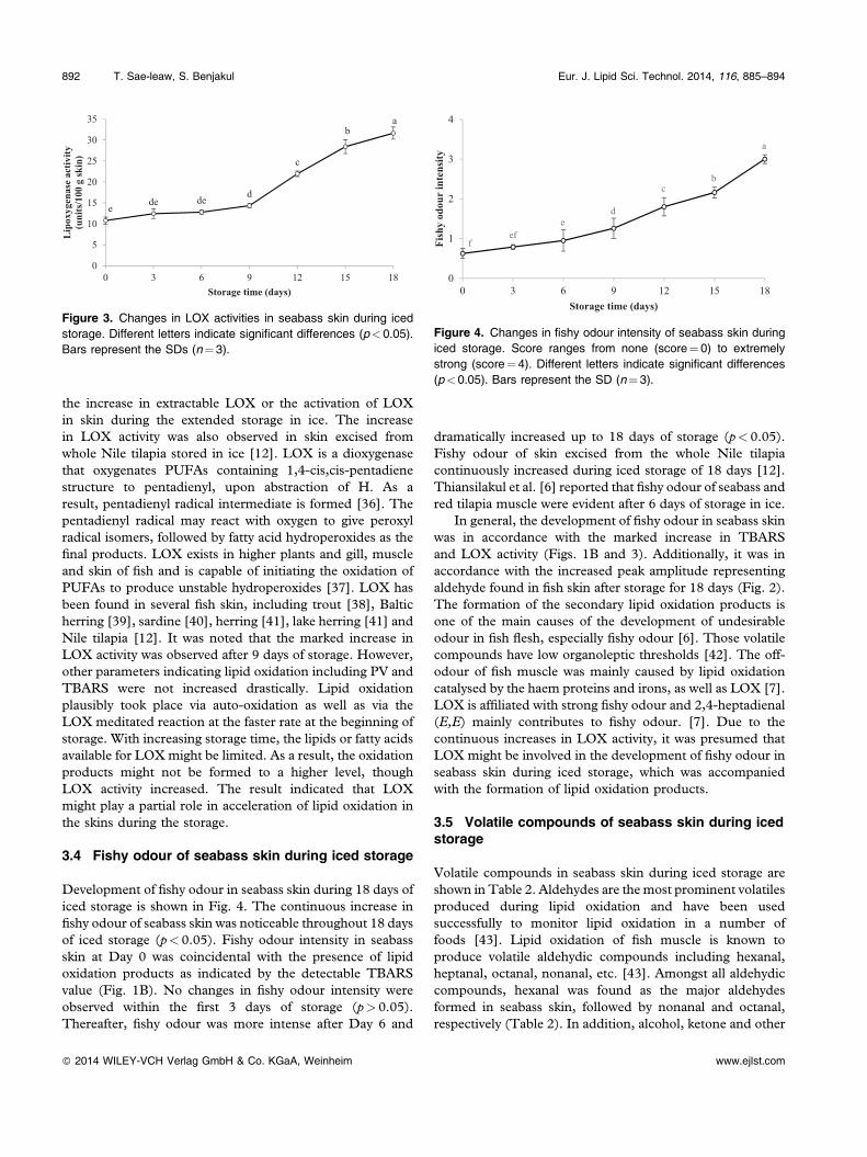
the increase in extractable LOX or the activation of LOXin skin during the extended storage in ice. The increasein LOX activity was also observed in skin excised fromwhole Nile tilapia stored in ice [12]. LOX is a dioxygenasethat oxygenates PUFAs containing 1,4‐cis,cis‐pentadienestructure to pentadienyl, upon abstraction of H. As aresult, pentadienyl radical intermediate is formed [36]. Thepentadienyl radical may react with oxygen to give peroxylradical isomers, followed by fatty acid hydroperoxides as thefinal products. LOX exists in higher plants and gill, muscleand skin of fish and is capable of initiating the oxidation ofPUFAs to produce unstable hydroperoxides [37]. LOX hasbeen found in several fish skin, including trout [38], Balticherring [39], sardine [40], herring [41], lake herring [41] andNile tilapia [12]. It was noted that the marked increase inLOX activity was observed after 9 days of storage. However,other parameters indicating lipid oxidation including PV andTBARS were not increased drastically. Lipid oxidationplausibly took place via auto‐oxidation as well as via theLOX meditated reaction at the faster rate at the beginning ofstorage. With increasing storage time, the lipids or fatty acidsavailable for LOXmight be limited. As a result, the oxidationproducts might not be formed to a higher level, thoughLOX activity increased. The result indicated that LOXmight play a partial role in acceleration of lipid oxidation inthe skins during the storage.
3.4 Fishy odour of seabass skin during iced storage
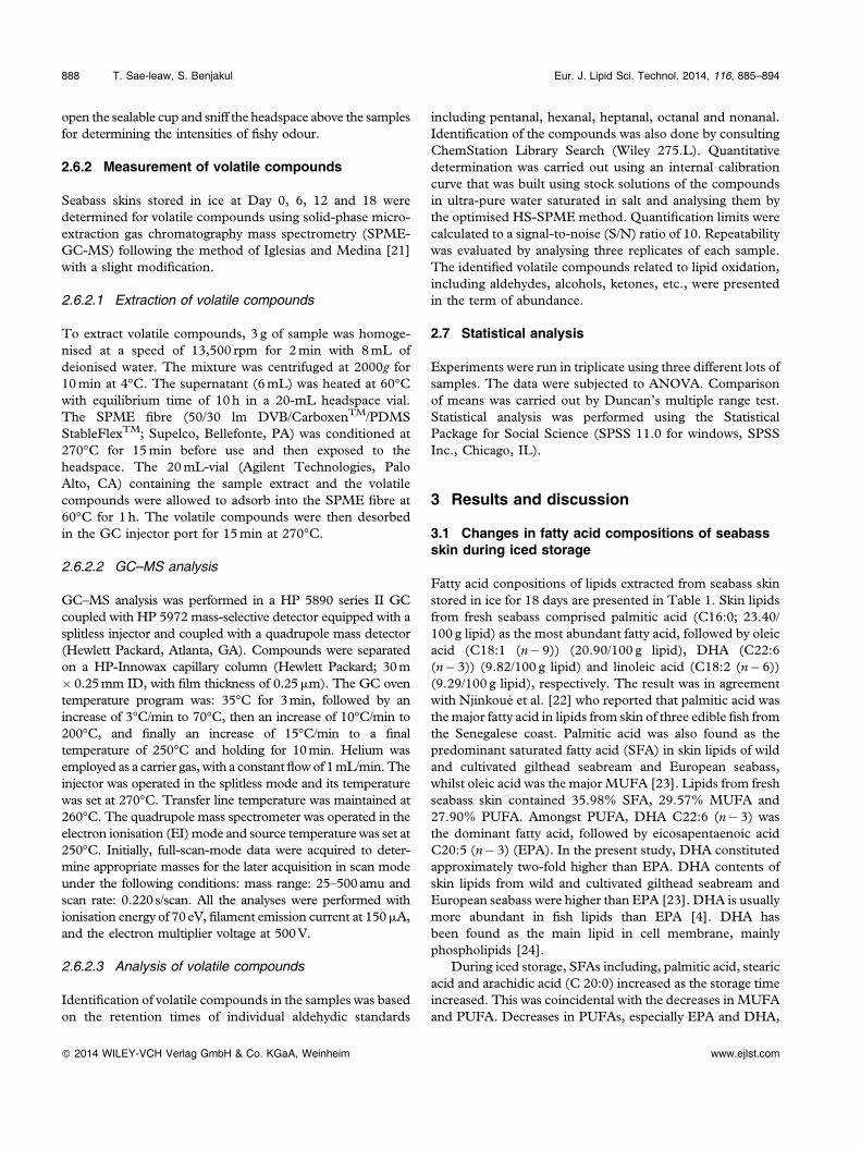
Development of fishy odour in seabass skin during 18 days oficed storage is shown in Fig. 4. The continuous increase infishy odour of seabass skin was noticeable throughout 18 daysof iced storage (p< 0.05). Fishy odour intensity in seabassskin at Day 0 was coincidental with the presence of lipidoxidation products as indicated by the detectable TBARSvalue (Fig. 1B). No changes in fishy odour intensity wereobserved within the first 3 days of storage (p> 0.05).Thereafter, fishy odour was more intense after Day 6 and
dramatically increased up to 18 days of storage (p< 0.05).Fishy odour of skin excised from the whole Nile tilapiacontinuously increased during iced storage of 18 days [12].Thiansilakul et al. [6] reported that fishy odour of seabass andred tilapia muscle were evident after 6 days of storage in ice.
In general, the development of fishy odour in seabass skinwas in accordance with the marked increase in TBARSand LOX activity (Figs. 1B and 3). Additionally, it was inaccordance with the increased peak amplitude representingaldehyde found in fish skin after storage for 18 days (Fig. 2).The formation of the secondary lipid oxidation products isone of the main causes of the development of undesirableodour in fish flesh, especially fishy odour [6]. Those volatilecompounds have low organoleptic thresholds [42]. The off‐odour of fish muscle was mainly caused by lipid oxidationcatalysed by the haem proteins and irons, as well as LOX [7].LOX is affiliated with strong fishy odour and 2,4‐heptadienal(E,E) mainly contributes to fishy odour. [7]. Due to thecontinuous increases in LOX activity, it was presumed thatLOX might be involved in the development of fishy odour inseabass skin during iced storage, which was accompaniedwith the formation of lipid oxidation products.
3.5 Volatile compounds of seabass skin during icedstorage
Volatile compounds in seabass skin during iced storage areshown in Table 2. Aldehydes are the most prominent volatilesproduced during lipid oxidation and have been usedsuccessfully to monitor lipid oxidation in a number offoods [43]. Lipid oxidation of fish muscle is known toproduce volatile aldehydic compounds including hexanal,heptanal, octanal, nonanal, etc. [43]. Amongst all aldehydiccompounds, hexanal was found as the major aldehydesformed in seabass skin, followed by nonanal and octanal,respectively (Table 2). In addition, alcohol, ketone and other
ede de d
c
ba
0
5
10
15
20
25
30
35
0 3 6 9 12 15 18
Lip
oxyg
enas
e ac
tivity
(u
nits
/100
g sk
in)
Storage time (days)
Figure 3. Changes in LOX activities in seabass skin during icedstorage. Different letters indicate significant differences (p<0.05).Bars represent the SDs (n¼3).
fef
ed
cb
a
0
1
2
3
4
0 3 6 9 12 15 18
Fish
y od
our
inte
nsity
Storage time (days)
Figure 4. Changes in fishy odour intensity of seabass skin duringiced storage. Score ranges from none (score¼ 0) to extremelystrong (score¼4). Different letters indicate significant differences(p<0.05). Bars represent the SD (n¼3).
892 T. Sae‐leaw, S. Benjakul Eur. J. Lipid Sci. Technol. 2014, 116, 885–894
� 2014 WILEY-VCH Verlag GmbH & Co. KGaA, Weinheim www.ejlst.com
volatile substances were also formed in seabass skin duringiced storage of 18 days. Fresh seabass skin contained thelower amounts of volatile compounds, when compared withthe skins stored in ice for a longer time. Higher formation ofvolatile lipid oxidation products in seabass skin correlatedwell with the fishy odour intensities and LOX activities(Figs. 3 and 4). Decomposition of hydroperoxides to thesecondary volatile oxidation products was more pronouncedin seabass skin stored in ice. Propanal and heptanal can serveas the reliable indicators of flavour deterioration for fishproducts, whilst hexanal contributes to the rancidity inmeats [43]. Iglesias and Medina [21] reported that propanal,1‐penten‐3‐ol, 2,3‐pentanedione, hexanal and 1‐octen‐3‐olwere the main volatile compounds formed during thestorage of Atlantic horse mackerel muscle at 4°C. Hexanal,heptanal and 1‐octen‐3‐ol are generated from n� 6 PUFAoxidation [21].
Alcohols such as 1‐octen‐3‐ol, 1,5‐octadien‐3‐ol, 2,6‐dimethyl‐7‐octen‐2‐ol increased throughout 18 days oficed storage. Alcohols are known as the secondary productsproduced by the decomposition of hydroperoxides of fattyacids [44]. 1‐Octen‐3‐ol is an important contributor to off‐flavour due to its low odour threshold and it is reported to beformed from oxidation of arachidonic acid by 12‐LOX [45].Hexanal and 1‐octen‐3‐ol, the major volatile compounds,contributed to the strong intensities of fishy and rancid off‐odours in washed Asian seabass mince containing myoglo-bin [46]. Varlet et al. [47] reported that carbonyl compoundsinvolving 4‐heptenal, octanal, decanal, and 2,4‐decadienalwere responsible for fishy odour in salmon flesh (Salmosalar).Carbonyl compounds, which are produced from oxidation ofPUFA by LOX or by autoxidation, might contribute to fishyodour/flavour [48]. Ketone, especially 3‐undecen‐2‐one,increased with increasing storage time. Since seabass skincontains high levels of PUFA and LOX, carbonyl compoundscould be easily generated via oxidation process. The fishyvolatiles identified in boiled sardines were dimethyl sulfide,acetaldehyde, propionaldehyde, butyraldehyde, 2‐ethylfuran,valeraldehyde, 2,3‐pentanedione, hexanal, and 1‐penten‐3‐ol[49]. The development of volatile compounds coincidentallyoccurred with the enhanced lipid oxidation. The subsequentdecomposition of primary lipid oxidation products led to theformation of several volatile compounds, which more likelycontributed to unacceptable offensive fishy odour in the skinof seabass prior to processing.
4 Conclusion
During iced storage, seabass skin underwent lipid oxidationwith the coincidental release or activation of LOX. Develop-ment of fishy odour took place along with the formation ofvolatile compounds. Therefore, skin from fresh fish should beused and the delay of further processing should be avoided toprevent undesirable fishy odour in skin and its products.
This research was supported by Prince of Songkla University andthe Grant‐in‐Aid for dissertation from Graduate School, Prince ofSongkla University, Thailand. The TRF senior research scholarprogram was also acknowledged for the financial support.
The authors have declared no conflict of interest.
References
[1] Sikorski, Z. E., Pan, B. S., in: Shahidi, F., Botta, J. R. (Eds.),Seafoods: Chemistry, Processing Technology and Quality, BlackieAcademic & Professional, London 1994, pp. 168–195.
[2] Pacheco‐Aguilar, R., Lugo‐Sánchez, M. E., Robles‐Bur-gueño, M. R., Postmortem biochemical and functionalcharacteristic of Monterey sardine muscle stored at 0°C. J.Food Sci. 2000, 65, 40–47.
[3] Toyomizu, M., Hanaoka, K., Yamaguchi, K., Effect ofrelease of free fatty acids by enzymatic hydrolysis ofphospholipids on lipid oxidation during storage of fishmuscle at –5°C. Bull. Jpn. Soc. Sci. Fish. 1981, 47, 615–620.
[4] Kolakowska, A., Olley, J., Dunstan, G. A., in: Sikorski, Z. E.,Kolakowska, A. (Eds.), Chemical and Functional Properties ofFood Lipids, CRC Press, New York 2002, pp. 221–264.
[5] Maqsood, S., Benjakul, S., Retardation of haemoglobin‐mediated lipid oxidation of Asian sea bass muscle by tannicacid during iced storage. Food Chem. 2011, 124, 1056–1062.
[6] Thiansilakul, Y., Benjakul, S., Richards, M. P., Changes inheme proteins and lipids associated with off‐odour of seabass(Lates calcarifer) and red tilapia (Oreochromis mossambicus �O. niloticus) during iced storage. Food Chem. 2010, 121,1109–1119.
[7] Fu, X., Xu, S., Wang, Z., Kinetics of lipid oxidation and off‐odor formation in silver carp mince: The effect of lip-oxygenase and hemoglobin. Food Res. Int. 2009, 42, 85–90.
[8] Yarnpakdee, S., Benjakul, S., Nalinanon, S., Kristinsson,H. G., Lipid oxidation and fishy odour development inprotein hydrolysate from Nile tilapia (Oreochromis niloticus)muscle as affected by freshness and antioxidants. Food Chem.2012, 132, 1781–1788.
[9] Anand, S., Kamath, S., Chuang, L., Kasapis, S., et al.,Biochemical and thermo‐mechanical analysis of collagen fromthe skin of Asian sea bass (Lates calcarifer) and Australasiansnapper (Pagrus auratus), an alternative for mammaliancollagen. Eur. Food Res. Technol. 2013, 236, 873–882.
[10] Nagai, T., Suzuki, N., Isolation of collagen from fish wastematerial — skin, bone and fins. Food Chem. 2000, 68, 277–281.
[11] Sinthusamran, S., Benjakul, S., Kishimura, H., Comparativestudy on molecular characteristics of acid soluble collagensfrom skin and swim bladder of seabass (Lates calcarifer). FoodChem. 2013, 138, 2435–2441.
[12] Sae‐leaw, T., Benjakul, S., Gokoglu, N., Nalinanon, S.,Changes in lipids and fishy odour development in skin fromNile tilapia (Oreochromis niloticus) stored in ice. Food Chem.2013, 141, 2466–2472.
[13] Bligh, E. G., Dyer, W. J., A rapid method of total lipidextraction and purification. Can. J. Biochem. Phys. 1959, 37,911–917.
[14] AOAC, Official methods of analysis, Association of OfficialAnalytical Chemists, Washington, DC 2000.
Eur. J. Lipid Sci. Technol. 2014, 116, 885–894 Changes in seabass skin during iced storage 893
� 2014 WILEY-VCH Verlag GmbH & Co. KGaA, Weinheim www.ejlst.com
[15] Richards, M. P., Hultin, H. O., Contribution of blood andblood components to lipid oxidation in fish muscle. J. Agric.Food Chem. 2002, 50, 555–564.
[16] Buege, J. A., Aust, S. D., Microsomal lipid peroxidation.Methods Enzymol. 1978, 52, 302–310.
[17] Lowry, R., Tinsley, I., Rapid colorimetric determination offree fatty acids. J. Am. Oil Chem. Soc. 1976, 53, 470–472.
[18] Hamberg, M., Samuelsson, B., On the specificity of theoxygenation of unsaturated fatty acids catalyzed by soybeanlipoxidase. J. Biol. Chem. 1967, 242, 5329–5335.
[19] Liu, Y. J., Pan, B. S., Inhibition of fish gill lipoxygenase andblood thinning effects of green tea extract. J. Agric. FoodChem. 2004, 52, 4860–4864.
[20] Sohn, J.‐H., Taki, Y., Ushio, H., Kohata, T, et al., Lipidoxidations in ordinary and dark muscles of fish: Influenceson rancid off‐odor development and color darkening ofyellowtail flesh during ice storage. J. Food Sci. 2005, 70,S490–S496.
[21] Iglesias, J., Medina, I., Solid‐phase microextraction methodfor the determination of volatile compounds associated tooxidation of fish muscle. J. Chromatogr. A. 2008, 1192, 9–16.
[22] Njinkoué, J.‐M., Barnathan, G., Miralles, J., Gaydou, E.‐M.,et al., Lipids and fatty acids in muscle, liver and skin ofthree edible fish from the Senegalese coast: Sardinellamaderensis, Sardinella aurita and Cephalopholis taeniops. Comp.Biochem. Physiol., Part B: Biochem.Mol. Biol. 2002, 131, 395–402.
[23] Sa�gglık, S., Alpaslan, M., Gezgin, T., Çetintürkc, K., et al.,Fatty acid composition of wild and cultivated giltheadseabream (Sparus aurata) and sea bass (Dicentrarchus labrax).Eur. J. Lipid Sci. Technol. 2003, 105, 104–107.
[24] Erickson, M. C., in: Akoh, C. C., Min, D. B. (Eds.), FoodLipids: Chemistry, Nutrition, and Biotechnology, CRC Press,Boca Raton 2008, pp. 39–62.
[25] Wang, T., Hammond, E. G., in: Decker, E. A., Elias, R. J.,McClements, D. J. (Eds.), Oxidation in Foods and Beveragesand Antioxidant Applications, Woodhead Publishing Limited,Cambridge 2010, pp. 105–121.
[26] Boselli, E., Caboni, M. F., Rodriguez‐Estrada, M. T.,Toschi, T. G., et al., Photoxidation of cholesterol and lipidsof turkey meat during storage under commercial retailconditions. Food Chem. 2005, 91, 705–713.
[27] Nawar, W.W., in: Fennema, O.R. (Ed.), Food Chem, MarcelDekker, New York 1996, pp. 225–319.
[28] Jacobsen, C., in: Decker, E. A., Elias, R. J., McClements,D. J. (Eds.),Oxidation in Foods and Beverages and AntioxidantApplications, Woodhead Publishing Limited, Cambridge2010, pp. 122–142.
[29] Hwang, K. T., Regenstein, J. M., Characteristics of mackerelmince lipid hydrolysis. J. Food Sci. 1993, 58, 79–83.
[30] Nayak, J., Viswanathan Nair, P. G., Ammu, K., Mathew, S.,Lipase activity in different tissues of four species of fish: Rohu(Labeo rohita Hamilton), oil sardine (Sardinella longicepsLinnaeus), mullet (Liza subviridis Valenciennes) and Indianmackerel (Rastrelliger kanagurta Cuvier). J. Sci. Food Agric.2003, 83, 1139–1142.
[31] Audley, M. A., Shetty, K. J., Kinsella, J. E., Isolation andproperties of phospholipase A from pollock muscle. J. FoodSci. 1978, 43, 1771–1775.
[32] Guillén, M. D., Cabo, N., Infrared spectroscopy in the studyof edible oils and fats. J. Sci. Food Agric. 1997, 75, 1–11.
[33] Van de Voort, F. R., Ismail, A. A., Sedman, J., Dubois, J.,et al., The determination of peroxide value by fouriertransform infrared spectroscopy. J. Am. Oil Chem. Soc. 1994,71, 921–926.
[34] Guillén, M. D., Cabo, N., Study of the effects of smokeflavourings on the oxidative stability of the lipids of porkadipose tissue by means of Fourier transform infraredspectroscopy. Meat Sci. 2004, 66, 647–657.
[35] Guillén, M. D., Cabo, N., Characterization of edible oils andlard by fourier transform infrared spectroscopy. Relation-ships between composition and frequency of concrete bandsin the fingerprint region. J. Am. Oil Chem. Soc. 1997, 74,1281–1286.
[36] Kanner, J., Rosenthal, I., An assessment of lipid oxidation infoods. Pure Appl. Chem. 1992, 64, 1959–1964.
[37] Hsieh, R. J., German, J. B., Kinsella, J. E., Lipoxygenase infish tissue: Some properties of the 12‐lipoxygenase from troutgill. J. Agric. Food Chem. 1988, 36, 680–685.
[38] German, J. B., Chen, S. E., Kinsella, J. E., Lipid oxidation infish tissue. Enzymic initiation via lipoxygenase. J. Agric. FoodChem. 1985, 33, 680–683.
[39] Stodolnik, L., Samson, E., Lipoxygenase activity of selectedtissues and organs of Baltic herring. Acta Ichthyol. Piscat.2000, 30, 47–57.
[40] Mohri, S., Cho, S.‐Y., Endo, Y., Fujimoto, K., Lipoxygenaseactivity in sardine skin. Agric. Biol. Chem. 1990, 54, 1889–1891.
[41] Medina, I., Saeed, S., Howell, N., Enzymatic oxidativeactivity in sardine (Sardina pilchardus) and herring (Clupeaharengus) during chilling and correlation with quality. Eur.Food Res. Technol. 1999, 210, 34–38.
[42] McGill, A. S., Hardy, R., Gunstone, F. D., Further analysisof the volatile components of frozen cold stored cod and theinfluence of these on flavour. J. Sci. Food Agric. 1977, 28,200–205.
[43] Ross, C. F., Smith, D. M., Use of volatiles as indicators oflipid oxidation in muscle foods. Compr. Rev. Food Sci. F.2006, 5, 18–25.
[44] Girard, B., Durance, T., Headspace volatiles of sockeye andpink salmon as affected by retort process. J. Food Sci. 2000,65, 34–39.
[45] Hsieh, R. J., Kinsella, J. E., Lipoxygenase generation ofspecific volatile flavor carbonyl compounds in fish tissues. J.Agric. Food Chem. 1989, 37, 279–286.
[46] Thiansilakul, Y., Benjakul, S., Richards, M. P., Effect ofmyoglobin from Eastern little tuna muscle on lipid oxidationof washed Asian seabass mince at different pH conditions.J. Food Sci. 2011, 76, C242–C249.
[47] Varlet, V., Knockaert, C., Prost, C., Serot, T., Comparisonof odor‐active volatile compounds of fresh and smokedsalmon. J. Agric. Food Chem. 2006, 54, 3391–3401.
[48] Josephson, D. B., Lindsay, R. C., Stuiber, D. A., Biogenesisof lipid‐derived volatile aroma compounds in the emeraldshiner (Notropis atherinoides). J. Agric. Food Chem. 1984, 32,1347–1352.
[49] Kasahara, K., Osawa, C., Combination effects of spices onmasking of odor in boiled sardine. Fish Sci. 1998, 64, 415–418.
894 T. Sae‐leaw, S. Benjakul Eur. J. Lipid Sci. Technol. 2014, 116, 885–894
� 2014 WILEY-VCH Verlag GmbH & Co. KGaA, Weinheim www.ejlst.com


