Embed Size (px)
Citation preview
Free Radical Biology & Medicine 50 (2011) 179–195
Contents lists available at ScienceDirect
Free Radical Biology & Medicine
j ourna l homepage: www.e lsev ie r.com/ locate / f reeradb iomed
Original Contribution
Fatty acid amide hydrolase is a key regulator of endocannabinoid-inducedmyocardial tissue injury
Partha Mukhopadhyay a,1, Bėla Horváth a,e,1, Mohanraj Rajesh a, Shingo Matsumoto b, Keita Saito b,Sándor Bátkai a, Vivek Patel a, Galin Tanchian a, Rachel Y. Gao a, Benjamin F. Cravatt c,György Haskó d, Pál Pacher a,⁎a Laboratory of Physiologic Studies, National Institute on Alcohol Abuse and Alcoholism, National Cancer Institute, National Institutes of Health, Bethesda, MD 20892, USAb Radiation Biology Branch, National Cancer Institute, National Institutes of Health, Bethesda, MD 20892, USAc The Skaggs Institute for Chemical Biology and Department of Cell Biology, The Scripps Research Institute, La Jolla, CA 92037, USAd Department of Surgery, University of Medicine and Dentistry of New Jersey–New Jersey Medical School, Newark, NJ 07103, USAe Institute of Human Physiology and Clinical Experimental Research, Semmelweis University, Budapest, Hungary
⁎ Corresponding author. Fax: +1 301 480 0257.E-mail address: [email protected] (P. Pacher).
1 These authors contributed equally to this work.
0891-5849/$ – see front matter. Published by Elsevierdoi:10.1016/j.freeradbiomed.2010.11.002
a b s t r a c t
a r t i c l e i n f oArticle history:Received 28 May 2010Revised 28 October 2010Accepted 1 November 2010Available online 9 November 2010
Keywords:CardiomyopathyEndocannabinoidsCannabinoid receptorsCell deathOxidative stressNitrosative stressFree radicals
Previous studies have suggested that increased levels of endocannabinoids in various cardiovascular disorders(e.g., various forms of shock, cardiomyopathies, atherosclerosis) through the activation of CB1 cannabinoidreceptors may promote cardiovascular dysfunction and tissue injury. We have investigated the role of themain endocannabinoid anandamide-metabolizing enzyme (fatty acid amide hydrolase; FAAH) in myocardialinjury induced by an important chemotherapeutic drug, doxorubicin (DOX; known for its cardiotoxicitymediated by increased reactive oxygen and nitrogen species generation), using well-established acute andchronic cardiomyopathy models in mice. The DOX-induced myocardial oxidative/nitrative stress (increased4-hydroxynonenal, protein carbonyl, and nitrotyrosine levels and decreased glutathione content) correlatedwith multiple cell death markers, which were enhanced in FAAH knockout mice exhibiting significantlyincreased DOX-induced mortality and cardiac dysfunction compared to their wild type. The effects of DOX inFAAH knockouts were attenuated by CB1 receptor antagonists. Furthermore, anandamide induced enhancedcell death in human cardiomyocytes pretreated with FAAH inhibitor and enhanced sensitivity to ROSgeneration in inflammatory cells of FAAH knockouts. These results suggest that in pathological conditionsassociated with acute oxidative/nitrative stress FAAH plays a key role in controlling the tissue injury that is, atleast in part, mediated by the activation of CB1 receptors by endocannabinoids.
Inc.
Published by Elsevier Inc.
Endocannabinoids are part of a novel bioactive lipid signaling systemin both the central nervous system and various peripheral organs [1,2].Increasing recent evidence implicates dysregulation of the endocanna-binoid system in the pathogenesis of various cardiovascular diseases,ranging from myocardial infarction, shock, and cardiomyopathy/heartfailure to cardiovascular complications of liver cirrhosis and atheroscle-rosis [3]. These preclinical reports demonstrated increased plasma,circulating inflammatory cell, and/or myocardial endocannabinoidlevels in these pathologies and implicated activation of the cannabi-noid-1 (CB1) receptor by endocannabinoids in the pathogenesis ofcardiovascular dysfunction and/or disease progression [4,5]. In thesestudies, CB1 antagonists improved the hemodynamic alterations [2,5].Furthermore, CB1 receptor activation by endocannabinoid anandamideor synthetic ligands in primary human endothelial cells [6], cardiomyo-cytes [7], and macrophages [8] promotes increased reactive oxygen
species (ROS) generation and cell death, thereby contributing to tissueinjury. In models of atherosclerosis pharmacological inhibition orgenetic deletion of CB1 attenuates the vascular inflammation andinterrelated disease progression and also decreases smooth muscleproliferation [9–11].
In contrast, activation of CB2 receptors by endocannabinoids orsynthetic ligands on inflammatory cells may limit the inflammatoryresponse and ROS generation by reducing migration and/or activationof these cells in various models [3,12–14]. Dysregulation of theendocannabinoid system (e.g., tonic activation of CB1 receptors byendocannabinoids) has also been implicated in the development ofvarious cardiovascular risk factors in obesity/metabolic syndrome anddiabetes (e.g., plasma lipid alterations, hepatic steatosis, abdominalobesity, inflammation) in humans [4,15,16].
Fatty acid amide hydrolase (FAAH), the enzyme responsible for thedegradation of themain endocannabinoid anandamide and related fattyacid amides in vivo [17], has emerged as a target for modulatingendocannabinoid signaling [18], with a therapeutic potential in anxiety,pain, and various inflammatory disorders [19,20]. However, genetic
180 P. Mukhopadhyay et al. / Free Radical Biology & Medicine 50 (2011) 179–195
deletion or pharmacological inhibition of FAAH may also promote ROSgeneration and liver injury under pathological conditions [21].
In this study using genetic deletion of FAAH, we aimed to explorethe role of this key endocannabinoid-metabolizing enzyme in thedevelopment of myocardial injury induced by an important chemo-therapeutic drug, doxorubicin (DOX), known for its cardiotoxicitymediated by increased reactive oxygen and nitrogen species gener-ation [22–25], utilizing well-established acute and chronic cardiomy-opathy models in mice [7,24,26–30] in which increased myocardialendocannabinoid levels and CB1 receptors were implicated in thedevelopment of cardiac dysfunction [7,28].
Materials and methods
Animals/drugs
Protocols involving the use of animals were approved by theInstitutional Animal Care and Use Committees and were performed inline with the National Institutes of Health Guidelines for the Care andUse of Laboratory Animals. FAAH knockout (FAAH−/−) mice werebackcrossed into the C57BL/6 J background as previously described[19,31]. The FAAH−/− or wild-type (FAAH+/+) mice were generatedfrom double-heterozygous breeding pairs and were genotyped(Transnetyx, Cordova, TN, USA) at 4–5 weeks. Seven- to 11-week-old male FAAH+/+ and FAAH−/− mice were acutely administered asingle high dose (20 mg/kg) of DOX–HCl (termed aDOX; SigmaChemicals, St. Louis, MO, USA) intraperitoneally (ip). Treatment withthe CB1 antagonists AM281 (Tocris, MI, USA) or SR141716 (rimona-bant; NIDA Drug Supply Program, SC, USA), 10 mg/kg ip, started 1.5 hbefore the DOX injection and continued every 24 h. After 3 (FAAH+/+
and FAAH−/− mice) or 5 (C57BL/6 J mice) days the animals wereeither subjected to hemodynamic measurements or sacrificed; ofthose, the hearts were excised and snap-frozen in liquid nitrogen forbiochemical measurements as previously described [25,28]. For thechronic model (termed cDOX), 8- to 12-week-old male FAAH+/+ orFAAH−/− mice were administered multiple lower doses of DOX(5 mg/kg/dose) ip at days 1, 8, 14, and 22. After 30 days, themiceweresacrificed and their hearts were excised and snap-frozen in liquidnitrogen for biochemical measurements as described [7].
Hemodynamic measurements in mice
Left-ventricular performance was analyzed in mice anesthetizedwith 2% isoflurane using a Millar pressure–volume conductancesystem (Millar Instruments, TX, USA) as described [28,32].
Myocardial 4-hydroxynonenal–protein adduct content
Lipid peroxides are unstable indicators of oxidative stress in cellsthat decompose to form more complex and reactive compounds suchas 4-hydroxynonenal (HNE), which has been shown to be capable ofbinding to proteins and forming stable adducts. There is also emergingrecent evidence implicating HNE in various key signaling processes[33–35]. HNE in the myocardial tissues was determined using a kit(Cell Biolabs, CA, USA). In brief, bovine serum albumin (BSA) ormyocardial tissue extracts (10 μg/ml) were adsorbed onto a 96-wellplate for 12 h at 4 °C. HNE adducts present in the sample or standardwere probed with anti-HNE antibody, followed by a horseradishperoxidase (HRP)-conjugated secondary antibody. The HNE–proteinadduct content in an unknown sample was determined by comparingwith a standard curve and expressed as fold change relative to vehicle.
Myocardial carbonyl content
Protein oxidation is the covalent modification of a protein inducedeither directly by reactive oxygen and nitrogen species or indirectly
by their reaction with secondary by-products. The most commonproducts of protein oxidation in biological samples are the proteincarbonyl derivatives of proline, arginine, lysine, and threonine. Thesederivatives are chemically stable, have been shown to increase in avariety of diseases and pathological processes, and serve as markers ofoxidative stress [36]. Carbonyl content in myocardial tissues wasdetermined using the OxiSelect Protein Carbonyl ELISA Kit (CellBiolabs). In brief, BSA standards or protein samples (10 μg/ml) wereadsorbed onto a 96-well plate for 2 h at 37 °C. The protein carbonylspresent in the sample or standard were derivatized to DNP hydrazoneand probed with an anti-DNP antibody, followed by an HRP-conjugated secondary antibody. The protein carbonyl content in theunknown sample was determined by comparing with a standardcurve that was prepared from predetermined reduced and oxidizedBSA standards and expressed as fold change relative to vehicle.
Myocardial nitrotyrosine (NT) determination
Nitrotyrosine formation was initially considered a specific markerof in vivo peroxynitrite generation, but now it is rather used as acollective index of protein nitration, because other pathways havealso been proposed to be involved in its formation (e.g., myeloper-oxidase under certain inflammatory conditions [37–40]). Myocardialnitrotyrosine content was determined by nitrotyrosine ELISA accord-ing to the protocol supplied with the kit (Hycult Biotechnology, CellSciences, Canton, MA, USA) as described [25].
Immunoprecipitation for nitrotyrosine detection
Equal amounts of 200 μg tissue homogenate from each samplewere incubated with 20 μg of NT affinity sorbent (NT antibody cross-linked to a protein A–agarose matrix; Cayman Chemicals, MI, USA)overnight at 4 °C in a rotating wheel. The NT affinity sorbent isdesigned for immune precipitation of nitrated proteins from biolog-ical samples. Immunoprecipitates were washed three times withphosphate-buffered saline containing 0.1% Triton X-100. Pellets weresuspended in 1× Laemmli buffer with dithiothreitol. SDS–PAGEanalysis was performed as described earlier, followed by silverstaining of protein gels [25].
Western blot analysis
Protein was extracted from myocardial tissue homogenates usingtissue protein extraction reagent containing protease inhibitorcocktail set III and phosphatase inhibitor cocktail set I (Calbiochem,EMD Biosciences, CA, USA). Protein content was determined using aBio-Rad DC protein assay kit. Fifty micrograms of protein was resolvedon 4–12% gradient Bis–Tris Criterion gels (Bio-Rad, Hercules, CA, USA)and transferred onto a nitrocellulose membrane (GE Biosciences, NY,USA). After being blocked, the blots were probed with the primaryantibodies (1:1000) at 4 °C overnight. After being washed, the blotswere incubated with HRP-conjugated secondary antibody for 1 h atroom temperature. After a final wash, the blots were incubated withSuperSignal West Pico chemiluminescence substrate reagent (PierceBiotechnology, Rockford, IL, USA) and developed using Kodak Biomaxfilm (PerkinElmer Life Sciences, Boston, MA, USA). To verify equalloading the blots were stripped and probed with β-actin antibody.Developed blots were scanned and the band intensities weredetermined using the Quantity One image analysis program (Bio-Rad), normalized to β-actin, and expressed as relative fold changecompared to the vehicle or control.
Antibodies were purchased as follows: cytochrome c (Cyt c), COX-IV (Cell Signaling Technologies, MA, USA) and β-actin (Chemicon, CA,USA).
181P. Mukhopadhyay et al. / Free Radical Biology & Medicine 50 (2011) 179–195
Determination of myocardial glutathione (GSH) content
Myocardial GSH content was determined using kits from Trevigen(Gaithersburg, MD, USA) according to the manufacturer's protocols,as previously reported [25].
Determination of myocardial caspase 3/7 and poly(ADP-ribose) polymerase(PARP) activities
Caspase 3/7 activity in the heart homogenates was determinedusing the fluorimetric based Apo-ONE homogeneous assay kit(Promega, Madison, WI, USA) as described [25]. Myocardial PARPactivity was assayed using a colorimetric kit according to themanufacturer's protocol (Trevigen) as described [25].
Myocardial terminal deoxynucleotidyl transferase-mediated nick-endlabeling (TUNEL) immunohistochemistry
Paraffin sections were dewaxed and in situ detection of apoptosisin the myocardial tissues was performed by TUNEL assay as per theinstructions provided with the kit (Roche Diagnostics, Indianapolis,IN, USA). The nuclei were labeled with Hoechst 33242 and the TUNEL-positive cells were observed using an IX81 fluorescence microscope(Olympus; using 40× objective).
Myocardial quantitative TUNEL assay
Quantitative TUNEL assay was determined using a DELFIA DNAfragmentation assay kit (PerkinElmer Life Sciences) and was per-formed in 96-well microplates (Pall Life Sciences, Ann Arbor, MI, USA)as described previously [28]. In brief, equal amounts of myocardialtotal lysates were adsorbed onto the plates, and after fixation, 50 μl ofreaction solution (0.01% Chaps buffer, 5.5 units TdT enzyme, 15 μMdTTP, 5 μM Bio-dUTP, and TdT buffer) was added and incubated at37 °C for 30 min. Then the wells were washed with the DELFIA platewash solution. Then europium-labeled streptavidin was diluted in theDELFIA assay buffer and incubated on the lysates for 1 h at roomtemperature. After a wash, 200 μl/well enhancement solution wasadded and the fluorescence was measured in a Victor plate reader(PerkinElmer Life Sciences).
Myocardial DNA fragmentation ELISA
The quantitative determinations of cytoplasmic histone-associatedDNA fragmentation (mono and oligonucleosomes) due to in vivo celldeath were made using an ELISA kit (Roche Diagnostics).
Measurement of myeloperoxidase activity
The activity of myeloperoxidase (a marker of neutrophil infiltra-tion) was measured using a homogeneous fluorimetric detection kit(Assay Designs, Plymouth Meeting, PA, USA) in myocardial tissueextracts according to the manufacturer's protocol.
Determination of myocardial ROS by electron paramagnetic resonance(EPR) spectrometry
Myocardial ROS levels were determined by EPR using the spinprobe 1-hydroxy-3-methoxycarbonyl-2,2,5,5-tetramethylpyrrolidine(CMH) obtained from Enzo Life Sciences (PlymouthMeeting, PA, USA)as described earlier [41,42]. The measurements were performed usinga Varian X-band EPR system (Walnut Creek, CA, USA) with thefollowing settings: center field 3364 G, field sweep 80 G, microwavepower 20 mW,modulation amplitude 2 G, fieldmodulation frequency100 kHz, microwave frequency 9.358 GHz, and time constant 64 ms.In brief, 10–15 mg of myocardial tissue from each sample was rinsed
thoroughly in 20 mM KHB buffer containing the iron chelator DETC(Diethyldithiocarbamate) (5 μM), to remove the contamination andto reduce the artifact. Then the samples were minced with finescissors and incubated in KHB buffer containing the spin probe CMH(200 μM) at 37 °C for 30 min, and then the reaction was arrested byplacing the samples in ice. Subsequently an aliquot (150 μl) from thesamples was loaded onto a capillary tube and ROS levels weredetermined using the EPR system as described above. As CMH canreact with superoxide, hydroxyl radical, and other reactive oxygenspecies, it is used as a universal ROS detection trap. In a subset ofexperiments tissue samples were preincubated with superoxidedismutase (Sigma Chemicals) at a concentration of 400 U/tube beforeCMH addition to dissect the contribution of superoxide.
Primary cell culture and flow cytometric cell death assay
Primary human cardiomyocytes were purchased from ScienCellResearch Laboratories (Carlsbad, CA, USA) and grown according to themanufacturer's recommended media and growth supplements. Celldeath was measured using Sytox green (FL1) and annexin V–APC(FL4) in a FACSCalibur flow cytometer (BD Bioscience, Franklin Lakes,NJ, USA) as described previously [7,32].
Flow cytometric determination of respiratory burst
For detection of superoxide production we used a dihydrorhoda-mine 123 (DHR; Invitrogen, Carlsbad, CA, USA) 30 mM stock indimethyl sulfoxide (Sigma Chemicals) at a final concentration of100 μM and a modified method of Rothe et al. [43]. Briefly, FAAH+/+
and FAAH−/− mice were deeply anesthetized with isoflurane, andblood was collected after decapitation. We used whole blood for ourassay as it has been shown that cell isolation procedures maythemselves cause priming and/or activation of neutrophil leukocytesand may affect cell function [44]. We diluted the obtained blood 1:1with Hanks’ balanced salt solution (HBSS; Mediatech, Herndon, VA,USA) and incubated it 5 min at 37 °C. Then the cells were incubatedwith DHR and catalase (1000 U/ml; Sigma Chemicals) in the dark at37 °C for 20 min. Thereafter, we placed the samples on ice and lysedthe erythrocytes with ACK lysing buffer (Quality Biological, Gaithers-burg, MD, USA), and the remaining leukocytes were washed threetimes with HBSS. Cells loaded with DHR were treated withanandamide (AEA) and phorbol ester (PMA) at the indicatedconcentrations for 30 min and DHR intensity were measured byflow cytometry (FACSCalibur; BD Bioscience) in the FL1 channel [45].Polymorphonuclear neutrophils were gated on the basis of theirforward scatter and side scatter properties. Kinetics of the sameexperiments were also carried out in parallel experiments in a plate-based assay using a fluorescence plate reader (Victor, PerkinElmer).
Statistical analysis
Results are expressed as means±SEM. Statistical significanceamong groups was determined by one-way ANOVA followed byNewman–Keuls post hoc analysis using GraphPad Prism 5 software(San Diego, CA, USA). Correlation analyses were carried out pair-wiseusing the same program. Kaplan–Meier survival analysis and curvefitting were also performed using GraphPad Prism 5 software.Probability values of Pb0.05 were considered significant.
Results
Effects of DOX treatment on body weight in FAAH+/+ and FAAH−/− mice
Because increased endocannabinoid levels may promote increasedfood intake, in some mice body weight was also measured before theDOX administration and at various time points after DOX
182 P. Mukhopadhyay et al. / Free Radical Biology & Medicine 50 (2011) 179–195
administration. There was no difference in the initial body weights ofthese 7- to 12-week-old male mice kept on regular diet. The bodyweights of FAAH+/+ and FAAH−/− mice before the acute DOXadministration and at a time of the sacrifice 3 days later were 20.7±0.3, 19.5±0.6, 17.2±0.3, and 16.9±0.6 (n=10/group), respectively.The body weights of FAAH+/+ and FAAH−/− mice before the chronicDOX administration and at a time of the sacrifice 30 days later were23.8±0.6, 23.4±1.0, 21.0±0.5, and 21.7±1.0 (n=11 or 12/group),respectively.
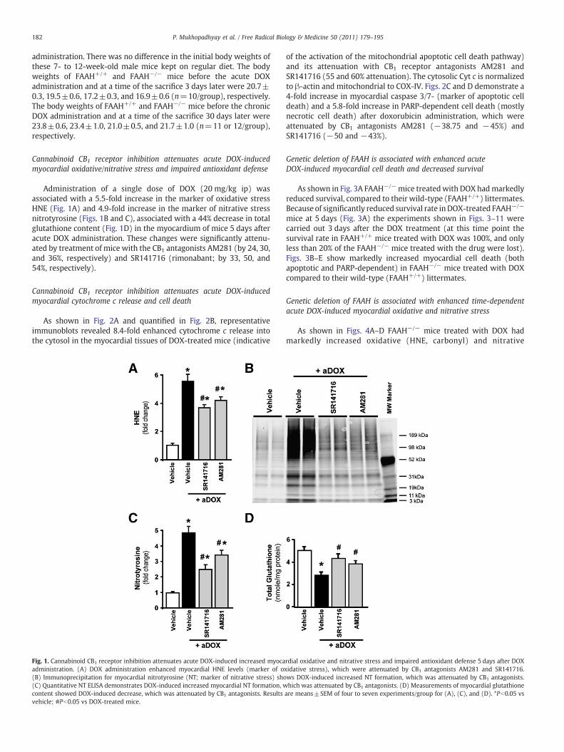
Cannabinoid CB1 receptor inhibition attenuates acute DOX-inducedmyocardial oxidative/nitrative stress and impaired antioxidant defense
Administration of a single dose of DOX (20 mg/kg ip) wasassociated with a 5.5-fold increase in the marker of oxidative stressHNE (Fig. 1A) and 4.9-fold increase in the marker of nitrative stressnitrotyrosine (Figs. 1B and C), associated with a 44% decrease in totalglutathione content (Fig. 1D) in the myocardium of mice 5 days afteracute DOX administration. These changes were significantly attenu-ated by treatment of mice with the CB1 antagonists AM281 (by 24, 30,and 36%, respectively) and SR141716 (rimonabant; by 33, 50, and54%, respectively).
Cannabinoid CB1 receptor inhibition attenuates acute DOX-inducedmyocardial cytochrome c release and cell death
As shown in Fig. 2A and quantified in Fig. 2B, representativeimmunoblots revealed 8.4-fold enhanced cytochrome c release intothe cytosol in the myocardial tissues of DOX-treated mice (indicative
Fig. 1. Cannabinoid CB1 receptor inhibition attenuates acute DOX-induced increased myocaadministration. (A) DOX administration enhanced myocardial HNE levels (marker of o(B) Immunoprecipitation for myocardial nitrotyrosine (NT; marker of nitrative stress) sho(C) Quantitative NT ELISA demonstrates DOX-induced increased myocardial NT formation, wcontent showed DOX-induced decrease, which was attenuated by CB1 antagonists. Resultsvehicle; #Pb0.05 vs DOX-treated mice.
of the activation of the mitochondrial apoptotic cell death pathway)and its attenuation with CB1 receptor antagonists AM281 andSR141716 (55 and 60% attenuation). The cytosolic Cyt c is normalizedto β-actin and mitochondrial to COX-IV. Figs. 2C and D demonstrate a4-fold increase in myocardial caspase 3/7- (marker of apoptotic celldeath) and a 5.8-fold increase in PARP-dependent cell death (mostlynecrotic cell death) after doxorubicin administration, which wereattenuated by CB1 antagonists AM281 (−38.75 and −45%) andSR141716 (−50 and −43%).
Genetic deletion of FAAH is associated with enhanced acuteDOX-induced myocardial cell death and decreased survival
As shown in Fig. 3A FAAH−/−mice treatedwith DOX hadmarkedlyreduced survival, compared to their wild-type (FAAH+/+) littermates.Because of significantly reduced survival rate inDOX-treated FAAH−/−
mice at 5 days (Fig. 3A) the experiments shown in Figs. 3–11 werecarried out 3 days after the DOX treatment (at this time point thesurvival rate in FAAH+/+ mice treated with DOX was 100%, and onlyless than 20% of the FAAH−/− mice treated with the drug were lost).Figs. 3B–E show markedly increased myocardial cell death (bothapoptotic and PARP-dependent) in FAAH−/− mice treated with DOXcompared to their wild-type (FAAH+/+) littermates.
Genetic deletion of FAAH is associated with enhanced time-dependentacute DOX-induced myocardial oxidative and nitrative stress
As shown in Figs. 4A–D FAAH−/− mice treated with DOX hadmarkedly increased oxidative (HNE, carbonyl) and nitrative
rdial oxidative and nitrative stress and impaired antioxidant defense 5 days after DOXxidative stress), which were attenuated by CB1 antagonists AM281 and SR141716.ws DOX-induced increased NT formation, which was attenuated by CB1 antagonists.hich was attenuated by CB1 antagonists. (D) Measurements of myocardial glutathione
are means±SEM of four to seven experiments/group for (A), (C), and (D). *Pb0.05 vs
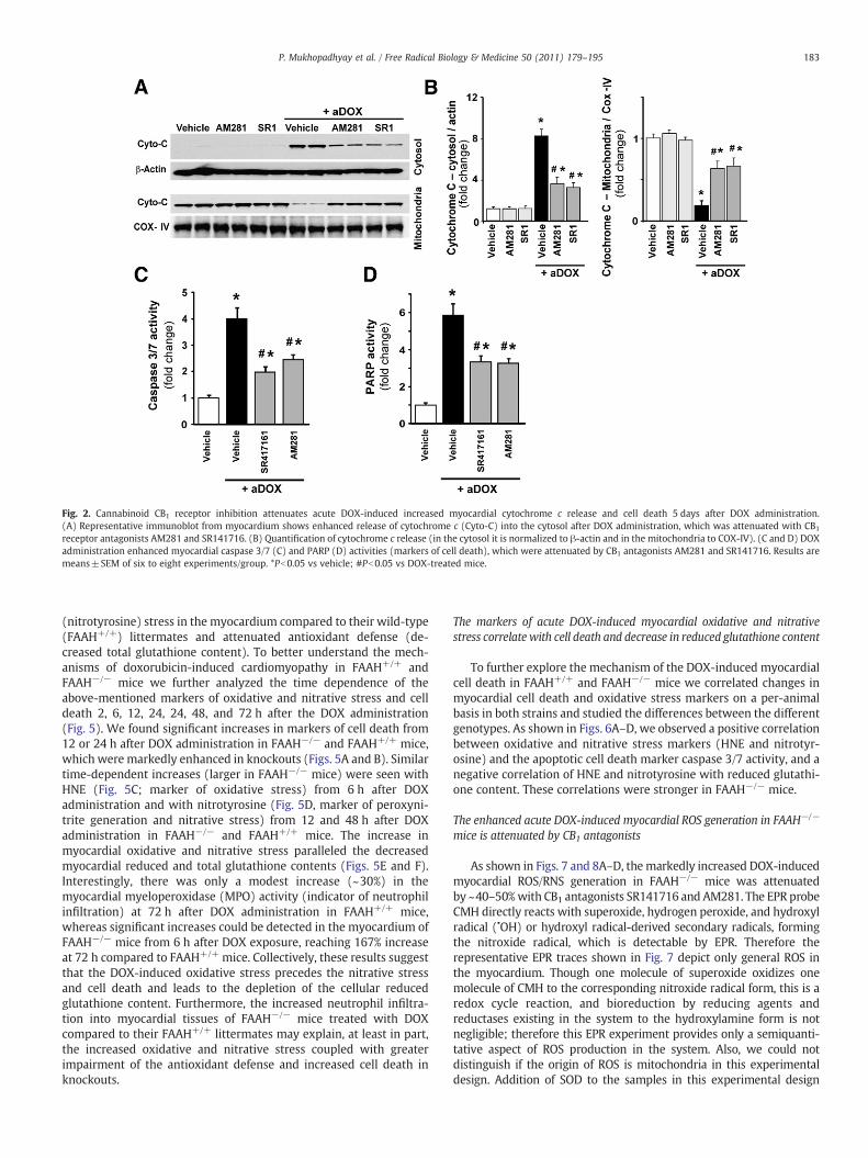
Fig. 2. Cannabinoid CB1 receptor inhibition attenuates acute DOX-induced increased myocardial cytochrome c release and cell death 5 days after DOX administration.(A) Representative immunoblot from myocardium shows enhanced release of cytochrome c (Cyto-C) into the cytosol after DOX administration, which was attenuated with CB1
receptor antagonists AM281 and SR141716. (B) Quantification of cytochrome c release (in the cytosol it is normalized to β-actin and in the mitochondria to COX-IV). (C and D) DOXadministration enhanced myocardial caspase 3/7 (C) and PARP (D) activities (markers of cell death), which were attenuated by CB1 antagonists AM281 and SR141716. Results aremeans±SEM of six to eight experiments/group. *Pb0.05 vs vehicle; #Pb0.05 vs DOX-treated mice.
183P. Mukhopadhyay et al. / Free Radical Biology & Medicine 50 (2011) 179–195
(nitrotyrosine) stress in the myocardium compared to their wild-type(FAAH+/+) littermates and attenuated antioxidant defense (de-creased total glutathione content). To better understand the mech-anisms of doxorubicin-induced cardiomyopathy in FAAH+/+ andFAAH−/− mice we further analyzed the time dependence of theabove-mentioned markers of oxidative and nitrative stress and celldeath 2, 6, 12, 24, 24, 48, and 72 h after the DOX administration(Fig. 5). We found significant increases in markers of cell death from12 or 24 h after DOX administration in FAAH−/− and FAAH+/+ mice,which were markedly enhanced in knockouts (Figs. 5A and B). Similartime-dependent increases (larger in FAAH−/− mice) were seen withHNE (Fig. 5C; marker of oxidative stress) from 6 h after DOXadministration and with nitrotyrosine (Fig. 5D, marker of peroxyni-trite generation and nitrative stress) from 12 and 48 h after DOXadministration in FAAH−/− and FAAH+/+ mice. The increase inmyocardial oxidative and nitrative stress paralleled the decreasedmyocardial reduced and total glutathione contents (Figs. 5E and F).Interestingly, there was only a modest increase (~30%) in themyocardial myeloperoxidase (MPO) activity (indicator of neutrophilinfiltration) at 72 h after DOX administration in FAAH+/+ mice,whereas significant increases could be detected in the myocardium ofFAAH−/− mice from 6 h after DOX exposure, reaching 167% increaseat 72 h compared to FAAH+/+ mice. Collectively, these results suggestthat the DOX-induced oxidative stress precedes the nitrative stressand cell death and leads to the depletion of the cellular reducedglutathione content. Furthermore, the increased neutrophil infiltra-tion into myocardial tissues of FAAH−/− mice treated with DOXcompared to their FAAH+/+ littermates may explain, at least in part,the increased oxidative and nitrative stress coupled with greaterimpairment of the antioxidant defense and increased cell death inknockouts.
The markers of acute DOX-induced myocardial oxidative and nitrativestress correlate with cell death and decrease in reduced glutathione content
To further explore the mechanism of the DOX-induced myocardialcell death in FAAH+/+ and FAAH−/− mice we correlated changes inmyocardial cell death and oxidative stress markers on a per-animalbasis in both strains and studied the differences between the differentgenotypes. As shown in Figs. 6A–D, we observed a positive correlationbetween oxidative and nitrative stress markers (HNE and nitrotyr-osine) and the apoptotic cell death marker caspase 3/7 activity, and anegative correlation of HNE and nitrotyrosine with reduced glutathi-one content. These correlations were stronger in FAAH−/− mice.
The enhanced acute DOX-induced myocardial ROS generation in FAAH−/−
mice is attenuated by CB1 antagonists
As shown in Figs. 7 and 8A–D, themarkedly increased DOX-inducedmyocardial ROS/RNS generation in FAAH−/− mice was attenuatedby ~40–50%with CB1 antagonists SR141716 and AM281. The EPR probeCMH directly reacts with superoxide, hydrogen peroxide, and hydroxylradical (•OH) or hydroxyl radical-derived secondary radicals, formingthe nitroxide radical, which is detectable by EPR. Therefore therepresentative EPR traces shown in Fig. 7 depict only general ROS inthe myocardium. Though one molecule of superoxide oxidizes onemolecule of CMH to the corresponding nitroxide radical form, this is aredox cycle reaction, and bioreduction by reducing agents andreductases existing in the system to the hydroxylamine form is notnegligible; therefore this EPR experiment provides only a semiquanti-tative aspect of ROS production in the system. Also, we could notdistinguish if the origin of ROS is mitochondria in this experimentaldesign. Addition of SOD to the samples in this experimental design
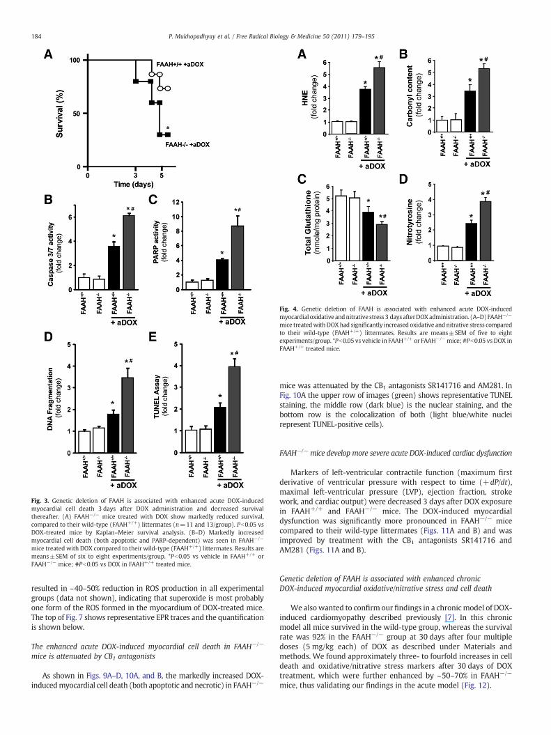
Fig. 3. Genetic deletion of FAAH is associated with enhanced acute DOX-inducedmyocardial cell death 3 days after DOX administration and decreased survivalthereafter. (A) FAAH−/− mice treated with DOX show markedly reduced survival,compared to their wild-type (FAAH+/+) littermates (n=11 and 13/group). Pb0.05 vsDOX-treated mice by Kaplan–Meier survival analysis. (B–D) Markedly increasedmyocardial cell death (both apoptotic and PARP-dependent) was seen in FAAH−/−
mice treated with DOX compared to their wild-type (FAAH+/+) littermates. Results aremeans±SEM of six to eight experiments/group. *Pb0.05 vs vehicle in FAAH+/+ orFAAH−/− mice; #Pb0.05 vs DOX in FAAH+/+ treated mice.
Fig. 4. Genetic deletion of FAAH is associated with enhanced acute DOX-inducedmyocardial oxidative andnitrative stress 3 days afterDOXadministration. (A–D)FAAH−/−
mice treatedwithDOXhad significantly increased oxidative andnitrative stress comparedto their wild-type (FAAH+/+) littermates. Results are means±SEM of five to eightexperiments/group. *Pb0.05 vs vehicle in FAAH+/+ or FAAH−/−mice; #Pb0.05 vs DOX inFAAH+/+ treated mice.
184 P. Mukhopadhyay et al. / Free Radical Biology & Medicine 50 (2011) 179–195
resulted in ~40–50% reduction in ROS production in all experimentalgroups (data not shown), indicating that superoxide is most probablyone form of the ROS formed in the myocardium of DOX-treated mice.The top of Fig. 7 shows representative EPR traces and the quantificationis shown below.
The enhanced acute DOX-induced myocardial cell death in FAAH−/−
mice is attenuated by CB1 antagonists
As shown in Figs. 9A–D, 10A, and B, the markedly increased DOX-inducedmyocardial cell death (both apoptotic and necrotic) in FAAH−/−
mice was attenuated by the CB1 antagonists SR141716 and AM281. InFig. 10A the upper row of images (green) shows representative TUNELstaining, the middle row (dark blue) is the nuclear staining, and thebottom row is the colocalization of both (light blue/white nucleirepresent TUNEL-positive cells).
FAAH−/− mice develop more severe acute DOX-induced cardiac dysfunction
Markers of left-ventricular contractile function (maximum firstderivative of ventricular pressure with respect to time (+dP/dt),maximal left-ventricular pressure (LVP), ejection fraction, strokework, and cardiac output) were decreased 3 days after DOX exposurein FAAH+/+ and FAAH−/− mice. The DOX-induced myocardialdysfunction was significantly more pronounced in FAAH−/− micecompared to their wild-type littermates (Figs. 11A and B) and wasimproved by treatment with the CB1 antagonists SR141716 andAM281 (Figs. 11A and B).
Genetic deletion of FAAH is associated with enhanced chronicDOX-induced myocardial oxidative/nitrative stress and cell death
We also wanted to confirm our findings in a chronicmodel of DOX-induced cardiomyopathy described previously [7]. In this chronicmodel all mice survived in the wild-type group, whereas the survivalrate was 92% in the FAAH−/− group at 30 days after four multipledoses (5 mg/kg each) of DOX as described under Materials andmethods. We found approximately three- to fourfold increases in celldeath and oxidative/nitrative stress markers after 30 days of DOXtreatment, which were further enhanced by ~50–70% in FAAH−/−
mice, thus validating our findings in the acute model (Fig. 12).
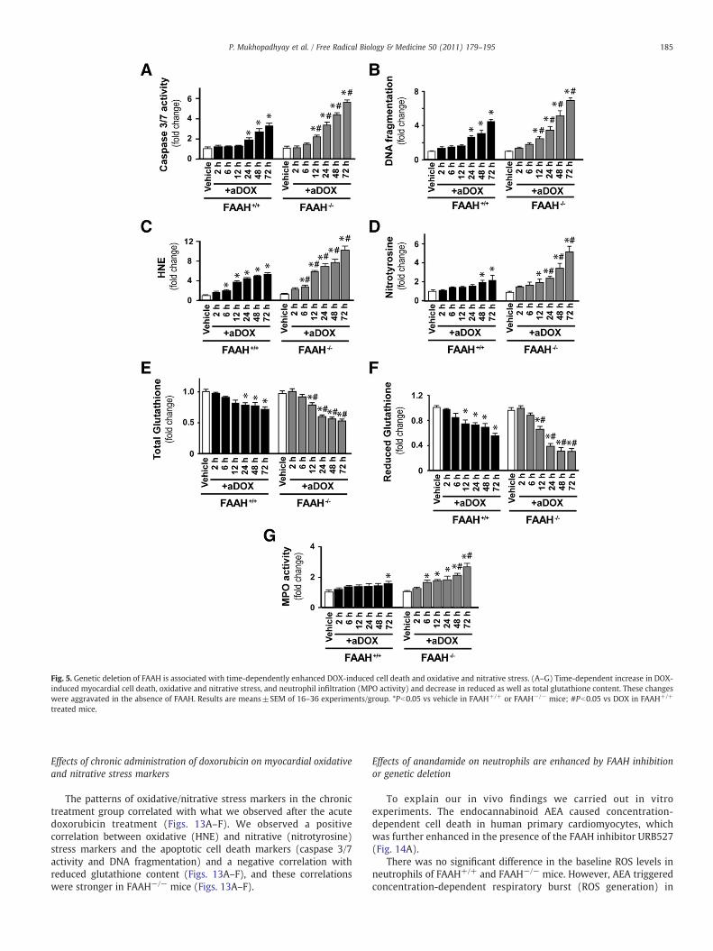
Fig. 5. Genetic deletion of FAAH is associated with time-dependently enhanced DOX-induced cell death and oxidative and nitrative stress. (A–G) Time-dependent increase in DOX-induced myocardial cell death, oxidative and nitrative stress, and neutrophil infiltration (MPO activity) and decrease in reduced as well as total glutathione content. These changeswere aggravated in the absence of FAAH. Results are means±SEM of 16–36 experiments/group. *Pb0.05 vs vehicle in FAAH+/+ or FAAH−/− mice; #Pb0.05 vs DOX in FAAH+/+
treated mice.
185P. Mukhopadhyay et al. / Free Radical Biology & Medicine 50 (2011) 179–195
Effects of chronic administration of doxorubicin on myocardial oxidativeand nitrative stress markers
The patterns of oxidative/nitrative stress markers in the chronictreatment group correlated with what we observed after the acutedoxorubicin treatment (Figs. 13A–F). We observed a positivecorrelation between oxidative (HNE) and nitrative (nitrotyrosine)stress markers and the apoptotic cell death markers (caspase 3/7activity and DNA fragmentation) and a negative correlation withreduced glutathione content (Figs. 13A–F), and these correlationswere stronger in FAAH−/− mice (Figs. 13A–F).
Effects of anandamide on neutrophils are enhanced by FAAH inhibitionor genetic deletion
To explain our in vivo findings we carried out in vitroexperiments. The endocannabinoid AEA caused concentration-dependent cell death in human primary cardiomyocytes, whichwas further enhanced in the presence of the FAAH inhibitor URB527(Fig. 14A).
There was no significant difference in the baseline ROS levels inneutrophils of FAAH+/+ and FAAH−/− mice. However, AEA triggeredconcentration-dependent respiratory burst (ROS generation) in
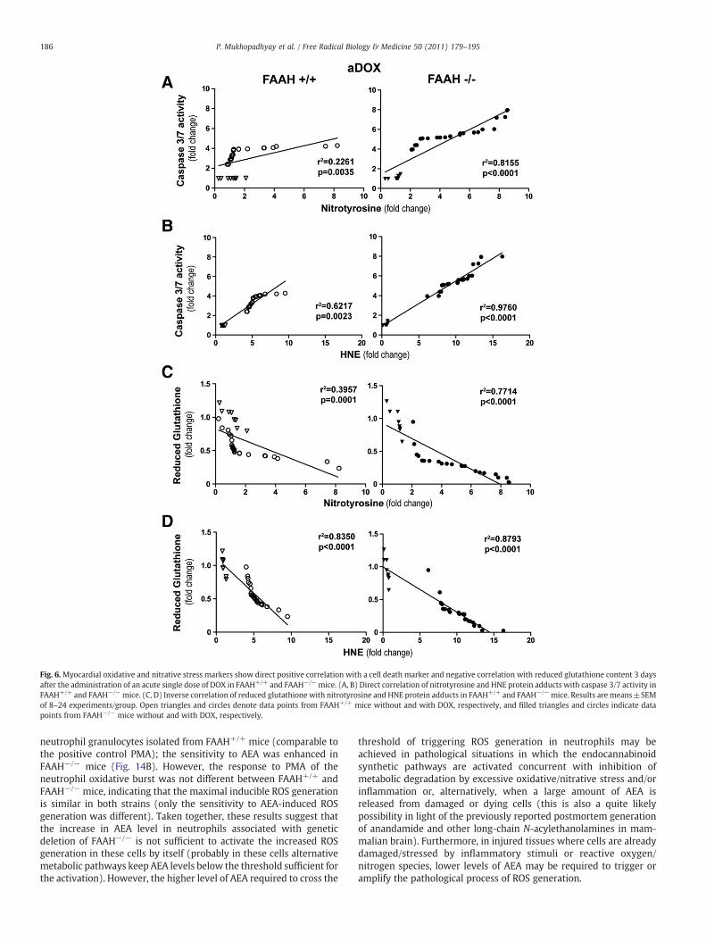
Fig. 6.Myocardial oxidative and nitrative stress markers show direct positive correlation with a cell death marker and negative correlation with reduced glutathione content 3 daysafter the administration of an acute single dose of DOX in FAAH+/+ and FAAH−/−mice. (A, B) Direct correlation of nitrotyrosine and HNE protein adducts with caspase 3/7 activity inFAAH+/+ and FAAH−/−mice. (C, D) Inverse correlation of reduced glutathione with nitrotyrosine and HNE protein adducts in FAAH+/+ and FAAH−/−mice. Results are means±SEMof 8–24 experiments/group. Open triangles and circles denote data points from FAAH+/+ mice without and with DOX, respectively, and filled triangles and circles indicate datapoints from FAAH−/− mice without and with DOX, respectively.
186 P. Mukhopadhyay et al. / Free Radical Biology & Medicine 50 (2011) 179–195
neutrophil granulocytes isolated from FAAH+/+ mice (comparable tothe positive control PMA); the sensitivity to AEA was enhanced inFAAH−/− mice (Fig. 14B). However, the response to PMA of theneutrophil oxidative burst was not different between FAAH+/+ andFAAH−/− mice, indicating that the maximal inducible ROS generationis similar in both strains (only the sensitivity to AEA-induced ROSgeneration was different). Taken together, these results suggest thatthe increase in AEA level in neutrophils associated with geneticdeletion of FAAH−/− is not sufficient to activate the increased ROSgeneration in these cells by itself (probably in these cells alternativemetabolic pathways keep AEA levels below the threshold sufficient forthe activation). However, the higher level of AEA required to cross the
threshold of triggering ROS generation in neutrophils may beachieved in pathological situations in which the endocannabinoidsynthetic pathways are activated concurrent with inhibition ofmetabolic degradation by excessive oxidative/nitrative stress and/orinflammation or, alternatively, when a large amount of AEA isreleased from damaged or dying cells (this is also a quite likelypossibility in light of the previously reported postmortem generationof anandamide and other long-chain N-acylethanolamines in mam-malian brain). Furthermore, in injured tissues where cells are alreadydamaged/stressed by inflammatory stimuli or reactive oxygen/nitrogen species, lower levels of AEA may be required to trigger oramplify the pathological process of ROS generation.
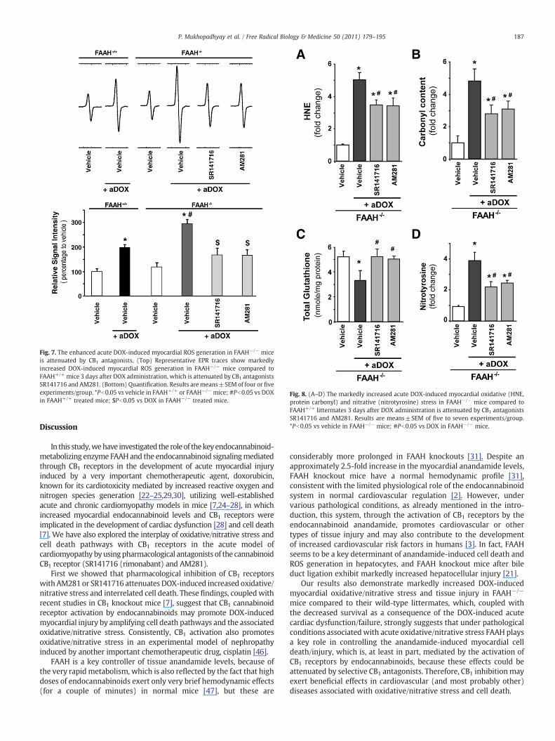
Fig. 7. The enhanced acute DOX-induced myocardial ROS generation in FAAH−/− miceis attenuated by CB1 antagonists. (Top) Representative EPR traces show markedlyincreased DOX-induced myocardial ROS generation in FAAH−/− mice compared toFAAH+/+ mice 3 days after DOX administration, which is attenuated by CB1 antagonistsSR141716 and AM281. (Bottom) Quantification. Results are means±SEM of four or fiveexperiments/group. *Pb0.05 vs vehicle in FAAH+/+ or FAAH−/− mice; #Pb0.05 vs DOXin FAAH+/+ treated mice; $Pb0.05 vs DOX in FAAH−/− treated mice.
Fig. 8. (A–D) The markedly increased acute DOX-induced myocardial oxidative (HNE,protein carbonyl) and nitrative (nitrotyrosine) stress in FAAH−/− mice compared toFAAH+/+ littermates 3 days after DOX administration is attenuated by CB1 antagonistsSR141716 and AM281. Results are means±SEM of five to seven experiments/group.*Pb0.05 vs vehicle in FAAH−/− mice; #Pb0.05 vs DOX in FAAH−/− mice.
187P. Mukhopadhyay et al. / Free Radical Biology & Medicine 50 (2011) 179–195
Discussion
In this study,wehave investigated the roleof thekeyendocannabinoid-metabolizing enzyme FAAHand the endocannabinoid signalingmediatedthrough CB1 receptors in the development of acute myocardial injuryinduced by a very important chemotherapeutic agent, doxorubicin,known for its cardiotoxicity mediated by increased reactive oxygen andnitrogen species generation [22–25,29,30], utilizing well-establishedacute and chronic cardiomyopathy models in mice [7,24–28], in whichincreased myocardial endocannabinoid levels and CB1 receptors wereimplicated in the development of cardiac dysfunction [28] and cell death[7]. We have also explored the interplay of oxidative/nitrative stress andcell death pathways with CB1 receptors in the acute model ofcardiomyopathy byusingpharmacological antagonists of the cannabinoidCB1 receptor (SR141716 (rimonabant) and AM281).
First we showed that pharmacological inhibition of CB1 receptorswith AM281 or SR141716 attenuates DOX-induced increased oxidative/nitrative stress and interrelated cell death. These findings, coupledwithrecent studies in CB1 knockout mice [7], suggest that CB1 cannabinoidreceptor activation by endocannabinoids may promote DOX-inducedmyocardial injury by amplifying cell death pathways and the associatedoxidative/nitrative stress. Consistently, CB1 activation also promotesoxidative/nitrative stress in an experimental model of nephropathyinduced by another important chemotherapeutic drug, cisplatin [46].
FAAH is a key controller of tissue anandamide levels, because ofthe very rapidmetabolism, which is also reflected by the fact that highdoses of endocannabinoids exert only very brief hemodynamic effects(for a couple of minutes) in normal mice [47], but these are
considerably more prolonged in FAAH knockouts [31]. Despite anapproximately 2.5-fold increase in the myocardial anandamide levels,FAAH knockout mice have a normal hemodynamic profile [31],consistent with the limited physiological role of the endocannabinoidsystem in normal cardiovascular regulation [2]. However, undervarious pathological conditions, as already mentioned in the intro-duction, this system, through the activation of CB1 receptors by theendocannabinoid anandamide, promotes cardiovascular or othertypes of tissue injury and may also contribute to the developmentof increased cardiovascular risk factors in humans [3]. In fact, FAAHseems to be a key determinant of anandamide-induced cell death andROS generation in hepatocytes, and FAAH knockout mice after bileduct ligation exhibit markedly increased hepatocellular injury [21].
Our results also demonstrate markedly increased DOX-inducedmyocardial oxidative/nitrative stress and tissue injury in FAAH−/−
mice compared to their wild-type littermates, which, coupled withthe decreased survival as a consequence of the DOX-induced acutecardiac dysfunction/failure, strongly suggests that under pathologicalconditions associated with acute oxidative/nitrative stress FAAH playsa key role in controlling the anandamide-induced myocardial celldeath/injury, which is, at least in part, mediated by the activation ofCB1 receptors by endocannabinoids, because these effects could beattenuated by selective CB1 antagonists. Therefore, CB1 inhibitionmayexert beneficial effects in cardiovascular (and most probably other)diseases associated with oxidative/nitrative stress and cell death.
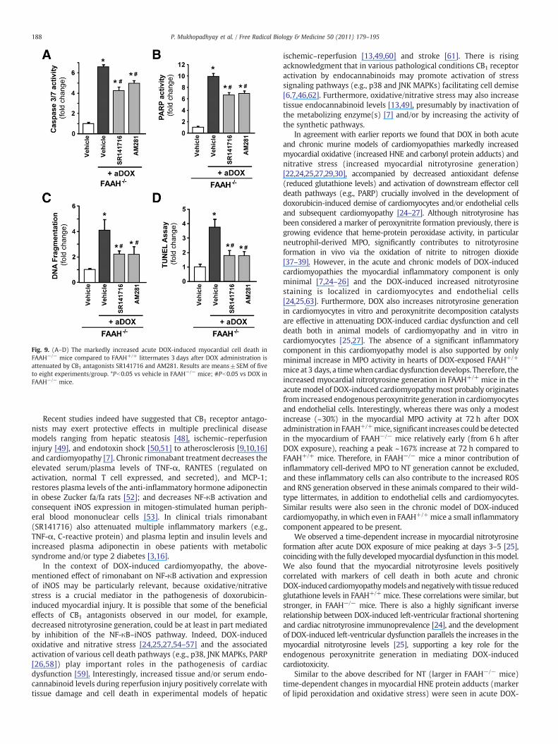
Fig. 9. (A–D) The markedly increased acute DOX-induced myocardial cell death inFAAH−/− mice compared to FAAH+/+ littermates 3 days after DOX administration isattenuated by CB1 antagonists SR141716 and AM281. Results are means±SEM of fiveto eight experiments/group. *Pb0.05 vs vehicle in FAAH−/− mice; #Pb0.05 vs DOX inFAAH−/− mice.
188 P. Mukhopadhyay et al. / Free Radical Biology & Medicine 50 (2011) 179–195
Recent studies indeed have suggested that CB1 receptor antago-nists may exert protective effects in multiple preclinical diseasemodels ranging from hepatic steatosis [48], ischemic–reperfusioninjury [49], and endotoxin shock [50,51] to atherosclerosis [9,10,16]and cardiomyopathy [7]. Chronic rimonabant treatment decreases theelevated serum/plasma levels of TNF-α, RANTES (regulated onactivation, normal T cell expressed, and secreted), and MCP-1;restores plasma levels of the anti-inflammatory hormone adiponectinin obese Zucker fa/fa rats [52]; and decreases NF-κB activation andconsequent iNOS expression in mitogen-stimulated human periph-eral blood mononuclear cells [53]. In clinical trials rimonabant(SR141716) also attenuated multiple inflammatory markers (e.g.,TNF-α, C-reactive protein) and plasma leptin and insulin levels andincreased plasma adiponectin in obese patients with metabolicsyndrome and/or type 2 diabetes [3,16].
In the context of DOX-induced cardiomyopathy, the above-mentioned effect of rimonabant on NF-κB activation and expressionof iNOS may be particularly relevant, because oxidative/nitrativestress is a crucial mediator in the pathogenesis of doxorubicin-induced myocardial injury. It is possible that some of the beneficialeffects of CB1 antagonists observed in our model, for example,decreased nitrotyrosine generation, could be at least in part mediatedby inhibition of the NF-κB–iNOS pathway. Indeed, DOX-inducedoxidative and nitrative stress [24,25,27,54–57] and the associatedactivation of various cell death pathways (e.g., p38, JNK MAPKs, PARP[26,58]) play important roles in the pathogenesis of cardiacdysfunction [59]. Interestingly, increased tissue and/or serum endo-cannabinoid levels during reperfusion injury positively correlate withtissue damage and cell death in experimental models of hepatic
ischemic–reperfusion [13,49,60] and stroke [61]. There is risingacknowledgment that in various pathological conditions CB1 receptoractivation by endocannabinoids may promote activation of stresssignaling pathways (e.g., p38 and JNK MAPKs) facilitating cell demise[6,7,46,62]. Furthermore, oxidative/nitrative stress may also increasetissue endocannabinoid levels [13,49], presumably by inactivation ofthe metabolizing enzyme(s) [7] and/or by increasing the activity ofthe synthetic pathways.
In agreement with earlier reports we found that DOX in both acuteand chronic murine models of cardiomyopathies markedly increasedmyocardial oxidative (increased HNE and carbonyl protein adducts) andnitrative stress (increased myocardial nitrotyrosine generation)[22,24,25,27,29,30], accompanied by decreased antioxidant defense(reduced glutathione levels) and activation of downstream effector celldeath pathways (e.g., PARP) crucially involved in the development ofdoxorubicin-induced demise of cardiomyocytes and/or endothelial cellsand subsequent cardiomyopathy [24–27]. Although nitrotyrosine hasbeen considered a marker of peroxynitrite formation previously, there isgrowing evidence that heme-protein peroxidase activity, in particularneutrophil-derived MPO, significantly contributes to nitrotyrosineformation in vivo via the oxidation of nitrite to nitrogen dioxide[37–39]. However, in the acute and chronic models of DOX-inducedcardiomyopathies the myocardial inflammatory component is onlyminimal [7,24–26] and the DOX-induced increased nitrotyrosinestaining is localized in cardiomyocytes and endothelial cells[24,25,63]. Furthermore, DOX also increases nitrotyrosine generationin cardiomyocytes in vitro and peroxynitrite decomposition catalystsare effective in attenuating DOX-induced cardiac dysfunction and celldeath both in animal models of cardiomyopathy and in vitro incardiomyocytes [25,27]. The absence of a significant inflammatorycomponent in this cardiomyopathy model is also supported by onlyminimal increase in MPO activity in hearts of DOX-exposed FAAH+/+
mice at 3 days, a timewhen cardiac dysfunction develops. Therefore, theincreased myocardial nitrotyrosine generation in FAAH+/+ mice in theacutemodel of DOX-induced cardiomyopathymost probably originatesfrom increased endogenousperoxynitrite generation in cardiomyocytesand endothelial cells. Interestingly, whereas there was only a modestincrease (~30%) in the myocardial MPO activity at 72 h after DOXadministration in FAAH+/+mice, significant increases couldbedetectedin the myocardium of FAAH−/− mice relatively early (from 6 h afterDOX exposure), reaching a peak ~167% increase at 72 h compared toFAAH+/+ mice. Therefore, in FAAH−/− mice a minor contribution ofinflammatory cell-derived MPO to NT generation cannot be excluded,and these inflammatory cells can also contribute to the increased ROSand RNS generation observed in these animals compared to their wild-type littermates, in addition to endothelial cells and cardiomyocytes.Similar results were also seen in the chronic model of DOX-inducedcardiomyopathy, in which even in FAAH+/+mice a small inflammatorycomponent appeared to be present.
We observed a time-dependent increase in myocardial nitrotyrosineformation after acute DOX exposure of mice peaking at days 3–5 [25],coincidingwith the fully developedmyocardial dysfunction in thismodel.We also found that the myocardial nitrotyrosine levels positivelycorrelated with markers of cell death in both acute and chronicDOX-induced cardiomyopathymodels andnegativelywith tissue reducedglutathione levels in FAAH+/+ mice. These correlations were similar, butstronger, in FAAH−/− mice. There is also a highly significant inverserelationship between DOX-induced left-ventricular fractional shorteningand cardiac nitrotyrosine immunoprevalence [24], and the developmentof DOX-induced left-ventricular dysfunction parallels the increases in themyocardial nitrotyrosine levels [25], supporting a key role for theendogenous peroxynitrite generation in mediating DOX-inducedcardiotoxicity.
Similar to the above described for NT (larger in FAAH−/− mice)time-dependent changes in myocardial HNE protein adducts (markerof lipid peroxidation and oxidative stress) were seen in acute DOX-
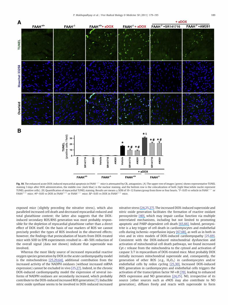
Fig. 10. The enhanced acute DOX-inducedmyocardial apoptosis in FAAH−/−mice is attenuated by CB1 antagonists. (A) The upper row of images (green) shows representative TUNELstaining 3 days after DOX administration, the middle row (dark blue) is the nuclear staining, and the bottom row is the colocalization of both (light blue/white nuclei representTUNEL-positive cells). (B) Quantification of myocardial TUNEL staining. Results are means±SEM of 10–12 frames/group from three or four hearts. *Pb0.05 vs vehicle in FAAH−/− orFAAH+/+ mice; #Pb0.05 vs DOX in FAAH+/+ or FAAH−/− mice; $Pb0.05 vs DOX in FAAH+/+ mice.
189P. Mukhopadhyay et al. / Free Radical Biology & Medicine 50 (2011) 179–195
exposed mice (slightly preceding the nitrative stress), which alsoparalleled increased cell death and decreasedmyocardial reduced andtotal glutathione content; the latter also suggests that the DOX-induced secondary ROS/RNS generation was most probably respon-sible for the depletion of myocardial glutathione rather than a directeffect of DOX itself. On the basis of our markers of ROS we cannotprecisely predict the types of ROS involved in the observed effects;however, the findings that preincubation of hearts from DOX-treatedmice with SOD in EPR experiments resulted in ~40–50% reduction ofthe overall signal (data not shown) indicate that superoxide wasinvolved.
Whereas the most likely source of increased myocardial reactiveoxygen species generation byDOX in the acute cardiomyopathymodelis the mitochondrion [25,29,64], additional contribution from theincreased activity of the NADPH oxidases (without increased mRNAexpression) cannot be excluded in vivo [25,27]. Indeed, in the chronicDOX-induced cardiomyopathy model the expression of several iso-forms of NADPH oxidases are secondarily increased, which probablycontribute to theDOX-induced increasedROS generation [7]. Induciblenitric oxide synthase seems to be involved in DOX-induced increased
nitrative stress [24,25,27]. The increasedDOX-induced superoxide andnitric oxide generation facilitates the formation of reactive oxidantperoxynitrite [40], which may impair cardiac function via multipleinterrelated mechanisms, including but not limited to promotingapoptotic and PARP-dependent cell death [65,66]. Indeed, peroxyni-trite is a key trigger of cell death in cardiomyocytes and endothelialcells during ischemic–reperfusion injury [67,68], as well as in both invivo and in vitro models of DOX-induced cardiomyopathy [25,69].Consistent with the DOX-induced mitochondrial dysfunction andactivation of mitochondrial cell death pathways, we found increasedCyt c release from the mitochondria to the cytosol and activation ofcaspase 3/7 in myocardium of DOX-treated mice. Most probably DOXinitially increases mitochondrial superoxide and, consequently, thegeneration of other ROS (e.g., H2O2) in cardiomyocytes and/orendothelial cells by redox cycling [25,30]. Increased DOX-inducedROS generation in cardiomyocytes and endothelial cells triggers theactivation of the transcription factor NF-κB [70], leading to enhancediNOS expression and NO generation [24,25]. NO, irrespective of itssource (other sources such as eNOS may also contribute to NOgeneration), diffuses freely and reacts with superoxide to form
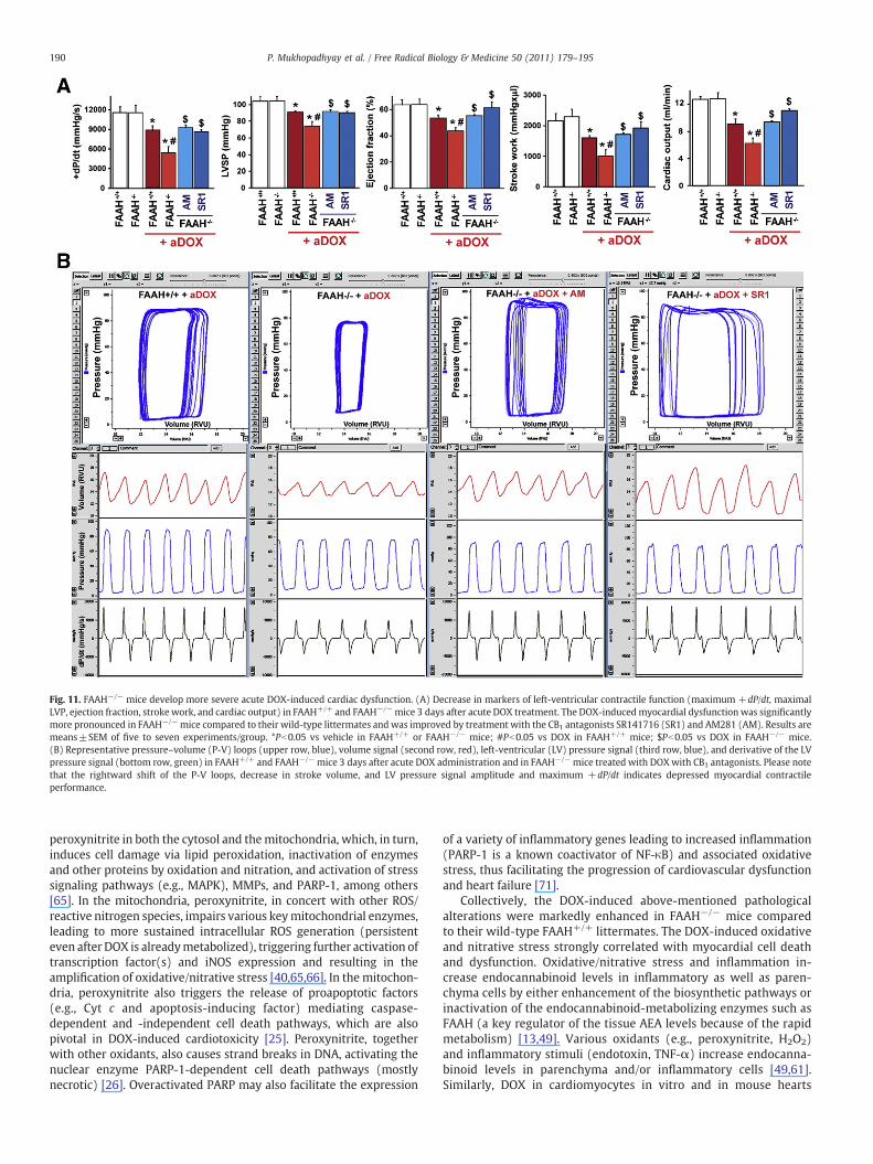
Fig. 11. FAAH−/− mice develop more severe acute DOX-induced cardiac dysfunction. (A) Decrease in markers of left-ventricular contractile function (maximum +dP/dt, maximalLVP, ejection fraction, stroke work, and cardiac output) in FAAH+/+ and FAAH−/−mice 3 days after acute DOX treatment. The DOX-inducedmyocardial dysfunction was significantlymore pronounced in FAAH−/−mice compared to their wild-type littermates and was improved by treatment with the CB1 antagonists SR141716 (SR1) and AM281 (AM). Results aremeans±SEM of five to seven experiments/group. *Pb0.05 vs vehicle in FAAH+/+ or FAAH−/− mice; #Pb0.05 vs DOX in FAAH+/+ mice; $Pb0.05 vs DOX in FAAH−/− mice.(B) Representative pressure–volume (P-V) loops (upper row, blue), volume signal (second row, red), left-ventricular (LV) pressure signal (third row, blue), and derivative of the LVpressure signal (bottom row, green) in FAAH+/+ and FAAH−/−mice 3 days after acute DOX administration and in FAAH−/−mice treated with DOXwith CB1 antagonists. Please notethat the rightward shift of the P-V loops, decrease in stroke volume, and LV pressure signal amplitude and maximum +dP/dt indicates depressed myocardial contractileperformance.
190 P. Mukhopadhyay et al. / Free Radical Biology & Medicine 50 (2011) 179–195
peroxynitrite in both the cytosol and themitochondria, which, in turn,induces cell damage via lipid peroxidation, inactivation of enzymesand other proteins by oxidation and nitration, and activation of stresssignaling pathways (e.g., MAPK), MMPs, and PARP-1, among others[65]. In the mitochondria, peroxynitrite, in concert with other ROS/reactive nitrogen species, impairs various keymitochondrial enzymes,leading to more sustained intracellular ROS generation (persistenteven after DOX is alreadymetabolized), triggering further activation oftranscription factor(s) and iNOS expression and resulting in theamplification of oxidative/nitrative stress [40,65,66]. In the mitochon-dria, peroxynitrite also triggers the release of proapoptotic factors(e.g., Cyt c and apoptosis-inducing factor) mediating caspase-dependent and -independent cell death pathways, which are alsopivotal in DOX-induced cardiotoxicity [25]. Peroxynitrite, togetherwith other oxidants, also causes strand breaks in DNA, activating thenuclear enzyme PARP-1-dependent cell death pathways (mostlynecrotic) [26]. Overactivated PARP may also facilitate the expression
of a variety of inflammatory genes leading to increased inflammation(PARP-1 is a known coactivator of NF-κB) and associated oxidativestress, thus facilitating the progression of cardiovascular dysfunctionand heart failure [71].
Collectively, the DOX-induced above-mentioned pathologicalalterations were markedly enhanced in FAAH−/− mice comparedto their wild-type FAAH+/+ littermates. The DOX-induced oxidativeand nitrative stress strongly correlated with myocardial cell deathand dysfunction. Oxidative/nitrative stress and inflammation in-crease endocannabinoid levels in inflammatory as well as paren-chyma cells by either enhancement of the biosynthetic pathways orinactivation of the endocannabinoid-metabolizing enzymes such asFAAH (a key regulator of the tissue AEA levels because of the rapidmetabolism) [13,49]. Various oxidants (e.g., peroxynitrite, H2O2)and inflammatory stimuli (endotoxin, TNF-α) increase endocanna-binoid levels in parenchyma and/or inflammatory cells [49,61].Similarly, DOX in cardiomyocytes in vitro and in mouse hearts
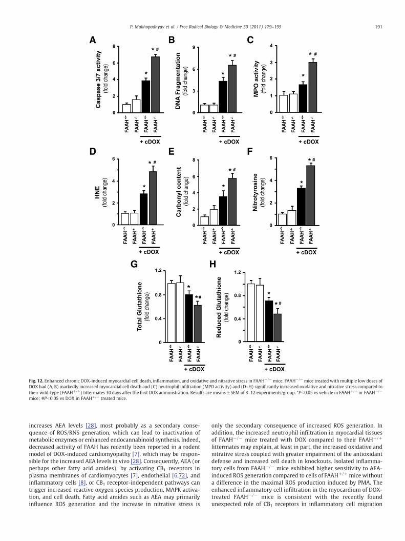
Fig. 12. Enhanced chronic DOX-induced myocardial cell death, inflammation, and oxidative and nitrative stress in FAAH−/− mice. FAAH−/− mice treated with multiple low doses ofDOX had (A, B) markedly increased myocardial cell death and (C) neutrophil infiltration (MPO activity) and (D–H) significantly increased oxidative and nitrative stress compared totheir wild-type (FAAH+/+) littermates 30 days after the first DOX administration. Results are means±SEM of 8–12 experiments/group. *Pb0.05 vs vehicle in FAAH+/+ or FAAH−/−
mice; #Pb0.05 vs DOX in FAAH+/+ treated mice.
191P. Mukhopadhyay et al. / Free Radical Biology & Medicine 50 (2011) 179–195
increases AEA levels [28], most probably as a secondary conse-quence of ROS/RNS generation, which can lead to inactivation ofmetabolic enzymes or enhanced endocannabinoid synthesis. Indeed,decreased activity of FAAH has recently been reported in a rodentmodel of DOX-induced cardiomyopathy [7], which may be respon-sible for the increased AEA levels in vivo [28]. Consequently, AEA (orperhaps other fatty acid amides), by activating CB1 receptors inplasma membranes of cardiomyocytes [7], endothelial [6,72], andinflammatory cells [8], or CB1 receptor-independent pathways cantrigger increased reactive oxygen species production, MAPK activa-tion, and cell death. Fatty acid amides such as AEA may primarilyinfluence ROS generation and the increase in nitrative stress is
only the secondary consequence of increased ROS generation. Inaddition, the increased neutrophil infiltration in myocardial tissuesof FAAH−/− mice treated with DOX compared to their FAAH+/+
littermates may explain, at least in part, the increased oxidative andnitrative stress coupled with greater impairment of the antioxidantdefense and increased cell death in knockouts. Isolated inflamma-tory cells from FAAH−/− mice exhibited higher sensitivity to AEA-induced ROS generation compared to cells of FAAH+/+ mice withouta difference in the maximal ROS production induced by PMA. Theenhanced inflammatory cell infiltration in the myocardium of DOX-treated FAAH−/− mice is consistent with the recently foundunexpected role of CB1 receptors in inflammatory cell migration
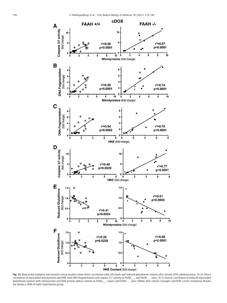
Fig. 13. Myocardial oxidative and nitrative stress markers show direct correlation with cell death and reduced glutathione content after chronic DOX administration. (A–D) Directcorrelation of myocardial nitrotyrosine and HNE with DNA fragmentation and caspase 3/7 activity in FAAH+/+ and FAAH−/− mice. (E, F) Inverse correlation of reduced myocardialglutathione content with nitrotyrosine and HNE protein adduct content in FAAH+/+ (open) and FAAH−/− mice (filled) after vehicle (triangle) and DOX (circle) treatment. Resultsare means±SEM of eight experiments/group.
192 P. Mukhopadhyay et al. / Free Radical Biology & Medicine 50 (2011) 179–195
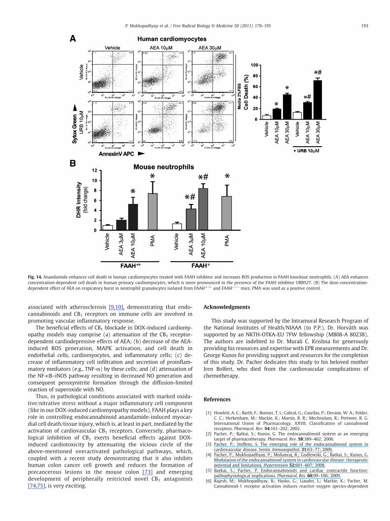
Fig. 14. Anandamide enhances cell death in human cardiomyocytes treated with FAAH inhibitor and increases ROS production in FAAH knockout neutrophils. (A) AEA enhancesconcentration-dependent cell death in human primary cardiomyocytes, which is more pronounced in the presence of the FAAH inhibitor URB527. (B) The dose-concentration-dependent effect of AEA on respiratory burst in neutrophil granulocytes isolated from FAAH+/+ and FAAH−/− mice. PMA was used as a positive control.
193P. Mukhopadhyay et al. / Free Radical Biology & Medicine 50 (2011) 179–195
associated with atherosclerosis [9,10], demonstrating that endo-cannabinoids and CB1 receptors on immune cells are involved inpromoting vascular inflammatory response.
The beneficial effects of CB1 blockade in DOX-induced cardiomy-opathy models may comprise (a) attenuation of the CB1 receptor-dependent cardiodepressive effects of AEA; (b) decrease of the AEA-induced ROS generation, MAPK activation, and cell death inendothelial cells, cardiomyocytes, and inflammatory cells; (c) de-crease of inflammatory cell infiltration and secretion of proinflam-matory mediators (e.g., TNF-α) by these cells; and (d) attenuation ofthe NF-κB–iNOS pathway resulting in decreased NO generation andconsequent peroxynitrite formation through the diffusion-limitedreaction of superoxide with NO.
Thus, in pathological conditions associated with marked oxida-tive/nitrative stress without a major inflammatory cell component(like in our DOX-induced cardiomyopathymodels), FAAH plays a keyrole in controlling endocannabinoid anandamide-induced myocar-dial cell death/tissue injury, which is, at least in part, mediated by theactivation of cardiovascular CB1 receptors. Conversely, pharmaco-logical inhibition of CB1 exerts beneficial effects against DOX-induced cardiotoxicity by attenuating the vicious circle of theabove-mentioned overactivated pathological pathways, which,coupled with a recent study demonstrating that it also inhibitshuman colon cancer cell growth and reduces the formation ofprecancerous lesions in the mouse colon [73] and emergingdevelopment of peripherally restricted novel CB1 antagonists[74,75], is very exciting.
Acknowledgments
This study was supported by the Intramural Research Program ofthe National Institutes of Health/NIAAA (to P.P.). Dr. Horváth wassupported by an NKTH-OTKA-EU 7FW fellowship (MB08-A 80238).The authors are indebted to Dr. Murali C. Krishna for generouslyproviding his resources and expertise with EPRmeasurements and Dr.George Kunos for providing support and resources for the completionof this study. Dr. Pacher dedicates this study to his beloved motherIren Bolfert, who died from the cardiovascular complications ofchemotherapy.
References
[1] Howlett, A. C.; Barth, F.; Bonner, T. I.; Cabral, G.; Casellas, P.; Devane, W. A.; Felder,C. C.; Herkenham, M.; Mackie, K.; Martin, B. R.; Mechoulam, R.; Pertwee, R. G.International Union of Pharmacology. XXVII. Classification of cannabinoidreceptors. Pharmacol. Rev. 54:161–202; 2002.
[2] Pacher, P.; Batkai, S.; Kunos, G. The endocannabinoid system as an emergingtarget of pharmacotherapy. Pharmacol. Rev. 58:389–462; 2006.
[3] Pacher, P.; Steffens, S. The emerging role of the endocannabinoid system incardiovascular disease. Semin. Immunopathol. 31:63–77; 2009.
[4] Pacher, P.; Mukhopadhyay, P.; Mohanraj, R.; Godlewski, G.; Batkai, S.; Kunos, G.Modulation of the endocannabinoid system in cardiovascular disease: therapeuticpotential and limitations. Hypertension 52:601–607; 2008.
[5] Batkai, S.; Pacher, P. Endocannabinoids and cardiac contractile function:pathophysiological implications. Pharmacol. Res. 60:99–106; 2009.
[6] Rajesh, M.; Mukhopadhyay, B.; Hasko, G.; Liaudet, L.; Mackie, K.; Pacher, M.Cannabinoid-1 receptor activation induces reactive oxygen species-dependent
194 P. Mukhopadhyay et al. / Free Radical Biology & Medicine 50 (2011) 179–195
and -independent mitogen-activated protein kinase activation and cell death inhuman coronary artery endothelial cells. Br. J. Pharmacol. 160:688–700; 2010.
[7] Mukhopadhyay, P.; Rajesh, M.; Batkai, S.; Patel, V.; Kashiwaya, Y.; Liaudet, L.;Evgenov, O. V.; Mackie, K.; Hasko, G.; Pacher, P. CB1 cannabinoid receptors promoteoxidative stress and cell death in murine models of doxorubicin-inducedcardiomyopathy and in human cardiomyocytes. Cardiovasc. Res. 85:773–784; 2010.
[8] Han, K. H.; Lim, S.; Ryu, J.; Lee, C. W.; Kim, Y.; Kang, J. H.; Kang, S. S.; Ahn, Y. K.;Park, C. S.; Kim, J. J. CB1 and CB2 cannabinoid receptors differentially regulate theproduction of reactive oxygen species by macrophages. Cardiovasc. Res. 84:378–386; 2009.
[9] Dol-Gleizes, F.; Paumelle, R.; Visentin, V.; Mares, A. M.; Desitter, P.; Hennuyer, N.;Gilde, A.; Staels, B.; Schaeffer, P.; Bono, F. Rimonabant, a selective cannabinoid CB1receptor antagonist, inhibits atherosclerosis in LDL receptor-deficient mice.Arterioscler. Thromb. Vasc. Biol. 29:12–18; 2009.
[10] Sugamura, K.; Sugiyama, S.; Nozaki, T.; Matsuzawa, Y.; Izumiya, Y.; Miyata, K.;Nakayama, M.; Kaikita, K.; Obata, T.; Takeya, M.; Ogawa, H. Activatedendocannabinoid system in coronary artery disease and antiinflammatory effectsof cannabinoid 1 receptor blockade onmacrophages. Circulation 119:28–36; 2009.
[11] Rajesh, M.; Mukhopadhyay, P.; Hasko, G.; Pacher, P. Cannabinoid CB1 receptorinhibition decreases vascular smooth muscle migration and proliferation.Biochem. Biophys. Res. Commun. 377:1248–1252; 2008.
[12] Montecucco, F.; Lenglet, S.; Braunersreuther, V.; Burger, F.; Pelli, G.; Bertolotto, M.;Mach, F.; Steffens, S. CB(2) cannabinoid receptor activation is cardioprotective in amouse model of ischemia/reperfusion. J. Mol. Cell. Cardiol. 46:612–620; 2009.
[13] Batkai, S.; Osei-Hyiaman, D.; Pan, H.; El-Assal, O.; Rajesh, M.; Mukhopadhyay, P.;Hong, F.; Harvey-White, J.; Jafri, A.; Hasko, G.; Huffman, J. W.; Gao, B.; Kunos, G.;Pacher, P. Cannabinoid-2 receptor mediates protection against hepatic ischemia/reperfusion injury. FASEB J. 21:1788–1800; 2007.
[14] Mukhopadhyay, P.; Rajesh, M.; Pan, H.; Patel, V.; Mukhopadhyay, B.; Batkai, S.;Gao, B.; Hasko, G.; Pacher, P. Cannabinoid-2 receptor limits inflammation,oxidative/nitrosative stress, and cell death in nephropathy. Free Radic. Biol. Med.48:457–467; 2010.
[15] Di Marzo, V.; Cote, M.; Matias, I.; Lemieux, I.; Arsenault, B. J.; Cartier, A.; Piscitelli,F.; Petrosino, S.; Almeras, N.; Despres, J. P. Changes in plasma endocannabinoidlevels in viscerally obesemen following a 1 year lifestyle modification programmeand waist circumference reduction: associations with changes in metabolic riskfactors. Diabetologia 52:213–217; 2009.
[16] Pacher, P. Cannabinoid CB1 receptor antagonists for atherosclerosis andcardiometabolic disorders: new hopes, old concerns? Arterioscler. Thromb. Vasc.Biol. 29:7–9; 2009.
[17] Cravatt, B. F.; Giang, D. K.; Mayfield, S. P.; Boger, D. L.; Lerner, R. A.; Gilula, N. B.Molecular characterization of an enzyme that degrades neuromodulatory fatty-acid amides. Nature 384:83–87; 1996.
[18] Ahn, K.; McKinney, M. K.; Cravatt, B. F. Enzymatic pathways that regulateendocannabinoid signaling in the nervous system. Chem. Rev.108:1687–1707; 2008.
[19] Cravatt, B. F.; Demarest, K.; Patricelli, M. P.; Bracey, M. H.; Giang, D. K.; Martin, B. R.;Lichtman, A. H. Supersensitivity to anandamide and enhanced endogenouscannabinoid signaling in mice lacking fatty acid amide hydrolase. Proc. Natl Acad.Sci. USA 98:9371–9376; 2001.
[20] Cravatt, B. F.; Saghatelian, A.; Hawkins, E. G.; Clement, A. B.; Bracey, M. H.;Lichtman, A. H. Functional disassociation of the central and peripheral fatty acidamide signaling systems. Proc. Natl Acad. Sci. USA 101:10821–10826; 2004.
[21] Siegmund, S. V.; Seki, E.; Osawa, Y.; Uchinami, H.; Cravatt, B. F.; Schwabe, R. F.Fatty acid amide hydrolase determines anandamide-induced cell death in theliver. J. Biol. Chem. 281:10431–10438; 2006.
[22] Kotamraju, S.; Konorev, E. A.; Joseph, J.; Kalyanaraman, B. Doxorubicin-inducedapoptosis in endothelial cells and cardiomyocytes is ameliorated by nitrone spintraps and ebselen: role of reactive oxygen and nitrogen species. J. Biol. Chem. 275:33585–33592; 2000.
[23] Wang, S.; Konorev, E. A.; Kotamraju, S.; Joseph, J.; Kalivendi, S.; Kalyanaraman, B.Doxorubicin induces apoptosis in normal and tumor cells via distinctly differentmechanisms. intermediacy of H2O2- and p53-dependent pathways. J. Biol. Chem.279:25535–25543; 2004.
[24] Weinstein, D. M.; Mihm, M. J.; Bauer, J. A. Cardiac peroxynitrite formation and leftventricular dysfunction following doxorubicin treatment in mice. J. Pharmacol.Exp. Ther. 294:396–401; 2000.
[25] Mukhopadhyay, P.; Rajesh, M.; Batkai, S.; Kashiwaya, Y.; Hasko, G.; Liaudet, L.;Szabo, C.; Pacher, P. Role of superoxide, nitric oxide, and peroxynitrite indoxorubicin-induced cell death in vivo and in vitro. Am. J. Physiol. Heart Circ.Physiol. 296:H1466–H1483; 2009.
[26] Pacher, P.; Liaudet, L.; Bai, P.; Virag, L.; Mabley, J. G.; Hasko, G.; Szabo, C. Activationof poly(ADP-ribose) polymerase contributes to development of doxorubicin-induced heart failure. J. Pharmacol. Exp. Ther. 300:862–867; 2002.
[27] Pacher, P.; Liaudet, L.; Bai, P.; Mabley, J. G.; Kaminski, P. M.; Virag, L.; Deb, A.;Szabo, E.; Ungvari, Z.; Wolin, M. S.; Groves, J. T.; Szabo, C. Potent metalloporphyrinperoxynitrite decomposition catalyst protects against the development ofdoxorubicin-induced cardiac dysfunction. Circulation 107:896–904; 2003.
[28] Mukhopadhyay, P.; Batkai, S.; Rajesh, M.; Czifra, N.; Harvey-White, J.; Hasko, G.;Zsengeller, Z.; Gerard, N. P.; Liaudet, L.; Kunos, G.; Pacher, P. Pharmacologicalinhibition of CB1 cannabinoid receptor protects against doxorubicin-inducedcardiotoxicity. J. Am. Coll. Cardiol. 50:528–536; 2007.
[29] Chandran, K.; Aggarwal, D.; Migrino, R. Q.; Joseph, J.; McAllister, D.; Konorev, E. A.;Antholine, W. E.; Zielonka, J.; Srinivasan, S.; Avadhani, N. G.; Kalyanaraman, B.Doxorubicin inactivates myocardial cytochrome c oxidase in rats: cardioprotectionby Mito-Q. Biophys. J. 96:1388–1398; 2009.
[30] Doroshow, J. H.; Davies, K. J. Redox cycling of anthracyclines by cardiacmitochondria. II. Formation of superoxide anion, hydrogen peroxide, andhydroxyl radical. J. Biol. Chem. 261:3068–3074; 1986.
[31] Pacher, P.; Batkai, S.; Osei-Hyiaman, D.; Offertaler, L.; Liu, J.; Harvey-White, J.;Brassai, A.; Jarai, Z.; Cravatt, B. F.; Kunos, G. Hemodynamic profile, responsivenessto anandamide, and baroreflex sensitivity of mice lacking fatty acid amidehydrolase. Am. J. Physiol. Heart Circ. Physiol. 289:H533–H541; 2005.
[32] Pacher, P.; Nagayama, T.; Mukhopadhyay, P.; Batkai, S.; Kass, D. A. Measurementof cardiac function using pressure–volume conductance catheter technique inmice and rats. Nat. Protoc. 3:1422–1434; 2008.
[33] Forman, H. J. Reactive oxygen species and alpha, beta-unsaturated aldehydesas second messengers in signal transduction. Ann. NY Acad. Sci. 1203:35–44;2010.
[34] Forman, H. J.; Dickinson, D. A. Introduction to serial reviews on 4-hydroxy-2-nonenal as a signaling molecule. Free Radic. Biol. Med. 37:594–596; 2004.
[35] Forman, H. J.; Fukuto, J. M.; Miller, T.; Zhang, H.; Rinna, A.; Levy, S. The chemistryof cell signaling by reactive oxygen and nitrogen species and 4-hydroxynonenal.Arch. Biochem. Biophys. 477:183–195; 2008.
[36] Levine, R. L. Carbonyl modified proteins in cellular regulation, aging, and disease.Free Radic. Biol. Med. 32:790–796; 2002.
[37] Eiserich, J. P.; Hristova, M.; Cross, C. E.; Jones, A. D.; Freeman, B. A.; Halliwell, B.;van der Vliet, A. Formation of nitric oxide-derived inflammatory oxidants bymyeloperoxidase in neutrophils. Nature 391:393–397; 1998.
[38] Baldus, S.; Eiserich, J. P.; Brennan, M. L.; Jackson, R. M.; Alexander, C. B.; Freeman,B. A. Spatial mapping of pulmonary and vascular nitrotyrosine reveals the pivotalrole of myeloperoxidase as a catalyst for tyrosine nitration in inflammatorydiseases. Free Radic. Biol. Med. 33:1010; 2002.
[39] Schopfer, F. J.; Baker, P. R.; Freeman, B. A. NO-dependent protein nitration: a cellsignaling event or an oxidative inflammatory response? Trends Biochem. Sci. 28:646–654; 2003.
[40] Radi, R. Nitric oxide, oxidants, and protein tyrosine nitration. Proc. Natl Acad. Sci.USA 101:4003–4008; 2004.
[41] Dikalov, S. I.; Li, W.; Mehranpour, P.; Wang, S. S.; Zafari, A. M. Production ofextracellular superoxide by human lymphoblast cell lines: comparison of electronspin resonance techniques and cytochrome C reduction assay. Biochem.Pharmacol. 73:972–980; 2007.
[42] Mariappan, N.; Elks, C. M.; Fink, B.; Francis, J. TNF-induced mitochondrial damage:a link between mitochondrial complex I activity and left ventricular dysfunction.Free Radic. Biol. Med. 46:462–470; 2009.
[43] Rothe, G.; Oser, A.; Valet, G. Dihydrorhodamine 123: a new flowcytometric indicatorfor respiratory burst activity in neutrophil granulocytes. Naturwissenschaften 75:354–355; 1988.
[44] Fearon, D. T.; Collins, L. A. Increased expression of C3b receptors on polymorpho-nuclear leukocytes induced by chemotactic factors and by purification procedures.J. Immunol. 130:370–375; 1983.
[45] Vowells, S. J.; Sekhsaria, S.; Malech, H. L.; Shalit, M.; Fleisher, T. A. Flow cytometricanalysis of the granulocyte respiratory burst: a comparison study of fluorescentprobes. J. Immunol. Meth. 178:89–97; 1995.
[46] Mukhopadhyay, P.; Pan, H.; Rajesh, M.; Batkai, S.; Patel, V.; Harvey-White, J.;Mukhopadhyay, B.; Hasko, G.; Gao, B.; Mackie, K.; Pacher, P. CB1 cannabinoidreceptors promote oxidative/nitrosative stress, inflammation and cell death in amurine nephropathy model. Br. J. Pharmacol. 160:657–668; 2010.
[47] Pacher, P.; Batkai, S.; Kunos, G. Haemodynamic profile and responsiveness toanandamide of TRPV1 receptor knock-out mice. J. Physiol. 558:647–657; 2004.
[48] Gary-Bobo,M.; Elachouri, G.; Gallas, J. F.; Janiak, P.;Marini, P.; Ravinet-Trillou, C.;Chabbert, M.; Cruccioli, N.; Pfersdorff, C.; Roque, C.; Arnone, M.; Croci, T.;Soubrie, P.; Oury-Donat, F.; Maffrand, J. P.; Scatton, B.; Lacheretz, F.; Le Fur, G.;Herbert, J. M.; Bensaid, M. Rimonabant reduces obesity-associated hepaticsteatosis and features of metabolic syndrome in obese Zucker fa/fa rats.Hepatology 46:122–129; 2007.
[49] Pacher, P.; Hasko, G. Endocannabinoids and cannabinoid receptors in ischaemia–reperfusion injury and preconditioning. Br. J. Pharmacol. 153:252–262; 2008.
[50] Kadoi, Y.; Goto, F. Effects of AM281, a cannabinoid antagonist, on circulatorydeterioration and cytokine production in an endotoxin shock model: comparisonwith norepinephrine. J. Anesth. 20:284–289; 2006.
[51] Villanueva, A.; Yilmaz, M. S.; Millington, W. R.; Cutrera, R. A.; Stouffer, D. G.;Parsons, L. H.; Cheer, J. F.; Feleder, C. Central cannabinoid-1 receptor antagonistadministration prevents endotoxic hypotension affecting norepinephrine releasein the preoptic anterior hypothalamic area. Shock 32:614–620; 2009.
[52] Schafer, A.; Pfrang, J.; Neumuller, J.; Fiedler, S.; Ertl, G.; Bauersachs, J. Thecannabinoid receptor-1 antagonist rimonabant inhibits platelet activation andreduces pro-inflammatory chemokines and leukocytes in Zucker rats. Br. J.Pharmacol. 154:1047–1054; 2008.
[53] Malfitano, A. M.; Laezza, C.; Pisanti, S.; Gazzerro, P.; Bifulco, M. Rimonabant(SR141716) exerts anti-proliferative and immunomodulatory effects in humanperipheral blood mononuclear cells. Br. J. Pharmacol. 153:1003–1010; 2008.
[54] Myers, C. E.; McGuire, W. P.; Liss, R. H.; Ifrim, I.; Grotzinger, K.; Young, R. C.Adriamycin: the role of lipid peroxidation in cardiac toxicity and tumor response.Science 197:165–167; 1977.
[55] Doroshow, J. H.; Locker, G. Y.; Ifrim, I.; Myers, C. E. Prevention of doxorubicin cardiactoxicity in the mouse by N-acetylcysteine. J. Clin. Invest. 68:1053–1064; 1981.
[56] Konorev, E. A.; Kennedy, M. C.; Kalyanaraman, B. Cell-permeable superoxidedismutase and glutathione peroxidase mimetics afford superior protectionagainst doxorubicin-induced cardiotoxicity: the role of reactive oxygen andnitrogen intermediates. Arch. Biochem. Biophys. 368:421–428; 1999.
195P. Mukhopadhyay et al. / Free Radical Biology & Medicine 50 (2011) 179–195
[57] Kalivendi, S. V.; Konorev, E. A.; Cunningham, S.; Vanamala, S. K.; Kaji, E. H.; Joseph, J.;Kalyanaraman, B. Doxorubicin activates nuclear factor of activated T-lymphocytesand Fas ligand transcription: role of mitochondrial reactive oxygen species andcalcium. Biochem. J. 389:527–539; 2005.
[58] Zhu, W.; Zou, Y.; Aikawa, R.; Harada, K.; Kudoh, S.; Uozumi, H.; Hayashi, D.; Gu, Y.;Yamazaki, T.; Nagai, R.; Yazaki, Y.; Komuro, I. MAPK superfamily plays animportant role in daunomycin-induced apoptosis of cardiac myocytes. Circulation100:2100–2107; 1999.
[59] Singal, P. K.; Iliskovic, N. Doxorubicin-induced cardiomyopathy. N. Engl. J. Med.339:900–905; 1998.
[60] Ishii, Y.; Sakamoto, T.; Ito, R.; Yanaga, K. F2-isoprostanes and 2-arachidonylglycerolas biomarkers of lipid peroxidation in pigs with hepatic ischemia/reperfusioninjury. J. Surg. Res. 161:139–145; 2009.
[61] Pacher, P.; Gao, B. Endocannabinoids and liver disease. III. Endocannabinoideffects on immune cells: implications for inflammatory liver diseases. Am. J.Physiol. Gastrointest. Liver Physiol. 294:G850–G854; 2008.
[62] Dalton, G. D.; Bass, C. E.; Van Horn, C.; Howlett, A. C. Signal transduction viacannabinoid receptors. CNS Neurol. Disord. Drug Targets 8:422–431; 2009.
[63] Riad, A.; Bien, S.; Westermann, D.; Becher, P. M.; Loya, K.; Landmesser, U.; Kroemer,H. K.; Schultheiss, H. P.; Tschope, C. Pretreatment with statin attenuates thecardiotoxicity of doxorubicin in mice. Cancer Res. 69:695–699; 2009.
[64] Davies, K. J.; Doroshow, J.H. Redox cycling of anthracyclinesbycardiacmitochondria.I. Anthracycline radical formation by NADH dehydrogenase. J. Biol. Chem. 261:3060–3067; 1986.
[65] Pacher, P.; Beckman, J. S.; Liaudet, L. Nitric oxide and peroxynitrite in health anddisease. Physiol. Rev. 87:315–424; 2007.
[66] Szabo, C.; Ischiropoulos, H.; Radi, R. Peroxynitrite: biochemistry, pathophysiologyand development of therapeutics. Nat. Rev. Drug Discovery 6:662–680; 2007.
[67] Levrand, S.; Vannay-Bouchiche, C.; Pesse, B.; Pacher, P.; Feihl, F.; Waeber, B.;Liaudet, L. Peroxynitrite is a major trigger of cardiomyocyte apoptosis in vitro andin vivo. Free Radic. Biol. Med. 41:886–895; 2006.
[68] Obrosova, I. G. Peroxynitrite and cardiomyocyte cell death: an evolving story.A commentary on "Peroxynitrite is a major trigger of cardiomyocyte apoptosisin vitro and in vivo". Free Radic. Biol. Med. 41:866–868; 2006.
[69] Denicola, A.; Radi, R. Peroxynitrite and drug-dependent toxicity. Toxicology 208:273–288; 2005.
[70] Wang, S.; Kotamraju, S.; Konorev, E.; Kalivendi, S.; Joseph, J.; Kalyanaraman, B.Activation of nuclear factor-kappaB during doxorubicin-induced apoptosis inendothelial cells and myocytes is pro-apoptotic: the role of hydrogen peroxide.Biochem. J. 367:729–740; 2002.
[71] Pacher, P.; Szabo, C. Role of the peroxynitrite-poly(ADP-ribose) polymerasepathway in human disease. Am. J. Pathol. 173:2–13; 2008.
[72] Liu, J.; Gao, B.; Mirshahi, F.; Sanyal, A. J.; Khanolkar, A. D.; Makriyannis, A.; Kunos,G. Functional CB1 cannabinoid receptors in human vascular endothelial cells.Biochem. J. 346:835–840; 2000.
[73] Santoro,A.; Pisanti, S.;Grimaldi,C.; Izzo,A.A.; Borrelli, F.; Proto,M.C.;Malfitano,A.M.;Gazzerro, P.; Laezza, C.; Bifulco, M. Rimonabant inhibits human colon cancer cellgrowth and reduces the formation of precancerous lesions in the mouse colon. Int. J.Cancer 125:996–1003; 2009.
[74] Kunos, G.; Osei-Hyiaman, D.; Batkai, S.; Sharkey, K. A.; Makriyannis, A. Shouldperipheral CB(1) cannabinoid receptors be selectively targeted for therapeuticgain? Trends Pharmacol. Sci. 30:1–7; 2009.
[75] Tam, J., Vemuri, V.K., Liu, J., Batkai, S., Mukhopadhyay, B., Godlewski, G., Osei-Hyiaman, D., Ohnuma, S., Ambudkar, S.V., Pickel, J., Makriyannis, A., Kunos, G.Peripheral CB1 cannabinoid receptor blockade improves cardiometabolic risk inmouse models of obesity. J. Clin. Invest. 120:2953–2966; 2010.


