Embed Size (px)
Citation preview
UNIVERSIDADE TÉCNICA DE LISBOA
Faculdade de Medicina Veterinária
EXPRESSION AND CHARACTERIZATION OF CALRETICULIN GENE ISOLATED FROM
RHIPICEPHALUS ANNULATUS AFTER BABESIA BIGEMINA INFECTION.
JOANA RAMOS RAPAZ LÉRIAS
CONSTITUIÇÃO DO JÚRI
Doutora Isabel Maria Soares Pereira da Fonseca de Sampaio Doutor Luís Manuel Madeira de Carvalho Doutor Vítor Manuel Diogo de Oliveira Alves Doutora Ana Isabel Amaro Gonçalves Domingos
ORIENTADOR
Doutora Ana Isabel Amaro Gonçalves Domingos
CO-ORIENTADOR
Doutor Luís Manuel Madeira de Carvalho
2012
LISBOA

UNIVERSIDADE TÉCNICA DE LISBOA
Faculdade de Medicina Veterinária
EXPRESSION AND CHARACTERIZATION OF CALRETICULIN GENE ISOLATED FROM
RHIPICEPHALUS ANNULATUS AFTER BABESIA BIGEMINA INFECTION.
JOANA RAMOS RAPAZ LÉRIAS
DISSERTAÇÃO DE MESTRADO EM MEDICINA VETERINÁRIA.
CONSTITUIÇÃO DO JÚRI
Doutora Isabel Maria Soares Pereira da Fonseca de Sampaio Doutor Luís Manuel Madeira de Carvalho Doutor Vítor Manuel Diogo de Oliveira Alves Doutora Ana Isabel Amaro Gonçalves Domingos
ORIENTADOR
Doutora Ana Isabel Amaro Gonçalves Domingos
CO-ORIENTADOR
Doutor Luís Manuel Madeira de Carvalho
2012
LISBOA
iii
Acknowledgements
I would like to thank my supervisor Professor Doctor Ana Domingos, for all the help, support,
patience and guidance during my Master project. Her knowledge and experience with ticks
were essential to improve my work.
I express my gratitude to my co-supervisor Professor Doctor Luís Madeira de Carvalho, who
encouraged my passion for parasites. My “work” in FMV/UTL - Parasitology Laboratory was
essential to choose this Master theme.
I also thank Sandra Antunes, for her help at the bench, her guidance and patience.
The development of this Master project would not have been possible without the
collaboration of Fábia Gomes, a dearest friend of mine and colleague at IHMT, who always
showed herself available to participate in my laboratorial work.
A word of appreciation to my colleagues in IHMT and my fellow students along the 6 years
of my faculty life, who always showed their sympathy, friendship and availability to help me
to carrying out my tasks and duties.
I would also like to thank to João, who understood my absences while accomplishing this
work and always provided me with the necessary support.
Especially to my family, mum, dad and sisters Muri and Sofia, and to my dearest dog, my
four-leg best friend, Phoenix, I want to express my gratitude for all the unconditional support.
Without you, I would not have become a Veterinary and certainly I would not be the same
person.

v
Expression and characterization of calreticulin gene isolated from
Rhipicephalus (Boophilus) annulatus after Babesia bigemina
infection.
Abstract
Ticks are obligate parasites in a large variety of hosts and are considered to be, after
mosquitoes, the second worldwide vector of human and animal diseases.
Bovine babesiosis causes substantial economic losses due to animal mortality, abortions and
anaemia, among other effects. Calreticulin protein that has been identified in several species
including ticks and previous experiments showed that calreticulin gene was up-regulated in R.
annulatus ticks infected with B. bigemina and that its knockdown by RNAi technique leads to
a reduction of both pathogen transmission and ticks weight.
This Master thesis was developed within a project on “Differential expression and functional
characterization of tick (Rhipicephalus annulatus) genes in response to pathogen infection
(Babesia bigemina)”, financed by the Science and Technology Foundation (FCT), with the
project number of PTDC/CVT/112050/2009.
The aim of this study was the isolation of calreticulin gene, purification of calreticulin protein
and its further use to produce antibodies for the purpose of immunolocalization studies and
vaccination tests. In this Master thesis, calreticulin gene was amplified by PCR technique,
sequenced and compared with calreticulin from the R. annulatus sequence, showing 98%
identity. Afterwards, an Escherichia coli recombinant system was used in order to produce a
calreticulin protein. Finally, recombinant proteins were purified using IMAC technique, due
to the affinity of expressed calreticulin protein histidine tail to nickel ions. After specific
elution and a final sample concentration, a unique protein was achieved in the purified
sample, corresponding to recombinant calreticulin.
The results of this study were optimistic and represent one more step to improve ticks control,
as we showed in this study that CRT can be produced and purified without contaminants,
though further vaccination and immunolocalization studies will be the key to understand CRT
future use.
Keywords: Babesia bigemina/ Rhipicephalus (Boophilus) annulatus/ Calreticulin/ Escherichia
coli/ Immobilized metal ion affinity chromatography.

vii
Expressão e caracterização do gene calreticulina isolado de
Rhipicephalus (Boophilus) annulatus após infecção com Babesia
bigemina.
Resumo
As carraças são parasitas obrigatórios de uma grande variedade de hospedeiros, sendo
consideradas, depois dos mosquitos, os mais importantes vectores de doenças em humanos e
animais. A babesiose bovina conduz a elevados prejuízos económicos, devido ao aumento da
mortalidade animal, abortos e anemia, entre outros. A calreticulina é uma proteína já
identificada em várias espécies, incluindo carraças e estudos anteriores demonstraram que o
gene calreticulina estava sobreexpresso em carraças R. annulatus infectadas com B. bigemina
e, após o seu silenciamento através da técnica de RNAi, ocorria uma redução tanto na
transmissão do agente patogénico, como no peso das carraças.
Esta tese de Mestrado foi desenvolvida em paralelo com o projecto “Expressão diferencial e
caracterização de genes de carraça (Rhipicephalus annulatus) em resposta à infecção por
agente patogénico (Babesia bigemina)”, financiado pela Fundação para a Ciência e
Tecnologia, sendo o número do projecto PTDC/CVT/112050/2009.
A finalidade deste trabalho consistiu no isolamento do gene calreticulina e purificação da
correspondente proteína, para posteriormente ser usada na produção de anticorpos destinados
a estudos de imunolocalização e de vacinação. Nesta tese de mestrado, amplificou-se o gene
da calreticulina pela técnica de PCR, e sequenciou-se esse gene e comparou-se com a
sequência da calreticulina da carraça R. annulatus, obtendo-se uma identidade de 98%.
Posteriormente, o gene foi expresso em Escherichia coli de modo a produzir-se calreticulina.
Finalmente, as proteínas recombinantes foram purificadas através do método IMAC, dado a
calreticulina expressa ter uma cauda de histina com afinidade para iões níquel. Após a eluição
específica e a concentração das amostras finais, verificou-se que uma única proteína,
correspondente à calreticulina recombinante, se encontrava presente na amostra purificada.
Os resultados deste estudo foram positivos e representam mais um passo para melhorar o
controlo das carraças, uma vez que este estudo demonstrou que a CRT pode ser produzida e
purificada sem contaminantes, apesar de estudos posteriores de vacinação e imunolocalização
serão essenciais para perceber qual o futuro da CRT.
Palavras-chave: Babesia bigemina/ Rhipicephalus (Boophilus) annulatus/ Calreticulina/
Escherichia coli/ Cromatografia de afinidade por iões metálicos imobilizados.

ix
TABLE OF CONTENTS
ACKNOWLEDGEMENTS ............................................................................................... III
ABSTRACT ........................................................................................................................ V
RESUMO ......................................................................................................................... VII
TABLE OF CONTENTS ................................................................................................... IX
FIGURES ........................................................................................................................... XI
TABLES ........................................................................................................................... XII
ABBREVIATIONS AND SYMBOLS ............................................................................ XIII
CHAPTER 1. PREAMBLE ................................................................................................. 1
CHAPTER 2. LITERATURE REVIEW ............................................................................ 3
2.1. Ticks ............................................................................................................................... 3
2.1.1. Introduction ......................................................................................................................................... 3
2.1.2. Characterization, identification and morphology ................................................................................... 4
2.1.3. Life cycle ............................................................................................................................................ 4
2.1.4. Tick control ......................................................................................................................................... 6
2.1.4.1. Introduction .................................................................................................................................. 6
2.1.4.2. Chemoprophylaxis ........................................................................................................................ 6
2.1.4.3. Biocontrol .................................................................................................................................... 7
2.1.4.4. Genetic resistance ......................................................................................................................... 7
2.1.4.5. Vaccines ....................................................................................................................................... 7
2.1.4.5.1. Introduction ........................................................................................................................... 7
2.1.4.5.2. Recombinant vaccines............................................................................................................ 8
2.1.4.5.3. Antigens selection ................................................................................................................ 10
2.2. Babesia ......................................................................................................................... 11
2.2.1. Introduction ....................................................................................................................................... 11
2.2.2. Characterization and life cycle ........................................................................................................... 11
2.2.3. Symptoms of bovine babesiosis .......................................................................................................... 13
2.2.4. Diagnosis........................................................................................................................................... 15
2.2.5. Human babesiosis .............................................................................................................................. 16
2.2.6. Babesiosis control .............................................................................................................................. 16
2.2.6.1. Introduction ................................................................................................................................ 16
2.2.6.2. Chemoprophylaxis ...................................................................................................................... 17

x
2.2.6.3. Genetic resistance ....................................................................................................................... 17
2.2.6.4. Vaccines..................................................................................................................................... 17
2.3. Calreticulin.................................................................................................................... 21
2.3.1. Introduction ....................................................................................................................................... 21
2.3.2. Calreticulin structure ......................................................................................................................... 22
2.3.3. Calreticulin in ticks and other parasites .............................................................................................. 24
CHAPTER 3. MATERIAL AND METHODS .................................................................. 27
3.1. Rhipicephalus annulatus ticks ....................................................................................... 27
3.2. Amplification of the calreticulin gene ........................................................................... 27
3.3. Expression of recombinantcalreticulin ........................................................................... 27
3.3.1. Cloning and transformation................................................................................................................ 28
3.3.1.1. Screening of the transformed colonies ......................................................................................... 28
3.3.1.2. Plasmid purification for sequencing ............................................................................................ 28
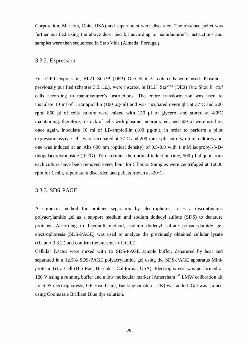
3.3.2. Expression ......................................................................................................................................... 29
3.3.3. SDS-PAGE ....................................................................................................................................... 29
3.3.4. Western-Blot ..................................................................................................................................... 29
3.4. Production of soluble calreticulin .................................................................................. 30
3.5. Purification of recombinant calreticulin ......................................................................... 31
CHAPTER 4. RESULTS ................................................................................................... 33
4.1. Amplification of calreticulin cDNA fragment ............................................................... 33
4.2. Cloning and transformation ........................................................................................... 34
4.3. Recombinant calreticulin expression ............................................................................. 35
4.4. Purification of recombinant calreticulin ......................................................................... 37
CHAPTER 5. DISCUSSION ............................................................................................. 39
CHAPTER 6. CONCLUSION ........................................................................................... 47
CHAPTER 7. REFERENCES ........................................................................................... 49
ANNEX I - MATERIAL AND METHODS APPENDIX ................................................. 64
xi
FIGURES
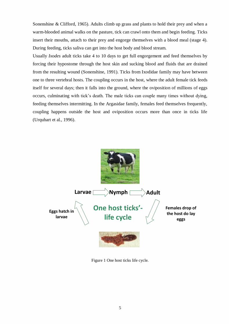
Figure 1 One host ticks life cycle. ...................................................................................................... 5
Figure 2 Giemsa staining of B. bigemina (A) and B. bovis (B) infected red blood cells. .....................12
Figure 3 The life cycle of Babesia bigemina in cattle and in the ixodid tick vector R. microplus. .......13
Figure 4 Cerebral form of babesiosis (A); icterus liver (B) ................................................................14
Figure 5 Kidneys with edema and congestion (A); hematuria (“Red water”) (B) ................................14
Figure 6 CRT action as a chaperone. .................................................................................................22
Figure 7 Structure and functions of the CRT domains. .......................................................................24
Figure 8 PCR results. Samples were electrophoresed in a 1% Agarose/CYBRsafe gel, 1x TAE. ........33
Figure 9 Sequence of the purified sample with the amplified fragment. .............................................34
Figure 10 PCR results. Samples were electrophoresed in a 1% Agarose/CYBRsafe gel, 1x TAE. ......35
Figure 11 SDS-PAGE results.Protein bands from cell cultures induced by IPTG and non-induced at
different time points (A) T0 to T3 and (B) T4 to T5...........................................................................36
Figure 12 Western-Blot results. Protein bands from cell cultures induced by IPTG and non-induced at
different time points (A) T0 to T2 and (B) T3 to T5...........................................................................36
Figure 13 Western-Blot results. Protein bands from soluble and insoluble fractions. ..........................38
Figure 14 Western-Blot results. Protein bands from soluble and insoluble fractions. ..........................38
Figure 15 Western-Blot results. Protein bands from soluble and insoluble fractions. ..........................38
Figure 16 SDS-PAGE results. CRT band from purified samples (2 to 6). ...........................................38
xii
TABLES
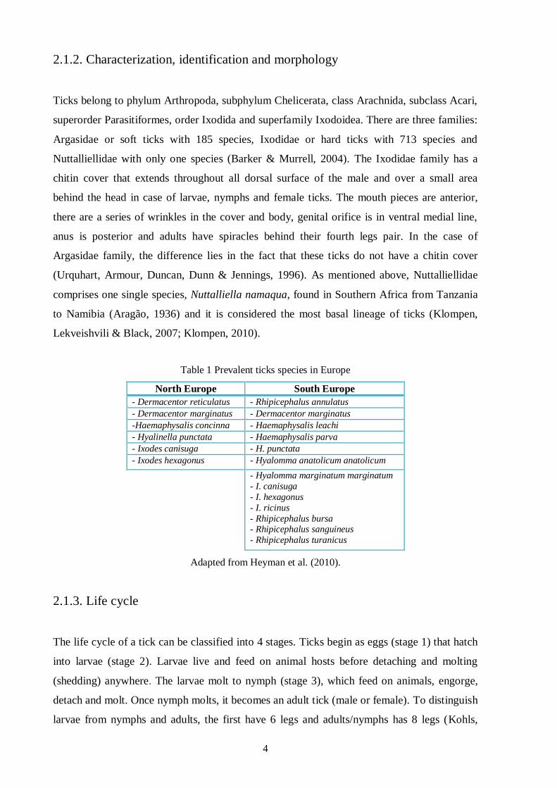
Table 1 Prevalent ticks species in Europe ............................................................................................ 4
Table 2 CRT functions in parasites that cause disease in humans and animals ................................... 26
Table 3 Identity of calreticulin sequences from different ticks ........................................................... 34
Table 4 Gel percentage according to protein size. .............................................................................. 41
xiii
Abbreviations and Symbols
μg Microgram
μl Microliter
µm Micrometer
A, C, G, T Adenine, cytosine, guanine, thymine
Abs Absorbance
A.D. Anno Domini
ArgE Acetylornithinase
AMA-1 Apical membrane antigen 1
BbAMA-1 Babesia bovis apical membrane antigen 1
Bbo-MIC1 Recombinant microneme protein from B. bovis
B.C. Before Christ
Bp Base pair
cDNA Complementary DNA
CRP cAMP regulatory protein
CRT Calreticulin
Cu/Zn-SODM Cu/Zn-superoxide dismutase
DDT Dichlorodiphenyltrichloroethane
DNA Deoxyribonucleic acid
EF-1a Elongation factor-1 alpha
e.g. exempli gratia
EgCRT Recombinant CRT protein from Echinococcus granulosus
ELISA Enzyme-linked immunosorbent assay
ER Endoplasmatic reticulum
F2 Second generation after cross-breeding
FPLC Fast protein liquid chromatography
GST Glutathione-S transferase
Hz Hertz
i.e. id est
IFAT Indirect immunofluorescent antibody test
IMAC Immobilized metal ion affinity chromatography
IPTG Isopropyl-β-D-thiogalactopyranoside
Kb Kilobase
kDa Kilodalton
kHz Kilohertz
LAMP Loop-mediated isothermal PCR
LB-agar Luria Bertani-agar
M Mole
mA Milliampere
ml Millilitre
min Minute
mM Millimole
mRNA Messenger RNA
xiv
MSA Merozoite surface antigen
nm Nanometer
OD Optical density
PBS Phosphate buffered saline
PCR Polymerase chain reaction
pI Isoelectric point
PVDF Polyvinylidene fluoride
rDNA Ribossomal DNA
rBmCRT Recombinant calreticulin protein from R. annulatus
rCRT Recombinant calreticulin
rHlCRT Recombinant calreticulin protein from Haemaphysalis longicornis
rHqCRT Recombinant calreticulin protein from H. qinghaiensis
RLB Reverse line blot
RNA Ribonucleic acid
RNAi RNA interference
rpm Rotations per minute
TAE Tris acetate buffer
TBE Tickborne encephalitis virus
TBV Transmission blocking vaccine
TEMED Tetramethylethylenediamine
Tris-HCl Tris hydrochloride
TTBS Tween-Tris buffer saline
s Second
SDS Sodium dodecyl sulfate
SDS-PAGE Sodium dodecyl sulfate polyacrylamide gel electrophoresis
SEL Selenoprotein W
SlyD Peptidoylproline cis-trans isomerase
SPA Soluble parasite antigens
spp. Species (plural)
S.O.C. Super-optimal catabolite repression medium
UBQ Ubiquitin
V Volt
v/v Volume per volume
w/v Weight per volume
YodA Metal-binding lipocalin
1
CChhaapptteerr 11.. PPrreeaammbbllee
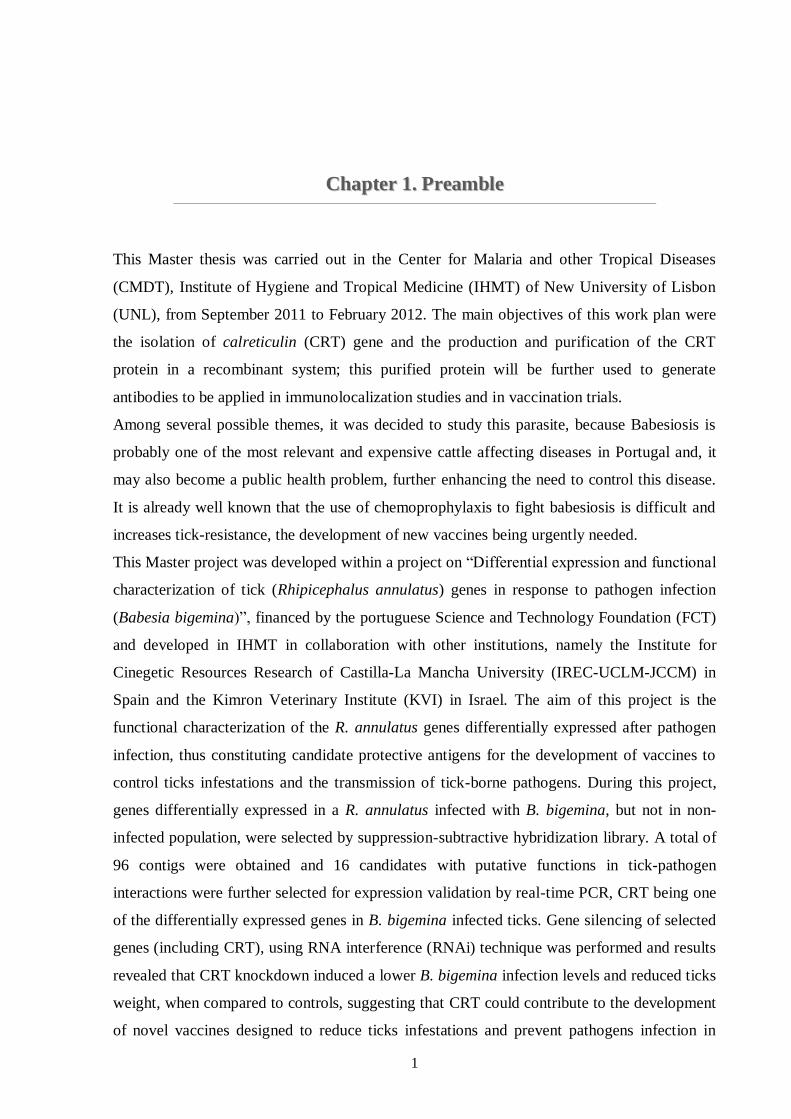
This Master thesis was carried out in the Center for Malaria and other Tropical Diseases
(CMDT), Institute of Hygiene and Tropical Medicine (IHMT) of New University of Lisbon
(UNL), from September 2011 to February 2012. The main objectives of this work plan were
the isolation of calreticulin (CRT) gene and the production and purification of the CRT
protein in a recombinant system; this purified protein will be further used to generate
antibodies to be applied in immunolocalization studies and in vaccination trials.
Among several possible themes, it was decided to study this parasite, because Babesiosis is
probably one of the most relevant and expensive cattle affecting diseases in Portugal and, it
may also become a public health problem, further enhancing the need to control this disease.
It is already well known that the use of chemoprophylaxis to fight babesiosis is difficult and
increases tick-resistance, the development of new vaccines being urgently needed.
This Master project was developed within a project on “Differential expression and functional
characterization of tick (Rhipicephalus annulatus) genes in response to pathogen infection
(Babesia bigemina)”, financed by the portuguese Science and Technology Foundation (FCT)
and developed in IHMT in collaboration with other institutions, namely the Institute for
Cinegetic Resources Research of Castilla-La Mancha University (IREC-UCLM-JCCM) in
Spain and the Kimron Veterinary Institute (KVI) in Israel. The aim of this project is the
functional characterization of the R. annulatus genes differentially expressed after pathogen
infection, thus constituting candidate protective antigens for the development of vaccines to
control ticks infestations and the transmission of tick-borne pathogens. During this project,
genes differentially expressed in a R. annulatus infected with B. bigemina, but not in non-
infected population, were selected by suppression-subtractive hybridization library. A total of
96 contigs were obtained and 16 candidates with putative functions in tick-pathogen
interactions were further selected for expression validation by real-time PCR, CRT being one
of the differentially expressed genes in B. bigemina infected ticks. Gene silencing of selected
genes (including CRT), using RNA interference (RNAi) technique was performed and results
revealed that CRT knockdown induced a lower B. bigemina infection levels and reduced ticks
weight, when compared to controls, suggesting that CRT could contribute to the development
of novel vaccines designed to reduce ticks infestations and prevent pathogens infection in

2
ticks and, consequently, in vertebrate hosts. So, based on these results, CRT was chosen for
gene isolation and recombinant protein production, being the subject of this Master project.
3
CChhaapptteerr 22.. LLiitteerraattuurree RReevviieeww
2.1. Ticks
2.1.1. Introduction
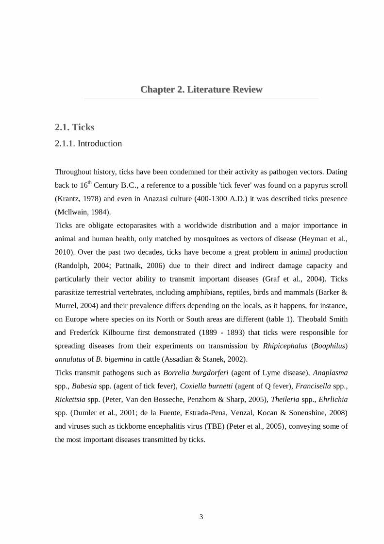
Throughout history, ticks have been condemned for their activity as pathogen vectors. Dating
back to 16th Century B.C., a reference to a possible 'tick fever' was found on a papyrus scroll
(Krantz, 1978) and even in Anazasi culture (400-1300 A.D.) it was described ticks presence
(Mcllwain, 1984).
Ticks are obligate ectoparasites with a worldwide distribution and a major importance in
animal and human health, only matched by mosquitoes as vectors of disease (Heyman et al.,
2010). Over the past two decades, ticks have become a great problem in animal production
(Randolph, 2004; Pattnaik, 2006) due to their direct and indirect damage capacity and
particularly their vector ability to transmit important diseases (Graf et al., 2004). Ticks
parasitize terrestrial vertebrates, including amphibians, reptiles, birds and mammals (Barker &
Murrel, 2004) and their prevalence differs depending on the locals, as it happens, for instance,
on Europe where species on its North or South areas are different (table 1). Theobald Smith
and Frederíck Kilbourne first demonstrated (1889 - 1893) that ticks were responsible for
spreading diseases from their experiments on transmission by Rhipicephalus (Boophilus)
annulatus of B. bigemina in cattle (Assadian & Stanek, 2002).
Ticks transmit pathogens such as Borrelia burgdorferi (agent of Lyme disease), Anaplasma
spp., Babesia spp. (agent of tick fever), Coxiella burnetti (agent of Q fever), Francisella spp.,
Rickettsia spp. (Peter, Van den Bosseche, Penzhom & Sharp, 2005), Theileria spp., Ehrlichia
spp. (Dumler et al., 2001; de la Fuente, Estrada-Pena, Venzal, Kocan & Sonenshine, 2008)
and viruses such as tickborne encephalitis virus (TBE) (Peter et al., 2005), conveying some of
the most important diseases transmitted by ticks.
4
2.1.2. Characterization, identification and morphology
Ticks belong to phylum Arthropoda, subphylum Chelicerata, class Arachnida, subclass Acari,
superorder Parasitiformes, order Ixodida and superfamily Ixodoidea. There are three families:
Argasidae or soft ticks with 185 species, Ixodidae or hard ticks with 713 species and
Nuttalliellidae with only one species (Barker & Murrell, 2004). The Ixodidae family has a
chitin cover that extends throughout all dorsal surface of the male and over a small area
behind the head in case of larvae, nymphs and female ticks. The mouth pieces are anterior,
there are a series of wrinkles in the cover and body, genital orifice is in ventral medial line,
anus is posterior and adults have spiracles behind their fourth legs pair. In the case of
Argasidae family, the difference lies in the fact that these ticks do not have a chitin cover
(Urquhart, Armour, Duncan, Dunn & Jennings, 1996). As mentioned above, Nuttalliellidae
comprises one single species, Nuttalliella namaqua, found in Southern Africa from Tanzania
to Namibia (Aragão, 1936) and it is considered the most basal lineage of ticks (Klompen,
Lekveishvili & Black, 2007; Klompen, 2010).
Table 1 Prevalent ticks species in Europe
North Europe South Europe
- Dermacentor reticulatus - Rhipicephalus annulatus
- Dermacentor marginatus - Dermacentor marginatus
-Haemaphysalis concinna - Haemaphysalis leachi
- Hyalinella punctata - Haemaphysalis parva
- Ixodes canisuga - H. punctata
- Ixodes hexagonus - Hyalomma anatolicum anatolicum
- Hyalomma marginatum marginatum
- I. canisuga
- I. hexagonus
- I. ricinus
- Rhipicephalus bursa - Rhipicephalus sanguineus
- Rhipicephalus turanicus
Adapted from Heyman et al. (2010).
2.1.3. Life cycle
The life cycle of a tick can be classified into 4 stages. Ticks begin as eggs (stage 1) that hatch
into larvae (stage 2). Larvae live and feed on animal hosts before detaching and molting
(shedding) anywhere. The larvae molt to nymph (stage 3), which feed on animals, engorge,
detach and molt. Once nymph molts, it becomes an adult tick (male or female). To distinguish
larvae from nymphs and adults, the first have 6 legs and adults/nymphs has 8 legs (Kohls,
5
Sonenshine & Clifford, 1965). Adults climb up grass and plants to hold their prey and when a
warm-blooded animal walks on the pasture, tick can crawl onto them and begin feeding. Ticks
insert their mouths, attach to their prey and engorge themselves with a blood meal (stage 4).
During feeding, ticks saliva can get into the host body and blood stream.
Usually Ixodes adult ticks take 4 to 10 days to get full engorgement and feed themselves by
forcing their hypostome through the host skin and sucking blood and fluids that are drained
from the resulting wound (Sonenshine, 1991). Ticks from Ixodidae family may have between
one to three vertebral hosts. The coupling occurs in the host, where the adult female tick feeds
itself for several days; then it falls into the ground, where the oviposition of millions of eggs
occurs, culminating with tick’s death. The male ticks can couple many times without dying,
feeding themselves intermitting. In the Argasidae family, females feed themselves frequently,
coupling happens outside the host and oviposition occurs more than once in ticks life
(Urquhart et al., 1996).
Figure 1 One host ticks life cycle.
Adult Larvae Nymph
Females drop of the host do lay
eggs
Eggs hatch in larvae
One host ticks’-life cycle
6
2.1.4. Tick control
2.1.4.1. Introduction
Ticks control is essential to diminish tick-borne diseases prevalence and, therefore, reduce
ticks impact on livestock productivity. Nowadays, ticks control is mainly based in the
application of acaricides, despite their disadvantages and limitations, although other control
methods as vaccination or biocontrol agents are available (Willadsen, 2006). Probably,
vaccination will be one of the most important methods for ticks control and its transmited
pathogens, because several different vaccines have already been used against B. bovis and B.
bigemina in Australia (Bock & de Vos, 2001) or against Theileria annulata in Israel (Shkap &
Pipano, 2000) and China (Gu et al., 1997; Shirong, 1997) with good results.
2.1.4.2. Chemoprophylaxis
There are many acaricides that can be used against ticks: pyrethroids as flumethrin and
deltamethrin; organochlorines as dichlorodiphenyltrichloroethane (DDT); organophosphates
as diazinon and coumaphos; carbamates as carbaril; formamidines as amitraz; cicloamidines
as clenpirin and macrocyclic lactones (avermectins and milbemycins), among others (Botana,
Landoni & Martín-Jiménez, 2002). Synthetic flumethrin in pour-on protects cattle from ticks
for 2 weeks and, in case of deltamethrin, for 3 weeks; this approach, in the case of babesiosis
control, led to a significant decay in clinical disease (Zintl, Mulcahy, Skerrett, Taylor & Gray,
2003). The application of acaricides has some drawbacks: the appearance of residues in milk
and meat products, the environmental contamination and, most important nowadays, the
development of ticks’ resistance against acaricides (Botana et al., 2002). This resistance is
mainly due to mutations in genes encoding detoxificating enzymes, as esterases, glutathione-
S-transferases and monooxidases and due to genetic drift (Rosario-Cruz et al., 2009).
Resistance to compounds such as organophosphorous, pyrethroids and amitraz has been
described for R. microplus (Ortiz et al., 1995; Soberanes, Santamaría, Fragoso & García,
2002). Pesticides rotation is used in crop agriculture to minimize resistance, but its application
in ticks control is not widely used yet. The association of acaricides with vaccines acts in a
synergetic manner, because, in the case of R. microplus, efficacy of macrocyclic lactone
acaricides is deeply enhanced in cattle vaccinated against those ticks (Kemp, McKenna,
Thullner & Willadsen, 1999).
7
2.1.4.3. Biocontrol
Currently, an appeal is being made to biocontrol and to the use of more “environmental
friendly” acaricide products. Like other parasites, ticks carry some microorganisms in their
bodies, which, along with ticks, are essential for the survival of each other (endosymbiosis).
Since endosymbionts are essential for ticks’ survival, elimination of those microorganisms
would be deleterious for the continued existence, growth and development of ticks. Those
endosymbionts could be either destroyed by a chemical approach or used to animals’
immunization, in order to interfere in ticks’ nutrition (Noda, Munderloh & Kuffi, 1997;
Benson, Grawronski, Eveleigh & Benson, 2004). The use of other organisms pathogenic to
ticks, like fungi of genera Beauveria and Metarhizium (Frazzon, Vaz Junior, Masuda,
Scharank & Vainstein, 2000; Gindin, Samish, Zangi, Mishoutchenko & Glazer, 2002) or
herbal acaricides (Khudrathulla & Jagannath, 2000; Lundh, Wiktelius & Chirico, 2005) are an
alternative and important possibility of achieve the control of ticks.
2.1.4.4. Genetic resistance
The genetic resistance to ticks and tick-borne diseases is complex, but, facing other control
methods and their problems, breeds resistance has become an important parameter in some
regions.
Bos indicus cattle is more resistant to R. microplus ticks and babesiosis than Bos taurus, this
acquire resistance being heritable, though those animals remain susceptible to A. marginale
(Bock et al., 1997, 1999). This additional resistance is possibly due to the fact that zebuine
cattle breeds have more dermal mast cells than taurine breeds, as F2 crossbreed cattle has a
higher developed resistance against ticks correlated with an increased number of mast cells in
dermis (Engracia Filho, Bechara & Teodoro, 2006).
2.1.4.5. Vaccines
2.1.4.5.1. Introduction
Major alternatives to conventional acaricide treatments have been developed in recent years
and vaccines are among the most important developments. It is now a decade since the first
commercial vaccine against R. microplus based on the recombinant antigen Bm86 was
released (Willadsen, Bird, Cobon & Hungerford, 1995; de la Fuente et al., 1999).
In the market there are two vaccines based on recombinant R. microplus Bm86 gut antigen:
8
Gavac™ vaccine (Heber Biotec S.A., Havana, Cuba) and TickGARD (Hoechst Animal
Health, Australia), which confirming the advantages of tick control by this method, because it
is cost-effective, it reduces environmental contamination, it prevents acaricide-resistant ticks
selection and it can reduce pathogens transmission, by decreasing ticks number and/or
changing their vector capacity. This control scheme also has another advantage, as vaccines
can protect the animal-host against both pathogens and vector, especially if antigens are
conserved in both species (de la Fuente et al., 1998; de la Fuente & Kocan, 2003; de la
Fuente, Kocan & Blouin, 2007).
Nevertheless, there is a problem with vaccines formulation because many of tick-protective
antigens studied for propose of a future application are cytoplasmic and highly conserved,
which can favour host tolerance to them and a total inefficacy of vaccine; yet this is one of the
most promisor methods to fight these ectoparasites (Almazán et al., 2010).
There are two different kinds of antigens, the exposed and the concealed antigens (Willadsen,
1980). The first type is the most used in vaccines production, even though vaccines with
concealed antigens may inhibit other parasites development, such as Babesia spp., by altering
gut homeostasis and preventing pathogen transmission (Rachinsky, Guerrero & Scoles, 2007).
Another kind of vaccines are the transmission blocking vaccines (TBV), which are supposed
to block pathogen development in arthropod vectors through targeting pathogen or arthropod
molecules as transmission blocking targets (Carter, 2001).
2.1.4.5.2. Recombinant vaccines
These vaccines use recombinant proteins as antigens to immunize animals and have several
advantages, such as prevention or reduction of pathogens transmission (de la Fuente et al.,
1998), environmental safety, low cost production (Odongo et al., 2007), prevention of drug-
resistant ticks selection and inclusion of multiple antigens that could target several tick
species (de la Fuente & Kocan, 2006), among others.
Canales, Almazán, Naranjo, Jongejan & de la Fuente (2009) have cloned ortholog genes
(Ba86 and Bm86 genes) from R. annulatus and R. microplus, respectively, which were used
in vaccine trials later on. Vaccination of cattle with Ba86 reduced, respectively for R.
annulatus and R. microplus, ticks infestations (71% and 40%), ticks weight (8% and 15%),
oviposition (22% and 5%) and eggs fertility (25% and 50%). For R. decoloratus, Odongo et
al. (2007), using a Bm86 based-vaccine, found a reduction of 46% on engorged adult female
ticks number, 56% on ticks weight and 61% on eggs weight after cattle immunization and de
9
Vos, Zeinstra, Taoufik, Willadsen & Jongejan (2001) described a reduction of 70% in R.
decoloratus reproductive capacity after feeding on vaccinated calves and 50% in engorged H.
anatolicum anatolicum nymphs total weight. Despite these results, vaccine had no action
against A. variegatum and R. appendiculatus nymphs. Pipano et al. (2003) tested the efficacy
of a Bm86 vaccine in protection against ticks and pathogens (B. bovis and B. bigemina)
transmitted by those ectoparasites. The results showed that immunized cattle, when
challenged with B. bovis-infected ticks, continued to become infected, but in the case of
B. bigemina, Bm86-immunized animals remained protected against infection, probably due to
the fact that larvae didn’t molt to nymph. Still related to the Bm86 gene, Bastos, Ueti,
Knowles & Scoles (2010) studied the effect of Bm86 gene silencing on fitness of R.
microplus ticks fed in B. bovis infected cattle, showing that this procedure decreased survival
engorged ticks rate and eggs weight. A gene from R. microplus (strain A) called Bm95,
homologue to Bm86, was used to immunize cattle, after it has been discovered that some R.
microplus ticks (strain A) had a moderate low susceptibly to Bm86 vaccine, possibly due to a
genetic variation in ticks (Freeman, Davey, Kappmeyer, Kammlah & Olafson, 2010). Bm95
immunized cattle showed a higher protection efficacy to both susceptible and strain A than
Bm86 vaccine (García-García et al., 2000). Other authors showed that Bm95 antigen based
vaccine presents better responses than Bm86 vaccine to low susceptibility ticks populations
(Jittapalapong et al., 2010).
Immunization trials in cattle with recombinant subolesin, a conserved protein among
vertebrates and insects, decreased R. microplus survival and reproduction rates (Merino et al.,
2011), weight and oviposition (de la Fuente et al., 2011) and, moreover, protein knockdown
led to degeneration of ticks parts as embryos, salivary glands and reproductive tissues
(Merino et al., 2011). Further studies with this antigen showed that immunization of rats
decreased vector capacity of I. scapularis nymphs for A. phagocytophilum (de la Fuente &
Kocan, 2006).
The recombinant antigen 64P from R. appendiculatus was found to be involved in ticks
attachment and feeding and was used to immunize guinea pigs, reducing, respectively, 48%
and 70% nymph and adult infestation rates; additionally, response of animals immunized with
64P was similar to those observed during development of natural resistance in guinea pigs
infested with R. appendiculatus, showing the typical local cutaneous inflammatory immune
response (Trimnell, Hails & Nuttall, 2002). The recombinant protein from R. sanguineus and
I. ricinus was again tested by Trimnell et al. (2002), respectively in guinea pigs and hamsters,
demonstrating damage on ticks post-challenge. Other study indicated that there is an antigenic
cross-reactivity between tick extracts of R. sanguineus, I. ricinus, A. variegatum and R.
10
microplus, showing the potential of this protein to develop a large-spectrum anti-tick vaccine
(Havlíková et al., 2009).
2.1.4.5.3. Antigens selection
Molecular tools are important experimental components for the study of tick gene functions.
RNAi technique allows silencing of gene expression, contributing to the characterization of
gene function and its phenotypic effect (Fire et al., 1998). The first report related to the use of
RNAi in ticks belongs to Aljamali, Sauer & Esseberg (2002) and quickly that technique has
become universally adopted for gene-silencing in ticks (de la Fuente & Kocan, 2006).
Kocan, Manzano-Roman & de la Fuente (2007) showed that subolesin knockdown in I.
scapularis, D. variabilis and A. americanum also affected oviposition, eggs embryogenesis,
larval hatching and fertility (reduction of 93% and 71% for D. variabilis and I. scapularis,
respectively). Subolesin expression is activated when ticks are infected with A. marginale and
B. bigemina, suggesting a connection between tick gene expression and pathogen-infection
(Merino et al., 2011). Zivkovic et al. (2010) obtained the opposite result, showing that D.
variabilis, D. andersoni, D. reticulatus, R. sanguineus, R. microplus and R. annulatus had
equal or even lower mRNAs level than control groups.
Almazán et al. (2010) studied the knockdown effect of several R. microplus genes, such as
glutathione-S transferase (GST), ubiquitin (UBQ), selenoprotein W (SEL), elongation factor-
1 alpha (EF-1a) and subolesin, and found out that GST and SEL genes knockdown lead to a
lower ticks attachment when compared to control group, but did not influence ticks mortality
or oviposition, though the other three genes results showed an increase on R. microplus
mortality and reduction of oviposition.
11
2.2. Babesia
2.2.1. Introduction
Babesia spp. was first discovered in the 19th
century in association to bovine hemoglobinuria
(Babes, 1888). In 1893, Smith and Kilbourne recognized B. bigemina as the causative agent
of Texas Cattle Fever (Smith & Kilbourne, 1893).
All over the world, there are some 100 Babesia species or even more (Homer, Aguilar-Delfin,
Telford 3rd
, Krause & Persing, 2000), Rhipicephalus spp. being one of its main tick vector
(Urquhart et al., 1996). Bovine babesiosis are generally caused by B. bovis, B. bigemina and
B. divergens which are the most important Babesia species for cattle babesiosis; however B.
major can also cause disease in cattle (Zintl et al., 2003) and in the case of human babesiosis,
B. divergens and B. microti are the most dangerous species (Antunes, 2008). The different
Babesia species infect a large variety of animals (Bock, Jackson, de Vos & Jorgensen, 2004;
de Vos & Geysen, 2004) and in cattle they can induce animal mortality, abortions, reduction
of milk/meat production, and, sometimes, neurological symptoms (Saegerman et al., 2003).
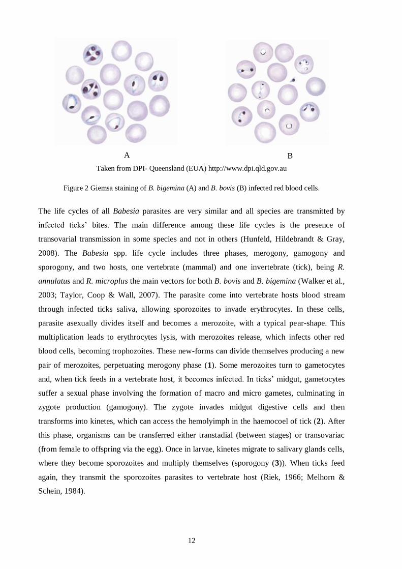
2.2.2. Characterization and life cycle
Babesia spp. belongs to phylum Apicomplexa, class Aconoidasida, family Babesiidae and
genus Babesia. In blood smears stained by Giemsa method, Babesia parasites usually appear
in pairs, with a pear-shape, an elongated or a cigar form with a red nucleus and a blue
cytoplasm (figure 2). Concerning to its size, Babesia species can be divided in small and big
Babesia, with 1.0-2.5 µm and 2.5-5.0 µm of length, respectively (Urquhart et al., 1996).
These morphological categorizations are usually consistent with the phylogenetic
characterization based on nuclear small subunit-ribossomal RNA gene (18S rDNA)
sequences, showing that small babesias are divided in two different phylogenetic clusters
(Homer et al., 2000).
12
Taken from DPI- Queensland (EUA) http://www.dpi.qld.gov.au
Figure 2 Giemsa staining of B. bigemina (A) and B. bovis (B) infected red blood cells.
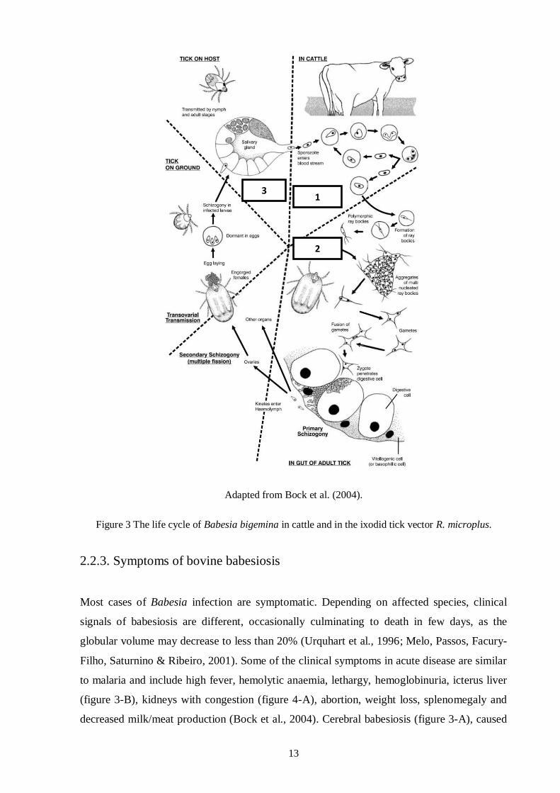
The life cycles of all Babesia parasites are very similar and all species are transmitted by
infected ticks’ bites. The main difference among these life cycles is the presence of
transovarial transmission in some species and not in others (Hunfeld, Hildebrandt & Gray,
2008). The Babesia spp. life cycle includes three phases, merogony, gamogony and
sporogony, and two hosts, one vertebrate (mammal) and one invertebrate (tick), being R.
annulatus and R. microplus the main vectors for both B. bovis and B. bigemina (Walker et al.,
2003; Taylor, Coop & Wall, 2007). The parasite come into vertebrate hosts blood stream
through infected ticks saliva, allowing sporozoites to invade erythrocytes. In these cells,
parasite asexually divides itself and becomes a merozoite, with a typical pear-shape. This
multiplication leads to erythrocytes lysis, with merozoites release, which infects other red
blood cells, becoming trophozoites. These new-forms can divide themselves producing a new
pair of merozoites, perpetuating merogony phase (1). Some merozoites turn to gametocytes
and, when tick feeds in a vertebrate host, it becomes infected. In ticks’ midgut, gametocytes
suffer a sexual phase involving the formation of macro and micro gametes, culminating in
zygote production (gamogony). The zygote invades midgut digestive cells and then
transforms into kinetes, which can access the hemolyimph in the haemocoel of tick (2). After
this phase, organisms can be transferred either transtadial (between stages) or transovariac
(from female to offspring via the egg). Once in larvae, kinetes migrate to salivary glands cells,
where they become sporozoites and multiply themselves (sporogony (3)). When ticks feed
again, they transmit the sporozoites parasites to vertebrate host (Riek, 1966; Melhorn &
Schein, 1984).
A B
13
Adapted from Bock et al. (2004).
Figure 3 The life cycle of Babesia bigemina in cattle and in the ixodid tick vector R. microplus.
2.2.3. Symptoms of bovine babesiosis
Most cases of Babesia infection are symptomatic. Depending on affected species, clinical
signals of babesiosis are different, occasionally culminating to death in few days, as the
globular volume may decrease to less than 20% (Urquhart et al., 1996; Melo, Passos, Facury-
Filho, Saturnino & Ribeiro, 2001). Some of the clinical symptoms in acute disease are similar
to malaria and include high fever, hemolytic anaemia, lethargy, hemoglobinuria, icterus liver
(figure 3-B), kidneys with congestion (figure 4-A), abortion, weight loss, splenomegaly and
decreased milk/meat production (Bock et al., 2004). Cerebral babesiosis (figure 3-A), caused
3 1
2
14
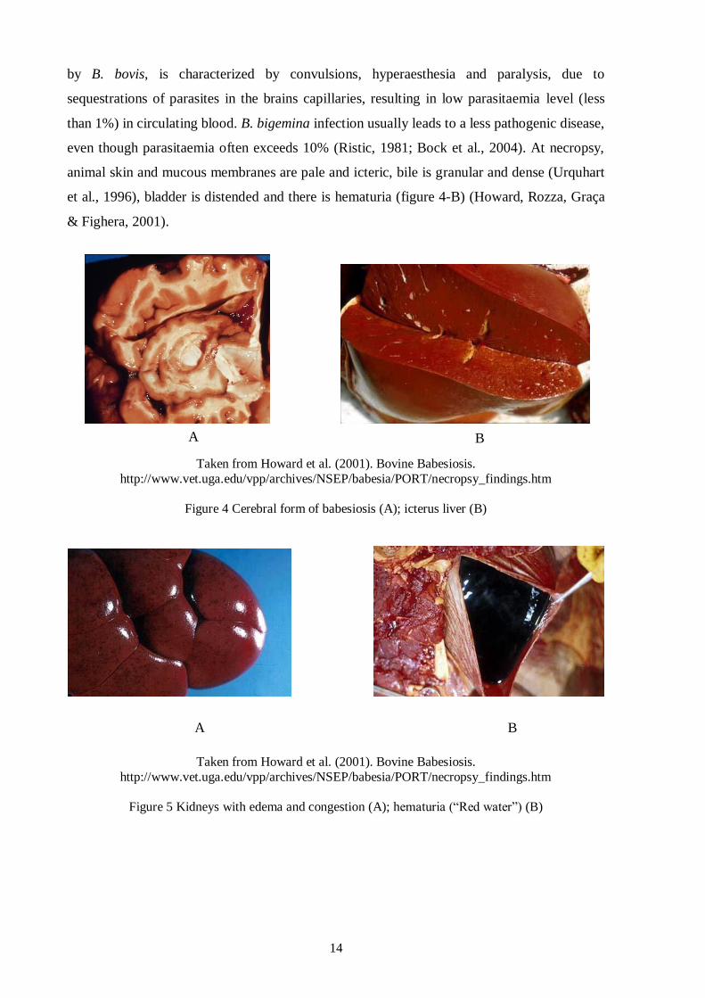
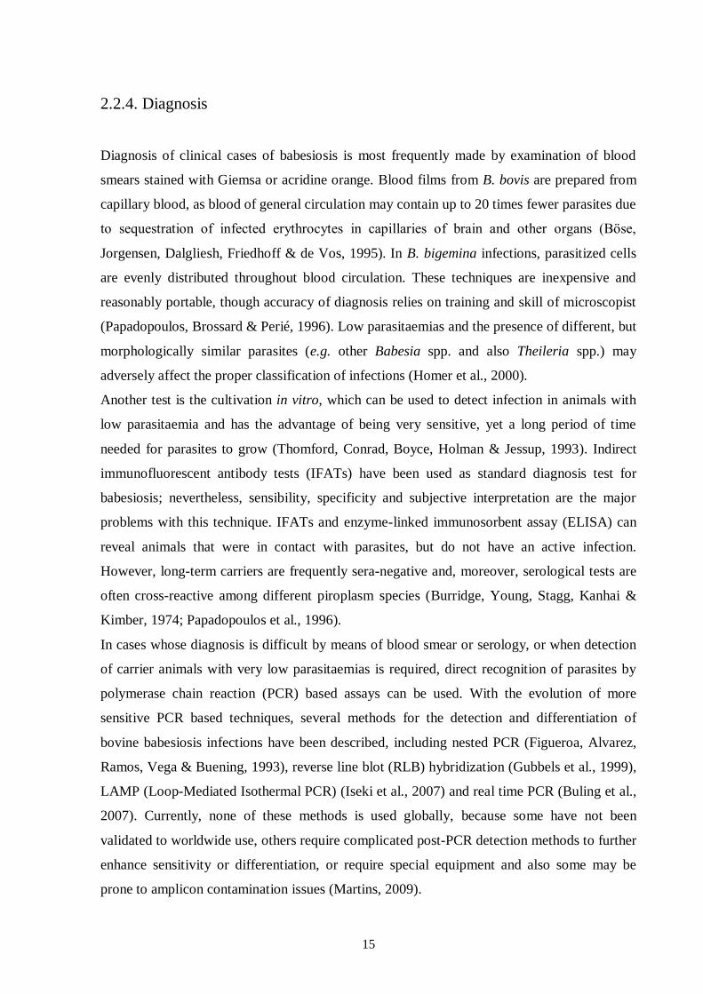
by B. bovis, is characterized by convulsions, hyperaesthesia and paralysis, due to
sequestrations of parasites in the brains capillaries, resulting in low parasitaemia level (less
than 1%) in circulating blood. B. bigemina infection usually leads to a less pathogenic disease,
even though parasitaemia often exceeds 10% (Ristic, 1981; Bock et al., 2004). At necropsy,
animal skin and mucous membranes are pale and icteric, bile is granular and dense (Urquhart
et al., 1996), bladder is distended and there is hematuria (figure 4-B) (Howard, Rozza, Graça
& Fighera, 2001).
Taken from Howard et al. (2001). Bovine Babesiosis.
http://www.vet.uga.edu/vpp/archives/NSEP/babesia/PORT/necropsy_findings.htm
Figure 4 Cerebral form of babesiosis (A); icterus liver (B)
Taken from Howard et al. (2001). Bovine Babesiosis.
http://www.vet.uga.edu/vpp/archives/NSEP/babesia/PORT/necropsy_findings.htm
Figure 5 Kidneys with edema and congestion (A); hematuria (“Red water”) (B)
A B
A B
15
2.2.4. Diagnosis
Diagnosis of clinical cases of babesiosis is most frequently made by examination of blood
smears stained with Giemsa or acridine orange. Blood films from B. bovis are prepared from
capillary blood, as blood of general circulation may contain up to 20 times fewer parasites due
to sequestration of infected erythrocytes in capillaries of brain and other organs (Bӧse,
Jorgensen, Dalgliesh, Friedhoff & de Vos, 1995). In B. bigemina infections, parasitized cells
are evenly distributed throughout blood circulation. These techniques are inexpensive and
reasonably portable, though accuracy of diagnosis relies on training and skill of microscopist
(Papadopoulos, Brossard & Perié, 1996). Low parasitaemias and the presence of different, but
morphologically similar parasites (e.g. other Babesia spp. and also Theileria spp.) may
adversely affect the proper classification of infections (Homer et al., 2000).
Another test is the cultivation in vitro, which can be used to detect infection in animals with
low parasitaemia and has the advantage of being very sensitive, yet a long period of time
needed for parasites to grow (Thomford, Conrad, Boyce, Holman & Jessup, 1993). Indirect
immunofluorescent antibody tests (IFATs) have been used as standard diagnosis test for
babesiosis; nevertheless, sensibility, specificity and subjective interpretation are the major
problems with this technique. IFATs and enzyme-linked immunosorbent assay (ELISA) can
reveal animals that were in contact with parasites, but do not have an active infection.
However, long-term carriers are frequently sera-negative and, moreover, serological tests are
often cross-reactive among different piroplasm species (Burridge, Young, Stagg, Kanhai &
Kimber, 1974; Papadopoulos et al., 1996).
In cases whose diagnosis is difficult by means of blood smear or serology, or when detection
of carrier animals with very low parasitaemias is required, direct recognition of parasites by
polymerase chain reaction (PCR) based assays can be used. With the evolution of more
sensitive PCR based techniques, several methods for the detection and differentiation of
bovine babesiosis infections have been described, including nested PCR (Figueroa, Alvarez,
Ramos, Vega & Buening, 1993), reverse line blot (RLB) hybridization (Gubbels et al., 1999),
LAMP (Loop-Mediated Isothermal PCR) (Iseki et al., 2007) and real time PCR (Buling et al.,
2007). Currently, none of these methods is used globally, because some have not been
validated to worldwide use, others require complicated post-PCR detection methods to further
enhance sensitivity or differentiation, or require special equipment and also some may be
prone to amplicon contamination issues (Martins, 2009).
16
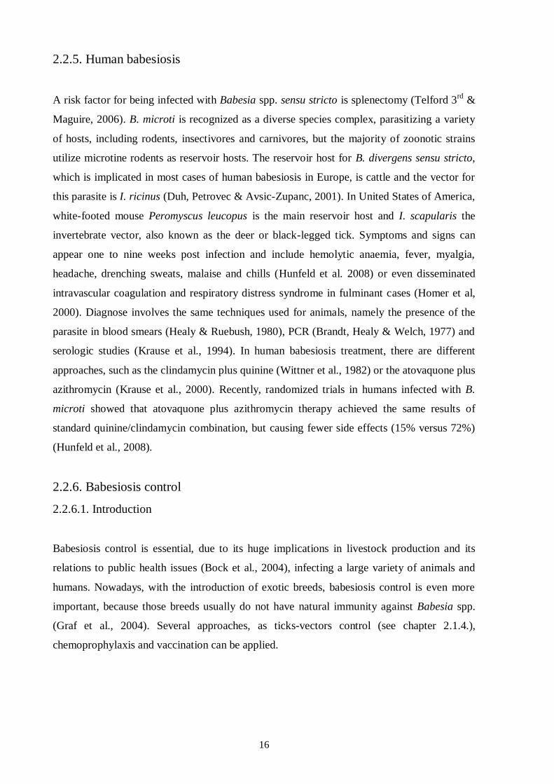
2.2.5. Human babesiosis
A risk factor for being infected with Babesia spp. sensu stricto is splenectomy (Telford 3rd
&
Maguire, 2006). B. microti is recognized as a diverse species complex, parasitizing a variety
of hosts, including rodents, insectivores and carnivores, but the majority of zoonotic strains
utilize microtine rodents as reservoir hosts. The reservoir host for B. divergens sensu stricto,
which is implicated in most cases of human babesiosis in Europe, is cattle and the vector for
this parasite is I. ricinus (Duh, Petrovec & Avsic-Zupanc, 2001). In United States of America,
white-footed mouse Peromyscus leucopus is the main reservoir host and I. scapularis the
invertebrate vector, also known as the deer or black-legged tick. Symptoms and signs can
appear one to nine weeks post infection and include hemolytic anaemia, fever, myalgia,
headache, drenching sweats, malaise and chills (Hunfeld et al. 2008) or even disseminated
intravascular coagulation and respiratory distress syndrome in fulminant cases (Homer et al,
2000). Diagnose involves the same techniques used for animals, namely the presence of the
parasite in blood smears (Healy & Ruebush, 1980), PCR (Brandt, Healy & Welch, 1977) and
serologic studies (Krause et al., 1994). In human babesiosis treatment, there are different
approaches, such as the clindamycin plus quinine (Wittner et al., 1982) or the atovaquone plus
azithromycin (Krause et al., 2000). Recently, randomized trials in humans infected with B.
microti showed that atovaquone plus azithromycin therapy achieved the same results of
standard quinine/clindamycin combination, but causing fewer side effects (15% versus 72%)
(Hunfeld et al., 2008).
2.2.6. Babesiosis control
2.2.6.1. Introduction
Babesiosis control is essential, due to its huge implications in livestock production and its
relations to public health issues (Bock et al., 2004), infecting a large variety of animals and
humans. Nowadays, with the introduction of exotic breeds, babesiosis control is even more
important, because those breeds usually do not have natural immunity against Babesia spp.
(Graf et al., 2004). Several approaches, as ticks-vectors control (see chapter 2.1.4.),
chemoprophylaxis and vaccination can be applied.
17
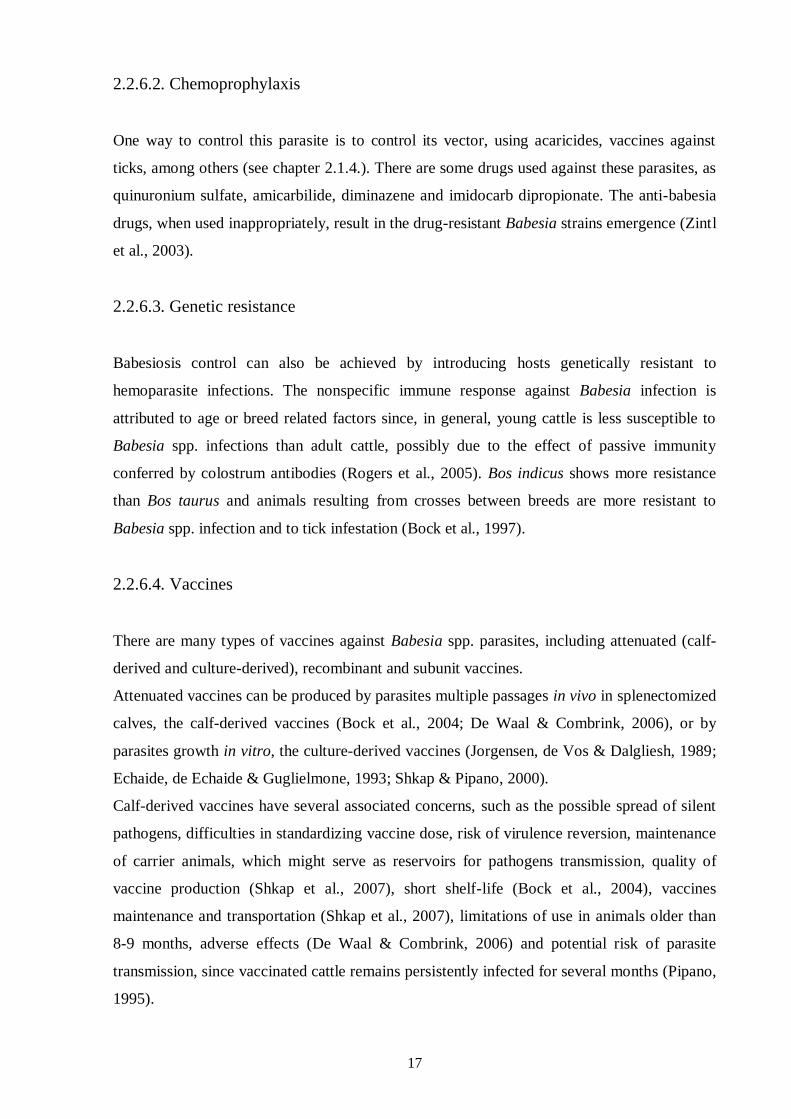
2.2.6.2. Chemoprophylaxis
One way to control this parasite is to control its vector, using acaricides, vaccines against
ticks, among others (see chapter 2.1.4.). There are some drugs used against these parasites, as
quinuronium sulfate, amicarbilide, diminazene and imidocarb dipropionate. The anti-babesia
drugs, when used inappropriately, result in the drug-resistant Babesia strains emergence (Zintl
et al., 2003).
2.2.6.3. Genetic resistance
Babesiosis control can also be achieved by introducing hosts genetically resistant to
hemoparasite infections. The nonspecific immune response against Babesia infection is
attributed to age or breed related factors since, in general, young cattle is less susceptible to
Babesia spp. infections than adult cattle, possibly due to the effect of passive immunity
conferred by colostrum antibodies (Rogers et al., 2005). Bos indicus shows more resistance
than Bos taurus and animals resulting from crosses between breeds are more resistant to
Babesia spp. infection and to tick infestation (Bock et al., 1997).
2.2.6.4. Vaccines
There are many types of vaccines against Babesia spp. parasites, including attenuated (calf-
derived and culture-derived), recombinant and subunit vaccines.
Attenuated vaccines can be produced by parasites multiple passages in vivo in splenectomized
calves, the calf-derived vaccines (Bock et al., 2004; De Waal & Combrink, 2006), or by
parasites growth in vitro, the culture-derived vaccines (Jorgensen, de Vos & Dalgliesh, 1989;
Echaide, de Echaide & Guglielmone, 1993; Shkap & Pipano, 2000).
Calf-derived vaccines have several associated concerns, such as the possible spread of silent
pathogens, difficulties in standardizing vaccine dose, risk of virulence reversion, maintenance
of carrier animals, which might serve as reservoirs for pathogens transmission, quality of
vaccine production (Shkap et al., 2007), short shelf-life (Bock et al., 2004), vaccines
maintenance and transportation (Shkap et al., 2007), limitations of use in animals older than
8-9 months, adverse effects (De Waal & Combrink, 2006) and potential risk of parasite
transmission, since vaccinated cattle remains persistently infected for several months (Pipano,
1995).
18
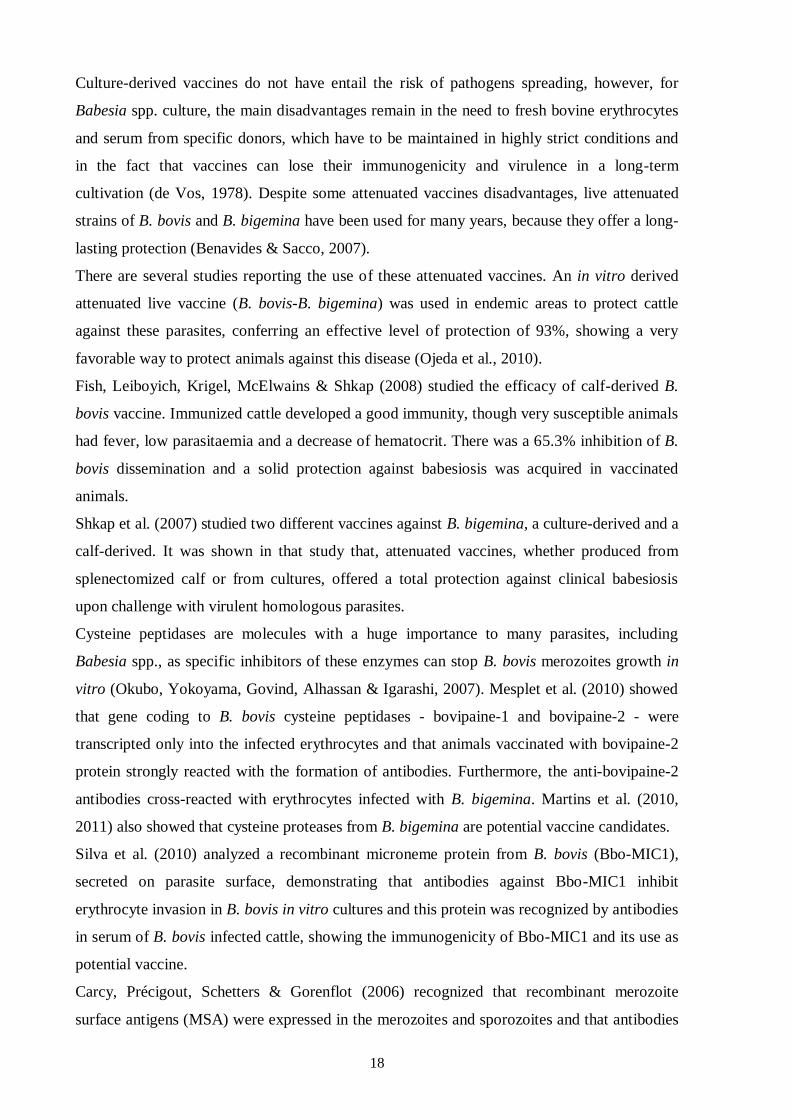
Culture-derived vaccines do not have entail the risk of pathogens spreading, however, for
Babesia spp. culture, the main disadvantages remain in the need to fresh bovine erythrocytes
and serum from specific donors, which have to be maintained in highly strict conditions and
in the fact that vaccines can lose their immunogenicity and virulence in a long-term
cultivation (de Vos, 1978). Despite some attenuated vaccines disadvantages, live attenuated
strains of B. bovis and B. bigemina have been used for many years, because they offer a long-
lasting protection (Benavides & Sacco, 2007).
There are several studies reporting the use of these attenuated vaccines. An in vitro derived
attenuated live vaccine (B. bovis-B. bigemina) was used in endemic areas to protect cattle
against these parasites, conferring an effective level of protection of 93%, showing a very
favorable way to protect animals against this disease (Ojeda et al., 2010).
Fish, Leiboyich, Krigel, McElwains & Shkap (2008) studied the efficacy of calf-derived B.
bovis vaccine. Immunized cattle developed a good immunity, though very susceptible animals
had fever, low parasitaemia and a decrease of hematocrit. There was a 65.3% inhibition of B.
bovis dissemination and a solid protection against babesiosis was acquired in vaccinated
animals.
Shkap et al. (2007) studied two different vaccines against B. bigemina, a culture-derived and a
calf-derived. It was shown in that study that, attenuated vaccines, whether produced from
splenectomized calf or from cultures, offered a total protection against clinical babesiosis
upon challenge with virulent homologous parasites.
Cysteine peptidases are molecules with a huge importance to many parasites, including
Babesia spp., as specific inhibitors of these enzymes can stop B. bovis merozoites growth in
vitro (Okubo, Yokoyama, Govind, Alhassan & Igarashi, 2007). Mesplet et al. (2010) showed
that gene coding to B. bovis cysteine peptidases - bovipaine-1 and bovipaine-2 - were
transcripted only into the infected erythrocytes and that animals vaccinated with bovipaine-2
protein strongly reacted with the formation of antibodies. Furthermore, the anti-bovipaine-2
antibodies cross-reacted with erythrocytes infected with B. bigemina. Martins et al. (2010,
2011) also showed that cysteine proteases from B. bigemina are potential vaccine candidates.
Silva et al. (2010) analyzed a recombinant microneme protein from B. bovis (Bbo-MIC1),
secreted on parasite surface, demonstrating that antibodies against Bbo-MIC1 inhibit
erythrocyte invasion in B. bovis in vitro cultures and this protein was recognized by antibodies
in serum of B. bovis infected cattle, showing the immunogenicity of Bbo-MIC1 and its use as
potential vaccine.
Carcy, Précigout, Schetters & Gorenflot (2006) recognized that recombinant merozoite
surface antigens (MSA) were expressed in the merozoites and sporozoites and that antibodies
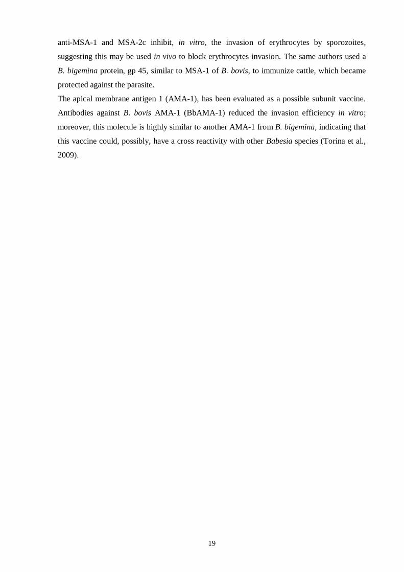
19
anti-MSA-1 and MSA-2c inhibit, in vitro, the invasion of erythrocytes by sporozoites,
suggesting this may be used in vivo to block erythrocytes invasion. The same authors used a
B. bigemina protein, gp 45, similar to MSA-1 of B. bovis, to immunize cattle, which became
protected against the parasite.
The apical membrane antigen 1 (AMA-1), has been evaluated as a possible subunit vaccine.
Antibodies against B. bovis AMA-1 (BbAMA-1) reduced the invasion efficiency in vitro;
moreover, this molecule is highly similar to another AMA-1 from B. bigemina, indicating that
this vaccine could, possibly, have a cross reactivity with other Babesia species (Torina et al.,
2009).

21
2.3. Calreticulin
2.3.1. Introduction
CRT was first isolated by Ostwald and Maclennan in 1974 and further cloned by Fliegel,
Burns, MacLennan, Reithmeier & Michalak in 1989 (Fliegel et al., 1989).
CRT has been identified in several species, but there is no CRT genes identified in yeasts and
prokaryotes, whose genomes were totally sequenced (Persson, Rosenquist & Sommarin,
2002). CRT importance to cells is relevant, given that the absence of CRT gene is
embryonically lethal (Mesaeli et al., 1999). Since its first detection, CRT has been identified
in other cellular structures, as cytoplasm, cell membranes and extracellular matrix (Burns,
Atkinson, Bleackley & Michalak, 1994); several functions have been attributed to CRT, such
as acting as a chaperone to help other proteins to fold correctly (Nauseef, McCormick &
Clark, 1995). This activity was demonstrated by immunoprecipitation experiments, which
showed that CRT associates itself transiently to several cellular proteins immediately after
their synthesis (Peterson, Ora, Van & Helenius, 1995). When a polypeptide is incorrectly
folded, it bounds to one or more chaperones (including CRT) and is retained in endoplasmatic
reticulum (ER), mechanism that prevents expression, aggregation and secretion of misfolded
proteins (Nauseef et al., 1995).
22
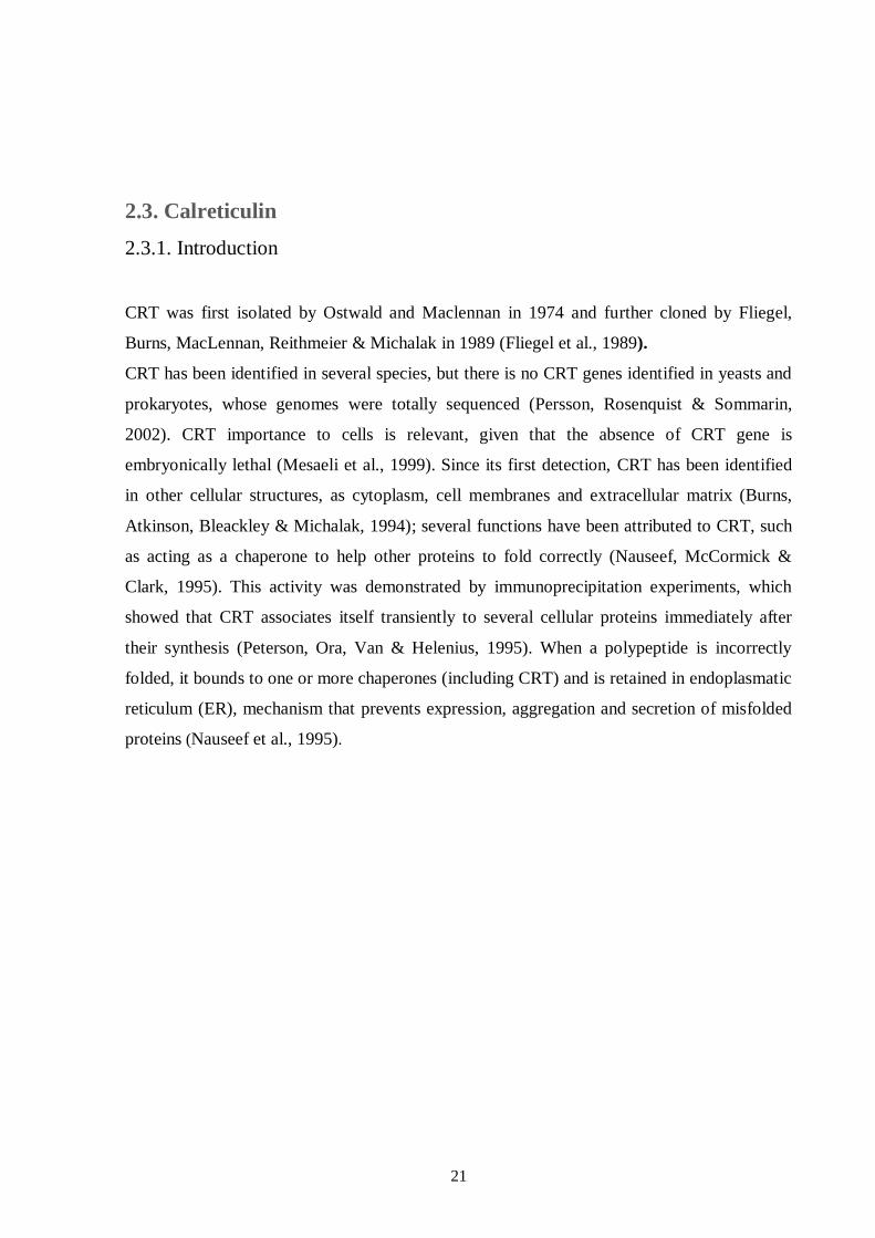
Adapted from Mendlovic & Conconi (2011).
Figure 6 CRT action as a chaperone.
Besides the essential functions that CRT performs in ER lumen, this multifaceted protein has
also been implicated in many unexpected roles that occur at cells surface, cytosol, nucleus and
extracellular matrix. Indeed, since discovery of CRT chaperone and calcium-regulating
functions, scientists have learned that CRT has many other duties in the cell. This protein has
been implicated in diverse cellular processes including signaling (Mitra & Schlaepfer, 2006),
regulation of gene expression (Gardai et al., 2005), wound healing (Pallero, Elzie, Chen,
Mosher & Murphy-Ullrich, 2008), removal of cancer cells (Tesniere et al., 2008) and
autoimmunity (Eggleton & Llewellyn, 1999).
2.3.2. Calreticulin structure
CRT has two codifying genes, CRT-1 and CRT-2 (Persson et al., 2002), and is highly
conservative with a 96% amino acid identity for CRT from human, rabbit, rat and mouse
(Waser, Mesaeli, Spencer & Michalak, 1997). The CRT gene has nine exons and is localized
in chromosomes 19 and 8, respectively for human and mouse genes. These sequences have
more than 70% identity, with the exception of introns 3 and 6, showing a great gene
23
evolutionary conservation (McCauliffe, Yang, Wilson, Sontheimer & Capra, 1992). CRT
gene from ticks has two exons and one intron, the last one with a conserved position, but
variable size and nucleotide sequence. In R. annulatus CRT gene, intron is localized between
nucleotides 88 and 412 and has 1559 base pairs (bp) (Xu, Fang, Sun, Keirans & Durden,
2005).
CRT proteins have a molecular weight around 46 kDa (Fliegel et al., 1989), an N-terminal
cleavable amino acid signal sequence and a KDEL ER retrieval signal in the C-terminal
domain. These amino acids are responsible for CRT targeting and retaining in ER lumen
(Michalak et al., 1999). The protein has three cysteine residues, all located in N-domain of the
protein. Two of those cysteine residues form a disulphide bridge (Matsuoka et al., 1994),
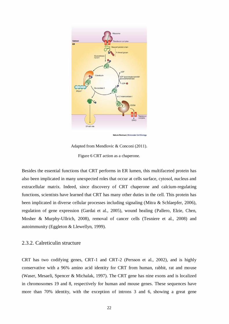
which probably is responsible for correct folding of CRT N-terminal region. Structural studies
show that CRT has three domains (Bedard, Szabo, Michalak & Opas, 2005), as illustrated in
figure 7:
The N-terminal domain (amino acid residues 1 to 180) is the most conserved domain,
contains a binding site to monoglycosylated oligosaccharides (Schrag et al., 2001),
several phosphorylation sites and is anticipated to have a folded globular structure
with eight anti-parallel β-strands connected by protein loops (Michalak et al., 1999);
The P-domain (amino acid residues 181 to 290) forms an β-stranded hairpin
configuration extended curved-arm structure rich in prolin, which binds to Ca2+
with
high affinity and low capacity (Ellgaard et al., 2001) and interacts with ER chaperones
(Michalak, Groenendyk, Szabo, Gold & Opas, 2009);
The C-terminal region or (C-domain) (amino acids residues 291-400) (Giraldo et al.,
2010), characterized by a helix form (Del Cid et al., 2010), mainly constituted by
aspartic acid and glutamic acid amino acids (Michalak et al., 1999), shows low affinity
to Ca2+
, but a high capacity, important for Ca2+
storage in ER (Castãneda-Patlán,
Razo-Paredes, Carrisoza-Gaytán, González-Mariscal & Robles-Flores, 2010). The
structure of CRT differs at C-domain considering Ca2+
level, since this domain has a
disordered structure with a low calcium concentration, but when this level rises,
protein becomes more rigid and compact (Giraldo et al., 2010).
24
Adapted from Mendlovic & Conconi (2011).
Figure 7 Structure and functions of the CRT domains.
2.3.3. Calreticulin in ticks and other parasites
CRT is a protein that exists in ticks salivary glands and saliva; probably, this protein is
essential to ticks feeding and pathogen transmission, through its anti-thrombotic and
complement inhibition functions. All these facts reveal the possibility of CRT being used as
an antigen in a vaccine against cattle ticks (Kaewhom, Stich, Needham & Jittapalapong,
2008).
Immunization assays with CRT showed its importance to immune reaction against ticks and,
consequently, its relevance as a possible global anti-ticks vaccine component. Immunization
of rabbits with CRT from A. americanum, followed by rabbits’ infestation with those ticks led
to necrotic lesions in ticks’ local bite, demonstrating an immune reaction capable to
interfering with ticks feeding (Jaworsky et al., 1995).
Gao et al. (2008) used a recombinant CRT (rCRT) protein from H. qinghaiensis (rHqCRT) to
immunize sheep and results showed a reduced ticks weight and oviposition and a higher
mortality comparing to control group.
There are various evidences of cross-reactivity between different anti-CRT antibodies,
probably due to the fact that CRT is a highly conserved protein. Parizi et al. (2009) used rCRT
from H. longicornis (rHlCRT) to immunize cattle and their sera reacted to both rHlCRT and
rCRT from R. annulatus (rBmCRT). Besides, sera from cattle, whether immunized with
rHlCRT or with rBmCRT, recognized native BmCRT.
25
Immunolocalization experiments using polyclonal antibodies anti-CRT from A. americanum
and D. variabilis, revealed a specific protein from salivary glands homologous to CRT, which
appears to be secreted during tick feeding (Jaworsky et al., 1995).
Finally, as CRT is a conserved protein, its use in a vaccine could protect animals not only
from ticks, but also from tick-borne diseases. Concerning this aspect, Rachinsky et al. (2007)
associated CRT with R. microplus infection by B. bovis, when they tested proteins up and
down-regulated in infected and uninfected ticks and discovered that, among up-regulated
proteins, there was CRT. Antunes et al. (2012) studied differentially expressed genes in R.
microplus and R. annulatus after infection by B. bigemina, showing that gene encoding CRT
was overexpressed. Knockdown of CRT in R. microplus reduced 73% the pathogen
transmission as well as ticks weight.
As described previously with respect to ticks, there are several studies referring the use of
CRT to protect animals from other parasites. Winter et al. (2005) used a N. americanus-CRT
based vaccine to immunize mice, resulting in a significant reduction of worm number in lungs
(43-49%) comparing with control group. In another study, hamsters were immunized with
Taenia solium-CRT and, besides reduction of parasite numbers, worms were unable to mature
in vaccinated animals (Mendlovic et al., 2004).
Immunolocalization studies using Haemonchus contortus-CRT antibodies showed that this
protein is localized in external openings, such as the buccal cavity, vaginal tipi of female and
bursa of male worms (Suchitra & Joshi, 2005). CRT from Echinococcus granulosus (EgCRT)
is expressed in external tegument and cellular region of hydatic cysts germinal layer, possibly
with the intent to inhibit classical complement pathway or to lead to an antiangiogenic effect
in cysts periphery (Cabezón, Cabrera, Paredes, Ferreira & Galanti, 2008).
Debrabant, Lee, Pogue, Dwyer & Nakhasi (2002) evaluated the effect of Leishmania
donovani-CRT P-domain overexpression in transfected parasites, which resulted in reduction
of acid phosphatase-secretion and in a survival decrease in human macrophages, showing that
changes in CRT expression may affect the “virulence” of the parasite.
26
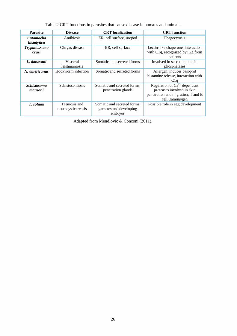
Table 2 CRT functions in parasites that cause disease in humans and animals
Parasite Disease CRT localization CRT function
Entamoeba
histolytica
Amibiosis ER, cell surface, uropod Phagocytosis
Trypanossoma
cruzi
Chagas disease ER, cell surface Lectin-like chaperone, interaction
with C1q, recognized by iGg from
patients
L. donovani Visceral
leishmaniosis
Somatic and secreted forms Involved in secretion of acid
phosphatases
N. americanus Hookworm infection Somatic and secreted forms Allergen, induces basophil
histamine release, interaction with C1q
Schistosoma
mansoni
Schistosomiosis Somatic and secreted forms, penetration glands
Regulation of Ca2+ dependent proteases involved in skin
penetration and migration, T and B
cell immunogen
T. solium Taeniosis and
neurocysticercosis
Somatic and secreted forms,
gametes and developing
embryos
Possible role in egg development
Adapted from Mendlovic & Conconi (2011).
27
CChhaapptteerr 33.. MMaatteerriiaall aanndd MMeetthhooddss
3.1. Rhipicephalus annulatus ticks
Total RNA was extracted from R. annulatus female ticks as described in Antunes et al.
(2012). Briefly, ticks were rinsed in distilled water and 75% (v/v) ethanol, dissected and the
whole of the internal organs were placed in a 2 ml tube with 1 ml of Tri Reagent (Sigma-
Aldrich, St. Louis, Missouri, USA). Total RNA was used in the synthesis of approximately 1
μg of cDNA using the iScript™ cDNA synthesis (Bio-Rad, Hercules, California, USA).
3.2. Amplification of the calreticulin gene
The CRT sequence was amplified by PCR technique. Primers were designed based on the
CRT gene sequence of R. annulatus (accession number AY395253), ensuring full coding
region coverage. The primers used were: forward 5’-CACC AT GCG GCT TCT CTG CAT
TTT G -3’ and reverse 5’- CAG TTC TTC GTG CTT GTG GTC -3’.
PCR was performed using Kit GoTaq® (Promega, Madison, Wisconsin, USA) under the
following conditions: one amplification round in a final volume of 25 μl, including 1 μl of
template cDNA and 1 μl of each primer (95ºC for 2 min, then 40 cycles: 30 s at 94ºC, 45 s at
55ºC and 2 min at 72ºC; final extension 2 min at 72ºC). PCR assays were performed in MJ
Research PTC-200 Thermo Cycler (GMI Biotech, Minnesota, USA).
The positive PCR products were purified using the illustra GFX
TM PCR DNA and Gel Band
Purification Kit (GE Healthcare, Buckinghamshire, UK) according to manufacturer’s
instructions. Purified samples were sequenced at Stab Vida (Almada, Portugal) and further
analysed.
3.3. Expression of recombinant calreticulin
For rCRT expression in Escherichia coli system, ChampionTM
pET101 directional TOPO®
Expression Kit (Invitrogen Life Technologies, Carlsbad, California, USA) was used. This
28
expression kit uses a highly efficient 5-min cloning strategy to insert a blunt end PCR product
into a vector with no requirement of post-PCR procedures or restriction enzymes (Invitrogen,
2010). The recombinant proteins produced, using this expression kit, have a histidine tail
attached.
3.3.1. Cloning and transformation
The purified PCR products, previously obtained (chapter 3.2.), were initially cloned into
plasmids pET101/D-TOPO vectors using the above described kit according to manufacturer’s
instructions. 10 µl of pET TOPO® (Invitrogen Life Technologies, Carlsbad, California, USA)
construct, previously obtained, was mixed with 50 µl of the E. coli OneShot® cells
(Invitrogen Life Technologies, Carlsbad, California, USA), and then incubated in ice during
30 min, followed by incubation for 30 s at 42ºC and further incubated in ice. Afterwards, 250
µl S.O.C. medium were added to the previous sample and incubated at 37ºC, 200 rpm during
1 hour. After that period, 200 µl of the sample were used to seed 4 LB-agar/ampicillin (100
µg/ml) Petri-dishes, followed by overnight incubation at 37ºC.
3.3.1.1. Screening of the transformed colonies
Cell colonies were analyzed by PCR to confirm plasmids incorporation. Six colonies were
picked up and individually suspended into 12 µl of water. Kit GoTaq® (Promega, Madison,
Wisconsin, USA) was used. The PCR conditions were as followed: 94ºC for 10 min to lyse
cells and inactivate nucleases, then 35 cycles: 94ºC for 3 min, 53ºC for 30 s and 72ºC for 1
min, followed by 72ºC for 10 min, in MJ Research PTC-200 Thermo Cycler (GMI Biotech,
Minnesota, USA).
3.3.1.2. Plasmid purification for sequencing
For plasmids purification, illustra plasmidPrep Mini Spin Kit (GE Healthcare,
Buckinghamshire, UK) was used, since it applies a simple plasmid DNA purification protocol
involving a modified alkaline lysis procedure and a silica-based membrane to achieve highly
efficient plasmid DNA purification (GE Healthcare, 2008).
The colonies, previously obtained (chapter 3.3.1.), were picked and suspended in 5 ml of
LB/ampicillin (100 µg/ml) and incubated overnight at 37ºC and 200 rpm. Samples were then
centrifuged at 16000 rpm during 10 min using a Heraeuspico 17 centrifuge (Thermo Electron
29
Corporation, Marietta, Ohio, USA) and supernatant were discarded. The obtained pellet was
further purified using the above described kit according to manufacturer’s instructions and
samples were then sequenced in Stab Vida (Almada, Portugal).
3.3.2. Expression
For rCRT expression, BL21 Star™ (DE3) One Shot E. coli cells were used. Plasmids,
previously purified (chapter 3.3.1.2.), were inserted in BL21 Star™ (DE3) One Shot E. coli
cells according to manufacturer’s instructions. The entire transformation was used to
inoculate 10 ml of LB/ampicillin (100 µg/ml) and was incubated overnight at 37ºC and 200
rpm. 850 µl of cells culture were mixed with 150 µl of glycerol and stored at -80ºC
maintaining, therefore, a stock of cells with plasmid incorporated, and 500 µl were used to,
once again, inoculate 10 ml of LB/ampicillin (100 µg/ml), in order to perform a pilot
expression assay. Cells were incubated at 37ºC and 200 rpm, split into two 5 ml cultures and
one was induced at an Abs 600 nm (optical density) of 0.5-0.8 with 1 mM isopropyl-β-D-
thiogalactopyranoside (IPTG). To determine the optimal induction time, 500 µl aliquot from
each culture have been removed every hour for 5 hours. Samples were centrifuged at 16000
rpm for 1 min, supernatant discarded and pellets frozen at -20ºC.
3.3.3. SDS-PAGE
A common method for proteins separation by electrophoresis uses a discontinuous
polyacrylamide gel as a support medium and sodium dodecyl sulfate (SDS) to denature
proteins. According to Laemmli method, sodium dodecyl sulfate polyacrylamide gel
electrophoresis (SDS-PAGE) was used to analyze the previously obtained cellular lysate
(chapter 3.3.2.) and confirm the presence of rCRT.
Cellular lysates were mixed with 1x SDS-PAGE sample buffer, denatured by heat and
separated in a 12.5% SDS-PAGE polyacrylamide gel using the SDS-PAGE apparatus Mini-
protean Tetra Cell (Bio-Rad, Hercules, California, USA). Electrophoresis was performed at
120 V using a running buffer and a low molecular marker (AmershamTM
LMW calibration kit
for SDS electrophoresis, GE Healthcare, Buckinghamshire, UK) was added. Gel was stained
using Coomassie Brilliant Blue dye solution.
30
3.3.4. Western-Blot
Western-Blot is a technique that identifies proteins which have been separated according to
their size by gel electrophoresis, using specific antibodies. The blotting membrane, usually
made of nitrocellulose or PVDF (polyvinylidene fluoride), binds to proteins. With this
method, the polyacrylamide gel is placed over the membrane and the application of electrical
current forces proteins to move from gel to membrane, where they adhere and can be
subsequently linked to a specific antibody. This binding can then be visualized using a second
antibody which recognizes the first one, developing a visual signal in the presence of the
appropriate substrate (Abcam, 2012).
To detect the rCRT using Western-Blot technique, a SDS-PAGE gel was prepared without
Coomassie Brilliant Blue staining, as previously described. Briefly, a cassette was constructed
with sponge, filter papers, SDS-PAGE gel and nitrocellulose membrane (Trans-Blot®
Transfer Medium pure cellulose membrane (0.45 µm), Bio-Rad, Hercules, California, USA)
and was applied on a Western-Blot apparatus (Mini Trans-blot® Cell, Bio-Rad, Hercules,
California, USA) with transfer buffer running overnight at 90 mA. Nitrocellulose membrane
was then placed in a container with Ponceau Red 0.2% (w/v) diluted in acetic acid 3% (v/v)
allowing bands visualization. Membrane was cutted into strips, which were placed in 1.5 ml
of blockage solution for 1 hour. Subsequently, the strips were washed 3 times with 2 ml of
TTBS for 15 min. Strips were incubated in 1 ml of anti-histidine antibodies solution (1:5000)
(Anti-His (C-term) antibody, Invitrogen Life Technologies, Carlsbad, California, USA) for 1
hour and washed again as described above. Then, the strips were incubated in 1 ml of
polyvalent anti-mouse antibody solution (1:10000) (Anti-Mouse Polyvalent Immunoglobulins
(G, A, M) - Alkaline Phosphatase, Sigma-Aldrich, St. Louis, Missouri, USA) for 1 hour and
the wash procedure was repeated. 1.5 ml of revelation buffer (AP Color Development Buffer,
Bio-Rad, Hercules, California, USA) was used to develop the visual signal. The reaction was
stopped by adding water and keeping the membrane in the dark.
3.4. Production of soluble calreticulin
The previously produced stock of BL21 Star™ (DE3) One Shot E. coli cells (chapter 3.3.2.)
was used to seed a LB/ampicillin (100 µg/ml) Petri-dish and was incubated overnight at 37ºC.
Then, six colonies were used to perform a PCR in the same conditions previously described in
chapter 3.3.1.1.. Afterwards, four colonies were used to inoculate 200 ml of LB/ampicillin
(100 µg/ml). Cells were induced with IPTG and were grown for two hours at 37ºC and 200
31
rpm as described in chapter 3.3.2.. Then, pellet was suspended in 20 ml lysis buffer and
divided into 2 fractions; in one of the samples a sonicator (Ultrasonic Homogenizer Power
Supply 4710 Series, Cole Parmer, Illinois, USA) with an amplitude of 80%, a frequency of 20
kHz and cycles of 10 s/min for a total of 5 min were applied and the other fraction was
thawed at 42ºC and frozen at -80ºC for three times. Subsequently, samples were centrifuged
for 15 min at 16000 rpm, pellet was suspended in 1 ml of PBS and supernatant stored. These
samples were used to perform a SDS-PAGE and Western-Blot, as described in chapters 3.3.3.
and 3.3.4., respectively, in order to determine whether the protein was present in the soluble
or insoluble fractions of lysate.
3.5. Purification of recombinant calreticulin
To perform the purification of rCRT, HisTrap HP column (GE Healthcare, Buckinghamshire,
UK) associated with fast performance liquid chromatography (System Pump-500, GE
Healthcare, Buckinghamshire, UK) was used. Prior to purification, samples were diluted in
binding buffer. Purification protocol, according to manufacturer’s instructions, was as
follows:
Column was washed with 15 ml of distilled water and equilibrated with 25 ml of
binding buffer at a flow-rate of 5 ml/min;
Samples were applied with a pump and column washed with 75 ml of binding buffer;
25 ml of elution buffer were added.
The above described protocol was also performed with some adjustments in order to select
optimal purification conditions:
Elution buffer without imidazole and with a pH of 4.5;
Different binding buffer imidazole concentrations (20 mM, 30 mM and 40 mM) with
two washes;
Concentration of purified samples (1.5 ml Microcon® YM-3 tube, membrane:
regenerated cellulose MCO 3000).
The obtained samples (non-purified, purified and samples resulting from washes) were loaded
into the SDS-PAGE, as described in chapter 3.3.3..

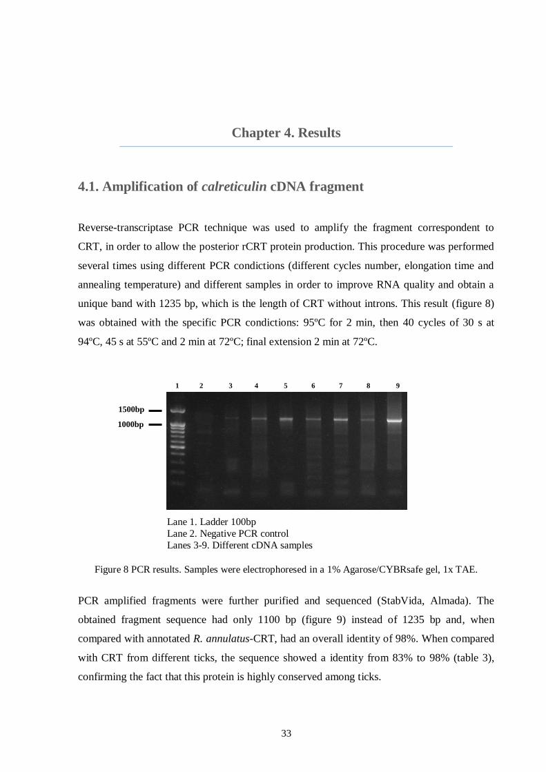
33
Chapter 4. Results
4.1. Amplification of calreticulin cDNA fragment
Reverse-transcriptase PCR technique was used to amplify the fragment correspondent to
CRT, in order to allow the posterior rCRT protein production. This procedure was performed
several times using different PCR condictions (different cycles number, elongation time and
annealing temperature) and different samples in order to improve RNA quality and obtain a
unique band with 1235 bp, which is the length of CRT without introns. This result (figure 8)
was obtained with the specific PCR condictions: 95ºC for 2 min, then 40 cycles of 30 s at
94ºC, 45 s at 55ºC and 2 min at 72ºC; final extension 2 min at 72ºC.
Lane 1. Ladder 100bp
Lane 2. Negative PCR control
Lanes 3-9. Different cDNA samples
Figure 8 PCR results. Samples were electrophoresed in a 1% Agarose/CYBRsafe gel, 1x TAE.
PCR amplified fragments were further purified and sequenced (StabVida, Almada). The
obtained fragment sequence had only 1100 bp (figure 9) instead of 1235 bp and, when
compared with annotated R. annulatus-CRT, had an overall identity of 98%. When compared
with CRT from different ticks, the sequence showed a identity from 83% to 98% (table 3),
confirming the fact that this protein is highly conserved among ticks.
1500bp
1000bp
1 2 3 4 5 6 7 8 9
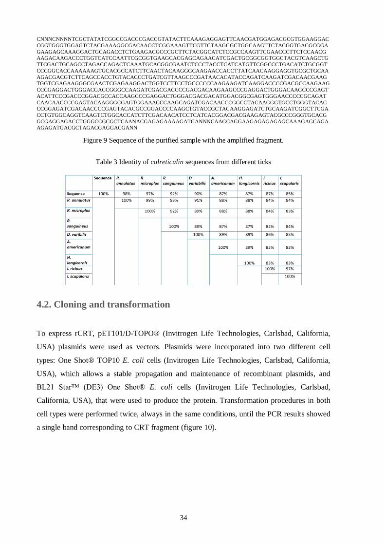
34
CNNNCNNNNTCGCTATATCGGCCGACCCGACCGTATACTTCAAAGAGGAGTTCAACGATGGAGACGCGTGGAAGGAC
CGGTGGGTGGAGTCTACGAAAGGCGACAACCTCGGAAAGTTCGTTCTAAGCGCTGGCAAGTTCTACGGTGACGCGGA
GAAGAGCAAAGGACTGCAGACCTCTGAAGACGCCCGCTTCTACGGCATCTCCGCCAAGTTCGAACCCTTCTCCAACG
AAGACAAGACCCTGGTCATCCAATTCGCGGTGAAGCACGAGCAGAACATCGACTGCGGCGGTGGCTACGTCAAGCTG
TTCGACTGCAGCCTAGACCAGACTCAAATGCACGGCGAATCTCCCTACCTCATCATGTTCGGCCCTGACATCTGCGGT
CCCGGCACCAAAAAAGTGCACGCCATCTTCAACTACAAGGGCAAGAACCACCTTATCAACAAGGAGGTGCGCTGCAA
AGACGACGTCTTCAGCCACCTGTACACCCTGATCGTTAAGCCCGATAACACATACCAGATCAAGATCGACAACGAAG
TGGTCGAGAAGGGCGAACTCGAGAAGGACTGGTCCTTCCTGCCCCCCAAGAAGATCAAGGACCCCGACGCCAAGAAG
CCCGAGGACTGGGACGACCGGGCCAAGATCGACGACCCCGACGACAAGAAGCCCGAGGACTGGGACAAGCCCGAGT
ACATTCCCGACCCGGACGCCACCAAGCCCGAGGACTGGGACGACGACATGGACGGCGAGTGGGAACCCCCGCAGAT
CAACAACCCCGAGTACAAGGGCGAGTGGAAACCCAAGCAGATCGACAACCCGGCCTACAAGGGTGCCTGGGTACAC
CCGGAGATCGACAACCCCGAGTACACGCCGGACCCCAAGCTGTACCGCTACAAGGAGATCTGCAAGATCGGCTTCGA
CCTGTGGCAGGTCAAGTCTGGCACCATCTTCGACAACATCCTCATCACGGACGACGAAGAGTACGCCCGGGTGCACG
GCGAGGAGACCTGGGCCGCGCTCAANACGAGAGAAAAGATGANNNCAAGCAGGAAGAGAGAGAGCAAAGAGCAGA
AGAGATGACGCTAGACGAGGACGANN
Figure 9 Sequence of the purified sample with the amplified fragment.
Table 3 Identity of calreticulin sequences from different ticks
4.2. Cloning and transformation
To express rCRT, pET101/D-TOPO® (Invitrogen Life Technologies, Carlsbad, California,
USA) plasmids were used as vectors. Plasmids were incorporated into two different cell
types: One Shot® TOP10 E. coli cells (Invitrogen Life Technologies, Carlsbad, California,
USA), which allows a stable propagation and maintenance of recombinant plasmids, and
BL21 Star™ (DE3) One Shot® E. coli cells (Invitrogen Life Technologies, Carlsbad,
California, USA), that were used to produce the protein. Transformation procedures in both
cell types were performed twice, always in the same conditions, until the PCR results showed
a single band corresponding to CRT fragment (figure 10).
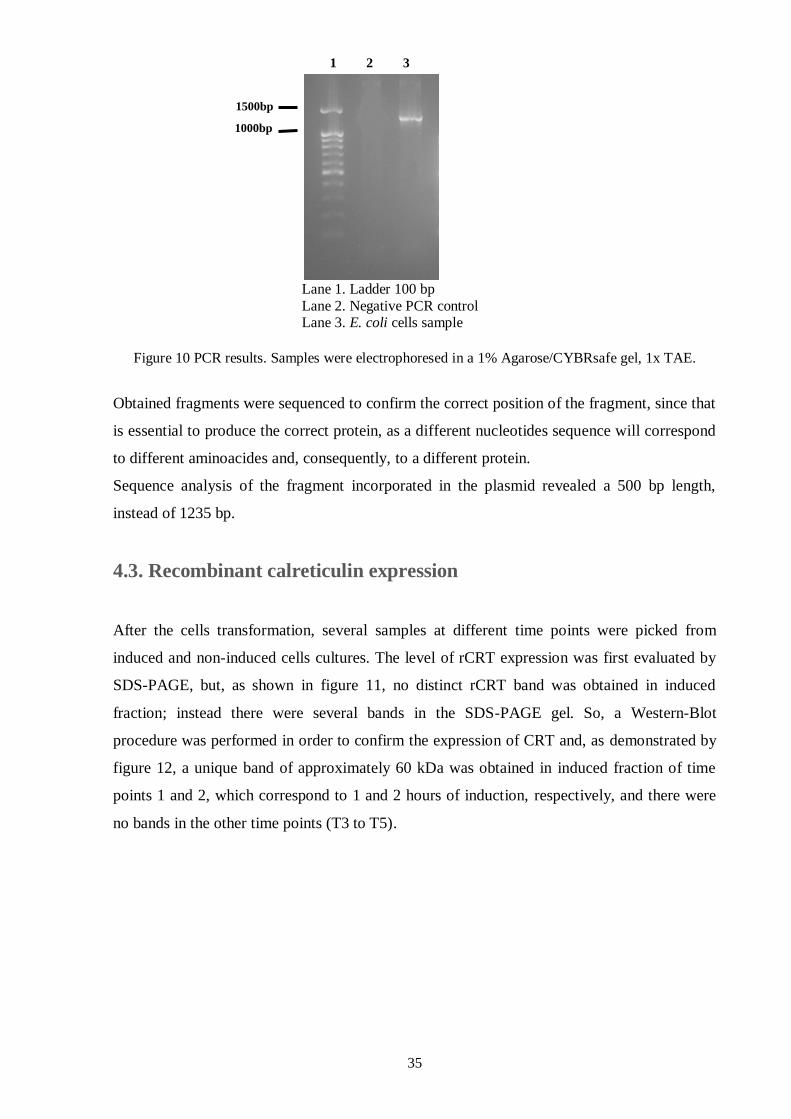
35
Lane 1. Ladder 100 bp
Lane 2. Negative PCR control Lane 3. E. coli cells sample
Figure 10 PCR results. Samples were electrophoresed in a 1% Agarose/CYBRsafe gel, 1x TAE.
Obtained fragments were sequenced to confirm the correct position of the fragment, since that
is essential to produce the correct protein, as a different nucleotides sequence will correspond
to different aminoacides and, consequently, to a different protein.
Sequence analysis of the fragment incorporated in the plasmid revealed a 500 bp length,
instead of 1235 bp.
4.3. Recombinant calreticulin expression
After the cells transformation, several samples at different time points were picked from
induced and non-induced cells cultures. The level of rCRT expression was first evaluated by
SDS-PAGE, but, as shown in figure 11, no distinct rCRT band was obtained in induced
fraction; instead there were several bands in the SDS-PAGE gel. So, a Western-Blot
procedure was performed in order to confirm the expression of CRT and, as demonstrated by
figure 12, a unique band of approximately 60 kDa was obtained in induced fraction of time
points 1 and 2, which correspond to 1 and 2 hours of induction, respectively, and there were
no bands in the other time points (T3 to T5).
1 2 3
1500bp
1000bp
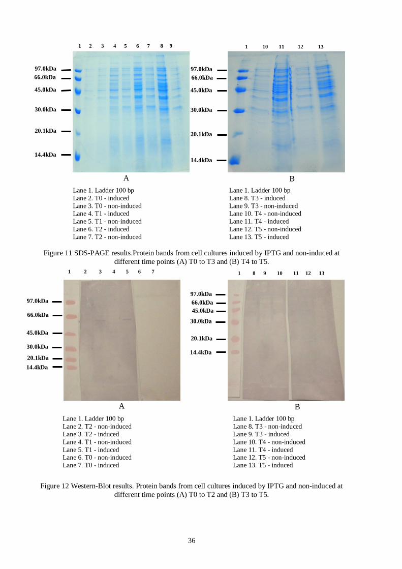
36
Lane 1. Ladder 100 bp Lane 1. Ladder 100 bp
Lane 2. T0 - induced Lane 8. T3 - induced
Lane 3. T0 - non-induced Lane 9. T3 - non-induced Lane 4. T1 - induced Lane 10. T4 - non-induced
Lane 5. T1 - non-induced Lane 11. T4 - induced
Lane 6. T2 - induced Lane 12. T5 - non-induced
Lane 7. T2 - non-induced Lane 13. T5 - induced
Figure 11 SDS-PAGE results.Protein bands from cell cultures induced by IPTG and non-induced at
different time points (A) T0 to T3 and (B) T4 to T5.
Lane 1. Ladder 100 bp Lane 1. Ladder 100 bp
Lane 2. T2 - non-induced Lane 8. T3 - non-induced
Lane 3. T2 - induced Lane 9. T3 - induced
Lane 4. T1 - non-induced Lane 10. T4 - non-induced
Lane 5. T1 - induced Lane 11. T4 - induced Lane 6. T0 - non-induced Lane 12. T5 - non-induced
Lane 7. T0 - induced Lane 13. T5 - induced
Figure 12 Western-Blot results. Protein bands from cell cultures induced by IPTG and non-induced at
different time points (A) T0 to T2 and (B) T3 to T5.
97.0kDa
66.0kDa
45.0kDa
30.0kDa
20.1kDa
14.4kDa
97.0kDa
66.0kDa
45.0kDa
30.0kDa
20.1kDa
14.4kDa
1 2 3 4 5 6 7 8 9 1 10 11 12 13
A B
1 2 3 4 5 6 7
97.0kDa
66.0kDa
45.0kDa
30.0kDa
20.1kDa
14.4kDa
97.0kDa
66.0kDa
45.0kDa
30.0kDa
20.1kDa
14.4kDa
1 8 9 10 11 12 13
A B
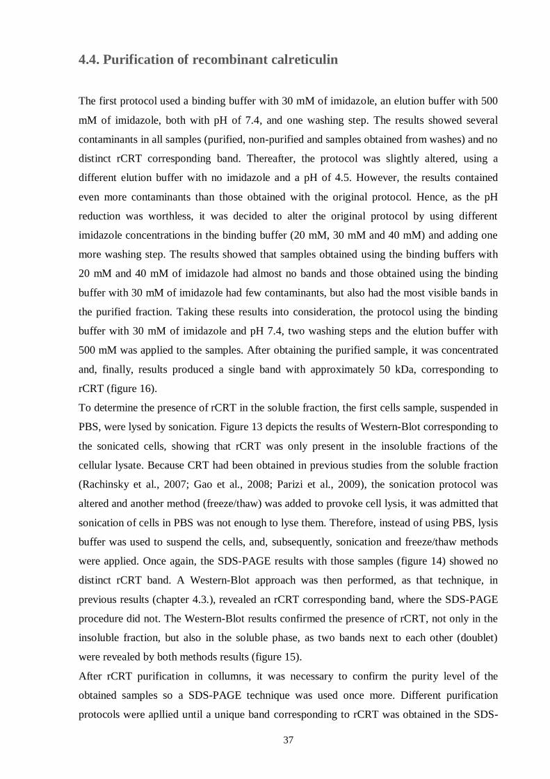
37
4.4. Purification of recombinant calreticulin
The first protocol used a binding buffer with 30 mM of imidazole, an elution buffer with 500
mM of imidazole, both with pH of 7.4, and one washing step. The results showed several
contaminants in all samples (purified, non-purified and samples obtained from washes) and no
distinct rCRT corresponding band. Thereafter, the protocol was slightly altered, using a
different elution buffer with no imidazole and a pH of 4.5. However, the results contained
even more contaminants than those obtained with the original protocol. Hence, as the pH
reduction was worthless, it was decided to alter the original protocol by using different
imidazole concentrations in the binding buffer (20 mM, 30 mM and 40 mM) and adding one
more washing step. The results showed that samples obtained using the binding buffers with
20 mM and 40 mM of imidazole had almost no bands and those obtained using the binding
buffer with 30 mM of imidazole had few contaminants, but also had the most visible bands in
the purified fraction. Taking these results into consideration, the protocol using the binding
buffer with 30 mM of imidazole and pH 7.4, two washing steps and the elution buffer with
500 mM was applied to the samples. After obtaining the purified sample, it was concentrated
and, finally, results produced a single band with approximately 50 kDa, corresponding to
rCRT (figure 16).
To determine the presence of rCRT in the soluble fraction, the first cells sample, suspended in
PBS, were lysed by sonication. Figure 13 depicts the results of Western-Blot corresponding to
the sonicated cells, showing that rCRT was only present in the insoluble fractions of the
cellular lysate. Because CRT had been obtained in previous studies from the soluble fraction
(Rachinsky et al., 2007; Gao et al., 2008; Parizi et al., 2009), the sonication protocol was
altered and another method (freeze/thaw) was added to provoke cell lysis, it was admitted that
sonication of cells in PBS was not enough to lyse them. Therefore, instead of using PBS, lysis
buffer was used to suspend the cells, and, subsequently, sonication and freeze/thaw methods
were applied. Once again, the SDS-PAGE results with those samples (figure 14) showed no
distinct rCRT band. A Western-Blot approach was then performed, as that technique, in
previous results (chapter 4.3.), revealed an rCRT corresponding band, where the SDS-PAGE
procedure did not. The Western-Blot results confirmed the presence of rCRT, not only in the
insoluble fraction, but also in the soluble phase, as two bands next to each other (doublet)
were revealed by both methods results (figure 15).
After rCRT purification in collumns, it was necessary to confirm the purity level of the
obtained samples so a SDS-PAGE technique was used once more. Different purification
protocols were apllied until a unique band corresponding to rCRT was obtained in the SDS-
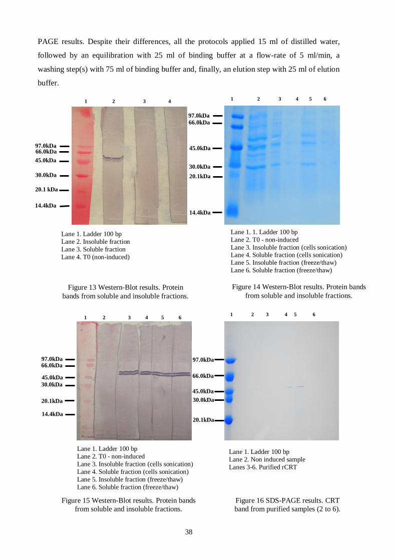
38
PAGE results. Despite their differences, all the protocols applied 15 ml of distilled water,
followed by an equilibration with 25 ml of binding buffer at a flow-rate of 5 ml/min, a
washing step(s) with 75 ml of binding buffer and, finally, an elution step with 25 ml of elution
buffer.
1 2 3 4
97.0kDa
45.0kDa
30.0kDa
20.1 kDa
14.4kDa
66.0kDa
66.0kDa
97.0kDa
45.0kDa
30.0kDa
20.1kDa
14.4kDa
1 2 3 4 5 6
Lane 1. Ladder 100 bp
Lane 2. Insoluble fraction
Lane 3. Soluble fraction
Lane 4. T0 (non-induced)
Figure 13 Western-Blot results. Protein
bands from soluble and insoluble fractions.
Lane 1. 1. Ladder 100 bp
Lane 2. T0 - non-induced
Lane 3. Insoluble fraction (cells sonication) Lane 4. Soluble fraction (cells sonication)
Lane 5. Insoluble fraction (freeze/thaw)
Lane 6. Soluble fraction (freeze/thaw)
Figure 14 Western-Blot results. Protein bands
from soluble and insoluble fractions.
66.0kDa 97.0kDa
45.0kDa
30.0kDa
20.1kDa
14.4kDa
1 2 3 4 5 6
66.0kDa
97.0kDa
45.0kDa
30.0kDa
20.1kDa
1 2 3 4 5 6
Lane 1. Ladder 100 bp
Lane 2. T0 - non-induced
Lane 3. Insoluble fraction (cells sonication)
Lane 4. Soluble fraction (cells sonication)
Lane 5. Insoluble fraction (freeze/thaw)
Lane 6. Soluble fraction (freeze/thaw)
Figure 15 Western-Blot results. Protein bands
from soluble and insoluble fractions.
Lane 1. Ladder 100 bp
Lane 2. Non induced sample
Lanes 3-6. Purified rCRT
Figure 16 SDS-PAGE results. CRT
band from purified samples (2 to 6).
39
CChhaapptteerr 55.. DDiissccuussssiioonn
Babesiosis and other tick-borne diseases are relevant issues all over the world, especially in
countries where cattle production plays a major role in the economy. Nowadays, vaccines
against both tick-vector and parasites became an innovation and tend to be the most capable
method to control ticks and their transmitted pathogens (de la Fuente et al., 2007).
This study reports the production of rCRT protein from R. annulatus, that is up-regulated in
ticks infected with B. bigemina (Antunes et al., 2012), having in mind its further application
in vaccination and immunolocalization tests.
The study is focused in the amplification of CRT gene by PCR technique. The results of the
purified PCR product sequence showed 98% identity with native R. annulatus-CRT (table 3),
though amplified fragment had only 1100 bp (instead of 1235 bp, as in the original one)
(figure 9). It was considered that the fragment had been fully amplified within the PCR
performance and the different length was due to a sequencing error. This hypothesis is
corroborated by the high level of identity between original gene and the amplified fragment
and also by the discrepancy found between bands length (in agarose gel and in the sequenced
one), pointing out to the conclusion that the fragment probably had a length of 1235 bp, but
only 1100 bp were amplified.
The following task involved rCRT cloning into a plasmid for its posterior use in bacteria
transformation. Subsequent actions were facilitated due to the characteristics of the chosen
plasmid, such as the C-terminal fusion tag for detection and purification of recombinant
fusion proteins (V5 epitope and 6xHis, composed by 6 histidine residues), the antibiotic
resistance marker for selection in E. coli and a T7 promoter that allows an IPTG-inducible
expression of recombinant protein in E. coli strains (Invitrogen, 2010). The first characteristic
is important, not only to protein detection by Western-Blot technique, but also to its
purification, since rCRT has a histidine tail. The antibiotic resistance marker permits a
“selector mechanism” that, after cell transformation, allow only those with an antibiotic
resistance gene to grow in a selective medium. Finally, IPTG-inducible expression allows the
growth of two groups of colonies, one induced and another uninduced, achieving the
emergence of a negative control. Lastly, to clone the fragment into the plasmid, the forward
primer had to have nucleotides CACC in 5’ end, so that incorporation could take place
40
properly and in the correct sense. In the case of the primer reverse, it had to anneal to the last
codon before the stop codon so that recombinant protein could have a histidine tail
(Invitrogen, 2010).
Purified plasmids were sent to be sequenced, though the result showed only 500 bp instead of
the expected 1235 bp. It was considered that this result was due to a sequencing error,
corroborated by the fact the PCR previously performed using those purified plasmids
produced a band with approximately 1200 bp. Moreover, previously purified PCR fragment
sequence had also a different length with respect to the expected one (figure 9) and it was
considered to be due to a sequencing error.
There are several recombinant systems that may be used to express recombinant proteins. In
this study, an E. coli recombinant system was used, what is in line with other studies of
recombinant proteins from ticks (Gao et al., 2008; Parizi et al., 2009; Tanaka et al., 2010;
Almazán et al., 2012). As an alternative, a recombinant system with Pichia pastoris could
have been used, because it is commonly chosen to express recombinant proteins from ticks
and have been applied with success by several authors (Canales et al., 2008; Cunha, Andreotti
& Leite, 2011; Ebrahimi, Dabaghian, Jazi, Mohammadi & Saberfar, 2012; Said et al., 2012)
There are numerous reported methods to include foreign DNA into E. coli cells
(transformation), though chemical methods are the most commonly used, due to their
accessibility and cost effectiveness (Singh, Yadav, Ma & Amoah, 2010). In this work, the
transformation kit already provided chemical competent E. coli cells and transformation was
performed through a brief incubation at 42ºC, followed by ice immersion, as it has already
been demonstrated that these sequence improves transformation (Yoshida & Sato, 2009). The
S.O.C. medium was added to the transformed cells, because it is a rich medium used in
recovery step of E. coli cells transformation, maximizing its efficiency. Moreover, S.O.C.
medium has glucose, which prevents induction of lac promoter by lactose, so preventing
transcription of target gene (Sun et al., 2009).
To confirm transformation efficiency, colonies were screened by PCR and several did not
generate the expected band in agarose gel. Supposedly, only cells with plasmid would be able
to grow in LB medium with ampicillin, as it was previously explained. There are some
possible explanations for this fact: ampicillin was not stable, primers did not anneal properly
within PCR procedure or plasmid was unstable. Among all these possible causes, the most
likely is the lack of ampicillin stability, since antibiotic solution was frozen and unfrozen
several times impairing and corrupting its stability (Okerman, Van Hende & De Zutter, 2007).
The other possible explanations are unlikely, since a previously performed PCR with purified
plasmid, the same primers were used and a band with the expected length was obtained. In the
41
case of plasmid instability, although this phenomenon occurs during cellular growth period
(Monbouquette & Ollis, 1986), Xu et al. (2006) showed that under resistance-selective
pressure, cells with plasmid remain in a higher level than under nonselective pressure after
induction, turning this hypothesis unlikely as well.
In rCRT expression, two cultures were used in order to provide induce and non-induce
samples, what concurs to achieve more credible results, as negative control (non-induce
sample) was available. The various time points adopted were decise to understand when rCRT
production was at the highest level. This information was relevant to make decisions about
subsequent cells cultures and the production and purification of rCRT. Therefore, having in
hand the various time points samples, they were applied in a SDS-PAGE gel with the purpose
to visualize which one had a band corresponding to rCRT (figure 11). In this procedure,
proteins were denatured to achieve their primary conformation, so that the test would only be
influenced by proteins molecular weight. In order to meet this goal, a SDS buffer was used,
embodying a component that attaches to proteins and confers them a negative charge; β-
mercaptoethanol was used to disrupt proteins disulphide bridges allowing molecules to adopt
an extended monomeric form (EncorBio, 2012); glycerol was used to increase sample density,
maintaining proteins on the wells bottom, overflow or uneven gel loading is restricted and
bromophenol blue was used to visualize proteins migration, as it is a small molecule and
migrates faster than all the other components (Grabski & Burgess, 2001). The heating process
is important, because proteins heated with SDS are denatured and become negative charged
(Hames, 1990). Concerning gel percentage, that was chosen according to proteins size and,
since CRT would have, supposedly, 46 kDa (Fliegel et al., 1989) a percentage of 12.5 was
selected.
Table 4 Gel percentage according to protein size.
Protein size (kDa) Gel percentage (%)
4-40 20
12-45 15
10-70 12.5
15-100 10
25-200 8
Adapted from Abcam (2012).
However, with the SDS-PAGE gel, there was not a significant difference between induced
and non-induced bands, so it was impossible to understand if rCRT had been produced or not.
A Western-Blot was carried out because this technique favours the appearance of bands by
42
antibody-antigen binding, what is much more specific, and, as rCRT had a histidine tail, anti-
histidine antibodies were used. The Western-Blot results showed a band corresponding to a
protein with approximately 60 kDa, but only in time points 1 and 2 (figure 12). The predicted
rCRT molecular weight was 46 kDa (Fliegel et al. 1989) plus 0.84 kDa, corresponding to
hexa-histidine molecular weight (Terpe, 2003). The discrepancy between predicted and
apparent rCRT molecular weight have already been described in CRT from other organisms,
as ticks (Gao et al., 2008; Parizi et al., 2009), rats (Zhu, Zelinka, White & Tanzer, 1997;
Rendón-Huerta, Mendoza-Hernández & Robles-Flores, 1999; Coling et al., 2007), humans
(Coppolino & Dedhar, 1998; Hong et al., 2004), nematodes as Heligmosomoides polygyrus
(Rzepecka et al., 2009) and N. americanus (Kasper et al., 2001; Winter et al., 2005) and
insects as Cotesia rubecula (Zhang, Schmidt & Asgari, 2006), among others. The authors of
these studies had two main explanations to molecular weight discrepancy: CRT glycosylation
(Rendón-Huerta et al., 1999; Hong et al., 2004; Coling et al., 2007; Rzepecka et al., 2009),
phenomenon that is common in proteins and the fact that CRT is highly negative charged with
a pI of 4.7 (Coppolino & Dedhar, 1998; Rendón-Huerta et al., 1999; Gao et al., 2008;
Rzepecka et al., 2009; Parizi et al., 2009). Besides those hypotheses, Zhu et al. (1997)
described the putative existence of two isoforms of CRT, an endocalreticulin with 52 kDa and
an exocalreticulin with 62 kDa. These results could also be due to the connection of another
protein to collumns. Concerning that possibility, although it has been described the natural
occurrence of histidine residues in E. coli proteins (Robichon, Luo, Causey, Benner &
Samuelson, 2011), those molecules have non-consecutive histide residues and not a hexa-
histidine molecule; moreover, those proteins were described as contaminants in IMAC
technique and not in Western-Blot procedures. The exo/endocalreticulin hypothesis is also
unlikely, since results showed only one band. Probably, the increased rCRT apparent
molecular weight was due to either posttranslational modification that can increase proteins
predicted molecular weight (Totten & Lory, 1990), as glycosylation, or to CRT negative
charge. These hypothesis are more likely because CRT has several described binding sites to
glycosylation (Schrag et al., 2001) and has already been defined as a negative charged protein
(Michalak et al., 2009). About the fact that CRT was only detected in T1 and T2 time points,
the result was unexpected, because the detectable rCRT at T1, supposedly should imply a
higher protein level in the other time points. One explanation for these results may be related
to technical errors during the manipulation of T3, T4 and T5 samples, namely incorrect
sample loading, errors in proteins transference to blotting membrane or contamination of cell
cultures. On the other hand, these explanations are not very likely, because cell cultures were
always manipulated in a laminar flow chamber with all aseptic concerns; a transference error,
43
i.e., proteins not being transferred to the membrane or an incorrect sample loading are also
unlikely because membrane coloring by means of Ponceau solution and SDS-PAGE gel,
showed bands in all time points. Comparing with our cells cultures, their concentration was
always increasing with time and, as IPTG was only added once at T0, there could be a lower
impact of IPTG in distant time points, as described by Lewis, Taylor, Nienow & Hewitt
(2004). Furthermore, substrate growth was always the same, so, as the experiment progressed
in time, nutrients were degraded and excretion products accumulated, including acetate,
which could inhibit cellular growth (Andersson, 1996) and recombinant protein production
(Shimizu et al., 1998). Probably, from T2 on, there was a reduction of rCRT production due to
lower impact of IPTG and to inhibition of recombinant protein production caused by either
IPTG lower impact or acetate production, culminating in the absence of rCRT or such a low
production level that would not be detectable at those time points samples.
Next, at the first attempt to produce soluble rCRT, cells were suspended in PBS and sonicated
to provoke their lysis, as sonication was used in several studies of recombinant proteins from
ticks (Liao et al., 2007; Gao et al., 2008; Almazán et al., 2012; Ebrahimi et al., 2012). As
described in other studies, this method could have been associated to freeze/thaw technique
(Parizi et al., 2009; Tian et al., 2011), a lysozyme addition (Miyoshi, Tsuji, Islam, Kamio &
Fujisaki, 2004), cells could have been suspended in binding buffer instead of PBS and
sonicated (Díaz-Martín, Manzano-Román, Siles-Lucas, Oleaga & Pérez-Sánchez, 2011); it
could also have been used protein extraction with Triton X-100 in TBS (Almazán et al., 2005)
or vortexing with glass beads (García-García et al., 2000). The sonicator has the ability to
destroy cells membrane by powerful ultrasounds, which rise molecules distance exceeding the
minimum molecular distance essential to maintain membrane intact, culminating with lysis
(Santos, Lodeiro & Capelo-Martínez, 2009). The freeze/thaw method involved cells
suspended in lysis buffer freezing at -80ºC and then thawing at 42ºC. This technique forms ice
crystals in freezing phase, which contracts and breaks during thawing and, consequently,
causes cells lysis (Thermo Scientific, 2009).
The Western-Blot results of the sonicated cells (figure 13) showed, in the insoluble fraction,
two close bands (approximately 55 and 60 kDa). The lack of bands in the soluble phase may
be explained by the presence of rCRT only in the other fraction or due to the fact that cells
were not correctly lysed and, consequently, remained in the pellet (insoluble phase) and so
was rCRT. The second explanation is most likely, because CRT from several ticks has already
been produced as a soluble protein (Rachinsky et al., 2007; Gao et al., 2008; Parizi et al.,
2009). Subsequently, sonication was used once more, but PBS was replaced by lysis buffer
and another method (frozen and thaw samples suspended in lysis buffer) was adopted. When
44
the membrane was revealed (figure 15), there were bands in insoluble and soluble phases,
probably due to an incomplete cells lysis and, consequently, part of rCRT was still in cells
and so in pellet (insoluble phase). In each fraction, only one band corresponding to rCRT
should appear; however, two close bands became visible (approximately 55 and 60 kDa),
similar to the first attempt result. The two bands found in acrylamide gel are probably due to
either protein denaturation, as the electrophoresis run in denaturing conditions (in the
presence of SDS) or to protein degradation.
Concerning recombinant proteins purification, there are many described processes: IMAC
(López et al., 2009), purification according to proteins sizes (Canales et al., 2008; Said et al.,
2012) and excision from polyacrylamide gel (Ebrahimi et al., 2012) among others. In the
process of rCRT purification, IMAC was the chosen method, because it is a quick and widely
used method for histidine-tagged proteins purification (Kuo & Chase, 2011) and it has already
been used successfully with recombinant proteins from ticks (Miyoshi et al., 2004; Almazán
et al., 2005; Tanaka et al., 2010; Cunha et al., 2011; Díaz-Martín et al., 2011; Tian et al.,
2011). There are other described purifying methods to recombinant proteins from ticks, such
as purification according to proteins sizes (Canales et al., 2008; Said et al., 2012) and excision
from polyacrylamide (SDS-PAGE) gel (Ebrahimi et al., 2012).
The columns used to purify rCRT have nickel ions. The imidazole concentration of binding
buffer was low in order to minimize binding of host-cell proteins, though it may decrease
binding of histidine-tagged proteins (GE Healthcare, 2005). The elution buffer had a higher
imidazole quantity than the binding buffer, because these molecules have a greater affinity to
the column than the protein, allowing the rCRT elution (GE Healthcare, 2005), though the pH
diminishment would have the same effect (Du, Zhang, JieWang, Yao & Hu, 2008). Despite
the facts described before, results in the first attempt showed that almost no rCRT was
purified and that there was a huge contamination with other proteins, causing the appearance
of several bands instead of just one band corresponding to rCRT. First of all, column protocol
refers that an optimization should be performed, i.e., different factors should be tested, such as
imidazole concentration, in order to obtain the best balance of high purity and high yield, as
well as the use of different metals, since protein-metal ion binding strength is affected by
length, position and exposure of proteins affinity tag, ions and buffers pH. From the above
mentioned aspects, the use of different metals is not of great importance, since nickel and
cobalt are usually the chosen by several authors as the metal ions to bind to histidine-tagged
proteins (Sun et al., 2005; Chong, Tan, Biak, Ling & Tey, 2009; Knecht, Ricklin, Eberle &
Ernst, 2009). The use of pH reduction, instead of imidazole different concentrations, to purify
rCRT revealed to be worthless, since there were almost no bands. On the other hand, lack of
45
optimization with different imidazole concentrations could be one of the main reasons for
these results once, as explained above, imidazole concentration can decrease binding of
histidine-tagged proteins. Next attempt, where three different binding buffer imidazole
concentrations were tested, all samples had contaminants. However, the one washed with
binding buffer with 30 mM of imidazole had the most visible bands, including rCRT
corresponding band, being this imidazole concentration the one used in the first attempt. This
result made clear that imidazole concentration was not influencing purification process. Low
purification level could be related to some recombinant proteins having its histidine-tag
partially hidden from protein surface, caused by intra or inter-molecular interactions
(Mohanty & Wiener, 2004), leading to a lower liaison of rCRT to the columns and,
consequently, to lower recombinant protein purification. There are many E. coli proteins
described as contaminants when IMAC technique is used to purify histidine-tagged proteins.
Most of them are probably due to bacteria response to stress conditions (nutrient starvation,
heat shock or oxidative damage). Contaminant proteins production depends on culture
conditions, media compositions and genetic background of expression strain (Bolanos-Garcia
& Davies, 2006). Bolanos-Garcia & Davies (2006) described three contaminant proteins
classes based on imidazole concentration required for elution: class I (≥80 mM), class II (55
to 80 mM) and class III (30 to 50 mM), placing our samples contaminants in class I, which
includes contaminants protein ferric update regulator (Fur), cAMP regulatory protein (CRP),
peptidoylproline cis-trans isomerase (SlyD), acetylornithinase (ArgE), Cu/Zn-superoxide
dismutase (Cu/Zn-SODM) and metal-binding lipocalin (YodA). These proteins may bind to
metal-chelating resins by possession of native metal-binding sites to Ni2+
or Co2+
(Fur, YodA,
Cu/Zn SODM and ArgE) or due to the presence of histidine clusters (CRP and SlyD), both
binding equally as histidine residues of a histidine-tagged protein, showing that contaminant
proteins may not have histidine residues at all. Probably, the low concentration of rCRT and
their histidine-tag, partially hidden from protein surface, consequently led to the liaison of
contaminants to the column and their elution, instead of rCRT. In order to improve rCRT
purification other methods were used, and only when two washes were applied, plus the
concentration of purified samples, a single band correspondent to CRT appeared in the SDS-
PAGE gel (figure 16). The IMAC technique might have been associated to a second
purification step. Applying two different purification principles in sequence would improve
the degree of purity, because each stage separates a certain amount of contaminants,
increasing its total level of removal.

47
CChhaapptteerr 66.. CCoonncclluussiioonn
The results of this project were optimistic and represent one more step to improve ticks
control, as we showed in this study that CRT can be produced and purified without
contaminants, though further vaccination and immunolocalization studies will be the key to
understand CRT future use.
B. bigemina has a great importance worldwide, as it provokes a disease that affects not only
animals, with a great impact in cattle production, but also humans, which turns it into a public
health issue and a priority disease to be eliminated or, at least, to be controlled. The first
attempt to control ticks and their transmitted pathogens was using molecules that would kill
them in a quick and efficient way. However, all this pressure in the parasites survival leads to
one of the biggest disasters of our existence: the appearance of resistant ticks against almost
all acaricides. At this point, vaccines are the future of ticks and babesiosis control, especially
vaccines that have action against more than one target, either several ticks or ticks and their
transmitted pathogens. Possibly, due to be highly conserved among ticks, CRT may be a
component of a large-spectrum anti-tick vaccine or in a vaccine against both vector and its
transmitted pathogen, but only the future will confirm that.

49
CChhaapptteerr 77.. RReeffeerreenncceess
Abcam. (2012). Western Blotting - A Beginner’s guide. Accessed in January 10th
, 2012,
available in:
www.abcam.com/technical
Aljamali, M.N., Sauer, J.R. & Essenberg, R.C. (2002). RNA interference: applicability in tick
research. Experimental and Applied Acarology, 28(1-4), 89-96.
Almazán, C., Blas-Machado, U., Kocan, K.M., Yoshioka, J.H., Blouin, E.F., Mangold, A.J. &
de la Fuente, J. (2005). Characterization of three Ixodes scapularis cDNAs protective against
tick infestations. Vaccine, 23, 4403-4416.
Almazán, C., Lagunes, R., Villar,
M., Canales, M., Rosario-Cruz, R., Jongejan, F. & de la
Fuente, J. (2010). Identification and characterization of Rhipicephalus (Boophilus) microplus
candidate protective antigens for the control of cattle tick infestations. Parasitology Research,
106(2), 471-479.
Almazán, C., Moreno-Cantú, O., Moreno-Cid, J.A., Galindo, R.C., Canales, M., Villar, M. &
de la Fuente, J. (2012). Control of tick infestations in cattle vaccinated with bacterial
membranes containing surface-exposed tick protective antigens. Vaccine, 30, 265– 272.
Andersson, L.C. (1996). Escherichia coli high cell density fed batch cultures: Kinetic and
physiological aspects. PhD thesis. Stockholm, Sweden: Royal Institute of Technology.
Antunes, G.M. (2008). Hemoparasitoses em Bovinos de carne. MSc thesis in Veterinary
Sciences. Lisbon: Veterinary Medicine Faculty - Technical University of Lisbon.
Antunes, S., Galindo, R.C., Almazán, C., Rudenko, N., Golovchenko, M., Grubhoffer, L.,
Shkap, V., do Rosário, V.E., de la Fuente, J. & Domingos, A. (2012). Functional genomics
studies of Rhipicephalus (Boophilus) annulatus ticks in response to infection with the cattle
protozoan parasite, Babesia bigemina. International Journal for Parasitology, 42(2), 187-195.
Aragão, H.B. (1936). Ixodidas brasileiros e de alguns países limítrofes. Memórias do Instituto
Oswaldo Cruz (4), 759-843.
Assadian, O. & Stanek, G. (2002). Theobald Smith - The discoverer of ticks as vectors of
disease. Wien Klin Wochenschr Journal, 114, 478-481.
Babes, V. (1888). Sur l’hemoglobinurie bacterienne du boeuf. Comptes Rendus de l'Académie
des Sciences - Series III - Sciences de la Vie, 107, 692–694.
Barker, S.C. & Murrel, A. (2004). Systematics and evolution of ticks with a list of valid genus
and species names. Parasitology, 129, 15-36.
50
Bastos, R.G., Ueti, M.W., Knowles, D.P. & Scoles, G.A. (2010). The Rhipicephalus
(Boophilus) microplus Bm86 gene plays a critical role in the fitness of ticks fed on cattle
during acute Babesia bovis infection. Parasites & Vectors, 3(111).
Bedard, K., Szabo, E., Michalak, M. & Opas, M. (2005). Cellular functions of endoplasmic
reticulum chaperones calreticulin, calnexin, and ERp57. International Review of Cytology,
245, 91-121.
Benavides, M. & Sacco, A.M. (2007). Differential Bos taurus cattle response to Babesia bovis
infection. Veterinary Parasitology, 150, 54–64.
Benson, M.J., Gawronski, J.D., Eveleigh, D.E. & Benson, D.R. (2004). Intracellular
symbionts and other bacteria associated with deer ticks (Ixodes scapularis) from Nantucket
and Wellfleet, Cape Cod, Massachussetts. Applied and Environmental Microbiology, 70, 616–
620.
Bock, R.E., de Vos, A.J., Kingston, T.G. & McLellan, D.J. (1997). Effect of breed of cattle on
innate resistance to infection with Babesia bovis, Babesia bigemina and Anaplasma
marginale. Australian Veterinary Journal, 75, 337–340.
Bock, R.E., Kingston, T.G. & de Vos, A.J. (1999). Effect of breed of cattle on transmission
rate and innate resistance to infection with Babesia bovis and Babesia bigemina transmitted
by Boophilus microplus. Australian Veterinary Journal, 77, 461–464.
Bock, R.E. & de Vos, A.J. (2001). Immunity following use of Australian tick fever vaccine: a
review of the evidence. Australian Veterinary Journal, 79(12), 832-839.
Bock, R.E., Jackson, L., de Vos, A.J. & Jorgensen, W. (2004). Babesiosis in cattle.
Parasitology, 129, 247-269.
Bolanos-Garcia, V.M. & Davies, O.R. (2006). Structural analysis and classification of native
proteins from E. coli commonly co-purified by immobilised metal affinity chromatography.
Biochimica et Biophysica Acta, 1760, 1304–1313.
Bӧse, R., Jorgensen, W.K., Dalgliesh, R.J., Friedhoff, K.T. & de Vos, A.J. (1995). Current
state and future trends in the diagnosis of babesiosis. Veterinary Parasitology, 7, 61-74.
Botana, L.M., Landoni, M.F. & Martín-Jiménez, T. (2002). Farmacología y Terapéutica
Veterinaria. Madrid, Spain: McGraw-Hill/Interamericana de España, S.A.U.
Brandt, F., Healy, G.R. & Welch, M. (1977). Human babesiosis: the isolation of Babesia
microti in golden hamsters. Journal of Parasitology, 63(5), 934-937.
Buling, A., Criado-Fornelio, A., Asenzo, G., Benitez, D., Barba-Carretero, J.C. & Florin-
Christensen, M. (2007). A quantitative PCR assay for the detection and quantification of
Babesia bovis and B. bigemina. Veterinary Parasitology, 147, 16-25.
Burns, K., Atkinson, E.A., Bleackley, R.C. & Michalak, M. (1994). Calreticulin: from Ca2+
binding to control of gene expression. Trends in Cell Biology, 4(5), 153-154.
51
Burridge, M.J., Young, A.S., Stagg, D.A., Kanhai, G.K. & Kimber, C.D. (1974). Theileria
lawrencei infection of cattle and African buffalo: evaluation of a buffalo cell culture schizont
antigen for the indirect fluorescent antibody test. Research in Veterinary Science, 17(3), 285-
289.
Cabezón, C., Cabrera, G., Paredes, R., Ferreira, A. & Galanti, N. (2008). Echinococcus
granulosus calreticulin: Molecular characterization and hydatid cyst localization. Molecular
Immunology, 45, 1431–1438.
Canales, M., de la Lastra, J.M., Naranjo, V., Nijhof, A.M., Hope, M., Jongejan, F. & de la
Fuente, J. (2008). Expression of recombinant Rhipicephalus (Boophilus) microplus,
R.annulatus and R. decoloratus Bm86 orthologs as secreted proteins in Pichia pastoris. BMC
Biotechnology, 8, 14.
Canales, M., Almazán, C., Naranjo, V., Jongejan, F. & de la Fuente, J. (2009). Vaccinat ion
with recombinant Boophilus annulatus Bm86 ortholog protein, Ba86, protects cattle against
B. annulatus and B. microplus infestations. BMC Biotechnology, 9-29.
Carcy, B., Précigout, E., Schetters, T. & Gorenflot, A. (2006). Genetic basis for GPI-anchor
merozoite surface antigen polymorphism of Babesia and resulting antigenic diversity.
Veterinary Parasitology, 138(1-2), 33-49.
Carter, R. (2001). Transmission blocking malaria vaccines. Vaccine, 19, 2309–2314.
Castãneda-Patlán, M.C., Razo-Paredes, R., Carrisoza-Gaytán, R., González-Mariscal, L. &
Robles-Flores, M. (2010). Protein kinase C is involved in the regulation of several calreticulin
posttranslational modifications. The International Journal of Biochemistry & Cell Biology,
42, 120–131.
Chong, F.C., Tan, W.S., Biak, D.R., Ling, T.C. & Tey, B.T. (2009). Purification of histidine-
tagged nucleocapsid protein of Nipah virus using immobilized metal affinity chromatography.
Journal of Chromatography B. Analytical Technologies in the Biomedical and Life Sciences,
877(14-15), 1561-1567.
Coling, D.E., Ding, D., Young, R., Lis, M., Stofko, E., Blumenthal, K.M. & Salvi, R.J.
(2007). Proteomic analysis of cisplatin-induced cochlear damage: Methods and early changes
in protein expression. Hearing Research, 226, 140–156
Coppolino, M.G. & Dedhar, S. (1998). Calreticulin. The International Journal of
Biochemistry & Cell Biology, 30, 553-558.
Cunha, R., Andreotti, R. & Leite, F. (2011). Rhipicephalus (Boophilus) microplus: expression
and characterization of Bm86-CG in Pichia pastoris. Revista Brasileira de Parasitologia
Veterinária, 20(2), 103-110.
de la Fuente, J., Rodríguez, M., Redondo, M., Montero, C., García-García, J.C., Méndez, L.,
Serrano, E., Valdés, M., Enriquez, A., Canales, M., Ramos, E., Boué, O., Machado, H.,
Lleonart, R., de Armas, C.A., Rey, S., Rodríguez, J.L., Artiles, M. & García, L. (1998). Field
studies and cost-effectiveness analysis of vaccination with Gavac against the cattle tick
Boophilus microplus. Vaccine, 16(4), 366-373.
52
de la Fuente, J., Rodriguez, M., Montero, C., Redondo, M., Garcia-Garcia, J.C., Mendez, L.,
Serrano, E., Valdes, M., Enriquez, A., Canales, M., Ramos, E., Boue, O., Machado, M. &
Lleonart, R. (1999). Vaccination against ticks (Boophilus spp.): the experience with the
Bm86-based vaccine Gavac (TM). Genetic Analysis Biomolecular Engineering, 15, 143–148.
de la Fuente, J. & Kocan, K.M. (2003). Advances in the identification and characterization of
protective antigens for recombinant vaccines against tick infestations. Expert Reviews
Vaccines, 2(4), 583-593.
de la Fuente, J. & Kocan, K.M. (2006). Strategies for development of vaccines for control of
ixodid tick species. Parasite Immunology, 28, 275–283.
de la Fuente, J., Kocan, K.M. & Blouin, E.F. (2007). Tick vaccines and the transmission of
tick-borne pathogens. Veterinary Research Communications, 31(Suppl. 1), 85–90.
de la Fuente, J., Estrada-Pena, A., Venzal, J.M., Kocan, K.M. & Sonenshine, D.E. (2008).
Overview: Ticks as vector of pathogens that cause disease in humans and animals. Frontiers
in Bioscience, 13, 6938-6946.
de la Fuente, J., Moreno-Cid, J.A., Canales, M., Villar, M., de la Lastra, J.M., Kocan, K.M.,
Galindo, R.C., Almazán, C. & Blouin, E.F. (2011). Targeting arthropod subolesin/akirin for
the development of a universal vaccine for control of vector infestations and pathogen
transmission. Veterinary Parasitology, 181(1), 17-22.
de Vos, A.J. (1978). Immunogenicity and pathogenicity of three South African strains of
Babesia bovis in Bos indicus cattle. Onderstepoort Journal of Veterinary Research, 45, 119–
124.
de Vos, S., Zeinstra, L., Taoufik, O, Willadsen, P. & Jongejan, F. (2001). Evidence for the
utility of the Bm86 antigen from Boophilus microplus in vaccination against other tick
species. Experimental and Applied Acarology, 25, 245–261.
de Vos, J. & Geysen, D. (2004). Epidemiological study of the prevalence of Babesia
divergens in a veterinary practice in the mid-east of France. Veterinary Parasitology, 125(3-
4), 237-249.
de Waal, D.T. & Combrink, M.P. (2006). Live vaccines against bovine babesiosis. Veterinary
Parasitology, 138(1-2), 88-96.
Debrabant, A., Lee, N., Pogue, G.P., Dwyer, D.M. & Nakhasi, H.L. (2002). Expression of
calreticulin P-domain results in impairment of secretory pathway in Leishmania donovani and
reduced parasite survival in macrophages. International Journal for Parasitology, 32, 1423–
1434.
Del Cid, N., Jeffery, E., Rizvi, S.M., Stamper, E., Peters, L.R., Brown, W.C., Provoda, C. &
Raghavan, M. (2010). Modes of calreticulin recruitment to the major histocompatibility
complex class I assembly pathway. Journal of Biological Chemistry, 285(7), 4520-4535.
Díaz-Martín, V., Manzano-Román, R., Siles-Lucas, M., Oleaga, A. & Pérez-Sánchez, R.
(2011). Cloning, characterization and diagnostic performance of the salivary lipocalin protein
TSGP1 from Ornithodoros moubata. Veterinary Parasitology, 178, 163–172.
53
DPI - Department of Primary Industries & Fisheries – Queensland (EUA). Accessed in
December 10th, 2011, available in:
http://www.dpi.qld.gov.au
Dumler, J.S., Barber, A.F., Bekker, C.P., Dasch, G.A., Palmer, G.H., Ray, S.C., Rikihisa, Y.
& Rurangirwa, F.R. (2001). Reorganization of genera in the families Rickettsiaceae and
Anaplasmataceae in the order Rickettsiales: unification of some species of Ehrlichia with
Anaplasma, Cowdria with Ehrlichia and Ehrlichia with Neorickettsia, descriptions of six new
species combinations and designation of Ehrlichia equi and ‘HGE agent’ as subjective
synonyms of Ehrlichia phagocytophila. International Journal of Systematic and Evolutionary
Microbiology, 51, 2145-2165.
Du, H., Zhang, X., Wang, J., Yao, X. & Hu, Z. (2008). Novel approaches to predict the
retention of histidine-containing peptides in immobilized metal-affinity chromatography.
Proteomics, 8, 2185–2195.
Duh, D., Petrovec, M. & Avsic-Zupanc, T. (2001). Diversity of Babesia infecting European
sheep ticks (Ixodes ricinus). Journal of Clinical Microbiology, 39, 3395–3397.
Ebrahimi, S.M., Dabaghian, M., Jazi, M.H., Mohammadi, M. & Saberfar, E. (2012).
Expression of recombinant HAO3 from an Iranian isolate of Hyalomma anatolicum
anatolicum in Pichia pastoris and evaluation of its antigenicity. Biologicals, 40, 72-78.
Echaide, I., de Echaide, S. & Guglielmone, A. (1993). Live and soluble antigens for cattle
protection to Babesia bigemina. Veterinay Parasitology, 51, 35–40.
Eggleton, P. & Llewellyn, D.H. (1999). Pathophysiological roles of calreticulin in
autoimmune disease. Scandinavian Journal of Immunology, 49, 466–473.
Ellgaard, L., Riek, R., Herrmann, T., Güntert, P., Braun, D., Helenius, A. & Wüthrich, K.
(2001). NMR structure of the calreticulin P-domain. Proceedings of the National Academy of
Sciences of the United States of America, 98, 3133–3138.
EnCor Biotechnology Incorporated. (2012). SDS-Polyacrylamide Gel Electrophoresis (SDS-
PAGE). Accessed in September 22th, 2011, available in:
http://www.encorbio.com/protocols/SDS-PAGE.htm
Engracia Filho, J.R., Bechara, G.H. & Teodoro, R.L. (2006). Dermal mast cell counts in F2
Holstein x Gir crossbred cattle artificially infested with the tick Boophilus microplus (Acari:
Ixodidae). Annals of the New York Academy of Sciences, 1081, 476-478.
Ferreira, C.A., Vaz Junior, I., Silva, S., Haag, K.L., Valenzuela, J.G. & Masuda, A. (2002).
Cloning and partial characterization of a Boophilus microplus (Acari: Ixodidae) calreticulin.
Experimental Parasitology, 101, 25–34.
Figueroa, J.V., Alvarez, J.A., Ramos, J.A., Vega, C.A. & Buening, G.M. (1993). Use of
multiplex polymerase chain reaction-based assay to conduct epidemiological studies on
bovine hemoparasites in Mexico. Revue d’elevage et de Medecine Veterinaire des Pays
Tropicaux, 46, 71–75.
54
Fire, A., Xu, S., Montgomery, M.K., Kostas, S.A., Driver, S.E. & Mello, C.C. (1998). Potent
and specific genetic interference by double-stranded RNA in Caenorhabditis elegans. Nature,
391, 806–811.
Fish, L., Leibovich, B., Krigel, Y., McElwain, T. & Shkarp, V. (2008). Vaccination of cattle
against B. bovis infection with live attenuated parasites and non-viable immunogens. Vaccine,
26, 29–33.
Fliegel, L., Burns, K., MacLennan, D.H., Reithmeier, R.A. & Michalak, M. (1989). Molecular
cloning of the high affinity calcium-binding protein (calreticulin) of skeletal muscle
sarcoplasmic reticulum. Journal of Biological Chemistry, 264(36), 21522-21528.
Frazzon, A.P., Vaz Junior, I., Masuda, A., Schrank, A. & Vainstein, M.H. (2000). In vitro
assessment of Metarhizium anisopliae isolates to control the cattle tick Boophilus microplus.
Veterinary Parasitology, 94(1-2), 117-25.
Freeman, J.M., Davey, R.B., Kappmeyer, L.S., Kammlah, D.M. & Olafson, P.U. (2010).
Bm86 midgut protein sequence variation in South Texas cattle fever ticks. Parasites &
Vectors, 3-101.
Gao, J., Luo, J., Fan, R., Fingerle, V., Guan, G., Liu, Z., Li, Y., Zhao, H., Ma, M., Liu, J., Liu,
A., Ren, Q., Dang, Z., Sugimoto, C. & Yin, H. (2008). Cloning and characterization of a
cDNA clone encoding calreticulin from Haemaphysalis qinghaiensis (Acari: Ixodidae).
Parasitology Research, 102, 737–746.
García-García, J.S., Monteroa, C., Redondo, M., Vargas, M., Canales, M., Boue, O.,
Rodríguez, M., Joglara, M., Machado, H., González, I., Valdés, M., Méndez, L. & de la
Fuente, J. (2000). Control of ticks resistant to immunization with Bm86 in cattle vaccinated
with the recombinant antigen Bm95 isolated from the cattle tick, Boophilus microplus.
Vaccine, 18, 2275-2287.
Gardai, S.J., McPhillips, K.A., Frasch, S.C., Janssen, W.J., Starefeldt, A., Murphy-Ullrich,
J.E., Bratton, D.L., Oldenborg, P.A., Michalak, M. & Henson, P.M. (2005). Cell-surface
calreticulin initiates clearance of viable or apoptotic cells through trans-activation of LRP on
the phagocyte. Cell, 123(2), 321-334.
GE Healthcare (2005). His Spin Trap instructions (11-0036-91 AA).
GE Healthcare (2008). illustra plasmidPrep MiniSpin Kit: Product Booklet, pp. 1-37.
Gindin, G., Samish, M., Zangi, G., Mishoutchenko, A. & Glazer, I. (2002). The susceptibility
of different species and stages of ticks to entomopathogenic fungi. Experimental and Applied
Acarology, 28(1-4), 283-288.
Giraldo, A.M., Medus, M., Lebrero, M., Pagano, R., Labriola, C.A., Landolfo, L., Delfino,
J.D., Paradi, A.J. & Caramelo, J.J. (2010). The structure of calreticulin C-terminal domain is
modulated by physiological variations of calcium concentration. Journal of Biological
Chemistry, 285(7), 4544–4553.
Grabski, A.C. & Burgess, R.R. (2001). Preparation of protein samples for SDS-
polyacrylamide gel electrophoresis: procedures and tips. InNovations, 13, 10-12.
55
Graf, J.F., Gogolewski, R., Leach-Bing, N., Sabatini, G.A., Molento, M.B., Bordin, E.L. &
Arantes, G.J. (2004). Tick control: an industry point of view. Parasitology, 129, 427–442.
Gu, G., Junkai, S., Meiying, Y., Jukui, Z., Shijun, M., Ali, N., Lixin, T., Tan, L. & Maliyam.
(1997). Research on the schizont cell culture vaccine against Theileria annulata infection in
Xinjiang. Tropical Animal Health and Production, 29, 98–100.
Gubbels, J.M., de Vos, A.P., van der Weide, M., Viseras, J., Schouls, L.M., de Vries, E. &
Jongejan, F. (1999). Simultaneous detection of bovine Theileria and Babesia species by
reverse line blot hybridization. Journal of Clinical Microbiology, 37(6), 1782-1789.
Hames, B.D. (Hames, B.D. & Rickwood, D., eds). (1990). Gel Electrophoresis of Proteins. A
Practical Approach. (pp.1-147). New York, United States of America: Oxford University
Press.
Havlíková, S., Roller, L., Koci, J., Trimnell, A.R., Kazimírová, M., Klempa, B. & Nuttall,
P.A. (2009). Functional role of 64P, the candidate transmission-blocking vaccine antigen from
the tick, Rhipicephalus appendiculatus. International Journal for Parasitology, 39, 1485–
1494.
Healy, G.R. & Ruebush, T.K. (1980). Morphology of Babesia microti in human blood
smears. American Journal of Clinical Pathology, 73(1), 107-109.
Heyman, P., Cochez C., Hofhuis A., van der Giesen, J., Sprong, H., Porter, S.R., Losson, B.,
Saegerman, C., Donoso-Mantke, O., Niedring, M. & Pappa, A. (2010). A clear and present
danger: Tick-borne diseases in Europe. Expert Review of Anti-infective Therapy, 8(1), 33-50.
Homer, M.J., Aguilar-Delfin, I., Telford 3rd
, S.R., Krause, P.J. & Persing, D.H. (2000).
Babesiosis. Clinical Microbiology Reviews, 13(3), 451-469.
Hong, S.H., Misek, D.E., Wang, H., Puravs, E., Giordano, T.J., Greenson, J.K., Brenner,
D.E., Simeone, D.M., Logsdon, C.D. & Hanash, S.M. (2004). An autoantibody-mediated
immune response to calreticulin isoforms in pancreatic cancer. Cancer Research, 64(15),
5504-5510.
Howard, J., Rozza, D., Graça, D. & Fighera, R. (2001). Bovine Babesiosis. Accessed in
January 4th
, 2012, available in:
http://www.vet.uga.edu/vpp/archives/NSEP/babesia/PORT/necropsy_findings.htm
Hunfeld, K.P., Hildebrandt, A. & Gray, J.S. (2008). Babesiosis: recent insights into an ancient
disease. International Journal for Parasitology, 38(11), 1219-1237.
Invitrogen (2010), Champion™ pET Directional TOPO® Expression Kits - User Manual,
Manual part no. 25-0400, pp. 1-74.
Iseki, H., Alhassan, A., Ohta, N., Thekisoe, O.M., Yokoyama, N., Inoue, N., Nambota, A.,
Yasuda, J. & Igarashi, I. (2007). Development of a multiplex loop-mediated isothermal
amplification (mLAMP) method for the simultaneous detection of bovine Babesia parasites.
Journal of Microbiological Methods, 71(3), 281-287.
56
Jaworsky, D.C., Simmen, F.A., Lamoreaux, W., Coons, L.B., Muller, M.T. & Needham, G.R.
(1995). A secreted calreticulin protein in Ixodid tick (Amblyomma americanum) saliva.
Journal of Insect Physiology, 41(4), 369-375.
Jittapalapong, S., Kaewhom, P., Kengradomkij, C., Saratapan, N., Canales, M., de la Fuente,
J. & Stich, R.W. (2010). Humoral immune response of dairy cattle immunized with rBm95
(KU-VAC1) derived from Thai Rhipicephalus microplus. Transboundary and Emerging
Diseases, 57, 91–95.
Jorgensen, W., de Vos, A. & Dalgliesh, R. (1989). Comparison of immunogenicity and
virulence between Babesia bigemina parasites from continuous culture and from
splenectomized calf. Australian Veterinary Journal, 66, 371–372.
Kaewhom, P., Stich, R.W., Needham, G.R. & Jittapalapong, S. (2008). Molecular analysis of
calreticulin expressed in salivary glands of Rhipicephalus (Boophilus) microplus indigenous
to Thailand. Annals of the New York Academy of Sciences, 1149, 53-57.
Kasper, G., Brown, A., Eberl, M., Vallar, L., Kieffer, N., Berry, C., Girdwood, K., Eggleton,
P., Quinnell, R. & Pritchard, D.I. (2001). A calreticulin-like molecule from the human
hookworm Necator americanus interacts with C1q and the cytoplasmic signalling domains of
some integrins. Parasite Immunology, 23, 141-152.
Kemp, D.H., McKenna, R.V., Thullner, R. & Willadsen, P. (1999). Strategies for tick control
in a world of acaricide resistance. Proceedings of the IV Seminario Internacional de
Parasitologia Animal, Puerto Vallarta, Mexico, 1–10.
Khudrathulla, M. & Jagannath, M.S. (2000). Effect of methanol extract of Stylosanthes
scabra on ixodid ticks of animals. Indian Journal Animal Sciences, 70(10), 1057-1058.
Klompen, H., Lekveishvili, M. & Black, W.C. (2007). Phylogeny of parasitiform mites
(Acari) based on rRNA. Molecular Phylogenetics and Evolution, 43, 936-951.
Klompen, H. (2010). Holothyrids and ticks: New insights from larval morphology and DNA
sequencing, with the description of a new species of diplothyrus (Parasitiformes:
Neothyridae). Acarologia, 50, 269–285.
Knecht, S., Ricklin, D., Eberle, A.N. & Ernst, B. (2009). Oligohis-tags: mechanisms of
binding to Ni2+
-NTA surfaces. Journal of Molecular Recognition, 22(4), 270-279.
Kocan, K.M., Manzano-Roman, R. & de la Fuente, J. (2007). Transovarial silencing of the
subolesin gene in three-host ixodid tick species after injection of replete females with
subolesin dsRNA. Parasitology Research, 100, 1411–1415.
Kohls, G.M., Sonenshine, D.E. & Clifford, C.M. (1965). The systematics of the subfamily
Ornithodorinae (Acarina: Argasidae). II. Identification of the larvae of the Western
Hemisphere and descriptions of three new species. Annals of the Entomological Society of
America, 58(3), 64-331.
Krantz, G.W. (1978). A manual of acarology. (2nd
edition). Oregon State, USA: University
Bookstores, Corvallis.
57
Krause, P.J., Telford 3rd
, S.R., Ryan, R., Conrad, P.A., Wilson. M., Thomford. J.W. &
Spielman, A. (1994). Diagnosis of babesiosis: evaluation of a serologic test for the detection
of Babesia microti antibody. Journal of Infectious Diseases, 169(4), 923-926.
Krause, P.J., Lepore, T., Sikand, V.K., Gadbaw Junior, J., Burke, G., Telford 3rd
, S.R.,
Brassard, P., Pearl, D., Azlanzadeh, J., Christianson, D., McGrath, D. & Spielman, A. (2000).
Atovaquone and azithromycin for the treatment of babesiosis. New England Journal of
Medicine, 343(20), 1454-1458.
Kuo, W.K. & Chase, H.A. (2011). Exploiting the interactions between poly-histidine fusion
tags and immobilized metal ions. Biotechnology Letters, 33, 1075–1084.
Lewis, G., Taylor, I.W., Nienow, A.W. & Hewitt, C.J. (2004). The application of multi-
parameter flow cytometry to the study of recombinant Escherichia coli batch fermentation
processes. Journal of Industrial Microbiology and Biotechnology, 31, 311–322.
Liao, M., Zhou, J., Hatta, T., Umemiya, R., Miyoshi, T., Tsuji, N., Xuan, X. & Fujisaki, K.
(2007). Molecular characterization of Rhipicephalus (Boophilus) microplus Bm86 homologue
from Haemaphysalis longicornis ticks. Veterinary Parasitology, 146, 148–157.
López, N.C., Valck, C., Ramírez, G., Rodríguez, M., Ribeiro, C., Orellana, J., Maldonado, I.,
Albini, A., Anacona, D., Lemus, D., Aguilar, L., Schwaeble, W. & Ferreira, A. (2010).
Antiangiogenic and antitumor effects of Trypanosoma cruzi calreticulin. PLoS Neglected
Tropical Diseases, 4(7).
Lundh, J., Wiktelius, D. & Chirico, J. (2005). Azadirachtin-impregnated traps for the control
of Dermanyssus gallinae. Veterinary Parasitology, 130 (3-4), 337-342.
Martins, T.M. (2009). Identification of proteases as diagnostic and drug targets in bovine
babesiosis. PhD Thesis. Lisbon: Institute of Hygiene and Tropical Medicine - New University
of Lisbon.
Martins, T.M., Gonçalves, L.M., Capela, R., Moreira, R., do Rosário, V.E. & Domingos, A.
(2010). Effect of synthesized inhibitors on babesipain-1, a new cysteine protease from the
bovine piroplasm Babesia bigemina. Transboundary and Emerging Diseases, 57(1-2), 68-69.
Martins, T.M., Rosário, V.E. & Domingos, A. (2011). Identification of papain-like cysteine
proteases from the bovine piroplasm Babesia bigemina and evolutionary relationship of
piroplasms C1 family of cysteine proteases. Experimental Parasitology, 127(1), 184-194.
Matsuoka, K., Seta, K., Yamakawa, Y., Okuyama, T., Shinoda, T. & Isobe, T. (1994).
Covalent structure of bovine brain calreticulin. Biochemistry Journal, 298(2), 435-442.
McCauliffe, D.P., Yang, Y.S., Wilson, J., Sontheimer, R.D. & Capra, J.D. (1992). The 5'-
flanking region of the human calreticulin gene shares homology with the human GRP78,
GRP94, and protein disulfide isomerase promoters. Journal of Biological Chemistry, 267(4),
2557-2562.
58
Mcllwain, J.T. (1984). ‘Bodelye Syeknes’ of Julian of Norwich. Journal of Medieval History,
10(3), 167-180.
Melhorn, H. & Schein, E. (1984). The piroplasms: life cycle and sexual stages. Advances in
Parasitology, 23, 37–103.
Melo, V.,Passos, L.,Facury-Filho, E., Saturnino, H. & Ribeiro, M. (2001). Natural infection of
calves by Anaplasma marginale in dairy herds of the Metalúrgica Region, Minas Gerais.
Pesquisa Veterinária Brasileira, 21 (4).
Mendlovic, F. & Conconi, M. (2011). Calreticulin: a Multifaceted Protein. Nature Education,
4(1), 1.
Mendlovic, F., Ostoa-Saloma, P., Solís, C.F., Martínez-Ocaña, J., Flisser, A. & Laclette, J.P.
(2004). Cloning, characterization, and functional expression of Taenia solium calreticulin.
International Journal for Parasitology, 90(4), 891–893.
Merino, O., Almazán, C., Canales, C., Villar, M., Moreno-Cid, J.A., Galindo, R.C. & de la
Fuente, J. (2011). Targeting the tick protective antigen subolesin reduces vector infestations
and pathogen infection by Anaplasma marginale and Babesia bigemina. Vaccine, 29(47),
8575-8579.
Mesaeli, N., Nakamura, K., Zvaritch, E., Dickie, P., Dziak, E., Krause, K.H., Opas, M.,
MacLennan, D.H. & Michalak, M. (1999). Calreticulin is essential for cardiac development.
Journal of Cell Biology, 144(5), 857-868.
Mesplet, M., Echaide, I., Dominguez, M., Mosqueda, J.J., Suarez, C.E., Schnittger, L. &
Florin-Christensen, M. (2010). Bovipain-2, the falcipain-2 ortholog, is expressed in
intraerythrocytic stages of the tick-transmitted hemoparasite Babesia bovis. Parasites &
Vectors, 21(3), 113.
Michalak, M., Corbett, E.F., Mesaeli, N., Nakamura, K. & Opas, M. (1999). Calreticulin: one
protein, one gene, many functions. Biochemical Journal, 344(2), 281-92.
Michalak, M., Groenendyk, J., Szabo, E., Gold, L.I. & Opas, M. (2009). Calreticulin, a
multiprocess calcium-buffering chaperone of the endoplasmic reticulum. Biochemical
Journal, 417, 651-666.
Mitra, S.K. & Schlaepfer, D.D. (2006). Integrin-regulated FAK-Src signaling in normal and
cancer cells. Current Opinion in Cell Biology, 18(5), 516-523.
Miyoshi, T., Tsuji, N., Islam, M.K., Kamio, T. & Fujisaki, K. (2004). Cloning and molecular
characterization of a cubilin-related serine proteinase from the hard tick Haemaphysalis
longicornis. Insect Biochemistry and Molecular Biology, 34, 799–808.
Mohanty, A.K. & Wiener, M.C. (2004). Membrane protein expression and production: effects
of polyhistidine tag length and position. Protein Expression and Purification, 33, 311–325.
Monbouquette, H.G. & Ollis, D.F. (1986). A structured model for immobilized cell kinetics.
Annals of the New York Academy of Sciences, 469, 230-244.
59
Nauseef, W.M., McCormick, S.J. & Clark, R.A. (1995). Calreticulin functions as a molecular
chaperone in the biosynthesis of myeloperoxidase. Journal of Biological Chemistry, 270(9),
4741-4747.
Noda, H., Munderloh, U.G. & Kurffi, T.J. (1997). Endosymbionts of ticks and their
relationship to Wolbachia spp. and tickborne pathogens of humans and animals. Applied and
Environmental Microbiology, 63, 3926–3932.
Odongo, D., Kamau, L., Skilton, R., Mwaura, S., Nitsch, C., Musoke, A., Taracha, E.,
Daubenberger, C. & Bishop, R. (2007). Vaccination of cattle with TickGARD induces cross-
reactive antibodies binding to conserved linear peptides of Bm86 homologues in Boophilus
decoloratus. Vaccine, 25, 1287–1296.
Ojeda, J.J., Orozco, J., Flores, R., Rojas, C., Figueroa, J.V. & Álvarez, J.A. (2010). Validation
of an attenuated live vaccine against babesiosis in native cattle in an endemic area.
Transboundary and Emerging Diseases, 57, 84-86.
Okerman, L., Van Hende, J. & De Zutter, L. (2007). Stability of frozen stock solutions of
beta-lactam antibiotics, cephalosporins, tetracyclines and quinolones used in antibiotic residue
screening and antibiotic susceptibility testing. Analytica Chimica Acta, 586, 284–288.
Okubo, K., Yokoyama, N., Govind, Y., Alhassan, A. & Igarashi, I. (2007). Babesia bovis:
effects of cysteine protease inhibitors on in vitro growth. Experimental Parasitology, 117(2),
214-217.
Ortiz, E.M., Santamaría, V.M., Ortiz, N.A., Soberanes, C.N., Osorio, M.J., Franco, B.R.,
Martinez, I.F., Quezada, D.R. & Fragoso, S.H. (1995). Characterization of Boophilus
microplus resistance to ixodicides in México. Seminario internacional de Parasitología
Animal, 58-66.
Pallero, M.A., Elzie, C.A., Chen, J., Mosher, D.F. & Murphy-Ullrich, J.E. (2008).
Thrombospondin 1 binding to calreticulin-LRP1 signals resistance to anoikis. Federation of
American Societies for Experimental Biology Journal, 22(11), 3968-3979.
Papadopoulos, B., Brossard, M. & Perié, N.M. (1996). Piroplasms of domestic animals in the
Macedonia region of Greece. 2. Piroplasms of cattle. Veterinary Parasitology, 63, 57–66.
Parizi, L.F., Rech, H., Ferreira, C.A., Imamura, S., Ohashi, K., Onuma, M., Masusa, A. & Vaz
Junior, I. (2009). Comparative immunogenicity of Haemaphysalis longicornis and
Rhipicephalus (Boophilus) microplus calreticulins. Veterinary Parasitology, 164, 282–290.
Pattnaik, P. (2006). Kyasanur forest disease: an epidemiological view in India. Reviews in
Medical Virology, 16(3) 65–151.
Peter, R.J., Van den Bossche, P., Penzhom, B.L. & Sharp, B. (2005). Tick, fly, and mosquito
control – Lessons from the past, solutions for the future. Veterinary Parasitology, 132, 205-
215.
Peterson, J.R., Ora, A., Van, P.N., & Helenius, A. (1995). Transient, lectin-like association of
calreticulin with folding intermediates of cellular and viral glycoproteins. Molecular Biology
of the Cell, 6, 1173-1184.
60
Persson, S., Rosenquist, M. & Sommarin, M. (2002). Identification of a novel calreticulin
isoform (Crt2) in human and mouse. Gene, 297(1-2), 151-158.
Pipano, E. (1995). Live vaccines against hemoparasitic diseases in livestock. Veterinary
Parasitology, 57(1-3), 213-231.
Pipano, E., Alekceev, E., Galker, F., Fish, L., Samish, M. & Shkap, V. (2003). Immunity
against Boophilus annulatus induced by the Bm86 (Tick-GARD) vaccine. Experimental and
Applied Acarology, 29, 141–149.
Rachinsky, A., Guerrero, D.F. & Scoles, G.A. (2007). Proteomic profiling of Rhipicephalus
(Boophilus) microplus midgut responses to infection with Babesia bovis. Veterinary
Parasitology, 152, 294–313.
Randolph, S. (2004). Evidence that climate change has caused emergence of tick-borne
diseases in Europe. International Journal of Medical Microbiology, 37, 5–15.
Rendón-Huerta, E., Mendoza-Hernández, G. & Robles-Flores, M. (1999). Characterization of
calreticulin as a protein interacting with protein kinase C. Biochemical Journal, 344, 469-475.
Riek, R.F. (1966). The life cycle of Babesia argentina (Lignieres, 1903) (Sporozoa:
piroplasmidea) in the tick vector Boophilus microplus (Canestrini). Australian Journal of
Agricultural Research, 17, 247–254.
Ristic, M. (1981). (Ristic, M. & MacIntyre, I., Eds.). Disease of Cattle in the Tropics. (pp.
443-468). The Hague, Netherlands: Martinus Nijhof Publishers.
Robichon, C., Luo, J., Causey, T.B., Benner, J.S. & Samuelson, J.C. (2011). Engineering
Escherichia coli BL21(DE3) derivative strains to minimize E. coli protein contamination after
purification by immobilized metal affinity chromatography. Applied and Environmental
Microbiology, 77(13), 4634-4646.
Rogers, A. N., Van Buren, D. G., Hedblom, E. E., Tilahum, M. E., Telfer, J. C. & Baldwin C.
L. (2005). Gamma-delta T cell function varies with expressed WC1 coreceptor. Journal of
Immunology, 174, 3386–3393.
Rosario-Cruz, R., Almazán, C., Miller, R.J., Dominguez-Garcia, D.I., Hernandez-Ortiz, R. &
de la Fuente, J. (2009). Genetic basis and impact of tick acaricide resistance. Frontiers in
Bioscience, 14, 2657-2665.
Rzepecka, J., Rausch, S., Klotz, C., Schnöller, C., Kornprobst, T., Hagen, J., Ignatius, R.,
Lucius, R. & Hartmann, S. (2009). Calreticulin from the intestinal nematode
Heligmosomoides polygyrus is a Th2-skewing protein and interacts with murine scavenger
receptor-A. Molecular Immunology, 46, 1109–1119.
Saegerman, C., Claes, L., Dewaele, A., Desmecht, D., Rollin, F., Hamoir, J., Gustin, P.,
Czaplicki, G., Bughin, J., Wullepit, J., Laureyns, J., Roels, S., Berkvens, D., Vanopdenbosch,
E. & Thiry, E. (2003). Differential diagnosis of neurologically expressed disorders in Western
European cattle. Revue scientifique et technique, 22(1), 83-102.
61
Said, M.B., Galai, Y., Canales, M., Nijhof, A.M., Mhadhbi, M., Jedidi, M., de la Fuente, J. &
Darghouth, M.A. (2012). Hd86, the Bm86 tick protein ortholog in Hyalomma scupense (syn.
H. detritum): Expression in Pichia pastoris and analysis of nucleotides and amino acids
sequences variations prior to vaccination trials. Veterinary Parasitology, 183, 215– 223.
Santos, H.M., Lodeiro, C. & Capelo-Martínez, J.L. (2009). The Power of Ultrasound.
Weinheim, Germany: Wiley-VCH Verlag GmbH & Company KGaA.
Schrag, J.D., Bergeron, J.J., Li, Y., Borisova, S., Hahn, M., Thomas, D.Y. & Cygler, M.
(2001). The structure of calnexin, an ER chaperone involved in quality control of protein
folding. Molecular Cell, 8(3), 633-644.
Shimizu, N., Fukuzono, S., Fujimori, K., Nishimura, N. & Odawara, Y. (1988). Fed-batch
cultures of recombinant Escherichia coli with inhibitory substance concentration monitoring.
Journal of Fermentation Technology, 66, 187-191.
Shirong, S. (1997). Application and effect of the schizont vaccine against Theileria annulata
in China. Tropical Animal Health and Production, 29, 101–103.
Shkap, V. & Pipano, E. (2000). Culture-derived parasites in vaccination of cattle against tick-
borne diseases. Annals of the New York Academy of Sciences, 916, 154-171.
Shkap, V., Rasulov, I., Abdurasulov, S., Fish, L., Leibovitz, B., Krigel, Y., Molad, T., Mazuz,
M.L. & Savitsky, I. (2007). Babesia bigemina: Attenuation of an Uzbek isolate for
immunization of cattle with live calf- or culture-derived parasites. Veterinary Parasitology,
146, 221–226.
Silva, M.G., Ueti, M.W., Norimine, J., Florin-Christensen, M., Bastos, R.G., Goff, W.L.,
Brown, W.C., Oliva, A. & Suarez, C.E. (2010). Babesia bovis expresses a neutralization-
sensitive antigen that contains a microneme adhesive repeat (MAR) domain. Parasitology
International, 59, 294-297.
Singh, M., Yadav, A., Ma, X. & Amoah, E. (2010). Plasmid DNA Transformation in
Escherichia coli: effect of heat shock temperature, duration, and cold incubation of CaCl2
treated cells. International Journal of Biotechnology and Biochemistry, 6(4), 561–568.
Smith, T. & Kilbourne, F.L. (1893). Investigation into the nature, causation and prevention of
Texas or South Cattle fever. United States Department of Agriculture Bureau Animal Industry
Bull, 1(1).
Soberanes, C.N., Santamaría, V.M., Fragoso, S.H. & García, V.Z. (2002). Primer caso de
resistencia al amitraz en la garrapata del ganado Boophilus microplus en México. Técnica
Pecuaria en México, 40, 81-92.
Sonenshine, D. E. (1991). Biology of Ticks. New York: Oxford University Press.
Suchitra, S. & Joshi, P. (2005). Characterization of Haemonchus contortus calreticulin
suggests its role in feeding and immune evasion by the parasite. Biochimica et Biophysica
Acta, 1722, 293– 303.
Sun, Q., Chen, L., Cao, L., Fang, L., Chen, C. & Hua, Z. (2005). An improved strategy for
high-level production of human vasostatin 120-180. Biotechnology Progress, 21, 1048-1052.
62
Sun, Q., Ding, L., He, L., Sun, Y., Shao, J., Luo, M. & Xu, Z. (2009). Culture of Escherichia
coli in SOC medium improves the cloning efficiency of toxic protein genes. Analytical
Biochemistry, 394, 144-146.
Tanaka, T., Kawano, S., Nakao, S., Umemiya-Shirafuji, R., Rahman, M.M., Boldbaatar, D.,
Battur, B., Liao, M. & Fujisaki, K. (2010). The identification and characterization of
lysozyme from the hard tick Haemaphysalis longicornis. Ticks and Tick-borne Diseases, 1,
178–185.
Taylor, M.A., Coop, R.L. & Wall, R.L. (Eds.) (2007). Veterinary Parasitology. (3th ed.),
Oxford, United Kingdom: Blackwell Publishing.
Telford 3rd
, S.R. & Maguire, J.H. (2006). (Guerrant, R.L., Walker, D.H. & Weller, P.F., Eds.).
Tropical infectious diseases. Principles, pathogens, and practice. (2nd
edition). (pp.1063-
1071). Philadelphia, United States of America: Elsevier.
Terpe K. (2003). Overview of tag protein fusions: from molecular and biochemical
fundamentals to commercial systems. Applied Microbiology and Biotechnology, 60(5), 523-
533.
Tesniere, A., Apetoh, L., Ghiringhelli, F., Joza, N., Panaretakis, T., Kepp, O., Schlemmer, F.,
Zitvogel, L. & Kroemer, G. (2008). Immunogenic cancer cell death: a key-lock paradigm.
Current Opinion in Immunology, 20, 504–511.
Thermo Scientific. (2009), Thermo Scientific Pierce Cell Lysis Technical Handbook, Version
2, pp. 1-54.
Thomford, J.W., Conrad, P.A., Boyce, W.M., Holman, P.J. & Jessup, D.A. (1993). Isolation
and in vitro cultivation of Babesia parasites from free-ranging desert bighorn sheep (Ovis
canadensis nelsoni) and mule deer (Odocoileus hemionus) in California. Journal of
Parasitology, 79(1), 77-84.
Tian, Z., Liu, G., Zhang, L., Yin, H., Wang, H., Xie, J., Zhang, P. & Luo, J. (2011).
Identification of the heat shock protein 70 (HLHsp70) in Haemaphysalis longicornis.
Veterinary Parasitology, 181, 282– 290.
Torina, A., Agnone, A., Sireci, G., Mosqueda, J.J., Blanda, V., Albanese, I., La Farina, M.,
Cerrone, A., Cusumano, F. & Caracappa, S. (2010). Characterization of the apical membrane
antigen-1 in Italian strains of Babesia bigemina. Transboundary and Emerging Diseases, 57,
52–56.
Totten, P.A. & Lory, S. (1990). Characterization of the type a flagellin gene from
Pseudomonas aeruginosa PAK. Journal of Bacteriology, 172(12), 7188-7199.
Trimnell, A.R., Hails, R.S. & Nuttall, P.A. (2002). Dual action ectoparasite vaccine targeting
‘exposed’ and ‘concealed’ antigens. Vaccine, 20, 3560–3568.
Urquhart, G.M., Armour, J., Duncan, J.L., Dunn, A.M. & Jennings J.W. (1996). Veterinary
Parasitology. (2nd
edition). Oxford, United Kingdom: Blackwell Science Limited.
63
Xu, G., Fang, Q.Q., Sun, Y., Keirans, J.E. & Durden, L.A. (2005). Hard tick calreticulin
(CRT) gene coding regions have only one intron with conserved positions and variable sizes.
Parasitology, 91(6), 1326-1331.
Xu, J., Li, W., Wu, J., Zhang, Y., Zhu, Z., Liu, J. & Hu, Z. (2006). Stability of plasmid and
expression of a recombinant gonadotropin-releasing hormone (GnRH) vaccine in Escherichia
coli. Applied Microbiology and Biotechnology, 73, 780-788.
Walker, A.R., Bouattour, A., Camicas, J.L., Estrada-Peña, A., Horak, I.G., Latif, A.A.,
Pegram, R.G. & Preston, P.M. (2003). Ticks of domestic animals in Africa: a guide to
identification of species. Edinburgh, Scotland, United Kingdom: Bioscience Reports.
Waser, M., Mesaeli, N., Spencer, C. & Michalak, M. (1997). Regulation of calreticulin gene
expression by calcium. Journal of Cell Biology, 138(3), 547-557.
Willadsen, P. (1980). Immunity to ticks. Advances in Parasitology, 18, 293-311.
Willadsen, P., Bird, P., Cobon, G.S. & Hungerford, J. (1995). Commercialization of a
recombinant vaccine against Boophilus microplus. Parasitology, 110, 43–50.
Willadsen, P. (2006). Tick control: Thoughts on a research agenda. Veterinary Parasitology,
138, 161–168.
Winter, J.A., Davies, O.R., Brown, A.P., Garnett, M.C., Stolnik, S. & Pritchard, D.I. (2005).
The assessment of hookworm calreticulin as a potential vaccine for necatoriasis. Parasite
Immunology, 27, 139-146.
Wittner, M., Rowin, K.S., Tanowitz, H.B., Hobbs, J.F., Saltzman, S., Wenz, B., Hirsch, R.,
Chisholm, E. & Healy, G.R. (1982). Successful chemotherapy of transfusion babesiosis.
Annals of Internal Medicine, 96(5), 601-604.
Yoshida, N. & Sato, M. (2009). Plasmid uptake by bacteria: A comparison of methods and
efficiencies. Applied Microbiology and Biotechnology, 83(5), 791-8.
Zhang, G., Schmidt, O. & Asgari, S. (2006). A calreticulin-like protein from endoparasitoid
venom fluid is involved in host hemocyte inactivation. Developmental and Comparative
Immunology, 30, 756–764.
Zhu, Q., Zelinka, P., White, T. & Tanzer, M.L. (1997). Calreticulin-integrin bidirectional
signaling complex. Biochemical and Biophysical Research Communications, 232, 354-358.
Zintl, A., Mulcahy, G., Skerrett, H.E., Taylor, S.M. & Gray, J.S. (2003). Babesia divergens, a
bovine blood parasite of veterinary and zoonotic importance. Clinical Microbiology Reviews,
16(4), 622-636.
Zivkovic, Z., Torina, A., Mitra, R., Alongi, A., Scimeca, S., Kocan, K.M., Galindo, R.C.,
Almazán, C, Blouin, E.F., Villar, M., Nijhof, A.M., Mani, R., La Barbera, G., Caracappa, S.,
Jongejan, F. & de la Fuente, J. (2010). Subolesin expression in response to pathogen infection
in ticks. BMC Immunology, 11(7).

64
AAnnnneexx II -- MMaatteerriiaall aanndd MMeetthhooddss AAppppeennddiixx
1. Mediums
1.1. Luria-Bertani (LB)-agar:
6.25g of LB and 3.75g of agar in 250ml of distilled water
1.2. Luria-Bertani
6,25g of LB in 250ml of distilled water
1.3. S.O.C.
2% (w/v) tryptone, 0.5% (w/v) yeast extract, 1.0mM NaCl, 2.5mM KCl, 10mM MgCl2,
10mM MgSO4 and 20mM glucose
2. Solutions
2.1. Lysis buffer
0.3ml KH2PO4, 4.7ml K2HPO4, 2.3g NaCl, 0.75g KCl, 10ml glycerol 100% (v/v), 0.5ml
Triton X-100 and 0.068mg imidazole, obtain the final volume of 100ml with water and a final
pH of 7.8
2.2. Coomassie Brilliant Blue dye
40% methanol, 10% acetic acid and 0.025% Coomassie Brilliant Blue R-250
2.3. Distaining solution
30% (v/v) of ethanol, 10% of acetic acid (v/v) and 60% of water
2.4. 1X SDS-PAGE sample buffer
1.25ml of 0.5M Tris-HCl at pH 6.8, 1.0ml of glycerol 100% (v/v), 0.2ml of β-
mercaptoethanol, 0.01g of bromophenol blue and 0.2g SDS, obtain the final volume of 10ml
with water
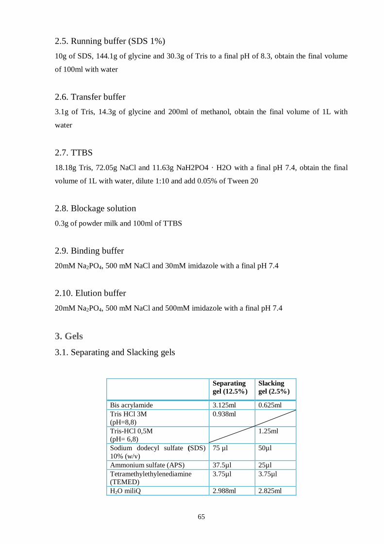
65
2.5. Running buffer (SDS 1%)
10g of SDS, 144.1g of glycine and 30.3g of Tris to a final pH of 8.3, obtain the final volume
of 100ml with water
2.6. Transfer buffer
3.1g of Tris, 14.3g of glycine and 200ml of methanol, obtain the final volume of 1L with
water
2.7. TTBS
18.18g Tris, 72.05g NaCl and 11.63g NaH2PO4 · H2O with a final pH 7.4, obtain the final
volume of 1L with water, dilute 1:10 and add 0.05% of Tween 20
2.8. Blockage solution
0.3g of powder milk and 100ml of TTBS
2.9. Binding buffer
20mM Na2PO4, 500 mM NaCl and 30mM imidazole with a final pH 7.4
2.10. Elution buffer
20mM Na2PO4, 500 mM NaCl and 500mM imidazole with a final pH 7.4
3. Gels
3.1. Separating and Slacking gels
Separating
gel (12.5%)
Slacking
gel (2.5%)
Bis acrylamide 3.125ml 0.625ml
Tris HCl 3M
(pH=8,8)
0.938ml
Tris-HCl 0,5M (pH= 6,8)
1.25ml
Sodium dodecyl sulfate (SDS)
10% (w/v)
75 µl 50µl
Ammonium sulfate (APS) 37.5µl 25µl
Tetramethylethylenediamine (TEMED)
3.75µl 3.75µl
H2O miliQ 2.988ml 2.825ml


