Embed Size (px)
Citation preview
Please cite this article in press as: Gardner et al., Extrathymic Aire-Expressing Cells Are a Distinct Bone Marrow-Derived Population that Induce Func-tional Inactivation of CD4+ T Cells, Immunity (2013), http://dx.doi.org/10.1016/j.immuni.2013.08.005
Immunity
Article
Extrathymic Aire-Expressing CellsAre a Distinct Bone Marrow-Derived Populationthat Induce Functional Inactivation of CD4+ T CellsJames M. Gardner,1,2,7 Todd C. Metzger,1,7 Eileen J. McMahon,1,3 Byron B. Au-Yeung,4 Anna K. Krawisz,1 Wen Lu,1
Jeffrey D. Price,5 Kellsey P. Johannes,1 Ansuman T. Satpathy,6 Kenneth M. Murphy,6 Kristin V. Tarbell,5 Arthur Weiss,4
and Mark S. Anderson1,*1Diabetes Center, University of California, San Francisco, San Francisco, CA 94143-0540, USA2Department of Surgery, University of California, San Francisco, San Francisco, CA 94143-0540, USA3Department of Biology, Westmont College, Santa Barbara, CA 93108, USA4Howard Hughes Medical Institute, Rosalind Russell Medical Research Center for Arthritis, Department of Medicine,
Department of Microbiology and Immunology, University of California, San Francisco, San Francisco, CA 94143-0540, USA5Immune Tolerance Section, Diabetes Branch, National Institute of Diabetes and Digestive and Kidney Diseases, National Institutes of Health,
Bethesda, MD 20892, USA6Howard Hughes Medical Institute, Department of Pathology and Immunology, School of Medicine, Washington University in St. Louis,
St. Louis, MO 63110, USA7These authors contributed equally to this work
*Correspondence: [email protected]
http://dx.doi.org/10.1016/j.immuni.2013.08.005
SUMMARY
The autoimmune regulator (Aire) is essential for pre-vention of autoimmunity; its role is best understoodin the thymus, where it promotes self-tolerancethrough tissue-specific antigen (TSA) expression.Recently, extrathymic Aire-expressing cells (eTACs)have been described in murine secondary lymphoidorgans, but the identity of such cells and their rolein immune tolerance remains unclear. Here we haveshown that eTACs are a discrete major histocompat-ibility complex class II (MHC II)hi, CD80lo, CD86lo,epithelial cell adhesion molecule (EpCAM)hi, CD45lo
bone marrow-derived peripheral antigen-presentingcell (APC) population. We also have demonstratedthat eTACs can functionally inactivate CD4+ T cellsthrough a mechanism that does not require regulato-ry T cells (Treg) and is resistant to innate inflamma-tory stimuli. Together, these findings further defineeTACs as a distinct tolerogenic cell population insecondary lymphoid organs.
INTRODUCTION
Clonal diversity within the adaptive immune system allows
responsiveness to a wide range of pathogens but can also
predispose to autoimmunity in the absence of appropriate self-
tolerance. Education of developing T cells in the thymus is a
critical component of such tolerance, and involves removal of
developing autoreactive T cells (Anderson et al., 2005; Metzger
and Anderson, 2011) in part through the activity of the autoim-
mune regulator, Aire. AIRE was first identified in humans by
positional cloning as the defective gene in the monogenic
autosomal-recessive Autoimmune Polyglandular Syndrome
Type I (APS-I) (Nagamine et al., 1997), and Aire�/� mice develop
severe, pleiotropic, multiorgan autoimmunity (Anderson et al.,
2002). Thymic Aire expression prevents autoimmunity by
exposing developing T cells in the thymus to otherwise tissue-
specific antigens (TSAs) like insulin (pancreas) (Anderson et al.,
2005; Derbinski et al., 2001) and vomeromodulin (lung) (Shum
et al., 2009), which are transcribed in specialized Aire-express-
ing medullary thymic epithelial cells (mTECs); exposure to such
antigens induces central T cell tolerance to the peripheral organs
in which these antigens are normally expressed.
Aire expression has also recently been described in a
population of extrathymic Aire-expressing cells (eTACs) in
murine peripheral lymphoid organs. These cells express unique,
Aire-regulated TSAs distinct from those driven by thymic Aire,
and can cause activation-induced cell death of interacting
CD8+ T cells (Gardner et al., 2008). Other cell populations in
the periphery have also been suggested to promote self-
tolerance through intrinsic expression of TSAs (Cohen et al.,
2010; Fletcher et al., 2010) and TSA expression in nonobese
diabetic (NOD) mice lymph nodes was recently suggested to
correlate with diabetes progression (Yip et al., 2009).
Despite this accumulating evidence of peripheral expression
of both Aire and diverse TSAs, there are conflicting reports on
the exact identity of theAire-expressing population in the periph-
ery.AIRE transcript has been found in human lymph nodes (Nag-
amine et al., 1997) and has also been detected at the protein
level (Poliani et al., 2010). Investigations into Aire expression in
mice have also identified Aire transcripts in secondary lymphoid
organs, but detection of Aire protein in these tissues has been
variable (Anderson et al., 2002; Halonen et al., 2001; Heino
et al., 2000; Hubert et al., 2008). Also, the type of cell expressing
Aire in the periphery has been controversial, with groups report-
ing Aire in both the hematopoietic and stromal lineages (Fletcher
et al., 2010; Gardner et al., 2008; Poliani et al., 2010).
Previously, we showed that eTACs express high amounts of
major histocompatibility complex class II (MHC II) and relevant
Immunity 39, 1–13, September 19, 2013 ª2013 Elsevier Inc. 1
B WT WT WT GFP GFP WT GFP GFP
0.05 0.08 3.29 2.70
MHCII
PFG-eri
A
Aire-GFP / B220C WT GFP GFP WT
Aire-GFP / Aire
A
WT WTWT Adig
Adig WTAdig Adig
)%(
xaM
CFSE
100 101 102 103 1040
20
40
60
80
100
0 102 103 104 105
0
102
103
104
105
0 102 103 104 105 0 102 103 104 105 0 102 103 104 105
Figure 1. eTACs Are Bone Marrow-Derived
(A) CFSE dilution among Thy1.1-labeled 8.3 T cells in nonpancreatic lymph nodes 3 days after adoptive transfer into reciprocal Adig chimeras. Representative of
two independent sets of chimeras.
(B) Top represents flow cytometric analysis of peripheral lymphoid organs from reciprocal bone marrow chimeras, made with WT and Aire-GFP reporter mice
(performed in three independent experiments with Adig or AdBDCmice, see Figure 4). Pregated on DAPI�, CD45lo events. Bottom represents immunofluorescent
images of lymph node sections from reciprocal chimeras, with Aire-driven GFP (green) and B220 (red) staining. Scale bars represent 50 mm. Performed in three
independent experiments with Adig or AdBDC mice, see Figure 4.
(C) Immunofluorescent detection of Aire protein (red) and Aire-driven GFP (green) in chimeras from (B). Scale bars represent 7 mm; see also Figure S1.
Immunity
eTACs Are Tolerogenic Bone Marrow-Derived APCs
Please cite this article in press as: Gardner et al., Extrathymic Aire-Expressing Cells Are a Distinct Bone Marrow-Derived Population that Induce Func-tional Inactivation of CD4+ T Cells, Immunity (2013), http://dx.doi.org/10.1016/j.immuni.2013.08.005
antigen-processing machinery, but lack the high expression of
CD80 and CD86 that characterizes other antigen-presenting
cell (APC) populations (Gardner et al., 2008). While low expres-
sion of CD80 and CD86 has also been described for other
peripheral TSA-expressing populations (Lukacs-Kornek et al.,
2011), these cells appear to express minimal MHC II in the
absence of inflammatory signals (Malhotra et al., 2012). Never-
theless, the ability of any peripheral TSA-expressing population
to interact with CD4+ T cells or affect the development of auto-
immune disease has remained unclear.
Here we have identified eTACs as a distinct CD45lo bone
marrow-derived APC population, thus reconciling conflicting
reports about the identity of eTACs. We also found that
targeted expression of pancreatic antigens in eTACs robustly
prevented CD4+ T cell-mediated autoimmune diabetes. We
demonstrated that such tolerance induction was highly resis-
tant to conversion from tolerance to immunogenicity and per-
sisted upon serial transfer to susceptible secondary hosts.
Finally, we have shown that the mechanism of eTAC-mediated
tolerance depends primarily on the induction of functional inac-
tivation among effectors and not on regulatory T cell (Treg)
enrichment, and does so through a mechanism involving defi-
2 Immunity 39, 1–13, September 19, 2013 ª2013 Elsevier Inc.
cient costimulation. Together, these results identify eTACs as
a discrete, unique population of bone marrow-derived tolero-
genic APCs.
RESULTS
Murine eTACs Are a Distinct Bone Marrow-Derived APCPopulationRecent evidence suggests that peripheral Aire expression
maps to a radioresistant cell population (Fletcher et al., 2010;
Gardner et al., 2008) but a lack of clarity on markers expressed
on eTACs has hindered direct analysis of these cells. Utilizing
our previously described Aire-reporter mouse (Adig) in which
Aire drives expression of GFP and the islet-specific glucose-
6-phosphatase-related protein (IGRP) antigen (Gardner et al.,
2008), we sought to more precisely define this cell population.
To map the origin of these cells, we first generated reciprocal
bone marrow chimeras and examined the ability of Aire-driven
antigen to induce proliferation of transferred IGRP-specific
T cells. Consistent with our previous work, we observed that
radioresistant cells drove T cell proliferation (Figure 1A) but
also found strong evidence for increased proliferation in
Immunity
eTACs Are Tolerogenic Bone Marrow-Derived APCs
Please cite this article in press as: Gardner et al., Extrathymic Aire-Expressing Cells Are a Distinct Bone Marrow-Derived Population that Induce Func-tional Inactivation of CD4+ T Cells, Immunity (2013), http://dx.doi.org/10.1016/j.immuni.2013.08.005
wild-type (WT) recipients of Adig bone marrow, suggesting that
eTACs might be a bone marrow-derived but partially radiore-
sistant population. Importantly, although residual radioresistant
eTACs were sufficient to induce 8.3 T cell deletion as we
reported previously (Gardner et al., 2008), eTACs recently
generated from the bone marrow were also capable of deleting
8.3 T cells (Figure S1A). We next examined peripheral lymphoid
organs of these reciprocal chimeric mice and observed that the
vast majority of GFP+ CD45lo MHCII+ eTACs were in fact
derived from the bone marrow compartment (Figure 1B), and
nuclear Aire staining colocalized with GFP only when the
Aire-GFP transgene was expressed by bone marrow-derived
cells (Figure 1C). However, we did occasionally observe
residual Aire-GFP+ eTACs in transgenic mice receiving WT
bone marrow (Figure S1B), consistent with prior identification of
some radioresistance by eTACs (Gardner et al., 2008).
Together, these results demonstrate that eTACs represent
a radioresistant bone marrow-derived and not a stromal
lineage.
We next revisited expression of the pan-hematopoietic marker
CD45 on eTACs. Through the use of additional markers such as
epithelial cell adhesion marker (EpCAM) and CD86, we found
that eTACs were not strictly negative for CD45, as reported
previously (Fletcher et al., 2010; Gardner et al., 2008) but rather
expressed low amounts of CD45 (Figures 2A and 2B; Fig-
ure S2A). Interestingly, we analyzed the appearance of eTACs
on the stromal cell gating strategy used previously (Fletcher
et al., 2010) to identify Aire message among gp38� CD31�
CD45lo events and found that eTACs likely fell within this gate
as a result of low but nonnegative CD45 expression (Figures
2C and 2D). This likely explains previous reports of Airemessage
being detectable in CD45� populations and definitively estab-
lishes eTACs as a bone marrow-derived population.
We next sorted eTACs for comparison to Aire+ mTECs and
conventional DCs (cDC), two APC populations with similar
transcriptional profiles to eTACs (Gardner et al., 2008) and
found that while TSA and Aire expression were highly
restricted to eTACs and mTECs, eTACs exhibited low expres-
sion of the characteristic DC markers CD45 and CD11c (Fig-
ure 2B). Importantly, eTACs and DCs had comparable expres-
sion of Zbtb46, a recently identified marker of the classical DC
lineage (Satpathy et al., 2012) (Figure 2B). We also examined
eTACs from Zbtb46 reporter mice and found equivalent GFP
expression in eTACs and cDCs (Figure 2E). In contrast to
DCs, eTACs only weakly expressed CD80 and CD86 by
qPCR (Figure 2F) and lacked expression of most Fc receptors
examined (Figure S2B), suggesting a limited potential to inter-
nalize and present opsonized foreign antigen. Of note, eTACs
and DCs both expressed Deaf-1, (Figure 2F), although the
transcriptional variant of Deaf-1 that Yip et al. (2009) associ-
ated with TSA expression was undetectable in sorted cell
populations. Finally, sorted eTACs also displayed cDC-like
morphological features, namely, a large, highly vacuolated
cytoplasm (Figure 2G). To further characterize the relationship
of cDCs and eTACs, we examined a panel of surface markers
characteristic of cDCs (Figure 2H) and plasmacytoid DCs
(Figure S2C) and found that eTACs fell into neither group
and appeared to represent a distinct, nonconventional APC
population.
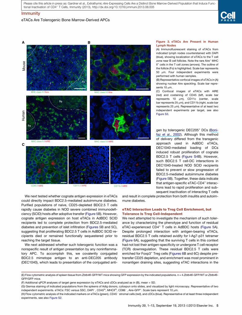
eTACs Are Present in Human Secondary LymphoidOrgansTo identify and characterize peripheral AIRE-expressing popula-
tions in humans, we examined lymph node sections by immuno-
fluorescence and were able to identify discrete cells expressing
intranuclear AIRE protein (Figure 3A) in all patient samples exam-
ined (6/6). Further, as in mice, human eTACs were uniformly
MHC II+ and localized to the subcapsular zone at the boundary
between the T cell paracortex and B cell follicles within the lymph
node (Figure 3A). Importantly, we found that AIRE protein in
human eTACs and mTECs was exclusively intranuclear and
concentrated in nuclear speckles (Figure 3B; Figures S3A and
S3B), similar to the localization of transcriptionally active AIRE
protein in the thymus (Su et al., 2008). As in mice (Gardner
et al., 2008) (Figure 2), human eTACs did not stain strongly for
the conventional DC markers CD11c and CD45 and also lacked
CD11b (Figure 3C). Together, these results identify a discrete
population of eTACs in human secondary lymphoid organs
closely resembling their murine counterparts.
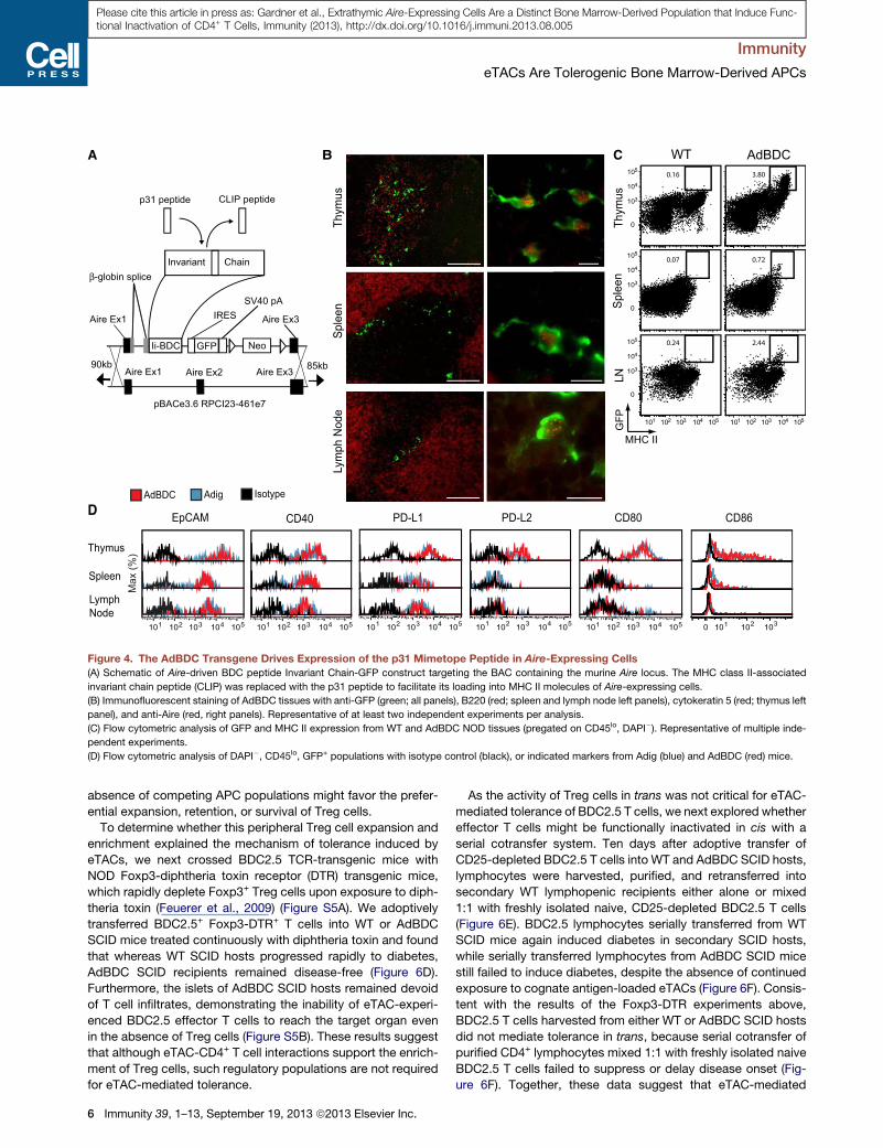
Transgenic Aire-Driven BDC Peptide Is Expressed inmTECs and eTACsGiven the high expression of MHC II by mouse and human
eTACs, we hypothesized that eTACs might interact with CD4+
T cells. To investigate this, we turned to the BDC2.5 TCR-trans-
genic mouse model of diabetes (Katz et al., 1993), in which dia-
betogenic CD4+ T cells react to a pancreatic antigen derived
from chromogranin A (Stadinski et al., 2010), as well as its mim-
etope peptide p31 (Judkowski et al., 2001). By inserting the BDC
mimetope peptide into the MHC class II molecule-associated
invariant chain (Ii or CD74) under direction of the Aire promoter
(van Santen et al., 2004), we were able to direct presentation
of p31 and expression of GFP to mTECs and eTACs in an Aire-
driven BDC antigen (AdBDC) transgenic mouse (Figure 4A).
Localization of mTECs and eTACs to the thymic medulla and
boundary of the T and B cell zones, respectively, was confirmed,
as was colabeling of Aire-expressing cells with both Aire protein
and GFP (Figure 4B). Examination of surface marker expression
on GFP+ cells from Adig and AdBDC mice confirmed that
Aire-expressing cells identified by the two reporters had identical
profiles (Figures 4C and 4D). Together, these results demon-
strate that the AdBDC transgene faithfully recapitulates endoge-
nous Aire expression in mTECs and eTACs.
eTACs Interact with Cognate CD4+ T Cells and DriveTolerance in AdBDC MiceTo test whether eTACs could present antigen to CD4+ T cells, we
performed adoptive transfers of congenically-marked, CFSE-
labeled BDC2.5 CD4+ T cells. Three days after transfer,
BDC2.5 T cells were found to proliferate only in the pancreatic
lymph nodes in WT recipients. In contrast, robust proliferation
of BDC2.5 T cells was observed in all secondary lymphoid
organs of AdBDC mice, and by 2 weeks after transfer, notable
residual populations remained, which had completely diluted
CFSE (Figure 5A). Importantly, proliferation was also observed
in AdBDCmice reconstituted with MHCII-deficient bone marrow
(Figure S4A), demonstrating that that direct antigen presentation
by radioresistant eTACs (Figure S1B) was sufficient to drive
BDC2.5 T cell proliferation.
Immunity 39, 1–13, September 19, 2013 ª2013 Elsevier Inc. 3
A
DAPI
IIC
HM
CD45
MACpE
GFP
B
H
CD11c
CD45 CCR7 F4/80
CD11b CD40
CD103
CD4 CD8a
CD80
cDC
CD45- stroma
eTAC
AdigWT
Rel
ativ
eE
xpre
ssio
n
nd nd
ndnd
C
CD45
SSC
CD31
gp38
D
CD31
gp38
0.76 0
099.2
38.6
CD45
SSC
cDC
seT
AC
s
G
E
Aire CD80 CD86 Deaf10.001
0.01
0.1
1
cDC eTACF
Zbtb46-GFP
CD11c- cII+cDCeTAC
Rel
ativ
eE
xpre
ssio
nR
elat
ive
Exp
ress
ion
0.001
0.01
0.1
1
0.01
0.1
1
Aire Grin2c Dsg1a
Zbtb46 Ptprc(CD45)
Itgax(CD11c)
Aire+mTEC eTAC cDC
)%(
xaM
)%(
xaM
)%(
xaM
0
20
40
60
80
100
)%(
xaM
0
20
40
60
80
100
0 102 103 104 105
0
102
103
104
105
12 1.57
4.0482.30 102 103 104 105
0
102
103
104
105
0 102 103 104 105
0
102
103
104
105
0 102 103 104 105
0.703
0 102 103 104 1050
50K
100K
150K
200K
250K
0
50K
100K
150K
200K
250K
0 102 103 104 105
0
102
103
104
105
0 102 103 104 105 0 102 103 104 105
0
20
40
60
80
100
0
20
40
60
80
100
0 103 104 1050 103 104 105 0 103 104 105 0 103 104 105 0 103 104 105
0 103 104 1050 103 104 1050 103 104 1050 103 104 1050 103 104 105
Figure 2. eTACs Are a Distinct Type of Antigen-Presenting Cell
(A) Identification of splenic eTACs without a GFP reporter. MHCII+ DAPI� events are shown on a CD45 versus EpCAM plot, and histograms of GFP expression are
shown among a distinct EpCAM+ CD45lo population.
(B) Quantitative PCR results of relative mRNA expression,Mean + SD, standardized to Ppia, in indicated populations. nd, not detected. Results are representative
of at least two independent experiments per target.
(C) Flow cytometric analysis of DAPI�, FSC versus SSC Percoll light fractions from WT and Aire-GFP spleen. Representative of three independent experiments.
(D) Back-gating of eTAC events, identified with an equivalent gating strategy to (A), onto the stromal cell identification approach shown in (C).
(legend continued on next page)
Immunity
eTACs Are Tolerogenic Bone Marrow-Derived APCs
4 Immunity 39, 1–13, September 19, 2013 ª2013 Elsevier Inc.
Please cite this article in press as: Gardner et al., Extrathymic Aire-Expressing Cells Are a Distinct Bone Marrow-Derived Population that Induce Func-tional Inactivation of CD4+ T Cells, Immunity (2013), http://dx.doi.org/10.1016/j.immuni.2013.08.005
Fo
Fo
Mou
seHu
man
Fo
Fo
A
C
B
MHC class II / Aire MHC class II / Aire
MHC class II / AIRE MHC class II / AIRE
CD45 / AIRE CD11c / AIRE CD11b / AIRE
Figure 3. eTACs Are Present in Human
Lymph Nodes
(A) Immunofluorescent staining of eTACs from
indicated lymph nodes counterstained with DAPI
(blue), showing localization of eTACs to the T cell
zone near B cell follicles. Note the rare Aire+ MHC
II+ cells in the T cell zones (arrows). The outline of
the follicle (Fo) is highlighted. Scale bar represents
50 mm. Four independent experiments were
performed with human samples.
(B) Representative confocal images of eTACs in (A)
showing nuclear Aire speckling. Scale bar repre-
sents 10 mm.
(C) Confocal images of eTACs with AIRE
(red) and costaining of CD45 (left, scale bar
represents 10 mm), CD11c (center, scale
bar represents 25 mm), and CD11b (right, scale bar
represents 25 mm). Representative of at least two
independent experiments per target, see also
Figure S3.
Immunity
eTACs Are Tolerogenic Bone Marrow-Derived APCs
Please cite this article in press as: Gardner et al., Extrathymic Aire-Expressing Cells Are a Distinct Bone Marrow-Derived Population that Induce Func-tional Inactivation of CD4+ T Cells, Immunity (2013), http://dx.doi.org/10.1016/j.immuni.2013.08.005
We next tested whether cognate antigen expression in eTACs
could directly impact BDC2.5-mediated autoimmune diabetes.
Purified populations of naive, CD25-depleted BDC2.5 T cells
rapidly cause diabetes in NOD severe combined immunodefi-
ciency (SCID) hosts after adoptive transfer (Figure 5B). However,
cognate antigen expression on host eTACs in AdBDC SCID
recipients led to complete protection from BDC2.5-mediated
diabetes and prevention of islet infiltration (Figures 5B and 5C),
suggesting that proliferating BDC2.5 T cells in AdBDC SCID re-
cipients died or remained functionally sequestered prior to
reaching the target tissue.
We next addressed whether such tolerogenic function was a
nonspecific result of antigen presentation by any noninflamma-
tory APC. To accomplish this, we covalently conjugated
BDC2.5 mimetope antigen to an anti-DEC205 antibody
(DEC1040), which leads to presentation of the conjugated anti-
(E) Flow cytometric analysis of spleen tissue from Zbtb46-GFP/WTmice showing GFP expression by the indic
GFP/GFP mice.
(F) Additional qPCR analyses of target gene expression by eTACs and cDCs analyzed as in (B), mean + SD.
(G) Giemsa staining of indicated populations from the spleens of Adig donors, cytospun onto slides, and vis
independent experiments. eTACs: FSC versus SSC, DAPI�, CD45lo, MHCIIhi, CD86�, Aire-GFP+. Scale bars
(H) Flow cytometric analysis of the indicated markers on eTACs (green), CD45� stromal cells (red), and cDCs
experiments, see also Figure S2.
Immunity 39, 1–13,
gen by tolerogenic DEC205+ DCs (Boni-
faz et al., 2002). Although this method
of delivery differed from the transgenic
approach used in AdBDC eTACs,
DEC1040-mediated loading of DCs
induced robust proliferation of cognate
BDC2.5 T cells (Figure S4B). However,
such BDC2.5 T cell-DC interactions in
DEC1040-treated NOD SCID recipients
failed to prevent or slow progression of
BDC2.5-mediated autoimmune diabetes
(Figure 5B). Together, these data indicate
that antigen-specific eTAC-CD4+ interac-
tions lead to rapid proliferation and sub-
sequent inactivation of interacting T cells
and result in complete protection from both insulitis and autoim-
mune diabetes.
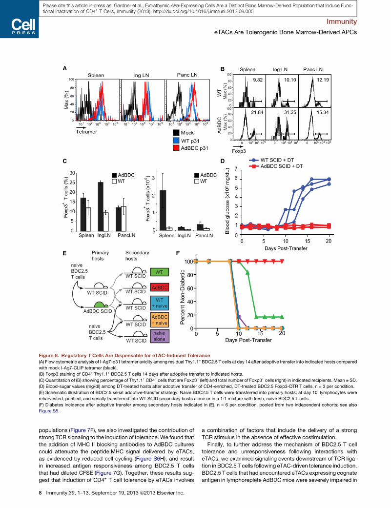
eTAC Interaction Leads to Treg Cell Enrichment, butTolerance Is Treg Cell-IndependentWe next attempted to investigate the mechanism of such toler-
ance by characterizing the phenotype and function of residual
eTAC-experienced CD4+ T cells in AdBDC hosts (Figure 5A).
Despite prolonged interaction with antigen-bearing eTACs,
residual BDC2.5 T cells retained avidity for I-Ag7-p31 tetramer
(Figure 6A), suggesting that the surviving T cells in this context
had not lost their antigen-specificity or undergone T cell receptor
(TCR) downregulation. These residual BDC2.5 T cells were
enriched for Foxp3+ Treg cells (Figures 6B and 6C) despite pre-
transfer CD25 depletion, and enrichment was most prominent in
nonantigen draining sites, suggesting eTAC interactions in the
ated populations. n = 4 Zbtb46-GFP/WT or Zbtb46-
ualized by light microscopy. Representative of two
represent 10 mm.
(blue). Representative of at least three independent
September 19, 2013 ª2013 Elsevier Inc. 5
A C WT AdBDC0.16
0.24 2.44
0.07 0.72
3.80
Thym
usSp
leen
LN
MHC II
GFP
Thym
usSp
leen
Lym
ph N
ode
0
103
104
105
0
103
104
105
0
103
104
105
102 103 104 105101102 103 104 105101
D EpCAM PD-L1
AdBDC Adig Isotype
Thymus
Spleen
Lymph Node
CD80 CD86CD40 PD-L2
)%(
xaM
102 103 104 105101102 103 104 105101 102 103 104 105101 102 103 104 105101 102 103 104 105101 102 1031010
Neo
Aire Ex3Aire Ex1
Aire Ex1 Aire Ex2 Aire Ex3
β-globin splice
SV40 pAIRES
90kb 85kb
pBACe3.6 RPCI23-461e7
Invariant Chain
p31 peptide
Ii-BDC GFP
CLIP peptide
B
Figure 4. The AdBDC Transgene Drives Expression of the p31 Mimetope Peptide in Aire-Expressing Cells
(A) Schematic of Aire-driven BDC peptide Invariant Chain-GFP construct targeting the BAC containing the murine Aire locus. The MHC class II-associated
invariant chain peptide (CLIP) was replaced with the p31 peptide to facilitate its loading into MHC II molecules of Aire-expressing cells.
(B) Immunofluorescent staining of AdBDC tissues with anti-GFP (green; all panels), B220 (red; spleen and lymph node left panels), cytokeratin 5 (red; thymus left
panel), and anti-Aire (red, right panels). Representative of at least two independent experiments per analysis.
(C) Flow cytometric analysis of GFP and MHC II expression from WT and AdBDC NOD tissues (pregated on CD45lo, DAPI�). Representative of multiple inde-
pendent experiments.
(D) Flow cytometric analysis of DAPI�, CD45lo, GFP+ populations with isotype control (black), or indicated markers from Adig (blue) and AdBDC (red) mice.
Immunity
eTACs Are Tolerogenic Bone Marrow-Derived APCs
Please cite this article in press as: Gardner et al., Extrathymic Aire-Expressing Cells Are a Distinct Bone Marrow-Derived Population that Induce Func-tional Inactivation of CD4+ T Cells, Immunity (2013), http://dx.doi.org/10.1016/j.immuni.2013.08.005
absence of competing APC populations might favor the prefer-
ential expansion, retention, or survival of Treg cells.
To determine whether this peripheral Treg cell expansion and
enrichment explained the mechanism of tolerance induced by
eTACs, we next crossed BDC2.5 TCR-transgenic mice with
NOD Foxp3-diphtheria toxin receptor (DTR) transgenic mice,
which rapidly deplete Foxp3+ Treg cells upon exposure to diph-
theria toxin (Feuerer et al., 2009) (Figure S5A). We adoptively
transferred BDC2.5+ Foxp3-DTR+ T cells into WT or AdBDC
SCID mice treated continuously with diphtheria toxin and found
that whereas WT SCID hosts progressed rapidly to diabetes,
AdBDC SCID recipients remained disease-free (Figure 6D).
Furthermore, the islets of AdBDC SCID hosts remained devoid
of T cell infiltrates, demonstrating the inability of eTAC-experi-
enced BDC2.5 effector T cells to reach the target organ even
in the absence of Treg cells (Figure S5B). These results suggest
that although eTAC-CD4+ T cell interactions support the enrich-
ment of Treg cells, such regulatory populations are not required
for eTAC-mediated tolerance.
6 Immunity 39, 1–13, September 19, 2013 ª2013 Elsevier Inc.
As the activity of Treg cells in trans was not critical for eTAC-
mediated tolerance of BDC2.5 T cells, we next explored whether
effector T cells might be functionally inactivated in cis with a
serial cotransfer system. Ten days after adoptive transfer of
CD25-depleted BDC2.5 T cells into WT and AdBDC SCID hosts,
lymphocytes were harvested, purified, and retransferred into
secondary WT lymphopenic recipients either alone or mixed
1:1 with freshly isolated naive, CD25-depleted BDC2.5 T cells
(Figure 6E). BDC2.5 lymphocytes serially transferred from WT
SCID mice again induced diabetes in secondary SCID hosts,
while serially transferred lymphocytes from AdBDC SCID mice
still failed to induce diabetes, despite the absence of continued
exposure to cognate antigen-loaded eTACs (Figure 6F). Consis-
tent with the results of the Foxp3-DTR experiments above,
BDC2.5 T cells harvested from either WT or AdBDC SCID hosts
did not mediate tolerance in trans, because serial cotransfer of
purified CD4+ lymphocytes mixed 1:1 with freshly isolated naive
BDC2.5 T cells failed to suppress or delay disease onset (Fig-
ure 6F). Together, these data suggest that eTAC-mediated
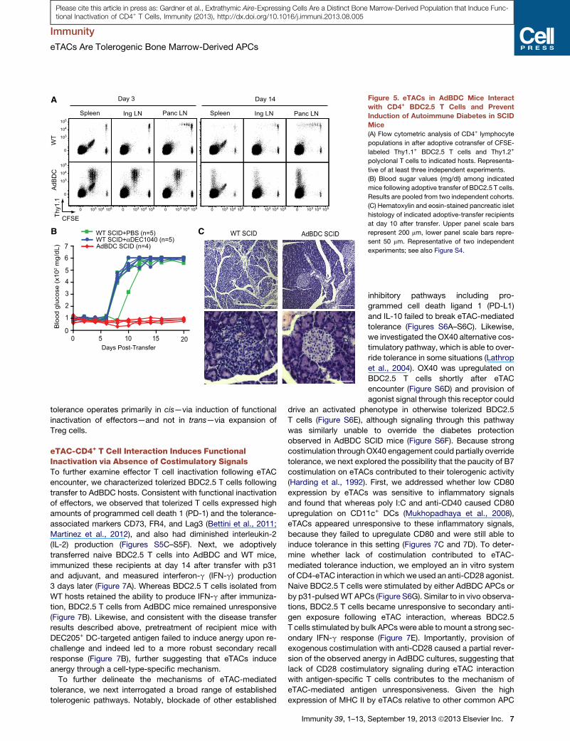
A
CFSEThy1
.1
Spleen
WT
AdBD
C
Ing LN
Day 3
Panc LN Spleen Ing LN Panc LN
Day 14
B CWT SCID+PBS (n=5) WT SCID+αDEC1040 (n=5)AdBDC SCID (n=4)
0 5 10 15 200
1
2
3
45
67
Blo
od g
luco
se (
x102 m
g/dL
)
WT SCID AdBDC SCID
Days Post-Transfer
0 103 104 105 0 103 104 105 0 103 104 105 0 103 104 105 0 103 104 105 0 103 104 105
0
103
104
105
0
103
104
105
Figure 5. eTACs in AdBDC Mice Interact
with CD4+ BDC2.5 T Cells and Prevent
Induction of Autoimmune Diabetes in SCID
Mice
(A) Flow cytometric analysis of CD4+ lymphocyte
populations in after adoptive cotransfer of CFSE-
labeled Thy1.1+ BDC2.5 T cells and Thy1.2+
polyclonal T cells to indicated hosts. Representa-
tive of at least three independent experiments.
(B) Blood sugar values (mg/dl) among indicated
mice following adoptive transfer of BDC2.5 T cells.
Results are pooled from two independent cohorts.
(C) Hematoxylin and eosin-stained pancreatic islet
histology of indicated adoptive-transfer recipients
at day 10 after transfer. Upper panel scale bars
represent 200 mm, lower panel scale bars repre-
sent 50 mm. Representative of two independent
experiments; see also Figure S4.
Immunity
eTACs Are Tolerogenic Bone Marrow-Derived APCs
Please cite this article in press as: Gardner et al., Extrathymic Aire-Expressing Cells Are a Distinct Bone Marrow-Derived Population that Induce Func-tional Inactivation of CD4+ T Cells, Immunity (2013), http://dx.doi.org/10.1016/j.immuni.2013.08.005
tolerance operates primarily in cis—via induction of functional
inactivation of effectors—and not in trans—via expansion of
Treg cells.
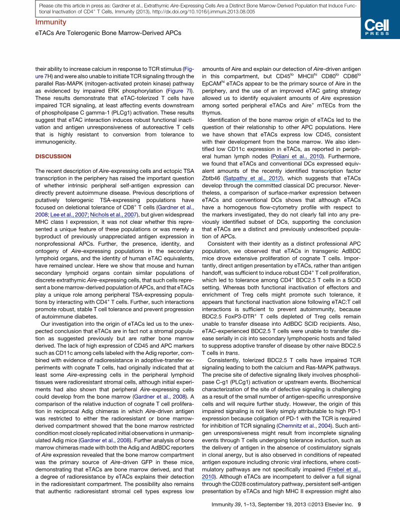
eTAC-CD4+ T Cell Interaction Induces FunctionalInactivation via Absence of Costimulatory SignalsTo further examine effector T cell inactivation following eTAC
encounter, we characterized tolerized BDC2.5 T cells following
transfer to AdBDC hosts. Consistent with functional inactivation
of effectors, we observed that tolerized T cells expressed high
amounts of programmed cell death 1 (PD-1) and the tolerance-
associated markers CD73, FR4, and Lag3 (Bettini et al., 2011;
Martinez et al., 2012), and also had diminished interleukin-2
(IL-2) production (Figures S5C–S5F). Next, we adoptively
transferred naive BDC2.5 T cells into AdBDC and WT mice,
immunized these recipients at day 14 after transfer with p31
and adjuvant, and measured interferon-g (IFN-g) production
3 days later (Figure 7A). Whereas BDC2.5 T cells isolated from
WT hosts retained the ability to produce IFN-g after immuniza-
tion, BDC2.5 T cells from AdBDC mice remained unresponsive
(Figure 7B). Likewise, and consistent with the disease transfer
results described above, pretreatment of recipient mice with
DEC205+ DC-targeted antigen failed to induce anergy upon re-
challenge and indeed led to a more robust secondary recall
response (Figure 7B), further suggesting that eTACs induce
anergy through a cell-type-specific mechanism.
To further delineate the mechanisms of eTAC-mediated
tolerance, we next interrogated a broad range of established
tolerogenic pathways. Notably, blockade of other established
Immunity 39, 1–13,
inhibitory pathways including pro-
grammed cell death ligand 1 (PD-L1)
and IL-10 failed to break eTAC-mediated
tolerance (Figures S6A–S6C). Likewise,
we investigated the OX40 alternative cos-
timulatory pathway, which is able to over-
ride tolerance in some situations (Lathrop
et al., 2004). OX40 was upregulated on
BDC2.5 T cells shortly after eTAC
encounter (Figure S6D) and provision of
agonist signal through this receptor could
drive an activated phenotype in otherwise tolerized BDC2.5
T cells (Figure S6E), although signaling through this pathway
was similarly unable to override the diabetes protection
observed in AdBDC SCID mice (Figure S6F). Because strong
costimulation throughOX40 engagement could partially override
tolerance, we next explored the possibility that the paucity of B7
costimulation on eTACs contributed to their tolerogenic activity
(Harding et al., 1992). First, we addressed whether low CD80
expression by eTACs was sensitive to inflammatory signals
and found that whereas poly I:C and anti-CD40 caused CD80
upregulation on CD11c+ DCs (Mukhopadhaya et al., 2008),
eTACs appeared unresponsive to these inflammatory signals,
because they failed to upregulate CD80 and were still able to
induce tolerance in this setting (Figures 7C and 7D). To deter-
mine whether lack of costimulation contributed to eTAC-
mediated tolerance induction, we employed an in vitro system
of CD4-eTAC interaction in which we used an anti-CD28 agonist.
Naive BDC2.5 T cells were stimulated by either AdBDC APCs or
by p31-pulsedWT APCs (Figure S6G). Similar to in vivo observa-
tions, BDC2.5 T cells became unresponsive to secondary anti-
gen exposure following eTAC interaction, whereas BDC2.5
T cells stimulated by bulk APCswere able tomount a strong sec-
ondary IFN-g response (Figure 7E). Importantly, provision of
exogenous costimulation with anti-CD28 caused a partial rever-
sion of the observed anergy in AdBDC cultures, suggesting that
lack of CD28 costimulatory signaling during eTAC interaction
with antigen-specific T cells contributes to the mechanism of
eTAC-mediated antigen unresponsiveness. Given the high
expression of MHC II by eTACs relative to other common APC
September 19, 2013 ª2013 Elsevier Inc. 7
AdBDC SCID + DT WT SCID + DT
0
1
2
3
45
67
Blo
od g
luco
se (
x102 m
g/dL
)
0 5 10 15 20Days Post-Transfer
ASpleen Ing LN Panc LN
Tetramer Mock
WT p31AdBDC p31
30
25
20
15
10
5
0IngLN PancLNSpleen
Foxp
3T
cells
3
2
1
0IngLN PancLNSpleen
Foxp
3T
cells
(x10
)Foxp3
Spleen Ing LN Panc LN
AdBDCWT
AdBDCWT
WT
AdB
DC
B
C D
F
naiveBDC2.5T cells
WT SCID
WT
+ naive
naivealone
AdBDC
AdBDC+ naive
E Primaryhosts
Secondaryhosts
WT SCID
WT SCID
WT SCID
WT SCID
WT SCID
AdBDC SCID
naiveBDC2.5T cells
WT
0
20
40
60
80
100
Days Post-Transfer
Per
cent
Non
-Di a
b etic
0 5 10 15 20
)%(
xaM
0
20
40
60
80
100
102 103 104 105101 102 103 104 105101 102 103 104 105101
020
40
60
80
100
0
20
40
60
80
100
)%(
xaM
)%(
xaM
0 103 104 105 0 103 104 105 0 103 104 1054
(%)
++
Figure 6. Regulatory T Cells Are Dispensable for eTAC-Induced Tolerance
(A) Flow cytometric analysis of I-Ag7-p31 tetramer avidity among residual Thy1.1+ BDC2.5 T cells at day 14 after adoptive transfer into indicated hosts compared
with mock I-Ag7-CLIP tetramer (black).
(B) Foxp3 staining of CD4+ Thy1.1+ BDC2.5 T cells 14 days after adoptive transfer to indicated hosts.
(C) Quantitation of (B) showing percentage of Thy1.1+ CD4+ cells that are Foxp3+ (left) and total number of Foxp3+ cells (right) in indicated recipients. Mean ± SD.
(D) Blood-sugar values (mg/dl) among DT-treated hosts after adoptive transfer of CD4-enriched, DT-treated BDC2.5 Foxp3-DTR T cells, n = 3 per condition.
(E) Schematic illustration of BDC2.5 serial adoptive-transfer strategy. Naive BDC2.5 T cells were transferred into primary hosts; at day 10, lymphocytes were
reharvested, purified, and serially transferred into WT SCID secondary hosts alone or in a 1:1 mixture with fresh, naive BDC2.5 T cells.
(F) Diabetes incidence after adoptive transfer among secondary hosts indicated in (E), n = 6 per condition, pooled from two independent cohorts; see also
Figure S5.
Immunity
eTACs Are Tolerogenic Bone Marrow-Derived APCs
Please cite this article in press as: Gardner et al., Extrathymic Aire-Expressing Cells Are a Distinct Bone Marrow-Derived Population that Induce Func-tional Inactivation of CD4+ T Cells, Immunity (2013), http://dx.doi.org/10.1016/j.immuni.2013.08.005
populations (Figure 7F), we also investigated the contribution of
strong TCR signaling to the induction of tolerance. We found that
the addition of MHC II blocking antibodies to AdBDC cultures
could attenuate the peptide:MHC signal delivered by eTACs,
as evidenced by reduced cell cycling (Figure S6H), and result
in increased antigen responsiveness among BDC2.5 T cells
that had diluted CFSE (Figure 7G). Together, these results sug-
gest that induction of CD4+ T cell tolerance by eTACs involves
8 Immunity 39, 1–13, September 19, 2013 ª2013 Elsevier Inc.
a combination of factors that include the delivery of a strong
TCR stimulus in the absence of effective costimulation.
Finally, to further address the mechanism of BDC2.5 T cell
tolerance and unresponsiveness following interactions with
eTACs, we examined signaling events downstream of TCR liga-
tion in BDC2.5 T cells following eTAC-driven tolerance induction.
BDC2.5 T cells that had encountered eTACs expressing cognate
antigen in lymphoreplete AdBDCmice were severely impaired in
Immunity
eTACs Are Tolerogenic Bone Marrow-Derived APCs
Please cite this article in press as: Gardner et al., Extrathymic Aire-Expressing Cells Are a Distinct Bone Marrow-Derived Population that Induce Func-tional Inactivation of CD4+ T Cells, Immunity (2013), http://dx.doi.org/10.1016/j.immuni.2013.08.005
their ability to increase calcium in response to TCR stimulus (Fig-
ure 7H) andwere also unable to initiate TCR signaling through the
parallel Ras-MAPK (mitogen-activated protein kinase) pathway
as evidenced by impaired ERK phosphorylation (Figure 7I).
These results demonstrate that eTAC-tolerized T cells have
impaired TCR signaling, at least affecting events downstream
of phospholipase C gamma-1 (PLCg1) activation. These results
suggest that eTAC interaction induces robust functional inacti-
vation and antigen unresponsiveness of autoreactive T cells
that is highly resistant to conversion from tolerance to
immunogenicity.
DISCUSSION
The recent description of Aire-expressing cells and ectopic TSA
transcription in the periphery has raised the important question
of whether intrinsic peripheral self-antigen expression can
directly prevent autoimmune disease. Previous descriptions of
putatively tolerogenic TSA-expressing populations have
focused on deletional tolerance of CD8+ T cells (Gardner et al.,
2008; Lee et al., 2007; Nichols et al., 2007), but given widespread
MHC class I expression, it was not clear whether this repre-
sented a unique feature of these populations or was merely a
byproduct of previously unappreciated antigen expression in
nonprofessional APCs. Further, the presence, identity, and
ontogeny of Aire-expressing populations in the secondary
lymphoid organs, and the identity of human eTAC equivalents,
have remained unclear. Here we show that mouse and human
secondary lymphoid organs contain similar populations of
discrete extrathymic Aire-expressing cells, that such cells repre-
sent a bonemarrow-derived population of APCs, and that eTACs
play a unique role among peripheral TSA-expressing popula-
tions by interacting with CD4+ T cells. Further, such interactions
promote robust, stable T cell tolerance and prevent progression
of autoimmune diabetes.
Our investigation into the origin of eTACs led us to the unex-
pected conclusion that eTACs are in fact not a stromal popula-
tion as suggested previously but are rather bone marrow
derived. The lack of high expression of CD45 and APC markers
such as CD11c among cells labeled with the Adig reporter, com-
bined with evidence of radioresistance in adoptive-transfer ex-
periments with cognate T cells, had originally indicated that at
least some Aire-expressing cells in the peripheral lymphoid
tissues were radioresistant stromal cells, although initial experi-
ments had also shown that peripheral Aire-expressing cells
could develop from the bone marrow (Gardner et al., 2008). A
comparison of the relative induction of cognate T cell prolifera-
tion in reciprocal Adig chimeras in which Aire-driven antigen
was restricted to either the radioresistant or bone marrow-
derived compartment showed that the bone marrow restricted
conditionmost closely replicated initial observations in unmanip-
ulated Adig mice (Gardner et al., 2008). Further analysis of bone
marrow chimeras made with both the Adig and AdBDC reporters
of Aire expression revealed that the bone marrow compartment
was the primary source of Aire-driven GFP in these mice,
demonstrating that eTACs are bone marrow derived, and that
a degree of radioresistance by eTACs explains their detection
in the radioresistant compartment. The possibility also remains
that authentic radioresistant stromal cell types express low
amounts of Aire and explain our detection of Aire-driven antigen
in this compartment, but CD45lo MHCIIhi CD80lo CD86lo
EpCAMhi eTACs appear to be the primary source of Aire in the
periphery, and the use of an improved eTAC gating strategy
allowed us to identify equivalent amounts of Aire expression
among sorted peripheral eTACs and Aire+ mTECs from the
thymus.
Identification of the bone marrow origin of eTACs led to the
question of their relationship to other APC populations. Here
we have shown that eTACs express low CD45, consistent
with their development from the bone marrow. We also iden-
tified low CD11c expression in eTACs, as reported in periph-
eral human lymph nodes (Poliani et al., 2010). Furthermore,
we found that eTACs and conventional DCs expressed equiv-
alent amounts of the recently identified transcription factor
Zbtb46 (Satpathy et al., 2012), which suggests that eTACs
develop through the committed classical DC precursor. Never-
theless, a comparison of surface-marker expression between
eTACs and conventional DCs shows that although eTACs
have a homogenous flow-cytometry profile with respect to
the markers investigated, they do not clearly fall into any pre-
viously identified subset of DCs, supporting the conclusion
that eTACs are a distinct and previously undescribed popula-
tion of APCs.
Consistent with their identity as a distinct professional APC
population, we observed that eTACs in transgenic AdBDC
mice drove extensive proliferation of cognate T cells. Impor-
tantly, direct antigen presentation by eTACs, rather than antigen
handoff, was sufficient to induce robust CD4+ T cell proliferation,
which led to tolerance among CD4+ BDC2.5 T cells in a SCID
setting. Whereas both functional inactivation of effectors and
enrichment of Treg cells might promote such tolerance, it
appears that functional inactivation alone following eTAC:T cell
interactions is sufficient to prevent autoimmunity, because
BDC2.5 FoxP3-DTR+ T cells depleted of Treg cells remain
unable to transfer disease into AdBDC SCID recipients. Also,
eTAC-experienced BDC2.5 T cells were unable to transfer dis-
ease serially in cis into secondary lymphopenic hosts and failed
to suppress adoptive transfer of disease by other naive BDC2.5
T cells in trans.
Consistently, tolerized BDC2.5 T cells have impaired TCR
signaling leading to both the calcium and Ras-MAPK pathways.
The precise site of defective signaling likely involves phospholi-
pase C-g1 (PLCg1) activation or upstream events. Biochemical
characterization of the site of defective signaling is challenging
as a result of the small number of antigen-specific unresponsive
cells and will require further study. However, the origin of this
impaired signaling is not likely simply attributable to high PD-1
expression because coligation of PD-1 with the TCR is required
for inhibition of TCR signaling (Chemnitz et al., 2004). Such anti-
gen unresponsiveness might result from incomplete signaling
events through T cells undergoing tolerance induction, such as
the delivery of antigen in the absence of costimulatory signals
in clonal anergy, but is also observed in conditions of repeated
antigen exposure including chronic viral infections, where costi-
mulatory pathways are not specifically impaired (Frebel et al.,
2010). Although eTACs are incompetent to deliver a full signal
through the CD28 costimulatory pathway, persistent self-antigen
presentation by eTACs and high MHC II expression might also
Immunity 39, 1–13, September 19, 2013 ª2013 Elsevier Inc. 9
A
4.28 9.07
7.73 14.52
0.60 0.14
LN
WT+
PB
S
WT+
αD
EC
-10
40A
dBD
C
Spleen
0
4
8
12
16
WT+PBS
WT+αDEC
AdBDC
IFN
-γ
p<.05
p<.01naiveBDC2.5T cells
AdBDC
WT
day 0
BDC2.5adoptivetransfer
WT + αDEC1040
day 14
immunizep31 in CFA
day 17
analyze
IFN-γThy1
.1
05
1015202530354045
WTAdBDC
IFN
-γ (
%)
p<.05C E
DC
CD
80 M
FI (x
10 )2
IsotypePBS DC
eTAC
CD80α-CD40/Poly I:C
-5
0
5
10
15
20
25
eTA
C
-αCD28 +αCD28Primary stimulation condition
B
0 5 10 15 200
100200
300400
500600
Days Post-Transfer
Blo
odG
luco
se (m
g/dL
)
D
LNSpleen
Iso WT SCIDαCD40 WT SCIDαCD40 AdBDC SCID
H
50 100 150Time
xulF muicl a
C evit al eR
WT hostAdBDC host
I
p-Erk
WT host AdBDC host
unstimCD3PMA
unstimCD3PMA
F GeTACscDCsB cellsMΦT cells
MHC IIIso I-Ag7
0
10
20
IFN
- γ (
%)
p<.05
102 103100 101102 103100 101
101
102
103
100
101
102
103
100
101
102
103
100
104 1050 103
No tx CD40
+
+
)%( xa
M
0
20
40
60
80
100
+
A C I
)%( xa
M
0
20
40
60
80
100
0 102 103 104 1050 102 103 104 105
0 103 104 105
Figure 7. eTACs Induce Functional Inactivation of Cognate T Cells by Presenting Antigen in the Absence of Costimulation
(A) Schematic of p31+CFA immunization protocol to measure recall response of adoptively transferred BDC2.5 T cells. Naive BDC2.5 T cells were transferred to
indicated mice, which were immunized with p31 in complete Freund’s adjuvant (CFA) 14 days later. IFN-g production to this second stimulation was assessed
ex vivo after 3 days.
(legend continued on next page)
Immunity
eTACs Are Tolerogenic Bone Marrow-Derived APCs
10 Immunity 39, 1–13, September 19, 2013 ª2013 Elsevier Inc.
Please cite this article in press as: Gardner et al., Extrathymic Aire-Expressing Cells Are a Distinct Bone Marrow-Derived Population that Induce Func-tional Inactivation of CD4+ T Cells, Immunity (2013), http://dx.doi.org/10.1016/j.immuni.2013.08.005
Immunity
eTACs Are Tolerogenic Bone Marrow-Derived APCs
Please cite this article in press as: Gardner et al., Extrathymic Aire-Expressing Cells Are a Distinct Bone Marrow-Derived Population that Induce Func-tional Inactivation of CD4+ T Cells, Immunity (2013), http://dx.doi.org/10.1016/j.immuni.2013.08.005
contribute to their tolerance induction through an exhaustion-like
pathway of acquired antigen unresponsiveness in which costi-
mulatory signaling is not a major factor in tolerance induction
(Singh et al., 2007). However, the inactivation of Erk signaling
that we observed in eTAC-tolerized cells was more associated
with clonal anergy than exhaustion or adaptive tolerance in a
biochemical comparison of models for the two states (Chiodetti
et al., 2006). Taken together, our results indicate that the primary
mechanism of eTAC-mediated disease prevention in this model
is recessive—via functional anergy of effectors as opposed to
enrichment of Treg cells.
Our results also suggest that eTACs might have uniquely
protolerogenic capabilities. In support of this, targeting of a
BDC mimetope peptide to another putative tolerogenic APC
population, DEC205+ DCs, induces proliferation of adoptively
transferred BDC2.5 T cells similar to that seen in AdBDC recip-
ients but fails to protect from adoptively transferred diabetes.
While the lack of tolerance by DEC1040 was surprising given
the established role for immature DEC205+ DCs in tolerance in-
duction (Bonifaz et al., 2002; Mukhopadhaya et al., 2008), DCs
from NOD mice might have impaired tolerance (Hamilton-
Williams et al., 2009), and prior studies of DEC205+ DCs
have focused on tolerance induction of CD8+ T cells. Also,
the lymphopenia in SCID mice might override tolerogenic
DEC205+ DC antigen presentation, because peripheral antigen
display can convert from tolerogenic to immunogenic in the
context of lymphopenia (King et al., 2004). It should also be
mentioned that delivery of antigen via anti-DEC1040 and via
the Aire promoter might differ in the dose and length of antigen
exposure, which could affect the differential tolerance induc-
tion by DEC205+ DCs and eTACs. However, our DEC1040 ex-
periments suggest that eTAC-induced tolerance is not simply a
result of the presence of cognate antigen in our disease model,
but instead likely depends on additional antigen presentation
qualities of eTACs. Indeed, T cell tolerance induced upon
eTAC encounter appears insensitive to the perturbations that
mitigate many other characterized forms of peripheral toler-
ance. For example, it is striking that self-antigen expression
in eTACs prevents autoimmunity even in lymphopenic SCID
mice and that CD40 and Toll-like receptor (TLR) stimuli do
not revert eTAC-induced tolerance, in contrast to studies
with conventional DC-targeted antigens (Mukhopadhaya
et al., 2008). Together these findings suggest that self-antigen
expression in eTACs induces a remarkably robust T cell toler-
(B) IFN-g production among CD4+ Thy1.1+ BDC2.5 T cells isolated from mice ind
group. Mean ± SD. Results are pooled from two independent experiments.
(C) Representative flow cytometric analysis of CD80 staining in DAPI�, CD45+, CCD40+PolyI:C stimulus; quantification of MFI shifts above isotype amounts show
(D) Blood-sugar values (mg/dL) among anti-CD40+PolyI:C treated hosts after ad
independent experiments.
(E) IFN-g recall responses (percentage of CD4+ T cells) by CD4-enriched BDC2.5
CD28 where indicated, responding to p31 + APC restimulation. Mean + SD. Dat
(F) Representative flow cytometric analysis of MHC II expression among by indic
(G) IFN-g recall responses by CD4+ Thy1.1+ CFSElo BDC2.5 T cells following cultu
Mean + SD.
(H) Kinetic plots of relative calcium signaling over time (s) of ex vivo BDC2.5 Thy1.1
by Indo-1 dyes. A, anti-CD3; C, crosslinking secondary; I, ionomycin. Results ar
(I) Flow cytometric analysis of Erk phosphorylation of rested Thy1.1+ CD4+ T cell
Results are representative of three independent experiments, see also Figure S6
ance that is highly resistant to conversion from tolerance to
immunogenicity.
Further, these findings distinguish eTACs among putative
TSA-expressing peripheral populations in the secondary
lymphoid organs. Although expression of individual TSA genes
has been reported in fibroblastic reticular cells and lymphatic
endothelium, these populations do not express Aire (Cohen
et al., 2010; Fletcher et al., 2010; Lee et al., 2007; Nichols
et al., 2007), and although other transcriptional regulators such
as Deaf1 might fulfill this same function (Yip et al., 2009), the
role of any such factor(s) has not yet been linked to TSA expres-
sion in a specific population or to the promotion of immunologic
tolerance. Further, although the identification of individual
candidate TSA genes expressed in such populations might
represent genuine antigen production for the sake of immune
education, enrichment for a broad range of ectopically ex-
pressed TSA genes appears unique to eTACs (Gardner et al.,
2008). This provides an appealing model for complementary
tolerance induction to self-reactive T cells that evade adequate
negative selection in the thymus, and the fact that such TSA-
expressing eTACs can induce tolerance among both CD4+ and
CD8+ T cells further supports a broad role for this population in
peripheral tolerance.
Together, these results demonstrate that eTACs are a distinct
type of bone marrow-derived APC, and that targeted antigen
expression in eTACs induces robust peripheral self-tolerance
through the induction of antigen unresponsiveness. The ability
of eTACs to confer such tolerance in secondary hosts represents
an attractive therapeutic application for this population in the re-
establishment of immune tolerance in both autoimmunity and
transplantation. Finally, our findings that eTACs are a distinct
bone marrow-derived population from the classical DC lineage
should facilitate the identification and development of in vitro
differentiation and expansion conditions that will make thera-
peutic eTAC transfers feasible for the induction of antigen-
specific immune tolerance.
EXPERIMENTAL PROCEDURES
Transgene Construction, BAC Recombineering, and Purification
Aire-driven BDC2.5 peptide (AdBDC) transgenic mice were generated by
standard cloningmethods with a bacterial artificial chromosome (BAC) recom-
bineering and transgenesis strategy. Full description of primers, reagents, and
recombineering strategy can be found in Supplemental Experimental
Procedures.
icated in (A); bar graph shows quantification of pooled data with n = 4 for each
D11c+ DCs and DAPI�, CD45lo, MHCII+, and GFP+ eTACs in response to anti-
n at right.
optive transfer of naive BDC2.5 T cells, n = 4 per condition, pooled from three
T cells cultured with AdBDC or p31-pulsed WT primary APCs, and with anti-
a were pooled from three independent experiments.
ated populations. MF represents macrophages.
re with AdBDC APCs and either anti-I-Ag7 MHC II blocking antibody or isotype.
+ T cells recovered from indicated hosts and 14 days after transfer, as detected
e representative of three independent experiments.
populations in (H) after a 2 min stimulation with anti-CD3 (blue) or PMA (green).
.
Immunity 39, 1–13, September 19, 2013 ª2013 Elsevier Inc. 11
Immunity
eTACs Are Tolerogenic Bone Marrow-Derived APCs
Please cite this article in press as: Gardner et al., Extrathymic Aire-Expressing Cells Are a Distinct Bone Marrow-Derived Population that Induce Func-tional Inactivation of CD4+ T Cells, Immunity (2013), http://dx.doi.org/10.1016/j.immuni.2013.08.005
Mice and Genotyping
AdBDC and Adig transgenic NOD mice were screened by real-time PCR for
GFP, and were maintained in heterozygosity for all experiments. Additional in-
formation on mouse strains and genotyping appears in the Supplemental
Experimental Procedures. All mice were maintained in microisolator cages
and treated in accordance with the National Institutes of Health (NIH) and
American Association of Laboratory Animal Care standards and consistent
with the animal care and use regulations of the University of California, San
Francisco.
Flow Cytometry and Cell Sorting
All flow cytometry antibodies were purchased from BD Pharmingen, eBio-
science, Invitrogen, or Southern Biotech with the exception of anti-CD16/32
(clone 2.4G2), which was purified by the UCSF hybridoma core. Lymphocytes
for flow cytometry were prepared by mashing thymi, lymph nodes, or spleens,
filtering through a 70 mm cell strainer, ACK lysis of red blood cells (spleen only),
counting by trypan blue exclusion, resuspending in Fc block (2.4G2), and
staining in FACS buffer.
eTAC, mTEC, DC, and macrophage populations were isolated by digestion
in a mixture of collagenase, dispase, and DNase, followed by Percoll density
centrifugation, as described by Gardner et al. (2008). Populations were identi-
fied as follows: cDCs (DAPI�, CD45hi, MHCII+, CD11chi), Aire+ mTECs (DAPI�,CD11c�, EpCAM+, CD45�, MHCII+, Aire-GFP+), B cells (FSC versus SSC,
CD19+, CD45+, CD11c�), CD4+ T cells (FSC versus SSC, CD4+, CD45+,
CD11c�), and macrophages (FSC versus SSC, F4/80+, CD45+). Cells were
analyzed with a BD LSRII cytometer and sorted on a BD FACSAria III cell
sorter. Tetramer, Foxp3, phospho-Erk, Calcium FACS assays, and mRNA
transcript analysis by qPCR were performed as described in Supplemental
Experimental Procedures.
Immunofluorescent Staining and Histology
Immunofluorescent staining was conducted as described in the Supplemental
Experimental Procedures. To visualize cytosolic GFP in AdBDCmice, we fixed
tissues in 1.5% PFA followed TSA amplification of GFP staining (Perkin Elmer).
For human sections, formalin-fixed, paraffin-embedded sections obtained
from the JDRF nPOD project were processed for antigen retrieval prior to
TSA amplification of AIRE signal. All of the human tissue evaluated in this study
was obtained with approval from and under the guidelines of the UCSF Com-
mittee on Human Research.
Reciprocal Bone Marrow Chimeras
WT and Aire-GFP reporter mice (Adig for 8.3 adoptive transfers, AdBDC mice
for the displayed Aire-GFP plots) were used as donors and/or recipients to
generate chimeric mice to restrict transgene expression to the bone marrow,
radioresistant host, neither, or both compartments. Recipient mice received
two irradiation cycles totaling 1,300 rads (900 rads, 400 rads), with at least
3 hr between doses. Donor bone marrow was harvested, depleted of T cells
as described previously (Gardner et al., 2008), and injected intravenously
(i.v.) at 1 3 107 cells per host. Mice were harvested after at least 8 weeks of
reconstitution.
Purification, CFSE-Labeling, and Adoptive and Serial Transfers
of T Cells
For adoptive transfer of naive, CD25-depleted, CD4-enriched T cells, spleen
and nonpancreatic lymph nodes were harvested from nondiabetic donors,
pooled by group, ACK-lysed and counted. Cells were CD25-depleted with
anti-CD25 (7D4) and CD4-enriched with a Mouse CD4+ Negative Selection
Kit (StemCell). Aliquots at each step were analyzed to confirm purity. Purified
cells were pooled in a �1:1 ratio of BDC2.5 T cells to polyclonal CD4+ T cells,
labeled in 2.5 mM CFSE (Invitrogen) for proliferation experiments, and injected
i.v. at 1–2 3 106 cells per mouse in HBSS. 8.3 CD8+ T cells were prepared
similarly with a Robosep CD8+ T cell Negative Selection kit. For adoptive
transfer of diabetes, 23 105 unlabeled cells per mouse were injected IV. Serial
cotransfer of T cells was performed by harvesting nonpancreatic lymph nodes
and spleen from SCID and AdBDC SCID primary recipients at day 10. Cells
were CD4-purified as described above and injected to secondary SCID hosts
either alone or mixed 1:1 with naive BDC2.5 T cells at 2 3 105 cells per
recipient.
12 Immunity 39, 1–13, September 19, 2013 ª2013 Elsevier Inc.
In Vitro Tolerance Assay
Naive T cells were prepared and mixed with Percoll-enriched APCs isolated
from WT or AdBDC mice by density centrifugation as described previously
(Gardner et al., 2008). We mixed 1 3 105 naive BDC2.5 T cells with 1 3 106
APCs in 24 well plates and incubated them for 7 days at 37�C in complete
DMEM media. Acetylated p31 peptide (YVRPLWVRME, GenScript) (Judkow-
ski et al., 2001) was added to control activating conditions at 1 ug/mL.
Anti-CD28 (PV-1) was added to indicated cultures at 0.5 ug/mL. Stimulated
T cells were isolated by Robosep CD4 enrichment, rested overnight in com-
plete DMEMmedia, and analyzed for IFN-g production to a second stimulation
with 1 ug/mL p31 and bulk Thy1.2+ splenocytes (described for CFA immuniza-
tion assay). For MHC II blocking experiments, anti-I-Ag7 (clone AG2.42.7, pro-
vided by E. Unanue) was used at 10 ug/mL. We mixed 2 3 104 naive BDC2.5
T cells with 2.5 3 104 APCs in flat-bottom 96 well plates and incubated them
for 7 days at 37�C in complete DMEMmedia before being rested overnight and
restimulated with PMA and Ionomycin.
Statistical Analysis
Statistical analysis of data was performed with Microsoft Excel 2003 and
GraphPad Prism 4.0. Statistical comparisons were made with paired t tests
and a two-tailed 95% confidence interval.
SUPPLEMENTAL INFORMATION
Supplemental Information includes six figures and Supplemental Experimental
Procedures and can be found with this article online at http://dx.doi.org/10.
1016/j.immuni.2013.08.005.
ACKNOWLEDGMENTS
We thank M. Cheng for critical reading of the manuscript; J. Esensten,
J. Bluestone, Q. Tang, H. van Santen, P. Peterson, H. Scott, E. Unanue,
D. Mathis, and C. Benoist for reagents and mice, and N. Killeen and the
UCSF Transgenic Core for help generating transgenic mice. This work was
supported by the US National Institutes of Health AI035297 (M.S.A),
DK59958 and DK063720 for core support, the Helmsley Charitable Trust
(M.S.A, T.C.M), the American Diabetes Association (J.M.G.), the UCSF Medi-
cal Scientist Training Program (J.M.G.), the UCSF Department of Surgery
(J.M.G.), and the intramural research program of the National Institute of
Diabetes and Digestive and Kidney Diseases, NIH (J.D.P. and K.V.T.). This
research was performed with the support of the Network for Pancreatic Organ
Donors with Diabetes (nPOD), a collaborative type 1 diabetes research project
sponsored by the Juvenile Diabetes Research Foundation International
(JDRF). Organ Procurement Organizations (OPO) partnering with nPOD to pro-
vide research resources are listed at www.jdrfnpod.org/our-partners.php.
Received: August 7, 2012
Accepted: May 18, 2013
Published: August 29, 2013
REFERENCES
Anderson, M.S., Venanzi, E.S., Klein, L., Chen, Z., Berzins, S.P., Turley, S.J.,
von Boehmer, H., Bronson, R., Dierich, A., Benoist, C., and Mathis, D.
(2002). Projection of an immunological self shadow within the thymus by the
aire protein. Science 298, 1395–1401.
Anderson, M.S., Venanzi, E.S., Chen, Z., Berzins, S.P., Benoist, C., and
Mathis, D. (2005). The cellular mechanism of Aire control of T cell tolerance.
Immunity 23, 227–239.
Bettini, M., Szymczak-Workman, A.L., Forbes, K., Castellaw, A.H., Selby, M.,
Pan, X., Drake, C.G., Korman, A.J., and Vignali, D.A. (2011). Cutting edge:
accelerated autoimmune diabetes in the absence of LAG-3. Journal of immu-
nology 187, 3493–3498.
Bonifaz, L., Bonnyay, D., Mahnke, K., Rivera, M., Nussenzweig, M.C., and
Steinman, R.M. (2002). Efficient targeting of protein antigen to the dendritic
cell receptor DEC-205 in the steady state leads to antigen presentation on
Immunity
eTACs Are Tolerogenic Bone Marrow-Derived APCs
Please cite this article in press as: Gardner et al., Extrathymic Aire-Expressing Cells Are a Distinct Bone Marrow-Derived Population that Induce Func-tional Inactivation of CD4+ T Cells, Immunity (2013), http://dx.doi.org/10.1016/j.immuni.2013.08.005
major histocompatibility complex class I products and peripheral CD8+ T cell
tolerance. J. Exp. Med. 196, 1627–1638.
Chemnitz, J.M., Parry, R.V., Nichols, K.E., June, C.H., and Riley, J.L. (2004).
SHP-1 and SHP-2 associate with immunoreceptor tyrosine-based switch
motif of programmed death 1 upon primary human T cell stimulation, but
only receptor ligation prevents T cell activation. Journal of immunology 173,
945–954.
Chiodetti, L., Choi, S., Barber, D.L., and Schwartz, R.H. (2006). Adaptive toler-
ance and clonal anergy are distinct biochemical states. Journal of immunology
176, 2279–2291.
Cohen, J.N., Guidi, C.J., Tewalt, E.F., Qiao, H., Rouhani, S.J., Ruddell, A., Farr,
A.G., Tung, K.S., and Engelhard, V.H. (2010). Lymph node-resident lymphatic
endothelial cells mediate peripheral tolerance via Aire-independent direct an-
tigen presentation. J. Exp. Med. 207, 681–688.
Derbinski, J., Schulte, A., Kyewski, B., and Klein, L. (2001). Promiscuous gene
expression in medullary thymic epithelial cells mirrors the peripheral self. Nat.
Immunol. 2, 1032–1039.
Feuerer, M., Shen, Y., Littman, D.R., Benoist, C., and Mathis, D. (2009). How
punctual ablation of regulatory T cells unleashes an autoimmune lesion within
the pancreatic islets. Immunity 31, 654–664.
Fletcher, A.L., Lukacs-Kornek, V., Reynoso, E.D., Pinner, S.E., Bellemare-
Pelletier, A., Curry, M.S., Collier, A.R., Boyd, R.L., and Turley, S.J. (2010).
Lymphnode fibroblastic reticular cells directly present peripheral tissue antigen
under steady-state and inflammatory conditions. J. Exp. Med. 207, 689–697.
Frebel, H., Richter, K., and Oxenius, A. (2010). How chronic viral infections
impact on antigen-specific T-cell responses. Eur. J. Immunol. 40, 654–663.
Gardner, J.M., Devoss, J.J., Friedman, R.S., Wong, D.J., Tan, Y.X., Zhou, X.,
Johannes, K.P., Su, M.A., Chang, H.Y., Krummel, M.F., and Anderson, M.S.
(2008). Deletional tolerance mediated by extrathymic Aire-expressing cells.
Science 321, 843–847.
Halonen, M., Pelto-Huikko, M., Eskelin, P., Peltonen, L., Ulmanen, I., and
Kolmer, M. (2001). Subcellular location and expression pattern of autoimmune
regulator (Aire), the mouse orthologue for human gene defective in autoim-
mune polyendocrinopathy candidiasis ectodermal dystrophy (APECED). The
journal of histochemistry and cytochemistry: official journal of the
Histochemistry Society 49, 197–208.
Hamilton-Williams, E.E., Martinez, X., Clark, J., Howlett, S., Hunter, K.M.,
Rainbow, D.B., Wen, L., Shlomchik, M.J., Katz, J.D., Beilhack, G.F., et al.
(2009). Expression of diabetes-associated genes by dendritic cells and CD4
T cells drives the loss of tolerance in nonobese diabetic mice. Journal of immu-
nology 183, 1533–1541.
Harding, F.A., McArthur, J.G., Gross, J.A., Raulet, D.H., and Allison, J.P.
(1992). CD28-mediated signalling co-stimulates murine T cells and prevents
induction of anergy in T-cell clones. Nature 356, 607–609.
Heino, M., Peterson, P., Sillanpaa, N., Guerin, S., Wu, L., Anderson, G., Scott,
H.S., Antonarakis, S.E., Kudoh, J., Shimizu, N., et al. (2000). RNA and protein
expression of the murine autoimmune regulator gene (Aire) in normal, RelB-
deficient and in NOD mouse. Eur. J. Immunol. 30, 1884–1893.
Hubert, F.X., Kinkel, S.A., Webster, K.E., Cannon, P., Crewther, P.E., Proeitto,
A.I., Wu, L., Heath, W.R., and Scott, H.S. (2008). A specific anti-Aire antibody
reveals aire expression is restricted to medullary thymic epithelial cells and not
expressed in periphery. Journal of immunology 180, 3824–3832.
Judkowski, V., Pinilla, C., Schroder, K., Tucker, L., Sarvetnick, N., and Wilson,
D.B. (2001). Identification of MHC class II-restricted peptide ligands, including
a glutamic acid decarboxylase 65 sequence, that stimulate diabetogenic
T cells from transgenic BDC2.5 nonobese diabetic mice. Journal of immu-
nology 166, 908–917.
Katz, J.D., Wang, B., Haskins, K., Benoist, C., and Mathis, D. (1993). Following
a diabetogenic T cell from genesis through pathogenesis. Cell 74, 1089–1100.
King, C., Ilic, A., Koelsch, K., and Sarvetnick, N. (2004). Homeostatic expan-
sion of T cells during immune insufficiency generates autoimmunity. Cell
117, 265–277.
Lathrop, S.K., Huddleston, C.A., Dullforce, P.A., Montfort, M.J., Weinberg,
A.D., and Parker, D.C. (2004). A signal through OX40 (CD134) allows anergic,
autoreactive T cells to acquire effector cell functions. Journal of immunology
172, 6735–6743.
Lee, J.W., Epardaud, M., Sun, J., Becker, J.E., Cheng, A.C., Yonekura, A.R.,
Heath, J.K., and Turley, S.J. (2007). Peripheral antigen display by lymph node
stroma promotes T cell tolerance to intestinal self. Nat. Immunol. 8, 181–190.
Lukacs-Kornek, V., Malhotra, D., Fletcher, A.L., Acton, S.E., Elpek, K.G.,
Tayalia, P., Collier, A.R., and Turley, S.J. (2011). Regulated release of nitric ox-
ide by nonhematopoietic stroma controls expansion of the activated T cell pool
in lymph nodes. Nat. Immunol. 12, 1096–1104.
Malhotra, D., Fletcher, A.L., Astarita, J., Lukacs-Kornek, V., Tayalia, P.,
Gonzalez, S.F., Elpek, K.G., Chang, S.K., Knoblich, K., Hemler, M.E., et al.;
Immunological Genome Project Consortium. (2012). Transcriptional profiling
of stroma from inflamed and resting lymph nodes defines immunological
hallmarks. Nat. Immunol. 13, 499–510.
Martinez, R.J., Zhang, N., Thomas, S.R., Nandiwada, S.L., Jenkins, M.K.,
Binstadt, B.A., and Mueller, D.L. (2012). Arthritogenic self-reactive CD4+
T cells acquire an FR4hiCD73hi anergic state in the presence of Foxp3+ reg-
ulatory T cells. Journal of immunology 188, 170–181.
Metzger, T.C., and Anderson, M.S. (2011). Control of central and peripheral
tolerance by Aire. Immunol. Rev. 241, 89–103.
Mukhopadhaya, A., Hanafusa, T., Jarchum, I., Chen, Y.G., Iwai, Y., Serreze,
D.V., Steinman, R.M., Tarbell, K.V., and DiLorenzo, T.P. (2008). Selective
delivery of beta cell antigen to dendritic cells in vivo leads to deletion and
tolerance of autoreactive CD8+ T cells in NOD mice. Proc. Natl. Acad. Sci.
USA 105, 6374–6379.
Nagamine, K., Peterson, P., Scott, H.S., Kudoh, J., Minoshima, S., Heino, M.,
Krohn, K.J., Lalioti, M.D.,Mullis, P.E., Antonarakis, S.E., et al. (1997). Positional
cloning of the APECED gene. Nat. Genet. 17, 393–398.
Nichols, L.A., Chen, Y., Colella, T.A., Bennett, C.L., Clausen, B.E., and
Engelhard, V.H. (2007). Deletional self-tolerance to a melanocyte/melanoma
antigen derived from tyrosinase is mediated by a radio-resistant cell in periph-
eral and mesenteric lymph nodes. Journal of immunology 179, 993–1003.
Poliani, P.L., Kisand, K., Marrella, V., Ravanini, M., Notarangelo, L.D., Villa, A.,
Peterson, P., and Facchetti, F. (2010). Human peripheral lymphoid tissues
contain autoimmune regulator-expressing dendritic cells. Am. J. Pathol. 176,
1104–1112.
Satpathy, A.T., Kc, W., Albring, J.C., Edelson, B.T., Kretzer, N.M.,
Bhattacharya, D., Murphy, T.L., and Murphy, K.M. (2012). Zbtb46 expression
distinguishes classical dendritic cells and their committed progenitors from
other immune lineages. J. Exp. Med. 209, 1135–1152.
Shum, A.K., DeVoss, J., Tan, C.L., Hou, Y., Johannes, K., O’Gorman, C.S.,
Jones, K.D., Sochett, E.B., Fong, L., and Anderson, M.S. (2009).
Identification of an autoantigen demonstrates a link between interstitial lung
disease and a defect in central tolerance. Science translational medicine 1,
9ra20.
Singh, N.J., Cox, M., and Schwartz, R.H. (2007). TLR ligands differentially
modulate T cell responses to acute and chronic antigen presentation.
Journal of immunology 179, 7999–8008.
Stadinski, B.D., Delong, T., Reisdorph, N., Reisdorph, R., Powell, R.L.,
Armstrong, M., Piganelli, J.D., Barbour, G., Bradley, B., Crawford, F., et al.
(2010). Chromogranin A is an autoantigen in type 1 diabetes. Nat. Immunol.
11, 225–231.
Su, M.A., Giang, K., Zumer, K., Jiang, H., Oven, I., Rinn, J.L., Devoss, J.J.,
Johannes, K.P., Lu,W., Gardner, J., et al. (2008). Mechanisms of an autoimmu-
nity syndrome in mice caused by a dominant mutation in Aire. J. Clin. Invest.
118, 1712–1726.
van Santen, H.M., Benoist, C., andMathis, D. (2004). Number of T reg cells that
differentiate does not increase upon encounter of agonist ligand on thymic
epithelial cells. J. Exp. Med. 200, 1221–1230.
Yip, L., Su, L., Sheng, D., Chang, P., Atkinson, M., Czesak, M., Albert, P.R.,
Collier, A.R., Turley, S.J., Fathman, C.G., and Creusot, R.J. (2009). Deaf1
isoforms control the expression of genes encoding peripheral tissue antigens
in the pancreatic lymph nodes during type 1 diabetes. Nat. Immunol. 10,
1026–1033.
Immunity 39, 1–13, September 19, 2013 ª2013 Elsevier Inc. 13


