Embed Size (px)
Citation preview
Expression of the metabotropic glutamate receptor 5(mGluR5) induces melanoma in transgenic miceKyu Yeong Choia, Kai Changa, James M. Pickelb, John D. Badger IIa, and Katherine W. Rochea,1
aReceptor Biology Section, National Institute of Neurological Disorders and Stroke, and bTransgenic Core Facility, National Institute of Mental Health,National Institutes of Health, Bethesda, MD 20892
Edited by Richard L. Huganir, The Johns Hopkins University School of Medicine, Baltimore, MD, and approved August 9, 2011 (received for reviewMay 6, 2011)
Glutamate is the major excitatory neurotransmitter in the mam-malian CNS and mediates fast synaptic transmission upon activa-tion of glutamate-gated ion channels. In addition, glutamatemodulates a variety of other synaptic responses and intracel-lular signaling by activating metabotropic glutamate receptors(mGluRs), which are G protein-coupled receptors. The mGluRs arealso expressed in nonneuronal tissues and are implicated in avariety of normal biological functions as well as diseases. To studymGluR-activated calcium signaling in neurons, we generatedmGluR5 transgenic animals using a Thy1 promoter to driveexpression in the forebrain, and one founder unexpectedly de-veloped melanoma. To directly investigate the role of mGluR5 inmelanoma formation, we generated mGluR5 transgenic linesunder a melanocyte-specific promoter, tyrosinase-related protein1. A majority of the founders showed a severe phenotype withearly onset. Hyperpigmentation of the pinnae and tail could bedetected as early as 3–5 d after birth for most of the mGluR5transgene-positive mice. There was 100% penetrance in the prog-eny from the tyrosinase-related protein 1-mGluR5 lines generatedfrom founders that developed melanoma. Expression of mGluR5was detected in melanoma samples by RT-PCR, immunoblotting,and immunohistochemistry. We evaluated the expression of sev-eral cancer-related proteins in tumor samples and observed a dra-matic increase in the phosphorylation of ERK, implicating ERK as adownstream effector of mGluR5 signaling in tumors. Our findingsshow that mGluR5-mediated glutamatergic signaling can triggermelanoma in vivo. The aggressive growth and severe phenotypemake these mouse lines unique and a potentially powerful tool fortherapeutic studies.
Gq | mGluR1 | melanin
Metabotropic glutamate receptors (mGluRs) are G protein-coupled receptors that are widely expressed in the brain and
modulate many diverse signaling pathways. In the CNS, mGluRactivation regulates ion channels, mediates slow excitatory andinhibitory responses, modulates neurotransmitter release, andregulates neuronal development and growth (1–3). In addition,various neurological disorders have been attributed to functionalimpairment of mGluRs in the CNS. The mGluRs (mGluR1–8)are classified into three groups on the basis of sequence identityand pharmacological properties. Group I mGluRs (mGluR1and mGluR5) are Gq-coupled receptors that activate PLCβ,resulting in intracellular Ca2+ release and protein kinase C (PKC)activation (4).Although glutamate signaling is usually investigated in the
context of CNS function, it clearly plays a role in nonneuronalcells as well. In particular, glutamate signaling has been de-scribed in astrocytes, cerebral endothelial cells, bone, and skin(5, 6). In bone cells, glutamate or NMDA application increasesNMDA receptor currents (7). Similarly, mGluR5 expression hasbeen observed in many types of cells other than neurons, in-cluding astrocytes, hepatocytes, melanocytes, osteoblast cells (8–11), fibroblast cells (12), and more recently, stem cells (13, 14).In astrocytes, mGluR5 triggers intracellular Ca2+ release that is
potentiated by adenosine receptor activation (8). Furthermore,mGluR3 and mGluR5 regulate proliferation, differentiation, andself-renewal of stem cells of different origin (14).Glutamatergic signaling has also been implicated in the biology
of cancer (5, 12). For example, glutamate has been demonstratedto stimulate proliferation and migration of tumor cell lines (15,16). Gliomas with high glutamate release show a distinct growthadvantage, and the NMDA receptor antagonists MK801 andmemantine slow the growth of glutamate-secreting tumors,suggesting that NMDA receptor activation facilitates tumor ex-pansion (17). In addition, the NMDA receptor antagonist, dizo-cilpine, and/or the AMPA receptor antagonist, GYKI52466, exertantiproliferative effects in human tumor cell lines, including colonadenocarcinoma, astrocytoma, breast and lung carcinoma, andneuroblastoma cells (15).Moreover, NMDA receptor and AMPAreceptor antagonists inhibit the ERK pathway, resulting in sup-pression of cancer growth (16, 18). The mGluRs have also beenstudied, with mGluR5 expression reported in human tumors andcancer cell lines (5, 12), such as breast cancer (12), colon cancer(12), sarcomas (10), squamous cell carcinomas (19) and varioustypes of brain tumors (20). Interestingly, ectopic expression ofmGluR1 induces melanoma (21). Altogether, these data suggestthat mGluRs may be a relevant factor for development, pro-liferation, and progression of certain types of cancers.Previously, we reported that calmodulin (CaM) binding to
mGluR5 enhances receptor surface expression and Ca2+ signal-ing, whereas PKC phosphorylation of serine 901 (S901) inhibitsCaM binding and decreases surface expression (22, 23). We alsoshowed that mGluR5 S901A, which cannot be phosphorylated,binds to CaM independently of PKC activity and triggers pro-longed Ca2+ oscillations. To investigate the in vivo effects ofS901 phosphorylation of mGluR5, we generated transgenicmouse strains for mGluR5 wild type and S901A under the con-trol of the Thy-1 promoter. Unexpectedly, we observed a pro-found tumor/melanoma phenotype in one of the mGluR5 S901Atransgenic founders, which was propagated to offspring. To de-termine if mGluR5 expression in melanocytes was sufficient toinduce melanoma, we next generated transgenic mice expressingmGluR5 wild type or S901A using a melanocyte-specific pro-moter, tyrosinase-related protein 1 (TRP1). Strikingly, like theoriginal Thy1 line, both founders and progenies from TRP1-mGluR5 transgenic lines developed severe melanoma. Theselines all developed aggressive tumors with early onset. In thisreport, we characterized these melanoma mice with molecularand histopathological methods and found that mGluR5 expres-sion alone drives melanoma formation, demonstrating a role for
Author contributions: K.Y.C., K.C., J.M.P., and K.W.R. designed research; K.Y.C., K.C., J.M.P.,and J.D.B. performed research; K.Y.C., K.C., J.M.P., J.D.B., and K.W.R. analyzed data; andK.Y.C., K.C., and K.W.R. wrote the paper.
The authors declare no conflict of interest.
This article is a PNAS Direct Submission.1To whom correspondence should be addressed. E-mail: [email protected].
This article contains supporting information online at www.pnas.org/lookup/suppl/doi:10.1073/pnas.1107304108/-/DCSupplemental.
www.pnas.org/cgi/doi/10.1073/pnas.1107304108 PNAS | September 13, 2011 | vol. 108 | no. 37 | 15219e15224
CELL
BIOLO
GY
glutamatergic signaling and specifically for mGluR5 in mela-noma in vivo. These mouse lines potentially provide a model forstudying the role of glutamatergic signaling in cancer.
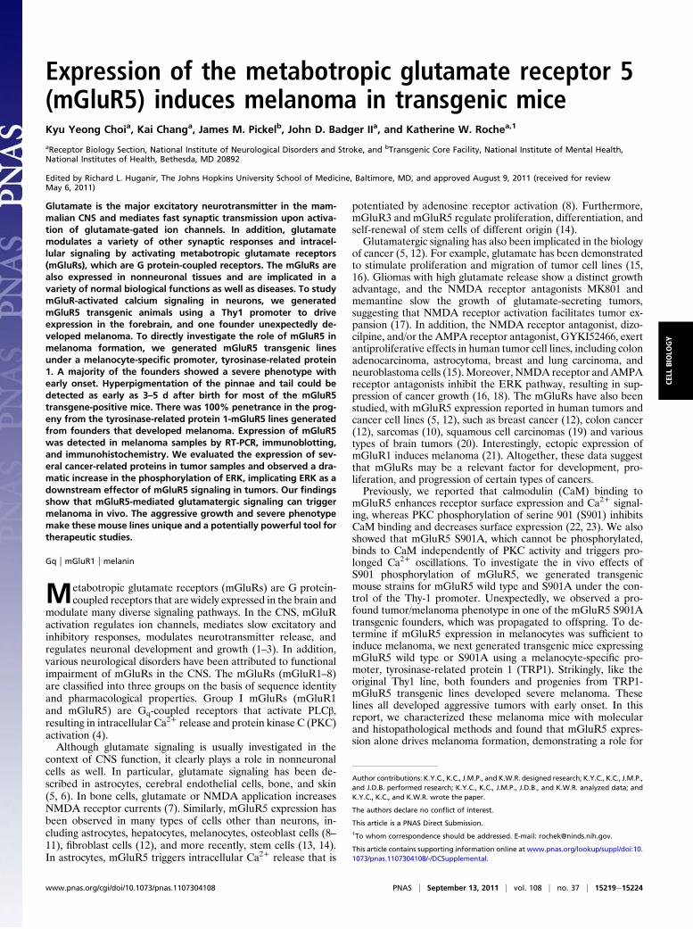
ResultsGeneration of mGluR5 Transgenic Mice. CaM binding to mGluR5competes with PKC phosphorylation of S901 in the mGluR5 C-terminal domain (23). Mutating this residue to block phos-phorylation, S901A affects CaM binding and the trafficking ofmGluR5. To test the role of mGluR5 S901 in vivo, we producedtransgenic mice expressing mGluR5 wild type or S901A undercontrol of the Thy1 promoter, which is expressed in most regionsof the forebrain (Fig. 1A) (24). Surprisingly, one Thy1-mGluR5S901A founder developed pigmented tumors that were firstidentified as malignant melanoma in a pathology report. Wehave been able to propagate this melanoma-bearing transgenicline to date. To determine if mGluR5 expression in skin isa melanoma inducer, we constructed new transgenic lines inwhich the expression of mGluR5 is specifically targeted to mel-anocytes under control of the TRP1 promoter (25). By pro-nuclear injection of TRP1-mGluR5 (wild type or S901A)transgenes (Fig. 1A), we produced 21 genotypically positivefounders for TRP1-mGluR5 wild type and 10 positive foundersfor TRP1-mGluR5 S901A (Fig. 1B). We found that 12 of the 21mGluR5 wild-type founders displayed melanoma phenotypes(56.3% penetrance), and 6 of the 10 mGluR5 S901A foundersdeveloped melanoma (60% penetrance; Fig. 1B). We havemaintained these TRP1-mGluR5 transgenic lines through sev-eral generations and find 100% penetrance of the melanomaphenotype in the progeny (Fig. 1B). In contrast, the originalThy1-mGluR5 S901A transgenic line showed 80% penetrance.The animals expressing TRP1-mGluR5 S901A developed severemelanoma on ears, nose, and tail. In particular, this strain dis-
played melanoma lesions encompassing the entire tail (Fig. 1C).The Thy1-mGluR5 S901A line also developed melanoma onears, nose, and tail (Fig. 1D), which were typically focal andless severe.
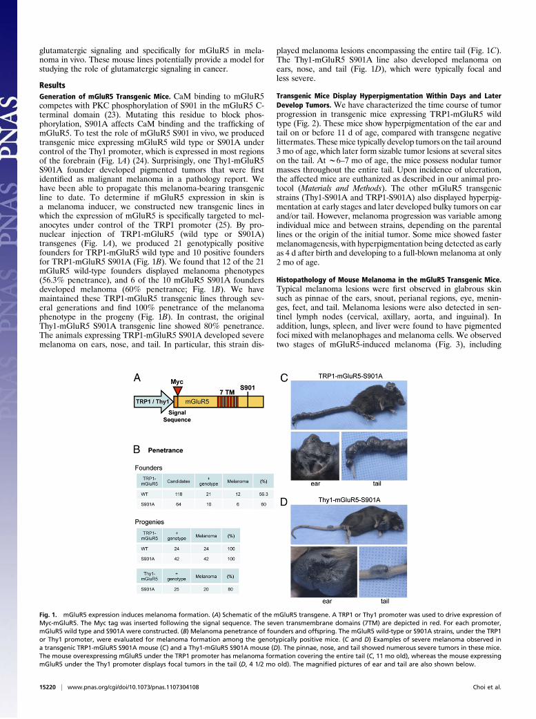
Transgenic Mice Display Hyperpigmentation Within Days and LaterDevelop Tumors. We have characterized the time course of tumorprogression in transgenic mice expressing TRP1-mGluR5 wildtype (Fig. 2). These mice show hyperpigmentation of the ear andtail on or before 11 d of age, compared with transgene negativelittermates. Thesemice typically develop tumors on the tail around3 mo of age, which later form sizable tumor lesions at several siteson the tail. At w6–7 mo of age, the mice possess nodular tumormasses throughout the entire tail. Upon incidence of ulceration,the affected mice are euthanized as described in our animal pro-tocol (Materials and Methods). The other mGluR5 transgenicstrains (Thy1-S901A and TRP1-S901A) also displayed hyperpig-mentation at early stages and later developed bulky tumors on earand/or tail. However, melanoma progression was variable amongindividual mice and between strains, depending on the parentallines or the origin of the initial tumor. Some mice showed fastermelanomagenesis, with hyperpigmentation being detected as earlyas 4 d after birth and developing to a full-blown melanoma at only2 mo of age.
Histopathology of Mouse Melanoma in the mGluR5 Transgenic Mice.Typical melanoma lesions were first observed in glabrous skinsuch as pinnae of the ears, snout, perianal regions, eye, menin-ges, feet, and tail. Melanoma lesions were also detected in sen-tinel lymph nodes (cervical, axillary, aorta, and inguinal). Inaddition, lungs, spleen, and liver were found to have pigmentedfoci mixed with melanophages and melanoma cells. We observedtwo stages of mGluR5-induced melanoma (Fig. 3), including
Fig. 1. mGluR5 expression induces melanoma formation. (A) Schematic of the mGluR5 transgene. A TRP1 or Thy1 promoter was used to drive expression ofMyc-mGluR5. The Myc tag was inserted following the signal sequence. The seven transmembrane domains (7TM) are depicted in red. For each promoter,mGluR5 wild type and S901A were constructed. (B) Melanoma penetrance of founders and offspring. The mGluR5 wild-type or S901A strains, under the TRP1or Thy1 promoter, were evaluated for melanoma formation among the genotypically positive mice. (C and D) Examples of severe melanoma observed ina transgenic TRP1-mGluR5 S901A mouse (C) and a Thy1-mGluR5 S901A mouse (D). The pinnae, nose, and tail showed numerous severe tumors in these mice.The mouse overexpressing mGluR5 under the TRP1 promoter has melanoma formation covering the entire tail (C, 11 mo old), whereas the mouse expressingmGluR5 under the Thy1 promoter displays focal tumors in the tail (D, 4 1/2 mo old). The magnified pictures of ear and tail are also shown below.
15220 | www.pnas.org/cgi/doi/10.1073/pnas.1107304108 Choi et al.
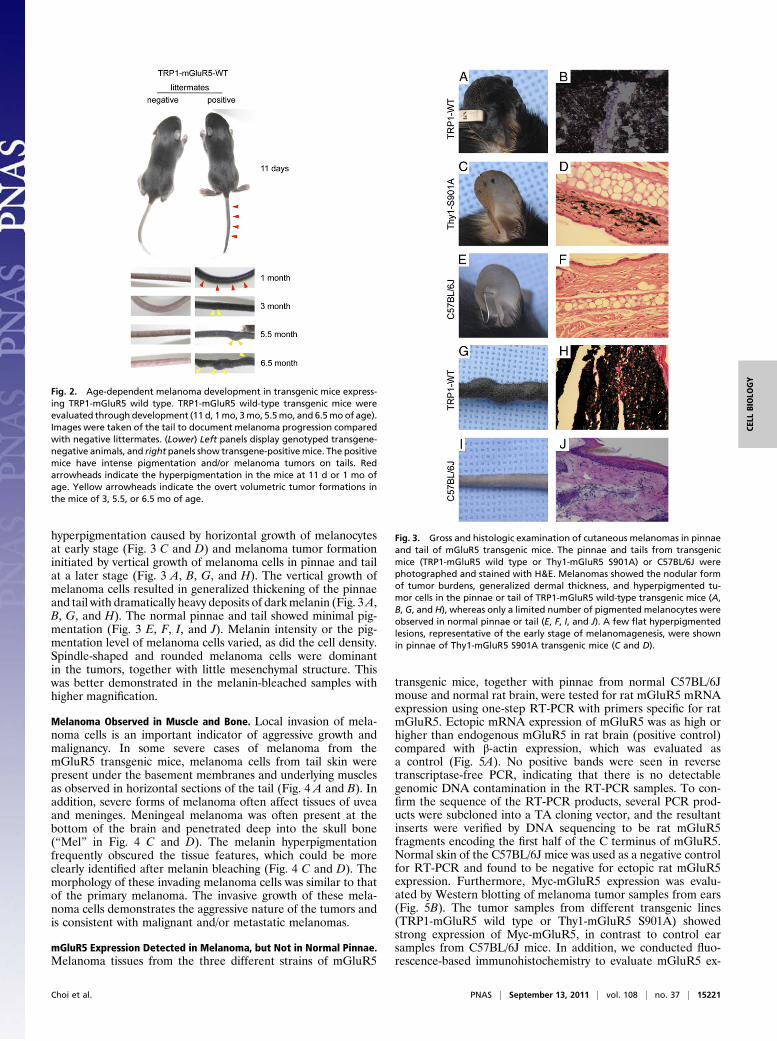
hyperpigmentation caused by horizontal growth of melanocytesat early stage (Fig. 3 C and D) and melanoma tumor formationinitiated by vertical growth of melanoma cells in pinnae and tailat a later stage (Fig. 3 A, B, G, and H). The vertical growth ofmelanoma cells resulted in generalized thickening of the pinnaeand tail with dramatically heavy deposits of darkmelanin (Fig. 3A,B, G, and H). The normal pinnae and tail showed minimal pig-mentation (Fig. 3 E, F, I, and J). Melanin intensity or the pig-mentation level of melanoma cells varied, as did the cell density.Spindle-shaped and rounded melanoma cells were dominantin the tumors, together with little mesenchymal structure. Thiswas better demonstrated in the melanin-bleached samples withhigher magnification.
Melanoma Observed in Muscle and Bone. Local invasion of mela-noma cells is an important indicator of aggressive growth andmalignancy. In some severe cases of melanoma from themGluR5 transgenic mice, melanoma cells from tail skin werepresent under the basement membranes and underlying musclesas observed in horizontal sections of the tail (Fig. 4 A and B). Inaddition, severe forms of melanoma often affect tissues of uveaand meninges. Meningeal melanoma was often present at thebottom of the brain and penetrated deep into the skull bone(“Mel” in Fig. 4 C and D). The melanin hyperpigmentationfrequently obscured the tissue features, which could be moreclearly identified after melanin bleaching (Fig. 4 C and D). Themorphology of these invading melanoma cells was similar to thatof the primary melanoma. The invasive growth of these mela-noma cells demonstrates the aggressive nature of the tumors andis consistent with malignant and/or metastatic melanomas.
mGluR5 Expression Detected in Melanoma, but Not in Normal Pinnae.Melanoma tissues from the three different strains of mGluR5
transgenic mice, together with pinnae from normal C57BL/6Jmouse and normal rat brain, were tested for rat mGluR5 mRNAexpression using one-step RT-PCR with primers specific for ratmGluR5. Ectopic mRNA expression of mGluR5 was as high orhigher than endogenous mGluR5 in rat brain (positive control)compared with β-actin expression, which was evaluated asa control (Fig. 5A). No positive bands were seen in reversetranscriptase-free PCR, indicating that there is no detectablegenomic DNA contamination in the RT-PCR samples. To con-firm the sequence of the RT-PCR products, several PCR prod-ucts were subcloned into a TA cloning vector, and the resultantinserts were verified by DNA sequencing to be rat mGluR5fragments encoding the first half of the C terminus of mGluR5.Normal skin of the C57BL/6J mice was used as a negative controlfor RT-PCR and found to be negative for ectopic rat mGluR5expression. Furthermore, Myc-mGluR5 expression was evalu-ated by Western blotting of melanoma tumor samples from ears(Fig. 5B). The tumor samples from different transgenic lines(TRP1-mGluR5 wild type or Thy1-mGluR5 S901A) showedstrong expression of Myc-mGluR5, in contrast to control earsamples from C57BL/6J mice. In addition, we conducted fluo-rescence-based immunohistochemistry to evaluate mGluR5 ex-
Fig. 2. Age-dependent melanoma development in transgenic mice express-ing TRP1-mGluR5 wild type. TRP1-mGluR5 wild-type transgenic mice wereevaluated through development (11 d, 1mo, 3mo, 5.5mo, and 6.5moof age).Images were taken of the tail to document melanoma progression comparedwith negative littermates. (Lower) Left panels display genotyped transgene-negative animals, and right panels show transgene-positive mice. The positivemice have intense pigmentation and/or melanoma tumors on tails. Redarrowheads indicate the hyperpigmentation in the mice at 11 d or 1 mo ofage. Yellow arrowheads indicate the overt volumetric tumor formations inthe mice of 3, 5.5, or 6.5 mo of age.
Fig. 3. Gross and histologic examination of cutaneous melanomas in pinnaeand tail of mGluR5 transgenic mice. The pinnae and tails from transgenicmice (TRP1-mGluR5 wild type or Thy1-mGluR5 S901A) or C57BL/6J werephotographed and stained with H&E. Melanomas showed the nodular formof tumor burdens, generalized dermal thickness, and hyperpigmented tu-mor cells in the pinnae or tail of TRP1-mGluR5 wild-type transgenic mice (A,B, G, and H), whereas only a limited number of pigmented melanocytes wereobserved in normal pinnae or tail (E, F, I, and J). A few flat hyperpigmentedlesions, representative of the early stage of melanomagenesis, were shownin pinnae of Thy1-mGluR5 S901A transgenic mice (C and D).
Choi et al. PNAS | September 13, 2011 | vol. 108 | no. 37 | 15221
CELL
BIOLO
GY

pression in tissues from the transgenic animals. By staining sec-tions of tumors, we observed robust expression of Myc-mGluR5compared with control (Fig. 5C). Taken together, the results fromRT-PCR, Western blotting, and immunohistochemistry demon-strate that the mGluR5 transprotein is strongly expressed intumor tissues.Moreover, to investigate whether mGluR5 is present in human
melanoma, a sensitive real time RT-PCR analysis was carried outusing a pair of intron-spanning primers complementary to humanmGluR5 exons. We used mGluR1, normal human melanocytes,and human brain as controls (Fig. S1). All of the mGluR1 andmGluR5 values were normalized with the level of β-actin for eachsample. mGluR5 was detected in both humanmelanoma cell linesand in metastatic melanoma tissues (Fig. S1 A and B). Note thatthe amount of mGluR5 transcripts in human metastatic mela-noma tissues (Fig. S1B) was generally higher than in humanmelanoma cell lines (Fig. S1A). Although mGluR5 mRNA wasdetected in normal human melanocytes (“Melanocytes” in Fig.S1A), each melanoma cell line also had higher amounts ofmGluR5 than of mGluR1. In fresh human metastatic melanomatissue, mGluR5 was highly expressed in all seven samples (Fig.S1B), whereas mGluR1 expression was detected in only one ofseven samples examined (“HMM3” in Fig. S1B).
ERK1/2 Activation in mGluR5-Mediated Melanoma. Many cellularproteins have been implicated in melanoma formation. For ex-ample, human melanoma cells frequently exhibit ERK activation,suggesting that constitutively activated ERK contributes to mel-anoma cell proliferation, invasion, and metastasis (26, 27). Thetails from melanoma mice expressing TRP1-mGluR5 wild typewere subjected to Western blotting to evaluate the expression ofseveral proteins implicated in a variety of cancers. In particular,we monitored the phosphorylation state of ERK. The samplesfrom transgene-positive mice displayed increased ERK1/2 phos-phorylation, compared with those of negative littermates, whereasthe total ERK1/2 expression level was not changed (Fig. 5D).Other oncogenic proteins, EGFR and PCNA, were also tested butdid not show any striking changes in their expression levels. Theanalyses of the expression of other proteins, includingN-cadherin,E-cadherin, NF-κB, tyrosinase, TRP1, and PTEN, revealed in-consistent changes in protein levels. We thus found variationsbetween individuals and between strains that were likely due totumor heterogeneity and different stages of tumor progression.
DiscussionIn this study, we demonstrate that mGluR5 induces melanomaformation in vivo, providing evidence for the role of gluta-matergic signaling in cancer. We generated transgenic miceexpressing mGluR5 under the Thy1 or TRP1 promoter. At earlystages, these mice displayed hyperpigmentation on ears and tails;and, at later stages, they developed dramatic tumor phenotypes,which were heritable with a high penetrance (over 80%). More-over, melanoma cells were observed in the muscle, bone, andregional lymph nodes of the affected animals.Signaling of group I mGluRs is regulated by CaM binding and
PKC phosphorylation (22, 23). Specifically, phosphorylation ofmGluR5 on S901 regulates CaM binding and receptor surfaceexpression, and the mGluR5 S901A mutant, which cannot bephosphorylated, displays altered Ca2+ signaling (23). To inves-tigate the S901A mutation in vivo, we generated mGluR5 trans-genic mice (wild type and S901A). We unexpectedly observedmelanoma in an mGluR5 S901A transgenic founder and sus-pected that this residue might be involved in the pathology. Toaddress this hypothesis, we generated mGluR5 transgenic miceunder the melanocyte-specific TRP1 promoter. We found thattransgenic mice expressing either wild-type or S901A mGluR5developed severe melanoma. The time course and severity of
Fig. 4. Local invasion of tail and meningeal melanomas. In the late stage ofmelanomagenesis, highly aggressive melanoma cells from tail or meningespenetrate the underlying muscular layer (A and B) or bone tissues (C and D).(A and B) The H&E staining of tails from TRP1-mGluR5 wild-type transgenicmice. Arrows indicate invasion of melanoma cells to skeletal muscle. (C andD) Melanin-bleached specimen followed by H&E staining of Thy1-mGluR5S901A transgenic mice. Mel, melanoma; Cb, cerebellum.
Fig. 5. Protein expression in melanoma tissue of transgenic mice. (A) TotalRNA from normal pinnae, rat brain, and three different mGluR5 transgenicstrains (TRP1-mGluR5 wild type, S901A, or Thy1-mGluR5 S901A) were DNase I-treated before they were used in PCR with or without reverse transcriptase(RT). Using RT-PCR, ratmGluR5was expressed in themelanoma tissues fromallthree strains of transgenic mice, but not in the pinnae of normal non-transgenic mice (C57BL/6J pinnae) used as a negative control. mGluR5 is alsodetected in normal rat brain. (Upper) The strong band represents mGluR5b.(Lower) The weaker band is rat mGluR5a, which is the cDNA used as thetransgene. β-Actin was used as a positive control that is present in all RNA inRT-PCR, and RT-free PCRwere used as a negative control for assessing possiblegenomic DNA contamination, if any, in the RNA preparations. (B) The ex-pression of the Myc-mGluR5 transgene. Tissue was analyzed by Westernblotting using anti-Myc antibody. Ear samples from C57BL/6J mice and thetumors on ears of transgenic mice expressing TRP1-mGluR5 wild type or Thy1-mGluR5 S901A were tested for transgene expression. The melanoma tumorsexpressed Myc-mGluR5, whereas C57BL/6J mice did not. β-Actin was used forthe internal control. (C) Ear sections of tumors from Thy1-Myc-mGluR5transgenic mice (6 mo of age) were stained with Myc antibody to detect Myc-mGluR5, and receptor expression was visualized with Alexa 568-conjugatedanti-rabbit IgG secondary antibody (red) and counterstained with Hoechst(blue). (Right panels) Merged images. Buffer alone (CTL) was used in place ofprimary antibody for the negative control. (D) Phosphorylation of ERK1/2 isincreased in the TRP1-mGluR5 transgenic mice. The mouse tails from litter-mates, either positive or negative for TRP1-mGluR5 wild type (6 mo of age),were subjected to Western blotting with phospho-ERK1/2, ERK1/2, EGFR, andPCNA antibodies. Tubulin was used as a loading control.
15222 | www.pnas.org/cgi/doi/10.1073/pnas.1107304108 Choi et al.

melanoma varied a great deal depending on the site of the initialtumor. However, the percentage of progeny displaying melanomawas the same with TRP1-mGluR5 wild type or S901A, suggestingthat overexpression of mGluR5 in melanocytes induces the mel-anoma, irrespective of the phosphorylation of S901. The trans-genic mGluR5 wild-type lines under the Thy1 promoter neverproduced a founder with melanoma, so we could not compareThy1-mGluR5 wild type to mutant.Although both mGluR1 and mGluR5 are Gq-coupled recep-
tors that stimulate intracellular Ca2+ release, they have distinctsignaling properties. For example, activation of mGluR5 evokesCa2+ oscillations, whereas mGluR1 activation results in a singlepeak Ca2+ response (28). In addition, mGluR5 strongly binds toCaM, but mGluR1 does not (22). Furthermore, expression ofmGluR1 and mGluR5 varies throughout the CNS, consistentwith unique roles for these two receptors. Previous studies haveimplicated mGluR1 in melanoma (21, 29), but, until now, therewas no evidence of a similar role for mGluR5. The studies onmGluR1 and melanoma began with the characterization of aninsertional mouse mutant line, TG3, that was predisposed todevelop multiple melanomas affecting the pinnae of the ear,perianal reion, eyelid, snout, trunk, and legs (30–32). Physicalmapping of this TG3 line revealed that intron 3 of the Grm1(mGluR1) gene was affected and resulted in aberrant expressionof mGluR1 in skin tissues (21). Driving mGluR1 expressionunder the dopachrome taumerase (Dct) promoter in melano-cytes also resulted in animals that developed melanoma (21). Inaddition, another melanoma mouse model was generated using atetracycline regulatory system to induce mGluR1 expression(29). However, our findings that mGluR5 causes melanoma werenot predicted. In fact, one study specifically tested the potentialrole of mGluR5 in the TG3 mGluR1-expressing melanoma miceby breeding the mGluR1 transgenic mice with mGluR5 knockoutmice (33). Although the absence of mGluR5 did not eliminatemGluR1-induced melanoma, we now clearly show that mGluR5overexpression alone induces severe melanoma, consistent withparallel mGluR pathways being capable of driving aggressivetumor formation.The melanoma induced by our transgenic TRP1-mGluR5
mouse lines appears to be more severe than that reported for theDct-mGluR1 lines. The TRP1-mGluR5 wild-type mice showedhyperpigmentation on the ear and tail at 4 d of age, developedtumor lesions by 3–4 mo of age, and formed large pigmentedmasses by 6–7 mo of age (as shown in Fig. 2). In contrast, Dct-mGluR1 E lines reportedly displayed pigmented lesions at 5–6mo old, which progressed into tumors by 6–7 mo (21). Althoughthe time course and severity of the lesions vary between themGluR1 and mGluR5 mice, the affected tissues and histopath-ological evaluation are remarkably similar. Both mGluR1 andmGluR5 melanoma mice (TG3, Dct-mGluR1, TRE-mGluR1,TRP1-mGluR5, and Thy1-mGluR5) exhibited tumor formationon the hairless skin, including pinnae and tails, rather than ontrunk areas (21, 29, 32). Taken together, these results implicateboth group I mGluRs in melanocytic neoplasia. Several studiesof the mGluR1 melanoma mice demonstrate that pharmaceuti-cal inhibition can diminish the tumor progression. For example,treatment with mGluR1 antagonists (LY367385 or BAY36-7620) or a glutamate release inhibitor (riluzole) suppressed theproliferation of mGluR1-expressing human melanoma cells andinhibited the tumor growth in mGluR1 transgenic mice (29, 34).We predict that the mGluR5 transgenic mice will enable similarstudies to investigate mGluR signaling in tumor formation.Although mGluR5 has been largely investigated in the central
nervous system, a variety of studies report the expression and/orfunction of mGluR5 in nonneuronal cells and cancer cells (8, 10,13, 14, 19, 20). In particular, patients with oral squamous cellcarcinoma tissues that are strongly positive for mGluR5 immu-noreactivity had a decreased 5-y survival rate compared with
mGluR5-negative patients (19). In addition, the mGluR5 agonistDHPG increased tumor cell migration, invasion, and adhesion inHSC3 oral tongue cancer cells, which was reversed by themGluR5-specific antagonist MPEP (19). Taken together, thesefindings suggest that mGluR5 may be a relevant factor for thedevelopment, proliferation, and progression of multiple types ofcancers and that mGluR5 might have potential as a biomarkerfor cancer cell proliferation.mGluR1 has been studied extensively as a transforming factor
for mouse melanomas using mGluR1 transgenic models (21, 29).Our current study reveals that expression of a related group Imetabotropic glutamate receptor, mGluR5, leads to melano-magenesis in mice. Although mGluR5 is expressed in primarycultured human melanocytes (9) and mGluR1 is expressed inhuman melanoma samples (21), neither mGluR1 nor mGluR5have been detected in human melanoma tissue via varioustranscriptomic studies (35). Using a sensitive real-time RT-PCRmethod and intron-spanning primers, we detected mGluR5mRNA transcripts in both human melanoma cell lines and freshhuman melanoma tissues at a level comparable to or higher thanhuman brain tissue (Fig. S1). The amount of mGluR5 transcriptsin human metastatic melanoma tissues (Fig. S1B) was generallyhigher than in human melanoma cell lines (Fig. S1A), suggestingthat long-term culture of melanoma cells might alter the level ofmGluR5 gene expression. Perhaps the normal expression ofmGluR5 in human melanocytes is the reason that no differencein mGluR5 expression has been reported in microarrays ofmelanoma.It is well established that group I mGluRs (mGluR1 and
mGluR5) activate ERK in neuronal systems. In the spinal cord,intrathecal injection ofDHPG, a group ImGluR agonist, activatesERK1/2, resulting in enhanced pain sensitivity (36). In addition,ERK activation is required for mGluR-LTD in area CA1 of thehippocampus (37, 38).Here, we show that tumor samples from themGluR5 melanoma mice exhibit a high level of ERK1/2 phos-phorylation compared with wild-type littermates. Our findings areconsistent with studies on mGluR1-mediated melanoma, in whichpersistent mGluR1 expression correlated with high ERK1/2phosphorylation in immunohistochemical analysis (29). Previousreports have shown that mGluR1 activates ERK in melanomacells and, vice versa, that the ERK pathway plays a key role inmelanoma growth (34, 39, 40). Stimulation of mGluR1 with L-quisqualate, a group I mGluR agonist, leads to IP3 accumulation,and activates ERK1/2 via PKCε in melanoma cells derived fromthe tumors expressing mGluR1 (40). In addition, in human mel-anoma cell lines expressing mGluR1, L-quisqualate–inducedmGluR1 stimulation led to the ERK activation (34). Taken to-gether, these results suggest that group I mGluRs activate theERK pathway and that ERK activation is involved in mGluR-mediated melanoma growth.In conclusion, we show a profound effect of mGluR5 expres-
sion in melanocytes, resulting in severe tumors. These mouselines are striking because of the severe phenotype, early onset,and complete penetrance. Our findings show an important rolefor glutamatergic signaling in melanoma development in vivo.Together with the previous studies on mGluR1 melanoma mice,our mGluR5-mediated melanoma lines show that Gq-coupledsignaling has profound effects on tumor progression in melano-cytes. These mGluR5 transgenic mice provide a potentiallyvaluable tool for drug screening as well as for preclinical testingin the future.
Materials and MethodsDetailed information on histopathological analysis, RT-PCR, Western blot-ting, and immunohistochemistry are available in SI Materials and Methods.
AMyc epitopewas inserted into the N terminus ofmGluR5 between aminoacids 22 and 23 (23). To prepare TRP1 promoter-Myc-mGluR5, TRP1 promoter(1,201 bp), the ORF of Myc-tagged rat mGluR5 (3,558 bp), and a SV-40 poly
Choi et al. PNAS | September 13, 2011 | vol. 108 | no. 37 | 15223
CELL
BIOLO
GY
(A) signal sequence (293 bp) were ligated into the pBluescript SK vector(Stratagene). The point mutation, S901A, was introduced using the Quik-Change site-directed mutagenesis system (Stratagene). The construct wasverified by DNA sequencing, and the expression of Myc-mGluR5 was eval-uated in the heterologous cells by Western blotting. Similarly, Thy1-Myc-mGluR5 was constructed by combining the Thy1 promoter, Myc-mGluR5 ORF, and poly(A) sequence into the pUC18 vector. The diagram oftransgenic constructs is shown in Fig. 1A. Each Myc-mGluR5 transgenicconstruct was gel-purified with the QIAGEN gel extraction kit. The DNA wasfiltered with a 0.22-μm Millipore filter and diluted with 1 mM Tris$HCl(pH 7.5), 0.05 mM EDTA buffer to 10 μg/mL. The DNA was microinjected intothe pronuclei of fertilized embryos from the mating of C57BL/6J mice. Themicroinjected embryos were implanted into pseudopregnant recipients. The
founder mice were identified by PCR genotyping with rat mGluR5-specificprimers (59-AGTGCCACAGTGGCCCTGGGTTGC-39 and 59-TGCCTCCGCCACAT-CATAAAGCGC-39). The use and care of animals in this study followed theguidelines of the National Institutes of Health Animal Research AdvisoryCommittee (animal protocol no. 1313–10).
ACKNOWLEDGMENTS. We thank Michael Spencer and Bill Branson (Na-tional Institutes of Health Medical Art) for assistance with high-resolutionphotography; James Nagle and Debbie Kauffman (National Institute ofNeurological Disorders and Stroke Sequencing Facility) for DNA sequenceanalysis; and the animal care staff of the Porter Neuroscience Center. TheNational Institute of Neurological Disorders and Stroke Intramural ResearchProgram supported this work.
1. Ferraguti F, Shigemoto R (2006) Metabotropic glutamate receptors. Cell Tissue Res326:483e504.
2. Hermans E, Challiss RA (2001) Structural, signalling and regulatory properties of thegroup I metabotropic glutamate receptors: Prototypic family C G-protein-coupledreceptors. Biochem J 359:465e484.
3. Kim CH, Lee J, Lee JY, Roche KW (2008) Metabotropic glutamate receptors: Phos-phorylation and receptor signaling. J Neurosci Res 86:1e10.
4. Conn PJ, Pin JP (1997) Pharmacology and functions of metabotropic glutamate re-ceptors. Annu Rev Pharmacol Toxicol 37:205e237.
5. Kalariti N, Pissimissis N, Koutsilieris M (2005) The glutamatergic system outside theCNS and in cancer biology. Expert Opin Investig Drugs 14:1487e1496.
6. Skerry TM, Genever PG (2001) Glutamate signalling in non-neuronal tissues. TrendsPharmacol Sci 22:174e181.
7. Laketi�c-Ljubojevi�c I, Suva LJ, Maathuis FJ, Sanders D, Skerry TM (1999) Functionalcharacterization of N-methyl-D-aspartic acid-gated channels in bone cells. Bone 25:631e637.
8. Cormier RJ, Mennerick S, Melbostad H, Zorumski CF (2001) Basal levels of adenosinemodulate mGluR5 on rat hippocampal astrocytes. Glia 33:24e35.
9. Frati C, et al. (2000) Expression of functional mGlu5 metabotropic glutamate re-ceptors in human melanocytes. J Cell Physiol 183:364e372.
10. Kalariti N, Lembessis P, Papageorgiou E, Pissimissis N, Koutsilieris M (2007) Regulationof the mGluR5, EAAT1 and GS expression by glucocorticoids in MG-63 osteoblast-likeosteosarcoma cells. J Musculoskelet Neuronal Interact 7:113e118.
11. Storto M, et al. (2000) Selective blockade of mGlu5 metabotropic glutamate receptorsprotects rat hepatocytes against hypoxic damage. Hepatology 31:649e655.
12. Stepulak A, et al. (2009) Expression of glutamate receptor subunits in human cancers.Histochem Cell Biol 132:435e445.
13. Castiglione M, et al. (2008) Group I metabotropic glutamate receptors control pro-liferation, survival and differentiation of cultured neural progenitor cells isolatedfrom the subventricular zone of adult mice. Neuropharmacology 55:560e567.
14. Melchiorri D, et al. (2007) Metabotropic glutamate receptors in stem/progenitor cells.Neuropharmacology 53:473e480.
15. Rzeski W, Turski L, Ikonomidou C (2001) Glutamate antagonists limit tumor growth.Proc Natl Acad Sci USA 98:6372e6377.
16. Stepulak A, et al. (2005) NMDA antagonist inhibits the extracellular signal-regulatedkinase pathway and suppresses cancer growth. Proc Natl Acad Sci USA 102:15605e15610.
17. Takano T, et al. (2001) Glutamate release promotes growth of malignant gliomas.Nat Med 7:1010e1015.
18. Stepulak A, et al. (2007) AMPA antagonists inhibit the extracellular signal regulatedkinase pathway and suppress lung cancer growth. Cancer Biol Ther 6:1908e1915.
19. Park SY, et al. (2007) Clinical significance of metabotropic glutamate receptor 5 ex-pression in oral squamous cell carcinoma. Oncol Rep 17:81e87.
20. Brocke KS, et al. (2010) Glutamate receptors in pediatric tumors of the central nervoussystem. Cancer Biol Ther 9:455e468.
21. Pollock PM, et al. (2003) Melanoma mouse model implicates metabotropic glutamatesignaling in melanocytic neoplasia. Nat Genet 34:108e112.
22. Choi KY, Chung S, Roche KW (2011) Differential binding of calmodulin to groupI metabotropic glutamate receptors regulates receptor trafficking and signaling.J Neurosci 31:5921e5930.
23. Lee JH, et al. (2008) Calmodulin dynamically regulates the trafficking of the metab-otropic glutamate receptor mGluR5. Proc Natl Acad Sci USA 105:12575e12580.
24. Han W, et al. (2005) C-terminal ECFP fusion impairs synaptotagmin 1 function:Crowding out synaptotagmin 1. J Biol Chem 280:5089e5100.
25. Lowings P, Yavuzer U, Goding CR (1992) Positive and negative elements regulatea melanocyte-specific promoter. Mol Cell Biol 12:3653e3662.
26. Huntington JT, et al. (2004) Overexpression of collagenase 1 (MMP-1) is mediated bythe ERK pathway in invasive melanoma cells: Role of BRAF mutation and fibroblastgrowth factor signaling. J Biol Chem 279:33168e33176.
27. Satyamoorthy K, et al. (2003) Constitutive mitogen-activated protein kinase activa-tion in melanoma is mediated by both BRAF mutations and autocrine growth factorstimulation. Cancer Res 63:756e759.
28. Kawabata S, et al. (1996) Control of calcium oscillations by phosphorylation of me-tabotropic glutamate receptors. Nature 383:89e92.
29. Ohtani Y, et al. (2008) Metabotropic glutamate receptor subtype-1 is essential forin vivo growth of melanoma. Oncogene 27:7162e7170.
30. Chen S, Zhu H, Wetzel WJ, Philbert MA (1996) Spontaneous melanocytosis in trans-genic mice. J Invest Dermatol 106:1145e1151.
31. Zhu H, et al. (2000) Development of early melanocytic lesions in transgenic micepredisposed to melanoma. Pigment Cell Res 13:158e164.
32. Zhu H, et al. (1998) Development of heritable melanoma in transgenic mice. J InvestDermatol 110:247e252.
33. Marín YE, et al. (2005) Grm5 expression is not required for the oncogenic role of Grm1in melanocytes. Neuropharmacology 49(Suppl 1):70e79.
34. Namkoong J, et al. (2007) Metabotropic glutamate receptor 1 and glutamate sig-naling in human melanoma. Cancer Res 67:2298e2305.
35. Hoek KS (2007) DNA microarray analyses of melanoma gene expression: A decade inthe mines. Pigment Cell Res 20:466e484.
36. Karim F, Wang CC, Gereau RW, IV (2001) Metabotropic glutamate receptor subtypes1 and 5 are activators of extracellular signal-regulated kinase signaling required forinflammatory pain in mice. J Neurosci 21:3771e3779.
37. Gallagher SM, Daly CA, Bear MF, Huber KM (2004) Extracellular signal-regulatedprotein kinase activation is required for metabotropic glutamate receptor-dependentlong-term depression in hippocampal area CA1. J Neurosci 24:4859e4864.
38. Thiels E, Kanterewicz BI, Norman ED, Trzaskos JM, Klann E (2002) Long-term de-pression in the adult hippocampus in vivo involves activation of extracellular signal-regulated kinase and phosphorylation of Elk-1. J Neurosci 22:2054e2062.
39. Abdel-Daim M, et al. (2010) Pharmacogenomics of metabotropic glutamate receptorsubtype 1 and in vivo malignant melanoma formation. J Dermatol 37:635e646.
40. Marín YE, et al. (2006) Stimulation of oncogenic metabotropic glutamate receptor1 in melanoma cells activates ERK1/2 via PKCepsilon. Cell Signal 18:1279e1286.
15224 | www.pnas.org/cgi/doi/10.1073/pnas.1107304108 Choi et al.


