Embed Size (px)
Citation preview

Article
Exceptional multifunctionality in the feeding apparatus of a mid-Cambrian radiodont
Joseph Moysiuk* and Jean-Bernard Caron
Abstract.—Radiodonts (stem Euarthropoda) were ecologically diverse, but species generally displayedlimited functional specialization of appendages along the body axis compared with crown group euar-thropods. This is puzzling, because such functional specialization is considered to have been an importantdriver of euarthropod ecological diversification. One way to circumvent this constraint could have beenthe functional specialization of different parts of the frontal appendages, known to have been ecologicallyimportant in radiodonts. This hypothesis has yet to be tested explicitly. Here we redescribe the poorlyknown mid-Cambrian hurdiid radiodont Stanleycaris hirpex from the Burgess Shale (Stephen Formation)and quantitatively assess functional specialization of the frontal appendages of stem euarthropods. Theappendages of Stanleycaris are composed of 14 podomeres, variously differentiated by their possessionof pectinate endites, mono- to trifurcate medial gnathites, and outer spines. The oral cone is tetraradiallyorganized and can be uniquely distinguished from those of other hurdiids by the presence of 28 ratherthan 32 smooth tridentate plates. Our phylogenetic analysis finds Stanleycaris in a grade of hurdiids retain-ing plesiomorphic raptorial appendicular functionality alongside derived adaptations for sweep feedingand large, bilaterally opposed gnathites. We conclude that the latter performed a masticatory function,convergent with gnathal structures like mandibles in various panarthropods. Taken together, Stanleycarisand similar hurdiids provide an extreme example of the evolution of division of labor within the append-age of a stem euarthropod and suggest that this innovation may have facilitated the functional transition,from raptorial to sweep feeding, at the origin of the hurdiid clade.
Joseph Moysiuk. Department of Natural History, Royal Ontario Museum, 100 Queen’s Park, Toronto, OntarioM5S 2C6, Canada; Department of Ecology and Evolutionary Biology, University of Toronto, 25Willcocks Street,Toronto, Ontario M5S 3B2, Canada. E-mail: [email protected]
Jean-Bernard Caron. Department of Natural History, Royal Ontario Museum, 100 Queen’s Park, Toronto, OntarioM5S 2C6, Canada; Department of Ecology and Evolutionary Biology, University of Toronto, 25Willcocks Street,Toronto, Ontario M5S 3B2, Canada; Department of Earth Sciences, University of Toronto, 22 Russell Street,Toronto, Ontario, Canada M5S 3B1. E-mail: [email protected]
Accepted: 11 April 2021*Corresponding author.
Background
Arthropods are unparalleled in their diver-sity of appendages, which are involved inalmost every conceivable function—locomo-tion, sensing, feeding, respiration, grooming,mating, communication, defense, anchoring,and more. The ability to fulfill this wide rangeof requirements is considered to have beenmade possible in large part by an evolutionarycapacity to partition functional tasks betweendifferent specialized appendages (Cisne 1974).This division of labor, or functional speciali-zation, can circumvent trade-offs and is thus
hypothesized to be selectively favoredwheneverthe position of a specialized structure impacts itsperformance and as long as structural diver-gence is developmentally unconstrained (Ruef-fler et al. 2012). This in turn potentially opensopportunities for ecological diversification byallowingmodular change in one functional attri-bute without compromising others.A plethora of recent fossil discoveries suggest
that a high level of morphological and eco-logical diversity had already been achievedamong Cambrian euarthropods (Daley et al.2018; Aria 2020). Given the complexity of
© The Author(s), 2021. Published by Cambridge University Press on behalf of The Paleontological Society. This is an OpenAccess article, distributed under the terms of the Creative Commons Attribution-NonCommercial-ShareAlike licence(https://creativecommons.org/licenses/by-nc-sa/4.0/), which permits non-commercial re-use, distribution, and repro-duction in any medium, provided the same Creative Commons licence is included and the original work is properlycited. The written permission of Cambridge University Press must be obtained for commercial re-use. 0094-8373/21
Paleobiology, 47(4), 2021, pp. 704–724DOI: 10.1017/pab.2021.19
https://doi.org/10.1017/pab.2021.19 Published online by Cambridge University Press

Cambrian ecosystems (Caron and Jackson2008; Zhao et al. 2014; Nanglu et al. 2020) andthe marked appendicular differentiation seenamong crown group euarthropods, functionalspecialization would also be expected to beplentiful in stem groups (sensu Jefferies 1979).However, in contrast to this prediction, differ-entiation of sets of appendages is generallymore limited in stem euarthropods, as demon-strated by their relatively low “indices oftagmosis” on average compared with crowneuarthropods (Cisne 1974; Wills et al. 1998;Yang et al. 2015). Do these differences representa legitimate biological discrepancy, with stemgroups being ecologically hyperspecializedrelative to crown groups, or are they simplyan artifact of the way functional diversity hasbeen measured?Radiodonta provides an exemplary test case.
While phylogenetic placements outside theeuarthropod total group or within the crowngroup were historically hypothesized, Radio-donta is now generally recognized as the mostdiverse clade of stem group euarthropods(|Daley et al. 2009, 2018). Radiodonts exhibitadaptations to a range of different feedingniches, from raptorial predation and duro-phagy to sediment sifting and suspension feed-ing (Whittington and Briggs 1985; Daley andBudd 2010; Vinther et al. 2014; VanRoy et al.2015; Liu et al. 2018; Moysiuk and Caron2019). Interestingly, this ecological diversifica-tion appears to have occurred via fairly limitedmorphological variations on a conserved bodyplan, characterized by a single pair of arthro-dized frontal appendages followed by a seriesof relatively homonomous swimming flapsand sometimes rudder-like posterior bladesand elongate furcae (Whittington and Briggs1985; Chen et al. 1994; Daley et al. 2009;Cong et al. 2014; VanRoy et al. 2015; Moysiukand Caron 2019). Much of the morphologicaldivergence between the four major radiodontmorphogroups—Anomalocarididae, Amplec-tobeluidae, Tamisiocarididae, and especiallyHurdiidae—is concentrated in the appendages,illustrated by their broad dispersal in appen-dicular morphospace (Aria and Caron 2015),acknowledging the caveat that a number ofspecies are known from only appendicularremains. If radiodont ecological diversification
was not largely achieved by alteration of thetagmatic partitioning of functional roles, wehypothesize that differential functional special-ization of parts of the frontal feeding appen-dages, enabling appendage multifunctionality,could have provided a partial substitute.As a relevant case study, we contribute a
redescription of the feeding apparatus of themid-Cambrian hurdiid radiodont Stanleycarishirpex, based on exceptionally preserved fossilsfrom the Burgess Shale. Since the discovery ofStanleycaris (Caron et al. 2010) a decade ago, alarge amount of new radiodont fossil materialhas contributed immeasurably to knowledgeof this group of animals (e.g., Daley and Berg-ström 2012; Daley et al. 2013a,b; Cong et al.2014, 2017; Vinther et al. 2014; VanRoy et al.2015; Guo et al. 2018; Liu et al. 2018; Moysiukand Caron 2019). In light of these advances,along with new Stanleycaris appendages andmouthparts collected from the Burgess Shale,we here revisit the morphology of this taxon.Armed with these data, we undertake a quanti-tative assessment of the evolution of functionaldiversity of radiodont appendages.
Systematic Paleontology
Superphylum Panarthropoda Nielsen, 1995Order Radiodonta Collins, 1996
FamilyHurdiidae Lerosey-Aubril & Pates, 2018Genus Stanleycaris Pates et al., 2018, ex Caron
et al., 2010Emended Diagnosis and Description.—As for
species.Stanleycaris hirpex Pates et al., 2018, ex Caron
et al., 2010Locality and Stratigraphy.—“Thin” Stephen
Formation of Stanley Glacier (Caron et al.2010) and northern Tokumm Creek (Mayerset al. 2018), British Columbia, Canada. Allmaterial is housed at the Royal OntarioMuseum, Invertebrate Palaeobiology Section(ROMIP), in Toronto, Canada (see list in Sup-plementary Material). This taxon is probablyalso present in the Wheeler Formation ofUtah, USA (Pates et al. 2017).
Emended Diagnosis.—Hurdiid radiodontwith the following characteristics: appendagewith 14 podomeres, including proximal ped-uncle. Mesially curving, bladelike endites on
RADIODONT APPENDAGE MULTIFUNCTIONALITY 705
https://doi.org/10.1017/pab.2021.19 Published online by Cambridge University Press
second to seventh and ninth podomeres, withproximalmost endite inclined more distallythan subsequent ones and distalmost enditereduced in size. Enditic auxiliary spines short,numbering two to six. Medial gnathites ofappendage largewith one to three long, inwardcurving spines. Oral cone comprising fourlarge tridentate plates with paired triangularnodes separated by sets of six smaller tridentateplates. Inner oral plates absent.
Preservation.—Pairs of appendages and oralcones in close proximity suggest a residualcuticular connection between these structuressuch that they remained associated after trans-port and burial (Fig. 1). The occurrence ofmultiple radiodont appendages on individualbedding surfaces has been reported previouslyfrom the Burgess Shale in Anomalocaris(O’Brien et al. 2014) and Cambroraster (Moysiukand Caron 2019) and may evince gregariousmolting behavior followed by rapid burial. Amass molting event would also be consistentwith evidence for flexible deformation ofappendicular spines (Figs. 1C, 2E) facilitatedby the softening of the cuticle during ecdysis.The appendages had a complex three-
dimensional geometry such that no singleorientation of burial shows all features. Typic-ally, they are preserved with a strongly convexouter margin, with the distal end almost com-pletely recurved relative to the proximal end(Figs. 2F,G, and 3A,G,H); however, some speci-mens are approximately straight (Fig. 1E). Thissuggests that nearly 180° of tip flexure was pos-sible, with the distal five podomeres appearingparticularly articulable. A second aspect ofcurvature is also evident when comparingappendages preserved at different angles.Moving distally, the appendage twists suchthat the distal tip lies slightly medial to theproximal end and can articulate to oppose themesially curving endites.Stanleycaris oral cones are also preserved in a
variety of orientations and in variable states ofdisarticulation (Figs. 1A–E, 3). The more scler-otized oral margins of the plates may be theonly remnants in some cases (Fig. 3G–I).
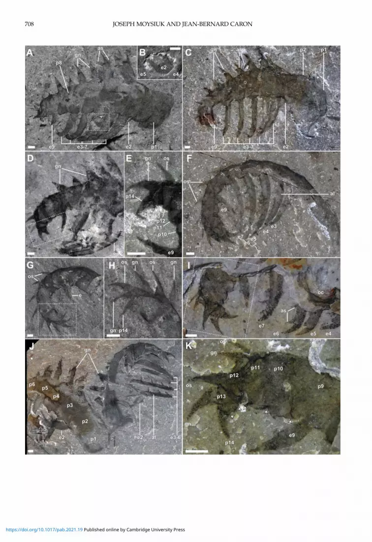
Description.—Appendages range in sizefrom 3.5 to 32.2 mm, measured along the curv-ing outer margin (figured and referred speci-men table, Supplementary Material). They
consist of 14 podomeres, which taper graduallytoward the distal end of the appendage (Fig. 2).The first two proximal podomeres are moreelongate (1.3–2.5 times longer) than subsequentpodomeres (Figs. 2A,C,J, and 3A). The first inparticular tends to be more variably preservedand less consistent in shape along the proximalmargin. This suggests that it was less sclerot-ized than more distal podomeres and mayhave afforded limited flexibility, assistingwith basal movement of the appendage. Thefollowing podomeres are subrectangular tosubtrapezoidal (ca. half as long as tall) andmay possess up to three spinous cuticular out-growths on the inner, medial, and outer sur-faces (Fig. 2). The distalmost podomereconsists of a terminal spine, about 0.15 timesthe height of the first podomere (Fig. 2E,H,K).The inner outgrowths (endites) are the most
conspicuous and occur on podomeres 2–7 and9 (Fig. 2A–F), and we number them accordingto the respective podomeres. They are blade-like, a third as wide as the length of the sup-porting podomeres. Each endite appears to beslightly crescentic in cross section, such that atsome burial orientations the narrow edgesmay be juxtaposed on the bedding plane, pro-ducing a high-relief axial line, giving theappearance of a double edge (Figs. 1A–C, 2F,J, and 3G,H). The endite on the second podo-mere projects toward the distal end of theappendage, medial to the more distal endites(Figs. 1A–C, and 2A–C,J). It is about as longas the height of the supporting podomere.The endite on the third podomere projectsroughly perpendicular to the supporting podo-mere and is about 1.7 times as long (Fig. 2A,C).Endites on the fourth to seventh podomeres areinclined distally at progressively more acuteangles, resulting in their orientation roughlyin parallel or slightly converging when theappendage is in its most common state of gentleflexure (Fig. 2A,C). These endites maintaintheir relative size ratio with respective support-ing podomeres, decreasing slightly in absolutelength distally, but because of the curvature ofthe appendage axis, their tips converge whenthe appendage is fully flexed (Fig. 2F,J). Theendite on the ninth podomere is the shortestof all (half the length of that on seventh podo-mere) and also projects roughly parallel to the
JOSEPH MOYSIUK AND JEAN‐BERNARD CARON706
https://doi.org/10.1017/pab.2021.19 Published online by Cambridge University Press

FIGURE 1. Stanleycaris hirpex assemblages. A–C, ROMIP 66118, assemblage slab representing at least five individuals; A,overview; B, line drawing, appendages and oral cones colored green and blue, respectively, burrows indicated in orange,organic stains in gray; C, close-up of upper boxed area in A, arrowhead indicating flexibly deformed endite; D, ROMIP59976, isolated assemblage; E, ROMIP 59977, isolated assemblage; F, ROMIP 59975, Stanleycaris and Hurdia appendagespreserved together. Abbreviations: fa, frontal appendage; Hc, Haplophrentis carinatus; Ht, Hurdia triangulata; oc, oralcone. Scale bars, 5 mm. (Color online.)
RADIODONT APPENDAGE MULTIFUNCTIONALITY 707
https://doi.org/10.1017/pab.2021.19 Published online by Cambridge University Press
JOSEPH MOYSIUK AND JEAN‐BERNARD CARON708
https://doi.org/10.1017/pab.2021.19 Published online by Cambridge University Press
more proximal endites (Fig. 2A,C–F,I,K). Allendites display a strong mesial curvature andmay appear shorter in oblique views as theirdistal tips curve into the matrix (Figs. 1E, 2A).Each endite bears a pectinate series of shortauxiliary spines about 1.5 times the width ofthe endite, situated perpendicular to the enditelong axis and projecting toward the distal endof the appendage (anterior surface; Figs. 1A–C,and 2C,I,J). Not counting the curving spinoustermini of the endites, we interpret the numberof auxiliary spines per endite as follows, frompodomere 2 to 9: 3, 6, 5, 5, 4, 4, -, 2. Each auxiliaryspine has a gentle medial curvature.The medial spinous outgrowths of Stanley-
caris occur on podomeres 3 to 13 (Figs. 2A,C–K, and 3A). We will refer to these structureshere as gnathites (see “Evolution of Appendicu-lar Functional Specialization”). The proximalgnathites are as long as the podomeres arehigh and 0.6 times as wide at the base—butthey decrease in size distally until they areequivalent to the outer spines (described subse-quently). Each consists of a main axial spineand may additionally possess an anterior(podomeres 6–9) or anterior and posterior(podomeres 3–5) auxiliary spines. The auxil-iary spines splay away from the axial spine atan acute angle. The gnathites have overall astrong medial curvature. Because of this, thetips of the spines are often sharply foldedover onto more proximal parts of the gnathitewhen the appendage is compressed mediolat-erally (possibly exacerbated by flexible deform-ation in some cases; Fig. 2A,C). The curvature ismost strongly developed in the anterior auxil-iary spine, which is also slightly canted sothat its tip is directed toward the distal end ofthe appendage.The outer spines are simple, distally curving,
and relatively small (Figs. 2,D–I,K, and 3A).Their length reaches 0.7 times the height of
the supporting podomere distally. They areprobably present from the 3rd to 13th podo-meres, but they diminish in size proximally,such that those on the most proximal podo-meres are difficult to distinguish. Particularlyalong the distal part of the appendage, thesespines can be seen forming a row adjacent tothat of the gnathites, but each row resides ona separate shale lamina. Podomere 13 is soshort that its pair of spines project alongsidethe terminal spine, which is a similar size,together forming a distal trident (Fig. 2E,H,K).The oral cone is tetraradially organized, with
a large subsquare opening that is 0.4 times aswide as the total diameter (Figs. 1A–E, 3). Ofthe four large plates, the widest are positionedlaterally, with slightly narrower plates at theanterior and posterior. Each of these is triden-tate and additionally bears a pair of triangularnodes, slightly inset from the oral margin(Fig. 3E,F). Between the large plates are sets ofsix narrower plates (Figs. 1D, and 3C,D).These range from trapezoidal, adjacent to thelarge plates, to subrectangular in outline andfrom 0.7 to 0.4 times the maximum width ofthe large plates respectively. Their widthalong the oral margin is always less than halfthat of the large plates. Those in which theoral margin are well preserved also revealthree teeth (Fig. 3C–I). No inner plates are pre-sent within the oral cone. This is made clear bycomparing one specimen in lateral view, pre-serving remains of the foregut (Fig. 3A,B),with a Hurdia oral cone (inner plates present)in similar orientation (Fig. 5G,H; also see Moy-siuk and Caron 2019: fig. 2f,i).
Analytical Methods
Our Bayesian time tree makes use of anupdated version (see matrix and character list,Supplementary Material) of the data matrix
FIGURE 2. Morphology of Stanleycaris appendages. A, B, ROMIP 59975, isolated appendage, oblique lateral view; A, over-view, fused part and counterpart; B, close-up of tip of proximal endite behind more distal endites; C, ROMIP 59944, Holo-type, appendage in oblique lateral view, fused part and counterpart; D, E, ROMIP 66114, tilted appendage, showing distalpodomeres; D, overview, fused part and counterpart; E, close-up of distal end; F, ROMIP 66119, appendage in ventral view,showing narrow profile of podomeres; G, H, ROMIP 66115, distal end of an appendage; G, overview, fused part and coun-terpart; H, close-up of distal podomeres; I, ROMIP 66118, appendage partly covered by matrix, overview; J, ROMIP 66117,pair of appendages, left one showing well-preserved proximal section; K, close-up of distal podomeres fromI. Abbreviations: al, axial line on endite; as, auxiliary spine; ex, endite number (corresponding to px); gn, gnathite; os,outer spine; px, podomere number; other abbreviations as in Fig. 1. Scale bars, 1 mm.
RADIODONT APPENDAGE MULTIFUNCTIONALITY 709
https://doi.org/10.1017/pab.2021.19 Published online by Cambridge University Press

JOSEPH MOYSIUK AND JEAN‐BERNARD CARON710
https://doi.org/10.1017/pab.2021.19 Published online by Cambridge University Press

published in (Moysiuk and Caron 2019), usingthe same substitution model (Mkv with neo-morphic and transformational character parti-tions; Lewis 2001), a Strict clock model, and aUniform tree model (Ronquist et al. 2012a) inMrBayes 3.2.6 (Ronquist et al. 2012b). Halluci-genia+Ovatiovermiswas selected as an outgroupclade. The tree was tip calibrated using datesfrom the literature (references in character listin the Supplementary Material and Moysiukand Caron [2019]), in light of recent updatesto the Cambrian timescale (Karlstrom et al.2020). For the root age, we employed a uniformprior from 550 to 537Ma, as strongly supportedby the compendium of fossil evidence (Daleyet al. 2018). We selected a normal clock rateprior with mean 5 × 10⁻⁴, SD 1 × 10⁻⁴ substitu-tions per site perMa. Four runswere conductedfor 5 × 10⁶ generations, sampling every 1 × 10³
generations, discarding the first 20% as burn-in.Convergence was verified in Tracer 1.6 (Ram-baut et al. 2018).To quantify the modular organization of
frontal appendages in radiodonts and othertaxa, we calculated the Brillouin diversity indexfor the various cuticular outgrowths (enditesand outer spines) on the appendages of eachspecies, similar to the previous applicationof this index to describe degree of tagmosis(Cisne 1974; Wills et al. 1998; Adamowiczet al. 2008; Yang et al. 2015). Coding was con-ducted byexamination of fossilmaterial housedat the Royal Ontario Museum and SmithsonianNational Museum of Natural History (USNM),inWashington, D.C., aswell asmaterial figuredin the literature. The outgrowths were groupedby similarity into sets, and the sum totals ofeach set were used to calculate the index (seeSupplementary Material for a breakdown ofthe calculation for each species). Delimitationof outgrowths into sets was based on the identi-fication of discontinuous variation in shape orsize in adjacent outgrowths. Because this
method, as previously noted, relies on somewhatsubjective assessment of similarity, we alsochecked the sensitivity of our results to alterna-tive coding schemes (Supplementary Material).Zhenghecaris was excluded, as its appendagesare unknown. The resulting Appendicular Func-tional Specialization (AFS) index provides aproxy for the degree to which different regionsof the frontal appendagewere specialized to per-form different types of functional tasks.Once calculated, the AFS values were
mapped onto the phylogeny, and maximum-likelihood ancestral state reconstruction wasperformed using the R package phytools (Revell2012; R Core Team 2020). Corrected Akaikeinformation criterion (AICc) model comparisonwas conductedwith the fitcontinuous function inthe package geiger (Harmon et al. 2008) using1000 iterations each, resulting in decisive sup-port for the Brownian Motion (BM) modelover the White Noise model (ΔAICc = 28.32),and significant support for BM over theOrnstein-Uhlenbeck (ΔAICc = 2.27) and MeanTrend (ΔAICc = 2.28) models; BM results arereported in subsequent discussions.
Analytical Results
Our time tree topology (see “Evolution ofAppendicular Functional Specialization”) dif-fers in certain aspects relative to analyses con-ducted with previous versions of this matrixusing parsimony, maximum-likelihood, anduncalibrated Bayesian methodologies. Themost important results are detailed below. Alist of the characters supporting major cladescan be found in the Supplementary Material.Within Radiodonta, we find a polytomy of
threemajor clades, corresponding to Hurdiidae(0.80 posterior probability), Tamisiocarididae(including “Anomalocaris” saron; 0.90), and amore heterogenous grouping of various“anomalocaridid” and “amplectobeluid” taxa
FIGURE 3. Morphology of Stanleycaris oral cone. A, B, ROMIP 66116, assemblage in frontal view consisting of a pair ofappendages and oral cone; A, overview; B, close-up of oral cone; C–F, ROMIP 66118, obliquely oriented and overfoldedoral cone showing sets of six small plates; C, D, overviews of part and counterpart; E, close-up of boxed region from Cshowing marginal teeth and nodes; F, interpretive drawing of E; G–I, ROMIP 66118, assemblage in lateral view; G, H,part and counterpart overviews; I, composite close-up of partially preserved plates with oral teeth. Abbreviations: lp,large oral plate; nd, node; sp, small oral plate; to, tooth on oral plate; other abbreviations as in Figs.1, 2. Scale bars, A–D, G–I, 2 mm; E, 1 mm.
RADIODONT APPENDAGE MULTIFUNCTIONALITY 711
https://doi.org/10.1017/pab.2021.19 Published online by Cambridge University Press

(0.55). Within Hurdiidae, a clade of all formswith large carapaces (0.60) is internallyresolved, but found in a polytomywith Stanley-caris, Schinderhannes, Peytoia, and cf. Peytoia.Lobopodians are generally characterized by
zero AFS indices, with the exception of Pambde-lurion and Kerygmachela, which have flagellate(presumably sensory) extensions distally ontheir frontal appendages. The limited but repre-sentative sampling of Cambrian euarthropodsincluded in our tree likewise have zero AFSindices, whether or not the labrum is consid-ered as their frontalmost appendage (Ortega-Hernández et al. 2017; Aria et al. 2020). Surusi-caris has somewhat elevated AFS due to the dif-ferentiation of outer spines. By contrast, AFSindices in radiodonts are, with a few excep-tions, notably elevated. Ancestral state recon-struction suggests a moderate increase in AFSat the origin of the radiodont clade, with sub-clades decreasing or increasing further subse-quently. The highest values occur among thehurdiids, especially Stanleycaris, cf. Peytoia,and Peytoia, with an increase in AFS recon-structed at the base of the hurdiid clade.Moderately high values are also found inAmplectobelua stephenensis and Laminacaris chi-mera. The lowest values among radiodontsoccur in Caryosyntrips serratus, Tamisiocaris bor-ealis, Cordaticaris striatus, and Aegirocassis ben-moulai; however, it should be noted that poorpreservation of appendages in some of thesecould have resulted in underestimation ofAFS, such that values should be interpretedwith suitable caution. One alternative codingscheme, considering outer spines and distalgnathites in one group, had little overall qualita-tive effect, while another, omitting outer spinesentirely, further emphasizes AFS in hurdiidsrelative to other taxa (Supplementary Fig. 1).
Radiodont Comparative Morphology
Our description (Fig. 4, SupplementaryVideo) reveals a more complex appendagestructure than has been recognized in previousstudies of Stanleycaris and provides the firstdetailed account of oral morphology (Caronet al. 2010; Pates et al. 2017). The combinationof morphologies shared by other taxa empha-sizes the evolutionary lability of the hurdiid
appendage (Pates et al. 2019), perhaps relatedto interspecific differences in feeding ecology(see “Functional Implications”).Stanleycaris can be diagnosedmost readily by
the presence and large size of the medial spines(gnathites) and the unique pattern of enditeoccurrence. These characters also appear to bepresent in a single specimen described fromthe Wheeler Formation, supporting its identifi-cation as Stanleycaris (Pates et al. 2017).
Appendage: Endites.—As previously docu-mented, the appendage of Stanleycaris bearscharacteristics of hurdiid radiodonts. The pres-ence of five main bladelike endites is a key apo-morphy for this group (Vinther et al. 2014;Moysiuk and Caron 2019). The inclination ofthe proximalmost endite is similar to the condi-tion in Hurdia (Pates et al. 2019) and Peytoia(Fig. 5A–D). The presence of an intercalarypodomere, lacking an endite, separating theproximal and distal portions of the appendage,is in common with Cambroraster and Hurdia(Moysiuk and Caron 2019). The enditic auxil-iary spines of Stanleycaris are short, as in Peytoiaand cf. Peytoia (Daley and Budd 2010) but arealso fewer in number than in these forms.
Appendage: Gnathites.—The appendage ofStanleycaris is similar to Peytoia (Daley et al.2013a) and cf. Peytoia (Daley and Budd 2010),sharing with these forms the large medialspines. We introduce here the term “gnathite”to refer to these structures, which are consid-ered homologous with one of the rows of end-ites in non-hurdiid radiodonts (Vinther et al.2014) but have migrated medially to the extentthat they can no longer be appropriately termed“enditic.” Use of the term “gnathite” is consist-ent with its usage to describe masticatorycuticular projections on euarthropod limbs(Haug et al. 2012; see “Functional Implica-tions”). The gnathites of cf. Peytoia differ fromthose of Stanleycaris in possessing two auxiliaryspines along the anterior margin and nonealong the posterior (Fig. 6F,G). The gnathitesof Peytoia appear to be comparatively reducedand undivided (Fig. 5B). TheDevonian Schinder-hannes likely also possesses slender gnathites(Kühl et al. 2009), but poor preservation of theappendages prevents detailed comparison.Gnathites may also be present outside the
hurdiid clade. The distal reduction of the
JOSEPH MOYSIUK AND JEAN‐BERNARD CARON712
https://doi.org/10.1017/pab.2021.19 Published online by Cambridge University Press

endites and pairing of outer spines andgnathites in Stanleycaris is reminiscent of thepaired spines visible near the distal outer mar-gin in Amplectobelua stephenensis (Daley andBudd 2010; Fig. 5E,F). If the second pair ofspines in A. stephenensis can likewise be inter-preted as endite homologues that migrated tothe medial side of the appendage (i.e.,gnathites), this could represent a link between
amplectobeluids and hurdiids (Moysiuk andCaron 2019); however, parallelism seems to bea more likely interpretation based on our pre-sent phylogenetic results. The position of thegnathites of Stanleycaris is also reminiscent ofthe condition in Caryosyntrips, although herethe medially opposing spines are small, lackauxiliary spines, and are possibly paired(Daley and Budd 2010; Pates and Daley 2017).
FIGURE 4. Appendages and mouthparts of Stanleycaris hirpex. A, Left appendage, medial view; B, left appendage, lateralview; C, pair of appendages, frontal view, showing the gnathal armature; D, oral cone. Abbreviations as in Figs. 1–3. Art-work by S. Cappelli.
RADIODONT APPENDAGE MULTIFUNCTIONALITY 713
https://doi.org/10.1017/pab.2021.19 Published online by Cambridge University Press

When compressed in a dorsal-oblique orienta-tion, the podomere boundaries are diagonalto the appendage margins in Stanleycaris,which also resembles the situation in Caryosyn-trips (Fig. 6B,E,F). Caryosyntrips has beenconsistently found in a polytomy with Radio-donta and Euarthropoda (Cong et al. 2014;Vinther et al. 2014; VanRoy et al. 2015;
Lerosey-Aubril and Pates 2018; Liu et al.2018); however, this result seems to be primar-ily due to the paucity of preserved characters inCaryosyntrips. The lack of other radiodont apo-morphies (Supplementary Material) couldinstead be the result of secondary loss. Non-appendicular fossil discoveries may be criticalto resolving this issue.
FIGURE 5. Radiodont comparativemorphology. A–D, Peytoia nathorsti appendages; A, ROMIP 64257, overview, podomereboundaries marked with arrowheads; B, ROMIP 60043, overview, podomere boundaries marked with arrowheads; C,close-up of boxed region in A; D, close-up of boxed region in B; E, F, ROMIP 59492,Amplectobelua symbrachiata appendage,podomere boundaries marked with arrowheads; E, overview; F, close-up of boxed region in E; G, H, ROMIP 66120,Hurdiatriangulata assemblage; G, overview of part; H, close-up of oral cone of counterpart with inner plate rows. Abbreviations: lb,lamellar bands; other abbreviations as in Figs. 1–3. Scale bars, A–G, 5mm; H, 1mm.
JOSEPH MOYSIUK AND JEAN‐BERNARD CARON714
https://doi.org/10.1017/pab.2021.19 Published online by Cambridge University Press

The gnathites of Stanleycaris also resemblethe endites of Anomalocaris canadensis in theirtrident-like shape (Daley and Edgecombe
2014); however, they are set apart by theircurvature and attachment along the outer-medial rather than the inner margin of the
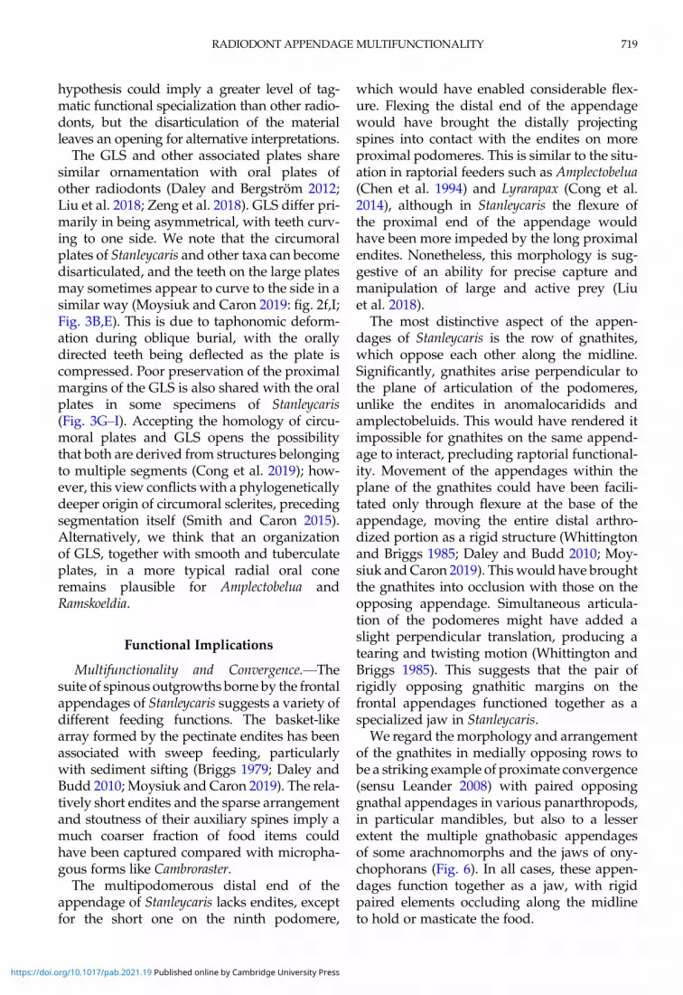
FIGURE 6. Gnathal convergence in radiodonts and euarthropods. A, Gnathobase of the xiphosuran Limulus polyphemus,courtesy of R. Bicknell; B, ROMIP 59975, frontal appendage of Stanleycaris hirpex; C, mandibular gnathobase of the duro-phagous copepod Calanus propinquus, courtesy of J. Michels; D, mandibular gnathobase of the predatory copepod Paraeu-chaeta antarctica, courtesy of J. Michels; E, ROMIP 59501, frontal appendages of Caryosyntrips serratus; F,G, USNM 57490,frontal appendages of cf. Peytoia; F, overview showing opposing gnathites; G, close-up of gnathites. Abbreviations as inFigs. 1–3. Arrowheads indicate one angular podomere boundary. Scale bars, A, B, E–G, 2mm; C, D, 50 μm.
RADIODONT APPENDAGE MULTIFUNCTIONALITY 715
https://doi.org/10.1017/pab.2021.19 Published online by Cambridge University Press
appendage. In curvature, the gnathites ofStanleycaris more closely resemble the toothedmargins of the oral cone, which could pointto functional similarities (see “FunctionalImplications”).
Distal End of the Appendage.—Stanleycaris,Peytoia, and cf. Peytoia also share a robust andmultisegmented distal portion of the append-age, resembling the raptorial ends of anomalo-caridid and amplectobeluid appendages (Daleyand Budd 2010; Moysiuk and Caron 2019).Other hurdiids with this characteristic includean unnamed hurdiid appendage from theFezouata Formation (Van Roy and Briggs 2011)and Ursulinacaris grallae (Pates et al. 2019). Com-parisons with these forms are hindered by thelimited material available, but see “Note onUrsulinacaris.” The distribution of multisegmen-ted distal regions of the frontal appendage inamplectobeluids, anomalocaridids, tamisiocar-idids, and early-diverging hurdiids points toretention from a common ancestor.
Note on Ursulinacaris.—Ursulinacaris wasrecently described as possessing narrow, pairedendites. This contrasts with the condition in allother hurdiids, in which one set of endites isbroad and the other has either been modifiedinto medial gnathites (as in Stanleycaris) or lostaltogether (e.g., Cambroraster) (Vinther et al.2014; Moysiuk and Caron 2019), and was sug-gested to provide a link with tamisiocaridids(Pates et al. 2019). We note, however, that thestructures interpreted as paired endites consistof lighter-colored, parallel marginal areasflanking a darker axial lineation, with no clearevidence of interspace. This resembles the speci-mens of Stanleycaris described earlier (Figs. 2F,J,and 3D,E), but here demonstrably only a singlerow of broad endites is present. In Stanleycaris,we interpret the dark axial lineation as thetapered edge of the single bladelike endite andthe occasional preservation of a “doubled” mar-ginwith two layers of cuticle as a consequence ofthe juxtaposition of the two narrowedges duringcompression. We think this interpretation likelyholds for Ursulinacaris as well. By contrast, if asecond set of endites were present in Ursulina-caris, we would expect to see them clearly anddivergently spaced apart from the first, as inother taxa (Daley et al. 2013b; Daley and Edge-combe 2014; Vinther et al. 2014).
Ursulinacaris resembles cf. Peytoia in terms ofthe number and position of endites and inclin-ation of the proximalmost endite. While it dif-fers in the apparent lack of gnathites, thesemight simply be hidden due to burial orienta-tion, as seen in specimens of Stanleycaris (e.g.,Figs. 2F, and 3D,E). New fossil material willbe needed to assess the phylogenetic signifi-cance of Ursulinacaris.
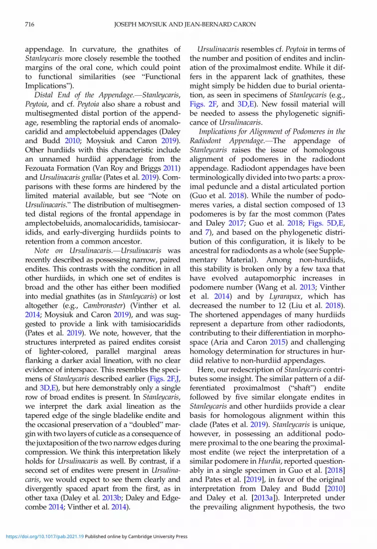
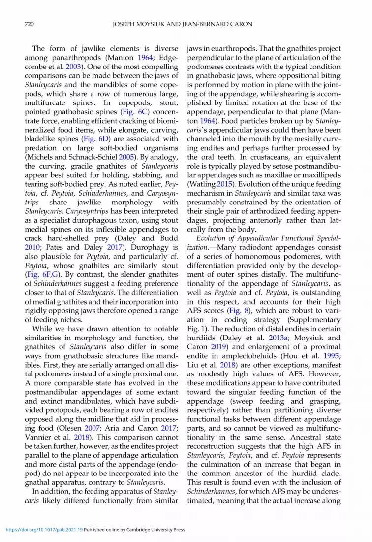
Implications for Alignment of Podomeres in theRadiodont Appendage.—The appendage ofStanleycaris raises the issue of homologousalignment of podomeres in the radiodontappendage. Radiodont appendages have beenterminologically divided into two parts: a prox-imal peduncle and a distal articulated portion(Guo et al. 2018). While the number of podo-meres varies, a distal section composed of 13podomeres is by far the most common (Patesand Daley 2017; Guo et al. 2018; Figs. 5D,E,and 7), and based on the phylogenetic distri-bution of this configuration, it is likely to beancestral for radiodonts as awhole (see Supple-mentary Material). Among non-hurdiids,this stability is broken only by a few taxa thathave evolved autapomorphic increases inpodomere number (Wang et al. 2013; Vintheret al. 2014) and by Lyrarapax, which hasdecreased the number to 12 (Liu et al. 2018).The shortened appendages of many hurdiidsrepresent a departure from other radiodonts,contributing to their differentiation in morpho-space (Aria and Caron 2015) and challenginghomology determination for structures in hur-diid relative to non-hurdiid appendages.Here, our redescription of Stanleycaris contri-
butes some insight. The similar pattern of a dif-ferentiated proximalmost (“shaft”) enditefollowed by five similar elongate endites inStanleycaris and other hurdiids provide a clearbasis for homologous alignment within thisclade (Pates et al. 2019). Stanleycaris is unique,however, in possessing an additional podo-mere proximal to the one bearing the proximal-most endite (we reject the interpretation of asimilar podomere inHurdia, reported question-ably in a single specimen in Guo et al. [2018]and Pates et al. [2019], in favor of the originalinterpretation from Daley and Budd [2010]and Daley et al. [2013a]). Interpreted underthe prevailing alignment hypothesis, the two
JOSEPH MOYSIUK AND JEAN‐BERNARD CARON716
https://doi.org/10.1017/pab.2021.19 Published online by Cambridge University Press

proximal podomeres of the Stanleycarisappendage would both be considered part ofa subdivided peduncle (Fig. 7). However, wethink that this alignment may not be optimalfor two key reasons, as follows.First, it has been controversial as to whether
various linear traces within the peduncle insome species are indeed segmental articulationsrather than annulations, cuticular folds, ortaphonomic artifacts (Lerosey-Aubril and Pates2018). The most compelling case for a multipo-domerous peduncle comes from Amplectobeluasymbrachiata (Cong et al. 2017), but even herethe expression of the putative boundaries issomewhat variable. In other species, the ped-uncle is either composed of a single podomere(Daley et al. 2013b; Daley and Edgecombe2014; Pates and Daley 2017) or we consider thenumber to be ambiguous (Cong et al. 2016,2018; Guo et al. 2018). Thus, in the phylogeneticscenario in which the first two podomeres in the
Stanleycaris appendage represent the peduncle,this subdivision would likely have to be inter-preted as an autapomorphy.Second, if we instead interpret only the prox-
imalmost podomere of Stanleycaris as thehomologue of the peduncle, then the distalarticulated portion of the appendage is seento consist of 13 podomeres, the probable ances-tral number for radiodonts (Fig. 7). Under thisalignment scheme the number of podomeresin Peytoia (Fig. 5A,B) and cf. Peytoia wouldalso exactly match the 13 distal podomeresseen in most other radiodonts, reinforcing thenotion of their phylogenetic divergence nearthe base of the hurdiid clade, with subsequentreduction in podomere number in derivedforms like Hurdia and Cambroraster (Daleyet al. 2013a; Moysiuk and Caron 2019). How-ever, this would also imply that the proximalpodomere of all hurdiids other than Stanleycariswould not be homologous with the peduncle of
FIGURE 7. Hypotheses of radiodont frontal appendage podomere homology. Simplified diagrams of radiodont appen-dages contrasting the hypothesis for the alignment of hurdiid with non-hurdiid appendages favored in this paper withthat suggested in, e.g., Guo et al. (2018) and Pates et al. (2019). Outer spines, second endites/gnathites, and auxiliary spinesomitted for clarity. Peduncle is labeled p (light green), while distal podomeres (blue) are numbered starting at the podo-mere adjacent to the peduncle (1). Species in bold are represented, with other similar species listed below. Podomere countsare based on examination of fossil material housed at the ROM (Stanleycaris hirpex, Peytoia nathorsti, cf. Peytoia, Amplecto-belua stephenensis) and in the published literature (Pates and Daley 2017; Guo et al. 2018). We interpret the articulated ter-minal spine observed in Amplectobelua symbrachiata (Cong et al. 2017: “ts” in their fig. 2) as the 13th podomere, and ourreexamination of fossil material of A. stephenensis finds the corresponding 13 podomeres (Fig. 5E,F). Based on the figuredmaterial, we think the same is also likely the case for “Anomalocaris” kunmingensis (Wang et al. 2013; Liu et al. 2018). (Coloronline.)
RADIODONT APPENDAGE MULTIFUNCTIONALITY 717
https://doi.org/10.1017/pab.2021.19 Published online by Cambridge University Press

non-hurdiids, but rather with the first podo-mere of the distal articulated region, with thepeduncle (or its distal articulation) being lostor not preserved (Fig. 7). Although both scen-arios remain plausible, the one we presentwould seem to be more parsimonious.
Oral Structures.—While the appendages ofStanleycaris are distinctive, the oral cone(Fig. 4) is more typical. As in Hurdia (Whitting-ton and Briggs 1985; Daley et al. 2013a) andCambroraster (Moysiuk and Caron 2019), it istetraradial with smooth plates bearing fewteeth (sometimes with paired nodes on thelarge plates; e.g., Daley et al. 2013a: fig. 2f,j).The number of teeth per small circumoralplate differentiates Stanleycaris from at leastHurdia, in which two rather than three teethare typically reported (Daley et al. 2013a). Thelack of inner oral plates is comparable to thecondition in oral cones identified to Peytoia(Daley et al. 2013a) and Cordaticaris (Sun et al.
2020). The most unusual feature in the Stanley-caris oral cone appears to be the presence of setsof six smaller circumoral plates, with sevenbeing typical for hurdiids (Daley et al. 2013a;Moysiuk and Caron 2019; Sun et al. 2020) andprobably for some amplectobeluids (Liu et al.2018; Zeng et al. 2018). This presumably repre-sents an autapomorphy for Stanleycaris, withan oral cone with 4 large and 28 small platespossibly being plesiomorphic for Radiodonta(Liu et al. 2018).Interrupting the conservativism of radiodont
oral cone morphology, Cong et al. (2017, 2018)observed disarticulated assemblages of A. sym-brachiata and Ramskoeldia spp. with severalsclerite types, leading them to hypothesize aradically different feeding apparatus. Theyspeculatively reconstructed a four-sided com-plex of plates followed by three bilaterallypaired, opposing “gnathobase-like structures”(GLS). In the context of this paper, this
FIGURE 8. Evolution of early panarthropod frontal appendicular functional diversity. Cambrian panarthropod time tree(majority rule consensus), with maximum-likelihood ancestral state reconstruction of Appendicular Functional Specializa-tion (AFS) index for frontal appendages. Numbers at nodes are posterior probabilities.
JOSEPH MOYSIUK AND JEAN‐BERNARD CARON718
https://doi.org/10.1017/pab.2021.19 Published online by Cambridge University Press
hypothesis could imply a greater level of tag-matic functional specialization than other radio-donts, but the disarticulation of the materialleaves an opening for alternative interpretations.The GLS and other associated plates share
similar ornamentation with oral plates ofother radiodonts (Daley and Bergström 2012;Liu et al. 2018; Zeng et al. 2018). GLS differ pri-marily in being asymmetrical, with teeth curv-ing to one side. We note that the circumoralplates of Stanleycaris and other taxa can becomedisarticulated, and the teeth on the large platesmay sometimes appear to curve to the side in asimilar way (Moysiuk and Caron 2019: fig. 2f,I;Fig. 3B,E). This is due to taphonomic deform-ation during oblique burial, with the orallydirected teeth being deflected as the plate iscompressed. Poor preservation of the proximalmargins of the GLS is also shared with the oralplates in some specimens of Stanleycaris(Fig. 3G–I). Accepting the homology of circu-moral plates and GLS opens the possibilitythat both are derived from structures belongingto multiple segments (Cong et al. 2019); how-ever, this view conflicts with a phylogeneticallydeeper origin of circumoral sclerites, precedingsegmentation itself (Smith and Caron 2015).Alternatively, we think that an organizationof GLS, together with smooth and tuberculateplates, in a more typical radial oral coneremains plausible for Amplectobelua andRamskoeldia.
Functional Implications
Multifunctionality and Convergence.—Thesuite of spinous outgrowths borne by the frontalappendages of Stanleycaris suggests a variety ofdifferent feeding functions. The basket-likearray formed by the pectinate endites has beenassociated with sweep feeding, particularlywith sediment sifting (Briggs 1979; Daley andBudd 2010; Moysiuk and Caron 2019). The rela-tively short endites and the sparse arrangementand stoutness of their auxiliary spines imply amuch coarser fraction of food items couldhave been captured compared with micropha-gous forms like Cambroraster.The multipodomerous distal end of the
appendage of Stanleycaris lacks endites, exceptfor the short one on the ninth podomere,
which would have enabled considerable flex-ure. Flexing the distal end of the appendagewould have brought the distally projectingspines into contact with the endites on moreproximal podomeres. This is similar to the situ-ation in raptorial feeders such as Amplectobelua(Chen et al. 1994) and Lyrarapax (Cong et al.2014), although in Stanleycaris the flexure ofthe proximal end of the appendage wouldhave been more impeded by the long proximalendites. Nonetheless, this morphology is sug-gestive of an ability for precise capture andmanipulation of large and active prey (Liuet al. 2018).The most distinctive aspect of the appen-
dages of Stanleycaris is the row of gnathites,which oppose each other along the midline.Significantly, gnathites arise perpendicular tothe plane of articulation of the podomeres,unlike the endites in anomalocaridids andamplectobeluids. This would have rendered itimpossible for gnathites on the same append-age to interact, precluding raptorial functional-ity. Movement of the appendages within theplane of the gnathites could have been facili-tated only through flexure at the base of theappendage, moving the entire distal arthro-dized portion as a rigid structure (Whittingtonand Briggs 1985; Daley and Budd 2010; Moy-siuk andCaron 2019). This would have broughtthe gnathites into occlusion with those on theopposing appendage. Simultaneous articula-tion of the podomeres might have added aslight perpendicular translation, producing atearing and twisting motion (Whittington andBriggs 1985). This suggests that the pair ofrigidly opposing gnathitic margins on thefrontal appendages functioned together as aspecialized jaw in Stanleycaris.We regard the morphology and arrangement
of the gnathites in medially opposing rows tobe a striking example of proximate convergence(sensu Leander 2008) with paired opposinggnathal appendages in various panarthropods,in particular mandibles, but also to a lesserextent the multiple gnathobasic appendagesof some arachnomorphs and the jaws of ony-chophorans (Fig. 6). In all cases, these appen-dages function together as a jaw, with rigidpaired elements occluding along the midlineto hold or masticate the food.
RADIODONT APPENDAGE MULTIFUNCTIONALITY 719
https://doi.org/10.1017/pab.2021.19 Published online by Cambridge University Press
The form of jawlike elements is diverseamong panarthropods (Manton 1964; Edge-combe et al. 2003). One of the most compellingcomparisons can be made between the jaws ofStanleycaris and the mandibles of some cope-pods, which share a row of numerous large,multifurcate spines. In copepods, stout,pointed gnathobasic spines (Fig. 6C) concen-trate force, enabling efficient cracking of biomi-neralized food items, while elongate, curving,bladelike spines (Fig. 6D) are associated withpredation on large soft-bodied organisms(Michels and Schnack-Schiel 2005). By analogy,the curving, gracile gnathites of Stanleycarisappear best suited for holding, stabbing, andtearing soft-bodied prey. As noted earlier, Pey-toia, cf. Peytoia, Schinderhannes, and Caryosyn-trips share jawlike morphology withStanleycaris. Caryosyntrips has been interpretedas a specialist durophagous taxon, using stoutmedial spines on its inflexible appendages tocrack hard-shelled prey (Daley and Budd2010; Pates and Daley 2017). Durophagy isalso plausible for Peytoia, and particularly cf.Peytoia, whose gnathites are similarly stout(Fig. 6F,G). By contrast, the slender gnathitesof Schinderhannes suggest a feeding preferencecloser to that of Stanleycaris. The differentiationof medial gnathites and their incorporation intorigidly opposing jaws therefore opened a rangeof feeding niches.While we have drawn attention to notable
similarities in morphology and function, thegnathites of Stanleycaris also differ in someways from gnathobasic structures like mand-ibles. First, they are serially arranged on all dis-tal podomeres instead of a single proximal one.A more comparable state has evolved in thepostmandibular appendages of some extantand extinct mandibulates, which have subdi-vided protopods, each bearing a row of enditesopposed along the midline that aid in process-ing food (Olesen 2007; Aria and Caron 2017;Vannier et al. 2018). This comparison cannotbe taken further, however, as the endites projectparallel to the plane of appendage articulationand more distal parts of the appendage (endo-pod) do not appear to be incorporated into thegnathal apparatus, contrary to Stanleycaris.In addition, the feeding apparatus of Stanley-
caris likely differed functionally from similar
jaws in euarthropods. That the gnathites projectperpendicular to the plane of articulation of thepodomeres contrasts with the typical conditionin gnathobasic jaws, where oppositional bitingis performed by motion in plane with the joint-ing of the appendage, while shearing is accom-plished by limited rotation at the base of theappendage, perpendicular to that plane (Man-ton 1964). Food particles broken up by Stanley-caris’s appendicular jaws could then have beenchanneled into the mouth by the mesially curv-ing endites and perhaps further processed bythe oral teeth. In crustaceans, an equivalentrole is typically played by setose postmandibu-lar appendages such as maxillae or maxillipeds(Watling 2015). Evolution of the unique feedingmechanism in Stanleycaris and similar taxa waspresumably constrained by the orientation oftheir single pair of arthrodized feeding appen-dages, projecting anteriorly rather than lat-erally from the body.
Evolution of Appendicular Functional Special-ization.—Many radiodont appendages consistof a series of homonomous podomeres, withdifferentiation provided only by the develop-ment of outer spines distally. The multifunc-tionality of the appendage of Stanleycaris, aswell as Peytoia and cf. Peytoia, is outstandingin this respect, and accounts for their highAFS scores (Fig. 8), which are robust to vari-ation in coding strategy (SupplementaryFig. 1). The reduction of distal endites in certainhurdiids (Daley et al. 2013a; Moysiuk andCaron 2019) and enlargement of a proximalendite in amplectobeluids (Hou et al. 1995;Liu et al. 2018) are other exceptions, manifestas modestly high values of AFS. However,these modifications appear to have contributedtoward the singular feeding function of theappendage (sweep feeding and grasping,respectively) rather than partitioning diversefunctional tasks between different appendageparts, and so cannot be viewed as multifunc-tionality in the same sense. Ancestral statereconstruction suggests that the high AFS inStanleycaris, Peytoia, and cf. Peytoia representsthe culmination of an increase that began inthe common ancestor of the hurdiid clade.This result is found even with the inclusion ofSchinderhannes, for which AFSmay be underes-timated, meaning that the actual increase along
JOSEPH MOYSIUK AND JEAN‐BERNARD CARON720
https://doi.org/10.1017/pab.2021.19 Published online by Cambridge University Press
the branch leading to Hurdiidae could havebeen greater.The appendages of Stanleycaris and other
early-diverging hurdiids suggest a plausiblescenario for the evolution of the divergentsweep-feeding ecology characterizing thisclade. The origin of the hurdiid-type append-age involved a drastic change in functionalmorphology, from a probable ancestral form,in which prey handling was accomplished bymultiarticulatory raptorial grasping, to aderived state, in which oppositional basolateralsweeping became primary (Moysiuk andCaron 2019). However, the morphology ofStanleycaris and similar hurdiids demonstratesthat this transformation did not occur whole-sale. Rather, the increase in AFS at the originof the hurdiid clade corresponds to the main-tenance of plesiomorphic raptorial functional-ity at the distal end of the appendage, freeingother appendage modules to evolve novelsweep-feeding and gnathal adaptations. Thedecrease in AFS in some derived hurdiidswould thus presumably correspond to subse-quent ecological specialization via reversionto less functionally differentiated states.This raises the question of whether evidence
exists that other ecological shifts among radio-donts might have been facilitated by similarpunctuational increases in AFS. While the pau-city of known instances of such shifts hindersour ability to robustly test this hypothesis, ourresults do provide apparently contradictorycases. Ancestral state reconstruction suggestsa decrease in AFS on the branches leading toAegirocassis and Tamisiocaris, correspondingto hypothesized transitions to a suspension-feeding ecology (Vinther et al. 2014; Van Royet al. 2015). Interestingly, the low AFS valuesobserved in Aegirocassis and Tamisiocarismatchthe pattern of low tagmatic functional special-ization observed previously in suspension-feeding euarthropods (Cisne 1974), suggestingthat the possession of serial undifferentiatedfeeding structures might be a common themefor this guild. Unlike in the case of the hurdiids,these transitions to suspension feeding seem-ingly did not involve major reorganizationof frontal appendage functional morphology,rather occurring via coordinated “global”increase in endite length and number of
auxiliary spines, presumably enabling captureof increasingly small-sized prey. Given theless drastic nature of these morphofunctionalchanges, it is perhaps unsurprising that eco-logical transition was able to occur in theabsence of a multifunctional transitional form.A similar case of integrated change in all enditesmay have occurred in the lineage leading toCar-yosyntrips, although this is more ambiguous,considering the poor constraint on its phylogen-etic position. Taken together, these cases suggestthat a high level of AFS was not a prerequisitefor ecological diversification among radiodonts,although it may have played a role in what wasarguably the most spectacular transition at theorigin of the hurdiid clade.Radiodonts occupy the extreme high values
in AFS relative to euarthropods and lobopo-dians. Thismay be in part an artifact of the data-set we employed, which includes only a highlylimited (though phylogenetically representa-tive) sampling of Cambrian euarthropods.Comparison of AFS values in a greater diversityof euarthropod taxa would be useful; however,controversy over the homology of radiodontand euarthropod frontal appendages (Conget al. 2014; Aria et al. 2020) remains a notableconstraint. Small sample size might likewisecontribute to favoring of Brownian motion inour analysis, which otherwise seems at oddswith the prediction of selection favoring func-tional specialization (Rueffler et al. 2012).Another factor is that AFS in this study consid-ers only frontal appendages, and we wouldexpect higher values of AFS in the postfrontalappendages of some euarthropods. A final cav-eat is that some types of functional specializa-tion are not captured by our AFS metric, suchas the multichelae and sensory flagellae formedby proximodistal subdivision of leanchoiliidendites (Aria et al. 2015). Thus, our results donot suggest that AFS in general was unimport-ant in the radiation of euarthropods, but theydo conform with the hypothesis that frontalAFS was relatively important compared withtagmatic functional specialization in the diversi-fication of radiodonts and that clade-specificpatterns of functional specialization evolved inthe context of the same Cambrian ecosystems.While radiodont appendages vary consider-
ably with their ecology, oral cone form is
RADIODONT APPENDAGE MULTIFUNCTIONALITY 721
https://doi.org/10.1017/pab.2021.19 Published online by Cambridge University Press

more conserved, and how its function mayhave differed depending on feeding habits isa more open question. Speculatively, innova-tions like the inner oral plates present in somederived hurdiids (Daley et al. 2013a; Moysiukand Caron 2019) could have provided releasefrom functional trade-offs by compensatingfor the loss of masticatory gnathites on theirfrontal appendages, enabling more radicalappendage specialization. Such oral modifica-tions could thus have provided an alternativeto appendicular functional specialization. Thefunctional significance, if any, of other varia-tions, such as the number of oral plates or pres-ence of nodes or furrows on their surfaces,remains unclear (Daley and Bergström 2012;Zeng et al. 2018), in part due to the poor knowl-edge of oral morphology in most taxa. It will beuseful to consider these aspects in futureassessments of radiodont functional diversityas new fossil evidence comes to light.
Conclusions
In euarthropods, food capture and process-ing are typically performed by several specia-lized appendage pairs working in concert(Watling 2015). Radiodonts, constrained bythe possession of only a single pair of arthro-dized appendages, met similar imposed func-tional requirements either by adapting torelatively narrow feeding niches through inte-grated morphological change or by the evolu-tion of increased functional specialization ofdifferent parts of the appendage. Stanleycarisis exemplary of the latter case, having appen-dages with strong differentiation of lateral–medial, inner–outer, and proximal–distalmorphologies. This permitted appendage mul-tifunctionality, including the ability to trap(endites), manipulate (distal raptorial portion),and masticate (gnathites) prey items. Multi-functionality may in turn have played a roleas an evolutionary bridge between feedingecologies in the ancestral hurdiid lineage,allowing dramatic niche diversification withinthe otherwise conserved radiodont body plan.Bilaterally opposed jawlike appendages have
evolved numerous times among panarthro-pods, most characteristically in the mandiblesof mandibulates. We argue that Stanleycaris
provides an equivalent example of evolution-ary convergence in a Cambrian stem euar-thropod. However, jaws in mandibulates,arachnomorphs, and onychophorans originatefrom different parts of the appendage (coxa,basipod, and distal claw, respectively; Boxshall2004). Stanleycaris and similar radiodontsachieved convergence by even more divergentmeans, with whole appendages forming theopposing jaws. While convergence is oftenseen as a prime example of the long-term pre-dictability of evolution (Blount et al. 2018),examples such as these underscore that conver-gent characters are mosaic products of both theecological context that drives their evolutionand the idiosyncrasies of the body plan inwhich they happen to emerge.
Acknowledgments
Fossils were collected by Royal OntarioMuseum field parties under several Parks Can-ada Research and Collections permits to J.-B.C.and D. Collins. We thank S. Cappelli for artisticreconstructions; R. Bicknell and J. Michels forphotos of the xiphosuran gnathobase and cope-pod mandibles, respectively; and M. Akramiand P. Fenton for assistance in the collections.J.M. also thanks A. Izquierdo López, J. Moon,andA.Daley for helpful discussions. Commentsfrom J. Haug and one anonymous reviewer sti-mulated significant improvement of this article.
Major funding support for fieldworkcomes from the Royal Ontario Museum(Research and collection grants, Natural His-tory fieldwork grants), the Polk Milstein Fam-ily, the National Geographic Society (no.9475-14 to J.-B.C.), the Swedish Research Coun-cil (to Michael Streng), the National ScienceFoundation (NSF-EAR-1556226, 1554897), andPomona College (to Robert R. Gaines). Thisresearch was also indirectly supported by theDorothy Strelsin Foundation (Royal OntarioMuseum). J.M.’s doctoral research is supportedby a National Science and EngineeringResearch Council (NSERC) Vanier CanadaGraduate Scholarship through the Universityof Toronto (Department of Ecology and Evolu-tion) and J.-B.C.’s NSERC Discovery Grant (no.341944). This is the Royal Ontario MuseumBurgess Shale project no. 89.
JOSEPH MOYSIUK AND JEAN‐BERNARD CARON722
https://doi.org/10.1017/pab.2021.19 Published online by Cambridge University Press
Data Availability Statement
Data available from the Dryad DigitalRepository: https://doi.org/10.5061/dryad.kd51c5b5c.
Literature CitedAdamowicz, S. J., A. Purvis, and M. A. Wills. 2008. Increasing mor-phological complexity in multiple parallel lineages of the Crust-acea. Proceedings of the National Academy of Sciences USA105:4786–4791.
Aria, C. 2020.Macroevolutionary patterns of body plan canalizationin euarthropods. Paleobiology 46:569–593.
Aria, C., and J.-B. Caron. 2015. Cephalic and limb anatomy of a newisoxyid from the Burgess Shale and the role of “stem bivalvedarthropods” in the disparity of the frontalmost appendage.PLoS ONE 10(6):e0124979.
Aria, C., and J.-B. Caron. 2017. Burgess Shale fossils illustrate theorigin of the mandibulate body plan. Nature 545:89–92.
Aria, C., J.-B. Caron, and R. Gaines. 2015. A large new leanchoiliidfrom the Burgess Shale and the influence of inapplicable states onstem arthropod phylogeny. Palaeontology 58:629–660.
Aria, C., F. Zhao, H. Zeng, J. Guo, and M. Zhu. 2020. Fossils fromSouth China redefine the ancestral euarthropod body plan.BMC Evolutionary Biology 20:4.
Blount, Z. D., R. E. Lenski, and J. B. Losos. 2018. Contingency anddeterminism in evolution: replaying life’s tape. Science 362:eaam5979.
Boxshall, G. A. 2004. The evolution of arthropod limbs. BiologicalReviews of the Cambridge Philosophical Society 79:253–300.
Briggs, D. E.G., 1979. Anomalocaris: the largest known Cambrianarthropod. Palaeontology 22:631–664.
Caron, J.-B., and D. A. Jackson. 2008. Paleoecology of the GreaterPhyllopod Bed Community, Burgess Shale. Palaeogeography,Palaeoclimatology, Palaeoecology 258:222–256.
Caron, J.-B., R. R. Gaines, M. G. Mángano, M. Streng, and A.C. Daley. 2010. A New Burgess Shale-type assemblage from the“thin” Stephen Formation of the southern Canadian Rockies.Geology 38:811–814.
Chen, J. Y., L. Ramsköld, and G. Q. Zhou. 1994. Evidence for mono-phyly and arthropod affinity of Cambrian giant predators. Sci-ence 264:1304–1308.
Cisne, J. L. 1974. Evolution of the world fauna of aquatic free-livingarthropods. Evolution 28:337–366.
Collins, D. 1996. The “evolution” of Anomalocaris and its classifica-tion in the arthropod class Dinocarida (Nov.) and order Radio-donta (Nov.). Journal of Paleontology 70:280–293.
Cong, P., X. Ma, X. Hou, G. D. Edgecombe, and N. J. Strausfeld.2014. Brain structure resolves the segmental affinity of anomalo-caridid appendages. Nature 513:538.
Cong, P., A. C. Daley, G. D. Edgecombe, X. Hou, and A. Chen. 2016.Morphology of the radiodontan Lyrarapax from the Early Cam-brian Chengjiang Biota. Journal of Paleontology 90:663–671.
Cong, P., A. C. Daley, G. D. Edgecombe, andX.Hou. 2017. The func-tional head of the Cambrian radiodontan (stem-group Euarthro-poda) Amplectobelua symbrachiata. BMC Evolutionary Biology17:208.
Cong, P., G. D. Edgecombe, A. C. Daley, J. Guo, S. Pates, and X.G. Hou. 2018. New radiodonts with gnathobase-like structuresfrom the Cambrian Chengjiang Biota and implications for the sys-tematics of Radiodonta. Papers in Palaeontology 4:605–621.
Cong, P., Jin Guo, G. D. Edgecombe, A. C. Daley, and X. Hou. 2019.Reconstructing the feeding apparatus of Amplectobeluidae(Radiodonta: Stem Euarthropoda). Palaeontological Association63rd Annual Meeting, Abstracts, p. 29.
Daley, A. C., and J. Bergström. 2012. The oral cone ofAnomalocaris isnot a classic “Peytoia.” Naturwissenschaften 99:501–504.
Daley, A. C., andG. E. Budd. 2010.Newanomalocaridid appendagesfrom the Burgess Shale, Canada. Palaeontology 53:721–738.
Daley, A. C., and G. D. Edgecombe. 2014. Morphology of Anomalo-caris canadensis from the Burgess Shale. Journal of Paleontology88:68–91.
Daley, A. C.,G. E. Budd, J.-B. Caron, G.D. Edgecombe, andD.Collins.2009. The Burgess Shale anomalocarididHurdia and its significancefor early euarthropod evolution. Science 323:1597–1600.
Daley, A. C., G. E. Budd, and J.-B. Caron. 2013a. Morphology andsystematics of the anomalocaridid arthropod Hurdia from theMiddle Cambrian of British Columbia and Utah. Journal of Sys-tematic Palaeontology 11:743–787.
Daley, A. C., J. R. Paterson, G. D. Edgecombe, D. C. García-Bellido,and J. B. Jago. 2013b. New anatomical information on Anomalo-caris from the Cambrian Emu Bay Shale of South Australia anda reassessment of its inferred predatory habits. Palaeontology56:971–990.
Daley, A. C., J. B. Antcliffe, H. B. Drage, and S. Pates. 2018. Earlyfossil record of Euarthropoda and the Cambrian explosion.Proceedings of the National Academy of Sciences USA115:5323–5331.
Edgecombe, G. D., S. Richter, and G. D. F. Wilson. 2003. The man-dibular gnathal edges: homologous structures throughout Man-dibulata? African Invertebrates 44:115–135.
Guo, J., S. Pates, P. Cong, A. C. Daley, G. D. Edgecombe, T. Chen,and X. Hou. 2018. A new radiodont (stem Euarthropoda) frontalappendagewith amosaic of characters from the Cambrian (Series2 Stage 3) Chengjiang Biota. Papers in Palaeontology 5:99–110.
Harmon, L. J., J. T. Weir, C. D. Brock, R. E. Glor, andW. Challenger.2008. GEIGER: investigating evolutionary radiations. Bioinfor-matics 24:129–131.
Haug, J. T., A. Maas, C. Haug, and D. Waloszek. 2012. Evolution ofcrustacean appendages. Pp. 34–73 in L.Watling andM. Thiel, eds.Functional morphology and diversity, 1st ed. Oxford UniversityPress, New York.
Hou, X., J. Bergström, and P. Ahlberg. 1995. Anomalocaris and otherlarge animals in the Lower Cambrian Chengjiang Fauna of south-west China. GFF 117:163–183.
Jefferies, R. P. S. 1979. The origin of chordates—a methodologicalessay. Pp. 443–477 in M. R. House, ed. The origin of the majorinvertebrate groups. Academic Press, London.
Karlstrom, K.E., M. T. Mohr, M. D. Schmitz, F. A. Sundberg, S.M. Rowland, R. Blakey, J. R. Foster, L. J. Crossey, C. M. Dehler,and J. W. Hagadorn. 2020. Redefining the Tonto Group ofGrand Canyon and recalibrating the Cambrian time scale. Geol-ogy 48:425–430.
Kühl, G., D. E. G., Briggs, and J. Rust. 2009. A great-appendagearthropod with a radial mouth from the Lower Devonian Huns-rück Slate, Germany. Science 323:771–773.
Leander, B. S. 2008. A hierarchical view of convergent evolutionin microbial eukaryotes. Journal of Eukaryotic Microbiology55:59–68..
Lerosey-Aubril, R., and S. Pates. 2018. New suspension-feedingradiodont suggests evolution of microplanktivory in Cambrianmacronekton. Nature Communications 9:3774.
Lewis, P. O. 2001. A likelihood approach to estimating phylogenyfrom discrete morphological character data. Systematic Biology50:913–925.
Liu, J., R. Lerosey-Aubril, M. Steiner, J. A Dunlop, D. Shu, and J.R Paterson. 2018. Origin of raptorial feeding in juvenile euarthro-pods revealed by a Cambrian radiodontan. National ScienceReview 5:863–869.
Manton, S. M. 1964. Mandibular mechanisms and evolution ofarthropods. Philosophical Transactions of the Royal Society ofLondon B 247:1–183.
RADIODONT APPENDAGE MULTIFUNCTIONALITY 723
https://doi.org/10.1017/pab.2021.19 Published online by Cambridge University Press
Mayers, B., C. Aria, and J.-B. Caron. 2018. Three new naraoiid spe-cies from the Burgess Shale, with a morphometric and phylogen-etic reinvestigation of Naraoiidae. Palaeontology 62:19–50.
Michels, J., and S. B. Schnack-Schiel. 2005. Feeding in dominantAntarctic copepods—does the morphology of the mandibulargnathobases relate to diet? Marine Biology 146:483–495.
Moysiuk, J., and J. B. Caron. 2019. A new hurdiid radiodont fromthe Burgess Shale evinces the exploitation of Cambrian infaunalfood sources. Proceedings of the Royal Society of London B286:20191079.
Nanglu, K., J.-B. Caron, and R. R. Gaines. 2020. The Burgess Shalepaleocommunity with new insights fromMarble Canyon, BritishColumbia. Paleobiology 46:58–81.
Nielsen, C. 1995. Animal evolution, interrelationships of the livingphyla, 1st ed. Oxford University Press, Oxford.
O’Brien, L. J., J.-B. Caron, and R. R. Gaines. 2014. Taphonomy anddepositional setting of the Burgess Shale Tulip Beds, Mount Ste-phen, British Columbia. Palaios 29:309–324.
Olesen, J. 2007. Monophyly and phylogeny of Branchiopoda, withfocus on morphology and homologies of branchiopod phyllopo-dous limbs. Journal of Crustacean Biology 27:165–183.
Ortega-Hernández, J., R. Janssen, and G. E. Budd. 2017. Origin andevolution of the panarthropod head—a palaeobiological anddevelopmental perspective. Arthropod Structure and Develop-ment 46:354–379.
Pates, S., and A. C. Daley. 2017. Caryosyntrips: a radiodontan fromthe Cambrian of Spain, USA and Canada. Papers in Palaeon-tology 3:461–470.
Pates, S., A. C. Daley, and J. Ortega-Hernández. 2017. Aysheaia pro-lata from the Utah Wheeler Formation (Drumian, Cambrian) is afrontal appendage of the radiodontan Stanleycaris. Acta Palaeon-tologica Polonica 62:619–625.
Pates, S., A. Daley, and J. Ortega-Hernández. 2018. Response tocomment on “Aysheaia prolata from the Utah Wheeler Formation(Drumian, Cambrian) Is a Frontal Appendage of the Radiodon-tan Stanleycaris” with the formal description of Stanleycaris.Acta Palaeontologica Polonica 63:105–110.
Pates, S., A. C. Daley, and N. J. Butterfield. 2019. First report ofpaired ventral endites in a hurdiid radiodont. Zoological Letters5:1–11.
Rambaut A., A. J. Drummond, D. Xie, G. Baele, and M. A. Suchard2018. Posterior summarisation in Bayesian phylogenetics usingTracer 1.7. Systematic Biology 67:901–904
R Core Team. 2020. R: a language and environment for statisticalcomputing. R Foundation for Statistical Computing, Vienna,Austria.
Revell, L. J. 2012. Phytools: an R package for phylogenetic compara-tive biology (and other things). Methods in Ecology and Evolu-tion 3:217–223.
Ronquist, F., S. Klopfstein, L. Vilhelmsen, S. Schulmeister, D.L. Murray, and A. P. Rasnitsyn. 2012a. A total-evidence approachto dating with fossils, applied to the early radiation of theHymenoptera. Systematic Biology 61:973–999.
Ronquist, F., P. Van Der Mark, M. Teslenko, D. L. Ayres, A. Darling,S. Höhna, B. Larget, L. Liu, M. A. Suchard, and J. P. Huelsenbeck.2012b. Mrbayes 3.2: efficient Bayesian phylogenetic inference andmodel choice across a large model space. Systematic Biology61:539–542.
Rueffler, C., J. Hermisson, and G. P. Wagner. 2012. Evolution offunctional specialization and division of labor. Proceedings ofthe National Academy of Sciences USA 109:326–335.
Smith, M. R., and J.-B. Caron. 2015.Hallucigenia’s head and the pha-ryngeal armature of early ecdysozoans. Nature 523:75.
Sun, Z., H. Zeng, and F. Zhao. 2020. A newMiddle Cambrian radio-dont fromNorth China: implications for morphological disparityand spatial distribution of hurdiids. Palaeogeography, Palaeo-climatology, Palaeoecology 558:109947.
Vannier, J., C. Aria, R. S. Taylor, and J.-B. Caron. 2018.Waptia fielden-sisWalcott, a mandibulate arthropod from the Middle CambrianBurgess Shale. Royal Society Open Science 5(6):172206.
Van Roy, P., and D. E. G. Briggs. 2011. A giant Ordovician anoma-locaridid. Nature 473:510.
Van Roy, P., A. C. Daley, and D. E. G. Briggs. 2015. Anomalocarididtrunk limb homology revealed by a giant filter-feeder with pairedflaps. Nature 522:77.
Vinther, J., M. Stein, N. R. Longrich, and D. A.T. Harper. 2014. Asuspension-feeding anomalocarid from the Early Cambrian.Nature 507:496.
Wang, Y. Y., D. Y. Huang, and S. X. Hu. 2013. New anomalocarididfrontal appendages from the Guanshan Biota, Eastern Yunnan.Chinese Science Bulletin 58:3937–3942.
Watling, L. 2015. Feeding and digestive system. Pp. 237–260 inL. Watling and M. Thiel, eds. The natural history of Crustacea:functional morphology and diversity. Oxford University Press,New York.
Whittington, H. B., and D. E. G. Briggs. 1985. The largest Cambriananimal, Anomalocaris, Burgess Shale, British Columbia. Philo-sophical Transactions of the Royal Society of London B309:569–609.
Wills, M. A., D. E. G. Briggs, and R. A. Fortey. 1998. Evolutionarycorrelates of arthropod tagmosis: scrambled legs. Pp. 57–65 inR. A. Fortey and R. H. Thomas, eds. Arthropod relationships.Springer, Dordrecht, Netherlands.
Yang, J., J. Ortega-Hernández, S. Gerber, N. J. Butterfield, J. Hou,T. Lan, and X. Zhang. 2015. A superarmored lobopodian fromthe Cambrian of China and early disparity in the evolution ofOnychophora. Proceedings of the National Academy of SciencesUSA 112:8678–8683.
Zeng, H., F. Zhao, Z. Yin, andM. Zhu. 2018. A new radiodontan oralcone with a unique combination of anatomical features from theEarly Cambrian Guanshan Lagerstätte, eastern Yunnan, SouthChina. Journal of Paleontology 92:40–48.
Zhao, F., J.-B. Caron, D. J. Bottjer, S. Hu, Z. Yin, and M. Zhu. 2014.Diversity and species abundance patterns of the Early Cambrian(Series 2, Stage 3) Chengjiang Biota from China. Paleobiology40:50–69.
JOSEPH MOYSIUK AND JEAN‐BERNARD CARON724
https://doi.org/10.1017/pab.2021.19 Published online by Cambridge University Press


