Embed Size (px)
Citation preview
HAL Id: tel-03209979https://tel.archives-ouvertes.fr/tel-03209979
Submitted on 27 Apr 2021
HAL is a multi-disciplinary open accessarchive for the deposit and dissemination of sci-entific research documents, whether they are pub-lished or not. The documents may come fromteaching and research institutions in France orabroad, or from public or private research centers.
L’archive ouverte pluridisciplinaire HAL, estdestinée au dépôt et à la diffusion de documentsscientifiques de niveau recherche, publiés ou non,émanant des établissements d’enseignement et derecherche français ou étrangers, des laboratoirespublics ou privés.
Évolutions structurales et propriétés biologiques despolyphénols au cours de la maturation des baies de vitis
viniferaNawel Benbouguerra
To cite this version:Nawel Benbouguerra. Évolutions structurales et propriétés biologiques des polyphénols au cours de lamaturation des baies de vitis vinifera. Médecine humaine et pathologie. Université Montpellier, 2020.Français. �NNT : 2020MONTG041�. �tel-03209979�
1
THÈSE POUR OBTENIR LE GRADE DE DOCTEUR DE L’UNIVERSITÉ DE
MONTPELLIER En Sciences Alimentaires
École doctorale GAIA – Biodiversité, Agriculture, Alimentation,
Environnement, Terre, Eau
Unité de recherche : UMR Sciences Pour l’œnologie
Évolutions structurales et propriétés biologiques des polyphénols
au cours de la maturation des baies de vitis vinifera
Présentée par Nawel Benbouguerra
Le 30 octobre 2020
Sous la direction de M. Cédric Saucier et M. Tristan Richard
Devant le jury composé de
Cédric SAUCIER, Professseur à l’Université de Montpellier Directeur
Tristan RICHARD, Professseur à l’Université de Bordeaux Directeur
Dominique DELMAS, Professeur à l’université de Bourgogne Président
Patricia TAILLANDIER, Professeur à Toulouse INP-ENSIACET Rapporteur
Grégory Da Costa, Maître de Conférences à l’Université de Bordeaux Examinateur
François GARCIA, Maître de Conférences à l’Université de Montpellier Examinateur
2
Dédicaces
À Dieu Tout-puissant : Merci de m’avoir tout donné pour réussir dans la vie.
À mes chers parents « Houria et Mebarek » qui n’ont jamais cessé, de formuler des prières à
mon égard de me soutenir et de m’épauler pour que je puisse atteindre mes objectifs, vous
êtes ma vie.
À mes chers frères et sœurs «Rachid, Youcef, Karim et Nabil, Nassima et Souad» : Merci de
me faire confiance et d’être toujours autour de moi, je vous aime beaucoup.
À mes beaux-frères (Abd el Kader et Saoudi), A mes belles sœurs (Samah, Amina, Fatiha et
Aya), merci pour votre soutien.
À mes anges ; Azzedine, Nabila, Raouf, Ammar, Djamila, Ilyes, Chaima, Donia, Zakaria,
Fadhila, Hadjer, Oussama, Assia, Ines et Sirine.
À mes copines, ma deuxième famille à l’étranger ; Rihab BENABBAS, Sonia BOUDAOUD
Meriem DADOUCH et Asma CHELAGHEMA, merci pour les bons moments inoubliables,
merci pour votre soutien, que Dieu vous protège.
3
Table des matières
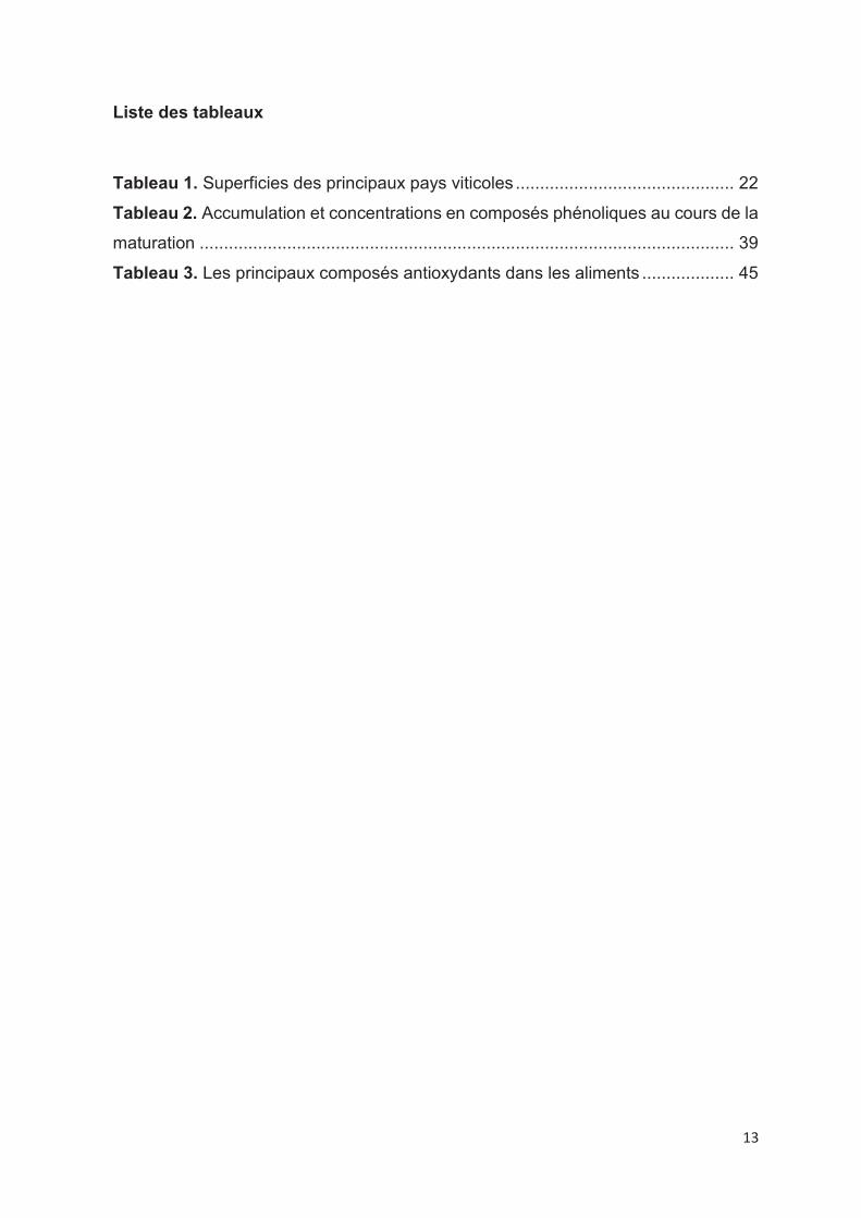
Dédicaces .................................................................................................................. 2
Résumé ..................................................................................................................... 7
Abstract ..................................................................................................................... 8
Remerciements ......................................................................................................... 9
Valorisation des travaux de recherche : .................................................................. 11
Liste des figures ...................................................................................................... 12
Liste des tableaux.................................................................................................... 13
Liste des abréviations .............................................................................................. 14
Introduction générale ............................................................................................... 17
Chapitre 1 : Bibliographie ....................................................................................... 20
1. Le raisin au cours de la maturation ................................................................ 21
1.1. Le raisin .................................................................................................... 21
1.2. Le développement de la baie du raisin ...................................................... 23
2. Les polyphénols du raisin………………………………………………………… 25
2.1. Biosynthèse générale, familles de composés (flavonoïdes/non-flavonoïdes)
…………………………………………………………………………………….26
2.1.1. Les flavonoïdes ............................................................................................. 28
2.1.1.1. Les flavanols ................................................................................ 28
2.1.1.2. Les anthocyanes .......................................................................... 29
2.1.1.3. Les flavanones ............................................................................. 30
2.1.1.4. Les flavonols ................................................................................ 31
2.1.1.5. Les flavones ................................................................................. 31
2.1.1.6. Les isoflavones............................................................................. 32
2.1.2. Les non- flavonoïdes ..................................................................................... 32
4
2.1.2.1. Les stilbènes ................................................................................ 32
2.1.2.2. Les acides phénols ....................................................................... 34
2.2. Accumulation et concentration en composés phénoliques au cours de la
maturation du raisin rouge................................................................................... 35
2.2.1. Les polyphénols totaux .................................................................................. 35
2.2.2. Les flavanols .................................................................................................. 35
2.2.3. Les anthocyanes ........................................................................................... 36
2.2.4. Les stilbènes .................................................................................................. 37
2.2.5. Les flavonols .................................................................................................. 37
3. Les activités biologiques du raisin ................................................................. 42
3.1. L’activité antioxydante ............................................................................... 42
3.1.1. Espèces réactives oxygénées et stress oxydant ........................................... 42
3.1.2. Antioxydants .................................................................................................. 44
3.1.3. Détermination de la capacité antioxydante .................................................... 47
3.1.3.1. Tests spectrophotométriques ....................................................... 47
3.1.3.2. Tests électrochimiques ................................................................. 50
3.2. L’activité anti-inflammatoire ....................................................................... 50
3.2.1. Inflammation .................................................................................................. 50
3.2.2. Marqueurs de l’inflammation .......................................................................... 51
3.2.3. Complications de l’inflammation .................................................................... 52
3.3. L’activité antidiabétique ............................................................................. 53
3.3.1. Physiopathologie du diabète .......................................................................... 53
3.3.2. Diagnostiques, symptômes et complications du diabète ............................... 56
3.3.3. Traitements pharmacologiques ..................................................................... 58
3.3.4. Traitements par les polyphénols .................................................................... 60
References .............................................................................................................. 61
Chapitre 2: Les stilbènes du raisin et du vin et leurs propriétés anti-obésité .......... 88

5
Background ............................................................................................................. 90
Scope an d Approach .............................................................................................. 90
Key Findings and Conclusions ................................................................................ 90
Abbreviation list ....................................................................................................... 91
Highlights ................................................................................................................. 95
Introduction .............................................................................................................. 95
Stilbenes content in grape berries and wine .................................................. ……104
Grape berries stilbenes ..................................................................................... 104
Stage of ripening ..................................................................................................... 107
Grape varieties and species .................................................................................... 107
External stimuli ........................................................................................................ 108
Wine stilbenes ................................................................................................... 109
Duration of maceration ............................................................................................ 112
Yeast activities ......................................................................................................... 112
Winemaking processes and wine ageing ................................................................ 113
Analytical methods for stilbenes analysis in grapes and wines ............................. 113
Capillary electrophoresis ................................................................................... 115
Gas chromatography ......................................................................................... 115
Liquid Chromatography ..................................................................................... 115
Stilbenes, in vitro and in vivo anti-obesity effects and molecular mechanisms ...... 120
Anti-obesity in vitro effects of stilbenes ............................................................. 122
Anti-obesity in vivo effects of stilbenes .............................................................. 136
Conclusion ............................................................................................................. 149
References ............................................................................................................ 151
Chapitre 3: Propriétés antioxydantes des extraits de pépins et de pellicules de trois
cépages rouges au cours de la maturité ................................................................ 170
6
Chapitre 4: Propriétés antioxydantes et anti-inflammatoires des extraits de pellicules
de 3 cépages rouges au cours de la maturation .................................................... 192
Chapitre 5: L’inhibition de l’enzyme alpha-glucosidase par des extraits de pépins et
de pellicules de Tannat au cours de la maturité .................................................... 222
Conclusion générale .............................................................................................. 243
Perspectives .......................................................................................................... 255
Résumé ................................................................................................................. 256
Abstract ................................................................................................................. 257

7
Résumé
Les polyphénols appartiennent à la famille des métabolites secondaires présents
dans les plantes et majoritairement dans les baies de raisin. Ils jouent un rôle
important dans la protection de la plante contre les stress biotiques et abiotiques. Ils
ont un impact sur la qualité organoleptique des certains aliments comme ceux
provenant du raisin et sont connus majoritairement pour leurs rôles bénéfiques pour
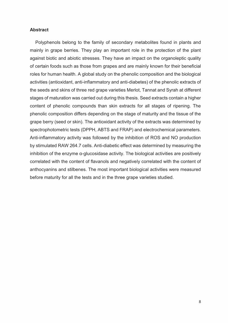
la santé humaine. Une étude globale sur la composition phénolique et les activités
biologiques (antioxydante, anti-inflammatoire et anti-diabète) d’extraits phénoliques
de pépins et pellicules de trois cépages rouges Merlot, Tannat et Syrah à différents
stades de maturité a été réalisée durant cette thèse. Les extraits de pépins ont une
teneur plus élevée en composés phénoliques que les extraits de pellicules pour tous
les stades de maturité. La composition phénolique diffère selon le stade de maturité
et le tissu de la baie de raisin (pépin ou pellicule). L’activité antioxydante des extraits
a été déterminée par des tests spectrophotométriques (DPPH, ABTS et FRAP) et
des paramètres électrochimiques. L’activité anti-inflammatoire a été suivie par
l’inhibition de la production des EROs et NO par les cellules RAW 264.7. L’activité
anti-diabétique a été déterminée par la mesure d’inhibition de l’enzyme α-
glucosidase. Les activités biologiques sont positivement corrélées avec la teneur en
flavanols et négativement corrélées avec la teneur en anthocyanes et stilbènes. Les
activités biologiques les plus importantes ont été mesurées avant la maturité pour
tous les tests et dans les trois cépages étudiés.
8
Abstract
Polyphenols belong to the family of secondary metabolites found in plants and
mainly in grape berries. They play an important role in the protection of the plant
against biotic and abiotic stresses. They have an impact on the organoleptic quality
of certain foods such as those from grapes and are mainly known for their beneficial
roles for human health. A global study on the phenolic composition and the biological
activities (antioxidant, anti-inflammatory and anti-diabetes) of the phenolic extracts of
the seeds and skins of three red grape varieties Merlot, Tannat and Syrah at different
stages of maturation was carried out during this thesis. Seed extracts contain a higher
content of phenolic compounds than skin extracts for all stages of ripening. The
phenolic composition differs depending on the stage of maturity and the tissue of the
grape berry (seed or skin). The antioxidant activity of the extracts was determined by
spectrophotometric tests (DPPH, ABTS and FRAP) and electrochemical parameters.
Anti-inflammatory activity was followed by the inhibition of ROS and NO production
by stimulated RAW 264.7 cells. Anti-diabetic effect was determined by measuring the
inhibition of the enzyme α-glucosidase activity. The biological activities are positively
correlated with the content of flavanols and negatively correlated with the content of
anthocyanins and stilbenes. The most important biological activities were measured
before maturity for all the tests and in the three grape varieties studied.

9
Remerciements
Ce travail a été réalisé au laboratoire d’œnologie de la Faculté de Pharmacie et à
INRAe de Montpellier (UMR-SPO) sous la co-direction de Monsieur le Professeur
Cédric Saucier. Je le remercie pour son accueil au laboratoire, son aide, sa
gentillesse et sa supervision.
Je tiens à remercier le Professeur Tristan Richard qui a accepté de co-diriger cette
thèse, pour son accueil au labo ISVV de Bordeaux, son sourire, sa modestie et son
intérêt pour ce travail.
Je remercie infiniment le Maitre de conférences François Garcia pour ses
encouragements, pour son aide tout au long de ce travail, sa gentillesse et sa
patience.
Mes remerciements vont également à l’ensemble des membres du jury pour
leur collaboration à l’examen de ce travail et leur participation à la soutenance.
J’adresse mes sincères remerciements à Auriane Dudoit, Gaëlle Coussot, Ruth
Hornedo Hortega, Josep Valls Fonayet et Stéphanie Krisa pour avoir participé à
l’encadrement de cette thèse. Merci à eux pour leurs disponibilités, leurs bonnes
humeurs car ce travail n’aurait pas abouti sans leurs aides.
Ce travail n’aurait pas abouti sans l’aide précieuse du stagiaire Antoine Cunin. Je
suis extrêmement reconnaissante à toi.
Un grand merci à Mélodie, Stacy, Marie, Sarah et Thibaut, de m’avoir supportée
et tant aidée au cours de ces années au laboratoire. Vos présences, vos aides, votre
bonne humeur, votre sympathie et votre soutien moral m’ont été précieux. Merci mes
chers.
Mes remerciements vont aussi à tous les membres du laboratoire d’œnologie, de
chimie analytique, à l’équipe BIO et à Cynthia, notre ancienne secrétaire, merci pour
tous les bons moments que nous avons passés ensemble.
Je tiens à remercier le ministère de la recherche et de l’enseignement
supérieur et le gouvernement algérien qui ont soutenu financièrement ce travail.

10
Mes remerciements s’adressent aussi à tous mes collègues et amies de la promo
de 2017.
Je dédie ce travail à toute la famille ‘Benbouguerra’ et ‘Farhati’

11
Valorisation des travaux de recherche :
Publications
1. Benbouguerra, N., Richard, T., Saucier, C., & Garcia, F. (2020). Voltammetric
Behavior, Flavanol and Anthocyanin Contents, and Antioxidant Capacity of Grape
Skins and Seeds during Ripening (Vitis vinifera var. Merlot, Tannat, and Syrah).
Antioxidants, 9(9), 800.
2. Dudoit, A., Benbouguerra, N., Richard, T., Hornedo-Ortega, R., Valls-Fonayet, J.,
Coussot, G., & Saucier, C. (2020). α-Glucosidase Inhibitory Activity of Tannat
Grape Phenolic Extracts in Relation to Their Ripening Stages. Biomolecules,
10(8), 1088.
3. Nawel Benbouguerra, Ruth Hornedo-Ortega, François Garcia, Toni El Khawand,
Cédric Saucier, et Tristan Richard. 2020. « Stilbenes in grape berries and wine
and their potential role as anti-obesity agents: a review », 63. Soumis (Avril 2020)
dans le journal Trends in Food and Technology.
4. Nawel Benbouguerra, Ruth Hornedo-Ortega, Josep Valls-Fonayet, Stéphanie
Krisa, François Garcia, Cédric Saucier et Tristan Richard. 2020. « Polyphenolic
characterization of Merlot, Tannat and Syrah skin extracts at different degrees of
maturity and anti-inflammatory potential in RAW 264.7 cells», 19. Soumis
(Septembre 2020) dans le journal Agricultural and Food Chemistry.
Communications à des congrès internationaux
1. N. Benbouguerra, M. Zerbib, R. Hornedo-Ortega, T. Richard, F. Garcia, C.
Saucier, “Evolution of antioxidant properties of seeds and skins extracts during
ripening of Vitis vinifera berries” oenoivas, Bordeaux, 25-28 Juin 2019.
Poster
2. N. Benbouguerra, M. Zerbib, R. Hornedo-Ortega, T. Richard, C. Saucier, F.
Garcia “Electrochemical characterization of grape extracts of seeds and skins
during ripening of Vitis vinifera grapes for antioxidant properties
determination”, oenoivas, Bordeaux, 25-28 Juin 2019. Poster
12
Liste des figures
Figure 1. Production mondiale du raisin en 2018................................................... 21
Figure 2. Structure d'une baie du raisin ................................................................. 22
Figure 3. Diagramme montrant la taille et la couleur relative des baies au cours de
la maturation ........................................................................................................... 24
Figure 4. La voie biosynthétique des principaux composés phénoliques du raisin. 27
Figure 5. Structure des principaux flavonoïdes ...................................................... 28
Figure 6. Les monomères de flavanols présents dans le raisin ............................. 29
Figure 7. Structures des anthocyanes les plus présentes dans le raisin ............... 30
Figure 8. Structures des flavanones présents dans le raisin ................................. 30
Figure 9. Structures des flavonols présents dans le raisin ..................................... 31
Figure 10. Structures des flavones présents dans le raisin ................................... 32
Figure 11. Structures des isoflavones présents dans le raisin ............................... 32
Figure 12. Les modifications les plus courantes des stilbènes végétaux ............... 33
Figure 13. Structures chimiques des acides hydroxycinnamiques ........................ 34
Figure 14. Structure des acides hydroxybenzoïques ............................................. 35
Figure 15. Illustration simplifiée des réactions impliquées dans la formation et
l'élimination des ERO. ............................................................................................ 43
Figure 16. Chronologie des principaux développements de la recherche sur les
antioxydants alimentaires ....................................................................................... 47
Figure 17. Stimulation des macrophages RAW 264.7 par les LPS ........................ 52
Figure 18. Troubles liés à l'inflammation................................................................ 53
Figure 19. Estimations de la prévalence mondiale du diabète ............................... 54
Figure 20. Physiopathologie du diabète de type 1 ................................................. 55
Figure 21. Physiopathologie du diabète de type 2 ................................................. 56
Figure 22. Critères de diagnostic du diabète ......................................................... 57
Figure 23. Réduction de la glycémie par l'inhibition de l'alpha glucosidase ........... 59
13
Liste des tableaux
Tableau 1. Superficies des principaux pays viticoles ............................................. 22
Tableau 2. Accumulation et concentrations en composés phénoliques au cours de la
maturation .............................................................................................................. 39
Tableau 3. Les principaux composés antioxydants dans les aliments ................... 45

14
Liste des abréviations
ABTS : l’acide 2,2’-azino-bis (3-éthylbenzthiazoline-6-sulfonique)
ADN : acide désoxyribonucléique
AINS : anti-inflammatoires non stéroïdiens
ANR : anthocyanidine réductase
C : catéchine
CAT : catalase
CHI : chalcone isomérase
CHS : chalcone synthase
COX-2 : cyclooxygénase-2
DFR : dihydroflavonol réductase
DPm : degré de polymérisation moyen
DPPH : 1,1 -diphényl-2-picrylhydrazyle
EAG : équivalent acide gallique
EC : épicatéchine
EC : équivalent catéchine
ECG : épicatéchine gallate
EM3G : équivalent malvidine-3-O-glucoside
ERAs : espèces réactives de l’azote
ERO : espèces réactives de l’oxygène
F3’5’H : flavonoïde 3’5’-hydroxylase
F3’H : flavonoïde 3’ –hydroxylase
F3H : flavanone 3-hydroxylase
FGT : flavonoïde 3-glucosyltransférase
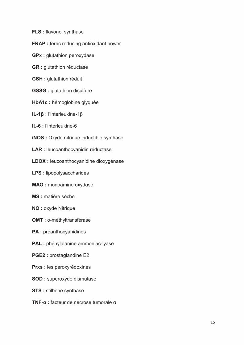
15
FLS : flavonol synthase
FRAP : ferric reducing antioxidant power
GPx : glutathion peroxydase
GR : glutathion réductase
GSH : glutathion réduit
GSSG : glutathion disulfure
HbA1c : hémoglobine glyquée
IL-1β : l’interleukine-1β
IL-6 : l’interleukine-6
iNOS : Oxyde nitrique inductible synthase
LAR : leucoanthocyanidin réductase
LDOX : leucoanthocyanidine dioxygénase
LPS : lipopolysaccharides
MAO : monoamine oxydase
MS : matière sèche
NO : oxyde Nitrique
OMT : o-méthyltransférase
PA : proanthocyanidines
PAL : phénylalanine ammoniac-lyase
PGE2 : prostaglandine E2
Prxs : les peroxyrédoxines
SOD : superoxyde dismutase
STS : stilbène synthase
TNF-α : facteur de nécrose tumorale α

16
UV : ultraviolet
17
Introduction générale
18
Vitis vinifera est l’espèce de vigne la plus cultivée dans le monde (Ky et al. 2014;
Ky et Teissedre 2015; Yilmaz et al. 2015; Tkacz et al. 2019) avec une production
mondiale de 78 millions tonnes de raisin en 2018 (http://www.oiv.int/en/oiv-life/oiv-
2019-report-on-the-world-vitivinicultural-situation). Les baies de raisin sont utilisées
comme fruits frais, produits secs ou pour la production de vin (Liang et al. 2011). En
plus de leur importance économique, un nombre croissant d'avantages pour la santé
ont été attribués aux raisins dus à la présence de composés phénoliques dans les
différents tissus de la baie (Ali et al. 2011; Pandey et Rizvi 2009), principalement
dans les pépins et les pellicules (Di Lecce et al. 2014; Ky et Teissedre 2015; Tkacz
et al. 2019; Yilmaz et al. 2015).
Les polyphénols sont les métabolites secondaires les plus abondants dans le
règne végétal (Ghani 2020; Lorrain et al. 2013). Ils constituent un groupe hétérogène
de composés chimiques comprenant des cycles phénoliques portant un ou plusieurs
groupes hydroxyles (Lorrain et al. 2013). Actuellement, il existe plus de 8000
polyphénols caractérisés et identifiés (Somerville, Bringans, et Braakhuis 2017). La
composition et la concentration des composés phénoliques dépendent d’un certain
nombre de facteurs tels que le stade de maturation (Bindon et al. 2013; Obreque-
Slier et al. 2010), le climat, la variété et le type du sol (J. Kennedy 2002; Coklar 2017).
Dans la plante, les polyphénols jouent un rôle important dans la croissance, la fertilité,
la reproduction et dans les réactions de protection de la plante contre les stress
biotiques et abiotiques (Gil-Muñoz et al. 2017). Ils possèdent aussi un large spectre
d’activités biologiques (Plaza et al. 2018).
Les polyphénols sont connus pour leur rôle antioxydant (Balík et al. 2009;
Rockenbach et al. 2011; Coklar 2017; Guendez 2005; Ky et Teissedre 2015; Lingua
et al. 2016). Ils pourraient jouer un rôle important dans la prévention des maladies
cardiovasculaires (Khurana et al. 2013; Amiot, Riollet, et Landrier 2009),
neurodégénératives (Amiot, Riollet, et Landrier 2009; Richard et al. 2014), le diabète
(Kadouh et al. 2016; Sales et al. 2012), l’inflammation (de plusieurs pathologies, telles
que la polyarthrite rhumatoïde, l'inflammation des maladies intestinales et
l'athérosclérose) (H. J. Lee, Kwon, et Kim 2018; Bak et al. 2013; Cheng et al. 2014;
Du et al. 2018) et l’obésité (H. J. Lee, Kwon, et Kim 2018; Jack et al. 2019; Callcott
et al. 2018). Enfin, leurs propriétés sont fortement liées à leurs structures chimiques
(Plaza et al. 2018).
19
Les objectifs de ce travail sont de suivre l’évolution de la composition
phénolique et des activités biologiques associées (antioxydante, anti-
inflammatoire et anti-diabète) d’extraits de pépins et de pellicules au cours de
la maturation de la baie de raisin.
Il est à noter que ce manuscrit est principalement rédigé sous forme d’articles.
Le premier chapitre expose la partie bibliographique concernant l’espèce Vitis
vinifera, la biosynthèse et la structure des polyphénols de raisin, l’évolution de la
composition phénolique (flavonols, stilbènes et anthocyanes) au cours de la
maturation, ainsi leurs activités biologiques (activités antioxydante, anti-inflammatoire
et anti-diabète).
Le deuxième chapitre est une revue bibliographique concernant les stilbènes du
raisin et du vin et leurs effets comme agents de prévention de l’obésité.
Le troisième chapitre présente les travaux concernant l’activité antioxydante
d’extraits phénoliques de pépins et de pellicules de trois cépages rouges (Merlot,
Tannat et Syrah) au cours de la maturation. Des tests spectrophotométriques et
électrochimiques ont été utilisés pour mesurer l’activité antioxydante. Le lien entre la
composition polyphénolique et les activités mesurées a été étudié.
Le quatrième chapitre présente les résultats concernant l’activité anti-
inflammatoire des extraits de pellicules de trois cépages rouges au cours de la
maturation et le lien avec la composition phénolique.
Le cinquième chapitre traite l’inhibition de l’enzyme α-glucosidase par les extraits
de pépins et de pellicules de Tannat au cours de la maturation en lien avec leurs
compositions phénoliques.
La rédaction de ce manuscrit se terminera par une conclusion générale et des
perspectives de travaux.
20
Chapitre 1 : Bibliographie

21
1. Le raisin au cours de la maturation 1.1. Le raisin
Le raisin est l’une des cultures fruitières les plus importantes au monde (Liang et
al. 2012; Bozan, Tosun, et Özcan 2008) avec production d’environ 78 millions de
tonnes en 2018 (Figure 1) sur 7.4 millions d’hectares de terres dédiées à sa culture
(http://www.oiv.int/fr/vie-de-loiv/bilan-de-loiv-2019-sur-la-situation-vitivinicole-
mondiale).
Figure 1. Production mondiale du raisin en 2018
Le raisin est produit par les plantes du genre Vitis qui regroupe environ 60 espèces
de vignes. Vitis vinifera est l’espèce la plus cultivée et représente plus de 90% des
baies de raisin sur le marché (Venkitasamy et al. 2019). Les baies sont consommées
sous forme de fruits frais ou d’autres produits transformés, notamment le jus de
raisin, la confiture, le vin, les raisins secs le vinaigre et l’huile de pépins de raisin
(Venkitasamy et al. 2019). Les baies de raisin contiennent trois principaux types de
tissus : la pellicule, les pépins et la pulpe (Figure 2) (Adams 2006; J. Kennedy 2002).
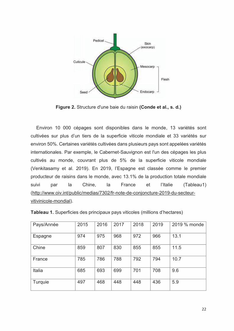
22
Figure 2. Structure d'une baie du raisin (Conde et al., s. d.)
Environ 10 000 cépages sont disponibles dans le monde, 13 variétés sont
cultivées sur plus d’un tiers de la superficie viticole mondiale et 33 variétés sur
environ 50%. Certaines variétés cultivées dans plusieurs pays sont appelées variétés
internationales. Par exemple, le Cabernet-Sauvignon est l'un des cépages les plus
cultivés au monde, couvrant plus de 5% de la superficie viticole mondiale
(Venkitasamy et al. 2019). En 2019, l’Espagne est classée comme le premier
producteur de raisins dans le monde, avec 13.1% de la production totale mondiale
suivi par la Chine, la France et l’Italie (Tableau1)
(http://www.oiv.int/public/medias/7302/fr-note-de-conjoncture-2019-du-secteur-
vitivinicole-mondial).
Tableau 1. Superficies des principaux pays viticoles (millions d’hectares)
Pays/Année 2015 2016 2017 2018 2019 2019 % monde
Espagne 974 975 968 972 966 13.1
Chine 859 807 830 855 855 11.5
France 785 786 788 792 794 10.7
Italia 685 693 699 701 708 9.6
Turquie 497 468 448 448 436 5.9

23
Etats-Unis 446 439 434 408 408 5.5
Argentine 225 224 222 218 215 2.9
Chili 214 209 207 203 200 2.7
Portugal 204 195 194 192 195 2.6
Roumanie 191 191 191 191 191 2.6
Iran 217 168 153 177 177 2.4
Inde 129 131 147 149 149 2
Australie 147 145 145 146 146 2
Afrique de Sud 133 130 128 123 122 1.7
Grèce 107 105 106 106 106 1.4
Allemagne 103 102 103 103 103 1.4
Russie 85 89 90 93 95 1.3
1.2. Le développement de la baie du raisin
L’évolution dans le temps et la croissance des raisins Vitis Vinifera est bien décrit
dans la littérature (J. Kennedy 2002; Adams 2006; Conde et al., s. d.). Les baies de
raisin subissent une complexe série de changements physiques et chimiques au
cours de leur développement qui peut être divisé en deux phases de croissance
sigmoïdales séparées par une phase de latence (Figure 3) (Fortes et Pais 2016; J.
Kennedy 2002; Deluc et al. 2007; B. G. Coombe 1992; Coombe et McCARTHY 2000;
Dokoozlian et Kliewer 1996).
La première phase : elle consiste à la formation des embryons de pépins et de
péricarpe. Cette étape est caractérisée par une croissance exponentielle rapide de
la baie qui se dilate en volume du fait de la multiplication cellulaire. La biosynthèse
des tanins et des acides hydroxycinnamiques et l’accumulation des acides
organiques comme le tartrate et le malate débutent à cette période (Fortes et Pais
2016; Conde et al., s. d.; Deluc et al. 2007).

24
La deuxième phase : c’est une phase de transition au cours de laquelle il n’y a pas
d’augmentation de la taille des baies. La véraison marque le début de la troisième
phase par l’initiation du développement de la couleur dû à l’accumulation des
anthocyanes dans le raisin rouge (Fortes et Pais 2016; Deluc et al. 2007).
La troisième phase : la maturation, implique une croissance supplémentaire des
baies en raison de l’expansion cellulaire. Il y a également une diminution de la teneur
en malate et une accumulation des sucres (principalement glucose et fructose) ainsi
que des anthocyanes (Fortes et Pais 2016). Les baies passent d’un état où elles sont
petites, dures at acides à un état où elles sont plus grosses, plus sucrés, moins acides
et fortement colorées et aromatiques (Conde et al., s. d.).
Figure 3. Diagramme montrant la taille et la couleur relative des baies au cours de
la maturation (J. Kennedy 2002)

25
2. Les polyphénols du raisin
Les polyphénols sont les métabolites secondaires les plus abondants du règne
végétal (Ghani 2020; Lorrain et al. 2013; Kundu, Talukder, et Sen Raychaudhuri
2019). Ils sont présents en plus grande quantité dans les raisins après les glucides
et les acides (Nawaz et al. 2006) et sont localisés principalement dans les pépins et
les pellicules (Pantelić et al. 2016; Fanzone et al. 2011; Giuffrè 2013; J. A. Kennedy
et al. 2001; Obreque-Slier et al. 2013). Les polyphénols ont une structure commune
possédant un noyau aromatique avec un ou plusieurs groupements hydroxyles
(Quideau et al. 2011; Kardum et Glibetic 2018; Ghani 2020). Il existe environ 8000
composés (Somerville, Bringans, et Braakhuis 2017; J. Santos et al. 2014;
Chandrasekara et Shahidi 2018) solubles dans l'eau et dans les solvants organiques
(Ashraf et al. 2018). On distingue deux grandes familles: les flavonoïdes et les non
flavonoïdes (Tkacz et al. 2019; Obreque-Slier et al. 2013).
Les polyphénols sont synthétisés à travers la voie des phénylpropanoïdes (Adams
2006), en réponse à des stimuli externes (Cavaliere et al. 2008; Versari et al. 2001)
et ils ont diverses activités (Bak et al. 2013; Cheng et al. 2014). Ils protègent les
plantes contre les facteurs de stress biotiques et abiotiques (Błaszczyk, Sady, et
Sielicka 2019; Gil-Muñoz et al. 2017). La plupart de ces métabolites sont
responsables des propriétés organoleptiques et qualitatives des aliments provenant
de ces plantes (del Llaudy et al. 2008; Asproudi et al. 2015; Rodríguez Montealegre
et al. 2006). Ces composés sont connus pour leurs impacts bénéfiques sur la santé
et par leurs nombreuses activités biologiques cardioprotective (Khurana et al. 2013;
Amiot, Riollet, et Landrier 2009), anti-inflammatoire (H. J. Lee, Kwon, et Kim 2018;
Bak et al. 2013; Cheng et al. 2014; Du et al. 2018; Terra et al. 2007), anti-diabète
(Kadouh et al. 2016; Sales et al. 2012) et principalement antioxydante (Balík et al.
2009; Aguirre et al. 2010; Rockenbach et al. 2011; Bozan, Tosun, et Özcan 2008).
26
2.1. Biosynthèse générale, familles de composés (flavonoïdes/non-
flavonoïdes)
La biosynthèse des phénylpropanoïdes commence habituellement par l’acide
aminée phénylalanine issue de la voie du shikimate (Conde et al., s. d.; Kundu,
Talukder, et Sen Raychaudhuri 2019). Cette voie est responsable de la production
de la phénylalanine et d’autres acides aminés aromatiques comme la tyrosine, le
tryptophane (Conde et al., s. d.), l'acide gallique et l'acide cinnamique (Fatland et al.
2002). La première enzyme responsable de la synthèse phénolique est la PAL
(phényl ammoniac lysase) qui convertit la phénylalanine en acide trans-cinnamique
(Conde et al., s. d.; Kundu, Talukder, et Sen Raychaudhuri 2019). Ce composé subit
une série de transformations entraînant la formation de précurseurs de plusieurs
composés phénoliques simples, comme les acides phénols. L'incorporation de
malonyl-CoA avec le coumaroyl-CoA donne la naringénine chalcone par l'enzyme
chalcone synthase (CHS). La chalcone est isomérisée par la chalcone flavanone
isomérase (CHI) en une flavanone ; l’intermédiaire de base pour les autres classes
et sous-classes de flavonoïdes par l’intervention des différentes enzymes (Figure 4)
(Bohm 1998; Tsao, Khanizadeh, et Dale 2006; Tsao 2010). Dans le cas des stilbènes
c’est l’enzyme stilbène synthase (STS) qui intervient (Chong, Poutaraud, et
Hugueney 2009).
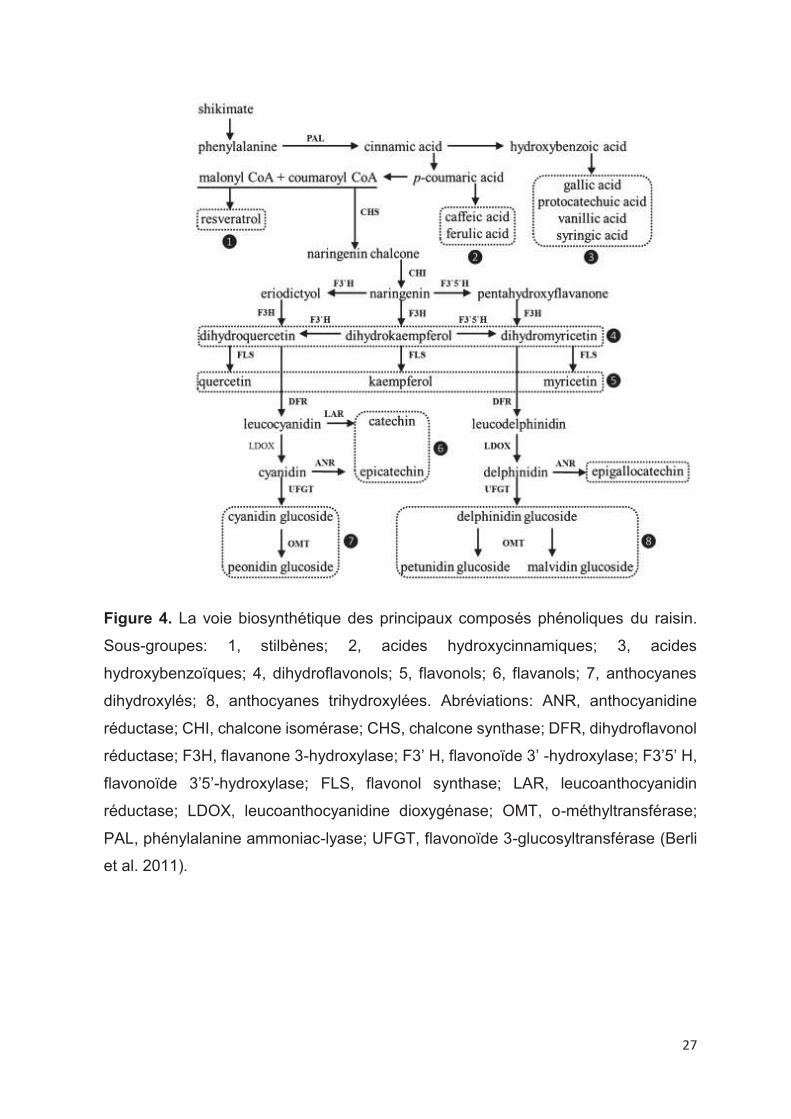
27
Figure 4. La voie biosynthétique des principaux composés phénoliques du raisin.
Sous-groupes: 1, stilbènes; 2, acides hydroxycinnamiques; 3, acides
hydroxybenzoïques; 4, dihydroflavonols; 5, flavonols; 6, flavanols; 7, anthocyanes
dihydroxylés; 8, anthocyanes trihydroxylées. Abréviations: ANR, anthocyanidine
réductase; CHI, chalcone isomérase; CHS, chalcone synthase; DFR, dihydroflavonol
réductase; F3H, flavanone 3-hydroxylase; F3’ H, flavonoïde 3’ -hydroxylase; F3’5’ H,
flavonoïde 3’5’-hydroxylase; FLS, flavonol synthase; LAR, leucoanthocyanidin
réductase; LDOX, leucoanthocyanidine dioxygénase; OMT, o-méthyltransférase;
PAL, phénylalanine ammoniac-lyase; UFGT, flavonoïde 3-glucosyltransférase (Berli
et al. 2011).
28
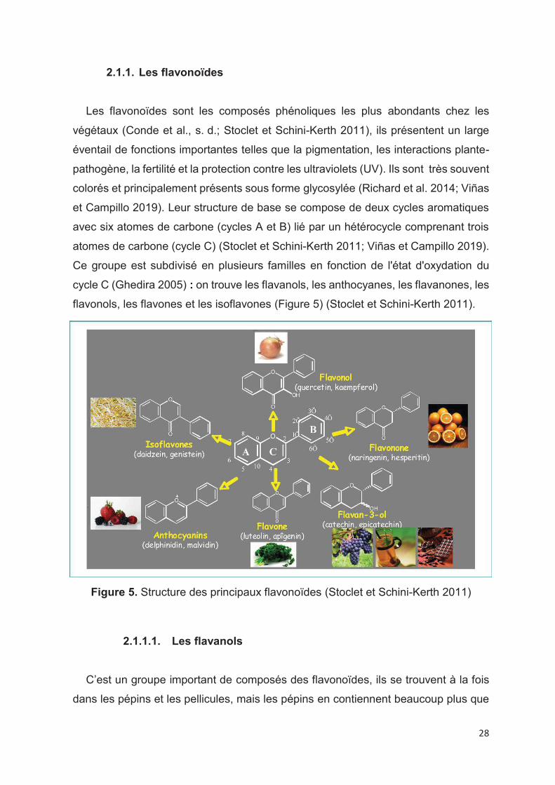
2.1.1. Les flavonoïdes
Les flavonoïdes sont les composés phénoliques les plus abondants chez les
végétaux (Conde et al., s. d.; Stoclet et Schini-Kerth 2011), ils présentent un large
éventail de fonctions importantes telles que la pigmentation, les interactions plante-
pathogène, la fertilité et la protection contre les ultraviolets (UV). Ils sont très souvent
colorés et principalement présents sous forme glycosylée (Richard et al. 2014; Viñas
et Campillo 2019). Leur structure de base se compose de deux cycles aromatiques
avec six atomes de carbone (cycles A et B) lié par un hétérocycle comprenant trois
atomes de carbone (cycle C) (Stoclet et Schini-Kerth 2011; Viñas et Campillo 2019).
Ce groupe est subdivisé en plusieurs familles en fonction de l'état d'oxydation du
cycle C (Ghedira 2005) : on trouve les flavanols, les anthocyanes, les flavanones, les
flavonols, les flavones et les isoflavones (Figure 5) (Stoclet et Schini-Kerth 2011).
Figure 5. Structure des principaux flavonoïdes (Stoclet et Schini-Kerth 2011)
2.1.1.1. Les flavanols
C’est un groupe important de composés des flavonoïdes, ils se trouvent à la fois
dans les pépins et les pellicules, mais les pépins en contiennent beaucoup plus que
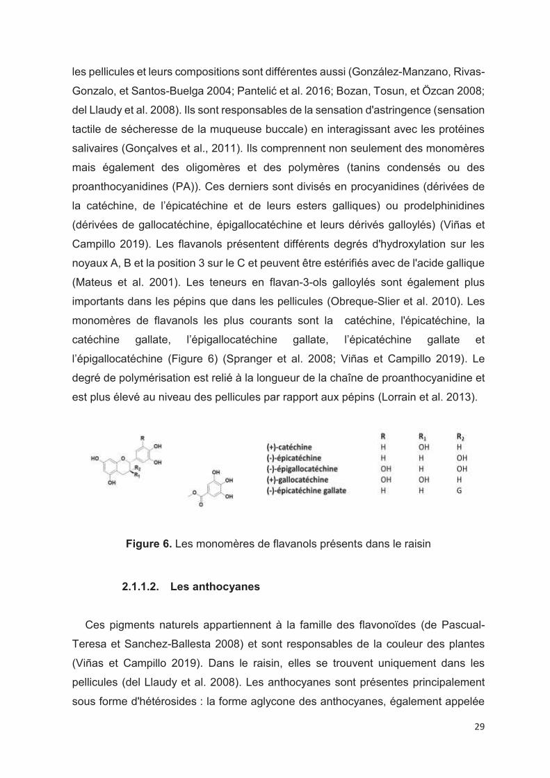
29
les pellicules et leurs compositions sont différentes aussi (González-Manzano, Rivas-
Gonzalo, et Santos-Buelga 2004; Pantelić et al. 2016; Bozan, Tosun, et Özcan 2008;
del Llaudy et al. 2008). Ils sont responsables de la sensation d'astringence (sensation
tactile de sécheresse de la muqueuse buccale) en interagissant avec les protéines
salivaires (Gonçalves et al., 2011). Ils comprennent non seulement des monomères
mais également des oligomères et des polymères (tanins condensés ou des
proanthocyanidines (PA)). Ces derniers sont divisés en procyanidines (dérivées de
la catéchine, de l’épicatéchine et de leurs esters galliques) ou prodelphinidines
(dérivées de gallocatéchine, épigallocatéchine et leurs dérivés galloylés) (Viñas et
Campillo 2019). Les flavanols présentent différents degrés d'hydroxylation sur les
noyaux A, B et la position 3 sur le C et peuvent être estérifiés avec de l'acide gallique
(Mateus et al. 2001). Les teneurs en flavan-3-ols galloylés sont également plus
importants dans les pépins que dans les pellicules (Obreque-Slier et al. 2010). Les
monomères de flavanols les plus courants sont la catéchine, l'épicatéchine, la
catéchine gallate, l’épigallocatéchine gallate, l’épicatéchine gallate et
l’épigallocatéchine (Figure 6) (Spranger et al. 2008; Viñas et Campillo 2019). Le
degré de polymérisation est relié à la longueur de la chaîne de proanthocyanidine et
est plus élevé au niveau des pellicules par rapport aux pépins (Lorrain et al. 2013).
Figure 6. Les monomères de flavanols présents dans le raisin
2.1.1.2. Les anthocyanes
Ces pigments naturels appartiennent à la famille des flavonoïdes (de Pascual-
Teresa et Sanchez-Ballesta 2008) et sont responsables de la couleur des plantes
(Viñas et Campillo 2019). Dans le raisin, elles se trouvent uniquement dans les
pellicules (del Llaudy et al. 2008). Les anthocyanes sont présentes principalement
sous forme d'hétérosides : la forme aglycone des anthocyanes, également appelée
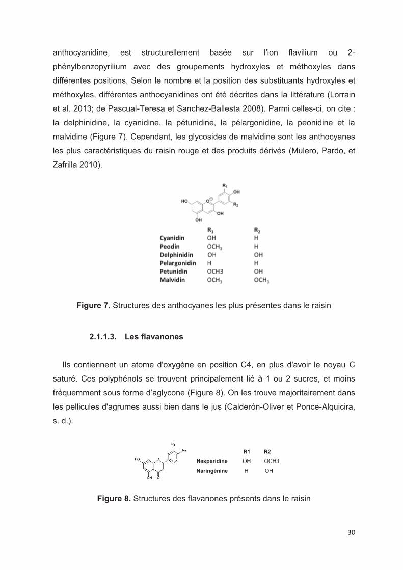
30
anthocyanidine, est structurellement basée sur l'ion flavilium ou 2-
phénylbenzopyrilium avec des groupements hydroxyles et méthoxyles dans
différentes positions. Selon le nombre et la position des substituants hydroxyles et
méthoxyles, différentes anthocyanidines ont été décrites dans la littérature (Lorrain
et al. 2013; de Pascual-Teresa et Sanchez-Ballesta 2008). Parmi celles-ci, on cite :
la delphinidine, la cyanidine, la pétunidine, la pélargonidine, la peonidine et la
malvidine (Figure 7). Cependant, les glycosides de malvidine sont les anthocyanes
les plus caractéristiques du raisin rouge et des produits dérivés (Mulero, Pardo, et
Zafrilla 2010).
Figure 7. Structures des anthocyanes les plus présentes dans le raisin
2.1.1.3. Les flavanones
Ils contiennent un atome d'oxygène en position C4, en plus d'avoir le noyau C
saturé. Ces polyphénols se trouvent principalement lié à 1 ou 2 sucres, et moins
fréquemment sous forme d’aglycone (Figure 8). On les trouve majoritairement dans
les pellicules d'agrumes aussi bien dans le jus (Calderón-Oliver et Ponce-Alquicira,
s. d.).
Figure 8. Structures des flavanones présents dans le raisin
R1 R2
Hespéridine OH OCH3
Naringénine H OH
31
2.1.1.4. Les flavonols
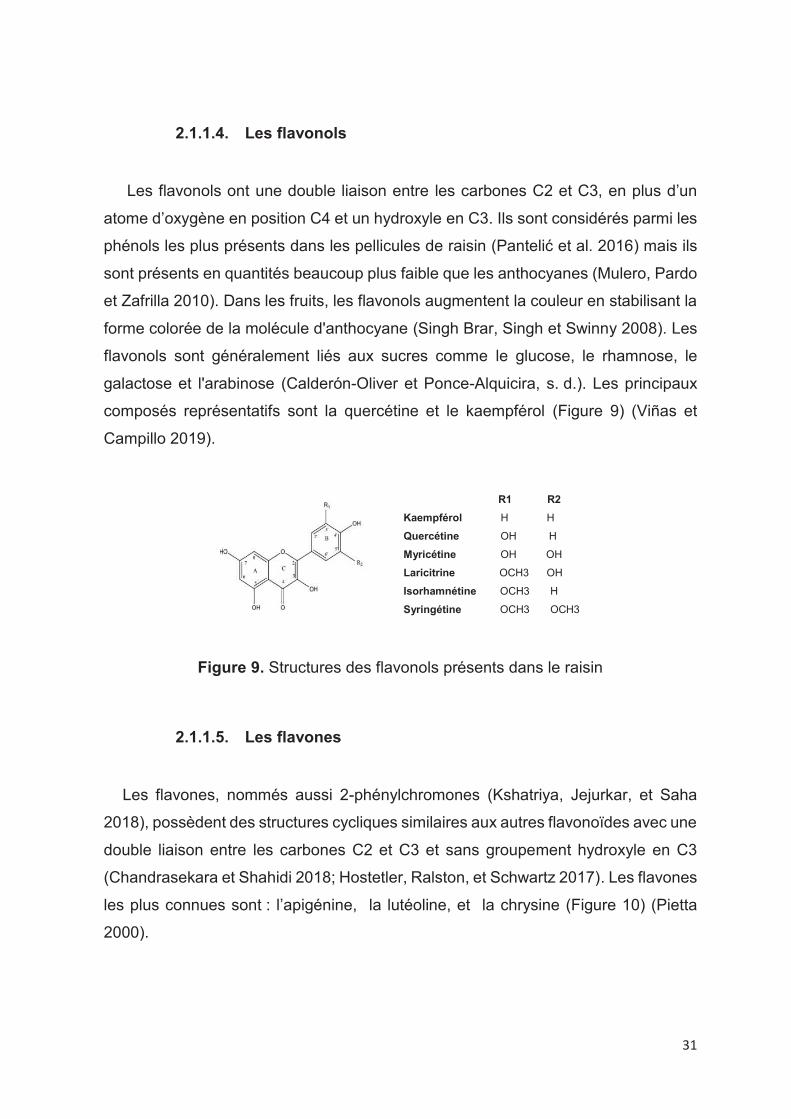
Les flavonols ont une double liaison entre les carbones C2 et C3, en plus d’un
atome d’oxygène en position C4 et un hydroxyle en C3. Ils sont considérés parmi les
phénols les plus présents dans les pellicules de raisin (Pantelić et al. 2016) mais ils
sont présents en quantités beaucoup plus faible que les anthocyanes (Mulero, Pardo
et Zafrilla 2010). Dans les fruits, les flavonols augmentent la couleur en stabilisant la
forme colorée de la molécule d'anthocyane (Singh Brar, Singh et Swinny 2008). Les
flavonols sont généralement liés aux sucres comme le glucose, le rhamnose, le
galactose et l'arabinose (Calderón-Oliver et Ponce-Alquicira, s. d.). Les principaux
composés représentatifs sont la quercétine et le kaempférol (Figure 9) (Viñas et
Campillo 2019).
Figure 9. Structures des flavonols présents dans le raisin
2.1.1.5. Les flavones
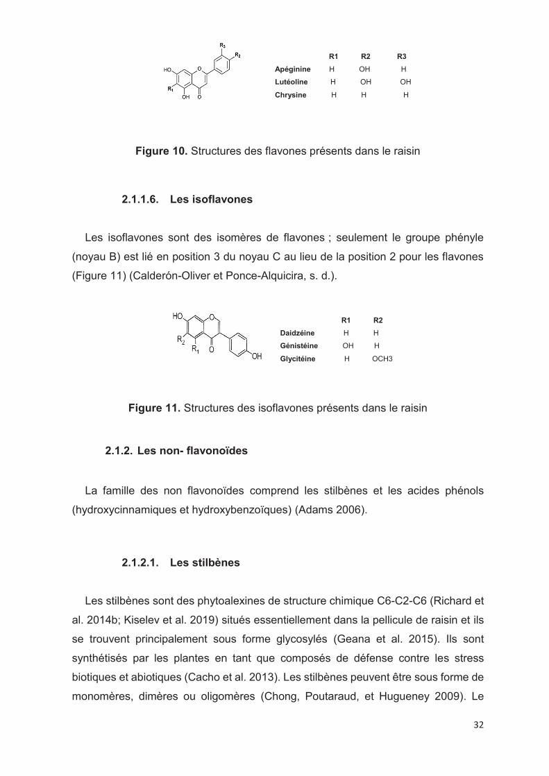
Les flavones, nommés aussi 2-phénylchromones (Kshatriya, Jejurkar, et Saha
2018), possèdent des structures cycliques similaires aux autres flavonoïdes avec une
double liaison entre les carbones C2 et C3 et sans groupement hydroxyle en C3
(Chandrasekara et Shahidi 2018; Hostetler, Ralston, et Schwartz 2017). Les flavones
les plus connues sont : l’apigénine, la lutéoline, et la chrysine (Figure 10) (Pietta
2000).
R1 R2
Kaempférol H H
Quercétine OH H
Myricétine OH OH
Laricitrine OCH3 OH
Isorhamnétine OCH3 H
Syringétine OCH3 OCH3
32
Figure 10. Structures des flavones présents dans le raisin
2.1.1.6. Les isoflavones
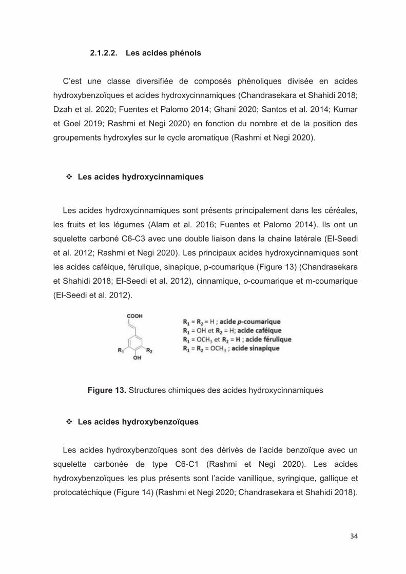
Les isoflavones sont des isomères de flavones ; seulement le groupe phényle
(noyau B) est lié en position 3 du noyau C au lieu de la position 2 pour les flavones
(Figure 11) (Calderón-Oliver et Ponce-Alquicira, s. d.).
Figure 11. Structures des isoflavones présents dans le raisin
2.1.2. Les non- flavonoïdes
La famille des non flavonoïdes comprend les stilbènes et les acides phénols
(hydroxycinnamiques et hydroxybenzoïques) (Adams 2006).
2.1.2.1. Les stilbènes
Les stilbènes sont des phytoalexines de structure chimique C6-C2-C6 (Richard et
al. 2014b; Kiselev et al. 2019) situés essentiellement dans la pellicule de raisin et ils
se trouvent principalement sous forme glycosylés (Geana et al. 2015). Ils sont
synthétisés par les plantes en tant que composés de défense contre les stress
biotiques et abiotiques (Cacho et al. 2013). Les stilbènes peuvent être sous forme de
monomères, dimères ou oligomères (Chong, Poutaraud, et Hugueney 2009). Le
R1 R2 R3
Apéginine H OH H
Lutéoline H OH OH
Chrysine H H H
R1 R2
Daidzéine H H
Génistéine OH H
Glycitéine H OCH3

33
resvératrol est le stilbène le plus connu dans le monde (Careri et al. 2003). Il subit
plusieurs modifications (glycosylation, methoxylation, oligomérisation et
isoprénylation) (Figure 12) pour donner des composés plus complexes (Chong,
Poutaraud, et Hugueney 2009). Les stilbènes monomères les plus abondants dans
les baies sont le trans-resvératrol et le trans-picéide (Błaszczyk, Sady et Sielicka
2019).
Figure 12. Les modifications les plus courantes des stilbènes végétaux (Chong,
Poutaraud, et Hugueney 2009)
34
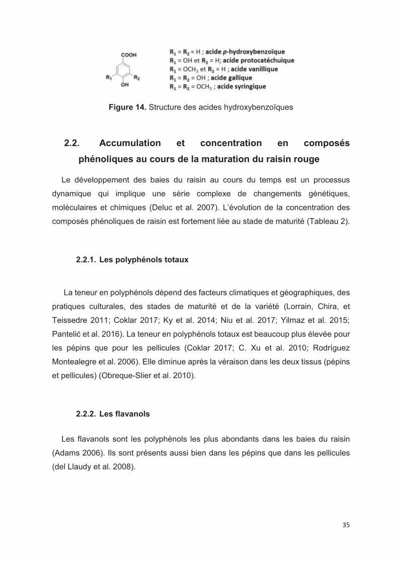
2.1.2.2. Les acides phénols
C’est une classe diversifiée de composés phénoliques divisée en acides
hydroxybenzoïques et acides hydroxycinnamiques (Chandrasekara et Shahidi 2018;
Dzah et al. 2020; Fuentes et Palomo 2014; Ghani 2020; Santos et al. 2014; Kumar
et Goel 2019; Rashmi et Negi 2020) en fonction du nombre et de la position des
groupements hydroxyles sur le cycle aromatique (Rashmi et Negi 2020).
v Les acides hydroxycinnamiques
Les acides hydroxycinnamiques sont présents principalement dans les céréales,
les fruits et les légumes (Alam et al. 2016; Fuentes et Palomo 2014). Ils ont un
squelette carboné C6-C3 avec une double liaison dans la chaine latérale (El-Seedi
et al. 2012; Rashmi et Negi 2020). Les principaux acides hydroxycinnamiques sont
les acides caféique, férulique, sinapique, p-coumarique (Figure 13) (Chandrasekara
et Shahidi 2018; El-Seedi et al. 2012), cinnamique, o-coumarique et m-coumarique
(El-Seedi et al. 2012).
Figure 13. Structures chimiques des acides hydroxycinnamiques
v Les acides hydroxybenzoïques
Les acides hydroxybenzoïques sont des dérivés de l’acide benzoïque avec un
squelette carbonée de type C6-C1 (Rashmi et Negi 2020). Les acides
hydroxybenzoïques les plus présents sont l’acide vanillique, syringique, gallique et
protocatéchique (Figure 14) (Rashmi et Negi 2020; Chandrasekara et Shahidi 2018).
35
Figure 14. Structure des acides hydroxybenzoïques
2.2. Accumulation et concentration en composés
phénoliques au cours de la maturation du raisin rouge
Le développement des baies du raisin au cours du temps est un processus
dynamique qui implique une série complexe de changements génétiques,
moléculaires et chimiques (Deluc et al. 2007). L’évolution de la concentration des
composés phénoliques de raisin est fortement liée au stade de maturité (Tableau 2).
2.2.1. Les polyphénols totaux
La teneur en polyphénols dépend des facteurs climatiques et géographiques, des
pratiques culturales, des stades de maturité et de la variété (Lorrain, Chira, et
Teissedre 2011; Coklar 2017; Ky et al. 2014; Niu et al. 2017; Yilmaz et al. 2015;
Pantelić et al. 2016). La teneur en polyphénols totaux est beaucoup plus élevée pour
les pépins que pour les pellicules (Coklar 2017; C. Xu et al. 2010; Rodríguez
Montealegre et al. 2006). Elle diminue après la véraison dans les deux tissus (pépins
et pellicules) (Obreque-Slier et al. 2010).
2.2.2. Les flavanols
Les flavanols sont les polyphénols les plus abondants dans les baies du raisin
(Adams 2006). Ils sont présents aussi bien dans les pépins que dans les pellicules
(del Llaudy et al. 2008).
36
v Les pépins : ils contiennent la catéchine (C), l’épicatéchine (EC) et
l’épicatéchine gallate (ECG) ainsi que les procyanidines qui sont des polymères de
flavanols (Ristic et Iland 2005; González-Manzano, Rivas-Gonzalo, et Santos-Buelga
2004; del Llaudy et al. 2008; Rolle et al. 2011; Cadot, Miñana Castelló, et Chevalier
2006). L’accumulation des flavanols commence au début du développement des
pépins, leur concentration est maximle à la véraison puis elle diminue à la maturité
(Downey, Harvey, et Robinson 2003; Ristic et Iland 2005; J. A. Kennedy, Matthews,
et Waterhouse 2000b; Rolle et al. 2011). La concentration des flavanols dépend de
la variété, des variétés comme le pinot noir, le grenache, et le tempranillo ont des
teneurs élevées en proanthocyanidines tandis que d’autres comme la syrah, et le
merlot ont des teneurs faibles (Ristic et Iland 2005).
v Les pellicules : elles contiennent l’épigallocatéchine et une proportion
beaucoup plus faible de l’épicatéchine-3-gallate plus les proanthocyanidines
(González-Manzano, Rivas-Gonzalo, et Santos-Buelga 2004; del Llaudy et al. 2008;
Rolle et al. 2011). La teneur en flavanols est plus élevée avant la véraison qu’après
(Asproudi et al. 2015; Lorrain, Chira, et Teissedre 2011; Obreque-Slier et al. 2010),
elle diminue lentement de la véraison à la maturité (Adams 2006). La diminution de
la teneur après la véraison est due à la déviation des métabolites intermédiaires
(cyanidines et delphinidines) vers la synthèse des anthocyanes ou à l’oxydation de
ces composés (Asproudi et al. 2015). Les proanthocyanidines présentent un degré
de polymérisation plus élevé pour les pellicules que pour les pépins (del Llaudy et al.
2008; Adams 2006).
2.2.3. Les anthocyanes
Les anthocyanes sont localisés uniquement dans les pellicules des cépages
rouges. La synthèse des anthocyanes commence à partir de la véraison, qui est
considérée comme le début de la maturation des baies (Adams 2006; Shahab et al.
2020). La malvidine -3-O- glucoside est la principal anthocyane présente dans les
raisins (Adams 2006; Lorrain, Chira, et Teissedre 2011; Fournand et al. 2006), elle
représente 44% et 55% des anthocyanes totales pour le Merlot et le Cabernet
Sauvignon, respectivement (Rodríguez Montealegre et al. 2006) suivie de la

37
delphinidine -3-O-glucoside. Les anthocyanes s'accumulent dans les pellicules des
baies de la véraison à la maturité du raisin (Fournand et al. 2006; Giuffrè 2013; J. A.
Kennedy, Matthews, et Waterhouse 2002; Esteban, Villanueva, et Lissarrague 2001;
Delgado et al. 2004). D’autres études montrent que la concentration des anthocyanes
augmente rapidement de la véraison au stade intermédiaire (entre la véraison et la
maturité) et plus lentement par la suite (Gil-Muñoz et al. 2011; Lorrain, Chira, et
Teissedre 2011). Il y a ensuite une diminution juste avant la récolte et ⁄ ou pendant la
surmaturation (Ryan et Revilla 2003; Ristic et Iland 2005; Mateus et al. 2001; Singh
Brar, Singh, et Swinny 2008; Gil-Muñoz et al. 2011; Fournand et al. 2006; Rolle et al.
2011). L’accumulation des anthocyanes est liée à différents facteurs tels que la
variété (Ryan et Revilla 2003; Gil-Muñoz et al. 2011), le climat, l’irrigation et le sol
(Ryan et Revilla 2003; del Llaudy et al. 2008; Esteban, Villanueva, et Lissarrague
2001; Pérez-Magariño et González-San José 2004).
2.2.4. Les stilbènes
Les stilbènes se trouvent principalement dans les pellicules du raisin (Gatto et al.
2008; Jeandet et al. 2002; Babazadeh et al. 2017). Ils sont considérés comme des
phytoalexines car ils sont associés à la résistance des plantes aux maladies et leur
synthèse est souvent due à une réponse aux attaques par des agents
phytopathogènes et d'autres facteurs de stress comme l'irradiation UV (Błaszczyk,
Sady, et Sielicka 2019; Jeandet et al. 2002; Gatto et al. 2008). Le stilbène le plus
important dans les raisins est le resvératrol et sa forme glycosylée, le picéide. La
concentration en stilbènes augmente au cours de la maturation (Gatto et al. 2008;
Geana et al. 2015).
2.2.5. Les flavonols
Les flavonols sont moins abondants que les flavanols et les anthocyanes. Ils se
présentent majoritairement dans le raisin sous forme glycosylés. Leur concentration
dépend de l’exposition à la lumière, les baies exposées au soleil ayant des niveaux
38
plus élevés que les baies non exposées (Adams 2006). La teneur en flavonols
augmente au cours de la maturation (Giuffrè 2013).

39
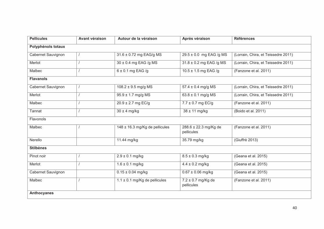
Tableau 2. Accumulation et concentrations en composés phénoliques au cours de la maturation
Pépins Avant véraison Autour de la véraison Après véraison Références
Polyphénols totaux
Cabernet Sauvignon / 56.1 ± 1.8 mg EAG /g MS 39.1 ± 2.5 mg EAG /g MS (Lorrain, Chira, et Teissedre 2011)
Merlot / 79.5 ± 0.6 mg EAG /g MS 45.3 ± 0.5 mg EAG /g MS (Lorrain, Chira, et Teissedre 2011)
Malbec / 38.3 ± 0.7 mg EAG /g 26.7 ± 2.5 mg EAG /g (Fanzone et al. 2011)
Flavanols
Cabernet Sauvignon / 94.0 ± 4.8 mg/g MS 90.1 ± 4.0 mg/g MS (Lorrain, Chira, et Teissedre 2011)
Merlot / 138.9 ± 5.3 mg/g MS 92.2 ± 4.5 mg/g MS (Lorrain, Chira, et Teissedre 2011)
Malbec / 123 ± 14.2 mg EC/g 125.2 ± 3.6 mg EC/g (Fanzone et al. 2011)
Tannat / 1576 ± 81 mg/kg 1931 ± 216 mg/kg (Boido et al. 2011)
DPm
Cabernet Sauvignon / 20.5 16.1 (Lorrain, Chira, et Teissedre 2011)
Merlot / 15 .1 11.5 (Lorrain, Chira, et Teissedre 2011)
Syrah 9.2 6 5.7 (J. A. Kennedy et al. 2000)
40
Pellicules Avant véraison Autour de la véraison Après véraison Références
Polyphénols totaux
Cabernet Sauvignon / 31.6 ± 0.72 mg EAG/g MS 29.5 ± 0.0 mg EAG /g MS (Lorrain, Chira, et Teissedre 2011)
Merlot / 30 ± 0.4 mg EAG /g MS 31.8 ± 0.2 mg EAG /g MS (Lorrain, Chira, et Teissedre 2011)
Malbec / 6 ± 0.1 mg EAG /g 10.5 ± 1.5 mg EAG /g (Fanzone et al. 2011)
Flavanols
Cabernet Sauvignon / 108.2 ± 9.5 mg/g MS 57.4 ± 0.4 mg/g MS (Lorrain, Chira, et Teissedre 2011)
Merlot / 95.9 ± 1.7 mg/g MS 63.8 ± 0.1 mg/g MS (Lorrain, Chira, et Teissedre 2011)
Malbec / 20.9 ± 2.7 mg EC/g 7.7 ± 0.7 mg EC/g (Fanzone et al. 2011)
Tannat / 30 ± 4 mg/kg 38 ± 11 mg/kg (Boido et al. 2011)
Flavonols
Malbec / 148 ± 16.3 mg/Kg de pellicules 288.6 ± 22.3 mg/Kg de pellicules
(Fanzone et al. 2011)
Nerello 11.44 mg/kg 35.79 mg/kg (Giuffrè 2013)
Stilbènes
Pinot noir / 2.9 ± 0.1 mg/kg 8.5 ± 0.3 mg/kg (Geana et al. 2015)
Merlot / 1.6 ± 0.1 mg/kg 4.4 ± 0.2 mg/kg (Geana et al. 2015)
Cabernet Sauvignon 0.15 ± 0.04 mg/kg 0.67 ± 0.06 mg/kg (Geana et al. 2015)
Malbec / 1.1 ± 0.1 mg/Kg de pellicules 7.2 ± 0.7 mg/Kg de pellicules
(Fanzone et al. 2011)
Anthocyanes

41
Cabernet Sauvignon / 4.7 ± 0.7 mg/g MS 19.8 ± 1.9 mg/g MS (Lorrain, Chira, et Teissedre 2011)
Merlot / 7.4 ± 0.7 mg/g MS 22.2 ± 0.5 mg/g MS
(Lorrain, Chira, et Teissedre 2011)
Malbec / 1.3 ± 0.1 mg EM3G/g 4.3 ± 0.6 mg EM3G/g (Fanzone et al. 2011)
Tannat 1695 ± 14 mg/kg 2528 ± 183 mg/kg (Boido et al. 2011)
Cabernet Sauvignon / 411 ± 22 mg/L 728 ± 30 mg/L (Bindon et al. 2013)
DPm
Cabernet Sauvignon / 7.15 ± 0.1 8.39 ± 0.07d (Bindon et al. 2013)
Cabernet Sauvignon 48.3 11.3 (Lorrain, Chira, et Teissedre 2011)
Merlot 49.1 37.8 (Lorrain, Chira, et Teissedre 2011)
Syrah 7.3 ± 0.1 11.3 ± 0.1 27 ± 0.1 (J. A. Kennedy et al. 2001)
Cabernet Sauvignon / 15 30 (J. A. Kennedy, Matthews, et Waterhouse 2002)
42
3. Les activités biologiques de raisin
3.1. L’activité antioxydante
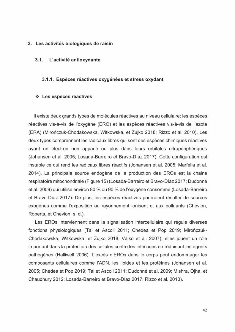
3.1.1. Espèces réactives oxygénées et stress oxydant
v Les espèces réactives
Il existe deux grands types de molécules réactives au niveau cellulaire: les espèces
réactives vis-à-vis de l’oxygène (ERO) et les espèces réactives vis-à-vis de l’azote
(ERA) (Mirończuk-Chodakowska, Witkowska, et Zujko 2018; Rizzo et al. 2010). Les
deux types comprennent les radicaux libres qui sont des espèces chimiques réactives
ayant un électron non apparié ou plus dans leurs orbitales ultrapériphériques
(Johansen et al. 2005; Losada-Barreiro et Bravo-Díaz 2017). Cette configuration est
instable ce qui rend les radicaux libres réactifs (Johansen et al. 2005; Marfella et al.
2014). La principale source endogène de la production des EROs est la chaine
respiratoire mitochondriale (Figure 15) (Losada-Barreiro et Bravo-Díaz 2017; Dudonné
et al. 2009) qui utilise environ 80 % ou 90 % de l’oxygène consommé (Losada-Barreiro
et Bravo-Díaz 2017). De plus, les espèces réactives pourraient résulter de sources
exogènes comme l’exposition au rayonnement ionisant et aux polluants (Chevion,
Roberts, et Chevion, s. d.).
Les EROs interviennent dans la signalisation intercellulaire qui régule diverses
fonctions physiologiques (Tai et Ascoli 2011; Chedea et Pop 2019; Mirończuk-
Chodakowska, Witkowska, et Zujko 2018; Valko et al. 2007), elles jouent un rôle
important dans la protection des cellules contre les infections en réduisant les agents
pathogènes (Halliwell 2006). L’excès d’EROs dans le corps peut endommager les
composants cellulaires comme l’ADN, les lipides et les protéines (Johansen et al.
2005; Chedea et Pop 2019; Tai et Ascoli 2011; Dudonné et al. 2009; Mishra, Ojha, et
Chaudhury 2012; Losada-Barreiro et Bravo-Díaz 2017; Rizzo et al. 2010).
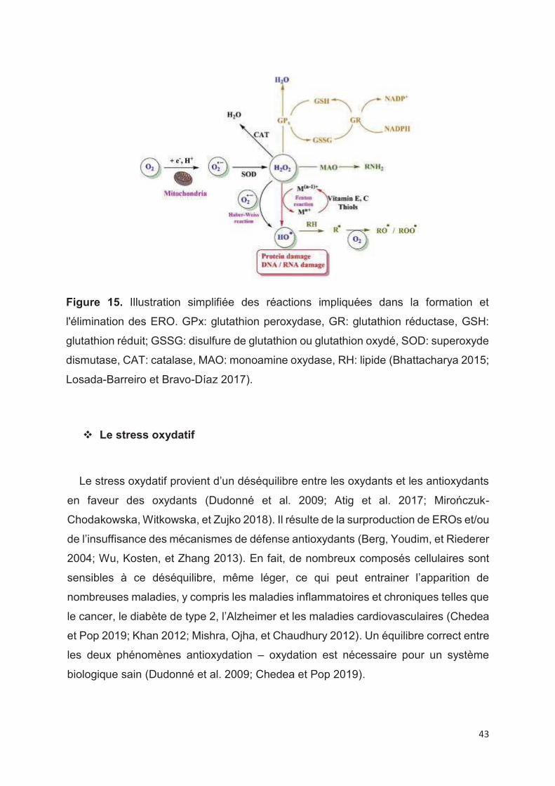
43
Figure 15. Illustration simplifiée des réactions impliquées dans la formation et
l'élimination des ERO. GPx: glutathion peroxydase, GR: glutathion réductase, GSH:
glutathion réduit; GSSG: disulfure de glutathion ou glutathion oxydé, SOD: superoxyde
dismutase, CAT: catalase, MAO: monoamine oxydase, RH: lipide (Bhattacharya 2015;
Losada-Barreiro et Bravo-Díaz 2017).
v Le stress oxydatif
Le stress oxydatif provient d’un déséquilibre entre les oxydants et les antioxydants
en faveur des oxydants (Dudonné et al. 2009; Atig et al. 2017; Mirończuk-
Chodakowska, Witkowska, et Zujko 2018). Il résulte de la surproduction de EROs et/ou
de l’insuffisance des mécanismes de défense antioxydants (Berg, Youdim, et Riederer
2004; Wu, Kosten, et Zhang 2013). En fait, de nombreux composés cellulaires sont
sensibles à ce déséquilibre, même léger, ce qui peut entrainer l’apparition de
nombreuses maladies, y compris les maladies inflammatoires et chroniques telles que
le cancer, le diabète de type 2, l’Alzheimer et les maladies cardiovasculaires (Chedea
et Pop 2019; Khan 2012; Mishra, Ojha, et Chaudhury 2012). Un équilibre correct entre
les deux phénomènes antioxydation – oxydation est nécessaire pour un système
biologique sain (Dudonné et al. 2009; Chedea et Pop 2019).
44
3.1.2. Antioxydants
Les antioxydants sont des composés capables de réduire ou d’empêcher la
destruction oxydative des composés biologiques (Balík et al. 2009). Ils protègent les
systèmes biologiques en utilisant différents mécanismes préventifs en tant que
première ligne de défense tels que le piégeage des radicaux libres, la rupture des
réactions en chaîne initiées par les radicaux libres et au final la réparation ou
l’élimination des structures endommagées (Rizzo et al. 2010; Mirończuk-
Chodakowska, Witkowska, et Zujko 2018). Différents antioxydants sont impliqués pour
maintenir l’équilibre du système biologique, y compris des antioxydants endogènes
(enzymatiques et non enzymatiques), (Chedea et Pop 2019; Mirończuk-
Chodakowska, Witkowska, et Zujko 2018; Nordberg et Arner, s. d.) et des antioxydants
alimentaires (Chedea et Pop 2019). Les deux types d’antioxydants agissent en
synergie afin de maintenir ou rétablir l'homéostasie redox (Zujko et al. 2012).
v Les antioxydants endogènes
Ce sont des produits du métabolisme du corps, ils peuvent être enzymatiques ou
non enzymatiques et ils interviennent en première ligne de défense (défense
préventive).
Antioxydants non enzymatiques
On trouve le glutathion, l’albumine, la bilirubine et l’acide urique (Chedea et Pop 2019;
Wu, Kosten, et Zhang 2013).
Antioxydants enzymatiques
On trouve la superoxyde dismutase (SOD) (EC 1.15.1.1), la glutathion peroxydase
(GPx) (EC 1.11.1.9) , la catalase (CAT) (EC 1.11.1.6) (Chedea et Pop 2019; Wu,
Kosten, et Zhang 2013; Steenvoorden et Beijersbergen van Henegouwen 1997), la
glutathion réductase (GR) et les peroxyrédoxines (Prxs) (Mirończuk-Chodakowska,
Witkowska, et Zujko 2018).

45
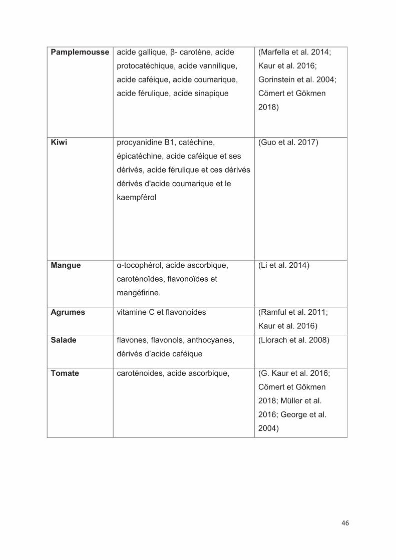
v Les antioxydants alimentaires
Les denrées alimentaires sont considérées comme des sources naturelles
contenant différents composés antioxydants comme les caroténoides, les vitamines C
et E et les polyphénols (Tableau 3) (Amarowicz et Pegg 2019; Marfella et al. 2014;
Guo et al. 2017; G. Kaur et al. 2016).
Les polyphénols sont de puissants antioxydants naturellement présents dans le
régime alimentaire, notamment dans les fruits, les légumes, les céréales et le thé
(Chedea et Pop 2019; Losada-Barreiro et Bravo-Díaz 2017). Les polyphénols
neutralisent les radicaux libres en cédant un électron ou un atome d’hydrogène pour
réduire le taux d’oxydation en inhibant la formation des radicaux libres ou en les
piégeant (Marfella et al. 2014). Le raisin est classé parmi les aliments les plus riches
en composés phénoliques. L’activité antioxydante du raisin est liée directement à la
concentration, la composition phénolique (Doshi, Adsule et Banerjee 2006), au
nombre et à la position des groupements OH (Losada-Barreiro et Bravo-Díaz 2017).
Tableau 3. Les principaux composés antioxydants dans les aliments
Aliments Principaux composés antioxydants Références
Pomme quercétine glycosides,
procyanidines B2, acide
chlorogénique, épicatéchine
et vitamine C
(K. W. Lee et al. 2003)
Abricot acide néochlorogénique, l'acide
chlorogénique, catéchine,
epicatechin, rutine, quercétine 3-
glucoside, acides
hydroxycinnamiques,
flavanols, flavonols, acide ascorbique
(Madrau et al. 2009)
Raisin polyphénols
(Lutz et al. 2011)
46
Pamplemousse acide gallique, β- carotène, acide
protocatéchique, acide vannilique,
acide caféique, acide coumarique,
acide férulique, acide sinapique
(Marfella et al. 2014;
Kaur et al. 2016;
Gorinstein et al. 2004;
Cömert et Gökmen
2018)
Kiwi procyanidine B1, catéchine,
épicatéchine, acide caféique et ses
dérivés, acide férulique et ces dérivés
dérivés d'acide coumarique et le
kaempférol
(Guo et al. 2017)
Mangue α-tocophérol, acide ascorbique,
caroténoïdes, flavonoïdes et
mangéfirine.
(Li et al. 2014)
Agrumes vitamine C et flavonoides (Ramful et al. 2011;
Kaur et al. 2016)
Salade flavones, flavonols, anthocyanes,
dérivés d’acide caféique
(Llorach et al. 2008)
Tomate caroténoides, acide ascorbique, (G. Kaur et al. 2016;
Cömert et Gökmen
2018; Müller et al.
2016; George et al.
2004)
47
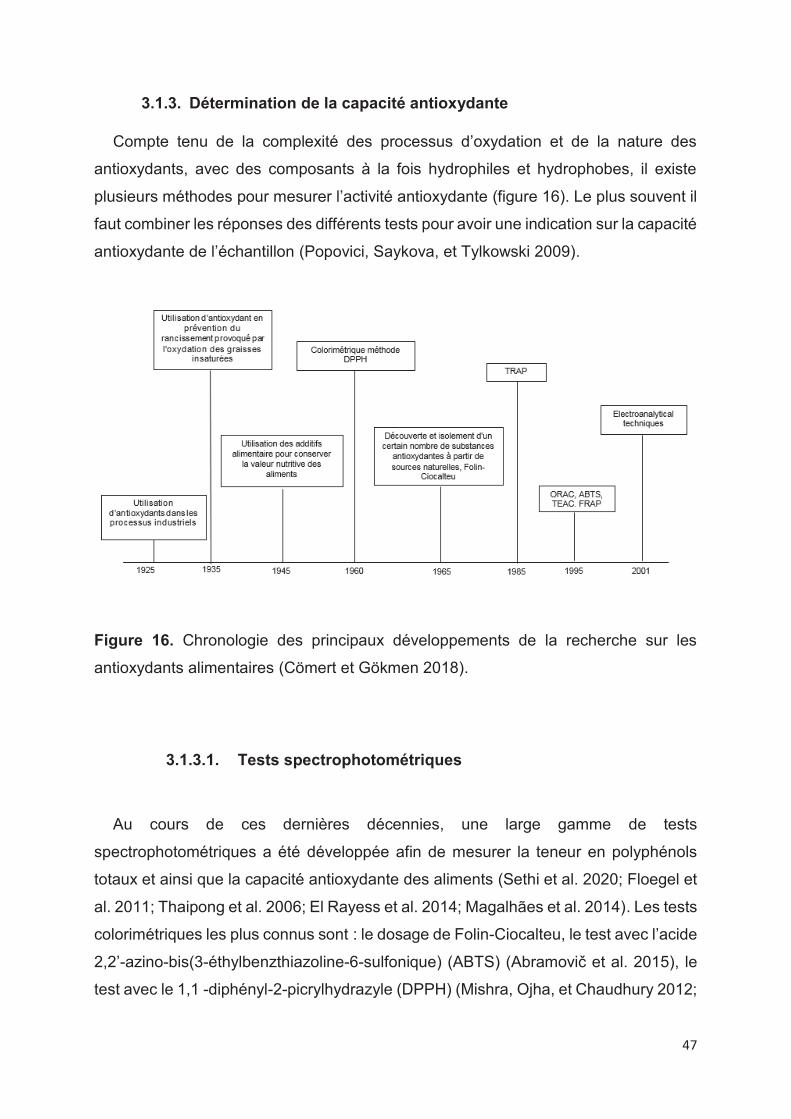
3.1.3. Détermination de la capacité antioxydante
Compte tenu de la complexité des processus d’oxydation et de la nature des
antioxydants, avec des composants à la fois hydrophiles et hydrophobes, il existe
plusieurs méthodes pour mesurer l’activité antioxydante (figure 16). Le plus souvent il
faut combiner les réponses des différents tests pour avoir une indication sur la capacité
antioxydante de l’échantillon (Popovici, Saykova, et Tylkowski 2009).
Figure 16. Chronologie des principaux développements de la recherche sur les
antioxydants alimentaires (Cömert et Gökmen 2018).
3.1.3.1. Tests spectrophotométriques
Au cours de ces dernières décennies, une large gamme de tests
spectrophotométriques a été développée afin de mesurer la teneur en polyphénols
totaux et ainsi que la capacité antioxydante des aliments (Sethi et al. 2020; Floegel et
al. 2011; Thaipong et al. 2006; El Rayess et al. 2014; Magalhães et al. 2014). Les tests
colorimétriques les plus connus sont : le dosage de Folin-Ciocalteu, le test avec l’acide
2,2’-azino-bis(3-éthylbenzthiazoline-6-sulfonique) (ABTS) (Abramovič et al. 2015), le
test avec le 1,1 -diphényl-2-picrylhydrazyle (DPPH) (Mishra, Ojha, et Chaudhury 2012;
48
Popovici, Saykova, et Tylkowski 2009; Danilewicz 2015; Abramovič et al. 2015) et la
capacité de réduction ferrique du plasma (FRAP) (Thaipong et al. 2006; Dudonné et
al. 2009).
Le test de Folin-Ciocalteu : il est largement utilisé et il donne une indication globale
sur la teneur en composés phénoliques totaux (Yilmaz et al. 2015; Coklar 2017; Lingua
et al. 2019; Ky et Teissedre 2015). Le réactif de Folin est un complexe de
phosphotungstate et phosphomolybdate qui sont réduits par les composés
phénoliques en condition alcaline (milieu réactif ajusté par du carbonate de sodium
avec un pH proche de 10) produit une coloration bleue avec une absorbance maximale
à 765 nm. Les résultats sont exprimés en équivalents d’acide gallique. Le réactif de
Folin n’est pas spécifique aux composés phénoliques car il peut réagir avec d’autres
composés comme l’acide ascorbique, le cuivre (Lorrain et al. 2013; Singleton,
Orthofer, et Lamuela-Raventós 1999; Huang, Ou, et Prior 2005; Stevanato, Fabris, et
Momo 2004; Danilewicz 2015), les sucres, les amines aromatiques (Prior, Wu, et
Schaich 2005). Le test de Folin Ciocalteu est un test simple, pratique et reproductible
(Huang, Ou, et Prior 2005; El Rayess et al. 2014; Magalhães et al. 2014).
Le test ABTS a été utilisé pour la première fois par Miller et Rice-Evans en 1993
puis il a été amélioré plus tard par (Re et al. 1999). Le radical ABTS• est généré par
l’oxydation du persulfate de 2,2′-azinobis (acide 3-éthylbenzothiazoline-6-sulfonique)
(Floegel et al. 2011). La capacité antioxydante est déterminée par la diminution de
l’absorbance du radical ABTS• en présence de l’échantillon à tester après un temps
fixe (4-6 min) à la longueur d’onde de 734 nm. Les résultats sont exprimés en
équivalent de trolox ou en pourcentage de diminution de l’absorbance (Huang, Ou, et
Prior 2005; Cano, Acosta, et Arnao 2000). L’ABTS est un test simple, rapide et peut
être utilisé sur une large gamme de pH (Lemańska et al. 2001; Magalhães et al. 2014).
Le test DPPH est utilisé pour la première fois par Brand-Williams et ses collègues
(Brand-Williams, Cuvelier, et Berset 1995). Il est basé sur la réduction du radical violet
DPPH• en hydrazine jaune pâle par les antioxydants (Magalhães et al. 2014). Les
polyphénols réduisent le radical avec une perte d’absorbance qui est suivie au cours
49
du temps (30 min) à 515 nm (Popovici, Saykova, et Tylkowski 2009; Katalinić et al.
2004). Les résultats sont exprimés soit en pourcentage de diminution de l’absorbance
(Katalinić et al. 2004) soit en équivalent trolox. Le DPPH est un test rapide et simple
ne nécessite pas beaucoup de réactifs, ce qui explique probablement son utilisation
répondue dans la détermination de la capacité antioxydante (Magalhães et al. 2014;
El Rayess et al. 2014).
Le test FRAP : ce test a été initialement développé par Benzie and Strain (Benzie
et Strain 1996) pour mesurer le pouvoir réducteur dans le plasma. Ce test a été adapté
par la suite aux antioxydants des végétaux (Ou et al. 2002; Gil et al. 2000; Proteggente
et al. 2002). Il est différent des autres (DPPH et ABTS) car il n’y a pas de radicaux
libres impliqués, il est basé sur la réduction du fer ferrique (Fe+3) en fer ferreux (Fe+2)
(Floegel et al. 2011) et la puissance de chélation sur les ions ferreux (Fe2 +) (Sudan
et al. 2014). L’absorbance est mesurée après 4 min à 593 nm. Les résultats sont
exprimés par rapport à une solution standard de Fe (II) (Dudonné et al. 2009; Thaipong
et al. 2006; Sethi et al. 2020). Le test FRAP est rapide, simple, peu coûteux et robuste
et ne nécessite pas d'équipement spécialisé (Prior, Wu, et Schaich 2005; Danilewicz
2015).
Les inconvénients des méthodes spectrophotométriques
Les analyses spectrophotométriques peuvent conduire à :
· une surestimation des teneurs en polyphénols et de leur capacité
antioxydante des échantillons due à l’interférence possible des molécules qui
absorbent en UV (Danilewicz 2015; El Rayess et al. 2014; Magalhães et al.
2014).
· la consommation des solvants et le temps d’analyse (Lorrain et al. 2013;
Danilewicz 2015).
D’autres méthodes simple, rapide, robuste et fiable a été développée pour déterminer
la capacité antioxydante : ce sont les techniques électrochimiques (Lorrain et al. 2013).
50
3.1.3.2. Tests électrochimiques
Différentes méthodes électrochimiques sont utilisées pour la caractérisation et la
quantification des polyphénols et de la capacité antioxydante : la chronoampérométrie,
la voltammétrie à impulsion différentielle et la voltammétrie cyclique (Hoyos-Arbeláez,
Vázquez, et Contreras-Calderón 2017) basées sur le fait que tous les composées
phénoliques sont électrochimiquement actifs (Mark, Scholz, et Matysik 2012;
Makhotkina et Kilmartin 2010; Arribas, Martínez-Fernández, et Chicharro 2012; Ricci
et al. 2019; Zou et al. 2002; Orlandi et al. 2018).
La voltammétrie cyclique est la méthode électrochimique la plus utilisée pour la
caractérisation des polyphénols et la détermination de la capacité antioxydante
(Kilmartin, Zou, et Waterhouse 2001; 2002; Hoyos-Arbeláez, Vázquez, et Contreras-
Calderón 2017). L’aire sous la courbe d’oxydation sur le voltammogramme représente
la capacité antioxydante totale de l’échantillon (Rockenbach et al. 2011; José Jara-
Palacios et al. 2017). La présence de pics à faible potentiel est corrélée à la présence
des polyphénols à forte activité antioxydante, tandis que les composés à faible pouvoir
antioxydant donnent des pics ayant un potentiel plus élevé (El Rayess et al. 2014;
Hoyos-Arbeláez, Vázquez, et Contreras-Calderón 2017).
En effet, les ortho-diphénols facilement oxydés donnent un pic de potentiel faible
autour de 400 mV, les anthocyanes produisent un pic à 650 mV, et les groupes
fonctionnels les plus difficiles à oxyder produisent des pics à des potentiels plus élevés,
permettant une discrimination entre ces types de molécules (Lorrain et al. 2013;
Newair, Kilmartin, et Garcia 2018; Kilmartin, Zou, et Waterhouse 2002).
3.2. L’activité anti-inflammatoire
3.2.1. Inflammation
L’inflammation est une réponse de défense du système immunitaire aux agents
pathogènes étrangers (Kishore et al. 2019; Riegsecker et al. 2013) notamment les
dommages physiques, l'irradiation aux ultraviolets et l'invasion microbienne, afin
d’éliminer les stimuli, les tissus endommagés et puis initier la guérison et la réparation
51
des tissus (Gautam et Jachak 2009; Mariathasan et Monack 2007). Les signes
physiques de l’inflammation les plus évidents sont la douleur, la chaleur, la rougeur et
l’enflure qui sont dues à l’augmentation du flux sanguin, de la vasodilatation, la
libération des médiateurs intracellulaires et aux fuites de liquide (Ferrero-Miliani et al.
2006). Les macrophages, les neutrophiles et les phagocytes mononucléaires sont les
principales cellules immunitaires impliquées dans la réponse inflammatoire.
Selon la réponse de l’action pour éradiquer l’agent étranger ou les tissus lésés, on
distingue deux types d’inflammation :
Inflammation aiguë : elle est très rapide et commence quelques minutes après la
lésion tissulaire visant à tuer les bactéries, virus ou les parasites sur les sites actifs.
Elle implique l’association de protéines plasmatiques et la migration des fluides et des
neutrophiles vers la région lésée (Heras et Hortelano 2009; Kishore et al. 2019).
Pendant la réponse inflammatoire aiguë, les neutrophiles et les macrophages sont
principalement stimulés (Ferrero-Miliani et al. 2006).
Inflammation chronique : ce processus est long et associé à la prolifération
vasculaire, aux macrophages, à la fibrose et à la destruction des tissus (Kishore et al.
2019; Riegsecker et al. 2013). Les lymphocytes T, les plasmocytes et les macrophages
sont les principaux propagateurs de l'inflammation chronique (Ferrero-Miliani et al.
2006; Riegsecker et al. 2013).
3.2.2. Marqueurs de l’inflammation
Les macrophages sont les principaux contributeurs à la réponse inflammatoire lors
de la stimulation par des stimuli exogènes tels que les lipopolysaccharides (LPS)
(Gautam et Jachak 2009; Han et al. 2017; Cheng et al. 2014; Wan et al. 2019) ou le
muramyl dipeptide (Cheng et al. 2014). Ils secrètent des cytokines pro inflammatoires,
notamment le facteur de nécrose tumorale α (TNF-α), l’interleukine-6 (IL-6) et l’IL-1β,
les médiateurs pro-inflammatoires dont l’oxyde nitrique (NO), la prostaglandine E2
(PGE2) et les espèces réactives de l’oxygène (ROS) (Du et al. 2018; Q. Xu et al. 2017)
et les protéines inflammatoires telles que l'oxyde nitrique inductible synthase (iNOS)
et cyclooxygénase-2 (COX-2). L’inhibition de la production des marqueurs

52
d’inflammation est le mécanisme clé dans le contrôle de l’inflammation (Q. Xu et al.
2017; Bak et al. 2013).
Figure 17. Stimulation des macrophages RAW 264.7 par les LPS (Linghu et al. 2020)
3.2.3. Complications de l’inflammation
L’inflammation chronique provoque des troubles au niveau des tissus sains
conduisant à plusieurs maladies (Kishore et al. 2019; Singh, Akhtar, et Haqqi 2010)
comme l’asthme, le vieillissement, l’athérosclérose, le trouble d’immunodéfience
acquise (SIDA), l’athérosclérose, la goutte, le diabète (Kishore et al. 2019), la maladie
d’Alzheimer, la maladie de Parkinson (Shao et al. 2013), le cancer et l’insuffisance
cardiaque (Bak et al. 2013).
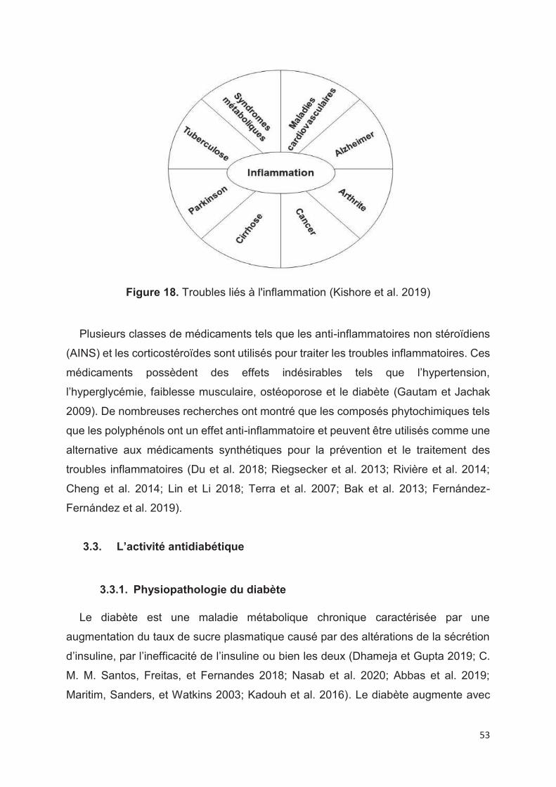
53
Figure 18. Troubles liés à l'inflammation (Kishore et al. 2019)
Plusieurs classes de médicaments tels que les anti-inflammatoires non stéroïdiens
(AINS) et les corticostéroïdes sont utilisés pour traiter les troubles inflammatoires. Ces
médicaments possèdent des effets indésirables tels que l’hypertension,
l’hyperglycémie, faiblesse musculaire, ostéoporose et le diabète (Gautam et Jachak
2009). De nombreuses recherches ont montré que les composés phytochimiques tels
que les polyphénols ont un effet anti-inflammatoire et peuvent être utilisés comme une
alternative aux médicaments synthétiques pour la prévention et le traitement des
troubles inflammatoires (Du et al. 2018; Riegsecker et al. 2013; Rivière et al. 2014;
Cheng et al. 2014; Lin et Li 2018; Terra et al. 2007; Bak et al. 2013; Fernández-
Fernández et al. 2019).
3.3. L’activité antidiabétique
3.3.1. Physiopathologie du diabète
Le diabète est une maladie métabolique chronique caractérisée par une
augmentation du taux de sucre plasmatique causé par des altérations de la sécrétion
d’insuline, par l’inefficacité de l’insuline ou bien les deux (Dhameja et Gupta 2019; C.
M. M. Santos, Freitas, et Fernandes 2018; Nasab et al. 2020; Abbas et al. 2019;
Maritim, Sanders, et Watkins 2003; Kadouh et al. 2016). Le diabète augmente avec
54
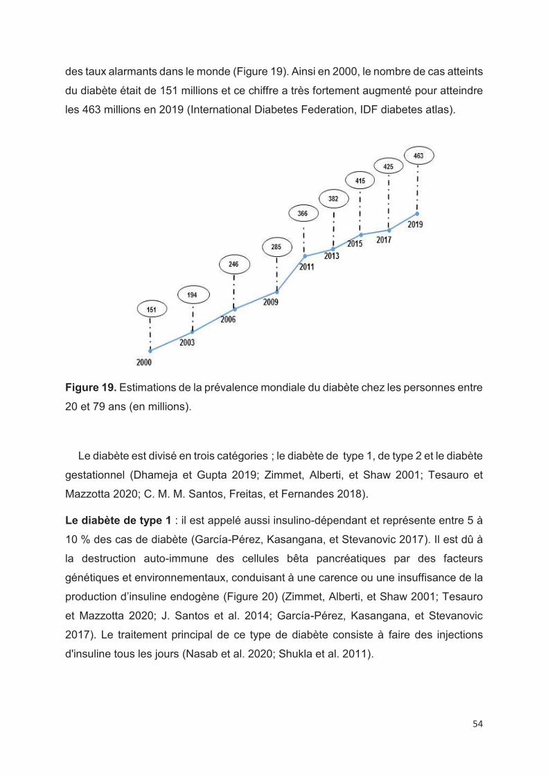
des taux alarmants dans le monde (Figure 19). Ainsi en 2000, le nombre de cas atteints
du diabète était de 151 millions et ce chiffre a très fortement augmenté pour atteindre
les 463 millions en 2019 (International Diabetes Federation, IDF diabetes atlas).
Figure 19. Estimations de la prévalence mondiale du diabète chez les personnes entre
20 et 79 ans (en millions).
Le diabète est divisé en trois catégories ; le diabète de type 1, de type 2 et le diabète
gestationnel (Dhameja et Gupta 2019; Zimmet, Alberti, et Shaw 2001; Tesauro et
Mazzotta 2020; C. M. M. Santos, Freitas, et Fernandes 2018).
Le diabète de type 1 : il est appelé aussi insulino-dépendant et représente entre 5 à
10 % des cas de diabète (García-Pérez, Kasangana, et Stevanovic 2017). Il est dû à
la destruction auto-immune des cellules bêta pancréatiques par des facteurs
génétiques et environnementaux, conduisant à une carence ou une insuffisance de la
production d’insuline endogène (Figure 20) (Zimmet, Alberti, et Shaw 2001; Tesauro
et Mazzotta 2020; J. Santos et al. 2014; García-Pérez, Kasangana, et Stevanovic
2017). Le traitement principal de ce type de diabète consiste à faire des injections
d'insuline tous les jours (Nasab et al. 2020; Shukla et al. 2011).
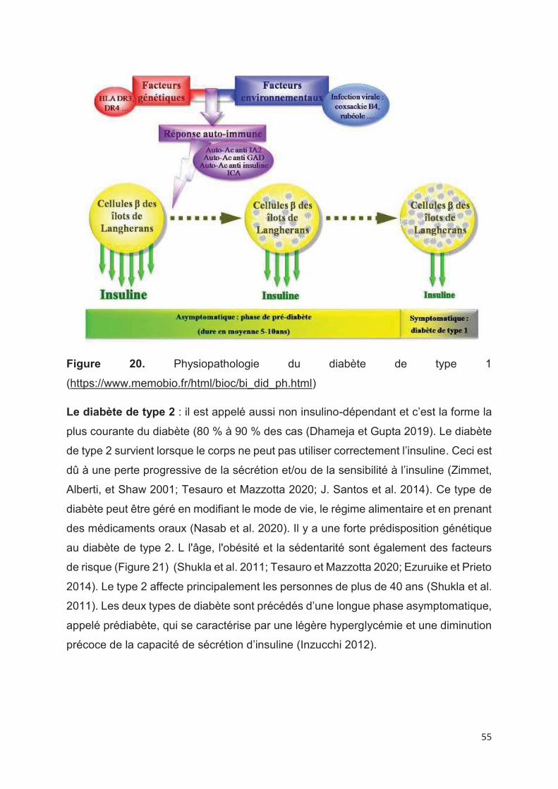
55
Figure 20. Physiopathologie du diabète de type 1
(https://www.memobio.fr/html/bioc/bi_did_ph.html)
Le diabète de type 2 : il est appelé aussi non insulino-dépendant et c’est la forme la
plus courante du diabète (80 % à 90 % des cas (Dhameja et Gupta 2019). Le diabète
de type 2 survient lorsque le corps ne peut pas utiliser correctement l’insuline. Ceci est
dû à une perte progressive de la sécrétion et/ou de la sensibilité à l’insuline (Zimmet,
Alberti, et Shaw 2001; Tesauro et Mazzotta 2020; J. Santos et al. 2014). Ce type de
diabète peut être géré en modifiant le mode de vie, le régime alimentaire et en prenant
des médicaments oraux (Nasab et al. 2020). Il y a une forte prédisposition génétique
au diabète de type 2. L l'âge, l'obésité et la sédentarité sont également des facteurs
de risque (Figure 21) (Shukla et al. 2011; Tesauro et Mazzotta 2020; Ezuruike et Prieto
2014). Le type 2 affecte principalement les personnes de plus de 40 ans (Shukla et al.
2011). Les deux types de diabète sont précédés d’une longue phase asymptomatique,
appelé prédiabète, qui se caractérise par une légère hyperglycémie et une diminution
précoce de la capacité de sécrétion d’insuline (Inzucchi 2012).
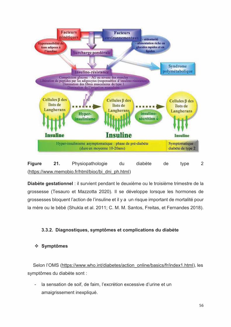
56
Figure 21. Physiopathologie du diabète de type 2
(https://www.memobio.fr/html/bioc/bi_dni_ph.html)
Diabète gestationnel : il survient pendant le deuxième ou le troisième trimestre de la
grossesse (Tesauro et Mazzotta 2020). Il se développe lorsque les hormones de
grossesses bloquent l’action de l’insuline et il y a un risque important de mortalité pour
la mère ou le bébé (Shukla et al. 2011; C. M. M. Santos, Freitas, et Fernandes 2018).
3.3.2. Diagnostiques, symptômes et complications du diabète
v Symptômes
Selon l’OMS (https://www.who.int/diabetes/action_online/basics/fr/index1.html), les
symptômes du diabète sont :
- la sensation de soif, de faim, l’excrétion excessive d’urine et un
amaigrissement inexpliqué.
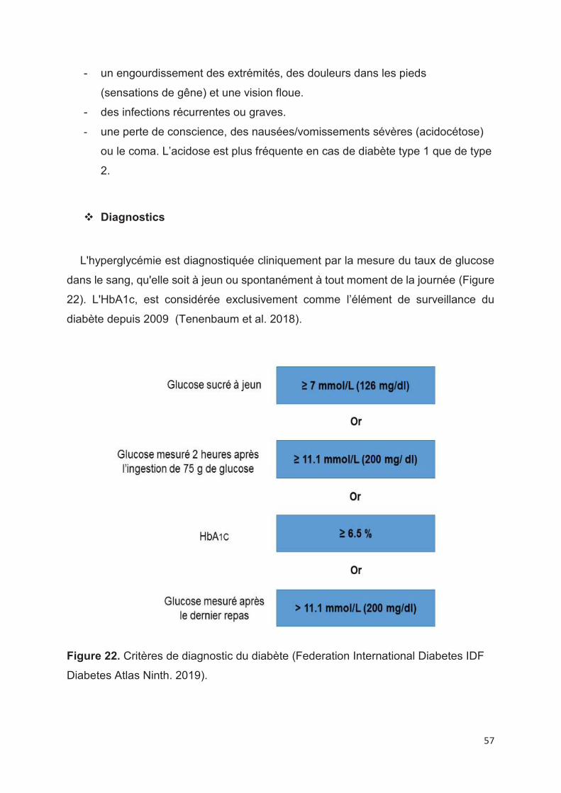
57
- un engourdissement des extrémités, des douleurs dans les pieds
(sensations de gêne) et une vision floue.
- des infections récurrentes ou graves.
- une perte de conscience, des nausées/vomissements sévères (acidocétose)
ou le coma. L’acidose est plus fréquente en cas de diabète type 1 que de type
2.
v Diagnostics
L'hyperglycémie est diagnostiquée cliniquement par la mesure du taux de glucose
dans le sang, qu'elle soit à jeun ou spontanément à tout moment de la journée (Figure
22). L'HbA1c, est considérée exclusivement comme l’élément de surveillance du
diabète depuis 2009 (Tenenbaum et al. 2018).
Figure 22. Critères de diagnostic du diabète (Federation International Diabetes IDF
Diabetes Atlas Ninth. 2019).
58
v Complications
Le diabète est généralement silencieux à ses débuts et, à long terme, il peut
entrainer des complications dues à la libération des radicaux libres qui jouent un rôle
important dans l’apparition et la progression des plusieurs maladies telles que les
maladies rénales, les maladies du système nerveux, les maladies cardiaques et la
rétinopathie (Dhameja et Gupta 2019; Nasab et al. 2020; Abbas et al. 2019; Buron et
Thaunat 2020; García-Pérez, Kasangana, et Stevanovic 2017).
3.3.3. Traitements pharmacologiques
L'une des stratégies thérapeutiques les plus importantes contre le diabète est de
diminuer le taux de glucose postprandial par l'inhibition de la dégradation enzymatique
des polysaccharides sans effet direct sur la sécrétion d’insuline (Nasab et al. 2020; Di
Stefano, Oliviero, et Udenigwe 2018). Par conséquent, l'inhibition des enzymes α-
amylase et α-glucosidase est une approche efficace pour gérer l'index glycémique
indépendamment de l'insuline (Figure 23) (Nasab et al. 2020; Dhameja et Gupta 2019;
Abbas et al. 2019). Ces inhibiteurs sont utilisés en monothérapie dans les états
diabétiques légers et sont utilisés en association avec l’insuline dans le diabète sévère
(Dhameja et Gupta 2019).
L’α-amylase (EC 3.2.1.1) : c’est une enzyme qui appartient à la classe des
hydrolases. Les α-amylases salivaires et pancréatiques hydrolysent l'amidon en
rompant les liaisons glycosidiques α-1,4 pour produire du maltose et d'autres
oligosaccharides qui sont ensuite hydrolysés en glucose par l’α- glucosidase situé
dans la membrane de surface en brosse des cellules intestinales (R. Kaur, Kaur, et
Gupta 2014; Nasab et al. 2020; C. M. M. Santos, Freitas, et Fernandes 2018; Nyambe-
Silavwe et al. 2015; Di Stefano, Oliviero, et Udenigwe 2018). Le glucose est absorbé
par l’intestin et provoque l’hyperglycémie (Nasab et al. 2020; C. M. M. Santos, Freitas,
et Fernandes 2018).
L’α-glucosidase (EC 3.2.1.20): c’est une enzyme qui catalyse le clivage des
oligosaccharides et des disaccharides en monosaccharides lord de la dernière étape

59
de la digestion des glucides et qui augmente ainsi la concentration de glucose dans le
corps (Abbas et al. 2019; Nasab et al. 2020; C. M. M. Santos, Freitas, et Fernandes
2018; Di Stefano, Oliviero, et Udenigwe 2018; Nyambe-Silavwe et al. 2015).
Figure 23. Réduction de la glycémie par l'inhibition de l'alpha glucosidase (Nasab et
al. 2020)
60
Actuellement, quatre inhibiteurs de l'α-glucosidase sont utilisés en thérapie :
l’acarbose, le miglitol, la voglibose et l’émiglitate dont l’acarbose est le premier
inhibiteur introduit sur le marché dans les années 1990. L’utilisation de ces inhibiteurs
est associée à des effets secondaires tels que les flatulences, les douleurs
abdominales, la diarrhée et les nausées (C. M. M. Santos, Freitas, et Fernandes 2018;
Nasab et al. 2020; Dhameja et Gupta 2019). Des aliments fonctionnels pourraient être
développés contenant des composés capables d'inhiber l'α-amylase et l’α-
glucosidase, une action de type acarbose, mais sans effet secondaire (Nyambe-
Silavwe et al. 2015).
3.3.4. Traitements par les polyphénols
Il a été rapporté dans la littérature que certains extraits de plantes peuvent exercer
une activité bénéfique pour la santé humaine en inhibant les activités des enzymes
d'hydrolyse des glucides complexes. Les principaux constituants actifs de ces extraits
de plantes sont des composés phénoliques tels que les flavonoïdes (Di Stefano,
Oliviero, et Udenigwe 2018; Sun et al. 2016; Ibrahim et al. 2017; Abbas et al. 2019).
L’activité inhibitrice dépend de la substitution de groupes hydroxyles qui peuvent
former des liaisons hydrogènes avec l’enzyme (Gutiérrez-Grijalva et al. 2019) et au
nombre des oligomères (les oligomères et les polymères ont plus d’activité en raison
de leur grand nombre de sites de liaison avec les enzymes) (Di Stefano, Oliviero, et
Udenigwe 2018).
61
References
Abbas, Ghulam, Ahmed Al Harrasi, Hidayat Hussain, Ahmed Hamaed, et Claudiu T.
Supuran. 2019. « The Management of Diabetes Mellitus-Imperative Role of
Natural Products against Dipeptidyl Peptidase-4, α-Glucosidase and Sodium-
Dependent Glucose Co-Transporter 2 (SGLT2) ». Bioorganic Chemistry 86: 305
315. https://doi.org/10.1016/j.bioorg.2019.02.009.
Abramovič, Helena, Tatjana Košmerl, Nataša Poklar Ulrih, et Blaž Cigić. 2015.
« Contribution of SO2 to Antioxidant Potential of White Wine ». Food Chemistry
174: 147 153. https://doi.org/10.1016/j.foodchem.2014.11.030.
Adams, Douglas O. 2006. « Phenolics and Ripening in Grape Berries », 8.
Aguirre, María J., Yo Y. Chen, Mauricio Isaacs, Betty Matsuhiro, Leonora Mendoza, et
Simonet Torres. 2010. « Electrochemical Behaviour and Antioxidant Capacity
of Anthocyanins from Chilean Red Wine, Grape and Raspberry ». Food
Chemistry 121 (1): 44 48. https://doi.org/10.1016/j.foodchem.2009.11.088.
Alam, Md Ashraful, Nusrat Subhan, Hemayet Hossain, Murad Hossain, Hasan
Mahmud Reza, Md Mahbubur Rahman, et M Obayed Ullah. 2016.
« Hydroxycinnamic Acid Derivatives: A Potential Class of Natural Compounds
for the Management of Lipid Metabolism and Obesity ». Nutrition & Metabolism
13 (1): 27. https://doi.org/10.1186/s12986-016-0080-3.
Ali, Kashif, Federica Maltese, Ana Margarida Fortes, Maria Salomé Pais, Young Hae
Choi, et Robert Verpoorte. 2011. « Monitoring Biochemical Changes during
Grape Berry Development in Portuguese Cultivars by NMR Spectroscopy ».
Food Chemistry 124 (4): 1760 1769.
https://doi.org/10.1016/j.foodchem.2010.08.015.
Amarowicz, Ryszard, et Ronald B. Pegg. 2019. « Natural Antioxidants of Plant
Origin ». In Advances in Food and Nutrition Research, 90:1 81. Elsevier.
https://doi.org/10.1016/bs.afnr.2019.02.011.
Amiot, M.-J., C. Riollet, et J.-F. Landrier. 2009. « Polyphénols et syndrome
métabolique ». Médecine des Maladies Métaboliques 3 (5): 476 482.
https://doi.org/10.1016/S1957-2557(09)73293-6.
62
Arribas, Alberto Sánchez, Marta Martínez-Fernández, et Manuel Chicharro. 2012.
« The role of electroanalytical techniques in analysis of polyphenols in wine ».
TrAc Trends in Analytical Chemistry 34: 78 96.
Ashraf, Muhammad A., Muhammad Iqbal, Rizwan Rasheed, Iqbal Hussain,
Muhammad Riaz, et Muhammad Saleem Arif. 2018. « Environmental Stress
and Secondary Metabolites in Plants ». In Plant Metabolites and Regulation
Under Environmental Stress, 153 167. Elsevier. https://doi.org/10.1016/B978-
0-12-812689-9.00008-X.
Asproudi, Andriani, Federico Piano, Giacomo Anselmi, Rocco Di Stefano, Eleonora
Bertolone, et Daniela Borsa. 2015. « Proanthocyanidin Composition and
Evolution during Grape Ripening as Affected by Variety: Nebbiolo and Barbera
Cv. » OENO One 49 (1): 59. https://doi.org/10.20870/oeno-one.2015.49.1.93.
Atig, Fatma, Abdelhamid Kerkeni, Ali Saad, et Mounir Ajina. 2017. « Effects of
Reduced Seminal Enzymatic Antioxidants on Sperm DNA Fragmentation and
Semen Quality of Tunisian Infertile Men ». Journal of Assisted Reproduction
and Genetics 34 (3): 373 381. https://doi.org/10.1007/s10815-013-9936-x.
B. G. Coombe. 1992. « Research on development and ripening of the grape berry ».
American Journal of Enology and Viticulture 43 (1): 101 110.
Babazadeh, Afshin, Arezou Taghvimi, Hamed Hamishehkar, et Mahnaz Tabibiazar.
2017. « Development of New Ultrasonic–Solvent Assisted Method for
Determination of Trans-Resveratrol from Red Grapes: Optimization,
Characterization, and Antioxidant Activity (ORAC Assay) ». Food Bioscience
20: 36 42. https://doi.org/10.1016/j.fbio.2017.08.003.
Bak, Min-Ji, Van Long Truong, Hey-Sook Kang, Mira Jun, et Woo-Sik Jeong. 2013.
« Anti-Inflammatory Effect of Procyanidins from Wild Grape ( Vitis Amurensis )
Seeds in LPS-Induced RAW 264.7 Cells ». Oxidative Medicine and Cellular
Longevity 2013: 1 11. https://doi.org/10.1155/2013/409321.
Balík, J., M. Kyseláková, N. Vrchotová, J. Tříska, M. Kumšta, J. Veverka, P. Híc, J.
Totušek, et D. Lefnerová. 2009. « Relations between Polyphenols Content and
Antioxidant Activity in Vine Grapes and Leaves ». Czech Journal of Food
Sciences 26 (Special Issue): 25 32. https://doi.org/10.17221/246/2008-CJFS.
Bautista-Ortín, Ana Belén, José Ignacio Fernández-Fernández, José María López-
Roca, et Encarnación Gómez-Plaza. 2006. « The Effect of Grape Ripening
63
Stage on Red Wine Color ». OENO One 40 (1): 15.
https://doi.org/10.20870/oeno-one.2006.40.1.879.
Benzie, Iris F.F., et J.J. Strain. 1996. « The Ferric Reducing Ability of Plasma (FRAP)
as a Measure of “Antioxidant Power”: The FRAP Assay ». Analytical
Biochemistry 239 (1): 70 76. https://doi.org/10.1006/abio.1996.0292.
Berg, Daniela, Moussa B. H. Youdim, et Peter Riederer. 2004. « Redox Imbalance ».
Cell and Tissue Research 318 (1): 201 213. https://doi.org/10.1007/s00441-
004-0976-5.
Berli, Federico J., Martín Fanzone, Patricia Piccoli, et Rubén Bottini. 2011. « Solar UV-
B and ABA Are Involved in Phenol Metabolism of Vitis Vinifera L. Increasing
Biosynthesis of Berry Skin Polyphenols ». Journal of Agricultural and Food
Chemistry 59 (9): 4874 4884. https://doi.org/10.1021/jf200040z.
Bhattacharya, Susinjan. 2015. « Reactive Oxygen Species and Cellular Defense
System ». In Free Radicals in Human Health and Disease 17 29.
https://doi.org/10.1007/978-81-322-2035-0_2.
Bindon, Keren, Cristian Varela, James Kennedy, Helen Holt, et Markus Herderich.
2013. « Relationships between Harvest Time and Wine Composition in Vitis
Vinifera L. Cv. Cabernet Sauvignon 1. Grape and Wine Chemistry ». Food
Chemistry 138 (2 3): 1696 1705.
https://doi.org/10.1016/j.foodchem.2012.09.146.
Błaszczyk, Alfred, Sylwia Sady, et Maria Sielicka. 2019. « The Stilbene Profile in Edible
Berries ». Phytochemistry Reviews 18 (1): 37 67.
https://doi.org/10.1007/s11101-018-9580-2.
Bohm, Bruce A. 1998. Introduction to flavonoids. Harwood Academic Publishers.
Boido, E., M. García-Marino, E. Dellacassa, F. Carrau, J.C. Rivas-Gonzalo, et M.T.
Escribano-Bailón. 2011. « Characterisation and Evolution of Grape Polyphenol
Profiles of Vitis Vinifera L. Cv. Tannat during Ripening and Vinification:
Polyphenolic Profiles of Tannat ». Australian Journal of Grape and Wine
Research 17 (3): 383 393. https://doi.org/10.1111/j.1755-0238.2011.00164.x.
Bozan, Berrin, Göksel Tosun, et Derya Özcan. 2008. « Study of Polyphenol Content
in the Seeds of Red Grape (Vitis Vinifera L.) Varieties Cultivated in Turkey and
64
Their Antiradical Activity ». Food Chemistry 109 (2): 426 430.
https://doi.org/10.1016/j.foodchem.2007.12.056.
Brand-Williams, W., M.E. Cuvelier, et C. Berset. 1995. « Use of a Free Radical Method
to Evaluate Antioxidant Activity ». LWT - Food Science and Technology 28 (1):
25 30. https://doi.org/10.1016/S0023-6438(95)80008-5.
Buron, Fanny, et Olivier Thaunat. 2020. « Secondary Complications of Diabetes ». In
Transplantation, Bioengineering, and Regeneration of the Endocrine Pancreas,
591 595. Elsevier. https://doi.org/10.1016/B978-0-12-814833-4.00047-2.
Cadot, Y., M.T. Miñana Castelló, et M. Chevalier. 2006. « Flavan-3-Ol Compositional
Changes in Grape Berries (Vitis Vinifera L. Cv Cabernet Franc) before Veraison,
Using Two Complementary Analytical Approaches, HPLC Reversed Phase and
Histochemistry ». Analytica Chimica Acta 563 (1 2): 65 75.
https://doi.org/10.1016/j.aca.2006.01.063.
Calderón-Oliver, Mariel, et Edith Ponce-Alquicira. s. d. « Fruits: A Source of
Polyphenols and Health Benefits », 40.
Callcott, Esther T., Abishek B. Santhakumar, Jixun Luo, et Christopher L. Blanchard.
2018. « Therapeutic Potential of Rice-Derived Polyphenols on Obesity-Related
Oxidative Stress and Inflammation ». Journal of Applied Biomedicine 16 (4): 255
262. https://doi.org/10.1016/j.jab.2018.03.001.
Cano, A., M. Acosta, et M.B. Arnao. 2000. « A Method to Measure Antioxidant Activity
in Organic Media: Application to Lipophilic Vitamins ». Redox Report 5 (6): 365
370. https://doi.org/10.1179/135100000101535933.
Careri, Maria, Claudio Corradini, Lisa Elviri, Isabella Nicoletti, et Ingrid Zagnoni. 2003.
« Direct HPLC Analysis of Quercetin and Trans -Resveratrol in Red Wine,
Grape, and Winemaking Byproducts ». Journal of Agricultural and Food
Chemistry 51 (18): 5226 5231. https://doi.org/10.1021/jf034149g.
Cavaliere, Chiara, Patrizia Foglia, Riccardo Gubbiotti, Paolo Sacchetti, Roberto
Samperi, et Aldo Laganà. 2008. « Rapid-Resolution Liquid
Chromatography/Mass Spectrometry for Determination and Quantitation of
Polyphenols in Grape Berries ». Rapid Communications in Mass Spectrometry
22 (20): 3089 3099. https://doi.org/10.1002/rcm.3705.
65
Chandrasekara, Anoma, et Fereidoon Shahidi. 2018. « Herbal Beverages: Bioactive
Compounds and Their Role in Disease Risk Reduction - A Review ». Journal of
Traditional and Complementary Medicine 8 (4): 451 458.
https://doi.org/10.1016/j.jtcme.2017.08.006.
Chedea, Veronica Sanda, et Raluca Maria Pop. 2019. « Total Polyphenols Content
and Antioxidant DPPH Assays on Biological Samples ». In Polyphenols in
Plants, 169 83. Elsevier. https://doi.org/10.1016/B978-0-12-813768-0.00011-6.
Cheng, Anwei, Haiqing Yan, Caijing Han, Wenliang Wang, Yaoqi Tian, et Xiangyan
Chen. 2014. « Polyphenols from Blueberries Modulate Inflammation Cytokines
in LPS-Induced RAW264.7 Macrophages ». International Journal of Biological
Macromolecules 69: 382 387. https://doi.org/10.1016/j.ijbiomac.2014.05.071.
Chevion, Shlomit, Matthew A Roberts, et Mordechai Chevion. s. d. « Forum: Oxidative
Stress Status », 11.
Chong, Julie, Anne Poutaraud, et Philippe Hugueney. 2009. « Metabolism and Roles
of Stilbenes in Plants ». Plant Science 177 (3): 143 155.
https://doi.org/10.1016/j.plantsci.2009.05.012.
Coklar, Hacer. 2017. « Antioxidant Capacity and Phenolic Profile of Berry, Seed, and
Skin of Ekşikara ( Vitis Vinifera L ) Grape: Influence of Harvest Year and
Altitude ». International Journal of Food Properties 20 (9): 2071 2087.
https://doi.org/10.1080/10942912.2016.1230870.
Cömert, Ezgi Doğan, et Vural Gökmen. 2018. « Evolution of Food Antioxidants as a
Core Topic of Food Science for a Century ». Food Research International 105:
76 93. https://doi.org/10.1016/j.foodres.2017.10.056.
Conde, Carlos, Paulo Silva, Natacha Fontes, Alberto C P Dias, Rui M Tavares, Maria
J Sousa, Alice Agasse, Serge Delrot, et Hernâni Gerós. s. d. « Biochemical
Changes throughout Grape Berry Development and Fruit and Wine Quality »,
22.
Coombe, B.G., et M.G. McCARTHY. 2000. « Dynamics of Grape Berry Growth and
Physiology of Ripening ». Australian Journal of Grape and Wine Research 6 (2):
131 135. https://doi.org/10.1111/j.1755-0238.2000.tb00171.x.
66
Danilewicz, J. C. 2015. « Folin-Ciocalteu, FRAP, and DPPH* Assays for Measuring
Polyphenol Concentration in White Wine ». American Journal of Enology and
Viticulture 66 (4): 463 471. https://doi.org/10.5344/ajev.2015.15025.
Delgado, Rubén, Pedro Martín, María delÁlamo, et María-Rosa González. 2004.
« Changes in the Phenolic Composition of Grape Berries during Ripening in
Relation to Vineyard Nitrogen and Potassium Fertilisation Rates ». Journal of
the Science of Food and Agriculture 84 (7): 623 630.
https://doi.org/10.1002/jsfa.1685.
Deluc, Laurent G, Jérôme Grimplet, Matthew D Wheatley, Richard L Tillett, David R
Quilici, Craig Osborne, David A Schooley, Karen A Schlauch, John C Cushman,
et Grant R Cramer. 2007. « Transcriptomic and Metabolite Analyses of
Cabernet Sauvignon Grape Berry Development ». BMC Genomics 8 (1): 429.
https://doi.org/10.1186/1471-2164-8-429.
Dhameja, Manoj, et Preeti Gupta. 2019. « Synthetic Heterocyclic Candidates as
Promising α-Glucosidase Inhibitors: An Overview ». European Journal of
Medicinal Chemistry 176: 343 377.
https://doi.org/10.1016/j.ejmech.2019.04.025.
Di Lecce, Giuseppe, Sara Arranz, Olga Jáuregui, Anna Tresserra-Rimbau, Paola
Quifer-Rada, et Rosa M. Lamuela-Raventós. 2014. « Phenolic Profiling of the
Skin, Pulp and Seeds of Albariño Grapes Using Hybrid Quadrupole Time-of-
Flight and Triple-Quadrupole Mass Spectrometry ». Food Chemistry 145: 874
882. https://doi.org/10.1016/j.foodchem.2013.08.115.
Di Stefano, Elisa, Teresa Oliviero, et Chibuike C Udenigwe. 2018. « Functional
Significance and Structure–Activity Relationship of Food-Derived α-
Glucosidase Inhibitors ». Current Opinion in Food Science 20: 7 12.
https://doi.org/10.1016/j.cofs.2018.02.008.
Dokoozlian, N. K., et W. M. Kliewer. 1996. « Influence of light on grape berry growth
and composition varies during fruit development ». Journal of the American
Society for Horticultural Science 121 (5): 869 874.
Downey, Mark O., John S. Harvey, et Simon P. Robinson. 2003. « Analysis of Tannins
in Seeds and Skins of Shiraz Grapes throughout Berry Development ».
Australian Journal of Grape and Wine Research 9 (1): 15 27.
https://doi.org/10.1111/j.1755-0238.2003.tb00228.x.
67
Du, Lin, Jianke Li, Xitong Zhang, Lifang Wang, et Weimin Zhang. 2018.
« Pomegranate Peel Polyphenols Inhibits Inflammation in LPS-Induced
RAW264.7 Macrophages via the Suppression of MAPKs Activation ». Journal
of Functional Foods 43: 62 69. https://doi.org/10.1016/j.jff.2018.01.028.
Dudonné, Stéphanie, Xavier Vitrac, Philippe Coutière, Marion Woillez, et Jean-Michel
Mérillon. 2009. « Comparative Study of Antioxidant Properties and Total
Phenolic Content of 30 Plant Extracts of Industrial Interest Using DPPH, ABTS,
FRAP, SOD, and ORAC Assays ». Journal of Agricultural and Food Chemistry
57 (5): 1768 1774. https://doi.org/10.1021/jf803011r.
Dzah, Courage Sedem, Yuqing Duan, Haihui Zhang, Nana Adwoa Serwah Boateng,
et Haile Ma. 2020. « Latest Developments in Polyphenol Recovery and
Purification from Plant By-Products: A Review ». Trends in Food Science &
Technology 99: 375 388. https://doi.org/10.1016/j.tifs.2020.03.003.
El Rayess, Youssef, Reine Barbar, Eli Adjélé Wilson, et Jalloul Bouajila. 2014.
Analytical Methods for Wine Polyphenols Analysis and for Their Antioxidant
Activity Evaluation. Nova Science Publishers: New York, NY, USA.
El-Seedi, Hesham R., Asmaa M. A. El-Said, Shaden A. M. Khalifa, Ulf Göransson, Lars
Bohlin, Anna-Karin Borg-Karlson, et Rob Verpoorte. 2012. « Biosynthesis,
Natural Sources, Dietary Intake, Pharmacokinetic Properties, and Biological
Activities of Hydroxycinnamic Acids ». Journal of Agricultural and Food
Chemistry 60 (44): 10877 10895. https://doi.org/10.1021/jf301807g.
Esteban, Marıa A, Marıa J Villanueva, et Jose R Lissarrague. 2001. « Effect of
Irrigation on Changes in the Anthocyanin Composition of the Skin of Cv
Tempranillo (Vitis Vinifera L) Grape Berries during Ripening ». J Sci Food Agric,
12.
Ezuruike, Udoamaka F., et Jose M. Prieto. 2014. « The Use of Plants in the Traditional
Management of Diabetes in Nigeria: Pharmacological and Toxicological
Considerations ». Journal of Ethnopharmacology 155 (2): 857 924.
https://doi.org/10.1016/j.jep.2014.05.055.
Fabjanowicz, Magdalena, Justyna Płotka-Wasylka, et Jacek Namieśnik. 2018.
« Detection, Identification and Determination of Resveratrol in Wine. Problems
and Challenges ». TrAC Trends in Analytical Chemistry 103: 21 33.
https://doi.org/10.1016/j.trac.2018.03.006.
68
Fanzone, Martín, Fernando Zamora, Viviana Jofré, Mariela Assof, et Álvaro Peña-
Neira. 2011. « Phenolic Composition of Malbec Grape Skins and Seeds from
Valle de Uco (Mendoza, Argentina) during Ripening. Effect of Cluster
Thinning ». Journal of Agricultural and Food Chemistry 59 (11): 6120 6136.
https://doi.org/10.1021/jf200073k.
Fatland, Beth L., Jinshan Ke, Marc D. Anderson, Wieslawa I. Mentzen, Li Wei Cui, C.
Christy Allred, Jerry L. Johnston, Basil J. Nikolau, et Eve Syrkin Wurtele. 2002.
« Molecular Characterization of a Heteromeric ATP-Citrate Lyase That
Generates Cytosolic Acetyl-Coenzyme A in Arabidopsis ». Plant Physiology 130
(2): 740 756. https://doi.org/10.1104/pp.008110.
Fernández-Fernández, Adriana Maite, Amaia Iriondo-DeHond, Eduardo Dellacassa,
Alejandra Medrano-Fernandez, et María Dolores del Castillo. 2019.
« Assessment of Antioxidant, Antidiabetic, Antiobesity, and Anti-Inflammatory
Properties of a Tannat Winemaking by-Product ». European Food Research
and Technology 245 (8): 1539 1551. https://doi.org/10.1007/s00217-019-
03252-w.
Ferrero-Miliani, L., O. H. Nielsen, P. S. Andersen, et S. E. Girardin. 2006. « Chronic
Inflammation: Importance of NOD2 and NALP3 in Interleukin-1? Generation ».
Clinical and Experimental Immunology 0 (0). https://doi.org/10.1111/j.1365-
2249.2006.03261.x.
Floegel, Anna, Dae-Ok Kim, Sang-Jin Chung, Sung I. Koo, et Ock K. Chun. 2011.
« Comparison of ABTS/DPPH Assays to Measure Antioxidant Capacity in
Popular Antioxidant-Rich US Foods ». Journal of Food Composition and
Analysis 24 (7): 1043 1048. https://doi.org/10.1016/j.jfca.2011.01.008.
Fortes, Ana M., et Maria S. Pais. 2016. « Grape (Vitis Species) ». In Nutritional
Composition of Fruit Cultivars, 257 286. Elsevier. https://doi.org/10.1016/B978-
0-12-408117-8.00012-X.
Fournand, David, Anysia Vicens, Louise Sidhoum, Jean-Marc Souquet, Michel
Moutounet, et Veronique Cheynier. 2006. « Accumulation and Extractability of
Grape Skin Tannins and Anthocyanins at Different Advanced Physiological
Stages ». Journal of Agricultural and Food Chemistry 54 (19): 7331 7338.
https://doi.org/10.1021/jf061467h.
69
Fuentes, Eduardo, et Iván Palomo. 2014. « Mechanisms of Endothelial Cell Protection
by Hydroxycinnamic Acids ». Vascular Pharmacology 63 (3): 155 161.
https://doi.org/10.1016/j.vph.2014.10.006.
García-Pérez, Martha Estrella, Pierre Betu Kasangana, et Tatjana Stevanovic. 2017.
« Bioactive Polyphenols for Diabetes and Inflammation in Psoriasis Disease ».
In Studies in Natural Products Chemistry, 52:231 268. Elsevier.
https://doi.org/10.1016/B978-0-444-63931-8.00006-0.
Gatto, Pamela, Urska Vrhovsek, Jost Muth, Cinzia Segala, Chiara Romualdi, Paolo
Fontana, Dirk Pruefer, et al. 2008. « Ripening and Genotype Control Stilbene
Accumulation in Healthy Grapes ». Journal of Agricultural and Food Chemistry
56 (24): 11773 11785. https://doi.org/10.1021/jf8017707.
Gautam, Raju, et Sanjay M. Jachak. 2009. « Recent Developments in Anti-
Inflammatory Natural Products ». Medicinal Research Reviews 29 (5): 767 820.
https://doi.org/10.1002/med.20156.
Geana, Elisabeta I., Oana R. Dinca, Victoria Artem, et Violeta C. Niculescu. 2015.
« Monitoring Trans-Resveratrol in Grape Berry Skins During Ripening and in
Corresponding Wines by HPLC ». Food Technology and Biotechnology 53 (1):
73 80. https://doi.org/10.17113/ftb.53.01.15.3762.
George, Binoy, Charanjit Kaur, D.S. Khurdiya, et H.C. Kapoor. 2004. « Antioxidants in
Tomato (Lycopersium Esculentum) as a Function of Genotype ». Food
Chemistry 84 (1): 45 51. https://doi.org/10.1016/S0308-8146(03)00165-1.
Ghani, Usman. 2020a. « Polyphenols ». In Alpha-Glucosidase Inhibitors, 61 100.
Elsevier. https://doi.org/10.1016/B978-0-08-102779-0.00003-4.
Ghedira, K. 2005. « Les flavonoïdes: structure, propriétés biologiques, rôle
prophylactique et emplois en thérapeutique ». Phytotherapie 3 (4): 162 169.
https://doi.org/10.1007/s10298-005-0096-8.
Gil, María I., Francisco A. Tomás-Barberán, Betty Hess-Pierce, Deirdre M. Holcroft, et
Adel A. Kader. 2000. « Antioxidant Activity of Pomegranate Juice and Its
Relationship with Phenolic Composition and Processing ». Journal of
Agricultural and Food Chemistry 48 (10): 4581 4589.
https://doi.org/10.1021/jf000404a.
70
Gil-Muñoz, Rocío, José Ignacio Fernández-Fernández, Oscar Crespo-Villegas, et
Teresa Garde-Cerdán. 2017a. « Elicitors Used as a Tool to Increase Stilbenes
in Grapes and Wines ». Food Research International 98: 34 39.
https://doi.org/10.1016/j.foodres.2016.11.035.
Gil-Muñoz, Rocío, Ana Moreno-Pérez, Rosario Vila-López, José I. Fernández-
Fernández, et Adrián Martínez-Cutillas. 2011. « Determination of Anthocyanin
Content in C.V Monastrell Grapes during Ripening Period Using Several
Procedures: Determination of Anthocyanin Content in C.V Monastrell Grapes ».
International Journal of Food Science & Technology 46 (9): 1986 1992.
https://doi.org/10.1111/j.1365-2621.2011.02711.x.
Giuffrè, A. M. 2013. « HPLC-DAD Detection of Changes in Phenol Content of Red
Berry Skins during Grape Ripening ». European Food Research and
Technology 237 (4): 555 564. https://doi.org/10.1007/s00217-013-2033-7.
González-Manzano, Susana, Julián C Rivas-Gonzalo, et Celestino Santos-Buelga.
2004. « Extraction of Flavan-3-Ols from Grape Seed and Skin into Wine Using
Simulated Maceration ». Analytica Chimica Acta 513 (1): 283 289.
https://doi.org/10.1016/j.aca.2003.10.019.
Guendez, R. 2005. « Determination of Low Molecular Weight Polyphenolic
Constituents in Grape (Vitis Vinifera Sp.) Seed Extracts: Correlation with
Antiradical Activity ». Food Chemistry 89 (1): 1 9.
https://doi.org/10.1016/j.foodchem.2004.02.010.
Guo, Jing, Yahong Yuan, Pei Dou, et Tianli Yue. 2017. « Multivariate Statistical
Analysis of the Polyphenolic Constituents in Kiwifruit Juices to Trace Fruit
Varieties and Geographical Origins ». Food Chemistry 232: 552 559.
https://doi.org/10.1016/j.foodchem.2017.04.037.
Gutiérrez-Grijalva, Erick P., Marilena Antunes-Ricardo, Beatriz A. Acosta-Estrada,
Janet A. Gutiérrez-Uribe, et J. Basilio Heredia. 2019. « Cellular Antioxidant
Activity and in Vitro Inhibition of α-Glucosidase, α-Amylase and Pancreatic
Lipase of Oregano Polyphenols under Simulated Gastrointestinal Digestion ».
Food Research International 116: 676 686.
https://doi.org/10.1016/j.foodres.2018.08.096.
71
Halliwell, Barry. 2006. « Reactive Species and Antioxidants. Redox Biology Is a
Fundamental Theme of Aerobic Life ». Plant Physiology 141 (2): 312 322.
https://doi.org/10.1104/pp.106.077073.
Han, Byung Hyuk, Yun Jung Lee, Jung Joo Yoon, Eun Sik Choi, Seung Namgung,
Xian Jun Jin, Da Hye Jeong, Dae Gill Kang, et Ho Sub Lee. 2017.
« Hwangryunhaedoktang Exerts Anti-Inflammation on LPS-Induced NO
Production by Suppressing MAPK and NF-ΚB Activation in RAW264.7
Macrophages ». Journal of Integrative Medicine 15 (4): 326 336.
https://doi.org/10.1016/S2095-4964 (17)60350-9.
Heras, B., et Sonsoles Hortelano. 2009. « Molecular Basis of the Anti-Inflammatory
Effects of Terpenoids ». Inflammation & Allergy-Drug Targets 8 (1): 28 39.
https://doi.org/10.2174/187152809787582534.
Hostetler, Gregory L, Robin A Ralston, et Steven J Schwartz. 2017. « Flavones: Food
Sources, Bioavailability, Metabolism, and Bioactivity ». Advances in Nutrition:
An International Review Journal 8 (3): 423 435.
https://doi.org/10.3945/an.116.012948.
Hosu, Anamaria D., Claudia Cimpoiu, Vasile Miclaus, et Lorentz Jantschi. 2011.
« Antioxidant Content of Three Different Varieties of Wine Grapes ».
Biotechnology & Biotechnological Equipment 25 (1): 2217 2221.
https://doi.org/10.5504/BBEQ.2011.0005.
Hoyos-Arbeláez, Jorge, Mario Vázquez, et José Contreras-Calderón. 2017.
« Electrochemical Methods as a Tool for Determining the Antioxidant Capacity
of Food and Beverages: A Review ». Food Chemistry 221: 1371 1381.
https://doi.org/10.1016/j.foodchem.2016.11.017.
Huang, Dejian, Boxin Ou, et Ronald L. Prior. 2005. « The Chemistry behind Antioxidant
Capacity Assays ». Journal of Agricultural and Food Chemistry 53 (6): 1841
1856. https://doi.org/10.1021/jf030723c.
Ibrahim, Sabrin, Abdulrahman Al-Ahdal, Amgad Khedr, et Gamal Mohamed. 2017.
« Antioxidant α-Amylase Inhibitors Flavonoids from Iris Germanica Rhizomes ».
Revista Brasileira de Farmacognosia 27 (2): 170 174.
https://doi.org/10.1016/j.bjp.2016.10.001.

72
Jack, Babalwa U., Christiaan J. Malherbe, Mokadi Mamushi, Christo J.F. Muller,
Elizabeth Joubert, Johan Louw, et Carmen Pheiffer. 2019. « Adipose Tissue as
a Possible Therapeutic Target for Polyphenols: A Case for Cyclopia Extracts as
Anti-Obesity Nutraceuticals ». Biomedicine & Pharmacotherapy 120: 109439.
https://doi.org/10.1016/j.biopha.2019.109439.
Jeandet, Philippe, Anne-Céline Douillet-Breuil, Roger Bessis, Sylvain Debord,
Mohamed Sbaghi, et Marielle Adrian. 2002. « Phytoalexins from the Vitaceae:
Biosynthesis, Phytoalexin Gene Expression in Transgenic Plants, Antifungal
Activity, and Metabolism ». Journal of Agricultural and Food Chemistry 50 (10):
2731 2741. https://doi.org/10.1021/jf011429s.
Johansen, Jeanette, Alex K Harris, David J Rychly, et Adviye Ergul. 2005. « [No Title
Found] ». Cardiovascular Diabetology 4 (1): 5. https://doi.org/10.1186/1475-
2840-4-5.
José Jara-Palacios, M., M. Luisa Escudero-Gilete, J. Miguel Hernández-Hierro,
Francisco J. Heredia, et Dolores Hernanz. 2017. « Cyclic Voltammetry to
Evaluate the Antioxidant Potential in Winemaking By-Products ». Talanta 165:
211 215. https://doi.org/10.1016/j.talanta.2016.12.058.
Kadouh, Hoda C., Shi Sun, Wenjun Zhu, et Kequan Zhou. 2016. « α-Glucosidase
Inhibiting Activity and Bioactive Compounds of Six Red Wine Grape Pomace
Extracts ». Journal of Functional Foods 26: 577 584.
https://doi.org/10.1016/j.jff.2016.08.022.
Kardum, Nevena, et Maria Glibetic. 2018. « Polyphenols and Their Interactions With
Other Dietary Compounds: Implications for Human Health ». In Advances in
Food and Nutrition Research, 84:103 144. Elsevier.
https://doi.org/10.1016/bs.afnr.2017.12.001.
Katalinić, V., M. Milos, D. Modun, I. Musić, et M. Boban. 2004. « Antioxidant
Effectiveness of Selected Wines in Comparison with (+)-Catechin ». Food
Chemistry 86 (4): 593 600. https://doi.org/10.1016/j.foodchem.2003.10.007.
Kaur, Gurbani, Rahul Kathariya, Shruti Bansal, Archana Singh, et Dipti Shahakar.
2016. « Dietary Antioxidants and Their Indispensable Role in Periodontal
Health ». Journal of Food and Drug Analysis 24 (2): 239 246.
https://doi.org/10.1016/j.jfda.2015.11.003.
73
Kaur, Rimaljeet, Narinder Kaur, et Anil Kumar Gupta. 2014. « Structural Features,
Substrate Specificity, Kinetic Properties of Insect α-Amylase and Specificity of
Plant α-Amylase Inhibitors ». Pesticide Biochemistry and Physiology 116: 83
93. https://doi.org/10.1016/j.pestbp.2014.09.005.
Kennedy, James. 2002. « Understanding grape berry development ». Practical winery
& vineyard 4: 1 5.
Kennedy, James A., Yoji Hayasaka, Stéphane Vidal, Elizabeth J. Waters, et Graham
P. Jones. 2001. « Composition of Grape Skin Proanthocyanidins at Different
Stages of Berry Development ». Journal of Agricultural and Food Chemistry 49
(11): 5348 5355. https://doi.org/10.1021/jf010758h.
Kennedy, James A, Mark A Matthews, et Andrew L Waterhouse. 2000a. « Changes in
Grape Seed Polyphenols during Fruit Ripening ». Phytochemistry 55 (1): 77
85. https://doi.org/10.1016/S0031-9422(00)00196-5.
Khan, Saeed R. 2012. « Is Oxidative Stress, a Link between Nephrolithiasis and
Obesity, Hypertension, Diabetes, Chronic Kidney Disease, Metabolic
Syndrome? » Urological Research 40 (2): 95 112.
https://doi.org/10.1007/s00240-011-0448-9.
Khurana, Sandhya, Matthew Piche, Amanda Hollingsworth, Krishnan Venkataraman,
et T.C. Tai. 2013. « Oxidative Stress and Cardiovascular Health: Therapeutic
Potential of Polyphenols ». Canadian Journal of Physiology and Pharmacology
91 (3): 198 212. https://doi.org/10.1139/cjpp-2012-0252.
Kilmartin, Paul A., Honglei Zou, et Andrew L. Waterhouse. 2001. « A Cyclic
Voltammetry Method Suitable for Characterizing Antioxidant Properties of Wine
and Wine Phenolics ». Journal of Agricultural and Food Chemistry 49 (4): 1957
1965. https://doi.org/10.1021/jf001044u.
Kilmartin, Paul A, Honglei Zou, et Andrew L Waterhouse. 2002. « Correlation of Wine
Phenolic Composition versus Cyclic Voltammetry Response », 9.
Kiselev, K.V., V.P. Grigorchuk, Z.V. Ogneva, A.R. Suprun, et A.S. Dubrovina. 2019.
« The Effect of Ultraviolet-C and Precursor Feeding on Stilbene Biosynthesis in
Spruce Picea Jezoensis ». Journal of Plant Physiology 234 235: 133 137.
https://doi.org/10.1016/j.jplph.2019.02.002.
74
Kishore, Navneet, Pradeep Kumar, Karuna Shanker, et Akhilesh Kumar Verma. 2019.
« Human Disorders Associated with Inflammation and the Evolving Role of
Natural Products to Overcome ». European Journal of Medicinal Chemistry 179:
272 309. https://doi.org/10.1016/j.ejmech.2019.06.034.
Kshatriya, Rajpratap, Valmik P. Jejurkar, et Satyajit Saha. 2018. « In Memory of Prof.
Venkataraman: Recent Advances in the Synthetic Methodologies of Flavones ».
Tetrahedron 74 (8): 811 833. https://doi.org/10.1016/j.tet.2017.12.052.
Kumar, Naresh, et Nidhi Goel. 2019. « Phenolic Acids: Natural Versatile Molecules
with Promising Therapeutic Applications ». Biotechnology Reports 24.
https://doi.org/10.1016/j.btre.2019.e00370.
Kundu, Debangana, Pratik Talukder, et Sarmistha Sen Raychaudhuri. 2019. « In Vitro
Biosynthesis of Polyphenols in the Presence of Elicitors and Upregulation of
Genes of the Phenylpropanoid Pathway in Plantago Ovata ». In Studies in
Natural Products Chemistry, 60: 299 344. Elsevier.
https://doi.org/10.1016/B978-0-444-64181-6.00008-5.
Ky, Isabelle, Bénédicte Lorrain, Natallia Kolbas, Alan Crozier, et Pierre-Louis
Teissedre. 2014. « Wine By-Products: Phenolic Characterization and
Antioxidant Activity Evaluation of Grapes and Grape Pomaces from Six Different
French Grape Varieties ». Molecules 19 (1): 482 506.
https://doi.org/10.3390/molecules19010482.
Ky, Isabelle, et Pierre-Louis Teissedre. 2015. « Characterisation of Mediterranean
Grape Pomace Seed and Skin Extracts: Polyphenolic Content and Antioxidant
Activity ». Molecules 20 (2): 2190 2207.
https://doi.org/10.3390/molecules20022190.
Lee, Hyo Jin, Oran Kwon, et Ji Yeon Kim. 2018. « Supplementation of a Polyphenol
Extract from Ecklonia Cava Reduces Body Fat, Oxidative and Inflammatory
Stress in Overweight Healthy Subjects with Abdominal Obesity: A Randomized,
Placebo-Controlled, Double-Blind Trial ». Journal of Functional Foods 46
(juillet): 356 64. https://doi.org/10.1016/j.jff.2018.04.062.
Lee, Ki Won, Young Jun Kim, Dae-Ok Kim, Hyong Joo Lee, et Chang Yong Lee. 2003.
« Major Phenolics in Apple and Their Contribution to the Total Antioxidant
Capacity ». Journal of Agricultural and Food Chemistry 51 (22): 6516 6520.
https://doi.org/10.1021/jf034475w.
75
Lemańska, Katarzyna, Henryk Szymusiak, Bożena Tyrakowska, Ryszard Zieliński,
Ans E.M.F Soffers, et Ivonne M.C.M Rietjens. 2001. « The Influence of PH on
Antioxidant Properties and the Mechanism of Antioxidant Action of
Hydroxyflavones ». Free Radical Biology and Medicine 31 (7): 869 881.
https://doi.org/10.1016/S0891-5849(01)00638-4.
Li, Li, Songbiao Wang, Jiezhong Chen, Jianghui Xie, Hongxia Wu, Rulin Zhan, et
Weiming Li. 2014. « Major Antioxidants and In Vitro Antioxidant Capacity of
Eleven Mango ( Mangifera Indica L.) Cultivars ». International Journal of Food
Properties 17 (8): 1872 1887. https://doi.org/10.1080/10942912.2012.687798.
Liang, Zhenchang, Christopher L. Owens, Gan-Yuan Zhong, et Lailiang Cheng. 2011.
« Polyphenolic Profiles Detected in the Ripe Berries of Vitis Vinifera
Germplasm ». Food Chemistry 129 (3): 940 50.
https://doi.org/10.1016/j.foodchem.2011.05.050.
Liang, Zhenchang, Yingzhen Yang, Lailiang Cheng, et Gan-Yuan Zhong. 2012.
« Polyphenolic Composition and Content in the Ripe Berries of Wild Vitis
Species ». Food Chemistry 132 (2): 730 738.
https://doi.org/10.1016/j.foodchem.2011.11.009.
Lin, G. David, et Rachel W. Li. 2018. « Natural Products Targeting Inflammation
Processes and Multiple Mediators ». In Natural Products and Drug Discovery,
277 308. Elsevier. https://doi.org/10.1016/B978-0-08-102081-4.00010-1.
Linghu, Ke-Gang, Qiu Shuo Ma, Guan Ding Zhao, Wei Xiong, Ligen Lin, Qing-Wen
Zhang, Zhaoxiang Bian, Yitao Wang, et Hua Yu. 2020. « Leocarpinolide B
Attenuates LPS-Induced Inflammation on RAW264.7 Macrophages by
Mediating NF-ΚB and Nrf2 Pathways ». European Journal of Pharmacology
868: 172854. https://doi.org/10.1016/j.ejphar.2019.172854.
Lingua, Mariana S., María P. Fabani, Daniel A. Wunderlin, et María V. Baroni. 2016.
« From Grape to Wine: Changes in Phenolic Composition and Its Influence on
Antioxidant Activity ». Food Chemistry 208: 228 238.
https://doi.org/10.1016/j.foodchem.2016.04.009.
Lingua, Mariana S., Martín G. Theumer, Paula Kruzynski, Daniel A. Wunderlin, et
María V. Baroni. 2019. « Bioaccessibility of Polyphenols and Antioxidant
Properties of the White Grape by Simulated Digestion and Caco-2 Cell Assays:
76
Comparative Study with Its Winemaking Product ». Food Research
International 122: 496 505. https://doi.org/10.1016/j.foodres.2019.05.022.
Llaudy, María Carmen del, Roser Canals, Joan Miquel Canals, et Fernando Zamora.
2008. « Influence of Ripening Stage and Maceration Length on the Contribution
of Grape Skins, Seeds and Stems to Phenolic Composition and Astringency in
Wine-Simulated Macerations ». European Food Research and Technology 226
(3): 337 344. https://doi.org/10.1007/s00217-006-0542-3.
Lorrain, Bénédicte, Kleopatra Chira, et Pierre-Louis Teissedre. 2011. « Phenolic
Composition of Merlot and Cabernet-Sauvignon Grapes from Bordeaux
Vineyard for the 2009-Vintage: Comparison to 2006, 2007 and 2008 Vintages ».
Food Chemistry 126 (4): 1991 1999.
https://doi.org/10.1016/j.foodchem.2010.12.062.
Lorrain, Bénédicte, Isabelle Ky, Laurent Pechamat, et Pierre-Louis Teissedre. 2013.
« Evolution of Analysis of Polyhenols from Grapes, Wines, and Extracts ».
Molecules 18 (1): 1076 1100. https://doi.org/10.3390/molecules18011076.
Losada-Barreiro, Sonia, et Carlos Bravo-Díaz. 2017. « Free Radicals and Polyphenols:
The Redox Chemistry of Neurodegenerative Diseases ». European Journal of
Medicinal Chemistry 133: 379 402.
https://doi.org/10.1016/j.ejmech.2017.03.061.
Lutz, Mariane, Katterin Jorquera, Beatriz Cancino, Rene Ruby, et Carolina Henriquez.
2011. « Phenolics and Antioxidant Capacity of Table Grape (Vitis Vinifera L.)
Cultivars Grown in Chile ». Journal of Food Science 76 (7): C1088 1093.
https://doi.org/10.1111/j.1750-3841.2011.02298.x.
Madrau, Monica A., Amalia Piscopo, Anna M. Sanguinetti, Alessandra Del Caro, Marco
Poiana, Flora V. Romeo, et Antonio Piga. 2009. « Effect of Drying Temperature
on Polyphenolic Content and Antioxidant Activity of Apricots ». European Food
Research and Technology 228 (3): 441 448. https://doi.org/10.1007/s00217-
008-0951-6.
Magalhães, L. M., I. I. Ramos, S. Reis, et M. A. Segundo. 2014. « Antioxidant Profile
of Commercial Oenological Tannins Determined by Multiple Chemical Assays:
Antioxidant Profile of Commercial Oenological Tannins ». Australian Journal of
Grape and Wine Research 20 (1): 72 79. https://doi.org/10.1111/ajgw.12058.
77
Makhotkina, Olga, et Paul A. Kilmartin. 2010. « The Use of Cyclic Voltammetry for
Wine Analysis: Determination of Polyphenols and Free Sulfur Dioxide ».
Analytica Chimica Acta 668 (2): 155 165.
https://doi.org/10.1016/j.aca.2010.03.064.
Marfella, Raffaele, Nunzia D’Onofrio, Ivana Sirangelo, Maria Rosaria Rizzo, Maria
Carmela Capoluongo, Luigi Servillo, Giuseppe Paolisso, et Maria Luisa
Balestrieri. 2014. « Polyphenols, Oxidative Stress, and Vascular Damage in
Diabetes ». In Diabetes: Oxidative Stress and Dietary Antioxidants, 145 156.
Elsevier. https://doi.org/10.1016/B978-0-12-405885-9.00015-2.
Mariathasan, Sanjeev, et Denise M. Monack. 2007. « Inflammasome Adaptors and
Sensors: Intracellular Regulators of Infection and Inflammation ». Nature
Reviews Immunology 7 (1): 31 40. https://doi.org/10.1038/nri1997.
Maritim, A. C., R. A. Sanders, et J. B. Watkins. 2003. « Diabetes, Oxidative Stress,
and Antioxidants: A Review ». Journal of Biochemical and Molecular Toxicology
17 (1): 24 38. https://doi.org/10.1002/jbt.10058.
Mark, Jonas J.P., Rebekka Scholz, et Frank-Michael Matysik. 2012. « Electrochemical
Methods in Conjunction with Capillary and Microchip Electrophoresis ». Journal
of Chromatography A 1267: 45 64.
https://doi.org/10.1016/j.chroma.2012.07.009.
Mateus, Nuno, Sara Marques, Ana C Gonçalves, José M Machado, et Víctor De
Freitas. 2001. « Proanthocyanidin Composition of Red Vitis Vinifera Varieties
from the Douro Valley during Ripening: Influence of Cultivation Altitude », 7.
Mirończuk-Chodakowska, Iwona, Anna Maria Witkowska, et Małgorzata Elżbieta
Zujko. 2018. « Endogenous Non-Enzymatic Antioxidants in the Human Body ».
Advances in Medical Sciences 63 (1): 68 78.
https://doi.org/10.1016/j.advms.2017.05.005.
Mishra, Krishnanand, Himanshu Ojha, et Nabo Kumar Chaudhury. 2012. « Estimation
of Antiradical Properties of Antioxidants Using DPPH Assay: A Critical Review
and Results ». Food Chemistry 130 (4): 1036 1043.
https://doi.org/10.1016/j.foodchem.2011.07.127.
Mulero, Juana, Francisco Pardo, et Pilar Zafrilla. 2010. « Antioxidant Activity and
Phenolic Composition of Organic and Conventional Grapes and Wines ».
78
Journal of Food Composition and Analysis 23 (6): 569 574.
https://doi.org/10.1016/j.jfca.2010.05.001.
Müller, Lars, Catherine Caris-Veyrat, Gordon Lowe, et Volker Böhm. 2016. « Lycopene
and Its Antioxidant Role in the Prevention of Cardiovascular Diseases—A
Critical Review ». Critical Reviews in Food Science and Nutrition 56 (11): 1868
1879. https://doi.org/10.1080/10408398.2013.801827.
Nasab, Soudeh Bahramian, Ahmad Homaei, Brett I. Pletschke, Carmen Salinas-
Salazar, Carlos Castillo-Zacarias, et Roberto Parra-Saldívar. 2020. « Marine
Resources Effective in Controlling and Treating Diabetes and Its Associated
Complications ». Process Biochemistry 92: 313 342.
https://doi.org/10.1016/j.procbio.2020.01.024.
Nawaz, Haseeb, John Shi, Gauri S. Mittal, et Yukio Kakuda. 2006. « Extraction of
Polyphenols from Grape Seeds and Concentration by Ultrafiltration ».
Separation and Purification Technology 48 (2): 176 181.
https://doi.org/10.1016/j.seppur.2005.07.006.
Newair, Emad F., Paul A. Kilmartin, et François Garcia. 2018. « Square Wave
Voltammetric Analysis of Polyphenol Content and Antioxidant Capacity of Red
Wines Using Glassy Carbon and Disposable Carbon Nanotubes Modified
Screen-Printed Electrodes ». European Food Research and Technology 244
(7): 1225 1237. https://doi.org/10.1007/s00217-018-3038-z.
Niu, Shengyang, Fengge Hao, Haizhen Mo, Jianfu Jiang, Hua Wang, Chonghuai Liu,
Xiucai Fan, et Ying Zhang. 2017. « Phenol Profiles and Antioxidant Properties
of White Skinned Grapes and Their Coloured Genotypes during Growth ».
Biotechnology & Biotechnological Equipment 31 (1): 58 67.
https://doi.org/10.1080/13102818.2016.1258329.
Nyambe-Silavwe, Hilda, Jose A. Villa-Rodriguez, Idolo Ifie, Melvin Holmes, Ebru Aydin,
Jane Møller Jensen, et Gary Williamson. 2015. « Inhibition of Human α-Amylase
by Dietary Polyphenols ». Journal of Functional Foods 19: 723 732.
https://doi.org/10.1016/j.jff.2015.10.003.
Obreque-Slier, Elías, Álvaro Peña-Neira, Remigio López-Solís, Alejandro Cáceres-
Mella, Héctor Toledo-Araya, et Américo López-Rivera. 2013. « Phenolic
Composition of Skins from Four Carmenet Grape Varieties (Vitis Vinifera L.)
79
during Ripening ». LWT - Food Science and Technology 54 (2): 404 413.
https://doi.org/10.1016/j.lwt.2013.06.009.
Obreque-Slier, Elías, Álvaro Peña-Neira, Remigio López-Solís, Fernando Zamora-
Marín, Jorge M. Ricardo-da Silva, et Olga Laureano. 2010. « Comparative
Study of the Phenolic Composition of Seeds and Skins from Carménère and
Cabernet Sauvignon Grape Varieties ( Vitis Vinifera L.) during Ripening ».
Journal of Agricultural and Food Chemistry 58 (6): 3591 3599.
https://doi.org/10.1021/jf904314u.
Orlandi, G., R. Calvini, L. Pigani, G. Foca, G. Vasile Simone, A. Antonelli, et A. Ulrici.
2018. « Electronic Eye for the Prediction of Parameters Related to Grape
Ripening ». Talanta 186: 381 388.
https://doi.org/10.1016/j.talanta.2018.04.076.
Ou, Boxin, Dejian Huang, Maureen Hampsch-Woodill, Judith A. Flanagan, et Elizabeth
K. Deemer. 2002. « Analysis of Antioxidant Activities of Common Vegetables
Employing Oxygen Radical Absorbance Capacity (ORAC) and Ferric Reducing
Antioxidant Power (FRAP) Assays: A Comparative Study ». Journal of
Agricultural and Food Chemistry 50 (11): 3122 3128.
https://doi.org/10.1021/jf0116606.
Pandey, Kanti Bhooshan, et Syed Ibrahim Rizvi. 2009. « Plant Polyphenols as Dietary
Antioxidants in Human Health and Disease ». Oxidative Medicine and Cellular
Longevity 2 (5): 270 278. https://doi.org/10.4161/oxim.2.5.9498.
Pantelić, Milica M., Dragana Č. Dabić Zagorac, Sonja M. Davidović, Slavica R. Todić,
Zoran S. Bešlić, Uroš M. Gašić, Živoslav Lj. Tešić, et Maja M. Natić. 2016.
« Identification and Quantification of Phenolic Compounds in Berry Skin, Pulp,
and Seeds in 13 Grapevine Varieties Grown in Serbia ». Food Chemistry 211:
243 252. https://doi.org/10.1016/j.foodchem.2016.05.051.
Pascual-Teresa, Sonia de, et Maria Teresa Sanchez-Ballesta. 2008. « Anthocyanins:
From Plant to Health ». Phytochemistry Reviews 7 (2): 281 99.
https://doi.org/10.1007/s11101-007-9074-0.
Pérez-Magariño, Silvia, et Maria Luisa González-San José. 2004. « Evolution of
Flavanols, Anthocyanins, and Their Derivatives during the Aging of Red Wines
Elaborated from Grapes Harvested at Different Stages of Ripening ». Journal of
80
Agricultural and Food Chemistry 52 (5): 1181 1189.
https://doi.org/10.1021/jf035099i.
Pietta, Pier-Giorgio. 2000. « Flavonoids as Antioxidants ». Journal of Natural Products
63 (7): 1035 1042. https://doi.org/10.1021/np9904509.
Plaza, Merichel, Gloria Domínguez-Rodríguez, María Castro-Puyana, et María Luisa
Marina. 2018. « Polyphenols Analysis and Related Challenges ». In
Polyphenols: Properties, Recovery, and Applications, 177 232. Elsevier.
https://doi.org/10.1016/B978-0-12-813572-3.00006-3.
Popovici, Cristina, Ilonka Saykova, et Bartek Tylkowski. 2009. « Evaluation de l’activité
antioxydant des composés phénoliques par la réactivité avec le radical libre
DPPH », 14.
Prior, Ronald L., Xianli Wu, et Karen Schaich. 2005. « Standardized Methods for the
Determination of Antioxidant Capacity and Phenolics in Foods and Dietary
Supplements ». Journal of Agricultural and Food Chemistry 53 (10): 4290 4302.
https://doi.org/10.1021/jf0502698.
Proteggente, Anna R., Ananth Sekher Pannala, George Paganga, Leo van Buren,
Eveline Wagner, Sheila Wiseman, Frans van de Put, Clive Dacombe, et
Catherine A. Rice-Evans. 2002. « The Antioxidant Activity of Regularly
Consumed Fruit and Vegetables Reflects Their Phenolic and Vitamin C
Composition ». Free Radical Research 36 (2): 217 233.
https://doi.org/10.1080/10715760290006484.
Quideau, Stéphane, Denis Deffieux, Céline Douat-Casassus, et Laurent Pouységu.
2011. « Plant Polyphenols: Chemical Properties, Biological Activities, and
Synthesis ». Angewandte Chemie International Edition 50 (3): 586 621.
https://doi.org/10.1002/anie.201000044.
Rashmi, Havalli Bommegowda, et Pradeep Singh Negi. 2020. « Phenolic Acids from
Vegetables: A Review on Processing Stability and Health Benefits ». Food
Research International 136: 109298.
https://doi.org/10.1016/j.foodres.2020.109298.
Re, Roberta, Nicoletta Pellegrini, Anna Proteggente, Ananth Pannala, Min Yang, et
Catherine Rice-Evans. 1999. « Antioxidant Activity Applying an Improved ABTS
Radical Cation Decolorization Assay ». Free Radical Biology and Medicine 26
(9 10): 1231 1237. https://doi.org/10.1016/S0891-5849(98)00315-3.
81
Ricci, Arianna, Giuseppina Paola Parpinello, Nemanja Teslić, Paul Andrew Kilmartin,
et Andrea Versari. 2019. « Suitability of the Cyclic Voltammetry Measurements
and DPPH• Spectrophotometric Assay to Determine the Antioxidant Capacity
of Food-Grade Oenological Tannins ». Molecules 24 (16): 2925.
https://doi.org/10.3390/molecules24162925.
Richard, Tristan, Hamza Temsamani, Jean-Claude Delaunay, Stéphanie Krisa, et
Jean-Michel Mérillon. 2014a. « Stilbènes : de la chimie à la neuroprotection ».
Cahiers de Nutrition et de Diététique 49 (4): 173 180.
https://doi.org/10.1016/j.cnd.2014.03.001.
Riegsecker, Sharayah, Dustin Wiczynski, Mariana J. Kaplan, et Salahuddin Ahmed.
2013. « Potential Benefits of Green Tea Polyphenol EGCG in the Prevention
and Treatment of Vascular Inflammation in Rheumatoid Arthritis ». Life
Sciences 93 (8): 307 312. https://doi.org/10.1016/j.lfs.2013.07.006.
Ristic, Renata, et Patrick G. Iland. 2005. « Relationships between Seed and Berry
Development of Vitis Vinifera L. Cv Shiraz: Developmental Changes in Seed
Morphology and Phenolic Composition ». Australian Journal of Grape and Wine
Research 11 (1): 43 58. https://doi.org/10.1111/j.1755-0238.2005.tb00278.x.
Rivière, Céline, Stéphanie Krisa, Laurent Péchamat, Merian Nassra, Jean-Claude
Delaunay, Axel Marchal, Alain Badoc, Pierre Waffo-Téguo, et Jean-Michel
Mérillon. 2014. « Polyphenols from the Stems of Morus Alba and Their Inhibitory
Activity against Nitric Oxide Production by Lipopolysaccharide-Activated
Microglia ». Fitoterapia 97: 253 260.
https://doi.org/10.1016/j.fitote.2014.06.001.
Rizzo, Angela Maria, Patrizia Berselli, Stefania Zava, Gigliola Montorfano, Manuela
Negroni, Paola Corsetto, et Bruno Berra. 2010. « Endogenous Antioxidants and
Radical Scavengers ». In Bio-Farms for Nutraceuticals, édité par Maria Teresa
Giardi, Giuseppina Rea, et Bruno Berra, 698:52 67. Boston, MA: Springer US.
https://doi.org/10.1007/978-1-4419-7347-4_5.
Rockenbach, Ismael Ivan, Eliseu Rodrigues, Luciano Valdemiro Gonzaga, Vinícius
Caliari, Maria Inés Genovese, Any Elisa de Souza Schmidt Gonçalves, et
Roseane Fett. 2011. « Phenolic Compounds Content and Antioxidant Activity in
Pomace from Selected Red Grapes (Vitis Vinifera L. and Vitis Labrusca L.)
82
Widely Produced in Brazil ». Food Chemistry 127 (1): 174 179.
https://doi.org/10.1016/j.foodchem.2010.12.137.
Rodríguez Montealegre, R., R. Romero Peces, J.L. Chacón Vozmediano, J. Martínez
Gascueña, et E. García Romero. 2006. « Phenolic Compounds in Skins and
Seeds of Ten Grape Vitis Vinifera Varieties Grown in a Warm Climate ». Journal
of Food Composition and Analysis 19 (6 7): 687 693.
https://doi.org/10.1016/j.jfca.2005.05.003.
Rolle, Luca, Susana Río Segade, Fabrizio Torchio, Simone Giacosa, Enzo Cagnasso,
Fabio Marengo, et Vincenzo Gerbi. 2011. « Influence of Grape Density and
Harvest Date on Changes in Phenolic Composition, Phenol Extractability
Indices, and Instrumental Texture Properties during Ripening ». Journal of
Agricultural and Food Chemistry 59 (16): 8796 8805.
https://doi.org/10.1021/jf201318x.
Ryan, José-María, et Eugenio Revilla. 2003. « Anthocyanin Composition of Cabernet
Sauvignon and Tempranillo Grapes at Different Stages of Ripening ». Journal
of Agricultural and Food Chemistry 51 (11): 3372 3378.
https://doi.org/10.1021/jf020849u.
Sales, Paloma Michelle, Paula Monteiro Souza, Luiz Alberto Simeoni, Pérola Oliveira
Magalhães, et Damaris Silveira. 2012. « α-Amylase Inhibitors: A Review of Raw
Material and Isolated Compounds from Plant Source ». Journal of Pharmacy &
Pharmaceutical Sciences 15 (1): 141. https://doi.org/10.18433/J35S3K.
Santos, Clementina M.M., Marisa Freitas, et Eduarda Fernandes. 2018. « A
Comprehensive Review on Xanthone Derivatives as α-Glucosidase Inhibitors ».
European Journal of Medicinal Chemistry 157: 1460 1479.
https://doi.org/10.1016/j.ejmech.2018.07.073.
Santos, J., M.B.P.P. Oliveira, E. Ibáñez, et M. Herrero. 2014. « Phenolic Profile
Evolution of Different Ready-to-Eat Baby-Leaf Vegetables during Storage ».
Journal of Chromatography A 1327: 118 131.
https://doi.org/10.1016/j.chroma.2013.12.085.
Sethi, Shruti, Alka Joshi, Bindvi Arora, Arpan Bhowmik, R. R. Sharma, et Pushpendra
Kumar. 2020. « Significance of FRAP, DPPH, and CUPRAC Assays for
Antioxidant Activity Determination in Apple Fruit Extracts ». European Food
83
Research and Technology 246 (3): 591 598. https://doi.org/10.1007/s00217-
020-03432-z.
Shahab, Muhammad, Sergio Ruffo Roberto, Saeed Ahmed, Ronan Carlos Colombo,
João Pedro Silvestre, Renata Koyama, et Reginaldo Teodoro de Souza. 2020.
« Relationship between Anthocyanins and Skin Color of Table Grapes Treated
with Abscisic Acid at Different Stages of Berry Ripening ». Scientia Horticulturae
259: 108859. https://doi.org/10.1016/j.scienta.2019.108859.
Shao, Jin, Yiquan Li, Ziyuan Wang, Mengmeng Xiao, Peihao Yin, Yanhua Lu, Xuhong
Qian, Yufang Xu, et Jianwen Liu. 2013. « 7b, a Novel Naphthalimide Derivative,
Exhibited Anti-Inflammatory Effects via Targeted-Inhibiting TAK1 Following
down-Regulation of ERK1/2- and P38 MAPK-Mediated Activation of NF-ΚB in
LPS-Stimulated RAW264.7 Macrophages ». International
Immunopharmacology 17 (2): 216 228.
https://doi.org/10.1016/j.intimp.2013.06.008.
Shukla, A., V. Bukhariya, J. Mehta, J. Bajaj, R. Charde, M. Charde, et B. Gandhare.
2011. « Herbal remedies for diabetes: an overview ». Int J Biomed Adv Res 2
(1): 57 58.
Singh Brar, Harsimranjit, Zora Singh, et Ewald Swinny. 2008. « Dynamics of
Anthocyanin and Flavonol Profiles in the ‘Crimson Seedless’ Grape Berry Skin
during Development and Ripening ». Scientia Horticulturae 117 (4): 349 56.
https://doi.org/10.1016/j.scienta.2008.05.007.
Singh, Rashmi, Nahid Akhtar, et Tariq M. Haqqi. 2010. « Green Tea Polyphenol
Epigallocatechi3-Gallate: Inflammation and Arthritis ». Life Sciences 86 (25
26): 907 918. https://doi.org/10.1016/j.lfs.2010.04.013.
Singleton, Vernon L., Rudolf Orthofer, et Rosa M. Lamuela-Raventós. 1999. « [14]
Analysis of Total Phenols and Other Oxidation Substrates and Antioxidants by
Means of Folin-Ciocalteu Reagent ». In Methods in Enzymology, 299:152 178.
Elsevier. https://doi.org/10.1016/S0076-6879(99)99017-1.
Somerville, Vaughan, Cameron Bringans, et Andrea Braakhuis. 2017. « Polyphenols
and Performance: A Systematic Review and Meta-Analysis ». Sports Medicine
47 (8): 1589 1599. https://doi.org/10.1007/s40279-017-0675-5.
84
Spranger, Isabel, Baoshan Sun, Ana M. Mateus, Vítor de Freitas, et Jorge M. Ricardo-
da-Silva. 2008. « Chemical Characterization and Antioxidant Activities of
Oligomeric and Polymeric Procyanidin Fractions from Grape Seeds ». Food
Chemistry 108 (2): 519 532. https://doi.org/10.1016/j.foodchem.2007.11.004.
Steenvoorden, David P.T., et Gerard M.J. Beijersbergen van Henegouwen. 1997.
« The Use of Endogenous Antioxidants to Improve Photoprotection ». Journal
of Photochemistry and Photobiology B: Biology 41 (1 2): 1 10.
https://doi.org/10.1016/S1011-1344(97)00081-X.
Stevanato, Roberto, Sabrina Fabris, et Federico Momo. 2004. « New Enzymatic
Method for the Determination of Total Phenolic Content in Tea and Wine ».
Journal of Agricultural and Food Chemistry 52 (20): 6287 6293.
https://doi.org/10.1021/jf049898s.
Stoclet, J.-C., et V. Schini-Kerth. 2011. « Flavonoïdes alimentaires et santé humaine ».
Annales Pharmaceutiques Françaises 69 (2): 78 90.
https://doi.org/10.1016/j.pharma.2010.11.004.
Sudan, Rasleen, Madhulika Bhagat, Sahil Gupta, Jasvinder Singh, et Anupurna Koul.
2014. « Iron (FeII) Chelation, Ferric Reducing Antioxidant Power, and Immune
Modulating Potential of Arisaema Jacquemontii (Himalayan Cobra Lily) ».
BioMed Research International 2014: 1 7.
https://doi.org/10.1155/2014/179865.
Sun, Lijun, Fredrick J. Warren, Gabriele Netzel, et Michael J. Gidley. 2016. « 3 or 3′-
Galloyl Substitution Plays an Important Role in Association of Catechins and
Theaflavins with Porcine Pancreatic α-Amylase: The Kinetics of Inhibition of α-
Amylase by Tea Polyphenols ». Journal of Functional Foods 26: 144 156.
https://doi.org/10.1016/j.jff.2016.07.012.
Tai, Ping, et Mario Ascoli. 2011. « Reactive Oxygen Species (ROS) Play a Critical Role
in the CAMP-Induced Activation of Ras and the Phosphorylation of ERK1/2 in
Leydig Cells ». Molecular Endocrinology 25 (5): 885 893.
https://doi.org/10.1210/me.2010-0489.
Tenenbaum, Mathie, Amélie Bonnefond, Philippe Froguel, et Amar Abderrahmani.
2018. « Physiopathologie du diabète ». Revue Francophone des Laboratoires
2018 (502): 26 32. https://doi.org/10.1016/S1773-035X(18)30145-X.
85
Terra, Ximena, Josep Valls, Xavier Vitrac, Jean-Michel Mérrillon, Lluís Arola, Anna
Ardèvol, Cinta Bladé, et al. 2007. « Grape-Seed Procyanidins Act as
Antiinflammatory Agents in Endotoxin-Stimulated RAW 264.7 Macrophages by
Inhibiting NFkB Signaling Pathway ». Journal of Agricultural and Food
Chemistry 55 (11): 4357 4365. https://doi.org/10.1021/jf0633185.
Tesauro, Manfredi, et Francesco Antonio Mazzotta. 2020. « Pathophysiology of
Diabetes ». In Transplantation, Bioengineering, and Regeneration of the
Endocrine Pancreas, 37 47. Elsevier. https://doi.org/10.1016/B978-0-12-
814833-4.00003-4.
Thaipong, Kriengsak, Unaroj Boonprakob, Kevin Crosby, Luis Cisneros-Zevallos, et
David Hawkins Byrne. 2006. « Comparison of ABTS, DPPH, FRAP, and ORAC
Assays for Estimating Antioxidant Activity from Guava Fruit Extracts ». Journal
of Food Composition and Analysis 19 (6 7): 669 675.
https://doi.org/10.1016/j.jfca.2006.01.003.
Tkacz, Karolina, Aneta Wojdyło, Paulina Nowicka, Igor Turkiewicz, et Tomasz Golis.
2019. « Characterization in Vitro Potency of Biological Active Fractions of
Seeds, Skins and Flesh from Selected Vitis Vinifera L. Cultivars and
Interspecific Hybrids ». Journal of Functional Foods 56: 353 363.
https://doi.org/10.1016/j.jff.2019.03.029.
Tsao, Rong. 2010. « Chemistry and Biochemistry of Dietary Polyphenols ». Nutrients
2 (12): 1231 46. https://doi.org/10.3390/nu2121231.
Tsao, Rong, Shahrokh Khanizadeh, et Adam Dale. 2006. « Designer Fruits and
Vegetables with Enriched Phytochemicals for Human Health ». Canadian
Journal of Plant Science 86 (3): 773 786. https://doi.org/10.4141/P05-138.
Valko, Marian, Dieter Leibfritz, Jan Moncol, Mark T.D. Cronin, Milan Mazur, et Joshua
Telser. 2007. « Free Radicals and Antioxidants in Normal Physiological
Functions and Human Disease ». The International Journal of Biochemistry &
Cell Biology 39 (1): 44 84. https://doi.org/10.1016/j.biocel.2006.07.001.
Venkitasamy, Chandrasekar, Liming Zhao, Ruihong Zhang, et Zhongli Pan. 2019.
« Grapes ». In Integrated Processing Technologies for Food and Agricultural
By-Products, 133 63. Elsevier. https://doi.org/10.1016/B978-0-12-814138-
0.00006-X.
86
Versari, Andrea, Giuseppina Paola Parpinello, Giovanni Battista Tornielli, Roberto
Ferrarini, et Claudio Giulivo. 2001. « Stilbene Compounds and Stilbene
Synthase Expression during Ripening, Wilting, and UV Treatment in Grape Cv.
Corvina ». Journal of Agricultural and Food Chemistry 49 (11): 5531 5536.
https://doi.org/10.1021/jf010672o.
Viñas, Pilar, et Natalia Campillo. 2019. « Gas Chromatography: Mass Spectrometry
Analysis of Polyphenols in Foods ». In Polyphenols in Plants, 285 316.
Elsevier. https://doi.org/10.1016/B978-0-12-813768-0.00016-5.
Wan, Peng, Minhao Xie, Guijie Chen, Zhuqing Dai, Bing Hu, Xiaoxiong Zeng, et Yi
Sun. 2019. « Anti-Inflammatory Effects of Dicaffeoylquinic Acids from Ilex
Kudingcha on Lipopolysaccharide-Treated RAW264.7 Macrophages and
Potential Mechanisms ». Food and Chemical Toxicology 126: 332 342.
https://doi.org/10.1016/j.fct.2019.01.011.
Wu, Jing Qin, Thomas R. Kosten, et Xiang Yang Zhang. 2013. « Free Radicals,
Antioxidant Defense Systems, and Schizophrenia ». Progress in Neuro-
Psychopharmacology and Biological Psychiatry 46: 200 206.
https://doi.org/10.1016/j.pnpbp.2013.02.015.
Xu, Changmou, Yali Zhang, Lei Cao, et Jiang Lu. 2010. « Phenolic Compounds and
Antioxidant Properties of Different Grape Cultivars Grown in China ». Food
Chemistry 119 (4): 1557 1565.
https://doi.org/10.1016/j.foodchem.2009.09.042.
Xu, Qingsong, Meisi Liu, Qishun Liu, Wenxia Wang, Yuguang Du, et Heng Yin. 2017.
« The Inhibition of LPS-Induced Inflammation in RAW264.7 Macrophages via
the PI3K/Akt Pathway by Highly N-Acetylated Chitooligosaccharide ».
Carbohydrate Polymers 174: 1138 1143.
https://doi.org/10.1016/j.carbpol.2017.07.051.
Yilmaz, Yusuf, Zekiye Göksel, S. Seçil Erdoğan, Aysun Öztürk, Arif Atak, et Cengiz
Özer. 2015. « Antioxidant Activity and Phenolic Content of Seed, Skin and Pulp
Parts of 22 Grape ( V Itis Vinifera L.) Cultivars (4 Common and 18 Registered
or Candidate for Registration): Antioxidant Activity of Grapes ». Journal of Food
Processing and Preservation 39 (6): 1682 1691.
https://doi.org/10.1111/jfpp.12399.

87
Zimmet, Paul, K. G. M. M. Alberti, et Jonathan Shaw. 2001. « Global and Societal
Implications of the Diabetes Epidemic ». Nature 414 (6865): 782 87.
https://doi.org/10.1038/414782a.
Zou, Honglei, Paul A. Kilmartin, Mark J. Inglis, et Andrew Frost. 2002. « Extraction of
Phenolic Compounds during Vinification of Pinot Noir Wine Examined by HPLC
and Cyclic Voltammetry ». Australian Journal of Grape and Wine Research 8
(3): 163 174. https://doi.org/10.1111/j.1755-0238.2002.tb00252.x.
Zujko, Me, Am Witkowska, A Waśkiewicz, et E Sygnowska. 2012. « Estimation of
Dietary Intake and Patterns of Polyphenol Consumption in Polish Adult
Population ». Advances in Medical Sciences 57 (2): 375 384.
https://doi.org/10.2478/v10039-012-0026-6.
88
Chapitre 2
Les stilbènes du raisin et du vin et leurs
propriétés anti-obésité
89
Les objectifs de ce chapitre consistent à faire une étude bibliographique sur :
· les stilbènes présents dans les pellicules de raisin et leurs concentrations.
· les facteurs qui impactent la synthèse et l’accumulation des stilbènes dans
les pellicules de raisin.
· les stilbènes présents dans le vin et leurs concentrations.
· les facteurs qui influencent la concentration des stilbènes dans le vin.
· les méthodes analytiques utilisées pour la détection et la quantification des
stilbènes dans le raisin et le vin
· les effets anti-obésité in vitro et in vivo des stilbènes et les mécanismes
moléculaires associés.
Cette partie a fait l’objet d’une revue, soumise dans le journal Trends in Food
Science and Technology
ET ci-après présentée.
90
Stilbenes in grape berries and wine and their potential role
as anti-obesity agents: a review
Nawel Benbouguerraa, Ruth Hornedo-Ortegab, François Garciaa, Toni El Khawandb,
Cédric Sauciera*, Tristan Richardb
a SPO, Univ. Montpellier, INRA SupAgro. 34093, Montpellier Cedex 5, France
b MIB, Unité de Recherche Oenologie, EA4577, USC 1366 INRA, ISVV, Université de
Bordeaux, Villenave d’Ornon Cedex, France
Corresponding author: [email protected]
Background
Stilbenes are a group of naturally occurring phenolic compounds. These compounds
are synthetized in response of biotic or abiotic stress situations. The major dietary
sources of stilbenes are grape berries and wine. To accurately identify and quantify
these compounds, the research community has undertaken considerable efforts to
optimise samples preparations and analytical methods. In addition, stilbenes are well-
known to possess a wide range of biological activities for human health. One of the
most recent and promising properties demonstrated by stilbenes are their anti-obesity
effects.
Scope an d Approach
The principal objectives of this review to address and discuss about i) stilbenes in
grape berries and wine, the factors that can modulate stilbenes concentrations and the
most efficient sample preparation techniques and analytical methods used for their
detection; ii) the most important investigations related to the in vitro and in vivo anti-
obesity effects of grape and wine stilbenes and the associated molecular mechanisms.
Key Findings and Conclusions
Stilbene concentration in grape and wines can vary substantially. The composition of
stilbenes in red wine is much more complex than in white wine. Until today, more than
91
30 stilbenes have been identified in grapes and wines. The liquid chromatography
coupled to mass spectrometry is the most efficient method to investigate stilbene
content. Regarding anti-obesity properties of stilbenes, a great number of in vitro and
in vivo studies have allowed to demonstrate not only the positive implications of these
bioactives but also the underlying mechanisms of the observed effects.
Keywords: stilbenes; resveratrol; grape, wine; Vitis vinifera; obesity
Abbreviation list
ACC: acetylCoA carboxylase
ACO: acyl-coenzyme A oxidase
aP2: adipocyte protein 2
ATAD3: ATPase family AAA Domain-containing protein 3
ATGL: adipose triglyceride lipase
BAT: brown adipose tissue
Bmp2: bone morphogenetic protein 2
BSTFA: bis(trimethylsilyl)trifluoroacetamide
CE: capillary electrophoresis
C/EBPs: CCAAT/enhancer binding proteins
CHS: chalcone synthase
CLA: conjugated linoleic acid
COX: cyclooxygenase
CPT-1: carnitine palmitoyltranferase 1
CRP: C-reactive protein
Cyc-D: cyclin D
DAD: diode array detector
92
Ddit3: DNA-damage inducible transcript 3
DLLME: dispersive liquid-liquid microextraction
ERK: extracellular receptor kinase
FABP4: fatty acid binding protein 4
FAS: fatty acid synthase
Fgf10: fibroblast growth factor 10
Fiaf: fasting-induced adipose factor
FLRD: fluorescence detector
FW: fresh weight
GC: gas chromatography
G6PDH: glucose-6-phosphate dehydrogenase
GLUT4: glucose transporter 4
HFCSD: high fructose corn syrup diet
HFD: high fat diet
HO-1: heme-oxygenase 1
HPD: high protein diet
HPLC: high performance liquid chromatography
HSL: hormone-sensitive lipase
IFN: interferon
IL: interleukin
IR: insulin receptor
KLF9: Kruppel-like factor 9
KR: β-ketoacyl reductase
LDL: low density lipoprotein
93
LPL: lipoprotein lipase
LXRα: liver X Receptor alpha
MS: mass spectrometry
MCP-1: monocyte chemoattractant protein 1
ME: malic enzyme
MEPS: microextraction by packed sorbents
Mfn2: mitofusin 2
miRNA: microRNA
MRM: multiple reaction monitoring
MS: mass spectrometry
mTOR: mammalian target of rapamycin
MTTP: microsomal triglyceride transfer protein
NF-КB: nuclear Factor Kappa B
Nrf1: nuclear respiratory factor 1
PDA: photodiode array detector
PDE3B: phosphodiesterase 3B
PKA: protein kinase A
PKCδ: Protein Kinase C delta
PPARγ: peroxisome proliferator-activated receptor gamma
PRDM16: PR domain-containing 16
PVPP: polyvinylpolypyrrolidone
QqQ: triple quadrupole
QTOF: quadrupole time-of-flight
Rip 140: receptor interacting protein 140

94
ROS: reactive oxygen species
SBSE: stir-bar sorptive extraction
SCD-1: stearoyl-CoA desaturase-1
SD: standard diet
SIRT1-AMPK-FOXO1: sirtuin 1-AMP-activated protein kinase-Forkhead box
protein O1
SPE: solid phase microextraction
SPME: solid phase microextraction
SREBP1: sterol regulatory element binding transcription factor 1
STS: stilbene synthase
TG: triglycerides
TLR: Toll-like receptor
TNF-α: tumor necrosis factor alpha
UCP-1: uncoupling protein 1
UV: ultraviolet
WAT: white adipose tissue
WHO: world health organization
95
Highlights
· Grape berries and wines are among the major sources of stilbenes in human
nutrition
· Red wines contain more complex stilbenes than white wines
· Stilbenes could reduce obesity by regulating different pathways
· Some stilbenes show better anti-obesity activities than resveratrol
· Combination of stilbenes with others polyphenols give promising results
Introduction
Stilbenes (1,2-diphenylethylene) are phenolic compounds derived from the general
phenylalanine pathway (Figure 1). Among the commonly identified stilbenes,
resveratrol is the most popular compound (Vang et al., 2011). The structural unit is
constituted by two phenyl rings linked together by an ethylene bridge forming a C6-C2-
C6 chain. This double bond allows stilbenes to exist in the trans (E) and in the cis form
(Z) (Rivière et al., 2012). The aromatic rings could be substituted by different functional
groups such as hydroxyl, methoxyl, prenyl or geranyl groups. Moreover, monomeric
units can also be coupled, leading to the formation of oligomers. Over a thousand of
natural stilbenes have been described in the literature. Despite this chemical diversity,
only a limited number of plant families produce these secondary metabolites, such as
Polygonaceae, Cyperaceae, Pinaceae or Vitaceae (Rivière et al., 2012). Furthermore,
stilbenes are considered as phytoalexins since they are associated with the resistance
of plants to diseases and their synthesis is often a response to an attack by
phytopathogenic agents and other stress factors like UV irradiation, ozone, heavy
metal ions, injury or frost (Błaszczyk et al., 2019; Langcake and Pryce, 1977).
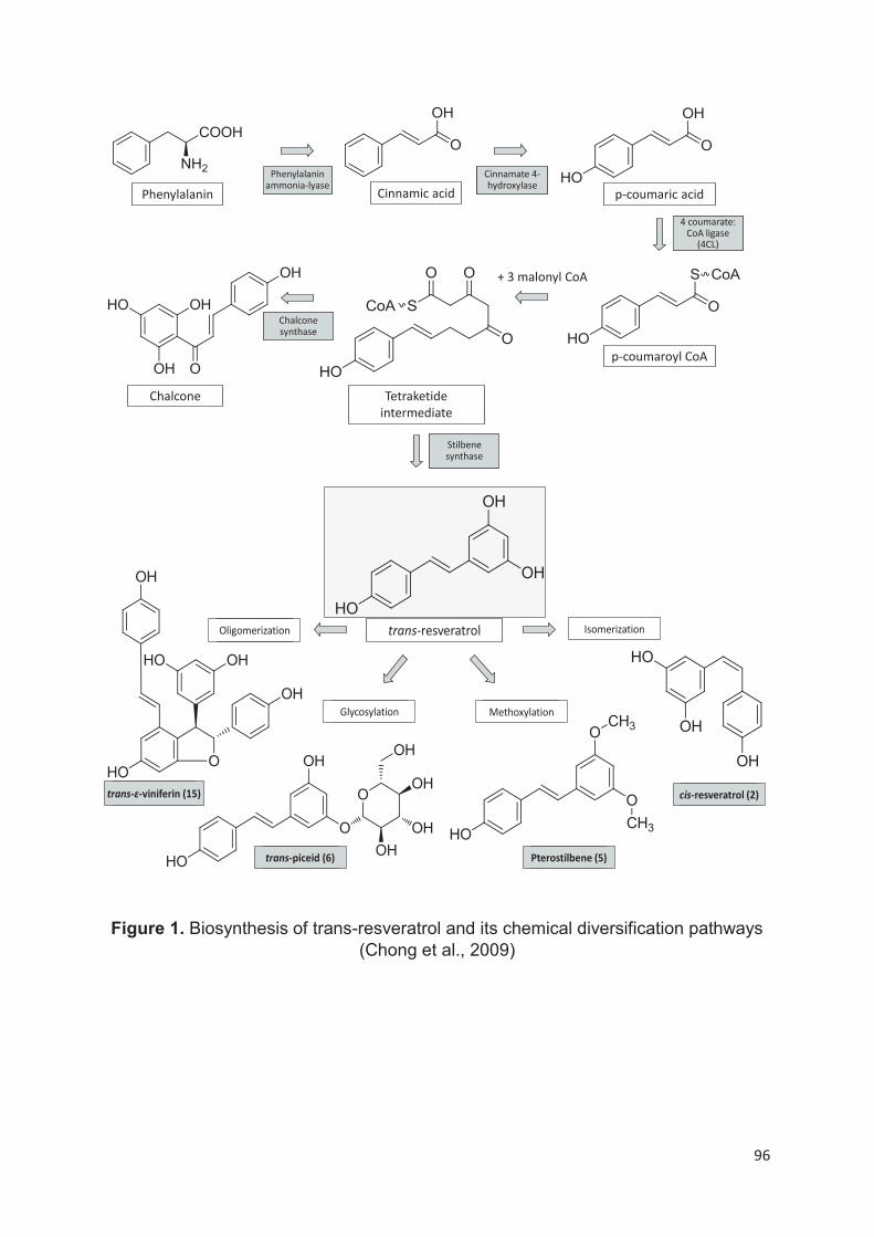
96
Figure 1. Biosynthesis of trans-resveratrol and its chemical diversification pathways (Chong et al., 2009)
Phenylalanin
Phenylalaninammonia-lyase
Cinnamic acid
Cinnamate 4-hydroxylase
p-coumaric acid
4 coumarate: CoA ligase
(4CL)
+ 3 malonyl CoA
Tetraketide
intermediate
Chalcone synthase
Stilbene synthase
p-coumaroyl CoA
Chalcone
trans-resveratrol Isomerization
MethoxylationGlycosylation
Oligomerization
cis-resveratrol (2)
Pterostilbene (5)trans-piceid (6)
trans-ɛ-viniferin (15)
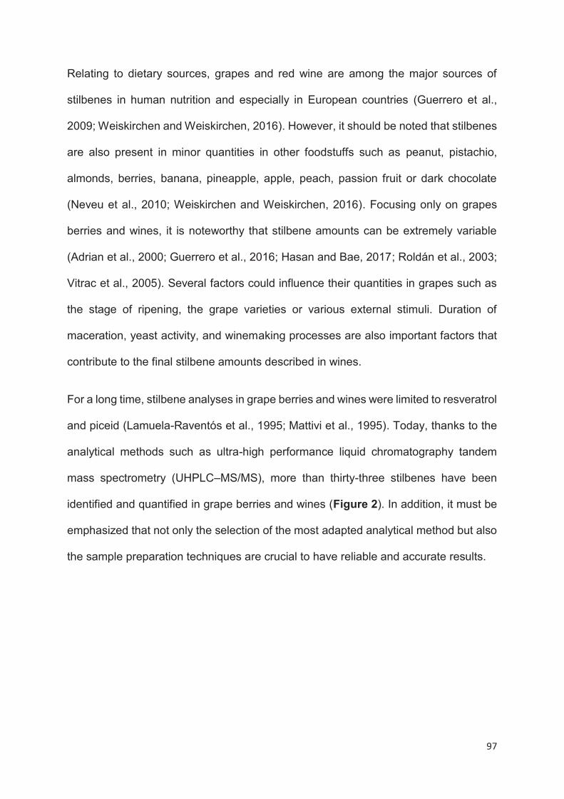
97
Relating to dietary sources, grapes and red wine are among the major sources of
stilbenes in human nutrition and especially in European countries (Guerrero et al.,
2009; Weiskirchen and Weiskirchen, 2016). However, it should be noted that stilbenes
are also present in minor quantities in other foodstuffs such as peanut, pistachio,
almonds, berries, banana, pineapple, apple, peach, passion fruit or dark chocolate
(Neveu et al., 2010; Weiskirchen and Weiskirchen, 2016). Focusing only on grapes
berries and wines, it is noteworthy that stilbene amounts can be extremely variable
(Adrian et al., 2000; Guerrero et al., 2016; Hasan and Bae, 2017; Roldán et al., 2003;
Vitrac et al., 2005). Several factors could influence their quantities in grapes such as
the stage of ripening, the grape varieties or various external stimuli. Duration of
maceration, yeast activity, and winemaking processes are also important factors that
contribute to the final stilbene amounts described in wines.
For a long time, stilbene analyses in grape berries and wines were limited to resveratrol
and piceid (Lamuela-Raventós et al., 1995; Mattivi et al., 1995). Today, thanks to the
analytical methods such as ultra-high performance liquid chromatography tandem
mass spectrometry (UHPLC–MS/MS), more than thirty-three stilbenes have been
identified and quantified in grape berries and wines (Figure 2). In addition, it must be
emphasized that not only the selection of the most adapted analytical method but also
the sample preparation techniques are crucial to have reliable and accurate results.
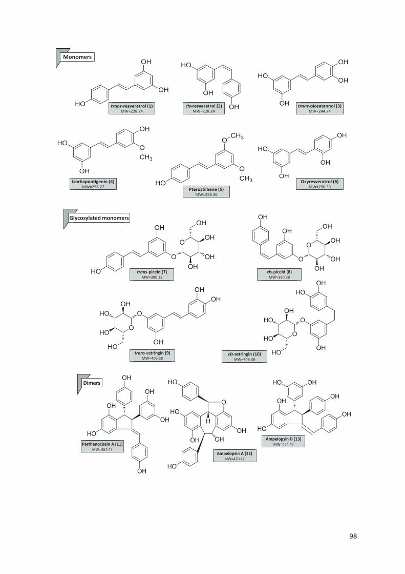
98
Monomers
cis-resveratrol (2)MW=228.24
trans-resveratrol (1)MW=228.24
trans-piceatannol (3)MW=244.24
Isorhapontigenin (4)MW=258.27
Pterostilbene (5)MW=256.30
Oxyresveratrol (6)MW=256.30
Glycosylated monomers
trans-piceid (7)MW=390.38
trans-astringin (9)MW=406.38
cis-astringin (10)MW=406.38
cis-piceid (8)MW=390.38
Dimers
Ampelopsin D (13)MW=454.47Parthenocissin A (11)
MW=457.47Ampelopsin A (12)
MW=470.47
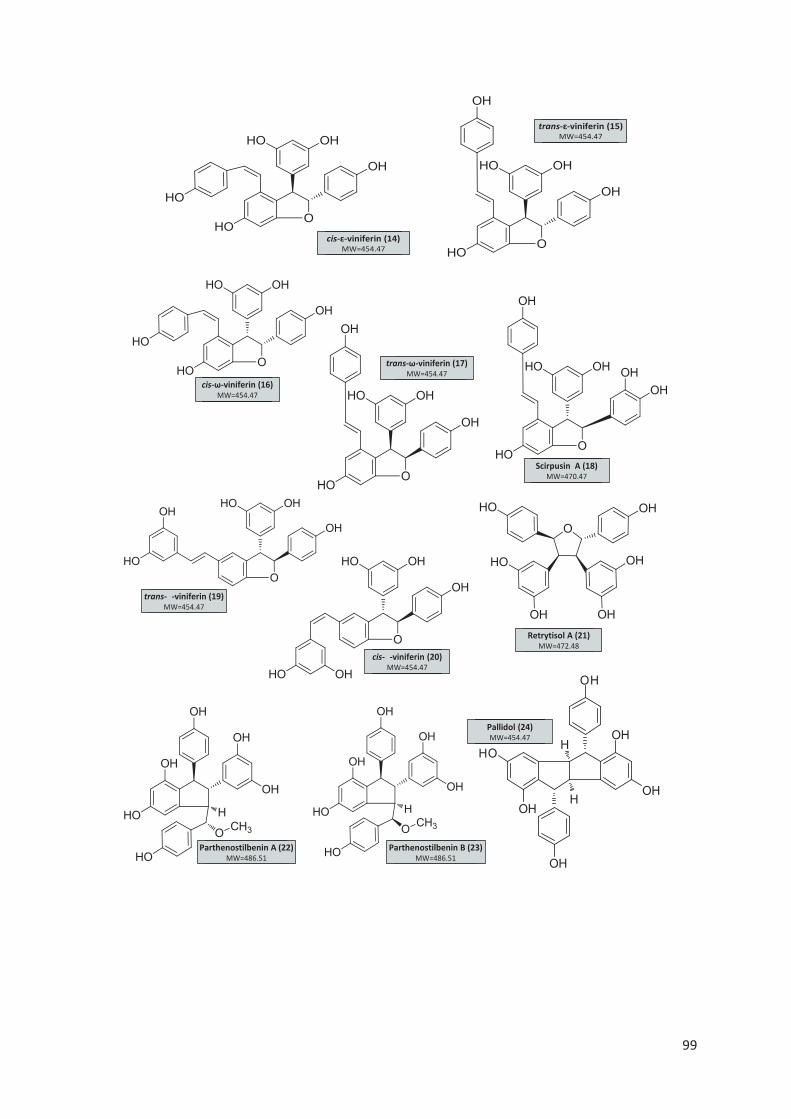
99
cis-ɛ-viniferin (14)MW=454.47
trans-ɛ-viniferin (15)MW=454.47
HO
OH
H
H
OH
OH
OH
OH
cis-�-viniferin (20)MW=454.47
trans-�-viniferin (19)MW=454.47
Pallidol (24)MW=454.47
Parthenostilbenin A (22)MW=486.51
Retrytisol A (21)MW=472.48
Parthenostilbenin B (23)MW=486.51
cis-ω-viniferin (16)MW=454.47
trans-ω-viniferin (17)MW=454.47
Scirpusin A (18)MW=470.47
100
Glycosylated dimer
trans-ɛ-viniferin glucoside (25)MW=616.61
Trimers
α-viniferin (27)MW=678.68
Ampelopsin C (26)MW=680.70
trans-miyabenol C (28)MW=680.70
cis-miyabenol C (29)MW=680.70

101
Figure 2. Main stilbenes identified in grape berries and win
Tetramers
Isohopeaphenol (32)MW=906.92
Vitisin A (33)MW=906.92
Vitisin B (34)MW=906.92
Hopeaphenol (31)MW=906.92
102
Stilbenes have demonstrated to possess a great range of biological activities
potentially beneficial for human health such as neuroprotective, antioxidant and
antitumor effects (Guerrero et al., 2009; Vang et al., 2011; Weiskirchen and
Weiskirchen, 2016). Among the more recent research lines, stilbenes are gaining
considerable interest as potential anti-obesity agents. Obesity is the most common
nutritional disorder in the world. According to the World Health Organization (WHO),
obesity is defined as an abnormal or excessive fat accumulation that may impact
health. Prevalence data estimate that 650 millions of adults were obese in 2016,
representing about 13% of the world adult population. Moreover, alarming figures are
presented regarding young population. In fact, more than 124 millions of children and
adolescents were obese in 2016 (WHO, 2018, http://www.who.int/news-room/fact-
sheets/detail/obesity-and-overweight). It is well known that the fundamental cause of
obesity is an energy imbalance between calories intake and loss, as a consequence
globally, of a high intake of energy-fat foods and a sedentary style of life. When a
positive energy imbalance occurs, triglycerides (TG) are accumulated in adipocytes
producing an increase in the number of adipocyte (hyperplasia) or an increase of its
size (hypertrophy) (Hausman et al., 2001). Additionally, obesity can also bring a
potential risk to develop cardiovascular diseases, diabetes and some types of cancer,
endometrial, breast, ovarian, prostate, liver, gallbladder, kidney, and colon, among
others (Gallagher and LeRoith, 2015). Due to the increase of the prevalence and the
associated complications, several global government policies, laws and regulations
have been developed to halt and reverse the obesity epidemic. The first solution to
stem obesity is focused in a change of lifestyle by decreasing the energy/fat/sugar
intake and increasing physical activity. However, the implementation and effectiveness
of these recommendations are usually unsatisfactory. It is therefore that research
103
community tries to find anti-obesity bioactive molecules, as stilbenes, that can be
combined with other recommendations and treatments in order to ameliorate the
results.
It has been shown that stilbenes may reduce obesity by regulating different pathways
related to fat metabolism as adipogenesis, lipogenesis, lipolysis and thermogenesis
(Chou et al., 2018; Fernández-Quintela et al., 2017). Resveratrol has been the most
studied bioactive compound, but a considerable number of works are already
indicating that other stilbenes are promising anti-obesity agents and may even be more
potent than resveratrol.
For all above explained, the aim of this review is to address and discuss about:
i) stilbene compounds in grape berries and wines, the principal factors that can
modulate stilbenes concentrations and the most efficient sample preparation
techniques and analytical methods used for their detection;
ii) the most important investigations related to the in vitro and in vivo anti-obesity
effects of grape and wine stilbenes and the associated molecular mechanisms.
104
Stilbenes content in grape berries and wine
Grape berries stilbenes
In grape berries, stilbenes as other polyphenols are mainly concentrated in skins
(Babazadeh et al., 2017). They present a great variability in composition and content
depending on different biotic and abiotic factors including grape variety, stage of
ripening, viticultural factors and practices (Bavaresco et al., 2012; Błaszczyk et al.,
2019). For example, according to Adrian et al. (Adrian et al., 2000), resveratrol
concentration was 2 to 30 fold higher in berries induced with UV light compared to non-
induced ones.
The stilbene content offers sharp contrasts due to many potential external stimuli
(Vincenzi et al., 2013). Red varieties seem to present higher stilbene content than white
ones (Guerrero et al., 2010; Viñas et al., 2011). Concerning monomers, glucosides
(piceid and astringin) are the main compounds identified in grape berries (Table 1).
The piceid (mean 1.36 mg/kg, cis- and trans-isomers) is the main compound followed
by astringin (mean 0.83 mg/kg), and resveratrol (mean 0.68 mg/kg). Both isomeric
forms, trans- and cis-isomers, were identified in similar levels for piceid and mainly
trans- forms for astringin and resveratrol. These compounds were observed in all berry
growth stages in different concentrations (Jeandet et al., 1991). They are subject to
enzymatic transformations leading to the formation of a pool of different compounds
(Chong et al., 2009). The hydroxylation of resveratrol and piceid leads to the formation
of piceatannol and its glucoside, astringin (Bavaresco et al., 2002). In addition to these
three monomers some other minor monomers were identified such as pterostilbene
(Pezet et al., 1994), and isorhapontigenin (Fernández-Marín et al., 2012). The
oxidative coupling of resveratrol induces the formation of more complex oligomers
including dimers, trimers and tetramers (Takaya et al., 2005). Significant levels of
oligomers were reported (Flamini et al., 2013; Rosso et al., 2016). In Vitis vinifera
cultivars, dimers compounds were identified such as pallidol (Vrhovsek et al., 2012),
ε-viniferin and δ-viniferin (Flamini et al., 2013).

105
In addition to these dimers, trimers were identified, like miyabenol C and α-viniferin,
and a pool of tetramers including hopeaphenol and isohopeaphenol (Flamini et al.,
2013; Flamini et al., 2016; Rosso et al., 2016). Unfortunetly, few studies were focused
on the quantification of these complex compounds in grape berries (Flamini et al.,
2013). In contrast, wild Vitis species appear to contain a greater number and diversity
of oligomeric stilbenes (He et al., 2009; Jiang et al., 2012).
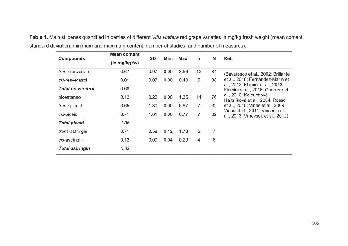
106
Table 1. Main stilbenes quantified in berries of different Vitis vinifera red grape varieties in mg/kg fresh weight (mean content,
standard deviation, minimum and maximum content, number of studies, and number of measures).
Compounds Mean content
(in mg/kg fw) SD Min. Max. n N Ref.
trans-resveratrol 0.67 0.97 0.00 3.56 12 84 (Bavaresco et al., 2002; Brillante et al., 2018; Fernández-Marín et al., 2013; Flamini et al., 2013; Flamini et al., 2016; Guerrero et al., 2010; Kolouchová-Hanzlíková et al., 2004; Rosso et al., 2016; Viñas et al., 2009; Viñas et al., 2011; Vincenzi et al., 2013; Vrhovsek et al., 2012)
cis-resveratrol 0.01 0.07 0.00 0.40 5 38
Total resveratrol 0.68
piceatannol 0.12 0.22 0.00 1.35 11 76
trans-piceid 0.65 1.30 0.00 6.87 7 32
cis-piceid 0.71 1.61 0.00 6.77 7 32
Total piceid 1.36
trans-astringin 0.71 0.58 0.12 1.73 5 7
cis-astringin 0.12 0.09 0.04 0.29 4 6
Total astringin 0.83

107
Different biotic and abiotic factors affect the stilbene content in grape berries, including
the grape varieties or species, stage of ripening, environmental conditions or
postharvest treatments (Błaszczyk et al., 2019; Hasan and Bae, 2017). The main
factors that can have an impact on stilbene quantities in grapes were described below.
Stage of ripening
The development of grape berries is a dynamic process that involves a complex series
of changes divided into three major phases: before veraison, veraison and after
veraison. Before veraison, the cell division is rapid and all cells are established within
two weeks of flowering, followed by a subsequent sigmoidal increase in berry size
(Deluc et al., 2007). At the end of this period, the STS is accumulated and reaches its
maximum two weeks before veraison. The stage after veraison is characterized by the
initiation of colour development (accumulation of anthocyanins in red grapes) until
maturity and the accumulation of stilbenes (Wang et al., 2016). Even if the STS its
maximum level two weeks before veraison, the accumulation of stilbenes only start in
the second week after veraison and then increases until maturity (Wang et al., 2016).
Grape varieties and species
The accumulation of stilbenes seems to depend on the Vitis vinifera grape cultivars. In
a comparative study between three different cultivars, Syrah, Tempranillo and Merlot,
Guerrero et al. have shown that Syrah had ten times more resveratrol than Tempranillo
(Guerrero et al., 2010). This result was confirmed by Gatto et al. that compared 78
different cultivars during three years (Gatto et al., 2008). Firstly, they observed higher
stilbene content in red varieties than in white or pink ones. Secondly, Pinots and related
cultivars exhibited the highest stilbene levels. Biochemical and transcriptomic analyses
have shown the genotype influence on stilbene accumulation in healthy grapevine
berries. In the high yielding varieties, glucosylated forms such as piceid were
accumulated preferentially, resveratrol being mainly produced after external stimuli
such as fungal infection.
Nevertheless, these data have to be substantiated by consistent results because
external stimuli, such as light or pathogen infection could have higher effect than the
intrinsic differences between cultivars (Hasan and Bae, 2017). As for other secondary
metabolites (Acevedo de la Cruz et al., 2012; Hilbert et al., 2015), it is likely that wild
108
species present higher content and diversity of stilbene than the cultivated one (Jiang
et al., 2012). In Southern China grapes (Vitis chunganensis), it was reported that
resveratrol content twenty times higher than in berries of cultivated grape(He et al.,
2009). In addition many oligomers were identified with high contents such as
amurensin G (262 mg/kg), amurensin B (242 mg/kg) or r2-viniferin (191 mg/kg). These
results are in agreement with the data concerning other parts of the grapevine, such
stem or roots. Wild Vitis species, that have higher resistance to external stress, seem
to contain more stilbenes than cultivated vine (Pawlus et al., 2013b).
External stimuli
Several external stimuli could modulate the stilbene content in grape berries including
pathogen infection, elicitor applications, or UVC treatments. The effects of stimuli could
increase the stilbene content more than ten times the normal. As stilbenes are well-
known phytoalexins (Langcake and Pryce, 1977), the effect of different pathogens on
stilbenes accumulation in grape was widely investigated. Infection by Botrytis cinerea
induce the accumulation of resveratrol mainly in its glucosidic form (Roldán et al.,
2003). Similar results were observed for Plasmopara viticola, Uncinula necator and
Rhizopus stolonifera (Hasan and Bae, 2017), with accumulation rate ranging between
three and twelve times than control. Remarkably, the increase of the resveratrol level
in berries due to pathogen infection does not lead to a significant modification of the
stilbene content in wines (Jeandet et al., 1995; Roldán et al., 2003). Authors suppose
that grape berries with a high Botrytis infestation presented lower resveratrol contents
due to the oxidation of resveratrol by the laccase enzymes secreted by fungi. In the
same manner, chemical compounds such as elicitors are able to induce the production
of stilbenes (Krisa et al., 1999). This effect was mainly demonstrated in grape cell
cultures using different chemicals such as methyl jasmonate, salicylic acid, glucan, or
chitosan (Vuong et al., 2014). The induction could rise up stilbene production to ten
times the control. This observation was confirmed in grape berries, where methyl
jasmonate induced a significant increase of resveratrol, piceatannol, isorhapontigenin
and ε-viniferin content up to two times in comparison to the control (Fernández-Marín
et al., 2014). Physical processes could also stimulate the production of stilbenes. Pre-
and post-harvest treatments by UVC induce an increase of the stilbene content in
grape berries (Adrian et al., 2000; Guerrero et al., 2010). The increase rate depends
on the variety, the wavelength of the radiation, and the duration of treatment. The

109
values were mainly ranged between one and twenty times the normal resveratrol
concentration (Błaszczyk et al., 2019). In addition to the increase in the amount of
resveratrol, levels of other stilbenes rise in the same proportion. This effect was
observed on different stilbenes including piceid, piceatannol, ε-viniferin, δ-viniferin and
hopeaphenol (Guerrero et al., 2016).
Wine stilbenes
Wine is an important dietary source of resveratrol (Kasiotis et al., 2013), in which it can
be found at concentrations up to 20 mg/L (Ribeiro De Lima et al., 1999). As for grape
berries, the monomeric forms are the most abundant stilbenes in wine, while oligomeric
forms were mainly identified in red wines (Guerrero et al., 2020). The main compounds
identified in wines were piceid (cis- and trans-isomers), resveratrol (cis- and trans-
isomers), astringin (cis- and trans-isomers) and piceatannol (Tables 2 and 3). In
addition to these compounds several other stilbenes were identified including dimers
such as pallidol (Landrault et al., 2002), ε-viniferin (Amira-Guebailia et al., 2009;
Landrault et al., 2002), δ-viniferin (Moss et al., 2013; Vitrac et al., 2005), ω-viniferin
(Guerrero et al., 2020), parthenocissin A (Vitrac et al., 2001), quadrangularin A (Pawlus
et al., 2013a); trimers such as miyabenol C (Guerrero et al., 2020), and α-viniferin
(Arraki et al., 2017); and tetramers such as hopeaphenol (Guebailia et al., 2006),
isohopeaphenol, and r2-viniferin (Guerrero et al., 2020). Glucosidic and diglucosidic
forms of these compounds were also identified including ε-viniferin diglucosides,
pallidol 3-O-glucoside, and pallidol diglucosides (Baderschneider and Winterhalter,
2000). In addition, analyses by mass spectrometry indicated the presence of several
other oligomers in wines (Moss et al., 2013).
Red wines (Table 2) have a higher content in stilbenes than white wines (Table 3).
Piceid is the main stilbene in wines (means 8.26 and 0.88 mg/L in red and white wines,
respectively). Surprisingly, the cis-isomer levels are higher in both cases (means 6.20
and 0.68 mg/L in red and white wines, respectively). In red wines, the piceid is followed
by resveratrol (mean 2.29 mg/L). The derivatives of piceatannol are present at a
concentration of about 1.73 mg/L. Oligomers are minor compounds except
isohopeaphenol with a mean value of 1.39 mg/L in red wines (Table 2). No oligomers
were reported in white wines.
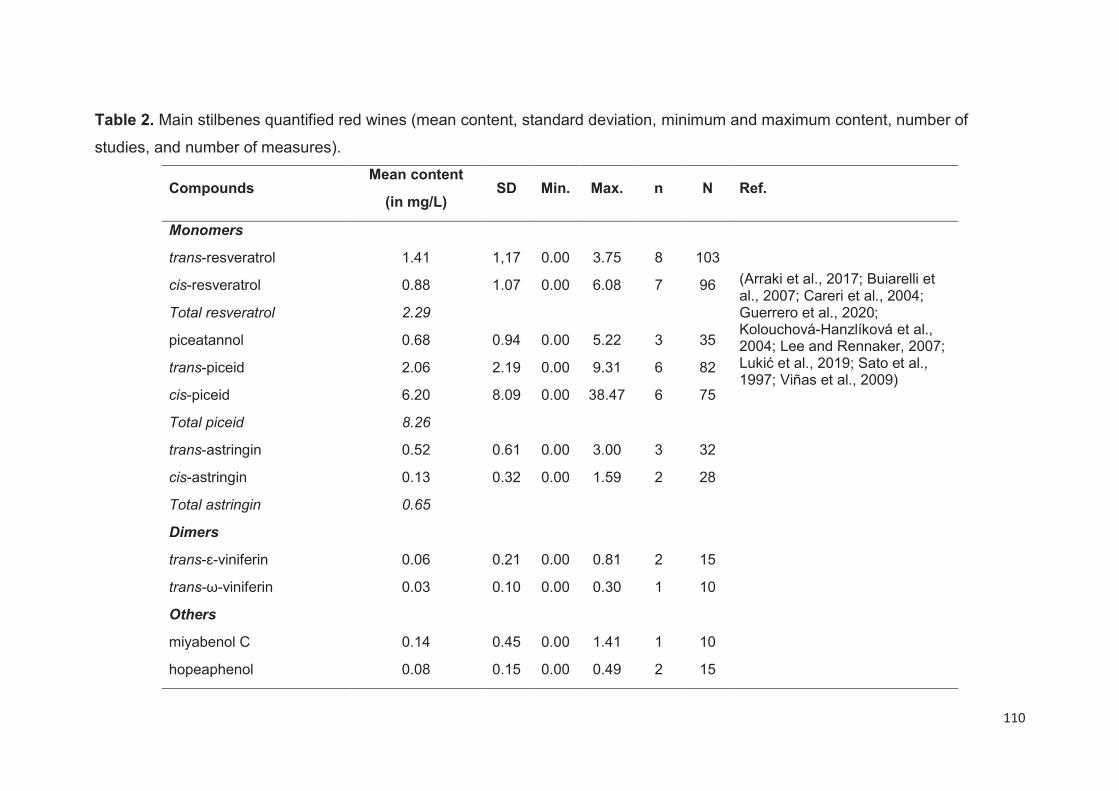
110
Table 2. Main stilbenes quantified red wines (mean content, standard deviation, minimum and maximum content, number of
studies, and number of measures).
Compounds Mean content
(in mg/L) SD Min. Max. n N Ref.
Monomers
(Arraki et al., 2017; Buiarelli et al., 2007; Careri et al., 2004; Guerrero et al., 2020; Kolouchová-Hanzlíková et al., 2004; Lee and Rennaker, 2007; Lukić et al., 2019; Sato et al., 1997; Viñas et al., 2009)
trans-resveratrol 1.41 1,17 0.00 3.75 8 103
cis-resveratrol 0.88 1.07 0.00 6.08 7 96
Total resveratrol 2.29
piceatannol 0.68 0.94 0.00 5.22 3 35
trans-piceid 2.06 2.19 0.00 9.31 6 82
cis-piceid 6.20 8.09 0.00 38.47 6 75
Total piceid 8.26
trans-astringin 0.52 0.61 0.00 3.00 3 32
cis-astringin 0.13 0.32 0.00 1.59 2 28
Total astringin 0.65
Dimers
trans-ԑ-viniferin 0.06 0.21 0.00 0.81 2 15
trans-ω-viniferin 0.03 0.10 0.00 0.30 1 10
Others
miyabenol C 0.14 0.45 0.00 1.41 1 10
hopeaphenol 0.08 0.15 0.00 0.49 2 15
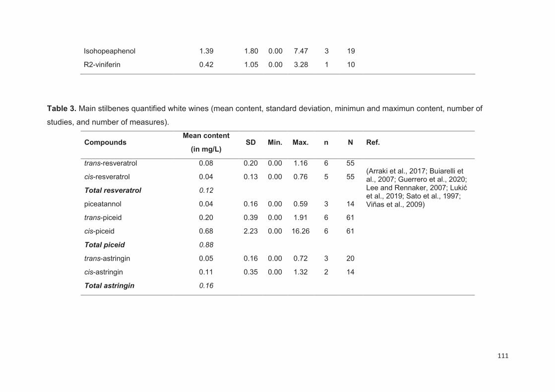
111
Isohopeaphenol 1.39 1.80 0.00 7.47 3 19
R2-viniferin 0.42 1.05 0.00 3.28 1 10
Table 3. Main stilbenes quantified white wines (mean content, standard deviation, minimun and maximun content, number of
studies, and number of measures).
Compounds Mean content
(in mg/L) SD Min. Max. n N Ref.
trans-resveratrol 0.08 0.20 0.00 1.16 6 55 (Arraki et al., 2017; Buiarelli et al., 2007; Guerrero et al., 2020; Lee and Rennaker, 2007; Lukić et al., 2019; Sato et al., 1997; Viñas et al., 2009)
cis-resveratrol 0.04 0.13 0.00 0.76 5 55
Total resveratrol 0.12
piceatannol 0.04 0.16 0.00 0.59 3 14
trans-piceid 0.20 0.39 0.00 1.91 6 61
cis-piceid 0.68 2.23 0.00 16.26 6 61
Total piceid 0.88
trans-astringin 0.05 0.16 0.00 0.72 3 20
cis-astringin 0.11 0.35 0.00 1.32 2 14
Total astringin 0.16

112
Several parameters are able to modulate the stilbene content in wine including mainly
the extraction from grape, but also yeast strains, and enological practices.
Duration of maceration
Red wines contain higher stilbene levels than white wines that are obtained from a
limited maceration with the pomaces (Tables 2 and 3). In fact, the stilbene content in
wines is strongly modulated by the duration of the maceration and the solubility of these
compounds in alcohol (Kostadinović et al., 2012; Mattivi et al., 1995). It has been
shown that wines obtained after 6 days of maceration had the highest concentration of
resveratrol and piceid than wine obtained after 3 days (Kostadinović et al., 2012),
reaching to the maximal content (Poussier et al., 2003). Due to their polarity,
monomeric glucosides are extracted before their aglycone forms (Mattivi et al., 1995).
While oligomers such as δ-viniferin are observed in wines after few days of maceration
(Poussier et al., 2003). After a long time of maceration the concentration of stilbenes
decrease mainly that of glucosides such as cis- and trans-piceid (Poussier et al., 2003).
The decrease of piceid could be attributed to the β-glucosidase activity of the different
yeasts (Jeandet et al., 1994). In addition to this phenomenon, the cis/trans
isomerization of stilbenes could explain the formation of some of these compounds
such as cis-resveratrol. The levels of this compound are similar to those of trans-
resveratrol in red wines (Table 2) and significantly lower in grapes (Table 1). Finally,
even if few studies about resveratrol degradation during maceration were conducted,
yeasts seem to be able to induce a degradation of resveratrol during wine fermentation
(Vacca et al., 1997).
Yeast activities
As previously mentioned, yeast activities influence the stilbene content in wines. A
comparison between French and Macedonian yeasts demonstrated a variation of
resveratrol content up to four times depending on the yeast and maceration times
(Kostadinović et al., 2012). The β-glucosidase activities of yeasts induce the hydrolyze
of glucosides which leads to the formation of non glucosylated stilbenes (Mattivi et al.,
1995). Thus, the selection of exogenous yeasts could significantly affect the stilbene
content in wines. The use of specific yeasts enriched in β-glucosidase could positively
113
increase the level of resveratrol in wines (González-Candelas et al., 2000).
Nevertheless more studies are needed to better understand the impact of yeast
activities on stilbene content in wines.
Winemaking processes and wine ageing
Few studies were focused on the impact of winemaking processes on the stilbene
content in wines. Grapes exposure to UVC radiations before winemaking induces an
increase of stilbene production in wines (Cantos et al., 2003; Threlfall et al., 1999).
This positive effect is observed on monomeric stilbenes such as resveratrol and
piceatannol whereas other stilbenes such as ε-viniferin are not affected (Fernández-
Marín et al., 2014). On the contrary, the use of some fining agents, such as
polyvinylpolypyrrolidone (PVPP), could reduce the stilbene content in wines (Threlfall
et al., 1999; Vrhovsek et al., 1997).
As for winemaking processes few studies were focusing on the stilbene stability during
wine ageing. Stilbenes are known to be relatively stable compounds (Bavaresco et al.,
2012; Gaudette and Pickering, 2011). Nevertheless, even if the total stilbene content
is not affected, the ratio between resveratrol and piceid isomers could be impacted
during wine ageing (Favre et al., 2020; Sun et al., 2006). In addition some specific
ageing processes such as those used in Sherry wines could reduce the resveratrol
content (Roldán et al., 2010). Finally, heat could induce oxidative coupling between
monomeric stilbenes inducing the formation of oligomers in wines (El Khawand et al.,
2020).
Analytical methods for stilbenes analysis in grapes and wines
The relatively low concentration of stilbenes, their structural diversity, and the
complexity of grape and wine matrices restrain their identification and quantification.
Today, the studies are mainly focused on the monomeric stilbenes. Several analytical
procedures were developed in order to determine the stilbene content in grapevines
and wines from monomers to complex oligomers. These strategies often require multi-
steps sample preparation (Baderschneider and Winterhalter, 2000; Liu et al., 2013;
Romero-Pérez et al., 2001).
Concerning grapevines, the first step consists of the extraction of stilbenes from the
114
raw material. Several extraction procedures were applied including classical solvent
extraction from fresh, frozen, dried or lyophilized berries (Romero-Pérez et al., 2001),
or more recently ultra sonication-assisted extraction (Cho et al., 2006). The classical
solvent extraction remains the most applied technique the main used methodology
using acetone, chloroform, ethanol, ethyl acetate, methanol, and some mixed solvents.
Methanol, pure or mixed, is the most widely used solvent for stilbene extraction from
grapevine berries. Several studies were focused on optimizing the extraction
conditions form grapevine berries (Liu et al., 2013; Romero-Pérez et al., 2001; Sun et
al., 2006). The main parameters analyzed were the solvent, the extraction time, the
temperature, and the ratio solid to solvent. Based on Liu et al. (2013) studies (Liu et
al., 2013), the best extraction solutions were: methanol or methanol/ethyl acetate (1:1
(v/v)) with a ratio solid to solvent of 1 g/10 mL at 25°C for 24 h. In addition, in order to
avoid cis/trans isomerization, extraction should be carried out in the dark (Careri et al.,
2003). Interestingly, innovative extraction techniques such as ultrasonication-assisted
extraction seem to be able to increase the stilbene extraction rate while reducing time
and solvent consumption (Cho et al., 2006).
As for grape berries, several methods were proposed to extract stilbenes in wines,
from direct analyses (Lamuela-Raventós et al., 1995) to more complex procedures
using solid phase extraction (Mattivi et al., 1995), stir bar sorptive extraction (Viñas et
al., 2008), microextraction by packed sorbents (Gonçalves and Câmara, 2011), liquid–
liquid extraction (Rabesiaka et al., 2011), dispersive liquid–liquid microextraction
(Rodríguez-Cabo et al., 2012), semi preparative liquid chromatography (Amira-
Guebailia et al., 2009), or counter current chromatography (Fernandez-Marin et al.,
2012). The main drawback of these methods is that they are mainly focused on
resveratrol and piceid. In the research conducted by Baderschneider and Winterhalter
a combination of solid phase extraction and counter current chromatography were
used to identify new stilbene skeletons in wines (Baderschneider and Winterhalter,
2000).
Several methods were used for stilbene analysis in grape berries and wines including
capillary electrophoresis (CE), gas chromatography (GC), high performance liquid
chromatography (HPLC) and ultra-high performance liquid chromatography (UHPLC).
115
Capillary electrophoresis
The capillary electrophoresis is appropriate for the separation of small molecules and
could separate some optical isomeric forms. This method allows separating positional
isomers as well as optical isomers. This method has different advantages such as fast
and accurate analysis, low amount and consumption (Coelho et al., 2016). This
technique was successfully applied for resveratrol analysis in wines (Gu et al., 2000),
and to discriminate cis- and trans-resveratrol (Paulo et al., 2011). Nevertheless, this
method was used to identify a limited number of stilbenes. In addition, due to its low
sensitivity, concentrating the wine sample is recommended using solid phase or liquid–
liquid extractions before proceeding to capillary electrophoresis (Gu et al., 2000;
Spanilá et al., 2005).
Gas chromatography
Gas chromatography was successfully used for resveratrol quantification in wines
(Barbanti et al., 1996), and grapes (Viñas et al., 2009). The use of GC for stilbene
analysis involves the derivatization of the compound using derivatizing reagents, such
as bis(trimethylsilyl)trifluoroacetamide (BSTFA) (Luan et al., 2000), or acetic anhydride
(Montes et al., 2010), in order to increase their volatility. Silylation is the most common
derivatization reaction used in combination with solid phase or liquid-liquid extraction
pretreatments for grape and wine analyses (Cai et al., 2009; Viñas et al., 2009). In
addition, GC analyses were coupled with mass spectrometry detector (GC–MS) in
order to increase the accuracy and sensibility of the detection (Rodríguez-Cabo et al.,
2016). These methods provide limits of detection close to ng/L in wines. However, GC
analyses have different drawbacks such as isomerization, degradation of analytes.
Liquid Chromatography
High-performance liquid chromatography (HPLC) is considered among the most
commonly applied method for stilbenes analysis in grapes (Błaszczyk et al., 2019) and
wines (Fabjanowicz et al., 2018), using different detection systems such as UV-visible
or diode array detectors (Mattivi et al., 1995; Sun et al., 2006), or fluorescence
detectors in order to increase sensitivity and specificity (Vitrac et al., 2002). Nowadays,
116
HPLC combined with mass spectrometry (HPLC-MS) has become the most used
technique for the determination of phenolic compounds in general and stilbenes in
particular(Pugajeva et al., 2018). This method allows the identification of all stilbenes
(free and conjugated) without any derivatization or hydrolysis of samples. Different
mass detectors were used including triple quadrupole (QqQ) and quadrupole time-of-
flight (QTOF). Information on liquid chromatography methods developed for stilbenes
identification and quantification in grape berries and wines are summarized in Table 4.
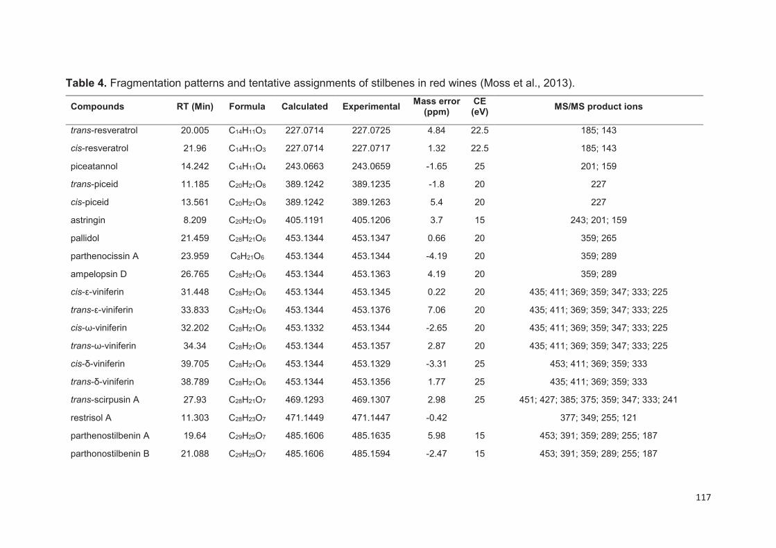
117
Table 4. Fragmentation patterns and tentative assignments of stilbenes in red wines (Moss et al., 2013).
Compounds RT (Min) Formula Calculated Experimental Mass error
(ppm) CE (eV)
MS/MS product ions
trans-resveratrol 20.005 C14H11O3 227.0714 227.0725 4.84 22.5 185; 143
cis-resveratrol 21.96 C14H11O3 227.0714 227.0717 1.32 22.5 185; 143
piceatannol 14.242 C14H11O4 243.0663 243.0659 -1.65 25 201; 159
trans-piceid 11.185 C20H21O8 389.1242 389.1235 -1.8 20 227
cis-piceid 13.561 C20H21O8 389.1242 389.1263 5.4 20 227
astringin 8.209 C20H21O9 405.1191 405.1206 3.7 15 243; 201; 159
pallidol 21.459 C28H21O6 453.1344 453.1347 0.66 20 359; 265
parthenocissin A 23.959 C8H21O6 453.1344 453.1344 -4.19 20 359; 289
ampelopsin D 26.765 C28H21O6 453.1344 453.1363 4.19 20 359; 289
cis-ε-viniferin 31.448 C28H21O6 453.1344 453.1345 0.22 20 435; 411; 369; 359; 347; 333; 225
trans-ε-viniferin 33.833 C28H21O6 453.1344 453.1376 7.06 20 435; 411; 369; 359; 347; 333; 225
cis-ω-viniferin 32.202 C28H21O6 453.1332 453.1344 -2.65 20 435; 411; 369; 359; 347; 333; 225
trans-ω-viniferin 34.34 C28H21O6 453.1344 453.1357 2.87 20 435; 411; 369; 359; 347; 333; 225
cis-δ-viniferin 39.705 C28H21O6 453.1344 453.1329 -3.31 25 453; 411; 369; 359; 333
trans-δ-viniferin 38.789 C28H21O6 453.1344 453.1356 1.77 25 435; 411; 369; 359; 333
trans-scirpusin A 27.93 C28H21O7 469.1293 469.1307 2.98 25 451; 427; 385; 375; 359; 347; 333; 241
restrisol A 11.303 C28H23O7 471.1449 471.1447 -0.42 377; 349; 255; 121
parthenostilbenin A 19.64 C29H25O7 485.1606 485.1635 5.98 15 453; 391; 359; 289; 255; 187
parthonostilbenin B 21.088 C29H25O7 485.1606 485.1594 -2.47 15 453; 391; 359; 289; 255; 187

118
ε-viniferin glucoside 25.202 C34H31O11 615.1872 615.1878 0.98 20 453; 411; 359; 347
ampelopsin C 30.044 C42H32O9 679.1974 679.1981 1.03 30 585; 573; 491; 479; 385
trans-miyabenol C 35.372 C42H32O9 679.1974 679.1978 0.59 30 661; 637; 585; 573; 555; 451; 479; 357; 345
cis-miyabenol C 36.322 C42H32O9 679.1974 679.1984 1.47 30 661; 637; 585; 573; 555; 479; 451; 357; 345
hopeaphenol 31.695 C56H41O12 905.2604 905.2573 -3.42 35 811; 717; 611; 451; 359; 265
119
In recent years, UHPLC coupled with mass spectrometry (UHPLC-MS) has been
widely used as an alternative for other methods due to its higher sensitivity, accuracy,
rapidity, and low solvent consumption for grape berry (Flamini et al., 2013), and wine
analyses (Guerrero et al., 2020; Moss et al., 2013). For example, based on a targeted
metabolomics approach using UHPLC-QTOF mass spectrometer, Flamini et al. have
identified and quantified eighteen stilbenes in grape berries including oligomers. In
wines, an UHPLC-QqQ-MS method was designed to identify and quantify fifteen
stilbenes in white and red wines with limit of detection ranging between 4 and 28 μg/L
(Guerrero et al., 2020). Similarly, using a UHPLC-QTOF mass spectrometer, forty-one
stilbenes were identified in red wines, including six monomers, twenty-three dimers,
eight trimers and four tetramers (Table 5).
Due to the complexity and the diversity of stilbene structures, pure compounds are
needed as standard to obtain accurate results. Unfortunately, since pure samples of
complex stilbene oligomers are often unavailable, results are expressed in resveratrol
or piceid equivalent (Flamini et al., 2013). This approach could lead to severe
underestimation of the oligomeric stilbene content (Biais et al., 2017). For example,
the quantification of ε-viniferin using resveratrol by a HPLC-DAD method
underestimated the concentration ε-viniferin, R-viniferin and isohopeaphenol by a
factors upper than two, five and ten, respectively.
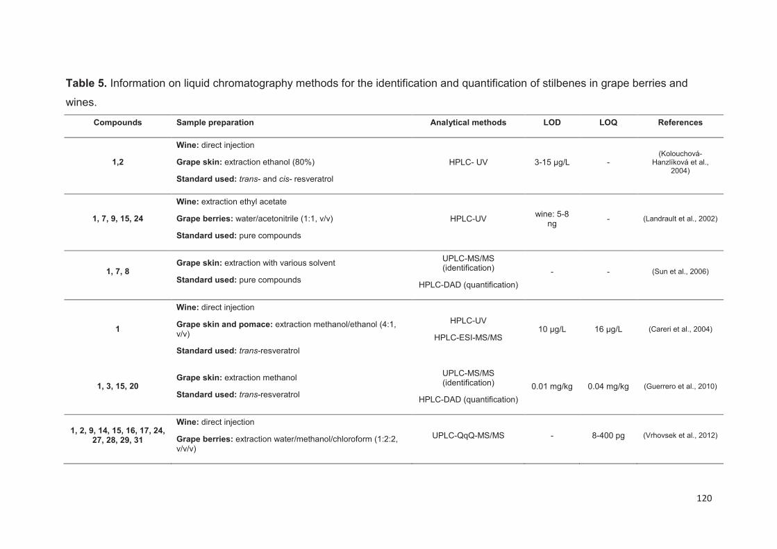
120
Table 5. Information on liquid chromatography methods for the identification and quantification of stilbenes in grape berries and
wines.
Compounds Sample preparation Analytical methods LOD LOQ References
1,2
Wine: direct injection
Grape skin: extraction ethanol (80%)
Standard used: trans- and cis- resveratrol
HPLC- UV 3-15 µg/L - (Kolouchová-
Hanzlíková et al., 2004)
1, 7, 9, 15, 24
Wine: extraction ethyl acetate
Grape berries: water/acetonitrile (1:1, v/v)
Standard used: pure compounds
HPLC-UV wine: 5-8
ng - (Landrault et al., 2002)
1, 7, 8 Grape skin: extraction with various solvent
Standard used: pure compounds
UPLC-MS/MS (identification)
HPLC-DAD (quantification)
- - (Sun et al., 2006)
1
Wine: direct injection
Grape skin and pomace: extraction methanol/ethanol (4:1, v/v)
Standard used: trans-resveratrol
HPLC-UV
HPLC-ESI-MS/MS 10 μg/L 16 μg/L (Careri et al., 2004)
1, 3, 15, 20 Grape skin: extraction methanol
Standard used: trans-resveratrol
UPLC-MS/MS (identification)
HPLC-DAD (quantification)
0.01 mg/kg 0.04 mg/kg (Guerrero et al., 2010)
1, 2, 9, 14, 15, 16, 17, 24, 27, 28, 29, 31
Wine: direct injection
Grape berries: extraction water/methanol/chloroform (1:2:2, v/v/v)
UPLC-QqQ-MS/MS - 8-400 pg (Vrhovsek et al., 2012)
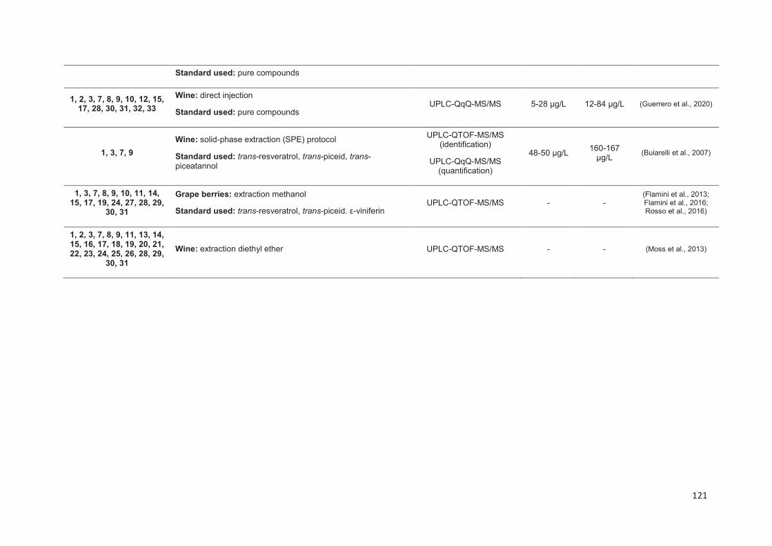
121
Standard used: pure compounds
1, 2, 3, 7, 8, 9, 10, 12, 15, 17, 28, 30, 31, 32, 33
Wine: direct injection
Standard used: pure compounds UPLC-QqQ-MS/MS 5-28 μg/L 12-84 μg/L (Guerrero et al., 2020)
1, 3, 7, 9
Wine: solid-phase extraction (SPE) protocol
Standard used: trans-resveratrol, trans-piceid, trans-piceatannol
UPLC-QTOF-MS/MS (identification)
UPLC-QqQ-MS/MS (quantification)
48-50 μg/L 160-167
μg/L (Buiarelli et al., 2007)
1, 3, 7, 8, 9, 10, 11, 14, 15, 17, 19, 24, 27, 28, 29,
30, 31
Grape berries: extraction methanol
Standard used: trans-resveratrol, trans-piceid. ε-viniferin UPLC-QTOF-MS/MS - -
(Flamini et al., 2013; Flamini et al., 2016; Rosso et al., 2016)
1, 2, 3, 7, 8, 9, 11, 13, 14, 15, 16, 17, 18, 19, 20, 21, 22, 23, 24, 25, 26, 28, 29,
30, 31
Wine: extraction diethyl ether UPLC-QTOF-MS/MS - - (Moss et al., 2013)
122
Stilbenes, in vitro and in vivo anti-obesity effects and molecular mechanisms
Anti-obesity in vitro effects of stilbenes
As was noted in the Introduction section, stilbenes might act as anti-obesity agents by
regulating different fat metabolism pathways such as adipogenesis, lipogenesis,
lipolysis and thermogenesis (Chou et al., 2018; Fernández-Quintela et al., 2017).
Adipogenesis is defined as the differentiation process of preadipocytes to fully mature
to adipocytes. This process can be divided in different stages: growth phase and
growth arrest, clonal expansion, gene expression of lipogenic proteins inducing
triglyceride (TG) accumulation and differentiation and cell death (apoptosis) (Esteve
Ràfols, 2014). Some transcription factors like the peroxisome proliferator-activated
receptor gamma (PPARγ), the sterol regulatory element binding protein 1c (SREBP-
1c) and CCAAT-enhancer binding proteins (C/EBPs) are key elements in production
of the fully mature adipocytes (Rosen et al., 2002). Moreover, others proteins such as
glucose transporter 4 (GLUT4), adipocyte protein 2 (aP2) or lipoprotein lipase (LPL)
are also involved in this process (MacDougald and Mandrup, 2002).
Concerning the anti-adipogenic effects of stilbenes, the most investigated molecule
was resveratrol. Several papers demonstrated that resveratrol is able to inhibit
preadipocyte and adipocyte differentiation through the decrease of gene and protein
expressions of PPARγ, C/EBPα and C/EBPβ at concentrations ranging between 0.03-
400 μM in mouse, bovine and human cells (Table 6). In addition to suppressing the
expression of several key lipogenic genes, resveratrol can interfere by diminishing
preadipocyte proliferation and inhibiting the clonal expansion stage or the cell cycle
entry to G2/M phase. Actually, a decrease of the expression of cell cycle genes, such
as cyclin D1 and A2, cyclin-dependent kinase 2 and 4, and DNA-damage inducible
transcript 3 (Ddit3, also known as Chop-10), was observed (Kwon et al., 2012b;
Mitterberger and Zwerschke, 2013; Santos et al., 2014). Furthermore, resveratrol
interacted with the insulin receptor (IR) in 3T3-L1 preadipocyte cells and inhibited the
insulin signaling pathway in the early phase of adipogenesis (Kwon et al., 2012b).
More recently, a very interesting work of Eseberri et al. showed that some resveratrol
metabolites, and specifically resveratrol 3'- and 4'-glucuronide (at 25 μΜ
concentration), were able to regulate and inhibit the expression of C/EBPβ and
123
Krüppel-like factor (KLF9) that mediates both the early and late stages of the
differentiation program (Eseberri et al., 2017).
Some of these studies, also showed that resveratrol decrease or attenuates the
production of other key adipogenic proteins such as fatty acid binding protein 4
(FABP4) that regulates adipogenesis by downregulating PPARγ (Garin-Shkolnik et al.,
2014). Santos et al. also reported an important modulation role of resveratrol on the
gene expression of Adipogenic, Bone morphogenetic protein 2 (Bmp2), fatty acid
synthase (FAS), fibroblast growth factor 10 (Fgf10) and leptin (Santos et al., 2014).
Likewise, this lipid-lowering effect has been associated and depends on the sirtuin 1-
AMP-activated protein kinase-Forkhead box protein O1 (SIRT1-AMPK-FOXO1)
pathway (Liu et al., 2018). It is well know that SIRT 1 is responsible of fat mobilization
in mature adipocytes and its activation give rise to the inhibition of PPARγ expression
(Picard et al., 2004). Additionally, AMPK pathway plays an important role on the control
of body fat stress. In fact, some studies have demonstrated that hormones, such as
leptin and adiponectin, adrenergic agonists, and metformin, activate AMPK in
adipocytes (Rossmeisl et al., 2004). AMPK negatively regulates white adipogenesis,
specifically blocking the clonal expansion of preadipocytes by attenuating adipocyte
differentiation (Kang et al., 2005). In addition, AMPK activation in the early phase of
differentiation inhibits PPARγ and C/EBPα expression as well as late adipogenic
markers such as FAS and acetylCoA carboxylase (ACC). For these reasons, it is well
accepted that AMPK activity is inversely related to white adipogenesis.
Moreover, resveratrol can induce the cell death in both mice 3T3-L1 cells (Rayalam et
al., 2008), and human pre-adipocytes (Liu et al., 2018). In fact, a modulation of the
expression of caspase-3, and the pro-apoptotic protein Bax was demonstrated after
resveratrol treatment that also implicated the SIRT1-AMPK-FOXO1 pathway (Liu et
al., 2018).
Concerning other stilbenes, r- and r2-viniferin, stilbene tetramers that were recently
identified in wine (Guerrero et al., 2020), are also thought to be able to inhibit
adipocytes differentiation and reduce lipid accumulation in 3T3-L1 cells by decreasing
the expression of PPARγ, C/EBPα and FABP4 genes (Tie et al., 2018). In accordance
with these results, r2-viniferin suppressed the adipogenic process by blocking the cell
cycle at the G1-S phase through p21- (CDK inhibitor) and Rb-dependent suppression

124
of transcription in 3T3-L1 cells (Kim et al., 2008). Long-term treatment at low
concentrations (5-10 μΜ) by pterostilbene, a methylated derivative of resveratrol,
inhibited adipocyte differentiation in 3T3-L1 preadipocytes and 3T3-F442A cells
(Gomez-Zorita et al., 2017; Hsu et al., 2012). This compound induced heme-
oxygenase 1 (HO-1) expression which acts as a regulator of Chop10, suppressing in
consequence the initiation of mitotic clonal expansion (Seo et al., 2017). Piceatannol
acts in the early phase of adipogenesis delaying the cell cycle entry into G2/M phase
at 24 h after initiation of adipogenesis and suppressing the mitotic clonal and the
activation of the insulin-signaling pathways (Kwon et al., 2012a). Furthermore,
ε-viniferin, a resveratrol dimer, showed anti-adipogenic effects by downregulating
PPARγ mRNA levels at 50 µM concentration (Ohara et al., 2015).
Additionally, the combination of stilbenes with other polyphenols may present
synergistic or additional effects. Some studies proved that the combination of
resveratrol with genistein, quercetin or epigallocatechin gallate enhanced the
resveratrol efficiency by inhibiting adipogenesis and decreasing the lipid accumulation
and TG content in 3T3-L1 and human primary adipocytes cells (Table 6). More
specifically, the treatment with these compounds (30 µM) decreased the protein
expression of PPARγ, C/EBPα, FABP4 and perilipin (Ahmed et al., 2017). Additionally,
an increase of the apoptotic process has also been observed in early- and mid-phase
maturing and lipid-filled mature human primary adypocites after resveratrol, genistein
and quercetin treatment (Park et al., 2008).
Lipogenesis process involves de novo fatty acid and TG synthesis from glucose
metabolism products (Wang et al., 2004). In human body this pathway is active in liver
and adipose tissue. Once glucose is incorporated into the cells, it undergoes a series
of biochemical transformations (glycolysis) to produce acetyl-CoA. This last compound
is transformed in malonyl CoA by acetyl-CoA carboxylase (ACC) which is a substrate
for fatty acid synthase (FAS) to synthetize fatty acids. Two other enzymes, malic
enzyme (ME) and glucose-6-phosphate dehydrogenase (G6PDH) supply NADPH to
fatty acid synthesis reactions. SREBP-1c has also an important role as a transcription
factor by regulating the expression of ACC and FAS (Vázquez-Vela et al., 2008).
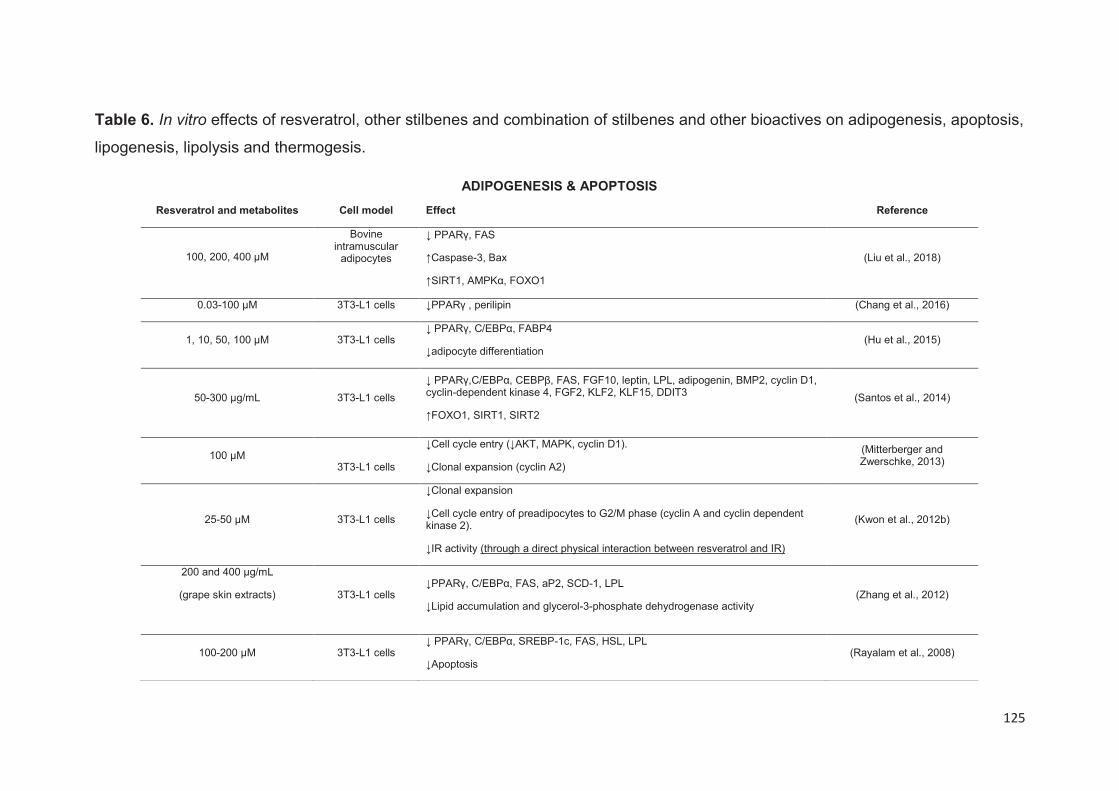
125
Table 6. In vitro effects of resveratrol, other stilbenes and combination of stilbenes and other bioactives on adipogenesis, apoptosis,
lipogenesis, lipolysis and thermogesis.
ADIPOGENESIS & APOPTOSIS
Resveratrol and metabolites Cell model Effect Reference
100, 200, 400 μΜ
Bovine intramuscular
adipocytes
↓ PPARγ, FAS
↑Caspase-3, Bax
↑SIRT1, AMPKα, FOXO1
(Liu et al., 2018)
0.03-100 µM 3T3-L1 cells ↓PPARγ , perilipin (Chang et al., 2016)
1, 10, 50, 100 μM 3T3-L1 cells ↓ PPARγ, C/EBPα, FABP4
↓adipocyte differentiation (Hu et al., 2015)
50-300 µg/mL
3T3-L1 cells
↓ PPARγ,C/EBPα, CEBPβ, FAS, FGF10, leptin, LPL, adipogenin, BMP2, cyclin D1, cyclin-dependent kinase 4, FGF2, KLF2, KLF15, DDIT3
↑FOXO1, SIRT1, SIRT2
(Santos et al., 2014)
100 μM
3T3-L1 cells
↓Cell cycle entry (↓AKT, MAPK, cyclin D1).
↓Clonal expansion (cyclin A2)
(Mitterberger and Zwerschke, 2013)
25-50 μM 3T3-L1 cells
↓Clonal expansion
↓Cell cycle entry of preadipocytes to G2/M phase (cyclin A and cyclin dependent kinase 2).
↓IR activity (through a direct physical interaction between resveratrol and IR)
(Kwon et al., 2012b)
200 and 400 μg/mL
(grape skin extracts)
3T3-L1 cells ↓PPARγ, C/EBPα, FAS, aP2, SCD-1, LPL
↓Lipid accumulation and glycerol-3-phosphate dehydrogenase activity (Zhang et al., 2012)
100-200 μM 3T3-L1 cells ↓ PPARγ, C/EBPα, SREBP-1c, FAS, HSL, LPL
↓Apoptosis (Rayalam et al., 2008)
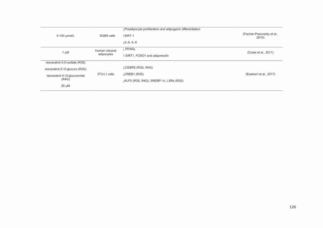
126
5-100 µmol/L SGBS cells
↓Preadipocyte proliferation and adipogenic differentiation
↑SIRT-1
↓IL-6, IL-8
(Fischer-Posovszky et al., 2010)
1 μM Human visceral
adipocytes
↓ PPARγ
↑ SIRT1, FOXO1 and adiponectin (Costa et al., 2011)
resveratrol-3-O-sulfate (R3S)
resveratrol-3’-O-glucuro (R3G)
resveratrol-4’-O-glucuronide (R4G)
25 μM
3T3-L1 cells
↓C/EBPβ (R3G, R4G)
↓CREB1 (R3S)
↓KLF5 (R3S, R4G), SREBP-1c, LXRα (R3S)
(Eseberri et al., 2017)
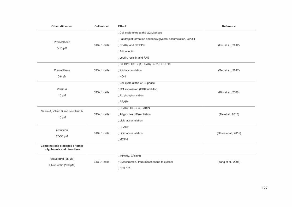
127
Other stilbenes Cell model Effect Reference
Pterostilbene
5-10 μM 3T3-L1 cells
↓Cell cycle entry at the G2/M phase
↓Fat droplet formation and triacylglycerol accumulation, GPDH
↓PPARγ and C/EBPα
↑Adiponectin
↓Leptin, resistin and FAS
(Hsu et al., 2012)
Pterostilbene
0-6 μM
3T3-L1 cells
↓C/EBPα, C/EBPβ, PPARγ, aP2, CHOP10
↓lipid accumulation
↑HO-1
(Seo et al., 2017)
Vitisin A
10 μΜ
3T3-L1 cells
↓Cell cycle at the G1-S phase
↑p21 expression (CDK inhibitor)
↓Rb phosphorylation
↓PPARγ
(Kim et al., 2008)
Vitisin A, Vitisin B and cis-vitisin A
10 μΜ 3T3-L1 cells
↓PPARγ, C/EBPα, FABP4
↓Adypocites differentiation
↓Lipid accumulation
(Tie et al., 2018)
ε-viniferin
25-50 μM
3T3-L1 cells
↓PPARγ
↓Lipid accumulation
↓MCP-1
(Ohara et al., 2015)
Combinations stilbenes or other polyphenols and bioactives
Resveratrol (25 μM)
+ Quercetin (100 μM) 3T3-L1 cells
↓ PPARγ, C/EBPα
↑Cytochrome C from mitochondria to cytosol
↓ERK 1/2
(Yang et al., 2008)
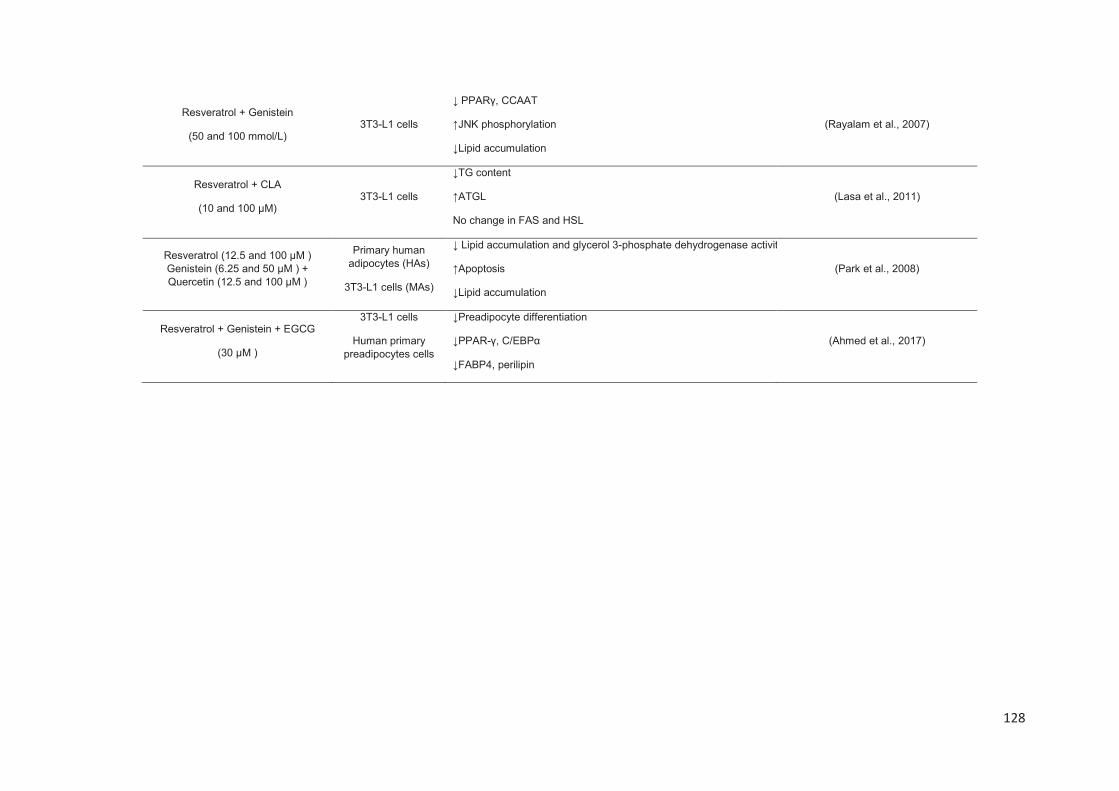
128
Resveratrol + Genistein
(50 and 100 mmol/L) 3T3-L1 cells
↓ PPARγ, CCAAT
↑JNK phosphorylation
↓Lipid accumulation
(Rayalam et al., 2007)
Resveratrol + CLA
(10 and 100 μM) 3T3-L1 cells
↓TG content
↑ATGL
No change in FAS and HSL
(Lasa et al., 2011)
Resveratrol (12.5 and 100 μΜ ) Genistein (6.25 and 50 μΜ ) + Quercetin (12.5 and 100 μΜ )
Primary human adipocytes (HAs)
3T3-L1 cells (MAs)
↓ Lipid accumulation and glycerol 3-phosphate dehydrogenase activity
↑Apoptosis
↓Lipid accumulation
(Park et al., 2008)
Resveratrol + Genistein + EGCG
(30 μΜ )
3T3-L1 cells
Human primary preadipocytes cells
↓Preadipocyte differentiation
↓PPAR-γ, C/EBPα
↓FABP4, perilipin
(Ahmed et al., 2017)
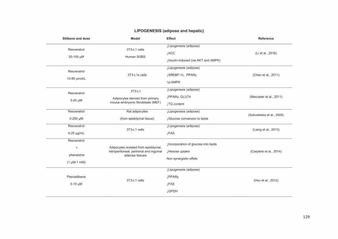
129
LIPOGENESIS (adipose and hepatic)
Stilbene and dose Model Effect Reference
Resveratrol
25-100 μM
3T3-L1 cells
Human SGBS
↓Lipogenesis (adipose)
↓ACC
↓Insulin-induced (via AKT and AMPK)
(Li et al., 2016)
Resveratrol
10-80 μmol/L 3T3-L1s cells
↓Lipogenesis (adipose)
↓SREBP-1c, PPARγ
↑p-AMPK
(Chen et al., 2011)
Resveratrol
5-20 μM
3T3-L1
Adipocytes derived from primary mouse embryonic fibroblasts (MEF)
↓Lipogenesis (adipose)
↓PPARγ, GLUT4
↓TG content
(Mercader et al., 2011)
Resveratrol
0-250 μM
Rat adipocytes
(from epididymal tissue)
↓Lipogenesis (adipose)
↓Glucose conversion to lipids (Szkudelska et al., 2009)
Resveratrol
0-25 μg/mL 3T3-L1 cells
↓Lipogenesis (adipose)
↓FAS (Liang et al., 2013)
Resveratrol
+
phenelzine
(1 µM-1 mM)
Adipocytes isolated from epididymal, retroperitoneal, perirenal and inguinal
adipose tissues
↓Incorporation of glucose into lipids
↓Hexose uptake
Non synergistic effets
(Carpéné et al., 2014)
Pterostilbene
5-10 μM 3T3-L1 cells
↓Lipogenesis (adipose)
↓PPARγ
↓FAS
↓GPDH
(Hsu et al., 2012)

130
Resveratrol
0-30 μM
Primary hepatocyte
HepG2
↓Lipogenesis (hepatic)
↓SREBP-1c
↓LXRα
↑SESN2 expression
(Jin et al., 2013)
Resveratrol
1-100 μM Hepatocytes from male Wistar rats
↓Lipogenesis (hepatic)
↓Total fatty acid synthesis
↓ACC
(Gnoni and Paglialonga, 2009)
Resveratrol +18 resveratrol synthetized derivatives
10-50 μM
H4IIEC3 cells
HepG2 cells
↓Lipogenesis (hepatic)
↓SREBP-1c, FASN, SCD-1
↓Lipid content
(Choi et al., 2014)
Oxyresveratrol
30 μM HepG2 cells
↓Lipogenesis (hepatic)
↓SREBP-1c, FAS, SCD-1
↓LRX agonists
(Lee et al., 2018)
LIPOLYSIS
Stilbene and dose Model Effect Reference
Resveratrol
100, 200, 400 μΜ Bovine intramuscular adipocytes
↓ PPARγ
↑HSL, ATGL (Liu et al., 2018)
Resveratrol
0.03-10 μM 3T3-L1 cells
↓ PPARγ
↑ glycerol release
↓TNFα-induced lipolysis
(Chang et al., 2016)
Resveratrol
+ CLA 10 and 100 μM
3T3-L1 cells
↑ ATGL
↓TG content (Lasa et al., 2011)
Resveratrol 3T3-L1 cells ↑FFA release (Lasa et al., 2012)

131
100 μM SGBS cells ↑ ATGL
Resveratrol
50, 75 and 100 μM SGBS cells
↓TG content
↑Glycerol release (Rosenow et al., 2012)
Resveratrol
1mM 3T3-F442A cells ↑Glycerol release (Gomez-Zorita et al., 2017)
Resveratrol
10-100 μM
Rat adipocytes
(epididymal tissue)
↑ Glycerol release
↑ Epinephrine-induced lipolysis (Szkudelska et al., 2009)
Pterostilbene
100 nM and 10 μM
3T3-F442A cells
↑ Glycerol release induced by isoprenaline (Gomez-Zorita et al., 2017)
Resveratrol + genistein
(25 mmol/L) 3T3-L1 cells
↓PPARγ
↑Glycerol release (Rayalam et al., 2007)
Resveratrol
20 μM
3T3-L1 cells
Primary MEF
↑ β-oxidation
↑ CPT-1
↓ rip 140
(Mercader et al., 2011)
Oxyresveratrol
30 μM HepG2 cells
↑ β-oxidation
↑ CPT-1 (Lee et al., 2018)

132
THERMOGENESIS/MITOCONDRIAL BIOGENESIS
Stilbene and dose Model Effect Reference
Resveratrol
10-40 μΜ
Vascular cells
isolated from iBAT
↑ PRDM16
↑ UCP-1 and cytochrome C, PGC1α, PDH
↑AMPKα
(Wang et al., 2017)
Resveratrol
25-100 μM
3T3-L1 cells
Human SGBS
↑ ATAD3
(Li et al., 2016)
Resveratrol
5-20 μM
3T3-L1 cells
Adipocytes derived from primary mouse embryonic fibroblasts
(MEF)
↑Mitochondrial DNA content
↑Subunit II of cytochrome oxidase
↑UCP-1
↓RBP4, resistin
(Mercader et al., 2011)
Resveratrol
10 μM
primary stromal vascular cells separated from iWAT
↑ UCP-1, PRDM16, Cell death-inducing DFFA-like effector A (Cidea), elongation of very long chain fatty acids protein 3 (Elovl3), PGC1α, cytochrome C and PDH
↑AMPKα1 phosphorylation
(Wang et al., 2015)
Resveratrol
100-200 μM 3T3-L1 cells ↑ SIRT3, UCP-1, Mfn2 (Rayalam et al., 2008)
Resveratrol
50-300 µg/mL
3T3-L1 cells
↑ UCP-1 (Santos et al., 2014)
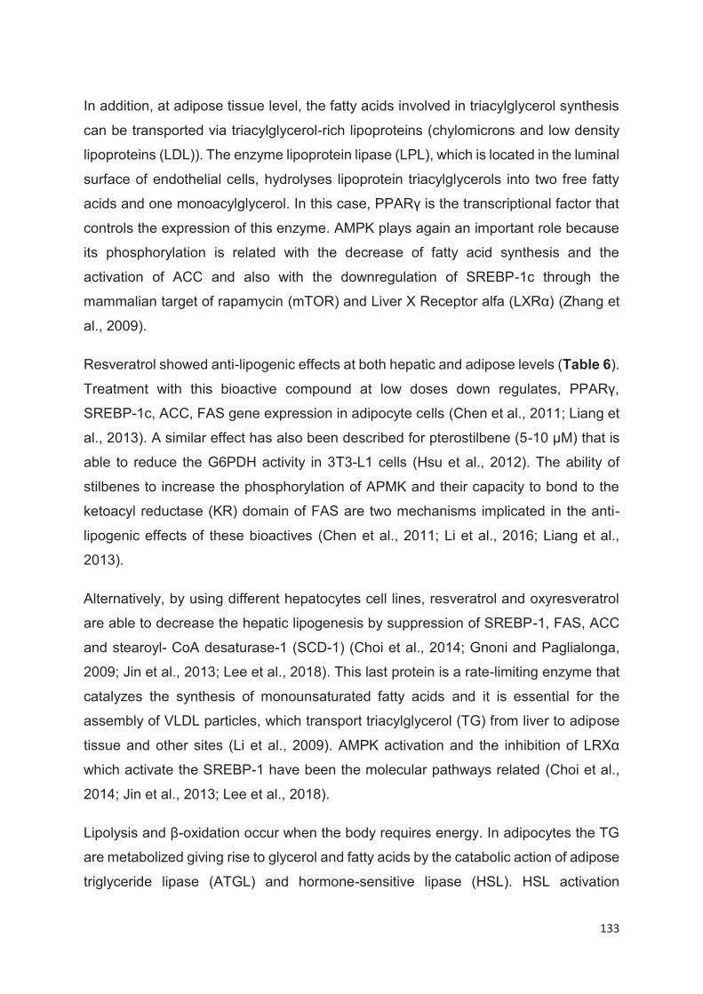
133
In addition, at adipose tissue level, the fatty acids involved in triacylglycerol synthesis
can be transported via triacylglycerol-rich lipoproteins (chylomicrons and low density
lipoproteins (LDL)). The enzyme lipoprotein lipase (LPL), which is located in the luminal
surface of endothelial cells, hydrolyses lipoprotein triacylglycerols into two free fatty
acids and one monoacylglycerol. In this case, PPARγ is the transcriptional factor that
controls the expression of this enzyme. AMPK plays again an important role because
its phosphorylation is related with the decrease of fatty acid synthesis and the
activation of ACC and also with the downregulation of SREBP-1c through the
mammalian target of rapamycin (mTOR) and Liver X Receptor alfa (LXRα) (Zhang et
al., 2009).
Resveratrol showed anti-lipogenic effects at both hepatic and adipose levels (Table 6).
Treatment with this bioactive compound at low doses down regulates, PPARγ,
SREBP-1c, ACC, FAS gene expression in adipocyte cells (Chen et al., 2011; Liang et
al., 2013). A similar effect has also been described for pterostilbene (5-10 µM) that is
able to reduce the G6PDH activity in 3T3-L1 cells (Hsu et al., 2012). The ability of
stilbenes to increase the phosphorylation of APMK and their capacity to bond to the
ketoacyl reductase (KR) domain of FAS are two mechanisms implicated in the anti-
lipogenic effects of these bioactives (Chen et al., 2011; Li et al., 2016; Liang et al.,
2013).
Alternatively, by using different hepatocytes cell lines, resveratrol and oxyresveratrol
are able to decrease the hepatic lipogenesis by suppression of SREBP-1, FAS, ACC
and stearoyl- CoA desaturase-1 (SCD-1) (Choi et al., 2014; Gnoni and Paglialonga,
2009; Jin et al., 2013; Lee et al., 2018). This last protein is a rate-limiting enzyme that
catalyzes the synthesis of monounsaturated fatty acids and it is essential for the
assembly of VLDL particles, which transport triacylglycerol (TG) from liver to adipose
tissue and other sites (Li et al., 2009). AMPK activation and the inhibition of LRXα
which activate the SREBP-1 have been the molecular pathways related (Choi et al.,
2014; Jin et al., 2013; Lee et al., 2018).
Lipolysis and β-oxidation occur when the body requires energy. In adipocytes the TG
are metabolized giving rise to glycerol and fatty acids by the catabolic action of adipose
triglyceride lipase (ATGL) and hormone-sensitive lipase (HSL). HSL activation
134
depends on protein kinase A (PKA) phosphorylation, which is mediated via the
accumulation of cAMP (Duncan et al., 2007). With regard to the lipolytic effects of
stilbenes, resveratrol was the main compound studied. Some published articles proved
that resveratrol, at different doses (0.03-400 µM), is able to enhance the free fatty acid
and the glycerol release by increasing HSL and ATGL expression in some cellular
models (bovine intramuscular adipocytes, 3T3-L1 and SGBS adipocytes) (Chang et
al., 2016; Lee et al., 2018; Rayalam et al., 2007). Interestingly, resveratrol had also
synergistic action with genistein, CLA and epinephrine enhancing its lipolytic capacity
(Lasa et al., 2011; Rayalam et al., 2007; Szkudelska et al., 2009).
An increment of β-oxidation capacity in adipocyte cells was demonstrated by
resveratrol and oxyresveratrol. In fact, the upregulation of carnitine
palmitoyltransferase 1 (CPT-1) which is necessary for mitochondrial import of fatty
acids and the repression of receptor interacting protein 140 (rip 140), a suppressor of
oxidative metabolism, was involved (Lee et al., 2018; Mercader et al., 2011).
Finally, another important pathway related to fat metabolism is thermogenesis that is
literally defined as heat production. The white adipose tissue (WAT) and the brown
adipose tissue (BAT) are the main types of adipose tissues with antagonistic functions.
WAT stores the excess of energy in the form of TG and BAT is specialized in heat
production. BAT is specialized in dissipate energy thanks to the high number of
mitochondria. Mitochondria membranes contain high amounts of Uncoupling protein 1
(UCP-1), an inner membrane protein that uncouples the electron transport chain from
ATP synthesis resulting in energy dissipation rather than ATP synthesis (Madden,
2017). Under certain conditions (e.g. extreme low temperature or β-3 adrenergic
agonist) the number of mitochondria increased drastically; this process is named
“browning” and the type of WAT is called “beige” (Bartelt and Heeren, 2014). Thus, an
increase of BAT thermogenesis is considered nowadays as a potential strategy to
reduce obesity.
Several recent in vitro studies have revealed that resveratrol (10 µM) can activate the
“browning” process. In particular an increase of UCP-1 expression in stromal vascular
cells from interscapular WAT and BAT has been observed (Wang et al., 2015; Wang
et al., 2017). Beyond this, these studies have also demonstrated an increase of
expression of others transcriptional factors of brown adipogenic program as PR
135
domain-containing 16 (PRDM16) that leads to the activation of mitochondrial
biogenesis. This effect was related with AMPK signaling pathway activation. Other
papers showed that resveratrol at 100 µM concentration up-regulated the gene
expression of some mitochondrial activity regulators such as SIRT3 that influences the
mitochondrial function by reducing membrane potential, mitofusin 2 (Mfn2) that
participates in mitochondrial fusion in mammalian cells, and ATPase family AAA
Domain-containing protein 3 (ATAD3), a protein that regulates mitochondrial
biogenesis (Li et al., 2016; Rayalam et al., 2008).

136
Anti-obesity in vivo effects of stilbenes
Regarding the in vivo effects and similarly with the in vitro studies the most examined
stilbene molecule was resveratrol.
Several animal models were used to investigate the anti-obesity effects of stilbenes,
mices (Swiss, C57BL/6J, FVB/N, CD1, kunming) and rats (Sprague-Dawley, Wistar,
Zucker (fa/fa)) being the most commonly used. In addition, other animal models such
as Megalobrama amblycephala (fish) or Caenorhabditis elegans were used.
Experimentally, these species were treated with standard (SD), high fat (HFD), high
protein (HPD), high fructose corn syrup (HFCSD) diets supplemented with stilbenes
(between 1 to 300 mg/kg/day) during different periods of time varying from some hours
to 20 weeks (Table 7). This large numbers of conditions (animal model, dose, diet and
period of treatment) makes sometimes difficult the understanding of results found by
these studies. Nevertheless, several studies showed that resveratrol is able to mitigate
the body weight gain in different animal models. In some instances, even a 50%
reduction was described (Choi et al., 2014; Jeon et al., 2014; Majumdar et al., 2014;
Mendes et al., 2016; Qiao et al., 2014).
In addition to weight reduction, an attenuation of lipid deposition on internal adipose
tissues such as epididymal and intraperitoneal tissues was observed after resveratrol
treatment (De Almeida Pinheiro et al., 2017; Gómez-Zorita et al., 2013; Jeon et al.,
2014; Zhang et al., 2018). This fact was related to the capacity of resveratrol to control
and reduce the adipogenesis process. Particularly, resveratrol (0.4%) reversed the
HFD-induced up-regulation of key adipogenic genes such as PPARγ, C/EBPα,
SREBP-1c, FAS, LPL, aP2, and leptin in mice adipose tissues (Kim et al., 2011). The
results reported by Kim et al. highlighted that galanin-mediated signaling molecules
are also implicated on the anti-adipogenesis effects of resveratrol. Galanin is a
neuropeptide that plays a role in food intake and its circulating serum levels are
elevated in obese individuals (Kim and Park, 2010). Resveratrol has demonstrated its
capacity to significantly reverse the HFD-induced up-regulation of galanin and its
receptors along with increased expression and/or activation of downstream molecules
related to adipogenesis, such as Protein Kinase C Delta (PKCd), cyclin D (Cyc-D),
transcriptional factor E2F1, and Extracellular Receptor Kinase (ERK) (Kim et al., 2011).
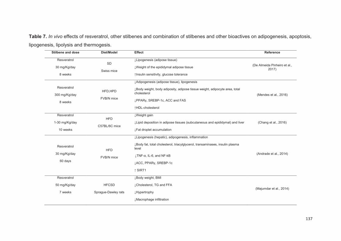
137
Table 7. In vivo effects of resveratrol, other stilbenes and combination of stilbenes and other bioactives on adipogenesis, apoptosis,
lipogenesis, lipolysis and thermogesis.
Stilbene and dose Diet/Model Effect Reference
Resveratrol
30 mg/Kg/day
8 weeks
SD
Swiss mice
↓Lipogenesis (adipose tissue)
↓Weight of the epididymal adipose tissue
↑Insulin sensitivity, glucose tolerance
(De Almeida Pinheiro et al., 2017)
Resveratrol
300 mg/Kg/day
8 weeks
HFD,HPD
FVB/N mice
↓Adipogenesis (adipose tissue), lipogenesis
↓Body weight, body adiposity, adipose tissue weight, adipocyte area, total cholesterol
↓PPARγ, SREBP-1c, ACC and FAS
↑HDL-cholesterol
(Mendes et al., 2016)
Resveratrol
1-30 mg/Kg/day
10 weeks
HFD
C57BL/6C mice
↓Weight gain
↓Lipid deposition in adipose tissues (subcutaneous and epididymal) and liver
↓Fat droplet accumulation
(Chang et al., 2016)
Resveratrol
30 mg/Kg/day
60 days
HFD
FVB/N mice
↓Lipogenesis (hepatic), adipogenesis, inflammation
↓Body fat, total cholesterol, triacylglycerol, transaminases, insulin plasma level
↓TNF-α, IL-6, and NF-kB
↓ACC, PPARγ, SREBP-1c
↑ SIRT1
(Andrade et al., 2014)
Resveratrol
50 mg/Kg/day
7 weeks
HFCSD
Sprague-Dawley rats
↓Body weight, BMI
↓Cholesterol, TG and FFA
↓Hypertrophy
↓Macrophage infiltration
(Majumdar et al., 2014)
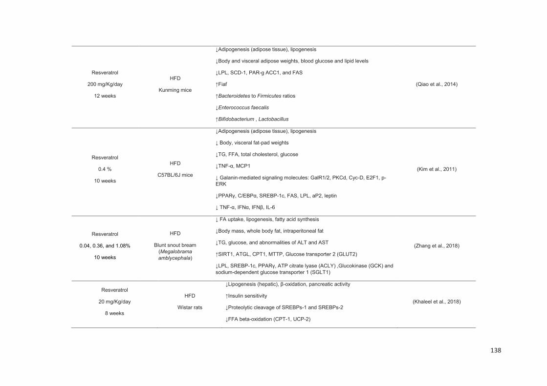
138
Resveratrol
200 mg/Kg/day
12 weeks
HFD
Kunming mice
↓Adipogenesis (adipose tissue), lipogenesis
↓Body and visceral adipose weights, blood glucose and lipid levels
↓LPL, SCD-1, PAR-g ACC1, and FAS
↑Fiaf
↑Bacteroidetes to Firmicutes ratios
↓Enterococcus faecalis
↑Bifidobacterium , Lactobacillus
(Qiao et al., 2014)
Resveratrol
0.4 %
10 weeks
HFD
C57BL/6J mice
↓Adipogenesis (adipose tissue), lipogenesis
↓ Body, visceral fat-pad weights
↓TG, FFA, total cholesterol, glucose
↓TNF-α, MCP1
↓ Galanin-mediated signaling molecules: GalR1/2, PKCd, Cyc-D, E2F1, p-ERK
↓PPARγ, C/EBPα, SREBP-1c, FAS, LPL, aP2, leptin
↓ TNF-α, IFNα, IFNβ, IL-6
(Kim et al., 2011)
Resveratrol
0.04, 0.36, and 1.08%
10 weeks
HFD
Blunt snout bream (Megalobrama
amblycephala)
↓ FA uptake, lipogenesis, fatty acid synthesis
↓Body mass, whole body fat, intraperitoneal fat
↓TG, glucose, and abnormalities of ALT and AST
↑SIRT1, ATGL, CPT1, MTTP, Glucose transporter 2 (GLUT2)
↓LPL, SREBP-1c, PPARγ, ATP citrate lyase (ACLY) ,Glucokinase (GCK) and sodium-dependent glucose transporter 1 (SGLT1)
(Zhang et al., 2018)
Resveratrol
20 mg/Kg/day
8 weeks
HFD
Wistar rats
↓Lipogenesis (hepatic), β-oxidation, pancreatic activity
↑Insulin sensitivity
↓Proteolytic cleavage of SREBPs-1 and SREBPs-2
↓FFA beta-oxidation (CPT-1, UCP-2)
(Khaleel et al., 2018)

139
Resveratrol
30 mg/Kg/day
6 weeks
HFD
Wistar rats
↓Lipogenesis (adipose tissue)
↑MicroRNA of PPARγ, HSL, and sp1
↓SREBP-1, FAS
(Gracia et al., 2016)
Resveratrol
28-46 mg/Kg/day
12 weeks
HFCSD
Wistar rats
↓Lipogenesis (hepatic)
↓Body weight
↓FAS, SREBP-1c
↑IRS-1, SIRT1, eNOS
(Sadi et al., 2015)
Resveratrol
15 and 45 mg/Kg/day
2 days
HFD
ICR mice
↓Lipogenesis (hepatic)
↓SREBP-1c, FAS
↓Hepatic TG
↑pAMPK
(Choi et al., 2014)
Resveratrol
0.1%
11 weeks
HFD
C57BL/6 mice
↓Lipogenesis (hepatic)
↓Body weight
↓PPARα, SREBP-1c, ACC1, FAS
↑SIRT 1
(Sun et al., 2015)
Resveratrol
0.02%
12 weeks
Atherogenic diet
Apo-E deficient mice
↑FA oxidation (liver and WAT), lipogenesis
↓Body weight, epididymal fat weight
↓Total cholesterol, LDL, ↑HDL
↓TG (plasma and hepatic)
↓G6PDH, FAS, ME, PAP
(Jeon et al., 2014)
Resveratrol
30 mg/kg/day
6 weeks
HFD
Wistar rats
↓Adipogenesis, lipogenesis (adipose tissue)
↓Rev-Erbα (Alberdi et al., 2013)
Resveratrol HFD ↓Lipogenesis (liver)↑FA oxidation (Alberdi et al., 2013)
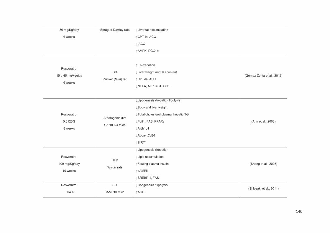
140
30 mg/Kg/day
6 weeks
Sprague-Dawley rats ↓Liver fat accumulation
↑CPT-Ia, ACO
↓ ACC
↑AMPK, PGC1α
Resveratrol
15 o 45 mg/kg/day
6 weeks
SD
Zucker (fa/fa) rat
↑FA oxidation
↓Liver weight and TG content
↑CPT-Ia, ACO
↓NEFA, ALP, AST, GOT
(Gómez-Zorita et al., 2012)
Resveratrol
0.0125%
8 weeks
Atherogenic diet
C57BL6/J mice
↓Lipogenesis (hepatic), lipolysis
↓Body and liver weight
↓Total cholesterol plasma, hepatic TG
↓Fdft1, FAS, PPARγ
↓Aldh1b1
↓Apoa4,Cd36
↑SIRT1
(Ahn et al., 2008)
Resveratrol
100 mg/Kg/day
10 weeks
HFD
Wistar rats
↓Lipogenesis (hepatic)
↓Lipid accumulation
↑Fasting plasma insulin
↑pAMPK
↓SREBP-1, FAS
(Shang et al., 2008)
Resveratrol
0.04%
SD
SAMP10 mice
↓ lipogenesis ↑lipolysis
↑ACC (Shiozaki et al., 2011)
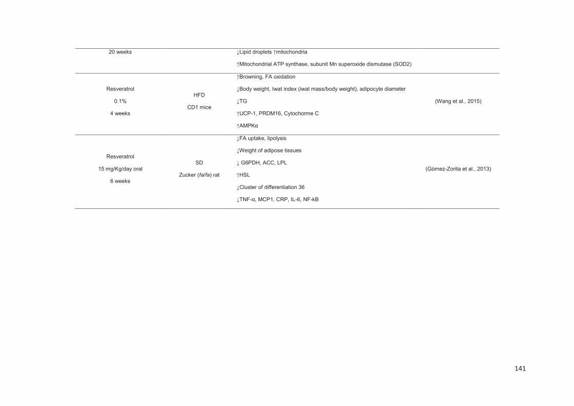
141
20 weeks ↓Lipid droplets ↑mitochondria
↑Mitochondrial ATP synthase, subunit Mn superoxide dismutase (SOD2)
Resveratrol
0.1%
4 weeks
HFD
CD1 mice
↑Browning, FA oxidation
↓Body weight, Iwat index (iwat mass/body weight), adipocyte diameter
↓TG
↑UCP-1, PRDM16, Cytochorme C
↑AMPKα
(Wang et al., 2015)
Resveratrol
15 mg/Kg/day oral
6 weeks
SD
Zucker (fa/fa) rat
↓FA uptake, lipolysis
↓Weight of adipose tissues
↓ G6PDH, ACC, LPL
↑HSL
↓Cluster of differentiation 36
↓TNF-α, MCP1, CRP, IL-6, NF-kB
(Gómez-Zorita et al., 2013)
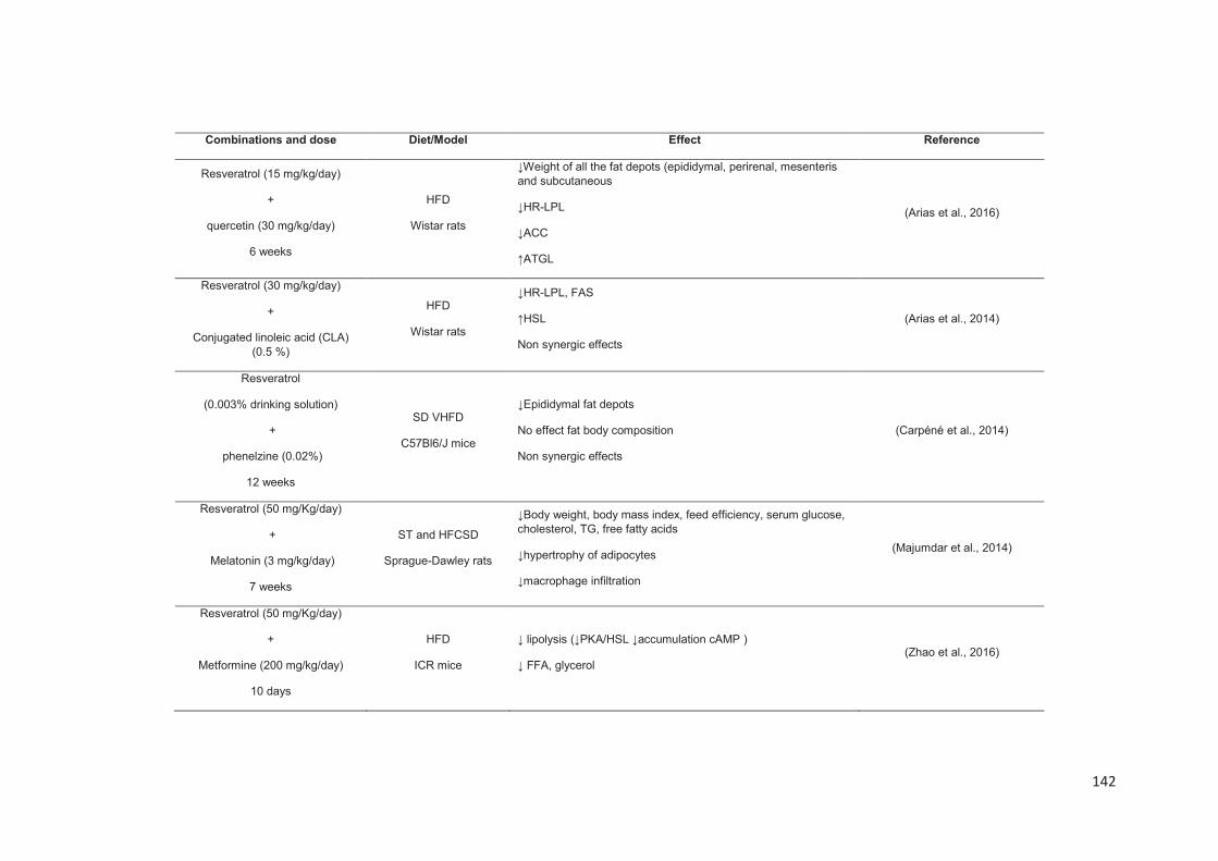
142
Combinations and dose Diet/Model Effect Reference
Resveratrol (15 mg/kg/day)
+
quercetin (30 mg/kg/day)
6 weeks
HFD
Wistar rats
↓Weight of all the fat depots (epididymal, perirenal, mesenteris and subcutaneous
↓HR-LPL
↓ACC
↑ATGL
(Arias et al., 2016)
Resveratrol (30 mg/kg/day)
+
Conjugated linoleic acid (CLA) (0.5 %)
HFD
Wistar rats
↓HR-LPL, FAS
↑HSL
Non synergic effects
(Arias et al., 2014)
Resveratrol
(0.003% drinking solution)
+
phenelzine (0.02%)
12 weeks
SD VHFD
C57Bl6/J mice
↓Epididymal fat depots
No effect fat body composition
Non synergic effects
(Carpéné et al., 2014)
Resveratrol (50 mg/Kg/day)
+
Melatonin (3 mg/kg/day)
7 weeks
ST and HFCSD
Sprague-Dawley rats
↓Body weight, body mass index, feed efficiency, serum glucose, cholesterol, TG, free fatty acids
↓hypertrophy of adipocytes
↓macrophage infiltration
(Majumdar et al., 2014)
Resveratrol (50 mg/Kg/day)
+
Metformine (200 mg/kg/day)
10 days
HFD
ICR mice
↓ lipolysis (↓PKA/HSL ↓accumulation cAMP )
↓ FFA, glycerol (Zhao et al., 2016)
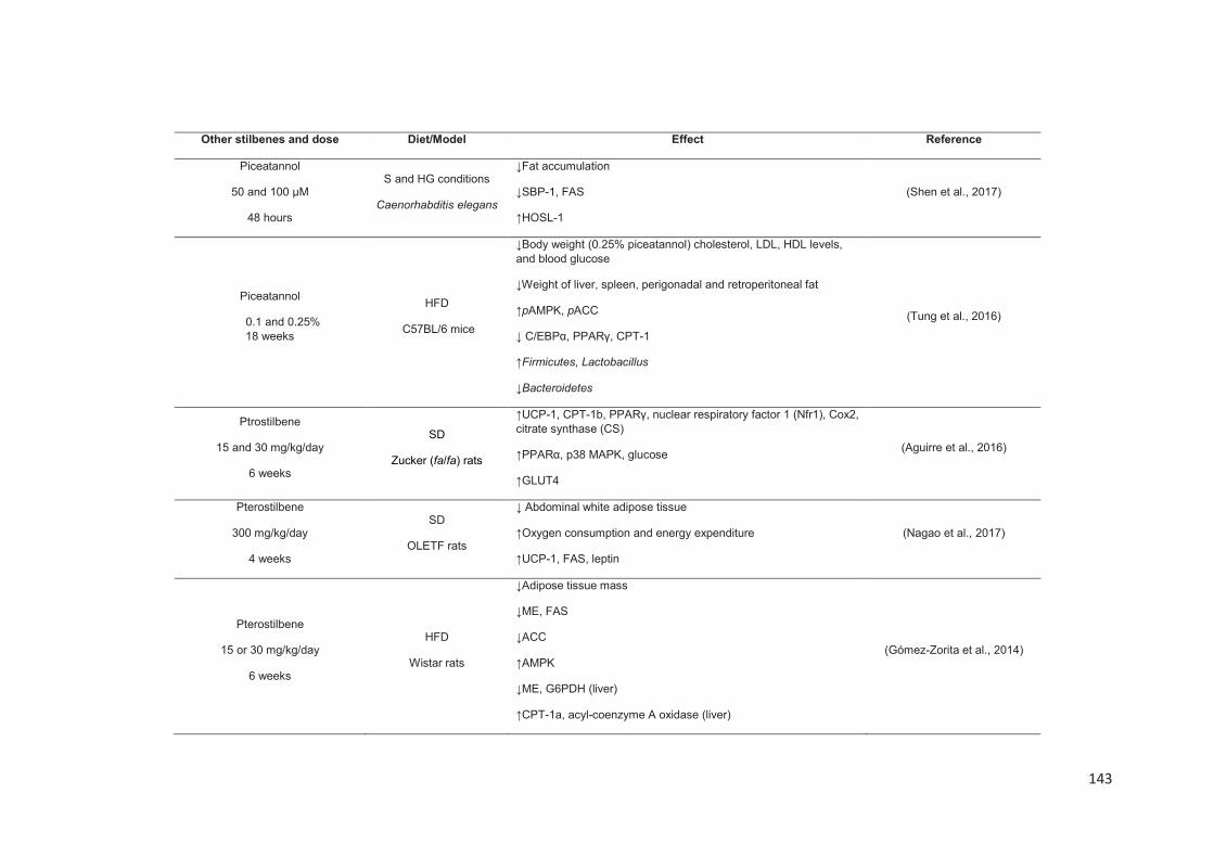
143
Other stilbenes and dose Diet/Model Effect Reference
Piceatannol
50 and 100 μM
48 hours
S and HG conditions
Caenorhabditis elegans
↓Fat accumulation
↓SBP-1, FAS
↑HOSL-1
(Shen et al., 2017)
Piceatannol
0.1 and 0.25% 18 weeks
HFD
C57BL/6 mice
↓Body weight (0.25% piceatannol) cholesterol, LDL, HDL levels, and blood glucose
↓Weight of liver, spleen, perigonadal and retroperitoneal fat
↑pAMPK, pACC
↓ C/EBPα, PPARγ, CPT-1
↑Firmicutes, Lactobacillus
↓Bacteroidetes
(Tung et al., 2016)
Ptrostilbene
15 and 30 mg/kg/day
6 weeks
SD
Zucker (fa/fa) rats
↑UCP-1, CPT-1b, PPARγ, nuclear respiratory factor 1 (Nfr1), Cox2, citrate synthase (CS)
↑PPARα, p38 MAPK, glucose
↑GLUT4
(Aguirre et al., 2016)
Pterostilbene
300 mg/kg/day
4 weeks
SD
OLETF rats
↓ Abdominal white adipose tissue
↑Oxygen consumption and energy expenditure
↑UCP-1, FAS, leptin
(Nagao et al., 2017)
Pterostilbene
15 or 30 mg/kg/day
6 weeks
HFD
Wistar rats
↓Adipose tissue mass
↓ME, FAS
↓ACC
↑AMPK
↓ME, G6PDH (liver)
↑CPT-1a, acyl-coenzyme A oxidase (liver)
(Gómez-Zorita et al., 2014)
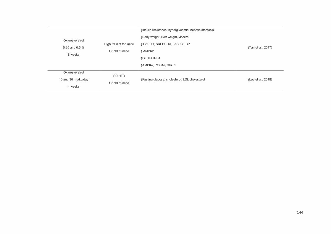
144
Oxyresveratrol
0.25 and 0.5 %
8 weeks
High fat diet fed mice
C57BL/6 mice
↓Insulin resistance, hyperglycemia, hepatic steatosis
↓Body weight, liver weight, visceral
↓ G6PDH, SREBP-1c, FAS, C/EBP
↑ AMPK2
↑GLUT4/IRS1
↑AMPKα, PGC1α, SIRT1
(Tan et al., 2017)
Oxyresveratrol
10 and 30 mg/kg/day
4 weeks
SD HFD
C57BL/6 mice ↓Fasting glucose, cholesterol, LDL cholesterol (Lee et al., 2018)
145
There is evidence that microbiota has an important role in obesity. In fact, the presence
of certain bacteria and overall, the relative proportions and composition of microbial
communities is key for energy homeostasis (Tennyson and Friedman, 2008). Although
this research line is recent and remains not fully explored, some studies were published
indicating that in obese people exposed to low calorie diet, Bacteriodetes level
increased while Firmicutes levels decreased (Ley et al., 2006). In addition, it seems
that gut microbiota can abolish the expression of fasting-induced adipose factor (Fiaf)
that belongs to the family of fibrinogen/angiopoietin-like proteins. When Fiaf was
suppressed an increase of LPL and a higher deposition of TG in adipose tissues have
been observed (Bäckhed et al., 2004). For this reason, the study of gut microbiota in
obesity research is gaining more and more relevance.
HFD was related with microbiota dysbiosis (promoting the growth of endotoxin
producers) producing a decrease of Lactobacillus and an increase of Enterococcus
faecalis. In this context it is worth mentioning that resveratrol long term (12 weeks)
treatment in Kunming mice’s demonstrated that this stilbene increased the
Bacteriodetes to Firmicutes ratio and diminished the growth of Enterococcus feacalis
(Qiao et al., 2014). Similarly, the numbers of Lactobacillus and Bifidobacterium were
significantly increased. At intestinal level, this work showed that resveratrol increases
the Fiaf expression which can be linked to the suppression of LPL and SCD-1
expressions in the liver, and with the expression of adipogenesis/ lipogenesis genes
(PPARγ, ACC1, and FAS) in adipose tissues (Qiao et al., 2014).
Likewise, some of the above-mentioned papers proved that resveratrol can also be
able to reduce lipogenesis process. A reduction of expression levels of FAS, ACC,
SCD-1, and G6PDH, and therefore, a suppression of fatty acid uptake and TG
synthesis at adipose and hepatic level has been observed (Table 7). Furthermore,
resveratrol causes the increase of GLUT2 mRNA expression allowing to restore the
normal glucose fluxes induced by HFD (Zhang et al., 2018). Another recent work
showed for the first time that resveratrol modifies the microRNA (miRNA) profile in
WAT. Actually, the reduction of protein levels of FAS, SREBP-1 and SP-1 (acts
together with SREBP1 to synergistically activate the promoter of FAS) has been linked
to the up-regulation of miR-539-5p (Gracia et al., 2016).
146
An inhibition of adipogenesis by resveratrol at hepatic level has also been evidenced.
Some works have mentioned a decrease of mRNA expression of PPARγ, SREBP-1c
and FAS after resveratrol supplementation with HFD, HFSCD or atherogenic diets
(Ahn et al., 2008; Alberdi et al., 2013; Andrade et al., 2014; Sadi et al., 2015; Shang et
al., 2008). Furthermore, this action is mediated by the activation of AMPK/SIRT1 axis
(Alberdi et al., 2013; Shang et al., 2008). Indeed, Shang et al. showed an increase of
164% of AMPK phosphorylation level in liver after the oral administration of resveratrol
(100 mg/Kg/day) during 10 weeks (Shang et al., 2008). Furthermore, higher gene and
protein expressions levels of a great number of insulin-signaling molecules including
IR, IRS-1/2, eNOS as well as SIRT1 have been outlined after resveratrol
supplementation (Sadi et al., 2015).
One of the key developments in obesity research is the recognition that this disorder
is also characterized by chronic mild inflammation. Indeed, an increase of circulating
levels of inflammatory markers in obese people such as CRP (C-reactive protein),
tumor necrosis factor alpha (TNF-α), interleukin (IL)-6 and IL-18, was described (Festa
et al., 2001; Monteiro and Azevedo, 2010). In this context, some papers indicated that
a reduction of inflammatory markers (TNF-α, interferon IFNα, IFNβ, IL-6, monocyte
chemoattractant protein 1 (MCP-1), and CRP) was observed in the adipose tissues of
mice and rat after resveratrol oral treatment, relating that with the repression of Toll-
like receptor (TLR)2- and TLR4-mediated pro-inflammatory signaling cascades (as
Nuclear Factor Kappa B (NF-КB) pathway) in the adipose tissues of mice and rat after
resveratrol oral treatment (Gómez-Zorita et al., 2013; Kim et al., 2011). This action was
also observed at hepatic level and evidenced by the reduction of TNF-α, IL-1β, IL-6
and NF-кB expression in liver in association with the up-regulation of SIRT1 in mice
treated with an HFD and resveratrol (Andrade et al., 2014).
Additionally, resveratrol is able to improve fatty acid oxidation in liver and WAT.
Particularly, Enzyme activities involved in fatty acid oxidation as CPT-Ia, a marker of
mitochondrial oxidation, and acyl-coenzyme A oxidase (ACO), a marker of peroxisome
oxidation, were significantly increased by resveratrol in liver (Alberdi et al., 2013;
Gómez-Zorita et al., 2012). It was furthermore demonstrated by other authors that the
increase of expression levels of both CPT-1 and UCP-2 may exert a protective effect
of resveratrol on mitochondrial dysfunction not only by inhibition of fatty acid oxidation

147
but also in association with reactive oxygen species (ROS) generation (Khaleel et al.,
2018). By using a fish animal model (M. amblycephala), the supplementation with
different doses of resveratrol (0.04, 0.36 and 1.08%) resulted in a significant reduction
of ATGL, CPT-1 and the microsomal triglyceride transfer protein (MTTP) implying up-
regulation of lipolysis and β-oxidation (Zhang et al., 2018). Finally, an augmentation of
HSL, without affecting the ATGL levels, was observed in Zucker (fa/fa) rats after oral
supplementation of 15 mg/Kg/day of resveratrol during 6 weeks (Gómez-Zorita et al.,
2013).
Finally, a strengthening of “browning” and mitochondrial biogenesis has been observed
by increasing protein contents of UCP1, PRDM16, and Cytochrome C along with an
increase of AMPK in CD1 mice treated with 0.1% of resveratrol (Wang et al., 2015).
As for in vitro tests, the combination of resveratrol with other polyphenols, bioactives
and drugs has been object of certain studies (Table 7). Two works published by the
same research group have proved that the mixture of resveratrol (15 mg/Kg/day) and
quercetin (30 mg/Kg/day) for a 6 weeks period of time decreases the weight of liver
and all the fats depots. This effect has been related with the reduction of LPL activity
and ACC and the increase of ATGL and CPT-1 (Arias et al., 2016; Arias et al., 2015).
However, non-synergistic effects have been found when resveratrol is combined with
conjugated linoleic acid (CLA) (Arias et al., 2014). Melatonin is a neurohormone related
with the circadian rhythms but also a bioactive found ubiquously in several foods and
also in wine (Hornedo-Ortega et al., 2016). The supplementation of melatonin
(3 mg/kg/day) with resveratrol in ovariectomized rats reduced the body weight by 16%
and body mass index (BMI) by 19%. Moreover, this combination is able to reduce the
insulin resistance and macrophage infiltration in liver (Majumdar et al., 2014). Lastly,
lipolysis was attenuated through prevention of PKA/HSL activation by decreasing the
accumulation of cAMP via preserving phosphodiesterase 3B (PDE3B) by the mixture
of metformin (oral antidiabetic drug) and resveratrol (Zhao et al., 2016).
With regard to other stilbenes, some in vivo studies pointed out that piceatannol,
pterostilbene and oxyresveratrol are interesting molecules to combat obesity (Table
7). Starting with the first one and by utilizing C. elegans as in vivo model, authors
demonstrated that this compound reduced the fat accumulation induced by high
glucose conditions; an attenuation of SBP-1 (encode SREBP-1c) and FAS and a

148
reduction on HOSL-1 expression (encodes HSL) prove that piceatannol can prevent
the lipid synthesis and stimulate the lipolysis (Shen et al., 2017). Furthermore, Tung et
al. showed that piceatannol (0.1 and 0.25%; 18 weeks) can decrease the C/EBPα,
PPARγ, FAS and CPT-1 and consequently promote the mitochondrial FA, oxidation
and lipid accumulation in adipocytes and liver. As was displayed for resveratrol, this
compound can also alter the composition of gut microbiota specifically by increasing
of Firmicutes/Lactobacillus and decreasing Bacteroidetes (Tung et al., 2016).
Pterostilbene at low doses (15 and 30 mg/Kg/day) can interfere in de novo lipogenesis
at adipose and hepatic level by reducing the activity of: ME, FAS, ACC, G6PDH,
CPT-1, which is in part explained by the increase of p-AMPK levels (Gómez-Zorita et
al., 2014). This paper highlighted that pterostilbene was more efficient than resveratrol
at a dose of 15 mg/kg/day while at 30 mg/kg/day both stilbenes had similar responses.
This fact can be explained, by the higher bioavailability of pterostilbene (Gómez-Zorita
et al., 2014). Using the same doses of pterostilbene, a thermogenic and oxidative
capacity by increasing of brown adipose tissue markers (UCP-1, CPT-1b, nuclear
respiratory factor 1 (Nrf1), cyclooxygenase 2 (COX2)) was observed (Aguirre et al.,
2016).
Finally, an improvement of insulin resistance and hyperglycemia accompanied by the
reduction in body weight (up to 26%), liver weight (up to 28%), and visceral fat (up to
51%) was observed for oxyresveratrol in C57BL/6 mouse experiments. The down
regulation of G6PDH expression was interpreted as the repression of free glucose
production in liver. They also observed an increase of GLUT4 and IRS1 that also plays
a vital role in intracellular glucose uptake (Tan et al., 2017).
149
Conclusion
Stilbene concentration in grape and wines can vary substantially. In fact, up to 20 times
more stilbenes can be found depending of the Vitis vinifera cultivars. The quantities
can also change significantly if the vine has suffered a pathogen infection, or if it has
been treated with elicitor or UVC treatments. In wines, the duration of maceration and
the activity of yeast can also influence the final amounts of stilbenes. Moreover, the
winemaking processes or the wine ageing can be not only affecting the stilbene
quantities but also inducing the formation of stilbene oligomers. However and relating
with this last one, more studies are needed to evaluate if this process can have a
relevant impact on the final product. On the contrary, it is a proven fact that the
composition of stilbenes in red wine is much more complex (oligomers) than in white
wine and that a greater complexity of stilbenes is observed in wild V.vinifera cultivars
in comparison with cultivated V.vinifera. Until today, more than 30 stilbenes have been
identified in grapes and wines using different separation and analytical methodologies.
In this context, the liquid chromatography coupled to mass spectrometry is the most
used method to determine stilbene compounds due to its sensitivity, rapidity and the
less consumption of solvents. In addition, the use of mass spectrometers of high
resolution (as QTOF) that provide information about the exact mass and fragments
(with 4-5 decimals) and molecular formula, allows more accurate stilbene identification.
However, a generalized problem is the lacks of commercial standards (overall of
oligomers) that force to express the results in resveratrol or piceid equivalent, and
could severally underestimate the oligomeric stilbene content. Regarding anti-obesity
properties of stilbenes, a great number of in vitro and in vivo studies have allowed to
demonstrate not only the positive implications of these bioactives but also the
underlying mechanisms of the observed effects. In fact, stilbenes can modulate all fat
metabolism pathways (adipogenesis, lipogenesis, lipolysis and thermogenesis) by
regulating gene and protein expression of receptors and key transcription factors,
enzymatic activities or even by diminishing the production of inflammatory markers with
a direct implication of different molecular mechanisms such as SIRT1-AMPK-FOXO1
and NF-КB pathways, among others. The understanding of these mechanisms is an
essential task since the scientific community seeks the development of more specific
dietary recommendations to prevent or combat obesity. It is noteworthy that although
150
the most studied molecule is resveratrol, other stilbenes are gaining attention as
pterostilbene, oxyresveratrol, viniferins and vitisins, some of them showing better anti-
obesity activities than resveratrol. In addition the combination of stilbenes with others
polyphenols or drugs is also reporting promising results. It is important to emphasize
that a great number of published papers use in general high doses, and without
reflecting important processes such as absorption and metabolism or even the role of
gut microbiota. Furthermore, intervention studies are still necessary in order to prove
these beneficial effects in humans.
151
References
Acevedo de la Cruz, A., Hilbert, G., Rivière, C., Mengin, V., Ollat, N., Bordenave, L.,
Decroocq, S., Delaunay, J.C., Delrot, S., Mérillon, J.M., Monti, J.P., Gomès, E.,
Richard, T., (2012). Anthocyanin identification and composition of wild Vitis spp.
accessions by using LC-MS and LC-NMR. Analytica Chimica Acta 732, 145-152.
Adrian, M., Jeandet, P., Douillet-Breuil, A.C., Tesson, L., Bessis, R., (2000). Stilbene
content of mature Vitis vinifera berries in response to UV-C elicitation. Journal of
Agricultural and Food Chemistry 48(12), 6103-6105.
Aguirre, L., Milton-Laskibar, I., Hijona, E., Bujanda, L., Rimando, A.M., Portillo, M.P.,
(2016). Effects of pterostilbene in brown adipose tissue from obese rats. Journal of
Physiology and Biochemistry 73(3), 457-464.
Ahmed, B., Liu, S., Si, H., (2017). Antiadipogenic Effects and Mechanisms of
Combinations of Genistein, Epigallocatechin-3-Gallate, and/or Resveratrol in
Preadipocytes. Journal of Medicinal Food 20(2), 162-170.
Ahn, J., Cho, I., Kim, S., Kwon, D., Ha, T., (2008). Dietary resveratrol alters lipid
metabolism-related gene expression of mice on an atherogenic diet. Journal of
Hepatology 49(6), 1019-1028.
Alberdi, G., Rodríguez, V.M., Macarulla, M.T., Miranda, J., Churruca, I., Portillo, M.P.,
(2013). Hepatic lipid metabolic pathways modified by resveratrol in rats fed an
obesogenic diet. Nutrition 29(3), 562-567.
Amira-Guebailia, H., Valls, J., Richard, T., Vitrac, X., Monti, J.P., Delaunay, J.C.,
Mérillon, J.M., (2009). Centrifugal partition chromatography followed by HPLC for the
isolation of cis-ε-viniferin, a resveratrol dimer newly extracted from a red Algerian wine.
Food Chemistry 113(1), 320-324.
Andrade, J.M.O., Paraíso, A.F., de Oliveira, M.V.M., Martins, A.M.E., Neto, J.F.,
Guimarães, A.L.S., de Paula, A.M., Qureshi, M., Santos, S.H.S., (2014). Resveratrol
attenuates hepatic steatosis in high-fat fed mice by decreasing lipogenesis and
inflammation. Nutrition 30(7-8), 915-919.
Arias, N., Macarulla, M.T., Aguirre, L., Milton, I., Portillo, M.P., (2016). The combination
of resveratrol and quercetin enhances the individual effects of these molecules on
triacylglycerol metabolism in white adipose tissue. European Journal of Nutrition 55(1),
341-348.
152
Arias, N., Macarulla, M.T., Aguirre, L., Miranda, J., Portillo, M.P., (2015). Liver
delipidating effect of a combination of resveratrol and quercetin in rats fed an
obesogenic diet. Journal of Physiology and Biochemistry 71(3), 569-576.
Arias, N., Miranda, J., Macarulla, M.T., Aguirre, L., Fernández-Quintela, A., Andres-
Lacueva, C., Urpi-Sarda, M., Portillo, M.P., (2014). The combination of resveratrol and
conjugated linoleic acid attenuates the individual effects of these molecules on
triacylglycerol metabolism in adipose tissue. European Journal of Nutrition 53(2), 575-
582.
Arraki, K., Renouf, E., Waffo Teguo, P., Merillon, J.M., Richard, T., Decendit, A.,
(2017). Identification and quantification of stilbenes in some Tunisian red wines using
UPLC-MS and HPLC-DAD. OENO One 51(2-3), 231-236.
Babazadeh, A., Taghvimi, A., Hamishehkar, H., Tabibiazar, M., (2017). Development
of new ultrasonic–solvent assisted method for determination of trans-resveratrol from
red grapes: Optimization, characterization, and antioxidant activity (ORAC assay).
Food Bioscience 20, 36-42.
Bäckhed, F., Ding, H., Wang, T., Hooper, L.V., Gou, Y.K., Nagy, A., Semenkovich,
C.F., Gordon, J.I., (2004). The gut microbiota as an environmental factor that regulates
fat storage. Proceedings of the National Academy of Sciences of the United States of
America 101(44), 15718-15723.
Baderschneider, B., Winterhalter, P., (2000). Isolation and characterization of novel
stilbene derivatives from Riesling wine. Journal of Agricultural and Food Chemistry
48(7), 2681-2686.
Barbanti, D., Galassi, S., Versari, A., Burattini, R., (1996). Gas chromatography and
high–pressure liquid chromatography determination of resveratrol in italian red wines.
International Journal of Phytoremediation 21(1), 5-11.
Bartelt, A., Heeren, J., (2014). Adipose tissue browning and metabolic health. Nature
Reviews Endocrinology 10(1), 24-36.
Bavaresco, L., Fregoni, M., Trevisan, M., Mattivi, F., Vrhovsek, U., Falchetti, R., (2002).
The occurrence of the stilbene piceatannol in grapes. Vitis 41(3), 133-136.
Bavaresco, L., Mattivi, F., de Rosso, M., Flamini, R., (2012). Effects of elicitors,
viticultural factors, and enological practices on resveratrol and stilbenes in Grapevine
and Wine. Mini-Reviews in Medicinal Chemistry 12(13), 1366-1381.

153
Biais, B., Krisa, S., Cluzet, S., Da Costa, G., Waffo-Teguo, P., Mérillon, J.M., Richard,
T., (2017). Antioxidant and cytoprotective activities of grapevine stilbenes. Journal of
Agricultural and Food Chemistry 65(24), 4952-4960.
Błaszczyk, A., Sady, S., Sielicka, M., (2019). The stilbene profile in edible berries.
Phytochemistry Reviews 18(1), 37-67.
Brillante, L., De Rosso, M., Dalla Vedova, A., Maoz, I., Flamini, R., Tomasi, D., (2018).
Insights on the stilbenes in Raboso Piave grape (Vitis vinifera L.) as a consequence of
postharvest vs on-vine dehydration. Journal of the Science of Food and Agriculture
98(5), 1961-1967.
Buiarelli, F., Coccioli, F., Jasionowska, R., Merolle, M., Terracciano, A., (2007).
Analysis of some stilbenes in Italian wines by liquid chromatography/tandem mass
spectrometry. Rapid Communications in Mass Spectrometry 21(18), 2955-2964.
Cai, L., Koziel, J.A., Dharmadhikari, M., van Leeuwen, J., (2009). Rapid determination
of trans-resveratrol in red wine by solid-phase microextraction with on-fiber
derivatization and multidimensional gas chromatography–mass spectrometry. Journal
of Chromatography A 1216(2), 281-287.
Cantos, E., Espín, J.C., Fernández, M.J., Oliva, J., Tomás-Barberán, F.A., (2003).
Postharvest UV-C-Irradiated Grapes as a Potential Source for Producing Stilbene-
Enriched Red Wines. Journal of Agricultural and Food Chemistry 51(5), 1208-1214.
Careri, M., Corradini, C., Elviri, L., Nicoletti, I., Zagnoni, I., (2003). Direct HPLC analysis
of quercetin and trans-resveratrol in red wine, grape, and winemaking byproducts.
Journal of Agricultural and Food Chemistry 51(18), 5226-5231.
Careri, M., Corradini, C., Elviri, L., Nicoletti, I., Zagnoni, I., (2004). Liquid
chromatography-electrospray tandem mass spectrometry of cis-resveratrol and trans-
resveratrol: Development, validation, and application of the method to red wine, grape,
and winemaking byproducts. Journal of Agricultural and Food Chemistry 52(23), 6868-
6874.
Carpéné, C., Gomez-Zorita, S., Gupta, R., Grès, S., Rancoule, C., Cadoudal, T.,
Mercader, J., Gomez, A., Bertrand, C., Iffiu-Soltész, Z., (2014). Combination of low
dose of the anti-adipogenic agents resveratrol and phenelzine in drinking water is not
sufficient to prevent obesity in very-high-fat diet-fed mice. European Journal of
Nutrition, 1625-1635.
154
Chang, C.C., Lin, K.Y., Peng, K.Y., Day, Y.J., Hung, L.M., (2016). Resveratrol exerts
anti-obesity effects in high-fat diet obese mice and displays differential dosage effects
on cytotoxicity, differentiation, and lipolysis in 3T3-L1 cells. Endocrine Journal 63(2),
169-178.
Chen, S., Li, Z., Li, W., Shan, Z., Zhu, W., (2011). Resveratrol inhibits cell differentiation
in 3T3-L1 adipocytes via activation of AMPK. Canadian Journal of Physiology and
Pharmacology 89(11), 793-799.
Cho, Y.J., Hong, J.Y., Chun, H.S., Lee, S.K., Min, H.Y., (2006). Ultrasonication-
assisted extraction of resveratrol from grapes. Journal of Food Engineering 77(3), 725-
730.
Choi, Y.J., Suh, H.R., Yoon, Y., Lee, K.J., Kim, D.G., Kim, S., Lee, B.H., (2014).
Protective effect of resveratrol derivatives on high-fat diet induced fatty liver by
activating AMP-activated protein kinase. Archives of Pharmacal Research 37(9), 1169-
1176.
Chong, J., Poutaraud, A., Hugueney, P., (2009). Metabolism and roles of stilbenes in
plants. Plant Science 177(3), 143-155.
Chou, Y.C., Ho, C.T., Pan, M.H., (2018). Stilbenes: Chemistry and Molecular
Mechanisms of Anti-obesity. Current Pharmacology Reports 4(3), 202-209.
Coelho, C., Bagala, F., Gougeon, R.D., Schmitt-Kopplin, P., (2016). Capillary
electrophoresis in wine science, Methods in Molecular Biology, pp. 509-523.
Costa, C.d.S., Rohden, F., Hammes, T.O., Margis, R., Bortolotto, J.W., Padoin, A.V.,
Mottin, C.C., Guaragna, R.M., (2011). Resveratrol Upregulated SIRT1, FOXO1, and
Adiponectin and Downregulated PPARγ1–3 mRNA Expression in Human Visceral
Adipocytes. Obesity Surgery 21(3), 356-361.
De Almeida Pinheiro, T., De Almeida Pinheiro, T., Feltenberger, J.D., Andrade, J.M.O.,
Ferreira, E.C.N., De Farias Lelis, D., Guimarães, A.L.S., De Paula, A.M.B., Caldeira,
A.P., Santos, S.H.S., (2017). Effects of resveratrol and ACE inhibitor enalapril on
glucose and lipid profiles in mice. Protein and Peptide Letters 24(9), 854-860.
Deluc, L.G., Grimplet, J., Wheatley, M.D., Tillett, R.L., Quilici, D.R., Osborne, C.,
Schooley, D.A., Schlauch, K.A., Cushman, J.C., Cramer, G.R., (2007). Transcriptomic
and metabolite analyses of Cabernet Sauvignon grape berry development. BMC
Genomics 8.

155
Duncan, R.E., Ahmadian, M., Jaworski, K., Sarkadi-Nagy, E., Sul, H.S., (2007).
Regulation of lipolysis in adipocytes, Annual Review of Nutrition, pp. 79-101.
El Khawand, T., Valls Fonayet, J., Da Costa, G., Hornedo-Ortega, R., Jourdes, M.,
Franc, C., de Revel, G., Decendit, A., Krisa, S., Richard, T., (2020). Resveratrol
transformation in red wine after heat treatment. Food Research International 132.
Eseberri, I., Lasa, A., Miranda, J., Gracia, A., Portillo, M.P., (2017). Potential miRNA
involvement in the anti-adipogenic effect of resveratrol and its metabolites. PloS One
12(9).
Esteve Ràfols, M., (2014). Adipose tissue: Cell heterogeneity and functional diversity.
Endocrinologia y Nutricion 61(2), 100-112.
Fabjanowicz, M., Płotka-Wasylka, J., Namieśnik, J., (2018). Detection, identification
and determination of resveratrol in wine. Problems and challenges. TRAC Trends in
Analytical Chemistry 103, 21-33.
Favre, G., Piccardo, D., Sergio, G.-A., Pérez-Navarro, J., García-Romero, E., Mena-
Morales, A., González-Neves, G., (2020). Stilbenes in grapes and wines of Tannat,
Marselan and Syrah from Uruguay. OENO One 54(1), 27-36.
Fernández-Marín, M.I., Guerrero, R.F., García-Parrilla, M.C., Puertas, B., Ramírez, P.,
Cantos-Villar, E., (2013). Terroir and variety: Two key factors for obtaining stilbene-
enriched grapes. Journal of Food Composition and Analysis 31(2), 191-198.
Fernandez-Marin, M.I., Guerrero, R.F., Garcia-Parrilla, M.C., Puertas, B., Richard, T.,
Rodriguez-Werner, M.A., Winterhalter, P., Monti, J.P., Cantos-Villar, E., (2012).
Isorhapontigenin: A novel bioactive stilbene from wine grapes. Food Chemistry 135(3),
1353-1359.
Fernández-Marín, M.I., Guerrero, R.F., García-Parrilla, M.C., Puertas, B., Richard, T.,
Rodriguez-Werner, M.A., Winterhalter, P., Monti, J.P., Cantos-Villar, E., (2012).
Isorhapontigenin: A novel bioactive stilbene from wine grapes. Food Chemistry 135(3),
1353-1359.
Fernández-Marín, M.I., Puertas, B., Guerrero, R.F., García-Parrilla, M.C., Cantos-
Villar, E., (2014). Preharvest Methyl Jasmonate and Postharvest UVC Treatments:
Increasing Stilbenes in Wine. Journal of Food Science 79(3), C310-C317.
Fernández-Quintela, A., Milton-Laskibar, I., González, M., Portillo, M.P., (2017).
Antiobesity effects of resveratrol: Which tissues are involved?, Annals of the New York
Academy of Sciences, pp. 118-131.
156
Festa, A., D'Agostino Jr, R., Williams, K., Karter, A.J., Mayer-Davis, E.J., Tracy, R.P.,
Haffner, S.M., (2001). The relation of body fat mass and distribution to markers of
chronic inflammation. International Journal of Obesity 25(10), 1407-1415.
Fischer-Posovszky, P., Kukulus, V., Tews, D., Unterkircher, T., Debatin, K.-M., Fulda,
S., Wabitsch, M., (2010). Resveratrol regulates human adipocyte number and function
in a Sirt1-dependent manner. The American Journal of Clinical Nutrition 92(1), 5-15.
Flamini, R., De Rosso, M., De Marchi, F., Dalla Vedova, A., Panighel, A., Gardiman,
M., Maoz, I., Bavaresco, L., (2013). An innovative approach to grape metabolomics:
stilbene profiling by suspect screening analysis. Metabolomics 9(6), 1243-1253.
Flamini, R., Zanzotto, A., de Rosso, M., Lucchetta, G., Vedova, A.D., Bavaresco, L.,
(2016). Stilbene oligomer phytoalexins in grape as a response to Aspergillus
carbonarius infection. Physiological and Molecular Plant Pathology 93, 112-118.
Gallagher, E.J., LeRoith, D., (2015). Obesity and Diabetes: The Increased Risk of
Cancer and Cancer-Related Mortality. Physiological Reviews 95(3), 727-748.
Garin-Shkolnik, T., Rudich, A., Hotamisligil, G.S., Rubinstein, M., (2014). FABP4
attenuates PPARγ and adipogenesis and is inversely correlated with PPARγ in
adipose tissues. Diabetes 63(3), 900-911.
Gatto, P., Vrhovsek, U., Muth, J., Segala, C., Romualdi, C., Fontana, P., Pruefer, D.,
Stefanini, M., Moser, C., Mattivi, F., Velasco, R., (2008). Ripening and genotype control
stilbene accumulation in healthy grapes. Journal of Agricultural and Food Chemistry
56(24), 11773-11785.
Gaudette, N.J., Pickering, G.J., (2011). Sensory and chemical characteristics of trans-
resveratrol-fortified wine. Australian Journal of Grape and Wine Research 17(2), 249-
257.
Gnoni, G.V., Paglialonga, G., (2009). Resveratrol inhibits fatty acid and triacylglycerol
synthesis in rat hepatocytes. European Journal of Clinical Investigation 39(3), 211-218.
Gomez-Zorita, S., Belles, C., Briot, A., Fernández-Quintela, A., Portillo, M.P., Carpéné,
C., (2017). Pterostilbene Inhibits Lipogenic Activity similar to Resveratrol or Caffeine
but Differently Modulates Lipolysis in Adipocytes. Phytotherapy Research 31(8), 1273-
1282.
Gómez-Zorita, S., Fernández-Quintela, A., Lasa, A., Aguirre, L., Rimando, A.M.,
Portillo, M.P., (2014). Pterostilbene, a dimethyl ether derivative of resveratrol, reduces
157
fat accumulation in rats fed an obesogenic diet. Journal of Agricultural and Food
Chemistry 62(33), 8371-8378.
Gómez-Zorita, S., Fernández-Quintela, A., Lasa, A., Hijona, E., Bujanda, L., Portillo,
M.P., (2013). Effects of resveratrol on obesity-related inflammation markers in adipose
tissue of genetically obese rats. Nutrition 29(11-12), 1374-1380.
Gómez-Zorita, S., Fernández-Quintela, A., MacArulla, M.T., Aguirre, L., Hijona, E.,
Bujanda, L., Milagro, F., Martínez, J.A., Portillo, M.P., (2012). Resveratrol attenuates
steatosis in obese Zucker rats by decreasing fatty acid availability and reducing
oxidative stress. British Journal of Nutrition 107(2), 202-210.
Gonçalves, J., Câmara, J.S., (2011). New method for determination of (E)-resveratrol
in wine based on microextraction using packed sorbent and ultra-performance liquid
chromatography. Journal of Separation Science 34(18), 2376-2384.
González-Candelas, L., Gil, J.V., Lamuela-Raventós, R.M., Ramón, D., (2000). The
use of transgenic yeasts expressing a gene encoding a glycosyl-hydrolase as a tool to
increase resveratrol content in wine. International Journal of Food Microbiology 59(3),
179-183.
Gracia, A., Miranda, J., Fernández-Quintela, A., Eseberri, I., Garcia-Lacarte, M.,
Milagro, F.I., Martínez, J.A., Aguirre, L., Portillo, M.P., (2016). Involvement of miR-539-
5p in the inhibition of de novo lipogenesis induced by resveratrol in white adipose
tissue. Food and Function 7(3), 1680-1688.
Gu, X., Chu, Q., O'Dwyer, M., Zeece, M., (2000). Analysis of resveratrol in wine by
capillary electrophoresis. Journal of Chromatography A 881(1-2), 471-481.
Guebailia, H.A., Chira, K., Richard, T., Mabrouk, T., Furiga, A., Vitrac, X., Monti, J.P.,
Delaunay, J.C., Merillon, J.M., (2006). Hopeaphenol: The first resveratrol tetramer in
wines from North Africa. Journal of Agricultural and Food Chemistry 54(25), 9559-
9564.
Guerrero, R.F., Cantos-Villar, E., Puertas, B., Richard, T., (2016). Daily Preharvest
UV-C Light Maintains the High Stilbenoid Concentration in Grapes. Journal of
Agricultural and Food Chemistry 64(25), 5139-5147.
Guerrero, R.F., García-Parrilla, M.C., Puertas, B., Cantos-Villar, E., (2009). Wine,
resveratrol and health: A review. Natural Product Communications 4(5), 635-658.
158
Guerrero, R.F., Puertas, B., Fernández, M.I., Palma, M., Cantos-Villar, E., (2010).
Induction of stilbenes in grapes by UV-C: Comparison of different subspecies of Vitis.
Innovative Food Science and Emerging Technologies 11(1), 231-238.
Guerrero, R.F., Valls-Fonayet, J., Richard, T., Cantos-Villar, E., (2020). A rapid
quantification of stilbene content in wine by ultra-high pressure liquid chromatography
– Mass spectrometry. Food Control 108.
Hasan, M.M., Bae, H., (2017). An overview of stress-induced resveratrol synthesis in
grapes: Perspectives for resveratrol-enriched grape products. Molecules 22(2).
Hausman, D.B., DiGirolamo, M., Bartness, T.J., Hausman, G.J., Martin, R.J., (2001).
The biology of white adipocyte proliferation. Obesity Reviews 2(4), 239-254.
He, S., Jiang, L., Wu, B., Li, C., Pan, Y., (2009). Chunganenol: An Unusual
Antioxidative Resveratrol Hexamer from Vitis chunganensis. The Journal of Organic
Chemistry 74(20), 7966-7969.
Hilbert, G., Temsamani, H., Bordenave, L., Pedrot, E., Chaher, N., Cluzet, S.,
Delaunay, J.C., Ollat, N., Delrot, S., Mérillon, J.M., Gomès, E., Richard, T., (2015).
Flavonol profiles in berries of wild Vitis accessions using liquid chromatography
coupled to mass spectrometry and nuclear magnetic resonance spectrometry. Food
Chemistry 169, 49-58.
Hornedo-Ortega, R., Cerezo, A.B., Troncoso, A.M., Garcia-Parrilla, M.C., Mas, A.,
(2016). Melatonin and other tryptophan metabolites produced by yeasts: Implications
in cardiovascular and neurodegenerative diseases. Frontiers in Microbiology 6(JAN).
Hsu, C.L., Lin, Y.J., Ho, C.T., Yen, G.C., (2012). Inhibitory effects of garcinol and
pterostilbene on cell proliferation and adipogenesis in 3T3-L1 cells. Food and Function
3(1), 49-57.
Hu, P., Zhao, L., Chen, J., (2015). Physiologically achievable doses of resveratrol
enhance 3T3-L1 adipocyte differentiation. European Journal of Nutrition 54(4), 569-
579.
Jeandet, P., Bessis, R., Gautheron, B., (1991). The Production of Resveratrol (3,5,4'-
trihydroxystilbene) by Grape Berries in Different Developmental Stages. American
Journal of Enology and Viticulture 42(1), 41-46.
Jeandet, P., Bessis, R., Maume, B.F., Meunier, P., Peyron, D., Trollat, P., (1995).
Effect of Enological Practices on the Resveratrol Isomer Content of Wine. Journal of
Agricultural and Food Chemistry 43(2), 316-319.
159
Jeandet, P., Bessis, R., Sbaghi, M., Meunier, P., (1994). Occurrence of a resveratrol
β-D-glucoside in wine: preliminary studies. Vitis - Journal of Grapevine Research 33(3),
183-184.
Jeon, S.M., Lee, S.A., Choi, M.S., (2014). Antiobesity and vasoprotective effects of
resveratrol in ApoE-deficient mice. Journal of Medicinal Food 17(3), 310-316.
Jiang, L., He, S., Sun, C., Pan, Y., (2012). Selective 1O2 quenchers, oligostilbenes,
from Vitis wilsonae: Structural identification and biogenetic relationship.
Phytochemistry 77, 294-303.
Jin, S.H., Yang, J.H., Shin, B.Y., Seo, K., Shin, S.M., Cho, I.J., Ki, S.H., (2013).
Resveratrol inhibits LXRα-dependent hepatic lipogenesis through novel antioxidant
Sestrin2 gene induction. Toxicology and Applied Pharmacology 271(1), 95-105.
Kang, H.K., Min, J.S., Chung, J., Park, H., Jae, B.K., (2005). Hypoxia inhibits adipocyte
differentiation in a HDAC-independent manner. Biochemical and Biophysical Research
Communications 333(4), 1178-1184.
Kasiotis, K.M., Pratsinis, H., Kletsas, D., Haroutounian, S.A., (2013). Resveratrol and
related stilbenes: Their anti-aging and anti-angiogenic properties. Food and Chemical
Toxicology 61, 112-120.
Khaleel, E.F., Abdel-Aleem, G.A., Mostafa, D.G., (2018). Resveratrol improves high-
fat diet induced fatty liver and insulin resistance by concomitantly inhibiting proteolytic
cleavage of sterol regulatory element-binding proteins, free fatty acid oxidation, and
intestinal triglyceride absorption. Canadian Journal of Physiology and Pharmacology
96(2), 145-157.
Kim, A., Park, T., (2010). Diet-induced obesity regulates the galanin-mediated
signaling cascade in the adipose tissue of mice. Molecular Nutrition and Food
Research 54(9), 1361-1370.
Kim, S., Jin, Y., Choi, Y., Park, T., (2011). Resveratrol exerts anti-obesity effects via
mechanisms involving down-regulation of adipogenic and inflammatory processes in
mice. Biochemical Pharmacology 81(11), 1343-1351.
Kim, S.h., Park, H.S., Lee, M.s., Cho, Y.J., Kim, Y.S., Hwang, J.T., Sung, M.J., Kim,
M.S., Kwon, D.Y., (2008). Vitisin A inhibits adipocyte differentiation through cell cycle
arrest in 3T3-L1 cells. Biochemical and Biophysical Research Communications 372(1),
108-113.

160
Kolouchová-Hanzlíková, I., Melzoch, K., Filip, V., Šmidrkal, J., (2004). Rapid method
for resveratrol determination by HPLC with electrochemical and UV detections in
wines. Food Chemistry 87(1), 151-158.
Kostadinović, S., Wilkens, A., Stefova, M., Ivanova, V., Vojnoski, B., Mirhosseini, H.,
Winterhalter, P., (2012). Stilbene levels and antioxidant activity of Vranec and Merlot
wines from Macedonia: Effect of variety and enological practices. Food Chemistry
135(4), 3003-3009.
Krisa, S., Larronde, F., Budzinski, H., Decendit, A., Deffieux, G., Mérillon, J.-M., (1999).
Stilbene Production by Vitis vinifera Cell Suspension Cultures: Methyl Jasmonate
Induction and 13C Biolabeling. Journal of Natural Products 62(12), 1688-1690.
Kwon, J.Y., Seo, S.G., Heo, Y.S., Yue, S., Cheng, J.X., Lee, K.W., Kim, K.H., (2012a).
Piceatannol, natural polyphenolic stilbene, inhibits adipogenesis via modulation of
mitotic clonal expansion and insulin receptor-dependent insulin signaling in early
phase of differentiation. Journal of Biological Chemistry 287(14), 11566-11578.
Kwon, J.Y., Seo, S.G., Yue, S., Cheng, J.X., Lee, K.W., Kim, K.H., (2012b). An
inhibitory effect of resveratrol in the mitotic clonal expansion and insulin signaling
pathway in the early phase of adipogenesis. Nutrition Research 32(8), 607-616.
Lamuela-Raventós, R.M., Romero-Pérez, A.I., Waterhouse, A.L., de la Torre-Boronat,
M.C., (1995). Direct HPLC Analysis of cis- and trans-Resveratrol and Piceid Isomers
in Spanish Red Vitis vinifera Wines. Journal of Agricultural and Food Chemistry 43(2),
281-283.
Landrault, N., Larronde, F., Delaunay, J.C., Castagnino, C., Vercauteren, J., Merillon,
J.M., Gasc, F., Cros, G., Teissedre, P.L., (2002). Levels of stilbene oligomers and
astilbin in French varietal wines and in grapes during noble rot development. Journal
of Agricultural and Food Chemistry 50(7), 2046-2052.
Langcake, P., Pryce, R.J., (1977). A new class of phytoalexins from grapevines.
Experientia 33(2), 151-152.
Lasa, A., Miranda, J., Churruca, I., Simón, E., Arias, N., Milagro, F., Martínez, J.A., del
Puy Portillo, M., (2011). The combination of resveratrol and CLA does not increase the
delipidating effect of each molecule in 3T3-L1 adipocytes. Nutricion Hospitalaria 26(5),
997-1003.
161
Lasa, A., Schweiger, M., Kotzbeck, P., Churruca, I., Simón, E., Zechner, R., Portillo,
M.d.P., (2012). Resveratrol regulates lipolysis via adipose triglyceride lipase. The
Journal of Nutritional Biochemistry 23(4), 379-384.
Lee, J., Rennaker, C., (2007). Antioxidant capacity and stilbene contents of wines
produced in the Snake River Valley of Idaho. Food Chemistry 105(1), 195-203.
Lee, J.H., Baek, S.Y., Jang, E.J., Ku, S.K., Kim, K.M., Ki, S.H., Kim, C.E., Park, K.I.,
Kim, S.C., Kim, Y.W., (2018). Oxyresveratrol ameliorates nonalcoholic fatty liver
disease by regulating hepatic lipogenesis and fatty acid oxidation through liver kinase
B1 and AMP-activated protein kinase. Chemico-Biological Interactions 289, 68-74.
Ley, R.E., Turnbaugh, P.J., Klein, S., Gordon, J.I., (2006). Microbial ecology: Human
gut microbes associated with obesity. Nature 444(7122), 1022-1023.
Li, S., Bouzar, C., Cottet-Rousselle, C., Zagotta, I., Lamarche, F., Wabitsch, M.,
Tokarska-Schlattner, M., Fischer-Posovszky, P., Schlattner, U., Rousseau, D., (2016).
Resveratrol inhibits lipogenesis of 3T3-L1 and SGBS cells by inhibition of insulin
signaling and mitochondrial mass increase. Biochimica et Biophysica Acta -
Bioenergetics 1857(6), 643-652.
Li, Z.Z., Berk, M., McIntyre, T.M., Feldstein, A.E., (2009). Hepatic lipid partitioning and
liver damage in nonalcoholic fatty liver disease: role of stearoyl-CoA desaturase. The
Journal of biological chemistry 284(9), 5637-5644.
Liang, Y., Tian, W., Ma, X., (2013). Inhibitory effects of grape skin extract and
resveratrol on fatty acid synthase. BMC Complementary and Alternative Medicine 13.
Liu, C., Wang, L., Wang, J., Wu, B., Liu, W., Fan, P., Liang, Z., Li, S., (2013).
Resveratrols in Vitis berry skins and leaves: Their extraction and analysis by HPLC.
Food Chemistry 136(2), 643-649.
Liu, X., Zhao, H., Jin, Q., You, W., Cheng, H., Liu, Y., Song, E., Liu, G., Tan, X., Zhang,
X., Wan, F., (2018). Resveratrol induces apoptosis and inhibits adipogenesis by
stimulating the SIRT1-AMPKα-FOXO1 signalling pathway in bovine intramuscular
adipocytes. Molecular and Cellular Biochemistry 439(1-2), 213-223.
Luan, T., Li, G., Zhang, Z., (2000). Gas-phase postderivatization following solid-phase
microextraction for rapid determination of trans-resveratrol in wine by gas
chromatography-mass spectrometry. Analytica Chimica Acta 424(1), 19-25.
162
Lukić, I., Radeka, S., Budić-Leto, I., Bubola, M., Vrhovsek, U., (2019). Targeted UPLC-
QqQ-MS/MS profiling of phenolic compounds for differentiation of monovarietal wines
and corroboration of particular varietal typicity concepts. Food Chemistry 300, 125251.
MacDougald, O.A., Mandrup, S., (2002). Adipogenesis: Forces that tip the scales.
Trends in Endocrinology and Metabolism 13(1), 5-11.
Madden, C.J., (2017). Brown fat in obesity: Uncoupling protein-1 versus thermogenic
activity. Temperature 4(2), 126-127.
Majumdar, A.S., Giri, P.R., Pai, S.A., (2014). Resveratrol- and melatonin-abated
ovariectomy and fructose diet-induced obesity and metabolic alterations in female rats.
Menopause 21(8), 876-885.
Mattivi, F., Reniero, F., Korhammer, S., (1995). Isolation, Characterization, and
Evolution in Red Wine Vinification of Resveratrol Monomers. Journal of Agricultural
and Food Chemistry 43(7), 1820-1823.
Mendes, K.L., De Pinho, L., Andrade, J.M.O., Paraíso, A.F., Lula, J.F., MacEdo, S.M.,
Feltenberger, J.D., Guimarães, A.L.S., De Paula, A.M.B., Santos, S.H.S., (2016).
Distinct metabolic effects of resveratrol on lipogenesis markers in mice adipose tissue
treated with high-polyunsaturated fat and high-protein diets. Life Sciences 153, 66-73.
Mercader, J., Palou, A., Bonet, M.L., (2011). Resveratrol enhances fatty acid oxidation
capacity and reduces resistin and Retinol-Binding Protein 4 expression in white
adipocytes. Journal of Nutritional Biochemistry 22(9), 828-834.
Mitterberger, M.C., Zwerschke, W., (2013). Mechanisms of resveratrol-induced
inhibition of clonal expansion and terminal adipogenic differentiation in 3T3-L1
preadipocytes. Journals of Gerontology - Series A Biological Sciences and Medical
Sciences 68(11), 1356-1376.
Monteiro, R., Azevedo, I., (2010). Chronic inflammation in obesity and the metabolic
syndrome. Mediators of Inflammation 2010.
Montes, R., García-López, M., Rodríguez, I., Cela, R., (2010). Mixed-mode solid-phase
extraction followed by acetylation and gas chromatography mass spectrometry for the
reliable determination of trans-resveratrol in wine samples. Analytica Chimica Acta
673(1), 47-53.
Moss, R., Mao, Q., Taylor, D., Saucier, C., (2013). Investigation of monomeric and
oligomeric wine stilbenoids in red wines by ultra-high-performance liquid
163
chromatography/electrospray ionization quadrupole time-of-flight mass spectrometry.
Rapid Communications in Mass Spectrometry 27(16), 1815-1827.
Nagao, K., Jinnouchi, T., Kai, S., Yanagita, T., (2017). Pterostilbene, a dimethylated
analog of resveratrol, promotes energy metabolism in obese rats. The Journal of
Nutritional Biochemistry 43, 151-155.
Neveu, V., Perez-Jiménez, J., Vos, F., Crespy, V., du Chaffaut, L., Mennen, L., Knox,
C., Eisner, R., Cruz, J., Wishart, D., Scalbert, A., (2010). Phenol-Explorer: an online
comprehensive database on polyphenol contents in foods. Database 2010, bap024.
Ohara, K., Kusano, K., Kitao, S., Yanai, T., Takata, R., Kanauchi, O., (2015). ε-
Viniferin, a resveratrol dimer, prevents diet-induced obesity in mice. Biochemical and
Biophysical Research Communications 468(4), 877-882.
Park, H.J., Yang, J.Y., Ambati, S., Della-Fera, M.A., Hausman, D.B., Rayalam, S.,
Baile, C.A., (2008). Combined effects of genistein, quercetin, and resveratrol in human
and 3T3-L1 adipocytes. Journal of Medicinal Food 11(4), 773-783.
Paulo, L., Domingues, F., Queiroz, J.A., Gallardo, E., (2011). Development and
validation of an analytical method for the determination of trans - And cis -resveratrol
in wine: Analysis of its contents in 186 Portuguese red wines. Journal of Agricultural
and Food Chemistry 59(6), 2157-2168.
Pawlus, A., Cantos-Villar, E., Richard, T., Bisson, J., Poupard, P., Papastamoulis, Y.,
Monti, J.-P., Teissedre, P.L., Waffo-Teguo, P., Merillon, J.-M., (2013a). Chemical
dereplication of wine stilbenoids using high performance liquid chromatography-
nuclear magnetic resonance spectroscopy. Journal of Chromatography A 1289, 19 -
26.
Pawlus, A.D., Sahli, R., Bisson, J., Rivière, C., Delaunay, J.C., Richard, T., Gomès, E.,
Bordenave, L., Waffo-Téguo, P., Mérillon, J.M., (2013b). Stilbenoid profiles of canes
from Vitis and Muscadinia species. Journal of Agricultural and Food Chemistry 61(3),
501-511.
Pezet, R., Pont, V., Cuenat, P., (1994). Method to determine resveratrol and
pterostilbene in grape berries and wines using high-performance liquid
chromatography and highly sensitive fluorimetric detection. Journal of
Chromatography A 663(2), 191-197.
164
Picard, F., Kurtev, M., Chung, N., Topark-Ngarm, A., Senawong, T., De Oliveira, R.M.,
Leid, M., McBurney, M.W., Guarente, L., (2004). Sirt1 promotes fat mobilization in
white adipocytes by repressing PPAR-γ. Nature 429(6993), 771-776.
Poussier, M., Guilloux-Benatier, M., Torres, M., Heras, E., Adrian, M., (2003). Influence
of Different Maceration Techniques and Microbial Enzymatic Activities on Wine
Stilbene Content. American Journal of Enology and Viticulture 54(4), 261-266.
Pugajeva, I., Perkons, I., Górnaś, P., (2018). Identification and determination of
stilbenes by Q-TOF in grape skins, seeds, juice and stems. Journal of Food
Composition and Analysis 74, 44-52.
Qiao, Y., Sun, J., Xia, S., Tang, X., Shi, Y., Le, G., (2014). Effects of resveratrol on gut
microbiota and fat storage in a mouse model with high-fat-induced obesity. Food and
Function 5(6), 1241-1249.
Rabesiaka, M., Rakotondramasy-Rabesiaka, L., Mabille, I., Porte, C., Havet, J.-L.,
(2011). Extraction of trans-resveratrol from red wine and optimization by response
surface methodology. Separation and Purification Technology 81(1), 56-61.
Rayalam, S., Della-Fera, M.A., Yang, J.Y., Hea, J.P., Ambati, S., Baile, C.A., (2007).
Resveratrol potentiates genistein's antiadipogenic and proapoptotic effects in 3T3-L1
adipocytes. Journal of Nutrition 137(12), 2668-2673.
Rayalam, S., Yang, J.Y., Ambati, S., Della-Fera, M.A., Baile, C.A., (2008). Resveratrol
induces apoptosis and inhibits adipogenesis in 3T3-L1 adipocytes. Phytotherapy
Research 22(10), 1367-1371.
Ribeiro De Lima, M.T., Waffo-Téguo, P., Teissedre, P.L., Pujolas, A., Vercauteren, J.,
Cabanis, J.C., Mérillon, J.M., (1999). Determination of stilbenes (trans-astringin, cis-
and trans-piceid, and cis- and trans-resveratrol) in Portuguese wines. Journal of
Agricultural and Food Chemistry 47(7), 2666-2670.
Rivière, C., Pawlus, A.D., Mérillon, J.M., (2012). Natural stilbenoids: distribution in the
plant kingdom and chemotaxonomic interest in Vitaceae. Natural Product Reports
29(11), 1317-1333.
Rodríguez-Cabo, T., Rodríguez, I., Cela, R., (2012). Determination of hydroxylated
stilbenes in wine by dispersive liquid–liquid microextraction followed by gas
chromatography mass spectrometry. Journal of Chromatography A 1258, 21-29.
165
Rodríguez-Cabo, T., Rodríguez, I., Ramil, M., Silva, A., Cela, R., (2016). Multiclass
semi-volatile compounds determination in wine by gas chromatography accurate time-
of-flight mass spectrometry. Journal of Chromatography A 1442, 107-117.
Roldán, A., Palacios, V., Caro, I., Pérez, L., (2003). Resveratrol content of Palomino
fino grapes: Influence of vintage and fungal infection. Journal of Agricultural and Food
Chemistry 51(5), 1464-1468.
Roldán, A., Palacios, V., Caro, I., Pérez, L., (2010). Evolution of Resveratrol and Piceid
Contents during the Industrial Winemaking Process of Sherry Wine. Journal of
Agricultural and Food Chemistry 58(7), 4268-4273.
Romero-Pérez, A.I., Lamuela-Raventós, R.M., Andrés-Lacueva, C., de la Torre-
Boronat, M.C., (2001). Method for the Quantitative Extraction of Resveratrol and Piceid
Isomers in Grape Berry Skins. Effect of Powdery Mildew on the Stilbene Content.
Journal of Agricultural and Food Chemistry 49(1), 210-215.
Rosen, E.D., Hsu, C.-H., Wang, X., Sakai, S., Freeman, M.W., Gonzalez, F.J.,
Spiegelman, B.M., (2002). C/EBPalpha induces adipogenesis through PPARgamma:
a unified pathway. Genes & Development 16(1), 22-26.
Rosenow, A., Noben, J.-P., Jocken, J., Kallendrusch, S., Fischer-Posovszky, P.,
Mariman, E.C.M., Renes, J., (2012). Resveratrol-Induced Changes of the Human
Adipocyte Secretion Profile. Journal of Proteome Research 11(9), 4733-4743.
Rossmeisl, M., Flachs, P., Brauner, P., Sponarova, J., Matejkova, O., Prazak, T.,
Ruzickova, J., Bardova, K., Kuda, O., Kopecky, J., (2004). Role of energy charge and
amp-activated protein kinase in adipocytes in the control of body fat stores.
International Journal of Obesity 28, S38-S44.
Rosso, M.D., Soligo, S., Panighel, A., Carraro, R., Vedova, A.D., Maoz, I., Tomasi, D.,
Flamini, R., (2016). Changes in grape polyphenols (V. vinifera L.) as a consequence
of post-harvest withering by high-resolution mass spectrometry: Raboso Piave versus
Corvina. Journal of Mass Spectrometry 51(9), 750-760.
Sadi, G., Ergin, V., Yilmaz, G., Pektas, M.B., Yildirim, O.G., Menevse, A., Akar, F.,
(2015). High-fructose corn syrup-induced hepatic dysfunction in rats: improving effect
of resveratrol. European Journal of Nutrition 54(6), 895-904.
Santos, J.C., Gotardo, E.M.F., Brianti, M.T., Piraee, M., Gambero, A., Ribeiro, M.L.,
(2014). Effects of yerba maté, a plant extract formulation ("YGD") and resveratrol in
3T3-L1 adipogenesis. Molecules 19(10), 16909-16924.

166
Sato, M., Suzuki, Y., Okuda, T., Yokotsuka, K., (1997). Contents of resveratrol, piceid,
and their isomers in commercially available wines made from grapes cultivated in
japan. Bioscience, Biotechnology and Biochemistry 61(11), 1800-1805.
Seo, Y.J., Kim, K.J., Koh, E.J., Choi, J., Lee, B.Y., (2017). Anti-adipogenesis
mechanism of pterostilbene through the activation of heme oxygenase-1 in 3T3-L1
cells. Phytomedicine 33, 7-13.
Shang, J., Chen, L.L., Xiao, F.X., Sun, H., Ding, H.C., Xiao, H., (2008). Resveratrol
improves non-alcoholic fatty liver disease by activating AMP-activated protein kinase.
Acta Pharmacologica Sinica 29(6), 698-706.
Shen, Y., Xu, Z., Sheng, Z., (2017). Ability of resveratrol to inhibit advanced glycation
end product formation and carbohydrate-hydrolyzing enzyme activity, and to conjugate
methylglyoxal. Food Chemistry 216, 153-160.
Shiozaki, M., Hayakawa, N., Shibata, M., Koike, M., Uchiyama, Y., Gotow, T., (2011).
Closer association of mitochondria with lipid droplets in hepatocytes and activation of
Kupffer cells in resveratrol-treated senescence-accelerated mice. Histochemistry and
Cell Biology 136(4), 475.
Spanilá, M., Pazourek, J., Farková, M., Havel, J., (2005). Optimization of solid-phase
extraction using artificial neural networks in combination with experimental design for
determination of resveratrol by capillary zone electrophoresis in wines. Journal of
Chromatography A 1084(1-2), 180-185.
Sun, B., Ribes, A.M., Leandro, M.C., Belchior, A.P., Spranger, M.I., (2006). Stilbenes:
Quantitative extraction from grape skins, contribution of grape solids to wine and
variation during wine maturation. Analytica Chimica Acta 563(1), 382-390.
Sun, L., Wang, Y., Song, Y., Cheng, X.-R., Xia, S., Rahman, M.R.T., Shi, Y., Le, G.,
(2015). Resveratrol restores the circadian rhythmic disorder of lipid metabolism
induced by high-fat diet in mice. Biochemical and Biophysical Research
Communications 458(1), 86-91.
Szkudelska, K., Nogowski, L., Szkudelski, T., (2009). Resveratrol, a naturally occurring
diphenolic compound, affects lipogenesis, lipolysis and the antilipolytic action of insulin
in isolated rat adipocytes. Journal of Steroid Biochemistry and Molecular Biology
113(1-2), 17-24.
Takaya, Y., Terashima, K., Ito, J., He, Y.-H., Tateoka, M., Yamaguchi, N., Niwa, M.,
(2005). Biomimic transformation of resveratrol. Tetrahedron 61(43), 10285-10290.
167
Tan, H.Y., Tse, I.M.Y., Li, E.T.S., Wang, M., (2017). Oxyresveratrol supplementation
to C57bl/6 mice fed with a high-fat diet ameliorates obesity-associated symptoms.
Nutrients 9(2).
Tennyson, C.A., Friedman, G., (2008). Microecology, obesity, and probiotics. Current
Opinion in Endocrinology, Diabetes and Obesity 15(5), 422-427.
Threlfall, R.T., Morris, J.R., Mauromoustakos, A., (1999). Effect of variety, ultraviolet
light exposure, and enological methods on the trans-resveratrol level of wine. American
Journal of Enology and Viticulture 50(1), 57-64.
Tie, F.F., Luan, G.X., Zhou, W.N., Wang, Z.H., Shi, X.B., Li, G., Wang, H.L., (2018).
Effects of the oligostilbenes from Iris lactea Pall. var. chinensis (Fisch.) Koidz on the
adipocytes differentiation of 3T3-L1 cells. Pharmazie 73(2), 98-103.
Tung, Y.C., Lin, Y.H., Chen, H.J., Chou, S.C., Cheng, A.C., Kalyanam, N., Ho, C.T.,
Pan, M.H., (2016). Piceatannol exerts anti-obesity effects in C57BL/6 mice through
modulating adipogenic proteins and gut microbiota. Molecules 21(11).
Vacca, V., Leccis, L., Fenu, P., Pretti, L., Farris, G.A., (1997). Wine yeasts and
resveratrol content. Biotechnology Letters 19(6), 497-498.
Vang, O., Ahmad, N., Baile, C.A., Baur, J.A., Brown, K., Csiszar, A., Das, D.K.,
Delmas, D., Gottfried, C., Lin, H.Y., Ma, Q.Y., Mukhopadhyay, P., Nalini, N., Pezzuto,
J.M., Richard, T., Shukla, Y., Surh, Y.J., Szekeres, T., Szkudelski, T., Walle, T., Wu,
J.M., (2011). What is new for an old molecule? systematic review and
recommendations on the use of resveratrol. PloS One 6(6), e19881.
Vázquez-Vela, M.E.F., Torres, N., Tovar, A.R., (2008). White Adipose Tissue as
Endocrine Organ and Its Role in Obesity. Archives of Medical Research 39(8), 715-
728.
Viñas, P., Campillo, N., Hernández-Pérez, M., Hernández-Córdoba, M., (2008). A
comparison of solid-phase microextraction and stir bar sorptive extraction coupled to
liquid chromatography for the rapid analysis of resveratrol isomers in wines, musts and
fruit juices. Analytica Chimica Acta 611(1), 119-125.
Viñas, P., Campillo, N., Martínez-Castillo, N., Hernández-Córdoba, M., (2009). Solid-
phase microextraction on-fiber derivatization for the analysis of some polyphenols in
wine and grapes using gas chromatography-mass spectrometry. Journal of
Chromatography A 1216(9), 1279-1284.

168
Viñas, P., Martínez-Castillo, N., Campillo, N., Hernández-Córdoba, M., (2011). Directly
suspended droplet microextraction with in injection-port derivatization coupled to gas
chromatography–mass spectrometry for the analysis of polyphenols in herbal
infusions, fruits and functional foods. Journal of Chromatography A 1218(5), 639-646.
Vincenzi, S., Tomasi, D., Gaiotti, F., Lovat, L., Giacosa, S., Torchio, F., Río Segade,
S., Rolle, L., (2013). Comparative study of the resveratrol content of twenty-one italian
red grape varieties. South African Journal of Enology and Viticulture 34(1), 30-35.
Vitrac, X., Bornet, A., Vanderlinde, R., Valls, J., Richard, T., Delaunay, J.C., Merillon,
J.M., Teissedre, P.L., (2005). Determination of stilbenes (δ-viniferin, trans-astringin,
trans-piceid, cis- and trans-resveratrol, ε-viniferin) in Brazilian wines. Journal of
Agricultural and Food Chemistry 53(14), 5664-5669.
Vitrac, X., Castagnino, C., Waffo-Téguo, P., Delaunay, J.-C., Vercauteren, J., Monti,
J.-P., Deffieux, G., Mérillon, J.-M., (2001). Polyphenols Newly Extracted in Red Wine
from Southwestern France by Centrifugal Partition Chromatography. Journal of
Agricultural and Food Chemistry 49(12), 5934-5938.
Vitrac, X., Monti, J.P., Vercauteren, J., Deffieux, G., Mérillon, J.M., (2002). Direct liquid
chromatographic analysis of resveratrol derivatives and flavanonols in wines with
absorbance and fluorescence detection. Analytica Chimica Acta 458(1), 103-110.
Vrhovsek, U., Masuero, D., Gasperotti, M., Franceschi, P., Caputi, L., Viola, R., Mattivi,
F., (2012). A Versatile Targeted Metabolomics Method for the Rapid Quantification of
Multiple Classes of Phenolics in Fruits and Beverages. Journal of Agricultural and Food
Chemistry 60(36), 8831-8840.
Vrhovsek, U., Wendelin, S., Eder, R., (1997). Effects of various vinification techniques
on the concentration of cis and trans-resveratrol and resveratrol glucoside isomers in
wine. American Journal of Enology and Viticulture 48(2), 214-219.
Vuong, T.V., Franco, C., Zhang, W., (2014). Treatment strategies for high resveratrol
induction in Vitis vinifera L. cell suspension culture. Biotechnology Reports 1-2, 15-21.
Wang, J., Wang, S., Liu, G., Edwards, E.J., Duan, W., Li, S., Wang, L., (2016). The
Synthesis and Accumulation of Resveratrol Are Associated with Veraison and Abscisic
Acid Concentration in Beihong (Vitis vinifera × Vitis amurensis) Berry Skin. Frontiers in
Plant Science 7, 1605-1605.
Wang, S., Liang, X., Yang, Q., Fu, X., Rogers, C.J., Zhu, M., Rodgers, B.D., Jiang, Q.,
Dodson, M.V., Du, M., (2015). Resveratrol induces brown-like adipocyte formation in

169
white fat through activation of AMP-activated protein kinase (AMPK) α1. International
Journal of Obesity 39(6), 967-976.
Wang, S., Liang, X., Yang, Q., Fu, X., Zhu, M., Rodgers, B.D., Jiang, Q., Dodson, M.V.,
Du, M., (2017). Resveratrol enhances brown adipocyte formation and function by
activating AMP-activated protein kinase (AMPK) α1 in mice fed high-fat diet. Molecular
Nutrition and Food Research 61(4).
Wang, Y., Voy, B.J., Urs, S., Kim, S., Soltani-Bejnood, M., Quigley, N., Heo, Y.R.,
Standridge, M., Andersen, B., Dhar, M., Joshi, R., Wortman, P., Taylor, J.W., Chun, J.,
Leuze, M., Claycombe, K., Saxton, A.M., Moustaid-Moussa, N., (2004). The Human
Fatty Acid Synthase Gene and De Novo Lipogenesis Are Coordinately Regulated in
Human Adipose Tissue. Journal of Nutrition 134(5), 1032-1038.
Weiskirchen, S., Weiskirchen, R., (2016). Resveratrol: How Much Wine Do You Have
to Drink to Stay Healthy? Advances in Nutrition 7(4), 706-718.
Yang, J.-Y., Della-Fera, M.A., Rayalam, S., Ambati, S., Hartzell, D.L., Park, H.J., Baile,
C.A., (2008). Enhanced inhibition of adipogenesis and induction of apoptosis in 3T3-
L1 adipocytes with combinations of resveratrol and quercetin. Life Sciences 82(19),
1032-1039.
Zhang, B.B., Zhou, G., Li, C., (2009). AMPK: An Emerging Drug Target for Diabetes
and the Metabolic Syndrome. Cell Metabolism 9(5), 407-416.
Zhang, D., Yan, Y., Tian, H., Jiang, G., Li, X., Liu, W., (2018). Resveratrol
supplementation improves lipid and glucose metabolism in high-fat diet-fed blunt snout
bream. Fish Physiology and Biochemistry 44(1), 163-173.
Zhang, X.-H., Huang, B., Choi, S.-K., Seo, J.-S., (2012). Anti-obesity effect of
resveratrol-amplified grape skin extracts on 3T3-L1 adipocytes differentiation. Nutrition
research and practice 6(4), 286-293.
Zhao, W., Li, A., Feng, X., Hou, T., Liu, K., Liu, B., Zhang, N., (2016). Metformin and
resveratrol ameliorate muscle insulin resistance through preventing lipolysis and
inflammation in hypoxic adipose tissue. Cellular Signalling 28(9), 1401-1411.
170
Chapitre 3 :
Propriétés antioxydantes des extraits de pépins et
de pellicules de trois cépages rouges au cours de la
maturité
171
Les objectifs de ce chapitre étaient de déterminer l’évolution des teneurs en
polyphénols et la capacité antioxydante au cours de la maturation de trois cépages
rouges (Merlot, Tannat et Syrah). Les paramètres suivants ont été mesurés : teneurs
en polyphénols, flavanols, anthocyanes et propriétés antioxydantes des extraits de
pépins et de pellicules (année 2017).
Les méthodes utilisées dans ces travaux ont été :
1. le test de Folin-Ciocalteu pour obtenir la teneur en polyphénols totaux ;
2. l’UPLC-MS pour déterminer les teneurs en flavanols et en anthocyanes ;
3. trois tests spectrophotométriques (ABTS, DPPH et FRAP) afin de déterminer
la capacité antioxydante totale ;
4. une méthode électrochimique (voltammétrie cyclique) utilisant des
électrodes jetables pour déterminer la capacité antioxydante totale des
extraits et caractériser les composés les plus facilement oxydables ;
Les hypothèses étaient :
1. la teneur en polyphénols totaux varie au cours de la maturation et en fonction
du tissu (pépins et pellicules).
2. la capacité antioxydante varie au cours de la maturation et en fonction du
tissu.
3. les résultats des tests électrochimiques utilisant les électrodes jetables sont
corrélés à ceux des tests spectrophotométriques.
4. les teneurs en flavanols et en anthocyanes évoluent au cours de la
maturation.
5. l’activité antioxydante est corrélée à la teneur en anthocyanes et flavanols.
Conclusions
1. la teneur en polyphénols varie au cours de la maturation. Les extraits de pépins
sont toujours plus riches en polyphénols comparés à ceux des pellicules, avec
une teneur plus élevée à la véraison pour les pépins et au stade vert pour les
pellicules.
172
2. la capacité antioxydante suit la même évolution. Celle-ci varie au cours de la
maturation du raisin. Elle est plus importante dans les pépins par rapport aux
pellicules. L’activité mesurée est maximale à la véraison pour les pépins et au
stade vert pour les pellicules.
3. les résultats des tests électrochimiques utilisant les électrodes jetables sont
bien corrélés avec ceux des tests spectrophotométriques. On peut considérer
la voltammétrie cyclique comme une méthode alternative pour les tests de
routine en raison de sa sensibilité, sa rapidité, sa facilité d'utilisation.
4. les teneurs en flavanols et en anthocyanes évoluent au cours de la maturation
et diffèrent entre pépins et pellicules. Les flavanols sont présents
majoritairement dans les pépins, leur teneur augmente jusqu’à la véraison puis
diminue. Dans les pellicules, leur teneur est plus élevée au stade vert. Les
anthocyanes, présentes uniquement dans les pellicules, voient leur teneur
augmenter de la véraison jusqu’à la maturité.
5. l’activité antioxydante déterminée en utilisant les deux méthodes
(spectrophotométriques et électrochimiques) suit la même évolution que la
teneur en flavanols. La forte corrélation entre ces composés et la charge
anodique du 1er pic de la voltammétrie cyclique confirme que les flavanols sont
de puissants antioxydants.
Cette étude a fait l’objet d’un article scientifique, publié dans le journal Antioxidants,
sous la référence :
Benbouguerra, N., Richard, T., Saucier, C., & Garcia, F. (2020). Voltammetric
Behavior, Flavanol and Anthocyanin Contents, and Antioxidant Capacity of Grape
Skins and Seeds during Ripening (Vitis vinifera var. Merlot, Tannat, and Syrah).
Antioxidants, 9(9), 800.
Et ci-après présentée.
173
174
175
176
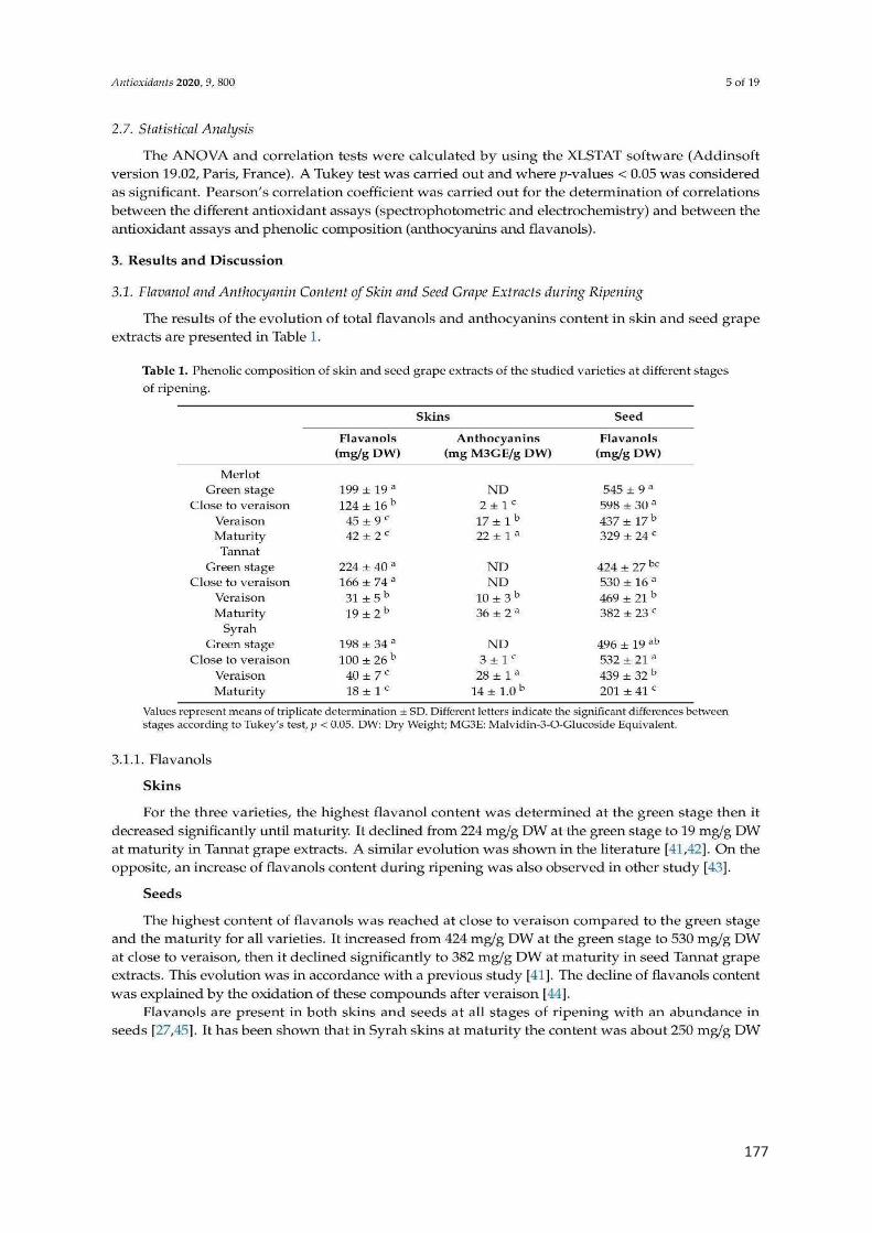
177
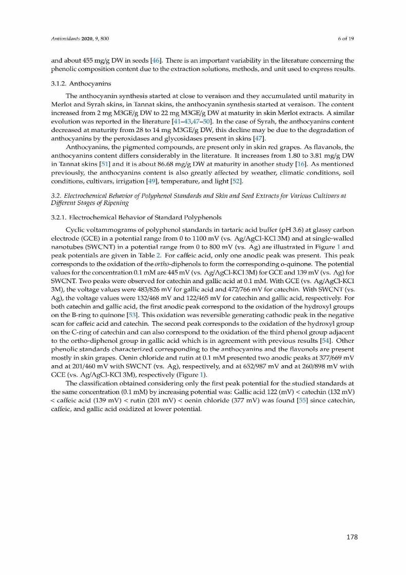
178
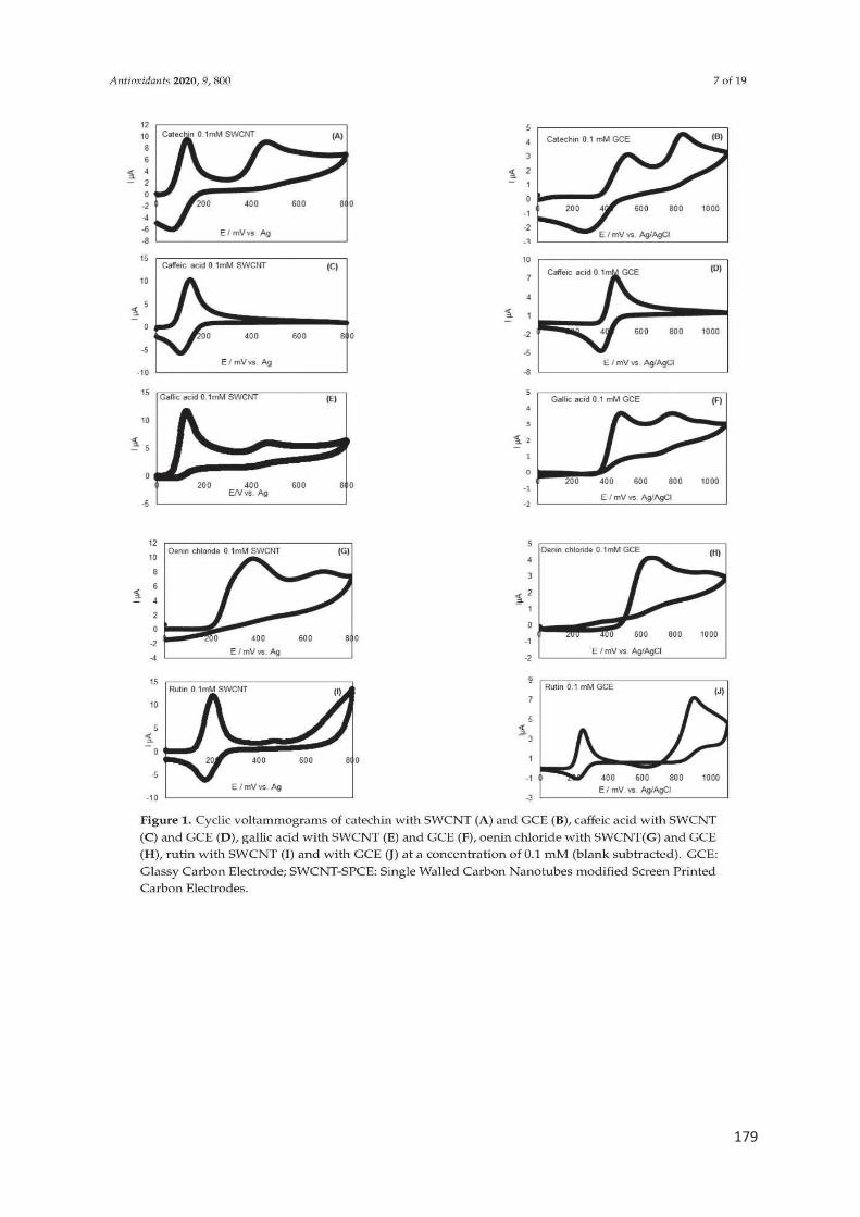
179
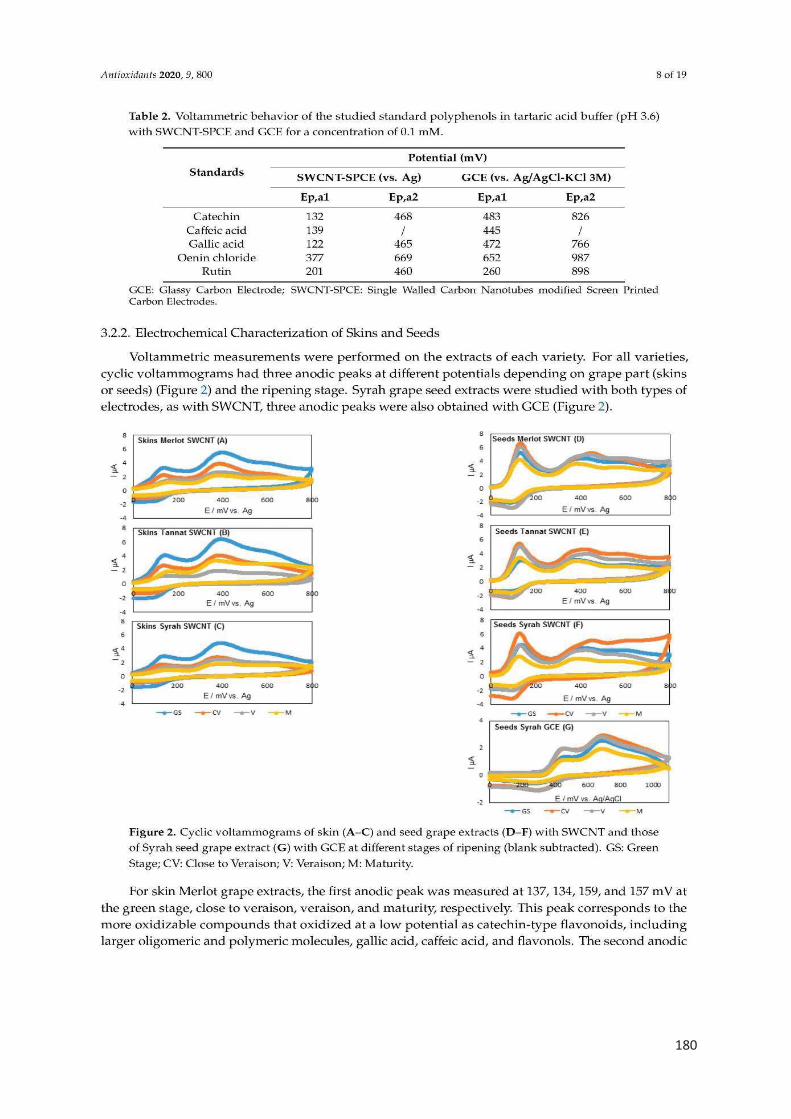
180
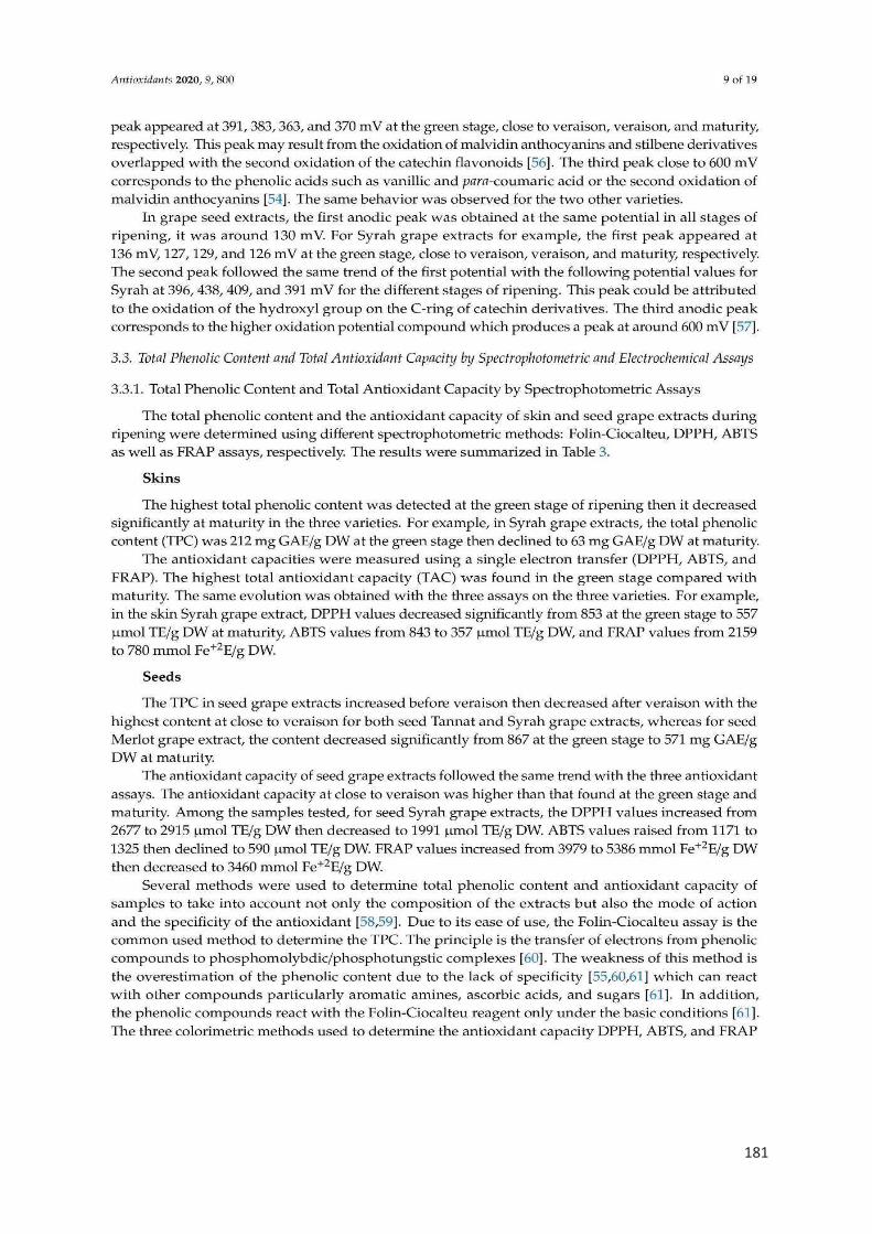
181
182
183

184
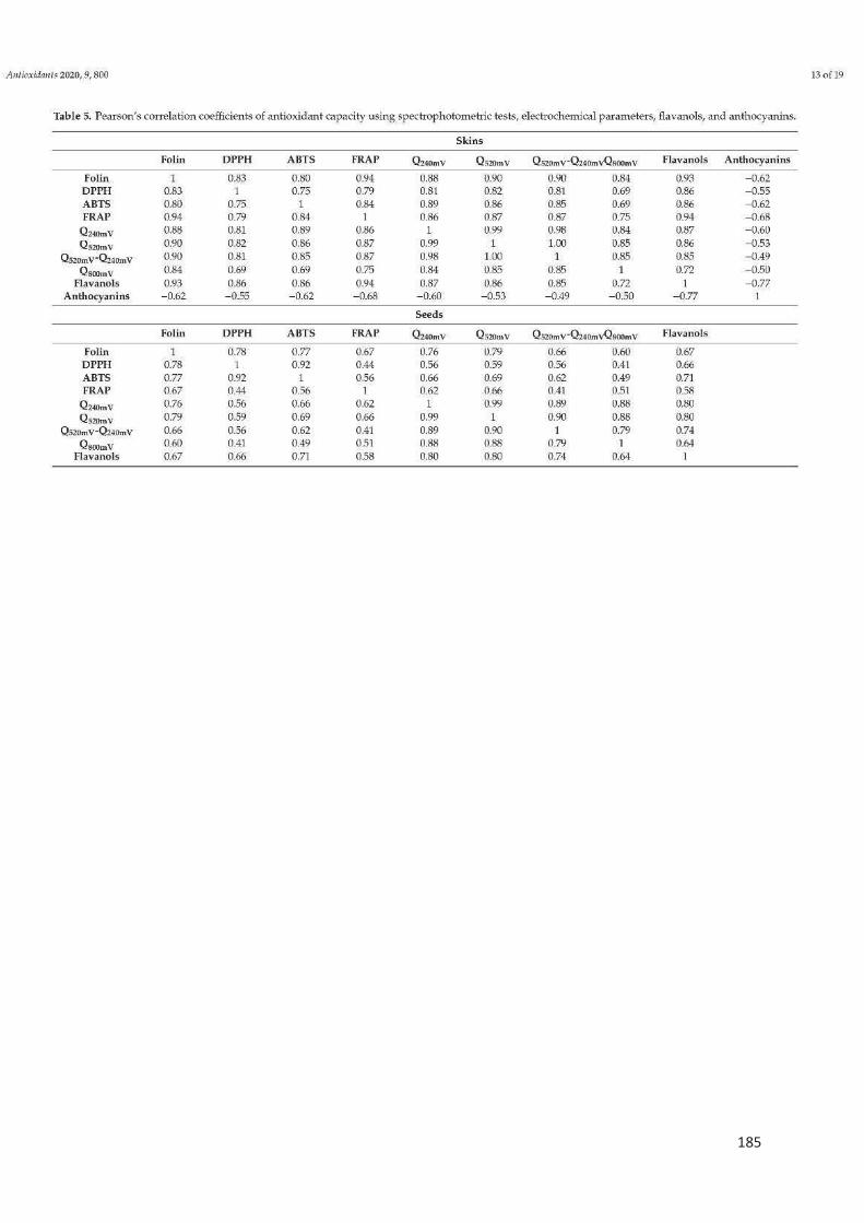
185

186

187

188
189
190
191

192
Chapitre 4 :
Propriétés antioxydantes et anti-inflammatoires des
extraits de pellicules de 3 cépages rouges au cours de
la maturation
193
Les objectifs de ce chapitre étaient de déterminer la capacité antioxydante et anti-
inflammatoire des extraits de pellicules de 3 cépages rouges (année 2018) et de leurs
teneurs en polyphénols totaux, flavanols, flavonols, anthocyanes et stilbènes.
Les méthodes utilisées dans ces travaux étaient :
1. folin-Ciocalteu pour obtenir la teneur en polyphénols totaux ;
2. dpph pour déterminer la capacité antioxydante totale ;
3. tests cellulaires pour déterminer la capacité anti-inflammatoire ;
4. l’UHPLC-MS pour déterminer la teneur des flavanols, anthocyanes, stilbènes
et flavonols ;
Les hypothèses
1. la teneur en polyphénols des extraits de pellicules varie au cours de la
maturation.
2. la capacité antioxydante des extraits de pellicules évolue au cours de la
maturation.
3. la capacité anti-inflammatoire des extraits de pellicules se change au cours
de la maturation.
4. la teneur en flavanols, flavonols, anthocyanes et stilbènes varie au cours de
la maturation.
Conclusions
1. la teneur en polyphénols varie au cours de la maturation, la teneur la plus élevée
est déterminée dans le stade vert.
2. la capacité antioxydante évolue au cours de la maturation, elle est plus
importante dans le stade vert.
3. la capacité anti-inflammatoire des extraits de pellicules varie au cours de la
maturation. Elle est plus importante dans le stade vert puis, elle diminue jusqu’à
la maturité.
4. la teneur en stilbènes et en anthocyanes augmente tandis que l’évolution de la
teneur en flavonols et flavonols change d’un cépage à l’autre.

194
Cette étude a fait l’objet d’un article scientifique, sera soumis dans le journal
agriculture and food chemistry
Et ci-après présenté

195
Polyphenolic characterization of Merlot, Tannat and Syrah skin extracts at 1
different degrees of maturity and anti-inflammatory potential in RAW 264.7 2
cells 3
Nawel Benbouguerra,† Ruth Hornedo-Ortega,‡ Josep Valls-Fonayet,‡ Stephanie 4
Krisa,‡ François Garcia,† Cédric Saucier,† Tristan Richard‡ 5
† SPO, Université de Montpellier, INRAe, Montpellier Supagro, 34000 Montpellier, 6
France 7
‡ Axe Molécules d’Intérêt Biologique, Unité de Recherche Œnologie, ISVV, EA 4577, 8
USC 1366 INRA Université de Bordeaux. 210 chemin de Leysotte, 33882, Villenave 9
d'Ornon, France 10
Corresponding author: [email protected] 11
Abstract 12
Especially in red grape varieties, the maceration and fermentation in presence of skins 13
plays an important role for the final sensory properties of wine but also for their human 14
health properties. Both sensory quality and healthy attributes are closely related with 15
the polyphenolic composition of grape skins. In this study, the polyphenolic 16
characterization (flavan-3-ols, procyanidins, flavonols, stilbenes, anthocyanins) by 17
UHPLC-QqQ-MS of skins from Vitis vinifera Merlot, Tannat and Syrah red grape 18
varieties cultivated in the south of France at different stages of ripening in 2018 were 19
determined. In addition, the anti-inflammatory and the antioxidant potential of the 20
extracts were evaluated by the measure of nitric oxide (NO) and the intracellular 21
production of reactive oxygen species (ROS) in LPS-stimulated macrophages. 22
Forty one individual polyphenols were quantified in all samples. Generally, the flavan-23
3-ol and procyanidin content decreased during ripening whereas the anthocyanins and 24
stilbenes increased. Several oligomeric stilbenes were identified and quantified for the 25
first time by using authentic standards. In addition, before veraison extracts exhibited 26
more than 50% of NO and ROS productions inhibition. 27
Keywords: phenolic compounds, Vitis vinifera, ripening, grapes, anti-inflammatory, 28
antioxidant activity. 29
30

196
1. Introduction 31
The cultivation of the vine is one of the largest crops in the world. Recent data reported 32
that in 2018, 7.4 million hectares was the global area under vines destined for the 33
production of wine grapes, table grapes or dried grapes. Specifically, the production of 34
wine from Vitis vinifera grapes reached 292 millions of hectoliters in 2018. V. vinifera 35
species are characteristic for their great diversity. In fact, the actual number of V. 36
vinifera varieties is estimated at 6 000.1 37
Polyphenolic compounds are secondary metabolites with undeniable demonstrated 38
biological properties.2,3 These compounds are usually divided in two important families: 39
flavonoids (flavan-3-ols, procyanidins, flavonols, anthocyanins) and non-flavonoids 40
(phenolic acids and stilbenes).4 Grapes are especially rich in these compounds, and 41
the polyphenolic content and composition varies substantially depending of grape 42
variety, stage of ripening, temperature, soil, fungal infection or radiation and soil 43
salinity.5,6 Morover, polyphenolic compounds are determinant for wine quality, due to 44
their contribution to sensory properties such as color, taste, mouthfeel, flavor, 45
astringency, and bitterness.7 In general, red grape skins are initially rich in flavan-3-ols 46
and procyanidins at pre-veraison state whereas at veraison and maturity stages 47
anthocyanins are the major compounds. However, the greater quantities of stilbenes 48
were found at maturity stage.8–10 49
The consumption of moderate quantities of red wine, pattern characteristic of the 50
Mediterranean countries, and their richness in polyphenols has been broadly 51
associated to the health benefits of this dietary pattern.11,12 Consequently, the study of 52
polyphenolics from grapes and red wines have enthralled the attention of scientists to 53
define their chemical composition and their properties concerning human health. 54
Indeed, grape polyphenols have evidenced beneficial effects to prevent 55
neurodegenerative, cardiovascular and metabolic disorders and certain types of 56
cancers.13,14 Oxidative stress and inflammation are important pathological hallmarks 57
related with the development and progression of several diseases.15 Therefore, in 58
order to counteract this reaction, there is a growing interest to identify grape anti-59
inflammatory and antioxidant compounds. 60
In this study, the polyphenolic characterization of flavan-3-ols, procyanidins, flavonols, 61
anthocyanins and stilbenes by UHPLC-QqQ-MS and UPLC-PAD of skins from V. 62
197
vinifera Merlot, Tannat and Syrah red grape varieties cultivated in the south of France 63
at different stages of ripening on 2018 (before veraison, veraison and maturity) were 64
evaluated. In addition, the anti-inflammatory and the antioxidant potential of extracts in 65
LPS-stimulated macrophages (RAW 264.7 cells) were determined. 66
2. Materials and Methods 67
2.1. Chemicals and reagents 68
Folin Ciocalteu phenol reagent, sodium carbonate, 1,1-diphenyl-2-picrylhydrazyl 69
(DPPH), Trolox, phloroglucinol, ascorbic acid, tartaric acid, sodium hydroxide, 70
hydrochloric acid, lipopolysaccharide (LPS), RPMI and DMEM mediums, fetal bovine 71
serum (FBS), Griess reagent, 2'7'-dichlorodihydrofluoroscein diacetate acetyl (DCFH2-72
DA), 3-(4,5-dimethylethiazol-2-yl)-2,5-diphenyl tetrazolium bromide (MTT), dimethyl 73
sulfoxide (DMSO), glutamine, gallic acid, catechin, malvidin-3-O-glucoside, trans-74
piceid, trans-resveratrol were obtained from Sigma Aldrich (Steinheim, Germany). 75
Trans-astringin was purchased by Carbosynth (UK) and trans-piceatannol by 76
ChromaDex (USA). Acetonitrile, methanol and water LC-MS were obtained by 77
Biosolve (Dieuze, France) and Trifluoroacetic acid and sodium acetate were 78
purchased by Carlo Erba (Peypin, France). 79
RAW 264.7 cells were provided by ATCC (Manassas, USA). ε-viniferin, δ-viniferin, ω-80
viniferin, pallidol, parthenocissin A, miyabenol C, hopeaphenol and isohopeaphenol 81
were isolated from a grapevine raw shoot in our laboratory. The cis-isomers were 82
obtained using Ultraviolet (UV)-C irradiation (254 nm) from trans-isomers. 83
2.2. Grape samples 84
Three red V. vinifera varieties: Merlot, Tannat and Syrah were harvested from the 85
vineyard of INRAe Montpellier (Coordinates: 43°37'02.7"N 3°51'22.3"E, average 86
annual temperature: 16.38° C, average annual precipitation: 1063.5 mm and soil: 87
gravels and river sand) at different stages of ripening: before veraison (18 June 2018), 88
veraison (27 July 2018) and maturity (3 September 2018). Grapes clusters were frozen 89
within an hour after sampling -at 80°C until sample preparation. 90
2.3. Grape samples preparation and polyphenolic extraction. 91
Grapes were thawed and pH and °Brix were measured. For this, 50 berries were 92
crushed, the pH was determined by a multi-parameter analyzer (Consort C3010) and 93

198
the concentration of sugar was determined using a hand-held refractometer (results 94
expressed as °Brix). 95
Skins of 30 grapes of each variety (Merlot, Tannat and Syrah) were manually 96
separated from the pulp. Then, a solid/liquid extraction using 100 mL of acetone/water 97
(70:30; v/v) previously deoxygenated with nitrogen for 5 minutes were performed in 98
order to extract the polyphenolic compounds. After stirring for 18 hours in the dark, the 99
solution was filtered through a 0.45 µm filter paper and evaporated in a rotavapor under 100
low pressure fixed at 37°C. Finally, the obtained extract was freeze-dried and stored 101
at -80°C until further analysis. 102
2.4. Determination of Total Polyphenol Content (TPC) 103
The total phenolic content of the extract was determined by the Folin–Ciocalteu 104
method.16 A stock solution of 5 g/L was prepared by diluting 5 mg of skins extracts in 105
1 mL of methanol. 20 μL of the diluted extract was mixed to 100 μL of diluted Folin-106
Ciocalteu reagent. Then, 80 μL of sodium carbonate solution (7.5%) were added. The 107
reaction mixture was thoroughly shaken and left for 30 min in the dark at room 108
temperature (20°C). Finally, the absorbance was measured at 760 nm using a 109
microplate reader (FLUOstar Optima, BMG Labtech). A calibration curve of gallic acid 110
(GAE) (0-0.5mg/mL) was used to determine the concentration of polyphenol in 111
samples. The results were expressed as g of gallic acid equivalents (GAE) per g of 112
skin grapes fresh weight (FW). 113
2.5. Determination of radical scavenging activity (DPPH˙ assay). 114
The antioxidant activity of the extract was determined by the DPPH assay, as 115
described earlier with some modifications.17 A stock solution of 5 g/L was prepared by 116
diluting 5 mg of skins extracts in 1 mL of methanol. Fifty µL of this stock solution was 117
diluted 4 times in methanol and mixed with 150 μL of a DPPH (200 μM) methanolic 118
solution. After 20 minutes of incubation in the dark at room temperature, the 119
absorbance was measured at 517 nm using a microplate reader (FLUOstar Optima, 120
BMG Labtech). A calibration curve of Trolox (TE) (0-300 µM) was used. The results 121
were expressed as µM TE/g of skin grapes fresh weight (FW). 122
2.6. Analysis of proanthocyanidins following acid catalysis with phloroglucinol. 123
199
Proanthocyanidin analyses were performed with a Waters Acquity Ultra Performance 124
Liquid Chromatography (UPLC) system connected to a photodiode array detector 125
(PDA). The proanthocyanidin assay was carried out as described by Kennedy et al.18 126
A solution of 0.1 N of HCl in methanol, containing 50 g/L phloroglucinol and 10 g/L 127
ascorbic acid was prepared. Samples (10 g/L in methanol) were mixed with this 128
solution for 20 minutes at 50°C, and then combined with 5 volumes of 40 mM aqueous 129
sodium acetate solution to stop the reaction, then filtrated through PTFE 0.45 μm filters 130
and injected. The column used to analyse phloroglucinol adducts was an Acquity 131
UPLC BEH C18 column (2.1 mm x 50 mm, 1.7 μm particle size) that was thermostated 132
at 40°C during the analysis. The injection volume was 7.5 µL and the flow rate set at 133
0.45 mL/min. The mobile phases consisted of solvent A (water/TFA 99:1 v/v) and 134
solvent B (acetonitrile 100%) with the following gradient: 0 min 2% B, 8 min 6% B, 14 135
min 20% B, 16 min 99% B, and 20 min 2% B. Eluting peaks were monitored at 280 136
nm. In order to calculate the mean Degree of Polymerization (mDP), the sum of all 137
subunits (flavan-3-ol monomers and phloroglucinol adducts expressed in millimoles) 138
was divided by the sum of all flavan-3-ols monomers (expressed in millimoles). 139
2.7. Individual determination of phenolic composition 140
2.7.1. Monomeric flavan-3-ols, procyanidins, flavonols and stilbenes. 141
The monomeric flavan-3-ols, procyanidins, flavonols and stilbenes analysis was 142
performed by UHPLC coupled to a triple quadrupole (QqQ) mass spectrometer 143
(UHPLC-QqQ-MS) based on a previous method.19 The freeze-dried skin samples and 144
standards were solubilized in methanol/water (1:1; v/v) at an appropriate concentration 145
(20 g/L). The system was composed of an UHPLC system (Agilent Technology 1260 146
Infinity, Agilent Technologies, Santa Clara, CA, United States), hyphenated to an 147
Agilent Technologies 6430 Triple Quadrupole Detector. An Agilent Poroshell 120 EC-148
C18 column (150 mm × 2.1 mm, 2.7 µm particle size) was used as a stationary phase. 149
The mobile phases consisted of 0.1% formic acid in water as solvent A and 0.1% formic 150
acid in acetonitrile as solvent B, using the following gradient: 5-17.5% B (0-5 min), 151
17.5%-33% B (5-7.5 min), 33% B (7.5-10 min), 33%-40% B (10-15 min), 40%-95% B 152
(15-16 min), 95% B (16-19 min) and 5% B (19-21 min). The flow rate was fixed at 0.3 153
mL/minute and the column temperature was set at 35°C. The injection volume was 4 154
µL. The detection of compounds was in the multiple reaction monitoring (MRM) mode 155
200
with specific transitions for each compound, and for quantification purposes a 156
calibration curve was prepared (0.05-26 mg/L) with pure standards. All tests were 157
carried out in triplicate and the results were expressed as mg per kg of skin grapes 158
fresh weight (FW). 159
2.7.2. Anthocyanins 160
The anthocyanins analyses were carried out in almost the same column, flow rate and 161
solvent used for proanthocyanidin analyses (2.6. Section). Skin extracts were 162
solubilized at 10 g/L in MeOH/eau (80:20; v/v), filtrated through PTFE 0.45 μm filters 163
then injected. The flow rate was fixed at 0.25 mL/minute. The mobile phases consisted 164
of solvent A (water/TFA 99:1 v/v) and solvent B (acetonitrile 100%) scheduled in the 165
following gradient: 0 min 1% B, 5 min 8.8% B, 30 min 20.6% B, 34 min 96% B, 34.1 166
min 1% and 40 min 1% B. Eluting peaks were monitored at 520 nm. A calibration curve 167
of malvidin-3-O-glucoside (0-200 mg/L) was used to quantify anthocyanins in all 168
samples.20 169
2.8. Cell culture and treatment 170
RAW 264.7 cells were cultured in Dulbecco’s Modified Eagle Medium (DMEM) 171
containing 10% of fetal bovine serum (FBS) and maintained in a humidified incubator 172
at 37°C with 5% of CO2. Cells were subcultured at a density of 50000 cells per well, in 173
96-well culture plates with 200 µL of culture medium. After 24 hours, cells were 174
incubated with skin extracts (50-300 µg/mL) in RPMI medium supplemented with 175
glutamine (4 mM) in presence or absence of LPS (0.1 µg/mL) (200 µL final volume 176
per well). 177
2.9. MTT cell viability 178
MTT reduction was employed as a cell viability indicator, extensively used as a 179
quantitative and consistent colorimetric assay.21 After 24 hours of treatment, RAW 180
264.7 cells were incubated with 0.5 mg/mL of MTT during 3 hours at 37°C. The 181
resulting crystals formed were dissolved with 100 µL of DMSO. After 30 minutes of 182
incubation in darkness, the absorbance was measured at a wavelength of 595 nm 183
using a microplate reader (FLUOstar Optima, BMG Labtech). 184
2.10. Intracellular NO measurement 185

201
In the same way and after 24 hours of treatment, 50 µL of supernatant mixed with 50 186
µL of Griess solution. After 15 minutes in darkness, the absorbance was measured at 187
a wavelength of 550 nm using a microplate reader (FLUOstar Optima, BMG Labtech). 188
A calibration curve of NO2 (0-100 mM) was used. Data were expressed as NO 189
production (µM) compared with cells treated only with LPS (positive control). 190
2.11. Intracellular ROS measurement 191
Generation of intracellular ROS in cells was analyzed using a fluorometric probe: 192
DCFH2-DA. After treatment, cells were washed with PBS and then 150 µL of DCFH2-193
DA (10 µM) was added. After 30 minutes at 37°C, the fluorescence intensity was 194
quantified using a spectrofluorometer (FLUOstar Optima, BMG Labtech). The 195
wavelengths of excitation and emission used to detect ROS were 485 nm and 520 nm, 196
respectively. All experiments were performed in the dark. Results were given as ROS 197
production (fluorescence intensity) compared with cells treated only with LPS (positive 198
control). 199
2.12. Statistical analysis 200
The data was subjected to one-way ANOVA test with XLSTAT version 19.02. 201
Comparison between the different stages of ripening was performed using Tukey’s test 202
and p < 0.05 was considered significant. 203
3. Results and discussion 204
Grapes of Merlot, Tannat and Syrah cultivars were harvested at different stages of 205
ripening: June 2018 (Before Veraison; BV), July 2018 (Veraison; V) and September 206
2018 (Maturity; M). Table 1 summarizes the °Brix and pH values of samples. 207
Table 1. °Brix and pH of Merlot, Tannat and Syrah berries at GS, V and M stages. 208
Grapes Merlot Tannat Syrah
°Brix pH °Brix pH °Brix pH
BV 4.37 2.56 4.3 2.57 4.37 2.58 V 8.57 3.34 11.64 2.75 16.97 3.42 M 25.97 3.78 25.84 3.87 25.37 4
209
210
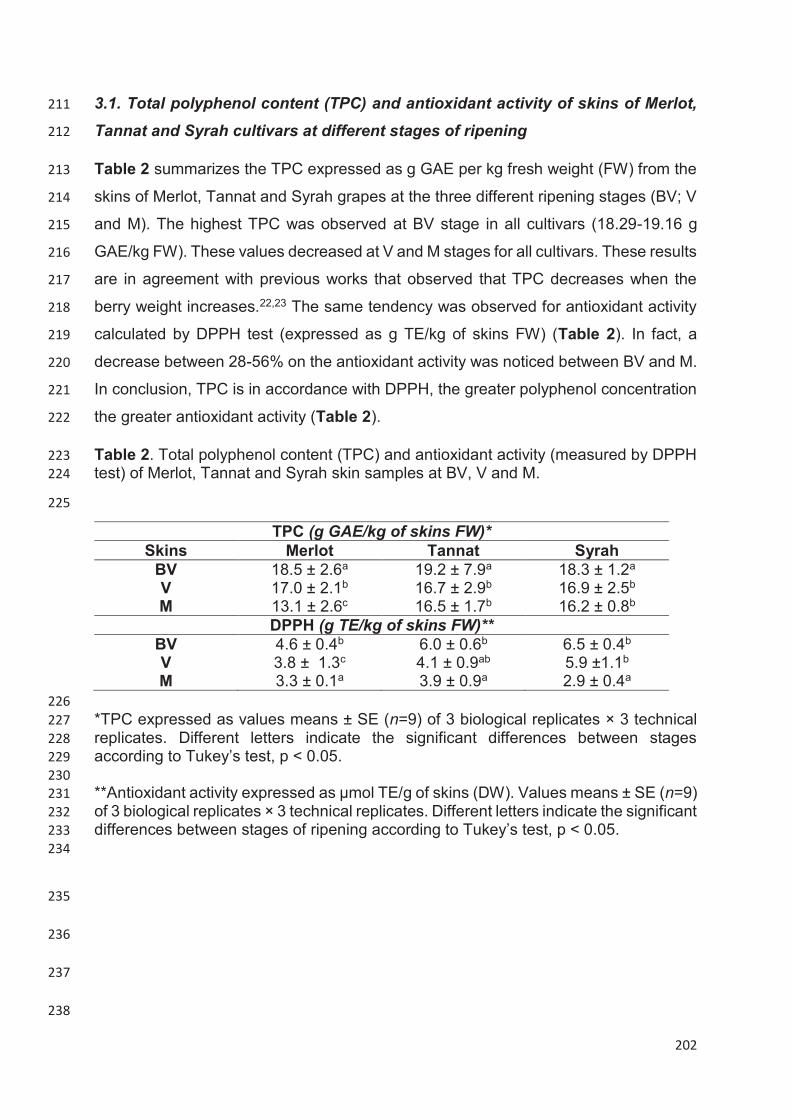
202
3.1. Total polyphenol content (TPC) and antioxidant activity of skins of Merlot, 211
Tannat and Syrah cultivars at different stages of ripening 212
Table 2 summarizes the TPC expressed as g GAE per kg fresh weight (FW) from the 213
skins of Merlot, Tannat and Syrah grapes at the three different ripening stages (BV; V 214
and M). The highest TPC was observed at BV stage in all cultivars (18.29-19.16 g 215
GAE/kg FW). These values decreased at V and M stages for all cultivars. These results 216
are in agreement with previous works that observed that TPC decreases when the 217
berry weight increases.22,23 The same tendency was observed for antioxidant activity 218
calculated by DPPH test (expressed as g TE/kg of skins FW) (Table 2). In fact, a 219
decrease between 28-56% on the antioxidant activity was noticed between BV and M. 220
In conclusion, TPC is in accordance with DPPH, the greater polyphenol concentration 221
the greater antioxidant activity (Table 2). 222
Table 2. Total polyphenol content (TPC) and antioxidant activity (measured by DPPH 223
test) of Merlot, Tannat and Syrah skin samples at BV, V and M. 224
225
TPC (g GAE/kg of skins FW)* Skins Merlot Tannat Syrah
BV 18.5 ± 2.6a 19.2 ± 7.9a 18.3 ± 1.2a V 17.0 ± 2.1b 16.7 ± 2.9b 16.9 ± 2.5b M 13.1 ± 2.6c 16.5 ± 1.7b 16.2 ± 0.8b
DPPH (g TE/kg of skins FW)** BV 4.6 ± 0.4b 6.0 ± 0.6b 6.5 ± 0.4b V 3.8 ± 1.3c 4.1 ± 0.9ab 5.9 ±1.1b M 3.3 ± 0.1a 3.9 ± 0.9a 2.9 ± 0.4a
226
*TPC expressed as values means ± SE (n=9) of 3 biological replicates × 3 technical 227
replicates. Different letters indicate the significant differences between stages 228
according to Tukey’s test, p ˂ 0.05. 229
230
**Antioxidant activity expressed as µmol TE/g of skins (DW). Values means ± SE (n=9) 231
of 3 biological replicates × 3 technical replicates. Different letters indicate the significant 232
differences between stages of ripening according to Tukey’s test, p ˂ 0.05. 233
234
235
236
237
238
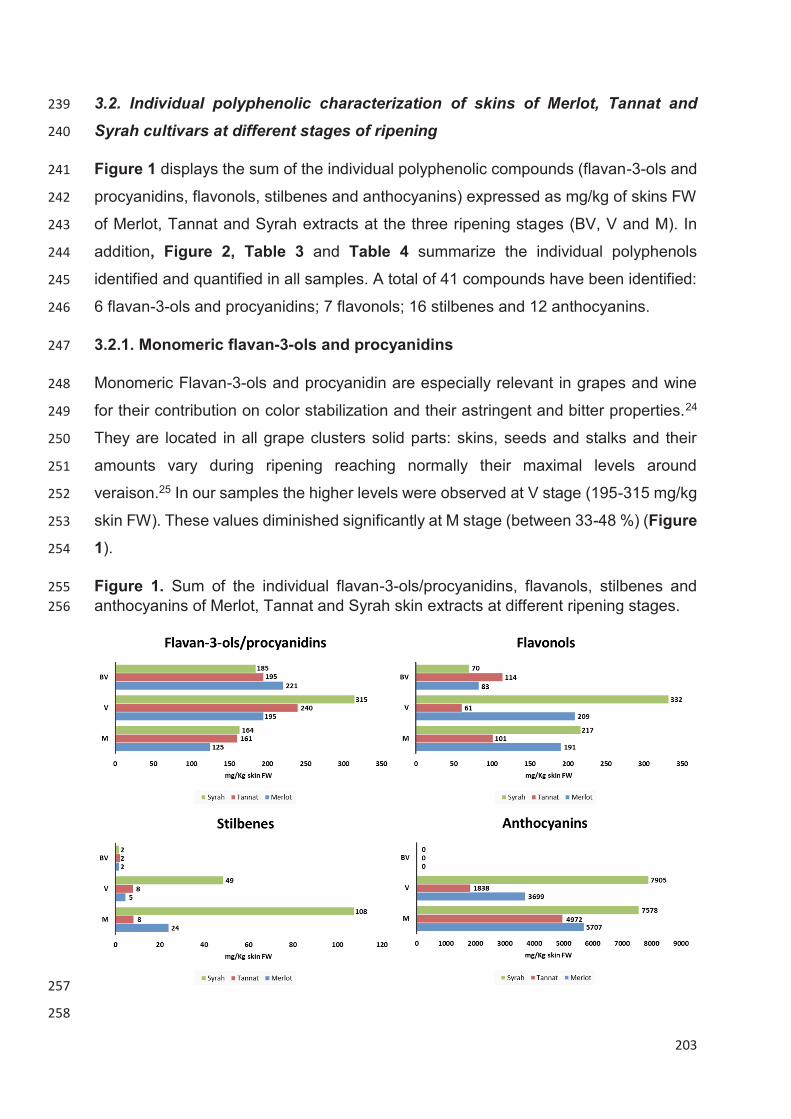
203
3.2. Individual polyphenolic characterization of skins of Merlot, Tannat and 239
Syrah cultivars at different stages of ripening 240
Figure 1 displays the sum of the individual polyphenolic compounds (flavan-3-ols and 241
procyanidins, flavonols, stilbenes and anthocyanins) expressed as mg/kg of skins FW 242
of Merlot, Tannat and Syrah extracts at the three ripening stages (BV, V and M). In 243
addition, Figure 2, Table 3 and Table 4 summarize the individual polyphenols 244
identified and quantified in all samples. A total of 41 compounds have been identified: 245
6 flavan-3-ols and procyanidins; 7 flavonols; 16 stilbenes and 12 anthocyanins. 246
3.2.1. Monomeric flavan-3-ols and procyanidins 247
Monomeric Flavan-3-ols and procyanidin are especially relevant in grapes and wine 248
for their contribution on color stabilization and their astringent and bitter properties.24 249
They are located in all grape clusters solid parts: skins, seeds and stalks and their 250
amounts vary during ripening reaching normally their maximal levels around 251
veraison.25 In our samples the higher levels were observed at V stage (195-315 mg/kg 252
skin FW). These values diminished significantly at M stage (between 33-48 %) (Figure 253
1). 254
Figure 1. Sum of the individual flavan-3-ols/procyanidins, flavanols, stilbenes and 255
anthocyanins of Merlot, Tannat and Syrah skin extracts at different ripening stages. 256
257
258

204
Regarding flavan-3-ols monomers individually, (+)-catechin is the major compound 259
followed by (-)-epicatechin at BV stage. (+)-catechin quantities diminished 260
considerably during ripening in all varieties (Table 3). This tendency has been 261
observed in previously studies for the variety Syrah,26 Tannat,22 and Merlot.23 262
Interestingly, (-)-epicatechin levels increased from BV to V stage to finally diminished 263
at M stage. In any case, the final amounts of (-)-epicatechin are significantly higher that 264
the initials (BV stage). This observation was also in accordance with other studies.22,23 265
Four B-type procyanidins were identified and quantified in all samples: B1, B2, B3 and 266
B4. Among them, procyanidin B1 (epicatechin-(4β→8)-catechin) was the predominant 267
with initial values (BV) ranging from 50.77-75.53 mg/kg skins FW. The major levels for 268
this compound were observed at the V stage to finally decrease at the M stage. 269
Important amounts of procyanidin B3 were also present in all samples, which also 270
decreased over the course of maturation in all cultivars and in all ripening stages. 271
Finally, procyanidins B2 and B4 were also present in all samples with levels between 272
4.5 and 9.57 mg/kg of FW (Table 3). In accordance with our study, the procyanidin B1 273
was quantified as the major compound in Syrah skin extracts at maturity.26 In a 274
previous study, these four B-type proanthocyanidins have also been quantified in 275
Merlot skins extracts at different maturity stages, however procyanidin B2 was the 276
predominant.23 277
278
279
280
281
282
283
284
285
286
287

205
Table 3. Quantification of individual flavan-3-ols, procyanidins, flavonols and stilbenes in skins extracts of Merlot, Tannat and Syrah 288
varieties at different stages of ripening. 289
Merlot Tannat Syrah BV V M BV V M BV V M Flavan-3-ols/procyanidins*
(+)-Catechin 107.04±1.48 53.36±0.51 23.95±0.95 114.45±4.66 90.11±5.18 29.43±6.56 97.72±16.66 111.11±.02 39.82±5.84 (-)-Epicatechin 1.38±0.44 5.30±0.37 5.90±2.47 2.32±0.78 9.85±0.96 7.31±1.50 0.86±0.08 30.94±5.54 8.75±0.30 Procyanidin B1 75.53±3.90 128.23±3.56 71.43±2.14 50.77±0.39 108.18±5.16 105.36±6.09 52.66±4.24 138.63±8.90 89.92±9.03 Procyanidin B2 2.09±0.36 3.83±0.50 9.21±0.76 3.07±0.11 4.27±0.03 4.50±0.17 1.16±0.10 6.82±2.44 9.57±0.20 Procyanidin B3 29.45±2.48 22.99±3.85 12.04±1.74 17.35±0.04 24.27±2.34 6.99±1.71 26.14±2.95 30.26±.64 16.51±3.56 Procyanidin B4 5.61±0.33 5.14±0.62 7.42±2.07 7.35±0.57 8.22±0.87 4.16±1.04 6.42±1.17 7.78±1.92 6.73±1.20 Flavonols * Quercetin 0.13±0.04 0.33±0.06 0.28±0.16 0.08±0.04 0.25±0.06 0.34±0.09 0.07±0.00 2.02±0.30 1.03±0.45 Quercetin-3-O-hexoside 2.78±0.49 28.35±5.83 38.07±3.01 3.21±0.65 6.09±2.28 8.56±0.11 2.61±1.03 75.88±1.34 148.13±25.29 Quercetin-3-O-galactoside 0.62±0.08 4.51±1.06 3.99±0.92 1.04±0.10 0.87±0.13 0.75±0.05 0.30±0.04 7.26±0.66 9.66±1.79 Quercetin-3-O-glucuronide
75.14±9.01 154.63±15.09 122.17±6.80 98.93±13.71 44.78±4.22 58.10±21.21 63.90±0.75 227.90±18.01 117.70±12.62
Quercetin-3-O-rhamnoside
0.03±0.02 1.06±0.43 0.45±0.18 0.01±0.00 0.02±0.00 0.02±0.00 0.02±0.00 1.16±0.12 1.10±0.84
Quercetin-3-O-rutinoside 1.33±0.23 1.75±0.11 0.49±0.05 5.46±0.78 3.34±0.28 0.80±0.46 0.38±0.21 0.58±0.50 1.22±1.10 Kaempferol-3-O-glucoside 0.29±0.10 6.78±1.75 4.28±0.10 0.40±0.15 1.67±0.52 0.32±0.12 0.08±0.05 3.87±1.38 9.06±2.37 Stilbenes* Cis-resveratrol 0.03±0.02 0.04±0.01 0.14±0.02 0.03±0.01 0.45±0.26 0.09±0.02 0.03±0.00 1.94±0.05 1.43±0.65 Trans-resveratrol 0.11±0.04 0.13±0.02 0.25±0.11 0.04±0.01 0.44±0.38 0.17±0.10 0.13±0.02 4.07±1.21 5.34±0.18 ∑ resveratrol 0.15 0.17 0.39 0.07 0.89 0.26 0.16 6.01 6.77 Cis-piceid 0.30±.0.06 1.39±0.44 9.63±0.93 1.04±0.48 2.36±1.31 3.00±1.12 0.73±0.21 15.74±6.84 41.99±1.41 Trans-piceid 0.10±0.02 0.36±0.01 1.93±0.37 0.68±0.32 0.52±0.25 1.41±0.28 0.20±0.08 2.12±1.14 7.65±1.53 ∑ piceid 0.39 1.75 11.56 1.72 2.88 4.41 0.93 17.86 49.64 Cis-piceatannol 0.03±0.01 0.38±0.19 0.26±0.07 0.05±0.02 0.03±0.00 0.09±0.03 0.03±0.00 9.18±1.52 1.52±0.53 Trans-piceatannol 0.02±0.01 0.23±0.11 1.38±0.80 0.02±0.01 0.02±0.01 0.32±0.15 0.02±0.00 5.48±0.91 10.49±1.93 ∑ piceatannol 0.04 0.61 1.64 0.06 0.23 0.41 0.05 14.66 12.01 Cis-astringin 0.03±0.00 0.24±0.00 2.93±0.49 0.04±0.02 0.13±0.08 0.28±0.01 0.04±0.02 0.83±0.33 3.77±0.98 Trans-astringin 0.04±0.01 0.72±0.10 5.91±1.14 0.09±0.06 0.29±0.16 1.70±0.81 0.04±0.02 1.97±0.33 13.27±1.03 ∑ astringin 0.07 0.96 8.84 0.13 0.42 1.98 0.08 2.79 17.05 ε-viniferin 0.73±0.19 0.93±0.37 1.61±0.26 0.12±0.04 1.70±0.70 0.63±0.16 0.27±0.11 1.38±0.37 4.77±0.21 ω-viniferin 0.03±0.01 0.04±0.01 0.10±0.04 0.01±0.000 0.07±0.03 0.06±0.01 0.01±0.00 0.27±0.05 0.95±0.04 σ-viniferin 0.01±0.00 0.01±0.00 0.95±0.14 0.01±0.01 0.07±0.02 0.08±0.02 0.01±0.00 0.84±0.13 2.97±0.70 Pallidol 0.02±0.01 0.03±0.01 1.88±1.11 0.03±0.01 0.48±0.30 0.28±0.05 0.05±0.02 1.06±0.46 6.73±2.57 Parthenocissin A 0.06±0.04 0.08±0.07 2.37±1.11 0.06±0.04 0.53±0.37 0.39±0.14 0.05±0.02 1.47±0.18 4.81±0.91 Miyabenol C 0.06±0.01 0.01±0.00 0.22±0.02 0.08±0.06 0.27±0.15 0.16±0.07 0.34±0.02 0.57±0.09 3.05±0.85 Hopeaphenol 0.03±0.01 0.06±0.01 0.26±0.04 0.01±0.00 0.02±0.00 0.03±0.01 0.02±0.00 0.05±0.01 0.22±0.08 Isohopeaphenol 0.03±0.00 0.04±0.01 0.44±0.19 0.04±0.01 0.04±0.00 0.61±0.07 0.03±0.01 1.07±0.31 2.95±0.22
206
*Flavan-3-ols, procyanidins, flavonols and stilbenes are expressed as mg of their corresponding standard per kg of skins FW. 290
Values means ± SE of 3 biological replicate291
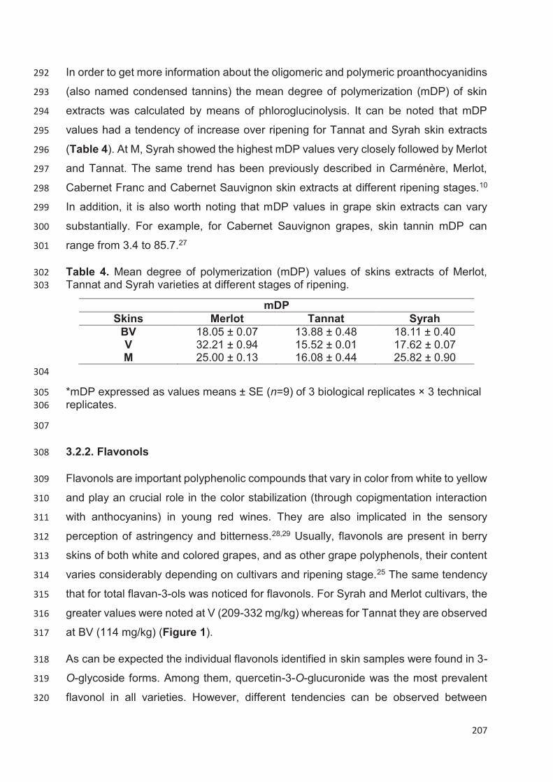
207
In order to get more information about the oligomeric and polymeric proanthocyanidins 292
(also named condensed tannins) the mean degree of polymerization (mDP) of skin 293
extracts was calculated by means of phloroglucinolysis. It can be noted that mDP 294
values had a tendency of increase over ripening for Tannat and Syrah skin extracts 295
(Table 4). At M, Syrah showed the highest mDP values very closely followed by Merlot 296
and Tannat. The same trend has been previously described in Carménère, Merlot, 297
Cabernet Franc and Cabernet Sauvignon skin extracts at different ripening stages.10 298
In addition, it is also worth noting that mDP values in grape skin extracts can vary 299
substantially. For example, for Cabernet Sauvignon grapes, skin tannin mDP can 300
range from 3.4 to 85.7.27 301
Table 4. Mean degree of polymerization (mDP) values of skins extracts of Merlot, 302
Tannat and Syrah varieties at different stages of ripening. 303
mDP Skins Merlot Tannat Syrah
BV 18.05 ± 0.07 13.88 ± 0.48 18.11 ± 0.40 V 32.21 ± 0.94 15.52 ± 0.01 17.62 ± 0.07 M 25.00 ± 0.13 16.08 ± 0.44 25.82 ± 0.90
304
*mDP expressed as values means ± SE (n=9) of 3 biological replicates × 3 technical 305
replicates. 306
307
3.2.2. Flavonols 308
Flavonols are important polyphenolic compounds that vary in color from white to yellow 309
and play an crucial role in the color stabilization (through copigmentation interaction 310
with anthocyanins) in young red wines. They are also implicated in the sensory 311
perception of astringency and bitterness.28,29 Usually, flavonols are present in berry 312
skins of both white and colored grapes, and as other grape polyphenols, their content 313
varies considerably depending on cultivars and ripening stage.25 The same tendency 314
that for total flavan-3-ols was noticed for flavonols. For Syrah and Merlot cultivars, the 315
greater values were noted at V (209-332 mg/kg) whereas for Tannat they are observed 316
at BV (114 mg/kg) (Figure 1). 317
As can be expected the individual flavonols identified in skin samples were found in 3-318
O-glycoside forms. Among them, quercetin-3-O-glucuronide was the most prevalent 319
flavonol in all varieties. However, different tendencies can be observed between 320

208
varieties. Indeed, in Merlot and Syrah cultivars their amounts increased greatly at V 321
stage then diminished at M stage (although the M stage quantities remain more 322
elevated that at BV). On the contrary, the major levels of quercetin-3-O-glucuronide 323
were observed at BV for Tannat skin extracts (Table 3). Substantial amounts of 324
quercetin-3-O-glucuronide have also been found in skins extracts of Merlot, Cabernet 325
Sauvignon, Petit Verdot, Syrah, Tempranillo, Garnacha and Garnacha Tintorera.30 326
Quercetin aglycone and other quercetin glycosides (3-O-hexoside, 3-O-galactoside, 3-327
O-rhamnoside and 3-O-rutinoside) are also present in small quantities in all samples 328
(Table 3). In addition to that, kaempferol-3-O-glucoside has also been quantified and 329
their amounts increase during ripening but only for Syrah variety. 330
3.2.3. Stilbenes 331
Stilbenes, polyphenolic compounds that belong to the non-flavonoid family, are present 332
in grapes at very low concentrations (1%) and are mainly concentrated in skins.31 333
However, due to their importance as bioactive compounds for plant and human health, 334
their characterization on grapes and other foodstuffs is especially interesting. 335
The stilbene content varies significantly versus the ripening stages and the maximal 336
concentrations of stilbenes in grape skin are reached at maturity stage (Figure 1). This 337
augmentation is closely related with the expression increase of key enzymes 338
responsible of stilbene synthesis and accumulation: stilbene synthase, phenylalanine 339
ammonia-lyase and 4-coumarate-CoA ligase.32 In accordance with this, our results 340
indicated that the total stilbenes increase at V stage to reach their maximum at M for 341
all studied varieties. Between cultivars, Syrah was the variety richer in stilbenes with 342
107.63 mg/kg skins followed by Merlot (24.11 mg/kg skins) and Tannat (8.17 mg/kg 343
skins) (Figure 1). 344
A great variety of stilbenes were identified in all samples (Table 3). The authentic 345
standards of several stilbenes (ε-viniferin, δ-viniferin, ω-viniferin, partenocissin A, 346
Miyabenol C, hopeaphenol and isohopeaphenol,) isolated and purified in our 347
laboratory were used to identification and quantification purposes. Among them, 4 348
monomers: resveratrol, piceid, piceatannol and astringin (in their -trans and –cis 349
configurations), 5 dimers: ε-viniferin, ω-viniferin, σ-viniferin, pallidol and partenocissin 350
A; one trimer: miyabenol C; and two tetramers: hopeaphenol and isohopeaphenol were 351
identified. Piceid (mainly in the –cis form) represents the major compound in all 352

209
varieties (ranging from 0.03-49.64 mg/kg skins) followed by astringin, piceatannol and 353
resveratrol. Important quantities of ε-viniferin were also quantified in samples (0.12-354
4.77 mg/kg skins). Concerning other oligomeric stilbenes, several differences were 355
observed between cultivars. In Merlot, Parthenocissin A was the main oligomeric 356
stilbene but for Syrah it was Pallidol. Very similar results have been recently published 357
in Tannat variety at different ripening stages during 2017 harvest.33 As far as we know, 358
this is the first time that an identification and quantification of complex stilbenes with 359
authentical standards was carried out in Merlot and Syrah varieties. In addition, 360
partenocissin A and miyabenol C were identified for the first time that in Merlot, Tannat 361
and Syrah cultivars. 362
3.2.3. Anthocyanins 363
Anthocyanins are the most important natural pigments in wine grapes. These 364
compounds are predominately accumulated in skins of red grapes during ripening, and 365
they are also present in the flesh of “teinturier” varieties.34 366
In red grape skins, anthocyanins were the more predominant polyphenolic compounds. 367
In fact, they represented between 84-91% at V and between 94-95% at M in Merlot, 368
Tannat and Syrah varieties (Figure 1). Once again, Syrah skin extract was the richest 369
in anthocyanins with values of 7578 mg/kg skins at maturity, followed by Merlot (5707 370
mg/kg skins) and Tannat (4972 mg/kg skins) (Figure 1). Fifteen individual 371
anthocyanins, divided in three families (five -3-O-glucosides, five 3-O-acetylglucosides 372
and five-3-O-coumaroylglucosides) were identified in all samples (Table 5) (Figure 2). 373
Figure 2. LC-DAD chromatogram of anthocyanins identified and quantified in samples. 374
375
376
210
1: Delphinidin-3-O-glucoside; 2: Cyanidin-3-O-glucoside; 3: Petunidin-3-O-glucoside; 377
4: Peonidin-3-O-glucoside; 5: Malvidin-3-O-glucoside; 6: Delphinidin-3-O-378
acetylglucoside; 7: Cyanidin-3-O-acetylglucoside; 8: Petunidin-3-O-acetylglucoside; 9: 379
Peonidin-3-O-acetylglucoside; 10: Malvidin-3-O-acetylglucoside; 11: Delphinidin-3-O-380
coumaroylglucoside; 12: Cyanidin-3-O-coumaroylglucoside; 13: Petunidin-3-O-381
coumaroylglucoside; 14: Peonidin-3-O-coumaroylglucoside; 15: Malvidin-3-O-382
coumaroylglucoside 383
384
It is well established in V.vinifera red skin grapes that the 3-O-glucosides are the main 385
forms followed by 3-O-acetylglucosides and 3-O-coumaroylglucosides. Delphinidin, 386
cyanidin, petunidin, peonidin and malvidin-3-O-glucosides were quantified. 387
Additionally, petunidin, peonidin and malvidin-3-O-acetylglucosides and delphinidin, 388
cyanidin, peonidin and malvidin -3-O-coumaroylglucosides were also quantified in all 389
samples. Malvidin-3-O-glucoside was the major anthocyanin followed by peonidin-3-390
O-glucoside in Merlot skin extracts. However, in the case of Tannat and Syrah, 391
petunidin-3-O-glucoside was the second more predominant anthocyanin. These 392
results were in accordance with other authors that have noticed the same observation 393
in Merlot,10 Tannat22 and Syrah.26 394
395
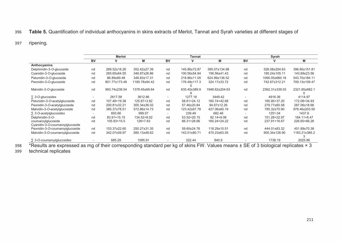
211
Table 5. Quantification of individual anthocyanins in skins extracts of Merlot, Tannat and Syrah varieties at different stages of 396
ripening. 397
Merlot Tannat Syrah BV V M BV V M BV V M Anthocyanins Delphinidin-3-O-glucoside nd 269.52±18.26 352.42±27.39 nd 145.86±72.87 395.07±134.98 nd 526.08±204.63 396.80±151.81 Cyanidin-3-O-glucoside nd 265.65±64.55 346.87±26.86 nd 100.56±54.84 156.56±41.43 nd 195.24±105.11 143.69±23.56 Petunidin-3-O-glucoside nd 86.89±69.48 348.83±17.31 nd 218.86±11.05 624.99±136.52 nd 1090.05±660.16 543.70±184.11 Peonidin-3-O-glucoside nd 801.77±173.48 1185.78±94.42 nd 176.48±117.3
5 324.17±33.72 nd 742.67±312.21 709.13±109.47
Malvidin-3-O-glucoside nd 993.74±238.54 1378.45±69.64 nd 635.40±389.99
1948.62±204.63 nd 2362.31±339.03 2321.65±682.10
∑ 3-O-glucosides - 2617.58 3612.86 - 1277.18 3449.42 - 4916.36 4114.97 Petunidin-3-O-acetylglucoside nd 107.48+19.38 125.87+3.82 nd 58.61+24.12 160.74+42.68 nd 185.95+37.20 172.08+34.93 Peonidin-3-O-acetylglucoside nd 200.81±32.21 300.34±36.02 nd 57.46±20.84 94.67±12.26 nd 279.77±60.58 287.06±18.66 Malvidin-3-O-acetylglucoside nd 385.37±78.51 572.86±14.73 nd 123.42±67.78 427.06±60.19 nd 785.32±70.90 978.46±203.50 ∑ 3-O-acetylglucosides - 693.66 999.07 - 239.49 682.46 - 1251.04 1437.6 Delphinidin-3-O-coumaroylglucoside Cyanidin-3-O-coumaroylglucoside
nd nd
83.91+15.15 105.93+15.5
134.52+8.02 126+7.63
nd nd
53.52+20.15 66.31+26.66
92.14+9.06 160.24+24.22
nd nd
151.28+22.97 237.91+16.67
164.11+8.47 226.65+66.26
Peonidin-3-O-coumaroylglucoside nd 153.37±22.60 250.27±31.35 nd 59.60±24.76 118.29±10.51 nd 444.01±83.32 451.89±70.38 Malvidin-3-O-coumaroylglucoside nd 342.07±59.97 585.13±48.62 nd 143.01±80.71 470.23±63.55 nd 905.34±126.90 1183.21±385.2
3 ∑ 3-O-coumaroylglucosides - 685.29 1095.91 - 322.44 840.9 - 1738.19 2025.86
*Results are expressed as mg of their corresponding standard per kg of skins FW. Values means ± SE of 3 biological replicates × 3 398
technical replicates399

212
3.3. Anti-inflammatory and antioxidant potential in vitro effects of skins extracts
of Merlot, Tannat and Syrah cultivars at different stages of ripening.
The anti-inflammatory potential of skins extracts of Merlot, Tannat and Syrah at BV, V
and M was assessed by the intracellular measure of nitric oxide (NO), and reactive
oxygen species (ROS) in a macrophage model cell line (RAW 264.7). NO levels are a
well-established marker for the inflammatory process. Indeed, after infection,
macrophages are stimulated and can secrete NO and several interleukins in order to
destroy the infectious agent. While inflammation is a priori a beneficial response, when
an excessive production of ROS is chronically generated, the consequence is an
important damage at cellular level. In fact, NO is one of the major contributors to the
formation of ROS.35 Indeed, chronic inflammation and oxidative stress are considered
as a major cause of age related diseases and cancer.15
Initially, the cytotoxicity of skin extracts was determined by using the MTT assay, a test
based on the reduction of a yellow tetrazolium salt (MTT) to purple formazan crystals
by metabolically active cells. For this, cells were treated with different concentrations
of skin extracts (50-300 μg/mL) at all experimental conditions (three varieties and three
ripening stages) during 24 hours. After analyzing the results, the concentration of 100
μg/mL was selected for further analysis to be non-toxic for the cellular model.
For NO measurement, RAW 264.7 cells were activated with LPS (0.1 µg/mL) in the
presence of 100 μg/mL of all skins extracts (three cultivars and three ripening stages).
After 24 hours of exposure, culture media were analyzed for nitrite (NO2) content by
the Griess reaction. Figure 4A shows the NO production (µM) when cells were treated
with LPS alone or with LPS and skin extracts. As can be expected, the NO
concentration for positive control (cells treated with LPS) increased threefold (21.2 µM)
in comparison with the negative control (7.9 µM) (cells without LPS treatment). When
cells were also treated with skin extracts, the NO concentration diminished significantly
(around half) for BV and V extracts. In all cases, the most anti-inflammatory sample
was the BV extract for all varieties.
In addition, in order to gain insight into the antioxidant potential of skin extracts, the
ROS production induced by LPS was investigated by employing the fluorometric probe
DCFH2-DA that is a widely used indicator to detect and quantify the intracellular
produced ROS. Figure 4B displays the effect exerted by skin extracts after 24 hours

213
of treatment on ROS production (expressed as fluorescence intensity). As can be
clearly observed, the treatment of cells with LPS corresponds to a marked increase of
fluorescence (about four times higher) in comparison with negative control proving that
the oxidant effect of LPS. However, and in the same way that for NO production, the
treatment with LPS + skin extracts produced a significant reduction of ROS production
for BV samples of Merlot and for BV and V samples of Tannat and Syrah varieties
(Figure 4B). Although the individual characterization shows that in general flavan-3-
ols, procyanidins, flavonols and stilbenes reached the highest values at V and M stage,
it should be taken in consideration that other polyphenolic compounds not quantified
in our samples can be the responsible of the observed effects. In fact, TPC values are
much higher than the sum of all quantified polyphenols, fact that can explain that BV
samples are generally the most anti-inflammatory and antioxidant extracts.
Additionally, other bioactive molecules not identified in this work can also contribute to
the effect.
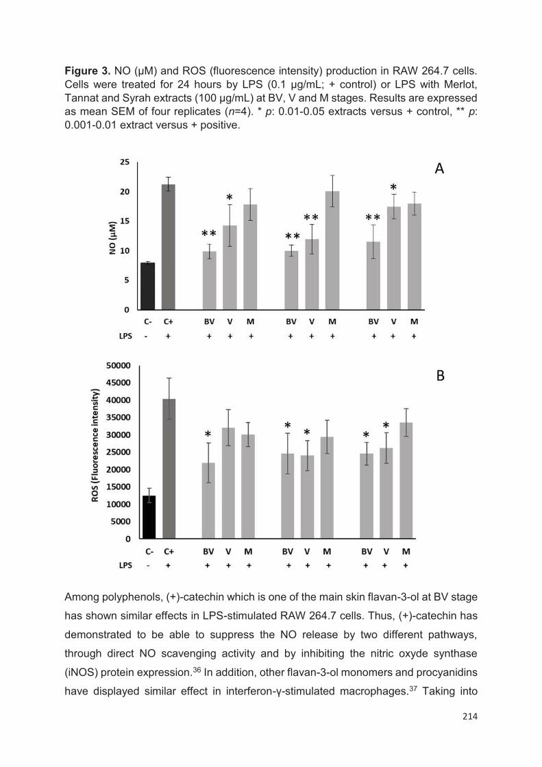
214
Figure 3. NO (µM) and ROS (fluorescence intensity) production in RAW 264.7 cells. Cells were treated for 24 hours by LPS (0.1 μg/mL; + control) or LPS with Merlot, Tannat and Syrah extracts (100 μg/mL) at BV, V and M stages. Results are expressed as mean SEM of four replicates (n=4). * p: 0.01-0.05 extracts versus + control, ** p: 0.001-0.01 extract versus + positive.
Among polyphenols, (+)-catechin which is one of the main skin flavan-3-ol at BV stage
has shown similar effects in LPS-stimulated RAW 264.7 cells. Thus, (+)-catechin has
demonstrated to be able to suppress the NO release by two different pathways,
through direct NO scavenging activity and by inhibiting the nitric oxyde synthase
(iNOS) protein expression.36 In addition, other flavan-3-ol monomers and procyanidins
have displayed similar effect in interferon-γ-stimulated macrophages.37 Taking into
215
account that the major polyphenols in our extracts at V and M stages are the
anthocyanins, we can hypothesize that they can be also the responsible molecules of
the observed anti-inflammatory and antioxidant effects. This theory can be based on
previously results that have demonstrated that malvidin-3-O-glucoside (the major
anthocyanin in grape skins) was able to attenuate LPS-induced nuclear factor-KappaB
(pro-inflammatory transcription factor associated with NO and interleukin liberation)
and ROS production in macrophages.38
In summary, this work provides a wide characterization of individual polyphenolic
compounds (41 compounds). This is the first time that an identification and
quantification of complex stilbenes with authentical standards have been carried out in
Merlot and Syrah varieties. Moreover, parthenocissin A and miyabenol C have been
identified in Merlot, Tannat and Syrah cultivars for the first time. Although the major
polyphenols content was displayed at BV stage, the HPLC-MS analysis has allowed
to monitoring the individual evolution and changes in these compounds during
ripening. Whereas anthocyanins, flavonols and stilbenes in general increase during
ripening, other compounds such as (+)-catechin (in all cultivars) or quercetin-3-O-
glucuronide (in Tannat variety) followed the inverse order. Skin extracts of all varieties
at BV stage, have proven to be effective to decrease (>50%) NO and ROS intracellular
production in macrophages. More studies should be conducted in order to demonstrate
the specific contribution of polyphenolic compounds or other bioactives present in
grape skins.
4. Acknowledgments
The work was supported by the Bordeaux Metabolome Facility and MetaboHUB (ANR-
11- INBS-0010 project). The work was supported in part by a PhD grant (N.B.) from
the government of Algeria (Ministère algérien de l’Enseignement Supérieur et de la
Recherche Scientifique).
5. Abbreviations used
BV, before veraison; DCFH2-DA, 2'7'-dichlorodihydrofluoroscein diacetate acetyl;
DPPH, 1,1-diphenyl-2-picrylhydrazyl; FW, fresh weight; GAE, gallic acid equivalent;
iNOS, oxyde nitrique synthase; M, maturity; MS, mass spectrometry; MTT, 3-(4,5-
dimethyl-2-thiazolyl)-2,5-diphenyl-2H-tetrazolium bromide; NO, nitric oxide; ROS,
216
reactive oxygen species; TPC, total phenolic content; TE, trolox equivalent; UHPLC,
ultra-high performance liquid chromatography; V, Veraison
217
References
(1) Lacombe, T. Contribution à l’étude de l’histoire Évolutive de La Vigne Cultivée
(Vitis Vinifera L.) Par l’analyse de La Diversité Génétique Neutre et de Gènes
d’intérêt; 2012.
(2) Han, X.; Shen, T.; Lou, H. Dietary Polyphenols and Their Biological
Significance. Int J Mol Sci 2007, 8 (9), 950–988.
(3) Vauzour, D.; Rodriguez-Mateos, A.; Corona, G.; Oruna-Concha, M. J.; Spencer,
J. P. E. Polyphenols and Human Health: Prevention of Disease and
Mechanisms of Action. Nutrients 2010, 2 (11), 1106–1131.
https://doi.org/10.3390/nu2111106.
(4) Saltveit, M. E. Synthesis and Metabolism of Phenolic Compounds. In Fruit and
Vegetable Phytochemicals; John Wiley & Sons, Ltd, 2009; pp 89–100.
https://doi.org/10.1002/9780813809397.ch3.
(5) Yilmaz, Y.; Göksel, Z.; Erdoğan, S. S.; Öztürk, A.; Atak, A.; Özer, C. Antioxidant
Activity and Phenolic Content of Seed, Skin and Pulp Parts of 22 Grape (Vitis
Vinifera L.) Cultivars (4 Common and 18 Registered or Candidate for
Registration). Journal of Food Processing and Preservation 2015, 39 (6), 1682–
1691. https://doi.org/10.1111/jfpp.12399.
(6) Ashraf, M. A.; Iqbal, M.; Rasheed, R.; Hussain, I.; Riaz, M.; Arif, M. S.
Environmental Stress and Secondary Metabolites in Plants. In Plant Metabolites
and Regulation Under Environmental Stress; Elsevier, 2018; pp 153–167.
https://doi.org/10.1016/B978-0-12-812689-9.00008-X.
(7) Hornedo-Ortega, R.; Reyes González-Centeno, M.; Chira, K.; Jourdes, M.;
Teissedre, P.-L. Phenolic Compounds of Grapes and Wines: Key Compounds
and Implications in Sensory Perception. In Winemaking - Stabilization, Aging
Chemistry and Biochemistry [Working Title]; IntechOpen, 2020.
https://doi.org/10.5772/intechopen.93127.
(8) Ivanova, V.; Stefova, M.; Vojnoski, B.; Dörnyei, Á.; Márk, L.; Dimovska, V.;
Stafilov, T.; Kilár, F. Identification of Polyphenolic Compounds in Red and White
Grape Varieties Grown in R. Macedonia and Changes of Their Content during
Ripening. Food Research International 2011, 44 (9), 2851–2860.
https://doi.org/10.1016/j.foodres.2011.06.046.
218
(9) Obreque-Slier, E.; Peña-Neira, A.; López-Solís, R.; Zamora-Marín, F.; Ricardo-
da Silva, J. M.; Laureano, O. Comparative Study of the Phenolic Composition
of Seeds and Skins from Carménère and Cabernet Sauvignon Grape Varieties
(Vitis Vinifera L.) during Ripening. J. Agric. Food Chem. 2010, 58 (6), 3591–
3599. https://doi.org/10.1021/jf904314u.
(10) Obreque-Slier, E.; Peña-Neira, Á.; López-Solís, R.; Cáceres-Mella, A.; Toledo-
Araya, H.; López-Rivera, A. Phenolic Composition of Skins from Four Carmenet
Grape Varieties (Vitis Vinifera L.) during Ripening. LWT - Food Science and
Technology 2013, 54 (2), 404–413. https://doi.org/10.1016/j.lwt.2013.06.009.
(11) di Giuseppe, R.; de Lorgeril, M.; Salen, P.; Laporte, F.; Di Castelnuovo, A.;
Krogh, V.; Siani, A.; Arnout, J.; Cappuccio, F. P.; van Dongen, M.; Donati, M.
B.; de Gaetano, G.; Iacoviello, L.; European Collaborative Group of the
IMMIDIET Project. Alcohol Consumption and N-3 Polyunsaturated Fatty Acids
in Healthy Men and Women from 3 European Populations. Am. J. Clin. Nutr.
2009, 89 (1), 354–362. https://doi.org/10.3945/ajcn.2008.26661.
(12) Urquiaga, I.; Guasch, V.; Marshall, G.; San Martín, A.; Castillo, O.; Rozowski,
J.; Leighton, F. Effect of Mediterranean and Occidental Diets, and Red Wine,
on Plasma Fatty Acids in Humans. An Intervention Study. Biol. Res. 2004, 37
(2), 253–261. https://doi.org/10.4067/s0716-97602004000200012.
(13) Pasinetti, G. M. Novel Role of Red Wine-Derived Polyphenols in the Prevention
of Alzheimer’s Disease Dementia and Brain Pathology: Experimental
Approaches and Clinical Implications. Planta Med. 2012, 78 (15), E24.
https://doi.org/10.1055/s-0035-1545846.
(14) Rasines-Perea, Z.; Teissedre, P.-L. Grape Polyphenols’ Effects in Human
Cardiovascular Diseases and Diabetes. Molecules 2017, 22 (1).
https://doi.org/10.3390/molecules22010068.
(15) Khansari, N.; Shakiba, Y.; Mahmoudi, M. Chronic Inflammation and Oxidative
Stress as a Major Cause of Age-Related Diseases and Cancer. Recent patents
on inflammation & allergy drug discovery 2009.
https://doi.org/10.2174/187221309787158371.
(16) Singleton, V. L.; Orthofer, R.; Lamuela-Raventós, R. M. Analysis of Total
Phenols and Other Oxidation Substrates and Antioxidants by Means of Folin-
Ciocalteu Reagent. In Methods in Enzymology; Oxidants and Antioxidants Part

219
A; Academic Press, 1999; Vol. 299, pp 152–178. https://doi.org/10.1016/S0076-
6879(99)99017-1.
(17) Villaño, D.; Fernández-Pachón, M. S.; Moyá, M. L.; Troncoso, A. M.; García-
Parrilla, M. C. Radical Scavenging Ability of Polyphenolic Compounds towards
DPPH Free Radical. Talanta 2007, 71 (1), 230–235.
https://doi.org/10.1016/j.talanta.2006.03.050.
(18) Kennedy, J. A.; Jones, G. P. Analysis of Proanthocyanidin Cleavage Products
Following Acid-Catalysis in the Presence of Excess Phloroglucinol. J. Agric.
Food Chem. 2001, 49 (4), 1740–1746. https://doi.org/10.1021/jf001030o.
(19) Loupit, G.; Prigent, S.; Franc, C.; De Revel, G.; Richard, T.; Cookson, S. J.;
Fonayet, J. V. Polyphenol Profiles of Just Pruned Grapevine Canes from Wild
Vitis Accessions and Vitis Vinifera Cultivars. J. Agric. Food Chem. 2020.
https://doi.org/10.1021/acs.jafc.9b08099.
(20) Giuffrè, A. M. HPLC-DAD Detection of Changes in Phenol Content of Red Berry
Skins during Grape Ripening. Eur Food Res Technol 2013, 237 (4), 555–564.
https://doi.org/10.1007/s00217-013-2033-7.
(21) Mosmann, T. Rapid Colorimetric Assay for Cellular Growth and Survival:
Application to Proliferation And. Journal of Inmunological Methods 1983, 55,
55–63.
(22) Boido, E.; García-Marino, M.; Dellacassa, E.; Carrau, F.; Rivas-Gonzalo, J. C.;
Escribano-Bailón, M. T. Characterisation and Evolution of Grape Polyphenol
Profiles of Vitis Vinifera L. Cv. Tannat during Ripening and Vinification.
Australian Journal of Grape and Wine Research 2011, 17 (3), 383–393.
https://doi.org/10.1111/j.1755-0238.2011.00164.x.
(23) Lorrain, B.; Chira, K.; Teissedre, P.-L. Phenolic Composition of Merlot and
Cabernet-Sauvignon Grapes from Bordeaux Vineyard for the 2009-Vintage:
Comparison to 2006, 2007 and 2008 Vintages. Food Chem 2011, 126 (4),
1991–1999. https://doi.org/10.1016/j.foodchem.2010.12.062.
(24) Chira, K.; Schmauch, G.; Saucier, C.; Fabre, S.; Teissedre, P.-L. Grape Variety
Effect on Proanthocyanidin Composition and Sensory Perception of Skin and
Seed Tannin Extracts from Bordeaux Wine Grapes (Cabernet Sauvignon and
Merlot) for Two Consecutive Vintages (2006 and 2007). J. Agric. Food Chem.
2009, 57 (2), 545–553. https://doi.org/10.1021/jf802301g.
220
(25) Downey, M. O.; Harvey, J. S.; Robinson, S. P. Analysis of Tannins in Seeds and
Skins of Shiraz Grapes throughout Berry Development. Aust J Grape Wine Res
2003, 9 (1), 15–27. https://doi.org/10.1111/j.1755-0238.2003.tb00228.x.
(26) Ky, I.; Lorrain, B.; Kolbas, N.; Crozier, A.; Teissedre, P.-L. Wine By-Products:
Phenolic Characterization and Antioxidant Activity Evaluation of Grapes and
Grape Pomaces from Six Different French Grape Varieties. Molecules 2014, 19
(1), 482–506. https://doi.org/10.3390/molecules19010482.
(27) Rousserie, P.; Rabot, A.; Geny-Denis, L. From Flavanols Biosynthesis to Wine
Tannins: What Place for Grape Seeds? J. Agric. Food Chem. 2019, 67 (5),
1325–1343. https://doi.org/10.1021/acs.jafc.8b05768.
(28) Heras-Roger, J.; Alonso-Alonso, O.; Gallo-Montesdeoca, A.; Díaz-Romero, C.;
Darias-Martín, J. Influence of Copigmentation and Phenolic Composition on
Wine Color. J Food Sci Technol 2016, 53 (6), 2540–2547.
https://doi.org/10.1007/s13197-016-2210-3.
(29) Ferrer-Gallego, R.; Brás, N. F.; García-Estévez, I.; Mateus, N.; Rivas-Gonzalo,
J. C.; de Freitas, V.; Escribano-Bailón, M. T. Effect of Flavonols on Wine
Astringency and Their Interaction with Human Saliva. Food Chem 2016, 209,
358–364. https://doi.org/10.1016/j.foodchem.2016.04.091.
(30) Castillo-Muñoz, N.; Gómez-Alonso, S.; García-Romero, E.; Hermosín-
Gutiérrez, I. Flavonol Profiles of Vitis Vinifera Red Grapes and Their Single-
Cultivar Wines. J. Agric. Food Chem. 2007, 55 (3), 992–1002.
https://doi.org/10.1021/jf062800k.
(31) Németh, G.; Hegyi, O.; Dunai, A.; Kocsis, L. Stilbenes in the Different Organs
of Vitis Vinifera Cv. Merlot Grafted on Teleki Kober 5BB Rootstock. 2017, 51
(3), 323–328.
(32) Gatto, P.; Vrhovsek, U.; Muth, J.; Segala, C.; Romualdi, C.; Fontana, P.;
Pruefer, D.; Stefanini, M.; Moser, C.; Mattivi, F.; Velasco, R. Ripening and
Genotype Control Stilbene Accumulation in Healthy Grapes. J. Agric. Food
Chem. 2008, 56 (24), 11773–11785. https://doi.org/10.1021/jf8017707.
(33) Dudoit, A.; Benbouguerra, N.; Richard, T.; Hornedo-Ortega, R.; Valls-Fonayet,
J.; Coussot, G.; Saucier, C. α-Glucosidase Inhibitory Activity of Tannat Grape
Phenolic Extracts in Relation to Their Ripening Stages. Biomolecules 2020, 10
(8). https://doi.org/10.3390/biom10081088.

221
(34) Burns, J.; Mullen, W.; Landrault, N.; Teissedre, P.-L.; Lean, M. E. J.; Crozier, A.
Variations in the Profile and Content of Anthocyanins in Wines Made from
Cabernet Sauvignon and Hybrid Grapes. J. Agric. Food Chem. 2002, 50 (14),
4096–4102. https://doi.org/10.1021/jf011233s.
(35) MacMicking, J.; Xie, Q. W.; Nathan, C. Nitric Oxide and Macrophage Function.
Annu. Rev. Immunol. 1997, 15, 323–350.
https://doi.org/10.1146/annurev.immunol.15.1.323.
(36) Cíz, M.; Pavelková, M.; Gallová, L.; Králová, J.; Kubala, L.; Lojek, A. The
Influence of Wine Polyphenols on Reactive Oxygen and Nitrogen Species
Production by Murine Macrophages RAW 264.7. Physiol Res 2008, 57 (3), 393–
402.
(37) Park, Y. C.; Rimbach, G.; Saliou, C.; Valacchi, G.; Packer, L. Activity of
Monomeric, Dimeric, and Trimeric Flavonoids on NO Production, TNF-α
Secretion, and NF-ΚB-Dependent Gene Expression in RAW 264.7
Macrophages. FEBS Letters 2000, 465 (2), 93–97.
https://doi.org/10.1016/S0014-5793(99)01735-4.
(38) Bognar, E.; Sarszegi, Z.; Szabo, A.; Debreceni, B.; Kalman, N.; Tucsek, Z.;
Sumegi, B.; Gallyas, F. Antioxidant and Anti-Inflammatory Effects in RAW264.7
Macrophages of Malvidin, a Major Red Wine Polyphenol. PLoS One 2013, 8
(6). https://doi.org/10.1371/journal.pone.0065355.

222
Chapitre 5
Inhibition de l’enzyme alpha-glucosidase par des
extraits de pépins et de pellicules de Tannat au
cours de la maturité
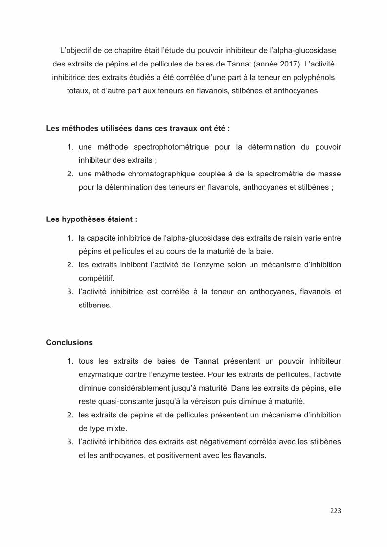
223
L’objectif de ce chapitre était l’étude du pouvoir inhibiteur de l’alpha-glucosidase
des extraits de pépins et de pellicules de baies de Tannat (année 2017). L’activité
inhibitrice des extraits étudiés a été corrélée d’une part à la teneur en polyphénols
totaux, et d’autre part aux teneurs en flavanols, stilbènes et anthocyanes.
Les méthodes utilisées dans ces travaux ont été :
1. une méthode spectrophotométrique pour la détermination du pouvoir
inhibiteur des extraits ;
2. une méthode chromatographique couplée à de la spectrométrie de masse
pour la détermination des teneurs en flavanols, anthocyanes et stilbènes ;
Les hypothèses étaient :
1. la capacité inhibitrice de l’alpha-glucosidase des extraits de raisin varie entre
pépins et pellicules et au cours de la maturité de la baie.
2. les extraits inhibent l’activité de l’enzyme selon un mécanisme d’inhibition
compétitif.
3. l’activité inhibitrice est corrélée à la teneur en anthocyanes, flavanols et
stilbenes.
Conclusions
1. tous les extraits de baies de Tannat présentent un pouvoir inhibiteur
enzymatique contre l’enzyme testée. Pour les extraits de pellicules, l’activité
diminue considérablement jusqu’à maturité. Dans les extraits de pépins, elle
reste quasi-constante jusqu’à la véraison puis diminue à maturité.
2. les extraits de pépins et de pellicules présentent un mécanisme d’inhibition
de type mixte.
3. l’activité inhibitrice des extraits est négativement corrélée avec les stilbènes
et les anthocyanes, et positivement avec les flavanols.
224
Cette étude a fait l’objet d’un article scientifique, publié dans le journal
Biomolécules, sous la référence :
Dudoit, A., Benbouguerra, N., Richard, T., Hornedo-Ortega, R., Valls-Fonayet, J.,
Coussot, G., & Saucier, C. (2020). α-Glucosidase Inhibitory Activity of Tannat Grape
Phenolic Extracts in Relation to Their Ripening Stages. Biomolecules, 10(8), 1088.
Et ci-après présenté.
225
226
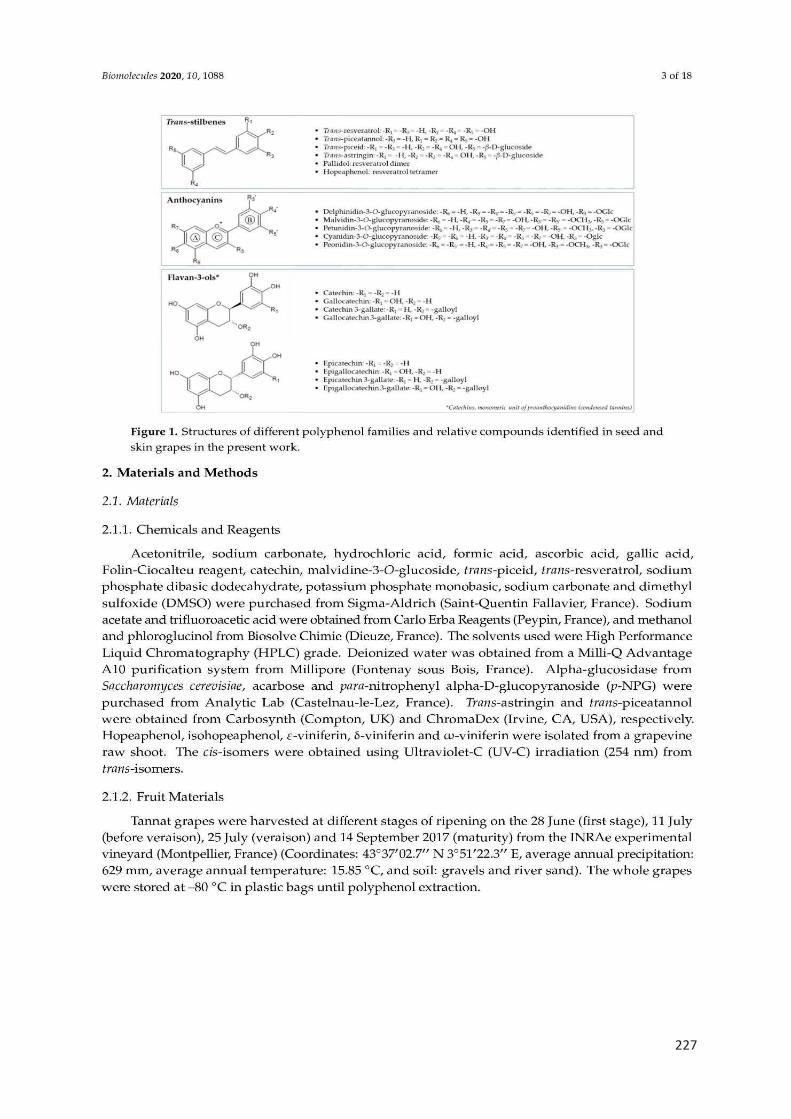
227
228
229
230
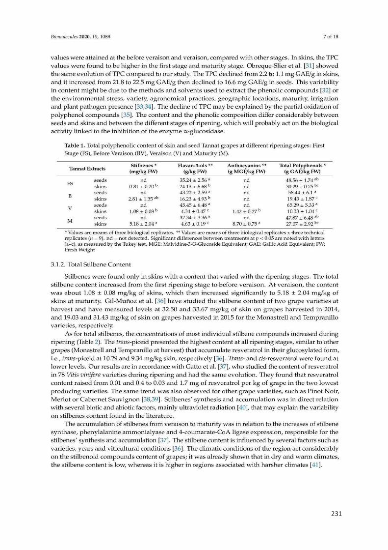
231
232

233
234
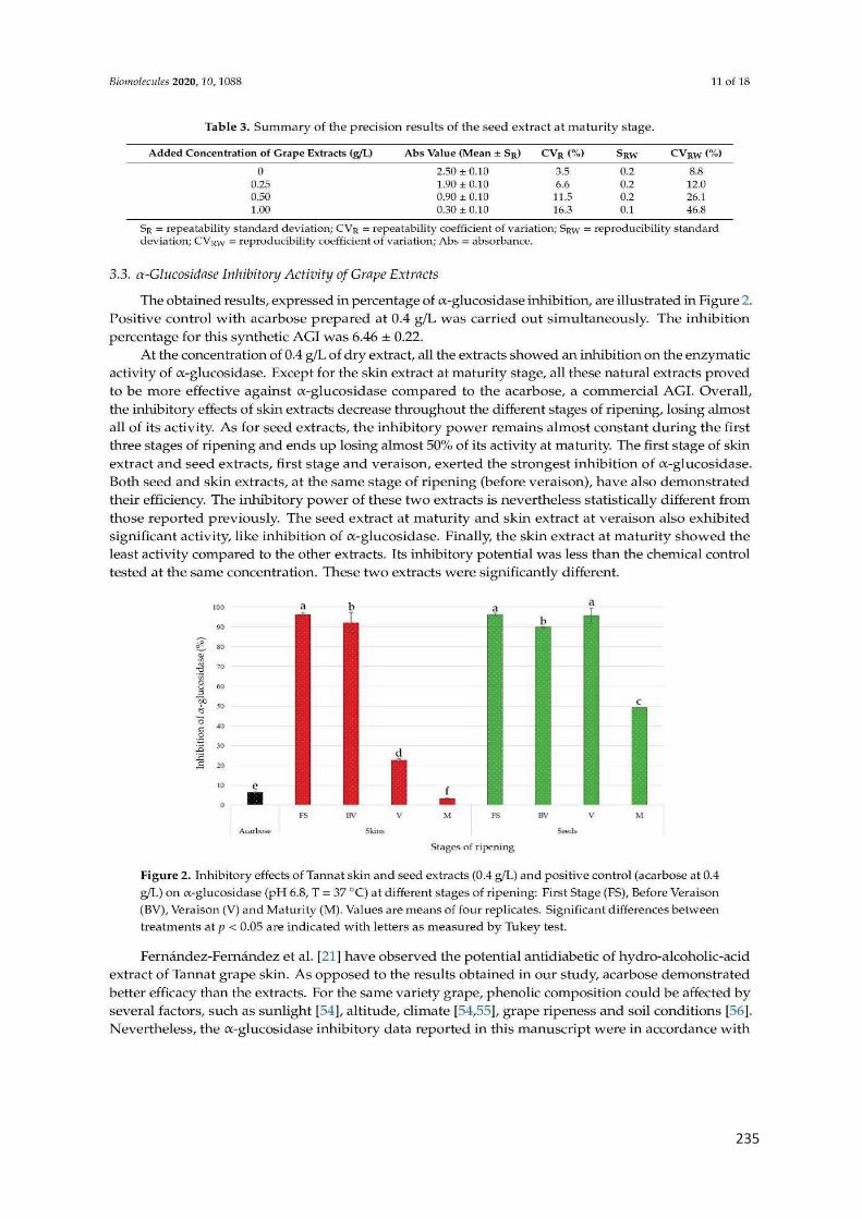
235
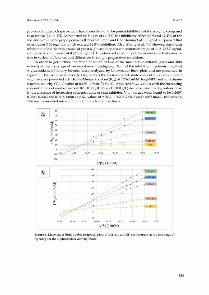
236
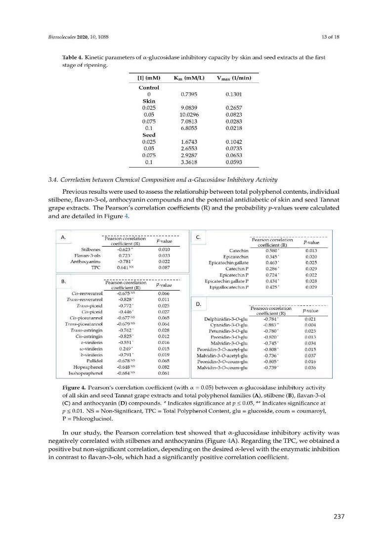
237
238
239
240
241

242
243
Conclusion générale

244
L’objectif global de ce travail était de caractériser la composition phénolique et les
activités biologiques (antioxydante, anti-inflammatoire et anti-diabète) d’extraits de
pépins et de pellicules de raisin au cours de la maturation. Ces travaux s’inscrivent
dans un objectif global de valorisation des extraits obtenus. Pour cela, trois cépages
rouges de Vitis vinifera (Merlot, Tannat et Syrah) ont été utilisés. Les baies ont été
prélevées à différents stades de maturation.
La partie bibliographique a permis de faire une synthèse des connaissances sur
l’espèce Vitis vinifera, la baie de raisin, les stades de maturation, les familles de
polyphénols, la voie de biosynthèse et l’accumulation des composés phénoliques dans
les différents tissus (pépins et pellicules) au cours de la maturité.
La deuxième partie de l’étude bibliographique a permis de faire le point sur les
activités biologiques des composés phénoliques de la baie de raisin : activités
antioxydante, anti-inflammatoire et anti-diabète.
Le deuxième chapitre était une étude bibliographique (revue) sur les stilbènes du
raisin et du vin et leur rôle potentiel en tant qu’agents anti-obésité. Les principaux
points abordés dans cet article, en cours de révision, ont été :
· La composition en stilbènes des baies de raisin et du vin et ainsi que les
méthodes analytiques les plus efficaces utilisées pour leur détection.
· Les effets anti-obésité in vitro et in vivo connus des stilbènes du raisin et du
vin et les mécanismes moléculaires associés.
Les principales conclusions obtenues ont été :
· Plus de 30 stilbènes ont été identifiés dans les raisins et les vins.
· La chromatographie liquide couplée à la spectrométrie de masse est la
méthode la plus efficace pour doser les stilbènes.
· Un grand nombre d'études in vitro et in vivo ont permis de mettre en
évidence l’implications positive des stilbènes (spécialement le resvératrol)
comme agents anti-obésité en régulant différentes voies du métabolisme
des graisses telles que l'adipogenèse, la lipogenèse, la lipolyse et la
thermogenèse.
Le troisième chapitre avait pour objectif d’étudier l’évolution de la composition
phénolique et de l’activité antioxydante des extraits de pépins et pellicules des trois
245
cépages rouges Merlot, Tannat et Syrah au cours de la maturité (année 2017). Les
principaux résultats obtenus sont résumés ci-dessous :
· Les pépins contiennent plus de polyphénols que les pellicules pour tous les
stades de maturité.
· La teneur en polyphénols des pellicules diminue au cours de la maturation, alors
que dans les pépins, celle-ci augmente jusqu’au stade proche de la véraison
puis diminue.
· Les extraits de pépins ont une activité antioxydante beaucoup plus importante
que les pellicules et ce quelle que soit la méthode utilisée.
· L’utilisation de la voltamétrie cyclique nous a permis non seulement de
déterminer la capacité antioxydante totale (cas des tests
spectrophotométriques) mais aussi de caractériser les composés les plus
facilement oxydables qui s’oxydent à des faibles potentiels (charge du premier
pic).
· La charge totale (capacité antioxydante totale) et la charge du premier pic des
voltammogrammes sont fortement corrélées avec la teneur en flavanols.
Le quatrième chapitre avait pour objectif d’étudier l’évolution de la composition
phénolique (flavanols, flavonols, stilbènes et anthocyanes) et des activités
antioxydante et anti-inflammatoire des extraits de pellicules des trois cépages rouges
Merlot, Tannat et Syrah au cours de la maturité (année 2018). Les principaux résultats
obtenus peuvent être résumés ainsi :
· 41 polyphénols ont été quantifiés au sein des échantillons (6 flavanols, 7
flavonols, 16 stilbènes et 12 anthocyanes). Leurs teneurs varient
considérablement au cours de la maturation. En général, les teneurs en
flavanols diminuent pendant la maturation tandis que celles en anthocyanes et
en stilbènes augmentent.
· Tous les extraits de pellicules sont capables de diminuer la production de NO
et de ROS dans les cellules, et ce à tous les stades de maturité. Les meilleurs
résultats ont été obtenus pour les extraits avant véraison, plus riches en
flavanols.
246
Le cinquième chapitre avait pour objectif d’étudier l’évolution de la composition
phénolique (flavanols, anthocyanes et stilbènes) d’extraits de pépins et pellicules du
cépage Tannat et de l’inhibition de l’enzyme alpha glucosidase est en lien avec la
prévention potentielle du diabète. Il a été montré que :
· La teneur en polyphénols est plus élevée dans les pépins que dans les
pellicules pour tous les stades de maturité.
· La teneur en flavanols diminue au cours de la maturation pour les pellicules
alors que pour les pépins, celle-ci augmente jusqu’aux stades proche de la
véraison et véraison puis diminue jusqu’à la maturité. Les stilbènes et les
anthocyanes ont été détectés uniquement dans les pellicules, leurs teneurs
augmentant avec les stades de maturation.
· Les extraits de pépins et de pellicules ont une activité inhibitrice beaucoup plus
élevée au stade vert par rapport à la maturité.
· Les études cinétiques enzymatiques ont révélé un type mixte d'inhibition de
l’enzyme alpha glucosidase par les extraits.
· L’activité inhibitrice est positivement corrélée avec la teneur en flavanols.
En résumé, nos résultats mettent en évidence l’effet de la maturation, le type de
tissu, le cépage et l’année sur l’évolution de la composition phénolique et des trois
activités biologiques (antioxydante, anti-inflammatoire et anti-diabète) des extraits de
pépins et de pellicules de trois cépages rouges (Merlot, Tannat et Syrah) au cours de
la maturation (du stade vert jusqu’à la maturité).
Concernant la composition phénolique
Ø Effet du stade de maturation : La teneur en flavanols et polyphénols totaux
diminue au cours de la maturation pour les extraits de pellicules et augmente jusqu’au
stade proche de la véraison pour les extraits de pépins. Les anthocyanes, qui sont
présentes uniquement au niveau des pellicules, leurs synthèses commencent au stade
proche de la véraison et s’accumule au cours de la maturation. La teneur en stilbènes
augmente au cours de la maturation.

247
0100200300400500600700800900
mg
/gD
WPolyphénols totaux (pellicules)
0100200300400500600700800900
mg
/g D
W
Polyphénols totaux (pépins)
0100200300400500600700800900
mg
/g D
W
Flavanols (pellicules)
0100200300400500600700800900
mg
/g D
WFlavanols (pépins)
0
5
10
15
20
25
30
35
mg
/kg
MF
Stilbènes
0
50
100
150
200
250
mg
/g D
W
Anthocyanes

248
Ø Effet du tissu : Les extraits de pépins contiennent plus de polyphénols totaux
et de flavanols par rapport aux extraits de pellicules. Les stilbènes et les anthocyanes
se trouvent uniquement au niveau des pellicules.
0
100
200
300
400
500
600
700
Pellicules Pépins
mg
/g M
S
Polyphénols totaux
0
100
200
300
400
500
600
700
Pellicules Pépins
mg
/g M
S
Flavanols
0
2
4
6
8
10
12
Pellicules Pépins
mg
/g M
S
Anthocyanes
0
2
4
6
8
10
12
Pellicules Pépins
mg
/kg
MF
Stilbènes

249
Effet du cépage : Les extraits de Merlot contiennent la teneur la plus élevée en
polyphénols totaux et stilbènes, par contre, les extraits de Tannat sont riches en
anthocyanes et flavanols.
0
100
200
300
400
500
600
700
800
900
1000
Merlot Tannat Syrah
mg
/g M
S
Polyphénols totaux
0
100
200
300
400
500
600
700
800
900
1000
Merlot Tannat Syrah
mg
/g M
S
Flavanols
0
5
10
15
20
25
30
35
40
Merlot Tannat Syrah
mg
/g M
S
Anthocyanes
0
5
10
15
20
25
30
35
40
Merlot Tannat Syrah
mg
/kg
MF
Stilbènes

250
Ø Effet de l’année : Les extraits récoltés en 2018 sont riches en polyphénols
totaux et en stilbènes alors que ceux récoltés en 2017 sont riches en
anthocyanes.
0
10
20
30
40
50
60
2017 2018
g E
AG
/ k
g F
W
Polyphénnols totaux
0
1
2
3
4
5
6
2017 2018
g /
kg F
W
Anthocyanes
0
0.5
1
1.5
2
2.5
3
3.5
4
4.5
2017 2018
mg
/ kg
FW
Stilbènes

251
Concernant les activités biologiques
Ø Effet du stade de la maturation : Pour toutes les activités étudiées, le stade
vert possède les activités biologiques les plus importantes (dans le cas de
l’activité anti-inflammatoire, la diminution de la production des espèces
réactives oxygénées et du monoxyde d’azote indique que l’activité anti-
inflammatoire est importante).
0
200
400
600
800
1000
1200
1400
1600
µC
Activité antioxydante (pellicules)
0
200
400
600
800
1000
1200
1400
1600
µC
Activité antioxydante (pépins)
0102030405060708090
100
% d
'inhi
biti
on
Activité anti-diabète (pellicules)
0102030405060708090
100
% d
'inhi
biti
on
Activité anti-diabète (pépins)
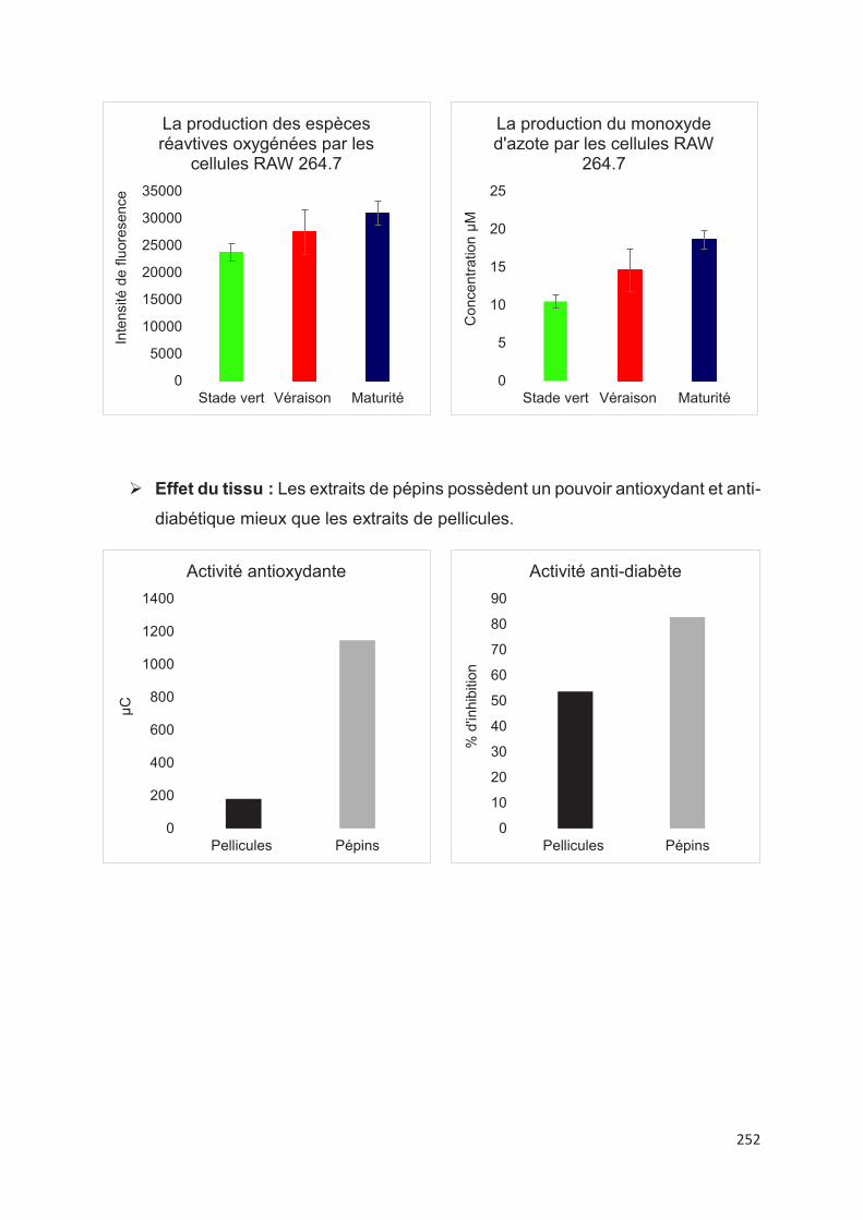
252
Ø Effet du tissu : Les extraits de pépins possèdent un pouvoir antioxydant et anti-
diabétique mieux que les extraits de pellicules.
0
5000
10000
15000
20000
25000
30000
35000
Stade vert Véraison Maturité
Inte
nsité
de
fluor
ese
nce
La production des espèces réavtives oxygénées par les
cellules RAW 264.7
0
5
10
15
20
25
Stade vert Véraison Maturité
Co
nce
ntra
tion
µM
La production du monoxyde d'azote par les cellules RAW
264.7
0
200
400
600
800
1000
1200
1400
Pellicules Pépins
µC
Activité antioxydante
0
10
20
30
40
50
60
70
80
90
Pellicules Pépins
% d
'inhi
biti
on
Activité anti-diabète
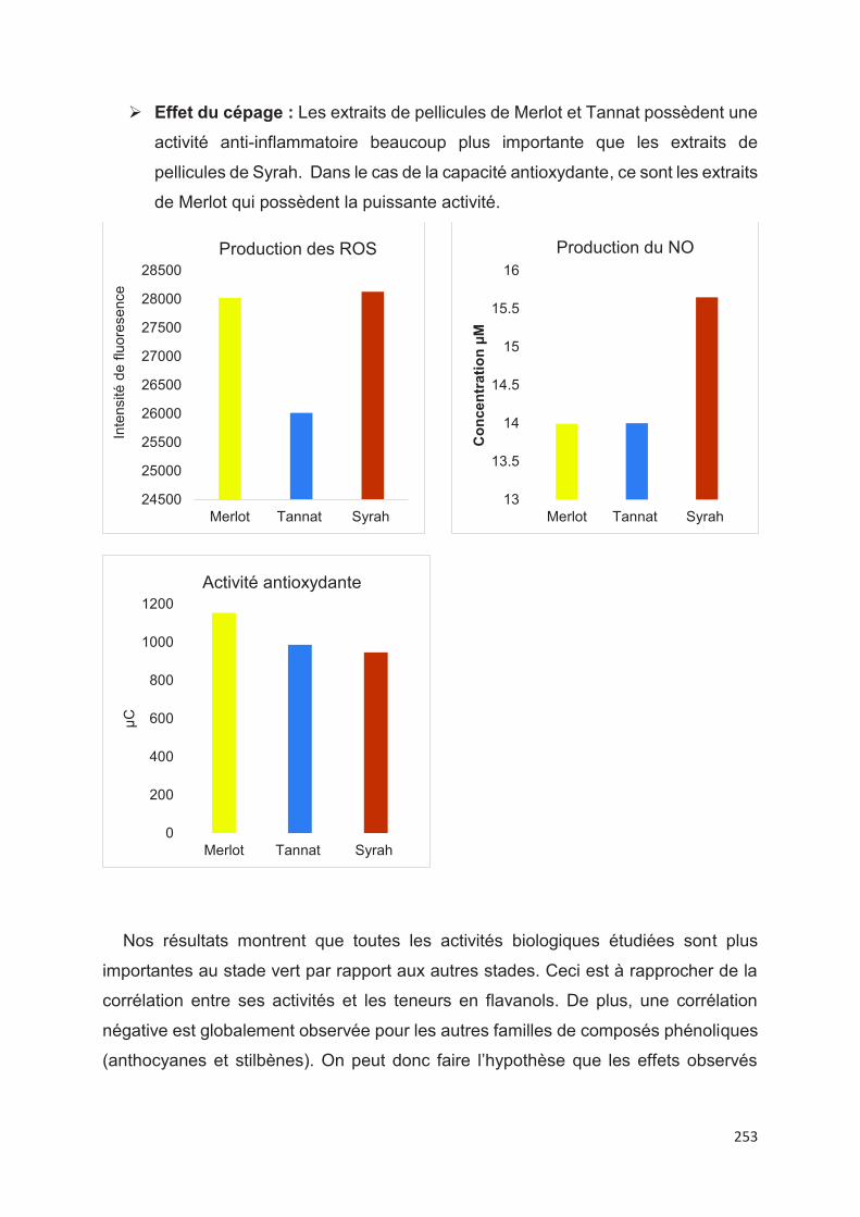
253
Ø Effet du cépage : Les extraits de pellicules de Merlot et Tannat possèdent une
activité anti-inflammatoire beaucoup plus importante que les extraits de
pellicules de Syrah. Dans le cas de la capacité antioxydante, ce sont les extraits
de Merlot qui possèdent la puissante activité.
Nos résultats montrent que toutes les activités biologiques étudiées sont plus
importantes au stade vert par rapport aux autres stades. Ceci est à rapprocher de la
corrélation entre ses activités et les teneurs en flavanols. De plus, une corrélation
négative est globalement observée pour les autres familles de composés phénoliques
(anthocyanes et stilbènes). On peut donc faire l’hypothèse que les effets observés
24500
25000
25500
26000
26500
27000
27500
28000
28500
Merlot Tannat Syrah
Inte
nsité
de
fluor
ese
nce
Production des ROS
13
13.5
14
14.5
15
15.5
16
Merlot Tannat Syrah
Co
nce
ntr
atio
n µ
M
Production du NO
0
200
400
600
800
1000
1200
Merlot Tannat Syrah
µC
Activité antioxydante

254
pour le stade vert pourraient être dus à certains flavanols présents à la fois dans les
pépins et les pellicules comme les proanthocyanidines.
255
Perspectives
Suite aux résultats obtenus au cours de cette thèse, différentes perspectives
peuvent être envisagées. Il serait souhaitable de poursuivre les travaux en se
concentrant sur le stade vert qui semble être le plus actif vis-à-vis des activités
biologiques pour fabriquer des produits pharmaceutiques, compléments alimentaires
ou des médicaments. Afin de poursuivre la compréhension des relations entre
structures présentes et activités biologiques des extraits il serait à envisager de :
· tester d’autres cépages ;
· tester différentes méthodes d’extraction des polyphénols en jouant sur le type
et la proportion des solvants utilisés ;
· fractionner les extraits à l’aide de gels et de résines pour mieux les
caractériser ;
· identifier les composés présents dans les fractions les plus actives ;
· utiliser d’autres tests cellulaires pour confirmer les activités ;
· passer in vivo sur les fractions les plus actives.
La poursuite de ces travaux pourrait ainsi ouvrir la voie à des applications dans le
domaine des compléments alimentaires ou des médicaments en santé humaine.
256
Résumé
Les polyphénols appartiennent à la famille des métabolites secondaires présents
dans les plantes et majoritairement dans les baies de raisin. Ils jouent un rôle
important dans la protection de la plante contre les stress biotiques et abiotiques. Ils
ont un impact sur la qualité organoleptique des certains aliments comme ceux
provenant du raisin et sont connus majoritairement pour leurs rôles bénéfiques pour
la santé humaine. Une étude globale sur la composition phénolique et les activités
biologiques (antioxydante, anti-inflammatoire et anti-diabète) d’extraits phénoliques
de pépins et pellicules de trois cépages rouges Merlot, Tannat et Syrah à différents
stades de maturité a été réalisée durant cette thèse. Les extraits de pépins ont une
teneur plus élevée en composés phénoliques que les extraits de pellicules pour tous
les stades de maturité. La composition phénolique diffère selon le stade de maturité
et le tissu de la baie de raisin (pépin ou pellicule). L’activité antioxydante des extraits
a été déterminée par des tests spectrophotométriques (DPPH, ABTS et FRAP) et
des paramètres électrochimiques. L’activité anti-inflammatoire a été suivie par
l’inhibition de la production des EROs et NO par les cellules RAW 264.7. L’activité
anti-diabétique a été déterminée par la mesure d’inhibition de l’enzyme α-
glucosidase. Les activités biologiques sont positivement corrélées avec la teneur en
flavanols et négativement corrélées avec la teneur en anthocyanes et stilbènes. Les
activités biologiques les plus importantes ont été mesurées avant la maturité pour
tous les tests et dans les trois cépages étudiés.

257
Abstract
Polyphenols belong to the family of secondary metabolites found in plants and
mainly in grape berries. They play an important role in the protection of the plant
against biotic and abiotic stresses. They have an impact on the organoleptic quality of
certain foods such as those from grapes and are mainly known for their beneficial roles
for human health. A global study on the phenolic composition and the biological
activities (antioxidant, anti-inflammatory and anti-diabetes) of the phenolic extracts of
the seeds and skins of three red grape varieties Merlot, Tannat and Syrah at different
stages of maturation was carried out during this thesis. Seed extracts contain a higher
content of phenolic compounds than skin extracts for all stages of ripening. The
phenolic composition differs depending on the stage of maturity and the tissue of the
grape berry (seed or skin). The antioxidant activity of the extracts was determined by
spectrophotometric tests (DPPH, ABTS and FRAP) and electrochemical parameters.
Anti-inflammatory activity was followed by the inhibition of ROS and NO production by
stimulated RAW 264.7 cells. Anti-diabetic effect was determined by measuring the
inhibition of the enzyme α-glucosidase activity. The biological activities are positively
correlated with the content of flavanols and negatively correlated with the content of
anthocyanins and stilbenes. The most important biological activities were measured
before maturity for all the tests and in the three grape varieties studied.


