Embed Size (px)
Citation preview
Molecular Biology of the CellVol. 13, 1806–1818, June 2002
ER-Golgi Traffic Is a Prerequisite for Efficient ERDegradation
Christof Taxis,* Frank Vogel,† and Dieter H. Wolf,*‡
*Institut fur Biochemie, Universitat Stuttgart, 70569 Stuttgart, Germany; and †Max-Delbruck-Centrumfur Molekulare Medizin, 13125 Berlin, Germany
Submitted August 9, 2001; Revised February 19, 2002; Accepted February 22, 2002Monitoring Editor: Howard Riezman
Protein quality control is an essential function of the endoplasmic reticulum. Misfolded proteinsunable to acquire their native conformation are retained in the endoplasmic reticulum, retro-translocated back into the cytosol, and degraded via the ubiquitin-proteasome system. We showthat efficient degradation of soluble malfolded proteins in yeast requires a fully competent earlysecretory pathway. Mutations in proteins essential for ER-Golgi protein traffic severely inhibit ERdegradation of the model substrate CPY*. We found ER localization of CPY* in WT cells, but noother specific organelle for ER degradation could be identified by electron microscopy studies.Because CPY* is degraded in COPI coat mutants, only a minor fraction of CPY* or of a protein-aceous factor required for degradation seems to enter the recycling pathway between ER andGolgi. Therefore, we propose that the disorganized structure of the ER and/or the mislocalizationof Kar2p, observed in early secretory mutants, is responsible for the reduction in CPY* degrada-tion. Further, we observed that mutations in proteins directly involved in degradation of mal-folded proteins (Der1p, Der3/Hrd1p, and Hrd3p) lead to morphological changes of the endo-plasmic reticulum and the Golgi, escape of CPY* into the secretory pathway and a slowermaturation rate of wild-type CPY.
INTRODUCTION
Protein quality control with subsequent elimination of mal-folded proteins or unassembled subunits is essential forcellular function. Disturbed quality control leads to diseaseand eventually to cell death (Plemper and Wolf, 1999; Ko-pito and Sitia, 2000). The endoplasmic reticulum (ER) is thefolding compartment for proteins destined to functionwithin the ER itself and for secretory proteins of the Golgi,endosomes, vacuoles, and plasma membrane as well as forproteins secreted extracellularly. It contains a multitude offolding enzymes and chaperones to perform this function(Ellgaard et al., 1999; Zapun et al., 1999). Failure in folding orin the assembly of multimeric complexes leads to recogni-tion by the quality control machinery in the ER. The proteinsare then transported back into the cytoplasm, most likely viathe Sec61 translocon, and degraded by the ubiquitin–protea-some system (Kopito, 1997; Sommer and Wolf, 1997; Brod-sky and McCracken, 1999; Plemper and Wolf, 1999). Polyu-biquitination is mediated through the action of the E2
enzymes, Ubc1p and Ubc7p, and the membrane boundRING-H2 ubiquitin–protein ligase (E3) Der3/Hrd1p, whichis complexed to another membrane protein, Hrd3p (Hiller etal., 1996; Plemper et al., 1999; Friedlander et al., 2000; Gard-ner et al., 2000; Bays et al., 2001a; Deak and Wolf, 2001). Thefinal degradation is carried out by the proteasome, a multi-catalytic and multifunctional proteinase machinery (Hiltand Wolf, 2000). Depending on the nature of the malfoldedsubstrate protein, additional components of the ubiquitina-tion machinery (i.e., the ubiquitin conjugating enzyme [E2Ubc6p; Biederer et al., 1996; Hiller et al., 1996) and of thelumenal ER folding machinery (the Hsp70 chaperone Kar2p,protein-disulfide isomerase [PDI], �-1,2 mannosidase, thelectin-like protein Mnl1/Htm1 (Knop et al., 1996b; Plemperet al., 1997; Brodsky et al., 1999; Gillece et al., 1999; Jakob et al.,2001; Nakatsukasa et al., 2001) or an ER membrane protein ofunknown function (Der1p; Knop et al., 1996a) are requiredfor the degradation event.
We had previously shown that a defect in ERD1, involvedin the retrieval of HDEL-containing proteins from the Golgito the ER (Hardwick et al., 1990), leads to the escape of CPY*from the ER, despite the fact that CPY* does not contain theHDEL retrieval sequence (Knop et al., 1996a). We were,therefore, interested in the question whether the secretorycompetence of the ER in general is necessary for the degra-dation of malfolded proteins. Intracellular transport of pro-
Article published online ahead of print. Mol. Biol. Cell 10.1091/mbc.01–08–0399. Article and publication date are at www.molbiol-cell.org/cgi/doi/10.1091/mbc.01–08–0399.
‡ Corresponding author. E-mail address: [email protected].
1806 © 2002 by The American Society for Cell Biology

teins is mediated by coated vesicles: proteins are packed intoCOPII-coated vesicles at ER exit sites and transported to theGolgi apparatus, where they fuse with the target membrane(Rothman and Wieland, 1996; Schekman and Orci, 1996;Kuehn et al., 1998). On arrival at the Golgi complex proteinsare sorted to the peripheral compartments of the cell such asvacuoles, plasma membrane, and secretory vesicles. Proteinscan also be retrieved to the ER by retrograde transport fromeither the ER-Golgi intermediate compartment or the Golgicomplex itself, by COPI-coated vesicles (Letourneur et al.,1994; Allan and Balch, 1999).
It has been previously shown that extensive protein mis-folding and accumulation in the ER activates the unfoldedprotein response (UPR; Knop et al., 1996a; Chapman et al.,1998; Casagrande et al., 2000; Friedlander et al., 2000; Traverset al., 2000). Signaling of malfolded proteins in the ER occursvia Ire1p, a kinase/nuclease of the ER and the nuclear en-velope, which activates the transcription factor Hac1p. Thisactivation leads to transcriptional upregulation of genes nec-essary to relieve the cell from ER stress (Chapman et al.,1998). As one might expect, genes involved in ER degrada-tion directly, such as DER1, DER3/HRD1, HRD3, and UBC7are targets of Hac1p (Travers et al., 2000). Furthermore,transcription of genes involved in ER-to-Golgi trafficking,protein targeting to the vacuole and the cell surface, lipidmetabolism, and glycosylation are also upregulated uponER stress (Travers et al., 2000). In this study, we investigatedthe requirement of a fully operational early secretory path-way and a competent UPR for efficient ER degradation. As asoluble model substrate of ER degradation in yeast we ex-amined the disappearance of malfolded carboxypeptidaseyscY (CPY*; Finger et al., 1993; Hiller et al., 1996; Knop et al.,1996a). In addition, we tested the influence of defective ERdegradation on the secretory system.
MATERIALS AND METHODS
Construction and Growth Conditions of StrainsPreviously described standard methods were used in media prep-aration and for genetic and molecular biological techniques (Guthrieand Fink, 1991; Ausubel, 1992). The Saccharomyces cerevisiae strainsused in this study are summarized in Table 1. Yeast cells weregrown at 25°C (temperature-sensitive strains) or 30°C. For genera-tion of the ufe1-1 integration plasmid, pUT1 (Lewis et al., 1997),containing the ufe1-1 allele, was digested with SalI and SpeI, and the1450 base-pair ufe1-1 fragment was ligated into pRS306 (Sikorskiand Hieter, 1989) to obtain pCT27. SnaBI linearized pCT27 was usedto replace the chromosomal UFE1 allele by two-step gene replace-ment (Scherer and Davis, 1979). RSY281 (sec23-1; Kaiser and Schek-man, 1990), CBY263 (sed5-1; Cao et al., 1998), and RSY277 (sec21-1;Letourneur et al., 1994) were backcrossed multiple times throughW303-1C or YCT458 to obtain the isogenic strains YCT441, YCT480,and YCT611, respectively. The IRE1 gene was deleted using plasmidpJU341, containing the IRE1 knock out fragment (Friedlander et al.,2000). Crossing of the respective single mutants to each other pro-duced the double-mutant strains YCT541 and YCT542. To obtainstrains YCT458, YCT462, YCT460, YCT437, and YCT438, BglII lin-earized plasmid pRS306prc1-1 (Knop et al., 1996a) was used tointroduce the prc1-1 allele into strains YR1070 (wild-type), YR1068(sec12-1), YR1069 (sec18-1; H. Rudolph), RH448 (wild-type), andRH2688 (sec27-1; Schroder-Kohne et al., 1998) using two-step genereplacement (Scherer and Davis, 1979). YCT614 was generated fromstrain YCT438 by deleting the PEP4 gene with PvuII-digestedpWO139. Integration was confirmed by Southern blotting. PlasmidpWO139 was generated through ligation of the 1.1-kb URA3 con-taining an EcoRI/SmaI fragment from Yep24 (Botstein et al., 1979)into EcoRI/MscI digested plasmid pWO261. For generation of plas-mid pWO261 a 1.9-kb SacI/XhoI fragment from pTZ18 (Rupp andWolf, 1995), containing PEP4, was ligated into pBluescript KS�
(Stratagene, La Jolla, CA).To express CPY*-HA, we fused prc1-1HA behind the TDH3
(pCT43) or CUP1 (pCT52) promoter. Similarly, PRC1 was fusedbehind the TDH3 promoter (pCT70) for expression of CPY. The
Table 1. Yeast strains used in this study
Name Genotype Source
W303-1B Mat � ade2-1 leu2-3,112 trp1-1 ura3-1 his3-11,15 can1-100 Chiang and Schekman (1991)W303-1C W303-1B prc1-1 Knop et al. (1996a)W303-CD W303-1C �der1�URA3 Knop et al. (1996a)W303-CF W303-1C �pep4�HIS3 Knop et al. (1996a)W303-CDF W303-1C �der1�URA3 �pep4�HIS3 Knop et al. (1996a)W303-BD W303-1B �der1�URA3 Knop et al. (1996a)YJB009 W303-1C �der3/hrd1�HIS3 Bordallo et al. (1998)W303 �C W303-1B �prc1�LEU2 Plemper et al. (1999)YCT431 W303-1C ufe1-1 This workYCT441 W303-1C sec23-1 This workYCT480 W303-1C sed5-1 This workYRH68 W303-1C �ire1�LEU2 This workYCT542 W303-1C sec23-1 �ire1�LEU2 This workYCT540 W303-1C ufe1-1 �ire1�LEU2 This workYCT458 Mat a ura3-52 prc1-1 This workYCT462 YCT458 sec12-1 This workYCT460 YCT458 sec18-1 This workYCT611 YCT458 sec21-1 This workYCT437 Mat a prc1-1 his4 lys2 ura3 leu2 bar1 This workYCT438 YCT437 sec27-1 This workYCT614 YCT437 sec27-1 �pep4�URA3 This work
ER Degradation Requires ER-Golgi Traffic
Vol. 13, June 2002 1807
cloning strategy to obtain plasmids pCT41, pCT43, pCT52, andpCT70 is available on request. The yeast strain bearing the prc1-1HAallele was obtained according to M. Longtine (Longtine et al., 1998)using plasmid pFA6a-3HA-kanMX6. For generation of plasmidpWO152, a 3-kb BamHI/HindIII fragment, containing SEC18, wasligated into pRS426 (Christianson et al., 1992).
Pulse-chase AnalysisYeast strains were grown logarithmically in CM medium and la-beled for 20 min with 35S-methionine (pulse). Temperature-sensitivestrains were shifted to 37°C for the times indicated in legends tofigures. After addition of an excess of nonradioactive methionine,samples were taken at the indicated time points. Cell disruption,immunoprecipitation, and SDS-PAGE were performed as previ-ously described (Plemper et al., 1999). Curves were generated plot-ting the mean values of two to four independent experiments.Maturation analysis of CPY and proteinase yscA (PrA) was per-formed similarly, except that the strains were labeled for only 5 min.
Electron MicroscopyFor immuno-electron microscopy (EM), cells were fixed in a mixtureof freshly prepared 4% formaldehyde and 0.5% glutaraldehyde in0.1 M citrate buffer. Temperature and pH were chosen according togrowth conditions, as described previously (Kargel et al., 1996;Zimmer et al., 1997). Cells were washed with PBS, and then 1%sodium metaperiodate was added for 1 h to prepare the cell wall forthe penetration of the cryoprotectant. The hydrated specimens wereimmersed in a cryoprotectant mixture of 25% polyvinyl-pyrrolidone(PVP, K15/MW 10000; Fluka, Buchs, Switzerland) and 1.6 M su-crose, as described in Tokuyasu (1989). To reach complete immer-sion of the cryoprotectant, incubation at 30°C for 2–3 h was strictlynecessary. The cells were subsequently mounted on specimen hold-ers, frozen in liquid nitrogen, and sectioned at �115°C with anultracryotom Ultracut S attached with a FCS unit (Leica, Heerbrugg,Switzerland). Ultra-thin, thawed cryosections were prepared withglass knifes and placed on formvar/carbon-coated copper grids(200 mesh, hexagonal).
Labeling with primary antibodies and immunogold-complexes(12 nm) was performed according to Griffiths (1993). Finally, thefrozen-thawed sections were stained and stabilized using a mixtureof freshly prepared 3% tungstosilicic acid hydrate (STA; Fluka) and2.5% polyvinylalcohol (Mr 10,000; Sigma, Deisenhofen, Germany).
CPY* SecretionCells were grown to late log phase and then sedimented by centrif-ugation at 500 � g for 5 min. Cells were washed once with ice-coldwater and centrifuged again, and the supernatants from both cen-trifugation steps were combined. Proteins were precipitated withtrichloroacetic acid (10%) for 30 min on ice and sedimented for 15min at 12,000 � g. The pellet was washed twice with ice-coldacetone and dissolved in 100 �l urea buffer (5% SDS, 8 M urea, 200mM Tris-HCl, pH 6.8, 0.1 mM EDTA 1.5% dithiothreithol, 0.03%bromophenol blue). To obtain the intracellular fraction of CPY*, anamount of cells corresponding to 2 OD600 were alkaline lysed (Hilleret al., 1996), and the samples were subjected to SDS-PAGE andimmunodetection as described previously (Knop et al., 1996a).
Cycloheximide ChaseCells were grown to log phase at 25°C and incubated for 1 h at 37°C.Cycloheximide was added (0.25 mg/ml), and 2 OD of cells weretaken after 0 and 60 min of chase. Cell extracts were prepared byalkaline lysis method (Hiller et al., 1996), and the samples weresubjected to SDS-PAGE followed by immunodetection (Knop et al.,1996a).
AntiseraPolyclonal anti-CPY and polyclonal anti-PrA sera were describedpreviously (Finger et al., 1993); anti-Emp47p serum was a generousgift of H. Riezman (Schroder et al., 1995). Monoclonal anti-CPY andanti-PGK sera were purchased from Molecular Probes (Eugene,OR), HRPO-conjugated anti-mouse antibody from Sigma, monoclo-nal anti-HA antibody from Babco (HA.11; Berkley, CA), and anti-mouse immunogold-complexes (12 nm) from Dianova (Hamburg,Germany).
RESULTS
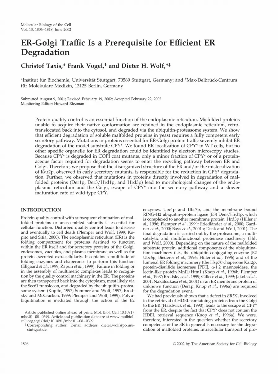
We analyzed the degradation of CPY* in mutants defectivein anterograde transport between the ER and Golgi. Weused the mutant alleles sec12-1, sec23-1, ufe1-1, sed5-1, andsec18-1, which block vesicular transport at restrictive condi-tions (Stevens et al., 1982; Hardwick and Pelham, 1992;Lewis and Pelham, 1996). Sec12p, the nucleotide-exchangefactor, which recruits Sar1p to the ER membrane (Campbelland Schekman, 1997), and Sec23p (Kuehn et al., 1998;Springer and Schekman, 1998) are both necessary for COPII-coated vesicle formation. Ufe1p (Lewis et al., 1997; Patel etal., 1998) and Sed5p (Banfield et al., 1995; Wooding andPelham, 1998; Tsui and Banfield, 2000) are t-SNAREs of theER and Golgi, respectively. They are involved in the fusionof vesicles with the target membrane. Sec18p, the yeasthomologue of the mammalian NSF, is required for vesiculartransport in multiple stages of the secretory pathway (Gra-ham and Emr, 1991; Mayer et al., 1996). A defect in any ofthese proteins leads to a considerably reduced degradationrate of CPY* (Figure 1, A and B). The half-life of CPY*increases �2- (sec12-1, sec23-1, and sed5-1) to 6- (ufe1-1) foldin mutant cells compared with wild type. We had previouslyanalyzed the function of Sec18p in the degradation of CPY*and PrA*, a rapidly degraded mutant form of proteinaseyscA (Finger et al., 1993). Because the data had not beenquantified, we had concluded that both misfolded proteinswere degraded under restrictive conditions in sec18-1 cells.Reinvestigation and quantification of CPY* degradation inthese sec18-1 mutant cells, however, revealed a 6- to 7-foldincrease in the half-life of CPY* (Figure 1B). The block ofanterograde transport between ER and Golgi was confirmedby monitoring the maturation of proteinase yscA (PrA) inthe mutant strains at restrictive conditions. In case of theufe1-1 strain, a tiny fraction of matured PrA was visible after60 min of chase; all the other mutants retained PrA in theproform (Figure 1, C and D). The degradation of CPY*observed in the sec12-1 mutant cells might be due to theaction of a close homologue, Sed4p, which is also involvedin the generation of COPII-coated vesicles at the ER mem-brane (Gimeno et al., 1995). We tested if a deletion of SED4influences the degradation rate of CPY*, either as a singleknockout or in conjunction with the sec12-1 mutation. Inboth cases there was no detectable change in the half-life ofCPY* (our unpublished results).
Ufe1p is known to function in two different membranefusion events: it is involved in the homotypic fusion of ERmembranes and in the heterotypic fusion of COPI-coatedvesicles with the ER membrane. To distinguish between thetwo different fusion events, we overexpressed the AAA-ATPases Cdc48p and Sec18p in temperature-sensitive ufe1-1mutant cells. CDC48p is involved in homotypic membranefusion, whereas Sec18p is the homologue used in heterotypic
C. Taxis et al.
Molecular Biology of the Cell1808

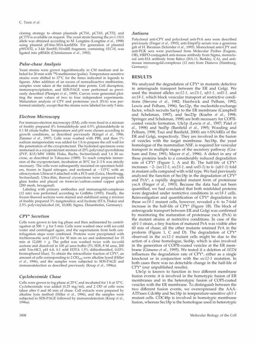
fusion of COPI vesicles with the ER membrane (Lewis et al.,1997; Patel et al., 1998). Overexpression of Cdc48p rescuedthe temperature sensitivity of the ufe1-1 mutant (Patel et al.,1998) but was unable to enhance the degradation of CPY*(our unpublished results). Overexpression of Sec18p, on theother hand, did not rescue the temperature sensitivity of theufe1-1 mutant (our unpublished results) but, interestingly,was able to partially overcome the ufe1-1–mediated degra-dation defect of CPY* in cells under restrictive conditions(Figure 2A).
As a control, we tested if Sec1p, a protein involved indocking of secretory transport vesicles to the plasma mem-brane (Novick and Schekman, 1979; Aalto et al., 1997), isinvolved in ER degradation. As expected, a temperature-sensitive mutant, sec1-1, did not alter the degradation ofCPY* under restrictive conditions compared with wild type(our unpublished results). This rules out any unspecific in-fluence of defects connected to various sec mutant alleles onER degradation (Mizuta and Warner, 1994).
Using the DNA microarray technique, Travers and co-workers could show that transcription of many genes in-volved in ER-Golgi transport is upregulated upon stress inthe ER. Transcription of Sec23p is enhanced by induction ofthe UPR, whereas transcription of Ufe1p is not (Travers et al.,2000). Previous studies did not find an alteration in CPY*degradation in �ire1 cells under nonstress conditions at 30°C(Friedlander et al., 2000). However, we observed that thedegradation rate of CPY* is prolonged in �ire1 cells at 37°C.To examine whether there is a synergistic effect betweenUPR and ER-Golgi transport, we combined the �ire1 muta-tion with mutations in Sec23p and Ufe1p. Indeed, doublemutants of �ire1 with either ufe1-1 or sec23-1 resulted innearly complete arrest of CPY* degradation under restrictiveconditions (Figure 2, B and C).
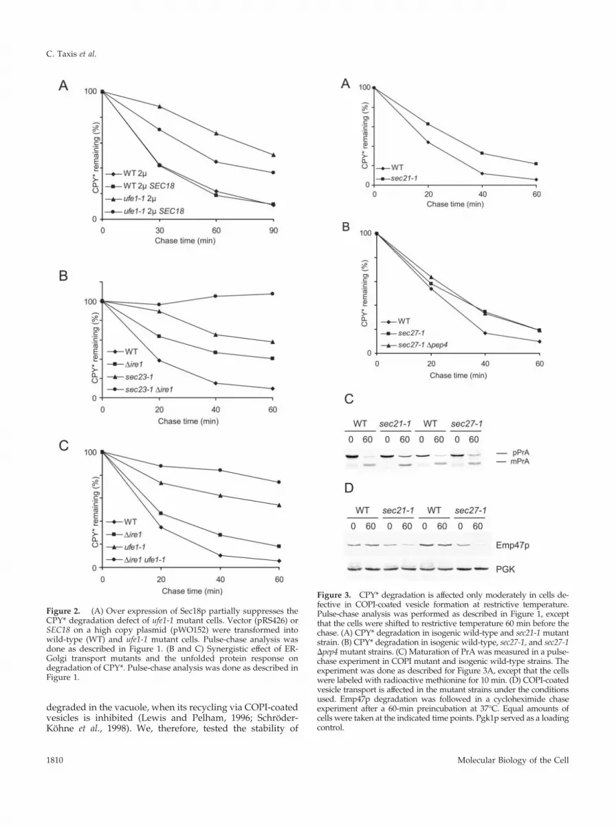
Consequently, we tested if a block in retrograde transportfrom the Golgi to the ER would influence the degradationrate of CPY* as much as a block in anterograde transportdoes. We measured the degradation of CPY* in yeast cellsdefective in Sec21p or Sec27p, two components of the COPIcomplex (Hosobuchi et al., 1992; Duden et al., 1994; Letour-neur et al., 1994). As shown in Figure 3, A and B, degradationof CPY* is only moderately affected in sec21-1 or sec27-1 cellsat restrictive conditions. It is known that proteins, whichcannot be retrograde transported from the Golgi to the ER,may travel to the vacuole for degradation. Therefore, wecombined a mutation in Sec27p with a deletion of the PEP4gene, leading to a proteolysis defective vacuole (Knop et al.,1993; Van Den Hazel et al., 1996), to check if vacuolar deg-radation contributes to the decay of CPY* in this COPImutant. We found that degradation of CPY* was not alteredin the double mutant (Figure 3B), whereas a pep4 singlemutant degrades CPY* like a wild-type strain (our unpub-lished results and Figure 5B). Anterograde transport is par-tially affected in sec21-1 and to a lesser degree in sec27-1 cells(Letourneur et al., 1994). We measured the maturation pro-cess of PrA, applying the conditions for the CPY* degrada-tion experiment, and found that in fact PrA is not fullymatured in the mutant cells after 60 min of chase (Figure3C). In wild-type cells 80% of PrA is matured after 60 min ofchase, whereas in sec21-1 cells only 64% and in sec27-1 cells74% is in the mature form. Retrograde transport is defectivein these two mutants at 37°C. It is known that Emp47p is
Figure 1. CPY* degradation is impaired in mutants defective inER-to-Golgi transport. Pulse-chase analysis was performed to measureCPY* degradation and maturation of PrA in wild-type and isogenicmutant strains. Cells were shifted to restrictive temperature 5 minbefore the chase and lysed at the indicated time points. CPY* or PrAwere immunoprecipitated and separated by SDS-PAGE. Quantifica-tion was done using a PhosphorImager. (A) Formation of COPII-coated vesicles (Sec23p) and ER and Golgi t-SNARES (Ufe1p and Sed5p) are necessary for efficient degradation of CPY*. (B) CPY* degrada-tion requires a functional Sar1p activating factor (Sec12p) and yeastNSF (Sec18p). (C and D) Transport is blocked in the mutant strains asevidenced by the maturation defect of PrA (pPrA, p1 PrA precursor ofthe ER; mPrA, mature PrA of the vacuole).
ER Degradation Requires ER-Golgi Traffic
Vol. 13, June 2002 1809
degraded in the vacuole, when its recycling via COPI-coatedvesicles is inhibited (Lewis and Pelham, 1996; Schroder-Kohne et al., 1998). We, therefore, tested the stability of
Figure 3. CPY* degradation is affected only moderately in cells de-fective in COPI-coated vesicle formation at restrictive temperature.Pulse-chase analysis was performed as described in Figure 1, exceptthat the cells were shifted to restrictive temperature 60 min before thechase. (A) CPY* degradation in isogenic wild-type and sec21-1 mutantstrain. (B) CPY* degradation in isogenic wild-type, sec27-1, and sec27-1�pep4 mutant strains. (C) Maturation of PrA was measured in a pulse-chase experiment in COPI mutant and isogenic wild-type strains. Theexperiment was done as described for Figure 3A, except that the cellswere labeled with radioactive methionine for 10 min. (D) COPI-coatedvesicle transport is affected in the mutant strains under the conditionsused. Emp47p degradation was followed in a cycloheximide chaseexperiment after a 60-min preincubation at 37°C. Equal amounts ofcells were taken at the indicated time points. Pgk1p served as a loadingcontrol.
Figure 2. (A) Over expression of Sec18p partially suppresses theCPY* degradation defect of ufe1-1 mutant cells. Vector (pRS426) orSEC18 on a high copy plasmid (pWO152) were transformed intowild-type (WT) and ufe1-1 mutant cells. Pulse-chase analysis wasdone as described in Figure 1. (B and C) Synergistic effect of ER-Golgi transport mutants and the unfolded protein response ondegradation of CPY*. Pulse-chase analysis was done as described inFigure 1.
C. Taxis et al.
Molecular Biology of the Cell1810

Emp47p in sec21-1 and sec27-1 cells and found that Emp47pis degraded rapidly in the mutants, but it is stable in therespective wild-type cells at restrictive conditions (Figure3D). This shows that even although retrograde transport isdefective in these cells, CPY* is degraded efficiently.
One of the possible models that would explain these re-sults is the existence of a special ER-derived compartmentfor degradation, as proposed recently for mammalian cellsby Kamhi-Nesher et al. (2001). To test this model, we ana-lyzed the localization of CPY* by electron microscopy andimmuno-gold visualization of an HA-tagged version ofCPY*. CPY*-HA is degraded in the same way and withsimilar kinetics as nontagged CPY* (our unpublished re-sults). CPY*-HA is localized to the ER/nuclear envelope andto the peripheral ER, whereas the later compartments of thesecretory pathway are almost free of label in wild-type cells(Figure 4A). A yeast strain deleted in Der1p, which is defec-tive in the degradation of CPY* (Knop et al., 1996a), was usedto analyze whether CPY*-HA is accumulating in a novelcompartment. In �der1 cells, as in wild-type, most of theCPY*-HA was found at the ER/nuclear envelope and theperipheral ER. Additionally, CPY*-HA was found in theGolgi apparatus and the vacuole, and a fraction was secretedoutside the cell (Figure 4, B and C). This finding comple-ments the results of Knop et al., 1996a, who showed Golgiglycosylation of CPY* in Der1p mutant cells. In control cells,not expressing CPY*-HA, few gold particles can typically befound inside the nucleus and in the cytosol but not in anymembraneous structures (Figure 4D).
The localization of CPY* was also analyzed by Westernblotting, which confirmed the EM data: in wild-type cells aminor fraction of CPY* was secreted into the medium,whereas in �der1 and �der3/hrd1 cells more CPY* appearedin the medium (Figure 5A). Additionally, the amount ofsecreted CPY* was higher than that of Kar2p in these cells, asevidenced by a very faint Kar2p signal in the extracellularfraction (Figure 5A). It is known that Kar2p is secreted whenthe HDEL retrieval pathway is saturated (Belden and Bar-lowe, 2001). To analyze the fate of CPY* in the vacuole, weperformed pulse-chase analysis in cells with impaired vac-uolar degradation due to deletion of the PEP4 gene (Knop etal., 1993; Van Den Hazel et al., 1996). In �pep4 cells CPY* wasdegraded as in wild-type cells; however, in �der1 �pep4double mutants the degradation was slower than in �der1single mutants (Figure 5B).
The EM images revealed that in cells deficient in ERdegradation some interesting morphological changes arevisible. In these cells (�der1, �der3/hrd1, and �hrd3) the Golgiapparatus appears as stacked cisternae in many cells (Figure6, B and C, and our unpublished results), whereas a typicalwild-type cell contains disconnected Golgi structures (Fig-ure 6A). Stacked Golgi structures were previously observedin yeast mutant cells defective in intra-Golgi transport, suchas in sec7-1 cells at nonpermissive conditions (Rambourg etal., 1993) but rarely in wild-type cells (Rambourg et al., 1995).Mutants defective in ER degradation (�der1, �der3/hrd1, and�hrd3) also exhibit a considerably proliferated ER (Figure 6Dand our unpublished results). These morphological changesare absent in cells where ER degradation is abolished inconjunction with the unfolded protein response. In �der1�ire1 cells the ER does not proliferate and the Golgi appa-ratus has normal appearance (our unpublished results).
These observations raised the question of whether themorphological changes in the ER and Golgi influence thesecretory function of these organelles. To address this ques-tion, we measured the maturation kinetics of wild-type CPYin wild-type and �der1 mutant cells. Distinct forms of CPYreflect the transport of this enzyme from the ER via the Golgito the vacuole. The core glycosylated pro-CPY precursor inthe ER has a molecular mass of �67 kDa (p1CPY). In theGolgi, the carbohydrate of CPY is modified, resulting in thep2CPY form of 69 kDa. After transport to the vacuole, CPYis matured (mCPY) to a final form of 61 kDa (Rendueles andWolf, 1988). Figure 7, A and B, shows that the transport ofwild-type CPY is not delayed to a significant degree in cellslacking Der1p. The calculated ratio between Golgi p2CPYand vacuolar mCPY species is almost the same in wild-typeand �der1 mutant cells at the different time points (Figure7B). However, when CPY*-HA is expressed simultaneouslywith wild-type CPY in Der1p-deficient cells, the maturationof CPY from the p2CPY Golgi form to the mature vacuolarform is considerably delayed (Figure 7C). The ratio betweenp2 and mCPY is higher in �der1 than in wild-type cells whenCPY*-HA is coexpressed, indicating a slower transport ofthe p2CPY form (Figure 7D). Surprisingly, the exit of pro-CPY from the ER seems to be undisturbed: there is nodifference in the rate of disappearance of p1CPY betweenwild-type and �der1 cells expressing CPY*-HA (Figure 7F),and it is also the same as that observed in wild-type cells notcoexpressing CPY or CPY*-HA (our unpublished results).As a control, we expressed CPY exogenously in wild-typeand �der1 cells and followed the maturation of CPY. Underthese conditions, every step of the process was delayed inboth strains, starting with a slower exit from the ER (Figure7, E and F). Taken together, the EM data and the CPYmaturation experiments suggest that escape of unfoldedproteins from the ER disturbs the secretory competence ofthe later stages of the secretory pathway.
DISCUSSION
Using mutants defective in ER-to-Golgi traffic, we discov-ered a connection between the secretory competence of theER itself and the degradation of the ERAD substrate CPY*.Mutants defective in Ufe1p, Sec12p, Sec23p, Sed5p, andSec18p exhibited an extended half-life of CPY* (Figure 1).After completion of our studies, reports appeared that alsocommunicate disturbed degradation of soluble ER degrada-tion substrates in mutants defective in ER-Golgi transport(Caldwell et al., 2001; Vashist et al., 2001). The authors givetwo explanations for the involvement of ER-to-Golgi trans-port in ER degradation: (i) Soluble ER substrates may travelto the Golgi and back to the ER, either to receive a modifi-cation that enhances degradation (Caldwell et al., 2001) or asthe default route to degradation (Vashist et al., 2001). (ii)Alternatively, an yet unidentified factor, which is requiredfor the degradation of soluble substrates, cycles between ERand Golgi. In this model, the substrate itself remains in theER (Caldwell et al., 2001).
Our results lead to different conclusions: in the sec12-1,sec18-1, and sec23-1 mutants, ER-to-Golgi transport isblocked completely as evidenced by the lack of the Golgilocalized p2CPY species (Stevens et al., 1982) or by thematuration defect of PrA (Figure 1, C and D). In contrast,
ER Degradation Requires ER-Golgi Traffic
Vol. 13, June 2002 1811

degradation of CPY* is not completely blocked in thesemutants (Figure 1, A and B). Instead, degradation of CPY*takes place at different rates in the various ER-Golgi traffick-
ing mutants tested; the delay in degradation ranges betweena factor of 2 (sec12-1, sec23-1, and sed5-1 mutant strains) and6–7 (sec18-1 and ufe1-1 mutants). Deletion of Sed4p, a close
Figure 4. Localization of CPY*-HA by immuno-EM. Ultra-thin cryosections of wild-type (A) and Der1p-deleted cells (B and C) expressingHA-tagged CPY* or wild-type cells without tag (D) were labeled with antibodies against HA and anti-mouse-gold complexes (12 nm). Yeaststrains in A, B, and C were transformed with plasmid pCT52, carrying CPY*-HA under the control of the CUP1 promoter. The expressionof CPY*-HA was induced with addition of copper sulfate (100 �M final concentration) for 3 h. (A) CPY*-HA is localized to the ER in wild-typecells. (B) CPY*-HA is distributed in structures of the secretory system in �der1 cells. (C) �der1 cells secrete CPY*-HA to the outside of the cell.(D) Unspecific staining of the used antibodies in wild-type cells. N, nucleus; M, mitochondrion; V, vacuole; G, Golgi elements. Bars, 500 nm.
C. Taxis et al.
Molecular Biology of the Cell1812

homologue of Sec12p, in the presence of the sec12-1 muta-tion, did not enhance the half-life of CPY*. Because ER-to-Golgi traffic is blocked in these mutants, it is hard to envis-age how some CPY* could still reach the Golgi to receive aspecific modification or why the degradation route would bechanged in sec12-1, sec23-1, or sed5-1 but not in sec18-1 orufe1-1 cells. In COPI mutant cells (sec21-1 and sec27-1), onlya moderate alteration in the degradation of CPY* comparedwith wild-type is visible (Figure 3, A and B), despite the factthat retrograde traffic from the Golgi to the ER is defective(Figure 3D and Letourneur et al., 1994). If CPY* would travelto the Golgi and back to the ER before degradation, onewould expect a much larger influence of COPI-coat mutantson degradation of CPY*. Because the half-life of CPY* is notincreased in sec27-1 �pep4 double-mutant cells, we concludethat the vacuole does not contribute to the degradation seenin sec27-1 cells (Figure 3B). All together, the transport defectsof the various mutants cannot be correlated with the degra-dation pattern of CPY*. These findings, therefore, argueagainst the idea of a relocation of CPY* to the Golgi and thenback to the ER or of the recycling of a proteinaceous factorbetween these compartments for efficient degradation ofCPY*. Only a yet-unknown Golgi-ER retrieval mechanismcould possibly apply for a cycling-dependent degradationmechanism. Another recent publication reports that muta-tions in the early secretory pathway severely affect the struc-ture of the ER (Prinz et al., 2000). They show that ufe1-1 orsec23-1 mutant cells, two mutants also used in our studies,exhibit a dramatically reduced amount of peripheral ER anda considerably disorganized organelle at nonpermissive
temperature (Prinz et al., 2000). We find that under the sameconditions degradation of CPY* is prolonged in these mu-tants (Figure 1A). Another possible explanation is that themislocalization of Kar2p observed in various ER-Golgitransport mutants (Nishikawa et al., 1994) leads to defectivedegradation. Kar2p is necessary for degradation of CPY*(Plemper et al., 1997) but is not involved in the degradationof membrane proteins (Plemper et al., 1998; Kiser et al., 2001;Zhang et al., 2001). This model would explain why mis-folded membrane proteins are degraded efficiently in theER-Golgi transport mutants (Biederer et al., 1996; Katzmannet al., 1999; Vashist et al., 2001), whereas CPY* is not. Takentogether, we conclude that the morphological disturbance ofthe ER, mislocalization of Kar2p, or both, are the cause of thechanges in CPY* degradation. Analysis of the localization ofHA-tagged CPY* via immuno-gold EM revealed that theprotein resides at the ER/nuclear envelope and the periph-eral ER. These EM images do not indicate the existence of anovel compartment, specialized in ER degradation (Figure 4,A–C), as recently proposed for mammalian cells (Kamhi-Nesher et al., 2001). In the light of the EM data, the degra-dation behavior of CPY* in the ER-Golgi transport mutantsas well as the data of Prinz et al. (2000) and Nishikawa et al.(1994), we suggest that the decrease in degradation of CPY*is due to indirect or secondary effects caused by the muta-tions that lead to impaired ER-Golgi transport. The severealteration in CPY* degradation observed in double mutantsdefective in Sec23p or Ufe1p and UPR signaling due todeletion of Ire1p (Figure 2, B and C), can be explainedsimilarly: the amount of ER is diminished, chaperones likeKar2p are mislocalized, and the possibility to increase ex-pression of proteins involved in ER-stress relieve is abol-ished. Recovery of CPY* degradation upon overexpressionof Sec18p in ufe1-1 mutants indicates that the involvement ofUfe1p in vesicular transport is necessary to maintain properCPY* degradation and that this is independent of its in-volvement in homotypic membrane fusion exerted togetherwith Cdc48p (Figure 2A). Recent studies show that Cdc48pdoes indeed take part in CPY* degradation, being involvedin retro-translocation of the malfolded protein into the cy-tosol (Ye et al., 2001; Bays et al., 2001b; Rabinovitch et al.,2002; Jarosch et al., 2002).
The ER is considerably enlarged in �der1, �der3/hrd1, and�hrd3 cells (Figure 6D and our unpublished results). Thisalteration is most likely controlled by the UPR, becausemany genes involved in lipid metabolism are also upregu-lated upon ER stress (Travers et al., 2000). As expected, a�der1 �ire1 double knockout strain has no proliferated ER(our unpublished results). The appearance of Golgi stackscould indicate a defect in intra-Golgi transport in the mu-tants deficient in ER degradation, which is less severe thanthe one seen in the sec7-1 mutant at restrictive conditions butsignificant enough to change the morphology of the or-ganelle. On disruption of CPY* degradation by deletion ofDer1p, we find some CPY*-HA in the Golgi apparatus, in thevacuole and in secreted form (Figure 4, B and C). Thisindicates a “leakage” of CPY*-HA out of the ER under theseconditions. In addition, in a strain deleted for Der1p andPep4p, CPY* has a longer half-life than in a strain lackingonly Der1p, indicating transport of a fraction of CPY* intothe vacuole. This finding complements the observation ofKnop et al. (1996a), who reported some Golgi glycosylation
Figure 5. (A) CPY* is secreted into the medium. Cellular andsecreted CPY* was analyzed by SDS-PAGE and immunodetection.The same blot was reprobed against Kar2p. Approximately fivetimes more material was loaded in the extracellular fraction. (B)Vacuolar hydrolysis contributes to degradation of CPY* in �der1cells. Degradation of CPY* was measured by pulse-chase analysis inyeast strains defective in vacuolar hydrolysis (�pep4), ER degrada-tion (�der1), or double mutants (�pep4 �der1).
ER Degradation Requires ER-Golgi Traffic
Vol. 13, June 2002 1813

of CPY* in �der1 cells. Soluble, misfolded proteins can es-cape from the ER when their degradation is abolished. Thereason is probably the saturation of the retrieval pathwayfrom the Golgi back to the ER when too many unfoldedproteins are present in the ER. It is known that the HDEL
receptor takes part in the retention of unfolded proteins inthe ER by recycling them back from the Golgi (Knop et al.,1996a; Yamamoto et al., 2001). Unfolded proteins that escapethe retention machinery travel along the secretory pathwayto the vacuole or are secreted (Figures 4, B and C, and 5).
Figure 6. Morphology of secretory organelles is changed in mutants defective in ER degradation. (A) Wild-type. (B) Golgi structure in �der1cells. (C) Golgi structures present in Der3/Hrd1p-deficient cells. (D) ER proliferates in �der3/hrd1 cells. N, nucleus; M, mitochondrion; V,vacuole; ER, endoplasmic reticulum. Bars, 1000 and 500 nm, respectively.
C. Taxis et al.
Molecular Biology of the Cell1814

This seems to disturb the secretory pathway in a yet un-known way, as delivery of wild-type CPY from the Golgi tothe vacuole is delayed in �der1 cells simultaneously express-ing malfolded CPY*-HA (Figure 7). This delay can be ex-plained by a defect in transport through the Golgi due to thepresence of misfolded proteins or, alternatively, by a com-petition between CPY and CPY*-HA for the CPY sortingreceptor, Vps10p (Stack et al., 1995). Perturbations in vacu-olar function may also explain this phenomenon. The mat-uration defect observed in �der1 cells coexpressingCPY*-HA is not simply a consequence of overloading in thesecretory pathway. Overloading through expression of ad-ditional CPY results in slower transport in every step of thesecretory pathway, e.g., the half-life in exit from the ER isroughly doubled (Figure 7F). In �der1 cells expressingCPY*-HA only the later transport or maturation steps areaffected, which seems to be a consequence of unfolded pro-teins present in the secretory pathway (Figure 7, C and D).The data presented here indicate that efficient ER degrada-tion requires an ER fully competent in secretion and, viceversa, that efficient secretion depends on an undisturbedquality control machinery in the ER.
ACKNOWLEDGMENTS
The authors thank M. Vogel for the preparation of the cryosections,R. Hitt, J. Strayle, H. Rudolph, H. Pelham, T. Sommer, H. Riezman,and C. Barlowe for antibodies, plasmids, and strains. We are grate-ful to Z. Kostova, R. Hitt, J. Strayle, S. Jager, and the members of theSommer lab for helpful discussions and Elisabeth Tosta for helpwith the manuscript. This work was supported by the DeutscheForschungsgemeinschaft, Bonn; the German-Israeli Project Cooper-ation (DIP) of the Bundesministerium fur Bildung und Forschung(BMBF); and the Fonds der Chemischen Industrie, Frankfurt.
REFERENCES
Aalto, M.K., Jantti, J., Ostling, J., Keranen, S., and Ronne, H. (1997).Mso1p: a yeast protein that functions in secretion and interactsphysically and genetically with Sec1p. Proc. Natl. Acad. Sci. USA 94,7331–7336.
Allan, B.B., and Balch, W.E. (1999). Protein sorting by directedmaturation of Golgi compartments. Science 285, 63–66.
Ausubel, F.M., Kingston, R.E., Seidman, F.G., Struhl, K., Moore,D.D., Brent, R., and Smith, F.A. (1992). Current protocols in molec-ular biology. New York: Greene.
Banfield, D.K., Lewis, M.J., and Pelham, H.R. (1995). A SNARE-likeprotein required for traffic through the Golgi complex. Nature 375,806–809.
Bays, N.W., Gardner, R.G., Seelig, L.P., Joazeiro, C.A., and Hamp-ton, R.Y. (2001a). Hrd1p/Der3p is a membrane-anchored ubiquitinligase required for ER- associated degradation. Nat. Cell Biol. 3,24–29.
Bays, N.W., Wilhovsky, S.K., Goradia, A., Hodgkiss-Harlow, K.,and Hampton, R.Y. (2001b). HRD4/NPL4 is required for the pro-teasomal processing of ubiquitinated ER proteins. Mol. Biol. Cell 12,4114–4128.
Belden, W.J., and Barlowe, C. (2001). Deletion of yeast p24 genesactivates the unfolded protein response. Mol. Biol. Cell 12, 957–969.
Biederer, T., Volkwein, C., and Sommer, T. (1996). Degradation ofsubunits of the Sec61p complex, an integral component of the ER
Figure 7. Maturation of CPY is prolonged, upon presence of un-folded proteins in the secretory pathway. Cells expressing CPYwere labeled for 5 min with radioactive methionine. Cells werelysed at the indicated time points and treated as described in Figure1. The positions of p1 (ER), p2 (Golgi), m (vacuolar) CPY andCPY*-HA are shown. (A) Maturation of CPY is not dependent onthe presence of Der1p. (B) Calculated ratio between p2 and mCPY inwild-type and �der1 cells. (C) Expression of CPY*-HA leads to delayin maturation of CPY in �der1 cells. Plasmid pCT43, carryingCPY*-HA under the control of the TDH3 promoter, was introducedinto wild-type and �der1 cells. (D) Calculated ratio between p2 andmCPY in wild-type and �der1 cells expressing additional CPY*-HA.(E) Expression of additional CPY from the TDH3 promoter leads todelayed maturation of CPY in wild-type and �der1 cells. (F) Con-version of the p1-form to the p2 Golgi form of CPY in wild-type and�der1 cells expressing additional CPY or CPY*-HA, respectively.
ER Degradation Requires ER-Golgi Traffic
Vol. 13, June 2002 1815

membrane, by the ubiquitin-proteasome pathway. EMBO J. 15,2069–2076.
Botstein, D., Falco, S.C., Stewart, S.E., Brennan, M., Scherer, S.,Stinchcomb, D.T., Struhl, K., and Davis, R.W. (1979). Sterile hostyeasts (SHY): a eukaryotic system of biological containment forrecombinant DNA experiments. Gene 8, 17–24.
Brodsky, J.L., and McCracken, A.A. (1999). ER protein quality con-trol and proteasome-mediated protein degradation. Semin. CellDev. Biol. 10, 507–513.
Brodsky, J.L., Werner, E.D., Dubas, M.E., Goeckeler, J.L., Kruse,K.B., and McCracken, A.A. (1999). The requirement for molecularchaperones during endoplasmic reticulum-associated protein deg-radation demonstrates that protein export and import are mecha-nistically distinct. J. Biol. Chem. 274, 3453–3460.
Caldwell, S.R., Hill, K.J., and Cooper, A.A. (2001). Degradation of endo-plasmic reticulum (ER) quality control substrates requires transport be-tween the ER and Golgi. J. Biol. Chem. 276, 23296–23303.
Campbell, J.L., and Schekman, R. (1997). Selective packaging ofcargo molecules into endoplasmic reticulum-derived COPII vesi-cles. Proc. Natl. Acad. Sci. USA 94, 837–842.
Cao, X., Ballew, N., and Barlowe, C. (1998). Initial docking of ER-derived vesicles requires Uso1p and Ypt1p but is independent ofSNARE proteins. EMBO J. 17, 2156–2165.
Casagrande, R., Stern, P., Diehn, M., Shamu, C., Osario, M., Zuniga,M., Brown, P.O., and Ploegh, H. (2000). Degradation of proteinsfrom the ER of S. cerevisiae requires an intact unfolded proteinresponse pathway. Mol. Cell 5, 729–735.
Chapman, R., Sidrauski, C., and Walter, P. (1998). Intracellularsignaling from the endoplasmic reticulum to the nucleus. Annu.Rev. Cell Dev. Biol. 14, 459–485.
Christianson, T.W., Sikorski, R.S., Dante, M., Shero, J.H., and Hieter,P. (1992). Multifunctional yeast high-copy-number shuttle vectors.Gene 110, 119–122.
Deak, P.M., and Wolf, D.H. (2001). Membrane topology and func-tion of Der3/Hrd1p as a ubiquitin-protein ligase (E3) involved inendoplasmic reticulum degradation. J. Biol. Chem. 276,10663–10669.
Duden, R., Hosobuchi, M., Hamamoto, S., Winey, M., Byers, B., andSchekman, R. (1994). Yeast beta- and beta�-coat proteins (COP). Twocoatomer subunits essential for endoplasmic reticulum-to-Golgiprotein traffic. J. Biol. Chem. 269, 24486–24495.
Ellgaard, L., Molinari, M., and Helenius, A. (1999). Setting thestandards: quality control in the secretory pathway. Science 286,1882–1888.
Finger, A., Knop, M., and Wolf, D.H. (1993). Analysis of two mu-tated vacuolar proteins reveals a degradation pathway in the endo-plasmic reticulum or a related compartment of yeast. Eur. J. Bio-chem. 218, 565–574.
Friedlander, R., Jarosch, E., Urban, J., Volkwein, C., and Sommer, T.(2000). A regulatory link between ER-associated protein degrada-tion and the unfolded-protein response. Nat. Cell Biol. 2, 379–384.
Gardner, R.G., Swarbrick, G.M., Bays, N.W., Cronin, S.R., Wil-hovsky, S., Seelig, L., Kim, C., and Hampton, R.Y. (2000). Endoplas-mic reticulum degradation requires lumen to cytosol signaling.Transmembrane control of Hrd1p by Hrd3p. J. Cell Biol. 151, 69–82.
Gillece, P., Luz, J. M., Lennarz, W. J., de La Cruz, F. J., and Romisch,K. (1999). Export of a cysteine-free misfolded secretory protein fromthe endoplasmic reticulum for degradation requires interaction withprotein disulfide isomerase. J. Cell Biol. 147, 1443–1456.
Gimeno, R.E., Espenshade, P., and Kaiser, C.A. (1995). SED4 en-codes a yeast endoplasmic reticulum protein that binds Sec16p andparticipates in vesicle formation. J. Cell Biol. 131, 325–338.
Graham, T.R., and Emr, S.D. (1991). Compartmental organization ofGolgi-specific protein modification and vacuolar protein sortingevents defined in a yeast sec18 (NSF) mutant. J. Cell Biol. 114,207–218.
Griffiths, G. (1993). Labeling reactions for immunocytochemistry. In:Fine structure immunocytochemistry, ed. G. Griffiths, Berlin, Ger-many: Springer, 237–275.
Guthrie, C., and Fink., G. R. (1991). Guide to yeast genetics andmolecular biology, vol. 194. New York: Academic Press.
Hardwick, K.G., Lewis, M.J., Semenza, J., Dean, N., and Pelham,H.R. (1990). ERD1, a yeast gene required for the retention of luminalendoplasmic reticulum proteins, affects glycoprotein processing inthe Golgi apparatus. EMBO J. 9, 623–630.
Hardwick, K.G., and Pelham, H.R. (1992). SED5 encodes a 39-kDintegral membrane protein required for vesicular transport betweenthe ER and the Golgi complex. J. Cell Biol. 119, 513–521.
Hiller, M.M., Finger, A., Schweiger, M., and Wolf, D.H. (1996). ERdegradation of a misfolded luminal protein by the cytosolic ubiq-uitin-proteasome pathway. Science 273, 1725–1728.
Hilt, W., and Wolf, D.H. (2000). Proteasomes. The world of regula-tory proteolysis, ed. W. Hilt, D. H. Wolf, Austin: Landes Bioscience(Georgetown/Eurekah.com), 1–387.
Hosobuchi, M., Kreis, T., and Schekman, R. (1992). SEC21 is a generequired for ER-to-Golgi protein transport that encodes a subunit ofa yeast coatomer. Nature 360, 603–605.
Jakob, C.A., Bodmer, D., Spirig, U., Battig, P., Marcil, A., Dignard,D., Bergeron, J.J., Thomas, D.Y., and Aebi, M. (2001). Htm1p, amannosidase-like protein, is involved in glycoprotein degradationin yeast. EMBO Rep. 2, 423–430.
Jarosch, E., Taxis, C., Volkwein, C., Bordallo, J., Finley, D., Wolf,D. H., and Sommer, T. (2002). Protein dislocation from the ERrequires polyubiquitination, and the AAA-ATPase Cdc48. Nat. CellBiol. 4, 134–139.
Kaiser, C.A., and Schekman, R. (1990). Distinct sets of SEC genesgovern transport vesicle formation and fusion early in the secretorypathway. Cell 61, 723–733.
Kamhi-Nesher, S., Shenkman, M., Tolchinsky, S., Fromm, S.V., Ehr-lich, R., and Lederkremer, G.Z. (2001). A novel quality controlcompartment derived from the endoplasmic reticulum. Mol. Biol.Cell 12, 1711–1723.
Kargel, E., Menzel, R., Honeck, H., Vogel, F., Bohmer, A., andSchunck, W.H. (1996). Candida maltosa NADPH-cytochrome P450reductase: cloning of a full-length cDNA, heterologous expressionin Saccharomyces cerevisiae and function of the N-terminal region formembrane anchoring and proliferation of the endoplasmic reticu-lum. Yeast 12, 333–348.
Katzmann, D.J., Epping, E.A., and Moye-Rowley, W.S. (1999). Mu-tational disruption of plasma membrane trafficking of Saccharomycescerevisiae Yor1p, a homologue of mammalian multidrug resistanceprotein. Mol. Cell Biol. 19, 2998–3009.
Kiser, G.L., Gentzsch, M., Kloser, A.K., Balzi, E., Wolf, D.H., Gof-feau, A., and Riordan, J.R. (2001). Expression and degradation of thecystic fibrosis transmembrane conductance regulator in Saccharomy-ces cerevisiae. Arch. Biochem. Biophys. 390, 195–205.
Knop, M., Finger, A., Braun, T., Hellmuth, K., and Wolf, D.H.(1996a). Der1, a novel protein specifically required for endoplasmicreticulum degradation in yeast. EMBO J. 15, 753–763.
Knop, M., Hauser, N., and Wolf, D.H. (1996b). N-Glycosylationaffects endoplasmic reticulum degradation of a mutated derivativeof carboxypeptidase yscY in yeast. Yeast 12, 1229–1238.
C. Taxis et al.
Molecular Biology of the Cell1816

Knop, M., Schiffer, H.H., Rupp, S., and Wolf, D.H. (1993). Vacuolar/lysosomal proteolysis: proteases, substrates, mechanisms. Curr.Opin. Cell Biol. 5, 990–996.
Kopito, R.R. (1997). ER quality control: the cytoplasmic connection.Cell 88, 427–430.
Kopito, R.R., and Sitia, R. (2000). Aggresomes, and Russell bodiesSymptoms of cellular indigestion? EMBO Rep. 1, 225–231.
Kuehn, M.J., Herrmann, J.M., and Schekman, R. (1998). COPII-cargointeractions direct protein sorting into ER-derived transport vesi-cles. Nature 391, 187–190.
Letourneur, F., Gaynor, E.C., Hennecke, S., Demolliere, C., Duden,R., Emr, S.D., Riezman, H., and Cosson, P. (1994). Coatomer isessential for retrieval of dilysine-tagged proteins to the endoplasmicreticulum. Cell 79, 1199–1207.
Lewis, M.J., and Pelham, H.R. (1996). SNARE-mediated retrogradetraffic from the Golgi complex to the endoplasmic reticulum. Cell 85,205–215.
Lewis, M.J., Rayner, J.C., and Pelham, H.R. (1997). A novel SNAREcomplex implicated in vesicle fusion with the endoplasmic reticu-lum. EMBO J. 16, 3017–3024.
Longtine, M.S., McKenzie, A., 3rd, Demarini, D.J., Shah, N.G., Wach,A., Brachat, A., Philippsen, P., and Pringle, J.R. (1998). Additionalmodules for versatile and economical PCR-based gene deletion andmodification in Saccharomyces cerevisiae. Yeast 14, 953–961.
Mayer, A., Wickner, W., and Haas, A. (1996). Sec18p (NSF)-drivenrelease of Sec17p (alpha-SNAP) can precede docking and fusion ofyeast vacuoles. Cell 85, 83–94.
Mizuta, K., and Warner, J.R. (1994). Continued functioning of thesecretory pathway is essential for ribosome synthesis. Mol. Cell.Biol. 14, 2493–2502.
Nakatsukasa, K., Nishikawa, S., Hosokawa, N., Nagata, K., and Endo,T. (2001). Mnl1p, an alpha-mannosidase-like protein in yeast Saccharo-myces cerevisiae, is required for endoplasmic reticulum-associated deg-radation of glycoproteins. J. Biol. Chem. 276, 8635–8638.
Nishikawa, S., Hirata, A., and Nakano, A. (1994). Inhibition ofendoplasmic reticulum (ER)-to-Golgi transport induces relocaliza-tion of binding protein (BiP) within the ER to form the BiP bodies.Mol. Biol. Cell 5, 1129–1143.
Novick, P., and Schekman, R. (1979). Secretion and cell-surfacegrowth are blocked in a temperature-sensitive mutant of Saccharo-myces cerevisiae. Proc. Natl. Acad. Sci. USA 76, 1858–1862.
Patel, S.K., Indig, F.E., Olivieri, N., Levine, N.D., and Latterich, M.(1998). Organelle membrane fusion: a novel function for the syn-taxin homolog Ufe1p in ER membrane fusion. Cell 92, 611–620.
Plemper, R.K., Bohmler, S., Bordallo, J., Sommer, T., and Wolf, D.H.(1997). Mutant analysis links the translocon and BiP to retrogradeprotein transport for ER degradation. Nature 388, 891–895.
Plemper, R.K., Bordallo, J., Deak, P.M., Taxis, C., Hitt, R., and Wolf,D.H. (1999). Genetic interactions of Hrd3p and Der3p/Hrd1p withSec61p suggest a retro-translocation complex mediating proteintransport for ER degradation. J. Cell Sci. 112, 4123–4134.
Plemper, R.K., Egner, R., Kuchler, K., and Wolf, D.H. (1998). Endo-plasmic reticulum degradation of a mutated ATP-binding cassettetransporter Pdr5 proceeds in a concerted action of Sec61 and theproteasome. J. Biol. Chem. 273, 32848–32856.
Plemper, R.K., and Wolf, D.H. (1999). Retrograde protein transloca-tion: ERADication of secretory proteins in health and disease.Trends Biochem. Sci. 24, 266–270.
Prinz, W.A., Grzyb, L., Veenhuis, M., Kahana, J.A., Silver, P.A., andRapoport, T.A. (2000). Mutants affecting the structure of the cortical
endoplasmic reticulum in Saccharomyces cerevisiae. J. Cell Biol. 150,461–474.
Rabinovich, E., Kerem, A., Frohlich, K.U., Diamant, N., and Bar-Nun, S. (2002). AAA-ATPase p97/Cdc48p, a cytosolic chaperonerequired for endoplasmic reticulum-associated protein degradation.Mol. Cell Biol. 22, 626–634.
Rambourg, A., Clermont, Y., and Kepes, F. (1993). Modulation of theGolgi apparatus in Saccharomyces cerevisiae sec7 mutants as seen bythree-dimensional electron microscopy. Anat. Rec. 237, 441–452.
Rambourg, A., Clermont, Y., Ovtracht, L., and Kepes, F. (1995).Three-dimensional structure of tubular networks, presumably Golgiin nature, in various yeast strains: a comparative study. Anat. Rec.243, 283–293.
Rendueles, P.S., and Wolf, D.H. (1988). Proteinase function in yeast:biochemical and genetic approaches to a central mechanism ofpost-translational control in the eukaryote cell. FEMS Microbiol.Rev. 4, 17–45.
Rothman, J.E., and Wieland, F.T. (1996). Protein sorting by transportvesicles. Science 272, 227–234.
Rupp, S., and Wolf, D.H. (1995). Biogenesis of the yeast vacuole(lysosome). The use of active-site mutants of proteinase yscA todetermine the necessity of the enzyme for vacuolar proteinase mat-uration and proteinase yscB stability. Eur. J. Biochem. 231, 115–125.
Schekman, R., and Orci, L. (1996). Coat proteins and vesicle bud-ding. Science 271, 1526–1533.
Scherer, S., and Davis, R.W. (1979). Replacement of chromosomesegments with altered DNA sequences constructed in vitro. Proc.Natl. Acad. Sci. USA 76, 4951–4955.
Schroder, S., Schimmoller, F., Singer-Kruger, B., and Riezman, H.(1995). The Golgi-localization of yeast Emp47p depends on its di-lysine motif but is not affected by the ret1–1 mutation in alpha-COP. J. Cell Biol. 131, 895–912.
Schroder-Kohne, S., Letourneur, F., and Riezman, H. (1998). Alpha-COP can discriminate between distinct, functional di-lysine signalsin vitro and regulates access into retrograde transport. J. Cell Sci.111, 3459–3470.
Sikorski, R.S., and Hieter, P. (1989). A system of shuttle vectors andyeast host strains designed for efficient manipulation of DNA inSaccharomyces cerevisiae. Genetics 122, 19–27.
Sommer, T., and Wolf, D.H. (1997). Endoplasmic reticulum degra-dation: reverse protein flow of no return. FASEB J. 11, 1227–1233.
Springer, S., and Schekman, R. (1998). Nucleation of COPII vesicularcoat complex by endoplasmic reticulum to Golgi vesicle SNAREs.Science 281, 698–700.
Stack, J.H., Horazdovsky, B., and Emr, S.D. (1995). Receptor-medi-ated protein sorting to the vacuole in yeast: roles for a proteinkinase, a lipid kinase and GTP-binding proteins. Annu. Rev. CellDev. Biol. 11, 1–33.
Stevens, T., Esmon, B., and Schekman, R. (1982). Early stages in theyeast secretory pathway are required for transport of carboxypep-tidase Y to the vacuole. Cell 30, 439–448.
Tokuyasu, K.T. (1989). Use of poly(vinylpyrrolidone) and poly(vi-nyl alcohol) for cryoultramicrotomy. Histochem. J. 21, 163–171.
Travers, K.J., Patil, C.K., Wodicka, L., Lockhart, D.J., Weissman, J.S.,and Walter, P. (2000). Functional and genomic analyses reveal anessential coordination between the unfolded protein response andER-associated degradation. Cell 101, 249–258.
Tsui, M.M., and Banfield, D.K. (2000). Yeast Golgi SNARE interac-tions are promiscuous. J. Cell Sci. 113, 145–152.
ER Degradation Requires ER-Golgi Traffic
Vol. 13, June 2002 1817

Van Den Hazel, H.B., Kielland-Brandt, M.C., and Winther, J.R.(1996). Review: biosynthesis and function of yeast vacuolar pro-teases. Yeast 12, 1–16.
Vashist, S., Kim, W., Belden, W.J., Spear, E.D., Barlowe, C., and Ng,D.T. (2001). Distinct retrieval and retention mechanisms are re-quired for the quality control of endoplasmic reticulum proteinfolding. J. Cell Biol. 155, 355–368.
Wooding, S., and Pelham, H.R. (1998). The dynamics of golgi proteintraffic visualized in living yeast cells. Mol. Biol. Cell 9, 2667–2680.
Yamamoto, K., Fujii, R., Toyofuku, Y., Saito, T., Koseki, H., Hsu,V.W., and Aoe, T. (2001). The KDEL receptor mediates a retrievalmechanism that contributes to quality control at the endoplasmicreticulum. EMBO J. 20, 3082–3091.
Ye, Y., Meyer, H.H., and Rapoport, T.A. (2001). The AAA ATPaseCdc48/p97 and its partners transport proteins from the ER into thecytosol. Nature 414, 652–656.
Zapun, A., Jakob, C.A., Thomas, D.Y., and Bergeron, J.J. (1999).Protein folding in a specialized compartment: the endoplasmic re-ticulum. Struct. Fold Des. 7, R173–R82.
Zhang, Y., Nijbroek, G., Sullivan, M. L., McCracken, A. A., Watkins,S. C., Michaelis, S., and Brodsky, J. L. (2001). Hsp70 molecularchaperone facilitates endoplasmic reticulum-associated protein deg-radation of cystic fibrosis transmembrane conductance regulator inyeast. Mol. Biol. Cell 12, 1303–1314.
Zimmer, T., Vogel, F., Ohta, A., Takagi, M., and Schunck, W.H. (1997).Protein quality—a determinant of the intracellular fate of membrane-bound cytochromes P450 in yeast. DNA Cell Biol. 16, 501–514.
C. Taxis et al.
Molecular Biology of the Cell1818


