Embed Size (px)
Citation preview
International Journal of
Molecular Sciences
Review
Epigenetics of Aging and Aging-Associated Diseases
Dominik Saul 1,2 and Robyn Laura Kosinsky 3,*
�����������������
Citation: Saul, D.; Kosinsky, R.L.
Epigenetics of Aging and
Aging-Associated Diseases. Int. J.
Mol. Sci. 2021, 22, 401. https://
doi.org/10.3390/ijms22010401
Received: 17 November 2020
Accepted: 26 December 2020
Published: 2 January 2021
Publisher’s Note: MDPI stays neu-
tral with regard to jurisdictional clai-
ms in published maps and institutio-
nal affiliations.
Copyright: © 2021 by the authors. Li-
censee MDPI, Basel, Switzerland.
This article is an open access article
distributed under the terms and con-
ditions of the Creative Commons At-
tribution (CC BY) license (https://
creativecommons.org/licenses/by/
4.0/).
1 Kogod Center on Aging and Division of Endocrinology, Mayo Clinic, 200 First St SW, Rochester, MN 55905,USA; [email protected]
2 Department of Trauma, Orthopedics and Reconstructive Surgery, Georg-August-University of Goettingen,37075 Goettingen, Germany
3 Division of Gastroenterology and Hepatology, Mayo Clinic, 200 First St SW, Rochester, MN 55905, USA* Correspondence: [email protected]; Tel.: +1-507-293-2386
Abstract: Aging represents the multifactorial decline in physiological function of every living or-ganism. Over the past decades, several hallmarks of aging have been defined, including epigeneticderegulation. Indeed, multiple epigenetic events were found altered across different species duringaging. Epigenetic changes directly contributing to aging and aging-related diseases include the accu-mulation of histone variants, changes in chromatin accessibility, loss of histones and heterochromatin,aberrant histone modifications, and deregulated expression/activity of miRNAs. As a consequence,cellular processes are affected, which results in the development or progression of several humanpathologies, including cancer, diabetes, osteoporosis, and neurodegenerative disorders. In thisreview, we focus on epigenetic mechanisms underlying aging-related processes in various speciesand describe how these deregulations contribute to human diseases.
Keywords: epigenetics; histones; histone modifications; aging; aging-associated diseases; diabetes;CDKN2A; osteoporosis; sarcopenia; gene expression
1. Introduction
Aging is a multifactorial biological process of declining physiological functions in-creasing the susceptibility to aging-related chronic diseases, such as cancer, metabolic,cardiovascular, musculoskeletal, as well as neurodegenerative diseases [1]. Numerousstudies have focused on the decipherment of the hallmarks of aging in order to identifypotential therapeutic targets to mitigate the aging process. Hallmarks of aging includestem cell exhaustion, altered intercellular communication, senescence, genomic instability,and epigenetic deregulation [2].
Epigenetics refers to reversible heritable mechanisms, which can affect gene expressionwithout underlying changes in DNA sequences, but rather via chromatin modifications.Eukaryotic chromatin is a highly condensed structure containing repeating structuralsubunits, the nucleosomes. Each nucleosome consists of a histone octamer assembledof two copies of each histone (H2A, H2B, H3, and H4, as well as histone variants, suchas macroH2A, H3.3 and H2A.Z), wrapped around by 147 base pairs of DNA [3,4]. Eachcore histone possesses histone-fold domains serving for the interaction of the histones andN-terminal histone-tails. These tails can be subjected to post-translational modifications,which frequently affect gene expression. These modifications include, for instance, histoneacetylation, methylation, phosphorylation and ubiquitination [5].
Epigenetics is a rapidly evolving research field and there is a profound interest intherapies targeting epigenetic as well as aging-related processes. In this review, we focuson aging-associated epigenetic regulatory mechanisms and highlight their implications inaging-related diseases.
Int. J. Mol. Sci. 2021, 22, 401. https://doi.org/10.3390/ijms22010401 https://www.mdpi.com/journal/ijms
Int. J. Mol. Sci. 2021, 22, 401 2 of 25
2. Epigenetics of Aging and Aging-Related Diseases2.1. Epigenetic Changes in Aging2.1.1. Histone and Heterochromatin Loss
The DNA is organized into complex three-dimensional structures; however, for genetranscription, the DNA sequence has to be accessible to key regulators, such as transcrip-tion factors and RNA polymerases. Besides chromatin remodeling, which results in therearrangement of chromatin structures, the global number of histones defines DNA acces-sibility [6]. In fact, the loss of histones during cellular aging is one of the key observationsfrom simple eukaryotic models, including yeast, to mice and humans. In a micrococcalnuclease-DNA sequencing (MNase-seq) approach detecting protein-unbound DNA regionsin young and old Saccharomyces cerevisiae, a nucleosome loss of approximately 50% wasdetected. As a consequence, global transcription levels were highly upregulated in agedcells [7]. Similarly, aging human fibroblasts grown in vitro showed a replication-associatedreduction in histone biosynthesis and quiescent satellite cells displayed decreased histoneexpression [8,9].
Reduced synthesis of histones together with changes in chromatin structure (seeSections 2.1.4 and 2.1.5) leads to a global loss of constitutive heterochromatin, one of theearliest models associated with aging. Heterochromatin loss, the transition from highlycondensed to tightly packed chromatin structures, during aging has been observed acrossmany species. As a consequence, modified chromatin architecture, the de-repression ofsilenced genes and global gene expression changes can occur [10].
2.1.2. Histone Variants
Besides the loss of histones, the exchange of canonical histones (H2A, H2B, H3, andH4) with histone variants was observed in aging organisms. These histone variants dis-play distinct primary sequence and properties compared to canonical histones, therebyregulating gene transcription programs. Various aging-related studies evaluating histonevariants in murine, primate and human cells implicate a high enrichment of macroH2A(mH2A), H3.3 and H2A.Z. In general, the incorporation of histone variants into the chro-matin can be replication-coupled or replication-independent. The replication-coupledprocess results in a genome-wide incorporation of new nucleosomes into gaps betweenpre-existing nucleosomes. In contrast, the replication-independent addition of nucleo-somes or subunits occurs locally. Thus, during the replication-independent process, histonevariants can replace canonical histones, thereby potentially altering gene expression pro-grams [11,12]. The mH2A isoforms are characterized by the presence of a C-terminal30 kDa non-histone macro domain [13,14], and were shown to facilitate the activationof transcription factors during differentiation processes [13] and the prevention of thereactivation of pluripotency-associated genes [15]. Notably, human fibroblasts undergoingreplicative senescence in vitro as well as several tissues isolated from aging mice andprimates displayed an enrichment in mH2A levels [16].
Another example is the H3 variant H3.3, which differs from the canonical form byonly four amino acids. It was shown to be incorporated only in a replication-independentmanner and to be enriched in transcriptionally active chromatin regions. Recent agingstudies in mice revealed that H3.3 accumulates in various tissues during aging and thatthe canonical isoforms have been almost completely replaced by this histone variant bythe age of 18 months [17]. Moreover, H3.3 was linked to aging processes in Caenorhab-ditis elegans. Here, the deletion of H3.3 resulted in profound transcription changes oflongevity-associated genes and in decreased survival [18]. Similar results were foundwhen analyzing postmortem human brains where H3.3 levels gradually increased over thefirst decade of life. In individuals who were 14 to 72 years old, H3.3 amounts remainedstable [19].
Another well-characterized histone variant is H2A.Z, which only shares approximately60% similarity with the canonical H2A form. There are numerous studies elucidating thefunction of H2A.Z; however, some findings are contradictory. For instance, H2A.Z was
Int. J. Mol. Sci. 2021, 22, 401 3 of 25
associated with transcriptional activation, transcriptional repression, cell cycle control,DNA replication and DNA damage repair [12]. A recent study uncovered a memory-suppressing function of H2A.Z in mice. While H2A.Z accumulated during aging, activelearning resulted in H2A.Z reduction and learning-induced gene expression patterns [20].
A variant of H2AX, which is phosphorylated at the C terminal serine-139 by Ataxia-Telangiectasia-Mutated and Ataxia Telangiectasia and Rad3-related (ATM/ATR), appearsduring the response to double strand breaks (DSBs). Radiation induced phosphorylationof H2AX in short time ranges, referred to as γH2AX, can be used as biological dosimeter.Together with Senescence-Associated β-galactosidase (SA-β-gal) staining, γH2AX is fre-quently used to detect senescent cells, highlighting DSBs and telomere shortening [21–23].
2.1.3. DNA Methylation
Besides histone methylation, DNA can be directly methylated through the cova-lent linkage of a methyl group to the fifth position of the cytosine ring to generate 5-methylcytosine (5mC). This modification is mainly present in DNA regions rich in cytosine-phospho-guanine (CpG) dinucleotides. While there is extensive evidence that DNA methy-lation at promoter regions is associated with gene silencing, the decipherment of thefunction of gene body methylation is still ongoing [24–26]. The repression of transcriptiondue to covalent addition of methyl groups onto the DNA can be mediated by interferingwith the site-specific binding of transcription factors or by the recruitment of methyl-CpG-binding domain proteins [27,28]. The transfer of this heritable epigenetic mark is mediatedby DNA methyltransferases (DNMTs) including DNMT1, DNMT2, DNMT3A, DNMT3B,and DNMT3L. While DNMT1 has a maintenance function, the de novo establishmentof DNA methylation is exerted by DNMT3A and DNMT3B alone or in a complex withDNMT3L [29,30].
The conversion of 5mC to the unmodified state is thought to be mediated in an“active”, enzyme-dependent or in a “passive” demethylation process. The family ofTen Eleven Translocation (TET) proteins, TET1, TET2, and TET3, are able to erase DNAmethylation in an “active” stepwise process [31,32]. These factors catalyze the oxida-tion of 5-methylcytosine (5mC) to the intermediates 5-hydroxymethylcytosine (5hmC),5-formylcytosine (5fC), and 5-carboxylcytosine (5caC). After the recognition of 5fC and5caC by the Thymine DNA Glycosylase (TDG), the oxidized cytosine base is excised. Fi-nally, this abasic site will be recognized and replaced by an unmodified cytosine residue byBase Excision Repair (BER). During “passive” DNA demethylation, 5-methylcytosine isdiluted in a replication-dependent process during cell division [33,34]. Interestingly, it hasbeen demonstrated that a high abundance of 5hmC represses DNMT1 activity by 60-fold,suggesting a role of TET-mediated induction of “passive” demethylation [35].
While methylation-associated control of gene expression pattern is essential for mam-malian development and further cellular processes, it was thought to be dispensable inseveral organisms such as Caenorhabditis elegans and Drosophila melanogaster [36]. Recentstudies describe the methylation of exocyclic NH2 groups at the sixth position of the purinering in adenines (6 mA) in C. elegans, which is thought to be regulated through the DNAdemethylase NMAD-1 and the DNA methyltransferase DAMT1 [37]. In addition, there isemerging knowledge on species- and life cycle-dependent 5mC levels among the genomesof the members of genus Drosophila [38].
While it has been known for several decades that DNA methylation can regulate geneexpression patterns, biological consequences are still not fully investigated. Generally, ithas been described that CpGs at promoter regions display hypermethylation while otherCpGs undergo hypomethylation during aging [39]. Remarkably, two large-scale studiessignificantly contributed to the understanding of the relevance of DNA methylation patternduring aging. The authors identified 353 and 71 CpG sites, respectively [40,41], whichwere differentially methylated during aging and, therefore, can serve as reliable age pre-dictors in human tissues. In fact, this “epigenetic clock” displays a robust correlation toage (r = 0.96 and r = 0.91, respectively) with minor deviations from the calendar age of
Int. J. Mol. Sci. 2021, 22, 401 4 of 25
analyzed individuals (3.6 and 4.9 years, respectively). In a comparative study evaluatingthe robustness of biological age predictors (i.e., epigenetic clock, telomere length, com-posite biomarker predictors, as well as transcriptome-, proteome- and metabolome-basedpredictors), the epigenetic clock was suggested to be the most reliable readout. However,further confirmatory studies will be needed to additionally evaluate the predictive valuesof these biological hallmarks of aging [42].
2.1.4. ATP-Dependent Chromatin Remodeling
The chromatin is organized in a highly compact structure and in order to initiatecellular processes such as transcription, DNA replication, and DNA damage repair, it mustbe remodeled to enable the accessibility for required factors to the DNA. The reorganizationof chromatin structures is facilitated by ATP-dependent chromatin remodeling complexesand results in activation or repression of transcription. These remodelers are multi-subunitcomplexes containing a highly conserved ATPase subunit which belongs to the super-family II helicase-related proteins [43]. Based on their ATPase-flanking domains, thesecomplexes are categorized into four major subfamilies: switch/sucrose non-fermentable(SWI/SNF), chromodomain helicase DNA-binding (CHD), INO80, and imitation switch(ISWI). They utilize ATP hydrolysis to disrupt interactions between DNA and histonesleading, for instance, to nucleosome sliding/repositioning, nucleosome eviction and hi-stone replacement/incorporation [44]. Despite the high complexity and redundance ofseveral remodeling subunits, the understanding of chromatin remodelers and aging hasincreased over the last years.
Recent data implicate that the two mutually exclusive catalytic ATPase subunits ofthe SWI/SNF complex, BRM (SMARCA2), or BRG1 (SMARCA4), are involved in telomeremaintenance. BRG1 was identified as a negative modulator of the human telomerasereverse transcriptase (hTERT), an enzyme maintaining telomere ends. It was discoveredthat BRG1 levels are negatively correlated with hTERT in human cervical cancer cells andthat BRG1 knockdown promoted hTERT transcription levels [45]. In a later study by theseauthors, it was shown in human fibroblasts and cervical cancer cells in vitro that BRM isrequired for the transcription of the telomere-binding proteins TRF1 and TRF2, which areessential to maintain telomere length and structure. This study provided novel insightsinto the longevity-associated functions of SWI/SNF by maintaining telomere structure andfunctionality [46]. In C. elegans, SWI/SNF and DAF16/FOXO co-localize at DAF16/FOXOtarget promoters to induce transcription of genes associated with stress resistance andlongevity [47]. Mutations in LET-418, the C. elegans homolog of CHD3/CHD4, increasedlifespan and stress resistance and this phenotype depends on DAF16/FOXO activity [48].
Similar to SWI/SNF, INO80 is essential for murine telomere maintenance and itsdeletion resulted in cellular proliferative defects and activation of p21-dependent cellularsenescence [49].
ISW2, the catalytic component of the ISW2 complex, was described as a regulator ofaging in S. cerevisiae since its deletion increased yeast replicative lifespan. By de-repressingstress response genes, the deletion of ISW2 contributed to promoting stress resistancepathways [50]. Subsequent research in C. elegans provided evidence that isw1 gene expres-sion is upregulated in response to multiple stressors changing histone levels. Accordingly,deletion of ISW1 resulted in reduced lifespan in the P0 and F1 generation, suggesting thatISW1 regulates longevity in worms [51].
2.1.5. Histone Modifications
N-terminal tails protruding from the four core histones can be subjected to post-translational modifications. Depending on the histone, residue and type of modification(e.g., methylation, acetylation, phosphorylation and ubiquitination), these histone modifi-cations can activate or repress transcription [52]. Importantly, the imbalance of repressingand activating histone modifications can change gene expression programs and, as aconsequence, promote aging-associated transcriptome-wide changes.
Int. J. Mol. Sci. 2021, 22, 401 5 of 25
Histone Methylation
While DNA methylation is highly associated with transcriptional repression, histonemethylation can have both activating and repressing effects on gene expression. Previousstudies imply that methylation of H3K4, H3K36, and H3K79 promotes transcription andthat H3K9, H3K27, and H4K20 negatively affect transcription levels [53].
In C. elegans, the trimethylation of lysine 4 of histone H3 (H3K4me3) was demonstratedto negatively affect longevity. In fact, the loss of the H3K4me3 methyltransferase SET2 aswell as WDR5 and ASH2 promoted survival, while the depletion of the H3K4 demethylaseRBR2 showed the opposite effect [54]. This finding was supported in a study in Drosophilawhere the loss of Lid, the ortholog of RBR2, reduced life span in male flies [55]. Notably,30% of H3K4me3-occupied genes displayed a substantial aging-related deregulation [56].In agreement with those studies, H3K4me3 levels declined during aging in yeast and theauthors described that its loss was linked to the induction of various aging-associatedgenes [57].
A survival screen in S. cerevisiae revealed that a deficiency in H3K36 methylationdecreased life span. Accordingly, deleting the H3K36me2/3 demethylase RPH1 increasedH3K36me3 levels and extended survival in yeast [58]. The decline in H3K36me3 occupancyduring aging was confirmed in C. elegans and was associated with a transcriptome-wideage-dependent change of gene expression patterns. When inactivating the methyltrans-ferase MET1 in Drosophila, animals displayed lower H3K36me3 levels as well as reducedlifespan [59]. Recently, the importance of H3K36me2 was demonstrated in aging C. elegans.When deleting the H3K36 dimethyltransferase SET18, lifespan was extended in a DAF16-dependent pattern [60]. Despite their distinct functions, these findings support a role of di-and trimethylation of H3K36 in longevity.
The trimethylation on lysine 27 of histone H3 (H3K27me3) is mediated by the Poly-comb Repressive Complex 2 (PRC2) and removed by Ubiquitously Transcribed TPR on X 1(UTX1). H3K27me3 negatively affects gene transcription and was shown, together withUTX1, to be reduced during aging in C. elegans. By depleting the demethylase UTX1 andthereby increasing H3K27me3 levels, the lifespan was increased [61]. Surprisingly, otherstudies revealed that increased H3K27me3 abundance was detected during aging in otherspecies. For instance, depleting homologs of PRC2 members in flies resulted in increasedlongevity [62,63]. Moreover, quiescent mouse muscle stem cells and killifish brain tissuesdisplayed an increase in H3K27me3 during aging [8,64].
The relevance of H3K9me3 was implicated in Drosophila when the deletion of KDM4A,a H3K9me3 demethylase, resulted in impaired wing extension and decreased lifespan inmale flies [65]. Accordingly, among the factors downregulated after KDM4A loss, was themale sex-determination gene, as well as several genes associated with longevity [65]. Inaddition, decreased expression of the H3K9me3 methyltransferase SUV39H1 was foundduring aging of human and murine hematopoietic stem cells [66].
Besides modifications of histone 3, also the trimethylation of lysine 20 on histone 4(H4K20me3) has been linked to aging. When kidney and liver samples were collected fromrats aged 10, 30, 300, and 450 days, a significant enrichment of H4K20me3 was detected inanimals older than 30 days. In contrast, the amounts of mono- and dimethylated H3K20did not change during aging processes [67]. In addition, it was described that senescentprimary human IMR90 fibroblasts accumulated H4K20me3 in vitro [68].
Histone Acetylation
The addition of an acetyl group to the ε-amino group onto a lysine residue of a histoneis believed to neutralize the positive charge of the lysine. As a result, the interactionbetween DNA and histone is weakened, thereby loosening chromatin structures andactivating transcription. The addition and removal of acetyl groups is orchestrated byhistone acetyltransferases (HATs) and histone deacetylases (HDACs), respectively.
When investigating histone modifications in livers isolated from 6, 15, and 30 monthold rats, it was discovered that H3K9ac levels decreased with age [69]. In addition, the
Int. J. Mol. Sci. 2021, 22, 401 6 of 25
function of sirtuin 6 (SIRT6), a H3K9 deacetylase, has been addressed in several studies. Inhuman fibroblasts in vitro, the depletion of SIRT6 resulted in abnormal telomere structures,which were similar to the situation observed in Werner syndrome, a premature agingdisorder. The authors postulated that this finding is based on H3K9ac-related changes inchromatin states at telomeres [70]. In addition, SIRT6 appears to severely impact survivalby regulating several essential biological functions. For instance, the global deletion of Sirt6in mice resulted in severe metabolic effects and an overall survival of only four weeks [71].The observation that Sirt6 deletion resulted in early lethality in mice was supported byanother study. Here, it was discovered that inflammatory signaling pathways are targetedby SIRT6. It was shown that SIRT6 represses the expression of Nuclear Factor kappa B (NF-κB) target genes by deacetylating H3K9ac on NF-κB promoters [72]. In addition, lifespanof Sirt6 knockout mice was described to be regulated via the insulin-like growth factor1 (IGF1) pathway [73]. In agreement with these findings, it was recently demonstrated in arat model that the overexpression of Sirt6 suppresses senescence as well as apoptosis [74].
After the detection of H3K56ac in yeast [75], it was also discovered in mammals.Interestingly, the mutation of this lysine residue to glutamine (K56Q) or arginine (K56R)was associated with increased levels of spontaneous DNA damage and genotoxic stress. Inaddition, it was demonstrated that H3K56ac required the presence of the histone chaperoneASF1 and that it is mainly present during the S phase [76]. Besides deacetylating H3K9as previously mentioned, SIRT6 has been described to deacetylate H3K56ac, therebycontrolling DNA damage response and genomic stability [77].
The deacetylase SIRT7 positively regulates HAT1, augmenting the acetylation ofH4K12 [78]. The function of SIRT7 lies in preserving the genome integrity and intestinalhomeostasis [78]. Interestingly, SIRT7 was described as a regulator of aging since its losscaused aneuploidy and aging phenotypes in mice [78].
In addition, the H4K16 acetylation holds an important role in the regulation of telom-ere silencing, nucleosome assembly and maintaining chromatin structure [79,80]. Thedeacetylation of H4K16 is performed by SIRT1, whereas the supplementation of SIRT7 wasable to prevent this event. A lower acetylation status was linked to defects in DNA repairand a senescent phenotype in mice [81].
The histone variant H2AX and its phosphorylation on serine 139 (γH2AX) have beentightly connected to aging since they depict an early event in the DNA damage response.Despite appearing in healthy brain areas, the cerebral cortex of senescent mice displayed astrong abundance of γH2AX at double strand breaks and repair sites [82]. In addition, thehistone H3 threonine 11 phosphorylation (H3pT11) was described as an indicator for stressand aging in yeast and H3pT11 defective mutants displayed prolonged lifespan [83].
Histone Ubiquitination
Histone ubiquitination constitutes a post-translational modification, the transfer ofa small regulatory protein, ubiquitin, to histone core members H2A and H2B. While theH2A ubiquitination mostly inhibits gene expression, H2B ubiquitination enhances tran-scriptional activity. H2B monoubiquitination is necessary for trimethylation of H3K4and H3K79 and controls the binding of Cps35 with COMPASS complex, actively regu-lating transcription [84–86]. The age-dependent ubiquitination of H2A was originallyfound in Drosophila, and verified as evolutionarily conserved in humans [87]. In humanglioma cells, the inhibition of the monoubiquitination H2Bub1 induced a cellular senescentphenotype [88].
2.1.6. miRNAs
MicroRNAs (miRNAs), short and noncoding single-stranded RNAs (19-22 nucleotides),have become an emerging aging research interest over the last years. Through sequence-specific binding to their gene targets, miRNAs are able to repress translation or inducemRNA degradation [89,90]. Thereby, they exert a regulatory function in various cellularprocesses such as proliferation, differentiation and cell death [91]. Nearly 2000 miRNAs
Int. J. Mol. Sci. 2021, 22, 401 7 of 25
have been identified in humans which appear to be involved in the regulation of approxi-mately 60% of all human genes [92,93]. An extensive overview on the individual functionsof miRNAs affecting lifespan can be found elsewhere [90].
The most widely used model organism to evaluate the role of miRNAs in aging isC. elegans. One of the first discoveries in this field was that the miRNA lin-4 targeting thetranscription factor lin-14 was not only required during the development, but also for agingprocesses in C. elegans. Lin-4 overexpression or interfering with lin-14 activity extendedlifespan in a DAF16/HSF1-dependent manner [94]. These findings were supported by astudy in Drosophila where the loss of mir-125 (a homolog of lin-4) reduced lifespan in maleflies [95]. However, the function of this miRNA in humans remains unclear.
While there is no to little knowledge regarding miRNA functions in aging acrossspecies, a recent study analyzed whole-blood samples from 5000 individuals and identified127 miRNAs expressed in an age-related pattern (r = 0.70) [96]. In addition, several miRNAshave been implicated during the development or progression of aging-associated diseasesas explained in Section 2.2.
2.2. Epigenetic Changes in Aging-Related Diseases
Epigenetic discoveries helped to lay the foundation for a deeper perception of multiplediseases. For instance, epigenetic events can contribute to the “hallmarks of cancer” (i.e.,chromatin structure affecting cellular identity or methylation patterns leading to evasionfrom apoptosis), having led to a revised “hallmark” definition [97,98]. Here, we summarizethe latest epigenetic discoveries in a selected range of medical conditions with a focuson certain cancer entities, inflammation, musculoskeletal disorders, neurodegenerativediseases, and nutritional diseases.
2.2.1. Cancer
DNA methylation patterns and miRNAs can influence chromatin state regulation [99,100].In cancer development, the degenerated cell may unrestrictedly proliferate as a conse-quence of DNA hypermethylation or deregulation of epigenetic modifiers. Methylationof the promotor region of the tumor suppressor genes, for instance VHL, was associatedwith angiogenesis, leading to an enhanced supply for the tumor environment. Cell deathwas shown to be impaired by epigenetic modification of apoptotic or cell cycle key play-ers including CDKN2A, hypermethylation of which leads to a loss-of-function gene innumerous cancers [99–101]. Here, we focus on two entities to exemplarily demonstrateage-related shifts and deregulations associated with leukemia and colorectal cancer.
Leukemia
Distinct chromatin states can favor oncogene activation driving cells to the hallmarksof cancer [97,100]. For instance, a mutation or generated fusion-protein in the histonemethyltransferase MLL or the histone acetyltransferase p300 can block regulatory regionsin leukemia, driving malignant transformation [100–108]. The pivotal role of p300 in agingwas demonstrated by Sen et al., who used 3000 shRNAs to silence well-known epigeneticproteins (n = 600) in order to identify candidates delaying replicative senescence. Upontreatment with shRNA directed against p300, the replicative lifespan of fibroblasts wassubstantially increased, accompanied by a reduced occurrence of telomere dysfunction-induced foci (TIFs) [109]. While the downregulation of p300 may delay cellular senescence,the downside of this targeting approach is that it appears to promote cancer. The im-portance of p300 for leukemogenesis was demonstrated by Cheng et al. The authorsused a bone marrow transplantation mouse model for acute leukemia (donors: NHD13p300flox/flox) in which the deletion of p300 led to reduced survival after acceleration ofleukemogenesis. An enhanced self-renewal of hematopoietic stem/progenitor cells, com-bined with a decreased apoptosis rate and upregulation of cytokine receptor genes inp300-deficient cells drove leukemogenesis, identifying p300 as tumor suppressor [110]. Incontrast, in a study where the suppression of p300 has been induced pharmacologically,
Int. J. Mol. Sci. 2021, 22, 401 8 of 25
the clonogenic growth of human leukemic cells was impaired [111]. Thus, these effectsof p300 suggest a beneficial effect of reducing p300 activity during aging by enhancingcellular lifespan. However, depending of the biological context, decreasing p300 levels andactivity may promote tumor development and progression due to its potential role as atumor suppressor.
Similarly, a dual function was described for the histone methyltransferase and Poly-comb Repressive Complex 2 component EZH2, which has been proposed both as a tumor-suppressor and oncogene [112]. A gain-of-function EZH2 mutation characterized by anaberrant H3K27me3 occupancy blocked B cell development and was found in severallymphomas [112,113], whereas a loss-of-function of EZH2 is common in myelodysplasticsyndromes [114]. Comparing human and murine methylation data sets, it was demon-strated that the pattern of overall DNA methylation with age is highly conserved betweenboth species and that CpG islands, which are hypermethylated in an age-dependent mannerassociate with EZH2 [115].
Leukemogenesis has been found to be linked to further aberrant fusion proteinscomprising RUNX1, RARα and CBFB [100,116]. In general, a feature of leukemia isthe profound increase in H3K27me3 which can be attributed to GSK126, an inhibitor ofEZH2 [113]. Subsequently, EZH2 inhibitors were discussed as therapeutic options in AMLpatients [117].
Colorectal Cancer
As one of the leading causes of cancer-related deaths worldwide, colorectal cancercan be driven by both genetic and epigenetic alterations within epithelial cells. An overallenhanced DNA methylation pattern has been found not only in inflammatory but also can-cerous epithelial tissue [118,119]. In fact, this increase in methylation was associated withhigher DNA methyltransferase 1 levels. Guo et al. characterized human colon cancer celllines (SW480, LoVo, and HT29) as well as publicly available data based on colorectal adeno-carcinoma samples to demonstrate a high DNMT1 expression levels, accompanied by DNAhypermethylation. Interestingly, the Wnt signaling pathway was activated in these samples,indicating a positive regulation effect of Wnt signaling members on DNMT1 expressionwhile Wnt signaling inhibition downregulated DNMT1 [120]. The strong associationbetween Wnt signaling and DNMT1 has been further elucidated using mass spectrometry-based proteomics. In colorectal cancer cells, a direct protein-protein interaction betweenβ-catenin, the central effector of canonical Wnt signaling and DNMT1 was identified.DNMT reduction via siRNA reduced β-catenin signaling by decreased methylation atseveral CpG loci confirming the tight interdependence between the two tumor-promotingfactors DNMT1 and β-catenin [121]. Besides epithelial cells, the function of peripheralblood mononuclear cells (PBMCs) was evaluated in colorectal cancer. It was demonstratedin PBMCs from 2453 European blood samples that DMNT1 levels gradually declined withaging until the age of 64 years [122–125]. Since the risk for colorectal cancer increaseswith aging, an in-depth analysis of the effect of DNMT1 reduction appears to be necessary.Surprisingly, Yung et al. showed an increased DNA methylation pattern via SssI methylaseassays in several tissues along with reduced signs of senescence in heterozygous Dnmt nullmice. In contrast, Laird et al. reduced the DNMT1 activity genetically and pharmacologi-cally, resulting in a colonic hypomethylation and reduced number of intestinal neoplasiain ApcMin mice [126,127]. Deductively, the role of DNMT1 in the aging intestine is stillambiguous and needs further in-depth characterization.
Colorectal inflammation, as one of the major risk factors of colorectal cancer, was foundto be tightly connected to p16INK4a methylation: Wang et al. detected hypermethylation inthe promoter region of p16 in human colonoscopic biopsies of rectal inflammatory mucosa,while Hsieh and colleagues suggested the hypermethylation of the p16INK4a promoterregion to occur early during the neoplastic progression of ulcerative colitis in colectomyspecimens [122,124]. Consequently, attempts have been made to identify molecular sub-groups according to their methylation status in distinct gene sets. Accordingly, a CpG
Int. J. Mol. Sci. 2021, 22, 401 9 of 25
island methylator phenotype (CIMP) was identified for diagnostic and therapeutic stratifi-cation [119–129]. Of particular interest is the hypermethylation of the hMLH1 promoter,which was found in half of the patients with microsatellite instability and was associatedwith drug resistance [130,131]. These findings increase the understanding of epigeneticchanges in colorectal carcinoma. The number of disparate critical drivers in colorectalcancer progressively leads to a “multi-gene, multi-drug” therapeutic strategy [132].
2.2.2. Inflammation
It is broadly accepted that inflammation is a common event during aging, referredto as “inflamm-aging” [133]. This process is a underlying condition of several diseasessuch as sarcopenia, osteoarthritis, and cancer [134]. A hallmark of these processes is anincrease in Tumor Necrosis Factor alpha (TNFα) levels. DNA methylation and histoneacetylation modify the promoter region of TNFα [135]. The TNFα gene itself does notcontain a classical CpG island, however, its promoter and first exon were described to berich in CpG sequences [135]. Accordingly, methylation on these gene regions has beendescribed to negatively regulate TNFα expression levels [136]. Wang et al. demonstratedin porcine spleens by bisulfite sequencing PCR and qPCR that the TNFα promoter regionwas increasingly methylated with age, correlating with decreased mRNA expression [137].
Similarly, NF-κB mediates acute, as well as chronic inflammation, and is proposed asone of the key regulators of aging. Via its transcriptional activity, the NF-κB family inducesthe expression of cytokines and genes associated with apoptosis and senescence as de-scribed elsewhere [138]. NF-κB levels can be regulated by various epigenetic mechanismsincluding the acetylation of histone H3 via the H3 lysine 4 methyltransferase SET7/9 [139]which represents a potential targeting strategy [136]. The link to aging has been validated inthe skin where C57BL/6 mice exposed to UVB light for 16 days displayed accelerated agingof the skin via NF-κB activation through the mTORC2 pathway. The post-translational mod-ification of the p65 member of NF-κB at Ser536 enhanced NF-κB activity via increased DNAbinding activity. The same phenomenon was detected in physiological aged skin of thesemice, demonstrating an accelerated inflammatory status in physiological as pathological(skin) aging [140,141].
Another epigenetic modification, which has been demonstrated to regulate NF-κBactivity during inflammation, is the monoubiquitination at lysine 120 of histone H2B(H2Bub1). This monoubiquitination is performed by the RNF20/RNF40 E3 ligase complexand leads to increased chromatin accessibility. This results in eased passage of RNAPolymerase II and highly active transcriptional elongation [142]. Recently, it has beendemonstrated that the monoubiquitination of histone H2B regulates NF-κB signaling inintestinal inflammation. However, the function of H2Bub1 in animal models for colitisremains inconclusive [143,144].
In general, the transformation from chronic inflammation to cancer can be pro-moted via DNA methylation, histone modifications, chromatin remodeling and noncod-ing RNA regulation, upon which the most important pathways are NF-κB- and STAT3-related [122,145]. The phosphorylation of Tyr705, Ser727, and Ser727 are known to posi-tively activate transcriptional activity of STAT3 [145,146]. One downstream target of thissignaling pathway is interleukin-6, which has been shown to be repressed via treatmentwith the DNMT1 inhibitor 5-azadeoxycytidine (5-AzaC) [122]. MicroRNAs are further keyregulators of inflammatory responses and inflamed tissues are characterized by downreg-ulation of TET gene expression due to the upregulation of TET-targeting miRNAs (e.g.,MiR20a, MiR26B, MiR29C, Let-7 microRNA) [147,148].
Interestingly, the “epigenetic clock” concept of Horvath, has been demonstratedto be very accurate when methylation levels of CpG sites from white blood cells, thecentral regulators of immune response, were used [149–151]. These methylation levelswere even able to predict mortality [152]. Despite a loss of T-cell diversity in old age, anexhausted/senescent CD8+ T cell population increases with age, possibly giving rise toassociated diseases [153–157].
Int. J. Mol. Sci. 2021, 22, 401 10 of 25
Excessive inflammatory response, immunosenescence, and autoimmunity outline theother detrimental side of the inflammatory spectrum. The identification of hypomethy-lated apoptosis-related genes in naïve CD4+ T cells led to the definition of an evolvingautoimmune epigenotype [158–160]. Accordingly, in chronic nonbacterial osteomyelitis, areduced expression of immunoregulatory cytokines (IL-10, IL-19) was centrally involved.The authors demonstrated in monocytes from chronic recurrent multifocal osteomyelitispatients that an altered SP1 activation negatively affected IL10 and IL19 expression. Mecha-nistically, the reduced phosphorylation of histone 3 serine 10 (H3S10P) and impaired SP1phosphorylation at the IL10 and IL19 promoter regions impaired IL10 expression. Thiscauses an imbalance towards proinflammatory cytokines (compared to anti-inflammatoryIL-10 and IL-19), leading to inflammatory bone loss [161]. Similar reductions of H3S1plevels have been identified in hippocampi of aged mice by Wu et al. [162], which wereassociated with the inflammation-related decline in spatial learning and memory.
2.2.3. Osteoporosis
Osteoporosis is tightly linked to aging via epigenetic changes in mesenchymal stemcells (MSCs) [163]. Physiologically, the Osterix promoter was shown to entail enrichedlevels of H3Ac/H3K4me3 and reduced levels of H3K9me3/H3K27me3, inducing thedifferentiation of MSCs into osteoblasts to mediate skeletal tissue homeostasis [163,164].Among the key transcription factors for osteogenesis are HOX and RUNX2, both of whichare hypermethylated in aged MSCs [165]. Bork and colleagues demonstrated in MSCsisolated from bone marrow aspirates from young and old human donors that long-term cellculture and regular aging result in similar epigenetic profiles. HOXA (2,5,6) and RUNX2,transcription factors involved in osteoblast differentiation, were the most prominent amonggenes hypermethylated during aging which leading to decreased gene expression andage-related bone loss [166].
To prevent the hypermethylation of RUNX2, the transcriptional activation of themethyltransferase DNMT1 can be inhibited via 5-AzaC treatment. As expected, 5-AzaCleads to a hypomethylation of genomic DNA resulting in increased expression of RUNX2,Osteocalcin (OCN) and Osterix (OSX). The beneficial effect of 5-AzaC was supportedby Zhou et al. in cell culture experiments using MSCs. Besides a global reduction inmethylation levels, an increase in osteogenic gene expression was detected as demon-strated via enhanced alkaline phosphatase (ALP) activity and, subsequently, aggrandizedmineralization [165].
It was shown in human bone marrow stromal cells (BMSCs) that in osteoporosisthe number of clonogenic BMSCs was reduced, corresponding to decreased levels ofTet1 and Tet2, factors, which are able to erase DNA methylation. During normal osteo-genesis, TET1 and TET2 levels were enhanced with an increased binding to the Osterixpromoter [163,167]. Yang et al. detected an osteopenic phenotype and decreased Runx2expression in Tet1−/−Prx1creTet2fl/fl mice. After the authors demonstrated that the useof siRNAs against TET1 and TET2 led to reduced stem cell properties in bone marrowMSCs (BMMSCs), they found via RNA-seq and qPCR in their mouse model that miRNAstargeting Runx2 gene expression were significantly higher in the knockout mice and atreatment with mimics of these miRNAs increased Runx2 expression in BMMSCs. Theauthors used chromatin immunoprecipitation (ChIP)-qPCR to demonstrate that TET1 andTET2 directly bind to the CpG island of the P2rX7 promoter, a gene which has been linkedto exosome release in earlier studies. After a depletion of Tet1 and Tet2, the subsequentmethylation led to impaired self-renewal and differentiation potential on the stem celllevel in the bone marrow, thus leading to an osteopenic phenotype [168]. Rising evidenceshows a broad involvement of several miRNAs in osteoporosis, such as miR-297a-5p,miR-297b-5p, and miR-297c-5p. These are accumulating intracellularly, inhibiting RUNX2expression, and thereby promoting an osteoporotic phenotype. This TET/P2rX7/RUNX2cascade may serve as a target for novel therapeutic approaches [168,169]. Again, the link ofTet1/2 to aging has been made by Gontier et al. in the mouse brain, where a Tet2 reduction
Int. J. Mol. Sci. 2021, 22, 401 11 of 25
was detected in the hippocampi of aged mice, and the application of high-titer lentivirusencoding for Tet2 shRNA in young adult mice caused deficits in short-term and long-termlearning [170].
Mechanistically, an imbalance between histone modifications of osteogenic and adi-pogenic genes was proposed as an underlying mechanism of the development of muscu-loskeletal diseases. For instance, HDAC3 promotes osteogenesis and inhibits lipogenesis,while EZH2 and HDAC6 show opposite effects [165,171]. In bone marrow aspirates fromhuman adults, it was demonstrated via retroviral-mediated enforced Ezh2 expression inMSCs that the differentiation potential into adipocytes was higher compared to vectorcontrol cells, along with reduced Runx2 transcription. Subsequent siRNA-mediated EZH2depletion led to enhanced RUNX2 expression. Using ChIP-qPCR, the authors demonstratedthat enforced EZH2 expression in MSCs resulted in increased H3K27me3 on transcriptionalstart sites of RUNX2, leading to a suppression of osteogenesis and marking EZH2 as posi-tive regulator of adipogenesis and negative regulator of osteogenesis [171–175]. In anotherstudy, age-related bone loss was found linked to an increase in EZH2. In osteoporoticmice, Jing et al. detected via ChIP-qPCR that EZH2 was enriched at promoters of Wntpathway members in BMSCs and a knockdown of EZH2 decreased H3K27me3 occupancyon these factors enhancing the Runx2 and Osterix expression and subsequently osteogenicdifferentiation. Accordingly, the authors suggested the H3K27me3 inhibitor DZNep as apotential therapeutic substance for anti-osteoporotic treatment [172].
This translation of epigenetic discoveries into the clinic has already been establishedfor several years. In fact, bisphosphonates (increasing MiR191c-5p and miR-497-5p) andmonoclonal antibodies, such as denosumab, regulate—among others—DNA methyltrans-ferases, histone acetylases, deacetalyses, and other key factors associated with detrimentalepigenetic alterations [173–175].
2.2.4. Neurodegenerative Diseases
Neurodegenerative diseases are tightly linked to the process of aging. Twin-studiesdemonstrated that accelerated epigenetic age, i.e., a higher methylation level, was linkedto the development and progression of neurodegenerative diseases [176,177]. Alzheimer’sdisease (AD) and Parkinson’s disease (PD) depict two distinct forms of neurodegenerativediseases that have been found associated with epigenetic modifications [178].
Alzheimer’s Disease
In our aging society, Alzheimer is displays a major health burden and cellular senes-cence was suggested as a key regulator controlling the shift from physiological aging intoneurodegeneration [179]. Interestingly, senescent cells show a decrease in repressive hete-rochromatin marks, including H3K9me3, H3K27me3, and H4K20me3, leading to alteredtranscription patterns. In particular, the H4K16ac occupancy was reduced in regulatoryregions of genes linked to aging in AD patients. Nativio et al. found in human postmortembrain samples from the temporal lobe via ChIP-seq analysis that H4K16ac levels increasedin elderly healthy, but not aged AD patients. In particular, the reduced acetylation inAD patients was prominent on HIC1 (p53-mediated DNA-damage response) and Wntpathway members (synaptic transmission and plasticity). A protective effect of H4K16acagainst neurodegenerative diseases has been suggested and therapeutically explored usinga miR-149-5p inhibitor to increase H4K16ac levels in the AD in vitro model 293/APPsw, re-sulting in reduced β-amyloid formation, potentially attenuating AD progression [180–182].Another gene of interest is the silencing transcription factor REST, promoting cellular celldeath in AD and protecting neurons against oxidative stress and β-amyloid toxicity. No-tably, it was demonstrated that physiological aged human brains showed increased RESTmRNA and protein levels compared to AD patients. While a conditional deletion of Rest inmouse brains (Nestin-Cre:RESTlx/lx) promoted age-related neurodegeneration, the neuropro-tective effect of REST was confirmed by transgenic expression of human REST in C. elegans,reducing the sensitivity to oxidative stress and Aβ toxicity [183,184]. Summarizing, REST
Int. J. Mol. Sci. 2021, 22, 401 12 of 25
induction frequently appears in the (beneficial) aging process, presumably protectingthe brain, while its decrease (as in AD) was associated with neurodegeneration [181,185].In addition, Hou and co-workers demonstrated in mice that the loss of REST in AD isconnected to abnormalities in miR124 signaling. In fact, Rest overexpression suppressedmiR-124 while, consequently, the inhibition of miR-124 decreased tau aggregation andrescued memory deficits in 10-month-old P3001S mutant mice [186]. Moreover, 7mi-RNAwas found to be significantly increased in the plasma of AD patients which was also truefor hsa-miR-27a-3p in the cerebrospinal fluid, whereas the plasma levels of hsa-let-7d-5pand hsa-let-7-g-5p were discovered as potential biomarkers [187,188].
Parkinson’s Disease
In PD, the methylation by DNA methyltransferase 1 (DNMT1) as regulator of thealpha-synuclein (SNCA) gene is crucial. DNMT1 interacts with α-synuclein and is requiredfor its localization in the cytoplasm, thereby reducing its effect on biophysical properties ofthe DNA [189,190]. However, as determined in a genome-wide association study in combi-nation with rigid-body dockings simulation, nucleotide polymorphisms in the methylationregion of the first intron of SNCA, which is required for the interaction with DNMT1,may influence the susceptibility to PD [191]. While overall a lower methylation rate wasobserved in diverse brain regions in PD patients, an enrichment of H3K27ac at enhancerregions was found at the SNCA locus in multiple brain regions associated with PD viahuman genome-scale enhancer identification via ChIP-seq. A possible targeting approachof this hyperacetylation was found by Mittal et al. who used human SK-N-MC neuroblas-toma cells to detect compounds to decrease SNCA expression. B2AR (β2-adrenoreceptor)agonists were found to modulate SNCA transcription through H3K27 deacetylation atits promoter and enhancer sites, promoting dopamine neuron health by reduced SNCAexpression. The authors subsequently demonstrated in a nationwide longitudinal analysisthat the most commonly used β2AR agonist, salbutamol, was associated with a reducedincidence rate of PD [192–194]. Moreover, it has been demonstrated that α-synuclein di-rectly binds histones and inhibits the acetylation of histone H3 via the SIRT2 deacetylase,leading to decreased H3K9 acetylation as found in postmortem Parkinson patients’ pri-mary motor cortex [195–197]. The central role of SIRT2 has been demonstrated by Estevesand colleagues in transmitochondrial cybrids, who demonstrated that SIRT2 increasesmicrotubule instability via α-tubulin deacetylation and tau hyperphosphorylation. SIRT2inhibition (via AK1), on the other side, improved intracellular trafficking [195]. Recently, apart of the SWI/SNF complex, SMARCA4, has been linked to age-related dopaminergicdegeneration. Making use of gene co-expression analysis in human brain samples andDrosophila PD models, Sun et al. demonstrated that SMARCA4 was upregulated with agingand a down-regulation of SMARCA4 via siRNAs (drosophila: Brm) in dopaminergic neu-rons restored life span, indicating possible future diagnostic and therapeutic approachesbased on SMRCA4 [198].
2.2.5. Diet, Nutrition, and Type 2 Diabetes
Obesity is pathophysiologically associated with the development of type II dia-betes [199,200]. Oxidative stress and inflammation, metabolic impairment and acceleratedaging on both the micro- and macrocellular level contribute to the pathogenesis of metabolicdiseases [201,202].
Dietary Restriction and Hunger
One of the earliest indications of a regulatory mechanism beyond sole genetics wasdeducted from the Dutch Hunger Winter families. In this observational study, an early-life(gestation) adverse environmental factor like famine (in the Netherlands and years ofWorld War II 1944–1945) led to decreased methylation levels at distinct CpG islands, forinstance on the Insulin-like Growth Factor II (IGF2) gene, influencing long-term metabolichealth even six decades later [203,204]. In particular, the risk for obesity and type 2 diabetes
Int. J. Mol. Sci. 2021, 22, 401 13 of 25
was significantly higher when certain CpG sites (cg00574958, cg06500161) exhibited a highmethylation status [205,206].
In general, nutritional influences were demonstrated to affect cellular longevity andcarcinogenesis by telomerase and CDKN2A (p16) modulation: glucose restriction inhibitedcellular senescence via chromatin remodeling and histone acetylation as well as methyla-tion of the CDKN2A promoter, impairing E2F1 binding [205,206]. Consequently, caloricrestriction builds a connection between aging and cancer: It has been indicated that caloricrestriction increases lifespan via modifying the mammalian Target of Rapamycin (mTOR)-pathway, insulin/IGF-1 like signaling and sirtuins. Li and Tollefsbol investigated theeffects of glucose restriction using lung fibroblasts in vitro and found that glucose restric-tion inhibited cellular senescence by measuring SA-βgal activity. Underlying CDKN2AmRNA did not accumulate in glucose-restricted medium compared to the normal glucosemedium which was due to increased H3K9me3 (inactive chromatin marker) occupancy atthe CDKN2A promoter. In contrast, active histone marks (H3 acetylation and H3K4me2)were detected to a lower extent at CDKN2A promoter region in glucose-restricted medium.Together, glucose deficiency led to a decrease of age-related histone modifications, subse-quently, to a reduced incidence of aging-associated diseases [205–207].
On the molecular level, dietary restrictions can be mimicked by polyphenols, a largefamily of organic compounds naturally contained in fruits, vegetables, and cereals [208].Interestingly, Sirtuin-1 and the senescence-associated secretory phenotype (SASP) areregulated by polyphenols. With molecular dynamics simulations and fragment-centric to-pographical binding interface mapping, Hou et al. demonstrated that resveratrol, a naturalphenol, stabilized SIRT1/peptide interactions by forming a new binding pocket [209]. SinceSIRT1 protein levels were increased after calorie restriction, mimetics such as resveratrolare of great interest in the treatment of aging-related diseases. Additional induction ofautophagy and neutralization of free radicals resulted in further modulation of age-relateddetrimental mechanisms [210,211].
High Fat Diet and Obesity
Since aging and obesity have been demonstrated to be closely linked to each other,demographical changes will lead to an increased incidence of obesity in the future [212].An increased variability in DNA methylation patterns was determined via observationalstudies in obesity and related phenotypes, indicating epigenetic dysregulation [213]. His-tone modifications display the interface through which diet and stress affect organisms ona subcellular level [214]. Analyzing genome-wide methylation profiles in peripheral bloodfrom obese and lean young humans, distinct CpG sites were associated with obesity and aprediction model was able to project adiposity with 70% confidence. In fact, obesity hasbeen linked to differential methylation patterns and variability in CpG sites in proximity tocertain genes, out of which HIF3A, CPT1A, CD38, and PHGDH were described to be mostimportant [202,215].
However, the effects of diet on epigenetic patterns appear to be long-lasting and do notaffect just one generation: A high-fat diet in embryonic developmental stages substantiallyaffected later life [216]. The maternal diet during gestational and lactational period resultedin severe long-term effects. Zheng et al. demonstrated that a high-fat diet in maternal ratsaffected the p16 (Ink4a) protein and mRNA levels in the mammary gland of the offspring.Subsequently, ChIP revealed reduced H4 acetylation at the p16 promoter at the CpG-richsites and enhanced recruitment of HDAC3 to the p16 promoter regulatory region in themammary glands of the offspring after a maternal high-fat diet [214,217]. While the effectsof a high-fat diet on crucial epigenetic promoter sites affecting Ink4a were investigatedin detail, the effects of this diet on p21 (Cip1), another cyclin-dependent kinase inhibitor,appeared to be less crucial. Zhang et al. analyzed the effect of a high-fat diet in the liver ofmale rats. After obtaining a diet rich in fat for 9 weeks, the senescence marker p16(INK4a)was increased due to a decrease of H3K27me3 in the coding region of p16. Histologically, ahepatic steatosis and fat accumulation were observed. Contrary, the expression and protein
Int. J. Mol. Sci. 2021, 22, 401 14 of 25
level of p21 was decreased after a high-fat diet with no significant chromatin modificationsof this gene [214,218]. Intriguingly, consequences of high-fat diets can even be detected aftera short time (5 days) overfeeding diet in healthy young men. When evaluating the DNAmethylation status in 21 healthy young men’s skeletal muscle biopsies after a diet with50% extra calories for five days, methylation changes were detected on 6508 genes in theskeletal muscle and could only be partly reversed after 6–8 weeks. This finding indicateda slow reversibility of methylation patterns which, in the long-term, may influence geneexpression levels [219].
Diabetes
Besides all other aforementioned conditions, diabetes has been previously associatedwith the involvement of epigenetics. In fact, epigenetic deregulation has frequently beenobserved in the β-cells of the pancreatic islands which showed an impaired capacity tosecrete insulin in diabetes patients [201]. Using pancreatic islets from human donors, itwas demonstrated that, along with DNA methylation changes at their CpG sites, a high-glucose stimulation altered expression patterns of a subset of genes including CHRNA5,GLRA1, and PDX1. Generally, diabetic islets-associated genes showed an overall trendtowards increased DNA methylation and a decreased expression of key genes linked withimpaired insulin secretion. Pathogenetically, the DNA methylation status (especially CpGsites annotated to the transcription factor PDX1) can be directly elevated via high glucoseand glycated hemoglobin (HbA1c) while differentially methylated regions in pancreaticislets (PDX1, TCF7L2, ADCY5) promote islet dysfunction [220,221]. Another underlyingmechanism, supporting the “inflamm-aging” theory, has been unveiled by Sandovici et al.The authors analyzed pancreatic islets from young and old rats and found differentialexpression of genes regulating islet cell function, type 2 diabetes genes and inflammatoryprocesses, while age-associated transcriptional differences were negatively correlated withpromoter DNA methylation at these gene loci [222].
The multifactorial and complex polygenicity of diabetes mellitus suggests that theeffect size of single genes and CpG sites is low. Among the genes that show decreasedDNA methylation and increased gene expression rates in diabetic patients, CDKN1A (p21),PDE7B, and SEPT19 are of predominant interest. It was discovered in pancreatic islandsfrom diabetic and non-diabetic donors that CDKN1A was differentially expressed in di-abetic islands. In fact, the impaired glucose-stimulated insulin secretion was associatedwith an increased expression and reduced DNA methylation pattern of CDKN1A. Viatransfection of clonal β-cells with mock-methylated or methylated constructs, the promotermethylation of CDKN1A (p21) has been demonstrated to directly negative affect the tran-scriptional activity [223]. As a cyclin-dependent kinase inhibitor, CDKN2A (p16) regulatesthe cell-cycle progression to G1 and an overexpression led to decreased cell proliferation,an “aged” phenotype, influencing both islet and non-islet mechanisms driving the diabeticphenotype [223–225].
The majority of association studies has shown multiple gene loci for epigenetic reg-ulation in these central mediators of type II diabetes, β-cells. Chen and colleagues char-acterized Ezh2fl/fl mice and Cdkn2a−/− mice to reveal that an increased Ink4a and Arfexpression in β-cells was linked to a reduced proliferative capacity. While Ezh2 levels de-clined throughout aging, INK4A levels increased. ChIP analysis uncovered that H3K27me3occupancy regulating Ink4a and Ezh2 was declining with age, while H3K4me3 and histoneacetylation at the Ink4a locus ascended in older mice. The authors concluded from theirstudy that EZH2-dependent histone methylation and repression of the Ink4a/Arf locus arerequired for β-cell expansion [223,226]. In a further study, the methylome of β cells wasanalyzed pancreatic islets from young and old mice using whole genome shotgun bisul-fite sequencing (WGSBS). Overall, higher methylation rates (especially in CpGs with lowmethylation levels in youth), accompanied by a decline in replicative capacity, increasedpromoter methylation and decreased expression of cell cycle regulators were detected
Int. J. Mol. Sci. 2021, 22, 401 15 of 25
in “healthy” old β-cells. Intriguingly, this observation was associated with a functionalimprovement in aged murine and human islets [223,227].
In addition, a high relevance of immunoregulatory mechanisms has been detectedin type I diabetes. Miao et al. compared blood cells from type I diabetes patients tohealthy controls and performed ChIP to identify diabetic methylation profiles. Variationsin H3K9ac levels were found at upstream regions of human leukocyte antigen (HLA)-DRB1and HLA-DQB1 in monocytes from diabetic patients. An increase in H3K9ac levels atthe same promoter regions of HLA-DQB1, which was hyperacetylated in type I diabetespatients was found after Interferon gamma (IFNγ) and TNFα stimulation. This suggeststhat methylation changes in the human leukocyte antigen (HLA) region could precede thedevelopment of type I diabetes [228,229]. HLA-DQB1 is among the most relevant HLAs forgenetic predisposition since it was identified to be highly probable to contain methylation-variable positions, possibly indicating an early etiological state in the development of typeI diabetes [230].
Upon further epigenetic regulatory elements in diabetes, micro-RNAs, such as miR-15a and miR-29b, were found to be downregulated in type 2 diabetes, whereas miR-27a and miR-320a were upregulated and might open the possibility for new diagnosticmarkers [187,231–233].
3. Conclusions and Perspectives
This review provides a comprehensive overview of the key role of epigenetic mech-anisms in controlling aging as well as the development of aging-associated pathologies.Numerous studies focusing on aging-related molecular mechanisms using cell-based sys-tems, experimental animal models, as well as primary tissues have profoundly contributedto the current knowledge on this topic.
It has been shown that distinct epigenetic mechanisms are enriched in aging organismsincluding the accumulation of histone variants as well as the loss of histones and hete-rochromatin (Figure 1). In addition, histones show aberrant post-translational modificationsleading to the imbalance of activating and repressing modifications. Moreover, remodelingcomplexes modulate chromatin accessibility and there is an aberrant expression/activity ofmiRNAs. Together, these epigenetic deregulations contribute to aging-associated changesin gene transcription and, as a consequence, translation as well as the stabilization ordegradation of molecular factors. While mechanisms underlying aging-related pathologiesremain to be elucidated in detail, various studies demonstrate an epigenetic component.In fact, the aforementioned epigenetic modifications were shown to play essential rolesin diseases including inflammation, cancer, osteoporosis, neurodegenerative diseases,and diabetes.
While the precise mechanisms and connections between several epigenetic changesand human pathologies are still poorly understood, state-of-the-art next generation se-quencing methods will allow researchers to address remaining questions. For instance,chromatin accessibility using Assay for Transposase-Accessible Chromatin using sequenc-ing (ATAC-seq) can be coupled to ChIP-seq as well as gene expression studies (mRNA-seq)using bulk mRNA or even analyzing single cells (scRNA-seq). In addition, advances inmolecular biology and cell culture approaches (for instance Clustered Regularly InterspacedShort Palindromic Repeats (CRISPR)/Cas9) will be beneficial in clarifying aging-processesacross species.
An improved understanding of epigenetic mechanisms affecting longevity will bedeciding crucial step towards the identification of new potential therapeutic targets. Infact, epigenetic drugs are of particular interest to the clinic due to their reversible andtransient effect.
Int. J. Mol. Sci. 2021, 22, 401 16 of 25
Int. J. Mol. Sci. 2021, 22, x FOR PEER REVIEW 16 of 26
fications leading to the imbalance of activating and repressing modifications. Moreover, remodeling complexes modulate chromatin accessibility and there is an aberrant expres-sion/activity of miRNAs. Together, these epigenetic deregulations contribute to ag-ing-associated changes in gene transcription and, as a consequence, translation as well as the stabilization or degradation of molecular factors. While mechanisms underlying ag-ing-related pathologies remain to be elucidated in detail, various studies demonstrate an epigenetic component. In fact, the aforementioned epigenetic modifications were shown to play essential roles in diseases including inflammation, cancer, osteoporosis, neuro-degenerative diseases, and diabetes.
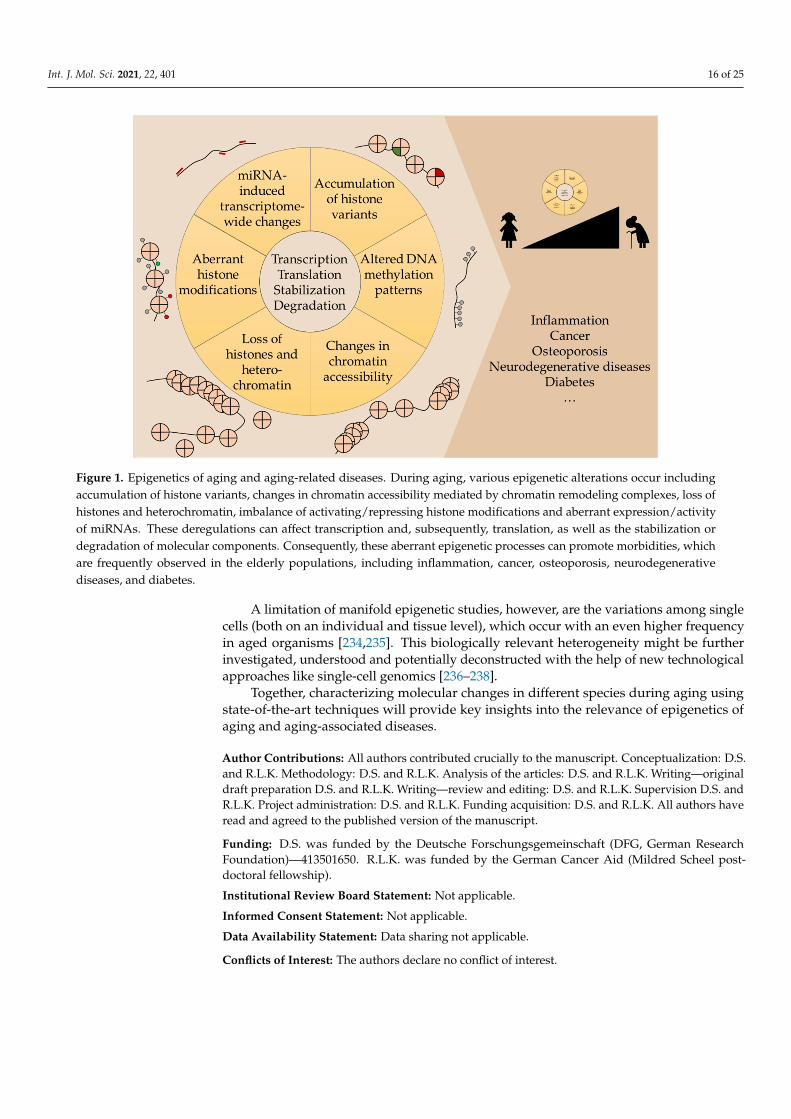
Figure 1. Epigenetics of aging and aging-related diseases. During aging, various epigenetic alterations occur including accumulation of histone variants, changes in chromatin accessibility mediated by chromatin remodeling complexes, loss of histones and heterochromatin, imbalance of activating/repressing histone modifications and aberrant expres-sion/activity of miRNAs. These deregulations can affect transcription and, subsequently, translation, as well as the stabi-lization or degradation of molecular components. Consequently, these aberrant epigenetic processes can promote mor-bidities, which are frequently observed in the elderly populations, including inflammation, cancer, osteoporosis, neuro-degenerative diseases, and diabetes.
While the precise mechanisms and connections between several epigenetic changes and human pathologies are still poorly understood, state-of-the-art next generation se-quencing methods will allow researchers to address remaining questions. For instance, chromatin accessibility using Assay for Transposase-Accessible Chromatin using se-quencing (ATAC-seq) can be coupled to ChIP-seq as well as gene expression studies (mRNA-seq) using bulk mRNA or even analyzing single cells (scRNA-seq). In addition, advances in molecular biology and cell culture approaches (for instance Clustered Reg-ularly Interspaced Short Palindromic Repeats (CRISPR)/Cas9) will be beneficial in clari-fying aging-processes across species.
An improved understanding of epigenetic mechanisms affecting longevity will be deciding crucial step towards the identification of new potential therapeutic targets. In fact, epigenetic drugs are of particular interest to the clinic due to their reversible and transient effect.
A limitation of manifold epigenetic studies, however, are the variations among sin-gle cells (both on an individual and tissue level), which occur with an even higher fre-quency in aged organisms [234,235]. This biologically relevant heterogeneity might be
Figure 1. Epigenetics of aging and aging-related diseases. During aging, various epigenetic alterations occur includingaccumulation of histone variants, changes in chromatin accessibility mediated by chromatin remodeling complexes, loss ofhistones and heterochromatin, imbalance of activating/repressing histone modifications and aberrant expression/activityof miRNAs. These deregulations can affect transcription and, subsequently, translation, as well as the stabilization ordegradation of molecular components. Consequently, these aberrant epigenetic processes can promote morbidities, whichare frequently observed in the elderly populations, including inflammation, cancer, osteoporosis, neurodegenerativediseases, and diabetes.
A limitation of manifold epigenetic studies, however, are the variations among singlecells (both on an individual and tissue level), which occur with an even higher frequencyin aged organisms [234,235]. This biologically relevant heterogeneity might be furtherinvestigated, understood and potentially deconstructed with the help of new technologicalapproaches like single-cell genomics [236–238].
Together, characterizing molecular changes in different species during aging usingstate-of-the-art techniques will provide key insights into the relevance of epigenetics ofaging and aging-associated diseases.
Author Contributions: All authors contributed crucially to the manuscript. Conceptualization: D.S.and R.L.K. Methodology: D.S. and R.L.K. Analysis of the articles: D.S. and R.L.K. Writing—originaldraft preparation D.S. and R.L.K. Writing—review and editing: D.S. and R.L.K. Supervision D.S. andR.L.K. Project administration: D.S. and R.L.K. Funding acquisition: D.S. and R.L.K. All authors haveread and agreed to the published version of the manuscript.
Funding: D.S. was funded by the Deutsche Forschungsgemeinschaft (DFG, German ResearchFoundation)—413501650. R.L.K. was funded by the German Cancer Aid (Mildred Scheel post-doctoral fellowship).
Institutional Review Board Statement: Not applicable.
Informed Consent Statement: Not applicable.
Data Availability Statement: Data sharing not applicable.
Conflicts of Interest: The authors declare no conflict of interest.
Int. J. Mol. Sci. 2021, 22, 401 17 of 25
References1. Kennedy, B.K.; Berger, S.L.; Brunet, A.; Campisi, J.; Cuervo, A.M.; Epel, E.S.; Franceschi, C.; Lithgow, G.J.; Morimoto, R.I.; Pessin,
J.E.; et al. Geroscience: Linking aging to chronic disease. Cell 2014, 159, 709–713. [CrossRef] [PubMed]2. López-Otín, C.; Blasco, M.A.; Partridge, L.; Serrano, M.; Kroemer, G. The hallmarks of aging. Cell 2013, 153, 1194–1217. [CrossRef]
[PubMed]3. Luger, K.; Mäder, A.W.; Richmond, R.K.; Sargent, D.F.; Richmond, T.J. Crystal structure of the nucleosome core particle at 2.8 A
resolution. Nature 1997, 389, 251–260. [CrossRef] [PubMed]4. Richmond, T.J.; Davey, C.A. The structure of DNA in the nucleosome core. Nature 2003, 423, 145–150. [CrossRef] [PubMed]5. Torres, I.O.; Fujimori, D.G. Functional coupling between writers, erasers and readers of histone and DNA methylation. Curr.
Opin. Struct. Biol. 2015, 35, 68–75. [CrossRef] [PubMed]6. Zhang, W.; Qu, J.; Liu, G.-H.; Belmonte, J.C.I. The ageing epigenome and its rejuvenation. Nat. Rev. Mol. Cell Biol. 2020, 21,
137–150. [CrossRef] [PubMed]7. Hu, Z.; Chen, K.; Xia, Z.; Chavez, M.; Pal, S.; Seol, J.-H.; Chen, C.-C.; Li, W.; Tyler, J.K. Nucleosome loss leads to global
transcriptional up-regulation and genomic instability during yeast aging. Genes Dev. 2014, 28, 396–408. [CrossRef]8. Liu, L.; Cheung, T.H.; Charville, G.W.; Hurgo, B.M.C.; Leavitt, T.; Shih, J.; Brunet, A.; Rando, T.A. Chromatin modifications as
determinants of muscle stem cell quiescence and chronological aging. Cell Rep. 2013, 4, 189–204. [CrossRef]9. O’Sullivan, R.J.; Kubicek, S.; Schreiber, S.L.; Karlseder, J. Reduced histone biosynthesis and chromatin changes arising from a
damage signal at telomeres. Nat. Struct. Mol. Biol. 2010, 17, 1218–1225. [CrossRef]10. Lee, J.-H.; Kim, E.W.; Croteau, D.L.; Bohr, V.A. Heterochromatin: An epigenetic point of view in aging. Exp. Mol. Med. 2020, 52,
1466–1474. [CrossRef]11. Marzluff, W.F.; Gongidi, P.; Woods, K.R.; Jin, J.; Maltais, L.J. The human and mouse replication-dependent histone genes. Genomics
2002, 80, 487–498. [CrossRef] [PubMed]12. Henikoff, S.; Smith, M.M. Histone variants and epigenetics. Cold Spring Harb. Perspect. Biol. 2015, 7, a019364. [CrossRef] [PubMed]13. Buschbeck, M.; Uribesalgo, I.; Wibowo, I.; Rué, P.; Martin, D.; Gutierrez, A.; Morey, L.; Guigó, R.; López-Schier, H.; Di Croce, L.
The histone variant macroH2A is an epigenetic regulator of key developmental genes. Nat. Struct. Mol. Biol. 2009, 16, 1074–1079.[CrossRef] [PubMed]
14. Pehrson, J.R.; Fried, V.A. MacroH2A, a core histone containing a large nonhistone region. Science 1992, 257, 1398–1400. [CrossRef][PubMed]
15. Gaspar-Maia, A.; Qadeer, Z.A.; Hasson, D.; Ratnakumar, K.; Leu, N.A.; Leroy, G.; Liu, S.; Costanzi, C.; Valle-Garcia, D.; Schaniel,C.; et al. MacroH2A histone variants act as a barrier upon reprogramming towards pluripotency. Nat. Commun. 2013, 4, 1565.[CrossRef] [PubMed]
16. Kreiling, J.A.; Tamamori-Adachi, M.; Sexton, A.N.; Jeyapalan, J.C.; Munoz-Najar, U.; Peterson, A.L.; Manivannan, J.; Rogers, E.S.;Pchelintsev, N.A.; Adams, P.D.; et al. Age-associated increase in heterochromatic marks in murine and primate tissues. Aging Cell2011, 10, 292–304. [CrossRef]
17. Tvardovskiy, A.; Schwämmle, V.; Kempf, S.J.; Rogowska-Wrzesinska, A.; Jensen, O.N. Accumulation of histone variant H3.3 withage is associated with profound changes in the histone methylation landscape. Nucleic Acids Res. 2017, 45, 9272–9289. [CrossRef]
18. Piazzesi, A.; Papic, D.; Bertan, F.; Salomoni, P.; Nicotera, P.; Bano, D. Replication-Independent Histone Variant H3.3 ControlsAnimal Lifespan through the Regulation of Pro-longevity Transcriptional Programs. Cell Rep. 2016, 17, 987–996. [CrossRef]
19. Maze, I.; Wenderski, W.; Noh, K.-M.; Bagot, R.C.; Tzavaras, N.; Purushothaman, I.; Elsässer, S.J.; Guo, Y.; Ionete, C.; Hurd, Y.L.;et al. Critical Role of Histone Turnover in Neuronal Transcription and Plasticity. Neuron 2015, 87, 77–94. [CrossRef]
20. Stefanelli, G.; Azam, A.B.; Walters, B.J.; Brimble, M.A.; Gettens, C.P.; Bouchard-Cannon, P.; Cheng, H.-Y.M.; Davidoff, A.M.;Narkaj, K.; Day, J.J.; et al. Learning and Age-Related Changes in Genome-wide H2A.Z Binding in the Mouse Hippocampus. CellRep. 2018, 22, 1124–1131. [CrossRef]
21. Biran, A.; Zada, L.; Abou Karam, P.; Vadai, E.; Roitman, L.; Ovadya, Y.; Porat, Z.; Krizhanovsky, V. Quantitative identification ofsenescent cells in aging and disease. Aging Cell 2017, 16, 661–671. [CrossRef] [PubMed]
22. Pilch, D.R.; Sedelnikova, O.A.; Redon, C.; Celeste, A.; Nussenzweig, A.; Bonner, W.M. Characteristics of gamma-H2AX foci atDNA double-strand breaks sites. Biochem. Cell Biol. 2003, 81, 123–129. [CrossRef] [PubMed]
23. Vidanes, G.M.; Bonilla, C.Y.; Toczyski, D.P. Complicated tails: Histone modifications and the DNA damage response. Cell 2005,121, 973–976. [CrossRef] [PubMed]
24. Balajee, A.S.; Geard, C.R. Replication protein A and gamma-H2AX foci assembly is triggered by cellular response to DNAdouble-strand breaks. Exp. Cell Res. 2004, 300, 320–334. [CrossRef] [PubMed]
25. Sharma, A.; Singh, K.; Almasan, A. Histone H2AX phosphorylation: A marker for DNA damage. Methods Mol. Biol. 2012, 920,613–626. [CrossRef] [PubMed]
26. Jin, B.; Li, Y.; Robertson, K.D. DNA methylation: Superior or subordinate in the epigenetic hierarchy? Genes Cancer 2011, 2,607–617. [CrossRef] [PubMed]
27. Auclair, G.; Weber, M. Mechanisms of DNA methylation and demethylation in mammals. Biochimie 2012, 94, 2202–2211. [CrossRef]28. Hyun, J.; Jung, Y. DNA Methylation in Nonalcoholic Fatty Liver Disease. Int. J. Mol. Sci. 2020, 21, 8138. [CrossRef]29. Lister, R.; Pelizzola, M.; Dowen, R.H.; Hawkins, R.D.; Hon, G.; Tonti-Filippini, J.; Nery, J.R.; Lee, L.; Ye, Z.; Ngo, Q.-M.; et al.
Human DNA methylomes at base resolution show widespread epigenomic differences. Nature 2009, 462, 315–322. [CrossRef]
Int. J. Mol. Sci. 2021, 22, 401 18 of 25
30. Schübeler, D. Function and information content of DNA methylation. Nature 2015, 517, 321–326. [CrossRef]31. He, Y.-F.; Li, B.-Z.; Li, Z.; Liu, P.; Wang, Y.; Tang, Q.; Ding, J.; Jia, Y.; Chen, Z.; Li, L.; et al. Tet-mediated formation of
5-carboxylcytosine and its excision by TDG in mammalian DNA. Science 2011, 333, 1303–1307. [CrossRef] [PubMed]32. Ito, S.; Shen, L.; Dai, Q.; Wu, S.C.; Collins, L.B.; Swenberg, J.A.; He, C.; Zhang, Y. Tet proteins can convert 5-methylcytosine to
5-formylcytosine and 5-carboxylcytosine. Science 2011, 333, 1300–1303. [CrossRef] [PubMed]33. Wu, H.; Zhang, Y. Reversing DNA methylation: Mechanisms, genomics, and biological functions. Cell 2014, 156, 45–68. [CrossRef]
[PubMed]34. Rasmussen, K.D.; Helin, K. Role of TET enzymes in DNA methylation, development, and cancer. Genes Dev. 2016, 30, 733–750.
[CrossRef]35. Hashimoto, H.; Liu, Y.; Upadhyay, A.K.; Chang, Y.; Howerton, S.B.; Vertino, P.M.; Zhang, X.; Cheng, X. Recognition and potential
mechanisms for replication and erasure of cytosine hydroxymethylation. Nucleic Acids Res. 2012, 40, 4841–4849. [CrossRef]36. Weinhouse, C.; Truong, L.; Meyer, J.N.; Allard, P. Caenorhabditis elegans as an emerging model system in environmental
epigenetics. Environ. Mol. Mutagen. 2018, 59, 560–575. [CrossRef]37. Greer, E.L.; Blanco, M.A.; Gu, L.; Sendinc, E.; Liu, J.; Aristizábal-Corrales, D.; Hsu, C.-H.; Aravind, L.; He, C.; Shi, Y. DNA
Methylation on N6-Adenine in C. elegans. Cell 2015, 161, 868–878. [CrossRef]38. Deshmukh, S.; Ponnaluri, V.C.; Dai, N.; Pradhan, S.; Deobagkar, D. Levels of DNA cytosine methylation in the Drosophila
genome. PeerJ 2018, 6, e5119. [CrossRef]39. Day, K.; Waite, L.L.; Thalacker-Mercer, A.; West, A.; Bamman, M.M.; Brooks, J.D.; Myers, R.M.; Absher, D. Differential DNA
methylation with age displays both common and dynamic features across human tissues that are influenced by CpG landscape.Genome Biol. 2013, 14, R102. [CrossRef]
40. Horvath, S. DNA methylation age of human tissues and cell types. Genome Biol. 2013, 14, R115. [CrossRef]41. Horvath, S. Erratum to: DNA methylation age of human tissues and cell types. Genome Biol. 2015, 16, 96. [CrossRef] [PubMed]42. Jylhävä, J.; Pedersen, N.L.; Hägg, S. Biological Age Predictors. EBioMedicine 2017, 21, 29–36. [CrossRef] [PubMed]43. Liu, B.; Yip, R.K.; Zhou, Z. Chromatin remodeling, DNA damage repair and aging. Curr. Genomics 2012, 13, 533–547. [CrossRef]
[PubMed]44. Clapier, C.R.; Cairns, B.R. The biology of chromatin remodeling complexes. Annu. Rev. Biochem. 2009, 78, 273–304. [CrossRef]45. Wu, S.; Ge, Y.; Huang, L.; Liu, H.; Xue, Y.; Zhao, Y. BRG1, the ATPase subunit of SWI/SNF chromatin remodeling complex,
interacts with HDAC2 to modulate telomerase expression in human cancer cells. Cell Cycle 2014, 13, 2869–2878. [CrossRef]46. Wu, S.; Ge, Y.; Li, X.; Yang, Y.; Zhou, H.; Lin, K.; Zhang, Z.; Zhao, Y. BRM-SWI/SNF chromatin remodeling complex enables
functional telomeres by promoting co-expression of TRF2 and TRF1. PLoS Genet. 2020, 16, e1008799. [CrossRef]47. Riedel, C.G.; Dowen, R.H.; Lourenco, G.F.; Kirienko, N.V.; Heimbucher, T.; West, J.A.; Bowman, S.K.; Kingston, R.E.; Dillin, A.;
Asara, J.M.; et al. DAF-16 employs the chromatin remodeller SWI/SNF to promote stress resistance and longevity. Nat. Cell Biol.2013, 15, 491–501. [CrossRef]
48. de Vaux, V.; Pfefferli, C.; Passannante, M.; Belhaj, K.; von Essen, A.; Sprecher, S.G.; Müller, F.; Wicky, C. The Caenorhabditiselegans LET-418/Mi2 plays a conserved role in lifespan regulation. Aging Cell 2013, 12, 1012–1020. [CrossRef]
49. Min, J.-N.; Tian, Y.; Xiao, Y.; Wu, L.; Li, L.; Chang, S. The mINO80 chromatin remodeling complex is required for efficient telomerereplication and maintenance of genome stability. Cell Res. 2013, 23, 1396–1413. [CrossRef]
50. Dang, W.; Sutphin, G.L.; Dorsey, J.A.; Otte, G.L.; Cao, K.; Perry, R.M.; Wanat, J.J.; Saviolaki, D.; Murakami, C.J.; Tsuchiyama, S.;et al. Inactivation of yeast Isw2 chromatin remodeling enzyme mimics longevity effect of calorie restriction via induction ofgenotoxic stress response. Cell Metab. 2014, 19, 952–966. [CrossRef]
51. Matilainen, O.; Sleiman, M.S.B.; Quiros, P.M.; Garcia, S.M.D.A.; Auwerx, J. The chromatin remodeling factor ISW-1 integratesorganismal responses against nuclear and mitochondrial stress. Nat. Commun. 2017, 8, 1818. [CrossRef] [PubMed]
52. Iwasaki, W.; Miya, Y.; Horikoshi, N.; Osakabe, A.; Taguchi, H.; Tachiwana, H.; Shibata, T.; Kagawa, W.; Kurumizaka, H.Contribution of histone N-terminal tails to the structure and stability of nucleosomes. FEBS Open Bio 2013, 3, 363–369. [CrossRef][PubMed]
53. Greer, E.L.; Shi, Y. Histone methylation: A dynamic mark in health, disease and inheritance. Nat. Rev. Genet. 2012, 13, 343–357.[CrossRef] [PubMed]
54. Greer, E.L.; Maures, T.J.; Hauswirth, A.G.; Green, E.M.; Leeman, D.S.; Maro, G.S.; Han, S.; Banko, M.R.; Gozani, O.; Brunet, A.Members of the H3K4 trimethylation complex regulate lifespan in a germline-dependent manner in C. elegans. Nature 2010, 466,383–387. [CrossRef]
55. Li, L.; Greer, C.; Eisenman, R.N.; Secombe, J. Essential functions of the histone demethylase lid. PLoS Genet. 2010, 6, e1001221.[CrossRef]
56. Pu, M.; Wang, M.; Wang, W.; Velayudhan, S.S.; Lee, S.S. Unique patterns of trimethylation of histone H3 lysine 4 are prone tochanges during aging in Caenorhabditis elegans somatic cells. PLoS Genet. 2018, 14, e1007466. [CrossRef]
57. Cruz, C.; Della Rosa, M.; Krueger, C.; Gao, Q.; Horkai, D.; King, M.; Field, L.; Houseley, J. Tri-methylation of histone H3 lysine 4facilitates gene expression in ageing cells. Elife 2018, 7. [CrossRef]
58. Sen, P.; Dang, W.; Donahue, G.; Dai, J.; Dorsey, J.; Cao, X.; Liu, W.; Cao, K.; Perry, R.; Lee, J.Y.; et al. H3K36 methylation promoteslongevity by enhancing transcriptional fidelity. Genes Dev. 2015, 29, 1362–1376. [CrossRef]
Int. J. Mol. Sci. 2021, 22, 401 19 of 25
59. Pu, M.; Ni, Z.; Wang, M.; Wang, X.; Wood, J.G.; Helfand, S.L.; Yu, H.; Lee, S.S. Trimethylation of Lys36 on H3 restricts geneexpression change during aging and impacts life span. Genes Dev. 2015, 29, 718–731. [CrossRef]
60. Su, L.; Li, H.; Huang, C.; Zhao, T.; Zhang, Y.; Ba, X.; Li, Z.; Zhang, Y.; Huang, B.; Lu, J.; et al. Muscle-Specific Histone H3K36Dimethyltransferase SET-18 Shortens Lifespan of Caenorhabditis elegans by Repressing daf-16a Expression. Cell Rep. 2018, 22,2716–2729. [CrossRef]
61. Jin, C.; Li, J.; Green, C.D.; Yu, X.; Tang, X.; Han, D.; Xian, B.; Wang, D.; Huang, X.; Cao, X.; et al. Histone demethylase UTX-1regulates C. elegans life span by targeting the insulin/IGF-1 signaling pathway. Cell Metab. 2011, 14, 161–172. [CrossRef][PubMed]
62. Kelly, J.T.; Feldman, S.E.; Ross, J.E. Quality review in the Peer Review Organization Program. Qual. Assur. Util. Rev. 1987, 2,107–110. [CrossRef] [PubMed]
63. Siebold, A.P.; Banerjee, R.; Tie, F.; Kiss, D.L.; Moskowitz, J.; Harte, P.J. Polycomb Repressive Complex 2 and Trithorax modulateDrosophila longevity and stress resistance. Proc. Natl. Acad. Sci. USA 2010, 107, 169–174. [CrossRef] [PubMed]
64. Baumgart, M.; Groth, M.; Priebe, S.; Savino, A.; Testa, G.; Dix, A.; Ripa, R.; Spallotta, F.; Gaetano, C.; Ori, M.; et al. RNA-seq of theaging brain in the short-lived fish N. furzeri—conserved pathways and novel genes associated with neurogenesis. Aging Cell2014, 13, 965–974. [CrossRef]
65. Lorbeck, M.T.; Singh, N.; Zervos, A.; Dhatta, M.; Lapchenko, M.; Yang, C.; Elefant, F. The histone demethylase Dmel\Kdm4Acontrols genes required for life span and male-specific sex determination in Drosophila. Gene 2010, 450, 8–17. [CrossRef]
66. Djeghloul, D.; Kuranda, K.; Kuzniak, I.; Barbieri, D.; Naguibneva, I.; Choisy, C.; Bories, J.-C.; Dosquet, C.; Pla, M.; Vanneaux, V.;et al. Age-Associated Decrease of the Histone Methyltransferase SUV39H1 in HSC Perturbs Heterochromatin and B LymphoidDifferentiation. Stem Cell Reports 2016, 6, 970–984. [CrossRef]
67. Sarg, B.; Koutzamani, E.; Helliger, W.; Rundquist, I.; Lindner, H.H. Postsynthetic trimethylation of histone H4 at lysine 20 inmammalian tissues is associated with aging. J. Biol. Chem. 2002, 277, 39195–39201. [CrossRef]
68. Nelson, D.M.; Jaber-Hijazi, F.; Cole, J.J.; Robertson, N.A.; Pawlikowski, J.S.; Norris, K.T.; Criscione, S.W.; Pchelintsev, N.A.;Piscitello, D.; Stong, N.; et al. Mapping H4K20me3 onto the chromatin landscape of senescent cells indicates a function in controlof cell senescence and tumor suppression through preservation of genetic and epigenetic stability. Genome Biol. 2016, 17, 158.[CrossRef]
69. Kawakami, K.; Nakamura, A.; Ishigami, A.; Goto, S.; Takahashi, R. Age-related difference of site-specific histone modifications inrat liver. Biogerontology 2009, 10, 415–421. [CrossRef]
70. Michishita, E.; McCord, R.A.; Berber, E.; Kioi, M.; Padilla-Nash, H.; Damian, M.; Cheung, P.; Kusumoto, R.; Kawahara, T.L.A.;Barrett, J.C.; et al. SIRT6 is a histone H3 lysine 9 deacetylase that modulates telomeric chromatin. Nature 2008, 452, 492–496.[CrossRef]
71. Mostoslavsky, R.; Chua, K.F.; Lombard, D.B.; Pang, W.W.; Fischer, M.R.; Gellon, L.; Liu, P.; Mostoslavsky, G.; Franco, S.; Murphy,M.M.; et al. Genomic instability and aging-like phenotype in the absence of mammalian SIRT6. Cell 2006, 124, 315–329. [CrossRef][PubMed]
72. Kawahara, T.L.A.; Michishita, E.; Adler, A.S.; Damian, M.; Berber, E.; Lin, M.; McCord, R.A.; Ongaigui, K.C.L.; Boxer, L.D.; Chang,H.Y.; et al. SIRT6 links histone H3 lysine 9 deacetylation to NF-kappaB-dependent gene expression and organismal life span. Cell2009, 136, 62–74. [CrossRef] [PubMed]
73. Kanfi, Y.; Naiman, S.; Amir, G.; Peshti, V.; Zinman, G.; Nahum, L.; Bar-Joseph, Z.; Cohen, H.Y. The sirtuin SIRT6 regulates lifespanin male mice. Nature 2012, 483, 218–221. [CrossRef] [PubMed]
74. Chen, J.; Xie, J.-J.; Jin, M.-Y.; Gu, Y.-T.; Wu, C.-C.; Guo, W.-J.; Yan, Y.-Z.; Zhang, Z.-J.; Wang, J.-L.; Zhang, X.-L.; et al. Sirt6overexpression suppresses senescence and apoptosis of nucleus pulposus cells by inducing autophagy in a model of intervertebraldisc degeneration. Cell Death Dis. 2018, 9, 56. [CrossRef] [PubMed]
75. Ozdemir, A.; Spicuglia, S.; Lasonder, E.; Vermeulen, M.; Campsteijn, C.; Stunnenberg, H.G.; Logie, C. Characterization of lysine56 of histone H3 as an acetylation site in Saccharomyces cerevisiae. J. Biol. Chem. 2005, 280, 25949–25952. [CrossRef] [PubMed]
76. Yuan, J.; Pu, M.; Zhang, Z.; Lou, Z. Histone H3-K56 acetylation is important for genomic stability in mammals. Cell Cycle 2009, 8,1747–1753. [CrossRef] [PubMed]
77. Kokkonen, P.; Rahnasto-Rilla, M.; Mellini, P.; Jarho, E.; Lahtela-Kakkonen, M.; Kokkola, T. Studying SIRT6 regulation usingH3K56 based substrate and small molecules. Eur. J. Pharm. Sci. 2014, 63, 71–76. [CrossRef]
78. Liu, X.; Li, C.; Li, Q.; Chang, H.-C.; Tang, Y.-C. SIRT7 Facilitates CENP-A Nucleosome Assembly and Suppresses IntestinalTumorigenesis. iScience 2020, 23, 101461. [CrossRef]
79. Krishnan, V.; Liu, B.; Zhou, Z. ‘Relax and Repair’ to restrain aging. Aging (Albany NY) 2011, 3, 943–954. [CrossRef]80. Xu, H.; Su, T.; Xue, Y. Histone H3 N-terminal acetylation sites especially K14 are important for rDNA silencing and aging. Sci.
Rep. 2016, 6, 21900. [CrossRef]81. Krishnan, V.; Chow, M.Z.Y.; Wang, Z.; Zhang, L.; Liu, B.; Liu, X.; Zhou, Z. Histone H4 lysine 16 hypoacetylation is associated with
defective DNA repair and premature senescence in Zmpste24-deficient mice. Proc. Natl. Acad. Sci. USA 2011, 108, 12325–12330.[CrossRef] [PubMed]
82. Barral, S.; Beltramo, R.; Salio, C.; Aimar, P.; Lossi, L.; Merighi, A. Phosphorylation of Histone H2AX in the Mouse Brain fromDevelopment to Senescence. Int. J. Mol. Sci. 2014, 15, 1554–1573. [CrossRef] [PubMed]
Int. J. Mol. Sci. 2021, 22, 401 20 of 25
83. Oh, S.; Suganuma, T.; Gogol, M.M.; Workman, J.L. Histone H3 threonine 11 phosphorylation by Sch9 and CK2 regulateschronological lifespan by controlling the nutritional stress response. Elife 2018, 7. [CrossRef] [PubMed]
84. Maleszewska, M.; Mawer, J.S.P.; Tessarz, P. Histone Modifications in Ageing and Lifespan Regulation. Curr. Mol. Biol. Rep. 2016,2, 26–35. [CrossRef]
85. Lee, J.-S.; Shukla, A.; Schneider, J.; Swanson, S.K.; Washburn, M.P.; Florens, L.; Bhaumik, S.R.; Shilatifard, A. Histone crosstalkbetween H2B monoubiquitination and H3 methylation mediated by COMPASS. Cell 2007, 131, 1084–1096. [CrossRef] [PubMed]
86. Cao, J.; Yan, Q. Histone ubiquitination and deubiquitination in transcription, DNA damage response, and cancer. Front. Oncol.2012, 2, 26. [CrossRef]
87. Yang, L.; Ma, Z.; Wang, H.; Niu, K.; Cao, Y.; Le, S.; Geng, Y.; Yang, B.; Gao, F.; Chen, Z.; et al. Ubiquitylome study identifiesincreased histone 2A ubiquitylation as an evolutionarily conserved aging biomarker. Nat. Commun. 2019, 10, 2191. [CrossRef]
88. Gao, Z.; Xu, M.S.; Barnett, T.L.; Xu, C.W. Resveratrol induces cellular senescence with attenuated mono-ubiquitination of histoneH2B in glioma cells. Biochem. Biophys. Res. Commun. 2011, 407, 271–276. [CrossRef]
89. Bushati, N.; Cohen, S.M. microRNA functions. Annu. Rev. Cell Dev. Biol. 2007, 23, 175–205. [CrossRef]90. Kinser, H.E.; Pincus, Z. MicroRNAs as modulators of longevity and the aging process. Hum. Genet. 2020, 139, 291–308. [CrossRef]91. Lai, W.-F.; Lin, M.; Wong, W.-T. Tackling Aging by Using miRNA as a Target and a Tool. Trends Mol. Med. 2019, 25, 673–684.
[CrossRef] [PubMed]92. Kozomara, A.; Griffiths-Jones, S. miRBase: Annotating high confidence microRNAs using deep sequencing data. Nucleic Acids
Res. 2014, 42, D68–D73. [CrossRef] [PubMed]93. Lewis, B.P.; Burge, C.B.; Bartel, D.P. Conserved seed pairing, often flanked by adenosines, indicates that thousands of human
genes are microRNA targets. Cell 2005, 120, 15–20. [CrossRef] [PubMed]94. Boehm, M.; Slack, F. A developmental timing microRNA and its target regulate life span in C. elegans. Science 2005, 310, 1954–1957.
[CrossRef] [PubMed]95. Chawla, G.; Deosthale, P.; Childress, S.; Wu, Y.-C.; Sokol, N.S. A let-7-to-miR-125 MicroRNA Switch Regulates Neuronal Integrity
and Lifespan in Drosophila. PLoS Genet. 2016, 12, e1006247. [CrossRef] [PubMed]96. Huan, T.; Chen, G.; Liu, C.; Bhattacharya, A.; Rong, J.; Chen, B.H.; Seshadri, S.; Tanriverdi, K.; Freedman, J.E.; Larson, M.G.; et al.
Age-associated microRNA expression in human peripheral blood is associated with all-cause mortality and age-related traits.Aging Cell 2018, 17. [CrossRef]
97. Hanahan, D.; Weinberg, R.A. The hallmarks of cancer. Cell 2000, 100, 57–70. [CrossRef]98. Hanahan, D.; Weinberg, R.A. Hallmarks of cancer: The next generation. Cell 2011, 144, 646–674. [CrossRef]99. Flavahan, W.A.; Gaskell, E.; Bernstein, B.E. Epigenetic plasticity and the hallmarks of cancer. Science 2017, 357, eaal2380.
[CrossRef]100. Nebbioso, A.; Tambaro, F.P.; Dell’Aversana, C.; Altucci, L. Cancer epigenetics: Moving forward. PLoS Genet. 2018, 14, e1007362.
[CrossRef]101. Baylin, S.B.; Jones, P.A. A decade of exploring the cancer epigenome—Biological and translational implications. Nat. Rev. Cancer
2011, 11, 726–734. [CrossRef] [PubMed]102. Cancer Genome Atlas Network. Comprehensive molecular portraits of human breast tumours. Nature 2012, 490, 61–70. [CrossRef]
[PubMed]103. Karsli-Ceppioglu, S.; Dagdemir, A.; Judes, G.; Ngollo, M.; Penault-Llorca, F.; Pajon, A.; Bignon, Y.-J.; Bernard-Gallon, D. Epigenetic
mechanisms of breast cancer: An update of the current knowledge. Epigenomics 2014, 6, 651–664. [CrossRef] [PubMed]104. Yegnasubramanian, S.; Kowalski, J.; Gonzalgo, M.L.; Zahurak, M.; Piantadosi, S.; Walsh, P.C.; Bova, G.S.; de Marzo, A.M.; Isaacs,
W.B.; Nelson, W.G. Hypermethylation of CpG islands in primary and metastatic human prostate cancer. Cancer Res. 2004, 64,1975–1986. [CrossRef] [PubMed]
105. Wu, Y.; Sarkissyan, M.; Vadgama, J.V. Epigenetics in Breast and Prostate Cancer. Methods Mol. Biol. 2015, 1238, 425–466. [CrossRef]106. Xu, W.; Yang, H.; Liu, Y.; Yang, Y.; Wang, P.; Kim, S.-H.; Ito, S.; Yang, C.; Wang, P.; Xiao, M.-T.; et al. Oncometabolite
2-hydroxyglutarate is a competitive inhibitor of α-ketoglutarate-dependent dioxygenases. Cancer Cell 2011, 19, 17–30. [CrossRef]107. Su, R.; Dong, L.; Li, C.; Nachtergaele, S.; Wunderlich, M.; Qing, Y.; Deng, X.; Wang, Y.; Weng, X.; Hu, C.; et al. R-2HG Exhibits
Anti-tumor Activity by Targeting FTO/m6A/MYC/CEBPA Signaling. Cell 2018, 172, 90–105.e23. [CrossRef]108. Winters, A.C.; Bernt, K.M. MLL-Rearranged Leukemias-An Update on Science and Clinical Approaches. Front. Pediatr. 2017, 5, 4.
[CrossRef]109. Sen, P.; Lan, Y.; Li, C.Y.; Sidoli, S.; Donahue, G.; Dou, Z.; Frederick, B.; Chen, Q.; Luense, L.J.; Garcia, B.A.; et al. Histone
Acetyltransferase p300 Induces De Novo Super-Enhancers to Drive Cellular Senescence. Mol. Cell 2019, 73, 684–698.e8. [CrossRef]110. Cheng, G.; Liu, F.; Asai, T.; Lai, F.; Man, N.; Xu, H.; Chen, S.; Greenblatt, S.; Hamard, P.-J.; Ando, K.; et al. Loss of p300 Accelerates
MDS-associated Leukemogenesis. Leukemia 2016, 31, 1382–1390. [CrossRef]111. Picaud, S.; Fedorov, O.; Thanasopoulou, A.; Leonards, K.; Jones, K.; Meier, J.; Olzscha, H.; Monteiro, O.; Martin, S.; Philpott, M.;
et al. Generation of a Selective Small Molecule Inhibitor of the CBP/p300 Bromodomain for Leukemia Therapy. Cancer Res. 2015,75, 5106–5119. [CrossRef] [PubMed]
112. Comet, I.; Riising, E.M.; Leblanc, B.; Helin, K. Maintaining cell identity: PRC2-mediated regulation of transcription and cancer.Nat. Rev. Cancer 2016, 16, 803–810. [CrossRef] [PubMed]
Int. J. Mol. Sci. 2021, 22, 401 21 of 25
113. McCabe, M.T.; Ott, H.M.; Ganji, G.; Korenchuk, S.; Thompson, C.; van Aller, G.S.; Liu, Y.; Graves, A.P.; Della Pietra, A.; Diaz,E.; et al. EZH2 inhibition as a therapeutic strategy for lymphoma with EZH2-activating mutations. Nature 2012, 492, 108–112.[CrossRef] [PubMed]
114. Sashida, G.; Iwama, A. Multifaceted role of the polycomb-group gene EZH2 in hematological malignancies. Int. J. Hematol. 2017,105, 23–30. [CrossRef] [PubMed]
115. Mozhui, K.; Pandey, A.K. Conserved effect of aging on DNA methylation and association with EZH2 polycomb protein in miceand humans. Mech. Ageing Dev. 2017, 162, 27–37. [CrossRef] [PubMed]
116. Singh, A.A.; Mandoli, A.; Prange, K.H.M.; Laakso, M.; Martens, J.H.A. AML associated oncofusion proteins PML-RARA, AML1-ETO and CBFB-MYH11 target RUNX/ETS-factor binding sites to modulate H3ac levels and drive leukemogenesis. Oncotarget2017, 8, 12855–12865. [CrossRef] [PubMed]
117. Skoda, R.C.; Schwaller, J. Dual roles of EZH2 in acute myeloid leukemia. J. Exp. Med. 2019, 216, 725–727. [CrossRef]118. Okugawa, Y.; Grady, W.M.; Goel, A. Epigenetic Alterations in Colorectal Cancer: Emerging Biomarkers. Gastroenterology 2015,
149, 1204–1225.e12. [CrossRef]119. Lao, V.V.; Grady, W.M. Epigenetics and colorectal cancer. Nat. Rev. Gastroenterol. Hepatol. 2011, 8, 686–700. [CrossRef]120. Guo, Y.; Wang, M.; Jia, X.; Zhu, H.; Zhi, Y.; Yuan, L. Wnt signaling pathway upregulates DNMT1 to trigger NHERF1 promoter
hypermethylation in colon cancer. Oncol. Rep. 2018, 40, 1165–1173. [CrossRef]121. Song, J.; Du, Z.; Ravasz, M.; Dong, B.; Wang, Z.; Ewing, R.M. A Protein Interaction between β-Catenin and Dnmt1 Regulates Wnt
Signaling and DNA Methylation in Colorectal Cancer Cells. Mol. Cancer Res. 2015, 13, 969–981. [CrossRef] [PubMed]122. Yang, Z.-H.; Dang, Y.-Q.; Ji, G. Role of epigenetics in transformation of inflammation into colorectal cancer. World J. Gastroenterol.
2019, 25, 2863–2877. [CrossRef] [PubMed]123. Foran, E.; Garrity-Park, M.M.; Mureau, C.; Newell, J.; Smyrk, T.C.; Limburg, P.J.; Egan, L.J. Upregulation of DNA
methyltransferase-mediated gene silencing, anchorage-independent growth, and migration of colon cancer cells by interleukin-6.Mol. Cancer Res. 2010, 8, 471–481. [CrossRef] [PubMed]
124. Fearon, E.R.; Vogelstein, B. A genetic model for colorectal tumorigenesis. Cell 1990, 61, 759–767. [CrossRef]125. Ciccarone, F.; Malavolta, M.; Calabrese, R.; Guastafierro, T.; Bacalini, M.G.; Reale, A.; Franceschi, C.; Capri, M.; Hervonen, A.;
Hurme, M.; et al. Age-dependent expression of DNMT1 and DNMT3B in PBMCs from a large European population enrolled inthe MARK-AGE study. Aging Cell 2016, 15, 755–765. [CrossRef]
126. Yung, R.; Ray, D.; Eisenbraun, J.K.; Deng, C.; Attwood, J.; Eisenbraun, M.D.; Johnson, K.; Miller, R.A.; Hanash, S.; Richardson,B. Unexpected effects of a heterozygous dnmt1 null mutation on age-dependent DNA hypomethylation and autoimmunity.J. Gerontol. A Biol. Sci. Med. Sci. 2001, 56, B268–B276. [CrossRef]
127. Laird, P.W.; Jackson-Grusby, L.; Fazeli, A.; Dickinson, S.L.; Jung, W.E.; Li, E.; Weinberg, R.A.; Jaenisch, R. Suppression of intestinalneoplasia by DNA hypomethylation. Cell 1995, 81, 197–205. [CrossRef]
128. Gerecke, C.; Scholtka, B.; Löwenstein, Y.; Fait, I.; Gottschalk, U.; Rogoll, D.; Melcher, R.; Kleuser, B. Hypermethylation of ITGA4,TFPI2 and VIMENTIN promoters is increased in inflamed colon tissue: Putative risk markers for colitis-associated cancer. J. CancerRes. Clin. Oncol. 2015, 141, 2097–2107. [CrossRef]
129. Requena, D.O.; Garcia-Buitrago, M. Molecular Insights Into Colorectal Carcinoma. Arch. Med. Res. 2020, 51, 839–844. [CrossRef]130. Fleisher, A.S.; Esteller, M.; Harpaz, N.; Leytin, A.; Rashid, A.; Xu, Y.; Liang, J.; Stine, O.C.; Yin, J.; Zou, T.T.; et al. Microsatellite
instability in inflammatory bowel disease-associated neoplastic lesions is associated with hypermethylation and diminishedexpression of the DNA mismatch repair gene, hMLH1. Cancer Res. 2000, 60, 4864–4868.
131. Arnold, C.N.; Goel, A.; Boland, C.R. Role of hMLH1 promoter hypermethylation in drug resistance to 5-fluorouracil in colorectalcancer cell lines. Int. J. Cancer 2003, 106, 66–73. [CrossRef] [PubMed]
132. Dienstmann, R.; Vermeulen, L.; Guinney, J.; Kopetz, S.; Tejpar, S.; Tabernero, J. Consensus molecular subtypes and the evolutionof precision medicine in colorectal cancer. Nat. Rev. Cancer 2017, 17, 79–92. [CrossRef] [PubMed]
133. Franceschi, C.; Bonafè, M.; Valensin, S.; Olivieri, F.; de Luca, M.; Ottaviani, E.; de Benedictis, G. Inflamm-aging. An evolutionaryperspective on immunosenescence. Ann. N. Y. Acad. Sci. 2000, 908, 244–254. [CrossRef] [PubMed]
134. Fulop, T.; Larbi, A.; Dupuis, G.; Le Page, A.; Frost, E.H.; Cohen, A.A.; Witkowski, J.M.; Franceschi, C. Immunosenescence andInflamm-Aging As Two Sides of the Same Coin: Friends or Foes? Front. Immunol. 2017, 8, 1960. [CrossRef]
135. Sullivan, K.E.; Reddy, A.B.M.; Dietzmann, K.; Suriano, A.R.; Kocieda, V.P.; Stewart, M.; Bhatia, M. Epigenetic regulation of tumornecrosis factor alpha. Mol. Cell. Biol. 2007, 27, 5147–5160. [CrossRef]
136. Ray, D.; Yung, R. Immune Senescence, Epigenetics and Autoimmunity. Clin. Immunol. 2018, 196, 59–63. [CrossRef]137. Wang, H.; Feng, H.; Sun, J.; Zhou, Y.; Zhu, G.; Wu, S.; Bao, W. Age-associated changes in DNA methylation and expression of the
TNFα gene in pigs. Genes Genet. Syst. 2018, 93, 191–198. [CrossRef]138. Tilstra, J.S.; Clauson, C.L.; Niedernhofer, L.J.; Robbins, P.D. NF-κB in Aging and Disease. Aging Dis. 2011, 2, 449–465.139. Li, Y.; Reddy, M.A.; Miao, F.; Shanmugam, N.; Yee, J.-K.; Hawkins, D.; Ren, B.; Natarajan, R. Role of the histone H3 lysine
4 methyltransferase, SET7/9, in the regulation of NF-kappaB-dependent inflammatory genes. Relevance to diabetes andinflammation. J. Biol. Chem. 2008, 283, 26771–26781. [CrossRef]
140. Wang, Y.; Wang, L.; Wen, X.; Hao, D.; Zhang, N.; He, G.; Jiang, X. NF-κB signaling in skin aging. Mech. Ageing Dev. 2019, 184,111160. [CrossRef]
Int. J. Mol. Sci. 2021, 22, 401 22 of 25
141. Choi, Y.J.; Moon, K.M.; Chung, K.W.; Jeong, J.W.; Park, D.; Kim, D.H.; Yu, B.P.; Chung, H.Y. The underlying mechanism ofproinflammatory NF-κB activation by the mTORC2/Akt/IKKα pathway during skin aging. Oncotarget 2016, 7, 52685–52694.[CrossRef] [PubMed]
142. Johnsen, S.A. The enigmatic role of H2Bub1 in cancer. FEBS Lett. 2012, 586, 1592–1601. [CrossRef] [PubMed]143. Tarcic, O.; Pateras, I.S.; Cooks, T.; Shema, E.; Kanterman, J.; Ashkenazi, H.; Boocholez, H.; Hubert, A.; Rotkopf, R.; Baniyash,
M.; et al. RNF20 Links Histone H2B Ubiquitylation with Inflammation and Inflammation-Associated Cancer. Cell Rep. 2016, 14,1462–1476. [CrossRef] [PubMed]
144. Kosinsky, R.L.; Chua, R.L.; Qui, M.; Saul, D.; Mehlich, D.; Ströbel, P.; Schildhaus, H.-U.; Wegwitz, F.; Faubion, W.A.; Johnsen, S.A.Loss of RNF40 Decreases NF-κB Activity in Colorectal Cancer Cells and Reduces Colitis Burden in Mice. J. Crohns. Colitis 2019,13, 362–373. [CrossRef]
145. Yuan, J.; Zhang, F.; Niu, R. Multiple regulation pathways and pivotal biological functions of STAT3 in cancer. Sci. Rep. 2015, 5,17663. [CrossRef]
146. Zimmers, T.A.; Fishel, M.L.; Bonetto, A. STAT3 in the Systemic Inflammation of Cancer Cachexia. Semin. Cell Dev. Biol. 2016, 54,28–41. [CrossRef]
147. Takeshima, H.; Niwa, T.; Yamashita, S.; Takamura-Enya, T.; Iida, N.; Wakabayashi, M.; Nanjo, S.; Abe, M.; Sugiyama, T.; Kim, Y.-J.;et al. TET repression and increased DNMT activity synergistically induce aberrant DNA methylation. J. Clin. Investig. 2020, 130,5370–5379. [CrossRef]
148. Jiang, S.; Yan, W.; Wang, S.E.; Baltimore, D. Dual mechanisms of posttranscriptional regulation of Tet2 by Let-7 microRNA inmacrophages. Proc. Natl. Acad. Sci. USA 2019, 116, 12416–12421. [CrossRef]
149. Horvath, S.; Erhart, W.; Brosch, M.; Ammerpohl, O.; von Schönfels, W.; Ahrens, M.; Heits, N.; Bell, J.T.; Tsai, P.-C.; Spector, T.D.;et al. Obesity accelerates epigenetic aging of human liver. Proc. Natl. Acad. Sci. USA 2014, 111, 15538–15543. [CrossRef]
150. Hanson, B.M.; Tao, X.; Zhan, Y.; Jenkins, T.G.; Morin, S.J.; Scott, R.T.; Seli, E.U. Young women with poor ovarian response exhibitepigenetic age acceleration based on evaluation of white blood cells using a DNA methylation-derived age prediction model.Hum. Reprod. 2020, 35, 2579–2588. [CrossRef]
151. Morin, S.J.; Tao, X.; Marin, D.; Zhan, Y.; Landis, J.; Bedard, J.; Scott, R.T.; Seli, E. DNA methylation-based age prediction andtelomere length in white blood cells and cumulus cells of infertile women with normal or poor response to ovarian stimulation.Aging (Albany NY) 2018, 10, 3761–3773. [CrossRef] [PubMed]
152. Marioni, R.E.; Shah, S.; McRae, A.F.; Chen, B.H.; Colicino, E.; Harris, S.E.; Gibson, J.; Henders, A.K.; Redmond, P.; Cox, S.R.; et al.DNA methylation age of blood predicts all-cause mortality in later life. Genome Biol. 2015, 16, 25. [CrossRef] [PubMed]
153. Johnson, P.L.F.; Goronzy, J.J.; Antia, R. A population biological approach to understanding the maintenance and loss of the T-cellrepertoire during aging. Immunology 2014, 142, 167–175. [CrossRef] [PubMed]
154. Stevenson, A.J.; McCartney, D.L.; Harris, S.E.; Taylor, A.M.; Redmond, P.; Starr, J.M.; Zhang, Q.; McRae, A.F.; Wray, N.R.;Spires-Jones, T.L.; et al. Trajectories of inflammatory biomarkers over the eighth decade and their associations with immune cellprofiles and epigenetic ageing. Clin. Epigenetics 2018, 10, 159. [CrossRef]
155. Quinn, K.M.; Fox, A.; Harland, K.L.; Russ, B.E.; Li, J.; Nguyen, T.H.O.; Loh, L.; Olshanksy, M.; Naeem, H.; Tsyganov, K.; et al.Age-Related Decline in Primary CD8+ T Cell Responses Is Associated with the Development of Senescence in Virtual MemoryCD8+ T Cells. Cell Rep. 2018, 23, 3512–3524. [CrossRef]
156. Lau, E.Y.M.; Carroll, E.C.; Callender, L.A.; Hood, G.A.; Berryman, V.; Pattrick, M.; Finer, S.; Hitman, G.A.; Ackland, G.L.; Henson,S.M. Type 2 diabetes is associated with the accumulation of senescent T cells. Clin. Exp. Immunol. 2019, 197, 205–213. [CrossRef]
157. Bektas, A.; Schurman, S.H.; Sen, R.; Ferrucci, L. Human T cell immunosenescence and inflammation in aging. J. Leukoc. Biol. 2017,102, 977–988. [CrossRef]
158. Surace, A.E.A.; Hedrich, C.M. The Role of Epigenetics in Autoimmune/Inflammatory Disease. Front. Immunol. 2019, 10, 1525.[CrossRef]
159. Mikami, N.; Kawakami, R.; Sakaguchi, S. New Treg cell-based therapies of autoimmune diseases: Towards antigen-specificimmune suppression. Curr. Opin. Immunol. 2020, 67, 36–41. [CrossRef]
160. Dozmorov, M.G.; Coit, P.; Maksimowicz-McKinnon, K.; Sawalha, A.H. Age-associated DNA methylation changes in naive CD4+T cells suggest an evolving autoimmune epigenotype in aging T cells. Epigenomics 2017, 9, 429–445. [CrossRef]
161. Hofmann, S.R.; Kubasch, A.S.; Ioannidis, C.; Rösen-Wolff, A.; Girschick, H.J.; Morbach, H.; Hedrich, C.M. Altered expression ofIL-10 family cytokines in monocytes from CRMO patients result in enhanced IL-1β expression and release. Clin. Immunol. 2015,161, 300–307. [CrossRef] [PubMed]
162. Wu, Z.-X.; Cao, L.; Li, X.-W.; Jiang, W.; Li, X.-Y.; Xu, J.; Wang, F.; Chen, G.-H. Accelerated Deficits of Spatial Learning and MemoryResulting From Prenatal Inflammatory Insult Are Correlated With Abnormal Phosphorylation and Methylation of Histone 3 inCD-1 Mice. Front. Aging Neurosci. 2019, 11. [CrossRef] [PubMed]
163. Cakouros, D.; Gronthos, S. Epigenetic Regulators of Mesenchymal Stem/Stromal Cell Lineage Determination. Curr. Osteoporos.Rep. 2020, 18, 597–605. [CrossRef] [PubMed]
164. Sepulveda, H.; Villagra, A.; Montecino, M. Tet-Mediated DNA Demethylation Is Required for SWI/SNF-Dependent ChromatinRemodeling and Histone-Modifying Activities That Trigger Expression of the Sp7 Osteoblast Master Gene during MesenchymalLineage Commitment. Mol. Cell. Biol. 2017, 37, e00177-17. [CrossRef]
Int. J. Mol. Sci. 2021, 22, 401 23 of 25
165. Wang, R.; Wang, Y.; Zhu, L.; Liu, Y.; Li, W. Epigenetic Regulation in Mesenchymal Stem Cell Aging and Differentiation andOsteoporosis. Stem Cells Int. 2020, 2020, 1–17. [CrossRef]
166. Bork, S.; Pfister, S.; Witt, H.; Horn, P.; Korn, B.; Ho, A.D.; Wagner, W. DNA methylation pattern changes upon long-term cultureand aging of human mesenchymal stromal cells. Aging Cell 2010, 9, 54–63. [CrossRef]
167. Cakouros, D.; Hemming, S.; Gronthos, K.; Liu, R.; Zannettino, A.; Shi, S.; Gronthos, S. Specific functions of TET1 and TET2 inregulating mesenchymal cell lineage determination. Epigenetics Chromatin 2019, 12, 3. [CrossRef]
168. Yang, R.; Yu, T.; Kou, X.; Gao, X.; Chen, C.; Liu, D.; Zhou, Y.; Shi, S. Tet1 and Tet2 maintain mesenchymal stem cell homeostasisvia demethylation of the P2rX7 promoter. Nat. Commun. 2018, 9, 2143. [CrossRef]
169. Ko, N.-Y.; Chen, L.-R.; Chen, K.-H. The Role of Micro RNA and Long-Non-Coding RNA in Osteoporosis. Int. J. Mol. Sci. 2020, 21,4886. [CrossRef]
170. Gontier, G.; Iyer, M.; Shea, J.M.; Bieri, G.; Wheatley, E.G.; Ramalho-Santos, M.; Villeda, S.A. Tet2 Rescues Age-Related RegenerativeDecline and Enhances Cognitive Function in the Adult Mouse Brain. Cell Rep. 2018, 22, 1974–1981. [CrossRef]
171. Ren, J.; Huang, D.; Li, R.; Wang, W.; Zhou, C. Control of mesenchymal stem cell biology by histone modifications. Cell Biosci.2020, 10, 1–15. [CrossRef] [PubMed]
172. Jing, H.; Liao, L.; An, Y.; Su, X.; Liu, S.; Shuai, Y.; Zhang, X.; Jin, Y. Suppression of EZH2 Prevents the Shift of Osteoporotic MSCFate to Adipocyte and Enhances Bone Formation During Osteoporosis. Mol. Ther. 2016, 24, 217–229. [CrossRef] [PubMed]
173. Ma, J.; Lin, X.; Chen, C.; Li, S.; Zhang, S.; Chen, Z.; Li, D.; Zhao, F.; Yang, C.; Yin, C.; et al. Circulating miR-181c-5p and miR-497-5pAre Potential Biomarkers for Prognosis and Diagnosis of Osteoporosis. J. Clin. Endocrinol. Metab. 2020, 105, 1445–1460. [CrossRef][PubMed]
174. Bone, H.G.; Wagman, R.B.; Brandi, M.L.; Brown, J.P.; Chapurlat, R.; Cummings, S.R.; Czerwinski, E.; Fahrleitner-Pammer, A.;Kendler, D.L.; Lippuner, K.; et al. 10 years of denosumab treatment in postmenopausal women with osteoporosis: Results fromthe phase 3 randomised FREEDOM trial and open-label extension. Lancet Diabetes Endocrinol. 2017, 5, 513–523. [CrossRef]
175. de Nigris, F.; Ruosi, C.; Colella, G.; Napoli, C. Epigenetic therapies of osteoporosis. Bone 2020, 142, 115680. [CrossRef]176. Dolinar, A.; Ravnik-Glavac, M.; Glavac, D. Epigenetic mechanisms in amyotrophic lateral sclerosis: A short review. Mech. Ageing
Dev. 2018, 174, 103–110. [CrossRef]177. Ebbert, M.T.W.; Ross, C.A.; Pregent, L.J.; Lank, R.J.; Zhang, C.; Katzman, R.B.; Jansen-West, K.; Song, Y.; Da Rocha, E.L.;
Palmucci, C.; et al. Conserved DNA methylation combined with differential frontal cortex and cerebellar expression distinguishesC9orf72-associated and sporadic ALS, and implicates SERPINA1 in disease. Acta Neuropathol. 2017, 134, 715–728. [CrossRef]
178. Tecalco-Cruz, A.C.; Ramírez-Jarquín, J.O.; Alvarez-Sánchez, M.E.; Zepeda-Cervantes, J. Epigenetic basis of Alzheimer disease.World J. Biol. Chem. 2020, 11, 62–75. [CrossRef]
179. Saez-Atienzar, S.; Masliah, E. Cellular senescence and Alzheimer disease: The egg and the chicken scenario. Nat. Rev. Neurosci.2020, 21, 433–444. [CrossRef]
180. Valenta, T.; Lukas, J.; Doubravska, L.; Fafilek, B.; Korinek, V. HIC1 attenuates Wnt signaling by recruitment of TCF-4 andbeta-catenin to the nuclear bodies. EMBO J. 2006, 25, 2326–2337. [CrossRef]
181. Nativio, R.; Donahue, G.; Berson, A.; Lan, Y.; Amlie-Wolf, A.; Tuzer, F.; Toledo, J.B.; Gosai, S.J.; Gregory, B.D.; Torres, C.; et al.Dysregulation of the epigenetic landscape of normal aging in Alzheimer’s disease. Nat. Neurosci. 2018, 21, 497–505. [CrossRef][PubMed]
182. Chen, W.Y.; Wang, D.H.; Yen, R.C.; Luo, J.; Gu, W.; Baylin, S.B. Tumor suppressor HIC1 directly regulates SIRT1 to modulatep53-dependent DNA-damage responses. Cell 2005, 123, 437–448. [CrossRef] [PubMed]
183. Lu, T.; Aron, L.; Zullo, J.; Pan, Y.; Kim, H.; Chen, Y.; Yang, T.-H.; Kim, H.-M.; Drake, D.; Liu, X.S.; et al. Addendum: REST andstress resistance in ageing and Alzheimer’s disease. Nature 2016, 540, 470. [CrossRef] [PubMed]
184. Lu, T.; Aron, L.; Zullo, J.; Pan, Y.; Kim, H.; Chen, Y.; Yang, T.-H.; Kim, H.-M.; Drake, D.; Liu, X.S.; et al. REST and Stress Resistancein Aging and Alzheimer’s Disease. Nature 2014, 507, 448–454. [CrossRef]
185. Hwang, J.-Y.; Zukin, R.S. REST, a master transcriptional regulator in neurodegenerative disease. Curr. Opin. Neurobiol. 2018, 48,193–200. [CrossRef]
186. Hou, T.-Y.; Zhou, Y.; Zhu, L.-S.; Wang, X.; Pang, P.; Wang, D.-Q.; Liuyang, Z.-Y.; Man, H.; Lu, Y.; Zhu, L.-Q.; et al. Correctingabnormalities in miR-124/PTPN1 signaling rescues tau pathology in Alzheimer’s disease. J. Neurochem. 2020, 154, 441–457.[CrossRef]
187. Williams, J.; Smith, F.; Kumar, S.; Vijayan, M.; Reddy, P.H. Are microRNAs true sensors of ageing and cellular senescence? AgeingRes. Rev. 2017, 35, 350–363. [CrossRef]
188. Kumar, P.; Dezso, Z.; MacKenzie, C.; Oestreicher, J.; Agoulnik, S.; Byrne, M.; Bernier, F.; Yanagimachi, M.; Aoshima, K.; Oda, Y.Circulating miRNA biomarkers for Alzheimer’s disease. PLoS ONE 2013, 8, e69807. [CrossRef]
189. Labbé, C.; Lorenzo-Betancor, O.; Ross, O.A. Epigenetic regulation in Parkinson’s disease. Acta Neuropathol. 2016, 132, 515–530.[CrossRef]
190. Jiang, K.; Rocha, S.; Westling, A.; Kesarimangalam, S.; Dorfman, K.D.; Wittung-Stafshede, P.; Westerlund, F. Alpha-SynucleinModulates the Physical Properties of DNA. Chemistry 2018, 24, 15685–15690. [CrossRef]
191. Sharma, A.; Osato, N.; Liu, H.; Asthana, S.; Dakal, T.C.; Ambrosini, G.; Bucher, P.; Schmitt, I.; Wüllner, U. Common geneticvariants associated with Parkinson’s disease display widespread signature of epigenetic plasticity. Sci. Rep. 2019, 9, 1–19.[CrossRef] [PubMed]
Int. J. Mol. Sci. 2021, 22, 401 24 of 25
192. Vermunt, M.W.; Reinink, P.; Korving, J.; de Bruijn, E.; Creyghton, P.M.; Basak, O.; Geeven, G.; Toonen, P.W.; Lansu, N.; Meunier, C.;et al. Large-scale identification of coregulated enhancer networks in the adult human brain. Cell Rep. 2014, 9, 767–779. [CrossRef][PubMed]
193. Chen, X.; Scholz, S.W. Identification of new α-synuclein regulator by nontraditional drug development pipeline. Mov. Disord.2018, 33, 402. [CrossRef] [PubMed]
194. Mittal, S.; Bjørnevik, K.; Soon Im, D.; Flierl, A.; Dong, X.; Locascio, J.J.; Abo, K.M.; Long, E.; Jin, M.; Xu, B.; et al. β2-Adrenoreceptoris a Regulator of the α-Synuclein Gene Driving Risk of Parkinson’s Disease. Science 2017, 357, 891–898. [CrossRef]
195. Esteves, A.R.; Palma, A.M.; Gomes, R.; Santos, D.; Silva, D.F.; Cardoso, S.M. Acetylation as a major determinant to microtubule-dependent autophagy: Relevance to Alzheimer’s and Parkinson disease pathology. Biochim. Biophys. Acta Mol. Basis Dis. 2019,1865, 2008–2023. [CrossRef]
196. Gebremedhin, K.G.; Rademacher, D.J. Histone H3 Acetylation in the Postmortem Parkinson’s Disease Primary Motor Cortex.Neurosci. Lett. 2016, 627, 121–125. [CrossRef]
197. Kontopoulos, E.; Parvin, J.D.; Feany, M.B. Alpha-synuclein acts in the nucleus to inhibit histone acetylation and promoteneurotoxicity. Hum. Mol. Genet. 2006, 15, 3012–3023. [CrossRef]
198. Sun, L.; Zhang, J.; Chen, W.; Chen, Y.; Zhang, X.; Yang, M.; Xiao, M.; Ma, F.; Yao, Y.; Ye, M.; et al. Attenuation of epigeneticregulator SMARCA4 and ERK-ETS signaling suppresses aging-related dopaminergic degeneration. Aging Cell 2020, 19, e13210.[CrossRef]
199. Kahn, S.E.; Hull, R.L.; Utzschneider, K.M. Mechanisms linking obesity to insulin resistance and type 2 diabetes. Nature 2006, 444,840–846. [CrossRef]
200. Desiderio, A.; Spinelli, R.; Ciccarelli, M.; Nigro, C.; Miele, C.; Beguinot, F.; Raciti, G.A. Epigenetics: Spotlight on type 2 diabetesand obesity. J. Endocrinol. Investig. 2016, 39, 1095–1103. [CrossRef]
201. Guzik, T.J.; Cosentino, F. Epigenetics and Immunometabolism in Diabetes and Aging. Antioxid. Redox Signal. 2018, 29, 257–274.[CrossRef] [PubMed]
202. Ling, C.; Rönn, T. Epigenetics in Human Obesity and Type 2 Diabetes. Cell Metab. 2019, 29, 1028–1044. [CrossRef] [PubMed]203. Heijmans, B.T.; Tobi, E.W.; Stein, A.D.; Putter, H.; Blauw, G.J.; Susser, E.S.; Slagboom, P.E.; Lumey, L.H. Persistent epigenetic
differences associated with prenatal exposure to famine in humans. Proc. Natl. Acad. Sci. USA 2008, 105, 17046–17049. [CrossRef][PubMed]
204. Lumey, L.H.; Stein, A.D.; Kahn, H.S.; van der Pal-de Bruin, K.M.; Blauw, G.J.; Zybert, P.A.; Susser, E.S. Cohort profile: The DutchHunger Winter families study. Int. J. Epidemiol. 2007, 36, 1196–1204. [CrossRef]
205. Li, Y.; Tollefsbol, T.O. p16INK4a Suppression by Glucose Restriction Contributes to Human Cellular Lifespan Extension throughSIRT1-Mediated Epigenetic and Genetic Mechanisms. PLoS ONE 2011, 6, e17421. [CrossRef]
206. Daniel, M.; Tollefsbol, T.O. Epigenetic linkage of aging, cancer and nutrition. J. Exp. Biol. 2015, 218, 59–70. [CrossRef]207. Kapahi, P.; Kaeberlein, M.; Hansen, M. Dietary restriction and lifespan: Lessons from invertebrate models. Ageing Res. Rev. 2017,
39, 3–14. [CrossRef]208. Joseph, S.V.; Edirisinghe, I.; Burton-Freeman, B.M. Fruit Polyphenols: A Review of Anti-inflammatory Effects in Humans. Crit.
Rev. Food Sci. Nutr. 2016, 56, 419–444. [CrossRef]209. Hou, X.; Rooklin, D.; Fang, H.; Zhang, Y. Resveratrol serves as a protein-substrate interaction stabilizer in human SIRT1 activation.
Sci. Rep. 2016, 6, 38186. [CrossRef]210. Li, Y.-R.; Li, S.; Lin, C.-C. Effect of resveratrol and pterostilbene on aging and longevity. Biofactors 2018, 44, 69–82. [CrossRef]211. Yessenkyzy, A.; Saliev, T.; Zhanaliyeva, M.; Masoud, A.-R.; Umbayev, B.; Sergazy, S.; Krivykh, E.; Gulyayev, A.; Nurgozhin, T.
Polyphenols as Caloric-Restriction Mimetics and Autophagy Inducers in Aging Research. Nutrients 2020, 12, 1344. [CrossRef][PubMed]
212. Almén, M.S.; Nilsson, E.K.; Jacobsson, J.A.; Kalnina, I.; Klovins, J.; Fredriksson, R.; Schiöth, H.B. Genome-wide analysis revealsDNA methylation markers that vary with both age and obesity. Gene 2014, 548, 61–67. [CrossRef] [PubMed]
213. Hannon, E.; Knox, O.; Sugden, K.; Burrage, J.; Wong, C.C.Y.; Belsky, D.W.; Corcoran, D.L.; Arseneault, L.; Moffitt, T.E.; Caspi, A.;et al. Characterizing genetic and environmental influences on variable DNA methylation using monozygotic and dizygotic twins.PLoS Genet. 2018, 14, e1007544. [CrossRef] [PubMed]
214. Molina-Serrano, D.; Kyriakou, D.; Kirmizis, A. Histone Modifications as an Intersection Between Diet and Longevity. Front. Genet.2019, 10, 192. [CrossRef] [PubMed]
215. Xu, X.; Su, S.; Barnes, V.A.; de Miguel, C.; Pollock, J.; Ownby, D.; Shi, H.; Zhu, H.; Snieder, H.; Wang, X. A genome-widemethylation study on obesity: Differential variability and differential methylation. Epigenetics 2013, 8, 522–533. [CrossRef][PubMed]
216. Andreas, E.; Reid, M.; Zhang, W.; Moley, K.H. The effect of maternal high-fat/high-sugar diet on offspring oocytes and earlyembryo development. Mol. Hum. Reprod. 2019, 25, 717–728. [CrossRef]
217. Zheng, S.; Li, Q.; Zhang, Y.; Balluff, Z.; Pan, Y.-X. Histone deacetylase 3 (HDAC3) participates in the transcriptional repression ofthe p16 (INK4a) gene in mammary gland of the female rat offspring exposed to an early-life high-fat diet. Epigenetics 2012, 7,183–190. [CrossRef]
218. Zhang, X.; Xu, G.B.; Zhou, D.; Pan, Y.-X. High-fat diet modifies expression of hepatic cellular senescence gene p16(INK4a) throughchromatin modifications in adult male rats. Genes Nutr. 2018, 13, 6. [CrossRef]
Int. J. Mol. Sci. 2021, 22, 401 25 of 25
219. Jacobsen, S.C.; Brøns, C.; Bork-Jensen, J.; Ribel-Madsen, R.; Yang, B.; Lara, E.; Hall, E.; Calvanese, V.; Nilsson, E.; Jørgensen, S.W.;et al. Effects of short-term high-fat overfeeding on genome-wide DNA methylation in the skeletal muscle of healthy young men.Diabetologia 2012, 55, 3341–3349. [CrossRef]
220. Hall, E.; Dekker Nitert, M.; Volkov, P.; Malmgren, S.; Mulder, H.; Bacos, K.; Ling, C. The effects of high glucose exposure on globalgene expression and DNA methylation in human pancreatic islets. Mol. Cell. Endocrinol. 2018, 472, 57–67. [CrossRef]
221. Sommese, L.; Benincasa, G.; Lanza, M.; Sorriento, A.; Schiano, C.; Lucchese, R.; Alfano, R.; Nicoletti, G.F.; Napoli, C. Novelepigenetic-sensitive clinical challenges both in type 1 and type 2 diabetes. J. Diabetes Complicat. 2018, 32, 1076–1084. [CrossRef][PubMed]
222. Sandovici, I.; Hammerle, C.M.; Cooper, W.N.; Smith, N.H.; Tarry-Adkins, J.L.; Dunmore, B.J.; Bauer, J.; Andrews, S.R.; Yeo, G.S.H.;Ozanne, S.E.; et al. Ageing is associated with molecular signatures of inflammation and type 2 diabetes in rat pancreatic islets.Diabetologia 2016, 59, 502–511. [CrossRef] [PubMed]
223. Dayeh, T.; Volkov, P.; Salö, S.; Hall, E.; Nilsson, E.; Olsson, A.H.; Kirkpatrick, C.L.; Wollheim, C.B.; Eliasson, L.; Rönn, T.; et al.Genome-wide DNA methylation analysis of human pancreatic islets from type 2 diabetic and non-diabetic donors identifiescandidate genes that influence insulin secretion. PLoS Genet. 2014, 10, e1004160. [CrossRef] [PubMed]
224. Baker, D.J.; Jin, F.; van Deursen, J.M. The yin and yang of the Cdkn2a locus in senescence and aging. Cell Cycle 2008, 7, 2795–2802.[CrossRef]
225. Kong, Y.; Sharma, R.B.; Nwosu, B.U.; Alonso, L.C. Islet biology, the CDKN2A/B locus and type 2 diabetes risk. Diabetologia 2016,59, 1579–1593. [CrossRef]
226. Chen, H.; Gu, X.; Su, I.; Bottino, R.; Contreras, J.L.; Tarakhovsky, A.; Kim, S.K. Polycomb protein Ezh2 regulates pancreaticbeta-cell Ink4a/Arf expression and regeneration in diabetes mellitus. Genes Dev. 2009, 23, 975–985. [CrossRef]
227. Avrahami, D.; Li, C.; Zhang, J.; Schug, J.; Avrahami, R.; Rao, S.; Stadler, M.B.; Burger, L.; Schübeler, D.; Glaser, B.; et al. Aging-Dependent Demethylation of Regulatory Elements Correlates with Chromatin State and Improved β Cell Function. Cell Metab.2015, 22, 619–632. [CrossRef]
228. Zhang, H.; Pollin, T.I. Epigenetics Variation and Pathogenesis in Diabetes. Curr. Diab. Rep. 2018, 18, 121. [CrossRef]229. Miao, F.; Chen, Z.; Zhang, L.; Liu, Z.; Wu, X.; Yuan, Y.-C.; Natarajan, R. Profiles of epigenetic histone post-translational
modifications at type 1 diabetes susceptible genes. J. Biol. Chem. 2012, 287, 16335–16345. [CrossRef]230. Rakyan, V.K.; Beyan, H.; Down, T.A.; Hawa, M.I.; Maslau, S.; Aden, D.; Daunay, A.; Busato, F.; Mein, C.A.; Manfras, B.; et al.
Identification of type 1 diabetes-associated DNA methylation variable positions that precede disease diagnosis. PLoS Genet. 2011,7, e1002300. [CrossRef]
231. Vasu, S.; Kumano, K.; Darden, C.M.; Rahman, I.; Lawrence, M.C.; Naziruddin, B. MicroRNA Signatures as Future Biomarkers forDiagnosis of Diabetes States. Cells 2019, 8, 1533. [CrossRef] [PubMed]
232. Taheri, M.; Eghtedarian, R.; Ghafouri-Fard, S.; Omrani, M.D. Non-coding RNAs and type 2 diabetes mellitus. Arch. Physiol.Biochem. 2020, 1–10. [CrossRef] [PubMed]
233. Kamalden, T.A.; Macgregor-Das, A.M.; Kannan, S.M.; Dunkerly-Eyring, B.; Khaliddin, N.; Xu, Z.; Fusco, A.P.; Yazib, S.A.; Chow,R.C.; Duh, E.J.; et al. Exosomal MicroRNA-15a Transfer from the Pancreas Augments Diabetic Complications by InducingOxidative Stress. Antioxid. Redox Signal. 2017, 27, 913–930. [CrossRef] [PubMed]
234. Bell, C.G.; Lowe, R.; Adams, P.D.; Baccarelli, A.A.; Beck, S.; Bell, J.T.; Christensen, B.C.; Gladyshev, V.N.; Heijmans, B.T.; Horvath,S.; et al. DNA methylation aging clocks: Challenges and recommendations. Genome Biol. 2019, 20, 249. [CrossRef] [PubMed]
235. Pal, S.; Tyler, J.K. Epigenetics and aging. Sci. Adv. 2016, 2, e1600584. [CrossRef] [PubMed]236. Povinelli, B.J.; Rodriguez-Meira, A.; Mead, A.J. Single cell analysis of normal and leukemic hematopoiesis. Mol. Aspects Med.
2018, 59, 85–94. [CrossRef]237. Guo, M.; Peng, Y.; Gao, A.; Du, C.; Herman, J.G. Epigenetic heterogeneity in cancer. Biomark. Res. 2019, 7, 23. [CrossRef]238. Wu, J.; Tzanakakis, E.S. Deconstructing stem cell population heterogeneity: Single-cell analysis and modeling approaches.
Biotechnol. Adv. 2013, 31, 1047–1062. [CrossRef]


